 E-submission
Search
- Page Path
- HOME > Search
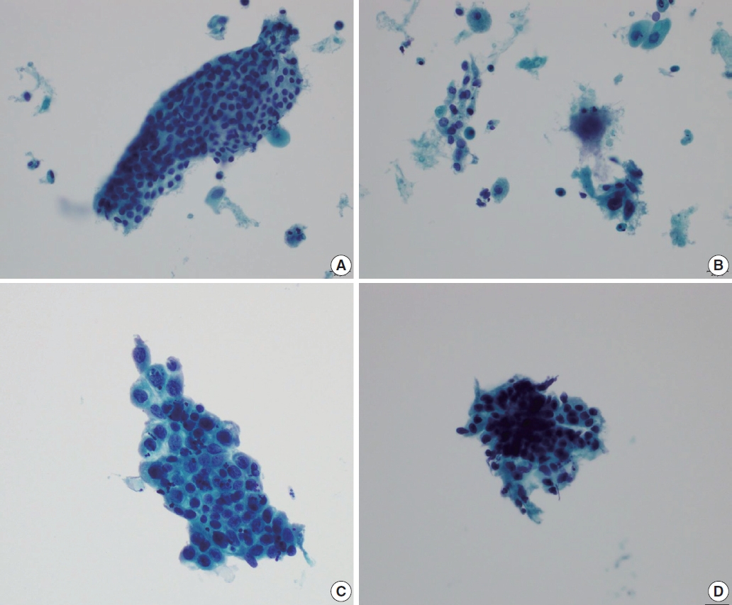
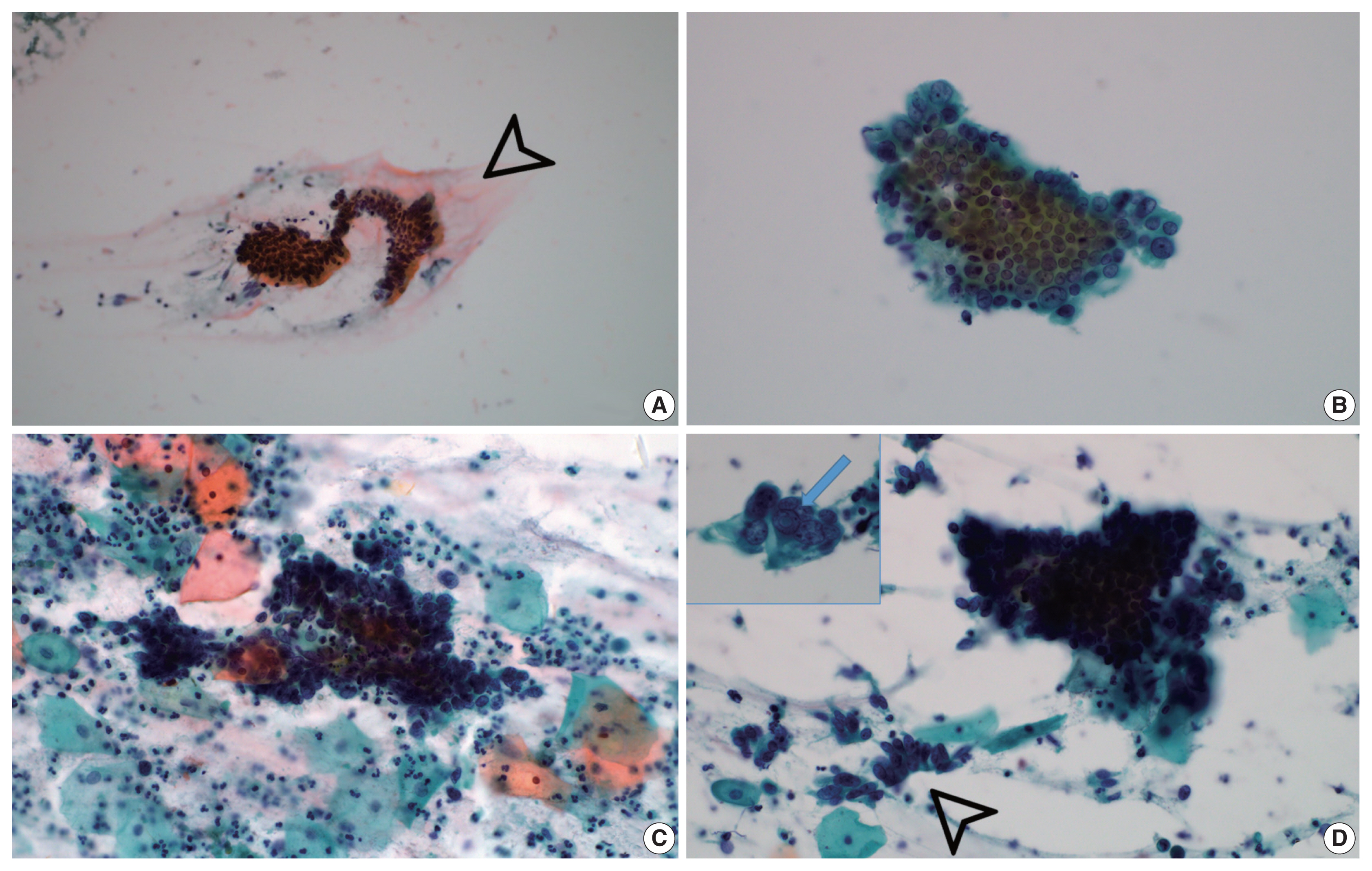
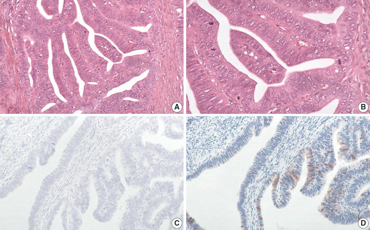
- Trouble-makers in cytologic interpretation of the uterine cervix
- Eunah Shin, Jaeeun Yu, Soon Won Hong
- J Pathol Transl Med. 2023;57(3):139-146. Published online May 15, 2023
- DOI: https://doi.org/10.4132/jptm.2023.04.25
- 12,068 View
- 476 Download
- 3 Web of Science
- 4 Crossref
-
 Abstract
 PDF
PDF - The development and standardization of cytologic screening of the uterine cervix has dramatically decreased the prevalence of squamous cell carcinoma of the uterine cervix. Advances in the understanding of biology of human papillomavirus have contributed to upgrading the histologic diagnosis of the uterine cervix; however, cytologic screening that should triage those that need further management still poses several difficulties in interpretation. Cytologic features of high grade intraepithelial squamous lesion (HSIL) mimics including atrophy, immature metaplasia, and transitional metaplasia, and glandular lesion masquerades including tubal metaplasia and HSIL with glandular involvement are described with accentuation mainly on the differential points. When the cytologic features lie in a gray zone between the differentials, the most important key to the more accurate interpretation is sticking to the very basics of cytology; screening the background and cellular architecture, and then scrutinizing the nuclear and cytoplasmic details.
-
Citations
Citations to this article as recorded by
- Cytology–Biopsy Concordance in High-Risk Human Papillomavirus–Positive Women with Abnormal Cytology Findings: Menopause-Stratified Analysis
Isik Sozen, Gozde Sahin, Yuksel Ulu, Dilara Yitiz, Basak Ozge Kayan, Ilkbal Temel Yuksel
Medicina.2026; 62(4): 631. CrossRef - Risk of cervical stenosis after cervical excision in postmenopausal patients
Eva Hauge, Line Winther Gustafson, Mette Tranberg, Pinar Bor
European Journal of Obstetrics & Gynecology and Reproductive Biology.2025; 308: 208. CrossRef - Pitfalls in Gynecological Cytology: Review of the Common and Less Frequent Entities in Pap Test
Danijela Vrdoljak-Mozetič, Snježana Štemberger-Papić, Damjana Verša Ostojić, Roberta Rubeša, Marko Klarić, Senija Eminović
Acta Cytologica.2024; 68(3): 281. CrossRef - Cytological features of human papillomavirus‐infected immature squamous metaplastic cells from cervical intraepithelial neoplasia grade 2
Mitsuaki Okodo, Kaori Okayama, Koji Teruya, Ruku Shinohara, Shuichi Mizuno, Rei Settsu, Yasuyoshi Ishii, Masahiko Fujii, Hirokazu Kimura, Mizue Oda
Journal of Medical Virology.2023;[Epub] CrossRef
- Cytology–Biopsy Concordance in High-Risk Human Papillomavirus–Positive Women with Abnormal Cytology Findings: Menopause-Stratified Analysis
- Cytopathologic features of human papillomavirus–independent, gastric-type endocervical adenocarcinoma
- Min-Kyung Yeo, Go Eun Bae, Dong-Hyun Kim, In-Ock Seong, Kwang-Sun Suh
- J Pathol Transl Med. 2022;56(5):260-269. Published online September 13, 2022
- DOI: https://doi.org/10.4132/jptm.2022.07.05
- 6,845 View
- 167 Download
- 5 Web of Science
- 5 Crossref
-
Abstract
PDF
- Background
Gastric-type endocervical adenocarcinoma (GEA) is unrelated to human papillomavirus (HPV) infection and is clinically aggressive compared with HPV-associated usual-type endocervical adenocarcinoma (UEA). The cytological diagnosis falls short of a definitive diagnosis of GEA and is often categorized as atypical glandular cells (AGCs). To improve cytologic recognition, cytological findings of HPV-independent GEA were analyzed and the results compared with HPV-associated UEA.
Methods
Cervical Papanicolaou (Pap) smears from eight patients with a histopathologic diagnosis of GEA and 12 control cases of UEA were reviewed. All slides were conventionally prepared and/or liquid-based prepared (ThinPrep) and stained following the Pap method. A mucinous background, architectural, nuclear, and cytoplasmic features were analyzed and compared with UEA.
Results
Preoperative cytologic diagnoses of the eight GEA cases were AGCs, favor neoplastic in three cases, adenocarcinoma in situ in one case, and adenocarcinoma in four cases. Cytologically, monolayered honeycomb-like sheets (p = .002) of atypical endocervical cells with vacuolar granular cytoplasm (p = .001) were extensive in GEA, and three-dimensional clusters (p = .010) were extensive in UEA. Although the differences were not statistically significant, background mucin (p = .058), vesicular nuclei (p = .057), and golden-brown intracytoplasmic mucin (p = .089) were also discriminatory findings for GEA versus UEA.
Conclusions
Although GEA is difficult to diagnose on cytologic screening, GEA can be recognized based on cytologic features of monolayered honeycomb sheets of atypical endocervical cells with abundant vacuolar cytoplasm and some golden-brown intracytoplasmic mucin. UEA cases are characterized by three-dimensional clusters. -
Citations
Citations to this article as recorded by- Gastric-Type Cervical Adenocarcinoma: Clinicopathologic Features, Molecular Landscape, and Therapeutic Challenges
Hiroshi Yoshida, Daiki Higuchi, Waku Takigawa, Nao Kikkawa, Taro Yamanaka, Ayaka Nagao, Mayumi Kobayashi-Kato, Masaya Uno, Mitsuya Ishikawa, Kouya Shiraishi
Journal of Personalized Medicine.2026; 16(2): 72. CrossRef - A Comparative Analysis of Usual- and Gastric-Type Cervical Adenocarcinoma in a Japanese Population Reveals Distinct Clinicopathological and Molecular Features with Prognostic and Therapeutic Insights
Umme Farzana Zahan, Hasibul Islam Sohel, Kentaro Nakayama, Masako Ishikawa, Mamiko Nagase, Sultana Razia, Kosuke Kanno, Hitomi Yamashita, Shahataj Begum Sonia, Satoru Kyo
International Journal of Molecular Sciences.2025; 26(15): 7469. CrossRef - Diagnostic value of cytology in detecting human papillomavirus–independent cervical malignancies: a nation-wide study in Korea
Hye-Ra Jung, Junyoung Shin, Chong Woo Yoo, Eun Na Kim, Cheol Lee, Kyeongmin Kim, Ho-chang Lee, Yonghee Lee, Ji Hye Kim, Soo Jin Jung, Yumin Chung, Joo Yeon Kim, Hye Eun Park, Tae Hoen Kim, Wonae Lee, Min-Sun Cho, Ran Hong, Yoon Jung Choi, Younghee Choi, Y
Journal of Pathology and Translational Medicine.2025; 59(6): 444. CrossRef - Risk Factors Affecting Clinical Outcomes of Low-risk Early-stage Human Papillomavirus–Associated Endocervical Adenocarcinoma Treated by Surgery Alone: Application of Silva Pattern
Bong Kyung Bae, Hyunsik Bae, Won Kyung Cho, Byoung-Gie Kim, Chel Hun Choi, Tae-Joong Kim, Yoo-Young Lee, Jeong-Won Lee, Hyun-Soo Kim, Won Park
International Journal of Gynecological Pathology.2024; 43(5): 447. CrossRef - Tall‐columnar glandular cells in SurePath™ liquid‐based cytology Pap sample: Learning from mimics/pitfalls
Nalini Gupta, Vanita Jain, Radhika Srinivasan, Tulika Singh
Cytopathology.2024; 35(4): 510. CrossRef
- Gastric-Type Cervical Adenocarcinoma: Clinicopathologic Features, Molecular Landscape, and Therapeutic Challenges
- Evaluation of human papillomavirus (HPV) prediction using the International Endocervical Adenocarcinoma Criteria and Classification system, compared to p16 immunohistochemistry and HPV RNA in-situ hybridization
- Hezhen Ren, Jennifer Pors, Christine Chow, Monica Ta, Simona Stolnicu, Robert Soslow, David Huntsman, Lynn Hoang
- J Pathol Transl Med. 2020;54(6):480-488. Published online August 31, 2020
- DOI: https://doi.org/10.4132/jptm.2020.07.18
- 9,099 View
- 175 Download
- 11 Web of Science
- 11 Crossref
-
Abstract
PDF
- Background
The International Endocervical Adenocarcinoma Criteria and Classification (IECC) separated endocervical adenocarcinomas into human papillomavirus (HPV) associated (HPVA) and non–HPV-associated (NHPVA) categories by morphology alone. Our primary objective was to assess the accuracy of HPV prediction by the IECC system compared to p16 immunohistochemistry and HPV RNA in-situ hybridization (RISH). Our secondary goal was to directly compare p16 and HPV RISH concordance.
Methods
Cases were classified by IECC and stained for p16 and HPV RISH on tissue microarray, with discordant p16/HPV RISH cases re-stained on whole tissue sections. Remaining discordant cases (p16/HPV, IECC/p16, IECC/HPV discordances) were re-reviewed by the original pathologists (n = 3) and external expert pathologists (n = 2) blinded to the p16 and HPV RISH results. Final IECC diagnosis was assigned upon independent agreement between all reviewers.
Results
One hundred and eleven endocervical adenocarcinomas were classified originally into 94 HPVA and 17 NHPVA cases. p16 and HPV RISH was concordant in 108/111 cases (97%) independent of the IECC. HPV RISH and p16 was concordant with IECC in 103/111 (93%) and 106/111 (95%), respectively. After expert review, concordance improved to 107/111 (96%) for HPV RISH. After review of the eight discordant cases, one remained as HPVA, four were reclassified to NHPVA from HPVA, two were unclassifiable, and one possibly represented a mixed usual and gastric-type adenocarcinoma.
Conclusions
p16 and HPV RISH have excellent concordance in endocervical adenocarcinomas, and IECC can predict HPV status in most cases. Focal apical mitoses and apoptotic debris on original review led to the misclassification of several NHPVA as HPVA. -
Citations
Citations to this article as recorded by- Role of human papillomavirus status in the classification, diagnosis, and prognosis of malignant cervical epithelial tumors and precursor lesions
Simona Stolnicu
Die Pathologie.2026; 47(S1): 97. CrossRef - EdgeNeXt-SEDP for cervical adenocarcinoma HPV-associated and non-HPV-associated diagnosis and decision support
Qi Chen, Hao Wang, Hao Zhang, Zhenkun Zhu, Xi Wei
Life Sciences.2025; 380: 123931. CrossRef - Cytology and histology of endocervical glandular lesions: a review with emphasis on recent developments
Natalie Banet, Karen L. Talia
Pathology.2025; 57(7): 817. CrossRef - Joint detection of multiple HPV-testing technologies and evaluation of clinicopathological characteristics discriminate between HPV-independent and low-copy HPV-associated cervical squamous cell carcinoma (CSCC) -an analysis of 3869 cases
Linghui Lu, Tianqi Liu, Shunni Wang, Jing Li, Feiran Zhang, Yan Ning, Yiqin Wang
Gynecologic Oncology.2023; 170: 59. CrossRef - Incidence and Clinicopathologic Characteristics of Human Papillomavirus–independent Invasive Squamous Cell Carcinomas of the Cervix
Simona Stolnicu, Douglas Allison, Aaron M. Praiss, Basile Tessier-Cloutier, Amir Momeni Boroujeni, Jessica Flynn, Alexia Iasonos, Rene Serrette, Lien Hoang, Andrei Patrichi, Cristina Terinte, Anna Pesci, Claudia Mateoiu, Ricardo R. Lastra, Takako Kiyokawa
American Journal of Surgical Pathology.2023; 47(12): 1376. CrossRef - Testing Algorithms for the Diagnosis of Malignant Glandular Tumors of the Uterine Cervix Histotyped per the International Endocervical Adenocarcinoma Criteria and Classification (IECC) System
Máire A. Duggan, Qiuli Duan, Ruth M. Pfeiffer, Mary Anne Brett, Sandra Lee, Mustapha Abubakar, Martin Köbel, Monica Rodriguez, Aylin Sar
Applied Immunohistochemistry & Molecular Morphology.2022; 30(2): 91. CrossRef - Local and Metastatic Relapses in a Young Woman with Papillary Squamous Cell Carcinoma of the Uterine Cervix
Ha Young Woo, Hyun-Soo Kim
Diagnostics.2022; 12(3): 599. CrossRef - Clinical correlation of lymphovascular invasion and Silva pattern of invasion in early-stage endocervical adenocarcinoma: proposed binary Silva classification system
Simona Stolnicu, Lien Hoang, Noorah Almadani, Louise De Brot, Glauco Baiocchi, Graziele Bovolim, Maria Jose Brito, Georgia Karpathiou, Antonio Ieni, Esther Guerra, Takako Kiyokawa, Pavel Dundr, Carlos Parra-Herran, Sofia Lérias, Ana Felix, Andres Roma, An
Pathology.2022; 54(5): 548. CrossRef - Reproducibility of Morphologic Parameters of the International Endocervical Adenocarcinoma Criteria and Classification System and Correlation With Clinicopathologic Parameters: A Multi-Institutional Study
Pinar Bulutay, Nihan Haberal, Özlem Özen, Özlem Erdem, Emine H. Zeren, İbrahim Kulac, Çagatay Taskiran, Dogan Vatansever, Ali Ayhan, Nilgün Kapucuoğlu
International Journal of Gynecological Pathology.2022; 41(5): 447. CrossRef - HPV-Negative Cervical Cancer: A Narrative Review
Francesca Arezzo, Gennaro Cormio, Vera Loizzi, Gerardo Cazzato, Viviana Cataldo, Claudio Lombardi, Giuseppe Ingravallo, Leonardo Resta, Ettore Cicinelli
Diagnostics.2021; 11(6): 952. CrossRef - International Endocervical Adenocarcinoma Criteria and Classification (IECC): An Independent Cohort With Clinical and Molecular Findings
Hezhen Ren, Noorah Almadani, Jennifer Pors, Samuel Leung, Julie Ho, Christine Chow, Monica Ta, Kay J. Park, Simona Stolnicu, Robert Soslow, David Huntsman, Blake C. Gilks, Lynn Hoang
International Journal of Gynecological Pathology.2021; 40(6): 533. CrossRef
- Role of human papillomavirus status in the classification, diagnosis, and prognosis of malignant cervical epithelial tumors and precursor lesions
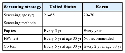
- Clinical management of abnormal Pap tests: differences between US and Korean guidelines
- Seyeon Won, Mi Kyoung Kim, Seok Ju Seong
- J Pathol Transl Med. 2020;54(3):213-219. Published online April 15, 2020
- DOI: https://doi.org/10.4132/jptm.2020.03.11
- 15,286 View
- 165 Download
- 2 Web of Science
- 2 Crossref
-
Abstract
PDF
- Cervical cancer has been the most common gynecological cancer in Korea but has become a preventable disease with regular screening and proper vaccination. If regular screening is provided, cervical cancer does not progress to more than carcinoma in situ, due to its comparatively long precancerous duration (years to decades). In 2012, the American Society for Colposcopy and Cervical Pathology published guidelines to aid clinicians in managing women with abnormal Papanicolaou (Pap) tests, and they soon became the standard in the United States. Not long thereafter, the Korean Society of Gynecologic Oncology and the Korean Society for Cytopathology published practical guidelines to reflect the specific situation in Korea. The detailed screening guidelines and management options in the case of abnormal Pap test results are sometimes the same and sometimes different in the United States and Korean guidelines. In this article, we summarize the differences between the United States and Korean guidelines in order to facilitate physicians’ proper management of abnormal Pap test results.
-
Citations
Citations to this article as recorded by- Analysis of HR-HPV Infection Concordance Rates in Cervical and Urine Specimens; Proposal of Additional Cervical Screening Process for Women Who Refuse Invasive Cervical Sampling
Dong Hyeok Kim, Hyunwoo Jin, Kyung Eun Lee
Journal of Personalized Medicine.2022; 12(12): 1949. CrossRef - Analysis of HR-HPV Prevalence among Unvaccinated Busan Women
Dong Hyeok Kim, Kyung Eun Lee
Biomedical Science Letters.2022; 28(4): 229. CrossRef
- Analysis of HR-HPV Infection Concordance Rates in Cervical and Urine Specimens; Proposal of Additional Cervical Screening Process for Women Who Refuse Invasive Cervical Sampling
- Current Status of and Perspectives on Cervical Cancer Screening in Korea
- Sung-Chul Lim, Chong Woo Yoo
- J Pathol Transl Med. 2019;53(4):210-216. Published online May 16, 2019
- DOI: https://doi.org/10.4132/jptm.2019.04.11
- 14,434 View
- 278 Download
- 12 Web of Science
- 13 Crossref
-
Abstract
PDF
- Since the introduction of the Papanicolaou (Pap) smear system in 1943, cervicovaginal cytology has been used as a standard screening test for cervical cancer. The dissemination of this test contributed to reductions of the incidence and mortality of cervical cancer worldwide. In Korea, regular health check-ups for industrial workers and their family members were introduced in 1988 and were performed as part of the National Cancer Screening Program in 1999. As a result, the incidence of cervical cancer in Korea has been steadily decreasing. However, about 800 cases of cervical cancer-related deaths are reported each year due to false-negative test results. Hence, new screening methods have been proposed. Liquid-based cytology (LBC) was introduced in 1996 to overcome the limitations of conventional Pap smears. Since then, other LBC methods have been developed and utilized, including the human papilloma virus test—a method with higher sensitivity that requires fewer screenings. In this study, we review current issues and future perspectives related to cervical cancer screening in Korea.
-
Citations
Citations to this article as recorded by- Practice of Cytopathology in Korea: A 40‐Year Evolution Through Standardization, Digital Transformation, and Global Partnership
Yosep Chong, Ran Hong, Hyeong Ju Kwon, Haeryoung Kim, Lucia Kim, Soon Jae Kim, Yoon Jung Choi
Diagnostic Cytopathology.2026; 54(2): 146. CrossRef - A Study on the Workload of Cytotechnologists: Focus on Commercial Laboratories
Eun-Suk PARK
Korean Journal of Clinical Laboratory Science.2025; 57(2): 228. CrossRef - Metastatic Cervical Cancer in the Asia-Pacific Region: Current Treatment Landscape and Barriers
Jeffrey Chee-Hong Goh, Chyong-Huey Lai, Efren Javier Domingo, Jae Hoon Kim, Carmel Spiteri, Danny Hsu, Soo Yeon Ihm, Peng Peng
Cancer Research Communications.2025; 5(8): 1429. CrossRef - Mathematical Assessment of the Roles of Vaccination and Pap Screening on the Burden of HPV and Related Cancers in Korea
Soyoung Park, Hyunah Lim, Abba B. Gumel
Bulletin of Mathematical Biology.2025;[Epub] CrossRef - A questionnaire study on disparity of cervical cancer prevention programs in Asia‐Oceania
Ka Yu Tse, Kimio Ushijima, Ai Ling Tan, Perapong Intasorn, Jitendra Pariyar, Chih‐Long Chang, Efren J. Domingo, Hiralal Konar, Suresh Kumarasamy, Brahmana Askandar Tjokroprawiro, Sarikapan Wilailak
Journal of Obstetrics and Gynaecology Research.2023; 49(4): 1230. CrossRef - Current state of cytopathology residency training: a Korean national survey of pathologists
Uiju Cho, Tae Jung Kim, Wan Seop Kim, Kyo Young Lee, Hye Kyoung Yoon, Hyun Joo Choi
Journal of Pathology and Translational Medicine.2023; 57(2): 95. CrossRef - Meeting the challenges of cervical cancer screening and HPV vaccination in the UK
Roxanne Westwood, Joanna Lavery
Primary Health Care.2022; 32(01): 22. CrossRef - Local and Metastatic Relapses in a Young Woman with Papillary Squamous Cell Carcinoma of the Uterine Cervix
Ha Young Woo, Hyun-Soo Kim
Diagnostics.2022; 12(3): 599. CrossRef - Serum Human Epididymis Protein 4 as a Prognostic Marker in Cervical Cancer
Woo Yeon Hwang, Dong Hoon Suh, Kidong Kim, Yong Beom Kim, Jae Hong No
Cancer Control.2022;[Epub] CrossRef - HPV detection and/or cytological diagnostics
Sanja Milenković
Glasnik javnog zdravlja.2022; 96(3): 313. CrossRef - Clinical management of abnormal Pap tests: differences between US and Korean guidelines
Seyeon Won, Mi Kyoung Kim, Seok Ju Seong
Journal of Pathology and Translational Medicine.2020; 54(3): 213. CrossRef - Current status of cytopathology practices in Korea: annual report on the Continuous Quality Improvement program of the Korean Society for Cytopathology for 2018
Yosep Chong, Haeyoen Jung, Jung-Soo Pyo, Soon Won Hong, Hoon Kyu Oh
Journal of Pathology and Translational Medicine.2020; 54(4): 318. CrossRef - Cytomorphological Features of Hyperchromatic Crowded Groups in Liquid-Based Cervicovaginal Cytology: A Single Institutional Experience
Youngeun Lee, Cheol Lee, In Ae Park, Hyoung Jin An, Haeryoung Kim
Journal of Pathology and Translational Medicine.2019; 53(6): 393. CrossRef
- Practice of Cytopathology in Korea: A 40‐Year Evolution Through Standardization, Digital Transformation, and Global Partnership
- Colloid Carcinoma of the Uterine Cervix and Its Immunohistochemical Analysis: A Case Report
- Nermin Koc, Sevcan Arzu Arinkan, Nurver Ozel Ozbay, Selcuk Selcuk
- J Pathol Transl Med. 2018;52(1):56-60. Published online January 15, 2018
- DOI: https://doi.org/10.4132/jptm.2017.04.08
- 8,946 View
- 136 Download
-
Abstract
PDF
- Colloid carcinoma, which is a very rare tumor of the uterine cervix, is composed of an excessive amount of mucus and a relative paucity of tumoral glandular cells within them. Herein, we report a rare case of colloid carcinoma of the cervix with adenocarcinoma in situ (AIS), intestinal and usual types, and endocervical adenocarcinoma (usual type) components. We also discuss the morphological and immunohistochemical characteristics of this tumor. A 51-year-old woman was referred to our outpatient clinic with the symptom of genital bleeding lasting for 5 months. She had a cervix surrounded by an irregular tumor with a diameter of 5 cm. The colloid carcinoma cells were positive for MUC2, MUC5AC, and cytokeratin (CK) 7, focal positive for CDX2, and negative for MUC6 and CK20. Also, the intestinal type AIS showed a similar staining pattern. Colloid carcinoma cells producing mucin showed an intestinal phenotype and AIS. The intestinal type can be considered as a precursor lesion of colloid carcinoma.
- Comparison of Analytical and Clinical Performance of HPV 9G DNA Chip, PANArray HPV Genotyping Chip, and Hybrid-Capture II Assay in Cervicovaginal Swabs
- Ho Young Jung, Hye Seung Han, Hyo Bin Kim, Seo Young Oh, Sun-Joo Lee, Wook Youn Kim
- J Pathol Transl Med. 2016;50(2):138-146. Published online January 13, 2016
- DOI: https://doi.org/10.4132/jptm.2015.10.21
- 10,587 View
- 68 Download
- 4 Web of Science
- 3 Crossref
-
Abstract
PDF
- Background
Human papillomavirus (HPV) infection can be detected by using several molecular methods, including Hybrid-Capture II (HC2) assay and variable HPV DNA chip tests, although each method has different sensitivities and specificities. Methods: We performed HPV 9G DNA Chip (9G) and PANArray HPV Genotyping Chip (PANArray) tests on 118 cervicovaginal swabs and compared the results with HC2, cytology, histology, and direct sequencing results. Results: The overall and high-risk HPV (HR-HPV) positivity rates were 62.7% and 44.9% using 9G, and 61.0% and 30.5% using PANArray, respectively. The positivity rates for HR-HPV with these two chips were significantly lower than 55.1% when HC2 was used. The sensitivity of overall HPV positivity in detecting histologically confirmed low-grade cervical squamous intraepithelial lesions or higher was 88.7% for all three tests. The specificity was 58.5% for 9G and 61.5% for PANArray, which was significantly lower than the 72.3% for HC2. With the HR-HPV+ genotype threshold, the sensitivity decreased to 75.5% for 9G and 52.8% for PANArray, which was significantly lower than the 88.7% for HC2. Comparison of the two chips showed concordant results in 55.1% of the samples, compatible results in 16.9%, and discordant results in 28.0%, exhibiting poor agreement in detecting certain HPV genotypes. Compared with direct sequencing, 9G yielded no discordant results, whereas PANArray yielded 31 discordant results (26.7%). Conclusions: Compared with HC2, the HPV genotyping tests showed lower sensitivity in histologic correlation. When the two chips were compared, the 9G was more sensitive and accurate for detecting HR-HPV than the PANArray. -
Citations
Citations to this article as recorded by- Concordance of Anyplex™ II HPV HR assays with reference HPV assays in cervical cancer screening: Systematic review
Habtamu Biazin
Journal of Virological Methods.2022; 301: 114435. CrossRef - The clinical performance of human papillomavirus genotyping using PANArray HPV chip: Comparison to ThinPrep cytology alone and co-testing
Jiyoung Kim, Sun-Young Jun, Lee-So Maeng
Pathology - Research and Practice.2020; 216(9): 153121. CrossRef - Analytic performance of PANArray HPV and HPV 9G DNA chip tests for genotyping of high-risk human papillomavirus in cervical ThinPrep PreservCyt samples
Jiyoung Kim, Sun-Young Jun, Magdalena Grce
PLOS ONE.2019; 14(10): e0224483. CrossRef
- Concordance of Anyplex™ II HPV HR assays with reference HPV assays in cervical cancer screening: Systematic review
- A Ciliated Cyst with Müllerian Differentiation Arising in the Posterior Mediastinum
- So Jung Lee, Chung Su Hwang, Do Youn Park, Gi Young Huh, Chang Hun Lee
- Korean J Pathol. 2014;48(5):401-404. Published online October 27, 2014
- DOI: https://doi.org/10.4132/KoreanJPathol.2014.48.5.401
- 9,916 View
- 80 Download
- 10 Crossref
-
PDF
-
Citations
Citations to this article as recorded by- Cyst of Hattori: literature review and case presentation
R. B. Berdnikov, K. A. Andryuschenko, N. S. Zavarov, E. M. Petrunina, A. V. Bazhenov, A. S. Romakhin
PULMONOLOGIYA.2025; 35(4): 553. CrossRef - Cyst of Hattori: A Rare Cyst in the Posterior Mediastinum
Matthew D. Turner, Elicia Goodale, Barry C. Gibney, Maria Cecilia D. Reyes
International Journal of Surgical Pathology.2023; 31(4): 431. CrossRef - A large retroperitoneal Mullerian cyst: case report and review of the literature
Elena Parmentier, Jody Valk, Paul Willemsen, Caroline Mattelaer
Acta Chirurgica Belgica.2021; 121(4): 278. CrossRef - A case of resected Mullerian cyst in posterior mediastinum
Yoshiyuki Susaki, Noriyoshi Sawabata
The Journal of the Japanese Association for Chest Surgery.2020; 34(2): 137. CrossRef - Serosal Inclusion Cysts and Arteriovenous Fistulas in Paraprostatic Area of a Dog
Daisuke KOJIMA, Kyoko KOJIMA, Kazumi OTA, Yoshihiko KOJIMA
Journal of the Japan Veterinary Medical Association.2020; 73(9): 511. CrossRef - A surgical case of Mullerian cyst in the posterior mediastinum
Yusuke Kita, Yoshimasa Tokunaga, Taku Okamoto
The Journal of the Japanese Association for Chest Surgery.2019; 33(1): 68. CrossRef - CT and MRI characteristics for differentiating mediastinal Müllerian cysts from bronchogenic cysts
M. Kawaguchi, H. Kato, A. Hara, N. Suzui, H. Tomita, T. Miyazaki, H. Iwata, M. Matsuo
Clinical Radiology.2019; 74(12): 976.e19. CrossRef - A case of Mullerian cyst arising in the posterior mediastinum
Masahiro Adachi, Isao Sano, Shintaro Hashimoto, Ryoichiro Doi, Hideki Taniguchi, Kazuto Shigematsu
The Journal of the Japanese Association for Chest Surgery.2018; 32(6): 713. CrossRef - Two resected cases of Mullerian cyst in the posterior mediastinum
Shotaro Hashimoto, Masato Hisano, Masato Morimoto
The Journal of the Japanese Association for Chest Surgery.2018; 32(7): 818. CrossRef - Posterior mediastinal Müllerian cyst: a rare cause of pain in a young woman
Rebecca Weedle, Keith Conway, Igor Saftic, Alan Soo
Asian Cardiovascular and Thoracic Annals.2017; 25(6): 466. CrossRef
- Cyst of Hattori: literature review and case presentation
- Uncommon and Rare Human Papillomavirus Genotypes Relating to Cervical Carcinomas
- Na Rae Kim, Myunghee Kang, Soon Pyo Lee, Hyunchul Kim, Jungsuk An, Dong Hae Chung, Seung Yeon Ha, Hyun Yee Cho
- Korean J Pathol. 2014;48(1):43-49. Published online February 25, 2014
- DOI: https://doi.org/10.4132/KoreanJPathol.2014.48.1.43
- 11,023 View
- 57 Download
- 10 Crossref
-
Abstract
PDF
Background Human papillomavirus (HPV) is an oncogenic virus in cervical cancer and most invasive carcinomas (ICs) are caused by HPV16 and 18. However, the roles and contributions of other uncommon and rare genotypes remain uncertain.
Methods HPV genotypes were retrospectively assessed using an HPV DNA chip that can specify up to 32 HPV genotypes. We arbitrarily regarded genotypes accounting for less than 6% of the total as uncommon and rare genotypes.
Results A total of 3,164 HPV-positive cases were enrolled. In groups 2A, 2B, 3, and unclassified HPV genotypes, 2.4% of cases with uncommon HPV genotypes (68, 26, 34, 53, 66, 69, 70, 73, 40, 42, 43, 44, 54, 55, 61, 62, 6, and 11) showed high grade squamous intraepithelial lesions and ICs. There were no HPV32- and 57-infected cases.
Conclusions We found that the uncommon and rare HPV genotypes may provide incremental etiologic contributions in cervical carcinogenesis, especially HPV68, 70, and 53. Further studies on these uncommon and rare HPV genotypes will be of importance in establishing the significance of genotypes in different regions, especially in planning a strategy for further vaccine development as well as follow-up on the effectiveness of the currently used vaccines.
-
Citations
Citations to this article as recorded by- High-risk human papillomavirus diversity among indigenous women of western Botswana with normal cervical cytology and dysplasia
Patricia S. Rantshabeng, Billy M. Tsima, Andrew K. Ndlovu, Keneilwe Motlhatlhedi, Kirthana Sharma, Carol B. Masole, Natasha O. Moraka, Kesego Motsumi, Angela K. T. Maoto-Mokote, Alemayehu B. Eshetu, Leabaneng Tawe, Tendani Gaolathe, Sikhulile Moyo, Lynnet
BMC Infectious Diseases.2024;[Epub] CrossRef - Human Papillomavirus (HPV69/HPV73) Coinfection associated with Simultaneous Squamous Cell Carcinoma of the Anus and Presumed Lung Metastasis
Stephanie Shea, Marina Muñoz, Stephen C. Ward, Mary B. Beasley, Melissa R Gitman, Michael D Nowak, Jane Houldsworth, Emilia Mia Sordillo, Juan David Ramirez, Alberto E. Paniz Mondolfi
Viruses.2020; 12(3): 349. CrossRef - Human Papillomavirus Selected Properties and Related Cervical Cancer Prevention Issues
Saule Balmagambetova, Andrea Tinelli, Ospan A. Mynbaev, Arip Koyshybaev, Olzhas Urazayev, Nurgul Kereyeva, Elnara Ismagulova
Current Pharmaceutical Design.2020; 26(18): 2073. CrossRef - Periungual Bowen's disease with a narrow longitudinal melanonychia mimicking periungual warts
Taiyo HITAKA, Michiko HASEGAWA, Akira SHIMIZU, Yuko KURIYAMA, Atsushi TAMURA
Skin Cancer.2019; 33(3): 211. CrossRef - Detection of HPV RNA molecules in stratified mucin-producing intraepithelial lesion (SMILE) with concurrent cervical intraepithelial lesion: a case report
Shiho Fukui, Kazunori Nagasaka, Naoko Iimura, Ranka Kanda, Takayuki Ichinose, Takeru Sugihara, Haruko Hiraike, Shunsuke Nakagawa, Yuko Sasajima, Takuya Ayabe
Virology Journal.2019;[Epub] CrossRef - Pitfalls of commercially available HPV tests in HPV68a detection
Hana Jaworek, Katerina Kubanova, Vladimira Koudelakova, Rastislav Slavkovsky, Jiri Drabek, Marian Hajduch, Craig Meyers
PLOS ONE.2019; 14(8): e0220373. CrossRef - Overall accuracy of cervical cytology and clinicopathological significance of LSIL cells in ASC‐H cytology
S. H. Kim, J. M. Lee, H. G. Yun, U. S. Park, S. U. Hwang, J.‐S. Pyo, J. H. Sohn
Cytopathology.2017; 28(1): 16. CrossRef - Human papillomavirus genotyping by Linear Array and Next-Generation Sequencing in cervical samples from Western Mexico
María Guadalupe Flores-Miramontes, Luis Alberto Torres-Reyes, Liliana Alvarado-Ruíz, Salvador Angel Romero-Martínez, Verenice Ramírez-Rodríguez, Luz María Adriana Balderas-Peña, Verónica Vallejo-Ruíz, Patricia Piña-Sánchez, Elva Irene Cortés-Gutiérrez, Lu
Virology Journal.2015;[Epub] CrossRef - Impact of human papillomavirus coinfections on the risk of high-grade squamous intraepithelial lesion and cervical cancer
Adela Carrillo-García, Sergio Ponce-de-León-Rosales, David Cantú-de-León, Verónica Fragoso-Ontiveros, Imelda Martínez-Ramírez, Asunción Orozco-Colín, Alejandro Mohar, Marcela Lizano
Gynecologic Oncology.2014; 134(3): 534. CrossRef - Human papillomavirus 66‐associated subungual squamous cell carcinoma
Jin Hee Kang, Hwa young Ahn, Miri Kim, Shin Taek Oh, Baik Kee Cho, Hyun Jeong Park
The Journal of Dermatology.2014; 41(12): 1119. CrossRef
- High-risk human papillomavirus diversity among indigenous women of western Botswana with normal cervical cytology and dysplasia
- Outcome of "Atypical Squamous Cells" in Cervical Cytology: Follow-up Assessment by Loop Electrical Excision Procedure
- Joon Seon Song, Ilseon Hwang, Gyungyub Gong
- Korean J Pathol. 2012;46(4):359-364. Published online August 23, 2012
- DOI: https://doi.org/10.4132/KoreanJPathol.2012.46.4.359
- 10,585 View
- 68 Download
- 1 Crossref
-
Abstract
PDF
Background We have retrospectively assessed the incidence and outcome of women diagnosed during a hospital-based cytology screening program with "atypical squamous cells (ASC)" and followed-up with loop electrical excision procedure (LEEP).
Methods We analyzed 173,947 cases of cervical smears' follow-up cytology and histology findings. Previous or archival cytology with LEEP results were retrieved for 390 women with ASC of undetermined significance (ASC-US) and 112 with ASC, cannot exclude high-grade squamous intraepithelial lesion (ASC-H).
Results On the follow-up cytology, of the 390 women initially diagnosed with ASC-US, 130 (33.3%) had no follow-up records of smears before LEEP; smears of 18 (4.6%) were negative for cytologic abnormalities, 193 (49.5%) were ASC-US, 24 (6.2%) were ASC-H, 111 (28.5%) were low grade squamous intraepithelial lesion (SIL), and 44 (11.4%) were high grade SIL. LEEP findings in these 390 women showed that 183 (46.9%) were negative, 73 (18.7%) were graded as cervical intraepithelial neoplasia (CIN) 1, 25 (6.4%) as CIN 2, 102 (26.2%) as CIN 3, and 7 (1.8%) had carcinoma. LEEP was performed in 112 women initially diagnosed with ASC-H; 36 (32.1%) were negative, 4 (3.6%) were graded as CIN 1, 7 (6.3%) as CIN 2, 60 (53.6%) as CIN 3, and 5 (4.5%) with carcinoma.
Conclusions Patients with ASC-H smears were at increased risk of SIL or carcnoma compared with patients with ASC-US. Careful follow-up is required in ASC patients.
-
Citations
Citations to this article as recorded by- Incisal margin condition after LEEP for cervical intraepithelial neoplasia patients and prognosis
Hong Chen, Xiufeang Liu, Lina Xu
Experimental and Therapeutic Medicine.2016; 12(2): 1019. CrossRef
- Incisal margin condition after LEEP for cervical intraepithelial neoplasia patients and prognosis
- The Utility of p16INK4a and Ki-67 as a Conjunctive Tool in Uterine Cervical Lesions
- Sangho Lee, Hyunchul Kim, Hyesun Kim, Chulhwan Kim, Insun Kim
- Korean J Pathol. 2012;46(3):253-260. Published online June 22, 2012
- DOI: https://doi.org/10.4132/KoreanJPathol.2012.46.3.253
- 12,111 View
- 113 Download
- 5 Crossref
-
Abstract
PDF
Background Immunohistochemical staining for p16INK4a and Ki-67 has been used to improve the accuracy in making a diagnosis of the uterine cervix cancer on biopsy. This study was conducted to examine the usefulness of these markers in the pathological diagnosis based on cervical biopsy.
Methods We selected a consecutive series of 111 colposcopically directed cervical punch biopsies. Using these biopsy samples, we performed an immunohistochemical staining for p16INK4a and Ki-67 to establish a diagnosis. The slides were circulated among four pathologists in a sequential order: the hematoxylin and eosin (H&E) slide, H&E slide and p16INK4a-stained slide, and H&E slide, p16INK4a- and Ki-67-stained slides.
Results The overall rates of the concordance in the first, the second, and the third diagnoses were 77.5%, 82.0%, and 82.0%, respectively. The rate of the concordance in the diagnosis of cervical intraepithelial neoplasm (CIN) 2/3 was increased from 62.2% to 73.0%. But there was a variability in the rate of the revision of the diagnosis between the pathologists. With the application of criteria for interpreting the expressions of p16INK4a and Ki-67, benign and CIN 1 lesions showed a p16INK4a expression score of 0 or 1. But CIN 2 and CIN 3 lesions showed a p16INK4a expression score of 2 and 3, respectively.
Conclusions The immunostain for p16INK4a and Ki-67 might be useful in reducing an inter-observer variability. But criteria for interpreting both markers should be strictly applied.
-
Citations
Citations to this article as recorded by- Possible role of negative human papillomavirus E6/E7 mRNA as a predictor of regression of cervical intraepithelial neoplasia 2 lesions in hr-HPV positive women
Maria Teresa Bruno, Nazario Cassaro, Salvatore Giovanni Vitale, Arianna Guaita, Sara Boemi
Virology Journal.2022;[Epub] CrossRef - Evaluation of p16, human papillomavirus capsid protein L1 and Ki-67 in cervical intraepithelial lesions: Potential utility in diagnosis and prognosis
Hanan AlSaeid Alshenawy
Pathology - Research and Practice.2014; 210(12): 916. CrossRef - Distribution of Human Papillomavirus 52 and 58 Genotypes, and Their Expression of p16 and p53 in Cervical Neoplasia
Tae Eun Kim, Hwal Woong Kim, Kyung Eun Lee
Korean Journal of Pathology.2014; 48(1): 24. CrossRef - Detection and pathological value of papillomavirus DNA and p16INK4A and p53 protein expression in cervical intraepithelial neoplasia
JINGBO WU, XIAO-JING LI, WEI ZHU, XIU-PING LIU
Oncology Letters.2014; 7(3): 738. CrossRef - p16INK4a Immunohistochemistry in Cervical Biopsy Specimens
Miriam Reuschenbach, Nicolas Wentzensen, Maaike G. Dijkstra, Magnus von Knebel Doeberitz, Marc Arbyn
American Journal of Clinical Pathology.2014; 142(6): 767. CrossRef
- Possible role of negative human papillomavirus E6/E7 mRNA as a predictor of regression of cervical intraepithelial neoplasia 2 lesions in hr-HPV positive women
- Composite Tumor of Adenocarcinoma and Small Cell Neuroendocrine Carcinoma of the Uterine Cervix: A Case Report.
- Hye Rim Park, Yong Woo Lee, Young Euy Park
- J Pathol Transl Med. 1990;1(1):111-120.
- 2,297 View
- 45 Download
-
Abstract
PDF
- Small cell neuroendocrine carcinoma of the uterine cervix is a distinct subtype of cervical cancer that appears analogous to oat cell carcinoma and carcinoid tumors of the lung. It has been assumed to be derived from the neural crest via argyrophilic cells in the normal endocervix. We have recently encountered a case of small cell neuroendocrine carcinoma of the uterine cervix coexisting with adenocarcinoma which was argyrophil negative. A 66-year-old multiparous woman was admitted because of vaginal bleeding for 2 months. Cervicovaginal smear revealed several scattered clusters and sheets of monotonous small cells with some peripheral palisading in the background of hemorrhage and necrosis. Radical hysterectomy specimen revealed and ulcerofungating tumor on endocervical canal which was composed of two components. Major component of the tumor was made up of monomorphic population of small oval-shaped tumor cells arranged in sheets and partly in acinar structeres or trabecular fashion. Other component was adenocarcinoma, endocervical well-differentiated type. Argyropilia was present on the Grimelius stain and immunohistochemical studies revealed diffuse positivity to neuron-specific enolase and carcinoembryonic antigen. Electron microscopic examination showed clusters of small round to oval cells, which had a few well-formed desmosomes and several membrane-bound, dense-core neurosectetory granules.
- Liquid-Based Pap Smear Findings of Uterine Cervical Lymphoma: Three Cases Report.
- Jiyoung Kim, Hyesun Kim, Sung Ran Hong, Yi Kyeong Chun, Hy Sook Kim
- Korean J Pathol. 2011;45(4):437-440.
- DOI: https://doi.org/10.4132/KoreanJPathol.2011.45.4.437
- 3,624 View
- 35 Download
-
Abstract
PDF
- Malignant lymphoma of the uterine cervix is rarely diagnosed by cytology because it presents as a subepithelial mass. We report three cases of diffuse large B-cell lymphoma in the uterine cervix with a description of liquid-based pap smear (LBP) findings. All patients were presented with cervical masses, but a suspicion of malignant lymphoma was made in only one case by preoperative LBP. The LBP of two cases showed several atypical lymphoid cells in a clear background. The other case revealed numerous atypical lymphoid cells in a necrotic background. Most tumor cells had an increased N/C ratio, round but focally irregular nuclei, coarse chromatin, and prominent nucleoli. Nuclear blebing, dimpling, and multi-lobulation were also found. Diagnosis of malignant lymphoma by LBP is usually more difficult than by conventional techniques, because of a sparse numbers of cells and the lack of necrotic background. However, well preserved morphological features and a better resolution of nuclear details could be the benefits of LBP.
- Evaluation of Low-Grade Squamous Intraepithelial Lesions, Cannot Exclude High-Grade Squamous Intraepithelial Lesions on Cervical Smear.
- Sung Ran Hong, Bock Man Kim, Hye Sun Kim, Yi Kyeong Chun, Hy Sook Kim
- Korean J Pathol. 2010;44(5):528-535.
- DOI: https://doi.org/10.4132/KoreanJPathol.2010.44.5.528
- 5,434 View
- 31 Download
- 1 Crossref
-
Abstract
PDF
- BACKGROUND
We examined cervicovaginal smears that contained definite low-grade squamous intraepithelial lesion (LSIL) cells and rare atypical cells suggestive of high-grade SIL (HSIL) (ASC-H) or contained borderline dysplastic cells between LSIL and HSIL. Such lesions were classified as LSIL-H. This study aimed to investigate the cytologic and histologic characteristics of LSIL-H category and we evaluated the associated clinical risk.
METHODS
The histologic outcomes of LSIL-H were compared with those of LSIL and ASC-H. Both the cytologic and histologic findings of LSIL-H that were confirmed as cervical intraepithelial neoplasia 2 (CIN2) or greater (CIN2+) were investigated.
RESULTS
LSIL-H accounted for 0.09% of the Pap tests. On the follow-up histology, the most frequent outcome was CIN2, and the risk of CIN2+ was higher than that for ASC-H. In the cases of LSIL-H that was histologically confirmed as CIN2+, most of the atypical cells suggestive of HSIL were cytologically similar to those of CIN2, and the corresponding cervical tissues were characterized by small CIN2+ lesions in a large background of flat condyloma/CIN1. The LSIL-H cases not confirmed on initial colposcopically-directed biopsy required further follow-up.
CONCLUSIONS
LSIL-H may be a valid diagnostic category with distinctive features that are different from LSIL or ASC-H. LSIL-H needs further follow-up for the proper management. -
Citations
Citations to this article as recorded by- The Clinical Significance of “Squamous Intraepithelial Lesion of Indeterminate Grade” as a Distinct Cytologic Category
Dorothy Wong, Crystal Teschendorf, Grace Y. Lin, Farnaz Hasteh
American Journal of Clinical Pathology.2012; 137(5): 753. CrossRef
- The Clinical Significance of “Squamous Intraepithelial Lesion of Indeterminate Grade” as a Distinct Cytologic Category
- The Analysis and Clinical Usefulness of HPV DNA Chip Test in the Uterine Cervix.
- Joo hyeon Jeong, Hyun Yee Cho, Na Rae Kim, Dong Hae Chung, Sanghui Park, Seung Yeon Ha
- Korean J Pathol. 2010;44(1):77-82.
- DOI: https://doi.org/10.4132/KoreanJPathol.2010.44.1.77
- 4,637 View
- 27 Download
- 3 Crossref
-
Abstract
PDF
- BACKGROUND
The genotypes of human papillomavirus (HPV) are important in carcinogenesis in uterine cervical cancer and may be different in geographic distribution.
METHODS
In 2,086 women, we analyzed the prevalence of HPV and HPV genotypes in uterine cervix by HPV-DNA chip test (n = 2,086), cytology (PAP smear, n = 1997) and biopsy (n = 546).
RESULTS
Of the 2,086 cases, 1,019 cases (48.8%) were HPV-positive and 1,067 cases (51.2%) were negative for HPV. Single infection occurred most commonly (72.1% of women). HPV genotypes in the high-risk and low-risk groups, respectively were HPV-16/-58/-18/-52/-53 and HPV-70/-6/-11. The detection rates of HPV-70 in subjects older than 50 years increased significantly (p < 0.05). Infection in high risk subjects was detected in high grade lesions compared with infection in low risk subjects (p < 0.05).
CONCLUSIONS
HPV-16/-58/-18/-52/-53/-70/-6/-11 genotypes were common in the patient group similar to findings in East Asia. HPV-70 infection is predominant in those older than 40 years. -
Citations
Citations to this article as recorded by- Current Status of and Perspectives on Cervical Cancer Screening in Korea
Sung-Chul Lim, Chong Woo Yoo
Journal of Pathology and Translational Medicine.2019; 53(4): 210. CrossRef - Cervical cytology of atypical squamous cells, cannot exclude high-grade squamous intra-epithelial lesion: significance of age, human papillomavirus DNA detection and previous abnormal cytology on follow-up outcomes
Chang Ohk Sung, Young Lyun Oh, Sang Yong Song
European Journal of Obstetrics & Gynecology and Reproductive Biology.2011; 159(1): 155. CrossRef - Cytomorphologic Features According to HPV DNA Type in Histologically Proven Cases of the Uterine Cervix
In Ho Choi, So-Young Jin, Dong Wha Lee, Dong Won Kim, Yoon Mi Jeen
The Korean Journal of Pathology.2011; 45(6): 612. CrossRef
- Current Status of and Perspectives on Cervical Cancer Screening in Korea
- Prevalence and Genotype Distribution of Cervical Human Papillomavirus DNA in Korean Women: A Multicenter Study.
- Sung Ran Hong, In Sun Kim, Dong Won Kim, Mi Jin Kim, Ae Ree Kim, Young Ok Kim, Hye Sun Kim, Seo Hee Rha, Gyeong Sin Park, Yong Koo Park, Yong Wook Park, Ho Sung Park, Kwang Sun Suh, Jin Hee Sohn, Mi Kyung Shin, Hoon Kyu Oh, Ki Jung Yun, Hye Kyoung Yoon, Shi Nae Lee, Ah Won Lee, Hyo Jin Lee, Hyun Yee Cho, Chan Choi, Woon Won Jung
- Korean J Pathol. 2009;43(4):342-350.
- DOI: https://doi.org/10.4132/KoreanJPathol.2009.43.4.342
- 6,525 View
- 63 Download
- 16 Crossref
-
Abstract
PDF
- Background
DNA prevalence and type distribution of human papillomavirus (HPV) varies geographically. We investigated HPV prevalence and type distribution in Korean women using the MyHPV DNA chip testing. Methods: A total of 2,368 women from five regions of the country underwent Pap smear examination and MyHPV chip testing. Results: Overall HPV positivity was 15.8% and 78.4% in women with normal and abnormal cytology, respectively. High-risk HPV infection was strongly correlated with cytological atypia. In women with abnormal cytology, the five most common HPV types were 16, 58, 18, 52, and 56/53, and HPV16 was significantly the most common type in most geographical regions. After HPV16, HPV58, and 52 were the next most frequently detected types. Women with normal cytology, in contrast, showed heterogeneity in HPV type distribution. High-grade intraepithelial lesions infected with HPV16, 18, 31 or 45 are more likely to progress to carcinoma. Conclusions: The HPV chip test can provide useful data regarding HPV positivity and type. The most common HPV type in Korean women with abnormal cytology is HPV16, with HPV58 and 52 being frequently present. Our data may have important implications for vaccination programs and the development of cervical screening. -
Citations
Citations to this article as recorded by- HPV genotyping by L1 amplicon sequencing of archived invasive cervical cancer samples: a pilot study
Charles D. Warden, Preetam Cholli, Hanjun Qin, Chao Guo, Yafan Wang, Chetan Kancharla, Angelique M. Russell, Sylvana Salvatierra, Lorraine Z. Mutsvunguma, Kerin K. Higa, Xiwei Wu, Sharon Wilczynski, Raju Pillai, Javier Gordon Ogembo
Infectious Agents and Cancer.2022;[Epub] CrossRef - Enhanced disease progression due to persistent HPV-16/58 infections in Korean women: a systematic review and the Korea HPV cohort study
Jaehyun Seong, Sangmi Ryou, JeongGyu Lee, Myeongsu Yoo, Sooyoung Hur, Byeong-Sun Choi
Virology Journal.2021;[Epub] CrossRef - Comparison of FFPE histological versus LBP cytological samples for HPV detection and typing in cervical cancer
Geehyuk Kim, Hyemi Cho, Dongsup Lee, Sunyoung Park, Jiyoung Lee, Hye-young Wang, Sunghyun Kim, Kwang Hwa Park, Hyeyoung Lee
Experimental and Molecular Pathology.2017; 102(2): 321. CrossRef - Distribution of Oncogenic Human Papillomavirus Genotypes at High Grade Cervical Lesions above CIN 2 Grade with Histological Diagnosis
Geehyuk Kim, Sungyoung Park, Hye-young Wang, Sunghyun Kim, Sangjung Park, Kwangmin Yu, Boohyung Lee, Seung-Ju Ahn, Eun-Joong Kim, Dongsup Lee
Biomedical Science Letters.2016; 22(2): 37. CrossRef - Human Papillomavirus Prevalence and Genotype Distribution in Normal and ASCUS Specimens: Comparison of a Reverse Blot Hybridization Assay with a DNA Chip Test
Sunghyun Kim, In-soo Lee, Dongsup Lee
Biomedical Science Letters.2015; 21(1): 32. CrossRef - Genotype Analysis of Human Papilloma Virus Infection in Accordance with Cytological Diagnoses
Mi-Suk Park, Hyun-Wook Cho, Jin-Gak Kim, Nan-Young Bae, Dong-Sun Oh, Ho-Hyun Park
Korean Journal of Clinical Laboratory Science.2015; 47(1): 39. CrossRef - Comparison of the Cobas 4800 HPV and HPV 9G DNA Chip Tests for Detection of High-Risk Human Papillomavirus in Cervical Specimens of Women with Consecutive Positive HPV Tests But Negative Pap Smears
Sun-Young Jun, Eun Su Park, Jiyoung Kim, Jun Kang, Jae Jun Lee, Yoonjin Bae, Sang-Il Kim, Lee-So Maeng, Magdalena Grce
PLOS ONE.2015; 10(10): e0140336. CrossRef - Uncommon and Rare Human Papillomavirus Genotypes Relating to Cervical Carcinomas
Na Rae Kim, Myunghee Kang, Soon Pyo Lee, Hyunchul Kim, Jungsuk An, Dong Hae Chung, Seung Yeon Ha, Hyun Yee Cho
Korean Journal of Pathology.2014; 48(1): 43. CrossRef - Evaluation of Human Papillomavirus Genotyping from Formalin-fixed Paraffin-embedded Specimens in Cervical Cancers
Hyunwoo Jin
Journal of Life Science.2014; 24(9): 1025. CrossRef - Comparative Evaluation of the HPV28 Detection and HPV DNA Chip Test for Detecting and Genotyping Human Papillomaviruses
Eunsim Shin, Heojin Bae, Wan-Keun Song, Sun-Kyung Jung, Yoo-Sung Hwang
Laboratory Medicine Online.2013; 3(4): 234. CrossRef - Significance of HPV-58 Infection in Women Who Are HPV-Positive, Cytology-Negative and Living in a Country with a High Prevalence of HPV-58 Infection
Joon Seon Song, Eun Ju Kim, Jene Choi, Gyungyub Gong, Chang Ohk Sung, Robert D. Burk
PLoS ONE.2013; 8(3): e58678. CrossRef - REBA HPV‐ID® for efficient genotyping of human papillomavirus in clinical samples from Korean patients
Sunghyun Kim, Dongsup Lee, Sangjung Park, Tae Ue Kim, Bo‐Young Jeon, Kwang Hwa Park, Hyeyoung Lee
Journal of Medical Virology.2012; 84(8): 1248. CrossRef - Dynamin 2 expression as a biomarker in grading of cervical intraepithelial neoplasia
Yoo-Young Lee, Sang Yong Song, In-Gu Do, Tae-Joong Kim, Byoung-Gie Kim, Jeong-Won Lee, Duk-Soo Bae
European Journal of Obstetrics & Gynecology and Reproductive Biology.2012; 164(2): 180. CrossRef - Cytomorphologic Features According to HPV DNA Type in Histologically Proven Cases of the Uterine Cervix
In Ho Choi, So-Young Jin, Dong Wha Lee, Dong Won Kim, Yoon Mi Jeen
The Korean Journal of Pathology.2011; 45(6): 612. CrossRef - Human Papillomavirus Prevalence in Gangwon Province Using Reverse Blot Hybridization Assay
Dongsup Lee, Sunghyun Kim, Sangjung Park, Hyunwoo Jin, Tae Ue Kim, Kwang Hwa Park, Hyeyoung Lee
The Korean Journal of Pathology.2011; 45(4): 348. CrossRef - Pediatric vulvar squamous cell carcinoma in a liver transplantation recipient: a case report
Na-Rae Kim, Soyi Lim, Hyun Yee Cho
Journal of Gynecologic Oncology.2011; 22(3): 207. CrossRef
- HPV genotyping by L1 amplicon sequencing of archived invasive cervical cancer samples: a pilot study
- Liquid-Based Cytology in Gynecologic Cytology.
- Yonghee Lee
- Korean J Pathol. 2009;43(4):291-300.
- DOI: https://doi.org/10.4132/KoreanJPathol.2009.43.4.291
- 5,167 View
- 80 Download
- 2 Crossref
-
Abstract
PDF
- Conventional cervical smears have been a great tool to reduce the incidence of cervical cancer; however, many studies have revealed significant false negative rates. To resolve this problem, the liquid based cytology (LBC) method was developed. The LBC method reduces the number of false positive and false negative smear results because LBC achieves an even distribution of monolayered cells, eliminated the obscuring effects of inflammation and blood. Although the LBC method has many advantages, there are several drawbacks. The LBC method requires an adaptation period for cytopathologists and cytotechnicians. Another drawback is the expense of the method. Thus, the LBC method has been questioned, and criticism has been raised regarding the design of the studies that assert its superiority. With a focus on the cytomorphologic and technical differences of LBC compared with conventional cervical smears, a review of the clinical and cost effectiveness of LBC, a brief comparison of two popular LBC methods, and the basic concepts of study design with respect to LBC are presented in this review.
-
Citations
Citations to this article as recorded by- The efficacy of pancreatic juice cytology with liquid-based cytology for evaluating malignancy in patients with intraductal papillary mucinous neoplasm
Kazuya Miyamoto, Kazuyuki Matsumoto, Hironari Kato, Ryuichi Yoshida, Yuzo Umeda, Hirohumi Inoue, Takehiro Tanaka, Akihiro Matsumi, Yosuke Saragai, Yuki Fujii, Tatsuhiro Yamazaki, Daisuke Uchida, Takeshi Tomoda, Shigeru Horiguchi, Takahito Yagi, Hiroyuki O
BMC Gastroenterology.2020;[Epub] CrossRef - Comparison of liquid-based cytology (CellPrepPlus) and conventional smears in pancreaticobiliary disease
Myeong Ho Yeon, Hee Seok Jeong, Hee Seung Lee, Jong Soon Jang, Seungho Lee, Soon Man Yoon, Hee Bok Chae, Seon Mee Park, Sei Jin Youn, Joung-Ho Han, Hye-Suk Han, Ho Chang Lee
The Korean Journal of Internal Medicine.2018; 33(5): 883. CrossRef
- The efficacy of pancreatic juice cytology with liquid-based cytology for evaluating malignancy in patients with intraductal papillary mucinous neoplasm
- Analysis of HPV-other Samples by Performing HPV DNA Sequencing.
- Yoo Duk Choi, Chang Woo Han, Woon Jae Chung, Woon Won Jung, Ji Shin Lee, Jong Hee Nam, Min Cheol Lee, Sang Woo Juhng, Ho Sun Choi, Chang Soo Park
- Korean J Pathol. 2009;43(3):250-253.
- DOI: https://doi.org/10.4132/KoreanJPathol.2009.43.3.250
- 5,169 View
- 48 Download
- 7 Crossref
-
Abstract
PDF
- BACKGROUND
HPV-other samples are designated as being positive on HPV-PCR, but negative when using specific HPV hybridization probes. We wanted to determine the types on the HPV-other samples by performing sequencing, and to know the pathologic status of the uterine cervix according to the HPV type detected on sequencing.
METHODS
For HPV genotying, we used the commercially available HPV DNA Chip test, which contains 15 types of high-risk HPV and 9 types of low-risk HPV. The HPV DNA sequencing was performed for the HPV-other samples of 209 patients who subsequently underwent cervical biopsy.
RESULTS
For 204 of the 209 samples, the HPV types detected by sequencing were absent types at used HPV DNA chip. For the remaining 5 samples, sequencing was impossible due to mixed peaks. HPV-81 (19.6%), HPV-61 (18.6%), HPV-62 (16.7%) and HPV-84 (13.9%) were frequently detected. For the HPV-81, -62, -71, and -72 samples, most of the samples displayed normal or LSIL. However, HPV-84 and -61 were more associated with HSIL or worse, as compared to the other types.
Conclusion
HPV-81, -61, -62 and -84 were frequently found on sequencing analysis of the HPV-other samples. The pathologic status was diverse, according to the HPV type detected on sequencing. -
Citations
Citations to this article as recorded by- Changes in microbial composition and interaction patterns of female urogenital tract and rectum in response to HPV infection
Yong-Hong Dong, Yu-Hua Luo, Chen-Jian Liu, Wen-Yu Huang, Lin Feng, Xing-Yuan Zou, Jin-Yan Zhou, Xiao-Ran Li
Journal of Translational Medicine.2024;[Epub] CrossRef - Cervical Dysplasia, Infection, and Phylogeny of Human Papillomavirus in HIV‐Infected and HIV‐Uninfected Women at a Reproductive Health Clinic in Nairobi, Kenya
Agnes Omire, Nancy L. M. Budambula, Leah Kirumbi, Hillary Langat, Danvas Kerosi, Washingtone Ochieng, Raphael Lwembe, Jorge F. Quarleri
BioMed Research International.2020;[Epub] CrossRef - Molecular characterisation of genital human papillomavirus among women in Southwestern, Nigeria
Yewande T. Nejo, David O. Olaleye, Georgina N. Odaibo, Jason Blackard
PLOS ONE.2019; 14(11): e0224748. CrossRef - Sequencing analysis of HPV-other type on an HPV DNA chip
Min-Jeong Kim, Jin Ju Kim, Sunmie Kim
Obstetrics & Gynecology Science.2018; 61(2): 235. CrossRef - Molecular epidemiology and genotype distribution of Human Papillomavirus (HPV) among Arab women in the state of Qatar
Devendra Bansal, Asha A Elmi, Sini Skariah, Pascale Haddad, Laith J Abu-Raddad, Aysha H Al Hamadi, Nady Mohamed-Nady, Nahla M Affifi, Randa Ghedira, Elham Hassen, Asma AJ Al-Thani, Afaf AHM Al-Ansari, Ali A Sultan
Journal of Translational Medicine.2014;[Epub] CrossRef - HPV Prevalence and Detection of Rare HPV Genotypes in Hong Kong Women from Southern China with Cytological Abnormalities
Ngai Na Chloe Co, Lai-On Chu, Joseph K. F. Chow, Joseph W. O. Tam, Enders K. O. Ng
ISRN Virology.2013; 2013: 1. CrossRef - Type-specific prevalence of high-risk human papillomavirus by cervical cytology and age: Data from the health check-ups of 7,014 Korean women
Min-Jeong Kim, Jin Ju Kim, Sunmie Kim
Obstetrics & Gynecology Science.2013; 56(2): 110. CrossRef
- Changes in microbial composition and interaction patterns of female urogenital tract and rectum in response to HPV infection
- The Expression of Hypoxia Inducible Factor-1alpha and Its Correlation with the Expressions of Cyclin A1 and Cyclin B1 and the Clinicopathologic Factors of Uterine Cervical Carcinoma.
- Ju Yeon Pyo, Jae Ho Cho, Hyunki Kim, Jong Pil Park, Young Tae Kim, Nam Hoon Cho
- Korean J Pathol. 2009;43(1):13-19.
- DOI: https://doi.org/10.4132/KoreanJPathol.2009.43.1.13
- 3,594 View
- 38 Download
-
Abstract
PDF
- BACKGROUND
Hypoxia inducible factor-1alpha(HIF-1alpha) is a transcription factor for various target genes that are involved in adapting cells to hypoxia. It promotes cell proliferation and survival via modulation of such cell cycle regulators such as cyclin A1 and cyclin B1 in response to hypoxia. This is associated with local failure of radiotherapy, which renders a poor prognosis for cervical carcinoma.
METHODS
Using the tissue histologic sections and a tissue microarray of the archived biopsy and surgical specimens of uterine cervical carcinoma from 57 patients who were treated with radiation therapy alone, we performed immunohistochemical staining for HIF-1alpha and cyclin A1 and B1 to evaluate the correlations between the expressions of these proteins in tumors and the clinicopathologic parameters associated with the prognosis.
RESULTS
The large tumor cell nests and invasive front margins of the tumors showed comparatively intense immunoreactivity of HIF-1alpha. There was no significant correlation between the HIF-1alpha, cyclin A1 and cyclin B1 expressions and the clinicopathologic factors.
CONCLUSIONS
The HIF-1alpha expression showed marked intra-tumoral heterogeneity. The HIF-1alpha expression is neither a powerful predictor of resistance to radiotherapy nor is it a poor prognostic marker in cervical carcinoma patients who are treated with radiotherapy. The expressions of cyclin A1 and cyclin B1 are neither independently associated with the response of radiation therapy nor are they associated with the prognostic parameters of uterine cervical carcinoma.
- Cytologic Findings of Malakoplakia of the Uterine Cervix and the Vagina: A Case Report.
- Yi Kyeong Chun, Sung Ran Hong, Hye Sun Kim, Ji Young Kim, Bok Man Kim, Hy Sook Kim
- J Pathol Transl Med. 2008;19(2):164-167.
- DOI: https://doi.org/10.3338/kjc.2008.19.2.164
- 2,858 View
- 49 Download
-
Abstract
PDF
- Malakoplakia is an uncommon chronic granulomatous inflammation that usually involves the urinary and gastrointestinal tracts, but rarely affects the female genital tract. We experienced a case of malakoplakia in a cervicovaginal smear in a 54-year-old woman. Colposcopic examination showed a friable, easily bleeding tissue in the uterine cervix and the vaginal fornix. The cervicovaginal smear consisted of numerous isolated histiocytes, polymorphonuclear leukocytes, lymphocytes, and plasma cells. The histiocytes had an abundant, granular, and degenerated cytoplasm with inflammatory cell debris. Michaelis-Gutmann bodies were readily identified.
- Diagnostic Accuracy of Cervicovaginal Cytology in the Detection of Squamous Epithelial Lesions of the Uterine Cervix; Cytologic/Histologic Correlation of 481 Cases.
- So Young Jin, Sang Mo Park, Mee Sun Kim, Yoon Mi Jeen, Dong Won Kim, Dong Wha Lee
- J Pathol Transl Med. 2008;19(2):111-118.
- DOI: https://doi.org/10.3338/kjc.2008.19.2.111
- 3,581 View
- 19 Download
- 2 Crossref
-
Abstract
PDF
- BACKGROUND
Cervicovaginal cytology is a screening test of uterine cervical cancer. The sensitivity of cervicovaginal cytology is less than 50%, but studies of cytologic/histologic correlation are limited. We analyzed the diagnostic accuracy of cervicovaginal cytology in the detection of the squamous epithelial lesions of the uterine cervix and investigate the cause of diagnostic discordance. MATERIALS AND METHODS: We collected a total of 481 sets of cervicovaginal cytology and biopsies over 5 years. The cytologic diagnoses were categorized based on The Bethesda System and the histologic diagnoses were classified as negative, flat condyloma, cervical intraepithelial neoplasia (CIN) I, CIN II, CIN III, or squamous cell carcinoma. Cytohistologic discrepancies were reviewed.
RESULTS
The concordance rate between the cytological and the histological diagnosis was 79.0%. The sensitivity and specificity of cervicovaginal cytology were 80.6% and 92.6%, respectively. Its positive predictive value and negative predictive value were 93.7% and 77.7%, respectively. The false negative rate was 19.4%. Among 54 false negative cytology cases, they were confirmed by histology as 50 flat condylomas, 2 CIN I, 1 CIN III, and 1 squamous cell carcinoma. The causes of false negative cytology were sampling errors in 75.6% and interpretation errors in 24.4%. The false positive rate was 7.4%. Among 15 false positive cytology cases, they were confirmed by histology as 12 atypical squamous cells of undetermined significance (ASCUS) and 3 low grade squamous intraepithelial lesions (LSIL). The cause of error was interpretation error in all cases. The overall diagnostic accuracy of cervicovaginal cytology was 85.7%.
CONCLUSIONS
Cervicovaginal cytology shows high overall diagnostic accuracy and is a useful primary screen of uterine cervical cancer. -
Citations
Citations to this article as recorded by- Overall accuracy of cervical cytology and clinicopathological significance of LSIL cells in ASC‐H cytology
S. H. Kim, J. M. Lee, H. G. Yun, U. S. Park, S. U. Hwang, J.‐S. Pyo, J. H. Sohn
Cytopathology.2017; 28(1): 16. CrossRef - Correlation Analysis Between Cervicovaginal Cytologic and Histopathologic Diagnoses in Cervical Squamous Cell Neoplasm
Kyoung Bun Lee, Woon Sun Park, Jin Hee Sohn, Min Kyung Kim, Dong Hoon Kim, Hee Sung Kim, Seoung Wan Chae, Sung Hee Kang, Young Hye Cho, Hee Dae Pak, Sun Hee Kim
The Korean Journal of Pathology.2009; 43(2): 157. CrossRef
- Overall accuracy of cervical cytology and clinicopathological significance of LSIL cells in ASC‐H cytology
- Cytologic Features of Adenoma Malignum of the Uterine Cervix: A Case Report.
- Hyun Joo Choi, Young Shin Kim, Kyo Young Lee, Chang Suk Kang, Sang In Shim
- J Pathol Transl Med. 1998;9(2):201-206.
- 2,382 View
- 22 Download
-
Abstract
PDF
- Adenoma malignum is an extremely well-differentiated variant of cervical adeno carcinoma in which the cells composing the tumor lack the typical cytological features of malignancy. The prognosis of this rare tumor is poor in spite of high degree of differentiation. The cytologic characteristics are extremely bland, so frequently make a confusion of adenoma malignum with endocervical glandular hyperplasia. We report a case of adenoma malignum in a 36-year-old woman who complained of mucoid vaginal discharge and vaginal bleeding. The cervicovaginal smear showed endocervical cells exhibiting a spectrum of atypical changes. The cells were arranged in multilayered strips and monolayered sheets. Individual cells ranged from cuboidal to columnar; typically the columnar cells had abundant lacy or vacuolated cytoplasm. The smear showed the majority of only atypical cells and small numbers of frankly malignant cells.
- The Cytologic Features of Adenoid Cystic Carcinoma of the Uterine Cervix: A Case Report .
- Seung Yeon Ha, Hyuni Cho, Young Ha Oh, Geun Shin Lyu
- J Pathol Transl Med. 1998;9(2):207-212.
- 2,288 View
- 22 Download
-
Abstract
PDF
- Adenoid cystic carcinoma of the uterine cervix is a rare tumor accounting for less than 1% of all cervical adenocarcinoma. This tumor is characterized by aggressive biological behavior with frequent local recurrence or metastatic spread, postmenopausal onset, and occasional association with conventional squamous cell carcinoma. The cytologic diagnosis of adenoid cystic carcinoma in the uterine cervix is often difficult because of negative smear due to intact overlying mucosa, cytologic findings mimicking endometrial cells, and masquerade as squamous cell carcinoma. Recently we have experienced a case of adenoid cystic carcinoma arising in the uterine cervix, which was identified on the routine Papanicolaou smear and was histologically confirmed by the consequent biopsy. The smear showed abundant cellularity composed of relatively uniform cells. The tumor cells were arranged in small clusters, acini, naked cells, and loose sheets with abortive cribriform pattern. There were scattered globoid basement membrane-like materials and tumor diathesis. The nuclei were pleomorphic and showed hyperchromatic and coarsely granular choromatin with inconspicuous nucleoli. The punch biopsy of the uterine cervix showed typical histologic findings of adenoid cystic carcinoma characterized by tumor nests composed of hyperchromatic uniform basaloid cells, cribriform pattern, and cylindrical hyaline bodies.
- In Situ mRNA Hybridization and an Immunohistochemical Study of EGFR in Uterine Cervix Cancer.
- Hyang Mi Ko, Chang Soo Park, Sang Woo Juhng
- Korean J Pathol. 1995;29(3):343-351.
- 1,859 View
- 11 Download
-
Abstract
PDF
- Epidermal growth factor receptor (EGFR) is an intergral membrane protein. Overexpression or mutation of EGFR may play a role in careinogenesis. Recently, many molecular biologic techniques have been used to study expression of oncogenes. One of them, in situ mRNA hybridization, using paraffin embedded blocks, offers a unique means to allow precise localization within histological preparations, and also overcomes problems relating to translation defects and abnormal translation. In order to confirm the usefulness of epidermal growth factor receptor as a tumor marker, and to compare the expression of EGFR between in situ MRNA hybridization and an immunohistochemical study, in situ MRNA hybridization was performed along with an immunohistochemical study for EGFR in paraffin sections of 84 uterine cervix carcinomas. A positive reaction for EGFR was observed mairdy in the cytoplasm of tumor cells. The vascular muscle layer and uterine muscle tissue around the cancer nest revealed a positive reaction in immunohistochemical stain for EGFR, with a negative reaction for EGFR mRNA. In the cancer nests, the immunohistochemical positive reaction for EGFR was strong in differentiated cells and keratin pearls, but a strong positive reaction for EGFR mRNA was localized in undifferentiated cells. The overall positive of immunostaing for EGFR was 77% for uterine cervix carcinoma; 71 % for carcinoma in situ, 71 % for microinvaseve carcinoma, and 89% for invasive carcinoma. The overall positivity of EGFR from in situ MRNA hybridization was 94% of the uterine cervix carcinoma; 93% for carcinoma in situ, 93% for microinvasive carcinoma, and 96% for invasive carcinoma. From these results, EGFR is a useful tumor marker for uterine cervix carcinoma, and in situ mRNA hybridization has greater sensitivity and specificity than immunohistochemistry.
- Minimal Deviation Adenocarcinoma, Mucinous Type, of the Uterine Cervix: Report of a Case with Extensive Metastasis to the Uterine Corpus and Bilateral Adnexae.
- Eundeok Chang, Eunjung Lee, Kyoungmee Kim, Okran Shin, Youngmi Ku, Heejung An, Changsuk Kang
- Korean J Pathol. 2004;38(2):121-125.
- 2,110 View
- 20 Download
-
Abstract
PDF
- Minimal deviation adenocarcinoma is an extremely well differentiated variant of cervical adenocarcinoma, and is frequently misdiagnosed due to its benign-looking histopathological features. A 38-year-old woman was diagnosed as having had a minimal deviation adenocarcinoma in the cervix, metastasizing to the uterine body and bilateral adnexae. She had a history of right salpingo-oophorectomy 3 years ago, and was diagnosed as having a mucinous cystadenoma. Histologically, the tumor cells were so well-differentiated that they appeared to be almost the same as those of the non-neoplastic cervical glands. Similar glands were found in both ovaries and in the left fallopian tube. PAS staining showed a negative or apical positive pattern in the endocervical-like glands. Immunohistochemical studies for CEA, ER/PR, cytokeratin 20, and p53 were negative, but positive for cytokeratin 7. The HPV DNA microarray test was negative. Clinically, this proved to be an advanced, biologically aggressive disease.
- Non-Hodgkin's Lymphoma of the Uterine Cervix: 3 cases report.
- Chan Pil Park, Young Hyeh Ko, Jung Dal Lee, Moon Il Park, Kyung Tai Kim, Sam Hyun Cho
- Korean J Pathol. 1995;29(3):368-373.
- 2,355 View
- 20 Download
-
Abstract
PDF
- Three patients with primary non-Hodgkin's lymphoma of the uterine cervix are reported and the literature is reviewed. All the three patients in the current study presented with vaginal bleeding. They were found to have diffuse large cleaved cell lymphoma, one of which was multilobated variant with marked sclerosis. Histologically, differential diagnsis from undifferentiated small cell carcinoma, endometrial stromal sarcoma & other sarcomas was difficult and requires special stains including immunobistochemical study. Vaginal pap smears were diagnosed as 'class V; malignant lymphoma' in only one patient. Immunologically, two cases were beta-cell lineage and one case was T-cell lineage. All the patients were treated with chemotherapy only and showed good responses.
- Study of Microsatellite Alterations of 3p and 11q Chromosomes in Uterine Cervical Adenocarcinoma.
- Eung Seok Lee, Hye Jin Jeong, Hee Jeoung Kim, Insun Kim
- Korean J Pathol. 2001;35(2):137-143.
- 2,005 View
- 14 Download
-
Abstract
PDF
- BACKGROUND
Uterine cervical cancer is the most prevalent cancer in Korean women, and the incidence of adenocarcinoma has been increasing. Loss of heterozygosity (LOH) analysis is used to identify regions which harbor a putative tumor suppressor gene.
METHODS
DNA was extracted from the microdissected normal and malignant lesions of 34 uterine cervical adenocarcinomas, 2 adenosquamous cell carcinomas, 13 squamous cell carcinomas, and 10 endometrial adenocarcinomas. LOH and microsatellite instability (MSI) analysis were performed using microsatellite markers, D3S4103 (3p14.2), D3S1284 (3p12), D3S1289 (3p21.2-21.1), D3S1307 (3p25-ter), THRB (3p22-24.1), and D11S35 (11q22). The expression of Fhit protein was compared with the genetic abnormalities.
RESULTS
Microsatellite alterations at 3p were detected in 37% of cervical adenocarcinomas, 16% of squamous cell carcinomas, and 43% of endometrial adenocarcinomas. The alterations of 11q were found in 17% of cervical adenocarcinomas. Microsatellite alterations of D3S1307 and D11S35 were detected in uterine cervical adenocarcinomas with high frequency. The frequency of FHIT protein loss is higher in the cervical squamous cell carcinoma than in cervical and endometrial adenocarcinomas.
CONCLUSION
Tumor suppressor gene of uterine cervical adenocarcinoma may be located in 3p25-ter and 11q22.
- Detection of Human Papillomavirus in Lesions of Uterine Cervix Immunohistochemistry and in situ Hybridization.
- Chang Soo Park, Jong Hee Nam, Jae Hyuk Lee, Jong Soon Kim, Seung Jin Oh
- Korean J Pathol. 1997;31(4):289-297.
- 2,221 View
- 14 Download
-
Abstract
PDF
- To evaluate the detection of HPV DNA according to subtype of lesions of uterine cervix and its clinical applicability, in situ hybridization (ISH) and immunohistochemistry for HPV were performed in 189 cases of uterine cervical lesion, including 23 cases of low grade squamous intraepithelial lesion (SIL), 115 cases of high grade SIL and 51 cases of invasive carcinoma. Positive immunostaining, brown precipitate, was mainly noted in the nucleus of koilocytes in the superficial and intermediate layer. Positivity of immunostaining was 21.7% in low grade SIL, 13.0% in high grade SIL and 9.8% in invasive carcinoma. Positive reaction in ISH, red precipitate, was noted in the nucleus of not only koilocytes but also non-koilocytes in the superficial and intermediate layer, and dot precipitate was rarely identified in the nest of squamous cell carcinoma. Based on HPV subtype, 6/11 was 21.7% in low grade SIL, 16/18 was 32.2% and 39.2% in high grade SIL and invasive carcinoma, respectively. With regard to their associated HPV types, low grade SILs were heterogeneous and high grade SILs and invasive carcinomas were related with the high oncogenic risk group only. The correlation of HPV subtypes with panHPV was 91.3% in low grade SIL, 91.3% in high grade SIL and 98.0% in invasive carcinoma. These results suggest that detection of HPV infection by ISH may be a more useful method than immunohistochemistry and application of the HPV subtype probe with the panHPV probe could improve the sensitivity of ISH.
- Detection of Human Papillomavirus DNA 16/18 in Cervical Adenocarcinomas by Polymerase Chain Reaction.
- Sang Sook Lee, Nam Jo Park, Chong Guk Yoon
- Korean J Pathol. 1995;29(4):502-510.
- 2,503 View
- 20 Download
-
Abstract
PDF
- Twenty-five paraffin-embedded tumor tissues were analyzed for detection of HPV 16 and 18 in cervical adenocarcinoma by polymerase chain reaction with type specific primers and by non-radioactive Southern blot hybridization for confirmation . The suitability of paraffin-embedded tissue as PCR material was confirmed by successful amplification of 100% of cervical specimens with human -globin specific primer. Eighty four percent of the cervical adenocarcinoma tissues were positive for HPV 16 and/or 18. HPV 16 positive rate was 68%, HPV 18 was 60%. The double infection with HPV 16 and 18 was found in 44%. Three cases of the negative specimen in PCR for each type of HPV DNA 16 and 18 were positive in Southern blot hybridization. The total positive rate was 92% for HPV 16 and/or HPV 18, HPV 16 positive rate was 80%. HPV 18 was 72%. The double infection with HPV 16 and 18 was 60%. These results suggest that the pattern of HPV types 16 and 18 is closely associated with carcinogenesis of cervical cancers. HPV type 18 appears to be preferentially related to cervical adenocarcinoma and the poor prognosis of these patients. Therefore, determination of HPV DNA type in cervical carcinoma patients is important in treatment and prognosis.
- Cytologic features of glassy cell carcinoma of the uterine cervix.
- Gu Kong, Eun Kyung Kim, Eun Kyung Hong, Jung Dal Lee
- J Pathol Transl Med. 1991;2(1):62-66.
- 1,917 View
- 10 Download
-
Abstract
- Glassy cell carcinoma is an unusual neoplasm of the uterine cervix with highly aggressive clinical behavior. On cervico-vaginal smear examination, the tumor has well confused of atypical repair cell of the endocervix. Recently, we have experienced two cases of glassy cell carcinoma of the uterine cervix, diagnosed on cervico-vaginal smears and confirmed on following histologic sections. The cervico-vaginal smears revealed abundant clusters with well defined boarders. The cell clusters were composed of large tumor cells. The tumor cells had distinct granular cytoplasm and eosinophilic macronucleoli. Characteristic cytologic features of this tumor were discussed in view of differential diagnosis.
- Deep Nabothian Cyst of Uterine Cervix: A Case Report.
- Hye Ra Jung, Yu Na Kang, Sun Young Kwon, Hoon Kyu Oh
- Korean J Pathol. 2004;38(4):273-275.
- 4,479 View
- 207 Download
-
Abstract
PDF
- Herein, a case of nabothian cysts, extending into the deep cervical wall is reported. The radiologic and clinical examinations were suggestive of adenoma malignum. The cysts were incidentally found in a 42 year-old woman with abnormal uterine bleeding of about ten days duration. The patient underwent a total abdominal hysterectomy. A gross examination of the cervix revealed multiple mucin-filled cysts that extended almost to the serosa. On microscopic examination, the cysts were lined by columnar to flattened endocervical type cells, which are usually found in nabothian cysts. No atypical features or mitotic activity were noted. A deep nabothian cyst is a rare benign lesion of the cervix that should be distinguished from a cystically dilated adenoma malignum.
- Detection of Human Papillomavirus DNA by In Situ Hybridization using Biotinylated DNA Probes in Cervical Intraepithelial Neoplasias and Squamous Cell Carcinomas.
- Sang Sook Lee, Ki Kwon Kim, Chai Hong Chung, Seung Won Jin, U Ik Sohn
- Korean J Pathol. 1990;24(1):16-26.
- 2,361 View
- 14 Download
-
Abstract
PDF
- The authors examined 9 condylomas, 26 cervical intraepithelial neoplasms(CIN) and 22 invasive squamous cell carcinomas for the presence of human papillomavirus(HPV) DNA sequences by DNA-DNA in situ hybridization. In situ hybridization revealed target HPV DNA sequences mostly in the nuclei of the superficial cells from epithelium which contained either maturation or koilocytotic atypias. With the use of biotinylated HPV DNA probes 6/11, 16/18 and 31/33/35, 42 of the 57(73.7%) were positive with HPV-6/11, 23 with HPV-16/18, 32 with HPV-31/33/35 and 18 with two or more mixed probes. HPV-31/33/35 was wht most prevalent in CIN and invasive squamous cell carcinomas, follwed by HPV-16/18. The incidence of HPV DNA increased from 66.7% to 86.4% with increasing severity of the lesions from condylomas to invasive squamous cell carcinomas. Flat condyloma was most freuently accompanied by CIN.
- Superficial Endometriosis of the Uterine Cervix as a Source of Atypical Glandular Cells of Undetermined Significance in the Cervicovaginal Smear: A Report of Two Cases.
- Hye Jeong Choi, Seung Koo Lee, Kyu Rae Kim
- Korean J Pathol. 2001;35(3):263-266.
- 2,917 View
- 16 Download
-
Abstract
PDF
- Superficial endometriosis of the uterine cervix refers to a lesion in which endometrial stroma and/or gland is located immediately subjacent to the surface epithelium. The cells on the cervicovaginal smear shed from the lesion can be a significant source of atypical glandular cells of undetermined significance (AGUS), or can be easily mistaken for glandular dysplasia or adenocarcinoma in situ. Not infrequently it occurs at the conization site for cervical neoplasia. We describe the cytologic and histologic features of 2 cases of superficial endometriosis of the uterine cervix, which were discovered during an evaluation for AGUS detected on the cervicovaginal smears, with an emphasis on the utility of trichrome and reticulin staining for the diagnosis of superficial endometriosis. The cervicovaginal smears show sheets and clusters of glandular cells having mild to moderately pleomorphic oval nuclei with inconspicuous nucleoli and indistinct cytoplasmic border. Single cells or tumor diathesis were not present. Diagnostic histologic findings were the absence of thick or abundant collagen bundles, the encasement of individual stromal cells by a fine reticulin network and thick reticulin fibers around the spiral arterioles within the endometriotic foci. Therefore, superficial endometriosis should be considered as a potential source of AGUS, especially during follow-up after conization for cervical intraepithelial neoplasia.
- Human Papillomavirus Type 16, 18, and 33 Infection in Adenocarcinoma of the Uterine Cervix: Analysis of the p53 Gene Mutation and the Clincopathologic Correlation.
- Kwang Sun Suh, Seong Jun Cho, Sun Young Na, Heung Tae Noh, Sang Ryun Nam
- Korean J Pathol. 2004;38(5):295-300.
- 2,193 View
- 17 Download
-
Abstract
PDF
- BACKGROUND
Current evidence implicates specific types of the human papillomavirus (HPV) are involved in the development of cervical cancer. In HPV-negative cervical carcinomas, p53 mutation is thought to be a mechanism of oncogenesis. The purpose of this study was to evaluate the prevalence of p53 mutations in cervical adenocarcinomas and to investigate their correlation with HPV status and clinicopathologic parameters.
METHODS
A series of 38 primary cervical adenocarcinomas was analyzed for both HPV infection and p53 mutations. The HPV 16, 18, and 33 status was investigated by PCR amplification. The point mutations of the p53 gene were detected by the PCR-SSCP technique.
RESULTS
The prevalence of HPV 16, 18, or 33 infection was 73.7% (28/38). HPV 16 was present in 12 cases, HPV 18 was present in 15 cases, and HPV 33 was positive in one case. There was only one case that was positive for 18 as well as a p53 mutation in exon 6.
CONCLUSIONS
Our results indicate that HPV 18 infection was more common in cervical adenocarcinomas than HPV 16 infection. Mutant p53 was rarely found in cervical adenocarcinomas regardless of the type of HPV infection. There was no correlation between HPV infection and clinical stage or pathologic type of tumor.
- Carcinoid Tumor of the Uterine Cercix: A light and electron microscopic study of two cases.
- Moon Hyang Park, Jung Dal Lee, Yoon Young Hwang
- Korean J Pathol. 1990;24(1):70-76.
- 2,056 View
- 12 Download
-
Abstract
PDF
- Two cases of carcinoid tumor of the uterine cervix were reported with emphasis on the histologic, cytologic, histochemical and electron microscopic appearance of tumor cells. Based on the light microscopic findings, one case was a well differentiated carcinoid with acinus formation, and the other was a poorly differentiated anaplastic type, being composed of small cells similar to those of oat cell carcinoma of the lung. Both tumors demonstrated scattered argyrophilic cells on Grimelius stain, and contained neurosecretory granules on electron microscopy. They were in stages II b and IV, respectively, at the time of presentation. The latter patient was treated with vinblastin, platinol and bleomycin, but died in 9 months after the initial diagnosis. The former was lost to follow-up study. Importance of distinction between this highly malignant tumor and other varieties of cervical cancer was emphasized.
- E-Cadherin Expression and DNA Ploidy Analysis in Invasive Squamous Cell Carcinoma of the Uterine Cervix Comparison with those of CIN.
- Yoo Jin Kim, Mee Young Sol, Man Ha Huh, Sun Kyung Lee
- Korean J Pathol. 1997;31(6):557-565.
- 2,068 View
- 13 Download
-
Abstract
PDF
- Epithelial cadherin (E-cadherin) is a Ca2+ -dependent cell-cell adhesion molecule that connects cells via homotypic interactions. Its function is critical in the induction and maintenance of cell polarity and differentiation, and its loss is associated with an invasive and poorly differentiated phenotype in a wide range of tumors. Formalin-fixed, paraffin-embedded tissue sections from 36 cases of cervical intraepithelial neoplasia (CIN) and 14 cervical squamous cell carcinomas were investigated for the expression of E-cadherin immunohistochemically. While E-cadherin expression was usually restricted on the cell membrane of basal and parabasal cells in normal cervix, the presence of cytoplasmic E-cadherin was found to be associated with its grade in CIN lesions. Also, marked cytoplasmic staining was commonly revealed in poorly differentiated ones than well-differentiated squamous cell carcinomas. More intense reactivity of cytoplasmic E-cadherin was frequently seen in the foci of invasion than adjacent carcinoma in situ, and in its periphery than the center of tumor islands. In addition, DNA ploidy and S-phase fraction of squamous cell carcinomas were analyzed and compared with those of CIN lesion. We found that invasive squamous cell carcinomas more frequently disclosed DNA aneuploidy than CIN lesions, and there was correlation between cytoplasmic E-cadherin expression and DNA aneuploidy. Also, cytoplasmic E-cadherin-reactive cervical neoplasms had a higher rate of cell proliferation than that of membranous E-cadherin-reactive cases. These data suggest that the increased cytoplasmic E-cadherin expression may represent one of the abnormalities underlying the loss of polarity and invasiveness of cancer cells, and the abnormal E-cadherin expression combined with/without DNA ploidy or S-phase fraction may serve as a prognostic indicator.
- Adenoid Basal Carcinoma of the Uterine Cervix.
- Youn Kyung Lee, Ho Jong Jeon, Keun Hong Kee
- Korean J Pathol. 2001;35(4):351-353.
- 2,269 View
- 21 Download
-
Abstract
PDF
- We report a case of adenoid basal carcinoma of the uterine cervix unexpectedly found in a 68-year-old female. She was diagnosed with a high-grade squamous intraepithelial lesion (HSIL) on a cervical smear and subsequent cervical punch biopsy. Total abdominal hysterectomy was performed, and there were no significant gross findings in the uterine cervix. Microscopically, the epithelial surface showed a HSIL with glandular extension. Below the neoplastic epithelial lesion were numerous small nests of uniform small cells, which extended to the parametrium. These tumor cells contained hyperchromatic nuclei, inconspicuous nucleoli, and scant cytoplasm. Peripheral palisading and gland-like or acinar structures in the nests were noted. The latter were positive for mucicarmine stain. Foci of squamous differentiation exhibiting occasional mitoses and large atypical cells were seen in the small nests. Stromal reaction was not obvious. Immunohistochemically, the HSIL lesion and adenoid basal carcinoma lesion were negative for human papillowa virus. The tumor cells forming nests were positive for carcinoembryonic antigen, S-100 protein, and high molecular weight cytokeratin, but were negative for -smooth muscle actin and chromogranin A.
- The Cytologic Analysis of Microinvasive Squamous Cell Carcinoma of the Uterine Cervix on Cervical Smear .
- Hyun Joo Choi, In Ae Park
- J Pathol Transl Med. 2001;12(1):31-37.
- 2,689 View
- 29 Download
-
Abstract
PDF
- While cytologic characteristics of squamous dysplasia, carcinoma in situ, and invasive squamous cell carcinoma of the uterine cervix are well documented, relatively few studies have dealt with the cellular features of microinvasive carcinoma. In order to describe the cellular characteristics of microinvasive squamous cell carcinoma, we retrospectively reviewed 45 cervovaginal smears(15 carcinoma in situ, 15 microinvasive cancer, 15 invasive cancer) which were confirmed by histologic examination of specimens obtained by hysterectomy at the Seoul National University Hospital during 5 years from 1995 to 1999. The cytologic features about tumor diathesis, inflammatory background, cell arrangement, anisonucleosis, nuclear membrane irregularity, nuclear chromatin pattern, and nucleoli were observed. The cytologic characteristics of microinvasive squamous cell carcinoma of the uterine cervix are syncytial pattern, mild tumor diathesis, the irregularity of nuclear membrane, irregularly distributed nuclear chromatin, and occurrence of micronucleoli. But, correlation between the depth of invasion and the cytologic feature had limited value.
- Early Detection of Metastasis by Immunohistochemistry in Uterine Cervical Carcinoma.
- Kwan Kyu Park, Sun Young Kwon, Eun Sook Chang, Moon Young Jegal, Sang Mi Han, Soon Do Cha, In Sook Han
- Korean J Pathol. 2001;35(5):391-340.
- 2,340 View
- 16 Download
-
Abstract
PDF
- BACKGROUND
Adhesion molecules are important in the maintenance of normal epithelial structure, and altered expression of these molecules may be important in epithelial tumors, particularly in the processes of invasion and metastasis.
METHODS
We have examined the expression of E-cadherin, cathepsin-D, CD44, CD44v6, nm23 and transforming growth factor-1 (TGF-1) proteins in the cervical squamous cell carcinoma to evaluate the prognostic significance of these molecules.
RESULTS
Immunostain for E-cadherin was highly expressed in the majority of cases of cervical carcinomatous lesions (85.7-100%), but cathepsin-D was very low (7.1-32%). Immunostain for CD44 showed a lower expression in invasive carcinoma with and without metastasis (50.4 and 52.2%) than in carcinoma in situ (68.0%). CD44v6 protein showed some controversy of expression between invasive carcinoma with metastasis (35.7%) without metastasis (56.5%). Staining for nm23 was observed in the high expression of invasive lesions (85.7%). TGF-1 and C-erbB-2 protein were highly expressed, especially in the microinvasive carcinoma (81.8%, 42.8%, respectively).
CONCLUSIONS
These results suggest that CD44 and CD44v6 were not highly expressed in the invasive squamous carcinoma of the uterine cervix. However, it is notable that TGF-1 and c-erbB-2 in the microinvasive carcinoma and nm23 in invasive carcinoma were highly expressed compared to these of the other lesions of the uterine cervix.
- Detection of Epstein-Barr virus in the inflammatory and neoplastic uterine cervical lesions.
- Hye Jin Jeong, Eung Seok Lee, Zhen Hua Lin, Seol Hee Park, In Sun Kim, Jae Sung Kang
- J Pathol Transl Med. 2001;12(2):73-80.
- 2,140 View
- 18 Download
-
Abstract
PDF
- The prevalence of Epstein-Barr virus(EBV) in the uterine cervix was investigated to define the possible etiologic role in cervical carcinogenesis. The viral genotyping and LMP-1 30bp deletion were also studied. The materials included 169 uterine cervical swabs(152 within normal limits, 12 atypical squamous cells of uncertain significance, 3 low grade intraepithelial lesions, and 2 high grade squamous intraepithelial lesion) and 104 uterine cervical tissues obtained from hysterectomy specimens(32 carcinoma in situ, 9 microinvasive squamous cell carcinomas, 37 invasive squamous cell carcinomas, 7 adenocarcinomas, 7 adenosquamous carcinomas, and 12 cervicitis). EBV detected by PCR for EBNA-1 was positive in 52(56.5%) of 92 invasive and noninvasive cervical carcinomas, and 80(48.8%) of 164 inflammatory or normal cervices. The viruses detected in carcinomas were all type A, and LMP-1 30bp deletion form was more frequent in premalignant and malignant cervical lesions than in nonneoplastic cervices. From the above results, it may be concluded that EBV is one of common viruses detected in uterine cervix of Korean women, and type A virus and LMP-1 30bp deletion form may have a role in cervical carcinogenesis.
- The Spontaneously Occurred Apoptosis in Squamous Carcinoams of the Uterine Cervix.
- Chan Hwan Kim, Kwan Kyu Park, Kun Young Kwon, Sang Sook Lee, Eun Sook Chang
- Korean J Pathol. 1990;24(3):254-266.
- 1,925 View
- 12 Download
-
Abstract
PDF
- The apoptosis, a distinctive type of individual cell necrosis, has been considered to play a complementary but opposite role to mitosis in the regulation of animal cell populations. It can be initiated or inhibited by a variety of environmental stimuli, physiologically and pathologically. Apoptosis seems to appear in either non-neoplastic or neoplastic tissues, even malignant tumors in the state of untreatment or irradiation. This study was carried out to investigate the spontaneous occurrence of apoptosis in squamous carcinomas of the uterine cervix and its mechanisms. Light microscopically, noted were the condensation and fragmentation of individual tumor cells with formation of apoptotic bodies that were frequently phagocytosed by nearby intact tumor cells. They were commonly seen in the neighbourhood of coagulative necrosis. Electron microscopically (TEM and SEM), noted were nuclear condensation, margination toward the nuclear membrane and fragmentation of membrane-bounded apoptotic bodies that were well preserved. The intracellular apoptotic bodies were phagosomes and reduced to electron-dense lysosomal residual bodies. The conclusion obtained was as follow: Apoptosis was found in all cases of squamous carcinoma of the uterine cervix, of which the frequency was higher in tumors of poor differentiation than those of well to moderate differentiation. The process of the apoptosis is considered to pass through the step of formation of the apoptotic bodies, phagocytosis by adjoining tumor cells or histiocytes, and then degradation as lysosmal residual bodies.
- Adenomyoma of Endocervical Type in a First-Trimester Gravid Uterine Corpus: A Brief Report.
- Hyunchul Kim, Hoiseon Jeong, Sangho Lee, Young Seok Lee, Bong Kyung Shin, Insun Kim
- Korean J Pathol. 2008;42(2):123-124.
- 2,263 View
- 22 Download
-
Abstract
PDF
- Uterine adenomyoma is a neoplasm that is composed of endometrial glands and smooth muscle, and this is found in the uterine corpus; however, the glandular component is occasionally of mucin-secreting endocervical type. We describe here a case of uterine adenomyoma of the endocervical type, and this presented in a 38-year-old pregnant woman.
- Atypical Condyloma of Uterine Cervix: It's Cytological Similarity to Squamous Cell Carcinoma.
- Chan Kum Park, Myung Sook Kim, Jung Dal Lee
- Korean J Pathol. 1990;24(3):310-315.
- 2,540 View
- 32 Download
-
Abstract
PDF
- Atypical condyloma is a variant of flat condyloma characterized by the presence of atypical koilocytes having large hyperchromatic, smudged and often bizarre nuclei. Atypical condyloma can be frequently misdiagnosed on cytologic smear and on tissue sections by its marked cellular atypia, as high grade dysplasia, in situ or even invasive squamous cell carcinoma. We described two cases of atypical condyloma of the female genital tract. The cytologic features and histologic changes were reminiscent of those of invasive squamous cell carcinoma and severe degree of dysplasia of the uterine cervix. The differential findings between atypical condyloma and squamous cell carcinoma were discussed in detail.
- Expression of ras Oncogene Product, MHC class II Antigen and Human Papillomavirus 16/18 DNA in Carcinomas of the Uterine Cervix.
- K J Cho, Ja June Jang
- Korean J Pathol. 1993;27(5):485-490.
- 2,029 View
- 10 Download
-
Abstract
PDF
- Immunohistochemical study for ras oncogene product(p21) and MHC class II(HLA-DR) antigen, and in situ hybridization for human papillomavirus(HPV) type 16/18 were performed on 50 squamous cell carcinomas of the uterine cervix. Activated ras and aberrant DR expression were noted in 26 cases(52%) and 11 cases(22%), respectively, without a difference between keratinizing and non-keratinizing types. No direct correlation between ras and DR expression was histologically found. p21 was diffusely distributed with a finely granular pattern in the cytoplasm. Aberrant DR expression was also diffuse, with linear staining along the cell membrane. In situ hybridization revealed HPV type 16/18 DNAs in superficial koilocytotic cells of 4 cases, in which ras or DR expression was not correlated with the presence of HPV DNA.
- A Study on an Automatic Detection of Uterine Cervical Pap-smears by Image Processing.
- Sung Kyung Un, Chan Mo Park, Hwa Choon Park, So Young Yoon, Min Sun Cho, Soo Yeon Cho, Sung Sook Kim
- J Pathol Transl Med. 1994;5(1):15-22.
- 2,105 View
- 11 Download
-
Abstract
PDF
- Cancer of the cervix is the most common malignancy in women in developing countries and the second most common cancer in women throughout the world with approximately 500,000 new cases each year. Prevention of this large number of premature deaths among women is, therefore, a goal worthy of urgent and serious consideration. In this thesis, an automatic cancerous nucleus detection method essential to a screening system with Papanicolaou stained specimens called Pap-smear is proposed which employs image processing techniques. It uses edge information to segment objects and morphologic as well as densitometric information to distinguish cancerous nuclei from dirts or normal nuclei. It has produced useful results in our study.
- Cervical Cancer Screening in Korea.
- Moon Hyang Park
- J Pathol Transl Med. 2003;14(2):43-52.
- 3,103 View
- 54 Download
-
Abstract
PDF
- The incidence of cervical cancer has been gradually decreased since 1990, now it ranks the fourth most common carcinoma among Korean women in 2001. If squamous cell carcinomas in situ are included, the cervical cancer is still the most frequent tumor in Korean women. However, cervical cancer mortality in Korea has been decreased over the last 10 years in large part attributable to the introduction of the Papanicolaou test (Pap. test). The guidelines for the early detection of cervical cancer recommend women aged 30 and more to take biennial screening with Pap. test. According to the screening data of National Health Insurance Corporation (NHIC), 4,425 women (0.94%) showed an abnormal Pap among 473,395 cases tested in 2001; dysplasia was in 3,953 (0.84%) women, in situ carcinoma in 357 (0.075%) women, and invasive carcinoma in 115 (0.024%) women. The detection rates of abnormal Pap. were 4.21% in Korean Society for Cytopathology (KSC-2001), 1.37% (ASCUS : 0.26%, AGUS : 0.03%, LSIL : 0.45%, HSIL : 0.55%, Carcinoma : 0.09%) in health check-up and 5.41% (ASCUS : 1.89%, AGUS : 0.69%, LSIL : 1.39%, HSIL : 0.84%, Carcinoma : 0.64%) of patients in out-patient clinic without having history of cervical neoplasia at Hanyang University Hospital in 2002. Low rate of cervical cancer screening (34%) in Korea is mainly due to the lack of information for the low income people regarding national cancer screening program. More adequate budget by government and more man-power for precise screening, new guideline and system for management of the cervical cancer patients are required.
- Cytologic Features of Signet Ring Cell Carcinoma of the Uterine Cervix: A Report of Two Cases.
- Hyun Yee Cho, Seung Yeon Ha, Jaegul Chung, Young Ha Oh, Dong Hae Chung, Na Rae Kim, Jong Min Lee, Eui Don Lee
- J Pathol Transl Med. 2003;14(2):66-70.
- 2,534 View
- 37 Download
-
Abstract
PDF
- Signet ring cell carcinoma is a rare type of mucinous adenocarcinoma of the uterine cervix. To the best of our knowledge, there is no report on cytologic findings of primary signet ring cell carcinoma of the uterine cervix in the literature. Recently, we experienced two cases of signet ring cell carcinoma of the uterine cervix. The finding of characteristic signet ring cells on cervicovaginal smear led to the diagnosis of signet ring cell carcinoma. However, primary signet ring cell carcinoma could not be cytologically distinguished from more common metastatic tumor. Therefore, diagnosis rests upon the recognition of signet ring cells and the absence of signet ring cell carcinoma elsewhere.
- Solitary Splenic Metastases from Uterine Cervical Cancer: Case Reports and Review of the Literature.
- Jo Heon Kim, Yoo Duk Choi, Jae Hyuk Lee, Jong Hee Nam, Sang Woo Juhng, Yang Seok Koh, Chol Kyoon Cho, Chan Choi
- Korean J Pathol. 2008;42(5):317-322.
- 2,422 View
- 24 Download
-
Abstract
PDF
- Splenic metastasis from gynecologic tumors is extremely rare, especially in the absence of apparent disease at other sites. We report two patients that underwent splenectomy for a solitary splenic metastasis from uterine cervical carcinoma. In case 1, a 54-year-old woman with FIGO Stage IIb squamous cell carcinoma of the uterine cervix treated with radiotherapy and chemotherapy developed a solitary splenic metastasis 10 months after initial treatment. In case 2, a 46-year-old woman with FIGO Stage IIb adenocarcinoma of the uterine cervix treated with radiotherapy and chemotherapy was found to have a solitary splenic metastasis 11 months after treatment. Thus all abdominal organs including the spleen must be evaluated for metastases during follow-up of gynecologic tumors.
- Cytologic Analysis of Microinvasive Squamous Cell Carcinoma of the Uterine Cervix.
- Eun Kyung Kim, Yee Jeong Kim, Jong Sook Park, Hy Sook Kim
- J Pathol Transl Med. 1994;5(2):99-105.
- 2,212 View
- 19 Download
-
Abstract
PDF
- We studied cervical cytology of 175 cases of histologically confirmed microinvasive squamous cell carcinoma of the uterine cervix in Cheil General Hospital from 1991 to 1993. Excluding 32 cases of insufficient smear, 143 cases were reviewed in view of background, cellularity, smear pattern, nuclear chromatin and presence of nucleoli. The characteristic findings of microinvasive carcinoma were syncytia and/or individual tumor cells in the focally necrotic inflammatory background. Nuclear chromatin was clear or fine. Nucleoli were observed in 55%. The prediction rate of microinvasive carcinoma was 74%. There is no significant relationship between the cellular features and depth of invasion.
- Uterine Cervical Large Cell Neuroendocrine Carcinoma Concurrent with High Grade Squamous Intraepithelial Neoplasia: A Case Report.
- Yun Kyung Kang, Jae Whoan Koh
- Korean J Pathol. 2008;42(6):389-392.
- 2,252 View
- 21 Download
-
Abstract
PDF
- Large cell neuroendocrine carcinoma (LCNEC) of the uterine cervix is a rare and aggressive malignancy. We report a case of uterine cervical LCNEC concurrent with high grade squamous intraepithelial neoplasia (HG-SIN). The LCNEC expressed chromogranin A and thyroid transcription factor 1 (TTF1). The HG-SIN was negative for these markers. Human papillomavirus (HPV) type 18 was positive in LCNEC whereas both type 16 and 18 were positive in HG-SIN by nested polymerase chain reaction. This case showed TTF1 positivity nonetheless diagnosed as a primary uterine cervical LCNEC confirmed by the detection of HPV genome within the tumor. It is critical to recognize LCNEC of the uterine cervix even in the small biopsy specimen because it is a distinctive clinicopathological entity with highly aggressive behavior and unfavorable outcome.
 First
First Prev
Prev


