
 E-submission
Most viewed
- Page Path
- HOME > Articles and issues > Most viewed
Most-read articles are from the articles published in 2024 during the last three month.
Newsletter
- What’s new in digital and computational pathology 2026: advances in adoption, standards, AI technologies, and clinical integration
- Selim Sevim, Chadi Hajar, Snehal Sonawane
- J Pathol Transl Med. 2026;60(3):364-370. Published online May 15, 2026
- DOI: https://doi.org/10.4132/jptm.2026.04.27

- 13,583 View
- 325 Download
- 1 Web of Science
- 2 Crossref
-
 Abstract
 PDF
PDF - Digital and computational pathology are expanding rapidly worldwide, driven by advances in whole-slide imaging, AI algorithms, multimodal data integration, and improved digital infrastructure. Adoption continues to accelerate in the United States and internationally, supported by professional guidelines, emerging reimbursement pathways, and the growing need for remote workflows and collaborative diagnostics. Progress in interoperability standards, regulatory frameworks, and FDA approvals has strengthened the foundation for clinical deployment, while large-scale data repositories and federated learning approaches enable more robust and privacy-preserving model development. Foundation models, multimodal AI systems, and LLM-based copilots are reshaping diagnostic support, prognostication, workflow efficiency, clinical trials and drug discovery.
-
Citations
Citations to this article as recorded by
- FTU-Seek: Foundation Model-Guided Hard-Negative Learning for Sparse Functional Tissue Unit Segmentation
Zonghao Liu, Lei Su, Jiguang Yu, Xuqing Geng, Louis Shuo Wang, Jianmin Wang, Jingfeng Liu
Biomedicines.2026; 14(9): 1935. CrossRef - Lightweight deep learning models for histopathological image analysis in resource-constrained healthcare environments
Ahmed Adedeji Adeniyi, Steve Adetunji Adeshina, Yusuf Abass Aleshinloye, Roseline Oluwaseun Ogundokun, Pius Adewale Owolawi
Intelligence-Based Medicine.2026; 15: 100475. CrossRef
- FTU-Seek: Foundation Model-Guided Hard-Negative Learning for Sparse Functional Tissue Unit Segmentation
Review Article
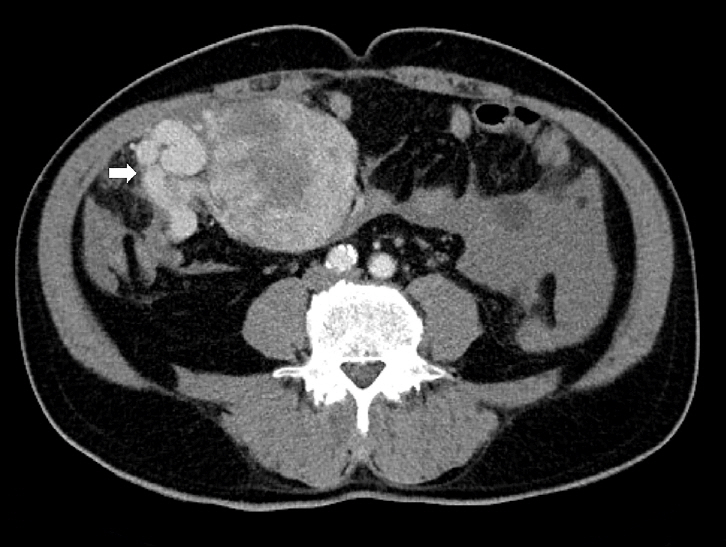
- Solitary fibrous tumor: an updated review
- Joon Hyuk Choi
- J Pathol Transl Med. 2026;60(1):20-46. Published online December 29, 2025
- DOI: https://doi.org/10.4132/jptm.2025.10.08
- 14,151 View
- 480 Download
- 6 Web of Science
- 8 Crossref
-
Abstract
PDF
- Solitary fibrous tumor (SFT) is a fibroblastic neoplasm characterized by a branching, thin-walled dilated staghorn-shaped (hemangiopericytoma-like) vasculature and a NAB2::STAT6 gene fusion. SFTs can occur in almost any anatomical location, including superficial and deep soft tissues, visceral organs, and bone. They most commonly occur in extrapleural locations, equally affect both sexes, and are typically present in adults. Although metastasis is rare, SFTs frequently show local recurrence. The diagnosis of SFTs is difficult because of their broad histological and morphological overlap with other neoplasms. An accurate diagnosis is important for guiding disease management and prognosis. Despite advances in molecular diagnostics and therapeutic strategies, the biological complexity and unpredictable clinical behavior of SFTs present significant challenges. This review provides an updated overview of SFT, with a focus on its molecular genetics, histopathological features, and diagnostic considerations.
-
Citations
Citations to this article as recorded by- Clinicopathological characteristics and prognosis of central nervous system solitary fibrous tumor: An analysis of 271 cases
Wanwan Gao, Ming Li, Xiaojia Liu, Lingyang Hua, Hong Chen, Haixia Cheng
Pathology - Research and Practice.2026; 284: 156520. CrossRef - Pelvic solitary fibrous tumor, historically classified as hemangiopericytoma, presenting with venous compression and pelvic congestion: A case report
Dejan Svilar, Jovana Đošić, Anđela Đurić, Bojan Stojanović
Halo 194.2026; 32(1): 31. CrossRef - Robot-assisted laparoscopic resection of giant pelvic solitary fibrous tumor: a case report with literature review
Binbin Wang, Gengchen Huang, Wei Wei, Tie Mao, Zihan Gao, Yutao Ma, Yiming Gu
Frontiers in Oncology.2026;[Epub] CrossRef - Laparoscopic Resection of a Mesorectal Solitary Fibrous Tumor: A Case Report and Review of the Literature
Atsushi Sugimoto, Hiroshi Tsuchihashi, Hiroyuki Fujimoto, Masayasu Kawasaki
Cureus.2026;[Epub] CrossRef - Construction and Characterization of Paired Patient-Derived Cell Models for Solitary Fibrous Tumors (SFTs)
Farhan Vahdat Azad, Paulina I. Trevino, Aishwarya Kappagantu, Maliha Mehjabin, Eleni Balla, Clark A. Meyer, Leonidas Bleris, Yi Li
Cancers.2026; 18(15): 2385. CrossRef - Case report: Reactive nodular fibrous pseudotumor: report of 4 new cases including a rare pulmonary presentation and a literature review covering 40 cases
Xinyao Liu, Chenghui Wang, Weiran Kong, Bangdong Liu, Tianlun Li, Susu Shi, Linlin Zhang, Shuai Chen
Frontiers in Medicine.2026;[Epub] CrossRef - Clinical and hematological indices as prognosticators in localized solitary fibrous tumors: A retrospective study
Ariel Yoong Yi KOH, Ryan Mao Heng LIM, Jason Yongsheng CHAN
Therapeutic Advances in Medical Oncology.2026;[Epub] CrossRef - Intrapulmonary solitary fibrous tumor: clinicopathological analysis of 29 cases with emphasis on morphological spectrum and immunohistochemical profile
Xin Zeng, Xiaodong Lin, Lifang Ye, Zhenyong Wu, Zeyun Lin, Shuting Li
Diagnostic Pathology.2026;[Epub] CrossRef
- Clinicopathological characteristics and prognosis of central nervous system solitary fibrous tumor: An analysis of 271 cases
Newsletters
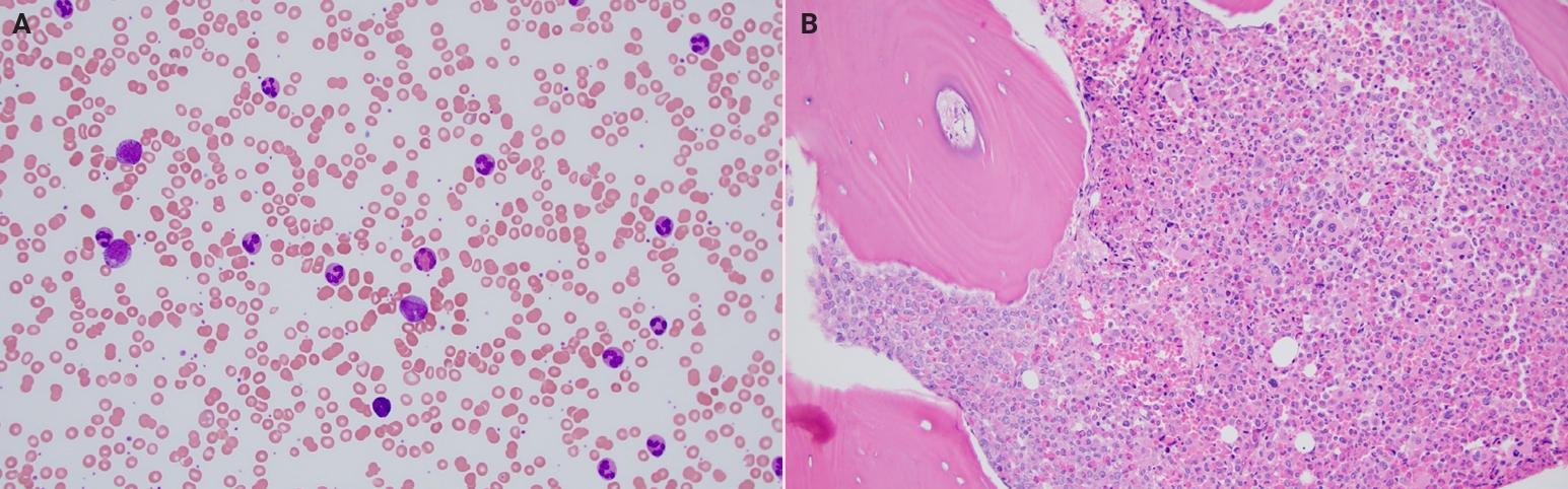
- What’s new in hematopathology 2025: myeloid neoplasms in the WHO 5th edition and ICC
- Barina Aqil
- J Pathol Transl Med. 2025;59(6):472-475. Published online October 22, 2025
- DOI: https://doi.org/10.4132/jptm.2025.09.24
- 22,197 View
- 716 Download
- 1 Web of Science
- 1 Crossref
-
Abstract
PDF
- The previous edition of the World Health Organization (WHO) classification of hematolymphoid neoplasms was published in 2008 and later revised in 2017. A new 5th edition of the WHO classification of hematolymphoid neoplasms was released in 2022. Additionally, the Clinical Advisory Committee developed the International Consensus Classification (ICC) of hematolymphoid tumors, which differs from the WHO classification in several key defining features as outlined below.
-
Citations
Citations to this article as recorded by- Molecular Pathogenesis and Targeted Therapies for Myeloproliferative Hypereosinophilic Neoplasms
Colette Hanna, Joe Rizkallah, Nicole Charbel, Shereen Sakkal, Fadi G. Haddad
Current Hematologic Malignancy Reports.2026;[Epub] CrossRef
- Molecular Pathogenesis and Targeted Therapies for Myeloproliferative Hypereosinophilic Neoplasms
- What’s new in neuropathology 2024: CNS WHO 5th edition updates
- Heather Smith, Jared T. Ahrendsen
- J Pathol Transl Med. 2024;58(6):346-349. Published online September 30, 2024
- DOI: https://doi.org/10.4132/jptm.2024.09.11

- 33,233 View
- 1,577 Download
- 9 Web of Science
- 9 Crossref
-
Abstract
PDF
- The fifth edition of the World Health Organization (WHO) Classification of Central Nervous System (CNS) Tumors was released in 2021, just five years following the updated fourth edition. Advanced molecular testing such as next-generation sequencing, RNA fusion analysis, and DNA methylation profiling has led to more precise grading and classification of pre-existing tumor types as well as the recognition of new ones. Herein, we outline the major updates of the 2021 WHO Classification of CNS tumors, with emphasis on the expanded molecular characterization of CNS tumors.
-
Citations
Citations to this article as recorded by- Primary CNS Neuroblastoma, FOXR2‐Activated: Clinicopathological Study of Two Cases With Immunohistochemical Characterization and Literature Review
Sumanta Das, Sunita Ahlawat, Komal Agrawal, Salman Shaikh, Rakesh Kumar Gupta, Suman S. Karanth, Sandeep Vaishya, Rana Patir, Mehar Chand Sharma
Neuropathology.2026;[Epub] CrossRef - Deep Learning for Brain Tumour Analysis: A Systematic Review of CNN‐Transformer Hybrids in Multimodal Imaging
Solomon Buabeng Antwi, Peter Appiahene, Ben Beklisi Kwame Ayawli, Peter Nimbe, Vincenzo Positano
International Journal of Biomedical Imaging.2026;[Epub] CrossRef - The Price of Precision: A Critical Review of Molecular Diagnostics in Glioma, From Guidelines to Global Disparities
Maria Guarnaccia, Sebastiano Cavallaro
Annals of Clinical and Translational Neurology.2026;[Epub] CrossRef - Bioinformatics insights into ACSL1 and ACSL5: prognostic and immune roles in low-grade glioma
Cheng Zhang, Zhonghua Lv, Hongsheng Liang, Fulan Hu, Haoran Bi
BMC Cancer.2025;[Epub] CrossRef - Current Understanding of the Exosomes and Their Associated Biomolecules in the Glioblastoma Biology, Clinical Treatment, and Diagnosis
Aghdas Ramezani, Maryam Rahnama, Fatemeh Mahmoudian, Fatemeh Shirazi, Mahmoud Ganji, Shohreh Bakhshi, Bahman Khalesi, Zahra Sadat Hashemi, Saeed Khalili
Journal of Neuroimmune Pharmacology.2025;[Epub] CrossRef - Diagnostic Utility of Intratumoral Susceptibility Signals in Adult Diffuse Gliomas: Tumor Grade Prediction and Correlation with Molecular Markers Within the WHO CNS5 (2021) Classification
José Ignacio Tudela Martínez, Victoria Vázquez Sáez, Guillermo Carbonell, Héctor Rodrigo Lara, Florentina Guzmán-Aroca, Juan de Dios Berna Mestre
Journal of Clinical Medicine.2025; 14(11): 4004. CrossRef - Glioblastoma in Puerto Rico: A 21-year population-based study
Carlos E Calderon-Valero, Esteban Rivera, Odaly Balasquide, Alejandro E Cedeño-Moran, Aixa De Jesus, Miguel Mayol Del Valle
Neuro-Oncology Advances.2025;[Epub] CrossRef - Brain Tumors, AI and Psychiatry: Predicting Tumor-Associated Psychiatric Syndromes with Machine Learning and Biomarkers
Matei Șerban, Corneliu Toader, Răzvan-Adrian Covache-Busuioc
International Journal of Molecular Sciences.2025; 26(17): 8114. CrossRef - Engineered bacteria/bacterial components strategy for glioma
Yan Zhu, Meilin Shen, Qi Chen, Huanghao Yang
Chemical Engineering Journal.2025; 525: 170539. CrossRef
- Primary CNS Neuroblastoma, FOXR2‐Activated: Clinicopathological Study of Two Cases With Immunohistochemical Characterization and Literature Review
Reviews
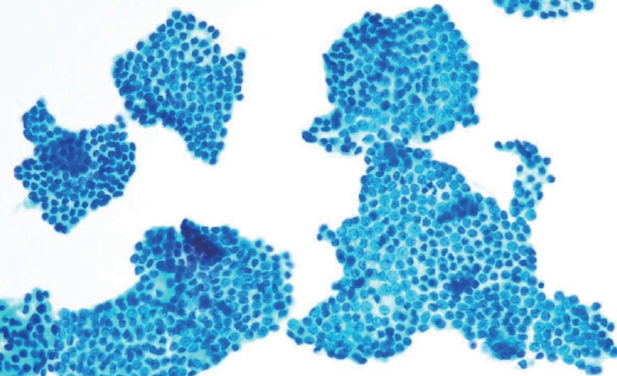
- Cytologic hallmarks and differential diagnosis of papillary thyroid carcinoma subtypes
- Agnes Stephanie Harahap, Chan Kwon Jung
- J Pathol Transl Med. 2024;58(6):265-282. Published online November 7, 2024
- DOI: https://doi.org/10.4132/jptm.2024.10.11
- 27,815 View
- 930 Download
- 18 Web of Science
- 20 Crossref
-
Abstract
PDF
- Papillary thyroid carcinoma (PTC) is the most common thyroid malignancy, characterized by a range of subtypes that differ in their cytologic features, clinical behavior, and prognosis. Accurate cytologic evaluation of PTC using fine-needle aspiration is essential but can be challenging due to the morphologic diversity among subtypes. This review focuses on the distinct cytologic characteristics of various PTC subtypes, including the classic type, follicular variant, tall cell, columnar cell, hobnail, diffuse sclerosing, Warthin-like, solid/trabecular, and oncocytic PTCs. Each subtype demonstrates unique nuclear features, architectural patterns, and background elements essential for diagnosis and differentiation from other thyroid lesions. Recognizing these distinct cytologic patterns is essential for identifying aggressive subtypes like tall cell, hobnail, and columnar cell PTCs, which have a higher risk of recurrence, metastasis, and poorer clinical outcomes. Additionally, rare subtypes such as diffuse sclerosing and Warthin-like PTCs present unique cytologic profiles that must be carefully interpreted to avoid diagnostic errors. The review also highlights the cytologic indicators of lymph node metastasis and high-grade features, such as differentiated high-grade thyroid carcinoma. The integration of molecular testing can further refine subtype diagnosis by identifying specific genetic mutations. A thorough understanding of these subtype-specific cytologic features and molecular profiles is vital for accurate diagnosis, risk stratification, and personalized management of PTC patients. Future improvements in diagnostic techniques and standardization are needed to enhance cytologic evaluation and clinical decision-making in thyroid cancer.
-
Citations
Citations to this article as recorded by- Oncocytic Thyroid Tumours With Pathogenic FLCN Mutations Mimic Oncocytic Papillary Thyroid Carcinoma on Fine‐Needle Aspiration
Adeel M. Ashraf, Faisal Hassan, Adrian A. Dawkins, Julie C. Dueber, Derek B. Allison, Thèrése J. Bocklage
Cytopathology.2026; 37(1): 108. CrossRef - Using a new type of visible light-based emission fluorescence microscope to identify the benign and malignant nature of thyroid tissue during the surgical process: Analysis of diagnostic results
Yu Miao, Liu Xiaowei, Li Muyang, Gao Jian, Chen Lu
Photodiagnosis and Photodynamic Therapy.2026; 57: 105324. CrossRef - Clinical Behavior of Aggressive Variants of Papillary Thyroid Carcinoma: A Retrospective Case–Control Study
Jovan Ilic, Nikola Slijepcevic, Katarina Tausanovic, Bozidar Odalovic, Goran Zoric, Marija Milinkovic, Branislav Rovcanin, Milan Jovanovic, Matija Buzejic, Duska Vucen, Boban Stepanovic, Sara Ivanis, Milan Parezanovic, Milan Marinkovic, Vladan Zivaljevic
Cancers.2026; 18(2): 345. CrossRef - Advantages of thyroid core needle biopsy: an emerging selective first-line biopsy modality
Jae Ho Shin, Yeseul Kim, Min Kyoung Lee, Jung Hwan Baek, So Lyung Jung
Ultrasonography.2026; 45(3): 205. CrossRef - Clinicopathological profile of high-grade differentiated thyroid carcinoma in an Indonesian tertiary hospital
Novita, Agnes Stephanie Harahap, Maria Francisca Ham, Alfianto Widiono, Chan Kwon Jung
Journal of Pathology and Translational Medicine.2026; 60(3): 338. CrossRef - Interpretable SVM-Based Integrated Ultrasound Model for Preoperative Thyroid Nodule Subtype Classification: Improved Identification of Follicular Variant Papillary Thyroid Carcinoma
Ran Zheng, Zhen Wang, Yongxin Li, Yuanqing Zhang, Fang Nie
Diagnostics.2026; 16(13): 1950. CrossRef - Papillary thyroid carcinoma in thyroglossal duct cyst: a Peruvian case series
José Luis Paz-Ibarra, Marialejandra Delgado Rojas, Edward Paucar Holgado, Jenyfer María Fuentes-Mendoza, Luis Concepción-Urteaga, Juan Eduardo Quiroz-Aldave, Marcio José Concepción-Zavaleta, José Somocurcio Peralta
Endocrinology, Diabetes & Metabolism Case Reports.2026;[Epub] CrossRef - Single-cell reveals age-dependent epithelial reprogramming and EMT vulnerability in THCA
Qiankun Zhang, Wei Pan, Xiaohua Gong, Qi Zhou
Endocrine-Related Cancer.2026;[Epub] CrossRef - Infiltrative follicular variant papillary thyroid carcinoma with bilateral cervical nodal metastases following benign cytology: Surgical management in a resource-limited setting
Anil Akulwar, Anjali Mane, Ashwini Kumar Rajkumar, Neha Misal
International Journal of Health & Allied Sciences.2026; 15(2): 120. CrossRef - Analysis of the value of contrast-enhanced ultrasound with CT imaging in the assessment of preoperative cervical lymph node metastases in papillary thyroid cancer
T. Yu, L. Liu, Z. Wang, R. Li, M. Zhang, C. Liu
International Journal of Radiation Research.2026; 24(2): 441. CrossRef - Non‐Oncocytic Thyroid Follicular Neoplasm: Cytological Atypia and Development of a Score for Malignancy Risk Stratification
Boris M. Shifman, Nadezhda M. Platonova, Fatima M. Abdulkhabirova, Ekaterina V. Bondarenko, Anastasia M. Lapshina, Liliya S. Urusova, Georgii S. Razmakhaev, Tatyana N. Kamneva, Ekaterina A. Troshina
Diagnostic Cytopathology.2026;[Epub] CrossRef - Impact of autoimmune thyroiditis on the aggressiveness of papillary thyroid carcinoma and new cytological diagnostic criteria
H.V. Zelinska, M.O. Mishchenko, O.I. Kefeli-Yanovska
INTERNATIONAL JOURNAL OF ENDOCRINOLOGY (Ukraine).2026; 22(6): 610. CrossRef - Hashimoto Thyroiditis Modifies the Association Between Nuclear Atypia and Malignancy in Bethesda III Thyroid Nodules
Adem Şentürk, Yusuf Öztürk, Emre Gönüllü, İsmail Özer, Kübra Tuba Çayir, Ahmet Tarık Harmantepe, Yavuz Selim Kahraman, Fuldem Mutlu
Diagnostics.2026; 16(19): 3235. CrossRef - Nuclear pseudoinclusion is associated with BRAFV600E mutation: Analysis of nuclear features in papillary thyroid carcinoma
Agnes Stephanie Harahap, Dina Khoirunnisa, Salinah, Maria Francisca Ham
Annals of Diagnostic Pathology.2025; 75: 152434. CrossRef - 2025 Korean Thyroid Association Clinical Management Guideline on Active Surveillance for Low-Risk Papillary Thyroid Carcinoma
Eun Kyung Lee, Min Joo Kim, Seung Heon Kang, Bon Seok Koo, Kyungsik Kim, Mijin Kim, Bo Hyun Kim, Ji-hoon Kim, Shin Je Moon, Kyorim Back, Young Shin Song, Jong-hyuk Ahn, Hwa Young Ahn, Ho-Ryun Won, Won Sang Yoo, Min Kyoung Lee, Jeongmin Lee, Ji Ye Lee, Kyo
International Journal of Thyroidology.2025; 18(1): 30. CrossRef - Structure-based molecular screening and dynamic simulation of phytocompounds targeting VEGFR-2: a novel therapeutic approach for papillary thyroid carcinoma
Shuai Wang, Lingqian Zhang, Wenjun Zhang, Xiong Zeng, Jie Mei, Weidong Xiao, Lijie Yang
Frontiers in Pharmacology.2025;[Epub] CrossRef - 2025 Korean Thyroid Association Clinical Management Guideline on Active Surveillance for Low-Risk Papillary Thyroid Carcinoma
Eun Kyung Lee, Min Joo Kim, Seung Heon Kang, Bon Seok Koo, Kyungsik Kim, Mijin Kim, Bo Hyun Kim, Ji-hoon Kim, Shinje Moon, Kyorim Back, Young Shin Song, Jong-hyuk Ahn, Hwa Young Ahn, Ho-Ryun Won, Won Sang Yoo, Min Kyoung Lee, Jeongmin Lee, Ji Ye Lee, Kyon
Endocrinology and Metabolism.2025; 40(3): 307. CrossRef - A Case of Warthin-Like Variant of Papillary Thyroid Cancer
Amy Chow, Israa Laklouk
Cureus.2025;[Epub] CrossRef - Propensity score-matched analysis of the ‘2+2’ parathyroid strategy in total thyroidectomy with central neck dissection
Hao Gong, Simei Yao, Tianyuchen Jiang, Yi Yang, Yuhan Jiang, Zhujuan Wu, Anping Su
Frontiers in Endocrinology.2025;[Epub] CrossRef - Cytological Findings in Pediatric Thoracic Tumors: A Review of Diagnostic Insights and Pitfalls
Parikshaa Gupta, Pranab Dey
Acta Cytologica.2025; 70(3): 320. CrossRef
- Oncocytic Thyroid Tumours With Pathogenic FLCN Mutations Mimic Oncocytic Papillary Thyroid Carcinoma on Fine‐Needle Aspiration
- Interpretation of PD-L1 expression in gastric cancer: summary of a consensus meeting of Korean gastrointestinal pathologists
- Soomin Ahn, Yoonjin Kwak, Gui Young Kwon, Kyoung-Mee Kim, Moonsik Kim, Hyunki Kim, Young Soo Park, Hyeon Jeong Oh, Kyoungyul Lee, Sung Hak Lee, Hye Seung Lee
- J Pathol Transl Med. 2024;58(3):103-116. Published online April 25, 2024
- DOI: https://doi.org/10.4132/jptm.2024.03.15

- 32,483 View
- 798 Download
- 13 Web of Science
- 14 Crossref
-
Abstract
PDF
 Supplementary Material
 - Nivolumab plus chemotherapy in the first-line setting has demonstrated clinical efficacy in patients with human epidermal growth factor receptor 2–negative advanced or metastatic gastric cancer, and is currently indicated as a standard treatment. Programmed death-ligand 1 (PD-L1) expression is an important biomarker for predicting response to anti–programmed death 1/PD-L1 agents in several solid tumors, including gastric cancer. In the CheckMate-649 trial, significant clinical improvements were observed in patients with PD-L1 combined positive score (CPS) ≥ 5, determined using the 28-8 pharmDx assay. Accordingly, an accurate interpretation of PD-L1 CPS, especially at a cutoff of 5, is important. The CPS method evaluates both immune and tumor cells and provides a comprehensive assessment of PD-L1 expression in the tumor microenvironment of gastric cancer. However, CPS evaluation has several limitations, one of which is poor interobserver concordance among pathologists. Despite these limitations, clinical indications relying on PD-L1 CPS are increasing. In response, Korean gastrointestinal pathologists held a consensus meeting for the interpretation of PD-L1 CPS in gastric cancer. Eleven pathologists reviewed 20 PD-L1 slides with a CPS cutoff close to 5, stained with the 28-8 pharmDx assay, and determined the consensus scores. The issues observed in discrepant cases were discussed. In this review, we present cases of gastric cancer with consensus PD-L1 CPS. In addition, we briefly touch upon current practices and clinical issues associated with assays used for the assessment of PD-L1 expression in gastric cancer.
-
Citations
Citations to this article as recorded by- Organ Preservation for Gastroesophageal Junction and Gastric Cancers: Ready for Primetime?
Winta Mehtsun, Lola Van Doosselaere, Ugwuji N. Maduekwe
American Society of Clinical Oncology Educational Book.2026;[Epub] CrossRef - Deep Learning Analysis Based on Dual-energy CT-Derived Iodine Map for Predicting PD-L1 Expression in Gastric Cancer: A Multicenter Study
Lihong Chen, Yuncong Zhao, Xiaomin Tian, Deye Zeng, Yongxiu Tong, Haiping Xu, Yaru You, Caiming Weng, Sen Lin, Keru Chen, Yilin Chen, Yunjing Xue
Academic Radiology.2026; 33(4): 1324. CrossRef - Artificial intelligence for biomarker prediction in gastric cancer: from histopathology to multimodal integration
Yesul Jeong, Sangjeong Ahn, Sung Hak Lee
Frontiers in Oncology.2026;[Epub] CrossRef - The research progress of the synergistic effect of Epstein-Barr virus and Helicobacter pylori infection in the occurrence and development of gastric cancer
Liming Gan, Liping Gong, Shan Wei, Junshan Deng
Frontiers in Oncology.2026;[Epub] CrossRef - Tumour immunotherapy in solid and hematologic malignancies: Progress, challenges, and a practical framework
Shang Li, Jiaxin Huang, Rui Fang, Ming Zhang, Yucui Jin, Yi Zhu, Changyan Ma
Seminars in Oncology.2026; 53(6): 152541. CrossRef - A deep learning-derived risk score model from digital pathology to forecast nivolumab outcomes in gastric carcinoma
Yiyu Hong, Inwoo Hwang, Min-Ji Kim, Yuyeon Kim, Soomin Ahn, Jeeyun Lee, Kyoung-Mee Kim
British Journal of Cancer.2026;[Epub] CrossRef - Adjuvant immunotherapy in patients with resected gastric and oesophagogastric junction cancer following preoperative chemotherapy with high risk for recurrence (ypN+ and/or R1): European Organisation of Research and Treatment of Cancer (EORTC) 1707 VESTIG
F. Lordick, M.E. Mauer, G. Stocker, C.A. Cella, I. Ben-Aharon, G. Piessen, L. Wyrwicz, G. Al-Haidari, T. Fleitas-Kanonnikoff, V. Boige, R. Lordick Obermannová, U.M. Martens, C. Gomez-Martin, P. Thuss-Patience, V. Arrazubi, A. Avallone, K.K. Shiu, P. Artru
Annals of Oncology.2025; 36(2): 197. CrossRef - PD-L1 as a Biomarker in Gastric Cancer Immunotherapy
Yunjoo Cho, Soomin Ahn, Kyoung-Mee Kim
Journal of Gastric Cancer.2025; 25(1): 177. CrossRef - PD-L1 importance in malignancies comprehensive insights into the role of PD-L1 in malignancies: from molecular mechanisms to therapeutic opportunities
Mojdeh Soltani, Mohammad Abbaszadeh, Hamed Fouladseresht, Mark J. M. Sullman, Nahid Eskandari
Clinical and Experimental Medicine.2025;[Epub] CrossRef - CLDN18.2 expression in gastroesophageal adenocarcinoma: prevalence, heterogeneity, and prognostic implications in Spanish patients
Carolina Martinez-Ciarpaglini, María Ortega, Sandra Pérez-Buira, Aitana Bolea, Beatriz Casado Guerra, Carmen Herencia Bellido, Paula Tornero Piñero, Dolores Naranjo-Hans, Brenda Palomar, Hernán Quiceno, Amanda Sardón Fernández, Ariadna Torner Calvo, Feder
Virchows Archiv.2025; 487(6): 1337. CrossRef - Distinct clinicopathological and survival profiles of CLDN18.2 and PD-L1 expression in advanced gastric cancer and gastroesophageal junction adenocarcinoma
D.R. Castillo, M. Guo, P. Shah, M. Hazeltin, D. Tai, F. Al-Manaseer, S. Mlamba, D. Perez, S. Yeremian, S. Guzman, R. Mannan, C. Crook, C. Lau, N. Tawar, G. Brar, M. Raoof, Y. Woo, S.P. Wu, D. Li
ESMO Gastrointestinal Oncology.2025; 10: 100261. CrossRef - Best Practice PD-L1 Staining and Interpretation in Gastric Cancer Using PD-L1 IHC PharmDx 22C3 and PD-L1 IHC PharmDx 28-8 Assays, with Reference to Common Issues and Solutions
Soomin Ahn, Inwoo Hwang, Yuyeon Kim, Somin Lee, Yunjoo Cho, So Young Kang, Deok Geun Kim, Jeeyun Lee, Kyoung-Mee Kim
Biomedicines.2025; 13(11): 2824. CrossRef - Intraperitoneal immune microenvironment and efficacy of intraperitoneal chemotherapy in patients with gastric cancer and peritoneal metastasis
Tomoya Nakanishi, Motohiro Imano, Masashi Kohda, Hiroaki Kato, Naoko Kounami, Atsushi Yamada, Masuhiro Terada, Yoko Hiraki, Osamu Shiraishi, Atsushi Yasuda, Masayuki Shinkai, Takushi Yasuda
Scientific Reports.2025;[Epub] CrossRef - PD-L1 thresholds predict efficacy of immune checkpoint inhibition in first-line treatment of advanced gastroesophageal adenocarcinoma. A systematic review and meta-analysis of seven phase III randomized trials
V. Formica, C. Morelli, L. Fornaro, S. Riondino, M. Rofei, E. Fontana, E.C. Smyth, M. Roselli, H.-T. Arkenau
ESMO Open.2024; 9(11): 103967. CrossRef
- Organ Preservation for Gastroesophageal Junction and Gastric Cancers: Ready for Primetime?
Newsletter
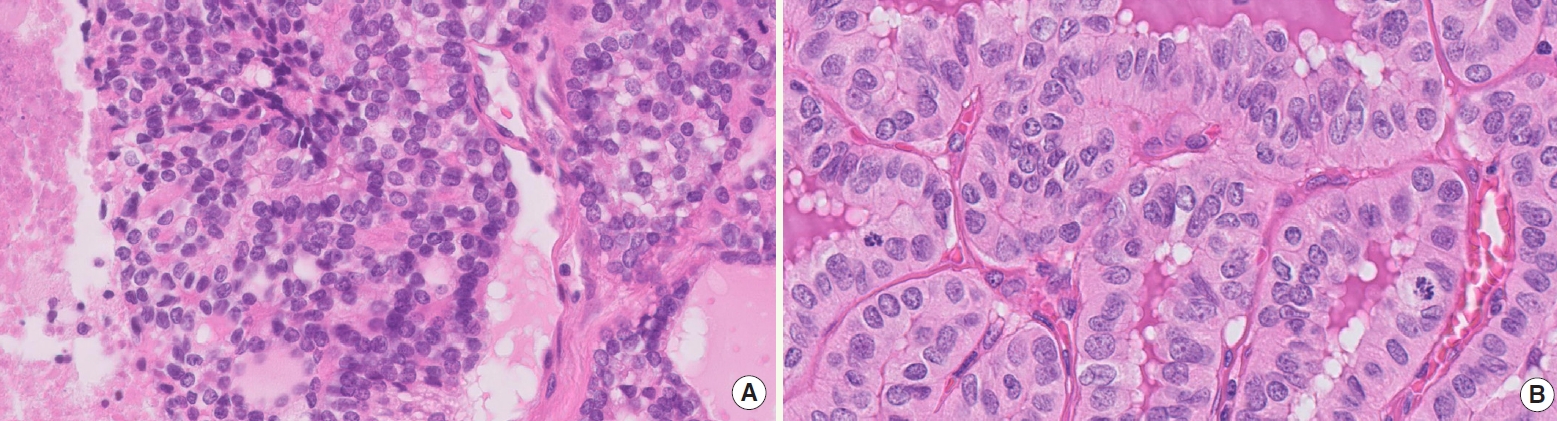
- What’s new in thyroid pathology 2024: updates from the new WHO classification and Bethesda system
- Andrey Bychkov, Chan Kwon Jung
- J Pathol Transl Med. 2024;58(2):98-101. Published online March 13, 2024
- DOI: https://doi.org/10.4132/jptm.2024.03.06
- 38,374 View
- 2,558 Download
- 10 Web of Science
- 12 Crossref
-
Abstract
PDF
- In line with the release of the 5th edition WHO Classification of Tumors of Endocrine Organs (2022) and the 3rd edition of the Bethesda System for Reporting Thyroid Cytopathology (2023), the field of thyroid pathology and cytopathology has witnessed key transformations. This digest brings to the fore the refined terminologies, newly introduced categories, and contentious methodological considerations pivotal to the updated classification.
-
Citations
Citations to this article as recorded by- Impact of thyroid Bethesda category IV (follicular neoplasm) terminology unification on atypia of undetermined significance reporting patterns in thyroid fine-needle aspiration
Shirin Abbasi, Lorena Marcano-Bonilla, Syed Z. Ali
Journal of the American Society of Cytopathology.2026; 15(2): 107. CrossRef - Clinical implication of the 2025 ATA risk stratification in follicular thyroid carcinoma: A comparison with the 2015 ATA risk stratification
Hyunju Park, Bo Ram Kim, Ji Hyun Yoo, Sun Wook Kim, Jae Hoon Chung, Bogyeong Han, Myoung Kyoung Kim, Jun Ho Choe, Man Ki Chung, Tae Hyuk Kim, Young Lyun Oh
Oral Oncology.2026; 175: 107912. CrossRef - Indeterminate Bethesda System Category (Bethesda Category III) Thyroid Nodules: Cytomorphologic Subclassification and Its Impact on Malignancy Risk
Rinë Limani, Zgjim Limani, Shkelzen Reçica, Labinota Kondirolli, Etnik Bajraktari, Brikenë Blakaj Gashi, Drita Miftari Pazhari
Acta Cytologica.2026; : 1. CrossRef - Multimodal Diagnostic Cross-Check: A Correlative Study of Fine-Needle Aspiration Cytology, Ultrasonography, Thyroid Profile, and Histopathology in Thyroid Swellings
Farheen Khan, Nishi Tandon, Yoshita Agnihotri, Suboohi Khanam, Andleeb Zehra, Nirupma Lal
Cureus.2026;[Epub] CrossRef - Improving diagnostic confidence in thyroid pathology in resource-limited settings: a practical guide for the general anatomical pathologist
Brendon Price
Diagnostic Histopathology.2026;[Epub] CrossRef - Diagnosis and management of thyroid nodule
Suganya Sekar, Deepak Thomas Abraham
Current Opinion in Endocrinology, Diabetes & Obesity.2025; 32(5): 167. CrossRef - Diagnostic Challenges, Prognostic Assessment, and Treatment Strategies in High-Grade Differentiated Thyroid Carcinoma
Chan Kwon Jung, Agnes Stephanie Harahap
Endocrinology and Metabolism.2025; 40(6): 830. CrossRef - Cytologic and Clinicopathologic Features of Papillary Thyroid Carcinoma with Prominent Hobnail Features on FNAC
Deepali Saxena, Ravi Hari Phulware, Prashant Durgapal, Arvind Kumar, Amit Kumar Tyagi
Indian Journal of Otolaryngology and Head & Neck Surgery.2024; 76(5): 4885. CrossRef - FHL1: A novel diagnostic marker for papillary thyroid carcinoma
Yeting Zeng, Dehua Zeng, Xingfeng Qi, Hanxi Wang, Xuzhou Wang, Xiaodong Dai, Lijuan Qu
Pathology International.2024; 74(9): 520. CrossRef - Nouveautés en pathologie thyroïdienne : classification OMS 2022, système Bethesda 2023, biologie moléculaire et testing moléculaire
Mohamed Amine Bani, Sophie Moog, Voichita Suciu, Livia Lamartina, Abir Al Ghuzlan
Bulletin du Cancer.2024; 111(10): 10S5. CrossRef - Cytologic hallmarks and differential diagnosis of papillary thyroid carcinoma subtypes
Agnes Stephanie Harahap, Chan Kwon Jung
Journal of Pathology and Translational Medicine.2024; 58(6): 265. CrossRef - Surgical and Pathological Challenges in Thyroidectomy after Thermal Ablation of Thyroid Nodules
Ting-Chun Kuo, Kuen-Yuan Chen, Hsiang-Wei Hu, Jie-Yang Jhuang, Ming-Tsan Lin, Chin-Hao Chang, Ming-Hsun Wu
Thyroid®.2024; 34(12): 1503. CrossRef
- Impact of thyroid Bethesda category IV (follicular neoplasm) terminology unification on atypia of undetermined significance reporting patterns in thyroid fine-needle aspiration
Original Article
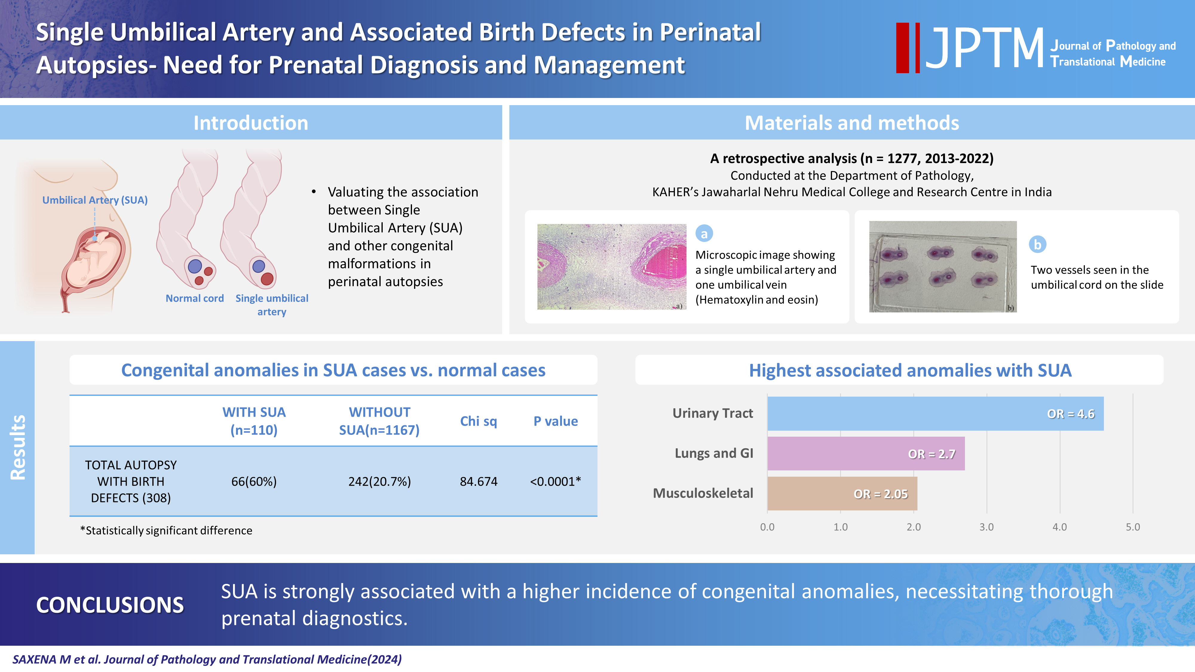
- Single umbilical artery and associated birth defects in perinatal autopsies: prenatal diagnosis and management
- Manushree Saxena, Bhagyashri Hungund
- J Pathol Transl Med. 2024;58(5):214-218. Published online July 9, 2024
- DOI: https://doi.org/10.4132/jptm.2024.07.03
- 29,754 View
- 518 Download
- 2 Web of Science
- 3 Crossref
-
Abstract
PDF
- Background
The umbilical cord forms the connection between the fetus and the placenta at the feto-maternal interface and normally comprises two umbilical arteries and one umbilical vein. In some cases, only a single umbilical artery (SUA) is present. This study was conducted to evaluate associations between SUA and other congenital malformations discovered in perinatal autopsies and to ascertain the existence of preferential associations between SUA and certain anomalies.
Methods
We evaluated records of all fetuses sent for autopsy to the Department of Pathology during the 10-year period from 2013 through 2022 (n = 1,277). The data were obtained from the hospital’s pathology laboratory records. The congenital anomalies were grouped by organ or system for analysis and included cardiovascular, urinary tract, nervous system, gastrointestinal tract, musculoskeletal, and lung anomalies.
Results
A SUA was present in 8.61% of the autopsies. The gestational age of the affected fetuses ranged between 13 to 40 weeks. An SUA presented as an isolated single anomaly in 44 cases (3.4%). Of the 110 SUA cases, 60% had other congenital anomalies. There was a significant association between birth defects and SUAs (p < .001). Strong associations between SUA and urinary tract, lung, and musculoskeletal anomalies were observed.
Conclusions
A SUA is usually seen in association with other congenital malformations rather than as an isolated defect. Therefore, examination for associated anomalies when an SUA is detected either antenatally or postnatally is imperative. The findings of this study should be helpful in counseling expectant mothers and their families in cases of SUA. -
Citations
Citations to this article as recorded by- Prevalence of Congenital Abnormalities in Abortuses and Stillbirths in India: A Systematic Review and Meta‐Analysis
Sushant Swaroop Das, Shalika Sharma, Reeha Mahajan, Sushruti Kaushal, Manupriya Sharma, Harsimranjit Singh, M. Ramkumar
Congenital Anomalies.2026;[Epub] CrossRef - Single Umbilical Artery with Symmetrical IUGR and Multiple Fetal Anomalies - An Interesting Case Report
Amulya Choudary Kotapati, Bhargavi Khandru, Vijayasree M.
Journal of Evolution of Medical and Dental Sciences.2025; : 10. CrossRef - Epidemiological and Histopathological Characteristics of Fetuses with Congenital Disorders: A Study in Greece
Despoina Nteli, Maria Nteli, Konstantinos Konstantinidis, Maria Ouzounidou, Paschalis Theotokis, Maria-Eleni Manthou, Iasonas Dermitzakis, Xeni Miliara, Chrysoula Gouta, Stamatia Angelidou, Dimosthenis Miliaras, Soultana Meditskou
Biology.2025; 14(6): 626. CrossRef
- Prevalence of Congenital Abnormalities in Abortuses and Stillbirths in India: A Systematic Review and Meta‐Analysis
Review Article
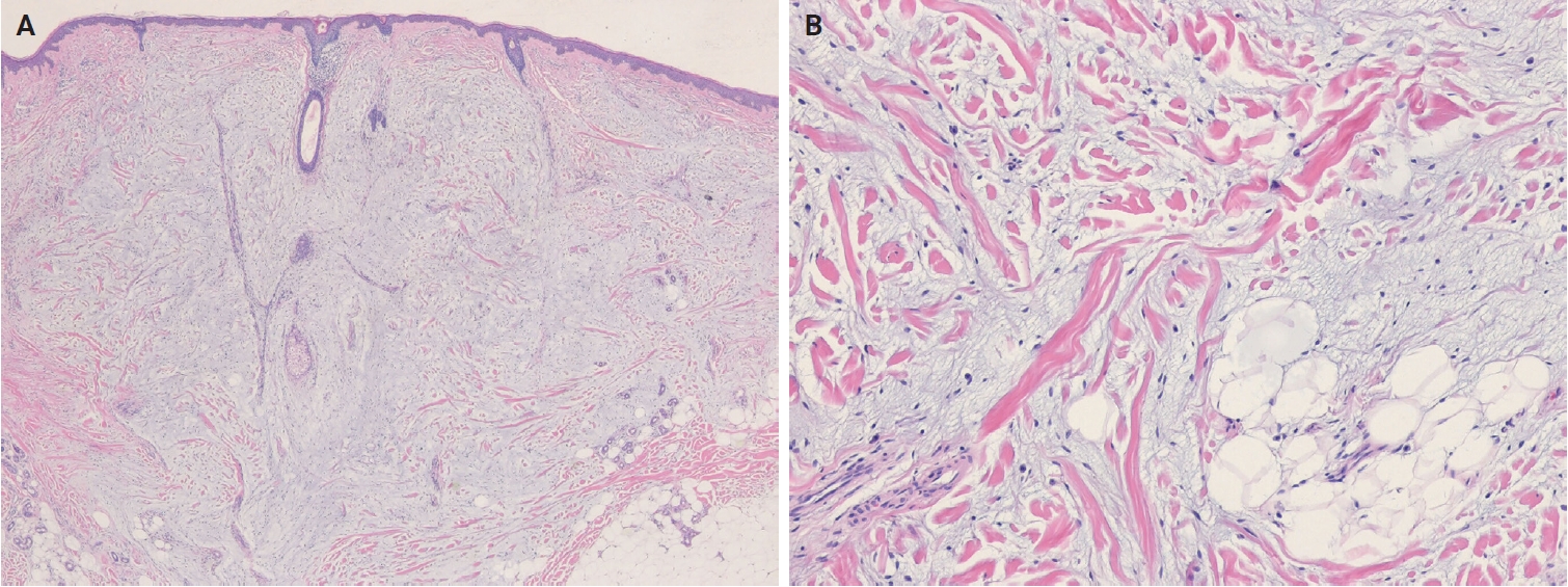
- Cutaneous soft tissue tumors in the 5th edition of the World Health Organization classification of skin tumors: key updates and new entities
- Joon Hyuk Choi
- J Pathol Transl Med. 2026;60(2):144-183. Published online March 13, 2026
- DOI: https://doi.org/10.4132/jptm.2026.01.09
- 7,531 View
- 343 Download
- 1 Crossref
-
Abstract
PDF
- The 5th edition of the World Health Organization (WHO) classification of skin tumors introduces a dedicated chapter on cutaneous soft tissue tumors, providing a comprehensive, standardized reference with updated diagnostic criteria that directly inform routine dermatopathology practice and molecular diagnostics. This edition incorporates several key changes, including newly recognized entities such as EWSR1::SMAD3-rearranged fibroblastic tumor, neurotrophic tyrosine receptor kinase (NTRK)–rearranged spindle cell neoplasm, superficial CD34-positive fibroblastic tumor, and CRTC1::TRIM11 cutaneous tumor. Diagnostic terminology has also been refined; for example, the term ‘atypical intradermal smooth muscle neoplasm’ replaces ‘cutaneous leiomyosarcoma’ for lesions confined to the dermis, whereas the designation leiomyosarcoma is reserved for tumors with overt subcutaneous infiltration. In addition, epithelioid fibrous histiocytoma has been reassigned to the family of tumors of uncertain differentiation. This review summarizes the key updates and newly recognized entities in the chapter on cutaneous soft tissue tumors in the 5th edition of the WHO classification of skin tumors, emphasizing their clinicopathological and molecular implications.
-
Citations
Citations to this article as recorded by- Leiomyosarcoma Involving the Calcaneum With Delayed Gastric and Sacroiliac Lesions: A Case Report
Asmita Kaur, Rajat Gupta, Harpreet Singh, Gurkirat S Bhatti
Cureus.2026;[Epub] CrossRef
- Leiomyosarcoma Involving the Calcaneum With Delayed Gastric and Sacroiliac Lesions: A Case Report
Review
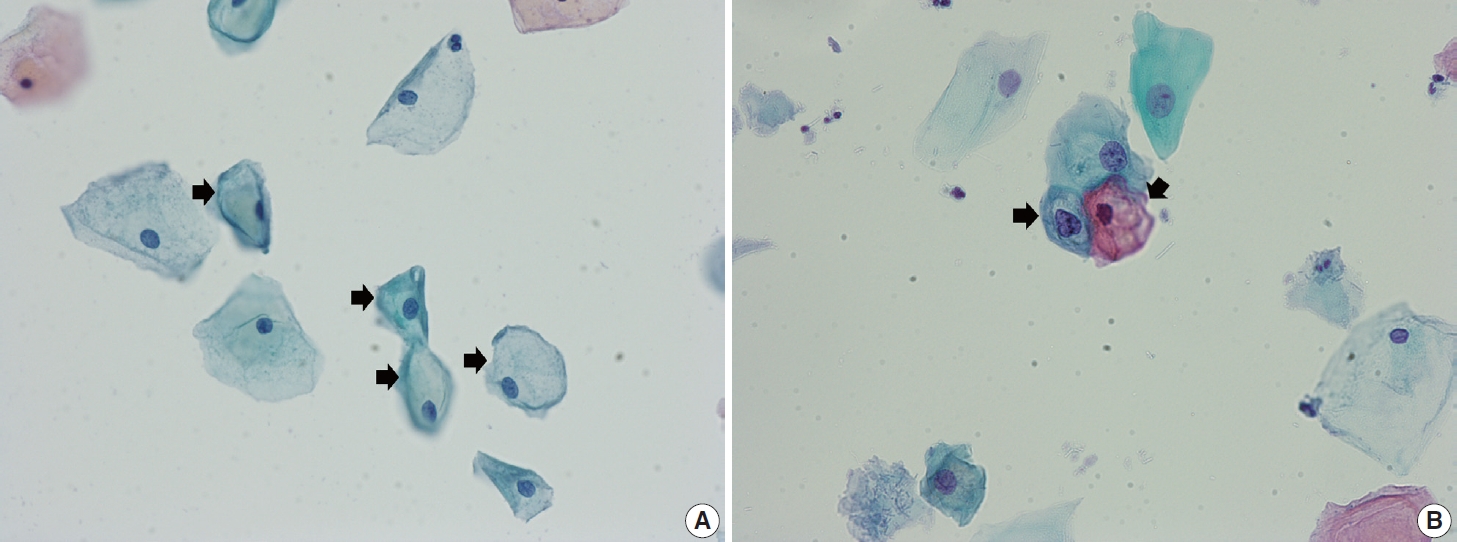
- Cervical intraepithelial neoplasia and cervical cytology in pregnancy
- Ji-Young Kim, Jeong Yun Shim
- J Pathol Transl Med. 2024;58(6):283-290. Published online November 7, 2024
- DOI: https://doi.org/10.4132/jptm.2024.10.17
- 17,994 View
- 566 Download
- 5 Web of Science
- 7 Crossref
-
Abstract
PDF
- Cervical cancer screening during pregnancy presents unique challenges for cytologic interpretation. This review focuses on pregnancy-associated cytomorphological changes and their impact on diagnosis of cervical intraepithelial neoplasia (CIN) and cervical cancer. Pregnancy-induced alterations include navicular cells, hyperplastic endocervical cells, immature metaplastic cells, and occasional decidual cells or trophoblasts. These changes can mimic abnormalities such as koilocytosis, adenocarcinoma in situ, and high-grade squamous intraepithelial lesions, potentially leading to misdiagnosis. Careful attention to nuclear features and awareness of pregnancy-related changes are crucial for correct interpretation. The natural history of CIN during pregnancy shows higher regression rates, particularly for CIN 2, with minimal risk of progression. Management of abnormal cytology follows modified risk-based guidelines to avoid invasive procedures, with treatment typically deferred until postpartum. The findings reported in this review emphasize the importance of considering pregnancy status in cytological interpretation, highlight potential problems, and provide guidance on differentiating benign pregnancy-related changes from true abnormalities. Understanding these nuances is essential for accurate diagnosis and proper management of cervical abnormalities in pregnant women.
-
Citations
Citations to this article as recorded by- HPV in Pregnancy: Implications for Screening, Vaccination, and Maternal–Fetal Health
Suman Kumar, Swati, Swati Salila, Akanksha Raj, Pratima Gupta, Neha Sharad, Nidhi Chaudhary
Journal of Pregnancy.2026;[Epub] CrossRef - Approaches to Intraepithelial Cervical Neoplasia Management in Pregnancy: A Narrative Review
Delia-Maria Bogheanu, Awatif Jaafar Sadeq Al Bayati, Mircea-Octavian Poenaru, Octavian Gabriel Olaru, Gabriel-Petre Gorecki, Andreea Gratiana Boiangiu, Bashar Haj Hamoud, Romina-Marina Sima, Liana Ples
Life.2026; 16(5): 809. CrossRef - From treatment to trauma: Womens lived experiences of adverse pregnancy outcomes following cervical intraepithelial neoplasia treatment in Zambia
Mwiinga-Kalusopa Victoria, E. Maree Johanna, N. Kwaleyela Concepta, Uwamahoro Marie-Claire, Mwila Musenge Emmanuel, Anila Nkhata Loveness, Katowa-Mukwato Patricia
International Journal of Nursing and Midwifery.2026; 18(2): 14. CrossRef - Cervical Cytological Findings and Vaginal Microbiota Alterations During Pregnancy: A Retrospective Analysis
Federica Cianfrini, Antonio d’Amati, Clelia Molinario, Belen Padial Urteta, Chiara Boccaccini, Antonio Benedetto Maria Donateo, Antonietta Vella, Rosaria Santangelo, Rosa Pasqualina De Vincenzo, Angela Santoro, Gian Franco Zannoni
Sage Open Pathology.2026;[Epub] CrossRef - Implementation of self-collected cervical screening in an under-screened, culturally and linguistically diverse antenatal population: a quality improvement study
Mandy Wang, Alison Brand, Martin Plymoth, Judy Chen, Therese McGee
BMJ Open Quality.2026; 15(3): e004525. CrossRef - The significance of biological samples from pregnant women in cervical intraepithelial neoplasia
Xue Mi, Maharjan Rashmi, Zangyu Pan, Di Wu, Jinwei Miao
Frontiers in Medicine.2025;[Epub] CrossRef - Oncologic and pregnancy outcomes of cervical high-grade intraepithelial lesions and delivery mode
Olga P. Matylevich, Ilya A. Tarasau, Sviatlana Y. Shelkovich, Aliaksandr F. Martsinkevich
Academia Oncology.2025;[Epub] CrossRef
- HPV in Pregnancy: Implications for Screening, Vaccination, and Maternal–Fetal Health
Original Article
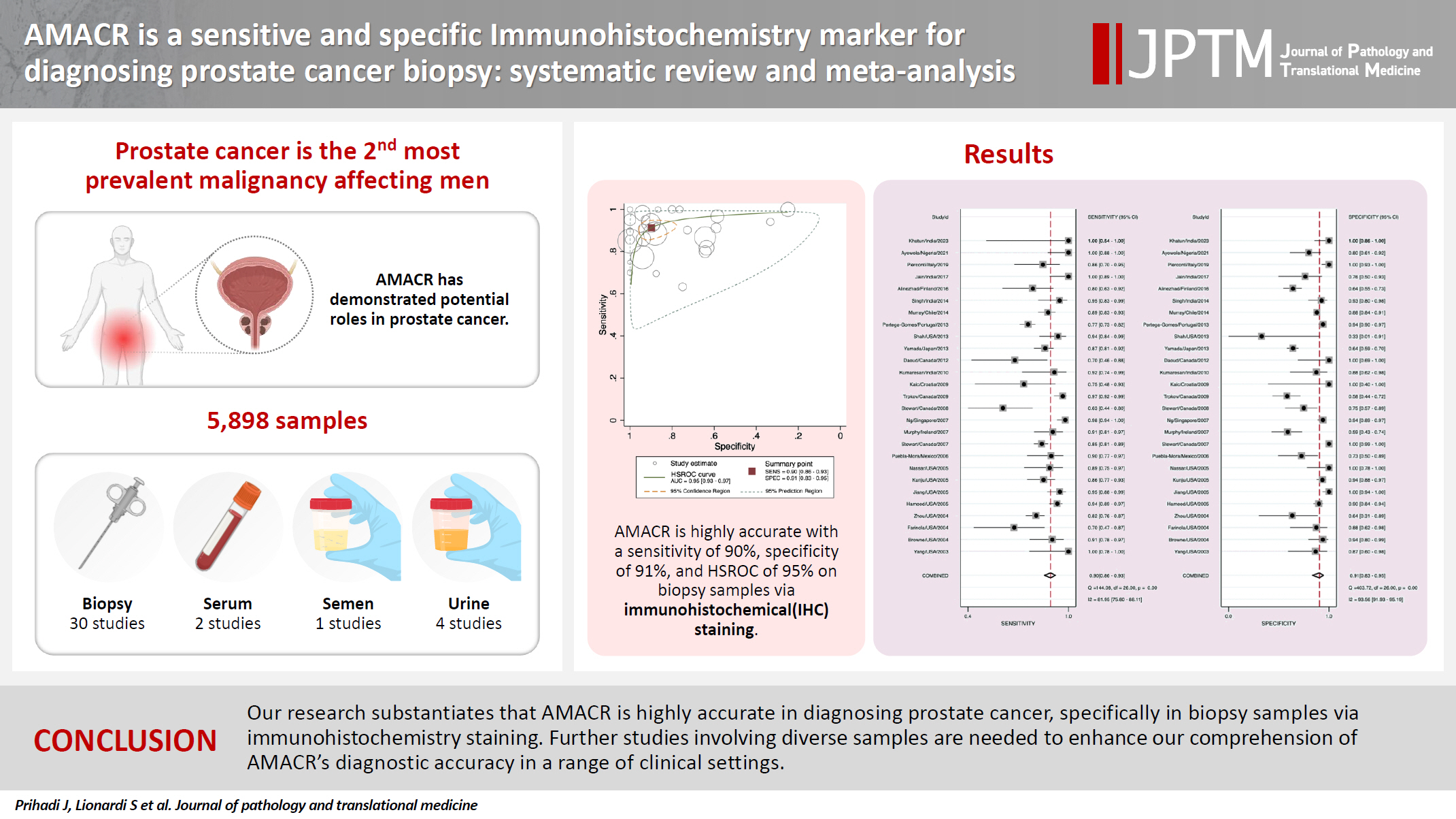
- AMACR is a highly sensitive and specific immunohistochemical marker for diagnosing prostate cancer on biopsy: a systematic review and meta-analysis
- Johannes Cansius Prihadi, Stevan Kristian Lionardi, Nicolas Daniel Widjanarko, Steven Alvianto, Fransiskus Xaverius Rinaldi, Archie Fontana Iskandar
- J Pathol Transl Med. 2025;59(4):235-248. Published online July 3, 2025
- DOI: https://doi.org/10.4132/jptm.2025.04.16
- 12,937 View
- 315 Download
- 7 Web of Science
- 7 Crossref
-
Abstract
PDFSupplementary Material
- Background
Alpha-methylacyl-CoA racemase (AMACR) is the preferred biomarker for distinguishing malignant from benign glands in prostate biopsies, showing high sensitivity and specificity for prostate cancer. A meta-analysis of immunohistochemistry (IHC) for AMACR is essential to further assess its diagnostic accuracy across diverse sample sources. Methods: A systematic search of databases including MEDLINE, ScienceDirect, ProQuest, Google Scholar, and the Cochrane Library was performed, focusing on studies of AMACR to diagnose prostate cancer, particularly in biopsy samples analyzed through IHC over the last 20 years. Quality of studies was assessed using the Quality Assessment of Diagnostic Accuracy Studies 2 tool, followed by a meta-analysis of regions and subgroups to calculate summary estimates of diagnostic test accuracy. Results: In the final analysis, 37 studies, with a pooled size of 5,898 samples, were included from the examination of 94 full-text papers. Among them, 27 studies with similar sample sources and testing methodologies underwent meta-analysis, yielding a combined sensitivity estimate of 0.90 (95% confidence interval [CI], 0.86 to 0.93) and specificity of 0.91 (95% CI, 0.83 to 0.95), both with significant heterogeneity (p < .01). The region beneath the hierarchical summary receiver operating characteristic curve was 0.95 (95% CI, 0.93 to 0.97), positive likelihood ratio was 9.6 (95% CI, 5.3 to 17.4), negative likelihood ratio was 0.11 (95% CI, 0.08 to 0.15), and diagnostic odds ratio was 88 (95% CI, 42 to 181). Conclusions: Our meta-analysis findings substantiate AMACR as a highly accurate tool for diagnosing prostate cancer, specifically in biopsy samples, via immunohistochemical staining. Further studies involving diverse samples are needed to enhance our understanding of the AMACR diagnostic accuracy in a range of clinical settings. -
Citations
Citations to this article as recorded by- ATP binding cassette subfamily D member 1: A highly sensitive diagnostic marker for solid pseudopapillary neoplasm of pancreas in biopsy samples——A multi-institutional study
Yuanhao Liu, Junya Peng, Ruizhe He, Xiaowei Xue, Jie Cui, Mengjie Li, Ying-ao Liu, Wanni Xu, Xiaohong Gao, Yingmei Wang, Zhe Zhang, Haizhen Lu, Zhigang Song, Peizhen Hu, Yupei Zhao, Wenze Wang
Human Pathology.2026; 174: 106125. CrossRef - Towards implementation of precision medicine biomarkers in early detection and prognostication of prostate cancer
Angela Yee, Jim Smith, Rajiv Kumar, Akash Sali, Euan J. Rodger, Aniruddha Chatterjee
Cancer and Metastasis Reviews.2026;[Epub] CrossRef - Phenotype-Oriented Characterization of NSC828786 Identifies Convergent HPN-AMACR-Associated Transcriptomic Signatures in Prostate Adenocarcinoma and Broad-Spectrum Antiproliferative Activity
Ya-Ting Wen, Rosario Trijuliamos Manalu, Han-Lin Hsu, Yu-Cheng Kuo, Ruey-Shyang Soong, Feng-Cheng Liu, Maryam Rachmawati Sumitra, Sheng-Liang Huang, Shih-Yu Lee, Sung-Ling Tang, I-Chuan Yen, Hong-Jaan Wang, Bashir Lawal, Alexander T. H. Wu, Hsu-Shan Huang
Cells.2026; 15(14): 1314. CrossRef - Sub-Saharan African Prostate Cancer Patient-Derived Cell Lines and High-Throughput Drug Screening: Addressing Ancestry Underrepresentation in Oncobiology Research
Carla S. Dos Santos, Ana C. Magalhães, Veronica Fernandes, António Pombinho, Lurdes Torres, Margarida André, Adelaide Sousa, Pedro Sequeira, Daniel Pinto, Cláudia Pereira, Paulo M. Costa, Lúcio Lara Santos, Luisa Pereira
Cancers.2026; 18(15): 2452. CrossRef - Prostate Cancer with Pleural Metastasis: A Case Report
金瑶 宋
Journal of Clinical Personalized Medicine.2026; 05(04): 190. CrossRef - Pathology-Guided Transcriptomic Profiling of Prostate Cancer Identifies Grade-Associated Proliferative and Immune Signatures in a MENA Cohort
Ranyah Al-Hakm, Alaa Muayad Altaie, Reem Sami Alhamidi, Anania Boghossian, Nival Ali, Alaa Mohamed Hamad, Eman Sheta, Nagwa Mashali, Riyad Bendardaf, Timo Gemoll, Iman M. Talaat, Rifat Hamoudi
Cancers.2026; 18(18): 2898. CrossRef - Pathogenesis-Guided Biomarker Assessment: A Shift in Prostate Cancer Diagnostics
Jessica M. Logan, Victoria Malone, John J. O’Leary, Doug A. Brooks
International Journal of Molecular Sciences.2025; 26(24): 11786. CrossRef
- ATP binding cassette subfamily D member 1: A highly sensitive diagnostic marker for solid pseudopapillary neoplasm of pancreas in biopsy samples——A multi-institutional study
Review Articles
- Multiple sclerosis: a practical review for pathologists
- Rachel A. Multz, Pouya Jamshidi, Jared T. Ahrendsen
- J Pathol Transl Med. 2025;59(4):203-213. Published online June 27, 2025
- DOI: https://doi.org/10.4132/jptm.2025.05.20

- 26,843 View
- 762 Download
- 7 Web of Science
- 9 Crossref
-
Abstract
PDF
- Multiple sclerosis (MS) is an immune-mediated demyelinating disorder of the central nervous system. It is a chronic disorder resulting in neurologic dysfunction that is disseminated both in time (multiple discrete episodes) and space (involving multiple sites). Histologically, MS is characterized by localized loss of myelin with relative preservation of axons. This review will discuss the epidemiology, clinical, laboratory, radiologic, and pathologic features of multiple sclerosis, as well as briefly touch on the differential diagnosis, treatment, and prognosis of the disease, especially as they relate to the pathologic interpretation of tissue specimens.
-
Citations
Citations to this article as recorded by- Immunodeficiency-autoimmunity syndromes
Gunnar Houen
Autoimmunity Reviews.2026; 25(6): 104059. CrossRef - Opioid Signaling in Multiple Sclerosis: Emerging Targets for Repair
Renata Perlikowska, Małgorzata Domowicz, Agnieszka Śliwińska, Mariusz Stasiołek
International Journal of Molecular Sciences.2026; 27(9): 4122. CrossRef - Unveiling Remyelinating Properties of Roflumilast in CPZ‐Induced Neuronal Demyelination in Mice
Ahmed S. Kamel, Israa Sameh, Mohamed A. Khattab, Ayman E. El‐Sahar, Hala F. Zaki, Osama A. Badary, Sama M. Farrag
Drug Development Research.2026;[Epub] CrossRef - Bayes at the Bedside: Biomarkers in Situations of Clinical Uncertainty
Uwe Klaus Zettl, Michael Hecker
Diagnostics.2026; 16(11): 1699. CrossRef - Successful treatment with rituximab in unilateral relapsing primary CNS vasculitis: a case report
Ryo Morikawa, Katsuhiko Kunitake, Junichiro Suzuki, Noriyoshi Nakai, Mari Yoshida, Yasuhiro Ito
Rinsho Shinkeigaku.2026; 66(8): 553. CrossRef - Combined clemastine fumarate and selenomethionine promote remyelination via PI3K/Akt/mTOR signaling in experimental multiple sclerosis
Rohit Kumar Singh, Sidharth Mehan
Naunyn-Schmiedeberg's Archives of Pharmacology.2026;[Epub] CrossRef - White Matter in Crisis: Oligodendrocytes and the Pathophysiology of Multiple Sclerosis
Mario García-Domínguez
Cells.2025; 14(18): 1408. CrossRef - Tumefactive demyelinating lesions: a case report and literature review
Raneem Jaki, Zyad Al-Frejat, Ziad Bitar
BMC Neurology.2025;[Epub] CrossRef - Liquerologia: Uma ferramenta no diagnóstico de esclerose múltipla e outras doenças neurodegenerativas e desmielinizantes
Laura Maria de Araújo Pereira, Talyta Valeria Siqueira do Monte Guedes, Rafaell Batista Pereira, Davi Abrantes Lucena Messias, Marfran José Cunha Urtiga, Davi Rodrigues Vieira, Samuel da Costa Chaves Trindade Martins, José Guedes da Silva Júnior
Research, Society and Development.2025; 14(12): e72141249815. CrossRef
- Immunodeficiency-autoimmunity syndromes
- Gene fusions in melanocytic lesions: an updated comprehensive review
- Volha Lenskaya, Larisa Erikson, Victor G. Prieto, Woo Cheal Cho
- J Pathol Transl Med. 2026;60(3):285-306. Published online May 8, 2026
- DOI: https://doi.org/10.4132/jptm.2026.03.11
- 6,832 View
- 222 Download
- 1 Web of Science
- 2 Crossref
-
Abstract
PDFSupplementary Material
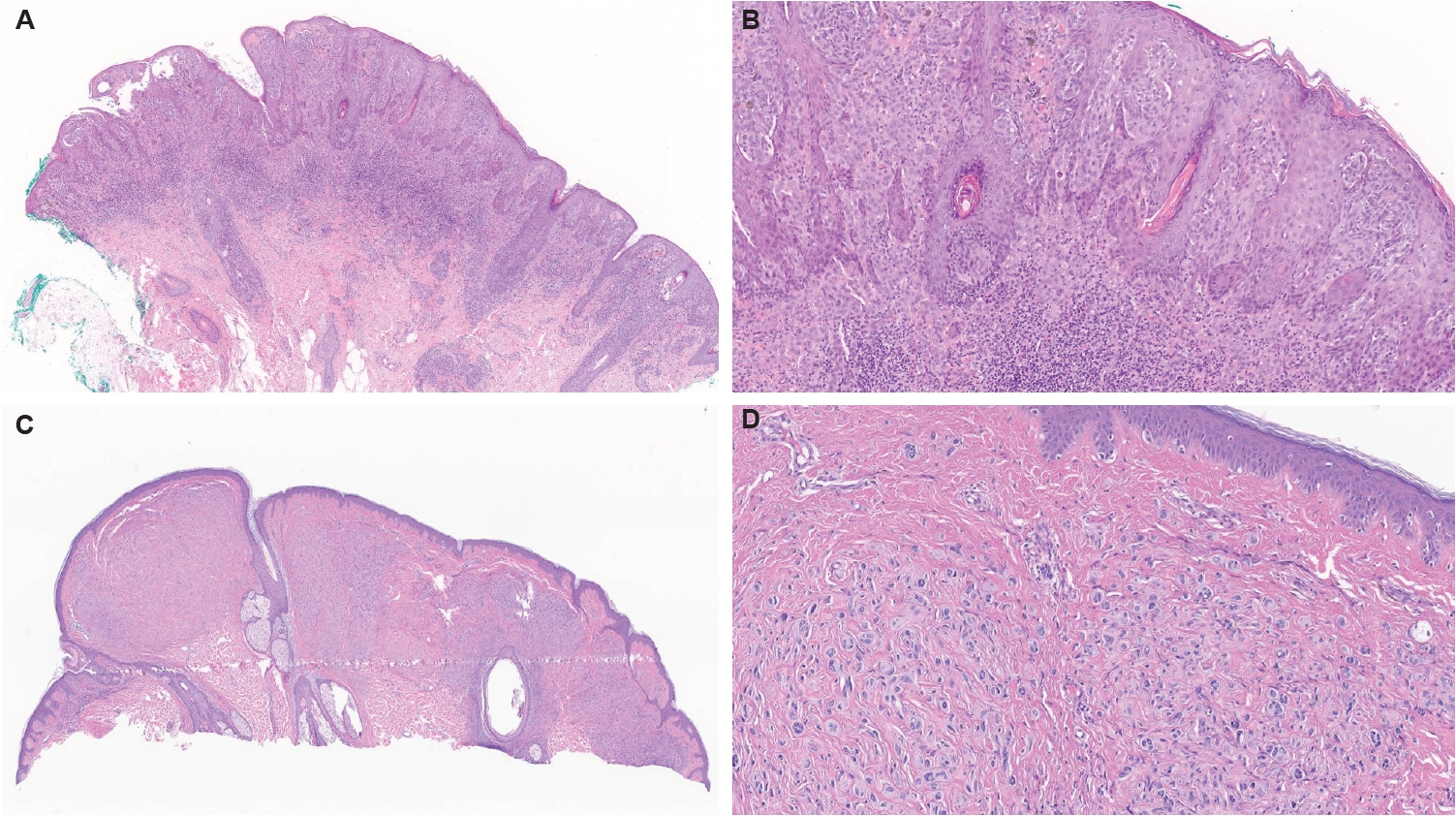
- The scope of gene fusions in melanocytic neoplasms is broader than previously recognized, extending well beyond the Spitz-lineage neoplasms where kinase fusions involving ALK, ROS1, NTRK1/2/3, RET, MET, BRAF, and MAP3K8 define biologically and morphologically distinct tumors. Emerging studies demonstrate that a meaningful proportion of conventional non-Spitz lineage melanomas harbor oncogenic fusions. Such fusions may impact clinical behavior, histopathologic presentation and provide opportunities for targeted therapy. The World Health Organization classification of skin tumors, 5th edition, now incorporates fusion status into taxonomy and risk stratification, yet some important questions remain for further investigation: fusion-associated neoplasms can mimic non-melanocytic neoplasm; Spitz-type fusions appear in non-Spitz lesions; and melanocytic differentiation may occur in some other fusion-driven lesions. Broad-panel next-generation sequencing (including RNAseq), together with targeted fluorescence in situ hybridization and immunohistochemistry enhances detection of known and novel fusion partners. Early clinical evidence of TRK, ALK, and ROS1 inhibitor efficacy underscores the translational promise of fusion testing and opens avenues for personalized therapy. This review synthesizes current knowledge on the genomics, histopathology, diagnosis, and therapeutic implications of fusion-driven melanocytic neoplasms, highlighting consensus points and remaining controversies.
-
Citations
Citations to this article as recorded by- Clinicopathologic and molecular characteristics of acral melanomas harboring RARA fusions
Mokhtar H. Abdelhammed, Richard K. Yang, Volha Lenskaya, Carlos A. Torres-Cabala, Woo Cheal Cho
Human Pathology.2026; 177: 106211. CrossRef - Targeted ALK inhibition achieving tumour control in metastatic melanoma: a case report
Cornelia Schuster, Irja Alida Oppedal , Liv Iren Vinnem, Eirin Lunden Abrahamsson, Torill Myklestad Barrett, Eli Sihn Samdal Steinskog, Hege Elisabeth Giercksky Russnes , Åslaug Helland
Acta Oncologica.2026; 65: 814. CrossRef
- Clinicopathologic and molecular characteristics of acral melanomas harboring RARA fusions
Newsletters
- What’s new in neoplastic pulmonary pathology 2026: updates on grading, staging and molecular testing
- Andreas Tiefenbacher, Luka Brčić
- J Pathol Transl Med. 2026;60(5):587-591. Published online August 31, 2026
- DOI: https://doi.org/10.4132/jptm.2026.07.28
- 2,249 View
- 180 Download
-
Abstract
PDF
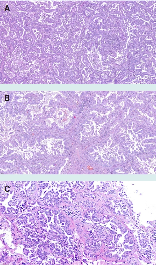
- Neoplastic pulmonary pathology has continued to advance in recent years, including the introduction of histologic grading systems for both adenocarcinoma and squamous cell carcinoma, implementation of the UICC TNM 9th Edition classification, expanded molecular testing requirements, refinements in neuroendocrine tumor classification, and improvements in mesothelioma diagnostics. This newsletter summarizes the most important developments relevant to practicing pathologists.
- What’s new in medical renal pathology 2025: Updates on podocytopathy and immunofluorescence staining in medical kidney
- Astrid Weins, Ibrahim Batal, Paola Romagnani, Geetika Singh, Rahul Raj, Nicole Andeen, Jonathan Zuckerman, Martina Uzzo, Mariam Priya Alexander, Anjali Satoskar
- J Pathol Transl Med. 2025;59(4):269-272. Published online July 10, 2025
- DOI: https://doi.org/10.4132/jptm.2025.06.19

- 11,912 View
- 569 Download
- 1 Web of Science
- 2 Crossref
-
Abstract
PDF
- Diffuse podocytopathy, including minimal change disease and primary focal segmental glomerulosclerosis, is a common cause of nephrotic syndrome in adults and children. It is increasingly recognized to be autoimmune-mediated associated with anti-nephrin and other emerging anti-slit diaphragm antibodies, and can recur in the kidney allograft. Immunofluorescence is routinely used in evaluation of kidney biopsies, and updates include those on fibrillar diseases, monoclonal staining, lupus-like staining, and use of antibody KM55 in IgA-dominant glomerulonephritis.
-
Citations
Citations to this article as recorded by- Opportunities and challenges in recurrent diffuse podocytopathy post-transplantation: the critical value of the definition
Rachel Nuccitelli, Amadea Toutoungis, Elena Martinelli, Simone Sanna-Cherchi, Astrid Weins, Heather K. Morris, Andrew S. Bomback, Ibrahim Batal
Frontiers in Immunology.2026;[Epub] CrossRef - Circulating Nephrin Antibodies and Post-Transplant Recurrence of Diffuse Podocytopathies: A Systematic Review
Noppawit Aiumtrakul, Charat Thongprayoon, Jing Miao, Wisit Cheungpasitporn
Kidney Medicine.2026; : 101521. CrossRef
- Opportunities and challenges in recurrent diffuse podocytopathy post-transplantation: the critical value of the definition
Review Article
- Sentinel lymph node biopsy in melanoma: pathologic evaluation and diagnostic considerations
- Dre Barnachea, Bonnie Lee
- J Pathol Transl Med. 2026;60(5):493-501. Published online September 1, 2026
- DOI: https://doi.org/10.4132/jptm.2026.07.01

- 2,154 View
- 61 Download
-
Abstract
PDF
- Sentinel lymph node biopsy (SLNB) is a widely used staging procedure in melanoma that provides important prognostic information and guides clinical management. The sentinel lymph node (SLN), defined as the first lymph node in the lymphatic drainage pathway from a primary tumor, represents the most likely site of early regional metastasis. Accordingly, identification of metastatic melanoma within SLNs has significant implications for staging and risk stratification and is associated with worse clinical outcomes. The SLNB procedure involves preoperative and intraoperative lymphatic mapping techniques, including radiotracer localization with or without blue dye injection, which allow identification of SLNs. Histopathologic evaluation includes careful gross examination, serial sectioning, and immunohistochemical analysis using melanocytic markers such as SOX10, Melan-A, HMB45, and occasionally PRAME to detect metastatic disease. In addition, prognostic features such as tumor burden, extranodal extension, and microanatomic location of metastases within the lymph node further refine risk assessment. This review provides an overview of SLNB in melanoma, with emphasis on clinical indications, histopathologic evaluation, and key diagnostic and prognostic considerations.
Review
- Next step of molecular pathology: next-generation sequencing in cytology
- Ricella Souza da Silva, Fernando Schmitt
- J Pathol Transl Med. 2024;58(6):291-298. Published online November 7, 2024
- DOI: https://doi.org/10.4132/jptm.2024.10.22
- 11,820 View
- 478 Download
- 8 Web of Science
- 6 Crossref
-
Abstract
PDF
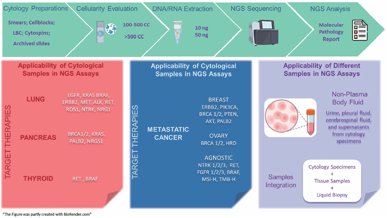
- The evolving landscape of precision oncology underscores the pivotal shift from morphological diagnosis to treatment decisions driven by molecular profiling. Recent guidelines from the European Society for Medical Oncology recomend the use of next-generation sequencing (NGS) across a broader range of cancers, reflecting its superior efficiency and clinical value. NGS not only updates oncology testing by offering quicker, sample-friendly, and sensitive analysis but also reduces the need for multiple individual tests. Cytology samples, often obtained through less invasive methods, can yield high-quality genetic material suitable for molecular analysis. This article focuses on optimizing the use of cytology samples in NGS, and outlines their potential benefits in identifying actionable molecular alterations for targeted therapies across various solid tumors. It also addresses the need for validation studies and the strategies to incorporate or combine different types of samples into routine clinical practice. Integrating cytological and liquid biopsies into routine clinical practice, alongside conventional tissue biopsies, offers a comprehensive approach to tumor genotyping, early disease detection, and monitoring of therapeutic responses across various solid tumor types. For comprehensive biomarker characterization, all patient specimens, although limited, is always valuable.
-
Citations
Citations to this article as recorded by- Unraveling the nexus: Tumor mutational burden, PD‐L1 expression, and oncogenic alterations in non–small cell lung cancer cytology specimens
Min Dai, Francis Anthony San Lucas, Hector Alvarez, Leomar Ballester, Hui Chen, Keyur P. Patel, Asif Rashid, Shun Rao, Mark J. Routbort, Gloria Sura, Keith Sweeney, Gokce Toruner, Peng Wei, Richard Yang, Hyvan Dang, Rajyalakshmi Luthra, Sinchita Roy‐Chowd
Cancer Cytopathology.2026;[Epub] CrossRef - The critical role of accurate neoplastic cell percentage (NCP) assessment: investigating targeted training strategies for pulmonary biopsy and cytology specimens
Thi Mai Phuong Pham, Dieter Peeters, Jan von der Thüsen, Myriam Remmelink, Birgit Weynand, Elisabeth Dequeker
Virchows Archiv.2026;[Epub] CrossRef - Benign Biliary Tumors and Precursor Neoplasms: An Updated Clinicopathological and Molecular Review Based on the 2026 WHO Classification
Joon Hyuk Choi
Biomedicines.2026; 14(7): 1548. CrossRef - The World Health Organization Reporting System for Lymph Node, Spleen, and Thymus Cytopathology: Part 1 – Lymph Node
Immacolata Cozzolino, Mats Ehinger, Maria Calaminici, Andrea Ronchi, Mousa A. Al-Abbadi, Helena Barroca, Beata Bode-Lesniewska, David F. Chhieng, Ruth L. Katz, Oscar Lin, L. Jeffrey Medeiros, Martha Bishop Pitman, Arvind Rajwanshi, Fernando C. Schmitt, Ph
Acta Cytologica.2025; 70(2): 185. CrossRef - The impact of cytological preparation techniques on RNA quality: A comparative study on smear samples
Cisel Aydin Mericoz, Gulsum Caylak, Elif Sevin Sanioglu, Zeynep Seçil Satilmis, Ayse Humeyra Dur Karasayar, Ibrahim Kulac
Cancer Cytopathology.2025;[Epub] CrossRef - Reimagining cytopathology in the molecular era: Integration or fragmentation?
Sumanta Das, R. Naveen Kumar, Biswajit Dey, Pranjal Kalita
Cytojournal.2025; 22: 94. CrossRef
- Unraveling the nexus: Tumor mutational burden, PD‐L1 expression, and oncogenic alterations in non–small cell lung cancer cytology specimens
Original Articles
- Thoracic aortic calcification as a predictor of coronary artery disease: a systematic review and meta-analysis
- Hussein Nafakhi, Alaa Salah Jumaah, Akeel Abed Yasseen
- J Pathol Transl Med. 2025;59(3):161-170. Published online April 30, 2025
- DOI: https://doi.org/10.4132/jptm.2025.03.05
- 14,011 View
- 202 Download
- 3 Web of Science
- 3 Crossref
-
Abstract
PDFSupplementary Material
- Background
The relationship between coronary atherosclerosis (progression, outcome) and calcification in the thoracic aorta (TAC), particularly across its various segments, is complex and often shows conflicting associations in the literature. To address this debated and complex relationship, we aimed to evaluate how TAC and its segments correlate with the presence and severity of coronary artery disease (CAD).
Methods
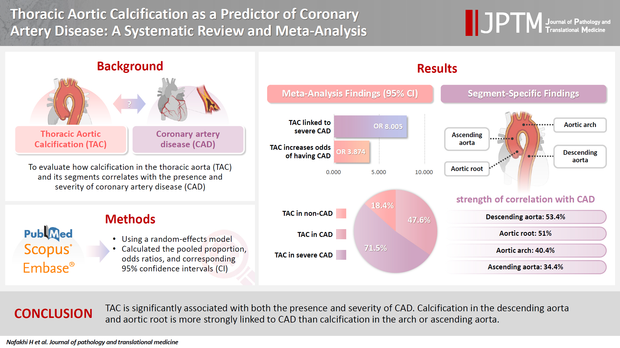
We reviewed all articles published between January 1990 and September 2024 that examined the link between TAC and CAD and were indexed in PubMed, Scopus, or EMBASE. Using a random-effects model, we calculated pooled proportions, odds ratios, and corresponding 95% confidence intervals (CIs) to evaluate the association between TAC and CAD, with consideration of severity.
Results
The study included 17 studies with 8,187 participants, 2,775 of whom had CAD (1,059 with severe CAD), and 5,412 of whom did not. The pooled odds ratio of TAC in patients with CAD compared to that in those without was 3.874 (95% CI, 2.789 to 5.381). For severe CAD versus mild CAD, the odds ratio was 8.005 (95% CI, 2.611 to 24.542). Calcification of the aortic root (pooled proportion, 51%; 95% CI, 0.282 to 0.733) or descending aorta (pooled proportion, 53.4%; 95% CI, 0.341 to 0.718) had the strongest association with CAD compared to calcification of the arch or ascending aorta.
Conclusions
TAC is significantly associated with both the presence and severity of CAD. Calcification in the descending aorta and aortic root is more strongly linked to CAD than calcification in the arch or ascending aorta. -
Citations
Citations to this article as recorded by- International Liver Transplantation Society/Liver Intensive Care Group of Europe guidelines for cardiovascular assessment before liver transplantation
Emmanuel Weiss, Gonzalo Crespo, Alexandra Anderson, Gianni Biancofiore, Ryan Chadha, Jacek B. Cywinski, Andrea De Gasperi, James Findlay, Marc Giménez-Milà, Constantine Karvellas, Michael Kaufman, Ashish Malik, Marina Moguilevitch, Sher-Lu Pai, Koen Reynt
American Journal of Transplantation.2026; 26(2): 219. CrossRef - Segmental thoracic aorta calcification in diabetic patients: Relationship with coronary atherosclerosis burden
Wasan Kadhum Abbas, Abdulameer A. Al-Mosawi, Ali M. Al-Shirazi, Hussein Nafakhi, Hayder Nafakhi
Journal of Diabetes & Metabolic Disorders.2026;[Epub] CrossRef - Association of serum uric acid with incident vascular calcification in patients with type 2 diabetes mellitus: a retrospective cohort study
Xinyu Li, Taoyuan He, Guosheng Li, Xue Liang, Bing Wang, Yudan Liu, Wei Zhao, Zhu Zhu, Xuhan Liu
Frontiers in Endocrinology.2026;[Epub] CrossRef
- International Liver Transplantation Society/Liver Intensive Care Group of Europe guidelines for cardiovascular assessment before liver transplantation
- Diagnostic yield of fine needle aspiration with simultaneous core needle biopsy for thyroid nodules
- Mohammad Ali Hasannia, Ramin Pourghorban, Hoda Asefi, Amir Aria, Elham Nazar, Hojat Ebrahiminik, Alireza Mohamadian
- J Pathol Transl Med. 2025;59(3):180-187. Published online April 16, 2025
- DOI: https://doi.org/10.4132/jptm.2025.03.04

- 17,725 View
- 303 Download
- 2 Web of Science
- 1 Crossref
-
Abstract
PDF
- Background
Fine needle aspiration (FNA) is a widely utilized technique for assessing thyroid nodules; however, its inherent non-diagnostic rate poses diagnostic challenges. The present study aimed to evaluate and compare the diagnostic efficacy of FNA, core needle biopsy (CNB), and their combined application in the assessment of thyroid nodules.
Methods
A total of 56 nodules from 50 patients was analyzed using both FNA and simultaneous CNB. The ultrasound characteristics were categorized according to the American College of Radiology Thyroid Imaging Reporting and Data Systems classification system. The study compared the sensitivity, specificity, and accuracy of FNA, CNB, and the combination of the two techniques.
Results
The concordance between FNA and CNB was notably high, with a kappa coefficient of 0.837. The sensitivity for detecting thyroid malignancy was found to be 25.0% for FNA, 66.7% for CNB, and 83.3% for the combined FNA/CNB approach, with corresponding specificities of 84.6%, 97.4%, and 97.4%. The accuracy of the FNA/CNB combination was the highest at 94.1%.
Conclusions
The findings of this study indicate that both CNB and the FNA/CNB combination offer greater diagnostic accuracy for thyroid malignancy compared to FNA alone, with no significant complications reported. Integrating CNB with FNA findings may enhance management strategies and treatment outcomes for patients with thyroid nodules. -
Citations
Citations to this article as recorded by- Clinicopathological Correlation of Core-Needle Biopsy in Thyroid Nodule Assessment: Insights from a Tertiary Care Center
Neha Nigam, Rishabh Sahai, Anil Singh, Anjali Mishra, Sabaretnam Mayilvaganan, Ojas Gupta, Prabhakar Mishra, Nirbhay Kumar, Amit Rastogi
Journal of Cytology.2026; 43(4): 235. CrossRef
- Clinicopathological Correlation of Core-Needle Biopsy in Thyroid Nodule Assessment: Insights from a Tertiary Care Center
Review
- Diagnosis of interstitial lung diseases: from Averill A. Liebow to artificial intelligence
- Eunhee S. Yi, Paul Wawryko, Jay H. Ryu
- J Pathol Transl Med. 2024;58(1):1-11. Published online January 10, 2024
- DOI: https://doi.org/10.4132/jptm.2023.11.17
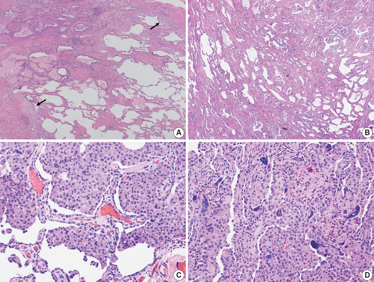
- 13,447 View
- 469 Download
- 3 Web of Science
- 4 Crossref
-
Abstract
PDF
- Histopathologic criteria of usual interstitial pneumonia (UIP)/idiopathic pulmonary fibrosis (IPF) were defined over the years and endorsed by leading organizations decades after Dr. Averill A. Liebow first coined the term UIP in the 1960s as a distinct pathologic pattern of fibrotic interstitial lung disease. Novel technology and recent research on interstitial lung diseases with genetic component shed light on molecular pathogenesis of UIP/IPF. Two antifibrotic agents introduced in the mid-2010s opened a new era of therapeutic approaches to UIP/IPF, albeit contentious issues regarding their efficacy, side effects, and costs. Recently, the concept of progressive pulmonary fibrosis was introduced to acknowledge additional types of progressive fibrosing interstitial lung diseases with the clinical and pathologic phenotypes comparable to those of UIP/IPF. Likewise, some authors have proposed a paradigm shift by considering UIP as a stand-alone diagnostic entity to encompass other fibrosing interstitial lung diseases that manifest a relentless progression as in IPF. These trends signal a pendulum moving toward the tendency of lumping diagnoses, which poses a risk of obscuring potentially important information crucial to both clinical and research purposes. Recent advances in whole slide imaging for digital pathology and artificial intelligence technology could offer an unprecedented opportunity to enhance histopathologic evaluation of interstitial lung diseases. However, current clinical practice trends of moving away from surgical lung biopsies in interstitial lung disease patients may become a limiting factor in this endeavor as it would be difficult to build a large histopathologic database with correlative clinical data required for artificial intelligence models.
-
Citations
Citations to this article as recorded by- Advances and Research Progress in the Application of Artificial Intelligence for Progressive Pulmonary Fibrosis in Connective Tissue Disease-Associated Interstitial Lung Disease
佳琳 王
Advances in Clinical Medicine.2026; 16(05): 2107. CrossRef - Lung biopsy strategies for childhood interstitial lung disease: Indications and techniques
Kuimiao Deng, Lin Lin, Wenhao Zhou, Junzheng Peng
Journal of International Medical Research.2026;[Epub] CrossRef - Identification of early genes in the pathophysiology of fibrotic interstitial lung disease in a new model of pulmonary fibrosis
Nathan Hennion, Corentin Bedart, Léonie Vandomber, Frédéric Gottrand, Sarah Humez, Cécile Chenivesse, Jean-Luc Desseyn, Valérie Gouyer
Cellular and Molecular Life Sciences.2025;[Epub] CrossRef - Radiological Insights into UIP Pattern: A Comparison Between IPF and Non-IPF Patients
Stefano Palmucci, Miriam Adorna, Angelica Rapisarda, Alessandro Libra, Sefora Fischetti, Gianluca Sambataro, Letizia Antonella Mauro, Emanuele David, Pietro Valerio Foti, Claudia Mattina, Corrado Spatola, Carlo Vancheri, Antonio Basile
Journal of Clinical Medicine.2025; 14(12): 4162. CrossRef
- Advances and Research Progress in the Application of Artificial Intelligence for Progressive Pulmonary Fibrosis in Connective Tissue Disease-Associated Interstitial Lung Disease
Review Articles
- Spread through air spaces as a marker of nonlinear evolutionary trajectories in lung cancer: implications for the 9th edition TNM classification
- Jin-Haeng Chung
- J Pathol Transl Med. 2026;60(5):463-475. Published online September 15, 2026
- DOI: https://doi.org/10.4132/jptm.2026.06.16

- 2,059 View
- 31 Download
-
Abstract
PDF
- Recent advancements in the diagnosis and therapeutic management of lung cancer have contributed to an evolution in pathological reporting standards. The 8th edition of the TNM classification of lung tumors has introduced the invasive size of adenocarcinoma—rather than total tumor size—as the primary determinant for staging. Concurrently, histological grading criteria proposed by the International Association for the Study of Lung Cancer (IASLC), based on predominant and highest-grade architectural patterns, appear to offer improved clinical utility over legacy systems. In addition, tumor spread through air spaces (STAS) has emerged as an actively investigated pattern of invasion, characterized by the presence of neoplastic cells beyond the main tumor mass within the adjacent pulmonary parenchyma. STAS is frequently observed in advanced-stage and node-positive non–small cell lung cancer and often correlates with distant metastasis. Recognizing its clinical relevance, the IASLC Staging Project recently recommended incorporating STAS as a histologic T descriptor in the 9th edition of the TNM classification. Expanding upon this framework, this review explores STAS not merely as a prognostic indicator, but as a potential reflection of divergent evolutionary trajectories influenced by underlying driver mutations. Accordingly, this review aims to comprehensively synthesize the prevailing controversies and recent academic milestones regarding STAS, the current limitations in preoperative STAS risk prediction, the logistical hurdles of intraoperative diagnosis, and critically evaluates the need for prospective clinical trials to validate STAS as a predictive biomarker for adjuvant systemic therapy.
- T-lymphoblastic leukemia/lymphoma: a comprehensive review of pathology, molecular features, and differential diagnosis
- David Danielson, Ian T. Lagerstrom, Max Rogers, Kyle Simon, Nadine S. Aguilera, Aaron Auerbach
- J Pathol Transl Med. 2026;60(5):502-515. Published online September 15, 2026
- DOI: https://doi.org/10.4132/jptm.2026.08.01

- 1,966 View
- 42 Download
-
Abstract
PDF
- T-lymphoblastic leukemia/lymphoma (T-ALL/LBL) is an aggressive neoplasm of immature T-lymphoid precursors that is classified as T-cell acute lymphoblastic leukemia when the primary site of involvement is the bone marrow and peripheral blood and as T-cell lymphoblastic lymphoma when it presents as a solid mass, most commonly in the anterior mediastinum. The neoplastic cells are defined by expression of T-lineage antigens (cytoplasmic or surface CD3) together with one or more markers of immaturity (terminal deoxynucleotidyl transferase, CD1a, CD34, CD99, or CD117) and must not fulfill criteria for early T-cell precursor acute lymphoblastic leukemia (ALL). This entity shows a marked male predominance (male:female ≈ 2:1) and predominantly affects children, adolescents, and young adults, accounting for approximately 15% of childhood ALL and 20%–25% of adult ALL cases. Pathologic diagnosis increasingly relies on recurrent molecular alterations such as activating NOTCH1 mutations (>60% of cases), transcription factor rearrangements, and cell-cycle regulator inactivation. Although intensive multiagent chemotherapy regimens have substantially improved outcomes, particularly in pediatric patients, adults and high-risk molecular subgroups continue to experience inferior survival. This review provides a practical, pathology-oriented update on the epidemiology, pathogenesis, diagnostic criteria, differential diagnosis, prognostic factors, and current treatment approaches for T-ALL/LBL.
Newsletters
- What’s new in genitourinary pathology 2023: WHO 5th edition updates for urinary tract, prostate, testis, and penis
- Bonnie Choy, Maria Tretiakova, Debra L. Zynger
- J Pathol Transl Med. 2024;58(1):45-48. Published online December 27, 2023
- DOI: https://doi.org/10.4132/jptm.2023.12.11

- 16,538 View
- 1,047 Download
- 3 Web of Science
- 3 Crossref
-
Abstract
PDF
- The 5th edition WHO Classification of Urinary and Male Genital Tumours (2022) introduced many significant changes relevant to urologic daily practice, mainly to renal tumors which was covered in the What’s New newsletter in September 2022. In this newsletter, we summarize the notable changes to bladder, prostate, testis, and penis based on the 5th edition of the WHO.
-
Citations
Citations to this article as recorded by- Predicting variant histology in bladder cancer: the role of multiparametric MRI and vesical imaging-reporting and data system (VI-RADS)
Serdar Aslan, Merve Nur Tasdemir, Ertugrul Cakir, Ural Oguz, Birgul Tok
Abdominal Radiology.2025; 50(10): 4700. CrossRef - Pictorial review of multiparametric MRI in bladder urothelial carcinoma with variant histology: pearls and pitfalls
Yuki Arita, Sungmin Woo, Lisa Ruby, Thomas C. Kwee, Keisuke Shigeta, Ryo Ueda, Sunny Nalavenkata, Hiromi Edo, Kosuke Miyai, Jeeban Das, Pamela I. Causa Andrieu, Hebert Alberto Vargas
Abdominal Radiology.2024; 49(8): 2797. CrossRef - Oncological outcomes and prognostic implications of T1 histo-anatomic substaging in the management of high-Grade non-muscle invasive bladder cancer: results from a large single centre series
Marco Finati, Antonio Fanelli, Francesco Cinelli, Nicola Schiavone, Ugo Giovanni Falagario, Anna Ricapito, Nicola d’Altilia, Richard Naspro, Angelo Porreca, Felice Crocetto, Biagio Barone, Ciro Imbimbo, Carlo Bettocchi, Francesca Sanguedolce, Luigi Cormio
World Journal of Urology.2024;[Epub] CrossRef
- Predicting variant histology in bladder cancer: the role of multiparametric MRI and vesical imaging-reporting and data system (VI-RADS)
- What’s new in adrenal gland pathology: WHO 5th edition for adrenal cortex
- Carol N. Rizkalla, Maria Tretiakova
- J Pathol Transl Med. 2024;58(4):201-204. Published online June 25, 2024
- DOI: https://doi.org/10.4132/jptm.2024.06.07

- 11,338 View
- 634 Download
- 2 Web of Science
- 3 Crossref
-
Abstract
PDF
- The 5th edition of WHO Classification of Endocrine and Neuroendocrine Tumors (2022) introduced many significant changes relevant to endocrine daily practice. In this newsletter, we summarize the notable changes to the adrenal cortex based on the 5th edition of the WHO classification [1].
-
Citations
Citations to this article as recorded by- Contemporary diagnostic criteria for adrenocortical neoplasms: from morphology to molecular classification
D. V. Bulanov, F. M. Kolzin, S. A. Dadayan, E. Yu. Berdyugina, I. I. Polubkov
Research and Practical Medicine Journal.2026; 13(2): 121. CrossRef - Giant adrenal pseudocyst with unclear preoperative origin: a case report and literature review
Shouyi Long, Bin Xiong, Zhen Long, Bo Li, Xiujun Yuan, Shulian Chen
Frontiers in Surgery.2026;[Epub] CrossRef - Ectopic adrenal gland in the liver leading to a misdiagnosis of hepatocellular carcinoma: A case report
Min-Qiu Qin, Yi-Peng Zhao, Ju-Ping Xie
World Journal of Hepatology.2025;[Epub] CrossRef
- Contemporary diagnostic criteria for adrenocortical neoplasms: from morphology to molecular classification
Original Article
- Significance of KM55 immunohistochemical staining in the diagnosis and prognosis of IgA nephropathy
- Hoe In Jeong, Beom Jin Lim, Minsun Jung
- J Pathol Transl Med. 2026;60(1):69-82. Published online January 14, 2026
- DOI: https://doi.org/10.4132/jptm.2025.09.17

- 7,607 View
- 293 Download
- 2 Web of Science
- 2 Crossref
-
Abstract
PDF
- Background
Galactose-deficient IgA1 (Gd-IgA1) plays a crucial role in IgA nephropathy (IgAN). The monoclonal antibody KM55 has emerged as a simplified method for detecting Gd-IgA1; however, the clinicopathological significance of immunohistochemistry for Gd-IgA1 remains underexplored. This study evaluated the prognostic and clinicopathological significance of KM55 immunohistochemistry in IgAN. Methods: A total of 114 native kidney biopsies showing at least mild mesangial IgA positivity on immunofluorescence were retrospectively analyzed. Patients were categorized as having IgAN or non-IgAN diseases. The KM55 immunohistochemical staining was graded as 0, 1+, 2+, 3, or 4+. Data on Oxford classification, laboratory parameters, and renal outcomes were collected. Results: The IgAN group showed significantly higher KM55 scores than the non-IgAN group (median: 3 vs. 1; p < .001). IgAN cases were further stratified into KM55-high (≥3+, n = 38) and -low groups (≤2+, n = 37). The KM55-high group had significantly higher diastolic blood pressure, blood urea nitrogen, creatinine, urine protein/creatinine ratio, and Oxford mesangial hypercellularity scores, along with lower estimated glomerular filtration rate (eGFR) and serum albumin. Cox analysis revealed significantly poorer outcomes in the KM55-high group for chronic kidney disease stage 4 (p = .015), end-stage renal disease (p = .024), and 75% eGFR decline (p = .016). Conclusions: Mesangial Gd-IgA1 deposition graded by KM55 immunohistochemistry may be a useful adjunct for IgAN diagnosis and a potential prognostic biomarker. -
Citations
Citations to this article as recorded by- IgA Nephropathy: Mechanisms, Risk Stratification, and Precision Therapy
Sami Alobaidi
Diagnostics.2026; 16(9): 1259. CrossRef - Development and validation of a predictive model for rapid renal function decline in early IgA nephropathy: a multicenter retrospective study
Junan Zhou, Yang Liu, Lixia Huo, Xu Wu, Zhelun Zhou, Haodong Liu, Rong Wu, Jiaai Zhang, Yiqing Yu, Deyong Fan, Xiaoyi Wang
Frontiers in Medicine.2026;[Epub] CrossRef
- IgA Nephropathy: Mechanisms, Risk Stratification, and Precision Therapy
Review
- Clinical practice recommendations for the use of next-generation sequencing in patients with solid cancer: a joint report from KSMO and KSP
- Miso Kim, Hyo Sup Shim, Sheehyun Kim, In Hee Lee, Jihun Kim, Shinkyo Yoon, Hyung-Don Kim, Inkeun Park, Jae Ho Jeong, Changhoon Yoo, Jaekyung Cheon, In-Ho Kim, Jieun Lee, Sook Hee Hong, Sehhoon Park, Hyun Ae Jung, Jin Won Kim, Han Jo Kim, Yongjun Cha, Sun Min Lim, Han Sang Kim, Choong-Kun Lee, Jee Hung Kim, Sang Hoon Chun, Jina Yun, So Yeon Park, Hye Seung Lee, Yong Mee Cho, Soo Jeong Nam, Kiyong Na, Sun Och Yoon, Ahwon Lee, Kee-Taek Jang, Hongseok Yun, Sungyoung Lee, Jee Hyun Kim, Wan-Seop Kim
- J Pathol Transl Med. 2024;58(4):147-164. Published online January 10, 2024
- DOI: https://doi.org/10.4132/jptm.2023.11.01
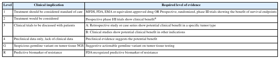
- 14,990 View
- 512 Download
- 2 Web of Science
- 2 Crossref
-
Abstract
PDF
- In recent years, next-generation sequencing (NGS)–based genetic testing has become crucial in cancer care. While its primary objective is to identify actionable genetic alterations to guide treatment decisions, its scope has broadened to encompass aiding in pathological diagnosis and exploring resistance mechanisms. With the ongoing expansion in NGS application and reliance, a compelling necessity arises for expert consensus on its application in solid cancers. To address this demand, the forthcoming recommendations not only provide pragmatic guidance for the clinical use of NGS but also systematically classify actionable genes based on specific cancer types. Additionally, these recommendations will incorporate expert perspectives on crucial biomarkers, ensuring informed decisions regarding circulating tumor DNA panel testing.
-
Citations
Citations to this article as recorded by- Apport de la génomique dans la prise en charge des cancers
Étienne Rouleau, Lucie Karayan-Tapon, Marie-Dominique Galibert, Alexandre Harlé, Isabelle Soubeyran
Revue Francophone des Laboratoires.2025; 2025(568): 67. CrossRef - The Redox–Adhesion–Exosome (RAX) Hub in Cancer: Lipid Peroxidation-Driven EMT Plasticity and Ferroptosis Defense with HNE/MDA Signaling and Lipidomic Perspectives
Moon Nyeo Park, Jinwon Choi, Rosy Iara Maciel de Azambuja Ribeiro, Domenico V. Delfino, Seong-Gyu Ko, Bonglee Kim
Antioxidants.2025; 14(12): 1474. CrossRef
- Apport de la génomique dans la prise en charge des cancers
Review Article
- Obesity-driven adipokine signaling in breast cancer: mechanistic insights, therapeutic challenges, and opportunities for precision oncology
- Vadakke Kunnumma Soonu, Praveen Kumar Shenoy, Valiyaparampil Gopi Deepak Roshan, Vipin Gopinath, Krishnan Sreejith
- J Pathol Transl Med. 2026;60(5):476-492. Published online September 9, 2026
- DOI: https://doi.org/10.4132/jptm.2026.06.30
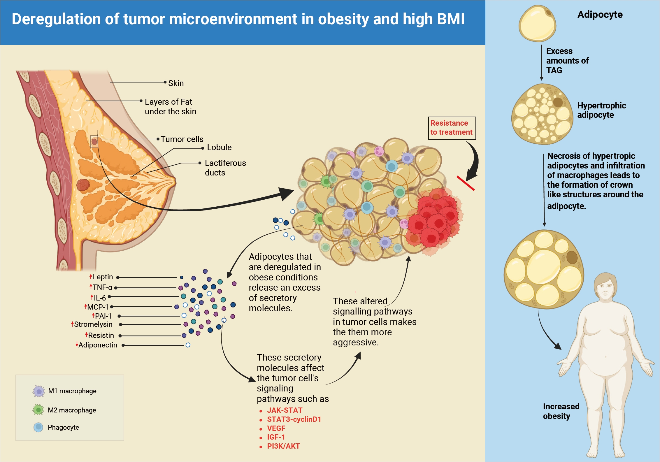
- 1,795 View
- 30 Download
-
Abstract
PDF
- Breast cancer is the leading cause of illness and death among women worldwide, with more than 2.3 million new cases diagnosed each year. The incidence and fatality rates are steadily increasing, notably in Asian countries. Obesity has been established as a major and growing risk factor for breast cancer progression. The World Health Organization reports that adult obesity rates have doubled since 1990. Obesity promotes tumor growth by inhibiting adipokine production and activating cancer-promoting pathways. In obese people, the microenvironment surrounding breast cancer cells is drastically altered. This is mostly due to the malfunctioning of adipocytes (fat cells) and macrophages. These defective cells' adipokines alter signaling pathways required for cancer cell proliferation, survival, and inflammation. This dysregulation has a significant role in tumor development, metastasis, and resistance to traditional cancer treatments, particularly in obese patients. This review highlights the role of adipokines in breast cancer, with a focus on disease progression, therapeutic challenges, and knowledge gaps. Clarifying how obesity alters tumor biology is key to advancing personalized and effective treatments for obese patients.
Original Articles
- Clinicopathological and molecular mechanisms of CLDN18.2 in gastric cancer aggressiveness: a high-risk population study with multi-omics profiling
- Hengquan Wu, Mei Li, Gang Wang, Peiqing Liao, Peng Zhang, Luxi Yang, Yumin Li, Tao Liu, Wenting He
- J Pathol Transl Med. 2026;60(1):47-57. Published online January 5, 2026
- DOI: https://doi.org/10.4132/jptm.2025.09.11
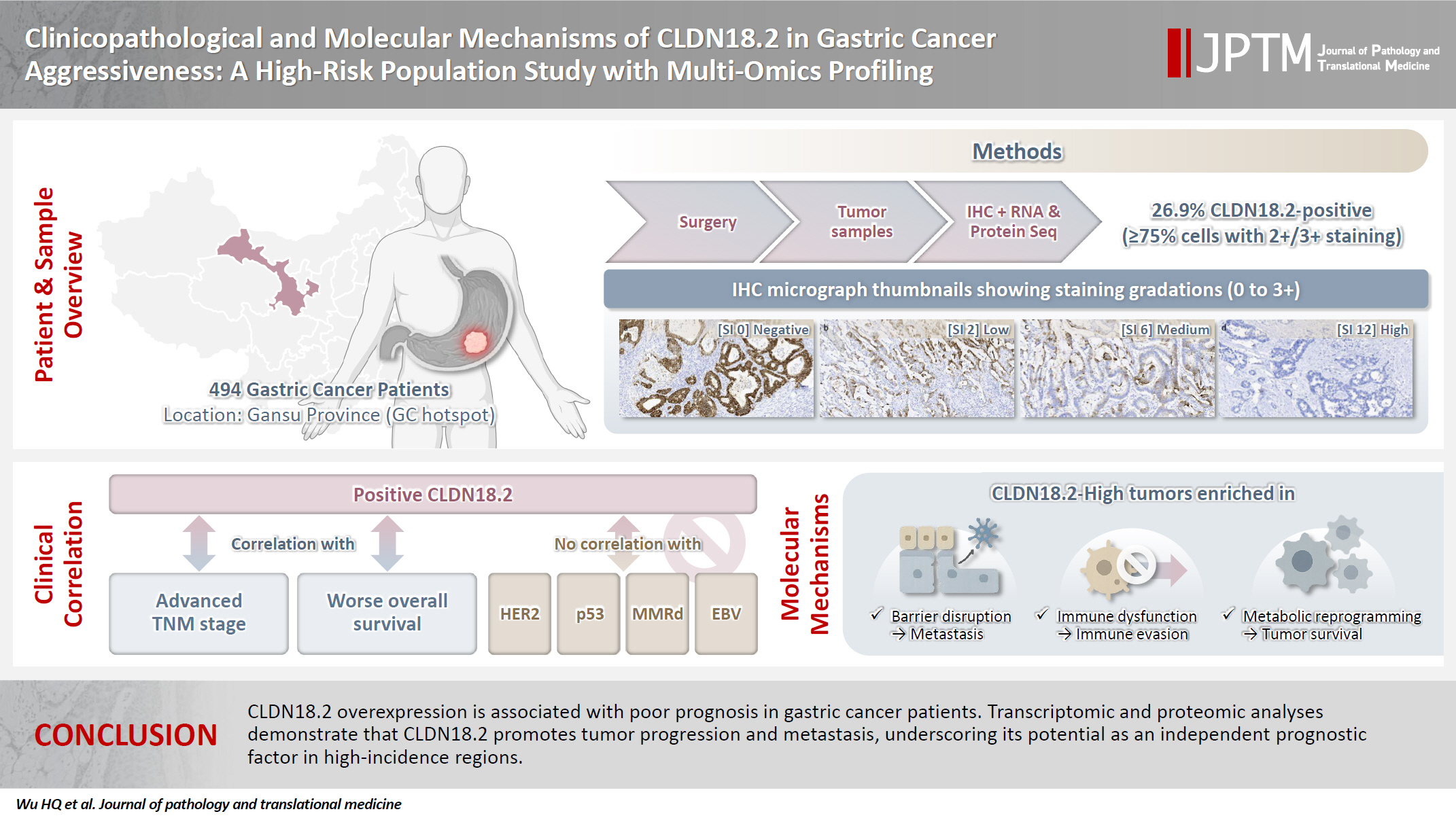
- 5,956 View
- 315 Download
- 4 Web of Science
- 4 Crossref
-
Abstract
PDFSupplementary Material
- Background
The tight junction protein claudin18.2 (CLDN18.2) has been implicated in poor prognosis and suboptimal immunotherapy response in gastric cancer (GC). This study investigates the clinicopathological relevance of CLDN18.2 expression and its association with molecular subtypes in GC patients from a high-incidence region, combining transcriptomic and proteomic approaches to explore how CLDN18.2 contributes to progression and metastasis.
Methods
A retrospective cohort of 494 GC patients (2019–2024) underwent immunohistochemical analysis for CLDN18.2, Epstein-Barr virus (Epstein–Barr virus–encoded RNA), p53, human epidermal growth factor receptor 2 (HER2), and mismatch repair proteins (MLH1, MSH2, PMS2, and MSH6). CLDN18.2 positivity was defined as moderate to strong (2+/3+) membranous staining in ≥75% of tumor cells. Clinicopathological correlations, biomarker associations, and survival outcomes were evaluated. Transcriptomic and proteomic sequencing was performed to explore molecular mechanisms.
Results
CLDN18.2 positivity was observed in 26.9% (133/494) of gastric adenocarcinomas. CLDN18.2-positive tumors correlated with TNM stage (p = .003) and shorter overall survival (p = .018). No associations were identified with age, sex, HER2 status, microsatellite instability, or Epstein-Barr virus infection. Transcriptomic profiling revealed CLDN18.2-high tumors enriched in pathways involving cell junction disruption, signaling regulation, and immune modulation. Proteomic profiling showed that tumors with high CLDN18.2 were enriched in multiple mechanism-related pathways such as integrated metabolic reprogramming, cytoskeletal recombination, immune microenvironment dysregulation, and pro-survival signaling. These mechanisms may collectively contribute to tumor progression and metastasis.
Conclusions
CLDN18.2 overexpression is associated with poor prognosis in GC patients. Transcriptomic and proteomic analyses demonstrate that CLDN18.2 promotes tumor progression and metastasis, underscoring its potential as an independent prognostic factor in regions with a high incidence of GC. -
Citations
Citations to this article as recorded by- The evolving role of OMICS in gastrointestinal tumor biology and clinical practice
Qing Li, Junfeng Zhang, Junli Chen, Qiang Zhang, Ruihan Liu, Jialin Zhu, Yi Qing, Xi Wei, Jianpeng Sheng
Molecular Cancer.2026;[Epub] CrossRef - Pretreatment Claudin-18.2 Expression Predicts Poorer Survival Outcomes in Locally Advanced Gastric Cancer Treated with Perioperative Chemotherapy
Gürkan Gül, Özlem Kutlu, Asuman Argon, Halil Taşkaynatan, Özlem Özdemir
Diagnostics.2026; 16(9): 1277. CrossRef - Prevalence of Claudin 18.2 Expression in Gastric and Gastroesophageal Junction Adenocarcinoma: A Systematic Review and Meta-Analysis
Fazal Elahi Khan, Rajendranandini Majumdar, Mohan Dodeja, Gabriel Amorim Moreira Alves, Amr Harby, Rida Siddiqui, Shahbaz Madappattuparambil
Journal of Gastrointestinal Cancer.2026;[Epub] CrossRef - Comparative efficacy of CLDN18.2-directed regimens in advanced gastric/gastroesophageal junction adenocarcinoma: a meta-analysis of single-arm trials
Yue Yuan, Hongkai Yan, Qinhang Wu
Investigational New Drugs.2026;[Epub] CrossRef
- The evolving role of OMICS in gastrointestinal tumor biology and clinical practice
- HER2-low and ultralow breast cancer: interobserver challenges and lessons from a consensus study
- Jiwon Koh, Yoon Jin Cha, Eun Yoon Cho, Ahwon Lee, Ja Seung Koo, So Yeon Park, Min Hwan Kim, Jae Ho Jeong, Gyungyub Gong
- J Pathol Transl Med. 2026;60(3):331-337. Published online March 20, 2026
- DOI: https://doi.org/10.4132/jptm.2026.01.08
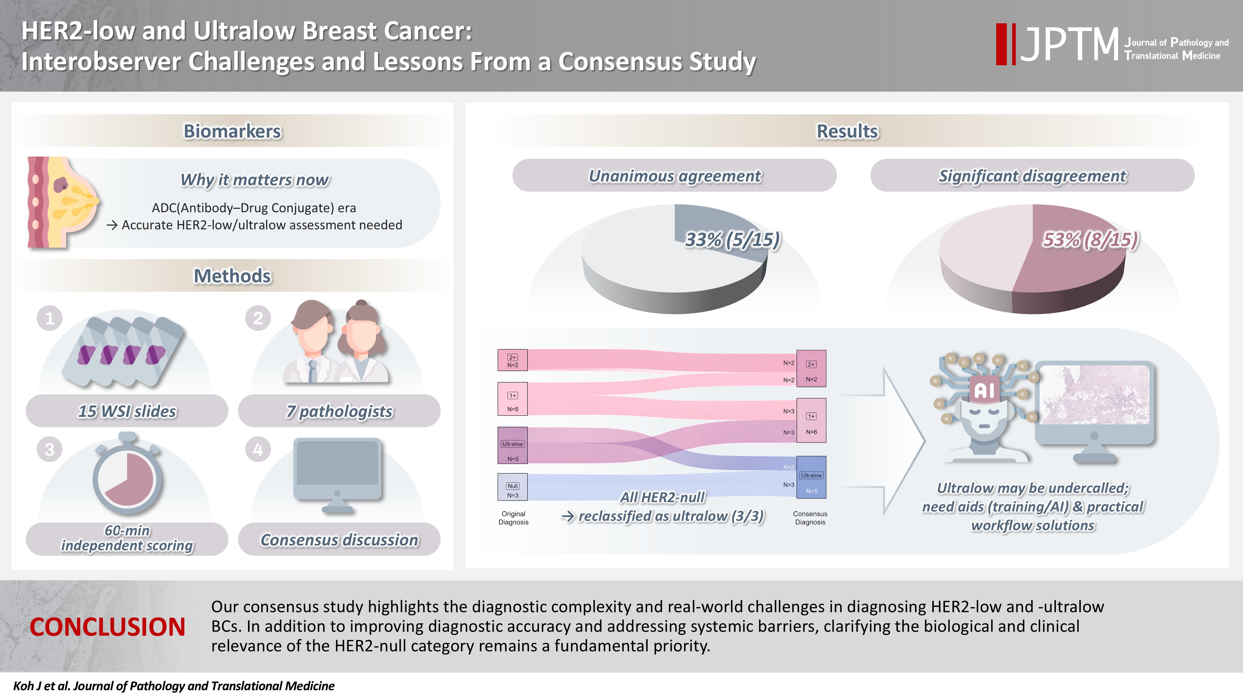
- 4,458 View
- 317 Download
- 1 Web of Science
- 3 Crossref
-
Abstract
PDF
- Background
The recent approval of trastuzumab deruxtecan for human epidermal growth factor receptor 2 (HER2)–low and HER2-ultralow breast cancer mandates an adequate assessment of these categories. Methods: Seven breast pathologists from the Breast Pathology Study Group of the Korean Society of Pathologists held an on-site expert consensus meeting. Fifteen sets of virtual whole slide images (WSI) of hematoxylin and eosin stain and HER2 immunohistochemistry were provided. The pathologists were given 60 minutes to submit their diagnosis of HER2 expression into null, ultralow, 1+, 2+, or 3+. Afterwards, in-depth discussion and consensus diagnoses were made by real-time visualization of the WSI. Results: After the consensus meeting, unanimous 100% agreements were seen only in five (33.3%) of the examined cases, which consisted of three 1+ cases and two 2+ cases. Two cases (13.3%) had mild disagreement, with only one pathologist’s disagreement. Of note, eight cases (53.3%) showed significant disagreement, defined by more than two pathologists’ disagreement. All HER2-null cases were reclassified as ultralow after consensus review, suggesting potential widespread underclassification of ultralow cases in clinical practice. Conclusions: Experts had significant discrepancies in interpreting HER2-low/ultralow status. It is important to assess if the distinction between HER2-low and ultralow is strictly required and if HER2-null breast cancer exists in reality. -
Citations
Citations to this article as recorded by- Comparative analysis of robotic- and endoscopic-assisted minimally invasive nipple–areola complex–sparing mastectomy for breast cancer: a meta-analysis with subgroup analyses
Xiaohan Wang, Hua Tian, Min Zhang, Jiangyong Shen, Yangxu Ding, Yupei Dai, Huijuan Shi
Journal of Robotic Surgery.2026;[Epub] CrossRef - HER2-Low Breast Cancer: A Review of Epidemiology, Diagnostic Challenges, Targeted Therapies, and Clinical Outcomes
Maedeh Mirasheh, Zahra Shahabinia, Mai Abdel Haleem A. Abusalah, Afrooz Mazidimoradi, Leila Allahqoli, Hamid Salehiniya, Do-Youn Lee
Journal of Clinical Medicine.2026; 15(19): 7559. CrossRef - Interobserver Reproducibility of Treatment-Defining HER2-Low and HER2-Ultralow Immunohistochemical Scoring in Breast Carcinoma: A Systematic Review
Vishal Tayade, Gyanendra Singh
Applied Immunohistochemistry & Molecular Morphology.2026;[Epub] CrossRef
- Comparative analysis of robotic- and endoscopic-assisted minimally invasive nipple–areola complex–sparing mastectomy for breast cancer: a meta-analysis with subgroup analyses
Review Article
- A comprehensive review of ossifying fibromyxoid tumor: insights into its clinical, pathological, and molecular landscape
- Kyriakos Chatzopoulos, Antonia Syrnioti, Mohamed Yakoub, Konstantinos Linos
- J Pathol Transl Med. 2026;60(1):6-19. Published online January 14, 2026
- DOI: https://doi.org/10.4132/jptm.2025.10.02

- 6,784 View
- 251 Download
-
Abstract
PDF
- Ossifying fibromyxoid tumor (OFMT) is a rare mesenchymal neoplasm first described in 1989. It typically arises in the superficial soft tissues of the extremities as a slow-growing, painless mass. Histologically, it is commonly characterized by a multilobular architecture composed of uniform epithelioid cells embedded in a fibromyxoid matrix, often surrounded by a rim of metaplastic bone. While classic cases are readily identifiable, the tumor's histopathological heterogeneity can mimic a range of benign and malignant neoplasms, posing significant diagnostic challenges. Molecularly, most OFMTs harbor PHF1 rearrangements, commonly involving fusion partners such as EP400, MEAF6, or TFE3. This review underscores the importance of an integrated diagnostic approach- incorporating histopathological, immunohistochemical, and molecular data- to accurately classify OFMT and distinguish it from its mimics. Expanding awareness of its morphologic and molecular spectrum is essential for precise diagnosis, optimal patient management, and a deeper understanding of this enigmatic neoplasm.
Case Study
- Cytological characteristics of Müllerian adenosarcoma of the uterine corpus: a case report and literature review
- Junko Kuramoto, Chihiro Matsubara, Yasuko Sasamoto, Hitomi Tsukada, Shigemichi Hirose
- J Pathol Transl Med. 2025;59(5):340-347. Published online September 11, 2025
- DOI: https://doi.org/10.4132/jptm.2025.08.11

- 8,298 View
- 100 Download
-
Abstract
PDF
- Müllerian adenosarcoma of the uterus is a rare morphological variant of uterine sarcoma. Müllerian adenosarcoma has been described histologically, though it is rare in the cytological literature. This report describes the cytological findings of a case of adenosarcoma arising from the endometrium. The patient was a Japanese woman in her 40s. Endometrial cytological and histological findings were observed for 5 years, from the appearance of a polypoid lesion until adenosarcoma was suspected, and then hysterectomy was performed. Based on these longitudinal cytological and histological observations, it was possible to identify the cytological characteristics of adenosarcoma: decrease in the glandular-to-stromal ratio; increase in stromal cell density; and progression of stromal cell atypia. This case stresses the importance and usefulness of endometrial cytology in the identification of the sarcomatous component in adenosarcoma.
Reviews
- Breast fine-needle aspiration cytology in the era of core-needle biopsy: what is its role?
- Ahrong Kim, Hyun Jung Lee, Jee Yeon Kim
- J Pathol Transl Med. 2025;59(1):26-38. Published online January 15, 2025
- DOI: https://doi.org/10.4132/jptm.2024.11.01
- Correction in: J Pathol Transl Med 2025;59(2):147
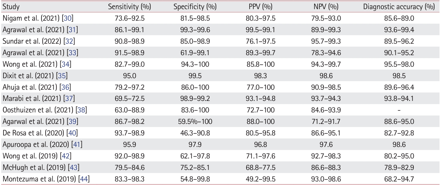
- 19,505 View
- 596 Download
- 7 Web of Science
- 11 Crossref
-
Abstract
PDF
- Fine-needle aspiration cytology (FNAC) has long been recognized as a minimally invasive, cost-effective, and reliable diagnostic tool for breast lesions. However, with the advent of core-needle biopsy (CNB), the role of FNAC has diminished in some clinical settings. This review aims to re-evaluate the diagnostic value of FNAC in the current era, focusing on its complementary use alongside CNB, the adoption of new approaches such as the International Academy of Cytology Yokohama System, and the implementation of rapid on-site evaluation to reduce inadequate sample rates. Advances in liquid-based cytology, receptor expression testing, molecular diagnostics, and artificial intelligence are discussed, highlighting their potential to enhance the diagnostic accuracy of FNAC. Despite challenges, FNAC remains a valuable diagnostic method, particularly in low-resource settings and specific clinical scenarios, and its role continues to evolve with technology.
-
Citations
Citations to this article as recorded by- Evaluation of Breast Lesions on Cytology Using International Academy of Cytology Yokohama Standardized Reporting System
Manish Jaiswal, Anurag Gupta, Tripti Verma, Pradyumn Singh, Rita Yadav, Akash Agarwal, Ashish Singhal, Nuzhat Husain, Shamrendra Narayan, Neha Singh
Diagnostic Cytopathology.2026; 54(3): 184. CrossRef - Personalizing therapies over the course of hormone receptor‐positive/HER2‐negative metastatic breast cancer
Akshara Singareeka Raghavendra, Senthil Damodaran, Carlos H. Barcenas, Suzanne A. Fuqua, Rachel M. Layman, Debu Tripathy
CA: A Cancer Journal for Clinicians.2026;[Epub] CrossRef - Transforming Breast Cancer Control in East Africa by Integrating Cytomorphology and Genetics Into National Policy
Josephine N Rioki, Mwangi Joseph, Rency Lel, Marshal Mweu, Lucy Muchiri
Cureus.2026;[Epub] CrossRef - Prélèvements mammaires percutanés
A. Ribrag, R. Foucher
EMC - Gynécologie.2026; 41(3): 1. CrossRef - Age and tumor size as independent predictors of malignancy in BI-RADS 4 and 5 breast lesions: A cross-sectional study in Vietnam
De Van Nguyen, Trung Van Pham, Tam Huu Dinh, Dung Ngoc Tran, Chung Thanh Dang, Dongling Wu
PLOS One.2026; 21(7): e0352690. CrossRef - Selective medical intelligence: Optimising AI-based breast cancer diagnosis classification through adaptive data filtering
Nicholas Christakis, Panagiotis Tirchas, Dimitris Drikakis
Neurocomputing.2026; 700: 134471. CrossRef - Diagnostic Performance of the International Academy of Cytology Yokohama System for Reporting Breast Fine-Needle Aspirates: A Cytology-Histopathology Correlation Study From Central India
Aditi Rathore, Archana Shrivastava, Maneesh Sulya
Cureus.2026;[Epub] CrossRef - Spontaneous metastatic mammary carcinomas in 2 capuchin monkeys: pathology and immunohistochemical findings with a brief literature review
Isabel L. Macêdo, Liz A. Cerqueira, Yasmin N. G. Fonseca, Antonizete R. Souza, Ana L. G. L. Chisté, Márcio B. Castro
Journal of Veterinary Diagnostic Investigation.2026;[Epub] CrossRef - Evolution, Synergy, and the Intersecting Futures of Breast FNAC and CNB
Naresh N. Rai
Journal of Modern Medicine.2026; 4(2): 27. CrossRef - Bulk-lysis protocols as a sensitive method for investigation of circulating CK19 cells in the peripheral blood of patients with breast cancer by flow cytometry
Daniella Serafin Couto Vieira, Laura Otto Walter, Maria Eduarda Cunha da Silva, Lisandra de Oliveira Silva, Heloísa Zorzi Costa, Chandra Chiappin Cardoso, Fernando Carlos de Lander Schmitt, Maria Cláudia Santos-Silva
Analytical Methods.2025; 17(23): 4771. CrossRef - Diagnostic Accuracy of BI-RADS Classification in Women With Breast Lump on Ultrasound, Keeping Histopathology as the Gold Standard
Saima Zeb, Nasreen Aman
Biological and Clinical Sciences Research Journal.2025; 6(5): 548. CrossRef
- Evaluation of Breast Lesions on Cytology Using International Academy of Cytology Yokohama Standardized Reporting System
- Exploring histological predictive biomarkers for immune checkpoint inhibitor therapy response in non–small cell lung cancer
- Uiju Cho, Soyoung Im, Hyung Soon Park
- J Pathol Transl Med. 2024;58(2):49-58. Published online February 26, 2024
- DOI: https://doi.org/10.4132/jptm.2024.01.31
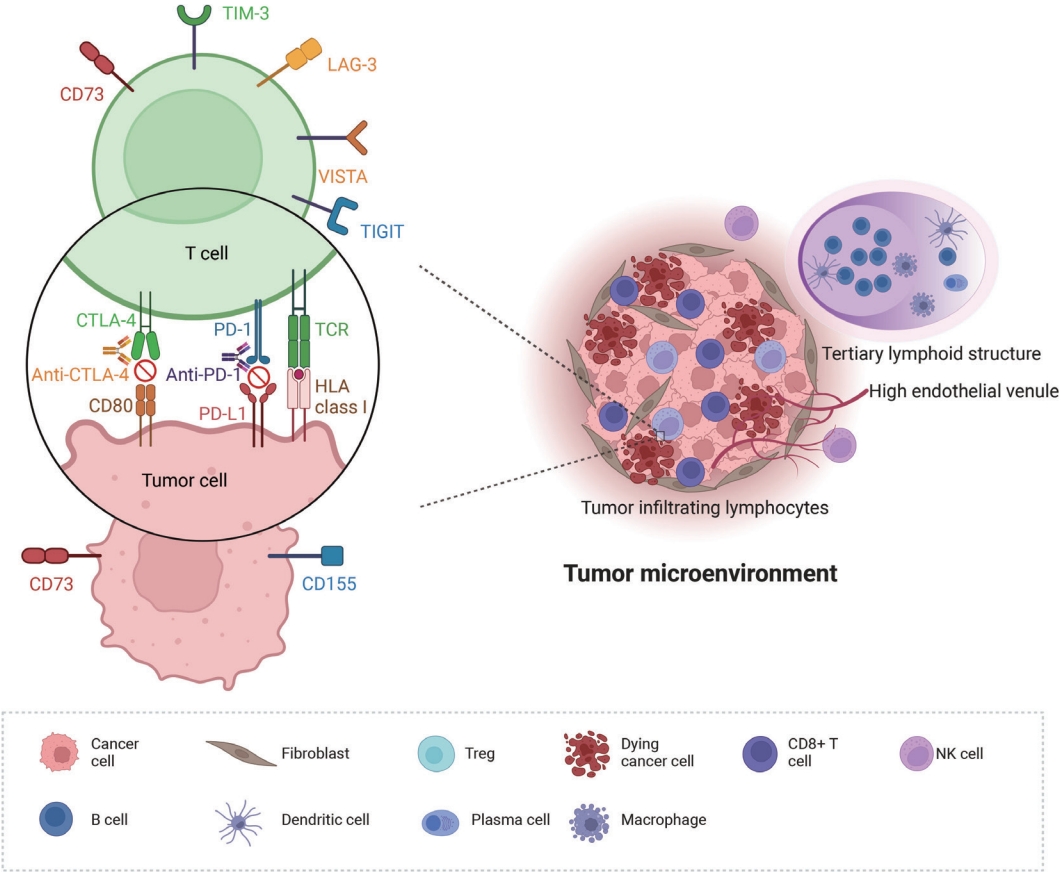
- 14,350 View
- 373 Download
- 5 Web of Science
- 6 Crossref
-
Abstract
PDF
- Treatment challenges persist in advanced lung cancer despite the development of therapies beyond the traditional platinum-based chemotherapy. The early 2000s marked a shift to tyrosine kinase inhibitors targeting epidermal growth factor receptor, ushering in personalized genetic-based treatment. A further significant advance was the development of immune checkpoint inhibitors (ICIs), especially for non–small cell lung cancer. These target programmed death-ligand 1 (PD-L1) and cytotoxic T lymphocyte antigen 4, which enhanced the immune response against tumor cells. However, not all patients respond, and immune-related toxicities arise. This review emphasizes identifying biomarkers for ICI response prediction. While PD-L1 is a widely used, validated biomarker, its predictive accuracy is imperfect. Investigating tumor-infiltrating lymphocytes, tertiary lymphoid structure, and emerging biomarkers such as high endothelial venule, Human leukocyte antigen class I, T-cell immunoreceptors with Ig and ITIM domains, and lymphocyte activation gene-3 counts is promising. Understanding and exploring additional predictive biomarkers for ICI response are crucial for enhancing patient stratification and overall care in lung cancer treatment.
-
Citations
Citations to this article as recorded by- Prognostic Value of the Lung Immune Prognostic Index in Advanced Non-Small Cell Lung Cancer Treated with Nivolumab: A Real-World Analysis Beyond PD-L1 Expression
Alperen Akansel Çağlar, Aykut Özmen, Tuğrul Burak Genç, Anıl Yıldız, Özde Melisa Celayir, Shamkal Safarov, Yunus Avcı, Gökmen Umut Erdem, Nilüfer Bulut
Diagnostics.2026; 16(18): 2890. CrossRef - Beyond immune surveillance: IFN-γ and PD-L1 as shapers of tumor plasticity in Non-Small Cell Lung Cancer (NSCLC)
Stefania Angelicola, Mariateresa Frascino, Ilaria C. Chillico, Patrick L. S. Michel, Damiano Pistone, Maria Sofia Semprini, Olga Maria Pittino, Andrea Ardizzoni, Niccolò Leandro Alessio, Francesco Agustoni, Francesco Gelsomino, Pier-Luigi Lollini, Arianna
Frontiers in Immunology.2026;[Epub] CrossRef - Inflammatory regulation of PD-L1 protein fate in the tumor microenvironment shapes immune escape and therapeutic targeting
Kuan Liang, Ziang Jia, Haiwen Lu, Qianqian Wang, Haihui Huang, Ziling Yan, Yukun Wang, Xia Liu
Discover Oncology.2026;[Epub] CrossRef - Machine learning methods for histopathological image analysis: Updates in 2024
Daisuke Komura, Mieko Ochi, Shumpei Ishikawa
Computational and Structural Biotechnology Journal.2025; 27: 383. CrossRef - Beyond single biomarkers: multi-omics strategies to predict immunotherapy outcomes in blood cancers
Mohammad Pirouzbakht, Soroosh Hamzeh, Hamed Soleimani Samarkhazan
Clinical and Experimental Medicine.2025;[Epub] CrossRef - Temporal changes in tongue color during immune checkpoint inhibitor therapy in patients with non-small-cell lung cancer: a prospective observational study using digital tongue diagnosis
Eunbyul Cho, Woosu Choi, Jun Hyeok Lim, Ji Woong Son, Seung Hun Jang, Seung Hyeun Lee, Jong Gwon Choi, In-Jae Oh, Tae-Won Jang, Seong Hoon Yoon, Seung Joon Kim, Chang-Min Choi, Sung Yong Lee, Mi Mi Ko, Mi-Kyung Jeong
Oncology Reviews.2025;[Epub] CrossRef
- Prognostic Value of the Lung Immune Prognostic Index in Advanced Non-Small Cell Lung Cancer Treated with Nivolumab: A Real-World Analysis Beyond PD-L1 Expression
Original Article
- Fine needle aspiration cytology diagnoses of follicular thyroid carcinoma: results from a multicenter study in Asia
- Hee Young Na, Miyoko Higuchi, Shinya Satoh, Kaori Kameyama, Chan Kwon Jung, Su-Jin Shin, Shipra Agarwal, Jen-Fan Hang, Yun Zhu, Zhiyan Liu, Andrey Bychkov, Kennichi Kakudo, So Yeon Park
- J Pathol Transl Med. 2024;58(6):331-340. Published online November 7, 2024
- DOI: https://doi.org/10.4132/jptm.2024.10.12
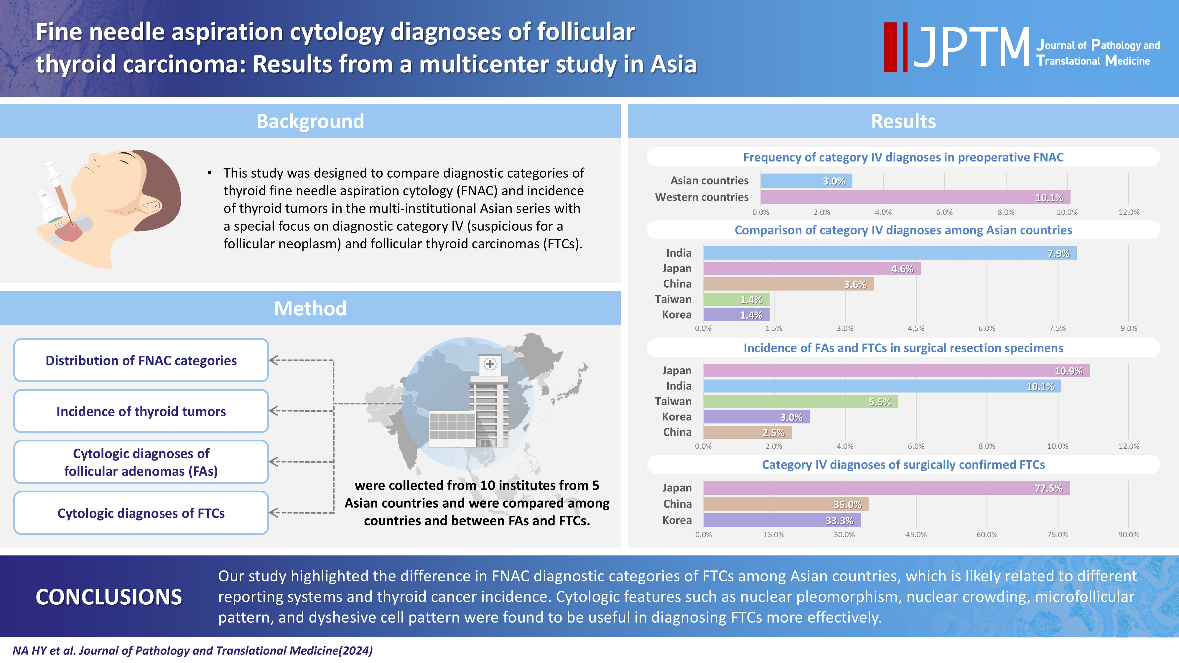
- 11,215 View
- 297 Download
- 5 Web of Science
- 6 Crossref
-
Abstract
PDFSupplementary Material
- Background
This study was designed to compare diagnostic categories of thyroid fine needle aspiration cytology (FNAC) and incidence of thyroid tumors in the multi-institutional Asian series with a special focus on diagnostic category IV (suspicious for a follicular neoplasm) and follicular thyroid carcinomas (FTCs). Methods: Distribution of FNAC categories, incidence of thyroid tumors in resection specimens and cytologic diagnoses of surgically confirmed follicular adenomas (FAs) and FTCs were collected from 10 institutes from five Asian countries and were compared among countries and between FAs and FTCs. Results: The frequency of category IV diagnoses (3.0%) in preoperative FNAC were significantly lower compared to those in Western countries (10.1%). When comparing diagnostic categories among Asian countries, category IV was more frequent in Japan (4.6%) and India (7.9%) than in Taiwan (1.4%), Korea (1.4%), and China (3.6%). Similarly, incidence of FAs and FTCs in surgical resection specimens was significantly higher in Japan (10.9%) and India (10.1%) than in Taiwan (5.5%), Korea (3.0%), and China (2.5%). FTCs were more commonly diagnosed as category IV in Japan (77.5%) than in Korea (33.3%) and China (35.0%). Nuclear pleomorphism, nuclear crowding, microfollicular pattern, and dyshesive cell pattern were more common in FTCs compared with FAs. Conclusions: Our study highlighted the difference in FNAC diagnostic categories of FTCs among Asian countries, which is likely related to different reporting systems and thyroid cancer incidence. Cytologic features such as nuclear pleomorphism, nuclear crowding, microfollicular pattern, and dyshesive cell pattern were found to be useful in diagnosing FTCs more effectively. -
Citations
Citations to this article as recorded by- Deep Learning-Based Multimodal Fusion of Ultrasound, Cytology, and Clinical Features to Distinguish Follicular Thyroid Carcinoma from Adenoma: A Multicenter Study
Xiao-Fei Guo, Li Zhou, Xin-Yi Bao, Shui-Qing Liu, Jia-Wei Feng, You-Long Zhu, Yong Jiang, Shu-Ying Zhang
Academic Radiology.2026; 33(7): 2921. CrossRef - Molecular Testing in Indeterminate Thyroid Nodules: Genomic Landscape, Diagnostic Performance, and Integrated Risk-Stratified Management
Sayaka Tanaka, Naomi Kitayama, Kyouko Kawamoto, Tomoko Wakasa, Yanhua Bai, Kennichi Kakudo
Cancers.2026; 18(10): 1661. CrossRef - A Clinicopathological Study on Thyroid Swellings with Comparison of Findings of Ultrasonography and Bethesda Reporting System of Fine Needle Aspiration Cytology to Histopathology: a Cross-Sectional Study
Amit Kumar Shukla, Debjit Jana, Maumita De, Krishna Kumar Yadav, Divya Daga, Diptanshu Mukherjee, Saumendra Nath Bandyopadhyay, Anukriti, Snehasish Halder
Indian Journal of Otolaryngology and Head & Neck Surgery.2026; 78(7): 3808. CrossRef - Diagnostic thresholds in thyroid cytopathology: toward biological risk stratification
Sayaka Tanaka, Tomoko Wakasa, Kennichi Kakudo
Journal of the American Society of Cytopathology.2026;[Epub] CrossRef - Western versus Asian utilization of molecular testing for indeterminate thyroid nodules: A meta‐analysis of divergent clinical impact
Chanchal Rana, Truong Phan‐Xuan Nguyen, Chan Kwon Jung, Andrey Bychkov, Kennichi Kakudo
Cancer Cytopathology.2026;[Epub] CrossRef - Misdiagnosed follicular adenoma with 11 year postoperative liver and lung metastases a case report and literature review
Kai-Li Yang, Heng-Tong Han, Shou-Hua Li, Xiao-Xiao Li, Ze Yang, Li-Bin Ma, Yong-Xun Zhao
Discover Oncology.2025;[Epub] CrossRef
- Deep Learning-Based Multimodal Fusion of Ultrasound, Cytology, and Clinical Features to Distinguish Follicular Thyroid Carcinoma from Adenoma: A Multicenter Study
Review Articles
- Ganglioglioma and gangliocytoma: a review for pathologists
- Gianfranco E. Umeres-Francia, Melissa Mejia-Bautista, Pouya Jamshidi, Jared T. Ahrendsen
- J Pathol Transl Med. 2026;60(4):379-387. Published online July 15, 2026
- DOI: https://doi.org/10.4132/jptm.2026.06.06
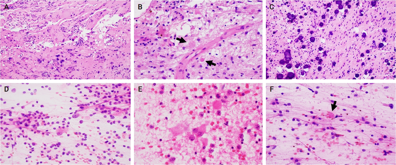
- 2,298 View
- 99 Download
-
Abstract
PDF
- Ganglioglioma and gangliocytoma are rare, predominantly low-grade neuroepithelial tumors that commonly present with epilepsy in children and young adults. Advances in molecular profiling have improved understanding of their pathogenesis, highlighting key roles for the mitogen-activated protein kinase/ERK signaling pathway. Diagnosis relies on a combination of clinical, radiologic, and histopathologic features, with complete surgical resection offering the best clinical outcomes. This review summarizes current knowledge on their epidemiology, etiology, clinical presentation, imaging characteristics, pathology, treatment strategies, and prognosis.
- Angiomatoid fibrous histiocytoma: a review
- Alexander N. Perez, Phyu P. Aung
- J Pathol Transl Med. 2026;60(4):371-378. Published online July 15, 2026
- DOI: https://doi.org/10.4132/jptm.2026.06.05
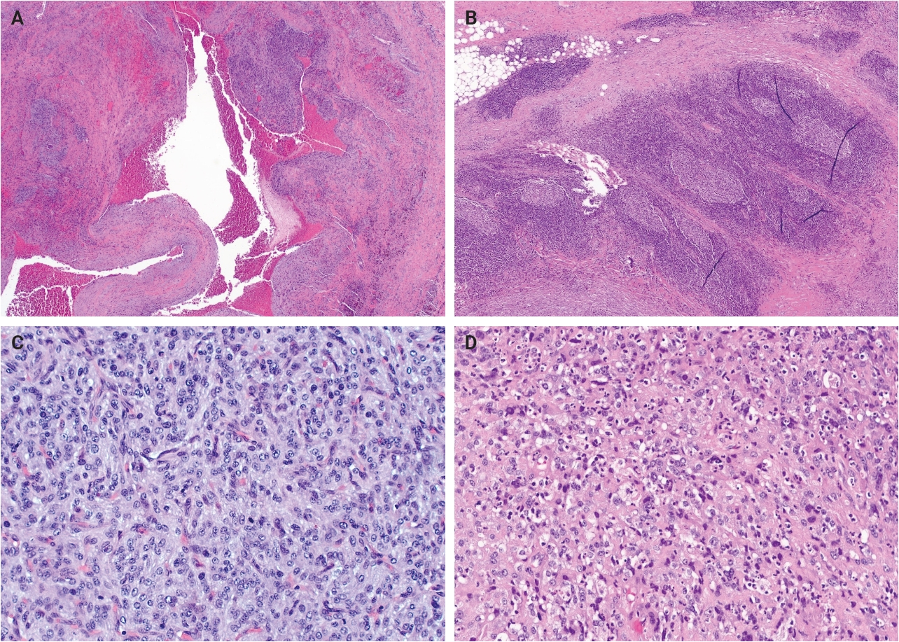
- 2,357 View
- 99 Download
-
Abstract
PDF
- Angiomatoid fibrous histiocytoma is a rare mesenchymal neoplasm of uncertain cell lineage with indeterminate behavior, hallmarked by EWSR1 translocations. This tumor typically arises in the subcutaneous or deep soft tissues and is composed of bland to mildly atypical histiocytoid cells with frequent intralesional hemorrhagic pseudocystic spaces. It affects both children and adults, without a significant sex predilection. Histologically, the tumor may be mistaken for a lymph node given the apparent predilection for node-bearing sites as well as the brisk lymphoid cuff featuring germinal centers. Surgical excision is often curative, with local recurrence occurring occasionally and metastasis only very rarely. A possible relationship to molecularly related entities arising primarily within the thoracic cavity and intracranial compartment has been proposed, although this association remains incompletely understood.
Original Articles
- Bronchial lesions in a high serum IgA mouse model: pulmonary venular IgA deposition and spatially distinct lymphoid cell aggregation
- Areum Kim, Minhyeok Lee, Yohan Park, Wan Jin Hwang, Hyeseung Lee, Joo Heon Kim, Jin Man Kim, Yong Min Kim, Jin Sun Park, Junguee Lee
- J Pathol Transl Med. 2026;60(5):516-526. Published online July 16, 2026
- DOI: https://doi.org/10.4132/jptm.2026.06.01
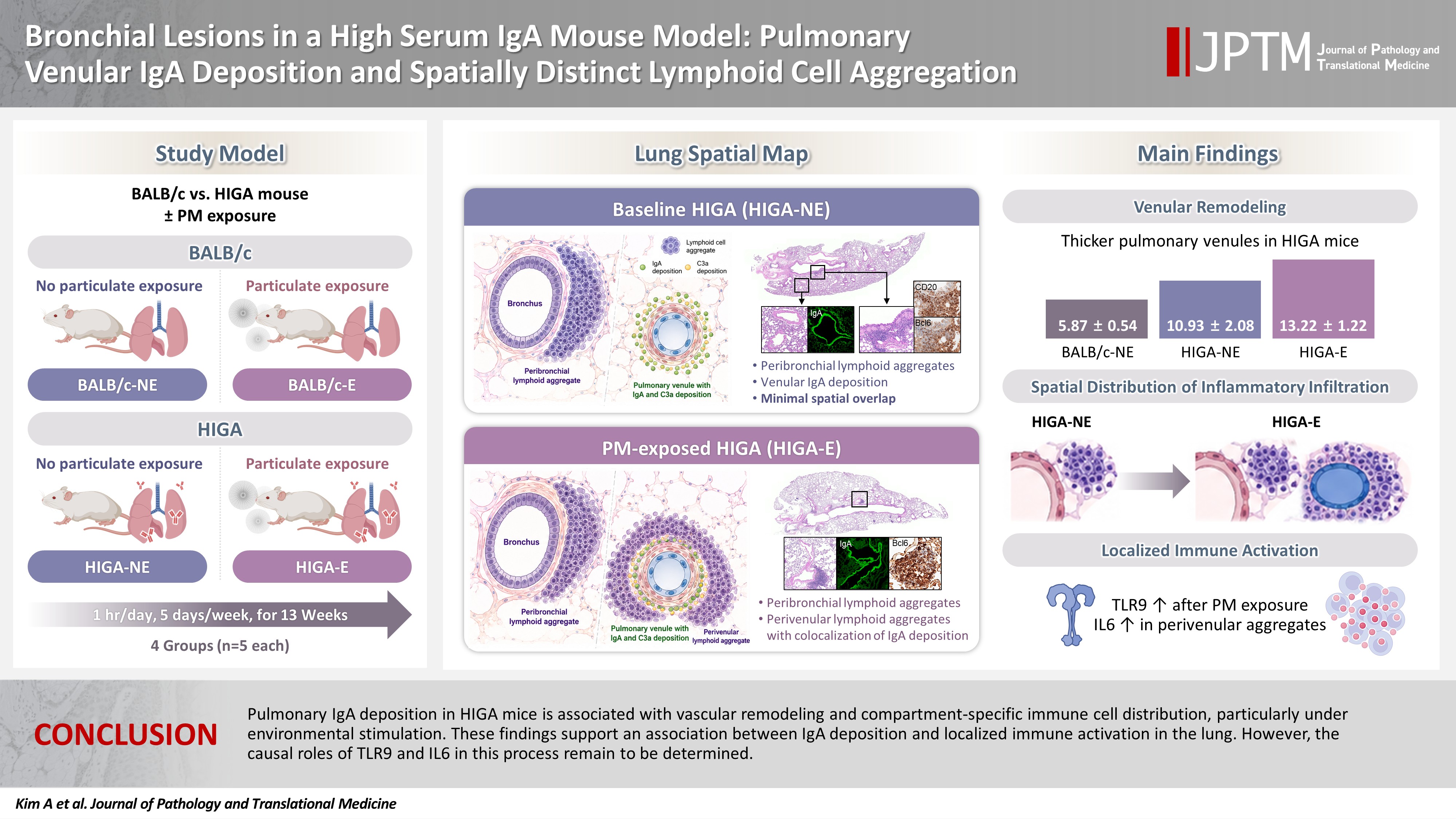
- 1,987 View
- 32 Download
-
Abstract
PDF
- Background
Immunoglobulin A (IgA) nephropathy is a systemic immune complex–mediated disease primarily affecting the kidneys, yet pulmonary involvement remains poorly characterized. This study investigated pulmonary structural alterations, IgA deposition, immune cell distribution, and the impact of chronic environmental immune stimulation. Methods: High-IgA (HIGA) mice and BALB/c controls were examined under baseline conditions and following chronic particulate matter (PM) exposure. Histopathology, immunofluorescence, immunohistochemistry, and lectin-based assays were used to assess pulmonary IgA deposition, lymphoid cell aggregation, and immune activation. Results: Compared with BALB/c controls, HIGA mice exhibited pulmonary venular remodeling characterized by thickening of the venular tunica media and IgA deposition within the smooth muscle layer. Under baseline conditions, lymphoid cell aggregation in HIGA mice was predominantly localized to peribronchial regions, whereas IgA deposition and C3a deposition were confined to pulmonary venules with minimal spatial overlap. Following PM exposure, HIGA mice developed additional perivenular lymphoid cell aggregation that spatially corresponded with IgA deposition, whereas BALB/c mice showed predominantly peribronchial aggregation. PM exposure was associated with increased pulmonary Toll-like receptor 9 (TLR9) expression in both strains. In HIGA mice, TLR9-positive immune cells and interleukin-6 (IL6) expression were enriched in perivenular lymphoid cell aggregates. Conclusions: Pulmonary IgA deposition in HIGA mice is associated with vascular remodeling and compartment-specific immune cell distribution, particularly under environmental stimulation. These findings support an association between IgA deposition and localized immune activation in the lung. However, the causal roles of TLR9 and IL6 in this process remain to be determined.
- Hyalinizing trabecular tumors with areas resembling noninvasive follicular thyroid neoplasm with papillary‑like nuclear features: an immunohistochemical and molecular analysis
- Risa Kanematsu, Mitsuyoshi Hirokawa, Ayana Suzuki, Miyoko Higuchi, Satomi Usuki, Hiroshi Kamma, Takashi Akamizu
- J Pathol Transl Med. 2026;60(4):436-443. Published online July 15, 2026
- DOI: https://doi.org/10.4132/jptm.2026.06.07
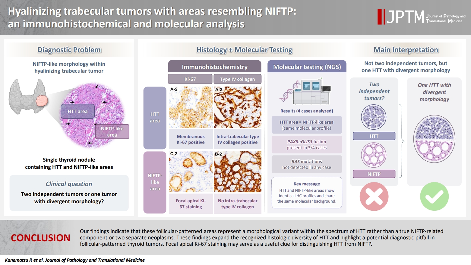
- 2,287 View
- 72 Download
-
Abstract
PDF
- Background
The coexistence of hyalinizing trabecular tumor (HTT) and areas with a morphology of noninvasive follicular thyroid neoplasm with papillary-like nuclear features (NIFTP) within a single thyroid nodule has not been previously reported. We aimed to determine whether such tumors represent two independent neoplasms or a single tumor exhibiting divergent morphology. Methods: Ten tumors containing both HTT and NIFTP-like areas were examined. The term “NIFTP-like” was used strictly as a descriptive morphological designation for areas that fulfill the histologic criteria of NIFTP. Immunohistochemical analyses of Ki-67 (MIB-1) and type IV collagen and targeted molecular testing were performed. Ten NIFTPs, 10 follicular adenomas, and three HTTs were used as controls. Results: HTT components consistently showed characteristic membranous Ki-67 staining and intra-trabecular type IV collagen deposition, whereas NIFTP-like areas lacked these features, except for focal apical Ki-67 staining. Intranuclear cytoplasmic inclusions in HTT were positive for type IV collagen. NIFTPs showed neither membranous Ki-67 nor intra-trabecular type IV collagen. Molecular analysis demonstrated identical profiles between HTT components and NIFTP-like areas: three tumors harbored PAX8::GLIS3 fusions, and none showed RAS mutations. Pure HTT controls exhibited the same pattern. Conclusions: Our findings indicate that these follicular-patterned areas represent a morphological variant within the spectrum of HTT rather than a true NIFTP-related component or two separate neoplasms. These findings expand the recognized histologic diversity of HTT and highlight a potential diagnostic pitfall in follicular-patterned thyroid tumors. Focal apical Ki-67 staining may serve as a useful clue for distinguishing HTT from NIFTP.
Case Study
- Cytological features of atypical adenomatous hyperplasia and adenocarcinoma in situ of the lung: a case report
- Misa Takahashi, Seiya Homma, Chisato Setoguchi, Yoko Umezawa, Atsuhiko Sakamoto
- J Pathol Transl Med. 2025;59(3):195-200. Published online May 9, 2025
- DOI: https://doi.org/10.4132/jptm.2025.04.09

- 8,351 View
- 147 Download
-
Abstract
PDF
- Atypical adenomatous hyperplasia (AAH) and adenocarcinoma in situ (AIS) are generally treated as different lesions, depending on the differences in lesion size and histological findings. However, these differences are not absolute; thus, AAH and AIS are often difficult to distinguish. Moreover, whether AAH and AIS can be regarded as different lesions remains unknown because cytological specimens, especially those of AAH, are rare. In this study, we examined these uncommon cytological specimens and compared the cytological findings between AAH and AIS. We observed many common cytological features with no obvious differences between AAH and AIS. These findings suggest that these two distinct lesions can be grouped into a single category. Therefore, we propose creating a new cytological category.
Review Article
- Lymphomatoid papulosis: a practical review for pathologists
- Mario L. Marques-Piubelli, Carlos A. Torres-Cabala, Roberto N. Miranda
- J Pathol Transl Med. 2026;60(4):388-397. Published online July 15, 2026
- DOI: https://doi.org/10.4132/jptm.2026.06.09
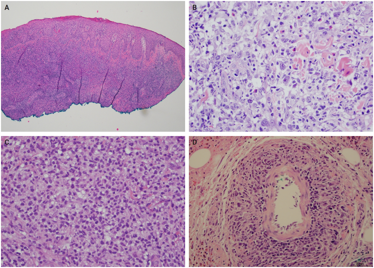
- 2,360 View
- 91 Download
-
Abstract
PDF
- Lymphomatoid papulosis (LyP) is a primary cutaneous CD30+ lymphoproliferative disorder characterized by a chronic and self-healing recurrent cluster of erythematous papules or nodules on the skin of the trunk and/or extremities. The disease has an indolent clinical course with spontaneous regression or waxing and waning clinical evolution. The histopathologic spectrum of LyP is vast and may show few to numerous atypical cells immersed in a mild to intense inflammatory background. The backbone for the diagnosis is the positivity for CD30, which is one of the criteria to define this group of lymphoproliferative disorders. The association of these different histological and immunophenotypical findings is used to subclassify this disease in different subtypes from A to E, associated with DUSP22/IRF4 rearrangement, and other rare forms. Although this differentiation is important to raise awareness of different differential diagnosis, it does not impact the prognosis or change the treatment, which is usually centered in symptom relief and faster regression. In this review, we aim to summarize the most updated information of the clinical, histopathological, and molecular characteristics of LyP and provide a practical assessment for the diagnostic features that could help with the main differential diagnosis.
Original Articles
- Categorizing high-grade serous ovarian carcinoma into clinically relevant subgroups using deep learning–based histomic clusters
- Byungsoo Ahn, Eunhyang Park
- J Pathol Transl Med. 2025;59(2):91-104. Published online February 18, 2025
- DOI: https://doi.org/10.4132/jptm.2024.10.23
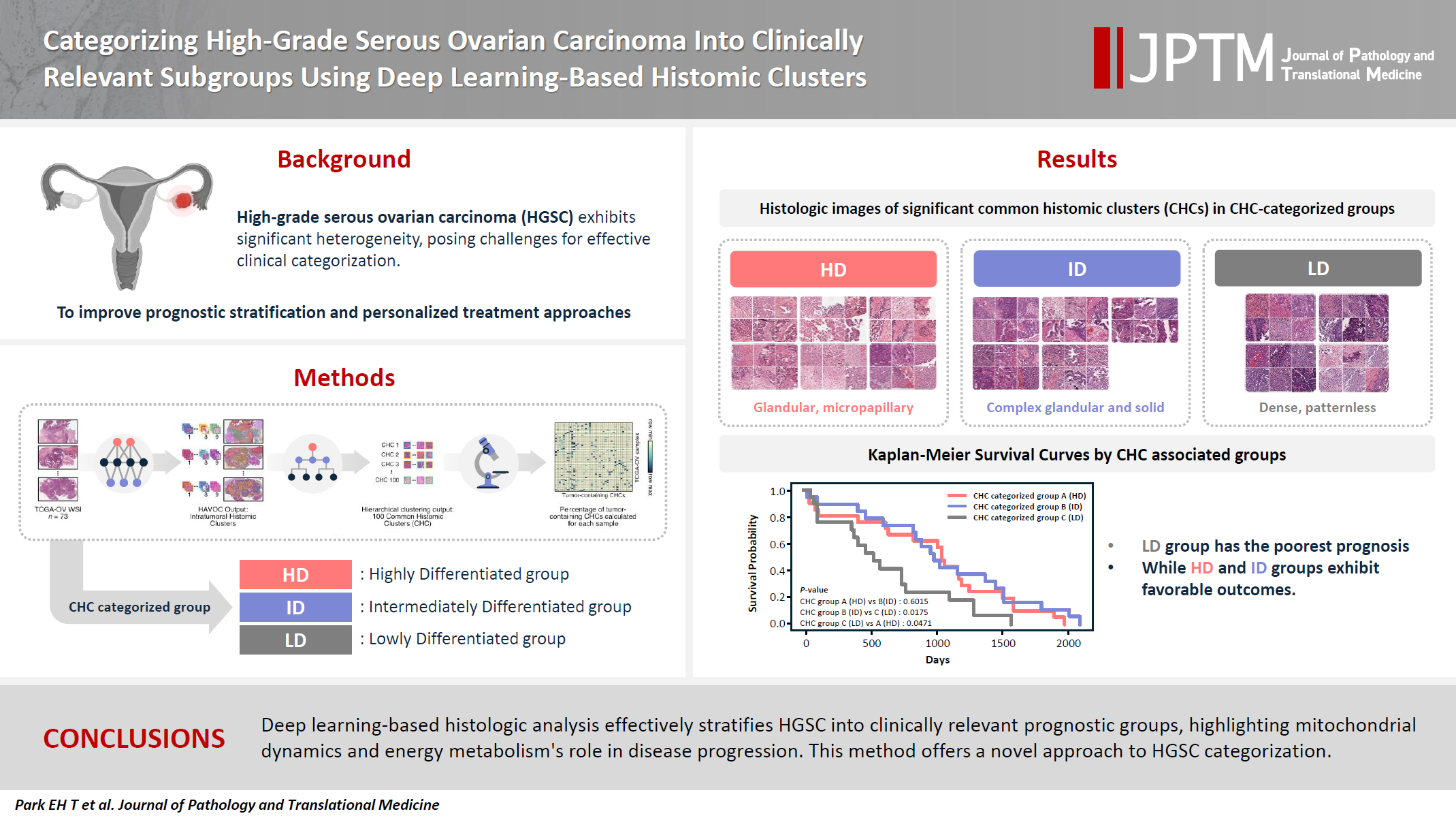
- 9,503 View
- 292 Download
- 1 Web of Science
- 2 Crossref
-
Abstract
PDFSupplementary Material
- Background
High-grade serous ovarian carcinoma (HGSC) exhibits significant heterogeneity, posing challenges for effective clinical categorization. Understanding the histomorphological diversity within HGSC could lead to improved prognostic stratification and personalized treatment approaches. Methods: We applied the Histomic Atlases of Variation Of Cancers model to whole slide images from The Cancer Genome Atlas dataset for ovarian cancer. Histologically distinct tumor clones were grouped into common histomic clusters. Principal component analysis and K-means clustering classified HGSC samples into three groups: highly differentiated (HD), intermediately differentiated (ID), and lowly differentiated (LD). Results: HD tumors showed diverse patterns, lower densities, and stronger eosin staining. ID tumors had intermediate densities and balanced staining, while LD tumors were dense, patternless, and strongly hematoxylin-stained. RNA sequencing revealed distinct patterns in mitochondrial oxidative phosphorylation and energy metabolism, with upregulation in the HD, downregulation in the LD, and the ID positioned in between. Survival analysis showed significantly lower overall survival for the LD compared to the HD and ID, underscoring the critical role of mitochondrial dynamics and energy metabolism in HGSC progression. Conclusions: Deep learning-based histologic analysis effectively stratifies HGSC into clinically relevant prognostic groups, highlighting the role of mitochondrial dynamics and energy metabolism in disease progression. This method offers a novel approach to HGSC categorization. -
Citations
Citations to this article as recorded by- Ovarian Cancer: Epidemiology, Disease Mechanisms, New Diagnosis and Treatment Strategies, and Research Directions
Zunera Khalid, Weirong Fan, Farah Nazir, Yixiang Xing, Tengchuan Jin
iNew Medicine.2026;[Epub] CrossRef - Learning Disabilities in the 21st Century: Integrating Neuroscience, Education, and Technology for Better Outcomes
Syed Mohammed Basheeruddin Asdaq, Ahmad H. Alhowail, Syed Imam Rabbani, Naira Nayeem, Syed Mohammed Emaduddin Asdaq, Faiqa Nausheen
SAGE Open.2025;[Epub] CrossRef
- Ovarian Cancer: Epidemiology, Disease Mechanisms, New Diagnosis and Treatment Strategies, and Research Directions
- Revisiting human sparganosis: a pathologic review from a single institution
- Jeemin Yim, Young A Kim, Jeong Hwan Park, Hye Eun Park, Hyun Beom Song, Ji Eun Kim
- J Pathol Transl Med. 2026;60(1):83-91. Published online January 9, 2026
- DOI: https://doi.org/10.4132/jptm.2025.10.14

- 6,634 View
- 169 Download
- 2 Web of Science
- 2 Crossref
-
Abstract
PDF
- Background
Sparganosis is a rare parasitic infection caused by Spirometra species. Although it was relatively common in the past, it is now often overlooked. In this study, we review cases diagnosed through histopathological examination at a single institution in recent years to raise awareness of this neglected parasitic disease. Methods: We retrospectively analyzed cases of human sparganosis identified in the pathology archives of a single institution in South Korea between 2004 and 2025. A comprehensive review was conducted, including demographic data, clinical features, lesion locations, imaging findings, exposure history (such as dietary habits), and histopathologic findings. Results: A total of 15 patients were identified, including 10 females and 5 males, with a mean age of 65.1 years. Lesions were most commonly located in the lower extremities and breast. Imaging findings were largely nonspecific, with ultrasonography being the most frequently used modality. In most cases, clinical suspicion of sparganosis was absent, and excision was performed under the impression of a benign or malignant tumor. Histologically, variably degenerated parasitic structures were identified within granulomatous inflammation. However, preserved features such as calcospherules and tegumental structures facilitated definitive diagnosis. Conclusions: This study underscores the importance of recognizing the characteristic histopathological features of sparganosis, which can allow for accurate diagnosis even in the absence of clinical suspicion. Although rare, sparganosis remains a relevant diagnostic consideration in endemic regions, particularly in East Asia. -
Citations
Citations to this article as recorded by- Current status and epidemiology of endemic parasitic infections in Korea as of 2026: a narrative review
Sun Huh
Journal of the Korean Medical Association.2026; 69(3): 240. CrossRef - Occurrence of Spirometra mansoni in Domestic Dogs from Rural Ecuador and Its Public Health Relevance
Roberto D. Coello Peralta, Zully Baquerizo Orrala, Aldo Rubén Andrada, Davis Calle Atariguana, Geraldine Ramallo, Alicia Rojas
Animals.2026; 16(13): 2049. CrossRef
- Current status and epidemiology of endemic parasitic infections in Korea as of 2026: a narrative review
Case Studies
- Primary renal BCOR::CCNB3 sarcoma in a female patient: case report
- Somang Lee, Binnari Kim
- J Pathol Transl Med. 2025;59(1):84-90. Published online January 15, 2025
- DOI: https://doi.org/10.4132/jptm.2024.09.30
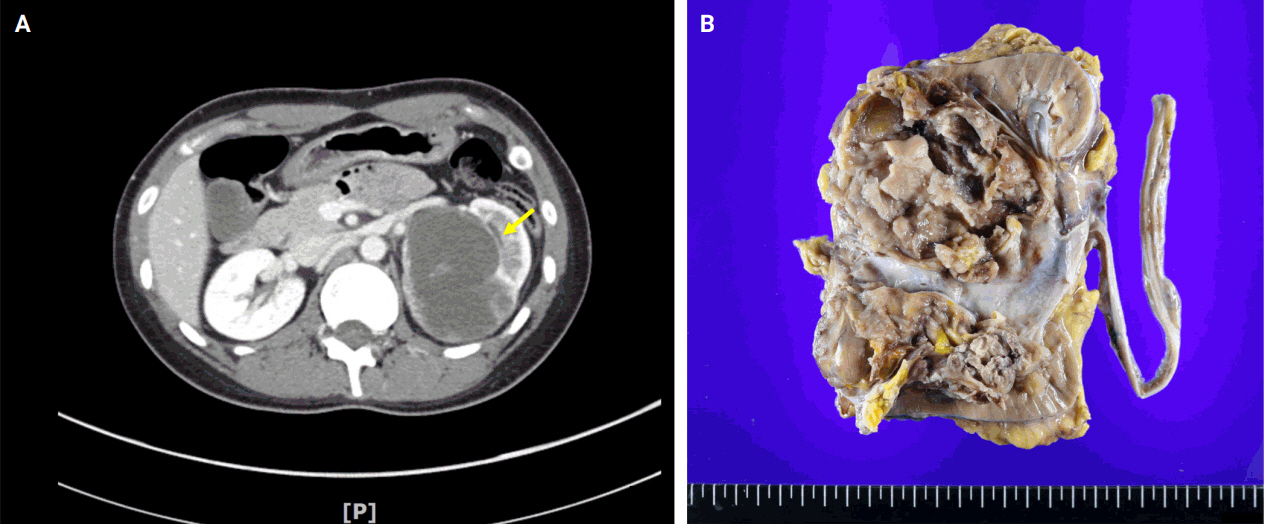
- 8,347 View
- 199 Download
- 1 Web of Science
- 1 Crossref
-
Abstract
PDF
- BCOR-rearranged sarcoma was classified by the World Health Organization in 2020 as a new subgroup of undifferentiated small round-cell sarcoma. It is known to occur very rarely in the kidney. This report presents the first case of a primary renal BCOR::CCNB3 sarcoma in a 22-year-old woman. An 8-cm cystic mass was identified in the left kidney by abdominal pelvic computed tomography. Histopathologic examination revealed the mass to be composed of small round to oval or spindle cells with fibrous septa and a delicate vascular network. A BCOR::CCNB3 fusion was detected by next-generation sequencing–based molecular testing. BCOR::CCNB3 sarcoma presents diagnostic difficulties, highlighting the importance of recognizing its histological features. Immunohistochemical markers are helpful for diagnosis, but genetic molecular testing is necessary for accurate diagnosis. These tumors have a very poor and aggressive prognosis, and an optimal therapeutic regimen has not yet been defined. Therefore, further studies are needed.
-
Citations
Citations to this article as recorded by- Update on the management of BCOR::CCNB3 sarcoma
Jungo Imanishi, Kenji Sato, Yoshinao Kikuchi, Asako Yamamoto, Shiori Watabe, Taisuke Matsuyama, Chiaki Sato, Hiroshi Kobayashi, Hirotaka Kawano
Japanese Journal of Clinical Oncology.2025; 55(10): 1097. CrossRef
- Update on the management of BCOR::CCNB3 sarcoma
- Clinicopathological characteristics of primary malignant cutaneous perivascular epithelioid cell tumor: case report and literature review
- Huaming Li, Dan Zhang, Shiwu Zhang
- J Pathol Transl Med. 2026;60(5):572-577. Published online July 20, 2026
- DOI: https://doi.org/10.4132/jptm.2026.05.10
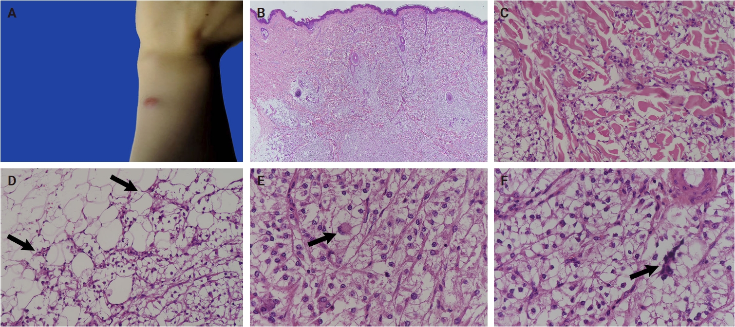
- 1,783 View
- 35 Download
-
Abstract
PDF
- Primary cutaneous malignant perivascular epithelioid cell tumors (PEComas) are extremely rare. Accurate diagnosis is critical for prognostic evaluation and guiding clinical management. Here, we report a case of a 19-year-old woman with a 1.1 cm exophytic red mass on her left upper limb. Histological examination revealed a dermal tumor composed of clear cells with prominent nucleoli. The tumor cells displayed cytologic atypia, nuclear pleomorphism, multinucleated giant cells, mitoses and invasive growth, with a multinodular distribution within the dermis and vascular invasion. Immunohistochemically, the tumor cells expressed cathepsin K, HMB45, and CD10. Based on morphological and immunohistochemical findings, we classified it as a primary malignant cutaneous PEComa. To date, only eight reported cases have been described as primary malignant cutaneous PEComas. The present case appears to be the youngest patient reported among all malignant PEComas.
Original Article
- Diagnosis of invasive encapsulated follicular variant papillary thyroid carcinoma by protein-based machine learning
- Truong Phan-Xuan Nguyen, Minh-Khang Le, Sittiruk Roytrakul, Shanop Shuangshoti, Nakarin Kitkumthorn, Somboon Keelawat
- J Pathol Transl Med. 2025;59(1):39-49. Published online October 24, 2024
- DOI: https://doi.org/10.4132/jptm.2024.09.14
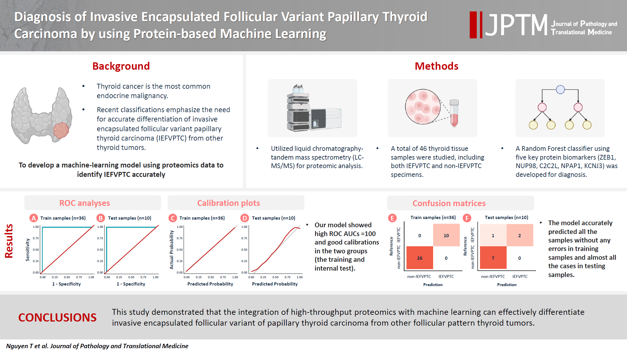
- 7,875 View
- 354 Download
- 2 Web of Science
- 2 Crossref
-
Abstract
PDFSupplementary Material
- Background
Although the criteria for follicular-pattern thyroid tumors are well-established, diagnosing these lesions remains challenging in some cases. In the recent World Health Organization Classification of Endocrine and Neuroendocrine Tumors (5th edition), the invasive encapsulated follicular variant of papillary thyroid carcinoma was reclassified as its own entity. It is crucial to differentiate this variant of papillary thyroid carcinoma from low-risk follicular pattern tumors due to their shared morphological characteristics. Proteomics holds significant promise for detecting and quantifying protein biomarkers. We investigated the potential value of a protein biomarker panel defined by machine learning for identifying the invasive encapsulated follicular variant of papillary thyroid carcinoma, initially using formalin- fixed paraffin-embedded samples.
Methods
We developed a supervised machine-learning model and tested its performance using proteomics data from 46 thyroid tissue samples.
Results
We applied a random forest classifier utilizing five protein biomarkers (ZEB1, NUP98, C2C2L, NPAP1, and KCNJ3). This classifier achieved areas under the curve (AUCs) of 1.00 and accuracy rates of 1.00 in training samples for distinguishing the invasive encapsulated follicular variant of papillary thyroid carcinoma from non-malignant samples. Additionally, we analyzed the performance of single-protein/gene receiver operating characteristic in differentiating the invasive encapsulated follicular variant of papillary thyroid carcinoma from others within The Cancer Genome Atlas projects, which yielded an AUC >0.5.
Conclusions
We demonstrated that integration of high-throughput proteomics with machine learning can effectively differentiate the invasive encapsulated follicular variant of papillary thyroid carcinoma from other follicular pattern thyroid tumors. -
Citations
Citations to this article as recorded by- Advances in immunotherapy for thyroid malignancies: from molecular targets to clinical outcomes
Shuo Lv, Jinbao Wang, Guohao Chen, Yongshun Wang, Naiqing Liu
Frontiers in Medicine.2026;[Epub] CrossRef - Misdiagnosed follicular adenoma with 11 year postoperative liver and lung metastases a case report and literature review
Kai-Li Yang, Heng-Tong Han, Shou-Hua Li, Xiao-Xiao Li, Ze Yang, Li-Bin Ma, Yong-Xun Zhao
Discover Oncology.2025;[Epub] CrossRef
- Advances in immunotherapy for thyroid malignancies: from molecular targets to clinical outcomes
Review Article
- The evolving role of TRPS1 in dermatopathology: insights from the past 4 years
- Mokhtar H. Abdelhammed, Woo Cheal Cho
- J Pathol Transl Med. 2026;60(2):129-143. Published online January 29, 2026
- DOI: https://doi.org/10.4132/jptm.2025.11.25
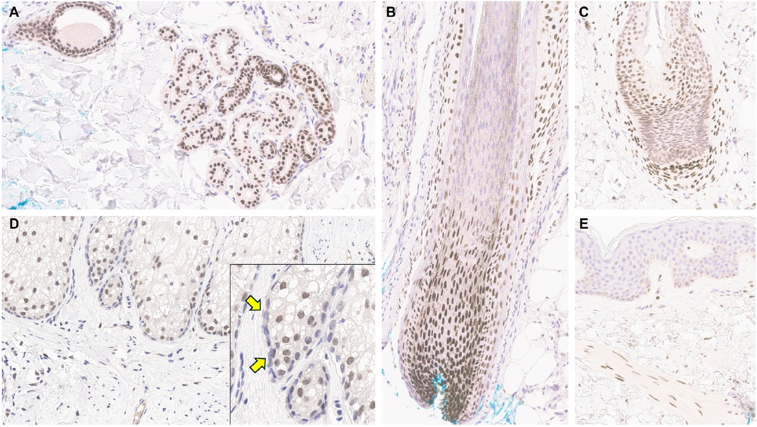
- 5,620 View
- 274 Download
-
Abstract
PDF
- Over the past 4 years, trichorhinophalangeal syndrome type 1 (TRPS1) has rapidly gained attention among practicing pathologists, with numerous studies emerging that both support and question its diagnostic utility. Initially regarded as a highly specific marker for tumors of mammary origin, TRPS1 is now recognized to have broader expression patterns, including in a variety of cutaneous neoplasms. This is likely due to embryologic parallels between breast tissue and skin adnexal structures, an overlap that was underappreciated in early investigations. Although TRPS1 lacks absolute specificity—even among cutaneous neoplasms—it can still offer meaningful diagnostic value when interpreted alongside conventional immunohistochemical markers and within the appropriate morphologic context. Noteworthy diagnostic applications include mammary Paget disease, primary extramammary Paget disease, rare adnexal neoplasms such as endocrine mucin-producing sweat gland carcinoma and primary cutaneous NUT adnexal carcinoma, and cutaneous metastases from breast carcinoma. In this review, we present the most comprehensive and up-to-date evaluation of the utility and limitations of TRPS1 immunohistochemistry in dermatopathology. Our aim is to deepen understanding of this emerging marker and provide practical guidance on its optimal integration with established immunohistochemical panels to enhance diagnostic accuracy in routine practice.
Original Articles
- Development of a prognostic prediction model for tumor recurrence or progression in gastrointestinal stromal tumors: integrating inflammatory blood indices with 5 mm2 versus 50 high-power field mitotic counts
- Waratchaya Tirasarnvong, Thammasin Ingviya, Paramee Thongsuksai
- J Pathol Transl Med. 2026;60(5):527-538. Published online July 31, 2026
- DOI: https://doi.org/10.4132/jptm.2026.06.15
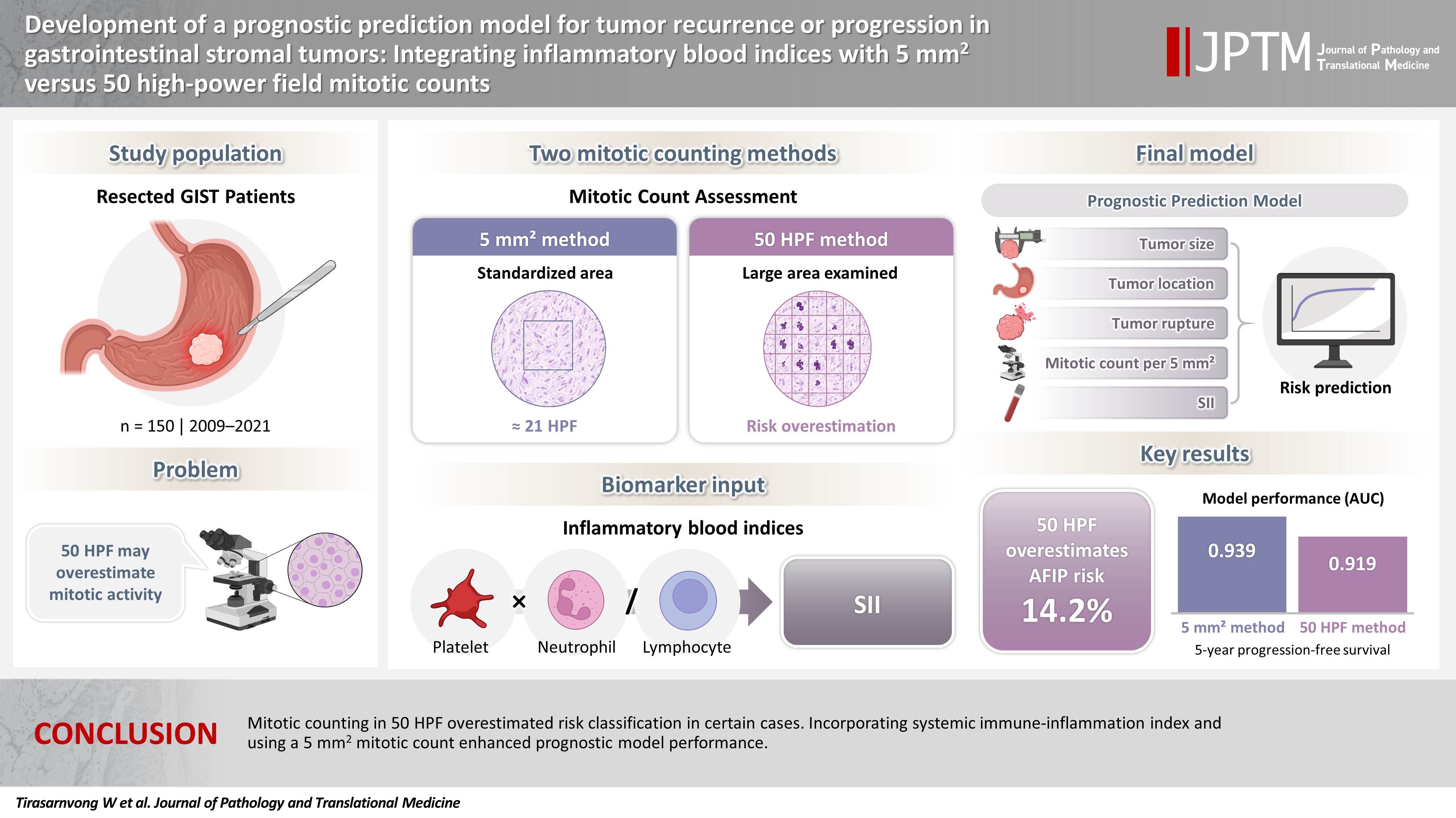
- 1,424 View
- 30 Download
-
Abstract
PDFSupplementary Material
- Background
The behavior of gastrointestinal stromal tumors varies according to tumor size, location, mitotic rate, and rupture. Traditional mitotic counting in 50 high-power fields (HPF; 5.3 mm2) may overestimate progression risk, as 5 mm2 corresponds to 20–25 HPF under modern microscopy. However, the 50-HPF method remains widely used. This study evaluated the impact of two mitotic counting methods on risk classification and developed a prognostic model combining inflammatory biomarkers with clinicopathological factors. Methods: We retrospectively analyzed 150 patients who underwent resection at Songklanagarind Hospital between 2009 and 2021. Patients who had received neoadjuvant therapy or had a second primary cancer were excluded. Mitosis was evaluated using both methods. Clinicopathological and serum biomarker data were collected. The primary outcome was 5-year disease-free or progression-free survival. The model was developed using forward stepwise variable selection based on the time-dependent area under the curve and internally validated by bootstrapping. Results: The 50-HPF method overestimated the Armed Forces Institute of Pathology classification in 14.2% of cases. A model incorporating tumor size, location, rupture, mitotic count within 5 mm2, and systemic immune-inflammation index achieved the highest area under the curve of 0.939 in both training and validation sets, outperforming previous models. The 5-mm2 method demonstrated superior performance to the 50-HPF method across all models (0.939 vs. 0.919 for the proposed model). Conclusions: Mitotic counting in 50 HPF overestimated risk classification in certain cases. Incorporating systemic immune-inflammation index and using a 5 mm2 mitotic count enhanced prognostic model performance.
- Immunohistochemical expression in idiopathic inflammatory myopathies at a single center in Vietnam
- Dat Quoc Ngo, Si Tri Le, Khanh Hoang Phuong Phan, Thao Thi Phuong Doan, Linh Ngoc Khanh Nguyen, Minh Hoang Dang, Thien Thanh Ly, Thu Dang Anh Phan
- J Pathol Transl Med. 2024;58(4):174-181. Published online June 25, 2024
- DOI: https://doi.org/10.4132/jptm.2024.05.02
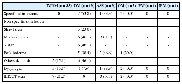
- 7,323 View
- 286 Download
- 1 Web of Science
- 3 Crossref
-
Abstract
PDF
- Background
The identification of idiopathic inflammatory myopathies (IIMs) requires a comprehensive analysis involving clinical manifestations and histological findings. This study aims to provide insights into the histopathological and immunohistochemical aspects of IIMs.
Methods
This retrospective case series involved 56 patients diagnosed with IIMs at the Department of Pathology, University of Medicine and Pharmacy at Ho Chi Minh City, from 2019 to 2023. The histology and immunohistochemical expression of HLA-ABC, HLA-DR, C5b-9, Mx1/2/3, and p62 were detected.
Results
We examined six categories of inflammatory myopathy, including immunemediated necrotizing myopathy (58.9%), dermatomyositis (DM; 23.2%), overlap myositis (8.9%), antisynthetase syndrome (5.4%), inclusion body myositis (IBM; 1.8%), and polymyositis (1.8%). The average age of the patients was 49.7 ± 16.1 years, with a female-to-male ratio of 3:1. Inflammatory cell infiltration in the endomysium was present in 62.5% of cases, perifascicular atrophy was found in 17.8%, and fiber necrosis was observed in 42 cases (75.0%). Rimmed vacuoles were present in 100% of cases in the IBM group. Immunohistochemistry showed the following positivity rates: HLA-ABC (89.2%), HLA-DR (19.6%), C5b-9 (57.1%), and Mx1/2/3 (10.7%). Mx1/2/3 expression was high in DM cases. p62 vacuole deposits were noted in the IBM case. The combination of membrane attack complex and major histocompatibility complex I helped detect IIMs in 96% of cases.
Conclusions
The diagnosis of IIMs and their subtypes should be based on clinical features and histopathological characteristics. Immunohistochemistry plays a crucial role in the diagnosis and differentiation of these subgroups. -
Citations
Citations to this article as recorded by- Evaluating the Diagnostic Potential of Myxovirus Resistance Protein 1 (MX1) and Myxovirus Resistance Protein 2 (MX2) As Biomarkers in Idiopathic Inflammatory Myopathies
Raghavee Neupane, Mustafa Haider, Perry Smith, Marc M Kesselman
Cureus.2026;[Epub] CrossRef - Rapidly Progressive Polymyositis With Vasculitis: The Pivotal Role of Histopathology in Diagnosis and Management
Amitha Venmanassery Karnalsingh, Arjun Karappilly Vijayan, Monica Roselin Edwin Peter, Dilan Davis
Cureus.2025;[Epub] CrossRef - Autoimmune Neuromuscular Disorders at a Molecular Crossroad: Linking Pathogenesis to Targeted Immunotherapy
Anca-Maria Florea, Dimela-Gabriela Luca, Eugenia Irene Davidescu, Bogdan-Ovidiu Popescu
International Journal of Molecular Sciences.2025; 26(23): 11736. CrossRef
- Evaluating the Diagnostic Potential of Myxovirus Resistance Protein 1 (MX1) and Myxovirus Resistance Protein 2 (MX2) As Biomarkers in Idiopathic Inflammatory Myopathies
- Histopathologic classification and immunohistochemical features of papillary renal neoplasm with potential therapeutic targets
- Jeong Hwan Park, Su-Jin Shin, Hyun-Jung Kim, Sohee Oh, Yong Mee Cho
- J Pathol Transl Med. 2024;58(6):321-330. Published online September 12, 2024
- DOI: https://doi.org/10.4132/jptm.2024.07.31

- 10,567 View
- 475 Download
- 2 Web of Science
- 2 Crossref
-
Abstract
PDF
- Background
Papillary renal cell carcinoma (pRCC) is the second most common histological subtype of renal cell carcinoma and is considered a morphologically and molecularly heterogeneous tumor. Accurate classification and assessment of the immunohistochemical features of possible therapeutic targets are needed for precise patient care. We aimed to evaluate immunohistochemical features and possible therapeutic targets of papillary renal neoplasms
Methods
We collected 140 papillary renal neoplasms from three different hospitals and conducted immunohistochemical studies on tissue microarray slides. We performed succinate dehydrogenase B, fumarate hydratase, and transcription factor E3 immunohistochemical studies for differential diagnosis and re-classified five cases (3.6%) of papillary renal neoplasms. In addition, we conducted c-MET, p16, c-Myc, Ki-67, p53, and stimulator of interferon genes (STING) immunohistochemical studies to evaluate their pathogenesis and value for therapeutic targets.
Results
We found that c-MET expression was more common in pRCC (classic) (p = .021) among papillary renal neoplasms and Ki-67 proliferation index was higher in pRCC (not otherwise specified, NOS) compared to that of pRCC (classic) and papillary neoplasm with reverse polarity (marginal significance, p = .080). Small subsets of cases with p16 block positivity (4.5%) (pRCC [NOS] only) and c-Myc expression (7.1%) (pRCC [classic] only) were found. Also, there were some cases showing STING expression and those cases were associated with increased Ki-67 proliferation index (marginal significance, p = .063).
Conclusions
Our findings suggested that there are subsets of pRCC with c-MET, p16, c-MYC, and STING expression and those cases could be potential candidates for targeted therapy. -
Citations
Citations to this article as recorded by- Tissue-Based Biomarkers Important for Prognostication and Management of Genitourinary Tumors, Including Surrogate Markers of Genomic Alterations
Leonie Beauchamp, Shreeya Indulkar, Eric Erak, Mohammad Salimian, Andres Matoso
Surgical Pathology Clinics.2025; 18(1): 175. CrossRef - Papillary renal neoplasm with reverse polarity: a case report and literature review
Diego Gonzalez, Kris Kokoneshi, Sam Kwon, Ryan Thomas Mathews, Ryan Michael Antar, Maher Ali, Abiye Kassa, Michael Whalen
Frontiers in Oncology.2025;[Epub] CrossRef
- Tissue-Based Biomarkers Important for Prognostication and Management of Genitourinary Tumors, Including Surrogate Markers of Genomic Alterations
Review Article
- Central nervous system tumors with BCOR internal tandem duplications: a systematic review of clinical, radiological, and pathological features in 69 cases
- Ji Young Lee, Sung Sun Kim, Hee Jo Baek, Tae-Young Jung, Kyung-Sub Moon, Jae-Hyuk Lee, Kyung-Hwa Lee
- J Pathol Transl Med. 2025;59(5):273-280. Published online September 1, 2025
- DOI: https://doi.org/10.4132/jptm.2025.07.23
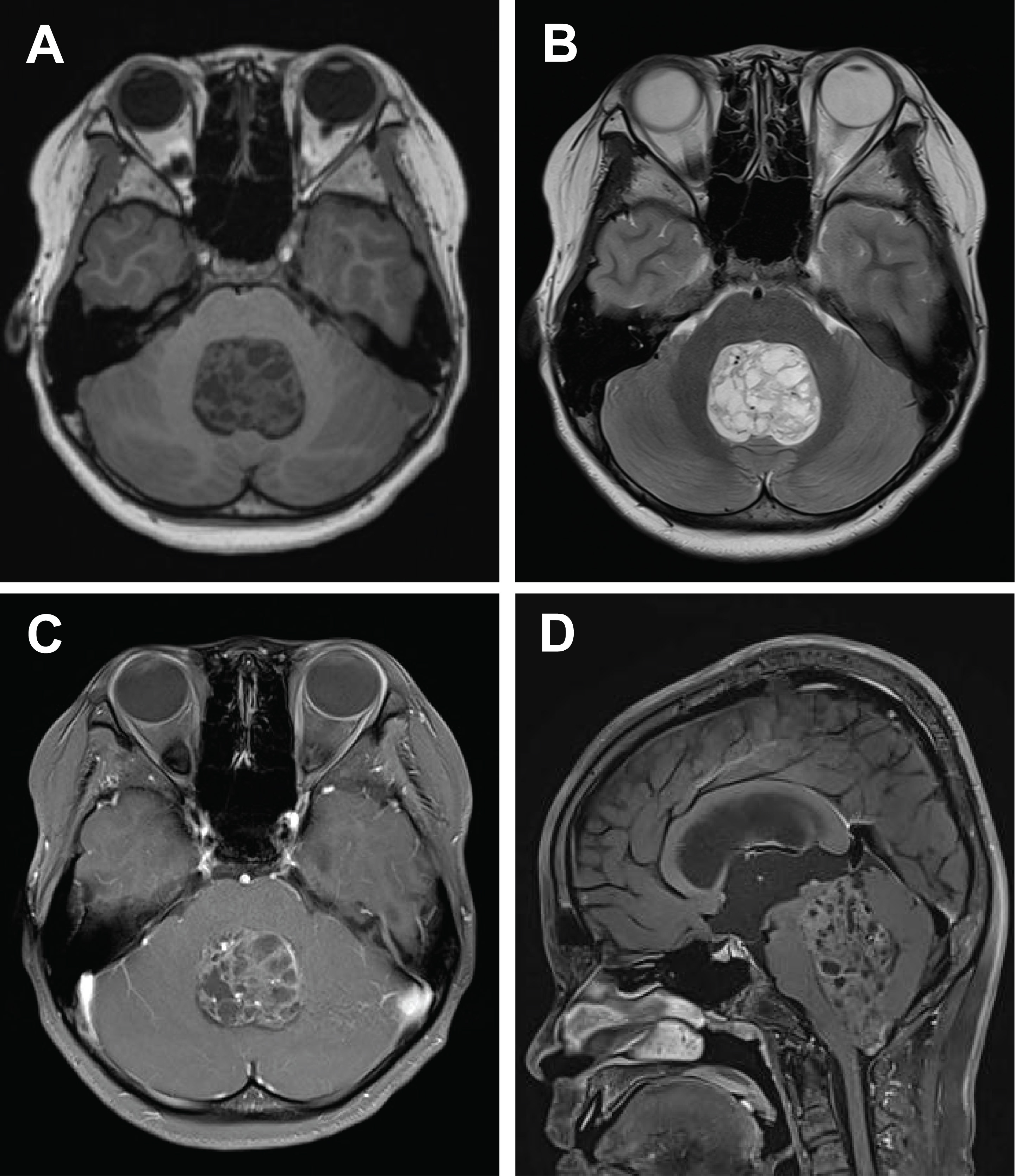
- 7,265 View
- 230 Download
- 2 Web of Science
- 2 Crossref
-
Abstract
PDFSupplementary Material
- Central nervous system tumors with BCL6 corepressor (BCOR) internal tandem duplications (ITDs) constitute a rare, recently characterized pediatric neoplasm with distinct molecular and histopathological features. To date, 69 cases have been documented in the literature, including our institutional case. These neoplasms predominantly occur in young children, with the cerebellum representing the most frequent anatomical location. Radiologically, these tumors present as large, well-circumscribed masses frequently demonstrating necrosis, hemorrhage, and heterogeneous enhancement. Histologically, they are characterized by a monomorphic cellular population featuring ependymoma-like perivascular pseudorosettes, myxoid stroma, and elevated mitotic activity. Immunohistochemically, these tumors exhibit sparse glial fibrillary acidic protein expression while consistently demonstrating positive staining for vimentin and CD56. The defining molecular hallmark is a heterozygous ITD within exon 15 of the BCOR gene, with insertions ranging from 9 to 42 amino acids in length. BCOR immunohistochemistry reveals nuclear positivity in 97.9% of examined cases, although this finding is not pathognomonic for BCOR ITDs. This comprehensive review synthesizes data from all published cases of this novel tumor entity, providing a detailed analysis of clinical presentation, neuroimaging findings, histopathological features with differential diagnostic considerations, therapeutic approaches, and prognostic outcomes.
-
Citations
Citations to this article as recorded by- Guidance for the Diagnosis and Treatment of Rare Embryonal and
Sarcomatous Brain Tumors—a Report from the Central Nervous System-International
Registry for Rare Embryonal and Sarcomatous Tumors German Society of Ped
Pascal Johann, Dominik Sturm, Rolf Kortmann, Brigitte Bison, David Capper, Ahmed El Damaty, Lars Behrens, Selma Manea, Johannes Gojo, Michael Frühwald, Ulrich-Wilhelm Thomale, Martin U. Schuhmann, Rudolf Schwarz, Anna Tietze, Matthias Wagner, Beate Timmer
Klinische Pädiatrie.2026;[Epub] CrossRef - “Updates on diagnostic and prognostic molecular biomarkers of CNS tumors”
Shane Eaton, Ahmad Almotairi, Adrian Levine, Cynthia Hawkins
Neurotherapeutics.2026; 23(6): e01066. CrossRef
- Guidance for the Diagnosis and Treatment of Rare Embryonal and
Sarcomatous Brain Tumors—a Report from the Central Nervous System-International
Registry for Rare Embryonal and Sarcomatous Tumors German Society of Ped
 First
First Prev
Prev



