 E-submission
Current issue
- Page Path
- HOME > Articles and issues > Current issue
- Volume 60(3); May 2026
-


Review Article
- Gene fusions in melanocytic lesions: an updated comprehensive review
- Volha Lenskaya, Larisa Erikson, Victor G. Prieto, Woo Cheal Cho
- J Pathol Transl Med. 2026;60(3):285-306. Published online May 8, 2026
- DOI: https://doi.org/10.4132/jptm.2026.03.11
- 3,971 View
- 139 Download
- 1 Crossref
-
 Abstract
 PDF
PDF Supplementary Material
 - The scope of gene fusions in melanocytic neoplasms is broader than previously recognized, extending well beyond the Spitz-lineage neoplasms where kinase fusions involving ALK, ROS1, NTRK1/2/3, RET, MET, BRAF, and MAP3K8 define biologically and morphologically distinct tumors. Emerging studies demonstrate that a meaningful proportion of conventional non-Spitz lineage melanomas harbor oncogenic fusions. Such fusions may impact clinical behavior, histopathologic presentation and provide opportunities for targeted therapy. The World Health Organization classification of skin tumors, 5th edition, now incorporates fusion status into taxonomy and risk stratification, yet some important questions remain for further investigation: fusion-associated neoplasms can mimic non-melanocytic neoplasm; Spitz-type fusions appear in non-Spitz lesions; and melanocytic differentiation may occur in some other fusion-driven lesions. Broad-panel next-generation sequencing (including RNAseq), together with targeted fluorescence in situ hybridization and immunohistochemistry enhances detection of known and novel fusion partners. Early clinical evidence of TRK, ALK, and ROS1 inhibitor efficacy underscores the translational promise of fusion testing and opens avenues for personalized therapy. This review synthesizes current knowledge on the genomics, histopathology, diagnosis, and therapeutic implications of fusion-driven melanocytic neoplasms, highlighting consensus points and remaining controversies.
-
Citations
Citations to this article as recorded by
- Clinicopathologic and molecular characteristics of acral melanomas harboring RARA fusions
Mokhtar H. Abdelhammed, Richard K. Yang, Volha Lenskaya, Carlos A. Torres-Cabala, Woo Cheal Cho
Human Pathology.2026; 177: 106211. CrossRef
- Clinicopathologic and molecular characteristics of acral melanomas harboring RARA fusions
Original Articles
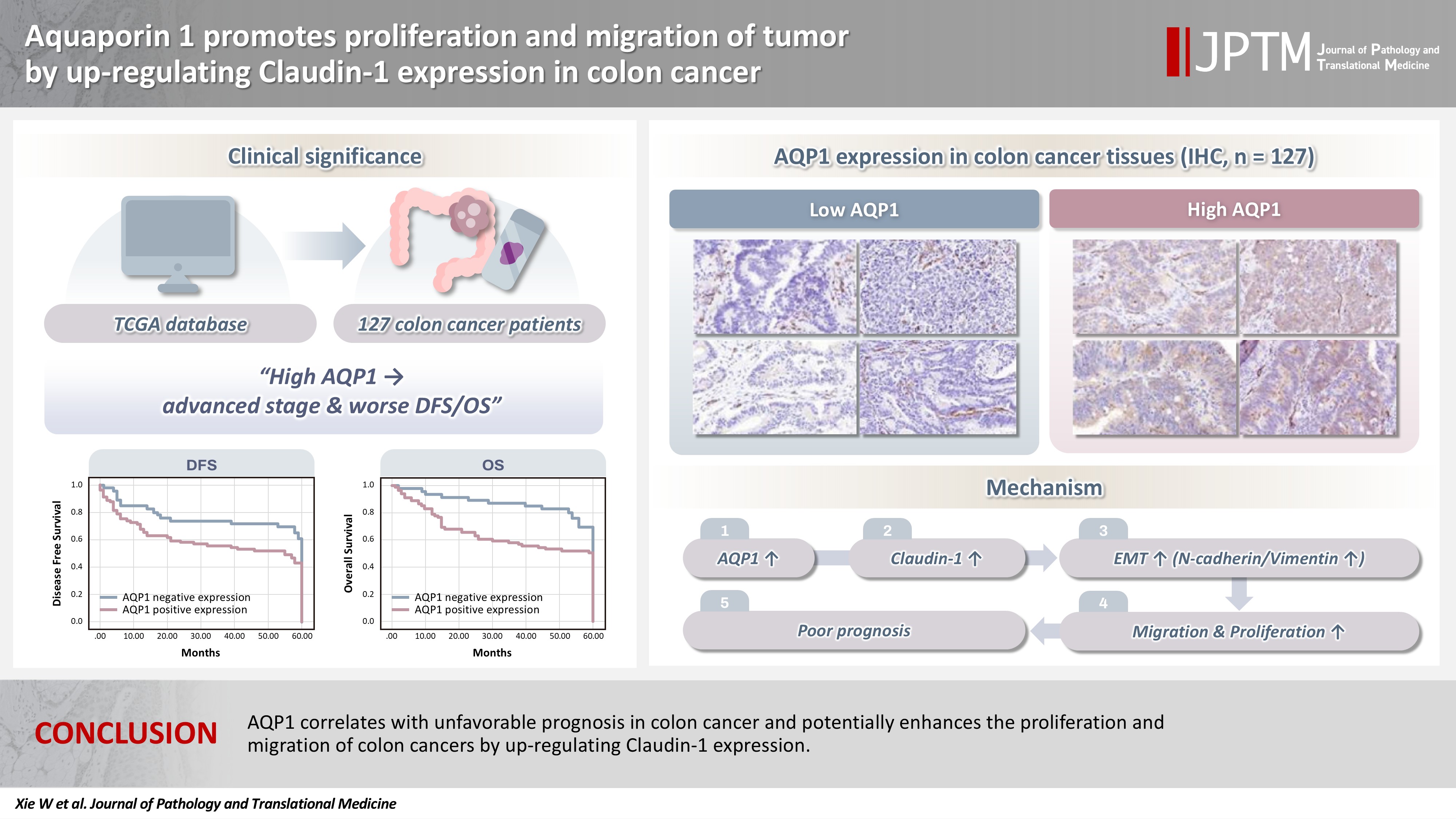
- Aquaporin 1 promotes proliferation and migration of tumor by up-regulating claudin-1 expression in colon cancer
- Wei Wei Xie, Lin Xu, Qian Li, Dao Quan Zhang, Yu Bao Zhou
- J Pathol Transl Med. 2026;60(3):307-318. Published online March 20, 2026
- DOI: https://doi.org/10.4132/jptm.2026.01.01
- 2,028 View
- 112 Download
-
Abstract
PDF
- Background
With the rising incidence of colon cancer, several studies have indicated that aquaporin 1 (AQP1) expression is associated with the development of colon cancer. This study aims to elucidate the potential molecular mechanisms between them. Methods: We screened data from The Cancer Genome Atlas (TCGA) database and retrospectively examined AQP1 protein expression in 127 colon cancer patients to analyze the relationship between AQP1 expression and pathological stages, prognosis. We created stable colon cancer cell lines with differential AQP1 expression, the effect of AQP1 expression on the proliferation and migration of colon cancer cells was assessed by in vitro and in vivo studies, and explored potential molecular mechanisms through Western blotting. Results: High AQP1 expression was associated with poorer survival (overall survival [OS], p = .028) in colon cancer patients from the TCGA database. Similarly, retrospective clinical data indicated that high AQP1 expression was associated with reduced disease-free survival and OS (p = .036 and p = .017, respectively). The low-expressing AQP1 colon cancer cells exhibited a decrease in proliferation and migration ability of colon cancer cells compared to the overexpressing AQP1 group (p < .05) in vitro and in vivo. Immunohistochemistry and western blotting experiments validated heightened expression of N-cadherin, vimentin, and claudin- 1 in the tumor tissues of the overexpressing AQP1 group. Conversely, reduced AQP1 expression resulted in decreased expression of claudin- 1. Conclusions: AQP1 correlates with unfavorable prognosis in colon cancer and potentially enhances the proliferation and migration of colon cancer by up-regulating claudin-1 expression.
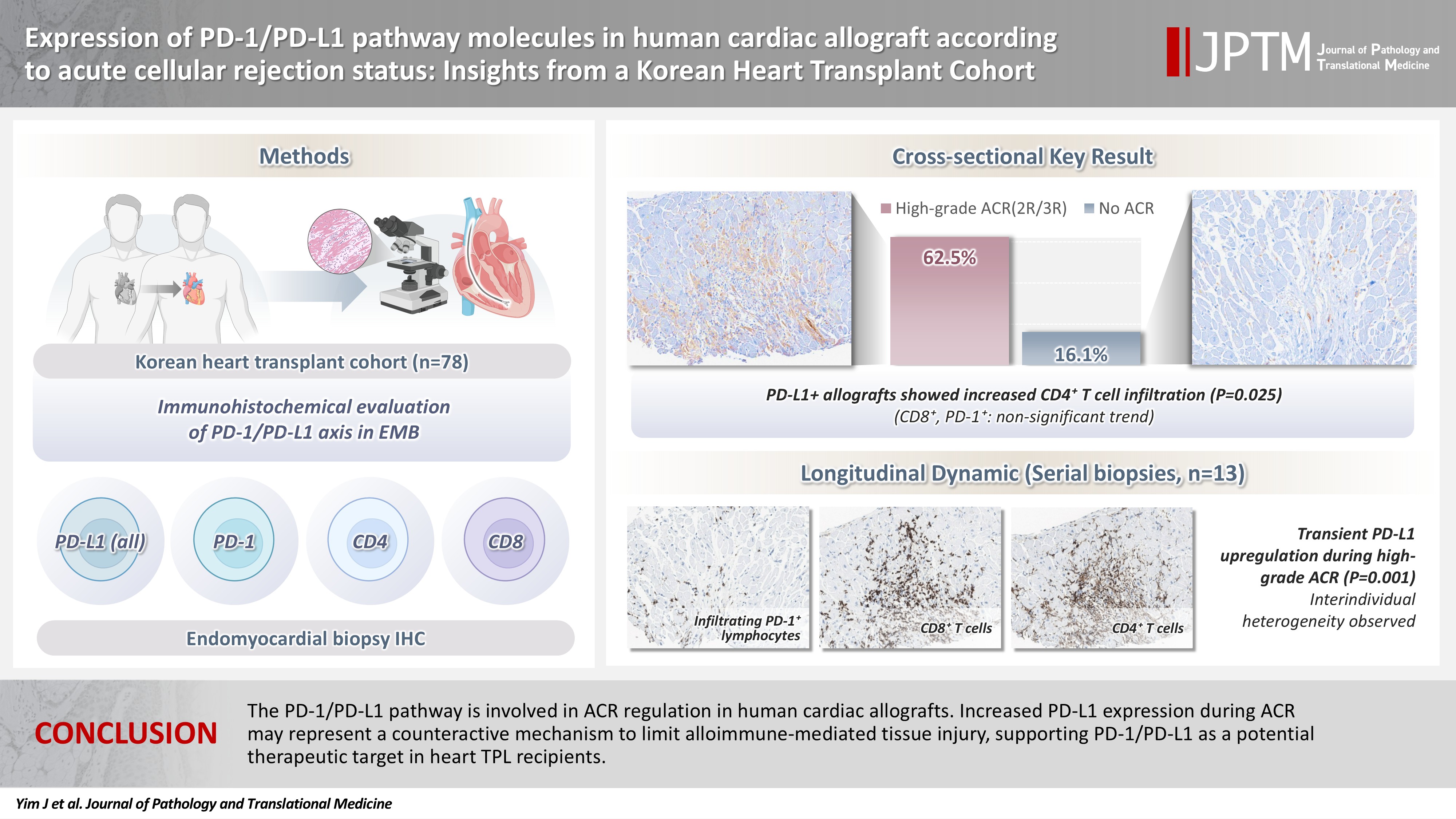
- Expression of PD-1/PD-L1 pathway molecules in human cardiac allograft according to acute cellular rejection status: insights from a Korean Heart Transplant Cohort
- Jeemin Yim, Yoon Kyung Jeon, Doo Hyun Chung, Jaemoon Koh
- J Pathol Transl Med. 2026;60(3):319-330. Published online March 27, 2026
- DOI: https://doi.org/10.4132/jptm.2026.01.02
- 2,177 View
- 75 Download
-
Abstract
PDF
- Background
Acute cellular rejection (ACR) following heart transplantation (TPL) compromises graft function and survival. The programmed cell death-1 (PD-1)/PD-1 ligand-1 (PD-L1) pathway represents an immune checkpoint that maintains peripheral immune tolerance, but its expression and significance in human cardiac allografts with ACR remain unclear. Thus, we investigated PD-1/ PD-L1 expression in endomyocardial biopsies from heart TPL recipients to clarify the role of this pathway in the ACR of human cardiac allografts and explore the potential of therapeutic modulation of PD-1/PD-L1 in this setting. Methods: Endomyocardial biopsies of 78 patients with heart TPL were subjected to immunohistochemistry for PD-L1, PD-1, CD4, and CD8. PD-L1 expression and quantities of PD-1+, CD4+, and CD8+ infiltrating lymphocytes were evaluated according to clinicopathological features, ACR presence, and clinical outcomes. Results: Allografts with high-grade ACR (International Society for Heart and Lung Transplantation grades 2R and 3R) demonstrated markedly higher PD-L1 expression than did those without ACR (62.5% vs. 16.1%, p < .001). PD-L1 expression was positively associated with CD4+ lymphocyte infiltration (p = .025), whereas CD8 and PD-1+ lymphocyte counts were higher in PD-L1-positive allografts without reaching statistical significance (p = .059 and p = .390, respectively). Serial biopsies revealed that PD-L1 expression was upregulated in patients with high-grade ACR compared with that in previous non-ACR tissues, and follow-up biopsies were performed after ACR resolution. Conclusions: The PD-1/PD-L1 pathway is involved in ACR regulation in human cardiac allografts. Increased PD-L1 expression during ACR may represent a counteractive mechanism to limit alloimmune-mediated tissue injury, supporting PD-1/PD-L1 as a potential therapeutic target in heart TPL recipients.
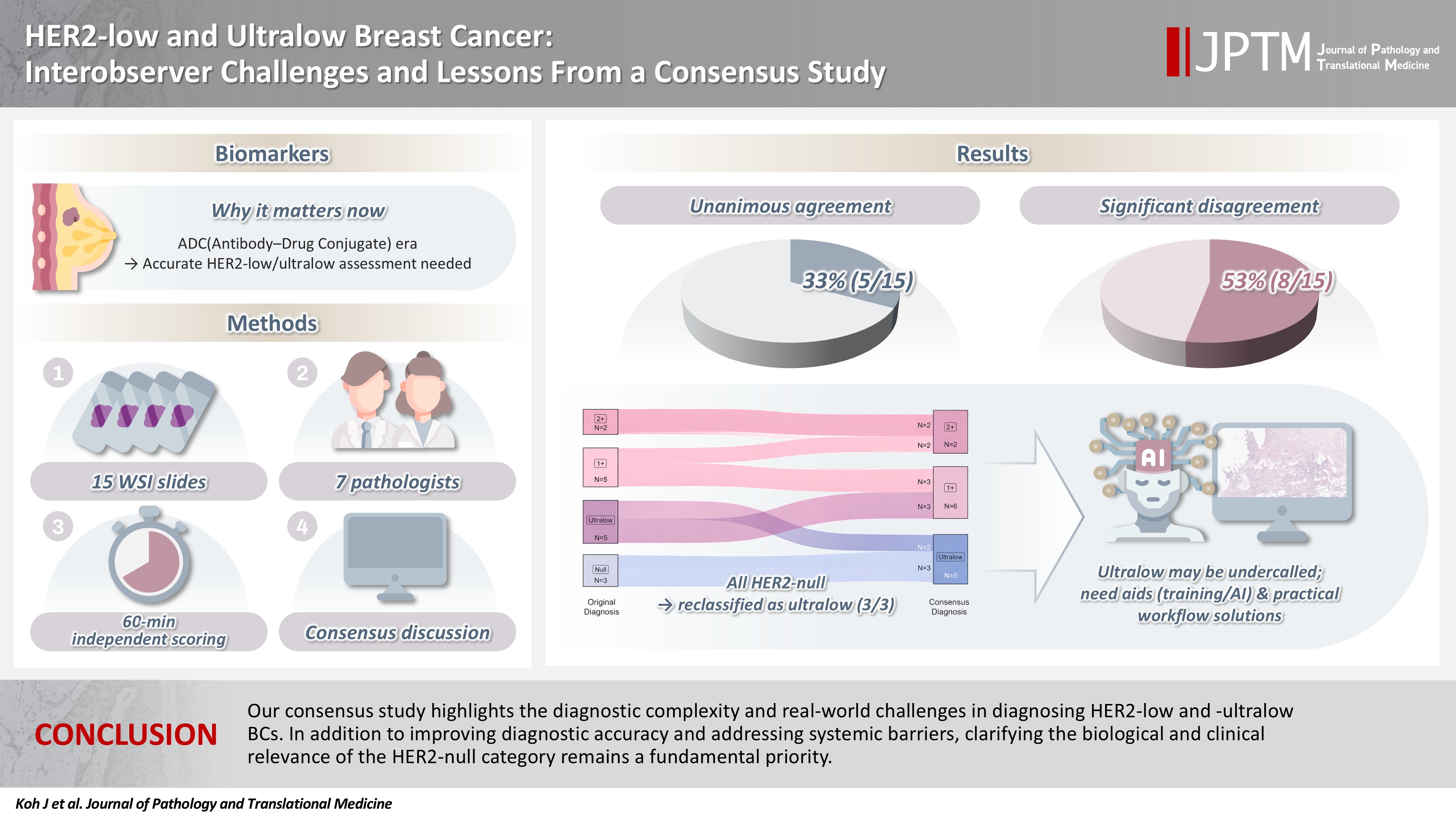
- HER2-low and ultralow breast cancer: interobserver challenges and lessons from a consensus study
- Jiwon Koh, Yoon Jin Cha, Eun Yoon Cho, Ahwon Lee, Ja Seung Koo, So Yeon Park, Min Hwan Kim, Jae Ho Jeong, Gyungyub Gong
- J Pathol Transl Med. 2026;60(3):331-337. Published online March 20, 2026
- DOI: https://doi.org/10.4132/jptm.2026.01.08
- 2,324 View
- 177 Download
-
Abstract
PDF
- Background
The recent approval of trastuzumab deruxtecan for human epidermal growth factor receptor 2 (HER2)–low and HER2-ultralow breast cancer mandates an adequate assessment of these categories. Methods: Seven breast pathologists from the Breast Pathology Study Group of the Korean Society of Pathologists held an on-site expert consensus meeting. Fifteen sets of virtual whole slide images (WSI) of hematoxylin and eosin stain and HER2 immunohistochemistry were provided. The pathologists were given 60 minutes to submit their diagnosis of HER2 expression into null, ultralow, 1+, 2+, or 3+. Afterwards, in-depth discussion and consensus diagnoses were made by real-time visualization of the WSI. Results: After the consensus meeting, unanimous 100% agreements were seen only in five (33.3%) of the examined cases, which consisted of three 1+ cases and two 2+ cases. Two cases (13.3%) had mild disagreement, with only one pathologist’s disagreement. Of note, eight cases (53.3%) showed significant disagreement, defined by more than two pathologists’ disagreement. All HER2-null cases were reclassified as ultralow after consensus review, suggesting potential widespread underclassification of ultralow cases in clinical practice. Conclusions: Experts had significant discrepancies in interpreting HER2-low/ultralow status. It is important to assess if the distinction between HER2-low and ultralow is strictly required and if HER2-null breast cancer exists in reality.
- Clinicopathological profile of high-grade differentiated thyroid carcinoma in an Indonesian tertiary hospital
- Novita , Agnes Stephanie Harahap, Maria Francisca Ham, Alfianto Widiono, Chan Kwon Jung
- J Pathol Transl Med. 2026;60(3):338-348. Published online April 23, 2026
- DOI: https://doi.org/10.4132/jptm.2026.01.15
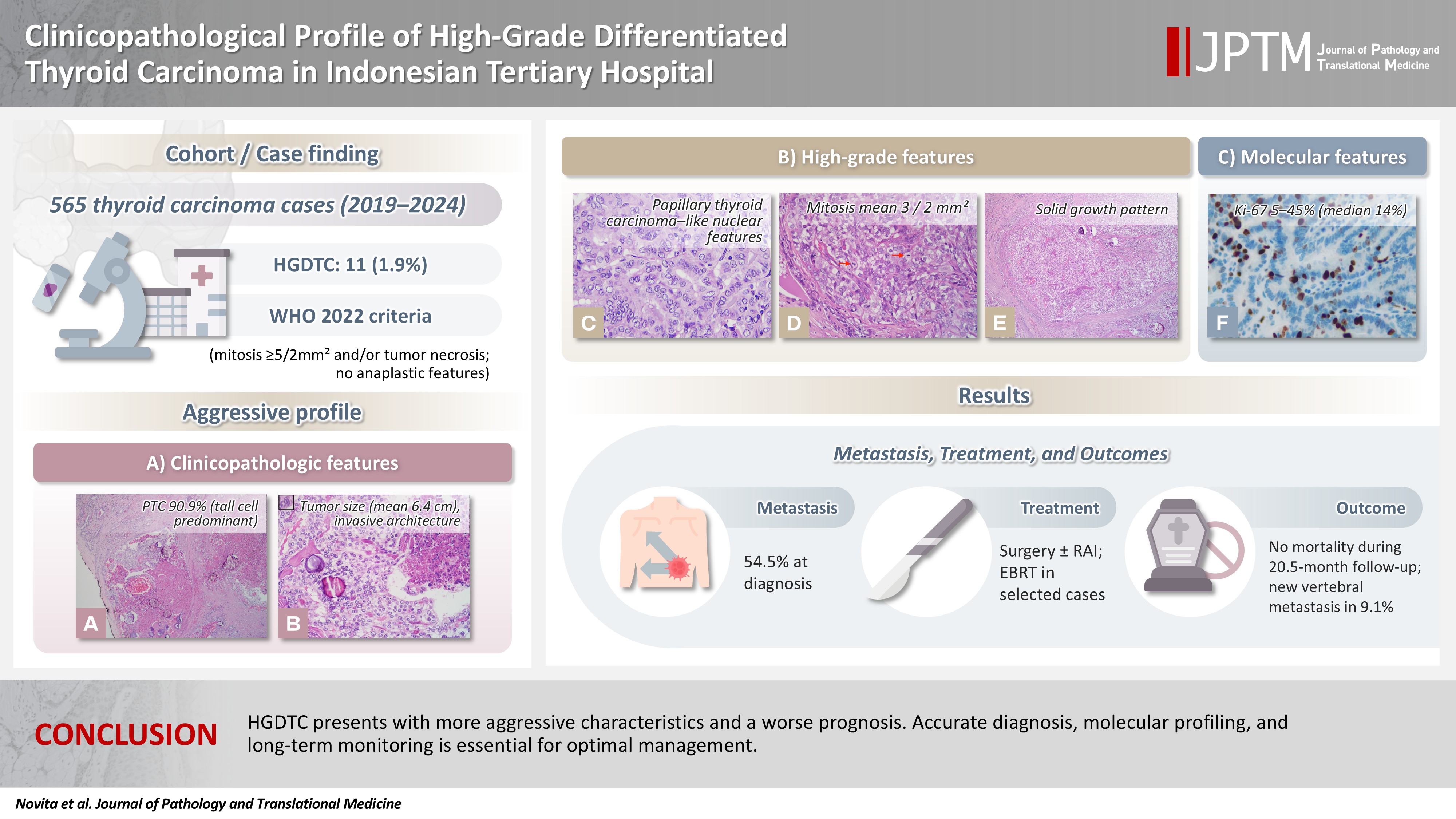
- 1,858 View
- 54 Download
-
Abstract
PDFSupplementary Material
- Background
High-grade differentiated thyroid carcinoma (HGDTC) is a recently recognized entity in the 2022 World Health Organization classification, representing a more aggressive subtype of differentiated thyroid carcinoma. Previously, high-grade features such as increased mitotic activity and tumor necrosis were often overlooked, despite being important independent prognostic factors. Although rare, HGDTC carries significant diagnostic, prognostic, and therapeutic implications. Data remain limited in Indonesia. Methods: This retrospective descriptive study reviewed 565 thyroid carcinoma cases diagnosed at Cipto Mangunkusumo Hospital from 2019 to 2024. Eleven cases (1.9%) met HGDTC criteria. Clinicopathological characteristics, histologic subtypes, Ki-67 proliferation index, molecular alterations, treatment modalities, and clinical outcomes were analyzed. Results: Patients had a mean age of 54.6 years, with a female-to-male ratio of 2.7:1. Papillary thyroid carcinoma was the main type (90.9%), with the tall cell subtype predominating. Mean tumor size was 6.4 cm. Lymphatic invasion, vascular invasion, and extrathyroidal extension were present in 54.5%, 18.2%, and 45.5% of cases, respectively. All tumors showed necrosis. Mean mitotic count was 3 per 2 mm². The Ki-67 index ranged from 5% to 45% (median, 14%). BRAFV600E and TERT promoter mutations were detected in 18.2% and 36.4% of cases, respectively, with co-mutations in 18.2%. Six cases (54.5%) had metastases at time of diagnosis. During a mean follow-up of 20.5 months, one patient (9.1%) developed new vertebral metastases and all patients (100%) remained alive. Conclusions: HGDTC presents with more aggressive characteristics and a worse prognosis. Accurate diagnosis, molecular profiling, and long-term monitoring are essential for optimal management.
- Incidental serrated lesions of the appendix: analysis of 2,137 appendectomy specimens
- Ömer Atmış, Ecem Dokuzlu Küçük, Hanife Seda Mavili, Fatma Seher Pehlivan, Ayça Tan, Semin Ayhan
- J Pathol Transl Med. 2026;60(3):349-355. Published online May 4, 2026
- DOI: https://doi.org/10.4132/jptm.2026.02.08
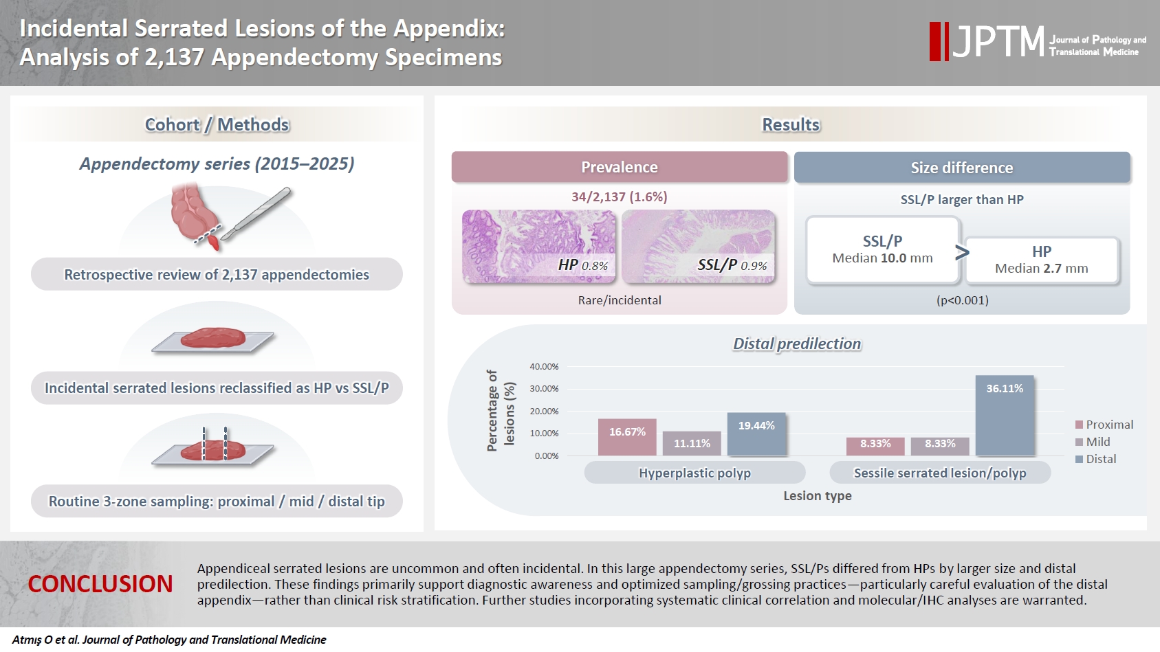
- 1,409 View
- 34 Download
-
Abstract
PDF
- Background
Serrated lesions of the appendix are rare, often incidental findings in routine appendectomy specimens. Their true frequency, histopathologic spectrum, and anatomic distribution remain incompletely characterized, partly due to variability in sampling practices. Methods: We retrospectively reviewed 2,137 appendectomy specimens (2015–2025) from a single tertiary pathology center. Cases with histologically confirmed serrated lesions were reexamined, classified as hyperplastic polyp (HP) or sessile serrated lesion/polyp (SSL/P), and assessed for clinicopathologic parameters including lesion size, location, and associated pathologies. Nonparametric tests were used, with statistical significance defined as p < .05. Results: Serrated lesions were identified in 34 cases (1.6%) with 36 serrated lesions, comprising 17 HPs (0.8%) and 19 SSL/Ps (0.9%). SSL/Ps were significantly larger than HPs (median 10.0 vs. 2.7 mm, p < .001) and were more frequently located in the distal appendix (68.4% vs. 33.3%, p = .045, one-tailed Fisher’s exact test). No dysplasia or traditional serrated adenoma was detected. Acute appendicitis was present in 88% of cases, and associated neoplasms in 9%. Conclusions: Appendiceal serrated lesions are uncommon and often incidental. In this large appendectomy series, SSL/Ps differed from HPs by larger size and distal predilection. These findings primarily support diagnostic awareness and optimized sampling/grossing practices—particularly careful evaluation of the distal appendix—rather than clinical risk stratification. Further studies incorporating systematic clinical correlation and molecular/immunohistochemistry analyses are warranted.
Case Study
- Multidimensional analysis of concurrent proximal bronchiolar adenoma and lung carcinoma
- Lu-Yao Li, Gong-Ming Dong, Yun-Peng Zhang, Ting-Ting Wang, Fu-Quan Jia, Guan-Jun Zhang
- J Pathol Transl Med. 2026;60(3):356-363. Published online March 23, 2026
- DOI: https://doi.org/10.4132/jptm.2025.12.31
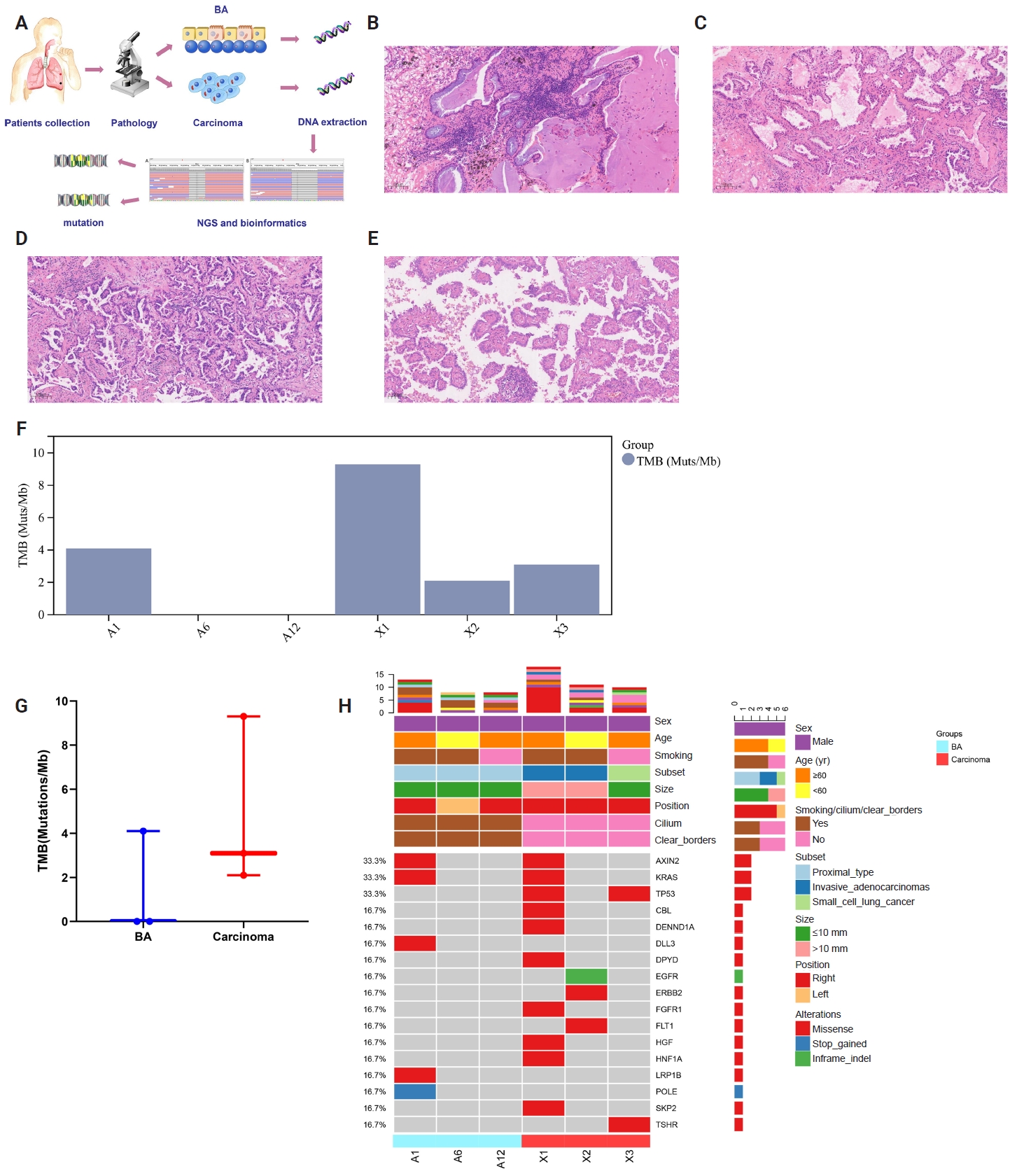
- 1,621 View
- 78 Download
-
Abstract
PDFSupplementary Material
- Bronchiolar adenoma (BA) is a rare type of lung tumor characterized by bilayered epithelial cells having a continuous basal layer and a luminal layer. It resembles mucinous adenocarcinoma (MA) on frozen section, with difficulty in distinguishing the basal layer. Immunohistochemistry is the best choice for verifying the diagnosis. This study aimed to comprehensively characterize three cases of BA-combined carcinoma using clinical, histopathological, and genetic features. BA and carcinoma sections were subjected to next-generation sequencing, respectively. It was hypothesized that while different mutation forms matched different regions, BA and lung adenocarcinoma shared the same gene mutation when they co-occurred in the same location. BA with extensive carcinoma is extremely rare and presents diagnostic challenges due to its overlap with conditions such as MA. Because of its distinctive morphological characteristics, BA may be regarded as a low-grade malignancy, particularly during a confusing evaluation. A multifaceted examination of clinical, radiological, immunohistochemical, and genetic data is necessary for an accurate diagnosis.
Newsletter
- What’s new in digital and computational pathology 2026: advances in adoption, standards, AI technologies, and clinical integration
- Selim Sevim, Chadi Hajar, Snehal Sonawane
- J Pathol Transl Med. 2026;60(3):364-370. Published online May 15, 2026
- DOI: https://doi.org/10.4132/jptm.2026.04.27

- 4,392 View
- 189 Download
-
Abstract
PDF
- Digital and computational pathology are expanding rapidly worldwide, driven by advances in whole-slide imaging, AI algorithms, multimodal data integration, and improved digital infrastructure. Adoption continues to accelerate in the United States and internationally, supported by professional guidelines, emerging reimbursement pathways, and the growing need for remote workflows and collaborative diagnostics. Progress in interoperability standards, regulatory frameworks, and FDA approvals has strengthened the foundation for clinical deployment, while large-scale data repositories and federated learning approaches enable more robust and privacy-preserving model development. Foundation models, multimodal AI systems, and LLM-based copilots are reshaping diagnostic support, prognostication, workflow efficiency, clinical trials and drug discovery.
 First
First Prev
Prev


