 E-submission
Search
- Page Path
- HOME > Search
Review Article
- Cutaneous soft tissue tumors in the 5th edition of the World Health Organization classification of skin tumors: key updates and new entities
- Joon Hyuk Choi
- J Pathol Transl Med. 2026;60(2):144-183. Published online March 13, 2026
- DOI: https://doi.org/10.4132/jptm.2026.01.09
- 1,897 View
- 129 Download
-
 Abstract
 PDF
PDF - The 5th edition of the World Health Organization (WHO) classification of skin tumors introduces a dedicated chapter on cutaneous soft tissue tumors, providing a comprehensive, standardized reference with updated diagnostic criteria that directly inform routine dermatopathology practice and molecular diagnostics. This edition incorporates several key changes, including newly recognized entities such as EWSR1::SMAD3-rearranged fibroblastic tumor, neurotrophic tyrosine receptor kinase (NTRK)–rearranged spindle cell neoplasm, superficial CD34-positive fibroblastic tumor, and CRTC1::TRIM11 cutaneous tumor. Diagnostic terminology has also been refined; for example, the term ‘atypical intradermal smooth muscle neoplasm’ replaces ‘cutaneous leiomyosarcoma’ for lesions confined to the dermis, whereas the designation leiomyosarcoma is reserved for tumors with overt subcutaneous infiltration. In addition, epithelioid fibrous histiocytoma has been reassigned to the family of tumors of uncertain differentiation. This review summarizes the key updates and newly recognized entities in the chapter on cutaneous soft tissue tumors in the 5th edition of the WHO classification of skin tumors, emphasizing their clinicopathological and molecular implications.
Original Article
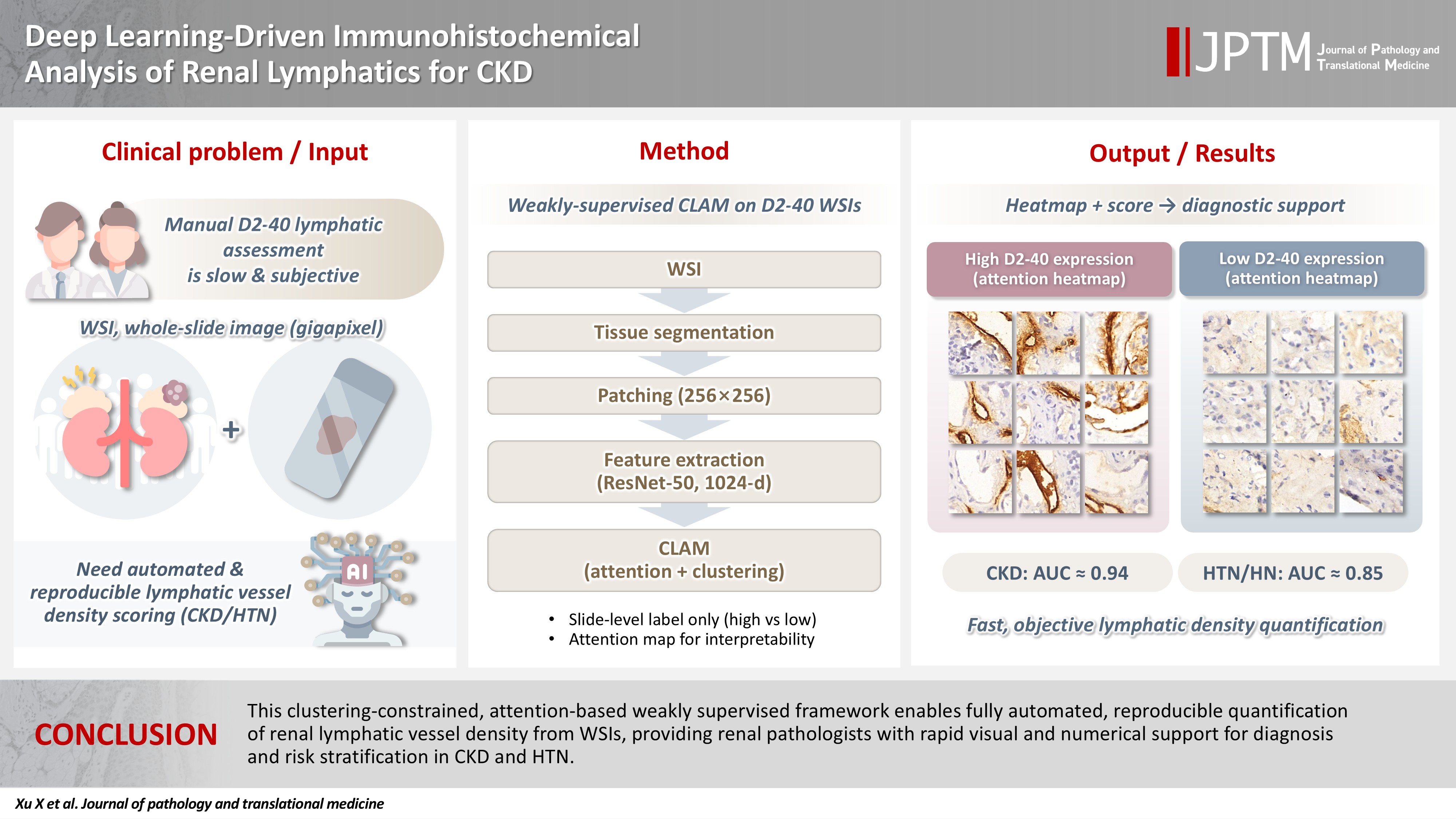
- Deep learning–driven immunohistochemical analysis of renal lymphatics for chronic kidney disease: bioinformatic and histopathological study
- Xin Xu, YanPing Lin, Guangchang Pei, Rui Zeng, Gang Xu
- J Pathol Transl Med. 2026;60(2):220-230. Published online March 13, 2026
- DOI: https://doi.org/10.4132/jptm.2025.12.15
- 1,068 View
- 68 Download
-
Abstract
PDF
 Supplementary Material
 - Background
Renal lymphatic vessel density is clinically relevant in kidney disease but is still assessed by slow, subjective visual estimation. We evaluated a weakly supervised, attention-based multiple-instance learning framework for automated detection and quantification of renal lymphatic vessel density on D2-40-stained whole-slide images (WSIs). Methods: Two independent internal datasets from Tongji Hospital were collected, including 198 cases of chronic kidney disease (CKD) and 50 cases of hypertensive nephropathy (HTN). All biopsies were immunohistochemically stained for D2-40 and digitized as WSIs. Pathologists provided only slide-level labels (D2-40 high vs. D2-40 low). Tissue regions were automatically segmented, tiled into patches, and encoded using a pretrained convolutional neural network. Patch embeddings were then analyzed with a clustering-constrained attention multiple-instance learning (CLAM) model. Unlike conventional multiple-instance learning (MIL) methods that only weight instances, CLAM jointly performs attention-based instance selection and instance-level clustering to distinguish positive from negative evidence within each slide, yielding more discriminative slide-level features and interpretable attention maps. Performance was compared with a classic MIL model trained on the same features. Results: CLAM achieved area under the receiver operating characteristic curves of 0.942 and 0.858 on the CKD and HTN datasets, respectively, outperforming classic MIL (0.866 and 0.801). Attention maps highlighted lymphatic-rich regions consistent with renal pathologists’ assessments. Conclusions: This clustering-constrained, attention-based weakly supervised framework enables fully automated, reproducible quantification of renal lymphatic vessel density from WSIs, providing renal pathologists with rapid visual and numerical support for diagnosis and risk stratification in CKD and HTN.
Review Article
- A comprehensive review of ossifying fibromyxoid tumor: insights into its clinical, pathological, and molecular landscape
- Kyriakos Chatzopoulos, Antonia Syrnioti, Mohamed Yakoub, Konstantinos Linos
- J Pathol Transl Med. 2026;60(1):6-19. Published online January 14, 2026
- DOI: https://doi.org/10.4132/jptm.2025.10.02

- 3,293 View
- 156 Download
-
Abstract
PDF
- Ossifying fibromyxoid tumor (OFMT) is a rare mesenchymal neoplasm first described in 1989. It typically arises in the superficial soft tissues of the extremities as a slow-growing, painless mass. Histologically, it is commonly characterized by a multilobular architecture composed of uniform epithelioid cells embedded in a fibromyxoid matrix, often surrounded by a rim of metaplastic bone. While classic cases are readily identifiable, the tumor's histopathological heterogeneity can mimic a range of benign and malignant neoplasms, posing significant diagnostic challenges. Molecularly, most OFMTs harbor PHF1 rearrangements, commonly involving fusion partners such as EP400, MEAF6, or TFE3. This review underscores the importance of an integrated diagnostic approach- incorporating histopathological, immunohistochemical, and molecular data- to accurately classify OFMT and distinguish it from its mimics. Expanding awareness of its morphologic and molecular spectrum is essential for precise diagnosis, optimal patient management, and a deeper understanding of this enigmatic neoplasm.
Original Articles
- E-cadherin expression and tumor-stroma ratio as prognostic biomarkers of peritoneal recurrence in advanced gastric cancer: a digital image analysis-based stratification study
- Somang Lee, Binnari Kim
- J Pathol Transl Med. 2025;59(6):408-420. Published online November 6, 2025
- DOI: https://doi.org/10.4132/jptm.2025.08.27

- 3,411 View
- 120 Download
-
Abstract
PDF
- Background
Gastric cancer remains a significant global health burden, with a high peritoneal recurrence rates after curative surgery. E-cadherin and the tumor-stroma ratio (TSR) have been proposed as prognostic indicators, but their combined prognostic utility remains unclear. Methods: This retrospective study included 130 patients with T3/T4a gastric cancer who underwent curative gastrectomy at Ulsan University Hospital between 2014 and 2019. Immunohistochemistry for E-cadherin and Vimentin was performed. Digital image analysis using QuPath’s object classifier quantified E-cadherin expression and TSR. Results: Low E-cadherin expression was associated with diffuse-type histology and advanced T stage. Low TSR was linked to younger age, female sex, and XELOX treatment. In Kaplan-Meier analysis, low TSR showed a non-significant trend toward higher peritoneal recurrence (p = .054), while low E-cadherin expression was significantly associated with increased peritoneal recurrence (p = .002). Combined biomarker analysis also revealed a significant difference in recurrence-free survival (RFS) among the four groups (p = .005); patients with both high TSR and high E-cadherin expression experienced the most favorable RFS. In multivariable analysis, E-cadherin expression remained the only independent predictor of peritoneal recurrence (high vs. low; hazard ratio, 0.348; 95% confidence interval, 0.149 to 0.816; p = .015). Conclusions: E-cadherin and TSR reflect distinct tumor biology such as epithelial integrity and stromal composition, and their combined evaluation improves prognostic stratification. Digital image analysis enhances reproducibility and objectivity, supporting their integration into clinical workflows.
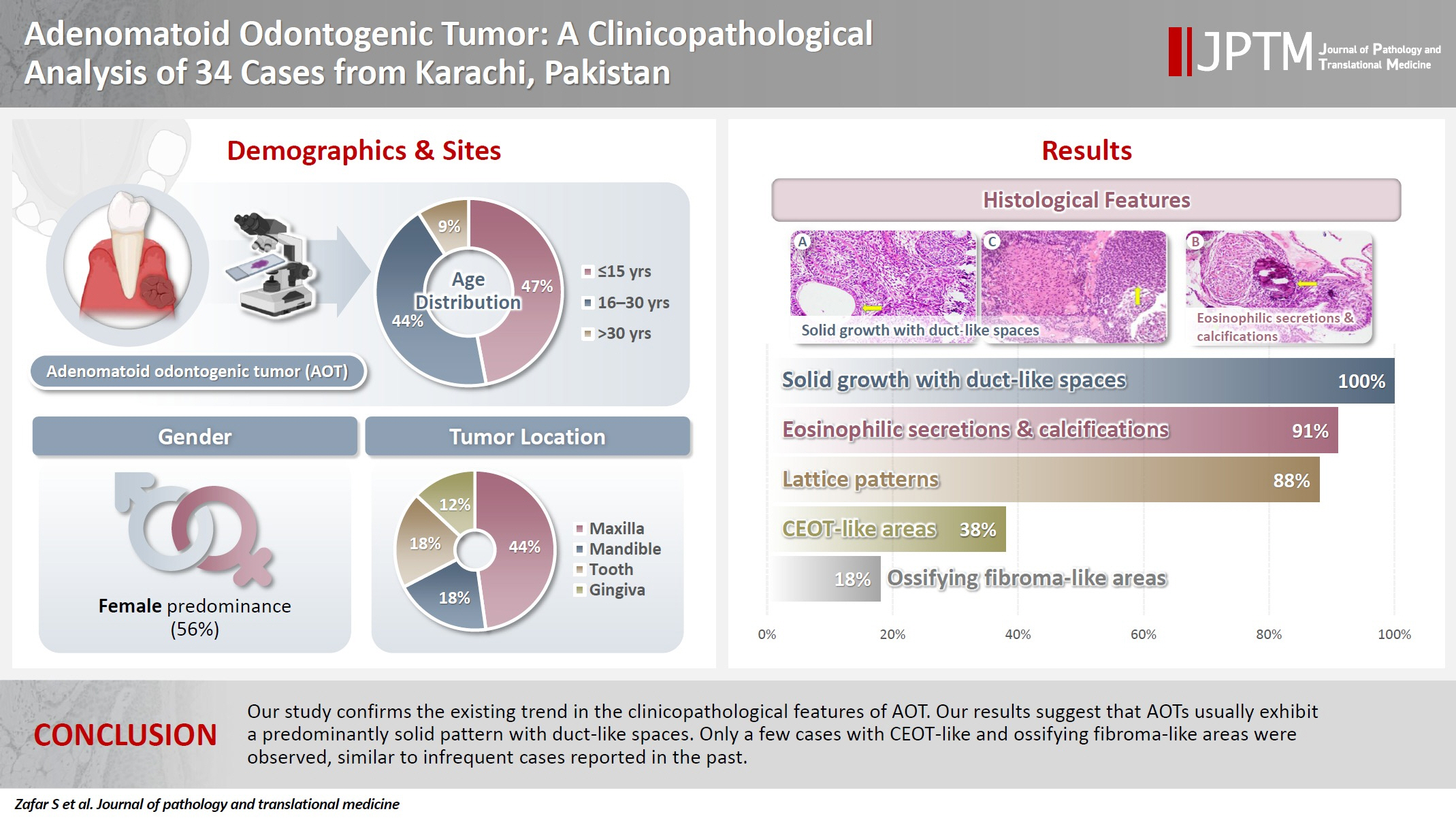
- Adenomatoid odontogenic tumor: clinicopathological analysis of 34 cases from Karachi, Pakistan
- Summaya Zafar, Sehar Sulaiman, Madeeha Nisar, Poonum Khan, Nasir Ud Din
- J Pathol Transl Med. 2025;59(6):390-397. Published online October 16, 2025
- DOI: https://doi.org/10.4132/jptm.2025.07.11
- 3,663 View
- 156 Download
- 1 Crossref
-
Abstract
PDF
- Background
Adenomatoid odontogenic tumor (AOT) is a benign slow-growing neoplasm of odontogenic epithelial origin that is relatively uncommon. Only a few studies have described its histological features. Hence, we aimed to describe the clinicopathological features of AOT in a cohort of patients. Methods: AOT cases diagnosed between 2009 and 2024 were searched electronically. Glass slides were retrieved from archives and were reviewed by two pathologists to record the associated morphological features. Other data including patient demographics and tumor site were collected by reviewing histopathology reports. Results: The age of patients ranged from 9 to 44 years (mean, 17.7 years), and most were female (55.9%). The maxilla (44.1%) was the most common tumor site. Histologically, a predominantly solid growth pattern (n = 34) accompanied by ducts with a cuboidal/columnar epithelial lining (n = 31), eosinophilic secretions (n = 31), calcifications (n = 31), lattice work pattern (n = 30), and cystic areas (n = 20) were observed. Less frequent features included calcifying epithelial odontogenic tumor (CEOT)–like areas (n = 13), osteodentin (n = 6), association with impacted tooth (n = 3), mucin in tubules (n = 7), fibrocollagenous stroma (n = 6), mucin in ducts (n = 3) and ossifying fibroma-like areas (n = 6). The association of ducts with a cuboidal/columnar epithelial lining, lattice work pattern, calcifications, and eosinophilic secretions with gingival tumors was statistically significant (p ≤ .05). Additionally, tooth tumors were significantly associated with CEOT-like areas (p = .03). Conclusions: Our study confirms the trends in the clinicopathological features of AOT in previous case reports. Our results suggest that AOTs usually exhibit a predominantly solid pattern with duct-like spaces. Only a few cases with CEOT-like and ossifying fibroma-like areas were observed, similar to infrequent cases reported in the past. -
Citations
Citations to this article as recorded by
- Intraosseous lesions of the jaw: a clinicohistological study
Hadeel Odeh, Esra Nsour, Muna A. Salameh, Zayed M. Al-Zu’bi, Ali Al Khader
BMC Oral Health.2026;[Epub] CrossRef
- Intraosseous lesions of the jaw: a clinicohistological study
- Attitudes toward artificial intelligence in pathology: a survey-based study of pathologists in northern India
- Manupriya Sharma, Kavita Kumari, Navpreet Navpreet, Sushma Bharti, Rajneesh Kumari
- J Pathol Transl Med. 2025;59(6):382-389. Published online October 2, 2025
- DOI: https://doi.org/10.4132/jptm.2025.07.10

- 6,343 View
- 204 Download
-
Abstract
PDFSupplementary Material
- Background
Artificial intelligence (AI) is transforming pathology by enhancing diagnostic accuracy, efficiency, and workflow standardization. Despite its growing presence, AI adoption remains limited, particularly in resource-constrained settings like India. This study assessed the knowledge, awareness, and perceptions of AI among pathologists in Northern India. Methods: A cross-sectional survey was conducted among 138 practicing pathologists in Northern India between April and June 2024. A structured online questionnaire was used to collect data on demographics, AI awareness, self-reported knowledge, sources of AI education, technological proficiency, and interest in AI-related training programs. Data analysis included descriptive statistics and chi-square tests, with p < .05 considered statistically significant. Results: AI awareness was high (88.4%), with significant sex differences (93.5% in females vs. 78.3% in males, p = .008). However, formal AI training was limited (6.5%), and only 16.7% had used AI as a diagnostic tool. Academic pathologists were more likely to engage with AI literature than their non-academic counterparts (p = .003). Interest in AI workshops was strong (92.8%). Access to whole slide imaging (WSI) correlated with higher AI knowledge (p = .008), as did self-reported technological proficiency (p = .001). Conclusions: Despite high AI awareness among pathologists, significant gaps remain in training, infrastructure, and practical application. Expanding access to digital pathology tools like WSI and improving digital literacy could facilitate AI adoption. Structured educational programs and greater investment in digital infrastructure are crucial for integrating AI into pathology practice.
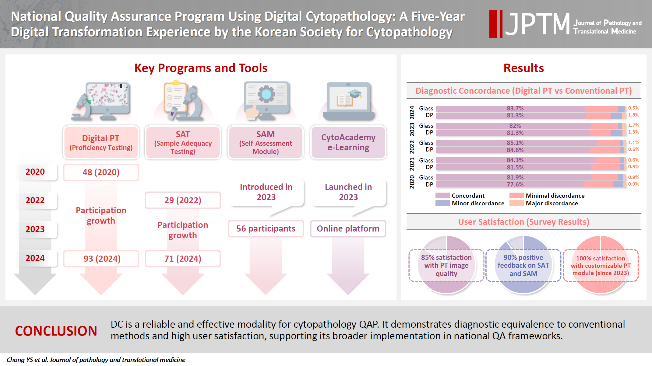
- National quality assurance program using digital cytopathology: a 5-year digital transformation experience by the Korean Society for Cytopathology
- Yosep Chong, Hyeong Ju Kwon, Soon Auck Hong, Sung Soon Kim, Bo-Sung Kim, Younghee Choi, Yoon Jung Choi, Jung-Soo Pyo, Ji Yun Jeong, Soo Jin Jung, Hoon Kyu Oh, Seung-Sook Lee
- J Pathol Transl Med. 2025;59(5):320-333. Published online September 15, 2025
- DOI: https://doi.org/10.4132/jptm.2025.06.27
- 4,530 View
- 111 Download
- 2 Web of Science
- 3 Crossref
-
Abstract
PDFSupplementary Material
- Background
Digital cytopathology (DC) is emerging as a transformative approach in quality assurance programs (QAP), though its comprehensive evaluation remains limited. Since 2020, the Korean Society for Cytopathology has progressively incorporated DC into its national QAP, including digital proficiency testing (PT), sample adequacy testing (SAT), a customizable PT module, and a self-assessment module (SAM), aiming for full digital implementation by 2026. Methods: This 5-year study assessed diagnostic concordance between conventional and digital PT formats and analyzed participant feedback on service quality and digital image usability across PT, SAT, and SAM. Parallel testing was conducted during the transitional phase, and satisfaction was measured through structured surveys. Results: Participation in digital PT increased from 48 institutions in 2020 to 93 in 2024, while digital SAT participation rose from 29 to 71 between 2022 and 2024. In 2023, 56 institutions joined SAM. Diagnostic concordance rates were comparable between digital and conventional PTs (78.6%–84.6% vs. 82.0%–85.1%), including similar category C (major discordance) rates. Satisfaction with digital PT services and image quality exceeded 85%, and over 90% of institutions reported positive feedback on SAT and SAM. Over 80% were satisfied with the customizable PT module. Conclusions: DC is a reliable and effective modality for cytopathology QAP. It demonstrates diagnostic equivalence to conventional methods and high user satisfaction, supporting its broader implementation in national quality assurance frameworks. -
Citations
Citations to this article as recorded by- Practice of Cytopathology in Korea: A 40‐Year Evolution Through Standardization, Digital Transformation, and Global Partnership
Yosep Chong, Ran Hong, Hyeong Ju Kwon, Haeryoung Kim, Lucia Kim, Soon Jae Kim, Yoon Jung Choi
Diagnostic Cytopathology.2026; 54(2): 146. CrossRef - Validation of Digital Cytology for Primary Diagnosis Across a Range of Specimen Types
Talisa Mistry, Harriet Hunter, Dahmane Oukrif, Sabine Pomplun, Reena Khiroya, Mary Falzon, Tanya Alan, Manuel Rodriguez‐Justo, Adam P. Levine
Cytopathology.2026; 37(3): 222. CrossRef - Review of the Changing Roles of Clinical Laboratory Scientists and Strategies for Curricular Innovation in the Era of Artificial Intelligence
Hee Sung KIM
Korean Journal of Clinical Laboratory Science.2026; 58(1): 1. CrossRef
- Practice of Cytopathology in Korea: A 40‐Year Evolution Through Standardization, Digital Transformation, and Global Partnership
- A single-institution demographic study of pathologically proven kidney disease in South Korea over the last 33 years
- Hyejin Noh, Jiyeon Kim, Yeong Jin Choi
- J Pathol Transl Med. 2025;59(5):306-319. Published online September 10, 2025
- DOI: https://doi.org/10.4132/jptm.2025.06.18

- 2,539 View
- 94 Download
-
Abstract
PDFSupplementary Material
- Background
To date, epidemiological studies on the entire spectrum of kidney disease based on pathology have been rarely reported. Methods: A retrospective study was conducted on patients diagnosed with kidney disease at Seoul St. Mary's Hospital between 1991 and 2023. Results: Among 7,803 patients with native kidney disease, glomerular disease (70.3%) was the most common, followed by tubulointerstitial (15.1%) and vascular disease (8.8%). In kidney biopsy, glomerular disease (77.8%) showed the highest frequency, particularly in those under 20s (95.6%) (p = .013). Primary glomerulonephritis (GN) (72.8%) was the predominant glomerular disease, with IgA nephropathy (IgAN) (47.3%) being the most common one. Tubulointerstitial and vascular diseases increased with age, showing the highest prevalence in those over 60 years (p = .008 and p = .032, respectively). Glomerular disease was diagnosed at a younger age (39.7 ± 16.7 years) than tubulointerstitial (49.1 ± 16.2) and vascular (48.1 ± 15.3) diseases (p < .001). When glomerular diseases were classified morphologically, proliferative GN (57.9%) was the most common, followed by non-proliferative (39.6%) and sclerosing (1.6%). When classified by etiology, primary GN accounted for the most (72.8%), followed by secondary (19.3%) and hereditary GN (5.7%). In nephrectomy, tubulointerstitial disease (64.6%) was the most common. Those with a tubulointerstitial disease had a higher mean age than those with a glomerular disease (p < .001). In cases where nephrectomy was performed for glomerular diseases, IgAN (34.1%) was the most common diagnosis. Conclusions: Kidney disease has been increasing in South Korea for 33 years. Glomerular disease was the most common across all age groups, tubulointerstitial and vascular diseases increased over 60 years.
- The Automatable Activity–Based Approach to Complexity Unit Scoring as a task-specific model approach to monetizing outcomes of pathology artificial intelligence solutions
- Stavros Pantelakos, Martha Nifora, Georgios Agrogiannis
- J Pathol Transl Med. 2025;59(4):225-234. Published online July 3, 2025
- DOI: https://doi.org/10.4132/jptm.2025.04.15
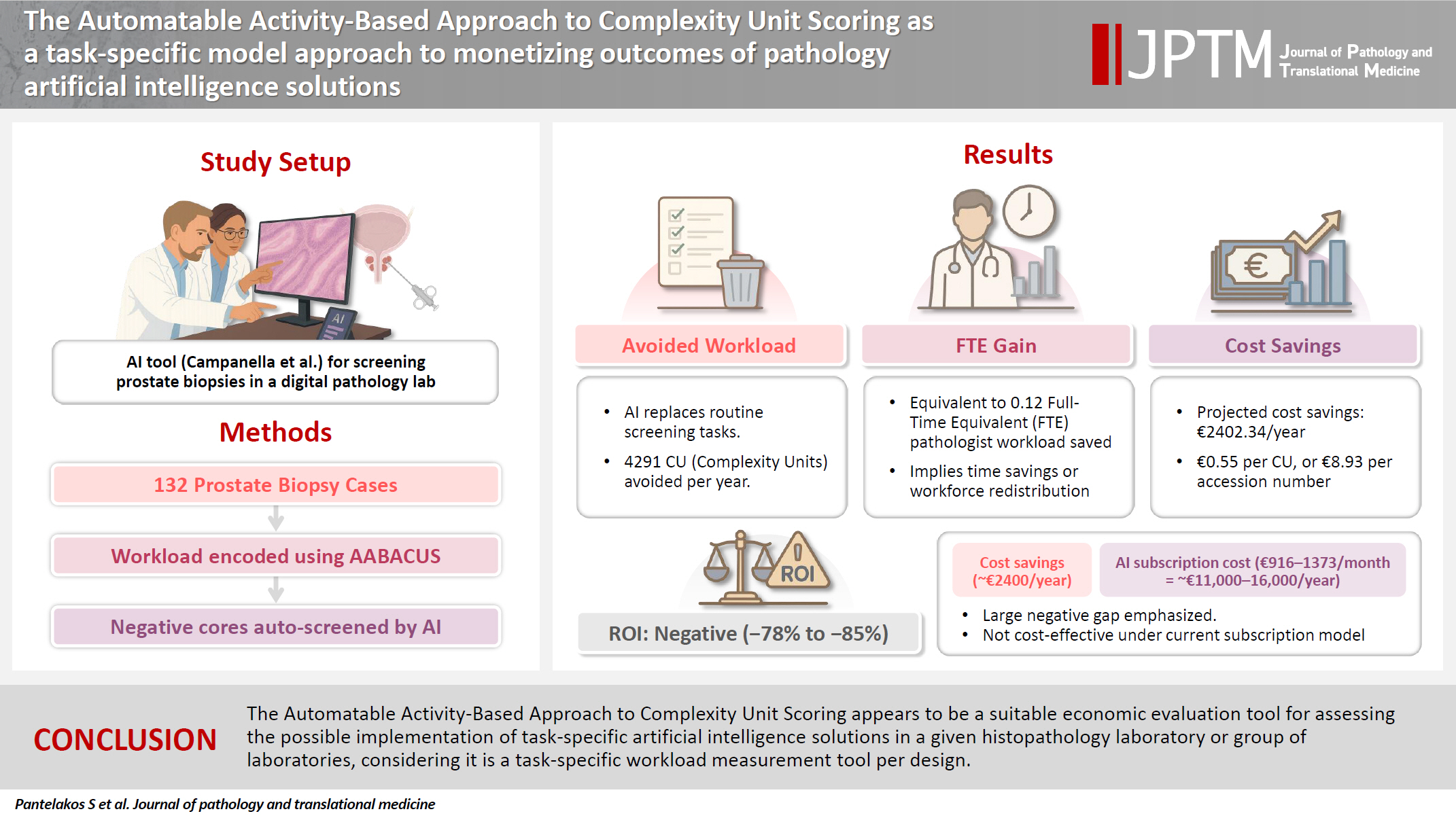
- 3,773 View
- 146 Download
-
Abstract
PDF
- Background
Cost-containment policies are increasingly affecting decision-making in healthcare. In this context, the need for monetization of digital health interventions has been recently emphasized. Previous studies have attempted to extrapolate cost containment in conjunction with the implementation of digital pathology solutions mostly on the basis of operational cost savings or diagnostic error reduction. However, no study has attempted to link a wider spectrum of potential diagnostic tasks performed by artificial intelligence algorithms to financial figures.
Methods
Herein, we employ a workload measurement tool for the purpose of monetizing particular outcomes associated with the implementation of a pathology artificial intelligence solution. A hundred and thirty-two prostate core biopsy samples were encoded for workload using the Automatable Activity–Based Approach to Complexity Unit Scoring. Subsequently, avoided workload, full-time equivalent gains, and corresponding cost savings were calculated assuming full clinical deployment of a well-developed prostate cancer screening tool.
Results
For a fixed percentage of negative cores and a steady yearly workload of prostate core biopsies, the estimated total avoided workload amounted to 4,291 complexity units per year, with an average avoidance of 16.25 complexity units per ascension number. The calculated full-time equivalent gains were 0.12, whereas projected cost savings were as high as €2,402.34 per year or €0.55 per complexity unit, which in turn would yield an average of €8.93 per ascension number.
Conclusions
The Automatable Activity–Based Approach to Complexity Unit Scoring appears to be a suitable economic evaluation tool for assessing the possible implementation of task-specific artificial intelligence solutions in a given histopathology laboratory or group of laboratories, considering it is a task-specific workload measurement tool per design.
Reviews
- Next step of molecular pathology: next-generation sequencing in cytology
- Ricella Souza da Silva, Fernando Schmitt
- J Pathol Transl Med. 2024;58(6):291-298. Published online November 7, 2024
- DOI: https://doi.org/10.4132/jptm.2024.10.22
- 7,776 View
- 413 Download
- 5 Web of Science
- 4 Crossref
-
Abstract
PDF
- The evolving landscape of precision oncology underscores the pivotal shift from morphological diagnosis to treatment decisions driven by molecular profiling. Recent guidelines from the European Society for Medical Oncology recomend the use of next-generation sequencing (NGS) across a broader range of cancers, reflecting its superior efficiency and clinical value. NGS not only updates oncology testing by offering quicker, sample-friendly, and sensitive analysis but also reduces the need for multiple individual tests. Cytology samples, often obtained through less invasive methods, can yield high-quality genetic material suitable for molecular analysis. This article focuses on optimizing the use of cytology samples in NGS, and outlines their potential benefits in identifying actionable molecular alterations for targeted therapies across various solid tumors. It also addresses the need for validation studies and the strategies to incorporate or combine different types of samples into routine clinical practice. Integrating cytological and liquid biopsies into routine clinical practice, alongside conventional tissue biopsies, offers a comprehensive approach to tumor genotyping, early disease detection, and monitoring of therapeutic responses across various solid tumor types. For comprehensive biomarker characterization, all patient specimens, although limited, is always valuable.
-
Citations
Citations to this article as recorded by- Unraveling the nexus: Tumor mutational burden, PD‐L1 expression, and oncogenic alterations in non–small cell lung cancer cytology specimens
Min Dai, Francis Anthony San Lucas, Hector Alvarez, Leomar Ballester, Hui Chen, Keyur P. Patel, Asif Rashid, Shun Rao, Mark J. Routbort, Gloria Sura, Keith Sweeney, Gokce Toruner, Peng Wei, Richard Yang, Hyvan Dang, Rajyalakshmi Luthra, Sinchita Roy‐Chowd
Cancer Cytopathology.2026;[Epub] CrossRef - The World Health Organization Reporting System for Lymph Node, Spleen, and Thymus Cytopathology: Part 1 – Lymph Node
Immacolata Cozzolino, Mats Ehinger, Maria Calaminici, Andrea Ronchi, Mousa A. Al-Abbadi, Helena Barroca, Beata Bode-Lesniewska, David F. Chhieng, Ruth L. Katz, Oscar Lin, L. Jeffrey Medeiros, Martha Bishop Pitman, Arvind Rajwanshi, Fernando C. Schmitt, Ph
Acta Cytologica.2025; 70(2): 185. CrossRef - The impact of cytological preparation techniques on RNA quality: A comparative study on smear samples
Cisel Aydin Mericoz, Gulsum Caylak, Elif Sevin Sanioglu, Zeynep Seçil Satilmis, Ayse Humeyra Dur Karasayar, Ibrahim Kulac
Cancer Cytopathology.2025;[Epub] CrossRef - Reimagining cytopathology in the molecular era: Integration or fragmentation?
Sumanta Das, R. Naveen Kumar, Biswajit Dey, Pranjal Kalita
Cytojournal.2025; 22: 94. CrossRef
- Unraveling the nexus: Tumor mutational burden, PD‐L1 expression, and oncogenic alterations in non–small cell lung cancer cytology specimens
- Diagnosis of interstitial lung diseases: from Averill A. Liebow to artificial intelligence
- Eunhee S. Yi, Paul Wawryko, Jay H. Ryu
- J Pathol Transl Med. 2024;58(1):1-11. Published online January 10, 2024
- DOI: https://doi.org/10.4132/jptm.2023.11.17
- 8,644 View
- 448 Download
- 2 Web of Science
- 2 Crossref
-
Abstract
PDF
- Histopathologic criteria of usual interstitial pneumonia (UIP)/idiopathic pulmonary fibrosis (IPF) were defined over the years and endorsed by leading organizations decades after Dr. Averill A. Liebow first coined the term UIP in the 1960s as a distinct pathologic pattern of fibrotic interstitial lung disease. Novel technology and recent research on interstitial lung diseases with genetic component shed light on molecular pathogenesis of UIP/IPF. Two antifibrotic agents introduced in the mid-2010s opened a new era of therapeutic approaches to UIP/IPF, albeit contentious issues regarding their efficacy, side effects, and costs. Recently, the concept of progressive pulmonary fibrosis was introduced to acknowledge additional types of progressive fibrosing interstitial lung diseases with the clinical and pathologic phenotypes comparable to those of UIP/IPF. Likewise, some authors have proposed a paradigm shift by considering UIP as a stand-alone diagnostic entity to encompass other fibrosing interstitial lung diseases that manifest a relentless progression as in IPF. These trends signal a pendulum moving toward the tendency of lumping diagnoses, which poses a risk of obscuring potentially important information crucial to both clinical and research purposes. Recent advances in whole slide imaging for digital pathology and artificial intelligence technology could offer an unprecedented opportunity to enhance histopathologic evaluation of interstitial lung diseases. However, current clinical practice trends of moving away from surgical lung biopsies in interstitial lung disease patients may become a limiting factor in this endeavor as it would be difficult to build a large histopathologic database with correlative clinical data required for artificial intelligence models.
-
Citations
Citations to this article as recorded by- Identification of early genes in the pathophysiology of fibrotic interstitial lung disease in a new model of pulmonary fibrosis
Nathan Hennion, Corentin Bedart, Léonie Vandomber, Frédéric Gottrand, Sarah Humez, Cécile Chenivesse, Jean-Luc Desseyn, Valérie Gouyer
Cellular and Molecular Life Sciences.2025;[Epub] CrossRef - Radiological Insights into UIP Pattern: A Comparison Between IPF and Non-IPF Patients
Stefano Palmucci, Miriam Adorna, Angelica Rapisarda, Alessandro Libra, Sefora Fischetti, Gianluca Sambataro, Letizia Antonella Mauro, Emanuele David, Pietro Valerio Foti, Claudia Mattina, Corrado Spatola, Carlo Vancheri, Antonio Basile
Journal of Clinical Medicine.2025; 14(12): 4162. CrossRef
- Identification of early genes in the pathophysiology of fibrotic interstitial lung disease in a new model of pulmonary fibrosis
Original Articles
- Tumor-infiltrating T lymphocytes evaluated using digital image analysis predict the prognosis of patients with diffuse large B-cell lymphoma
- Yunjoo Cho, Jiyeon Lee, Bogyeong Han, Sang Eun Yoon, Seok Jin Kim, Won Seog Kim, Junhun Cho
- J Pathol Transl Med. 2024;58(1):12-21. Published online January 10, 2024
- DOI: https://doi.org/10.4132/jptm.2023.11.02
- 5,970 View
- 277 Download
- 4 Web of Science
- 4 Crossref
-
Abstract
PDF
- Background
The implication of the presence of tumor-infiltrating T lymphocytes (TIL-T) in diffuse large B-cell lymphoma (DLBCL) is yet to be elucidated. We aimed to investigate the effect of TIL-T levels on the prognosis of patients with DLBCL.
Methods
Ninety-six patients with DLBCL were enrolled in the study. The TIL-T ratio was measured using QuPath, a digital pathology software package. The TIL-T ratio was investigated in three foci (highest, intermediate, and lowest) for each case, resulting in TIL-T–Max, TIL-T–Intermediate, and TIL-T–Min. The relationship between the TIL-T ratios and prognosis was investigated.
Results
When 19% was used as the cutoff value for TIL-T–Max, 72 (75.0%) and 24 (25.0%) patients had high and low TIL-T–Max, respectively. A high TIL-T–Max was significantly associated with lower serum lactate dehydrogenase levels (p < .001), with patient group who achieved complete remission after RCHOP therapy (p < .001), and a low-risk revised International Prognostic Index score (p < .001). Univariate analysis showed that patients with a low TIL-T–Max had a significantly worse prognosis in overall survival compared to those with a high TIL-T–Max (p < .001); this difference remained significant in a multivariate analysis with Cox proportional hazards (hazard ratio, 7.55; 95% confidence interval, 2.54 to 22.42; p < .001).
Conclusions
Patients with DLBCL with a high TIL-T–Max showed significantly better prognosis than those with a low TIL-T–Max, and the TIL-T–Max was an independent indicator of overall survival. These results suggest that evaluating TIL-T ratios using a digital pathology system is useful in predicting the prognosis of patients with DLBCL. -
Citations
Citations to this article as recorded by- Do Pre‐Treatment Biopsy Characteristics Predict Early Tumour Progression in Feline Diffuse Large B Cell Nasal Lymphoma Treated With Radiotherapy?
Valerie J. Poirier, Valeria Meier, Michelle Turek, Neil Christensen, Jacqueline Bowal, Matthew D. Ponzini, Stefan M. Keller
Veterinary and Comparative Oncology.2025; 23(1): 82. CrossRef - Comprehensive Analysis of Tumor Microenvironment and PD-L1 Expression Associations with Clinicopathological Features and Prognosis in Diffuse Large B-Cell Lymphoma
Yun-Li Xie, Long-Feng Ke, Wen-Wen Zhang, Fu Kang, Shu-Yi Lu, Chen-Yu Wu, Huan-Huan Zhu, Jian-Chao Wang, Gang Chen, Yan-Ping Chen
Blood and Lymphatic Cancer: Targets and Therapy.2025; Volume 15: 167. CrossRef - Metabolic-immune axis in the tumor microenvironment: a new strategy for prognostic assessment and precision therapy in DLBCL and FL
Chengqian Chen, Wei Guo, Haotian Wang, Luming Cao, Ou Bai
Frontiers in Immunology.2025;[Epub] CrossRef - Integrative analysis of a novel immunogenic PANoptosis‑related gene signature in diffuse large B-cell lymphoma for prognostication and therapeutic decision-making
Ming Xu, Ming Ruan, Wenhua Zhu, Jiayue Xu, Ling Lin, Weili Li, Weirong Zhu
Scientific Reports.2024;[Epub] CrossRef
- Do Pre‐Treatment Biopsy Characteristics Predict Early Tumour Progression in Feline Diffuse Large B Cell Nasal Lymphoma Treated With Radiotherapy?
- Establishing molecular pathology curriculum for pathology trainees and continued medical education: a collaborative work from the Molecular Pathology Study Group of the Korean Society of Pathologists
- Jiwon Koh, Ha Young Park, Jeong Mo Bae, Jun Kang, Uiju Cho, Seung Eun Lee, Haeyoun Kang, Min Eui Hong, Jae Kyung Won, Youn-La Choi, Wan-Seop Kim, Ahwon Lee
- J Pathol Transl Med. 2023;57(5):265-272. Published online September 15, 2023
- DOI: https://doi.org/10.4132/jptm.2023.08.26
- 6,163 View
- 209 Download
- 1 Web of Science
- 1 Crossref
-
Abstract
PDF
- Background
The importance of molecular pathology tests has increased during the last decade, and there is a great need for efficient training of molecular pathology for pathology trainees and as continued medical education.
Methods
The Molecular Pathology Study Group of the Korean Society of Pathologists appointed a task force composed of experienced molecular pathologists to develop a refined educational curriculum of molecular pathology. A 3-day online educational session was held based on the newly established structure of learning objectives; the audience were asked to score their understanding of 22 selected learning objectives before and after the session to assess the effect of structured education.
Results
The structured objectives and goals of molecular pathology was established and posted as a web-based interface which can serve as a knowledge bank of molecular pathology. A total of 201 pathologists participated in the educational session. For all 22 learning objectives, the scores of self-reported understanding increased after educational session by 9.9 points on average (range, 6.6 to 17.0). The most effectively improved items were objectives from next-generation sequencing (NGS) section: ‘NGS library preparation and quality control’ (score increased from 51.8 to 68.8), ‘NGS interpretation of variants and reference database’ (score increased from 54.1 to 68.0), and ‘whole genome, whole exome, and targeted gene sequencing’ (score increased from 58.2 to 71.2). Qualitative responses regarding the adequacy of refined educational curriculum were collected, where favorable comments dominated.
Conclusions
Approach toward the education of molecular pathology was refined, which would greatly benefit the future trainees. -
Citations
Citations to this article as recorded by- Presence of RB1 or Absence of LRP1B Mutation Predicts Poor Overall Survival in Patients with Gastric Neuroendocrine Carcinoma and Mixed Adenoneuroendocrine Carcinoma
In Hye Song, Bokyung Ahn, Young Soo Park, Deok Hoon Kim, Seung-Mo Hong
Cancer Research and Treatment.2025; 57(2): 492. CrossRef
- Presence of RB1 or Absence of LRP1B Mutation Predicts Poor Overall Survival in Patients with Gastric Neuroendocrine Carcinoma and Mixed Adenoneuroendocrine Carcinoma
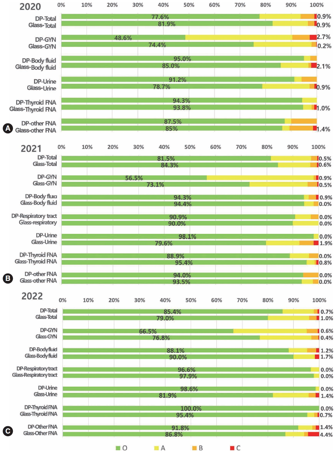
- Diagnostic proficiency test using digital cytopathology and comparative assessment of whole slide images of cytologic samples for quality assurance program in Korea
- Yosep Chong, Soon Auck Hong, Hoon Kyu Oh, Soo Jin Jung, Bo-Sung Kim, Ji Yun Jeong, Ho-Chang Lee, Gyungyub Gong
- J Pathol Transl Med. 2023;57(5):251-264. Published online August 24, 2023
- DOI: https://doi.org/10.4132/jptm.2023.07.17
- 8,680 View
- 344 Download
- 9 Web of Science
- 9 Crossref
-
Abstract
PDFSupplementary Material
- Background
The Korean Society for Cytopathology introduced a digital proficiency test (PT) in 2021. However, many doubtful opinions remain on whether digitally scanned images can satisfactorily present subtle differences in the nuclear features and chromatin patterns of cytological samples.
Methods
We prepared 30 whole-slide images (WSIs) from the conventional PT archive by a selection process for digital PT. Digital and conventional PT were performed in parallel for volunteer institutes, and the results were compared using feedback. To assess the quality of cytological assessment WSIs, 12 slides were collected and scanned using five different scanners, with four cytopathologists evaluating image quality through a questionnaire.
Results
Among the 215 institutes, 108 and 107 participated in glass and digital PT, respectively. No significant difference was noted in category C (major discordance), although the number of discordant cases was slightly higher in the digital PT group. Leica, 3DHistech Pannoramic 250 Flash, and Hamamatsu NanoZoomer 360 systems showed comparable results in terms of image quality, feature presentation, and error rates for most cytological samples. Overall satisfaction was observed with the general convenience and image quality of digital PT.
Conclusions
As three-dimensional clusters are common and nuclear/chromatin features are critical for cytological interpretation, careful selection of scanners and optimal conditions are mandatory for the successful establishment of digital quality assurance programs in cytology. -
Citations
Citations to this article as recorded by- Practice of Cytopathology in Korea: A 40‐Year Evolution Through Standardization, Digital Transformation, and Global Partnership
Yosep Chong, Ran Hong, Hyeong Ju Kwon, Haeryoung Kim, Lucia Kim, Soon Jae Kim, Yoon Jung Choi
Diagnostic Cytopathology.2026; 54(2): 146. CrossRef - ThinPrep® Whole-Slide Digital Images versus Conventional Microscopy in Negative for Intraepithelial Lesion or Malignancy, Atypical Squamous Cells of Undetermined Significance, and Low-Grade Squamous Intraepithelial Lesion Cervical Lesions: European Federa
Ester Puntonen, Sara Bazzon, Massimo Bongiovanni, Rosario Granados, Ines Krivak Bolanca, Maria Nasioutziki, Maurizio Pinamonti, Danijela Vrdoljak-Mozetic, Arrigo Capitanio, Beatrix Cochand-Priollet, Ambrogio Fassina, Giovanni Negri, Laura Ventura, Ivana K
Pathobiology.2026; : 1. CrossRef - Sensitivity, Specificity, and Cost–Benefit Effect Between Primary Human Papillomavirus Testing, Primary Liquid‐Based Cytology, and Co‐Testing Algorithms for Cervical Lesions
Chang Gok Woo, Seung‐Myoung Son, Hye‐Kyung Hwang, Jung‐Sil Bae, Ok‐Jun Lee, Ho‐Chang Lee
Diagnostic Cytopathology.2025; 53(1): 35. CrossRef - Integration of AI‐Assisted in Digital Cervical Cytology Training: A Comparative Study
Yihui Yang, Dongyi Xian, Lihua Yu, Yanqing Kong, Huaisheng Lv, Liujing Huang, Kai Liu, Hao Zhang, Weiwei Wei, Hongping Tang
Cytopathology.2025; 36(2): 156. CrossRef - National quality assurance program using digital cytopathology: a 5-year digital transformation experience by the Korean Society for Cytopathology
Yosep Chong, Hyeong Ju Kwon, Soon Auck Hong, Sung Soon Kim, Bo-Sung Kim, Younghee Choi, Yoon Jung Choi, Jung-Soo Pyo, Ji Yun Jeong, Soo Jin Jung, Hoon Kyu Oh, Seung-Sook Lee
Journal of Pathology and Translational Medicine.2025; 59(5): 320. CrossRef - Integration of Digital Cytology in Quality Assurance Programs for Cytopathology
Yosep Chong, Maria Jesús Fernández Aceñero, Zaibo Li, Andrey Bychkov
Acta Cytologica.2025; 70(1): 126. CrossRef - Quantitative Assessment of Focus Quality in Whole-Slide Imaging of Thyroid Liquid-Based Cytology Using Laplacian Variance
Chan Kwon Jung, Chankyung Kim, Sora Jeon, Andrey Bychkov
Endocrine Pathology.2025;[Epub] CrossRef - Validation of digital image slides for diagnosis in cervico-vaginal cytology
Francisco Tresserra, Gemma Fabra, Olga Luque, Miriam Castélla, Carla Gómez, Carmen Fernández-Cid, Ignacio Rodríguez
Revista Española de Patología.2024; 57(3): 182. CrossRef - Improved Diagnostic Accuracy of Thyroid Fine-Needle Aspiration Cytology with Artificial Intelligence Technology
Yujin Lee, Mohammad Rizwan Alam, Hongsik Park, Kwangil Yim, Kyung Jin Seo, Gisu Hwang, Dahyeon Kim, Yeonsoo Chung, Gyungyub Gong, Nam Hoon Cho, Chong Woo Yoo, Yosep Chong, Hyun Joo Choi
Thyroid®.2024; 34(6): 723. CrossRef
- Practice of Cytopathology in Korea: A 40‐Year Evolution Through Standardization, Digital Transformation, and Global Partnership
- Single-center study on clinicopathological and typical molecular pathologic features of metastatic brain tumor
- Su Hwa Kim, Young Suk Lee, Sung Hak Lee, Yeoun Eun Sung, Ahwon Lee, Jun Kang, Jae-Sung Park, Sin Soo Jeun, Youn Soo Lee
- J Pathol Transl Med. 2023;57(4):217-231. Published online July 11, 2023
- DOI: https://doi.org/10.4132/jptm.2023.06.10
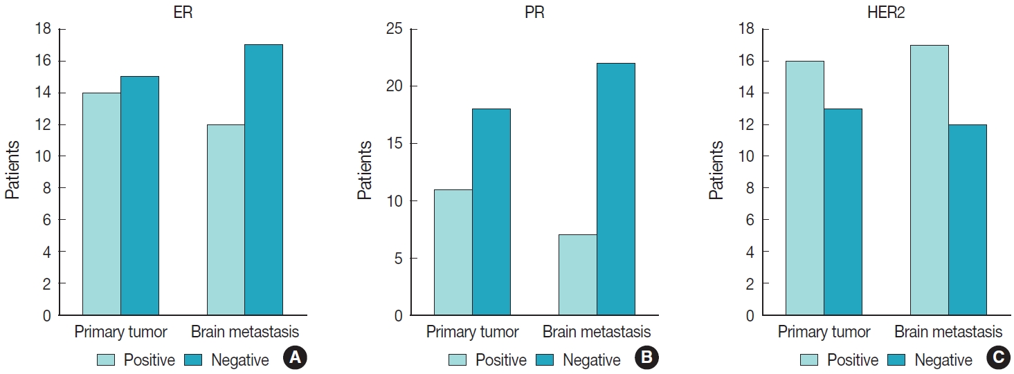
- 6,436 View
- 175 Download
- 1 Crossref
-
Abstract
PDF
- Background
The metastatic brain tumor is the most common brain tumor. The aim of this study was to demonstrate the clinicopathological and molecular pathologic features of brain metastases (BM).
Methods
A total of 269 patients were diagnosed with BM through surgical resection at Seoul St. Mary’s Hospital from January 2010 to March 2020. We reviewed the clinicopathological features and molecular status of primary and metastatic brain tissues using immunohistochemistry and molecular pathology results.
Results
Among 269 patients, 139 males and 130 females were included. The median age of primary tumor was 58 years (range, 13 to 87 years) and 86 patients (32.0%) had BM at initial presentation. Median BM free interval was 28.0 months (range, 1 to 286 months). The most frequent primary site was lung 46.5% (125/269), and followed by breast 15.6% (42/269), colorectum 10.0% (27/269). Epidermal growth factor receptor (EGFR) mutation was found in 50.8% (32/63) and 58.0% (40/69) of lung primary and BM, respectively. In both breast primary and breast cancer with BM, luminal B was the most frequent subtype at 37.9% (11/29) and 42.9% (18/42), respectively, followed by human epidermal growth factor receptor 2 with 31.0% (9/29) and 33.3% (14/42). Triple-negative was 20.7% (6/29) and 16.7% (7/42), and luminal A was 10.3% (3/29) and 7.1% (3/42) of breast primary and BM, respectively. In colorectal primary and colorectal cancer with BM, KRAS mutation was found in 76.9% (10/13) and 66.7% (2/3), respectively.
Conclusions
We report the clinicopathological and molecular pathologic features of BM that can provide useful information for understanding the pathogenesis of metastasis and for clinical trials based on the tumor’s molecular pathology. -
Citations
Citations to this article as recorded by- Colorectal cancer metastasis to the brain: A scoping review of incidence, treatment, and outcomes
Hunter J Hutchinson, Melanie Gonzalez, Diana Feier, Colin E Welch, Brandon Lucke-Wold
World Journal of Gastrointestinal Pathophysiology.2025;[Epub] CrossRef
- Colorectal cancer metastasis to the brain: A scoping review of incidence, treatment, and outcomes
Case Study
- Thyroid pathology, a clue to PTEN hamartoma tumor syndrome
- Yurimi Lee, Young Lyun Oh
- J Pathol Transl Med. 2023;57(3):178-183. Published online March 30, 2023
- DOI: https://doi.org/10.4132/jptm.2023.03.04
- 8,157 View
- 211 Download
- 10 Web of Science
- 8 Crossref
-
Abstract
PDF
- Phosphatase and tensin homolog (PTEN) hamartoma tumor syndrome (PHTS) is a hereditary disorder caused by germline inactivating mutations in the PTEN tumor suppressor gene. As a type of PHTS, Cowden syndrome is associated with abnormalities of the thyroid, breast, uterus, and gastrointestinal tract. A 52-year-old-woman visited the outpatient clinic of our endocrinology clinic with multiple thyroid nodules and Hashimoto's thyroiditis. Computed tomography imaging revealed a multinodular mass measuring up to 3.5 cm in the left thyroid lobe, causing laryngotracheal airway displacement. The total thyroidectomy specimen revealed multiple follicular adenomas and adenomatous nodules with lymphocytic thyroiditis and lipomatous metaplasia in the background. The patient was suspected of PTHS based on her thyroid pathology, family history, and numerous hamartomatous lesions of the breast, uterus, and skin. Her diagnosis was confirmed through molecular testing. This case demonstrates that pathologists must be well acquainted with thyroid pathology in PHTS.
-
Citations
Citations to this article as recorded by- Risk of malignancy in PTEN-altered thyroid nodules detected on preoperative FNA molecular testing: a systematic review and meta-analysis
Patrizia Straccia, Vincenzo Fiorentino, Belen Padial Urtueta, Qianqian Zhang, Alessia Piermattei, Federica Cianfrini, Antonino Mule, Esther Diana Rossi
Human Pathology.2026; : 106104. CrossRef - Dual primary malignancies in Kashmir: A five-year analysis of temporal patterns, gender-specific presentations and treatment outcomes in a high gastrointestinal cancer risk population
Ubaid Jeelani, Mushood Ghulam Nabi, Asim Ahmad Dar, Gowher Ahmad Wagai, Aadil Najeed, Sheikh Owais Ahmad, Lande Sagar Janardhan, Md Mayeen Afsan Ahmad, Uzma Majeed
Journal of Family Medicine and Primary Care.2026; 15(2): 530. CrossRef - A clinical case of papillary thyroid cancer associated with a PTEN gene defect
R. A. Atanesyan, L. Ja. Klimov, T. M. Vdovina, G. A. Saneeva, E. I. Andreeva, I. A. Stremenkova, R. I. Arakelyan, I. K. Gasparyan
Rossiyskiy Vestnik Perinatologii i Pediatrii (Russian Bulletin of Perinatology and Pediatrics).2025; 69(6): 85. CrossRef - Pediatric cancer predisposition syndromes involving non-central nervous system solid pediatric tumors: a review on their manifestations with a focus on histopathology
B. Schurink, M. Reyes-Múgica, R. R. de Krijger
Virchows Archiv.2025; 486(1): 3. CrossRef - Dedifferentiated Leiomyosarcoma of the Uterine Corpus with Heterologous Component: Clinicopathological Analysis of Five Consecutive Cases from a Single Institution and Comprehensive Literature Review
Suyeon Kim, Hyunsik Bae, Hyun-Soo Kim
Diagnostics.2024; 14(2): 160. CrossRef - Case report: Rare oral manifestations in Cowden syndrome with PTEN mutation
Wei Yuan, Yanbin Liu, Haibin Sun, Ming Su, Lizheng Qin, Xin Huang
Frontiers in Oncology.2024;[Epub] CrossRef - Can thyroid histomorphology identify patients with PTEN hamartoma tumour syndrome?
Melad N Dababneh, Laura Rabinowitz, Gilman Plitt, Charis Eng, Christopher C Griffith
Histopathology.2024; 85(6): 929. CrossRef - A novel mutation in PTEN in anaplastic thyroid carcinoma: A case report
Yanli Zhao
Biomedical Reports.2024;[Epub] CrossRef
- Risk of malignancy in PTEN-altered thyroid nodules detected on preoperative FNA molecular testing: a systematic review and meta-analysis
Original Article
- Current state of cytopathology residency training: a Korean national survey of pathologists
- Uiju Cho, Tae Jung Kim, Wan Seop Kim, Kyo Young Lee, Hye Kyoung Yoon, Hyun Joo Choi
- J Pathol Transl Med. 2023;57(2):95-101. Published online March 14, 2023
- DOI: https://doi.org/10.4132/jptm.2023.01.06
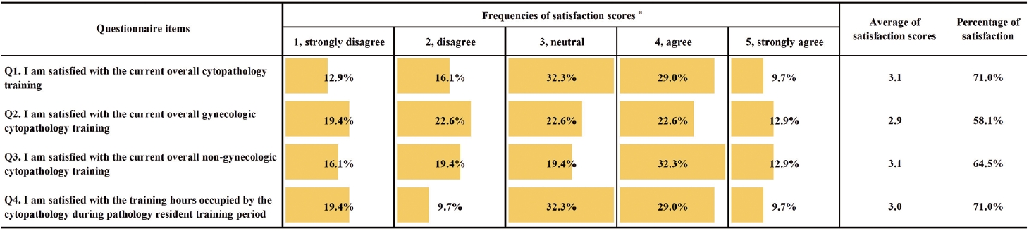
- 4,400 View
- 82 Download
- 1 Web of Science
- 1 Crossref
-
Abstract
PDFSupplementary Material
- Background
Although the Korean Society for Cytopathology has developed educational goals as guidelines for cytopathology education in Korea, there is still no systematic approach to cytopathology education status for pathology residents. Furthermore, satisfaction with cytopathology education and with the outcome of the current training/educational program has not been investigated in Korea. This study aimed to obtain comprehensive data on the current state of cytopathology education for residents and evaluate education outcomes.
Methods
An online survey was conducted in December 2020 for the board-certified pathologists and training residents registered as members of the Korean Society for Cytopathology. The questionnaire comprised questions that investigated the current status of cytopathology at each training institution, the degree of satisfaction with the work and education related to cytopathology, outcomes of cytopathology training, and educational accomplishments.
Results
Of the participants surveyed, 12.3% (132/1,075) completed the questionnaire, and 36.8% (32/87) of cytopathology residents participated. The mean overall satisfaction with cytopathology education was 3.1 points (on a 1- to 5-point scale, 5: very satisfied). The most frequent suggestion among the free description format responses was to expand educational opportunities, such as online education opportunities, outside of the individual institutions.
Conclusions
Our results showed that cytopathology training in Korea needs further improvement. We expect that this study will inform systematic training of competent medical personnel armed with broad cytopathology knowledge and strong problem-solving abilities. -
Citations
Citations to this article as recorded by- Artificial Intelligence–Assisted Daily Quality Control System for the Histologic Diagnosis of Gastrointestinal Endoscopic Biopsies: A 1-Year Experience
Seung-Yeon Yoo, Yuri Hwang, Seokju Yun, Ok Hee Lee, Jiwook Jang, Youngjin Park, Tae Young Cho, Young Sin Ko
Archives of Pathology & Laboratory Medicine.2025; 149(7): 659. CrossRef
- Artificial Intelligence–Assisted Daily Quality Control System for the Histologic Diagnosis of Gastrointestinal Endoscopic Biopsies: A 1-Year Experience
Reviews
- A standardized pathology report for gastric cancer: 2nd edition
- Young Soo Park, Myeong-Cherl Kook, Baek-hui Kim, Hye Seung Lee, Dong-Wook Kang, Mi-Jin Gu, Ok Ran Shin, Younghee Choi, Wonae Lee, Hyunki Kim, In Hye Song, Kyoung-Mee Kim, Hee Sung Kim, Guhyun Kang, Do Youn Park, So-Young Jin, Joon Mee Kim, Yoon Jung Choi, Hee Kyung Chang, Soomin Ahn, Mee Soo Chang, Song-Hee Han, Yoonjin Kwak, An Na Seo, Sung Hak Lee, Mee-Yon Cho
- J Pathol Transl Med. 2023;57(1):1-27. Published online January 15, 2023
- DOI: https://doi.org/10.4132/jptm.2022.12.23
- 40,496 View
- 1,592 Download
- 24 Web of Science
- 23 Crossref
-
Abstract
PDFSupplementary Material
- The first edition of ‘A Standardized Pathology Report for Gastric Cancer’ was initiated by the Gastrointestinal Pathology Study Group of the Korean Society of Pathologists and published 17 years ago. Since then, significant advances have been made in the pathologic diagnosis, molecular genetics, and management of gastric cancer (GC). To reflect those changes, a committee for publishing a second edition of the report was formed within the Gastrointestinal Pathology Study Group of the Korean Society of Pathologists. This second edition consists of two parts: standard data elements and conditional data elements. The standard data elements contain the basic pathologic findings and items necessary to predict the prognosis of GC patients, and they are adequate for routine surgical pathology service. Other diagnostic and prognostic factors relevant to adjuvant therapy, including molecular biomarkers, are classified as conditional data elements to allow each pathologist to selectively choose items appropriate to the environment in their institution. We trust that the standardized pathology report will be helpful for GC diagnosis and facilitate large-scale multidisciplinary collaborative studies.
-
Citations
Citations to this article as recorded by- GAST-NET: A multi-modal and multi-task deep learning framework for preoperative prediction of perineural invasion and prognostic risk in gastric cancer
Shidi Miao, Hexiang Dong, Jinyang Feng, Yuyang Jiang, Mengzhuo Sun, Zengyao Liu, Qiujun Wang, Xuemei Ding, Ruitao Wang
International Journal of Medical Informatics.2026; 212: 106348. CrossRef - Poorly cohesive carcinoma diffusely involving the whole gastrointestinal tract: a case report
Wei Gao, Yusheng Yang, Xinyi Hu, Yujuan Shi, Kai Liu, Minmin Gu, Jing Wang
Discover Oncology.2026;[Epub] CrossRef - Comprehensive Overview of Gastric Cancer Immunohistochemistry: Key Biomarkers, Advanced Detection Methods, and Perspectives
Bogdan Oprea
Medicina.2026; 62(4): 683. CrossRef - Tumor Budding in Gastric Carcinoma: Beyond Counting Cells at the Invasive Front—A Review of Current Evidence and Biological Perspectives
Catalin-Bogdan Satala, Gabriela Gurau, Alina-Mihaela Gurau, Gabriela Patrichi, Daniela Mihalache
International Journal of Molecular Sciences.2026; 27(9): 3787. CrossRef - Spatial and Temporal Tumor Heterogeneity in Gastric Cancer: Discordance of Predictive Biomarkers
Hye Seung Lee
Journal of Gastric Cancer.2025; 25(1): 192. CrossRef - PD-L1 as a Biomarker in Gastric Cancer Immunotherapy
Yunjoo Cho, Soomin Ahn, Kyoung-Mee Kim
Journal of Gastric Cancer.2025; 25(1): 177. CrossRef - Korean Gastric Cancer Association-Led Nationwide Survey on Surgically Treated Gastric Cancers in 2023
Dong Jin Kim, Jeong Ho Song, Ji-Hyeon Park, Sojung Kim, Sin Hye Park, Cheol Min Shin, Yoonjin Kwak, Kyunghye Bang, Chung-sik Gong, Sung Eun Oh, Yoo Min Kim, Young Suk Park, Jeesun Kim, Ji Eun Jung, Mi Ran Jung, Bang Wool Eom, Ki Bum Park, Jae Hun Chung, S
Journal of Gastric Cancer.2025; 25(1): 115. CrossRef - A Comprehensive and Comparative Review of Global Gastric Cancer Treatment Guidelines: 2024 Update
Sang Soo Eom, Keun Won Ryu, Hye Sook Han, Seong-Ho Kong
Journal of Gastric Cancer.2025; 25(1): 153. CrossRef - Korea, Japan, Europe, and the United States: Why are guidelines for gastric cancer different?
Emily E. Stroobant, Seong-Ho Kong, Maria Bencivenga, Takahiro Kinoshita, Tae-Han Kim, Takeshi Sano, Giovanni de Manzoni, Han-Kwang Yang, Yuko Kitagawa, Vivian E. Strong
Gastric Cancer.2025; 28(4): 559. CrossRef - Can the Japanese guidelines for endoscopic submucosal dissection be safely applied to Korean gastric cancer patients? A multicenter retrospective study based on the Korean Gastric Cancer Association nationwide survey
Hayemin Lee, Mi Ryeong Park, Junhyun Lee
Annals of Surgical Treatment and Research.2025; 109(2): 81. CrossRef - Double optimal transport for differential gene regulatory network inference with unpaired samples
Mengyu Li, Bencong Zhu, Cheng Meng, Xiaodan Fan, Laura Cantini
Bioinformatics.2025;[Epub] CrossRef - A Randomized Controlled Trial to Evaluate the Effect of Fibrin Glue on Bleeding after Gastric Endoscopic Submucosal Dissection
Tae-Se Kim, Tae-Jun Kim, Yang Won Min, Hyuk Lee, Byung-Hoon Min, Jun Haeng Lee, Poong-Lyul Rhee, Jae J. Kim
Gut and Liver.2025; 19(5): 677. CrossRef - Diagnostic accuracy of stereomicroscopy assessment of invasion depth in ex vivo specimens of early gastric cancer
Jing Wang, Lin Chang, Dong-Feng Niu, Yan Yan, Chang-Qi Cao, Shi-Jie Li, Qi Wu
World Journal of Gastroenterology.2025;[Epub] CrossRef - SMMILe enables accurate spatial quantification in digital pathology using multiple-instance learning
Zeyu Gao, Anyu Mao, Yuxing Dong, Hannah Clayton, Jialun Wu, Jiashuai Liu, ChunBao Wang, Kai He, Tieliang Gong, Chen Li, Mireia Crispin-Ortuzar
Nature Cancer.2025; 6(12): 2025. CrossRef - Genomic and Transcriptomic Characterization of Gastric Cancer with Bone Metastasis
Sujin Oh, Soo Kyung Nam, Keun-Wook Lee, Hye Seung Lee, Yujun Park, Yoonjin Kwak, Kyu Sang Lee, Ji-Won Kim, Jin Won Kim, Minsu Kang, Young Suk Park, Sang-Hoon Ahn, Yun-Suhk Suh, Do Joong Park, Hyung Ho Kim
Cancer Research and Treatment.2024; 56(1): 219. CrossRef - Microscopic tumor mapping of post-neoadjuvant therapy pancreatic cancer specimens to predict post-surgical recurrence: A prospective cohort study
Yeshong Park, Yeon Bi Han, Jinju Kim, MeeYoung Kang, Boram Lee, Eun Sung Ahn, Saemi Han, Haeryoung Kim, Hee-Young Na, Ho-Seong Han, Yoo-Seok Yoon
Pancreatology.2024; 24(4): 562. CrossRef - Effect of Neoadjuvant Chemotherapy on Tumor-Infiltrating Lymphocytes in Resectable Gastric Cancer: Analysis from a Western Academic Center
Elliott J. Yee, Danielle Gilbert, Jeffrey Kaplan, Sachin Wani, Sunnie S. Kim, Martin D. McCarter, Camille L. Stewart
Cancers.2024; 16(7): 1428. CrossRef - Interpretation of PD-L1 expression in gastric cancer: summary of a consensus meeting of Korean gastrointestinal pathologists
Soomin Ahn, Yoonjin Kwak, Gui Young Kwon, Kyoung-Mee Kim, Moonsik Kim, Hyunki Kim, Young Soo Park, Hyeon Jeong Oh, Kyoungyul Lee, Sung Hak Lee, Hye Seung Lee
Journal of Pathology and Translational Medicine.2024; 58(3): 103. CrossRef - Expression of claudin 18.2 in poorly cohesive carcinoma and its association with clinicopathologic parameters in East Asian patients
Moonsik Kim, Byung Woog Kang, Jihyun Park, Jin Ho Baek, Jong Gwang Kim
Pathology - Research and Practice.2024; 263: 155628. CrossRef - Clinicopathological analysis of claudin 18.2 focusing on intratumoral heterogeneity and survival in patients with metastatic or unresectable gastric cancer
T.-Y. Kim, Y. Kwak, S.K. Nam, D. Han, D.-Y. Oh, S.-A. Im, H.S. Lee
ESMO Open.2024; 9(12): 104000. CrossRef - Pathological Interpretation of Gastric Tumors in Endoscopic Submucosal Dissection
Jung Yeon Kim
Journal of Digestive Cancer Research.2023; 11(1): 15. CrossRef - Histopathology of Gastric Cancer
Baek-hui Kim, Sung Hak Lee
The Korean Journal of Helicobacter and Upper Gastrointestinal Research.2023; 23(2): 143. CrossRef - Endoscopic submucosal dissection hands-on training with artificial mucosal layer EndoGEL
Tae-Se Kim, Jun Haeng Lee
Journal of Innovative Medical Technology.2023; 1(1): 5. CrossRef
- GAST-NET: A multi-modal and multi-task deep learning framework for preoperative prediction of perineural invasion and prognostic risk in gastric cancer
- Single-cell and spatial sequencing application in pathology
- Yoon-Seob Kim, Jinyong Choi, Sug Hyung Lee
- J Pathol Transl Med. 2023;57(1):43-51. Published online January 10, 2023
- DOI: https://doi.org/10.4132/jptm.2022.12.12
- 11,346 View
- 417 Download
- 10 Web of Science
- 13 Crossref
-
Abstract
PDF
- Traditionally, diagnostic pathology uses histology representing structural alterations in a disease’s cells and tissues. In many cases, however, it is supplemented by other morphology-based methods such as immunohistochemistry and fluorescent in situ hybridization. Single-cell RNA sequencing (scRNA-seq) is one of the strategies that may help tackle the heterogeneous cells in a disease, but it does not usually provide histologic information. Spatial sequencing is designed to assign cell types, subtypes, or states according to the mRNA expression on a histological section by RNA sequencing. It can provide mRNA expressions not only of diseased cells, such as cancer cells but also of stromal cells, such as immune cells, fibroblasts, and vascular cells. In this review, we studied current methods of spatial transcriptome sequencing based on their technical backgrounds, tissue preparation, and analytic procedures. With the pathology examples, useful recommendations for pathologists who are just getting started to use spatial sequencing analysis in research are provided here. In addition, leveraging spatial sequencing by integration with scRNA-seq is reviewed. With the advantages of simultaneous histologic and single-cell information, spatial sequencing may give a molecular basis for pathological diagnosis, improve our understanding of diseases, and have potential clinical applications in prognostics and diagnostic pathology.
-
Citations
Citations to this article as recorded by- Trends and Challenges of the Modern Pathology Laboratory for Biopharmaceutical Research Excellence
Sílvia Sisó, Anoop Murthy Kavirayani, Suzana Couto, Birgit Stierstorfer, Sunish Mohanan, Caroline Morel, Mathiew Marella, Dinesh S. Bangari, Elizabeth Clark, Annette Schwartz, Vinicius Carreira
Toxicologic Pathology.2025; 53(1): 5. CrossRef - Single-cell RNA sequencing in osteosarcoma: applications in diagnosis, prognosis, and treatment
Christèle Asmar, Guy Awad, Marc Boutros, Simon Daccache, Alain Chebly, Catherine Alix-Panabières, Hampig-Raphael Kourié
Medical Oncology.2025;[Epub] CrossRef - Characterizing Stroke Clots Using Single‐Cell Sequencing
Daniela Renedo, Tanyeri Barak, Jonathan DeLong, Julian N. Acosta, Nanthiya Sujijantarat, Andrew Koo, Cyprien A. Rivier, Santiago Clocchiatti‐Tuozzo, Shufan Huo, Joseph Antonios, James Giles, Guido J. Falcone, Kevin N. Sheth, Ryan Hebert, Murat Gunel, Laur
Journal of the American Heart Association.2025;[Epub] CrossRef - Spatial transcriptomics meets diabetic kidney disease: Illuminating the path to precision medicine
Dan-Dan Liu, Han-Yue Hu, Fei-Fei Li, Qiu-Yue Hu, Ming-Wei Liu, You-Jin Hao, Bo Li
World Journal of Diabetes.2025;[Epub] CrossRef - Spatial Transcriptomics in Pancreatic Cancer: Current Insights and Future Directions
Jin Su Kim
Journal of Digestive Cancer Research.2025; 13(3): 288. CrossRef - Advances in Explainable Models for Single-cell Transcriptomics and Spatial Transcriptomics Data Analysis
Luchang Ge
Scientific Research Bulletin.2025; 2(6): 57. CrossRef - Incorporating Novel Technologies in Precision Oncology for Colorectal Cancer: Advancing Personalized Medicine
Pankaj Ahluwalia, Kalyani Ballur, Tiffanie Leeman, Ashutosh Vashisht, Harmanpreet Singh, Nivin Omar, Ashis K. Mondal, Kumar Vaibhav, Babak Baban, Ravindra Kolhe
Cancers.2024; 16(3): 480. CrossRef - Potential therapeutic targets for hypotension in duchenne muscular dystrophy
Harshi Saxena, Neal L. Weintraub, Yaoliang Tang
Medical Hypotheses.2024; 185: 111318. CrossRef - The crosstalk role of CDKN2A between tumor progression and cuproptosis resistance in colorectal cancer
Xifu Cheng, Famin Yang, Yuanheng Li, Yuke Cao, Meng Zhang, Jiameng JI, Yuxiao Bai, Qing Li, Qiongfang Yu, Dian Gao
Aging.2024; 16(12): 10512. CrossRef - Enquête exclusive sur le psoriasis
Imrane Ben Moussa, Bienfait Abasi-Ali, Fatima-Zahra Afarhkhane, Inès Mountadir, Claire Deligne
médecine/sciences.2024; 40(6-7): 584. CrossRef - Mechanisms of radiation‐induced tissue damage and response
Lin Zhou, Jiaojiao Zhu, Yuhao Liu, Ping‐Kun Zhou, Yongqing Gu
MedComm.2024;[Epub] CrossRef - A comparative analysis of single-cell transcriptomic technologies in plants and animals
Vamsidhar Reddy Netla, Harshraj Shinde, Gulshan Kumar, Ambika Dudhate, Jong Chan Hong, Ulhas Sopanrao Kadam
Current Plant Biology.2023; 35-36: 100289. CrossRef - Fibroblasts – the cellular choreographers of wound healing
Samuel Knoedler, Sonja Broichhausen, Ruiji Guo, Ruoxuan Dai, Leonard Knoedler, Martin Kauke-Navarro, Fortunay Diatta, Bohdan Pomahac, Hans-Guenther Machens, Dongsheng Jiang, Yuval Rinkevich
Frontiers in Immunology.2023;[Epub] CrossRef
- Trends and Challenges of the Modern Pathology Laboratory for Biopharmaceutical Research Excellence
Original Articles
- Development of quality assurance program for digital pathology by the Korean Society of Pathologists
- Yosep Chong, Jeong Mo Bae, Dong Wook Kang, Gwangil Kim, Hye Seung Han
- J Pathol Transl Med. 2022;56(6):370-382. Published online November 15, 2022
- DOI: https://doi.org/10.4132/jptm.2022.09.30
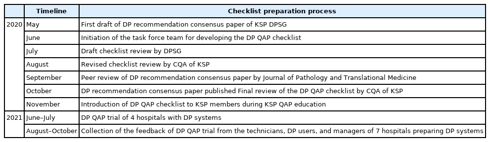
- 7,621 View
- 166 Download
- 5 Web of Science
- 5 Crossref
-
Abstract
PDFSupplementary Material
- Background
Digital pathology (DP) using whole slide imaging is a recently emerging game changer technology that can fundamentally change the way of working in pathology. The Digital Pathology Study Group (DPSG) of the Korean Society of Pathologists (KSP) published a consensus report on the recommendations for pathologic practice using DP. Accordingly, the need for the development and implementation of a quality assurance program (QAP) for DP has been raised.
Methods
To provide a standard baseline reference for internal and external QAP for DP, the members of the Committee of Quality Assurance of the KSP developed a checklist for the Redbook and a QAP trial for DP based on the prior DPSG consensus report. Four leading institutes participated in the QAP trial in the first year, and we gathered feedback from these institutes afterwards.
Results
The newly developed checklists of QAP for DP contain 39 items (216 score): eight items for quality control of DP systems; three for DP personnel; nine for hardware and software requirements for DP systems; 15 for validation, operation, and management of DP systems; and four for data security and personal information protection. Most participants in the QAP trial replied that continuous education on unfamiliar terminology and more practical experience is demanding.
Conclusions
The QAP for DP is essential for the safe implementation of DP in pathologic practice. Each laboratory should prepare an institutional QAP according to this checklist, and consecutive revision of the checklist with feedback from the QAP trial for DP needs to follow. -
Citations
Citations to this article as recorded by- Practice of Cytopathology in Korea: A 40‐Year Evolution Through Standardization, Digital Transformation, and Global Partnership
Yosep Chong, Ran Hong, Hyeong Ju Kwon, Haeryoung Kim, Lucia Kim, Soon Jae Kim, Yoon Jung Choi
Diagnostic Cytopathology.2026; 54(2): 146. CrossRef - An equivalency and efficiency study for one year digital pathology for clinical routine diagnostics in an accredited tertiary academic center
Viola Iwuajoku, Kübra Ekici, Anette Haas, Mohammed Zaid Khan, Azar Kazemi, Atsuko Kasajima, Claire Delbridge, Alexander Muckenhuber, Elisa Schmoeckel, Fabian Stögbauer, Christine Bollwein, Kristina Schwamborn, Katja Steiger, Carolin Mogler, Peter J. Schüf
Virchows Archiv.2025; 487(1): 3. CrossRef - Quality Assurance of the Whole Slide Image Evaluation in Digital Pathology: State of the Art and Development Results
Miklós Vincze, Béla Molnár, Miklós Kozlovszky
Electronics.2025; 14(10): 1943. CrossRef - Integration of Digital Cytology in Quality Assurance Programs for Cytopathology
Yosep Chong, Maria Jesús Fernández Aceñero, Zaibo Li, Andrey Bychkov
Acta Cytologica.2025; 70(1): 126. CrossRef - Diagnostic proficiency test using digital cytopathology and comparative assessment of whole slide images of cytologic samples for quality assurance program in Korea
Yosep Chong, Soon Auck Hong, Hoon Kyu Oh, Soo Jin Jung, Bo-Sung Kim, Ji Yun Jeong, Ho-Chang Lee, Gyungyub Gong
Journal of Pathology and Translational Medicine.2023; 57(5): 251. CrossRef
- Practice of Cytopathology in Korea: A 40‐Year Evolution Through Standardization, Digital Transformation, and Global Partnership
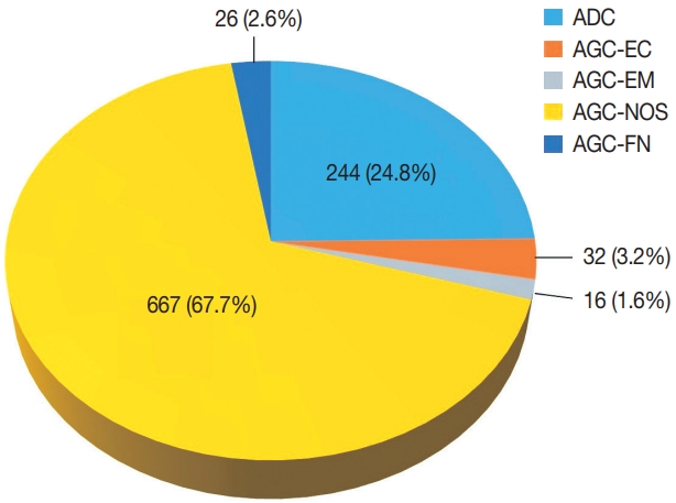
- Diagnostic distribution and pitfalls of glandular abnormalities in cervical cytology: a 25-year single-center study
- Jung-A Sung, Ilias P. Nikas, Haeryoung Kim, Han Suk Ryu, Cheol Lee
- J Pathol Transl Med. 2022;56(6):354-360. Published online November 9, 2022
- DOI: https://doi.org/10.4132/jptm.2022.09.05
- 9,495 View
- 162 Download
- 6 Web of Science
- 4 Crossref
-
Abstract
PDF
- Background
Detection of glandular abnormalities in Papanicolaou (Pap) tests is challenging. This study aimed to review our institute’s experience interpreting such abnormalities, assess cytohistologic concordance, and identify cytomorphologic features associated with malignancy in follow-up histology.
Methods
Patients with cytologically-detected glandular lesions identified in our pathology records from 1995 to 2020 were included in this study.
Results
Of the 683,197 Pap tests performed, 985 (0.144%) exhibited glandular abnormalities, 657 of which had tissue follow-up available. One hundred eighty-eight cases were cytologically interpreted as adenocarcinoma and histologically diagnosed as malignant tumors of various origins. There were 213 cases reported as atypical glandular cells (AGC) and nine cases as adenocarcinoma in cytology, yet they were found to be benign in follow-up histology. In addition, 48 cases diagnosed with AGC and six with adenocarcinoma cytology were found to have cervical squamous lesions in follow-up histology, including four squamous cell carcinomas. Among the cytomorphological features examined, nuclear membrane irregularity, three-dimensional clusters, single-cell pattern, and presence of mitoses were associated with malignant histology in follow-up.
Conclusions
This study showed our institute’s experience detecting glandular abnormalities in cervical cytology over a 25-year period, revealing the difficulty of this task. Nonetheless, the present study indicates that several cytological findings such as membrane irregularity, three-dimensional clusters, single-cell pattern, and evidence of proliferation could help distinguishing malignancy from a benign lesion. -
Citations
Citations to this article as recorded by- “Atypical Glandular Cells” on Cervical Cytology: Correlation Between Glandular Cell Component Volume and Histological Follow‐Up
Havva Gokce Terzioglu, Alessa Aragao, Julieta E. Barroeta
Diagnostic Cytopathology.2026; 54(2): 71. CrossRef - Expertise in Gynecological Pathology Impacts Diagnosis of Atypical Glandular Cell Category in Cervical Cytology
Havva Gökce Terzioglu, Alessa Aragao, Julieta E. Barroeta
Journal of Lower Genital Tract Disease.2025; 29(4): 297. CrossRef - Comparison of Cytological and/or Histopathological Results of Patients with Single and Multiple HPV Positivity
Fatih Mehmet Kaya, Şafak Ersöz, Cihan Comba, Ömer Demir
Acta Cytologica.2025; : 1. CrossRef - Analysis of atypical glandular cells in ThinPrep Pap smear and follow-up histopathology
Tengfei Wang, Yinan Hua, Lina Liu, Bing Leng
Baylor University Medical Center Proceedings.2024; 37(3): 403. CrossRef
- “Atypical Glandular Cells” on Cervical Cytology: Correlation Between Glandular Cell Component Volume and Histological Follow‐Up
Review
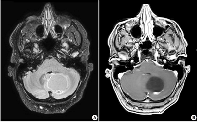
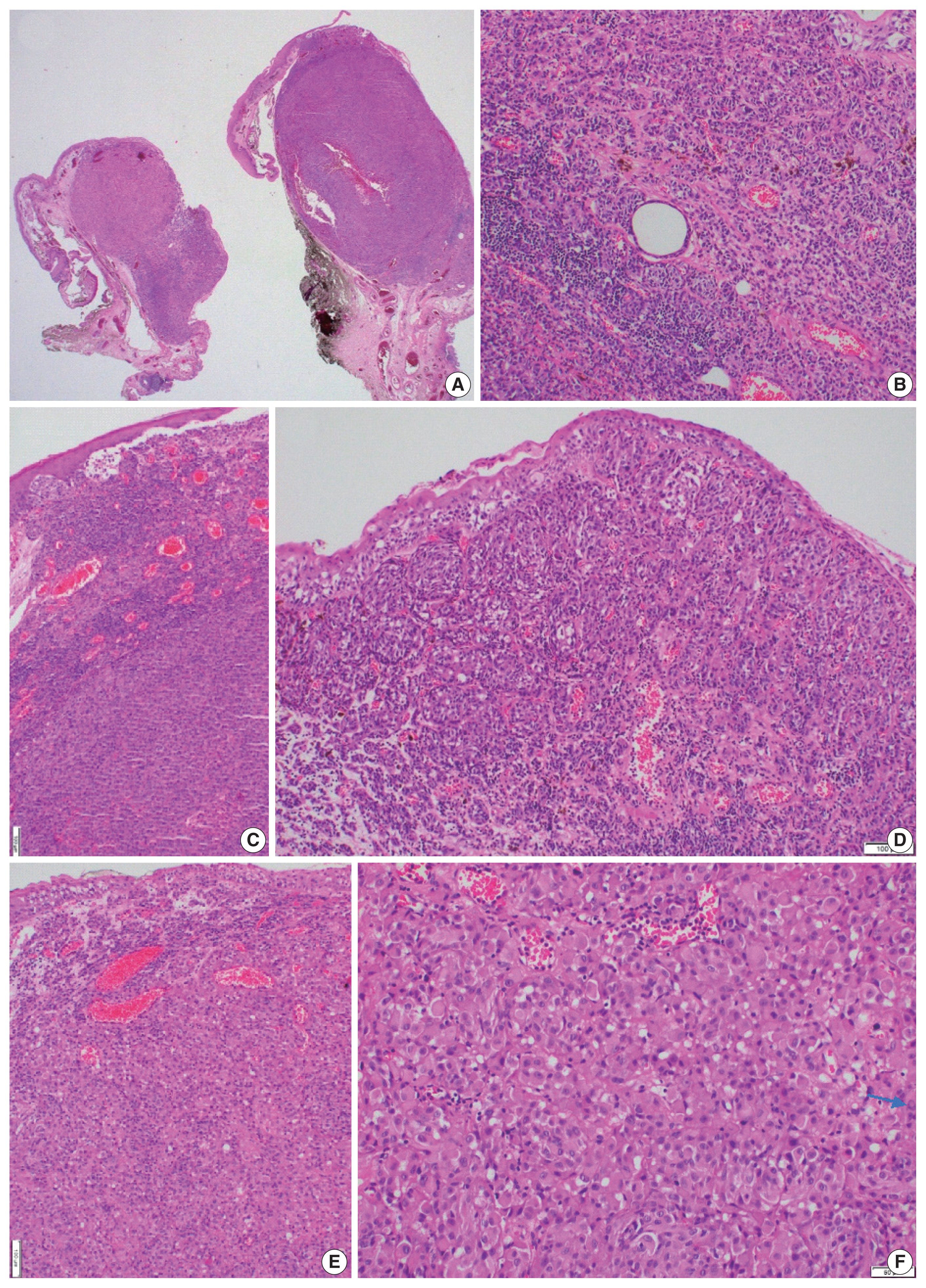
- Neuropathologic features of central nervous system hemangioblastoma
- Rebecca A. Yoda, Patrick J. Cimino
- J Pathol Transl Med. 2022;56(3):115-125. Published online May 3, 2022
- DOI: https://doi.org/10.4132/jptm.2022.04.13
- 18,449 View
- 381 Download
- 22 Web of Science
- 28 Crossref
-
Abstract
PDF
- Hemangioblastoma is a benign, highly vascularized neoplasm of the central nervous system (CNS). This tumor is associated with loss of function of the VHL gene and demonstrates frequent occurrence in von Hippel-Lindau (VHL) disease. While this entity is designated CNS World Health Organization grade 1, due to its predilection for the cerebellum, brainstem, and spinal cord, it is still an important cause of morbidity and mortality in affected patients. Recognition and accurate diagnosis of hemangioblastoma is essential for the practice of surgical neuropathology. Other CNS neoplasms, including several tumors associated with VHL disease, may present as histologic mimics, making diagnosis challenging. We outline key clinical and radiologic features, pathophysiology, treatment modalities, and prognostic information for hemangioblastoma, and provide a thorough review of the gross, microscopic, immunophenotypic, and molecular features used to guide diagnosis.
-
Citations
Citations to this article as recorded by- Renal hemangioblastoma and renal cell carcinoma with fibromyomatous stroma and hemangioblastoma-like areas belong to the spectrum of one entity
Kiril Trpkov, Norel Salut, Inmaculada Ribera-Cortada, Elías Tasso Xipell, Isabel Trias Puigsureda, Asli Yilmaz, Arjumand Riyaz Husain, Erik Nohr, Adrian Box, Farshid Siadat, Katherina Baranova, Rola M. Saleeb, Robert Stoehr, Arndt Hartmann, Abbas Agaimy
Virchows Archiv.2026; 488(4): 767. CrossRef - Hemangioblastoma of the Kidney—A Comprehensive Clinical, Pathological, and Genetic Analysis of Four Cases
Boglárka Pósfai, Alex Jenei, Gertrúd Forika, Attila Fintha, Zoltán Sápi, Áron Somorácz, Borbála Dénes, Ferenc Salamon, Kornélia Veronika Eizler, Nándor Giba, Dávid Semjén, Ildikó Illyés, Kristóf Attila Kovács, Gyöngyi Munkácsy, János Papp, Fanni Sánta, He
APMIS.2026;[Epub] CrossRef - Peritumoral Cystic Meningioma With Diagnostic Challenges: Two Case Reports and a Literature Review
Wei-Chih Chen, Zi-Jie Lin, Lam Chee-Tat
Cureus.2026;[Epub] CrossRef - Belzutifan-induced tumor regression in sporadic hemangioblastoma: a case report and literature review
Rebekka E. Hooks, Niket Yadav, Mark Willy L. Mondia, Georgios Mantziaris, Alaa Saleh, Anna Vi Jones, Matthew McCord, Melike Mut, Ashok R. Asthagiri, Benjamin W. Purow
Journal of Neuro-Oncology.2026;[Epub] CrossRef - Unusual immunohistochemical profiles in hemangioblastomas and their relevance in differential diagnosis: A comprehensive study of 112 cases
Jiri Soukup, Marie Novakova, Jan Hojny, Marketa Trnkova, Martin Syrucek, Tomas Jirasek, Patricie Delongova, Ales Kohout, Tomas Vebr, Jan Sroubek, Alena Sejkorova, Radim Lipina, Radim Brabec, Tomas Cesak, David Netuka
Journal of Neuropathology & Experimental Neurology.2026;[Epub] CrossRef - Pathological features and clinical outcomes of hemangioblastoma in the spinal cord of three dogs
Ryo SAITO, James K CHAMBERS, Kio YOSHIDA, Yutaro NAKAYAMA, Yukiko NAKANO, Kosuke HAII, Yumiko KAGAWA, Kazuyuki UCHIDA
Journal of Veterinary Medical Science.2026; 88(4): 568. CrossRef - Monitoring and treatment patterns of von Hippel-Lindau disease-associated central nervous system hemangioblastomas
Eric Jonasch, Yan Song, Jonathan Freimark, Manasi Mohan, James Signorovitch, Murali Sundaram
Hereditary Cancer in Clinical Practice.2026;[Epub] CrossRef - Immunohistochemical Expression of PAX8 in Central Nervous System Hemangioblastomas: A Potential Diagnostic Pitfall for Neuropathologists
Giuseppe Broggi, Jessica Farina, Valeria Barresi, Francesco Certo, Giuseppe Maria Vincenzo Barbagallo, Gaetano Magro, Rosario Caltabiano
Applied Immunohistochemistry & Molecular Morphology.2025; 33(3): 160. CrossRef - Endolymphatic Sac Tumor. Post-Radiosurgery Evaluation Using Time-Resolved Imaging of Contrast Kinetics MR Angiography
Antonella Blandino, Allegra Romano, Chiara Filippi, Sofia Pizzolante, Andrea Romano, Giulia Moltoni, Edoardo Covelli, Maurizio Barbara, Alessandro Bozzao
Ear, Nose & Throat Journal.2025;[Epub] CrossRef - Stereotactic radiosurgery in the management of central nervous system hemangioblastomas: a systematic review and meta-analysis
Amirhossein Zare, Amirhessam Zare, Alireza Soltani Khaboushan, Bardia Hajikarimloo, Jason P. Sheehan
Neurosurgical Review.2025;[Epub] CrossRef - Cerebellar medullary cistern hemangioblastoma
Dahai Cao, Qiang Zhang
Asian Journal of Surgery.2025; 48(9): 5843. CrossRef - Navigating rare vascular brain tumors: A retrospective observational study
Sana Ahuja, Dipanker S Mankotia, Naveen Kumar, Vyomika Teckchandani, Sufian Zaheer
Cancer Research, Statistics, and Treatment.2025; 8(2): 92. CrossRef - A potential new entity pending further validation of pulmonary primary interstitial Tumor: Lymphangioleiomyomatosis-like
Lingyu Zhao, Xiaochen Shen, Yun Niu, Huang Chen, Dingrong Zhong
Respiratory Medicine Case Reports.2025; 57: 102241. CrossRef - Renal cell carcinoma with fibromyomatous stroma (RCC FMS) and with hemangioblastoma‐like areas is part of the RCC FMS spectrum in patients with tuberous sclerosis complex
Katherina Baranova, Jacob A Houpt, Deaglan Arnold, Andrew A House, Laura Lockau, Lindsay Ninivirta, Stephen Pautler, Haiying Chen, Madeleine Moussa, Rola Saleeb, Jose A Gomez, Asli Yilmaz, Farshid Siadat, Adrian Box, Douglas J Mahoney, Franz J Zemp, Manal
Histopathology.2025; 87(5): 687. CrossRef - Primary hemangioblastoma of rectum: a rare case report and review of literature
Aiping Zheng, Shaojuan Zhang, Qiang Ma, Wenxu Yang, Hualiang Xiao, Xinyu Liang
Journal of Cancer Research and Clinical Oncology.2025;[Epub] CrossRef - Cerebellar Hemangioblastoma Resection Complicated by Postoperative Vasogenic Edema in the Setting of Concurrent Immunotherapy Treatment
Aashka Sheth, Nicholas Dietz, Andrea Becerril-Gaitan, Rahim Kasem, Akshitkumar Mistry, Brian J Williams, Dale Ding, Isaac Abecassis
Cureus.2025;[Epub] CrossRef - Familial Von Hippel–Lindau Disease: A Case Series of Cerebral Hemangioblastomas with MRI, Histopathological, and Genetic Correlations
Claudiu Matei, Ioana Boeras, Dan Orga Dumitriu, Cosmin Mutu, Adriana Popescu, Mihai Gabriel Cucu, Alexandru Calotă-Dobrescu, Bogdan Fetica, Diter Atasie
Life.2025; 15(11): 1649. CrossRef - Characterization of spinal hemangioblastomas in patients with and without von Hippel-Lindau, and YAP expression
Ana-Laura Calderón-Garcidueñas, Steven-Andrés Piña-Ballantyne, Eunice-Jazmín Espinosa-Aguilar, Rebeca de Jesús Ramos-Sánchez
Revista Española de Patología.2024; 57(3): 160. CrossRef - Patients With Hemangioblastoma: Mood Disorders and Sleep Quality
Ali Riazi, Yaser Emaeillou, Nima Najafi, Mohammad Hoseinimanesh, Mohammad Ibrahim Ashkaran, Donya Sheibani Tehrani
Brain Tumor Research and Treatment.2024; 12(2): 87. CrossRef - Radiosurgically Treated Recurrent Cerebellar Hemangioblastoma: A Case Report and Literature Review
François Fabi, Ève Chamberland, Myreille D’Astous, Karine Michaud, Martin Côté, Isabelle Thibault
Current Oncology.2024; 31(7): 3968. CrossRef - Dual manifestations: spinal and cerebellar hemangioblastomas indicative of von Hippel-Lindau syndrome
Nurhuda Hendra Setyawan, Rachmat Andi Hartanto, Rusdy Ghazali Malueka, Ery Kus Dwianingsih, Dito Pondra Dharma
Radiology Case Reports.2024; 19(11): 5000. CrossRef - Phenotypic and Genotypic Features of a Chinese Cohort with Retinal Hemangioblastoma
Liqin Gao, Feng Zhang, J. Fielding Hejtmancik, Xiaodong Jiao, Liyun Jia, Xiaoyan Peng, Kai Ma, Qian Li
Genes.2024; 15(9): 1192. CrossRef - Hemangioblastoma Incidentally Discovered at CT Scan in Bamako: About a Case
Traore Ousmane, N’Diaye Mamadou, Dembélé Mamadou, Dembélé Adama, Diakité Siaka, Sidibé Mansa Drissa, Camara Nagnoumague, Keita Adama Diaman
Open Journal of Medical Imaging.2024; 14(03): 123. CrossRef - Case report: Hemangioblastoma in the brainstem of a dog
Kirsten Landsgaard, Samantha St. Jean, Stephanie Lovell, Jonathan Levine, Christine Gremillion, Brian Summers, Raquel R. Rech
Frontiers in Veterinary Science.2023;[Epub] CrossRef - Intramedullary hemangioblastoma of the thoracic cord with a microsurgical approach: A case report and literature review
Eduardo Cattapan Piovesan, Werner Petry Silva, Adroaldo Baseggio Mallmann, Felipe Severo Lanzini, Bruna Zanatta de Freitas, Francisco Costa Beber Lemanski, Charles André Carazzo
Surgical Neurology International.2023; 14: 137. CrossRef - Secondary Holocord Syringomyelia Associated With Spinal Hemangioblastoma in a 29-Year-Old Female
Eric Chun-Pu Chu, Edouard Sabourdy, Benjamin Cheong
Cureus.2023;[Epub] CrossRef - Belzutifan in adults with VHL-associated central nervous system hemangioblastoma: a single-center experience
Bryan J. Neth, Mason J. Webb, Jessica White, Joon H. Uhm, Pavel N. Pichurin, Ugur Sener
Journal of Neuro-Oncology.2023; 164(1): 239. CrossRef - Resection of Intramedullary Hemangioblastoma: Timing of Surgery and Its Impact on Neurological Outcome and Quality of Life
Michael Schwake, Sarah Ricchizzi, Sophia Krahwinkel, Emanuele Maragno, Stephanie Schipmann, Walter Stummer, Marco Gallus, Markus Holling
Medicina.2023; 59(9): 1611. CrossRef
- Renal hemangioblastoma and renal cell carcinoma with fibromyomatous stroma and hemangioblastoma-like areas belong to the spectrum of one entity
Original Article
- Clinicopathologic features and survival outcomes of ocular melanoma: a series of 31 cases from a tertiary university hospital
- Selin Kestel, Feriha Pınar Uyar Göçün, Betül Öğüt, Özlem Erdem
- J Pathol Transl Med. 2022;56(4):187-198. Published online May 3, 2022
- DOI: https://doi.org/10.4132/jptm.2022.03.10
- 8,833 View
- 210 Download
- 5 Web of Science
- 5 Crossref
-
Abstract
PDFSupplementary Material
- Background
We aimed to determine the effect of clinicopathologic features on overall survival among Caucasian ocular melanoma patients in the Central Anatolia region of Turkey.
Methods
This single-center study included conjunctival (n = 12) and uveal (n = 19) melanoma patients diagnosed between January 2008 and March 2020. Clinicopathologic features and outcomes were reviewed retrospectively. Five cases were tested for BRAF V600 mutations with real-time polymerase chain reaction, and one case was tested with nextgeneration sequencing. Survival was calculated using the Kaplan-Meier method.
Results
Thirty-one patients had a mean initial age of 58.32 years (median, 61 years; range 25 to 78 years). There were 13 male and 18 female patients. The median follow-up time was 43.5 months (range, 6 to 155 months) for conjunctival melanoma and 35 months (range, 8 to 151 months) for uveal melanoma. When this study ended, eight of the 12 conjunctival melanoma patients (66.7%) and nine of the 19 uveal melanoma patients (47.4%) had died. The presence of tumor-infiltrating lymphocytes was related to improved overall survival in conjunctival melanoma (p = .014), whereas the presence of ulceration (p = .030), lymphovascular invasion (p = .051), tumor in the left eye (p = .012), tumor thickness of > 2 mm (p = .012), and mitotic count of >1/mm² (p = .012) reduced the overall survival in conjunctival melanoma. Uveal melanoma tumors with the largest diameter of 9.1–15 mm led to the lowest overall survival among subgroups (p = .035). Involvement of the conjunctiva (p=.005) and lens (p = .003) diminished overall survival in uveal melanoma. BRAF V600 mutation was present in one case of conjunctival melanoma, GNAQ R183Q mutation was present in one case of uveal melanoma. Patients with uveal melanoma presented with an advanced pathological tumor stage compared to those with conjunctival melanoma (p = .019).
Conclusions
This study confirmed the presence of tumor-infiltrating lymphocytes as a favorable factor in conjunctival melanoma and conjunctival and lens involvement as unfavorable prognostic factors in uveal melanoma for overall survival, respectively. -
Citations
Citations to this article as recorded by- Clinicopathological Profile of Conjunctival Melanoma at a Referral Ophthalmology Center in Peru
Andrea Concha-Tuero, Carlos Noel-Pallas, Claudia Sánchez-Huamash, Erick Urbano-Ale, Jorge Villalobos-Espinoza, Lelia Marroquín-Loayza, Marcos Espinola-Sánchez
Clinical Ophthalmology.2026; Volume 20: 1. CrossRef - Toward Precision Medicine: Gene Therapy Applications in the Management of Uveal Melanoma
Alireza Azani, Vahid Ghassemifar, Zahra Mehrdad, Maryam Saberivand, Anahid Bagheripour, Safa Tahmasebi, Hossein Gharedaghi, Malihe Sharafi, Hassan Foroozand, Mohammad Saeed Soleimani Meigoli, Saba Pourali, Arash Salmaninejad, Faeze Ahmadi Beni, Qumars Beh
Cancer Reports.2025;[Epub] CrossRef - Uveal melanoma in the Iranian population: two decades of patient management in a tertiary eye center
Hamid Riazi-Esfahani, Abdulrahim Amini, Babak Masoomian, Mehdi Yaseri, Siamak Sabour, Ali Rashidinia, Mojtaba Arjmand, Seyed Mohsen Rafizadeh, Mohammadkarim Johari, Elias Khalili Pour, Fariba Ghassemi
International Journal of Retina and Vitreous.2024;[Epub] CrossRef - Clinical features and prognosis of patients with metastatic ocular and orbital melanoma: A bi‐institutional study
Xin Liu, Han Yue, Shiyu Jiang, Lin Kong, Yu Xu, Yong Chen, Chunmeng Wang, Yan Wang, Xiaoli Zhu, Yunyi Kong, Xiaowei Zhang, Jiang Qian, Zhiguo Luo
Cancer Medicine.2023; 12(15): 16163. CrossRef - Metastatic melanoma: clinicopathologic features and overall survival comparison
Selin Kestel, Feriha Pınar Uyar Göçün, Betül Öğüt, Özlem Erdem
Acta Dermatovenerologica Alpina Pannonica et Adriatica.2022;[Epub] CrossRef
- Clinicopathological Profile of Conjunctival Melanoma at a Referral Ophthalmology Center in Peru
Review
- Hepatocellular adenomas: recent updates
- Haeryoung Kim, Young Nyun Park
- J Pathol Transl Med. 2021;55(3):171-180. Published online April 7, 2021
- DOI: https://doi.org/10.4132/jptm.2021.02.27
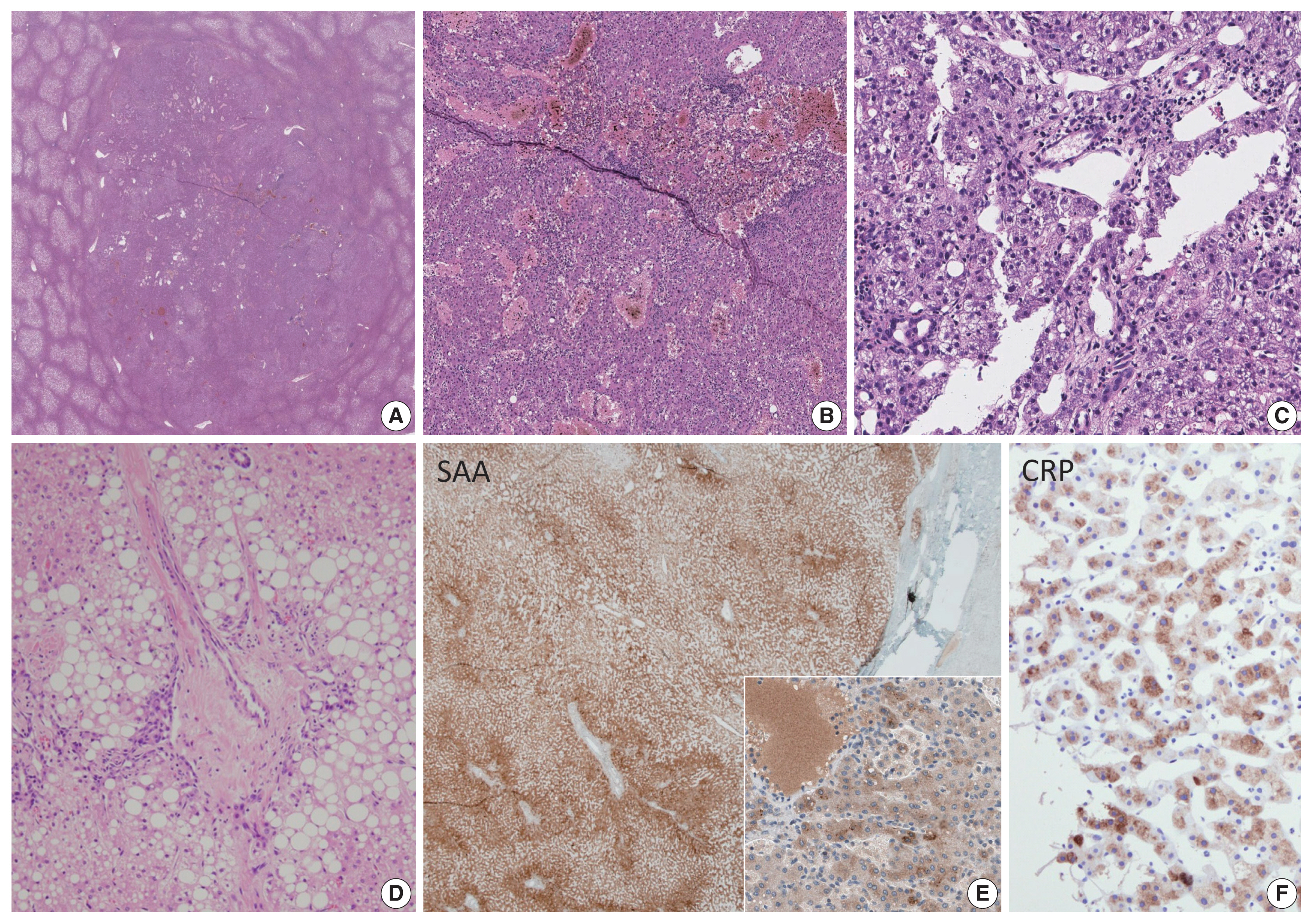
- 12,785 View
- 563 Download
- 10 Web of Science
- 13 Crossref
-
Abstract
PDF
- Hepatocellular adenoma (HCA) is a heterogeneous entity, from both the histomorphological and molecular aspects, and the resultant subclassification has brought a strong translational impact for both pathologists and clinicians. In this review, we provide an overview of the recent updates on HCA from the pathologists’ perspective and discuss several practical issues and pitfalls that may be useful for diagnostic practice.
-
Citations
Citations to this article as recorded by- The role of HNF4α in adenocarcinoma
Headtlove Essel Dadzie, Eric L. Snyder
Biochemical Society Transactions.2026; 54(4): 333. CrossRef - Large hepatocellular adenoma of the liver in a woman of reproductive age
AK Chichelnitsky, DA Savchenko, AS Kostina, DA Raklov, ER Zhuk, ES Buimova, AI Negodaeva, AO Ivanenko, VI Aduchieva
Bulletin of Russian State Medical University.2026;[Epub] CrossRef - A case of β-catenin-activated hepatocellular adenoma with concurrent hepatocellular carcinoma
Daichi Ito, Ryota Hyodo, Keisuke Kurimoto, Takashi Mizuno, Akira Satou, Motoko Sasaki, Yoji Ishizu, Mami Iima, Shinji Naganawa
Abdominal Radiology.2026;[Epub] CrossRef - Preventing false positive imaging diagnosis of HCC: differentiating HCC from mimickers and practical strategies
Ijin Joo
Journal of Liver Cancer.2025; 25(2): 217. CrossRef - Prognostic role of selection criteria for liver transplantation in patients with hepatocellular carcinoma: Review and bibliometric
Pamela Scarlett Espinoza Loyola, Diana Laura Muratalla Bautista, Karen Adela Hernández Bautista, Elizabeth Gil White, José Antonio González Moreno, Daniel Angel Torres del Real, Víctor Manuel Páez Zayas, Carla Escorza-Molina, Fernando Mondragón Rodríguez,
iLIVER.2024; 3(1): 100077. CrossRef - ACG Clinical Guideline: Focal Liver Lesions
Catherine Frenette, Mishal Mendiratta-Lala, Reena Salgia, Robert J. Wong, Bryan G. Sauer, Anjana Pillai
American Journal of Gastroenterology.2024; 119(7): 1235. CrossRef - Hepatocellular adenoma update: diagnosis, molecular classification, and clinical course
Sarah Poetter-Lang, Ahmed Ba-Ssalamah, Nina Bastati, Sami A Ba-Ssalamah, Jacqueline C Hodge, Giuseppe Brancatelli, Valérie Paradis, Valérie Vilgrain
British Journal of Radiology.2024; 97(1163): 1740. CrossRef - Fatal rupture of hepatic adenomatosis: Autopsy case and review of the literature
Sarra Ben Abderrahim, Khouloud Chérif, Zeineb Nfikha, Sarra Gharsallaoui, Imen El Aini, Maher Jedidi, Moncef Mokni, Mohamed Ben Dhiab
Journal of Forensic Sciences.2023; 68(4): 1393. CrossRef - Large Hepatocellular Adenoma Presenting with Iron Deficiency Anemia: A Case Report
Young Kwon Koh, Su Hyun Yoon, Sung Han Kang, Hyery Kim, Ho Joon Im, Suhyeon Ha, Jung-Man Namgoong, Kyung-Nam Koh
Clinical Pediatric Hematology-Oncology.2023; 30(1): 25. CrossRef - A Case Report on a Giant Hepatic Inflammatory Adenoma in a Young Female That Presented as Spontaneous Intrahepatic Hematoma
Andreas Kyvetos, Panagiota Voukelatou, Ioannis Vrettos, Spyridon Pantzios , Ioannis Elefsiniotis
Cureus.2023;[Epub] CrossRef - Advances in Histological and Molecular Classification of Hepatocellular Carcinoma
Joon Hyuk Choi, Swan N. Thung
Biomedicines.2023; 11(9): 2582. CrossRef - Estrobolome and Hepatocellular Adenomas—Connecting the Dots of the Gut Microbial β-Glucuronidase Pathway as a Metabolic Link
Sandica Bucurica, Mihaela Lupanciuc, Florentina Ionita-Radu, Ion Stefan, Alice Elena Munteanu, Daniela Anghel, Mariana Jinga, Elena Laura Gaman
International Journal of Molecular Sciences.2023; 24(22): 16034. CrossRef - Hepatocellular adenoma: what we know, what we do not know, and why it matters
Paulette Bioulac‐Sage, Annette S H Gouw, Charles Balabaud, Christine Sempoux
Histopathology.2022; 80(6): 878. CrossRef
- The role of HNF4α in adenocarcinoma
Original Article
- Deep learning for computer-assisted diagnosis of hereditary diffuse gastric cancer
- Sean A. Rasmussen, Thomas Arnason, Weei-Yuarn Huang
- J Pathol Transl Med. 2021;55(2):118-124. Published online January 22, 2021
- DOI: https://doi.org/10.4132/jptm.2020.12.22
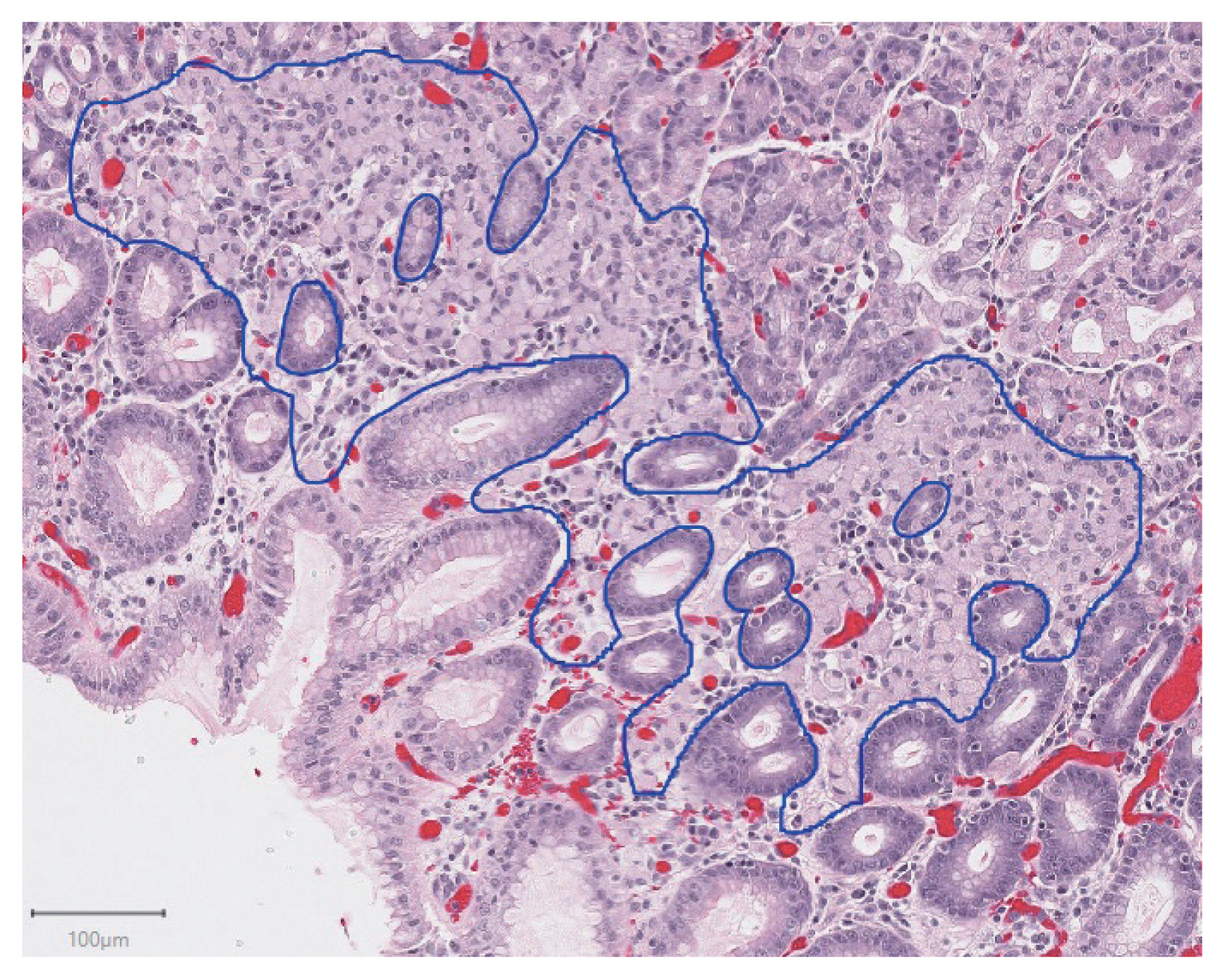
- 5,843 View
- 135 Download
- 9 Web of Science
- 9 Crossref
-
Abstract
PDF
- Background
Patients with hereditary diffuse gastric cancer often undergo prophylactic gastrectomy to minimize cancer risk. Because intramucosal poorly cohesive carcinomas in this setting are typically not grossly visible, many pathologists assess the entire gastrectomy specimen microscopically. With 150 or more slides per case, this is a major time burden for pathologists. This study utilizes deep learning methods to analyze digitized slides and detect regions of carcinoma.
Methods
Prophylactic gastrectomy specimens from seven patients with germline CDH1 mutations were analyzed (five for training/validation and two for testing, with a total of 133 tumor foci). All hematoxylin and eosin slides containing cancer foci were digitally scanned, and patches of size 256×256 pixels were randomly extracted from regions of cancer as well as from regions of normal background tissue, resulting in 15,851 images for training/validation and 970 images for testing. A model with DenseNet-169 architecture was trained for 150 epochs, then evaluated on images from the test set. External validation was conducted on 814 images scanned at an outside institution.
Results
On individual patches, the trained model achieved a receiver operating characteristic (ROC) area under the curve (AUC) of 0.9986. This enabled it to maintain a sensitivity of 90% with a false-positive rate of less than 0.1%. On the external validation dataset, the model achieved a similar ROC AUC of 0.9984. On whole slide images, the network detected 100% of tumor foci and correctly eliminated an average of 99.9% of the non-cancer slide area from consideration.
Conclusions
Overall, our model shows encouraging progress towards computer-assisted diagnosis of hereditary diffuse gastric cancer. -
Citations
Citations to this article as recorded by- Now and future of artificial intelligence-based signet ring cell diagnosis: A survey
Zhu Meng, Junhao Dong, Limei Guo, Fei Su, Jiaxuan Liu, Guangxi Wang, Zhicheng Zhao
Expert Systems with Applications.2026; 296: 129188. CrossRef - Development and application of deep learning-based diagnostics for pathologic diagnosis of gastric endoscopic submucosal dissection specimens
Soomin Ahn, Yiyu Hong, Sujin Park, Yunjoo Cho, Inwoo Hwang, Ji Min Na, Hyuk Lee, Byung-Hoon Min, Jun Haeng Lee, Jae J. Kim, Kyoung-Mee Kim
Gastric Cancer.2025; 28(4): 609. CrossRef - A Comprehensive Literature Review of the CDH1 Mutation and Its Role in Gastric Cancer
Malik Samardali, Jehad Samardaly, Ibrahim Shanti
Cureus.2025;[Epub] CrossRef - Deep learning for multiclass tumor cell detection in histopathology slides of hereditary diffuse gastric cancer
Robin Lomans, Valentina Angerilli, Joey Spronck, Liudmila L. Kodach, Irene Gullo, Fátima Carneiro, Rachel S. van der Post, Francesco Ciompi
iScience.2025; 28(8): 113064. CrossRef - Non-endoscopic Applications of Machine Learning in Gastric Cancer: A Systematic Review
Marianne Linley L. Sy-Janairo, Jose Isagani B. Janairo
Journal of Gastrointestinal Cancer.2024; 55(1): 47. CrossRef - Artificial intelligence applicated in gastric cancer: A bibliometric and visual analysis via CiteSpace
Guoyang Zhang, Jingjing Song, Zongfeng Feng, Wentao Zhao, Pan Huang, Li Liu, Yang Zhang, Xufeng Su, Yukang Wu, Yi Cao, Zhengrong Li, Zhigang Jie
Frontiers in Oncology.2023;[Epub] CrossRef - Deep learning of endoscopic features for the assessment of neoadjuvant therapy response in locally advanced rectal cancer
Anqi Wang, Jieli Zhou, Gang Wang, Beibei Zhang, Hongyi Xin, Haiyang Zhou
Asian Journal of Surgery.2023; 46(9): 3568. CrossRef - Preparing Data for Artificial Intelligence in Pathology with Clinical-Grade Performance
Yuanqing Yang, Kai Sun, Yanhua Gao, Kuansong Wang, Gang Yu
Diagnostics.2023; 13(19): 3115. CrossRef - Using Deep Learning to Predict Final HER2 Status in Invasive Breast Cancers That are Equivocal (2+) by Immunohistochemistry
Sean A. Rasmussen, Valerie J. Taylor, Alexi P. Surette, Penny J. Barnes, Gillian C. Bethune
Applied Immunohistochemistry & Molecular Morphology.2022; 30(10): 668. CrossRef
- Now and future of artificial intelligence-based signet ring cell diagnosis: A survey
Reviews
- Standardized pathology report for breast cancer
- Soo Youn Cho, So Yeon Park, Young Kyung Bae, Jee Yeon Kim, Eun Kyung Kim, Woo Gyeong Kim, Youngmee Kwon, Ahwon Lee, Hee Jin Lee, Ji Shin Lee, Jee Young Park, Gyungyub Gong, Hye Kyoung Yoon
- J Pathol Transl Med. 2021;55(1):1-15. Published online January 11, 2021
- DOI: https://doi.org/10.4132/jptm.2020.11.20
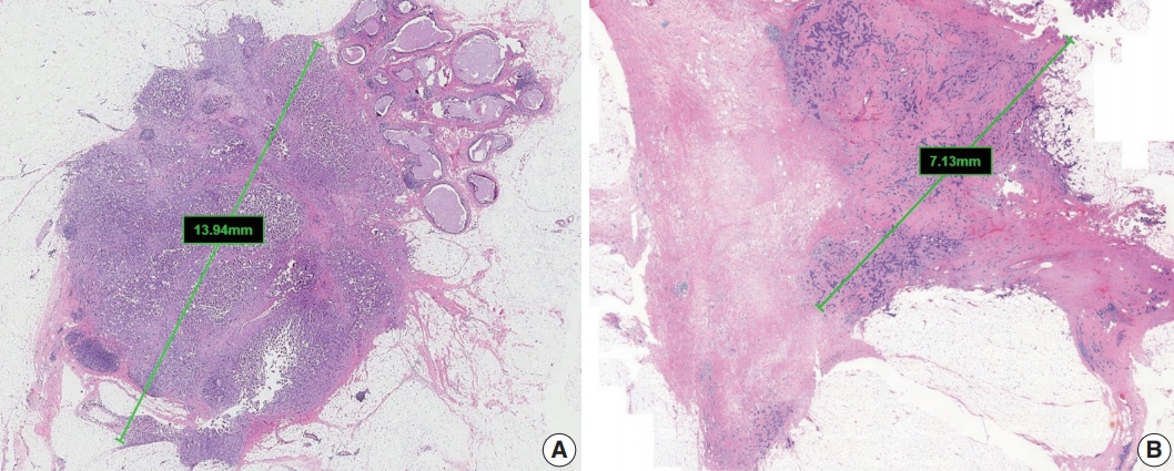
- 17,876 View
- 733 Download
- 9 Web of Science
- 6 Crossref
-
Abstract
PDFSupplementary Material
- Given the recent advances in management and understanding of breast cancer, a standardized pathology report reflecting these changes is critical. To meet this need, the Breast Pathology Study Group of the Korean Society of Pathologists has developed a standardized pathology reporting format for breast cancer, consisting of ‘standard data elements,’ ‘conditional data elements,’ and a biomarker report form. The ‘standard data elements’ consist of the basic pathologic features used for prognostication, while other factors related to prognosis or diagnosis are described in the ‘conditional data elements.’ In addition to standard data elements, all recommended issues are also presented. We expect that this standardized pathology report for breast cancer will improve diagnostic concordance and communication between pathologists and clinicians, as well as between pathologists inter-institutionally.
-
Citations
Citations to this article as recorded by- Adenoid Cystic Carcinoma of Breast Associated With an Incidental Radial Scar: A Cyto‐Histopathology Correlation
Rallapalli Rajyalakshmi, Valasapalli Rajani, Tanuku Sreedhar, Kollabathula Arpitha
Diagnostic Cytopathology.2026;[Epub] CrossRef - Refined risk stratification in residual triple-negative breast cancer after neoadjuvant therapy using residual cancer burden class and lymphovascular invasion
Tae Hoon Lee, Hyunwoo Lee, Jeong Yun Jang, Won Park, Won Kyung Cho, Eun Yoon Cho, Jin Seok Ahn, Yeon Hee Park, Seok Jin Nam, Seok Won Kim, Jeong Eon Lee, Haeyoung Kim
Breast Cancer Research and Treatment.2026;[Epub] CrossRef - Artificial intelligence-assisted three-dimensional imaging of breast microinvasive carcinoma reveals larger invasive focus size in a substantial proportion of cases
Yichieh Chien, Cher-Wei Liang, Chih-Yi Hsu, Yu-Chieh Lin, Yen-Yu Lin
Pathology - Research and Practice.2026; : 156388. CrossRef - Navigating discrepancies: The assessment of residual lymphovascular invasion in breast carcinoma after neoadjuvant treatment
Anikó Kovács, Åsa Rundgren-Sellei, Gunilla Rask, Annette Bauer, Anna Bodén, Johannes van Brakel, Eugenia Colón-Cervantes, Anna Ehinger, Johan Hartman, Balazs Acs
The Breast.2025; 82: 104519. CrossRef - Residual pure intralymphatic carcinoma component only (lymphovascular tumor emboli without invasive carcinoma) after neoadjuvant chemotherapy is associated with poor outcome: Not pathologic complete response
Hyunwoo Lee, Yunjeong Jang, Yoon Ah Cho, Eun Yoon Cho
Human Pathology.2024; 145: 1. CrossRef - Sentinel lymph node biopsy in patients with ductal carcinomain situ: systematic review and meta-analysis
Matthew G. Davey, Colm O’Flaherty, Eoin F. Cleere, Aoife Nohilly, James Phelan, Evan Ronane, Aoife J. Lowery, Michael J. Kerin
BJS Open.2022;[Epub] CrossRef
- Adenoid Cystic Carcinoma of Breast Associated With an Incidental Radial Scar: A Cyto‐Histopathology Correlation
- Recommendations for pathologic practice using digital pathology: consensus report of the Korean Society of Pathologists
- Yosep Chong, Dae Cheol Kim, Chan Kwon Jung, Dong-chul Kim, Sang Yong Song, Hee Jae Joo, Sang-Yeop Yi
- J Pathol Transl Med. 2020;54(6):437-452. Published online October 8, 2020
- DOI: https://doi.org/10.4132/jptm.2020.08.27
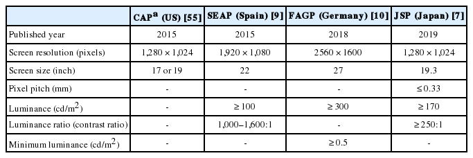
- 13,888 View
- 341 Download
- 26 Web of Science
- 30 Crossref
-
Abstract
PDFSupplementary Material
- Digital pathology (DP) using whole slide imaging (WSI) is becoming a fundamental issue in pathology with recent advances and the rapid development of associated technologies. However, the available evidence on its diagnostic uses and practical advice for pathologists on implementing DP remains insufficient, particularly in light of the exponential growth of this industry. To inform DP implementation in Korea, we developed relevant and timely recommendations. We first performed a literature review of DP guidelines, recommendations, and position papers from major countries, as well as a review of relevant studies validating WSI. Based on that information, we prepared a draft. After several revisions, we released this draft to the public and the members of the Korean Society of Pathologists through our homepage and held an open forum for interested parties. Through that process, this final manuscript has been prepared. This recommendation contains an overview describing the background, objectives, scope of application, and basic terminology; guidelines and considerations for the hardware and software used in DP systems and the validation required for DP implementation; conclusions; and references and appendices, including literature on DP from major countries and WSI validation studies.
-
Citations
Citations to this article as recorded by- Commercially Available Artificial Intelligence Solutions for Gynaecologic Cytology Screening and Their Integration Into Clinical Workflow
Yosep Chong, Andrey Bychkov
Cytopathology.2026; 37(1): 24. CrossRef - The impact of AI on modern oncology from early detection to personalized cancer treatment
Jun Li, Lei Zhang, Zhenglun Yu, Zhiye Bao, Danyang Li, Liming Wang
npj Precision Oncology.2026;[Epub] CrossRef - An equivalency and efficiency study for one year digital pathology for clinical routine diagnostics in an accredited tertiary academic center
Viola Iwuajoku, Kübra Ekici, Anette Haas, Mohammed Zaid Khan, Azar Kazemi, Atsuko Kasajima, Claire Delbridge, Alexander Muckenhuber, Elisa Schmoeckel, Fabian Stögbauer, Christine Bollwein, Kristina Schwamborn, Katja Steiger, Carolin Mogler, Peter J. Schüf
Virchows Archiv.2025; 487(1): 3. CrossRef - An adapted & improved validation protocol for digital pathology implementation
Ying-Han R. Hsu, Iman Ahmed, Juliana Phlamon, Charlotte Carment-Baker, Joyce Yin Tung Chan, Ioannis Prassas, Karen Weiser, Shaza Zeidan, Blaise Clarke, George M. Yousef
Seminars in Diagnostic Pathology.2025; 42(4): 150905. CrossRef - Transforming pathology into digital pathology: highway to hell or stairway to heaven?
Rainer Grobholz, Andrew Janowczyk, Inti Zlobec
Diagnostic Histopathology.2025; 31(7): 410. CrossRef - The Evolution of Digital Pathology in Infrastructure, Artificial Intelligence and Clinical Impact
Chan Kwon Jung
International Journal of Thyroidology.2025; 18(1): 6. CrossRef - Current Trends and Future Directions of Digital Pathology and Artificial Intelligence in Dermatopathology: A Scientometric-Based Review
Iuliu Gabriel Cocuz, Raluca Niculescu, Maria-Cătălina Popelea, Maria Elena Cocuz, Adrian-Horațiu Sabău, Andreea-Cătălina Tinca, Andreea Raluca Cozac-Szoke, Diana Maria Chiorean, Corina Eugenia Budin, Ovidiu Simion Cotoi
Diagnostics.2025; 15(17): 2196. CrossRef - Integration of Digital Cytology in Quality Assurance Programs for Cytopathology
Yosep Chong, Maria Jesús Fernández Aceñero, Zaibo Li, Andrey Bychkov
Acta Cytologica.2025; 70(1): 126. CrossRef - Quantitative Assessment of Focus Quality in Whole-Slide Imaging of Thyroid Liquid-Based Cytology Using Laplacian Variance
Chan Kwon Jung, Chankyung Kim, Sora Jeon, Andrey Bychkov
Endocrine Pathology.2025;[Epub] CrossRef - Performance of externally validated machine learning models based on histopathology images for the diagnosis, classification, prognosis, or treatment outcome prediction in female breast cancer: A systematic review
Ricardo Gonzalez, Peyman Nejat, Ashirbani Saha, Clinton J.V. Campbell, Andrew P. Norgan, Cynthia Lokker
Journal of Pathology Informatics.2024; 15: 100348. CrossRef - Swiss digital pathology recommendations: results from a Delphi process conducted by the Swiss Digital Pathology Consortium of the Swiss Society of Pathology
Andrew Janowczyk, Inti Zlobec, Cedric Walker, Sabina Berezowska, Viola Huschauer, Marianne Tinguely, Joel Kupferschmid, Thomas Mallet, Doron Merkler, Mario Kreutzfeldt, Radivoje Gasic, Tilman T. Rau, Luca Mazzucchelli, Isgard Eyberg, Gieri Cathomas, Kirst
Virchows Archiv.2024; 485(1): 13. CrossRef - ChatGPT as an aid for pathological diagnosis of cancer
Shaivy Malik, Sufian Zaheer
Pathology - Research and Practice.2024; 253: 154989. CrossRef - Possible benefits, challenges, pitfalls, and future perspective of using ChatGPT in pathology
Durre Aden, Sufian Zaheer, Sabina Khan
Revista Española de Patología.2024; 57(3): 198. CrossRef - Remote Placental Sign-Out: What Digital Pathology Can Offer for Pediatric Pathologists
Casey P. Schukow, Jacqueline K. Macknis
Pediatric and Developmental Pathology.2024; 27(4): 375. CrossRef - Digital Validation in Breast Cancer Needle Biopsies: Comparison of Histological Grade and Biomarker Expression Assessment Using Conventional Light Microscopy, Whole Slide Imaging, and Digital Image Analysis
Ji Eun Choi, Kyung-Hee Kim, Younju Lee, Dong-Wook Kang
Journal of Personalized Medicine.2024; 14(3): 312. CrossRef - Pathologists light level preferences using the microscope—study to guide digital pathology display use
Charlotte Jennings, Darren Treanor, David Brettle
Journal of Pathology Informatics.2024; 15: 100379. CrossRef - Eye tracking in digital pathology: A comprehensive literature review
Alana Lopes, Aaron D. Ward, Matthew Cecchini
Journal of Pathology Informatics.2024; 15: 100383. CrossRef - Diagnostic Assessment of Deep Learning Algorithms for Frozen Tissue Section Analysis in Women with Breast Cancer
Young-Gon Kim, In Hye Song, Seung Yeon Cho, Sungchul Kim, Milim Kim, Soomin Ahn, Hyunna Lee, Dong Hyun Yang, Namkug Kim, Sungwan Kim, Taewoo Kim, Daeyoung Kim, Jonghyeon Choi, Ki-Sun Lee, Minuk Ma, Minki Jo, So Yeon Park, Gyungyub Gong
Cancer Research and Treatment.2023; 55(2): 513. CrossRef - Recent application of artificial intelligence on histopathologic image-based prediction of gene mutation in solid cancers
Mohammad Rizwan Alam, Kyung Jin Seo, Jamshid Abdul-Ghafar, Kwangil Yim, Sung Hak Lee, Hyun-Jong Jang, Chan Kwon Jung, Yosep Chong
Briefings in Bioinformatics.2023;[Epub] CrossRef - Sustainable development goals applied to digital pathology and artificial intelligence applications in low- to middle-income countries
Sumi Piya, Jochen K. Lennerz
Frontiers in Medicine.2023;[Epub] CrossRef - Diagnostic proficiency test using digital cytopathology and comparative assessment of whole slide images of cytologic samples for quality assurance program in Korea
Yosep Chong, Soon Auck Hong, Hoon Kyu Oh, Soo Jin Jung, Bo-Sung Kim, Ji Yun Jeong, Ho-Chang Lee, Gyungyub Gong
Journal of Pathology and Translational Medicine.2023; 57(5): 251. CrossRef - Real-World Implementation of Digital Pathology: Results From an Intercontinental Survey
Daniel Gomes Pinto, Andrey Bychkov, Naoko Tsuyama, Junya Fukuoka, Catarina Eloy
Laboratory Investigation.2023; 103(12): 100261. CrossRef - National digital pathology projects in Switzerland: A 2023 update
Rainer Grobholz, Andrew Janowczyk, Ana Leni Frei, Mario Kreutzfeldt, Viktor H. Koelzer, Inti Zlobec
Die Pathologie.2023; 44(S3): 225. CrossRef - Understanding the ethical and legal considerations of Digital Pathology
Cheryl Coulter, Francis McKay, Nina Hallowell, Lisa Browning, Richard Colling, Philip Macklin, Tom Sorell, Muhammad Aslam, Gareth Bryson, Darren Treanor, Clare Verrill
The Journal of Pathology: Clinical Research.2022; 8(2): 101. CrossRef - Current Trend of Artificial Intelligence Patents in Digital Pathology: A Systematic Evaluation of the Patent Landscape
Muhammad Joan Ailia, Nishant Thakur, Jamshid Abdul-Ghafar, Chan Kwon Jung, Kwangil Yim, Yosep Chong
Cancers.2022; 14(10): 2400. CrossRef - Recent Applications of Artificial Intelligence from Histopathologic Image-Based Prediction of Microsatellite Instability in Solid Cancers: A Systematic Review
Mohammad Rizwan Alam, Jamshid Abdul-Ghafar, Kwangil Yim, Nishant Thakur, Sung Hak Lee, Hyun-Jong Jang, Chan Kwon Jung, Yosep Chong
Cancers.2022; 14(11): 2590. CrossRef - Automated Hybrid Model for Detecting Perineural Invasion in the Histology of Colorectal Cancer
Jiyoon Jung, Eunsu Kim, Hyeseong Lee, Sung Hak Lee, Sangjeong Ahn
Applied Sciences.2022; 12(18): 9159. CrossRef - Development of quality assurance program for digital pathology by the Korean Society of Pathologists
Yosep Chong, Jeong Mo Bae, Dong Wook Kang, Gwangil Kim, Hye Seung Han
Journal of Pathology and Translational Medicine.2022; 56(6): 370. CrossRef - Improving quality control in the routine practice for histopathological interpretation of gastrointestinal endoscopic biopsies using artificial intelligence
Young Sin Ko, Yoo Mi Choi, Mujin Kim, Youngjin Park, Murtaza Ashraf, Willmer Rafell Quiñones Robles, Min-Ju Kim, Jiwook Jang, Seokju Yun, Yuri Hwang, Hani Jang, Mun Yong Yi, Anwar P.P. Abdul Majeed
PLOS ONE.2022; 17(12): e0278542. CrossRef - What is Essential is (No More) Invisible to the Eyes: The Introduction of BlocDoc in the Digital Pathology Workflow
Vincenzo L’Imperio, Fabio Gibilisco, Filippo Fraggetta
Journal of Pathology Informatics.2021; 12(1): 32. CrossRef
- Commercially Available Artificial Intelligence Solutions for Gynaecologic Cytology Screening and Their Integration Into Clinical Workflow
- Pathologic interpretation of endoscopic ultrasound–guided fine needle aspiration cytology/biopsy for pancreatic lesions
- Haeryoung Kim, Kee-Taek Jang
- J Pathol Transl Med. 2020;54(5):367-377. Published online August 31, 2020
- DOI: https://doi.org/10.4132/jptm.2020.07.21
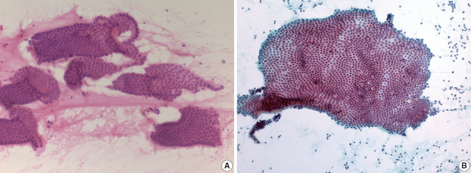
- 10,087 View
- 227 Download
- 5 Web of Science
- 5 Crossref
-
Abstract
PDF
- Pathologic interpretation of endoscopic ultrasound–guided fine needle aspiration (EUS-FNA) cytology/biopsy specimens is one of the most challenging tasks in cytology and surgical pathology practice, as the procedure often yields minimal amounts of diagnostic material and contains contaminants, such as blood cells and normal intestinal mucosa. EUS-FNA cytology/biopsy will nevertheless become a more popular procedure for evaluation of various pancreatic lesions because they are difficult to approach with conventional endoscopic procedures. Pathologists should understand the structural differences and limitations of EUS-FNA that make pathologic diagnosis difficult. Ancillary tests are available for differential diagnosis of EUS-FNA for various pancreatic lesions. Immunostains are the most commonly used ancillary tests, and pathologists should able to choose the necessary panel for differential diagnosis. Pathologists should review clinical history and radiologic and/or EUS findings before selecting an immunostain panel and making a pathologic diagnosis. In addition, one’s threshold of malignancy should be adjusted according to the appropriate clinical setting to avoid under-evaluation of pathologic diagnoses. Clinico-pathologic correlation is essential in pathologic evaluation of EUS-FNA for pancreatic lesions. Pathologists can reduce errors by correlating clinical and radiologic findings when evaluating EUS-FNA. Some molecular tests can be applied in differential diagnosis of pancreatic neoplastic and cystic lesions. Molecular data should be used as supportive evidence of a specific disease entity, rather than direct evidence, and should be correlated with clinico-pathologic findings to avoid errors in pathologic diagnosis.
-
Citations
Citations to this article as recorded by- Diagnostic Performance of EUS‐FNA for Pancreatic Lesions at Tertiary Centers in Iran Without Rapid On‐Site Evaluation
Maryam Bazmandegan, Gholam Reza Sivandzadeh, Kamran Bagheri Lankarani, Zahra Beyzaei, Bita Geramizadeh
Cytopathology.2026; 37(3): 263. CrossRef - Endoscopic Ultrasound-Guided Pancreatic Tissue Sampling: Lesion Assessment, Needles, and Techniques
Jahnvi Dhar, Jayanta Samanta, Zaheer Nabi, Manik Aggarwal, Maria Cristina Conti Bellocchi, Antonio Facciorusso, Luca Frulloni, Stefano Francesco Crinò
Medicina.2024; 60(12): 2021. CrossRef - A prospective randomized noninferiority trial comparing conventional smears and SurePathTM liquid-based cytology in endoscopic ultrasound-guided sampling of esophageal, gastric, and duodenal lesions
Jae Chang Jun, Sang Hyub Lee, Han Myung Lee, Sang Gyun Kim, Hyunsoo Chung, Joo Seong Kim, Namyoung Park, Jin Ho Choi, Yoonjin Kwak, Soo-Jeong Cho
Medicine.2023; 102(29): e34321. CrossRef - Double Ki-67 and synaptophysin labeling in pancreatic neuroendocrine tumor biopsies
Bokyung Ahn, Jin Kying Jung, HaeSung Jung, Yeon-Mi Ryu, Yeon Wook Kim, Tae Jun Song, Do Hyun Park, Dae wook Hwang, HyungJun Cho, Sang-Yeob Kim, Seung-Mo Hong
Pancreatology.2022; 22(3): 427. CrossRef - Comparison of Endoscopic Ultrasound-Guided Fine Needle Aspiration with 19-Gauge and 22-Gauge Needles for Solid Pancreatic Lesions
Changjuan Li, Jianwei Mi, Fulai Gao, Xinying Zhu, Miao Su, Xiaoli Xie, Dongqiang Zhao
International Journal of General Medicine.2021; Volume 14: 10439. CrossRef
- Diagnostic Performance of EUS‐FNA for Pancreatic Lesions at Tertiary Centers in Iran Without Rapid On‐Site Evaluation
Original Article
- A retrospective cytohistological correlation of fine-needle aspiration cytology with classification by the Milan System for Reporting Salivary Gland Cytopathology
- Ji Hyun Park, Yoon Jin Cha, Ja Yeong Seo, Jae Yol Lim, Soon Won Hong
- J Pathol Transl Med. 2020;54(5):419-425. Published online July 8, 2020
- DOI: https://doi.org/10.4132/jptm.2020.06.09
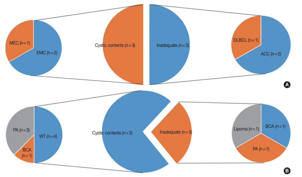
- 7,660 View
- 206 Download
- 12 Web of Science
- 15 Crossref
-
Abstract
PDF
- Background
Before publication of the new classification system named the Milan System for Reporting Salivary Gland Cytopathology (MSRSGC) in 2018, there was no standard classification for salivary gland lesions obtained by fine-needle aspiration (FNA). We therefore aimed to evaluate the diagnostic utility of this system by retrospectively reviewing FNA samples using the MSRSGC and to determine their risk of developing into neoplasms and becoming malignant.
Methods
Retrospective slide review and classification of salivary gland FNAs obtained over a 6-year period (2013–2018) at a single center were performed by two pathologists. The risks of neoplasm and malignancy for each category also were calculated.
Results
This study surveyed 374 FNAs (371 patients) performed over a six-year period and selected 148 cases that included documented surgical follow-up (39.6%). Among the surgically treated cases, the distributions of FNA categories were as follows: non-diagnostic (ND; 16.9%), non-neoplastic (NN; 2.7%), atypia of undetermined significance (AUS; 3.4%), benign (BN; 54.7%), salivary gland neoplasm of uncertain malignant potential (SUMP; 10.1%), suspicious for malignancy (SM; 6.8%), and malignant (M; 5.4%). The risk of malignancy (ROM) was 24.0% for ND, 0% for NN, 40.0% for AUS, 2.5% for BN, 46.7% for SUMP, 100% for SM, and 87.5% for M. The overall diagnostic accuracy was 95.9% (142/148 cases).
Conclusions
The newly proposed MSRSGC appears to be a reliable system for classification of salivary gland lesions according to the associated ROM. -
Citations
Citations to this article as recorded by- Optimizing preoperative assessment of salivary gland lesions: updates and practical implications in the Milan System 2nd edition
Patrizia Straccia, Vincenzo Fiorentino, Alessia Piermattei, Qianqian Zhang, Belen Padial-Urtueta, Federica Cianfrini, Antonino Mule’, Esther Diana Rossi
Virchows Archiv.2026;[Epub] CrossRef - The Impact of Lesion-Specific and Sampling-Related Factors on Success of Salivary Gland Fine-Needle Aspiration Cytology
Marcel Mayer, Mohammad Marwan Alfarra, Kathrin Möllenhoff, Marianne Engels, Christoph Arolt, Alexander Quaas, Philipp Wolber, Louis Jansen, Lisa Nachtsheim, Maria Grosheva, Jens Peter Klussmann, Sami Shabli
Head and Neck Pathology.2025;[Epub] CrossRef - The Myriad Spectrum of Salivary Gland Lesions: Cytohistological Correlation on Fine Needle Aspiration Cytology, Core Needle Biopsy, and Resections in a 5‐Year Single Institutional Experience of North India
Zachariah Chowdhury, Pallavi Majumdar, Sumeet Narain, Komal Lamba
Diagnostic Cytopathology.2025; 53(8): 391. CrossRef - Diagnostic Performance of the Milan System for Reporting Salivary Gland Cytopathology and a Proposed Algorithm for Fine-Needle Aspiration Cytology of Salivary Gland Lesions
Norihide Mochizuki, Hirotaka Fujita, Takuma Tajiri, Masataka Ueda, Makiko Kurata, Chie Inomoto, Tomoko Sugiyama, Daisuke Maki, Shuichi Shiraishi, Tomohisa Machida, Hitoshi Ito, Yohei Masugi, Naoya Nakamura
Acta Cytologica.2025; 69(4): 324. CrossRef - A study of fine needle aspiration cytology and histopathology correlation of salivary gland neoplasms in a tertiary care hospital: an observational study
Asima Malik, Ahlam Mushtaq, Salma Bhat, Suhail Naik
International Journal of Contemporary Pediatrics.2025; 13(1): 23. CrossRef - The Milan system for reporting salivary gland cytopathology – Assessment of utility and the risk of malignancy
Annu E. Prakash, Renu Sukumaran, Nileena Nayak, K. Lakshmi, Anitha Mathews, Jayasree Kattoor
Indian Journal of Cancer.2024; 61(3): 575. CrossRef - Salivary gland fine-needle aspiration biopsy: quality assurance results from a tertiary cancer center
Fanni Ratzon, Dominique L. Feliciano, Nora Katabi, Bin Xu, Oscar Lin, Xiao-Jun Wei
Journal of the American Society of Cytopathology.2023; 12(3): 206. CrossRef - Cytohistological correlation and risk stratification of salivary gland lesions using the Milan System for Reporting Salivary Gland Cytopathology: A tertiary care centre experience
Tarun Kumar, Prerna Tewari, Jitendra Singh Nigam, Shreekant Bharti, Surabhi, Ruchi Sinha, Punam Prasad Bhadani
Cytopathology.2023; 34(3): 225. CrossRef - Assessment of Risk of Malignancy of Fine-needle Aspiration Cytology in Salivary Gland Lesions Using the Milan System for Reporting Salivary Gland Cytopathology Categorization: A Systematic Review and Meta-analysis
Amit Kumar, Subhash Chandra, Bishnupati Singh, Swati Sharma, Ankita Tandon, Ajoy Kumar Shahi
The Journal of Contemporary Dental Practice.2023; 23(10): 1039. CrossRef - Milan Sınıflandırma Sistemi’ne Göre Değerlendirilen Tükürük Bezi İnce İğne Aspirasyon Sitolojilerinin Histopatolojik Tanı Uyumu
Özlem SARAYDAROĞLU, Selin YİRMİBEŞ
Uludağ Üniversitesi Tıp Fakültesi Dergisi.2023; 49(3): 285. CrossRef - Milan system for reporting salivary gland cytopathology: Adoption and outcomes in a community setting
Samih J. Nassif, Ali R. Sasani, Garrey T. Faller, Jennifer L. Harb, Jagdish K. Dhingra
Head & Neck.2022; 44(6): 1462. CrossRef - Nondiagnostic salivary gland FNAs are associated with decreased risk of malignancy compared with “all‐comer” patients: Analysis of the Milan System for Reporting Salivary Gland Cytopathology with a focus on Milan I: Nondiagnostic
Shu K. Lui, Troy Tenney, Patrick C. Mullane, Kartik Viswanathan, Daniel J. Lubin
Cancer Cytopathology.2022; 130(10): 800. CrossRef - Application of the Milan System for Reporting Salivary Gland Cytopathology: A systematic review and meta‐analysis
Zhaoyang Wang, Huan Zhao, Huiqin Guo, Changming An
Cancer Cytopathology.2022; 130(11): 849. CrossRef - Multiplexed single‐cell analysis of FNA allows accurate diagnosis of salivary gland tumors
Juhyun Oh, Tae Yeon Yoo, Talia M. Saal, Lisa Tsay, William C. Faquin, Jonathan C.T. Carlson, Daniel G. Deschler, Sara I. Pai, Ralph Weissleder
Cancer Cytopathology.2022; 130(8): 581. CrossRef - Cytologic analysis of vitreous fluids: A retrospective review of our 24 years of experience
Gabriel L. Collins, Elizabeth W. Hubbard, Christopher T. Clark, Lisa D. Duncan, Laurentia Nodit
Diagnostic Cytopathology.2021; 49(10): 1122. CrossRef
- Optimizing preoperative assessment of salivary gland lesions: updates and practical implications in the Milan System 2nd edition
Case Study
- Appendiceal actinomycosis mimicking appendiceal tumor, appendicitis or inflammatory bowel disease
- You-Na Sung, Jihun Kim
- J Pathol Transl Med. 2021;55(5):349-354. Published online June 26, 2020
- DOI: https://doi.org/10.4132/jptm.2020.05.17
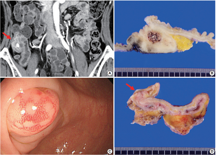
- 9,073 View
- 171 Download
- 5 Web of Science
- 4 Crossref
-
Abstract
PDF
- Appendiceal actinomycosis is very rare and its diagnosis is often difficult even in surgically resected specimens. Here we report two cases of appendiceal actinomycosis confirmed by pathologic examination of surgically resected specimens. Characteristic histologic features included transmural chronic inflammation with Crohn-like lymphoid aggregates and polypoid mucosal protrusion into cecal lumen through fibrous expansion of the submucosa. Chronic active inflammation involved the mucosa of the appendix and cecum around the appendiceal orifice. Crohn’s disease with predominant cecal involvement and inflammatory pseudotumor were considered as differential diagnoses. Careful examination revealed a few actinomycotic colonies in the mucosa, confirming the diagnosis. A high index of suspicion with awareness of the characteristic histologic features might prompt careful inspection for the actinomycotic colonies, leading to the appropriate diagnosis of this rare disease.
-
Citations
Citations to this article as recorded by- Persistent intra-abdominal abscess with intestinal obstruction following seven failed drainage procedures over 3.5 years: a case report
Ayman Shemes, Salma Samra, Ahmed Mohamed, Amr A. Elgharib
BMC Surgery.2025;[Epub] CrossRef - Appendicular actinomycosis: The first reported case of an uncommon finding of a common ailment from Nepal
Sujan Bohara, Manoj Khadka, Pawan Singh Bhat, Prajwal Syangtang, Badal Karki, Bhagawan Shrestha, Shoshan Arja Acharya, Khusbhu Khetan, Jyoti Rayamajhi, Sushil Bahadur Rawal
Clinical Case Reports.2023;[Epub] CrossRef - Abdominopelvic actinomycosis: An unexpected diagnosis in an elderly female with a destructive-appearing soft tissue mass
Elise Hyser, Drashti Antala, Harvey Friedman, Jonathan Stake
IDCases.2022; 28: e01479. CrossRef - Diagnosing granulomatous disease during appendectomy
Atilla Şenaylı
Clinical Case Reports.2021;[Epub] CrossRef
- Persistent intra-abdominal abscess with intestinal obstruction following seven failed drainage procedures over 3.5 years: a case report
Reviews
- Introduction to digital pathology and computer-aided pathology
- Soojeong Nam, Yosep Chong, Chan Kwon Jung, Tae-Yeong Kwak, Ji Youl Lee, Jihwan Park, Mi Jung Rho, Heounjeong Go
- J Pathol Transl Med. 2020;54(2):125-134. Published online February 13, 2020
- DOI: https://doi.org/10.4132/jptm.2019.12.31

- 22,426 View
- 657 Download
- 87 Web of Science
- 91 Crossref
-
Abstract
PDF
- Digital pathology (DP) is no longer an unfamiliar term for pathologists, but it is still difficult for many pathologists to understand the engineering and mathematics concepts involved in DP. Computer-aided pathology (CAP) aids pathologists in diagnosis. However, some consider CAP a threat to the existence of pathologists and are skeptical of its clinical utility. Implementation of DP is very burdensome for pathologists because technical factors, impact on workflow, and information technology infrastructure must be considered. In this paper, various terms related to DP and computer-aided pathologic diagnosis are defined, current applications of DP are discussed, and various issues related to implementation of DP are outlined. The development of computer-aided pathologic diagnostic tools and their limitations are also discussed.
-
Citations
Citations to this article as recorded by- Artificial Intelligence and Digital Pathology: Toward Next‐Generation Diagnostic Hematology
Valeriia Tsekhovska, Pier Paolo Piccaluga, Nikhat Kaura
International Journal of Clinical Practice.2026;[Epub] CrossRef - Deep learning model to diagnose cardiac amyloidosis from haematoxylin/eosin-stained myocardial tissue
Takeshi Tohyama, Takeshi Iwasaki, Masataka Ikeda, Masato Katsuki, Tatsuya Watanabe, Kayo Misumi, Keisuke Shinohara, Takeo Fujino, Toru Hashimoto, Shouji Matsushima, Tomomi Ide, Junji Kishimoto, Koji Todaka, Yoshinao Oda, Kohtaro Abe
European Heart Journal - Imaging Methods and Practice.2025;[Epub] CrossRef - The current landscape of artificial intelligence in oral and maxillofacial surgery– a narrative review
Rushil Rajiv Dang, Balram Kadaikal, Sam El Abbadi, Branden R. Brar, Amit Sethi, Radhika Chigurupati
Oral and Maxillofacial Surgery.2025;[Epub] CrossRef - Assessing the quality of whole slide images in cytology from nuclei features
Paul Barthe, Romain Brixtel, Yann Caillot, Benoît Lemoine, Arnaud Renouf, Vianney Thurotte, Ouarda Beniken, Sébastien Bougleux, Olivier Lézoray
Journal of Pathology Informatics.2025; 17: 100420. CrossRef - An update on applications of digital pathology: primary diagnosis; telepathology, education and research
Shamail Zia, Isil Z. Yildiz-Aktas, Fazail Zia, Anil V. Parwani
Diagnostic Pathology.2025;[Epub] CrossRef - Artificial intelligence–driven digital pathology in urological cancers: current trends and future directions
Inyoung Paik, Geongyu Lee, Joonho Lee, Tae-Yeong Kwak, Hong Koo Ha
Prostate International.2025; 13(4): 181. CrossRef - Label-free optical microscopy with artificial intelligence: a new paradigm in pathology
Chiho Yoon, Eunwoo Park, Donggyu Kim, Byullee Park, Chulhong Kim
Biophotonics Discovery.2025;[Epub] CrossRef - EPIIC: Edge-Preserving Method Increasing Nuclei Clarity for Compression Artifacts Removal in Whole-Slide Histopathological Images
Julia Merta, Michal Marczyk
Applied Sciences.2025; 15(8): 4450. CrossRef - Comparative analysis of a 5G campus network and existing networks for real-time consultation in remote pathology
Ilgar I. Guseinov, Arnab Bhowmik, Somaia AbuBaker, Anna E. Schmaus-Klughammer, Thomas Spittler
Journal of Pathology Informatics.2025; 18: 100444. CrossRef - The Evolution of Digital Pathology in Infrastructure, Artificial Intelligence and Clinical Impact
Chan Kwon Jung
International Journal of Thyroidology.2025; 18(1): 6. CrossRef - Role of Telepathology, Artificial Intelligence, and Emerging Technologies in Enhancing Diagnostic Accuracy
Yugeshwari R. Tiwade, Obaid Noman, Pratibha Dawande, Nandkishor J Bankar, Sweta Bahadure, Praful Patil
Journal of Nature and Science of Medicine.2025; 8(2): 115. CrossRef - Analysis of system and scanner downtime in a digital pathology–predominant institution: A 6-year experience
Ryan Reagans, Lokman Cevik, Himani Kumar, David Kellough, Abberly Lott Limbach, Giovanni Lujan, Anil Parwani, Hamza N Gokozan
American Journal of Clinical Pathology.2025; 164(4): 634. CrossRef - Integration of Digital Cytology in Quality Assurance Programs for Cytopathology
Yosep Chong, Maria Jesús Fernández Aceñero, Zaibo Li, Andrey Bychkov
Acta Cytologica.2025; 70(1): 126. CrossRef - Telepathology for Consultation in the Military Health System: An Evaluation of Pathologists’ Impressions of Facilitators and Barriers Prior to Implementation
Victoria Mahar, Zachary Colburn, Joshua Sakai
Laboratory Investigation.2025; 105(11): 104236. CrossRef - Online histostereometric analysis in digital forensic pathology: a technical report
Vladimir G. Nedugov, Anna V. Zhukova, German V. Nedugov
Russian Journal of Forensic Medicine.2025; 11(2): 145. CrossRef - Latent representation of H&E images retains biological information in a breast cancer cohort
Chloé Benmussa, Esther Sanfeliu, Anabel Martínez-Romero, Blanca González-Farré, Tomás Pascual, Joaquín Gavilá, Alona Levy-Jurgenson, Ariel Shamir, Fara Brasó-Maristany, Aleix Prat, Zohar Yakhini, Amgad Muneer
PLOS One.2025; 20(9): e0329221. CrossRef - Modernizing Colorectal Cancer Care With Artificial Intelligence: Real-Time Detection, Radiomics, and Digital Pathology
Elmoatazbellah Nasr, Zaid Al-Hamid, Mina H Younan, Mohamed Omran, Maan Sarsam, Mohamed Abdellatif
Cureus.2025;[Epub] CrossRef - A multi-task learning model for evaluating non-tumor gastric diseases indicators in whole slide images
Mingxi Fu, Liming Liu, Fanglei Fu, Jingli Ouyang, Xueying Shi, Song Duan, Tian Guan, Yonghong He, Zhiqiang Cheng, Lianghui Zhu
Scientific Reports.2025;[Epub] CrossRef - Performance Evaluation of MambaVision in Breast Cancer Detection from Histopathology Images
Hasan Zan
Dicle Üniversitesi Mühendislik Fakültesi Mühendislik Dergisi.2025; 16(4): 879. CrossRef - Artificial intelligence for automatic detection of basal cell carcinoma from frozen tissue tangential biopsies
Dennis H Murphree, Yong-hun Kim, Kirk A Sidey, Nneka I Comfere, Nahid Y Vidal
Clinical and Experimental Dermatology.2024; 49(7): 719. CrossRef - Performance of externally validated machine learning models based on histopathology images for the diagnosis, classification, prognosis, or treatment outcome prediction in female breast cancer: A systematic review
Ricardo Gonzalez, Peyman Nejat, Ashirbani Saha, Clinton J.V. Campbell, Andrew P. Norgan, Cynthia Lokker
Journal of Pathology Informatics.2024; 15: 100348. CrossRef - Invisible for a few but essential for many: the role of Histotechnologists in the establishment of digital pathology
Gisela Magalhães, Rita Calisto, Catarina Freire, Regina Silva, Diana Montezuma, Sule Canberk, Fernando Schmitt
Journal of Histotechnology.2024; 47(1): 39. CrossRef - Using digital pathology to analyze the murine cerebrovasculature
Dana M Niedowicz, Jenna L Gollihue, Erica M Weekman, Panhavuth Phe, Donna M Wilcock, Christopher M Norris, Peter T Nelson
Journal of Cerebral Blood Flow & Metabolism.2024; 44(4): 595. CrossRef - PATrans: Pixel-Adaptive Transformer for edge segmentation of cervical nuclei on small-scale datasets
Hexuan Hu, Jianyu Zhang, Tianjin Yang, Qiang Hu, Yufeng Yu, Qian Huang
Computers in Biology and Medicine.2024; 168: 107823. CrossRef - CNAC-Seg: Effective segmentation for cervical nuclei in adherent cells and clusters via exploring gaps of receptive fields
Hexuan Hu, Jianyu Zhang, Tianjin Yang, Qiang Hu, Yufeng Yu, Qian Huang
Biomedical Signal Processing and Control.2024; 90: 105833. CrossRef - Artificial Intelligence-Enabled Prostate Cancer Diagnosis and Prognosis: Current State and Future Implications
Swati Satturwar, Anil V. Parwani
Advances in Anatomic Pathology.2024; 31(2): 136. CrossRef - Ensemble Deep Learning Model to Predict Lymphovascular Invasion in Gastric Cancer
Jonghyun Lee, Seunghyun Cha, Jiwon Kim, Jung Joo Kim, Namkug Kim, Seong Gyu Jae Gal, Ju Han Kim, Jeong Hoon Lee, Yoo-Duk Choi, Sae-Ryung Kang, Ga-Young Song, Deok-Hwan Yang, Jae-Hyuk Lee, Kyung-Hwa Lee, Sangjeong Ahn, Kyoung Min Moon, Myung-Giun Noh
Cancers.2024; 16(2): 430. CrossRef - Artificial intelligence’s impact on breast cancer pathology: a literature review
Amr Soliman, Zaibo Li, Anil V. Parwani
Diagnostic Pathology.2024;[Epub] CrossRef - Automated Analysis of Nuclear Parameters in Oral Exfoliative Cytology Using Machine Learning
Shubhangi Mhaske, Karthikeyan Ramalingam, Preeti Nair, Shubham Patel, Arathi Menon P, Nida Malik, Sumedh Mhaske
Cureus.2024;[Epub] CrossRef - Enhancing AI Research for Breast Cancer: A Comprehensive Review of Tumor-Infiltrating Lymphocyte Datasets
Alessio Fiorin, Carlos López Pablo, Marylène Lejeune, Ameer Hamza Siraj, Vincenzo Della Mea
Journal of Imaging Informatics in Medicine.2024; 37(6): 2996. CrossRef - Current Developments in Diagnosis of Salivary Gland Tumors: From Structure to Artificial Intelligence
Alexandra Corina Faur, Roxana Buzaș, Adrian Emil Lăzărescu, Laura Andreea Ghenciu
Life.2024; 14(6): 727. CrossRef - Comparative analysis of chronic progressive nephropathy (CPN) diagnosis in rat kidneys using an artificial intelligence deep learning model
Yeji Bae, Jongsu Byun, Hangyu Lee, Beomseok Han
Toxicological Research.2024; 40(4): 551. CrossRef - A Pan-Cancer Patient-Derived Xenograft Histology Image Repository with Genomic and Pathologic Annotations Enables Deep Learning Analysis
Brian S. White, Xing Yi Woo, Soner Koc, Todd Sheridan, Steven B. Neuhauser, Shidan Wang, Yvonne A. Evrard, Li Chen, Ali Foroughi pour, John D. Landua, R. Jay Mashl, Sherri R. Davies, Bingliang Fang, Maria Gabriela Rosa, Kurt W. Evans, Matthew H. Bailey, Y
Cancer Research.2024; 84(13): 2060. CrossRef - Non-contrasted computed tomography (NCCT) based chronic thromboembolic pulmonary hypertension (CTEPH) automatic diagnosis using cascaded network with multiple instance learning
Mayang Zhao, Liming Song, Jiarui Zhu, Ta Zhou, Yuanpeng Zhang, Shu-Cheng Chen, Haojiang Li, Di Cao, Yi-Quan Jiang, Waiyin Ho, Jing Cai, Ge Ren
Physics in Medicine & Biology.2024; 69(18): 185011. CrossRef - MR_NET: A Method for Breast Cancer Detection and Localization from Histological Images Through Explainable Convolutional Neural Networks
Rachele Catalano, Myriam Giusy Tibaldi, Lucia Lombardi, Antonella Santone, Mario Cesarelli, Francesco Mercaldo
Sensors.2024; 24(21): 7022. CrossRef - Advances in AI-Enhanced Biomedical Imaging for Cancer Immunology
Willa Wen-You Yim, Felicia Wee, Zheng Yi Ho, Xinyun Feng, Marcia Zhang, Samuel Lee, Inti Zlobec, Joe Yeong, Mai Chan Lau
World Scientific Annual Review of Cancer Immunology.2024;[Epub] CrossRef - Blockchain: A safe digital technology to share cancer diagnostic results in pandemic times—Challenges and legacy for the future
Bruno Natan Santana Lima, Lucas Alves da Mota Santana, Rani Iani Costa Gonçalo, Carla Samily de Oliveira Costa, Daniel Pitanga de Sousa Nogueira, Cleverson Luciano Trento, Wilton Mitsunari Takeshita
Oral Surgery.2023; 16(3): 300. CrossRef - Pathologists’ acceptance of telepathology in the Ministry of National Guard Health Affairs Hospitals in Saudi Arabia: A survey study
Raneem Alawashiz, Sharifah Abdullah AlDossary
DIGITAL HEALTH.2023;[Epub] CrossRef - An Atrous Convolved Hybrid Seg-Net Model with residual and attention mechanism for gland detection and segmentation in histopathological images
Manju Dabass, Jyoti Dabass
Computers in Biology and Medicine.2023; 155: 106690. CrossRef - Validation of a Machine Learning Expert Supporting System, ImmunoGenius, Using Immunohistochemistry Results of 3000 Patients with Lymphoid Neoplasms
Jamshid Abdul-Ghafar, Kyung Jin Seo, Hye-Ra Jung, Gyeongsin Park, Seung-Sook Lee, Yosep Chong
Diagnostics.2023; 13(7): 1308. CrossRef - Diagnosing Infectious Diseases in Poultry Requires a Holistic Approach: A Review
Dieter Liebhart, Ivana Bilic, Beatrice Grafl, Claudia Hess, Michael Hess
Poultry.2023; 2(2): 252. CrossRef - Recent application of artificial intelligence on histopathologic image-based prediction of gene mutation in solid cancers
Mohammad Rizwan Alam, Kyung Jin Seo, Jamshid Abdul-Ghafar, Kwangil Yim, Sung Hak Lee, Hyun-Jong Jang, Chan Kwon Jung, Yosep Chong
Briefings in Bioinformatics.2023;[Epub] CrossRef - Canine Mammary Tumor Histopathological Image Classification via Computer-Aided Pathology: An Available Dataset for Imaging Analysis
Giovanni P. Burrai, Andrea Gabrieli, Marta Polinas, Claudio Murgia, Maria Paola Becchere, Pierfranco Demontis, Elisabetta Antuofermo
Animals.2023; 13(9): 1563. CrossRef - Rapid digital pathology of H&E-stained fresh human brain specimens as an alternative to frozen biopsy
Bhaskar Jyoti Borah, Yao-Chen Tseng, Kuo-Chuan Wang, Huan-Chih Wang, Hsin-Yi Huang, Koping Chang, Jhih Rong Lin, Yi-Hua Liao, Chi-Kuang Sun
Communications Medicine.2023;[Epub] CrossRef - Applied machine learning in hematopathology
Taher Dehkharghanian, Youqing Mu, Hamid R. Tizhoosh, Clinton J. V. Campbell
International Journal of Laboratory Hematology.2023; 45(S2): 87. CrossRef - Automated diagnosis of 7 canine skin tumors using machine learning on H&E-stained whole slide images
Marco Fragoso-Garcia, Frauke Wilm, Christof A. Bertram, Sophie Merz, Anja Schmidt, Taryn Donovan, Andrea Fuchs-Baumgartinger, Alexander Bartel, Christian Marzahl, Laura Diehl, Chloe Puget, Andreas Maier, Marc Aubreville, Katharina Breininger, Robert Klopf
Veterinary Pathology.2023; 60(6): 865. CrossRef - Artificial Intelligence in the Pathology of Gastric Cancer
Sangjoon Choi, Seokhwi Kim
Journal of Gastric Cancer.2023; 23(3): 410. CrossRef - Efficient Convolution Network to Assist Breast Cancer Diagnosis and Target Therapy
Ching-Wei Wang, Kai-Lin Chu, Hikam Muzakky, Yi-Jia Lin, Tai-Kuang Chao
Cancers.2023; 15(15): 3991. CrossRef - Multi-Configuration Analysis of DenseNet Architecture for Whole Slide Image Scoring of ER-IHC
Wan Siti Halimatul Munirah Wan Ahmad, Mohammad Faizal Ahmad Fauzi, Md Jahid Hasan, Zaka Ur Rehman, Jenny Tung Hiong Lee, See Yee Khor, Lai-Meng Looi, Fazly Salleh Abas, Afzan Adam, Elaine Wan Ling Chan, Sei-Ichiro Kamata
IEEE Access.2023; 11: 79911. CrossRef - Digitization of Pathology Labs: A Review of Lessons Learned
Lars Ole Schwen, Tim-Rasmus Kiehl, Rita Carvalho, Norman Zerbe, André Homeyer
Laboratory Investigation.2023; 103(11): 100244. CrossRef - Artificial Intelligence in Endoscopic Ultrasonography-Guided Fine-Needle Aspiration/Biopsy (EUS-FNA/B) for Solid Pancreatic Lesions: Opportunities and Challenges
Xianzheng Qin, Taojing Ran, Yifei Chen, Yao Zhang, Dong Wang, Chunhua Zhou, Duowu Zou
Diagnostics.2023; 13(19): 3054. CrossRef - Deep Learning for the Pathologic Diagnosis of Hepatocellular Carcinoma, Cholangiocarcinoma, and Metastatic Colorectal Cancer
Hyun-Jong Jang, Jai-Hyang Go, Younghoon Kim, Sung Hak Lee
Cancers.2023; 15(22): 5389. CrossRef - AIR-UNet++: a deep learning framework for histopathology image segmentation and detection
Anusree Kanadath, J. Angel Arul Jothi, Siddhaling Urolagin
Multimedia Tools and Applications.2023; 83(19): 57449. CrossRef - Deep Learning-Based Dermatological Condition Detection: A Systematic Review With Recent Methods, Datasets, Challenges, and Future Directions
Stephanie S. Noronha, Mayuri A. Mehta, Dweepna Garg, Ketan Kotecha, Ajith Abraham
IEEE Access.2023; 11: 140348. CrossRef - Digital pathology and artificial intelligence in translational medicine and clinical practice
Vipul Baxi, Robin Edwards, Michael Montalto, Saurabh Saha
Modern Pathology.2022; 35(1): 23. CrossRef - Artificial Intelligence in Toxicological Pathology: Quantitative Evaluation of Compound-Induced Follicular Cell Hypertrophy in Rat Thyroid Gland Using Deep Learning Models
Valeria Bertani, Olivier Blanck, Davy Guignard, Frederic Schorsch, Hannah Pischon
Toxicologic Pathology.2022; 50(1): 23. CrossRef - Investigating the genealogy of the literature on digital pathology: a two-dimensional bibliometric approach
Dayu Hu, Chengyuan Wang, Song Zheng, Xiaoyu Cui
Scientometrics.2022; 127(2): 785. CrossRef - Digital Dermatopathology and Its Application to Mohs Micrographic Surgery
Yeongjoo Oh, Hye Min Kim, Soon Won Hong, Eunah Shin, Jihee Kim, Yoon Jung Choi
Yonsei Medical Journal.2022; 63(Suppl): S112. CrossRef - Assessment of parathyroid gland cellularity by digital slide analysis
Rotem Sagiv, Bertha Delgado, Oleg Lavon, Vladislav Osipov, Re'em Sade, Sagi Shashar, Ksenia M. Yegodayev, Moshe Elkabets, Ben-Zion Joshua
Annals of Diagnostic Pathology.2022; 58: 151907. CrossRef - PancreaSys: An Automated Cloud-Based Pancreatic Cancer Grading System
Muhammad Nurmahir Mohamad Sehmi, Mohammad Faizal Ahmad Fauzi, Wan Siti Halimatul Munirah Wan Ahmad, Elaine Wan Ling Chan
Frontiers in Signal Processing.2022;[Epub] CrossRef - Classification of Mouse Lung Metastatic Tumor with Deep Learning
Ha Neul Lee, Hong-Deok Seo, Eui-Myoung Kim, Beom Seok Han, Jin Seok Kang
Biomolecules & Therapeutics.2022; 30(2): 179. CrossRef - Techniques for digital histological morphometry of the pineal gland
Bogdan-Alexandru Gheban, Horaţiu Alexandru Colosi, Ioana-Andreea Gheban-Roșca, Carmen Georgiu, Dan Gheban, Doiniţa Crişan, Maria Crişan
Acta Histochemica.2022; 124(4): 151897. CrossRef - Current Trend of Artificial Intelligence Patents in Digital Pathology: A Systematic Evaluation of the Patent Landscape
Muhammad Joan Ailia, Nishant Thakur, Jamshid Abdul-Ghafar, Chan Kwon Jung, Kwangil Yim, Yosep Chong
Cancers.2022; 14(10): 2400. CrossRef - Recent Applications of Artificial Intelligence from Histopathologic Image-Based Prediction of Microsatellite Instability in Solid Cancers: A Systematic Review
Mohammad Rizwan Alam, Jamshid Abdul-Ghafar, Kwangil Yim, Nishant Thakur, Sung Hak Lee, Hyun-Jong Jang, Chan Kwon Jung, Yosep Chong
Cancers.2022; 14(11): 2590. CrossRef - Development of a prognostic prediction support system for cervical intraepithelial neoplasia using artificial intelligence-based diagnosis
Takayuki Takahashi, Hikaru Matsuoka, Rieko Sakurai, Jun Akatsuka, Yusuke Kobayashi, Masaru Nakamura, Takashi Iwata, Kouji Banno, Motomichi Matsuzaki, Jun Takayama, Daisuke Aoki, Yoichiro Yamamoto, Gen Tamiya
Journal of Gynecologic Oncology.2022;[Epub] CrossRef - Digital Pathology and Artificial Intelligence Applications in Pathology
Heounjeong Go
Brain Tumor Research and Treatment.2022; 10(2): 76. CrossRef - Mass spectrometry imaging to explore molecular heterogeneity in cell culture
Tanja Bien, Krischan Koerfer, Jan Schwenzfeier, Klaus Dreisewerd, Jens Soltwisch
Proceedings of the National Academy of Sciences.2022;[Epub] CrossRef - Integrating artificial intelligence in pathology: a qualitative interview study of users' experiences and expectations
Jojanneke Drogt, Megan Milota, Shoko Vos, Annelien Bredenoord, Karin Jongsma
Modern Pathology.2022; 35(11): 1540. CrossRef - Deep Learning on Basal Cell Carcinoma In Vivo Reflectance Confocal Microscopy Data
Veronika Shavlokhova, Michael Vollmer, Patrick Gholam, Babak Saravi, Andreas Vollmer, Jürgen Hoffmann, Michael Engel, Christian Freudlsperger
Journal of Personalized Medicine.2022; 12(9): 1471. CrossRef - Deep Learning-Based Classification of Uterine Cervical and Endometrial Cancer Subtypes from Whole-Slide Histopathology Images
JaeYen Song, Soyoung Im, Sung Hak Lee, Hyun-Jong Jang
Diagnostics.2022; 12(11): 2623. CrossRef - A self-supervised contrastive learning approach for whole slide image representation in digital pathology
Parsa Ashrafi Fashi, Sobhan Hemati, Morteza Babaie, Ricardo Gonzalez, H.R. Tizhoosh
Journal of Pathology Informatics.2022; 13: 100133. CrossRef - A Matched-Pair Analysis of Nuclear Morphologic Features Between Core Needle Biopsy and Surgical Specimen in Thyroid Tumors Using a Deep Learning Model
Faridul Haq, Andrey Bychkov, Chan Kwon Jung
Endocrine Pathology.2022; 33(4): 472. CrossRef - Development of quality assurance program for digital pathology by the Korean Society of Pathologists
Yosep Chong, Jeong Mo Bae, Dong Wook Kang, Gwangil Kim, Hye Seung Han
Journal of Pathology and Translational Medicine.2022; 56(6): 370. CrossRef - Machine learning in renal pathology
Matthew Nicholas Basso, Moumita Barua, Julien Meyer, Rohan John, April Khademi
Frontiers in Nephrology.2022;[Epub] CrossRef - Whole Slide Image Quality in Digital Pathology: Review and Perspectives
Romain Brixtel, Sebastien Bougleux, Olivier Lezoray, Yann Caillot, Benoit Lemoine, Mathieu Fontaine, Dalal Nebati, Arnaud Renouf
IEEE Access.2022; 10: 131005. CrossRef - Generalizability of Deep Learning System for the Pathologic Diagnosis of Various Cancers
Hyun-Jong Jang, In Hye Song, Sung Hak Lee
Applied Sciences.2021; 11(2): 808. CrossRef - Recent advances in the use of stimulated Raman scattering in histopathology
Martin Lee, C. Simon Herrington, Manasa Ravindra, Kristel Sepp, Amy Davies, Alison N. Hulme, Valerie G. Brunton
The Analyst.2021; 146(3): 789. CrossRef - Preference and Demand for Digital Pathology and Computer-Aided Diagnosis among Korean Pathologists: A Survey Study Focused on Prostate Needle Biopsy
Soo Jeong Nam, Yosep Chong, Chan Kwon Jung, Tae-Yeong Kwak, Ji Youl Lee, Jihwan Park, Mi Jung Rho, Heounjeong Go
Applied Sciences.2021; 11(16): 7380. CrossRef - An SVM approach towards breast cancer classification from H&E-stained histopathology images based on integrated features
M. A. Aswathy, M. Jagannath
Medical & Biological Engineering & Computing.2021; 59(9): 1773. CrossRef - Diagnosis prediction of tumours of unknown origin using ImmunoGenius, a machine learning-based expert system for immunohistochemistry profile interpretation
Yosep Chong, Nishant Thakur, Ji Young Lee, Gyoyeon Hwang, Myungjin Choi, Yejin Kim, Hwanjo Yu, Mee Yon Cho
Diagnostic Pathology.2021;[Epub] CrossRef - Deep Learning for Automatic Subclassification of Gastric Carcinoma Using Whole-Slide Histopathology Images
Hyun-Jong Jang, In-Hye Song, Sung-Hak Lee
Cancers.2021; 13(15): 3811. CrossRef - A novel evaluation method for Ki-67 immunostaining in paraffin-embedded tissues
Eliane Pedra Dias, Nathália Silva Carlos Oliveira, Amanda Oliveira Serra-Campos, Anna Karoline Fausto da Silva, Licínio Esmeraldo da Silva, Karin Soares Cunha
Virchows Archiv.2021; 479(1): 121. CrossRef - Assessment of Digital Pathology Imaging Biomarkers Associated with Breast Cancer Histologic Grade
Andrew Lagree, Audrey Shiner, Marie Angeli Alera, Lauren Fleshner, Ethan Law, Brianna Law, Fang-I Lu, David Dodington, Sonal Gandhi, Elzbieta A. Slodkowska, Alex Shenfield, Katarzyna J. Jerzak, Ali Sadeghi-Naini, William T. Tran
Current Oncology.2021; 28(6): 4298. CrossRef - Prediction of genetic alterations from gastric cancer histopathology images using a fully automated deep learning approach
Hyun-Jong Jang, Ahwon Lee, Jun Kang, In Hye Song, Sung Hak Lee
World Journal of Gastroenterology.2021; 27(44): 7687. CrossRef - Clustered nuclei splitting based on recurrent distance transform in digital pathology images
Lukasz Roszkowiak, Anna Korzynska, Dorota Pijanowska, Ramon Bosch, Marylene Lejeune, Carlos Lopez
EURASIP Journal on Image and Video Processing.2020;[Epub] CrossRef - Current Trends of Artificial Intelligence for Colorectal Cancer Pathology Image Analysis: A Systematic Review
Nishant Thakur, Hongjun Yoon, Yosep Chong
Cancers.2020; 12(7): 1884. CrossRef - A bird’s-eye view of deep learning in bioimage analysis
Erik Meijering
Computational and Structural Biotechnology Journal.2020; 18: 2312. CrossRef - Pathomics in urology
Victor M. Schuettfort, Benjamin Pradere, Michael Rink, Eva Comperat, Shahrokh F. Shariat
Current Opinion in Urology.2020; 30(6): 823. CrossRef - Model Fooling Attacks Against Medical Imaging: A Short Survey
Tuomo Sipola, Samir Puuska, Tero Kokkonen
Information & Security: An International Journal.2020; 46(2): 215. CrossRef - Recommendations for pathologic practice using digital pathology: consensus report of the Korean Society of Pathologists
Yosep Chong, Dae Cheol Kim, Chan Kwon Jung, Dong-chul Kim, Sang Yong Song, Hee Jae Joo, Sang-Yeop Yi
Journal of Pathology and Translational Medicine.2020; 54(6): 437. CrossRef - A machine-learning expert-supporting system for diagnosis prediction of lymphoid neoplasms using a probabilistic decision-tree algorithm and immunohistochemistry profile database
Yosep Chong, Ji Young Lee, Yejin Kim, Jingyun Choi, Hwanjo Yu, Gyeongsin Park, Mee Yon Cho, Nishant Thakur
Journal of Pathology and Translational Medicine.2020; 54(6): 462. CrossRef
- Artificial Intelligence and Digital Pathology: Toward Next‐Generation Diagnostic Hematology
- Standardized Pathology Report for Colorectal Cancer, 2nd Edition
- Baek-hui Kim, Joon Mee Kim, Gyeong Hoon Kang, Hee Jin Chang, Dong Wook Kang, Jung Ho Kim, Jeong Mo Bae, An Na Seo, Ho Sung Park, Yun Kyung Kang, Kyung-Hwa Lee, Mee Yon Cho, In-Gu Do, Hye Seung Lee, Hee Kyung Chang, Do Youn Park, Hyo Jeong Kang, Jin Hee Sohn, Mee Soo Chang, Eun Sun Jung, So-Young Jin, Eunsil Yu, Hye Seung Han, Youn Wha Kim
- J Pathol Transl Med. 2020;54(1):1-19. Published online November 13, 2019
- DOI: https://doi.org/10.4132/jptm.2019.09.28

- 31,170 View
- 1,338 Download
- 46 Web of Science
- 40 Crossref
-
Abstract
PDFSupplementary Material
- The first edition of the ‘Standardized Pathology Report for Colorectal Cancer,’ which was developed by the Gastrointestinal Pathology Study Group (GIP) of the Korean Society of Pathologists, was published 13 years ago. Meanwhile, there have been many changes in the pathologic diagnosis of colorectal cancer (CRC), pathologic findings included in the pathology report, and immunohistochemical and molecular pathology required for the diagnosis and treatment of colorectal cancer. In order to reflect these changes, we (GIP) decided to make the second edition of the report. The purpose of this standardized pathology report is to provide a practical protocol for Korean pathologists, which could help diagnose and treat CRC patients. This report consists of “standard data elements” and “conditional data elements.” Basic pathologic findings and parts necessary for prognostication of CRC patients are classified as “standard data elements,” while other prognostic factors and factors related to adjuvant therapy are classified as “conditional data elements” so that each institution could select the contents according to the characteristics of the institution. The Korean version is also provided separately so that Korean pathologists can easily understand and use this report. We hope that this report will be helpful in the daily practice of CRC diagnosis.
-
Citations
Citations to this article as recorded by- Proteogenomic profiling predicts outcomes of adjuvant chemotherapy in extrahepatic cholangiocarcinoma
Hyehyun Jeong, Ji-Hye Oh, Hee-Sung Ahn, Baek-Yeol Ryoo, Kyu-pyo Kim, Jae Ho Jeong, Inkeun Park, Dae Wook Hwang, Jae Hoon Lee, Ki Byung Song, Woohyung Lee, Ki-Hun Kim, Deog-Bog Moon, Gi Won Song, Dong-Hwan Jung, Seung-Mo Hong, Chae Won Park, In-Pyo Baek, Y
Journal of Hepatology.2026; 84(1): 122. CrossRef - A study on the effect of CBSM-based psychological intervention on self-management ability and quality of life in colorectal cancer patients from the perspective of benefit finding: A quasi-experimental study
Linzhi Jiang, Zhouyuan Peng, Rongrong Liu, Lei Chen, Yang Wu, Xingqun Tan, Fan Wang, Liyuan Sun, Chong-Chi Chiu
PLOS One.2026; 21(1): e0339472. CrossRef - Impact of gross tumor morphology on the clinical outcomes of colon cancer: multicenter retrospective cohort study
So Jung Han, Hyun Seok Lee, Byung Ik Jang, Jae Hyun Kim, Hyun Gun Kim, Il Hyun Baek, Jun Lee, Bun Kim, Dae Bum Kim, Jae Jun Park
International Journal of Colorectal Disease.2026;[Epub] CrossRef - Diagnostic accuracy and pitfalls of MRI for restaging locally advanced rectal cancer in patients following anti-PD1 therapy plus neoadjuvant chemoradiotherapy: a multicenter study
Lixue Xu, Liting Sun, Yuhuan Fu, Guangyong Chen, Hongwei Yao, Zhenchang Wang, Zhenghan Yang, Jie Zhang
Abdominal Radiology.2025; 51(1): 14. CrossRef - Unraveling the role of perineural invasion in cancer progression across multiple tumor types
Muqtada Shaikh, Sanket Shirodkar, Gaurav Doshi
Medical Oncology.2025;[Epub] CrossRef - MALT lymphoma affecting the oral cavity: a clinical, pathologic and genetic study of MALT1 gene translocation
Juan Manuel Arteaga Legarrea, Mauro Lima dos Santos, Nathalia Gomes Rodrigues, Ricardo Santiago Gomez, Ricardo Alves Mesquita, Silvia Ferreira de Sousa, Cinthia Verónica Bardález López de Cáceres, Hélder Antônio Rebelo Pontes, Pablo Agustin Vargas, Luiz A
Oral Surgery, Oral Medicine, Oral Pathology and Oral Radiology.2025; 140(6): 740. CrossRef - Additional staining for lymphovascular invasion is associated with increased estimation of lymph node metastasis in patients with T1 colorectal cancer: Systematic review and meta‐analysis
Jun Watanabe, Katsuro Ichimasa, Yuki Kataoka, Atsushi Miki, Hidehiro Someko, Munenori Honda, Makiko Tahara, Takeshi Yamashina, Khay Guan Yeoh, Shigeo Kawai, Kazuhiko Kotani, Naohiro Sata
Digestive Endoscopy.2024; 36(5): 533. CrossRef - The use of core descriptors from the ENiGMA code study in recent literature: a systematic review
Saher‐Zahra Khan, Andrea Arline, Kate M. Williams, Matthew J. Lee, Emily Steinhagen, Sharon L. Stein
Colorectal Disease.2024; 26(3): 428. CrossRef - Efficacy and safety of PD-1 blockade plus long-course chemoradiotherapy in locally advanced rectal cancer (NECTAR): a multi-center phase 2 study
Zhengyang Yang, Jiale Gao, Jianyong Zheng, Jiagang Han, Ang Li, Gang Liu, Yi Sun, Jie Zhang, Guangyong Chen, Rui Xu, Xiao Zhang, Yishan Liu, Zhigang Bai, Wei Deng, Wei He, Hongwei Yao, Zhongtao Zhang
Signal Transduction and Targeted Therapy.2024;[Epub] CrossRef - Diagnostic Accuracy of Highest-Grade or Predominant Histological Differentiation of T1 Colorectal Cancer in Predicting Lymph Node Metastasis: A Systematic Review and Meta-Analysis
Jun Watanabe, Katsuro Ichimasa, Yuki Kataoka, Shoko Miyahara, Atsushi Miki, Khay Guan Yeoh, Shigeo Kawai, Fernando Martínez de Juan, Isidro Machado, Kazuhiko Kotani, Naohiro Sata
Clinical and Translational Gastroenterology.2024; 15(3): e00673. CrossRef - Comparative evaluation of CT and MRI in the preoperative staging of colon cancer
Effrosyni Bompou, Aikaterini Vassiou, Ioannis Baloyiannis, Konstantinos Perivoliotis, Ioannis Fezoulidis, George Tzovaras
Scientific Reports.2024;[Epub] CrossRef - Pathologic Implications of Magnetic Resonance Imaging-detected Extramural Venous Invasion of Rectal Cancer
Hyun Gu Lee, Chan Wook Kim, Jong Keon Jang, Seong Ho Park, Young Il Kim, Jong Lyul Lee, Yong Sik Yoon, In Ja Park, Seok-Byung Lim, Chang Sik Yu, Jin Cheon Kim
Clinical Colorectal Cancer.2023; 22(1): 129. CrossRef - A standardized pathology report for gastric cancer: 2nd edition
Young Soo Park, Myeong-Cherl Kook, Baek-hui Kim, Hye Seung Lee, Dong-Wook Kang, Mi-Jin Gu, Ok Ran Shin, Younghee Choi, Wonae Lee, Hyunki Kim, In Hye Song, Kyoung-Mee Kim, Hee Sung Kim, Guhyun Kang, Do Youn Park, So-Young Jin, Joon Mee Kim, Yoon Jung Choi,
Journal of Pathology and Translational Medicine.2023; 57(1): 1. CrossRef - IGFL2-AS1, a Long Non-Coding RNA, Is Associated with Radioresistance in Colorectal Cancer
Jeeyong Lee, Da Yeon Kim, Younjoo Kim, Ui Sup Shin, Kwang Seok Kim, Eun Ju Kim
International Journal of Molecular Sciences.2023; 24(2): 978. CrossRef - A Standardized Pathology Report for Gastric Cancer: 2nd Edition
Young Soo Park, Myeong-Cherl Kook, Baek-hui Kim, Hye Seung Lee, Dong-Wook Kang, Mi-Jin Gu, Ok Ran Shin, Younghee Choi, Wonae Lee, Hyunki Kim, In Hye Song, Kyoung-Mee Kim, Hee Sung Kim, Guhyun Kang, Do Youn Park, So-Young Jin, Joon Mee Kim, Yoon Jung Choi,
Journal of Gastric Cancer.2023; 23(1): 107. CrossRef - Incremental Detection Rate of Dysplasia and Sessile Serrated Polyps/Adenomas Using Narrow-Band Imaging and Dye Spray Chromoendoscopy in Addition to High-Definition Endoscopy in Patients with Long-Standing Extensive Ulcerative Colitis: Segmental Tandem End
Ji Eun Kim, Chang Wan Choi, Sung Noh Hong, Joo Hye Song, Eun Ran Kim, Dong Kyung Chang, Young-Ho Kim
Diagnostics.2023; 13(3): 516. CrossRef - Prognostic Impact of Extramural Lymphatic, Vascular, and Perineural Invasion in Stage II Colon Cancer: A Comparison With Intramural Invasion
Sang Sik Cho, Ji Won Park, Gyeong Hoon Kang, Jung Ho Kim, Jeong Mo Bae, Sae-Won Han, Tae-You Kim, Min Jung Kim, Seung-Bum Ryoo, Seung-Yong Jeong, Kyu Joo Park
Diseases of the Colon & Rectum.2023; 66(3): 366. CrossRef - Towards targeted colorectal cancer biopsy based on tissue morphology assessment by compression optical coherence elastography
Anton A. Plekhanov, Marina A. Sirotkina, Ekaterina V. Gubarkova, Elena B. Kiseleva, Alexander A. Sovetsky, Maria M. Karabut, Vladimir E. Zagainov, Sergey S. Kuznetsov, Anna V. Maslennikova, Elena V. Zagaynova, Vladimir Y. Zaitsev, Natalia D. Gladkova
Frontiers in Oncology.2023;[Epub] CrossRef - Is High-Grade Tumor Budding an Independent Prognostic Factor in Stage II Colon Cancer?
Jung Kyong Shin, Yoon Ah Park, Jung Wook Huh, Seong Hyeon Yun, Hee Cheol Kim, Woo Yong Lee, Seok Hyung Kim, Sang Yun Ha, Yong Beom Cho
Diseases of the Colon & Rectum.2023; 66(8): e801. CrossRef - Detection of Human cytomegalovirus UL55 Gene and IE/E Protein Expression in Colorectal Cancer Patients in Egypt
May Raouf, Ahmed A. Sabry, Mahinour A. Ragab, Samar El Achy, Amira Amer
BMC Cancer.2023;[Epub] CrossRef - Polo-like kinase 4 as a potential predictive biomarker of chemoradioresistance in locally advanced rectal cancer
Hyunseung Oh, Soon Gu Kim, Sung Uk Bae, Sang Jun Byun, Shin Kim, Jae-Ho Lee, Ilseon Hwang, Sun Young Kwon, Hye Won Lee
Journal of Pathology and Translational Medicine.2022; 56(1): 40. CrossRef - A Prediction Model for Tumor Recurrence in Stage II–III Colorectal Cancer Patients: From a Machine Learning Model to Genomic Profiling
Po-Chuan Chen, Yu-Min Yeh, Bo-Wen Lin, Ren-Hao Chan, Pei-Fang Su, Yi-Chia Liu, Chung-Ta Lee, Shang-Hung Chen, Peng-Chan Lin
Biomedicines.2022; 10(2): 340. CrossRef - Rationale and design of a prospective, multicenter, phase II clinical trial of safety and efficacy evaluation of long course neoadjuvant chemoradiotherapy plus tislelizumab followed by total mesorectal excision for locally advanced rectal cancer (NCRT-PD1
Zhengyang Yang, Xiao Zhang, Jie Zhang, Jiale Gao, Zhigang Bai, Wei Deng, Guangyong Chen, Yongbo An, Yishan Liu, Qi Wei, Jiagang Han, Ang Li, Gang Liu, Yi Sun, Dalu Kong, Hongwei Yao, Zhongtao Zhang
BMC Cancer.2022;[Epub] CrossRef - Potential of DEK proto‑oncogene as a prognostic biomarker for colorectal cancer: An evidence‑based review
Muhammad Habiburrahman, Muhammad Wardoyo, Stefanus Sutopo, Nur Rahadiani
Molecular and Clinical Oncology.2022;[Epub] CrossRef - Reproducibility and Feasibility of Classification and National Guidelines for Histological Diagnosis of Canine Mammary Gland Tumours: A Multi-Institutional Ring Study
Serenella Papparella, Maria Crescio, Valeria Baldassarre, Barbara Brunetti, Giovanni Burrai, Cristiano Cocumelli, Valeria Grieco, Selina Iussich, Lorella Maniscalco, Francesca Mariotti, Francesca Millanta, Orlando Paciello, Roberta Rasotto, Mariarita Roma
Veterinary Sciences.2022; 9(7): 357. CrossRef - Composite scoring system and optimal tumor budding cut-off number for estimating lymph node metastasis in submucosal colorectal cancer
Jeong-ki Kim, Ye-Young Rhee, Jeong Mo Bae, Jung Ho Kim, Seong-Joon Koh, Hyun Jung Lee, Jong Pil Im, Min Jung Kim, Seung-Bum Ryoo, Seung-Yong Jeong, Kyu Joo Park, Ji Won Park, Gyeong Hoon Kang
BMC Cancer.2022;[Epub] CrossRef - Automated Hybrid Model for Detecting Perineural Invasion in the Histology of Colorectal Cancer
Jiyoon Jung, Eunsu Kim, Hyeseong Lee, Sung Hak Lee, Sangjeong Ahn
Applied Sciences.2022; 12(18): 9159. CrossRef - Clinical Implication of Perineural and Lymphovascular Invasion in Rectal Cancer Patients Who Underwent Surgery After Preoperative Chemoradiotherapy
Young Il Kim, Chan Wook Kim, Jong Hoon Kim, Jihun Kim, Jun-Soo Ro, Jong Lyul Lee, Yong Sik Yoon, In Ja Park, Seok-Byung Lim, Chang Sik Yu, Jin Cheon Kim
Diseases of the Colon & Rectum.2022; 65(11): 1325. CrossRef - Molecular Pathology of Gastric Cancer
Moonsik Kim, An Na Seo
Journal of Gastric Cancer.2022; 22(4): 264. CrossRef - Selective approach to arterial ligation in radical sigmoid colon cancer surgery with D3 lymph node dissection: A multicenter comparative study
Sergey Efetov, Albina Zubayraeva, Cüneyt Kayaalp, Alisa Minenkova, Yusuf Bağ, Aftandil Alekberzade, Petr Tsarkov
Turkish Journal of Surgery.2022; 38(4): 382. CrossRef - Evaluation of lncRNA FOXD2-AS1 Expression as a Diagnostic Biomarker in Colorectal Cancer
Hooman Shalmashi, Sahar Safaei, Dariush Shanehbandi, Milad Asadi, Soghra Bornehdeli, Abdolreza Mehdi Navaz
Reports of Biochemistry and Molecular Biology.2022; 11(3): 471. CrossRef - Improvement in the Assessment of Response to Preoperative Chemoradiotherapy for Rectal Cancer Using Magnetic Resonance Imaging and a Multigene Biomarker
Eunhae Cho, Sung Woo Jung, In Ja Park, Jong Keon Jang, Seong Ho Park, Seung-Mo Hong, Jong Lyul Lee, Chan Wook Kim, Yong Sik Yoon, Seok-Byung Lim, Chang Sik Yu, Jin Cheon Kim
Cancers.2021; 13(14): 3480. CrossRef - Addition of V-Stage to Conventional TNM Staging to Create the TNVM Staging System for Accurate Prediction of Prognosis in Colon Cancer: A Multi-Institutional Retrospective Cohort Study
Jung Hoon Bae, Ji Hoon Kim, Jaeim Lee, Bong-Hyeon Kye, Sang Chul Lee, In Kyu Lee, Won Kyung Kang, Hyeon-Min Cho, Yoon Suk Lee
Biomedicines.2021; 9(8): 888. CrossRef - Gene Expression Profiles Associated with Radio-Responsiveness in Locally Advanced Rectal Cancer
Jeeyong Lee, Junhye Kwon, DaYeon Kim, Misun Park, KwangSeok Kim, InHwa Bae, Hyunkyung Kim, JoonSeog Kong, Younjoo Kim, UiSup Shin, EunJu Kim
Biology.2021; 10(6): 500. CrossRef - A Patient-Derived Organoid-Based Radiosensitivity Model for the Prediction of Radiation Responses in Patients with Rectal Cancer
Misun Park, Junhye Kwon, Joonseog Kong, Sun Mi Moon, Sangsik Cho, Ki Young Yang, Won Il Jang, Mi Sook Kim, Younjoo Kim, Ui Sup Shin
Cancers.2021; 13(15): 3760. CrossRef - Comparison between Local Excision and Radical Resection for the Treatment of Rectal Cancer in ypT0-1 Patients: An Analysis of the Clinicopathological Factors and Survival Rates
Soo Young Oh, In Ja Park, Young IL Kim, Jong-Lyul Lee, Chan Wook Kim, Yong Sik Yoon, Seok-Byung Lim, Chang Sik Yu, Jin Cheon Kim
Cancers.2021; 13(19): 4823. CrossRef - Comparison of Vascular Invasion With Lymph Node Metastasis as a Prognostic Factor in Stage I-III Colon Cancer: An Observational Cohort Study
Jung Hoon Bae, Ji Hoon Kim, Bong-Hyeon Kye, Abdullah Al-Sawat, Chul Seung Lee, Seung-Rim Han, In Kyu Lee, Sung Hak Lee, Yoon Suk Lee
Frontiers in Surgery.2021;[Epub] CrossRef - Clinicopathological significance of Ki67 expression in colorectal cancer
Jing Li, Zhi-ye Liu, Hai-bo Yu, Qing Xue, Wen-jie He, Hai-tao Yu
Medicine.2020; 99(20): e20136. CrossRef - Lateral lymph node and its association with distant recurrence in rectal cancer: A clue of systemic disease
Young Il Kim, Jong Keon Jang, In Ja Park, Seong Ho Park, Jong Beom Kim, Jin-Hong Park, Tae Won Kim, Jun-Soo Ro, Seok-Byung Lim, Chang Sik Yu, Jin Cheon Kim
Surgical Oncology.2020; 35: 174. CrossRef - Transformation of Pathology Reports Into the Common Data Model With Oncology Module: Use Case for Colon Cancer
Borim Ryu, Eunsil Yoon, Seok Kim, Sejoon Lee, Hyunyoung Baek, Soyoung Yi, Hee Young Na, Ji-Won Kim, Rong-Min Baek, Hee Hwang, Sooyoung Yoo
Journal of Medical Internet Research.2020; 22(12): e18526. CrossRef
- Proteogenomic profiling predicts outcomes of adjuvant chemotherapy in extrahepatic cholangiocarcinoma
- How to Foster Professional Values during Pathology Residency
- Yong-Jin Kim
- J Pathol Transl Med. 2019;53(4):207-209. Published online June 27, 2019
- DOI: https://doi.org/10.4132/jptm.2019.06.12
- 7,500 View
- 117 Download
- 2 Web of Science
- 2 Crossref
-
Abstract
PDF
- The importance of professional and ethical behavior by physicians both in training and in practice cannot be overemphasized, particularly in pathology. Professionalism education begins in medical school, and professional attitudes and behaviors are further internalized during residency. Learning how to be a professional is a vital part of residency training. While hospital- or institution-based lecture style educational programs exist, they are often ineffective because the curriculum is not applicable to all specialties, although the basic concepts are the same. In this paper, the author suggests ways for institutions to develop professional attitude assessments and to survey residents’ responses to various unprofessional situations using case scenarios.
-
Citations
Citations to this article as recorded by- A Scoping Review of Professionalism in Neurosurgery
William Mangham, Kara A. Parikh, Mustafa Motiwala, Andrew J. Gienapp, Jordan Roach, Michael Barats, Jock Lillard, Nickalus Khan, Adam Arthur, L. Madison Michael
Neurosurgery.2024; 94(3): 435. CrossRef - A modified Delphi approach to nurturing professionalism in postgraduate medical education in Singapore
Yao Hao Teo, Tan Ying Peh, Ahmad Bin Hanifah Marican Abdurrahman, Alexia Sze Inn Lee, Min Chiam, Warren Fong, Limin Wijaya, Lalit Kumar Radha Krishna
Singapore Medical Journal.2024; 65(6): 313. CrossRef
- A Scoping Review of Professionalism in Neurosurgery
- Artificial Intelligence in Pathology
- Hye Yoon Chang, Chan Kwon Jung, Junwoo Isaac Woo, Sanghun Lee, Joonyoung Cho, Sun Woo Kim, Tae-Yeong Kwak
- J Pathol Transl Med. 2019;53(1):1-12. Published online December 28, 2018
- DOI: https://doi.org/10.4132/jptm.2018.12.16
- 34,962 View
- 1,293 Download
- 130 Web of Science
- 146 Crossref
-
Abstract
PDF
- As in other domains, artificial intelligence is becoming increasingly important in medicine. In particular,deep learning-based pattern recognition methods can advance the field of pathology byincorporating clinical, radiologic, and genomic data to accurately diagnose diseases and predictpatient prognoses. In this review, we present an overview of artificial intelligence, the brief historyof artificial intelligence in the medical domain, recent advances in artificial intelligence applied topathology, and future prospects of pathology driven by artificial intelligence.
-
Citations
Citations to this article as recorded by- Interpretable Machine Learning Approaches for Identification of Acute Aortic Dissection in Chest Pain Patients
Shuangshuang Li, Kaiwen Zhao, Wen Li, Qingsheng Lu, Jian Zhou, Jia He
Annals of Vascular Surgery.2026; 122: 895. CrossRef - An automatic, rapid and accurate method for the annotation of tumor components on whole slide images
Hong Tang, Xiaodong Wang, Xiaolin Zhang, Xiaojun Wu, Xinyue Tang, Yaqiong Ma, Ying Chen, Guanzhen Yu
Journal of Histotechnology.2026; 49(1): 13. CrossRef - Pengaruh Personalisasi Artificial Intelligence dan Pengalaman Pelanggan terhadap Loyalitas Pelanggan
Mohammad Bastian Dwiki Rahmat, Darianto Darianto, Amanatul Khoiriyah
Jurnal Simki Economic.2026; 9(1): 168. CrossRef - Adaptive diagnostic reasoning framework for pathology with multimodal large language models
Yunqi Hong, Kuei-Chun Kao, Liam Edwards, Nein-Tzu Liu, Chung-Yen Huang, Alex Oliveira-Kowaleski, Cho-Jui Hsieh, Neil Y. C. Lin
Communications Medicine.2026;[Epub] CrossRef - Development and validation of a CNN model for grading oral epithelial dysplasia
Chetan Belaldavar, Punnya V. Angadi, Uma Mudenagudi, Ujwala Patil, Ramesh Ashok Tabib
Revista Española de Patología.2026; 59(2): 100869. CrossRef - Review of the Changing Roles of Clinical Laboratory Scientists and Strategies for Curricular Innovation in the Era of Artificial Intelligence
Hee Sung KIM
Korean Journal of Clinical Laboratory Science.2026; 58(1): 1. CrossRef - Exploring the status of artificial intelligence for healthcare research in Africa: a bibliometric and thematic analysis
Tabu S. Kondo, Salim A. Diwani, Ally S. Nyamawe, Mohamed M. Mjahidi
AI and Ethics.2025; 5(1): 117. CrossRef - Prioritize Threat Alerts Based on False Positives Qualifiers Provided by Multiple AI Models Using Evolutionary Computation and Reinforcement Learning
Anup Sharma, V. G. Kiran Kumar, Asmita Poojari
Journal of The Institution of Engineers (India): Series B.2025; 106(4): 1305. CrossRef - Artificial intelligence versus human analysis: Interpreting data in elderly fat reduction study
Piotr Sporek, Mariusz Konieczny
Advances in Integrative Medicine.2025; 12(1): 13. CrossRef - Artificial intelligence in healthcare applications targeting cancer diagnosis—part I: data structure, preprocessing and data organization
Anna Luíza Damaceno Araújo, Marcelo Sperandio, Giovanna Calabrese, Sarah S. Faria, Diego Armando Cardona Cardenas, Manoela Domingues Martins, Cristina Saldivia-Siracusa, Daniela Giraldo-Roldán, Caique Mariano Pedroso, Pablo Agustin Vargas, Marcio Ajudarte
Oral Surgery, Oral Medicine, Oral Pathology and Oral Radiology.2025; 140(1): 79. CrossRef - Artificial intelligence–driven digital pathology in urological cancers: current trends and future directions
Inyoung Paik, Geongyu Lee, Joonho Lee, Tae-Yeong Kwak, Hong Koo Ha
Prostate International.2025; 13(4): 181. CrossRef - Optimizing deep learning for accurate blood cell classification: A study on stain normalization and fine-tuning techniques
Mohammed Tareq Mutar, Jaffar Nouri Alalsaidissa, Mustafa Majid Hameed, Ali Almothaffar
Iraqi Journal of Hematology.2025; 14(1): 60. CrossRef - Structural imbalance of medical resources amid population mobility and digital empowerment: a study of national and port-developed provinces in China
Haiwei Fu, Junjie Lu
Frontiers in Public Health.2025;[Epub] CrossRef - Exploring the evolution of artificial intelligence in pathology: a bibliometric and network analysis
Burcu Sanal Yılmaz
Journal of Medicine and Palliative Care.2025; 6(3): 224. CrossRef - ШТУЧНИЙ ІНТЕЛЕКТ У СУЧАСНІЙ СТОМАТОЛОГІЇ
О. І. Бульбук, О. В. Бульбук, О. В. Шутак, Ю. І. Сухоребський
Art of Medicine.2025; : 101. CrossRef - Natural language processing in veterinary pathology: A review
Lev Stimmer, Raoul V. Kuiper, Laura Polledo, Lorenzo Ressel, Josep M. Monné Rodriguez, Inês B. Veiga, Jonathan Williams, Vanessa Herder
Veterinary Pathology.2025; 62(6): 829. CrossRef - Impact of Magnification, Image Type, and Number on Convolutional Neural Network Performance in Differentiating Canine Large Cell Lymphoma From Non‐Lymphoma via Lymph Node Cytology
Christina Pacholec, Hehuang Xie, Julianne Curnin, Amy Lin, Kurt Zimmerman
Veterinary Clinical Pathology.2025;[Epub] CrossRef - Pathology image-based predictive model for individual survival time of early-stage lung adenocarcinoma patients
Vi Thi-Tuong Vo, Hyung-Jeong Yang, Taebum Lee, Soo-Hyung Kim
Scientific Reports.2025;[Epub] CrossRef - Exploring Artificial Intelligence's Potential to Enhance Conventional Anticancer Drug Development
Sorin‐Ștefan Bobolea, Miruna‐Ioana Hinoveanu, Andreea Dimitriu, Miruna‐Andrada Brașoveanu, Cristian‐Nicolae Iliescu, Cristina‐Elena Dinu‐Pîrvu, Mihaela Violeta Ghica, Valentina Anuța, Lăcrămioara Popa, Răzvan Mihai Prisada
Drug Development Research.2025;[Epub] CrossRef - Predicability of PD-L1 expression in cancer cells based solely on H&E-stained sections
Gavino Faa, Matteo Fraschini, Pina Ziranu, Andrea Pretta, Giuseppe Porcu, Luca Saba, Mario Scartozzi, Nazar Shokun, Massimo Rugge
Journal of Pathology Informatics.2025; 19: 100524. CrossRef - Artificial Intelligence in Medicine
Umur Karan, Osman Elbek
Thoracic Research and Practice.2025;[Epub] CrossRef - Whole Slide Imaging Technology and Its Applications: Current and Emerging Perspectives
Ekta Jain, Ankush Patel, Anil V. Parwani, Saba Shafi, Zoya Brar, Shivani Sharma, Sambit K. Mohanty
International Journal of Surgical Pathology.2024; 32(3): 433. CrossRef - ChatGPT as an aid for pathological diagnosis of cancer
Shaivy Malik, Sufian Zaheer
Pathology - Research and Practice.2024; 253: 154989. CrossRef - Computational pathology: A survey review and the way forward
Mahdi S. Hosseini, Babak Ehteshami Bejnordi, Vincent Quoc-Huy Trinh, Lyndon Chan, Danial Hasan, Xingwen Li, Stephen Yang, Taehyo Kim, Haochen Zhang, Theodore Wu, Kajanan Chinniah, Sina Maghsoudlou, Ryan Zhang, Jiadai Zhu, Samir Khaki, Andrei Buin, Fatemeh
Journal of Pathology Informatics.2024; 15: 100357. CrossRef - Applications of artificial intelligence in the field of oral and maxillofacial pathology: a systematic review and meta-analysis
Nishath Sayed Abdul, Ganiga Channaiah Shivakumar, Sunila Bukanakere Sangappa, Marco Di Blasio, Salvatore Crimi, Marco Cicciù, Giuseppe Minervini
BMC Oral Health.2024;[Epub] CrossRef - Machine-learning models are superior to severity scoring systems for the prediction of the mortality of critically ill patients in a tertiary medical center
Ruey-Hsing Chou, Benny Wei-Yun Hsu, Chun-Lin Yu, Tai-Yuan Chen, Shuo-Ming Ou, Kuo-Hua Lee, Vincent S. Tseng, Po-Hsun Huang, Der-Cherng Tarng
Journal of the Chinese Medical Association.2024; 87(4): 369. CrossRef - The Evaluation of Artificial Intelligence Technology for the Differentiation of Fresh Human Blood Cells From Other Species Blood in the Investigation of Crime Scenes
Syed Sajid Hussain Shah, Ekramy Elmorsy, Rashad Qasem Ali Othman, Asmara Syed, Syed Umar Armaghan, Syed Usama Khalid Bokhari, Mahmoud E Elmorsy, Abdulhakim Bawadekji
Cureus.2024;[Epub] CrossRef - A Comparison of Diagnostic and Immunohistochemical Workup and Literature Review Capabilities of Online Artificial Intelligence Assistance Models in Pathology
Johnika Dougan, Netra Patel, Svetoslav Bardarov
Cureus.2024;[Epub] CrossRef - ChatENT: Augmented Large Language Model for Expert Knowledge Retrieval in Otolaryngology–Head and Neck Surgery
Cai Long, Deepak Subburam, Kayle Lowe, André dos Santos, Jessica Zhang, Sang Hwang, Neil Saduka, Yoav Horev, Tao Su, David W.J. Côté, Erin D. Wright
Otolaryngology–Head and Neck Surgery.2024; 171(4): 1042. CrossRef - Artificial intelligence in forensic medicine and related sciences – selected issues = Sztuczna inteligencja w medycynie sądowej i naukach pokrewnych – wybrane zagadnienia
Michał Szeremeta, Julia Janica, Anna Niemcunowicz-Janica
Archives of Forensic Medicine and Criminology.2024; 74(1): 64. CrossRef - Unveiling the landscape of pathomics in personalized immunotherapy for lung cancer: a bibliometric analysis
Lei Yuan, Zhiming Shen, Yibo Shan, Jianwei Zhu, Qi Wang, Yi Lu, Hongcan Shi
Frontiers in Oncology.2024;[Epub] CrossRef - PathEX: Make good choice for whole slide image extraction
Xinda Yang, Ranze Zhang, Yuan Yang, Yu Zhang, Kai Chen, Alberto Marchisio
PLOS ONE.2024; 19(8): e0304702. CrossRef - Automatic point detection on cephalograms using convolutional neural networks: A two-step method
Miki HORI, Makoto JINCHO, Tadasuke HORI, Hironao SEKINE, Akiko KATO, Ken MIYAZAWA, Tatsushi KAWAI
Dental Materials Journal.2024; 43(5): 701. CrossRef - The use of generative artificial intelligence (AI) in teaching and assessment of postgraduate students in pathology and microbiology
Dipmala Das, Asitava Deb Roy, Subhayan Dasgupta, Rohon Das Roy
Indian Journal of Microbiology Research.2024; 11(3): 140. CrossRef - Inteligencia artificial: desafíos éticos y futuros
Jhadson Silva Leonel, Camila Ferreira Silva Leonel, Jonas Byk, Silvania da Conceição Furtado
Revista Bioética.2024;[Epub] CrossRef - Artificial intelligence: ethical and future challenges
Jhadson Silva Leonel, Camila Ferreira Silva Leonel, Jonas Byk, Silvania da Conceição Furtado
Revista Bioética.2024;[Epub] CrossRef - Inteligência artificial: desafios éticos e futuros
Jhadson Silva Leonel, Camila Ferreira Silva Leonel, Jonas Byk, Silvania da Conceição Furtado
Revista Bioética.2024;[Epub] CrossRef - The Constrained-Disorder Principle Assists in Overcoming Significant Challenges in Digital Health: Moving from “Nice to Have” to Mandatory Systems
Noa Hurvitz, Yaron Ilan
Clinics and Practice.2023; 13(4): 994. CrossRef - Building a nonclinical pathology laboratory of the future for pharmaceutical research excellence
D.G. Rudmann, L. Bertrand, A. Zuraw, J. Deiters, M. Staup, Y. Rivenson, J. Kuklyte
Drug Discovery Today.2023; 28(10): 103747. CrossRef - Automated image analysis of keratin 7 staining can predict disease outcome in primary sclerosing cholangitis
Nelli Sjöblom, Sonja Boyd, Anniina Manninen, Sami Blom, Anna Knuuttila, Martti Färkkilä, Johanna Arola
Hepatology Research.2023; 53(4): 322. CrossRef - Application of convolutional neural network for analyzing hepatic fibrosis in mice
Hyun-Ji Kim, Eun Bok Baek, Ji-Hee Hwang, Minyoung Lim, Won Hoon Jung, Myung Ae Bae, Hwa-Young Son, Jae-Woo Cho
Journal of Toxicologic Pathology.2023; 36(1): 21. CrossRef - Machine Learning Techniques for Prognosis Estimation and Knowledge Discovery From Lab Test Results With Application to the COVID-19 Emergency
Alfonso Emilio Gerevini, Roberto Maroldi, Matteo Olivato, Luca Putelli, Ivan Serina
IEEE Access.2023; 11: 83905. CrossRef - Artificial intelligence in dentistry—A review
Hao Ding, Jiamin Wu, Wuyuan Zhao, Jukka P. Matinlinna, Michael F. Burrow, James K. H. Tsoi
Frontiers in Dental Medicine.2023;[Epub] CrossRef - Dental Age Estimation Using the Demirjian Method: Statistical Analysis Using Neural Networks
Byung-Yoon Roh, Jong-Seok Lee, Sang-Beom Lim, Hye-Won Ryu, Su-Jeong Jeon, Ju-Heon Lee, Yo-Seob Seo, Ji-Won Ryu, Jong-Mo Ahn
Korean Journal of Legal Medicine.2023; 47(1): 1. CrossRef - The use of artificial intelligence in health care. Problems of identification of patients' conditions in the processes of detailing the diagnosis
Mintser O
Artificial Intelligence.2023; 28(AI.2023.28): 8. CrossRef - The Effectiveness of Data Augmentation for Mature White Blood Cell Image Classification in Deep Learning — Selection of an Optimal Technique for Hematological Morphology Recognition —
Hiroyuki NOZAKA, Kosuke KAMATA, Kazufumi YAMAGATA
IEICE Transactions on Information and Systems.2023; E106.D(5): 707. CrossRef - Rectal Cancer Stages T2 and T3 Identification Based on Asymptotic Hybrid Feature Maps
Shujing Sun, Jiale Wu, Jian Yao, Yang Cheng, Xin Zhang, Zhihua Lu, Pengjiang Qian
Computer Modeling in Engineering & Sciences.2023; 137(1): 923. CrossRef - How to use AI in pathology
Peter Schüffler, Katja Steiger, Wilko Weichert
Genes, Chromosomes and Cancer.2023; 62(9): 564. CrossRef - Cutting-Edge Technologies for Digital Therapeutics: A Review and Architecture Proposals for Future Directions
Joo Hun Yoo, Harim Jeong, Tai-Myoung Chung
Applied Sciences.2023; 13(12): 6929. CrossRef - A convolutional neural network STIFMap reveals associations between stromal stiffness and EMT in breast cancer
Connor Stashko, Mary-Kate Hayward, Jason J. Northey, Neil Pearson, Alastair J. Ironside, Johnathon N. Lakins, Roger Oria, Marie-Anne Goyette, Lakyn Mayo, Hege G. Russnes, E. Shelley Hwang, Matthew L. Kutys, Kornelia Polyak, Valerie M. Weaver
Nature Communications.2023;[Epub] CrossRef - Artificial Intelligence-Based PTEN Loss Assessment as an Early Predictor of Prostate Cancer Metastasis After Surgery: A Multicenter Retrospective Study
Palak Patel, Stephanie Harmon, Rachael Iseman, Olga Ludkowski, Heidi Auman, Sarah Hawley, Lisa F. Newcomb, Daniel W. Lin, Peter S. Nelson, Ziding Feng, Hilary D. Boyer, Maria S. Tretiakova, Larry D. True, Funda Vakar-Lopez, Peter R. Carroll, Matthew R. Co
Modern Pathology.2023; 36(10): 100241. CrossRef - Minimum resolution requirements of digital pathology images for accurate classification
Lydia Neary-Zajiczek, Linas Beresna, Benjamin Razavi, Vijay Pawar, Michael Shaw, Danail Stoyanov
Medical Image Analysis.2023; 89: 102891. CrossRef - Artificial Intelligence in the Pathology of Gastric Cancer
Sangjoon Choi, Seokhwi Kim
Journal of Gastric Cancer.2023; 23(3): 410. CrossRef - Endoscopic Ultrasound-Based Artificial Intelligence Diagnosis of Pancreatic Cystic Neoplasms
Jin-Seok Park, Seok Jeong
The Korean Journal of Pancreas and Biliary Tract.2023; 28(3): 53. CrossRef - Framework for Classifying Explainable Artificial Intelligence (XAI) Algorithms in Clinical Medicine
Thomas Gniadek, Jason Kang, Talent Theparee, Jacob Krive
Online Journal of Public Health Informatics.2023; 15: e50934. CrossRef - A Literature Review of the Future of Oral Medicine and Radiology, Oral Pathology, and Oral Surgery in the Hands of Technology
Ishita Singhal, Geetpriya Kaur, Dirk Neefs, Aparna Pathak
Cureus.2023;[Epub] CrossRef - AI-Powered Biomolecular-Specific and Label-Free Multispectral Imaging Rapidly Detects Malignant Neoplasm in Surgically Excised Breast Tissue Specimens
Rishikesh Pandey, David Fournier, Gary Root, Machele Riccio, Aditya Shirvalkar, Gianfranco Zamora, Noel Daigneault, Michael Sapack, Minghao Zhong, Malini Harigopal
Archives of Pathology & Laboratory Medicine.2023; 147(11): 1298. CrossRef - Artificial intelligence for patient scheduling in the real-world health care setting: A metanarrative review
Dacre R.T. Knight, Christopher A. Aakre, Christopher V. Anstine, Bala Munipalli, Parisa Biazar, Ghada Mitri, Jose Raul Valery, Tara Brigham, Shehzad K. Niazi, Adam I. Perlman, John D. Halamka, Abd Moain Abu Dabrh
Health Policy and Technology.2023; 12(4): 100824. CrossRef - Towards Autonomous Healthcare: Integrating Artificial Intelligence (AI) for Personalized Medicine and Disease Prediction
Nitin Rane, Saurabh Choudhary, Jayesh Rane
SSRN Electronic Journal.2023;[Epub] CrossRef - Medical imaging and multimodal artificial intelligence models for streamlining and enhancing cancer care: opportunities and challenges
Kevin Pierre, Manas Gupta, Abheek Raviprasad, Seyedeh Mehrsa Sadat Razavi, Anjali Patel, Keith Peters, Bruno Hochhegger, Anthony Mancuso, Reza Forghani
Expert Review of Anticancer Therapy.2023; 23(12): 1265. CrossRef - Automated differential diagnostics of respiratory diseases using an electronic stethoscope
Diana Arhypenko, Denis Panaskin, Dmytro Babko
Polish Journal of Medical Physics and Engineering.2023; 29(4): 208. CrossRef - Application of machine learning in identification of pathogenic microbes
Lakshmi Venkata S Kutikuppala, Kanishk K Adhit, Reewen George D Silva
Digital Medicine.2023;[Epub] CrossRef - The Beginning of a New Era
C Nandini, Shaik Basha, Aarchi Agarawal, R Parikh Neelampari, Krishna P Miyapuram, R Jadeja Nileshwariba
Advances in Human Biology.2023; 13(1): 4. CrossRef - Artificial Intelligence in Respiratory Medicine
K Kalaiyarasan, R Sridhar
Journal of Association of Pulmonologist of Tamil Nadu.2023; 6(2): 53. CrossRef - Automated abstraction of myocardial perfusion imaging reports using natural language processing
Parija Sharedalal, Ajay Singh, Neal Shah, Diwakar Jain
Journal of Nuclear Cardiology.2022; 29(3): 1188. CrossRef - Polyploid giant cancer cell characterization: New frontiers in predicting response to chemotherapy in breast cancer
Geetanjali Saini, Shriya Joshi, Chakravarthy Garlapati, Hongxiao Li, Jun Kong, Jayashree Krishnamurthy, Michelle D. Reid, Ritu Aneja
Seminars in Cancer Biology.2022; 81: 220. CrossRef - A Comprehensive Review of Markov Random Field and Conditional Random Field Approaches in Pathology Image Analysis
Yixin Li, Chen Li, Xiaoyan Li, Kai Wang, Md Mamunur Rahaman, Changhao Sun, Hao Chen, Xinran Wu, Hong Zhang, Qian Wang
Archives of Computational Methods in Engineering.2022; 29(1): 609. CrossRef - Artificial intelligence in oncology: From bench to clinic
Jamal Elkhader, Olivier Elemento
Seminars in Cancer Biology.2022; 84: 113. CrossRef - Yeast‐like organisms phagocytosed by circulating neutrophils: Evidence of disseminated histoplasmosis
Yue Zhao, Jenna McCracken, Endi Wang
International Journal of Laboratory Hematology.2022; 44(1): 51. CrossRef - Whole-slide imaging, tissue image analysis, and artificial intelligence in veterinary pathology: An updated introduction and review
Aleksandra Zuraw, Famke Aeffner
Veterinary Pathology.2022; 59(1): 6. CrossRef - A comprehensive review of computer-aided whole-slide image analysis: from datasets to feature extraction, segmentation, classification and detection approaches
Xintong Li, Chen Li, Md Mamunur Rahaman, Hongzan Sun, Xiaoqi Li, Jian Wu, Yudong Yao, Marcin Grzegorzek
Artificial Intelligence Review.2022; 55(6): 4809. CrossRef - Liquid Biopsy and Artificial Intelligence as Tools to Detect Signatures of Colorectal Malignancies: A Modern Approach in Patient’s Stratification
Octav Ginghina, Ariana Hudita, Marius Zamfir, Andrada Spanu, Mara Mardare, Irina Bondoc, Laura Buburuzan, Sergiu Emil Georgescu, Marieta Costache, Carolina Negrei, Cornelia Nitipir, Bianca Galateanu
Frontiers in Oncology.2022;[Epub] CrossRef - Automated bone marrow cytology using deep learning to generate a histogram of cell types
Rohollah Moosavi Tayebi, Youqing Mu, Taher Dehkharghanian, Catherine Ross, Monalisa Sur, Ronan Foley, Hamid R. Tizhoosh, Clinton J. V. Campbell
Communications Medicine.2022;[Epub] CrossRef - Risultati di esami di laboratorio per intelligenza artificiale e "machine learning"
Marco PRADELLA
La Rivista Italiana della Medicina di Laboratorio.2022;[Epub] CrossRef - The Deception of Certainty: how Non-Interpretable Machine Learning Outcomes Challenge the Epistemic Authority of Physicians. A deliberative-relational Approach
Florian Funer
Medicine, Health Care and Philosophy.2022; 25(2): 167. CrossRef - Deep discriminative learning model with calibrated attention map for the automated diagnosis of diffuse large B-cell lymphoma
Sautami Basu, Ravinder Agarwal, Vishal Srivastava
Biomedical Signal Processing and Control.2022; 76: 103728. CrossRef - Question and Answer Techniques for Financial Audits in Universities Based on Deep Learning
Qiang Li, Hangjun Che
Mathematical Problems in Engineering.2022; 2022: 1. CrossRef - Noninvasive Screening Tool for Hyperkalemia Using a Single-Lead Electrocardiogram and Deep Learning: Development and Usability Study
Erdenebayar Urtnasan, Jung Hun Lee, Byungjin Moon, Hee Young Lee, Kyuhee Lee, Hyun Youk
JMIR Medical Informatics.2022; 10(6): e34724. CrossRef - Impact of artificial intelligence on pathologists’ decisions: an experiment
Julien Meyer, April Khademi, Bernard Têtu, Wencui Han, Pria Nippak, David Remisch
Journal of the American Medical Informatics Association.2022; 29(10): 1688. CrossRef - Rapid Screening Using Pathomorphologic Interpretation to Detect BRAFV600E Mutation and Microsatellite Instability in Colorectal Cancer
Satoshi Fujii, Daisuke Kotani, Masahiro Hattori, Masato Nishihara, Toshihide Shikanai, Junji Hashimoto, Yuki Hama, Takuya Nishino, Mizuto Suzuki, Ayatoshi Yoshidumi, Makoto Ueno, Yoshito Komatsu, Toshiki Masuishi, Hiroki Hara, Taito Esaki, Yoshiaki Nakamu
Clinical Cancer Research.2022; 28(12): 2623. CrossRef - Using Deep Learning to Predict Final HER2 Status in Invasive Breast Cancers That are Equivocal (2+) by Immunohistochemistry
Sean A. Rasmussen, Valerie J. Taylor, Alexi P. Surette, Penny J. Barnes, Gillian C. Bethune
Applied Immunohistochemistry & Molecular Morphology.2022; 30(10): 668. CrossRef - Deep Neural Network for the Prediction of KRAS Genotype in Rectal Cancer
Waleed M Ghareeb, Eman Draz, Khaled Madbouly, Ahmed H Hussein, Mohammed Faisal, Wagdi Elkashef, Mona Hany Emile, Marcus Edelhamre, Seon Hahn Kim, Sameh Hany Emile
Journal of the American College of Surgeons.2022; 235(3): 482. CrossRef - Next Generation Digital Pathology: Emerging Trends and Measurement Challenges for Molecular Pathology
Alex Dexter, Dimitrios Tsikritsis, Natalie A. Belsey, Spencer A. Thomas, Jenny Venton, Josephine Bunch, Marina Romanchikova
Journal of Molecular Pathology.2022; 3(3): 168. CrossRef - Animation Design of Multisensor Data Fusion Based on Optimized AVOD Algorithm
Li Ding, Guobing Wei, Kai Zhang, Gengxin Sun
Journal of Sensors.2022; 2022: 1. CrossRef - Study on Machine Translation Teaching Model Based on Translation Parallel Corpus and Exploitation for Multimedia Asian Information Processing
Yan Gong
ACM Transactions on Asian and Low-Resource Language Information Processing.2022;[Epub] CrossRef - Analysis and Estimation of Pathological Data and Findings with Deep Learning Methods
Ahmet Anıl ŞAKIR, Ali Hakan IŞIK, Özlem ÖZMEN, Volkan İPEK
Veterinary Journal of Mehmet Akif Ersoy University.2022; 7(3): 175. CrossRef - Artificial Intelligence in Pathology: Friend or Enemy?
Selim Sevim, Ezgi Dicle Serbes, Murat Bahadır, Mustafa Said Kartal, Serpil Dizbay Sak
Journal of Ankara University Faculty of Medicine.2022; 75(1): 13. CrossRef - Assessment of knowledge, attitude, and practice regarding artificial intelligence in histopathology: A cross-sectional study among oral pathologists in India
M. Indu, Vidya Gurram Shankar, Latha Mary Cherian, Revathi Krishna, Sabu Paul, Pradeesh Sathyan
Saudi Journal of Oral Sciences.2022; 9(3): 157. CrossRef - Evaluation Challenges in the Validation of B7-H3 as Oral Tongue Cancer Prognosticator
Meri Sieviläinen, Anna Maria Wirsing, Aini Hyytiäinen, Rabeia Almahmoudi, Priscila Rodrigues, Inger-Heidi Bjerkli, Pirjo Åström, Sanna Toppila-Salmi, Timo Paavonen, Ricardo D. Coletta, Elin Hadler-Olsen, Tuula Salo, Ahmed Al-Samadi
Head and Neck Pathology.2021; 15(2): 469. CrossRef - Amsterdam International Consensus Meeting: tumor response scoring in the pathology assessment of resected pancreatic cancer after neoadjuvant therapy
Boris V. Janssen, Faik Tutucu, Stijn van Roessel, Volkan Adsay, Olca Basturk, Fiona Campbell, Claudio Doglioni, Irene Esposito, Roger Feakins, Noriyoshi Fukushima, Anthony J. Gill, Ralph H. Hruban, Jeffrey Kaplan, Bas Groot Koerkamp, Seung-Mo Hong, Alyssa
Modern Pathology.2021; 34(1): 4. CrossRef - Fabrication of ultra-thin 2D covalent organic framework nanosheets and their application in functional electronic devices
Weikang Wang, Weiwei Zhao, Haotian Xu, Shujuan Liu, Wei Huang, Qiang Zhao
Coordination Chemistry Reviews.2021; 429: 213616. CrossRef - Generalizability of Deep Learning System for the Pathologic Diagnosis of Various Cancers
Hyun-Jong Jang, In Hye Song, Sung Hak Lee
Applied Sciences.2021; 11(2): 808. CrossRef - Integrated digital pathology at scale: A solution for clinical diagnostics and cancer research at a large academic medical center
Peter J Schüffler, Luke Geneslaw, D Vijay K Yarlagadda, Matthew G Hanna, Jennifer Samboy, Evangelos Stamelos, Chad Vanderbilt, John Philip, Marc-Henri Jean, Lorraine Corsale, Allyne Manzo, Neeraj H G Paramasivam, John S Ziegler, Jianjiong Gao, Juan C Peri
Journal of the American Medical Informatics Association.2021; 28(9): 1874. CrossRef - Translational Applications of Artificial Intelligence and Machine Learning for Diagnostic Pathology in Lymphoid Neoplasms: A Comprehensive and Evolutive Analysis
Julia Moran-Sanchez, Antonio Santisteban-Espejo, Miguel Angel Martin-Piedra, Jose Perez-Requena, Marcial Garcia-Rojo
Biomolecules.2021; 11(6): 793. CrossRef - Development and operation of a digital platform for sharing pathology image data
Yunsook Kang, Yoo Jung Kim, Seongkeun Park, Gun Ro, Choyeon Hong, Hyungjoon Jang, Sungduk Cho, Won Jae Hong, Dong Un Kang, Jonghoon Chun, Kyoungbun Lee, Gyeong Hoon Kang, Kyoung Chul Moon, Gheeyoung Choe, Kyu Sang Lee, Jeong Hwan Park, Won-Ki Jeong, Se Yo
BMC Medical Informatics and Decision Making.2021;[Epub] CrossRef - Sliding window based deep ensemble system for breast cancer classification
Amin Alqudah, Ali Mohammad Alqudah
Journal of Medical Engineering & Technology.2021; 45(4): 313. CrossRef - Artificial intelligence and computational pathology
Miao Cui, David Y. Zhang
Laboratory Investigation.2021; 101(4): 412. CrossRef - Effects of Image Quantity and Image Source Variation on Machine Learning Histology Differential Diagnosis Models
Elham Vali-Betts, Kevin J. Krause, Alanna Dubrovsky, Kristin Olson, John Paul Graff, Anupam Mitra, Ananya Datta-Mitra, Kenneth Beck, Aristotelis Tsirigos, Cynthia Loomis, Antonio Galvao Neto, Esther Adler, Hooman H. Rashidi
Journal of Pathology Informatics.2021; 12(1): 5. CrossRef - Feasibility of deep learning‐based fully automated classification of microsatellite instability in tissue slides of colorectal cancer
Sung Hak Lee, In Hye Song, Hyun‐Jong Jang
International Journal of Cancer.2021; 149(3): 728. CrossRef - Artificial intelligence in healthcare
Yamini D Shah, Shailvi M Soni, Manish P Patel
Indian Journal of Pharmacy and Pharmacology.2021; 8(2): 102. CrossRef - Proof of Concept for a Deep Learning Algorithm for Identification and Quantification of Key Microscopic Features in the Murine Model of DSS-Induced Colitis
Agathe Bédard, Thomas Westerling-Bui, Aleksandra Zuraw
Toxicologic Pathology.2021; 49(4): 897. CrossRef - An empirical analysis of machine learning frameworks for digital pathology in medical science
S.K.B. Sangeetha, R Dhaya, Dhruv T Shah, R Dharanidharan, K. Praneeth Sai Reddy
Journal of Physics: Conference Series.2021; 1767(1): 012031. CrossRef - Application of Single-Cell Approaches to Study Myeloproliferative Neoplasm Biology
Daniel Royston, Adam J. Mead, Bethan Psaila
Hematology/Oncology Clinics of North America.2021; 35(2): 279. CrossRef - Idiosyncratic Drug-Induced Liver Injury (DILI) and Herb-Induced Liver Injury (HILI): Diagnostic Algorithm Based on the Quantitative Roussel Uclaf Causality Assessment Method (RUCAM)
Rolf Teschke, Gaby Danan
Diagnostics.2021; 11(3): 458. CrossRef - Searching Images for Consensus
Hamid R. Tizhoosh, Phedias Diamandis, Clinton J.V. Campbell, Amir Safarpoor, Shivam Kalra, Danial Maleki, Abtin Riasatian, Morteza Babaie
The American Journal of Pathology.2021; 191(10): 1702. CrossRef - Automated Classification and Segmentation in Colorectal Images Based on Self‐Paced Transfer Network
Yao Yao, Shuiping Gou, Ru Tian, Xiangrong Zhang, Shuixiang He, Zhiguo Zhou
BioMed Research International.2021;[Epub] CrossRef - Artificial intelligence and sleep: Advancing sleep medicine
Nathaniel F. Watson, Christopher R. Fernandez
Sleep Medicine Reviews.2021; 59: 101512. CrossRef - Prospective Of Artificial Intelligence: Emerging Trends In Modern Biosciences Research
Pradeep Kumar, Ajit Kumar Singh Yadav, Abhishek Singh
IOP Conference Series: Materials Science and Engineering.2021; 1020(1): 012008. CrossRef - Use and Control of Artificial Intelligence in Patients Across the Medical Workflow: Single-Center Questionnaire Study of Patient Perspectives
Simon Lennartz, Thomas Dratsch, David Zopfs, Thorsten Persigehl, David Maintz, Nils Große Hokamp, Daniel Pinto dos Santos
Journal of Medical Internet Research.2021; 23(2): e24221. CrossRef - HEAL: an automated deep learning framework for cancer histopathology image analysis
Yanan Wang, Nicolas Coudray, Yun Zhao, Fuyi Li, Changyuan Hu, Yao-Zhong Zhang, Seiya Imoto, Aristotelis Tsirigos, Geoffrey I Webb, Roger J Daly, Jiangning Song, Zhiyong Lu
Bioinformatics.2021; 37(22): 4291. CrossRef - A Review of Applications of Artificial Intelligence in Gastroenterology
Khalid Nawab, Ravi Athwani, Awais Naeem, Muhammad Hamayun, Momna Wazir
Cureus.2021;[Epub] CrossRef - Evaluating Cancer-Related Biomarkers Based on Pathological Images: A Systematic Review
Xiaoliang Xie, Xulin Wang, Yuebin Liang, Jingya Yang, Yan Wu, Li Li, Xin Sun, Pingping Bing, Binsheng He, Geng Tian, Xiaoli Shi
Frontiers in Oncology.2021;[Epub] CrossRef - Deep learning-based histopathological segmentation for whole slide images of colorectal cancer in a compressed domain
Hyeongsub Kim, Hongjoon Yoon, Nishant Thakur, Gyoyeon Hwang, Eun Jung Lee, Chulhong Kim, Yosep Chong
Scientific Reports.2021;[Epub] CrossRef - Deep Learning on Oral Squamous Cell Carcinoma Ex Vivo Fluorescent Confocal Microscopy Data: A Feasibility Study
Veronika Shavlokhova, Sameena Sandhu, Christa Flechtenmacher, Istvan Koveshazi, Florian Neumeier, Víctor Padrón-Laso, Žan Jonke, Babak Saravi, Michael Vollmer, Andreas Vollmer, Jürgen Hoffmann, Michael Engel, Oliver Ristow, Christian Freudlsperger
Journal of Clinical Medicine.2021; 10(22): 5326. CrossRef - A Pathologist-Annotated Dataset for Validating Artificial Intelligence: A Project Description and Pilot Study
Sarah N. Dudgeon, Si Wen, Matthew G. Hanna, Rajarsi Gupta, Mohamed Amgad, Manasi Sheth, Hetal Marble, Richard Huang, Markus D. Herrmann, Clifford H. Szu, Darick Tong, Bruce Werness, Evan Szu, Denis Larsimont, Anant Madabhushi, Evangelos Hytopoulos, Weijie
Journal of Pathology Informatics.2021; 12(1): 45. CrossRef - Artificial Intelligence in Medicine: A Multinational Multi-Center Survey on the Medical and Dental Students' Perception
Sotirios Bisdas, Constantin-Cristian Topriceanu, Zosia Zakrzewska, Alexandra-Valentina Irimia, Loizos Shakallis, Jithu Subhash, Maria-Madalina Casapu, Jose Leon-Rojas, Daniel Pinto dos Santos, Dilys Miriam Andrews, Claudia Zeicu, Ahmad Mohammad Bouhuwaish
Frontiers in Public Health.2021;[Epub] CrossRef - Digital/Computational Technology for Molecular Cytology Testing: A Short Technical Note with Literature Review
Robert Y. Osamura, Naruaki Matsui, Masato Kawashima, Hiroyasu Saiga, Maki Ogura, Tomoharu Kiyuna
Acta Cytologica.2021; 65(4): 342. CrossRef - Advances in Digital Pathology: From Artificial Intelligence to Label-Free Imaging
Frederik Großerueschkamp, Hendrik Jütte, Klaus Gerwert, Andrea Tannapfel
Visceral Medicine.2021; 37(6): 482. CrossRef - Feasibility of fully automated classification of whole slide images based on deep learning
Kyung-Ok Cho, Sung Hak Lee, Hyun-Jong Jang
The Korean Journal of Physiology & Pharmacology.2020; 24(1): 89. CrossRef - Same same but different: A Web‐based deep learning application revealed classifying features for the histopathologic distinction of cortical malformations
Joshua Kubach, Angelika Muhlebner‐Fahrngruber, Figen Soylemezoglu, Hajime Miyata, Pitt Niehusmann, Mrinalini Honavar, Fabio Rogerio, Se‐Hoon Kim, Eleonora Aronica, Rita Garbelli, Samuel Vilz, Alexander Popp, Stefan Walcher, Christoph Neuner, Michael Schol
Epilepsia.2020; 61(3): 421. CrossRef - Segmentation and Classification in Digital Pathology for Glioma Research: Challenges and Deep Learning Approaches
Tahsin Kurc, Spyridon Bakas, Xuhua Ren, Aditya Bagari, Alexandre Momeni, Yue Huang, Lichi Zhang, Ashish Kumar, Marc Thibault, Qi Qi, Qian Wang, Avinash Kori, Olivier Gevaert, Yunlong Zhang, Dinggang Shen, Mahendra Khened, Xinghao Ding, Ganapathy Krishnamu
Frontiers in Neuroscience.2020;[Epub] CrossRef - Artificial intelligence as the next step towards precision pathology
B. Acs, M. Rantalainen, J. Hartman
Journal of Internal Medicine.2020; 288(1): 62. CrossRef - Introduction to digital pathology and computer-aided pathology
Soojeong Nam, Yosep Chong, Chan Kwon Jung, Tae-Yeong Kwak, Ji Youl Lee, Jihwan Park, Mi Jung Rho, Heounjeong Go
Journal of Pathology and Translational Medicine.2020; 54(2): 125. CrossRef - Artificial intelligence with multi-functional machine learning platform development for better healthcare and precision medicine
Zeeshan Ahmed, Khalid Mohamed, Saman Zeeshan, XinQi Dong
Database.2020;[Epub] CrossRef - Scoring pleurisy in slaughtered pigs using convolutional neural networks
Abigail R. Trachtman, Luca Bergamini, Andrea Palazzi, Angelo Porrello, Andrea Capobianco Dondona, Ercole Del Negro, Andrea Paolini, Giorgio Vignola, Simone Calderara, Giuseppe Marruchella
Veterinary Research.2020;[Epub] CrossRef - Current Status of Computational Intelligence Applications in Dermatological Clinical Practice
Carmen Rodríguez-Cerdeira, José Luís González-Cespón, Roberto Arenas
The Open Dermatology Journal.2020; 14(1): 6. CrossRef - New unified insights on deep learning in radiological and pathological images: Beyond quantitative performances to qualitative interpretation
Yoichi Hayashi
Informatics in Medicine Unlocked.2020; 19: 100329. CrossRef - Artificial Intelligence in Cardiology: Present and Future
Francisco Lopez-Jimenez, Zachi Attia, Adelaide M. Arruda-Olson, Rickey Carter, Panithaya Chareonthaitawee, Hayan Jouni, Suraj Kapa, Amir Lerman, Christina Luong, Jose R. Medina-Inojosa, Peter A. Noseworthy, Patricia A. Pellikka, Margaret M. Redfield, Vero
Mayo Clinic Proceedings.2020; 95(5): 1015. CrossRef - Artificial intelligence in oncology
Hideyuki Shimizu, Keiichi I. Nakayama
Cancer Science.2020; 111(5): 1452. CrossRef - Artificial intelligence and the future of global health
Nina Schwalbe, Brian Wahl
The Lancet.2020; 395(10236): 1579. CrossRef - The future of pathology is digital
J.D. Pallua, A. Brunner, B. Zelger, M. Schirmer, J. Haybaeck
Pathology - Research and Practice.2020; 216(9): 153040. CrossRef - Weakly-supervised learning for lung carcinoma classification using deep learning
Fahdi Kanavati, Gouji Toyokawa, Seiya Momosaki, Michael Rambeau, Yuka Kozuma, Fumihiro Shoji, Koji Yamazaki, Sadanori Takeo, Osamu Iizuka, Masayuki Tsuneki
Scientific Reports.2020;[Epub] CrossRef - The use of artificial intelligence, machine learning and deep learning in oncologic histopathology
Ahmed S. Sultan, Mohamed A. Elgharib, Tiffany Tavares, Maryam Jessri, John R. Basile
Journal of Oral Pathology & Medicine.2020; 49(9): 849. CrossRef - Convergence of Digital Pathology and Artificial Intelligence Tools in Anatomic Pathology Practice: Current Landscape and Future Directions
Anil V. Parwani, Mahul B. Amin
Advances in Anatomic Pathology.2020; 27(4): 221. CrossRef - Advances in tissue-based imaging: impact on oncology research and clinical practice
Arman Rahman, Chowdhury Jahangir, Seodhna M. Lynch, Nebras Alattar, Claudia Aura, Niamh Russell, Fiona Lanigan, William M. Gallagher
Expert Review of Molecular Diagnostics.2020; 20(10): 1027. CrossRef - Current Trends of Artificial Intelligence for Colorectal Cancer Pathology Image Analysis: A Systematic Review
Nishant Thakur, Hongjun Yoon, Yosep Chong
Cancers.2020; 12(7): 1884. CrossRef - Explainable Machine Learning Model for Predicting GI Bleed Mortality in the Intensive Care Unit
Farah Deshmukh, Shamel S. Merchant
American Journal of Gastroenterology.2020; 115(10): 1657. CrossRef - Prediction of clinically actionable genetic alterations from colorectal cancer histopathology images using deep learning
Hyun-Jong Jang, Ahwon Lee, J Kang, In Hye Song, Sung Hak Lee
World Journal of Gastroenterology.2020; 26(40): 6207. CrossRef - Application of system analysis methods for modeling the development of hand-arm vibration syndrome: problems and approaches to solution
M P Diakovich, M V Krivov
Journal of Physics: Conference Series.2020; 1661(1): 012029. CrossRef - Histo-ELISA technique for quantification and localization of tissue components
Zhongmin Li, Silvia Goebel, Andreas Reimann, Martin Ungerer
Scientific Reports.2020;[Epub] CrossRef - Role of artificial intelligence in diagnostic oral pathology-A modern approach
Ayinampudi Bhargavi Krishna, Azra Tanveer, Pancha Venkat Bhagirath, Ashalata Gannepalli
Journal of Oral and Maxillofacial Pathology.2020; 24(1): 152. CrossRef - Applications of deep learning for the analysis of medical data
Hyun-Jong Jang, Kyung-Ok Cho
Archives of Pharmacal Research.2019; 42(6): 492. CrossRef - PROMISE CLIP Project: A Retrospective, Multicenter Study for Prostate Cancer that Integrates Clinical, Imaging and Pathology Data
Jihwan Park, Mi Jung Rho, Yong Hyun Park, Chan Kwon Jung, Yosep Chong, Choung-Soo Kim, Heounjeong Go, Seong Soo Jeon, Minyong Kang, Hak Jong Lee, Sung Il Hwang, Ji Youl Lee
Applied Sciences.2019; 9(15): 2982. CrossRef - Key challenges for delivering clinical impact with artificial intelligence
Christopher J. Kelly, Alan Karthikesalingam, Mustafa Suleyman, Greg Corrado, Dominic King
BMC Medicine.2019;[Epub] CrossRef - Deep Learning for Whole Slide Image Analysis: An Overview
Neofytos Dimitriou, Ognjen Arandjelović, Peter D. Caie
Frontiers in Medicine.2019;[Epub] CrossRef - Barriers to Artificial Intelligence Adoption in Healthcare Management: A Systematic Review
Mir Mohammed Assadullah
SSRN Electronic Journal .2019;[Epub] CrossRef
- Interpretable Machine Learning Approaches for Identification of Acute Aortic Dissection in Chest Pain Patients
Case Study
- Squamous Metaplasia in Pleomorphic Adenoma: A Diagnostic and Prognostic Enigma
- Swati Sharma, Monica Mehendiratta, Nivedita Chaudhary, Vineet Gupta, Maulshree Kohli, Anjana Arora
- J Pathol Transl Med. 2018;52(6):411-415. Published online October 1, 2018
- DOI: https://doi.org/10.4132/jptm.2018.07.15
- 10,113 View
- 150 Download
- 12 Web of Science
- 20 Crossref
-
Abstract
PDF
- Pleomorphic adenoma (PA) is the most common benign salivary gland tumor. Histologically, squamous metaplasia has been reported in PA, but has rarely been documented as being extensive enough to cause significant misdiagnosis. Here, we present an unusual case of PA in a 50-year-old female patient presenting with swelling on the postero-lateral aspect of the palate for a week. Histopathologically, the tumor exhibited the features of conventional PA with extensive squamous metaplasia and giant keratotic lamellae in cyst-like areas. Such exuberant squamous metaplasia and keratin can be a diagnostic and prognostic pitfall and lead to overtreatment of the patient.
-
Citations
Citations to this article as recorded by- Retrospective Clinicopathological Study of 33 Cases of Pleomorphic Salivary Adenoma Diagnosed in Benghazi
Siraj S. Najem, Elhoni Ashour, Rehab Elmaddani, Ali M. Elmurtadi
Libyan Journal of Dentistry .2025; 8(2): 29. CrossRef - Fine‐Needle Aspiration Cytology Diagnosis of Pleomorphic Adenoma With Spontaneous Infarction in the Salivary Gland: A Multicenter Retrospective Study
Jie‐Qiong Wang, Ge Li, Shao‐Hua Wang, Bo Yang, Yun Liu, Yu Wan, Cong‐Gai Huang, Fan Li
Cytopathology.2025; 36(5): 484. CrossRef - Keratocystoma: Molecular insights and diagnostic challenges in a rare salivary gland tumor
Yoshitaka Utsumi, Masato Nakaguro, Justin A. Bishop, Toshitaka Nagao
Seminars in Diagnostic Pathology.2025; 42(5): 150940. CrossRef - Bronchial pleomorphic adenoma successfully diagnosed and resected with left lower sleeve lobectomy; a case report and literature review
Katsuhiro Itogawa, Tomohiro Oba, Mitsuru Maki, Masako Amano, Akiko Adachi, Hidekazu Matsushima
Respiratory Medicine Case Reports.2025; 57: 102253. CrossRef - Effective Management of a Giant Deforming Pleomorphic Adenoma With Airway Displacement in a 93-Year-Old Patient: A Case Report
Julio A Palomino-Payan, Jessica Guillen-Valles, Daniel A Meza-Martinez, Fernanda Urias, Luis D Montes de Oca-Gordoa
Cureus.2024;[Epub] CrossRef - ECTOPIC PLEOMORPHIC ADENOMA OF BUCCAL SPACE: CASE REPORT WITH REVIEW OF LITERATURE
SANCHIT BAJPAI
UP STATE JOURNAL OF OTOLARYNGOLOGY AND HEAD AND NECK SURGERY.2024; VOLUME 12(ISSUE 1): 55. CrossRef - Pleomorphic adenoma with extensive squamous metaplasia and keratinizing cysts: Diagnostic and clinical pitfalls – A report of two cases and review of literature
Mahadevi B. Hosur, Rudrayya S. Puranik, Satyajit G. Dandagi, Vivekanand M. Patil
Journal of Oral and Maxillofacial Pathology.2024; 28(4): 689. CrossRef - Pleomorphic adenoma of the upper lip: A rare site for a common tumor- Case report
Prasath Sathiah, Sujaya Mazumder, Santosh Tummidi, Vijay Kannaujiya
SN Comprehensive Clinical Medicine.2023;[Epub] CrossRef - Variable metaplastic entities in pleomorphic adenoma a review of a rare case report with a note on its significance
N. Mahapatra, L. Bhuyan, Dash Chandra, P. Mishra
Archive of Oncology.2023; 29(2): 18. CrossRef - Pleomorphic adenoma with extensive oncocytic papillary cystic areas and trichilemmal keratinisation – A unique presentation
CV Aiswarya, Raghunath Vandana, Kamal Firoz, Meda Samatha
Journal of Oral and Maxillofacial Pathology.2023; 27(3): 562. CrossRef - Pleomorphic Adenoma with Extensive Squamous and Adipocytic Metaplasia Mimicking as Low Grade Mucoepidermoid Carcinoma on FNAC
Anu Singh, Ravi Hari Phulware, Arvind Ahuja, Ankur Gupta, Manju Kaushal
Indian Journal of Otolaryngology and Head & Neck Surgery.2022; 74(S2): 2132. CrossRef - Aspiration cytology of pleomorphic adenoma with squamous metaplasia: A case series and literature review illustrating diagnostic challenges
Joshua J. X. Li, Joanna K. M. Ng, Eric H. L. Lau, Amy B. W. Chan
Diagnostic Cytopathology.2022; 50(2): 64. CrossRef - Pleomorphic adenoma with extensive squamous metaplasia: The first well-documented case involving the submandibular gland
David A. Gaskin, Alain Reid, Pamela S. Gaskin
Human Pathology Reports.2022; 27: 300600. CrossRef - Salivary Gland Pleomorphic Adenomas Presenting With Extremely Varied Clinical Courses. A Single Institution Case-Control Study†
Krzysztof Piwowarczyk, Ewelina Bartkowiak, Paweł Kosikowski, Jadzia Tin-Tsen Chou, Małgorzata Wierzbicka
Frontiers in Oncology.2021;[Epub] CrossRef - A case report of pleomorphic adenoma squamous metaplasia resembling metastatic oral squamous cell carcinoma
E. Donohoe, R. Courtney, S. Phelan, P.J. McCann
Advances in Oral and Maxillofacial Surgery.2021; 2: 100074. CrossRef - Extensive squamous metaplasia in minor salivary gland neoplasm mimicking squamous cell carcinoma: Diagnostic dilemma in aspiration cytology
Renu Sukumaran, Nileena Nayak, RariP Mony
Clinical Cancer Investigation Journal.2021; 10(5): 257. CrossRef - Pleomorphic Adenoma Consisting of Multiple Cysts with Squamous Epithelial Lining: Findings on MRI, FNAC, and Histopathological Examination
Hiroshi Yamamoto, Sakurako Yamaguchi, Erika Iwai, Yukiko Iizuka, Shu Fushimi, Kunio Hayashi, Takumi Kondo, Satoshi Tokunaga, Masaaki Suemitsu, Takashi Kaneda, Kayo Kuyama, Masamichi Komiya
Open Journal of Stomatology.2021; 11(06): 221. CrossRef - Navigating small biopsies of salivary gland tumors: a pattern-based approach
J. Stephen Nix, Lisa M. Rooper
Journal of the American Society of Cytopathology.2020; 9(5): 369. CrossRef - Giant Parotid Pleomorphic Adenoma with Atypical Histological Presentation and Long‐Term Recurrence‐Free Follow‐Up after Surgery: A Case Report and Review of the Literature
Mohammed AlKindi, Sundar Ramalingam, Lujain Abdulmajeed Hakeem, Manal A. AlSheddi, Pravinkumar G. Patil
Case Reports in Dentistry.2020;[Epub] CrossRef - Pleomorphic adenoma of soft palate with extensive squamous metaplasia – A diagnostic enigma
Rashmi Patnayak, Sandip Mohanty, Anjan Kumar Sahoo, Adya Kinkara Panda, Amitabh Jena
Journal of Dr. NTR University of Health Sciences.2019; 8(4): 268. CrossRef
- Retrospective Clinicopathological Study of 33 Cases of Pleomorphic Salivary Adenoma Diagnosed in Benghazi
Reviews
- Let Archived Paraffin Blocks Be Utilized for Research with Waiver of Informed Consent
- Yong-Jin Kim, Jeong Sik Park, Karam Ko, Chang Rok Jeong
- J Pathol Transl Med. 2018;52(3):141-147. Published online April 5, 2018
- DOI: https://doi.org/10.4132/jptm.2018.02.07
- 11,178 View
- 144 Download
- 3 Web of Science
- 4 Crossref
-
Abstract
PDF
- Advances in biomedical and genetic research have contributed to more effective public health improvement via bench-to-bed research and the emergence of personalized medicine. This has certainly showcased the importance of archived human tissues, especially paraffin-embedded blocks in pathology. Currently in Korea, undue legislative regulations of the Bioethics and Safety Act suspend and at times discourage studies from taking place. In this paper, the authors underline the value of paraffin blocks in the era of personalized and translational medicine. We discuss detailed clauses regarding the applicability of paraffin blocks from a legal perspective and compare Korea’s regulations with those of other countries. The necessity for allowing waived consent and Institutional Review Board (IRB) approval will be argued throughout. The authors suggest that researchers declare the following to obtain IRB approval and waiver of informed consents: research could not be practically carried out without a waiver of consent; the proposed research presents no more than minimal risk of harm to subjects, and the waiver of consent will not adversely affect the rights and welfare of subjects; and research will not utilize a tissue block if only 1 is available for each subject, to allow future clinical use such as re-evaluation or further studies.
-
Citations
Citations to this article as recorded by- Investigating GILZ and SGK-1 in Oral Lesions: Biomarker Potential in Malignant Transformation
Soo Min Lee, Nur S. Ismail, Dina M. Saleh
Journal of Current Research in Oral Surgery.2025; 5(1): 70. CrossRef - NaV1.7 channels are expressed in the lower airways of the human respiratory tract
Everardo Hernández-Plata, Ana Alfaro Cruz, Carina Becerril
Respiratory Physiology & Neurobiology.2023; 311: 104034. CrossRef - Expression Profiles of GILZ and SGK-1 in Potentially Malignant and Malignant Human Oral Lesions
Mahmood S. Mozaffari, Rafik Abdelsayed
Frontiers in Oral Health.2021;[Epub] CrossRef - IRB review points for studies utilizing paraffin blocks archived in the pathology laboratory
Yong-Jin Kim, Chang Rok Jeong, Jeong Sik Park
Yeungnam University Journal of Medicine.2018; 35(1): 36. CrossRef
- Investigating GILZ and SGK-1 in Oral Lesions: Biomarker Potential in Malignant Transformation
- Current Status of Thyroid Fine-Needle Aspiration Practice in Thailand
- Somboon Keelawat, Samreung Rangdaeng, Supinda Koonmee, Tikamporn Jitpasutham, Andrey Bychkov
- J Pathol Transl Med. 2017;51(6):565-570. Published online November 15, 2017
- DOI: https://doi.org/10.4132/jptm.2017.08.12
- 11,289 View
- 150 Download
- 9 Web of Science
- 6 Crossref
-
Abstract
PDF
- Thyroid carcinoma is one of the leading malignancies in Thailand increasingly prevalent in the female population. Fine-needle aspiration (FNA) cytology is a widely used diagnostic tool for evaluation of thyroid nodules and thyroid cancer. Thyroid FNA is a routine procedure universally performed in Thai hospitals by a variety of clinical specialists. Manual guidance is the first-line choice complemented by ultrasound assistance in selected cases. Despite national guidelines recommendations, the diagnostic criteria and terminology of the Bethesda System for Reporting Thyroid Cytopathology (TBSRTC) was slowly adopted in the local settings. Currently, the Bethesda system is actively promoted by the local professional societies as a uniform reporting system. Experience with thyroid FNA has been rarely reported to date—only a handful of publications are available in local journals. Our review, in addition to presenting various aspects of thyroid FNA in Thailand, established for the first time national references for a certain statistical outputs of TBSRTC based on the original multi-institutional cohort. The risk of malignancy in 2,017 operated thyroid nodules collected from three tertiary thyroid cancer centers was 21.7%, 14.7%, 35.9%, 44.4%, 76.7%, and 92.6% for categories I to VI, respectively. The malignancy risk in several diagnostic categories (II to IV) was higher than the risk estimated by TBSRTC and recent meta-analysis studies. We endorse the use of uniform terminology of the Bethesda system in Thailand, which will help facilitate communication among diverse medical professionals involved in the management of patients with thyroid nodules, to share local experience with the international audience.
-
Citations
Citations to this article as recorded by- The Asian Thyroid Working Group, from 2017 to 2023
Kennichi Kakudo, Chan Kwon Jung, Zhiyan Liu, Mitsuyoshi Hirokawa, Andrey Bychkov, Huy Gia Vuong, Somboon Keelawat, Radhika Srinivasan, Jen-Fan Hang, Chiung-Ru Lai
Journal of Pathology and Translational Medicine.2023; 57(6): 289. CrossRef - Application of the Bethesda system for reporting thyroid cytopathology for classification of thyroid nodules: A clinical and cytopathological characteristics in Bhutanese population
Sonam Choden, Chimi Wangmo, Sushna Maharjan
Diagnostic Cytopathology.2021; 49(11): 1179. CrossRef - Patient Discomfort in Relation to Thyroid Nodule Fine-Needle Aspiration (FNA) Performed with or without Parenteral and/or Topical Anesthetic
Chenxiang Cao, Sina Jasim, Amrita Cherian, Aziza Nassar, Ana-Maria Chindris, Ana Marcella Rivas, Stephanie Bonnett, Melanie Caserta, Marius N. Stan, Victor J. Bernet
Endocrine Practice.2020; 26(12): 1497. CrossRef - Incidence and malignancy rates classified by The Bethesda System for Reporting Thyroid Cytopathology (TBSRTC) – An 8-year tertiary center experience in Thailand
Yotsapon Thewjitcharoen, Siriwan Butadej, Soontaree Nakasatien, Phawinpon Chotwanvirat, Sriurai Porramatikul, Sirinate Krittiyawong, Nampetch Lekpittaya, Thep Himathongkam
Journal of Clinical & Translational Endocrinology.2019; 16: 100175. CrossRef - The Use of Fine-Needle Aspiration (FNA) Cytology in Patients with Thyroid Nodules in Asia: A Brief Overview of Studies from the Working Group of Asian Thyroid FNA Cytology
Chan Kwon Jung, SoonWon Hong, Andrey Bychkov, Kennichi Kakudo
Journal of Pathology and Translational Medicine.2017; 51(6): 571. CrossRef - Thyroid FNA cytology in Asian practice—Active surveillance for indeterminate thyroid nodules reduces overtreatment of thyroid carcinomas
K. Kakudo, M. Higuchi, M. Hirokawa, S. Satoh, C. K. Jung, A. Bychkov
Cytopathology.2017; 28(6): 455. CrossRef
- The Asian Thyroid Working Group, from 2017 to 2023
- Thyroid Cytology: The Japanese System and Experience at Yamashita Thyroid Hospital
- Shinya Satoh, Hiroyuki Yamashita, Kennichi Kakudo
- J Pathol Transl Med. 2017;51(6):548-554. Published online October 11, 2017
- DOI: https://doi.org/10.4132/jptm.2017.09.29
- 13,467 View
- 177 Download
- 26 Web of Science
- 22 Crossref
-
Abstract
PDF
- In Japan, fine-needle aspiration (FNA) cytology is the most important diagnostic modality for triaging patients with thyroid nodules. A clinician (endocrinologist, endocrine surgeon, or head and neck surgeon) generally performs FNA cytology at the outpatient clinic, and ultrasound (US)-guided FNA is widespread because US is extremely common and most clinicians are familiar with it. Although almost all FNA thyroid samples are examined by certified cytopathologists and pathologists, some clinicians assess cytological specimens themselves. In Japan, there are two clinical guidelines regarding the management of thyroid nodules. One is the General Rules for the Description of Thyroid Cancer (GRDTC) published by the Japanese Society of Thyroid Surgery (JSTS) in 2005, and the other is the national reporting system for thyroid FNA cytology published by the Japan Thyroid Association in 2013 (Japanese system). Although the Bethesda System for Reporting Thyroid Cytopathology (Bethesda system) is rarely used in Japan, both the GRDTC and Japanese system tried to incorporate the Bethesda system so that the cytological diagnoses would be compatible with each other. The essential point of the Japanese system is stratification of follicular neoplasm (FN) into three subgroups based on cytological features in order to reduce unnecessary diagnostic thyroidectomy, and this system has been successful in stratifying the risk of malignancy in FN patients at several high-volume thyroid surgery centers. In Japan, the measurement of thyroglobulin and/or calcitonin in FNA needle washings is often used as an adjunct for diagnosis of possible cervical lymph node metastasis when FNA cytology is performed.
-
Citations
Citations to this article as recorded by- High Rates of Unnecessary Surgery for Indeterminate Thyroid Nodules in the Absence of Molecular Test and the Cost-Effectiveness of Utilizing Molecular Test in an Asian Population: A Decision Analysis
Man Him Matrix Fung, Ching Tang, Gin Wai Kwok, Tin Ho Chan, Yan Luk, David Tak Wai Lui, Carlos King Ho Wong, Brian Hung Hin Lang
Thyroid®.2025; 35(2): 166. CrossRef - Recent topics on thyroid cytopathology: reporting systems and ancillary studies
Mitsuyoshi Hirokawa, Ayana Suzuki
Journal of Pathology and Translational Medicine.2025; 59(4): 214. CrossRef - Risk Stratification of Thyroid Nodules Diagnosed as Follicular Neoplasm on Core Needle Biopsy
Byeong-Joo Noh, Won Jun Kim, Jin Yub Kim, Ha Young Kim, Jong Cheol Lee, Myoung Sook Shim, Yong Jin Song, Kwang Hyun Yoon, In-Hye Jung, Hyo Sang Lee, Wooyul Paik, Dong Gyu Na
Endocrinology and Metabolism.2025; 40(4): 610. CrossRef - Fine needle aspiration cytology diagnoses of follicular thyroid carcinoma: results from a multicenter study in Asia
Hee Young Na, Miyoko Higuchi, Shinya Satoh, Kaori Kameyama, Chan Kwon Jung, Su-Jin Shin, Shipra Agarwal, Jen-Fan Hang, Yun Zhu, Zhiyan Liu, Andrey Bychkov, Kennichi Kakudo, So Yeon Park
Journal of Pathology and Translational Medicine.2024; 58(6): 331. CrossRef - Thyroid FNA cytology: The Eastern versus Western perspectives
Mitsuyoshi Hirokawa, Manon Auger, Chan Kwon Jung, Fabiano Mesquita Callegari
Cancer Cytopathology.2023; 131(7): 415. CrossRef - The Asian Thyroid Working Group, from 2017 to 2023
Kennichi Kakudo, Chan Kwon Jung, Zhiyan Liu, Mitsuyoshi Hirokawa, Andrey Bychkov, Huy Gia Vuong, Somboon Keelawat, Radhika Srinivasan, Jen-Fan Hang, Chiung-Ru Lai
Journal of Pathology and Translational Medicine.2023; 57(6): 289. CrossRef - Criteria for follow‐up of thyroid nodules diagnosed as follicular neoplasm without molecular testing – The experience of a high‐volume thyroid centre in Japan
Mitsuyoshi Hirokawa, Ayana Suzuki, Makoto Kawakami, Takumi Kudo, Akira Miyauchi
Diagnostic Cytopathology.2022; 50(5): 223. CrossRef - The Significance of RAS-Like Mutations and MicroRNA Profiling in Predicting Malignancy in Thyroid Biopsy Specimens
Nicole A. Cipriani, Daniel N. Johnson, David H. Sarne, Peter Angelos, Ward Reeves, Tatjana Antic
Endocrine Pathology.2022; 33(4): 446. CrossRef - Molecular Testing for Thyroid Nodules: The Experience at McGill University Teaching Hospitals in Canada
Mohannad Rajab, Richard J. Payne, Véronique-Isabelle Forest, Marc Pusztaszeri
Cancers.2022; 14(17): 4140. CrossRef - Impact of Molecular Testing on the Management of Indeterminate Thyroid Nodules Among Western and Asian Countries: a Systematic Review and Meta-analysis
Hanh Thi Tuyet Ngo, Truong Phan Xuan Nguyen, Trang Huyen Vu, Chan Kwon Jung, Lewis Hassell, Kennichi Kakudo, Huy Gia Vuong
Endocrine Pathology.2021; 32(2): 269. CrossRef - The Incidence of Noninvasive Follicular Thyroid Neoplasm with Papillary-Like Nuclear Features: A Meta-Analysis Assessing Worldwide Impact of the Reclassification
Chanchal Rana, Huy Gia Vuong, Thu Quynh Nguyen, Hoang Cong Nguyen, Chan Kwon Jung, Kennichi Kakudo, Andrey Bychkov
Thyroid.2021;[Epub] CrossRef - Ultrasound-guided Fine Needle Aspiration Cytological Examination of Thyroid Nodules: A Practical Guideline (2019 edition)
ADVANCED ULTRASOUND IN DIAGNOSIS AND THERAPY.2021; 5(2): 134. CrossRef - Differences in surgical resection rate and risk of malignancy in thyroid cytopathology practice between Western and Asian countries: A systematic review and meta‐analysis
Huy Gia Vuong, Hanh Thi Tuyet Ngo, Andrey Bychkov, Chan Kwon Jung, Trang Huyen Vu, Kim Bach Lu, Kennichi Kakudo, Tetsuo Kondo
Cancer Cytopathology.2020; 128(4): 238. CrossRef - Exploring the Inter-observer Agreement Among the Members of the Italian Consensus for the Classification and Reporting of Thyroid Cytology
Anna Crescenzi, Pierpaolo Trimboli, Fulvio Basolo, Andrea Frasoldati, Fabio Orlandi, Lucio Palombini, Enrico Papini, Alfredo Pontecorvi, Paolo Vitti, Michele Zini, Francesco Nardi, Guido Fadda
Endocrine Pathology.2020; 31(3): 301. CrossRef - The Current Histologic Classification of Thyroid Cancer
Sylvia L. Asa
Endocrinology and Metabolism Clinics of North America.2019; 48(1): 1. CrossRef - Clinical Impact of Non-Invasive Follicular Thyroid Neoplasm with Papillary-Like Nuclear Features on the Risk of Malignancy in the Bethesda System for Reporting Thyroid Cytopathology: A Meta-Analysis of 14,153 Resected Thyroid Nodules
Huy Gia Vuong, Thao T.K. Tran, Andre y. Bychkov, Chan Kwon Jung, Tadao Nakazawa, Kennichi Kakudo, R yohei Katoh, Tetsuo Kondo
Endocrine Practice.2019; 25(5): 491. CrossRef - A Multi-institutional Study of Prevalence and Clinicopathologic Features of Non-invasive Follicular Thyroid Neoplasm with Papillary-like Nuclear Features (NIFTP) in Korea
Ja Yeong Seo, Ji Hyun Park, Ju Yeon Pyo, Yoon Jin Cha, Chan Kwon Jung, Dong Eun Song, Jeong Ja Kwak, So Yeon Park, Hee Young Na, Jang-Hee Kim, Jae Yeon Seok, Hee Sung Kim, Soon Won Hong
Journal of Pathology and Translational Medicine.2019; 53(6): 378. CrossRef - Noninvasive follicular thyroid neoplasm with papillary‐like nuclear features (NIFTP) in thyroid tumor classification
Kennichi Kakudo, Adel K. El‐Naggar, Steven P. Hodak, Elham Khanafshar, Yuri E Nikiforov, Vania Nosé, Lester D. R. Thompson
Pathology International.2018; 68(6): 327. CrossRef - Diagnostic value of HBME‐1, CK19, Galectin 3, and CD56 in the subtypes of follicular variant of papillary thyroid carcinoma
Haeyon Cho, Ji‐Ye Kim, Young Lyun Oh
Pathology International.2018; 68(11): 605. CrossRef - The Usefulness of Immunocytochemistry of CD56 in Determining Malignancy from Indeterminate Thyroid Fine-Needle Aspiration Cytology
Hyunseo Cha, Ju Yeon Pyo, Soon Won Hong
Journal of Pathology and Translational Medicine.2018; 52(6): 404. CrossRef - Impact of the modification of the diagnostic criteria in the 2017 Bethesda System for Reporting Thyroid Cytopathology: a report of a single institution in Japan
Miyoko Higuchi, Mitsuyoshi Hirokawa, Risa Kanematsu, Aki Tanaka, Ayana Suzuki, Naoki Yamao, Toshitetsu Hayashi, Seiji Kuma, Akira Miyauchi
Endocrine Journal.2018; 65(12): 1193. CrossRef - The Use of Fine-Needle Aspiration (FNA) Cytology in Patients with Thyroid Nodules in Asia: A Brief Overview of Studies from the Working Group of Asian Thyroid FNA Cytology
Chan Kwon Jung, SoonWon Hong, Andrey Bychkov, Kennichi Kakudo
Journal of Pathology and Translational Medicine.2017; 51(6): 571. CrossRef
- High Rates of Unnecessary Surgery for Indeterminate Thyroid Nodules in the Absence of Molecular Test and the Cost-Effectiveness of Utilizing Molecular Test in an Asian Population: A Decision Analysis
- Thyroid Cytology in India: Contemporary Review and Meta-analysis
- Shipra Agarwal, Deepali Jain
- J Pathol Transl Med. 2017;51(6):533-547. Published online October 5, 2017
- DOI: https://doi.org/10.4132/jptm.2017.08.04
- 14,514 View
- 253 Download
- 17 Web of Science
- 20 Crossref
-
Abstract
PDF
- Fine-needle aspiration cytology (FNAC) is a screening test for triaging thyroid nodules, aiding in subsequent clinical management. However, the advantages have been overshadowed by the multiplicity of reporting systems and a wide range of nomenclature used. The Bethesda System for Reporting Thyroid Cytopathology (TBSRTC) was formulated in 2007, to give the world a uniform thyroid cytology reporting system, facilitating easy interpretation by the clinicians. Here, we review the status of thyroid FNAC in India in terms of various reporting systems used including a meta-analysis of the previously published data. An extensive literature search was performed using internet search engines. The reports with detailed classification system used in thyroid cytology were included. The meta-analysis of published data was compared with the implied risk of malignancy by TBSRTC. More than 50 studies were retrieved and evaluated. TBSRTC is currently the most widely used reporting system with different studies showing good efficacy and interobserver concordance. Ancillary techniques have, as of now, limited applicability and acceptability in thyroid cytology in India. Twenty-eight published articles met the criteria for inclusion in the meta-analysis. When compared with TBSRTC recommendations, the meta-analysis showed a higher risk of malignancy for categories I and III. Thyroid FNAC is practiced all over India. TBSRTC has found widespread acceptance, with most institutions using this system for routine thyroid cytology reporting. However, reasons for a high malignancy risk for categories I and III need to be looked into. Various possible contributing factors are discussed in the review.
-
Citations
Citations to this article as recorded by- Spinal metastases in primary thyroid malignancies: Single center experience of 44 cases
Basir Ahmed, Edmond Jonathan, M. J. Paul, Krishna Prabhu
World Journal of Surgery.2025; 49(2): 409. CrossRef - Cytopathology in India: Past, Present, and Future
Jitendra Singh Nigam, Jyotsna Naresh Bharti, Ashutosh Rath, Immanuel Pradeep, Biswajit Dey, Ravi Mehrotra
Diagnostic Cytopathology.2025; 53(9): 466. CrossRef - Evaluation of concordance between the Bethesda System for Reporting Thyroid Cytopathology 2023 (TBSRTC) and ACR-TIRADS at a tertiary care center in Gujarat
Sushrita Biswas, Ina Shah, Hansa Goswami, Avik Chaudhuri
Indian Journal of Pathology and Microbiology.2025; 68(2): 338. CrossRef - Thermal imaging based pre-diagnostics tool for Graves’ disease
Vaishali Sharma, Vandana K Dhingra, Snehlata Shakya, Ashok Kumar, Mayank Goswami
Measurement Science and Technology.2024; 35(3): 035702. CrossRef - High Malignancy Risk and Its Predictors in South Indian Patients With Bethesda II Thyroid Nodules
Sunanda Tirupati, Pradeep Puthenveetil, Shilpa Lakkundi, Anudeep Gaddam, Vijaya Sarathi
Cureus.2024;[Epub] CrossRef - Nuclear features in thyroid cytology: features helpful for a morphological diagnosis in routine practice
Priya Bhagwat, Sabine Pomplun
Diagnostic Histopathology.2024; 30(6): 312. CrossRef - DIAGNOSTIC EFFICACY OF FNAC IN THYROID LESIONS, CLASSIFIED ACCORDING TO BETHESDA SYSTEM WITH CYTOHISTOLOGICAL CORRELATION
KIRAN KUMARI MEENA, SANDHYA BORDIA, POOJA KANWAT, SEEMA MEENA, PRAGYA JAKHAR
Asian Journal of Pharmaceutical and Clinical Research.2024; : 125. CrossRef - Evaluation of Thyroid Lesions by the Bethesda System for Reporting Thyroid Cytopathology
Syed Asif Hashmi, Monika Aggrawal, Rahul Pandey, Deepika Gulati, Inam Danish Khan
Journal of Marine Medical Society.2023; 25(1): 73. CrossRef - Incidence and Malignancy Rates in Thyroid Nodules in North-East Indian Population by Bethesda System: A Single Institutional Experience of 3 Years
Suvamoy Chakraborty, Manu C. Balakrishnan, Vandana Raphael, Prachurya Tamuli, Anuradha Deka
South Asian Journal of Cancer.2023; 12(02): 166. CrossRef - Evaluation of Concordance of Ultrasound, Cytology, and Histopathology in Solitary Thyroid Nodules
Sunil Chumber, Surabhi Vyas, Kamal Kataria, Shipra Agarwal, Yashwant S Rathore, Gopal Puri, Sushma Yadav, Kanika Sharma, Amit Patidar
Indian Journal of Endocrine Surgery and Research.2023; 18(1): 17. CrossRef - Cytomorphological Spectrum of Head and Neck Lesions by Fine Needle Aspiration Cytology in a Tertiary Care Center
Amandeep Kaur, Sonali Poonia, Karandeep Singh, Dalbir Kaur, Mohit Madhukar, Ravish Godara
Journal of Pharmacy and Bioallied Sciences.2023; 15(Suppl 1): S315. CrossRef - The Asian Thyroid Working Group, from 2017 to 2023
Kennichi Kakudo, Chan Kwon Jung, Zhiyan Liu, Mitsuyoshi Hirokawa, Andrey Bychkov, Huy Gia Vuong, Somboon Keelawat, Radhika Srinivasan, Jen-Fan Hang, Chiung-Ru Lai
Journal of Pathology and Translational Medicine.2023; 57(6): 289. CrossRef - Cytomorphological Categorization of Thyroid Lesions according to The Bethesda System for Reporting Thyroid Cytology and Correlation with their Histological Outcome
Meenakshi Kamboj, Anurag Mehta, Sunil Pasricha, Gurudutt Gupta, Anila Sharma, Garima Durga
Journal of Cytology.2022; 39(1): 44. CrossRef - Is Surgery Necessary in Benign Thyroid Lesions?
Pushkar Chaudhary, Naseem Noorunnisa
Journal of Datta Meghe Institute of Medical Sciences University.2022; 17(3): 799. CrossRef - Effect of the Noninvasive Follicular Thyroid Neoplasm With Papillary-Like Nuclear Features (NIFTP) Nomenclature Revision on Indian Thyroid Fine-Needle Aspiration Practice
Chanchal Rana, Pooja Ramakant, Divya Goel, Akanksha Singh, KulRanjan Singh, Suresh Babu, Anand Mishra
American Journal of Clinical Pathology.2021; 156(2): 320. CrossRef - The combination of ACR‐Thyroid Imaging Reporting and Data system and The Bethesda System for Reporting Thyroid Cytopathology in the evaluation of thyroid nodules—An institutional experience
Shanmugasundaram Sakthisankari, Sreenivasan Vidhyalakshmi, Sivanandam Shanthakumari, Balalakshmoji Devanand, Udayasankar Nagul
Cytopathology.2021; 32(4): 472. CrossRef - Differentiated Thyroid Cancer
Anita M. Borges
Journal of Head & Neck Physicians and Surgeons.2021; 9(2): 69. CrossRef - Risk of malignancy in Thyroid “Atypia of undetermined significance/Follicular lesion of undetermined significance” and its subcategories – A 5-year experience
Abha Thakur, Haimanti Sarin, Dilpreet Kaur, Deepak Sarin
Indian Journal of Pathology and Microbiology.2019; 62(4): 544. CrossRef - Thyroid FNA cytology in Asian practice—Active surveillance for indeterminate thyroid nodules reduces overtreatment of thyroid carcinomas
K. Kakudo, M. Higuchi, M. Hirokawa, S. Satoh, C. K. Jung, A. Bychkov
Cytopathology.2017; 28(6): 455. CrossRef - The Use of Fine-Needle Aspiration (FNA) Cytology in Patients with Thyroid Nodules in Asia: A Brief Overview of Studies from the Working Group of Asian Thyroid FNA Cytology
Chan Kwon Jung, SoonWon Hong, Andrey Bychkov, Kennichi Kakudo
Journal of Pathology and Translational Medicine.2017; 51(6): 571. CrossRef
- Spinal metastases in primary thyroid malignancies: Single center experience of 44 cases
- Thyroid Fine-Needle Aspiration Practice in the Philippines
- Agustina D. Abelardo
- J Pathol Transl Med. 2017;51(6):555-559. Published online October 5, 2017
- DOI: https://doi.org/10.4132/jptm.2017.07.14
- 10,753 View
- 145 Download
- 7 Web of Science
- 7 Crossref
-
Abstract
PDF
- Fine-needle aspiration (FNA) is a well accepted initial approach in the management of thyroid lesions. It has come a long way since its introduction for nearly a century ago. In the Philippines, FNA of the thyroid was first introduced 30 years ago and has been utilized until now as a mainstay in the diagnosis of thyroid malignancy. The procedure is performed by pathologists, endocrinologists, surgeons, and radiologists. Most pathologists report the cytodiagnosis using a combination of the aspiration biopsy cytology method that closely resembles the histopathologic diagnosis of thyroid disorders and the six-tier nomenclature of The Bethesda System for Reporting Thyroid Cytopathology. Local endocrinologists and surgeons follow the guidelines of the 2015 American Thyroid Association in the management of thyroid disorders. There is still a paucity of local research studies but available data deal with cytohistologic correlations, sensitivity, specificity, and accuracy rates as well as usefulness of ultrasound-guided FNA. Cytohistologic correlations have a wide range of sensitivity from 30.7% to 73% and specificity from 83% to 100%. The low sensitivity can be attributed to poor tissue sampling since a majority of the thyroid FNA is done by palpation only. The reliability can be improved if FNA is guided by ultrasound as attested in both international and local studies. Overall, FNA of the thyroid has enabled the diagnosis of thyroid disorders with an accuracy of 72.8% to 87.2% and it correlates well with histopathology.
-
Citations
Citations to this article as recorded by- Combining radiomics and molecular biomarkers: a novel economic tool to improve diagnostic ability in papillary thyroid cancer
Qingxuan Wang, Linghui Dai, Sisi Lin, Shuwei Zhang, Jing Wen, Endong Chen, Quan Li, Jie You, Jinmiao Qu, Chunjue Ni, Yefeng Cai
Frontiers in Endocrinology.2024;[Epub] CrossRef - Agreement between Sonographic Features and Fine Needle Aspiration Cytology in the Diagnosis of Thyroid Nodules in a Tertiary Hospital
Danette Pabalan, Ricardo Victorio Quimbo
PJP.2024; 9(1): 37. CrossRef - The Asian Thyroid Working Group, from 2017 to 2023
Kennichi Kakudo, Chan Kwon Jung, Zhiyan Liu, Mitsuyoshi Hirokawa, Andrey Bychkov, Huy Gia Vuong, Somboon Keelawat, Radhika Srinivasan, Jen-Fan Hang, Chiung-Ru Lai
Journal of Pathology and Translational Medicine.2023; 57(6): 289. CrossRef - Application of the Bethesda System for Reporting Thyroid Cytopathology in the Pediatric Population
Huy Gia Vuong, Ayana Suzuki, Hee Young Na, Pham Van Tuyen, Doan Minh Khuy, Hiep Canh Nguyen, Tikamporn Jitpasutham, Agustina Abelardo, Takashi Amano, So Yeon Park, Chan Kwon Jung, Mitsuyoshi Hirokawa, Ryohei Katoh, Kennichi Kakudo, Andrey Bychkov
American Journal of Clinical Pathology.2021; 155(5): 680. CrossRef - Improvement in Specimen Adequacy with Ultrasound-guided Fine-Needle Aspiration Biopsy (FNAB) of Thyroid Nodules Using Rapid On-site Evaluation (ROSE): A Cross-sectional Study
Pia Pamela P Dungca, Francis Bryant G Chua, Elizabeth Ann S Alcazaren
Philippine Journal of Internal Medicine.2021; 59(4): 296. CrossRef - Thyroid FNA cytology in Asian practice—Active surveillance for indeterminate thyroid nodules reduces overtreatment of thyroid carcinomas
K. Kakudo, M. Higuchi, M. Hirokawa, S. Satoh, C. K. Jung, A. Bychkov
Cytopathology.2017; 28(6): 455. CrossRef - The Use of Fine-Needle Aspiration (FNA) Cytology in Patients with Thyroid Nodules in Asia: A Brief Overview of Studies from the Working Group of Asian Thyroid FNA Cytology
Chan Kwon Jung, SoonWon Hong, Andrey Bychkov, Kennichi Kakudo
Journal of Pathology and Translational Medicine.2017; 51(6): 571. CrossRef
- Combining radiomics and molecular biomarkers: a novel economic tool to improve diagnostic ability in papillary thyroid cancer
Original Articles
- Intraosseous Hibernoma: A Rare and Unique Intraosseous Lesion
- Boram Song, Hye Jin Ryu, Cheol Lee, Kyung Chul Moon
- J Pathol Transl Med. 2017;51(5):499-504. Published online August 22, 2017
- DOI: https://doi.org/10.4132/jptm.2017.07.28
- 11,476 View
- 136 Download
- 16 Web of Science
- 21 Crossref
-
Abstract
PDF
- Background
Hibernoma is a rare benign tumor of adults that is composed of multivacuolated adipocytes resembling brown fat cells. Hibernoma typically occurs in soft tissue, and intraosseous examples are very rare. Intraosseous hibernomas can radiologically mimic metastatic carcinoma and other tumorous conditions. Methods: To collect the intraosseous hibernomas, we searched the pathologic database and reviewed the hematoxylin and eosin (H&E)–stained slides of bone biopsy samples performed to differentiate radiologically abnormal bone lesions from 2006 to 2016. A total of six intraosseous hibernoma cases were collected, and clinical and radiological information was verified from electronic medical records. H&E slide review and immunohistochemical staining for CD68, pan-cytokeratin, and S-100 protein were performed. Results: Magnetic resonance imaging of intraosseous hibernomas showed low signal intensity with slightly hyperintense foci on T1 and intermediate to high signal intensity on T2 weighted images. Intraosseous hibernomas appeared as heterogeneous sclerotic lesions with trabecular thickening on computed tomography scans and revealed mild hypermetabolism on positron emission tomography scans. Histopathologically, the bone marrow space was replaced by sheets of multivacuolated, foamy adipocytes resembling brown fat cells, without destruction of bone trabeculae. In immunohistochemical analysis, the tumor cells were negative for CD68 and pan-cytokeratin and positive for S-100 protein. Conclusions: Intraosseous hibernoma is very rare. This tumor can be overlooked due to its rarity and resemblance to bone marrow fat. Pathologists need to be aware of this entity to avoid misdiagnosis of this rare lesion. -
Citations
Citations to this article as recorded by- Test yourself answer: 38-year-old female with left hip pain
Ruhaid Khurram, Amar Nitin Kanani, Mohammed Saif Sait, Khamaeel Khaleel Al Lami, Ramanan Rajakulasingam
Skeletal Radiology.2026;[Epub] CrossRef - Clinical, Radiological, and Pathological Features of Intraosseous Hibernoma: A Systematic Review of Case Reports and Case Series
Jawad Albashri, Ahmed Albashri, Muhannad Alhamrani, Abdulrahman Hassan, Hisham Shamah, Rayan Alhefzi, Najim Z. Alshahrani, Mohammed R. Algethami, Louis-Romée Le Nail, Ramy Samargandi
Current Oncology.2025; 32(10): 535. CrossRef - Imaging of Bone Surface Lesions
Utkarsh Parwal, Allison Khoo, Nicholas G. Rhodes, Patrick G. McEnulty, Eric V. Pang, Jonathan C. Baker, Benjamin E. Northrup, Theodore L. Vander Velde, Mariam A. Malik, Jack W. Jennings, Kelby B. Napier
RadioGraphics.2025;[Epub] CrossRef - Intraosseous hibernoma of the mandible: A case report
Jin-Woo Han
Journal of Korean Dental Association.2025; 63(10): 335. CrossRef - Intraosseous Lipoma of the Maxillary Sinus: First Documented Case in an Asian Patient and Review of the Literature
Eng Seng Yeoh, Tzy Harn Chua, Jacqueline S. G. Hwang, Sathiyamoorthy Selvarajan, Noah B. T. Teo, Kevin Seymour
Case Reports in Dentistry.2025;[Epub] CrossRef - A Rare Case of Large Lateral Chest Wall Hibernoma
Lyubomir Gaydarski, Boycho Landzhov, Ivaylo Kamenov, Julian M Ananiev, Georgi P Georgiev
Cureus.2024;[Epub] CrossRef - Intraosseous hibernoma mimicking sclerotic bone metastasis—a case report
Ali Shaikh, Adil Basha, George Ray, Justin A. Bishop, Avneesh Chhabra
Skeletal Radiology.2024;[Epub] CrossRef - Femoral hibernoma: unique intraosseous tumor
Gökhan Tonkaz, Ertugrul Cakir, Mehmet Tonkaz, Demet Sengul
Wiener klinische Wochenschrift.2024; 136(19-20): 581. CrossRef - Unusual Imaging Findings of Epithelioid Hemangioma: Case Report of Single Intramedullary Sclerotic Bone Lesion
Yun Chul Hwang, Tae Eun Kim, Jae Hyuck Yi
Journal of the Korean Society of Radiology.2024; 85(5): 986. CrossRef - Benign incidental do-not-touch bone lesions
Nuttaya Pattamapaspong, Wilfred CG Peh
The British Journal of Radiology.2023;[Epub] CrossRef - Intraosseous hibernoma: clinicopathologic and imaging analysis of 18 cases
Chiraag N Gangahar, Carina A Dehner, David P Wang, Behrang Amini, Travis Hillen, Christopher O'Conor, Sydney N Jennings, Kathleen Byrnes, Elizabeth A Montgomery, Bogdan A Czerniak, Julia A Bridge, Molly C Schroeder, Jack W Jennings, Wei‐Lien Wang, John S
Histopathology.2023; 83(1): 40. CrossRef - Intraosseous Hibernoma: A Rare Entity in Orthopedics With Peculiar Radiological Features
Ramy Samargandi, Louis-Romée Le Nail, Gonzague de Pinieux, Matthias Tallegas, Elodie Miquelestorena-Standley
Cureus.2023;[Epub] CrossRef - Intraosseous hibernoma of the appendicular skeleton
Salvatore Gitto, Thom Doeleman, Michiel A. J. van de Sande, Kirsten van Langevelde
Skeletal Radiology.2022; 51(6): 1325. CrossRef - Intraosseous hibernoma: Two case reports and a review of the literature
Samantha N. Weiss, Ankit Mohla, Gord Guo Zhu, Christina Gutowski, Tae Won B Kim, Rohan Amin
Radiology Case Reports.2022; 17(7): 2477. CrossRef - Hibernoma of two contiguous vertebrae: uniqueness of a lesion already rare in itself
Donato MASTRANTUONO, Domenico MARTORANO, Guido REGIS, Federica ARABIA, Alessandra LINARI, Federica SANTORO
Journal of Radiological Review.2022;[Epub] CrossRef - Primary extradural tumors of the spinal column
Varun Arvind, Edin Nevzati, Maged Ghaly, Mansoor Nasim, Mazda Farshad, Roman Guggenberger, Daniel Sciubba, Alexander Spiessberger
Journal of Craniovertebral Junction and Spine.2021; 12(4): 336. CrossRef - Spinal Intraosseous Hibernoma: A Case Report and Review of Literature
Mi-Kyung Um, Eugene Lee, Joon Woo Lee, Kyu Sang Lee, Yusuhn Kang, Joong Mo Ahn, Heung Sik Kang
Journal of the Korean Society of Radiology.2020; 81(4): 965. CrossRef - Intraosseous hibernoma: A metastatic mimicker to consider on the differential
Allen Ko, Colin C. Rowell, James B. Vogler, Dmitri E. Samoilov
Radiology Case Reports.2020; 15(12): 2677. CrossRef - Co-expression of MDM2 and CDK4 in transformed human mesenchymal stem cells causes high-grade sarcoma with a dedifferentiated liposarcoma-like morphology
Yu Jin Kim, Mingi Kim, Hyung Kyu Park, Dan Bi Yu, Kyungsoo Jung, Kyoung Song, Yoon-La Choi
Laboratory Investigation.2019; 99(9): 1309. CrossRef - Intraosseous Hibernoma: Five Cases and a Review of the Literature
Francisco A. Myslicki, Andrew E. Rosenberg, Ivan Chaitowitz, Ty K. Subhawong
Journal of Computer Assisted Tomography.2019; 43(5): 793. CrossRef - Hibernoma Mimicking Atypical Lipomatous Tumor
Youssef Al Hmada, Inga-Marie Schaefer, Christopher D.M. Fletcher
American Journal of Surgical Pathology.2018; 42(7): 951. CrossRef
- Test yourself answer: 38-year-old female with left hip pain
- The Use of the Bethesda System for Reporting Thyroid Cytopathology in Korea: A Nationwide Multicenter Survey by the Korean Society of Endocrine Pathologists
- Mimi Kim, Hyo Jin Park, Hye Sook Min, Hyeong Ju Kwon, Chan Kwon Jung, Seoung Wan Chae, Hyun Ju Yoo, Yoo Duk Choi, Mi Ja Lee, Jeong Ja Kwak, Dong Eun Song, Dong Hoon Kim, Hye Kyung Lee, Ji Yeon Kim, Sook Hee Hong, Jang Sihn Sohn, Hyun Seung Lee, So Yeon Park, Soon Won Hong, Mi Kyung Shin
- J Pathol Transl Med. 2017;51(4):410-417. Published online June 14, 2017
- DOI: https://doi.org/10.4132/jptm.2017.04.05
- 13,408 View
- 230 Download
- 25 Web of Science
- 23 Crossref
-
Abstract
PDF
- Background
The Bethesda System for Reporting Thyroid Cytopathology (TBSRTC) has standardized the reporting of thyroid cytology specimens. The objective of the current study was to evaluate the nationwide usage of TBSRTC and assess the malignancy rates in each category of TBSRTC in Korea.
Methods
Questionnaire surveys were used for data collection on the fine needle aspiration (FNA) of thyroid nodules at 74 institutes in 2012. The incidences and follow-up malignancy rates of each category diagnosed from January to December, 2011, in each institute were also collected and analyzed.
Results
Sixty out of 74 institutes answering the surveys reported the results of thyroid FNA in accordance with TBSRTC. The average malignancy rates for resected cases in 15 institutes were as follows: nondiagnostic, 45.6%; benign, 16.5%; atypical of undetermined significance, 68.8%; suspicious for follicular neoplasm (SFN), 30.2%; suspicious for malignancy, 97.5%; malignancy, 99.7%.
Conclusions
More than 80% of Korean institutes were using TBSRTC as of 2012. All malignancy rates other than the SFN and malignancy categories were higher than those reported by other countries. Therefore, the guidelines for treating patients with thyroid nodules in Korea should be revisited based on the malignancy rates reported in this study. -
Citations
Citations to this article as recorded by- High Rates of Unnecessary Surgery for Indeterminate Thyroid Nodules in the Absence of Molecular Test and the Cost-Effectiveness of Utilizing Molecular Test in an Asian Population: A Decision Analysis
Man Him Matrix Fung, Ching Tang, Gin Wai Kwok, Tin Ho Chan, Yan Luk, David Tak Wai Lui, Carlos King Ho Wong, Brian Hung Hin Lang
Thyroid®.2025; 35(2): 166. CrossRef - Inconclusive cytology results of fine-needle aspiration for thyroid nodules: the importance of strict guideline implementation
Sangwoo Cho, Kyunghwa Han, Jung Hyun Yoon, Vivian Youngjean Park, Miribi Rho, Jiyoung Yoon, Jin Young Kwak
Ultrasonography.2025; 44(4): 285. CrossRef - Improved Diagnostic Accuracy of Thyroid Fine-Needle Aspiration Cytology with Artificial Intelligence Technology
Yujin Lee, Mohammad Rizwan Alam, Hongsik Park, Kwangil Yim, Kyung Jin Seo, Gisu Hwang, Dahyeon Kim, Yeonsoo Chung, Gyungyub Gong, Nam Hoon Cho, Chong Woo Yoo, Yosep Chong, Hyun Joo Choi
Thyroid®.2024; 34(6): 723. CrossRef - Welcoming the new, revisiting the old: a brief glance at cytopathology reporting systems for lung, pancreas, and thyroid
Rita Luis, Balamurugan Thirunavukkarasu, Deepali Jain, Sule Canberk
Journal of Pathology and Translational Medicine.2024; 58(4): 165. CrossRef - Fine needle aspiration cytology diagnoses of follicular thyroid carcinoma: results from a multicenter study in Asia
Hee Young Na, Miyoko Higuchi, Shinya Satoh, Kaori Kameyama, Chan Kwon Jung, Su-Jin Shin, Shipra Agarwal, Jen-Fan Hang, Yun Zhu, Zhiyan Liu, Andrey Bychkov, Kennichi Kakudo, So Yeon Park
Journal of Pathology and Translational Medicine.2024; 58(6): 331. CrossRef - Predictors of Malignancy in Thyroid Nodules Classified as Bethesda Category III
Xiaoli Liu, Jingjing Wang, Wei Du, Liyuan Dai, Qigen Fang
Frontiers in Endocrinology.2022;[Epub] CrossRef - Risk stratification of indeterminate thyroid nodules by novel multigene testing: a study of Asians with a high risk of malignancy
Chunfang Hu, Weiwei Jing, Qing Chang, Zhihui Zhang, Zhenrong Liu, Jian Cao, Linlin Zhao, Yue Sun, Cong Wang, Huan Zhao, Ting Xiao, Huiqin Guo
Molecular Oncology.2022; 16(8): 1680. CrossRef - CD56 Expression in Papillary Thyroid Carcinoma Is Highly Dependent on the Histologic Subtype: A Potential Diagnostic Pitfall
Uiju Cho, Yourha Kim, Sora Jeon, Chan Kwon Jung
Applied Immunohistochemistry & Molecular Morphology.2022; 30(5): 389. CrossRef - Malignancy rates in thyroid nodules: a long-term cohort study of 17,592 patients
M Grussendorf, I Ruschenburg, G Brabant
European Thyroid Journal.2022;[Epub] CrossRef - Subclassification of the Bethesda Category III (AUS/FLUS): A study of thyroid FNA cytology based on ThinPrep slides from the National Cancer Center in China
Huan Zhao, HuiQin Guo, LinLin Zhao, Jian Cao, Yue Sun, Cong Wang, ZhiHui Zhang
Cancer Cytopathology.2021; 129(8): 642. CrossRef - Effect of the Noninvasive Follicular Thyroid Neoplasm With Papillary-Like Nuclear Features (NIFTP) Nomenclature Revision on Indian Thyroid Fine-Needle Aspiration Practice
Chanchal Rana, Pooja Ramakant, Divya Goel, Akanksha Singh, KulRanjan Singh, Suresh Babu, Anand Mishra
American Journal of Clinical Pathology.2021; 156(2): 320. CrossRef - Comprehensive DNA Methylation Profiling Identifies Novel Diagnostic Biomarkers for Thyroid Cancer
Jong-Lyul Park, Sora Jeon, Eun-Hye Seo, Dong Hyuck Bae, Young Mun Jeong, Yourha Kim, Ja Seong Bae, Seon-Kyu Kim, Chan Kwon Jung, Yong Sung Kim
Thyroid®.2020; 30(2): 192. CrossRef - Differences in surgical resection rate and risk of malignancy in thyroid cytopathology practice between Western and Asian countries: A systematic review and meta‐analysis
Huy Gia Vuong, Hanh Thi Tuyet Ngo, Andrey Bychkov, Chan Kwon Jung, Trang Huyen Vu, Kim Bach Lu, Kennichi Kakudo, Tetsuo Kondo
Cancer Cytopathology.2020; 128(4): 238. CrossRef - Thyroid cancer among patients with thyroid nodules in Yemen: a three-year retrospective study in a tertiary center and a specialty clinic
Butheinah A. Al-Sharafi, Jamila A. AlSanabani, Ibraheem M. Alboany, Amani M. Shamsher
Thyroid Research.2020;[Epub] CrossRef - Is Bethesda classification sufficient to predict thyroid cancer in endemic regions?
Gamze ÇITLAK, Bahar CANBAY TORUN
Journal of Surgery and Medicine.2020; 4(9): 794. CrossRef - Preoperative diagnostic categories of fine needle aspiration cytology for histologically proven thyroid follicular adenoma and carcinoma, and Hurthle cell adenoma and carcinoma: Analysis of cause of under- or misdiagnoses
Hee Young Na, Jae Hoon Moon, June Young Choi, Hyeong Won Yu, Woo-Jin Jeong, Yeo Koon Kim, Ji-Young Choe, So Yeon Park, Paula Soares
PLOS ONE.2020; 15(11): e0241597. CrossRef - Nuclear features of papillary thyroid carcinoma: Comparison of Core needle biopsy and thyroidectomy specimens
Jae Yeon Seok, Jungsuk An, Hyun Yee Cho, Younghye Kim, Seung Yeon Ha
Annals of Diagnostic Pathology.2018; 32: 35. CrossRef - Clinical utility of EZH1 mutations in the diagnosis of follicular-patterned thyroid tumors
Chan Kwon Jung, Yourha Kim, Sora Jeon, Kwanhoon Jo, Sohee Lee, Ja Seong Bae
Human Pathology.2018; 81: 9. CrossRef - The History of Korean Thyroid Pathology
Soon Won Hong, Chan Kwon Jung
International Journal of Thyroidology.2018; 11(1): 15. CrossRef - Thyroid FNA cytology in Asian practice—Active surveillance for indeterminate thyroid nodules reduces overtreatment of thyroid carcinomas
K. Kakudo, M. Higuchi, M. Hirokawa, S. Satoh, C. K. Jung, A. Bychkov
Cytopathology.2017; 28(6): 455. CrossRef - Thyroid Fine-Needle Aspiration Cytology Practice in Korea
Yoon Jin Cha, Ju Yeon Pyo, SoonWon Hong, Jae Yeon Seok, Kyung-Ju Kim, Jee-Young Han, Jeong Mo Bae, Hyeong Ju Kwon, Yeejeong Kim, Kyueng-Whan Min, Soonae Oak, Sunhee Chang
Journal of Pathology and Translational Medicine.2017; 51(6): 521. CrossRef - Current Practices of Thyroid Fine-Needle Aspiration in Asia: A Missing Voice
Andrey Bychkov, Kennichi Kakudo, SoonWon Hong
Journal of Pathology and Translational Medicine.2017; 51(6): 517. CrossRef - Current Status of Thyroid Fine-Needle Aspiration Practice in Thailand
Somboon Keelawat, Samreung Rangdaeng, Supinda Koonmee, Tikamporn Jitpasutham, Andrey Bychkov
Journal of Pathology and Translational Medicine.2017; 51(6): 565. CrossRef
- High Rates of Unnecessary Surgery for Indeterminate Thyroid Nodules in the Absence of Molecular Test and the Cost-Effectiveness of Utilizing Molecular Test in an Asian Population: A Decision Analysis
Reviews
- Molecular Testing of Lymphoproliferative Disorders: Current Status and Perspectives
- Yoon Kyung Jeon, Sun Och Yoon, Jin Ho Paik, Young A Kim, Bong Kyung Shin, Hyun-Jung Kim, Hee Jeong Cha, Ji Eun Kim, Jooryung Huh, Young-Hyeh Ko
- J Pathol Transl Med. 2017;51(3):224-241. Published online May 10, 2017
- DOI: https://doi.org/10.4132/jptm.2017.04.09
- 23,512 View
- 715 Download
- 14 Web of Science
- 16 Crossref
-
Abstract
PDF
- Molecular pathologic testing plays an important role for the diagnosis, prognostication and decision of treatment strategy in lymphoproliferative disease. Here, we briefly review the molecular tests currently used for lymphoproliferative disease and those which will be implicated in clinical practice in the near future. Specifically, this guideline addresses the clonality test for B- and T-cell proliferative lesions, molecular cytogenetic tests for malignant lymphoma, determination of cell-of-origin in diffuse large B-cell lymphoma, and molecular genetic alterations incorporated in the 2016 revision of the World Health Organization classification of lymphoid neoplasms. Finally, a new perspective on the next-generation sequencing for diagnostic, prognostic, and therapeutic purpose in malignant lymphoma will be summarized.
-
Citations
Citations to this article as recorded by- Pediatric lymphoproliferative disorders – Emerging insights and management: A narrative review
Emmanuel Ifeanyi Obeagu
Medicine.2026; 105(4): e47367. CrossRef - Presence of minimal residual disease determined by next-generation sequencing is not a reliable prognostic biomarker in children with acute lymphoblastic leukemia
Elizabeta Krstevska Bozhinovikj, Nadica Matevska-Geshkovska, Marija Staninova Stojovska, Emilija Gjorgievska, Aleksandra Jovanovska, Nevenka Ridova, Irina Panovska Stavridis, Svetlana Kocheva, Aleksandar Dimovski
Leukemia & Lymphoma.2025; 66(6): 1121. CrossRef - Haematogenous seeding in mycosis fungoides and Sézary syndrome: current evidence and clinical implications
Robert Gniadecki, Emmanuella Guenova, Christiane Querfeld, Jan P Nicolay, Julia Scarisbrick, Lubomir Sokol
British Journal of Dermatology.2025; 192(3): 381. CrossRef - Exploring External Quality Control Methods for PCR–Polyacrylamide Gel Electrophoresis–Based Lymphocyte Receptor Gene Rearrangement Assays in Korea
Jieun Kim, Ho Hyun Song, Soobin Chae, GeonWoo Choi, Jeong Won Shin
Journal of Laboratory Medicine and Quality Assurance.2025; 47(2): 43. CrossRef - Laboratory analysis of 182 cases of B-cell lymphoproliferative disorders other than typical chronic lymphocytic leukemia: Single-center study
Shams Salah Mahdi, Nuha Abd Ali Al-Sarai
Iraqi Journal of Hematology.2025; 14(2): 218. CrossRef - Assessment of Bone Marrow Involvement in B‐Cell non‐Hodgkin Lymphoma Using Immunoglobulin Gene Rearrangement Analysis with Next‐Generation Sequencing
Min Ji Jeon, Eun Sang Yu, Dae Sik Kim, Chul Won Choi, Ha Nui Kim, Jung Ah Kwon, Soo‐Young Yoon, Jung Yoon
Journal of Clinical Laboratory Analysis.2024;[Epub] CrossRef - Thymus and lung mucosa-associated lymphoid tissue lymphoma with adenocarcinoma of the lung: a case report and literature review
Yu Pang, Daosheng Li, Yiqian Chen, Qinqin Liu, Yuheng Wu, Qingliang Teng, Yuyu Liu
World Journal of Surgical Oncology.2023;[Epub] CrossRef - Development and implementation of an automated and highly accurate reporting process for NGS-based clonality testing
Sean T. Glenn, Phillip M. Galbo, Jesse D. Luce, Kiersten Marie Miles, Prashant K. Singh, Manuel J. Glynias, Carl Morrison
Oncotarget.2023; 14(1): 450. CrossRef - A comparison of capillary electrophoresis and next-generation sequencing in the detection of immunoglobulin heavy chain H and light chain κ gene rearrangements in the diagnosis of classic hodgkin’s lymphoma
Juan-Juan Zhang, Yu-Xin Xie, Li-Lin Luo, Xuan-Tao Yang, Yi-Xing Wang, Yue Cao, Zheng-Bo Long, Wan-Pu Wang
Bioengineered.2022; 13(3): 5868. CrossRef - Lymphoproliferative disorder involving body fluid: diagnostic approaches and roles of ancillary studies
Jiwon Koh, Sun Ah Shin, Ji Ae Lee, Yoon Kyung Jeon
Journal of Pathology and Translational Medicine.2022; 56(4): 173. CrossRef - Diagnostic Workup of Primary Cutaneous B Cell Lymphomas: A Clinician's Approach
Giulia Tadiotto Cicogna, Martina Ferranti, Mauro Alaibac
Frontiers in Oncology.2020;[Epub] CrossRef - Kappa and lambda immunohistochemistry and in situ hybridization in the evaluation of atypical cutaneous lymphoid infiltrates
Alexandra C. Hristov, Nneka I. Comfere, Claudia I. Vidal, Uma Sundram
Journal of Cutaneous Pathology.2020; 47(11): 1103. CrossRef - Primary lung mucosa-associated lymphoid tissue lymphoma accompanied by multiple sclerosis
Ke-Ke Yu, Lei Zhu, Ji-Kai Zhao, Rui-Ying Zhao, Yu-Chen Han
Chinese Medical Journal.2019; 132(13): 1625. CrossRef - Diagnostic accuracy of SOX11 immunohistochemistry in mantle cell lymphoma: A meta-analysis
Woojoo Lee, Eun Shin, Bo-Hyung Kim, Hyunchul Kim, Riccardo Dolcetti
PLOS ONE.2019; 14(11): e0225096. CrossRef - Views of dermatopathologists about clonality assays in the diagnosis of cutaneous T‐cell and B‐cell lymphoproliferative disorders
Nneka Comfere, Uma Sundram, Maria Yadira Hurley, Brian Swick
Journal of Cutaneous Pathology.2018; 45(1): 39. CrossRef - A Next-Generation Sequencing Primer—How Does It Work and What Can It Do?
Yuriy O. Alekseyev, Roghayeh Fazeli, Shi Yang, Raveen Basran, Thomas Maher, Nancy S. Miller, Daniel Remick
Academic Pathology.2018; 5: 2374289518766521. CrossRef
- Pediatric lymphoproliferative disorders – Emerging insights and management: A narrative review
- Good Laboratory Standards for Clinical Next-Generation Sequencing Cancer Panel Tests
- Jihun Kim, Woong-Yang Park, Nayoung K. D. Kim, Se Jin Jang, Sung-Min Chun, Chang-Ohk Sung, Jene Choi, Young-Hyeh Ko, Yoon-La Choi, Hyo Sup Shim, Jae-Kyung Won
- J Pathol Transl Med. 2017;51(3):191-204. Published online May 10, 2017
- DOI: https://doi.org/10.4132/jptm.2017.03.14
- 30,268 View
- 1,122 Download
- 36 Web of Science
- 38 Crossref
-
Abstract
PDF
- Next-generation sequencing (NGS) has recently emerged as an essential component of personalized cancer medicine due to its high throughput and low per-base cost. However, no sufficient guidelines for implementing NGS as a clinical molecular pathology test are established in Korea. To ensure clinical grade quality without inhibiting adoption of NGS, a taskforce team assembled by the Korean Society of Pathologists developed laboratory guidelines for NGS cancer panel testing procedures and requirements for clinical implementation of NGS. This consensus standard proposal consists of two parts: laboratory guidelines and requirements for clinical NGS laboratories. The laboratory guidelines part addressed several important issues across multistep NGS cancer panel tests including choice of gene panel and platform, sample handling, nucleic acid management, sample identity tracking, library preparation, sequencing, analysis and reporting. Requirements for clinical NGS tests were summarized in terms of documentation, validation, quality management, and other required written policies. Together with appropriate pathologist training and international laboratory standards, these laboratory standards would help molecular pathology laboratories to successfully implement NGS cancer panel tests in clinic. In this way, the oncology community would be able to help patients to benefit more from personalized cancer medicine.
-
Citations
Citations to this article as recorded by- Report of the Korean Association for External Quality Assessment of Next-Generation Sequencing Analysis for Somatic Variants in Hematologic Malignancies (2021–2025)
Dongju Won, Yu Jin Park, JoungEun Lee, Mijung Kwon, Saeam Shin, Seung-Tae Lee, Jong Rak Choi
Journal of Laboratory Medicine and Quality Assurance.2026; 48(1): 10. CrossRef - Tumour purity assessment with deep learning in colorectal cancer and impact on molecular analysis
Lydia A Schoenpflug, Aikaterini Chatzipli, Korsuk Sirinukunwattana, Susan Richman, Andrew Blake, James Robineau, Kirsten D Mertz, Clare Verrill, Simon J Leedham, Claire Hardy, Celina Whalley, Keara Redmond, Philip Dunne, Steven Walker, Andrew D Beggs, Ult
The Journal of Pathology.2025; 265(2): 184. CrossRef - Clinical Validation of Local Versus Commercial Genomic Testing in Cancer: A Comparison of Tissue and Plasma Concordance
Lucy G. Faulkner, Lynne Howells, Susann Lehman, Caroline Cowley, Zahirah Sidat, Jacqui Shaw, Anne L. Thomas
Cancer Investigation.2025; 43(2): 119. CrossRef - Diagnostic Implications of NGS-Based Molecular Profiling in Mature B-Cell Lymphomas with Potential Bone Marrow Involvement
Bernhard Strasser, Sebastian Mustafa, Josef Seier, Erich Wimmer, Josef Tomasits
Diagnostics.2025; 15(6): 727. CrossRef - Pragmatic nationwide master observational trial based on genomic alterations in advanced solid tumors: KOrean Precision Medicine Networking Group Study of MOlecular profiling guided therapy based on genomic alterations in advanced Solid tumors (KOSMOS)-II
Sun Young Kim, Jee Hyun Kim, Tae-Yong Kim, Sook Ryun Park, Shinkyo Yoon, Soohyeon Lee, Se-Hoon Lee, Tae Min Kim, Sae-Won Han, Hye Ryun Kim, Hongseok Yun, Sejoon Lee, Jihun Kim, Yoon-La Choi, Kui Son Choi, Heejung Chae, Hyewon Ryu, Gyeong-Won Lee, Dae Youn
BMC Cancer.2024;[Epub] CrossRef - Reporting of somatic variants in clinical cancer care: recommendations of the Swiss Society of Molecular Pathology
Yann Christinat, Baptiste Hamelin, Ilaria Alborelli, Paolo Angelino, Valérie Barbié, Bettina Bisig, Heather Dawson, Milo Frattini, Tobias Grob, Wolfram Jochum, Ronny Nienhold, Thomas McKee, Matthias Matter, Edoardo Missiaglia, Francesca Molinari, Sacha Ro
Virchows Archiv.2024; 485(6): 1033. CrossRef - Acute myeloid leukemia and myelodysplastic neoplasms: clinical implications of myelodysplasia-related genes mutations and TP53 aberrations
Hyunwoo Kim, Ja Young Lee, Shinae Yu, Eunkyoung Yoo, Hye Ran Kim, Sang Min Lee, Won Sik Lee
Blood Research.2024;[Epub] CrossRef - Validation and Clinical Application of ONCOaccuPanel for Targeted Next-Generation Sequencing of Solid Tumors
Moonsik Kim, Changseon Lee, Juyeon Hong, Juhee Kim, Ji Yun Jeong, Nora Jee-Young Park, Ji-Eun Kim, Ji Young Park
Cancer Research and Treatment.2023; 55(2): 429. CrossRef - Establishing molecular pathology curriculum for pathology trainees and continued medical education: a collaborative work from the Molecular Pathology Study Group of the Korean Society of Pathologists
Jiwon Koh, Ha Young Park, Jeong Mo Bae, Jun Kang, Uiju Cho, Seung Eun Lee, Haeyoun Kang, Min Eui Hong, Jae Kyung Won, Youn-La Choi, Wan-Seop Kim, Ahwon Lee
Journal of Pathology and Translational Medicine.2023; 57(5): 265. CrossRef - Clinical applications of next-generation sequencing in the diagnosis of genetic disorders in Korea: a narrative review
Jihoon G. Yoon, Man Jin Kim, Yong Jin Kwon, Jong-Hee Chae
Journal of the Korean Medical Association.2023; 66(10): 613. CrossRef - Obtaining spatially resolved tumor purity maps using deep multiple instance learning in a pan-cancer study
Mustafa Umit Oner, Jianbin Chen, Egor Revkov, Anne James, Seow Ye Heng, Arife Neslihan Kaya, Jacob Josiah Santiago Alvarez, Angela Takano, Xin Min Cheng, Tony Kiat Hon Lim, Daniel Shao Weng Tan, Weiwei Zhai, Anders Jacobsen Skanderup, Wing-Kin Sung, Hwee
Patterns.2022; 3(2): 100399. CrossRef - Update on Molecular Diagnosis in Extranodal NK/T-Cell Lymphoma and Its Role in the Era of Personalized Medicine
Ka-Hei (Murphy) Sun, Yin-Ting (Heylie) Wong, Ka-Man (Carmen) Cheung, Carmen (Michelle) Yuen, Yun-Tat (Ted) Chan, Wing-Yan (Jennifer) Lai, Chun (David) Chao, Wing-Sum (Katie) Fan, Yuen-Kiu (Karen) Chow, Man-Fai Law, Ho-Chi (Tommy) Tam
Diagnostics.2022; 12(2): 409. CrossRef - Defining Novel DNA Virus-Tumor Associations and Genomic Correlates Using Prospective Clinical Tumor/Normal Matched Sequencing Data
Chad M. Vanderbilt, Anita S. Bowman, Sumit Middha, Kseniya Petrova-Drus, Yi-Wei Tang, Xin Chen, Youxiang Wang, Jason Chang, Natasha Rekhtman, Klaus J. Busam, Sounak Gupta, Meera Hameed, Maria E. Arcila, Marc Ladanyi, Michael F. Berger, Snjezana Dogan, Ahm
The Journal of Molecular Diagnostics.2022; 24(5): 515. CrossRef - Performance Evaluation of Three DNA Sample Tracking Tools in a Whole Exome Sequencing Workflow
Gertjan Wils, Céline Helsmoortel, Pieter-Jan Volders, Inge Vereecke, Mauro Milazzo, Jo Vandesompele, Frauke Coppieters, Kim De Leeneer, Steve Lefever
Molecular Diagnosis & Therapy.2022; 26(4): 411. CrossRef - Clinical Quality Considerations when Using Next-Generation Sequencing (NGS) in Clinical Drug Development
Timothé Ménard, Alaina Barros, Christopher Ganter
Therapeutic Innovation & Regulatory Science.2021; 55(5): 1066. CrossRef - Fast Healthcare Interoperability Resources (FHIR)–Based Quality Information Exchange for Clinical Next-Generation Sequencing Genomic Testing: Implementation Study
Donghyeong Seong, Sungwon Jung, Sungchul Bae, Jongsuk Chung, Dae-Soon Son, Byoung-Kee Yi
Journal of Medical Internet Research.2021; 23(4): e26261. CrossRef - Status of Next-Generation Sequencing-Based Genetic Diagnosis in Hematologic Malignancies in Korea (2017-2018)
JinJu Kim, Ja Young Lee, Jungwon Huh, Myung-Hyun Nam, Myungshin Kim, Young-Uk Cho, Sun-Young Kong, Seung-Tae Lee, In-Suk Kim
Laboratory Medicine Online.2021; 11(1): 25. CrossRef - MSI-Testung
Josef Rüschoff, Gustavo Baretton, Hendrik Bläker, Wolfgang Dietmaier, Manfred Dietel, Arndt Hartmann, Lars-Christian Horn, Korinna Jöhrens, Thomas Kirchner, Ruth Knüchel, Doris Mayr, Sabine Merkelbach-Bruse, Hans-Ulrich Schildhaus, Peter Schirmacher, Mark
Der Pathologe.2021; 42(4): 414. CrossRef - Molecular biomarker testing for non–small cell lung cancer: consensus statement of the Korean Cardiopulmonary Pathology Study Group
Sunhee Chang, Hyo Sup Shim, Tae Jung Kim, Yoon-La Choi, Wan Seop Kim, Dong Hoon Shin, Lucia Kim, Heae Surng Park, Geon Kook Lee, Chang Hun Lee
Journal of Pathology and Translational Medicine.2021; 55(3): 181. CrossRef - MSI testing
Josef Rüschoff, Gustavo Baretton, Hendrik Bläker, Wolfgang Dietmaier, Manfred Dietel, Arndt Hartmann, Lars-Christian Horn, Korinna Jöhrens, Thomas Kirchner, Ruth Knüchel, Doris Mayr, Sabine Merkelbach-Bruse, Hans-Ulrich Schildhaus, Peter Schirmacher, Mark
Der Pathologe.2021; 42(S1): 110. CrossRef - 16S rDNA microbiome composition pattern analysis as a diagnostic biomarker for biliary tract cancer
Huisong Lee, Hyeon Kook Lee, Seog Ki Min, Won Hee Lee
World Journal of Surgical Oncology.2020;[Epub] CrossRef -
Risk Stratification Using a Novel Genetic Classifier Including
PLEKHS1
Promoter Mutations for Differentiated Thyroid Cancer with Distant Metastasis
Chan Kwon Jung, Seung-Hyun Jung, Sora Jeon, Young Mun Jeong, Yourha Kim, Sohee Lee, Ja-Seong Bae, Yeun-Jun Chung
Thyroid®.2020; 30(11): 1589. CrossRef - Biomarker testing for advanced lung cancer by next-generation sequencing; a valid method to achieve a comprehensive glimpse at mutational landscape
Anurag Mehta, Smreti Vasudevan, Sanjeev Kumar Sharma, Manoj Panigrahi, Moushumi Suryavanshi, Mumtaz Saifi, Ullas Batra
Applied Cancer Research.2020;[Epub] CrossRef - Application Areas of Traditional Molecular Genetic Methods and NGS in relation to Hereditary Urological Cancer Diagnosis
Dmitry S. Mikhaylenko, Alexander S. Tanas, Dmitry V. Zaletaev, Marina V. Nemtsova
Journal of Oncology.2020; 2020: 1. CrossRef - Assembling and Validating Bioinformatic Pipelines for Next-Generation Sequencing Clinical Assays
Jeffrey A SoRelle, Megan Wachsmann, Brandi L. Cantarel
Archives of Pathology & Laboratory Medicine.2020; 144(9): 1118. CrossRef - Standard operating procedure for somatic variant refinement of sequencing data with paired tumor and normal samples
Erica K. Barnell, Peter Ronning, Katie M. Campbell, Kilannin Krysiak, Benjamin J. Ainscough, Lana M. Sheta, Shahil P. Pema, Alina D. Schmidt, Megan Richters, Kelsy C. Cotto, Arpad M. Danos, Cody Ramirez, Zachary L. Skidmore, Nicholas C. Spies, Jasreet Hun
Genetics in Medicine.2019; 21(4): 972. CrossRef - A DNA pool of FLT3-ITD positive DNA samples can be used efficiently for analytical evaluation of NGS-based FLT3-ITD quantitation - Testing several different ITD sequences and rates, simultaneously
Zoltán A. Mezei, Dávid Tornai, Róza Földesi, László Madar, Andrea Sümegi, Mária Papp, Péter Antal-Szalmás
Journal of Biotechnology.2019; 303: 25. CrossRef - Pharmacogenomic Testing: Clinical Evidence and Implementation Challenges
Catriona Hippman, Corey Nislow
Journal of Personalized Medicine.2019; 9(3): 40. CrossRef - Cancer Panel Assay for Precision Oncology Clinic: Results from a 1-Year Study
Dohee Kwon, Binnari Kim, Hyeong Chan Shin, Eun Ji Kim, Sang Yun Ha, Kee-Taek Jang, Seung Tae Kim, Jeeyun Lee, Won Ki Kang, Joon Oh Park, Kyoung-Mee Kim
Translational Oncology.2019; 12(11): 1488. CrossRef - Analytical Evaluation of an NGS Testing Method for Routine Molecular Diagnostics on Melanoma Formalin-Fixed, Paraffin-Embedded Tumor-Derived DNA
Irene Mancini, Lisa Simi, Francesca Salvianti, Francesca Castiglione, Gemma Sonnati, Pamela Pinzani
Diagnostics.2019; 9(3): 117. CrossRef - Benchmark Database for Process Optimization and Quality Control of Clinical Cancer Panel Sequencing
Donghyeong Seong, Jongsuk Chung, Ki-Wook Lee, Sook-Young Kim, Byung-Suk Kim, Jung-Keun Song, Sungwon Jung, Taeseob Lee, Donghyun Park, Byoung-Kee Yi, Woong-Yang Park, Dae-Soon Son
Biotechnology and Bioprocess Engineering.2019; 24(5): 793. CrossRef - Use of the Ion PGM and the GeneReader NGS Systems in Daily Routine Practice for Advanced Lung Adenocarcinoma Patients: A Practical Point of View Reporting a Comparative Study and Assessment of 90 Patients
Simon Heeke, Véronique Hofman, Elodie Long-Mira, Virginie Lespinet, Salomé Lalvée, Olivier Bordone, Camille Ribeyre, Virginie Tanga, Jonathan Benzaquen, Sylvie Leroy, Charlotte Cohen, Jérôme Mouroux, Charles Marquette, Marius Ilié, Paul Hofman
Cancers.2018; 10(4): 88. CrossRef - Use of the Ion AmpliSeq Cancer Hotspot Panel in clinical molecular pathology laboratories for analysis of solid tumours: With emphasis on validation with relevant single molecular pathology tests and the Oncomine Focus Assay
Ahwon Lee, Sung-Hak Lee, Chan Kwon Jung, Gyungsin Park, Kyo Young Lee, Hyun Joo Choi, Ki Ouk Min, Tae Jung Kim, Eun Jung Lee, Youn Soo Lee
Pathology - Research and Practice.2018; 214(5): 713. CrossRef - Recent Advancement of the Molecular Diagnosis in Pediatric Brain Tumor
Jeong-Mo Bae, Jae-Kyung Won, Sung-Hye Park
Journal of Korean Neurosurgical Society.2018; 61(3): 376. CrossRef - The long tail of molecular alterations in non-small cell lung cancer: a single-institution experience of next-generation sequencing in clinical molecular diagnostics
Caterina Fumagalli, Davide Vacirca, Alessandra Rappa, Antonio Passaro, Juliana Guarize, Paola Rafaniello Raviele, Filippo de Marinis, Lorenzo Spaggiari, Chiara Casadio, Giuseppe Viale, Massimo Barberis, Elena Guerini-Rocco
Journal of Clinical Pathology.2018; 71(9): 767. CrossRef - Clinical laboratory utilization management and improved healthcare performance
Christopher Naugler, Deirdre L. Church
Critical Reviews in Clinical Laboratory Sciences.2018; 55(8): 535. CrossRef - Development of HLA-A, -B and -DR Typing Method Using Next-Generation Sequencing
Dong Hee Seo, Jeong Min Lee, Mi Ok Park, Hyun Ju Lee, Seo Yoon Moon, Mijin Oh, So Young Kim, Sang-Heon Lee, Ki-Eun Hyeong, Hae-Jin Hu, Dae-Yeon Cho
The Korean Journal of Blood Transfusion.2018; 29(3): 310. CrossRef - Value-based genomics
Jun Gong, Kathy Pan, Marwan Fakih, Sumanta Pal, Ravi Salgia
Oncotarget.2018; 9(21): 15792. CrossRef
- Report of the Korean Association for External Quality Assessment of Next-Generation Sequencing Analysis for Somatic Variants in Hematologic Malignancies (2021–2025)
Original Articles
- An Experimental Infarct Targeting the Internal Capsule: Histopathological and Ultrastructural Changes
- Chang-Woo Han, Kyung-Hwa Lee, Myung Giun Noh, Jin-Myung Kim, Hyung-Seok Kim, Hyung-Sun Kim, Ra Gyung Kim, Jongwook Cho, Hyoung-Ihl Kim, Min-Cheol Lee
- J Pathol Transl Med. 2017;51(3):292-305. Published online May 10, 2017
- DOI: https://doi.org/10.4132/jptm.2017.02.17
- 11,085 View
- 118 Download
- 7 Web of Science
- 7 Crossref
-
Abstract
PDF
- Background
Stroke involving the cerebral white matter (WM) has increased in prevalence, but most experimental studies have focused on ischemic injury of the gray matter. This study was performed to investigate the WM in a unique rat model of photothrombotic infarct targeting the posterior limb of internal capsule (PLIC), focusing on the identification of the most vulnerable structure in WM by ischemic injury, subsequent glial reaction to the injury, and the fundamental histopathologic feature causing different neurologic outcomes.
Methods
Light microscopy with immunohistochemical stains and electron microscopic examinations of the lesion were performed between 3 hours and 21 days post-ischemic injury.
Results
Initial pathological change develops in myelinated axon, concomitantly with reactive change of astrocytes. The first pathology to present is nodular loosening to separate the myelin sheath with axonal wrinkling. Subsequent pathologies include rupture of the myelin sheath with extrusion of axonal organelles, progressive necrosis, oligodendrocyte degeneration and death, and reactive gliosis. Increase of glial fibrillary acidic protein (GFAP) immunoreactivity is an early event in the ischemic lesion. WM pathologies result in motor dysfunction. Motor function recovery after the infarct was correlated to the extent of PLIC injury proper rather than the infarct volume.
Conclusions
Pathologic changes indicate that the cerebral WM, independent of cortical neurons, is highly vulnerable to the effects of focal ischemia, among which myelin sheath is first damaged. Early increase of GFAP immunoreactivity indicates that astrocyte response initially begins with myelinated axonal injury, and supports the biologic role related to WM injury or plasticity. The reaction of astrocytes in the experimental model might be important for the study of pathogenesis and treatment of the WM stroke. -
Citations
Citations to this article as recorded by- Neuroglia and immune cells play different roles in neuroinflammation and neuroimmune response in post-stroke neural injury and repair
Hui Guo, Wen-cao Liu, Yan-yun Sun, Xin-chun Jin, Pan-pan Geng
Acta Pharmacologica Sinica.2026; 47(2): 273. CrossRef - Animal models of focal ischemic stroke: brain size matters
Blazej Nowak, Piotr Rogujski, Raphael Guzman, Piotr Walczak, Anna Andrzejewska, Miroslaw Janowski
Frontiers in Stroke.2023;[Epub] CrossRef - Motor Cortex Plasticity During Functional Recovery Following Brain Damage
Noriyuki Higo
Journal of Robotics and Mechatronics.2022; 34(4): 700. CrossRef - Neurodegeneration, Myelin Loss and Glial Response in the Three-Vessel Global Ischemia Model in Rat
Tatiana Anan’ina, Alena Kisel, Marina Kudabaeva, Galina Chernysheva, Vera Smolyakova, Konstantin Usov, Elena Krutenkova, Mark Plotnikov, Marina Khodanovich
International Journal of Molecular Sciences.2020; 21(17): 6246. CrossRef - Quantitative assessment of demyelination in ischemic stroke in vivo using macromolecular proton fraction mapping
Marina Y Khodanovich, Alena A Kisel, Andrey E Akulov, Dmitriy N Atochin, Marina S Kudabaeva, Valentina Y Glazacheva, Michael V Svetlik, Yana A Medvednikova, Lilia R Mustafina, Vasily L Yarnykh
Journal of Cerebral Blood Flow & Metabolism.2018; 38(5): 919. CrossRef - Immunosignals of Oligodendrocyte Markers and Myelin-Associated Proteins Are Critically Affected after Experimental Stroke in Wild-Type and Alzheimer Modeling Mice of Different Ages
Dominik Michalski, Anna L. Keck, Jens Grosche, Henrik Martens, Wolfgang Härtig
Frontiers in Cellular Neuroscience.2018;[Epub] CrossRef - Administration of Downstream ApoE Attenuates the Adverse Effect of Brain ABCA1 Deficiency on Stroke
Xiaohui Wang, Rongwen Li, Alex Zacharek, Julie Landschoot-Ward, Fengjie Wang, Kuan-Han Hank Wu, Michael Chopp, Jieli Chen, Xu Cui
International Journal of Molecular Sciences.2018; 19(11): 3368. CrossRef
- Neuroglia and immune cells play different roles in neuroinflammation and neuroimmune response in post-stroke neural injury and repair
- Current Status of Pathologic Examinations in Korea, 2011–2015, Based on the Health Insurance Review and Assessment Service Dataset
- Sun-ju Byeon
- J Pathol Transl Med. 2017;51(2):137-147. Published online February 22, 2017
- DOI: https://doi.org/10.4132/jptm.2016.12.30
- 8,596 View
- 96 Download
- 1 Web of Science
- 1 Crossref
-
Abstract
PDFSupplementary Material
- Background
Pathologic examinations play an important role in medical services. Until recently, the overall status of pathologic examinations in Korea has not been identified. I conducted a nationwide survey of pathologic examination status using the insurance reimbursements (IRs) dataset from the Health Insurance Review and Assessment Service (HIRA). The aims of this study were to estimate current pathologic examination status in Korea and to provide information for future resource arrangement in the pathology area. Methods: I asked HIRA to provide data on IR requests, including pathologic examinations from 2011 to 2015. Pathologic examination status was investigated according to the following categories: annual statistics, requesting department, type of medical institution, administrative district, and location at which pathologic examinations were performed. Results: Histologic mapping, immunohistochemistry, and cervicovaginal examinations have increased in the last 5 years. Internal medicine, general surgery, obstetrics/gynecology, and urology were the most common medical departments requesting pathologic examinations. The majority of pathologic examinations were frequently performed in tertiary hospitals. About 60.3% of pathologic examinations were requested in medical institutions located in Seoul, Gyeonggi-do, and Busan. More than half of the biopsies and aspiration cytologic examinations were performed using outside services. The mean period between IR requests and 99 percentile IR request completion inspections was 6.2 months. Conclusions: This survey was based on the HIRA dataset, which is one of the largest medical datasets in Korea. The trends of some pathologic examinations were reflected in the policies and needs for detailed diagnosis. The numbers and proportions of pathologic examinations were correlated with the population and medical institutions of the area, as well as patient preference. These data will be helpful for future resource arrangement in the pathology area. -
Citations
Citations to this article as recorded by- Validation of the pathological prognostic staging system proposed in the revised eighth edition of the AJCC staging manual in different molecular subtypes of breast cancer
Nuri Jang, Jung Eun Choi, Su Hwan Kang, Young Kyung Bae
Virchows Archiv.2019; 474(2): 193. CrossRef
- Validation of the pathological prognostic staging system proposed in the revised eighth edition of the AJCC staging manual in different molecular subtypes of breast cancer
- Analysis of Surgical Pathology Data in the HIRA Database: Emphasis on Current Status and Endoscopic Submucosal Dissection Specimens
- Sun-ju Byeon, Woo Ho Kim
- J Pathol Transl Med. 2016;50(3):204-210. Published online April 4, 2016
- DOI: https://doi.org/10.4132/jptm.2016.03.04
- 10,556 View
- 73 Download
- 4 Web of Science
- 3 Crossref
-
Abstract
PDF
- Background
In Korea, medical institutions make claims for insurance reimbursement to the Health Insurance Review and Assessment Service (HIRA). Thus, HIRA databases reflect the general medical services that are provided in Korea. We conducted two pathology-related studies using a HIRA national patient sample (NPS) data (selection probability, 0.03). First, we evaluated the current status of general pathologic examination in Korea. Second, we evaluated pathologic issues associated with endoscopic submucosal dissection (ESD).
Methods
The sample data used in this study was HIRA-NPS-2013-0094.
Results
In the NPS dataset, 163,372 pathologic examinations were performed in 103,528 patients during the year 2013. Considering sampling weight (33.3), it is estimated that 5,440,288 (163,372 × 33.3) pathologic examinations were performed. Internal medicine and general surgery were the most common departments requesting pathologic examinations. The region performing pathologic examinations were different according to type of medical institution. In total, 490 patients underwent ESD, and 43.4% (213/490) underwent ESD due to gastric carcinoma. The results of the ESD led to a change in disease code for 10.5% (29/277) of non-gastric carcinoma patients. In addition, 21 patients (4.3%) underwent surgery following the ESD. The average period between ESD and surgery was 44 days.
Conclusions
HIRA sample data provide the nation-wide landscape of specific procedure. However, in order to reduce the statistical error, further studies using entire HIRA data are needed. -
Citations
Citations to this article as recorded by- Impact of the COVID-19 Pandemic on Esophagogastroduodenoscopy and Gastric Cancer Claims in South Korea: A Nationwide, Population-Based Study
Min Ah Suh, Su Bee Park, Min Seob Kwak, Jin Young Yoon, Jae Myung Cha
Yonsei Medical Journal.2023; 64(9): 549. CrossRef - Using big data to see the forest and the trees: endoscopic submucosal dissection of early gastric cancer in Korea
Chang Seok Bang, Gwang Ho Baik
The Korean Journal of Internal Medicine.2019; 34(4): 772. CrossRef - Current Status of Pathologic Examinations in Korea, 2011–2015, Based on the Health Insurance Review and Assessment Service Dataset
Sun-ju Byeon
Journal of Pathology and Translational Medicine.2017; 51(2): 137. CrossRef
- Impact of the COVID-19 Pandemic on Esophagogastroduodenoscopy and Gastric Cancer Claims in South Korea: A Nationwide, Population-Based Study
Reviews
- Idiopathic Noncirrhotic Portal Hypertension: An Appraisal
- Hwajeong Lee, Aseeb Ur Rehman, M. Isabel Fiel
- J Pathol Transl Med. 2016;50(1):17-25. Published online November 11, 2015
- DOI: https://doi.org/10.4132/jptm.2015.09.23
- 24,447 View
- 331 Download
- 31 Web of Science
- 31 Crossref
-
Abstract
PDF
- Idiopathic noncirrhotic portal hypertension is a poorly defined clinical condition of unknown etiology. Patients present with signs and symptoms of portal hypertension without evidence of cirrhosis. The disease course appears to be indolent and benign with an overall better outcome than cirrhosis, as long as the complications of portal hypertension are properly managed. This condition has been recognized in different parts of the world in diverse ethnic groups with variable risk factors, resulting in numerous terminologies and lack of standardized diagnostic criteria. Therefore, although the diagnosis of idiopathic noncirrhotic portal hypertension requires clinical exclusion of other conditions that can cause portal hypertension and histopathologic confirmation, this entity is under-recognized clinically as well as pathologically. Recent studies have demonstrated that variable histopathologic entities with different terms likely represent a histologic spectrum of a single entity of which obliterative portal venopathy might be an underlying pathogenesis. This perception calls for standardization of the nomenclature and formulation of widely accepted diagnostic criteria, which will facilitate easier recognition of this disorder and will highlight awareness of this entity.
-
Citations
Citations to this article as recorded by- Aetiology and clinical outcomes of non-cirrhotic portal hypertension in Singapore
Pik Eu Jason Chang, Kim Jun Kevin Teh, Mithun Sharma
Singapore Medical Journal.2026; 67(Suppl 1): S11. CrossRef - Nodular regenerative hyperplasia: The role of the CK7 immunohistochemistry pattern of expression in diagnosis
Brooke B Bartow, Deepti Dhall, Goo Lee, Manjula Garapati, Chirag R Patel, Sameer Al Diffalha
American Journal of Clinical Pathology.2025; 163(2): 196. CrossRef - The potential roles of gut microbiome in porto-sinusoidal vascular disease: an under-researched crossroad
Yangjie Li, Lingna Lyu, Huiguo Ding
Frontiers in Microbiology.2025;[Epub] CrossRef - A Case of Non-cirrhotic Portal Hypertension With Antiphospholipid Syndrome
Mili Shah, Razia Gill, Priya Hotwani, Hamsika Moparty, Naresh Kumar, Dhir Gala, Vikash Kumar
Cureus.2024;[Epub] CrossRef - Systemic Disease and Portal Hypertension
Talal Khurshid Bhatti, Paul Y. Kwo
Current Hepatology Reports.2024; 23(1): 162. CrossRef - Porto-sinusoidal Vascular Disease: Classification and Clinical Relevance
Madhumita Premkumar, Anil C. Anand
Journal of Clinical and Experimental Hepatology.2024; 14(5): 101396. CrossRef - Evaluation of the histologic and immunohistochemical (CD34, glutamine synthetase) findings in idiopathic non-cirrhotic portal hypertension (INCPH)
Melek Büyük, Neslihan Berker, Doğu Vurallı Bakkaloğlu, İbrahim Volkan Şenkal, Zerrin Önal, Mine Güllüoğlu
Hepatology International.2024; 18(3): 1011. CrossRef - Porto-sinusoidal Vascular Disease and Portal Hypertension
Sarah Noble, Marguerite Linz, Eduardo Correia, Akram Shalaby, Leonardo Kayat Bittencourt, Seth N. Sclair
Clinics in Liver Disease.2024; 28(3): 455. CrossRef - Histopathological features of idiopathic portal hypertension: A systematic review and meta-analysis
Adnan Malik, Sohira Malik, Ahsan Farooq, Muhammad Imran Malik, Sadia Javaid
Science Progress.2024;[Epub] CrossRef - Porto-Sinusoidal Vascular Disease: A Concise Updated Summary of Epidemiology, Pathophysiology, Imaging, Clinical Features, and Treatments
Su Jin Jin, Won-Mook Choi
Korean Journal of Radiology.2023; 24(1): 31. CrossRef - A Unique Presentation of Familial Idiopathic Colonic Varices
John Gallagher, Bill Quach, Tomoki Sempokuya, Anita Sivaraman
ACG Case Reports Journal.2023; 10(11): e01185. CrossRef - Obliterative Portal Venopathy
Thomas D. Schiano, Maria Isabel Fiel
Current Hepatology Reports.2023; 22(4): 263. CrossRef - Case report: Oxaliplatin-induced idiopathic non-cirrhotic portal hypertension: a case report and literature review
Jiayuan Ye, Yilian Xie, Yaojiang Xu, Nan Chen, Yifei Tu
Frontiers in Medicine.2023;[Epub] CrossRef - Clinical Course of Porto-Sinusoidal Vascular Disease Is Distinct From Idiopathic Noncirrhotic Portal Hypertension
Katharina Wöran, Georg Semmler, Mathias Jachs, Benedikt Simbrunner, David Josef Maria Bauer, Teresa Binter, Katharina Pomej, Albert Friedrich Stättermayer, Philipp Schwabl, Theresa Bucsics, Rafael Paternostro, Katharina Lampichler, Matthias Pinter, Michae
Clinical Gastroenterology and Hepatology.2022; 20(2): e251. CrossRef - Porto-sinusoidal vascular disorder
Andrea De Gottardi, Christine Sempoux, Annalisa Berzigotti
Journal of Hepatology.2022; 77(4): 1124. CrossRef - Interventional Management of Portal Hypertension in Cancer Patients
Max Kabolowsky, Lyndsey Nguyen, Brett E. Fortune, Ernesto Santos, Sirish Kishore, Juan C. Camacho
Current Oncology Reports.2022; 24(11): 1461. CrossRef - Pathological and imaging features of idiopathic non-cirrhotic portal hypertension
Ming-Jie Tan, Hui Liu, Hui-Guo Ding
World Chinese Journal of Digestology.2022; 30(16): 729. CrossRef - Bioinformatics Analysis of Common Genetic and Molecular Traits and Association of Portal Hypertension with Pulmonary Hypertension
MingYu Chen, YouPeng Chen, Ikram Ud Din
Journal of Healthcare Engineering.2022; 2022: 1. CrossRef - Key histopathologic features in idiopathic noncirrhotic portal hypertension: an interobserver agreement study and proposal for diagnostic criteria
Jiancong Liang, Chanjuan Shi, William D. Dupont, Safia N. Salaria, Won Jae Huh, Hernan Correa, Joseph T. Roland, Roman E. Perri, Mary Kay Washington
Modern Pathology.2021; 34(3): 592. CrossRef - Histological analyses of trucut liver biopsies from patients with noncirrhotic portal fibrosis and extra-hepatic portal vein obstruction
ArchanaGeorge Vallonthaiel, Vandana Baloda, Lavleen Singh, Rajni Yadav, Ragini Kilambi, Sudha Battu, Vishnubhatla Sreenivas, Sujoy Pal, SubratK Acharya, Siddhartha DattaGupta, Shalimar, Prasenjit Das
Indian Journal of Pathology and Microbiology.2021; 64(5): 127. CrossRef - Nodular regenerative hyperplasia – An under-recognized vascular disorder of liver
Neha Bakshi, Natasha Gulati, Archana Rastogi, Abhijit Chougule, Chhagan Bihari, Ankur Jindal
Pathology - Research and Practice.2020; 216(4): 152833. CrossRef - Interobserver study on histologic features of idiopathic non-cirrhotic portal hypertension
Michel Kmeid, Chunlai Zuo, Stephen M. Lagana, Won-Tak Choi, Jingmei Lin, Zhaohai Yang, Xiuli Liu, Maria Westerhoff, M. Isabel Fiel, Kajsa Affolter, Eun-Young K. Choi, Hwajeong Lee
Diagnostic Pathology.2020;[Epub] CrossRef - Histology of portal vascular changes associated with idiopathic non‐cirrhotic portal hypertension: nomenclature and definition
Maria Guido, Venancio A F Alves, Charles Balabaud, Prithi S Bathal, Paulette Bioulac‐Sage, Romano Colombari, James M Crawford, Amar P Dhillon, Linda D Ferrell, Ryan M Gill, Prodromos Hytiroglou, Yasuni Nakanuma, Valerie Paradis, Alberto Quaglia, Pierre E
Histopathology.2019; 74(2): 219. CrossRef - Idiopathic noncirrhotic portal hypertension
M. Isabel Fiel, Thomas D. Schiano
Seminars in Diagnostic Pathology.2019; 36(6): 395. CrossRef - Pathology of idiopathic non-cirrhotic portal hypertension
Maria Guido, Samantha Sarcognato, Diana Sacchi, Guido Colloredo
Virchows Archiv.2018; 473(1): 23. CrossRef - Spectrum of histopathological changes in patients with non-cirrhotic portal fibrosis
Abhijit Chougule, Archana Rastogi, Rakhi Maiwall, Chhagan Bihari, Vikrant Sood, Shiv Kumar Sarin
Hepatology International.2018; 12(2): 158. CrossRef - Hepatocellular nodules in vascular liver diseases
Christine Sempoux, Charles Balabaud, Valérie Paradis, Paulette Bioulac-Sage
Virchows Archiv.2018; 473(1): 33. CrossRef - Systemic lupus erythematosus complicated by noncirrhotic portal hypertension: A case report and review of literature
Qi-Bin Yang, Yong-Long He, Chun-Mei Peng, Yu-Feng Qing, Qi He, Jing-Guo Zhou
World Journal of Clinical Cases.2018; 6(13): 688. CrossRef - Prevalence of histological features of idiopathic noncirrhotic portal hypertension in general population: a retrospective study of incidental liver biopsies
Chunlai Zuo, Vaibhav Chumbalkar, Peter F. Ells, Daniel J. Bonville, Hwajeong Lee
Hepatology International.2017; 11(5): 452. CrossRef - The pathological differential diagnosis of portal hypertension
Raouf E. Nakhleh
Clinical Liver Disease.2017; 10(3): 57. CrossRef - Hepatic vascular diseases
Naziheh Assarzadegan, Robert A. Anders, Kiyoko Oshima
Diagnostic Histopathology.2017; 23(12): 553. CrossRef
- Aetiology and clinical outcomes of non-cirrhotic portal hypertension in Singapore
- Pathology-MRI Correlation of Hepatocarcinogenesis: Recent Update
- Jimi Huh, Kyung Won Kim, Jihun Kim, Eunsil Yu
- J Pathol Transl Med. 2015;49(3):218-229. Published online May 15, 2015
- DOI: https://doi.org/10.4132/jptm.2015.04.15
- 27,244 View
- 335 Download
- 13 Web of Science
- 11 Crossref
-
Abstract
PDF
- Understanding the important alterations during hepatocarcinogenesis as well as the characteristic magnetic resonance imaging (MRI) and histopathological features will be helpful for managing patients with chronic liver disease and hepatocellular carcinoma. Recent advances in MRI techniques, such as fat/iron quantification, diffusion-weighted images, and gadoxetic acid-enhanced MRI, have greatly enhanced our understanding of hepatocarcinogenesis.
-
Citations
Citations to this article as recorded by- Update on Hepatocellular Carcinoma Imaging Features Associated With Histology, Subtype, and Prognosis Along With Changes to LI-RADS in 2024
Sergio P. Klimkowski, Ann Shi, Omar Altabbakh, Janio Szklaruk, AnuradhaS. Shenoy-Bhangle, Gauruv S. Likhari, Khaled M. Elsayes
Seminars in Roentgenology.2025; 60(1): 6. CrossRef - Advances in the Diagnosis, Treatment, and Management of Liver Nodules: A Comprehensive Review
Chang Gao, Dongyang Chen, Youpeng Chen
Portal Hypertension & Cirrhosis.2025; 4(4): 232. CrossRef - Gadoxetic acid in hepatocellular carcinoma and liver metastases: pearls and pitfalls
H.M. Kwok, C.M. Chau, H.C.H. Lee, T. Wong, H.F. Chan, W.H. Luk, W.T.A. Yung, L.F. Cheng, K.F.J. Ma
Clinical Radiology.2023; 78(10): 715. CrossRef - Multi-phasic magnetic resonance imaging of hemodynamic interchanges in hepatocarcinogenesis
Ahmed Mahmoud Elzeneini, Mohsen Ahmed Abdelmohsen, Mohamed Ibrahim Yousef
Egyptian Journal of Radiology and Nuclear Medicine.2023;[Epub] CrossRef - Risk Factors for Hypervascularization in Hepatobiliary Phase Hypointense Nodules without Arterial Phase Hyperenhancement: A Systematic Review and Meta-analysis
Tae-Hyung Kim, Sungmin Woo, Sangwon Han, Chong Hyun Suh, Richard Kinh Gian Do, Jeong Min Lee
Academic Radiology.2022; 29(2): 198. CrossRef - Comparison of IDEAL-IQ and IVIM-DWI for Differentiating between Alpha Fetoprotein-Negative Hepatocellular Carcinoma and Focal Nodular Hyperplasia
Shaopeng Li, Peng Wang, Jun Qiu, Yiju Xie, Dawei Yin, Kexue Deng
Oncologie.2022; 24(3): 527. CrossRef - Hepatocarcinogenesis
Alice Fung, Krishna P. Shanbhogue, Myles T. Taffel, Brian T. Brinkerhoff, Neil D. Theise
Magnetic Resonance Imaging Clinics of North America.2021; 29(3): 359. CrossRef - Pathologic, Molecular, and Prognostic Radiologic Features of Hepatocellular Carcinoma
Kathryn J. Fowler, Adam Burgoyne, Tyler J. Fraum, Mojgan Hosseini, Shintaro Ichikawa, Sooah Kim, Azusa Kitao, Jeong Min Lee, Valérie Paradis, Bachir Taouli, Neil D. Theise, Valérie Vilgrain, Jin Wang, Claude B. Sirlin, Victoria Chernyak
RadioGraphics.2021; 41(6): 1611. CrossRef - Update on Hepatocellular Carcinoma: a Brief Review from Pathologist Standpoint
Nese Karadag Soylu
Journal of Gastrointestinal Cancer.2020; 51(4): 1176. CrossRef - Gadoxetate-enhanced dynamic contrast-enhanced MRI for evaluation of liver function and liver fibrosis in preclinical trials
Jimi Huh, Su Jung Ham, Young Chul Cho, Bumwoo Park, Bohyun Kim, Chul-Woong Woo, Yoonseok Choi, Dong-Cheol Woo, Kyung Won Kim
BMC Medical Imaging.2019;[Epub] CrossRef - Non-Hypervascular Hypointense Nodules at Gadoxetic Acid MRI: Hepatocellular Carcinoma Risk Assessment with Emphasis on the Role of Diffusion-Weighted Imaging
Chiara Briani, Marco Di Pietropaolo, Massimo Marignani, Francesco Carbonetti, Paola Begini, Vincenzo David, Elsa Iannicelli
Journal of Gastrointestinal Cancer.2018; 49(3): 302. CrossRef
- Update on Hepatocellular Carcinoma Imaging Features Associated With Histology, Subtype, and Prognosis Along With Changes to LI-RADS in 2024
Original Article
- Pathologic Factors Associated with Prognosis after Adjuvant Chemotherapy in Stage II/III Microsatellite-Unstable Colorectal Cancers
- Jung Ho Kim, Jeong Mo Bae, Hyeon Jeong Oh, Hye Seung Lee, Gyeong Hoon Kang
- J Pathol Transl Med. 2015;49(2):118-128. Published online March 12, 2015
- DOI: https://doi.org/10.4132/jptm.2015.02.05
- 13,657 View
- 120 Download
- 21 Web of Science
- 17 Crossref
-
Abstract
PDF
- Background
Although there are controversies regarding the benefit of fluoropyrimidine-based adjuvant chemotherapy in patients with microsatellite instability–high (MSI-H) colorectal cancer (CRC), the pathologic features affecting postchemotherapeutic prognosis in these patients have not been fully identified yet. Methods: A total of 26 histopathologic and immunohistochemical factors were comprehensively evaluated in 125 stage II or III MSI-H CRC patients who underwent curative resection followed by fluoropyrimidine-based adjuvant chemotherapy. We statistically analyzed the associations of these factors with disease-free survival (DFS). Results: Using a Kaplan- Meier analysis with log-rank test, we determined that ulceroinfiltrative gross type (p=.003), pT4 (p<.001), pN2 (p=.002), perineural invasion (p=.001), absence of peritumoral lymphoid reaction (p=.041), signet ring cell component (p=.006), and cribriform comedo component (p=.004) were significantly associated with worse DFS in patients receiving oxaliplatin-based adjuvant chemotherapy (n=45). By contrast, pT4 (p<.001) and tumor budding-positivity (p=.032) were significant predictors of poor survival in patients receiving non-oxaliplatin–based adjuvant chemotherapy (n=80). In Cox proportional hazards regression model-based univariate and multivariate analyses, pT category (pT1-3 vs pT4) was the only significant prognostic factor in patients receiving non-oxaliplatin–based adjuvant chemotherapy, whereas pT category, signet ring cell histology and cribriform comedo histology remained independent prognostic factors in patients receiving oxaliplatin-based adjuvant chemotherapy. Conclusions: pT4 status is the most significant pathologic determinant of poor outcome after fluoropyrimidine-based adjuvant chemotherapy in patients with stage II/III MSI-H CRC. -
Citations
Citations to this article as recorded by- Selective Benefit of Adjuvant Chemotherapy in Stage II dMMR Colon Cancer with High-Risk Features or Poorly Differentiated Histology: A Retrospective Study
Yonglin Huang, Yuye Gao, Yingjie Li, Xingyu Xie, Junpeng Pei, Yunfeng Yao, Tiancheng Zhan, Nan Chen, Jiahua Leng, Lin Wang, Jun Zhao, Aiwen Wu
Cancers.2026; 18(4): 629. CrossRef - Evaluation of D-Mannoheptulose and Doxorubicin as Potential Therapeutic Agents for Breast Cancer by Targeting Glycolysis and Inducing Apoptosis
Ahmed Ghdhban Al-Ziaydi
Indian Journal of Clinical Biochemistry.2025; 40(3): 412. CrossRef - Clinicopathological features and evaluation of microsatellite stability of colorectal carcinoma with cribriform comedo pattern
Tuğba Günler, Pinar Karabağli, Hicret Tiyek, Özge Keskin, Muslu K. Körez
Indian Journal of Pathology and Microbiology.2024; 67(2): 275. CrossRef - Cribriform colon cancer: a morphological growth pattern associated with extramural venous invasion, nodal metastases and microsatellite stability
Alexander S Taylor, Natalia Liu, Jiayun M Fang, Nicole Panarelli, Lili Zhao, Jerome Cheng, Purva Gopal, Suntrea Hammer, Jing Sun, Henry Appelman, Maria Westerhoff
Journal of Clinical Pathology.2022; 75(7): 483. CrossRef - HSP110 as a Diagnostic but Not a Prognostic Biomarker in Colorectal Cancer With Microsatellite Instability
Gaelle Tachon, Arnaud Chong-Si-Tsaon, Thierry Lecomte, Audelaure Junca, Éric Frouin, Elodie Miquelestorena-Standley, Julie Godet, Camille Evrard, Violaine Randrian, Romain Chautard, Marie-Luce Auriault, Valérie Moulin, Serge Guyetant, Gaelle Fromont, Luci
Frontiers in Genetics.2022;[Epub] CrossRef - Comparative expression of immunohistochemical biomarkers in cribriform and pattern 4 non-cribriform prostatic adenocarcinoma
Guang-Qian Xiao, Elise Nguyen, Pamela D. Unger, Andy E. Sherrod
Experimental and Molecular Pathology.2020; 114: 104400. CrossRef - Prognostic Predictability of American Joint Committee on Cancer 8th Staging System for Perihilar Cholangiocarcinoma: Limited Improvement Compared with the 7th Staging System
Jong Woo Lee, Jae Hoon Lee, Yejong Park, Woohyung Lee, Jaewoo Kwon, Ki Byung Song, Dae Wook Hwang, Song Cheol Kim
Cancer Research and Treatment.2020; 52(3): 886. CrossRef - Prognostic predictability of the new American Joint Committee on Cancer 8th staging system for distal bile duct cancer: limited usefulness compared with the 7th staging system
Jae Seung Kang, Seungyeoun Lee, Donghee Son, Youngmin Han, Kyung Bun Lee, Jae Ri Kim, Wooil Kwon, Sun‐Whe Kim, Jin‐Young Jang
Journal of Hepato-Biliary-Pancreatic Sciences.2018; 25(2): 124. CrossRef - Invasion Depth Measured in Millimeters is a Predictor of Survival in Patients with Distal Bile Duct Cancer: Decision Tree Approach
Kyueng‐Whan Min, Dong‐Hoon Kim, Byoung Kwan Son, Eun‐Kyung Kim, Sang Bong Ahn, Seong Hwan Kim, Yun Ju Jo, Young Sook Park, Jinwon Seo, Young Ha Oh, Sukjoong Oh, Ho Young Kim, Mi Jung Kwon, Soo Kee Min, Hye‐Rim Park, Ji‐Young Choe, Jang Yong Jeon, Hong Il
World Journal of Surgery.2017; 41(1): 232. CrossRef - BRAF-Mutated Colorectal Cancer Exhibits Distinct Clinicopathological Features from Wild-TypeBRAF-Expressing Cancer Independent of the Microsatellite Instability Status
Min Hye Jang, Sehun Kim, Dae Yong Hwang, Wook Youn Kim, So Dug Lim, Wan Seop Kim, Tea Sook Hwang, Hye Seung Han
Journal of Korean Medical Science.2017; 32(1): 38. CrossRef - Intratumoral Fusobacterium nucleatum abundance correlates with macrophage infiltration and CDKN2A methylation in microsatellite-unstable colorectal carcinoma
Hye Eun Park, Jung Ho Kim, Nam-Yun Cho, Hye Seung Lee, Gyeong Hoon Kang
Virchows Archiv.2017; 471(3): 329. CrossRef - Dominant high expression of wild‐type HSP110 defines a poor prognostic subgroup of colorectal carcinomas with microsatellite instability: a whole‐section immunohistochemical analysis
Hyeon Jeong Oh, Jung Ho Kim, Tae Hun Lee, Hye Eun Park, Jeong Mo Bae, Hye Seung Lee, Gyeong Hoon Kang
APMIS.2017; 125(12): 1076. CrossRef - TNM Staging of Colorectal Cancer Should be Reconsidered According to Weighting of the T Stage
Jun Li, Cheng-Hao Yi, Ye-Ting Hu, Jin-Song Li, Ying Yuan, Su-Zhan Zhang, Shu Zheng, Ke-Feng Ding
Medicine.2016; 95(6): e2711. CrossRef - Molecular genetics of colorectal cancer
James Church
Seminars in Colon and Rectal Surgery.2016; 27(4): 172. CrossRef - Characterisation of PD-L1-positive subsets of microsatellite-unstable colorectal cancers
Jung Ho Kim, Hye Eun Park, Nam-Yun Cho, Hye Seung Lee, Gyeong Hoon Kang
British Journal of Cancer.2016; 115(4): 490. CrossRef - Distinct features betweenMLH1-methylated and unmethylated colorectal carcinomas with the CpG island methylator phenotype: implications in the serrated neoplasia pathway
Jung Ho Kim, Jeong Mo Bae, Nam-Yun Cho, Gyeong Hoon Kang
Oncotarget.2016; 7(12): 14095. CrossRef - Tumor deposits: markers of poor prognosis in patients with locally advanced rectal cancer following neoadjuvant chemoradiotherapy
Lu-Ning Zhang, Wei-Wei Xiao, Shao-Yan Xi, Pu-Yun OuYang, Kai-Yun You, Zhi-Fan Zeng, Pei-Rong Ding, Hui-Zhong Zhang, Zhi-Zhong Pan, Rui-Hua Xu, Yuan-Hong Gao
Oncotarget.2016; 7(5): 6335. CrossRef
- Selective Benefit of Adjuvant Chemotherapy in Stage II dMMR Colon Cancer with High-Risk Features or Poorly Differentiated Histology: A Retrospective Study
 First
First Prev
Prev


