 E-submission
Search
- Page Path
- HOME > Search
- Mutational status of non-invasive follicular thyroid neoplasm with papillary-like nuclear features (NIFTP): molecular analysis should be performed for NIFTPs with nuclear score 3
- Ayaka Sako, Mitsuyoshi Hirokawa, Michiko Matsuse, Miyoko Higuchi, Akira Miyauchi, Takashi Akamizu, Atsushi Kawakami, Norisato Mitsutake
- J Pathol Transl Med. 2026;60(2):214-219. Published online February 23, 2026
- DOI: https://doi.org/10.4132/jptm.2025.12.06
- 1,450 View
- 172 Download
-
 Abstract
 PDF
PDF - Background
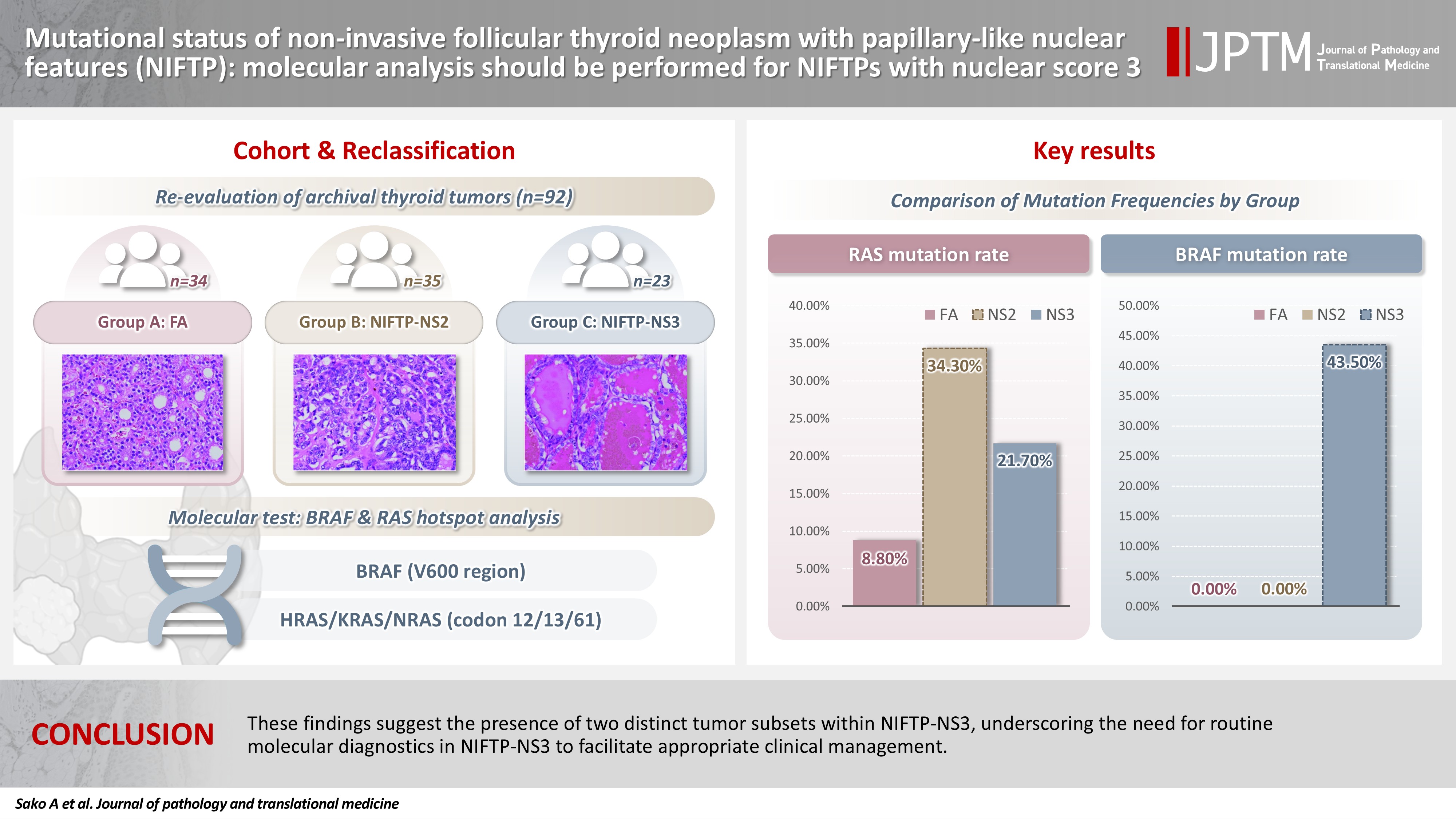
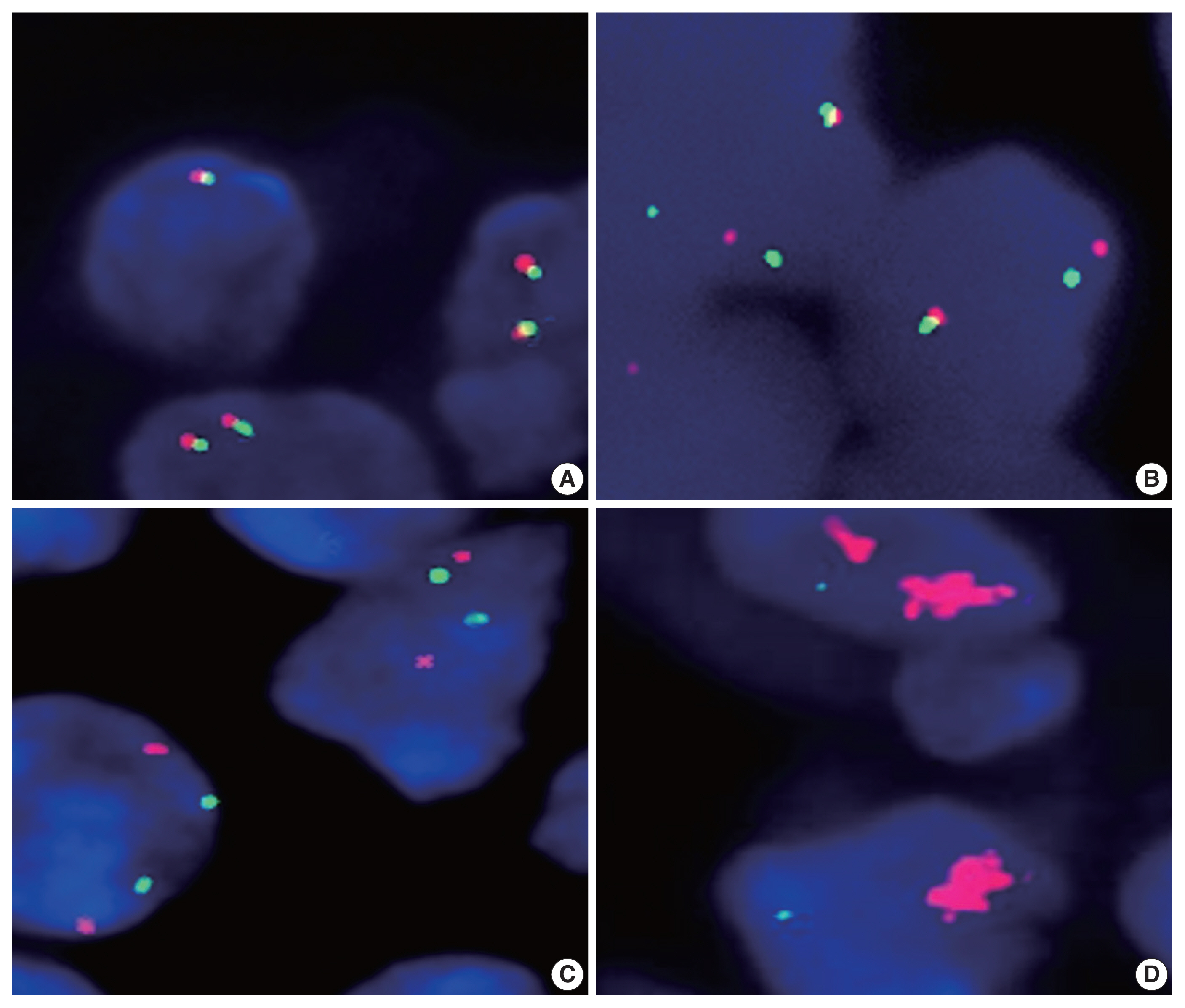
The classification of non-invasive follicular thyroid neoplasm with papillary-like nuclear features (NIFTP) was introduced to prevent the overtreatment of indolent tumors that were formerly diagnosed as non-invasive encapsulated follicular variant papillary thyroid carcinomas (NIEFV-PTCs). Although NIFTP was initially estimated to account for 10%–20% of papillary thyroid carcinomas in Western populations, its incidence is substantially lower in Asian cohorts. However, a multi-institutional Japanese study revealed that 31.0% of tumors previously diagnosed as follicular adenomas (FAs) were reclassified as NIFTPs. NIFTP diagnosis requires a nuclear score (NS) of 2–3, and according to the recent World Health Organization criteria, molecular analysis is recommended, but not mandatory, to exclude high-risk subtypes, namely cases with the BRAFV600E mutation, particularly for NS3 tumors. Methods: We performed genetic analysis on 92 archival thyroid tumor samples, including 69 previously diagnosed as FA, of which 34 remained as FA upon re-evaluation (group A) and 35 were reclassified as NIFTP with NS2 (group B). Additional 23 tumors previously diagnosed as NIEFV-PTC were reclassified as NIFTP with NS3 (group C). Results: RAS mutations were detected in 8.8%, 34.3%, and 21.7% of the tumor samples in groups A, B, and C, respectively, whereas BRAF mutations were present in 43.5% of the tumor samples in group C only. Conclusions: These findings suggest the presence of two distinct tumor subsets within NIFTP-NS3, underscoring the need for routine molecular diagnostics in NIFTP-NS3 to facilitate appropriate clinical management.
- Drug-induced phospholipidosis of the kidney suspected to be caused by atomoxetine
- Sung-Eun Choi, Kee Hyuck Kim, Minsun Jung, Jeong Hae Kie
- J Pathol Transl Med. 2026;60(1):124-128. Published online January 14, 2026
- DOI: https://doi.org/10.4132/jptm.2025.12.10
- 2,453 View
- 175 Download
- 1 Web of Science
- 3 Crossref
-
Abstract
PDF
- Drug-induced phospholipidosis (DIP) is characterized by intracellular accumulation of phospholipids with lamellar body formation secondary to drug-altered lipid metabolism, which can trigger inflammation and histopathological changes. Fabry disease and DIP both exhibit zebra bodies on electron microscopy, complicating differential diagnosis. A 17-year-old male with microscopic hematuria and proteinuria had received atomoxetine (40 mg) for 11 months to treat attention-deficit hyperactivity disorder. Light microscopy showed one glomerulus with perihilar sclerosis and periglomerular fibrosis. Kidney biopsy revealed zebra bodies in podocytes, initially suggesting Fabry disease. However, α-galactosidase A enzyme activity was normal on tandem mass spectrometry. Next-generation sequencing of GLA identified only three benign variants. This represents the first reported case of atomoxetine-induced DIP. When zebra bodies are observed, clinicians should consider DIP caused by cationic amphiphilic drugs alongside Fabry disease. Atomoxetine meets the structural criteria for inducing DIP, and awareness of this potential complication is essential.
-
Citations
Citations to this article as recorded by
- Atomoxetine
Reactions Weekly.2026; 2095(1): 19. CrossRef - Acute Interstitial Nephritis, Acute Tubular Injury, and Drug-Induced Phospholipidosis Associated with Combined KRAS G12C and RAF/MEK Inhibition in Non-Small Cell Lung Cancer
Jose Arriola-Montenegro, Poemlarp Mekraksakit, Sam T. Albadri, Maria L. Gonzalez Suarez
Kidney International Case Reports.2026; 1(1): 100018. CrossRef - Automated detection of mulberry bodies in urinary sediment for non-invasive Fabry disease screening
Hiroshi Yamanaka, Tetsumin So, Naoko Sakamoto, Saki Aoto, Xiao-Kang Li, Yi Wang, Qian Shen, Ohsuke Migita, Motomichi Kosuga, Kohji Okamura
Clinical Chemistry and Laboratory Medicine (CCLM).2026;[Epub] CrossRef
- Atomoxetine
- Association study of TYMS gene expression with TYMS and ENOSF1 genetic variants in neoadjuvant chemotherapy response of gastric cancer
- Khadijeh Arjmandi, Iman Salahshourifar, Shiva Irani, Fereshteh Ameli, Mohsen Esfandbod
- J Pathol Transl Med. 2025;59(2):105-114. Published online February 25, 2025
- DOI: https://doi.org/10.4132/jptm.2024.11.05
- 4,307 View
- 147 Download
- 3 Web of Science
- 3 Crossref
-
Abstract
PDF
- Background
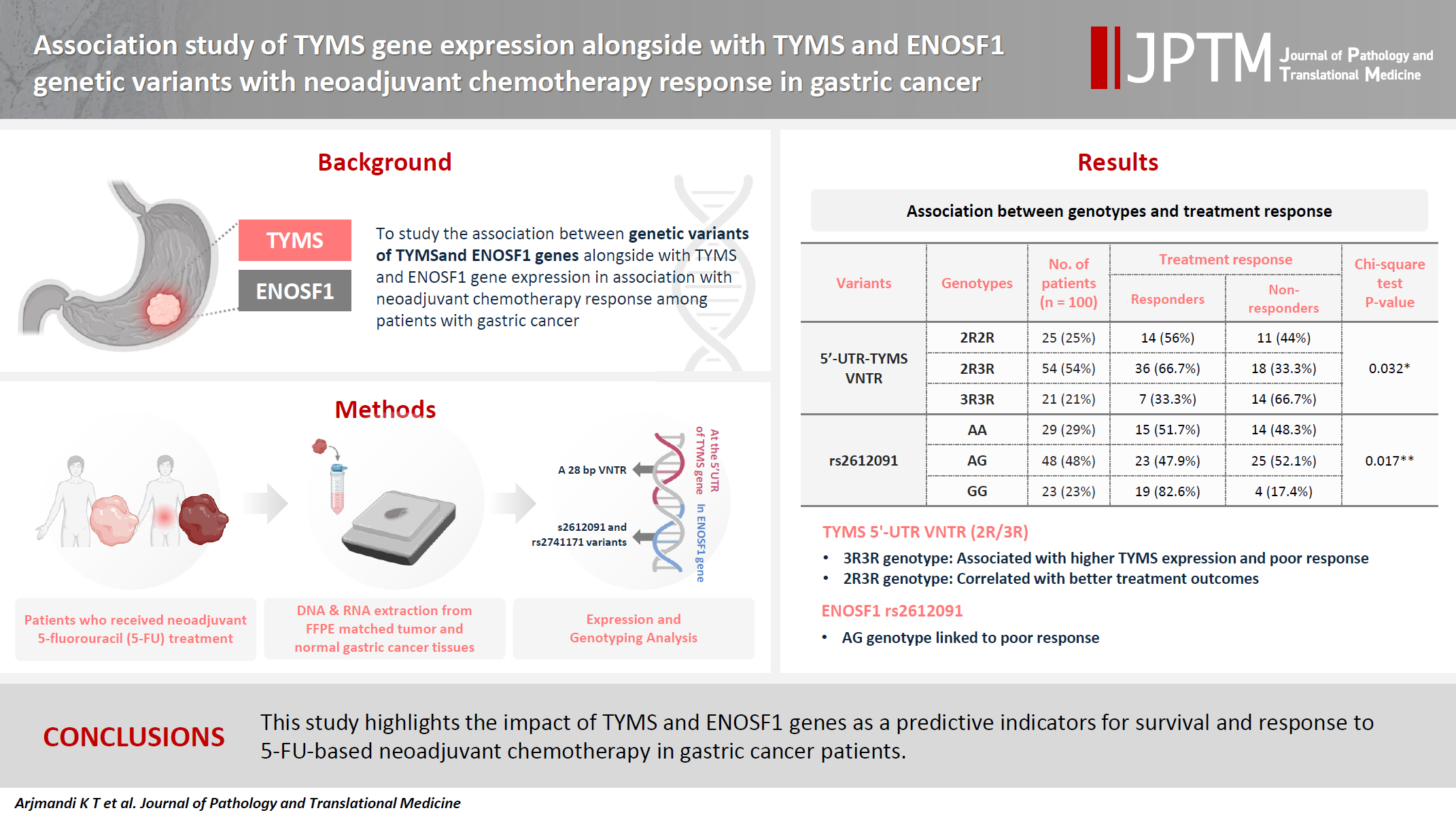
The present research was designed to study the associations between genetic variants of TYMS and ENOSF1 genes with TYMS and ENOSF1 gene expression in neoadjuvant chemotherapy response among patients with gastric cancer. Methods: Formalin-embedded and paraffin-fixed matched tumor and normal gastric cancer tissue samples from patients who received neoadjuvant 5-fluorouracil (5-FU) treatment were obtained. DNA and RNA were extracted for all samples. A 28-bp variable number tandem repeat (VNTR) at the 5' untranslated region of TYMS gene and rs2612091 and rs2741171 variants in the ENOSF1 gene were genotyped for normal tissue samples. The real-time polymerase chain reaction method was used to study the expression of ENOSF1 and TYMS genes in both normal and tumor tissues. Data were analyzed using REST 2000 and SPSS ver. 26.0 software programs. Results: A significant association between TYMS 2R3R VNTR genotypes and 5-FU therapy was found (p = .032). The 3R3R and 2R2R genotypes were significantly associated with increased and decreased survival time, respectively (p = .003). The 3R3R genotype was significantly associated with TYMS overexpression (p < .001). Moreover, a significant association was found between the rs2612091 genotype and treatment outcome (p = .017). Conclusions: This study highlights the impact of TYMS and ENOSF1 genes as predictive indicators for survival and response to 5-FU–based neoadjuvant chemotherapy in gastric cancer patients. -
Citations
Citations to this article as recorded by- Innovative biomaterial strategies for mitigating radiotherapy toxicity: multidimensional mechanistic interventions of nano-microscale materials and hydrogels
Yifan Liu, Fengdi Jiang, Jie Song, Huaijin Qiao, Junlong Dai, Hao Bai, Shuyu Zhang
Coordination Chemistry Reviews.2026; 549: 217313. CrossRef - Mebendazole impairs the expression and function of enzymes in nucleotide metabolism pathways, leading to Selective Cytotoxicity, Cell Cycle Arrest, and Damage to Cell Morphology in Gastric Cancer
Emerson Lucena da Silva, Felipe Pantoja Mesquita, Pedro Victor da Rocha Lima, José Hélio de Araújo Filho, Francisco Laio de Oliveira, Ana Beatriz da Lima, Pedro Filho Noronha Souza, Raquel Carvalho Montenegro
Chemico-Biological Interactions.2026; 430: 111973. CrossRef - Multi‐Omics Analysis of TYMS as a Prognostic Biomarker and Therapeutic Target for Lung Adenocarcinoma
Hansen Shi, Peijun Zhang, Jiayi Chen, Hua Li, Xiaohuai Zhang, Biyun zeng, Tiancai Liu, Tao Zeng
Cancer Medicine.2026;[Epub] CrossRef
- Innovative biomaterial strategies for mitigating radiotherapy toxicity: multidimensional mechanistic interventions of nano-microscale materials and hydrogels
- Uncommon granulomatous manifestation in Epstein-Barr virus–positive follicular dendritic cell sarcoma: a case report
- Henry Goh Di Shen, Yue Zhang, Wei Qiang Leow
- J Pathol Transl Med. 2025;59(2):133-138. Published online October 31, 2024
- DOI: https://doi.org/10.4132/jptm.2024.09.27
- 5,130 View
- 353 Download
- 6 Web of Science
- 6 Crossref
-
Abstract
PDF
- Hepatic Epstein-Barr virus–positive inflammatory follicular dendritic cell sarcoma (EBV+ IFDCS) represents a rare form of liver malignancy. The absence of distinct clinical and radiological characteristics, compounded by its rare occurrence, contributes to a challenging diagnosis. Here, we report a case of a 54-year-old Chinese female with a background of chronic hepatitis B virus treated with entecavir and complicated by advanced fibrosis presenting with a liver mass found on her annual surveillance ultrasound. Hepatectomy was performed under clinical suspicion of hepatocellular carcinoma. Immunomorphologic characteristics of the tumor were consistent with EBV+ IFDCS with distinct non-caseating granulomatous inflammation. Our case illustrates the importance of considering EBV+ IFDCS in the differential diagnosis of hepatic inflammatory lesions. Awareness of this entity and its characteristic features is essential for accurately diagnosing and managing this rare neoplasm.
-
Citations
Citations to this article as recorded by- Mesenchymal Tumors of the Liver: An Update Review
Joon Hyuk Choi, Swan N. Thung
Biomedicines.2025; 13(2): 479. CrossRef - EBV-positive inflammatory follicular dendritic cell sarcoma occurring in different organs: a case report and literature review
Wenhua Bai, Chunfang Hu, Zheng Zhu
Frontiers in Oncology.2025;[Epub] CrossRef - Spleen EBV-positive inflammatory follicular dendritic cell sarcoma: a case report and literature review
Yi Xiao, Lanlan Li, Xiumei Zhan, Juner Xu, Yewu Chen, Qiuchan Zhao, Yinghao Fu, Xian Luo, Huadi Chen, Hao Xu
Frontiers in Oncology.2025;[Epub] CrossRef - Epstein-Barr virus-positive inflammatory follicular dendritic cell sarcoma of the liver: clinical features, imaging findings and potential diagnostic clues
Gui-Ling Huang, Man-Qian Huang, Yu-Ting Zhang, Hui-Ning Huang, Hong-Tao Liu, Xiao-Qing Pei
Abdominal Radiology.2025;[Epub] CrossRef - Epstein‑Barr virus+ inflammatory follicular dendritic cell sarcoma with clonal immunoglobulin heavy chain gene rearrangement: A case report and literature review
Qian Ye, Juan Zhao, Jiao He, Weishan Zhang
Oncology Letters.2025; 31(2): 1. CrossRef - Primary hepatic follicular dendritic cell sarcoma: A case study and literature review
Junjie Zhu, Ying Liang, Li Zhang, Bingqi Li, Danfeng Zheng, Hangyan Wang
Journal of International Medical Research.2025;[Epub] CrossRef
- Mesenchymal Tumors of the Liver: An Update Review
- Immunohistochemical expression in idiopathic inflammatory myopathies at a single center in Vietnam
- Dat Quoc Ngo, Si Tri Le, Khanh Hoang Phuong Phan, Thao Thi Phuong Doan, Linh Ngoc Khanh Nguyen, Minh Hoang Dang, Thien Thanh Ly, Thu Dang Anh Phan
- J Pathol Transl Med. 2024;58(4):174-181. Published online June 25, 2024
- DOI: https://doi.org/10.4132/jptm.2024.05.02

- 5,172 View
- 271 Download
- 1 Web of Science
- 3 Crossref
-
Abstract
PDF
- Background
The identification of idiopathic inflammatory myopathies (IIMs) requires a comprehensive analysis involving clinical manifestations and histological findings. This study aims to provide insights into the histopathological and immunohistochemical aspects of IIMs.
Methods
This retrospective case series involved 56 patients diagnosed with IIMs at the Department of Pathology, University of Medicine and Pharmacy at Ho Chi Minh City, from 2019 to 2023. The histology and immunohistochemical expression of HLA-ABC, HLA-DR, C5b-9, Mx1/2/3, and p62 were detected.
Results
We examined six categories of inflammatory myopathy, including immunemediated necrotizing myopathy (58.9%), dermatomyositis (DM; 23.2%), overlap myositis (8.9%), antisynthetase syndrome (5.4%), inclusion body myositis (IBM; 1.8%), and polymyositis (1.8%). The average age of the patients was 49.7 ± 16.1 years, with a female-to-male ratio of 3:1. Inflammatory cell infiltration in the endomysium was present in 62.5% of cases, perifascicular atrophy was found in 17.8%, and fiber necrosis was observed in 42 cases (75.0%). Rimmed vacuoles were present in 100% of cases in the IBM group. Immunohistochemistry showed the following positivity rates: HLA-ABC (89.2%), HLA-DR (19.6%), C5b-9 (57.1%), and Mx1/2/3 (10.7%). Mx1/2/3 expression was high in DM cases. p62 vacuole deposits were noted in the IBM case. The combination of membrane attack complex and major histocompatibility complex I helped detect IIMs in 96% of cases.
Conclusions
The diagnosis of IIMs and their subtypes should be based on clinical features and histopathological characteristics. Immunohistochemistry plays a crucial role in the diagnosis and differentiation of these subgroups. -
Citations
Citations to this article as recorded by- Evaluating the Diagnostic Potential of Myxovirus Resistance Protein 1 (MX1) and Myxovirus Resistance Protein 2 (MX2) As Biomarkers in Idiopathic Inflammatory Myopathies
Raghavee Neupane, Mustafa Haider, Perry Smith, Marc M Kesselman
Cureus.2026;[Epub] CrossRef - Rapidly Progressive Polymyositis With Vasculitis: The Pivotal Role of Histopathology in Diagnosis and Management
Amitha Venmanassery Karnalsingh, Arjun Karappilly Vijayan, Monica Roselin Edwin Peter, Dilan Davis
Cureus.2025;[Epub] CrossRef - Autoimmune Neuromuscular Disorders at a Molecular Crossroad: Linking Pathogenesis to Targeted Immunotherapy
Anca-Maria Florea, Dimela-Gabriela Luca, Eugenia Irene Davidescu, Bogdan-Ovidiu Popescu
International Journal of Molecular Sciences.2025; 26(23): 11736. CrossRef
- Evaluating the Diagnostic Potential of Myxovirus Resistance Protein 1 (MX1) and Myxovirus Resistance Protein 2 (MX2) As Biomarkers in Idiopathic Inflammatory Myopathies
- Thyroid pathology, a clue to PTEN hamartoma tumor syndrome
- Yurimi Lee, Young Lyun Oh
- J Pathol Transl Med. 2023;57(3):178-183. Published online March 30, 2023
- DOI: https://doi.org/10.4132/jptm.2023.03.04
- 8,263 View
- 212 Download
- 10 Web of Science
- 8 Crossref
-
Abstract
PDF
- Phosphatase and tensin homolog (PTEN) hamartoma tumor syndrome (PHTS) is a hereditary disorder caused by germline inactivating mutations in the PTEN tumor suppressor gene. As a type of PHTS, Cowden syndrome is associated with abnormalities of the thyroid, breast, uterus, and gastrointestinal tract. A 52-year-old-woman visited the outpatient clinic of our endocrinology clinic with multiple thyroid nodules and Hashimoto's thyroiditis. Computed tomography imaging revealed a multinodular mass measuring up to 3.5 cm in the left thyroid lobe, causing laryngotracheal airway displacement. The total thyroidectomy specimen revealed multiple follicular adenomas and adenomatous nodules with lymphocytic thyroiditis and lipomatous metaplasia in the background. The patient was suspected of PTHS based on her thyroid pathology, family history, and numerous hamartomatous lesions of the breast, uterus, and skin. Her diagnosis was confirmed through molecular testing. This case demonstrates that pathologists must be well acquainted with thyroid pathology in PHTS.
-
Citations
Citations to this article as recorded by- Risk of malignancy in PTEN-altered thyroid nodules detected on preoperative FNA molecular testing: a systematic review and meta-analysis
Patrizia Straccia, Vincenzo Fiorentino, Belen Padial Urtueta, Qianqian Zhang, Alessia Piermattei, Federica Cianfrini, Antonino Mule, Esther Diana Rossi
Human Pathology.2026; : 106104. CrossRef - Dual primary malignancies in Kashmir: A five-year analysis of temporal patterns, gender-specific presentations and treatment outcomes in a high gastrointestinal cancer risk population
Ubaid Jeelani, Mushood Ghulam Nabi, Asim Ahmad Dar, Gowher Ahmad Wagai, Aadil Najeed, Sheikh Owais Ahmad, Lande Sagar Janardhan, Md Mayeen Afsan Ahmad, Uzma Majeed
Journal of Family Medicine and Primary Care.2026; 15(2): 530. CrossRef - A clinical case of papillary thyroid cancer associated with a PTEN gene defect
R. A. Atanesyan, L. Ja. Klimov, T. M. Vdovina, G. A. Saneeva, E. I. Andreeva, I. A. Stremenkova, R. I. Arakelyan, I. K. Gasparyan
Rossiyskiy Vestnik Perinatologii i Pediatrii (Russian Bulletin of Perinatology and Pediatrics).2025; 69(6): 85. CrossRef - Pediatric cancer predisposition syndromes involving non-central nervous system solid pediatric tumors: a review on their manifestations with a focus on histopathology
B. Schurink, M. Reyes-Múgica, R. R. de Krijger
Virchows Archiv.2025; 486(1): 3. CrossRef - Dedifferentiated Leiomyosarcoma of the Uterine Corpus with Heterologous Component: Clinicopathological Analysis of Five Consecutive Cases from a Single Institution and Comprehensive Literature Review
Suyeon Kim, Hyunsik Bae, Hyun-Soo Kim
Diagnostics.2024; 14(2): 160. CrossRef - Case report: Rare oral manifestations in Cowden syndrome with PTEN mutation
Wei Yuan, Yanbin Liu, Haibin Sun, Ming Su, Lizheng Qin, Xin Huang
Frontiers in Oncology.2024;[Epub] CrossRef - Can thyroid histomorphology identify patients with PTEN hamartoma tumour syndrome?
Melad N Dababneh, Laura Rabinowitz, Gilman Plitt, Charis Eng, Christopher C Griffith
Histopathology.2024; 85(6): 929. CrossRef - A novel mutation in PTEN in anaplastic thyroid carcinoma: A case report
Yanli Zhao
Biomedical Reports.2024;[Epub] CrossRef
- Risk of malignancy in PTEN-altered thyroid nodules detected on preoperative FNA molecular testing: a systematic review and meta-analysis
- Solitary Peutz-Jeghers type harmartomatous polyp in duodenum with gastric foveolar epithelium: a case report
- Eugene Choi, Junghwan Lee, Youngsoo Park
- J Pathol Transl Med. 2023;57(2):128-131. Published online January 10, 2023
- DOI: https://doi.org/10.4132/jptm.2022.11.07
- 5,975 View
- 197 Download
- 1 Web of Science
- 1 Crossref
-
Abstract
PDF
- Peutz-Jeghers type hamartomatous polyp is known to be associated with Peutz-Jeghers syndrome, which shows characteristic multiple hamartomatous polyp involvement in the gastrointestinal tract, combined with mucocutaneous symptom, familial history of Peutz- Jeghers syndrome or STK11/LTB1 mutation. However, some cases showing histologic appearance of the polyps discovered in Peutz- Jeghers syndrome while lacking other diagnostic criteria of the syndrome have been reported, and these are called solitary Peutz- Jeghers type polyps. Herein, we report a case of solitary Peutz-Jeghers type polyp covered with heterotopic epithelium. The patient was 47-year-old female without any mucocutaneous symptoms nor familial history of Peutz-Jeghers syndrome. Microscopic examination revealed Peutz-Jeghers type hamartomatous polyp in duodenum covered with gastric type foveolar epithelium. Considering the definition of hamartomatous polyp, which is, the abnormal overgrowth of the indigenous epithelial component, the histological feature of current case is noteworthy in a point that it shows proliferation of heterotopic component, rather than the indigenous component.
-
Citations
Citations to this article as recorded by- A Solitary Peutz-Jeghers Hamartomatous Polyp in the Gastric Body: A Case Report
Noelia Madera, Noemí Acevedo, Carmen González-Peralta, Rafael Castro, Vismelis Mezquita-Luna
Cureus.2024;[Epub] CrossRef
- A Solitary Peutz-Jeghers Hamartomatous Polyp in the Gastric Body: A Case Report
- Blocking Toll-like receptor 9 attenuates bleomycin-induced pulmonary injury
- Badr Alzahrani, Mohamed M. S. Gaballa, Ahmed A. Tantawy, Maha A. Moussa, Salma A. Shoulah, Said M. Elshafae
- J Pathol Transl Med. 2022;56(2):81-91. Published online March 2, 2022
- DOI: https://doi.org/10.4132/jptm.2021.12.27

- 8,563 View
- 150 Download
- 12 Web of Science
- 12 Crossref
-
Abstract
PDF
- Background
Acute respiratory distress syndrome (ARDS) is one of the most common complications in coronavirus disease 2019 patients suffering from acute lung injury (ALI). In ARDS, marked distortion of pulmonary architecture has been reported. The pulmonary lesions in ARDS include hemodynamic derangements (such as alveolar edema and hemorrhage), vascular and bronchiolar damage, interstitial inflammatory cellular aggregations, and eventually fibrosis. Bleomycin induces ARDS-representative pulmonary damage in mice and rats; therefore, we used bleomycin model mice in our study. Recently, Toll-like receptor 9 (TLR9) was implicated in the development of ARDS and ALI.
Methods
In this study, we evaluated the efficiency of a TLR9 blocker (ODN2088) on bleomycin-induced pulmonary damage. We measured the apoptosis rate, inflammatory reaction, and fibroplasia in bleomycin- and bleomycin + ODN2088-treated mice.
Results
Our results showed a significant amelioration in bleomycin-induced damage to pulmonary architecture following ODN2088 treatment. A marked decrease in pulmonary epithelial and endothelial apoptosis rate as measured by cleaved caspase-3 expression, inflammatory reaction as indicated by tumor necrosis factor α expression, and pulmonary fibrosis as demonstrated by Van Gieson staining and α-smooth muscle actin immunohistochemistry were observed following ODN2088 treatment.
Conclusions
All these findings indicate that blocking downstream TLR9 signaling could be beneficial in prevention or mitigation of ARDS through hemodynamic derangements, inflammation, apoptosis, and fibrosis. -
Citations
Citations to this article as recorded by- Nano-sized DNase scavenges cell-free DNA for acute lung injury treatment
Ruijie Chen, Yitianhe Xu, Zhanzheng Ye, Yixuan Zhu, Mengxue Zhang, Fangfang Lv, Yunzhi Wang, Xinyu Di, Yinhao Lin, Shengnan Song, Zihao Huang, Shize Li, Zhinan He, Hailin Zhang, Longfa Kou
Journal of Controlled Release.2026; 394: 114846. CrossRef - A novel mouse model of myositis-associated interstitial lung disease was established by using TLR9 agonist combined with muscle homogenate
Ling Bai, Jiarui Zhu, Wenlan Ma, Peipei Zhao, Feifei Li, Cen Zhang, Sigong Zhang
Clinical and Experimental Immunology.2025;[Epub] CrossRef - Toll-like Receptor 9 Inhibition Mitigates Fibroproliferative Responses in Translational Models of Pulmonary Fibrosis
Glenda Trujillo, Alicia Regueiro-Ren, Chunjian Liu, Buqu Hu, Ying Sun, Farida Ahangari, Vitoria Fiorini, Genta Ishikawa, Karam Al Jumaily, Johad Khoury, John McGovern, Chris J. Lee, Xue Yan Peng, Taylor Pivarnik, Huanxing Sun, Anjali Walia, Samuel Woo, Sh
American Journal of Respiratory and Critical Care Medicine.2025; 211(1): 91. CrossRef - CD103+ dendritic cell–fibroblast crosstalk via TLR9, TDO2, and AHR signaling drives lung fibrogenesis
Hannah Carter, Rita Medina Costa, Taylor S. Adams, Talon M. Gilchrist, Claire E. Emch, Monica Bame, Justin M. Oldham, Steven K. Huang, Angela L. Linderholm, Imre Noth, Naftali Kaminski, Bethany B. Moore, Stephen J. Gurczynski
JCI Insight.2025;[Epub] CrossRef - The Mitigated Effect of the Combination of Metformin and Stearic Acid to Ameliorate Bleomycin‐Induced Pulmonary Fibrosis in Rats via Inhibiting Gal‐3/Smad3/α‐SMA and TNF‐α/NF‐κβ Signaling Pathways
Maha M. Salem, Nermin S. Youssef, Mai El Keiy, Abeer A. Khamis
Journal of Biochemical and Molecular Toxicology.2025;[Epub] CrossRef - Unraveling the Interactive Role of Mitochondrial Dysfunction in Promoting Macrophage Polarization in Pulmonary Fibrosis
Jiajia Zou, Junling Jian, Dehua Ge, Bin Zhou, Jiaxiang Zhang
Journal of Biochemical and Molecular Toxicology.2025;[Epub] CrossRef - Mechanisms underlying dose-limiting toxicities of conventional chemotherapeutic agents
Mohammad Amin Manavi, Mohammad Hosein Fathian Nasab, Razieh Mohammad Jafari, Ahmad Reza Dehpour
Journal of Chemotherapy.2024; 36(8): 623. CrossRef - Innate Immune Response-Mediated Inflammation in Viral Pneumonia
Weiwei Ni, Xin Wei, Rui Wu
Journal of Pediatric Infectious Diseases.2024; 19(03): 140. CrossRef - Combination of losartan with pirfenidone: a protective anti-fibrotic against pulmonary fibrosis induced by bleomycin in rats
Arian Amirkhosravi, Maryamossadat Mirtajaddini Goki, Mahmoud Reza Heidari, Somayyeh Karami-Mohajeri, Maryam Iranpour, Maryam Torshabi, Mitra Mehrabani, Ali Mandegary, Mehrnaz Mehrabani
Scientific Reports.2024;[Epub] CrossRef - Suppression of miR-17 Alleviates Acute Respiratory Distress-associated Lung Fibrosis by Regulating Mfn2
Mei-xia Xu, Tao Xu, Ning An
Current Medical Science.2024; 44(5): 964. CrossRef - Study of Recombinant Interleukin-1 Receptor Antagonist Compositions Biological Activity After Injection and Inhalation in Mouse Model of Pulmonary Inflammation
Alexander M. Ischenko, Ksenia A. Nekrasova, Denis S. Laptev, Dmitry V. Bobkov, Alexander A. Kolobov, Andrey S. Simbirtsev
Cytokines and inflammation.2024; 21(3): 153. CrossRef - TLR9: A friend or a foe
Mona M. Saber, Nada Monir, Azza S. Awad, Marwa E. Elsherbiny, Hala F. Zaki
Life Sciences.2022; 307: 120874. CrossRef
- Nano-sized DNase scavenges cell-free DNA for acute lung injury treatment
- Adrenal hemangioblastoma
- Joo-Yeon Koo, Kyung-Hwa Lee, Joon Hyuk Choi, Ho Seok Chung, Chan Choi
- J Pathol Transl Med. 2022;56(3):161-166. Published online February 28, 2022
- DOI: https://doi.org/10.4132/jptm.2021.12.28
- 6,182 View
- 159 Download
- 1 Web of Science
- 1 Crossref
-
Abstract
PDF
- Hemangioblastoma (HB) is a rare benign tumor that most commonly occurs in the cerebellum. HB is composed of neoplastic stromal cells and abundant small vessels. However, the exact origin of stromal cells is controversial. Extraneural HBs have been reported in a small series, and peripheral HBs arising in the adrenal gland are extremely rare. Herein, we report a case of sporadic adrenal HB in a 54-year-old woman. The tumor was a well-circumscribed, yellow mass measuring 4.2 cm in diameter. Histologically, the tumor was composed of small blood vessels and vacuolated stromal cells with clear cytoplasm. On immunohistochemical stain, the stromal cells were positive for S-100 protein, neuron-specific enolase, and synaptophysin. The tumor did not reveal mutation of VHL alleles. We herein present a case of HB of the adrenal gland and review of the literature.
-
Citations
Citations to this article as recorded by- Familial Von Hippel–Lindau Disease: A Case Series of Cerebral Hemangioblastomas with MRI, Histopathological, and Genetic Correlations
Claudiu Matei, Ioana Boeras, Dan Orga Dumitriu, Cosmin Mutu, Adriana Popescu, Mihai Gabriel Cucu, Alexandru Calotă-Dobrescu, Bogdan Fetica, Diter Atasie
Life.2025; 15(11): 1649. CrossRef
- Familial Von Hippel–Lindau Disease: A Case Series of Cerebral Hemangioblastomas with MRI, Histopathological, and Genetic Correlations
- Juxtacortical chondromyxoid fibroma in the small bones: two cases with unusual location and a literature review
- Sun-Ju Oh, So Hak Chung
- J Pathol Transl Med. 2022;56(3):157-160. Published online January 21, 2022
- DOI: https://doi.org/10.4132/jptm.2021.12.15
- 6,748 View
- 198 Download
- 3 Web of Science
- 1 Crossref
-
Abstract
PDF
- Chondromyxoid fibroma is a rare bone tumor of cartilaginous origin, representing less than 1% of all bone tumors. It preferentially arises in the eccentric location of the metaphysis of a long tubular bone. Juxtacortical locations are reported infrequently in the long bones and even more rarely in short tubular bones, with only three cases documented. Here we present two new cases of juxtacortical chondromyxoid fibroma in the small bones. One was an intracortical osteolytic lesion of the metatarsal bone of the foot with degenerative atypia that histologically should be differentiated from chondrosarcoma. The other was a phalangeal mass protruding into the interphalangeal joint of the hand, which had been labeled mistakenly as a soft tissue mass preoperatively. These cases illustrated that chondromyxoid fibromas have various the manifestations and should be included in the differential diagnosis of an osteolytic lesion or an exophytic mass in the small bones.
-
Citations
Citations to this article as recorded by- Cartilage Forming Tumors of the Skeleton
Julio A. Diaz-Perez, Andrew E. Rosenberg
Advances in Anatomic Pathology.2025; 32(2): 132. CrossRef
- Cartilage Forming Tumors of the Skeleton
- Clinicopathologic implication of PD-L1 gene alteration in primary adrenal diffuse large B cell lymphoma
- Ki Rim Lee, Jiwon Koh, Yoon Kyung Jeon, Hyun Jung Kwon, Jeong-Ok Lee, Jin Ho Paik
- J Pathol Transl Med. 2022;56(1):32-39. Published online November 16, 2021
- DOI: https://doi.org/10.4132/jptm.2021.10.05
- 6,077 View
- 170 Download
- 2 Web of Science
- 1 Crossref
-
Abstract
PDF
- Background
Primary adrenal (PA) diffuse large B cell lymphoma (DLBCL) was previously reported as an aggressive subset of DLBCL, but its genetic features were not sufficiently characterized. From our previous study of DLBCL with programmed death-ligand 1 (PD-L1) gene alterations, we focused on PD-L1 gene alterations in PA-DLBCL with clinicopathologic implications.
Methods
We performed fluorescence in situ hybridization for PD-L1 gene translocation and amplification in PA-DLBCL (n = 18) and comparatively analyzed clinicopathologic characteristics with systemic non-adrenal (NA)-DLBCL (n = 90).
Results
PA-DLBCL harbored distinctive features (vs. NADLBCL), including high international prognostic index score (3–5) (72% [13/18] vs. 38% [34/90], p = .007), poor Eastern Cooperative Oncology Group performance score (≥ 2) (47% [7/15] vs. 11% [10/90], p = .003), elevated serum lactate dehydrogenase (LDH) (78% [14/18] vs. 51% [44/87], p = .035) and MUM1 expression (87% [13/15] vs. 60% [54/90], p = .047). Moreover, PA-DLBCL showed frequent PD-L1 gene alterations (vs. NA-DLBCL) (39% [7/18] vs. 6% [5/86], p = .001), including translocation (22% [4/18] vs. 3% [3/87], p = .016) and amplification (17% [3/18] vs. 2% [2/87], p = .034). Within the PA-DLBCL group, PD-L1 gene–altered cases (vs. non-altered cases) tended to have B symptoms (p = .145) and elevated LDH (p = .119) but less frequent bulky disease (≥ 10 cm) (p = .119). In the survival analysis, PA-DLBCL had a poor prognosis for overall survival (OS) and progression-free survival (PFS) (vs. NA-DLBCL; p = .014 and p = .004). Within the PA-DLBCL group, PD-L1 translocation was associated with shorter OS and PFS (p < .001 and p = .012).
Conclusions
PA-DLBCL is a clinically aggressive and distinct subset of DLBCL with frequent PD-L1 gene alterations. PD-L1 gene translocation was associated with poor prognosis in PA-DLBCL. -
Citations
Citations to this article as recorded by- Case Report: Diagnostic value of spectral CT in primary adrenal lymphoma
Xiang Zhuang, Xi xi Jin, Li wen Feng, Hui Zhang
Frontiers in Oncology.2026;[Epub] CrossRef
- Case Report: Diagnostic value of spectral CT in primary adrenal lymphoma
- Fibrocartilaginous mesenchymoma with an unusual location in the rib
- Sun-Ju Oh
- J Pathol Transl Med. 2021;55(1):75-78. Published online December 3, 2020
- DOI: https://doi.org/10.4132/jptm.2020.10.08
- 6,643 View
- 121 Download
- 2 Web of Science
- 3 Crossref
-
Abstract
PDF
- Fibrocartilaginous mesenchymoma is a rare bone tumor, with fewer than 35 cases reported in the literature since 1984. This tumor usually occurs in the long bones of children and adolescents. In the current case, the tumor affected a rib. A 17-year-old boy presented with a mass in the right fifth rib. Radiologic findings revealed an osteolytic mass with cortical destruction and calcification; en bloc resection was performed. The tumor showed three distinct histologic features: bland spindle cell proliferation, benign cartilage nodules, and epiphyseal plate-like enchondral ossification. The pathologic diagnosis was fibrocartilaginous mesenchymoma. The patient remains free of disease 1 year after the surgery. Pathological diagnosis of fibrocartilaginous mesenchymoma can be challenging, especially when the tumor occurs in an unusual site. When any fibro-osseous lesion with a cartilaginous component is encountered, the possibility of fibrocartilaginous mesenchymoma should be considered because of its locally aggressive behavior.
-
Citations
Citations to this article as recorded by- Fibrocartilaginous mesenchymoma: a case report and a literature review
A. A. Karyagina, V. Yu. Roshchin, I. V. Sidorov, D. M. Konovalov
Pediatric Hematology/Oncology and Immunopathology.2025; 23(3): 158. CrossRef - Fibrocartilaginous mesenchymoma of the rib with atypical imaging features
Rashed Al-Khudairi, Danielle Forster, Sofina Begum, Alexandra Rice, Adrienne M Flanagan, Fernanda Amary, Paul O’Donnell
BJR|Case Reports.2025;[Epub] CrossRef - Fibrocartilaginous mesenchymoma of pelvis—a potential diagnostic pitfall
Monalisa Hui, Shantveer G. Uppin, Ramakrishna Narayanan, K. Nageshwara Rao, B. Aravind Kumar
Skeletal Radiology.2023; 52(4): 791. CrossRef
- Fibrocartilaginous mesenchymoma: a case report and a literature review
- Pediatric granular cell tumor in the posterior wall of the larynx extending to the trachea
- Jungsuk Ahn, Na Rae Kim, Yong Han Sun
- J Pathol Transl Med. 2020;54(4):336-339. Published online April 15, 2020
- DOI: https://doi.org/10.4132/jptm.2020.02.28
- 6,858 View
- 125 Download
- 1 Web of Science
- 1 Crossref
-
Abstract
PDF
- Granular cell tumor (GCT) is a slow-growing benign neoplasm that can be found in any organ. Pediatric laryngotracheal GCT is rare. We experienced a 6-year-old boy suffering from a barking cough and symptoms of stridor and croup for one month. Head and neck computed tomography revealed a protruding mass that occluded 60% of the airway lumen. Under the impression of hemangioma or papilloma, excision revealed a submucosal non-encapsulated mass. Histologically, the mass was composed of sheets of large polyhedralshaped tumor cells containing plump eosinophilic granular cytoplasm and centrally placed, small, bland-appearing nuclei. The tumor cells were positive for S-100 protein, and voluminous eosinophilic cytoplasm was stained by diastase-resistant periodic acid-Schiff. The present report describes a unique case of a huge pediatric laryngeal GCT extending to the subglottic trachea. We also review the clinical course of pediatric laryngotracheal GCT and emphasize the importance of diagnosing GCT in children.
-
Citations
Citations to this article as recorded by- Pediatric granular cell tumor of the larynx: A case report and literature review
Jing Ke, Junwei Xiong, Juhong Zhang, Haiyu Ma, Wei Yuan
Journal of Cancer Research and Therapeutics.2023; 19(4): 1070. CrossRef
- Pediatric granular cell tumor of the larynx: A case report and literature review
- Comparison of Squamous Cell Carcinoma of the Tongue between Young and Old Patients
- Gyuheon Choi, Joon Seon Song, Seung-Ho Choi, Soon Yuhl Nam, Sang Yoon Kim, Jong-Lyel Roh, Bu-Kyu Lee, Kyung-Ja Cho
- J Pathol Transl Med. 2019;53(6):369-377. Published online October 11, 2019
- DOI: https://doi.org/10.4132/jptm.2019.09.16

- 9,032 View
- 193 Download
- 9 Web of Science
- 13 Crossref
-
Abstract
PDF
 Supplementary Material
 - Background
The worldwide incidence of squamous cell carcinoma of the tongue (SCCOT) in young patients has been increasing. We investigated clinicopathologic features of this unique population and compared them with those of SCCOT in the elderly to delineate its pathogenesis.
Methods
We compared clinicopathological parameters between patients under and over 45 years old. Immunohistochemical assays of estrogen receptor, progesterone receptor, androgen receptor, p53, p16, mdm2, cyclin D1, and glutathione S-transferase P1 were also compared between them.
Results
Among 189 cases, 51 patients (27.0%) were under 45 years of age. A higher proportion of women was seen in the young group, but was not statistically significant. Smoking and drinking behaviors between age groups were similar. Histopathological and immunohistochemical analysis showed no significant difference by age and sex other than higher histologic grades observed in young patients.
Conclusions
SCCOT in young adults has similar clinicopathological features to that in the elderly, suggesting that both progress via similar pathogenetic pathways. -
Citations
Citations to this article as recorded by- Epidemiological trends and characteristics of oral tongue cancer in females: systematic review and meta-analysis
J. Hu, N. Kaunein, A. DeAngelis
International Journal of Oral and Maxillofacial Surgery.2026; 55(3): 267. CrossRef - Oral Tongue Squamous Cell Carcinoma in Young Adults in Brazil: Temporal Trends From 2013 to 2023
Natália Santos Barcelos, Yohana Cordeiro de Miranda Magno, Juliana Maria Braga Sclauser, Renata de Castro Martins, Rodnei Alves Marques, Maria Cássia Ferreira de Aguiar, Patrícia Carlos Caldeira
Oral Diseases.2026;[Epub] CrossRef - The Tongue Tells a Tale: Clinicopathological Case Study of Squamous Cell Carcinoma
Pankaj Kshirsagar, Manu S. Babu
Medical Journal of Dr. D.Y. Patil Vidyapeeth.2026; 19(2): 158. CrossRef - Oral Cavity Squamous Cell Carcinoma in Young Patients: A Multi-Institutional Study of the Canadian Head & Neck Collaborative Research Initiative
Xinyuan Hong, Alexandra E. Quimby, Dorsa Mavedatnia, A. Travis Pickett, Martin Corsten, Tinghua Zhang, Angelina Tohme, Stephanie Johnson-Obaseki, Carlos Khalil, Mark Khoury, Antoine Eskander, Hesameddin Noroozi, David Goldstein, John De Almeida, James Fow
Journal of Otolaryngology - Head & Neck Surgery.2025;[Epub] CrossRef - High Failure Rates in Young Nonsmoker Nondrinkers With Squamous Cell Carcinoma of the Oral Tongue
Brianna M. Jones, Dillan F. Villavisanis, Eric J. Lehrer, Daniel R. Dickstein, Kunal K. Sindhu, Krzysztof J. Misiukiewicz, Marshall Posner, Jerry T. Liu, Vishal Gupta, Sonam Sharma, Scott A. Roof, Marita Teng, Eric M. Genden, Richard L. Bakst
The Laryngoscope.2023; 133(5): 1110. CrossRef - Characteristics of oral squamous cell carcinoma focusing on cases unaffected by smoking and drinking: A multicenter retrospective study
Hiroyuki Harada, Masahiro Kikuchi, Ryo Asato, Kiyomi Hamaguchi, Hisanobu Tamaki, Masanobu Mizuta, Ryusuke Hori, Tsuyoshi Kojima, Keigo Honda, Takashi Tsujimura, Yohei Kumabe, Kazuyuki Ichimaru, Yoshiharu Kitani, Koji Ushiro, Morimasa Kitamura, Shogo Shino
Head & Neck.2023; 45(7): 1812. CrossRef - Genetic characteristics of advanced oral tongue squamous cell carcinoma in young patients
Sehui Kim, Chung Lee, Hyangmi Kim, Sun Och Yoon
Oral Oncology.2023; 144: 106466. CrossRef - Oral Squamous Cell Carcinoma Frequency in Young Patients from Referral Centers Around the World
Rafael Ferreira e Costa, Marina Luiza Baião Leão, Maria Sissa Pereira Sant’Ana, Ricardo Alves Mesquita, Ricardo Santiago Gomez, Alan Roger Santos-Silva, Syed Ali Khurram, Artysha Tailor, Ciska-Mari Schouwstra, Liam Robinson, Willie F. P. van Heerden, Rami
Head and Neck Pathology.2022; 16(3): 755. CrossRef - Early-onset oral cancer as a clinical entity: aetiology and pathogenesis
E.S. Kolegova, M.R. Patysheva, I.V. Larionova, I.K. Fedorova, D.E. Kulbakin, E.L. Choinzonov, E.V. Denisov
International Journal of Oral and Maxillofacial Surgery.2022; 51(12): 1497. CrossRef - The effect of age on the clinicopathological features of oral squamous cell carcinoma
Alaa S Saeed, Bashar H Abdullah
Journal of Baghdad College of Dentistry.2022; 34(1): 25. CrossRef - Survival Outcomes in Oral Tongue Cancer: A Mono-Institutional Experience Focusing on Age
Mohssen Ansarin, Rita De Berardinis, Federica Corso, Gioacchino Giugliano, Roberto Bruschini, Luigi De Benedetto, Stefano Zorzi, Fausto Maffini, Fabio Sovardi, Carolina Pigni, Donatella Scaglione, Daniela Alterio, Maria Cossu Rocca, Susanna Chiocca, Sara
Frontiers in Oncology.2021;[Epub] CrossRef - A Meta-analysis of Oral Squamous Cell Carcinoma in Young Adults with a Comparison to the Older Group Patients (2014–2019)
Khadijah Mohideen, C. Krithika, Nadeem Jeddy, Thayumanavan Balakrishnan, R. Bharathi, S. Leena Sankari
Contemporary Clinical Dentistry.2021; 12(3): 213. CrossRef - Modern perspectives of oral squamous cell carcinoma
A.A. Ivina
Arkhiv patologii.2020; 82(3): 55. CrossRef
- Epidemiological trends and characteristics of oral tongue cancer in females: systematic review and meta-analysis
- Reclassification of Mongolian Diffuse Gliomas According to the Revised 2016 World Health Organization Central Nervous System Tumor Classification
- Enkhee Ochirjav, Bayarmaa Enkhbat, Tuul Baldandorj, Gheeyoung Choe
- J Pathol Transl Med. 2019;53(5):298-307. Published online August 2, 2019
- DOI: https://doi.org/10.4132/jptm.2019.07.15
- 8,131 View
- 111 Download
- 3 Web of Science
- 1 Crossref
-
Abstract
PDF
- Background
The 2016 World Health Organization (WHO) classification of central nervous system (CNS) tumors has been modified to incorporate the IDH mutation and 1p/19q co-deletion in the diagnosis of diffuse gliomas. In this study, we aimed to evaluate the feasibility and prognostic significance of the revised 2016 WHO classification of CNS tumors in Mongolian patients with diffuse gliomas.
Methods
A total of 124 cases of diffuse gliomas were collected, and tissue microarray blocks were made. IDH1 mutation was tested using immunohistochemistry, and 1p/19q co-deletion status was examined using fluorescence in situ hybridization analysis.
Results
According to the 2016 WHO classification, 124 cases of diffuse brain glioma were reclassified as follows: 10 oligodendroglioma, IDHmut and 1p/19q co-deleted; three anaplastic oligodendroglioma, IDHmut and 1p/19q co-deleted; 35 diffuse astrocytoma, IDHmut, 11 diffuse astrocytoma, IDHwt, not otherwise specified (NOS); 22 anaplastic astrocytoma, IDHmut, eight anaplastic astrocytoma, IDHwt, NOS; and 35 glioblastoma, IDHwt, NOS, respectively. The 2016 WHO classification presented better prognostic value for overall survival in patients with grade II tumors than traditional histological classification. Among patients with grade II tumors, those with oligodendroglioma IDHmut and 1p/19q co-deleted and diffuse astrocytoma IDHmut showed significantly higher survival than those with diffuse astrocytoma IDHwt, NOS (p<.01).
Conclusions
Mongolian diffuse gliomas could be reclassified according to the new 2016 WHO classification. Reclassification revealed substantial changes in diagnosis of both oligodendroglial and astrocytic entities. We have confirmed that the revised 2016 WHO CNS tumor classification has prognostic significance in Mongolian patients with diffuse gliomas, especially those with grade II tumors. -
Citations
Citations to this article as recorded by- Targeted next‐generation sequencing of adult gliomas for retrospective prognostic evaluation and up‐front diagnostics
J. K. Petersen, H. B. Boldt, M. D. Sørensen, S. Blach, R. H. Dahlrot, S. Hansen, M. Burton, M. Thomassen, T. Kruse, F. R. Poulsen, L. Andreasen, H. Hager, B. P. Ulhøi, S. Lukacova, G. Reifenberger, B. W. Kristensen
Neuropathology and Applied Neurobiology.2021; 47(1): 108. CrossRef
- Targeted next‐generation sequencing of adult gliomas for retrospective prognostic evaluation and up‐front diagnostics
- Association between Expression of 8-OHdG and Cigarette Smoking in Non-small Cell Lung Cancer
- Ae Ri An, Kyoung Min Kim, Ho Sung Park, Kyu Yun Jang, Woo Sung Moon, Myoung Jae Kang, Yong Chul Lee, Jong Hun Kim, Han Jung Chae, Myoung Ja Chung
- J Pathol Transl Med. 2019;53(4):217-224. Published online March 11, 2019
- DOI: https://doi.org/10.4132/jptm.2019.02.20
- 10,460 View
- 251 Download
- 24 Web of Science
- 24 Crossref
-
Abstract
PDF
- Background
Exposure to cigarette smoking (CS) is a major risk factor for the development of lung cancer. CS is known to cause oxidative DNA damage and mutation of tumor-related genes, and these factors are involved in carcinogenesis. 8-Hydroxydeoxyguanosine (8-OHdG) is considered to be a reliable biomarker for oxidative DNA damage. Increased levels of 8-OHdG are associated with a number of pathological conditions, including cancer. There are no reports on the expression of 8-OHdG by immunohistochemistry in non-small cell lung cancer (NSCLC).
Methods
We investigated the expression of 8-OHdG and p53 in 203 NSCLC tissues using immunohistochemistry and correlated it with clinicopathological features including smoking.
Results
The expression of 8-OHdG was observed in 83.3% of NSCLC. It was significantly correlated with a low T category, negative lymph node status, never-smoker, and longer overall survival (p < .05) by univariate analysis. But multivariate analysis revealed that 8-OHdG was not an independent prognostic factor for overall survival in NSCLC patients. The aberrant expression of p53 significantly correlated with smoking, male, squamous cell carcinoma, and Ki-67 positivity (p < .05).
Conclusions
The expression of 8-OHdG was associated with good prognostic factors. It was positively correlated with never-smokers in NSCLC, suggesting that oxidative damage of DNA cannot be explained by smoking alone and may depend on complex control mechanisms. -
Citations
Citations to this article as recorded by- N-acetyl-cysteine alleviates nandrolone decanoate-induced hippocampal cell apoptosis in rats via reversing protein expressions of S1P1, Akt and FOXO3a signaling pathway
Alireza Shirpoor, Zahra Zarrini, Roya Naderi
Steroids.2026; 228: 109759. CrossRef - The distinct roles of ROS in tumor immunity: from mechanisms to immunotherapeutic applications
Jiayi Li, Chen Huang, Pan Tang, Ruiyan Wu, Quanyou Wu, Chenliang Zhang
Journal of Hematology & Oncology.2026;[Epub] CrossRef - Sustainable framework for automated segmentation and prediction of lung cancer in CT image using CapsNet with U-net segmentation
S.R. Vijayakumar, S. Aarthy, D. Deepa, P. Suresh
Biomedical Signal Processing and Control.2025; 99: 106873. CrossRef - Endolysosomal cation channel MCOLN as the novel regulator of redox homeostasis
Yahao Gao, Lei Xu, Ying Chen
Biochimica et Biophysica Acta (BBA) - Molecular Basis of Disease.2025; 1871(7): 167910. CrossRef - Catalase: The golden key to regulate oxidative stress in breast cancer
Jia-Wei Liu, Wen-Jia Chen, Yang-Zheng Lan, Jing Liu
World Journal of Clinical Oncology.2025;[Epub] CrossRef - Association of sirtuin 1 rs10997868 and rs730821 polymorphisms with sirtuin 1 and hydroxy-2′-deoxyguanosine levels in healthy smokers: A case–control study
Samar Sultan
Journal of International Medical Research.2025;[Epub] CrossRef - Increased pretreatment triglyceride glucose-body mass index associated with poor prognosis in patients with advanced non-small cell lung cancer
Shaoming Guo, Yi Zhao, Yue Jiang, Huaping Ye, Ying Wang
Clinical Nutrition ESPEN.2024; 59: 412. CrossRef - Oxidative Damage and Telomere Length as Markers of Lung Cancer Development among Chronic Obstructive Pulmonary Disease (COPD) Smokers
Elizabeth Córdoba-Lanús, Luis M. Montuenga, Angélica Domínguez-de-Barros, Alexis Oliva, Delia Mayato, Ana Remírez-Sanz, Francisca Gonzalvo, Bartolomé Celli, Javier J. Zulueta, Ciro Casanova
Antioxidants.2024; 13(2): 156. CrossRef - Automated determination of 8-OHdG in cells and tissue via immunofluorescence using a specially created antibody
Tobias Jung, Nicole Findik, Bianca Hartmann, Katja Hanack, Kai Grossmann, Dirk Roggenbuck, Marc Wegmann, René Mantke, Markus Deckert, Tilman Grune
Biotechnology Reports.2024; 42: e00833. CrossRef - Combination treatment of zinc and selenium intervention ameliorated BPA-exposed germ cell damage in SD rats: elucidation of molecular mechanisms
Chittaranjan Sahu, Gopabandhu Jena
Naunyn-Schmiedeberg's Archives of Pharmacology.2024; 397(9): 6685. CrossRef - Interplay of arsenic exposure and cigarette smoking on oxidative DNA damage in healthy males
Sepideh Nemati-Mansour, Mohammad Mosaferi, Javad Babaie, Asghar Mohammadpoorasl, Reza Dehghanzadeh, Leila Nikniaz, Mohammad Miri
Environmental Sciences Europe.2024;[Epub] CrossRef - The role of tissue persistent organic pollutants and genetic polymorphisms in patients with benign and malignant kidney tumors
Rasih Kocagöz, İlgen Onat, Merve Demirbügen Öz, Burak Turna, Banu Sarsık Kumbaracı, Mehmet Nurullah Orman, Halit Sinan Süzen, Hilmi Orhan
Environmental Toxicology and Pharmacology.2024; 110: 104495. CrossRef - Mitochondrial Plasticity and Glucose Metabolic Alterations in Human Cancer under Oxidative Stress—From Viewpoints of Chronic Inflammation and Neutrophil Extracellular Traps (NETs)
Hui-Ting Lee, Chen-Sung Lin, Chao-Yu Liu, Po Chen, Chang-Youh Tsai, Yau-Huei Wei
International Journal of Molecular Sciences.2024; 25(17): 9458. CrossRef - Oxidative DNA Damage and Arterial Hypertension in Light of Current ESC Guidelines
Radka Hazuková, Zdeněk Zadák, Miloslav Pleskot, Petr Zdráhal, Martin Pumprla, Miloš Táborský
International Journal of Molecular Sciences.2024; 25(23): 12557. CrossRef - Significance of 8-OHdG Expression as a Predictor of Survival in Colorectal Cancer
Myunghee Kang, Soyeon Jeong, Sungjin Park, Seungyoon Nam, Jun-Won Chung, Kyoung Oh Kim, Jungsuk An, Jung Ho Kim
Cancers.2023; 15(18): 4613. CrossRef - Serum 8-Hydroxy-2′-deoxyguanosine Predicts Severity and Prognosis of Patients with Acute Exacerbation of Chronic Obstructive Pulmonary Disease
Peng Cao, Chen Zhang, Dong-Xu Hua, Meng-Die Li, Bian-Bian Lv, Lin Fu, Hui Zhao
Lung.2022; 200(1): 31. CrossRef - Redox signaling at the crossroads of human health and disease
Jing Zuo, Zhe Zhang, Maochao Luo, Li Zhou, Edouard C. Nice, Wei Zhang, Chuang Wang, Canhua Huang
MedComm.2022;[Epub] CrossRef - Assessment of MDA and 8-OHdG expressions in ovine pulmonary adenocarcinomas by immunohistochemical and immunofluorescence methods
Emin Karakurt, Enver Beytut, Serpil Dağ, Hilmi Nuhoğlu, Ayfer Yıldız, Emre Kurtbaş
Acta Veterinaria Brno.2022; 91(3): 235. CrossRef - Dietary Antioxidants and Lung Cancer Risk in Smokers and Non-Smokers
Naser A. Alsharairi
Healthcare.2022; 10(12): 2501. CrossRef - Targeting oxidative stress in disease: promise and limitations of antioxidant therapy
Henry Jay Forman, Hongqiao Zhang
Nature Reviews Drug Discovery.2021; 20(9): 689. CrossRef - Association between tobacco substance usage and a missense mutation in the tumor suppressor gene P53 in the Saudi Arabian population
Mikhlid H. Almutairi, Bader O. Almutairi, Turki M. Alrubie, Sultan N. Alharbi, Narasimha R. Parine, Abdulwahed F. Alrefaei, Ibrahim Aldeailej, Abdullah Alamri, Abdelhabib Semlali, Alvaro Galli
PLOS ONE.2021; 16(1): e0245133. CrossRef - Measurement of uranium concentrations in urine samples of adult healthy groups in Najaf governorate with estimation of urine concentrations of 8-OHdG compound as biomarker for DNA damage
Samia K. Abbas, Dhuha S. Saleh, Hayder S. Hussain
Journal of Physics: Conference Series.2021; 1879(3): 032097. CrossRef - Common Data Model and Database System Development for the Korea Biobank Network
Soo-Jeong Ko, Wona Choi, Ki-Hoon Kim, Seo-Joon Lee, Haesook Min, Seol-Whan Oh, In Young Choi
Applied Sciences.2021; 11(24): 11825. CrossRef - EVALUATION OF OXIDATIVE STATUS IN PATIENTS WITH CHRONIC PERIODONTITIS AND ADDITIONAL TOBACCO ABUSE: A CROSS-SECTIONAL STUDY
Didem ÖZKAL EMİNOĞLU, Varol ÇANAKÇI
Atatürk Üniversitesi Diş Hekimliği Fakültesi Dergisi.2020; : 1. CrossRef
- N-acetyl-cysteine alleviates nandrolone decanoate-induced hippocampal cell apoptosis in rats via reversing protein expressions of S1P1, Akt and FOXO3a signaling pathway
- Guanabenz Acetate Induces Endoplasmic Reticulum Stress–Related Cell Death in Hepatocellular Carcinoma Cells
- Hyo Jeong Kang, Hyang Sook Seol, Sang Eun Lee, Young-Ah Suh, Jihun Kim, Se Jin Jang, Eunsil Yu
- J Pathol Transl Med. 2019;53(2):94-103. Published online January 16, 2019
- DOI: https://doi.org/10.4132/jptm.2019.01.14
- 10,246 View
- 201 Download
- 14 Web of Science
- 14 Crossref
-
Abstract
PDFSupplementary Material
- Background
Development of chemotherapeutics for the treatment of advanced hepatocellular carcinoma (HCC) has been lagging. Screening of candidate therapeutic agents by using patient-derived preclinical models may facilitate drug discovery for HCC patients.
Methods
Four primary cultured HCC cells from surgically resected tumor tissues and six HCC cell lines were used for high-throughput screening of 252 drugs from the Prestwick Chemical Library. The efficacy and mechanisms of action of the candidate anti-cancer drug were analyzed via cell viability, cell cycle assays, and western blotting.
Results
Guanabenz acetate, which has been used as an antihypertensive drug, was screened as a candidate anti-cancer agent for HCC through a drug sensitivity assay by using the primary cultured HCC cells and HCC cell lines. Guanabenz acetate reduced HCC cell viability through apoptosis and autophagy. This occurred via inhibition of growth arrest and DNA damage-inducible protein 34, increased phosphorylation of eukaryotic initiation factor 2α, increased activating transcription factor 4, and cell cycle arrest.
Conclusions
Guanabenz acetate induces endoplasmic reticulum stress–related cell death in HCC and may be repositioned as an anti-cancer therapeutic agent for HCC patients. -
Citations
Citations to this article as recorded by- ER stress signaling at the interphase between MASH and HCC
Younis Hazari, Eric Chevet, Béatrice Bailly-Maitre, Claudio Hetz
Hepatology.2026; 83(2): 387. CrossRef - The Integrated Stress Response in Cancer: Paradox and Therapeutic Promise
Chenliang Zhang, Ting Zhang, Qiulin Tang, Yu Zeng, Dan Cao
Comprehensive Physiology.2026;[Epub] CrossRef - Optogenetic Control of the Integrated Stress Response Limits Glioblastoma Invasion
Lisa K. Månsson, Ethan Dickson, Lun Hao, Angela A. Pitenis, Maxwell Z. Wilson
Cell Biochemistry and Function.2026;[Epub] CrossRef - The Construction of ceRNA Regulatory Network Unraveled Prognostic Biomarkers and Repositioned Drug Candidates for the Management of Pancreatic Ductal Adenocarcinoma
Busra Aydin, Keziban Okutan, Ozge Onluturk Aydogan, Raghu Sinha, Beste Turanli
Current Issues in Molecular Biology.2025; 47(7): 496. CrossRef - Current trends and future prospects of drug repositioning in gastrointestinal oncology
Nayeralsadat Fatemi, Mina Karimpour, Hoda Bahrami, Mohammad Reza Zali, Vahid Chaleshi, Andrea Riccio, Ehsan Nazemalhosseini-Mojarad, Mehdi Totonchi
Frontiers in Pharmacology.2024;[Epub] CrossRef - Small molecules for impairing endoplasmic reticulum in cancer
Tripti Mishra, Navneet Dubey, Sudipta Basu
Organic & Biomolecular Chemistry.2024; 22(44): 8689. CrossRef -
Guanabenz acetate, an antihypertensive drug repurposed as an inhibitor of
Escherichia coli
biofilm
Arakkaveettil Kabeer Farha, Olivier Habimana, Harold Corke, Olaya Rendueles
Microbiology Spectrum.2024;[Epub] CrossRef - The integrated stress response in cancer progression: a force for plasticity and resistance
Caleb L. Lines, Morgan J. McGrath, Tanis Dorwart, Crystal S. Conn
Frontiers in Oncology.2023;[Epub] CrossRef - Endoplasmic reticulum stress: Multiple regulatory roles in hepatocellular carcinoma
Jiacheng Wu, Shan Qiao, Yien Xiang, Menying Cui, Xiaoxiao Yao, Ruixin Lin, Xuewen Zhang
Biomedicine & Pharmacotherapy.2021; 142: 112005. CrossRef - The two faces of the Integrated Stress Response in cancer progression and therapeutic strategies
Eugenia Licari, Luis Sánchez-del-Campo, Paola Falletta
The International Journal of Biochemistry & Cell Biology.2021; 139: 106059. CrossRef - Repurposing of Guanabenz acetate by encapsulation into long-circulating nanopolymersomes for treatment of triple-negative breast cancer
Yusuf A. Haggag, Mohamed Yasser, Murtaza M. Tambuwala, Suleiman S. El Tokhy, Mohammad Isreb, Ahmed A. Donia
International Journal of Pharmaceutics.2021; 600: 120532. CrossRef - Endoplasmic reticulum stress: New insights into the pathogenesis and treatment of retinal degenerative diseases
Marina S. Gorbatyuk, Christopher R. Starr, Oleg S. Gorbatyuk
Progress in Retinal and Eye Research.2020; 79: 100860. CrossRef - Delineating the role of eIF2α in retinal degeneration
Christopher R. Starr, Marina S. Gorbatyuk
Cell Death & Disease.2019;[Epub] CrossRef - Repositioning of Guanabenz in Conjugation with Gold and Silver Nanoparticles against Pathogenic Amoebae Acanthamoeba castellanii and Naegleria fowleri
Areeba Anwar, Mohammad Ridwane Mungroo, Ayaz Anwar, William J. Sullivan, Naveed Ahmed Khan, Ruqaiyyah Siddiqui
ACS Infectious Diseases.2019; 5(12): 2039. CrossRef
- ER stress signaling at the interphase between MASH and HCC
- Adrenal Cortical Neoplasm with Uncertain Malignant Potential Arising in the Heterotopic Adrenal Cortex in the Liver of a Patient with Beckwith-Wiedemann Syndrome
- Eun Na Kim, Dong Eun Song, Hee Mang Yoon, Beom Hee Lee, Chong Jai Kim
- J Pathol Transl Med. 2019;53(2):129-135. Published online November 26, 2018
- DOI: https://doi.org/10.4132/jptm.2018.11.13
- 8,282 View
- 110 Download
- 5 Web of Science
- 5 Crossref
-
Abstract
PDF
- Patients with Beckwith-Wiedemann syndrome (BWS) are predisposed to developing embryonal tumors, with hepatoblastoma being the most common type. Our patient showed hemihypertrophy, macroglossia, and paternal uniparental disomy in chromosome 11 and was diagnosed with BWS. When the patient was 9 months old, a 2.5×1.5 cm oval hypoechoic exophytic mass was detected in the inferior tip of his right liver. Preoperative imaging identified it as hepatoblastoma; however, histologic, immunohistochemistry, and electron microscopic findings were compatible with adrenal cortical neoplasm with uncertain malignant potential. The origin of the adrenal tissue seemed to be heterotopic. Here, we describe for the first time an adrenal cortical neoplasm with uncertain malignant potential arising in the heterotopic adrenal cortex located in the liver of a patient with BWS.
-
Citations
Citations to this article as recorded by- Adrenocortical tumors and hereditary syndromes
Kanakamani Jeyaraman, Paola Concolino, Henrik Falhammar
Expert Review of Endocrinology & Metabolism.2025; 20(1): 1. CrossRef - Functional adrenocortical carcinoma with adrenohepatic fusion: A case report
Pastor Escárcega-Fujigaki, Guillermo Hernández-Peredo Rezk, José de Jesús Loeza- Oliva, Anallely Luna-Hernández, Bethsaida Natali Arreguín-Cortés, Rafael López-Cruz
Journal of Pediatric Surgery Case Reports.2024; 107: 102841. CrossRef - Molecular and Clinical Features of Adrenocortical Tumors in Beckwith–Wiedemann Spectrum
Diana Carli, Federico Rondot, Maria Luca, Anna Campello, Stefano Gabriele Vallero, Elisa Tirtei, Andrea Gazzin, Simona Cardaropoli, Francesca Montanari, Claudio Graziano, Paola Quarello, Abu Saadat, Angela Sparago, Giovanni Battista Ferrero, Franca Fagiol
Cancers.2024; 16(23): 3967. CrossRef - Beckwith–Wiedemann syndrome: Clinical, histopathological and molecular study of two Tunisian patients and review of literature
Hela Sassi, Yasmina Elaribi, Houweyda Jilani, Imen Rejeb, Syrine Hizem, Molka Sebai, Nadia Kasdallah, Habib Bouthour, Samia Hannachi, Jasmin Beygo, Ali Saad, Karin Buiting, Dorra H’mida Ben‐Brahim, Lamia BenJemaa
Molecular Genetics & Genomic Medicine.2021;[Epub] CrossRef - Adrenocortical Tumors in Children With Constitutive Chromosome 11p15 Paternal Uniparental Disomy: Implications for Diagnosis and Treatment
Emilia Modolo Pinto, Carlos Rodriguez-Galindo, Catherine G. Lam, Robert E. Ruiz, Gerard P. Zambetti, Raul C. Ribeiro
Frontiers in Endocrinology.2021;[Epub] CrossRef
- Adrenocortical tumors and hereditary syndromes
- An Intrarenal Adrenocortical Carcinoma Arising in an Adrenal Rest
- Ji Hee Lee, Young Deuk Choi, Nam Hoon Cho
- J Pathol Transl Med. 2018;52(6):416-419. Published online October 1, 2018
- DOI: https://doi.org/10.4132/jptm.2018.07.20
- 7,815 View
- 98 Download
- 3 Web of Science
- 4 Crossref
-
Abstract
PDF
- We describe a case of a 61-year-old Korean man who was diagnosed with renal cell carcinoma that was discovered on abdominopelvic computed tomography obtained after the patient complained of back pain. A radical nephrectomy was performed, and the surgical specimen showed a relatively well-circumscribed and yellowish lobulated hard mass. Microscopically, the tumor showed sheets and nests of hypercellular pleomorphic cells with thick fibrous septation, frequent mitoses, and areas of adrenal cortical-like tissue. Immunohistochemical staining revealed that the tumor cells were positive for inhibin-α, vimentin, synaptophysin, and melan A. It also revealed that the tumor cells were negative for pan-cytokeratin, epithelial membrane antigen, paired box 8, α-methylacyl-coenzyme A racemase, CD10, cytokeratin 7, carbonic anhydrase 9, c-Kit, renal cell carcinoma, transcription factor E3, human melanoma black 45, desmin, smooth muscle actin, S-100, chromogranin A, CD34, anaplastic lymphoma kinase, and integrase interactor 1. Based on these histopathological and immunohistochemical findings, we diagnosed the tumor as intrarenal adrenocortical carcinoma arising in an adrenal rest. Several cases of intrarenal adrenocortical carcinoma have been reported, although they are very rare. Due to its poor prognosis and common recurrence or metastasis, clinicians and pathologists must be aware of this entity.
-
Citations
Citations to this article as recorded by- Non-functional Adrenocortical Carcinoma in the Wall of the Small Bowel
Shu-Juan Lin, Yan Gao, Chun-Juan Sun
Current Medical Imaging Reviews.2023;[Epub] CrossRef - Ectopic adrenal tissue in the kidney: A systematic review
Davide De Marchi, Alessandro Tafuri, Guglielmo Mantica, Aliasger Shakir, Federico Scarfò, Giovanni Passaretti, Salvatore Smelzo, Silvia Proietti, Lorenzo Rigatti, Roberta Luciano, Alessandro Antonelli, Vincenzo Pagliarulo, Rosario Leonardi, Gu
Archivio Italiano di Urologia e Andrologia.2021; 93(4): 481. CrossRef - Extra-adrenal, non-functional adrenocortical carcinoma presenting with acute abdomen: a case report
Alireza Mirsharifi, Mohammad Vasei, Ehsan Sadeghian, Ali Ghorbani-Abdehgah, Sara Naybandi Atashi
Journal of Medical Case Reports.2020;[Epub] CrossRef - Testicular Adrenal Rest Tumors: Current Insights on Prevalence, Characteristics, Origin, and Treatment
Manon Engels, Paul N Span, Antonius E van Herwaarden, Fred C G J Sweep, Nike M M L Stikkelbroeck, Hedi L Claahsen-van der Grinten
Endocrine Reviews.2019; 40(4): 973. CrossRef
- Non-functional Adrenocortical Carcinoma in the Wall of the Small Bowel
- Ovarian Gynandroblastoma with a Juvenile Granulosa Cell Tumor Component in a Postmenopausal Woman: A Case Report and Literature Review
- Nu Ri Jang, Dae Hyung Lee, Eun Jung Jang, Young Kyung Bae, Jina Baek, Min Hye Jang
- J Pathol Transl Med. 2018;52(5):344-348. Published online July 17, 2018
- DOI: https://doi.org/10.4132/jptm.2018.06.28
- 9,419 View
- 142 Download
- 6 Web of Science
- 8 Crossref
-
Abstract
PDF
- Gynandroblastoma is an extremely rare sex cord-stromal tumor with both female (granulosa cell tumor) and male (Sertoli-Leydig cell tumor) elements. Juvenile granulosa cell tumors are also very rare and are so named because they usually occur in children and adolescents. A 71-year-old woman with right upper quadrant abdominal pain visited our hospital. Pelvic computed tomography showed a large multilocular cystic mass, suspected to be of ovarian origin. We performed a total abdominal hysterectomy (total abdominal hysterectomy was performed) with bilateral salpingooophorectomy. A 13-cm multilocular cystic mass with serous fluid was observed in her right ovary. Upon microscopic examination, the solid component of the mass showed both Sertoli-Leydig cell and juvenile granulosa cell differentiation, which we diagnosed as gynandroblastoma. Gynandroblastoma with a juvenile granulosa cell tumor component is extremely rare and, until now, only six cases have been reported in the English literature. We report the first gynandroblastoma with a juvenile granulosa cell tumor component diagnosed in an elderly patient, along with a literature review.
-
Citations
Citations to this article as recorded by- Ovarian Gynandroblastoma: A Rare Sex Cord-Stromal Tumor with Unique Morphological, Genetic, and Clinical Features—A Series of Two Patients with Clinicopathological and Molecular Analysis, and a Review of the Literature
Elena Lucas, Kyle Molberg, Lesley Conrad, Shuang Niu, Hao Chen
International Journal of Surgical Pathology.2026; 34(2): 495. CrossRef - Case of Gynandroblastoma of the Ovary with Raised AFP and Associated DICER 1 Mutation
Dipak Limbachiya, Rajnish Tiwari, Rashmi Kumari, Priti Trivedi
The Journal of Obstetrics and Gynecology of India.2025; 75(S1): 549. CrossRef - Ovarian cancer in children and adolescents: A unique clinical challenge
Marina Jakimovska Stefanovska, Aleksandar Čelebić, Jean Calleja-Agius, Kristina Drusany Staric
European Journal of Surgical Oncology.2025; 51(4): 108785. CrossRef - Survival outcomes in patients with recurrent mixed sex cord-stromal tumors of the ovary
Elio Tahan, Allison L. Brodsky, Naomi R. Gonzales, Alexandra Bercow, Anil K. Sood, Lois M. Ramondetta, David M. Gershenson, R Tyler Hillman
International Journal of Gynecological Cancer.2025; 35(10): 102018. CrossRef - Rapid recurrence of ovarian mixed sex-cord-stromal tumor associated with DICER1 gene mutation: a case analysis and literature review
Fengyi Zhu, Hai Zhou, Weizheng Li
Annals of Medicine & Surgery.2025; 87(9): 6045. CrossRef - Gynandroblastoma With a Juvenile Granulosa Cell Tumor Component Presenting as Ovarian Torsion in a 14-Year-Old Female Patient
Lisa Su, Jasmine N Jefferson, Leslie Lopez-Calderon
Cureus.2025;[Epub] CrossRef - Ovarian Gynandroblastoma with a Juvenile Granulosa Cell Tumor Component in a Postmenopausal Woman
Soohyun Hwang, Byoung-Gie Kim, Sang Yong Song, Hyun-Soo Kim
Diagnostics.2020; 10(8): 537. CrossRef - Clinical and histological criteria for sex cord ovarian stromal tumors
A. М. Beishembaev, K. I. Zhordania
Obstetrics, Gynecology and Reproduction.2020; 14(3): 261. CrossRef
- Ovarian Gynandroblastoma: A Rare Sex Cord-Stromal Tumor with Unique Morphological, Genetic, and Clinical Features—A Series of Two Patients with Clinicopathological and Molecular Analysis, and a Review of the Literature
- Recurrent Indeterminate Dendritic Cell Tumor of the Skin
- Jin Woo Joo, Taek Chung, Yoon Ah Cho, Sang Kyum Kim
- J Pathol Transl Med. 2018;52(4):243-247. Published online April 5, 2018
- DOI: https://doi.org/10.4132/jptm.2018.03.27
- 9,202 View
- 117 Download
- 4 Web of Science
- 3 Crossref
-
Abstract
PDF
- Indeterminate dendritic cell tumor (IDCT) is a dendritic cell tumor that displays histologic features similar to those of Langerhans cells. The origin of the indeterminate cells may represent precursors of Langerhans cells or skin dendritic cells. IDCT is extremely rare, and tumor progression and predictive factors are not well known. Here, we report a case of a 61-year-old man who presented with a papule on his back and was finally diagnosed with IDCT based on histology and immunohistochemistry. The tumor recurred three months after surgical excision.
-
Citations
Citations to this article as recorded by- Pathologic characteristics of histiocytic and dendritic cell neoplasms
Sun Och Yoon
Blood Research.2024;[Epub] CrossRef - An unusual cause of erythroderma
Chng Wei Qiang, Benjamin Ho Wen Yang, Wang Shi, Lim Kok Hing, Laura Hui Li Yao
Clinical and Experimental Dermatology.2024; 50(1): 227. CrossRef - Indeterminate cell histiocytosis: A systematic review of the literature with a comprehensive revision of clinical, histopathological, and molecular features
Simone Zanella, Emilio Berti, Arturo Bonometti
Journal of the European Academy of Dermatology and Venereology.2023; 37(8): 1559. CrossRef
- Pathologic characteristics of histiocytic and dendritic cell neoplasms
- Protein Phosphatase Magnesium-Dependent 1δ (PPM1D) Expression as a Prognostic Marker in Adult Supratentorial Diffuse Astrocytic and Oligodendroglial Tumors
- Hui Jeong Jeong, Chang Gok Woo, Bora Lee, Shin Kwang Khang, Soo Jeong Nam, Jene Choi
- J Pathol Transl Med. 2018;52(2):71-78. Published online October 18, 2017
- DOI: https://doi.org/10.4132/jptm.2017.10.21
- 9,590 View
- 201 Download
- 2 Web of Science
- 2 Crossref
-
Abstract
PDFSupplementary Material
- Background
Protein phosphatase magnesium-dependent 1δ (PPM1D) is a p53-induced serine/ threonine phosphatase, which is overexpressed in various human cancers. A recent study reported that a mutation in the PPM1D gene is associated with poor prognosis in brainstem gliomas. In this study, we evaluated the utility of PPM1D as a prognostic biomarker of adult supratentorial diffuse astrocytic and oligodendroglial tumors.
Methods
To investigate PPM1D protein expression, mRNA expression, and copy number changes, immunohistochemistry, RNAscope in situ hybridization, and fluorescence in situ hybridization were performed in 84 adult supratentorial diffuse gliomas. We further analyzed clinical characteristics and overall survival (OS) according to PPM1D protein expression, and examined its correlation with other glioma biomarkers such as isocitrate dehydrogenase (IDH) mutation, and p53 expression.
Results
Forty-six cases (54.8%) were PPM1D-positive. PPM1D expression levels were significantly correlated with PPM1D transcript levels (p= .035), but marginally with PPM1D gene amplification (p=.079). Patients with high-grade gliomas showed a higher frequency of PPM1D expression than those with low-grade gliomas (p <.001). Multivariate analysis demonstrated that PPM1D expression (hazard ratio [HR], 2.58; p=.032), age over 60 years (HR, 2.55; p=.018), and IDH1 mutation (HR, 0.18; p=.002) were significantly independent prognostic factors; p53 expression had no prognostic significance (p=.986). The patients with tumor expressing PPM1D showed a shorter OS (p=.003). Moreover, patients with tumor harboring wild-type IDH1 and PPM1D expression had the worst OS (p<.001).
Conclusions
Our data suggest that a subset of gliomas express PPM1D; PPM1D expression is a significant marker of poor prognosis in adult supratentorial diffuse astrocytic and oligodendroglial tumors. -
Citations
Citations to this article as recorded by- Characteristic analysis and identification of novel molecular biomarkers in elderly glioblastoma patients using the 2021 WHO Classification of Central Nervous System Tumors
Yaning Wang, Junlin Li, Yaning Cao, Wenlin Chen, Hao Xing, Xiaopeng Guo, Yixin Shi, Yuekun Wang, Tingyu Liang, Liguo Ye, Delin Liu, Tianrui Yang, Yu Wang, Wenbin Ma
Frontiers in Neuroscience.2023;[Epub] CrossRef - Metal-dependent Ser/Thr protein phosphatase PPM family: Evolution, structures, diseases and inhibitors
Rui Kamada, Fuki Kudoh, Shogo Ito, Itsumi Tani, Jose Isagani B. Janairo, James G. Omichinski, Kazuyasu Sakaguchi
Pharmacology & Therapeutics.2020; 215: 107622. CrossRef
- Characteristic analysis and identification of novel molecular biomarkers in elderly glioblastoma patients using the 2021 WHO Classification of Central Nervous System Tumors
- Reclassification of Mixed Oligoastrocytic Tumors Using a Genetically Integrated Diagnostic Approach
- Seong-Ik Kim, Yujin Lee, Jae-Kyung Won, Chul-Kee Park, Seung Hong Choi, Sung-Hye Park
- J Pathol Transl Med. 2018;52(1):28-36. Published online September 29, 2017
- DOI: https://doi.org/10.4132/jptm.2017.09.25
- 10,406 View
- 233 Download
- 5 Web of Science
- 4 Crossref
-
Abstract
PDF
- Background
Mixed gliomas, such as oligoastrocytomas (OA), anaplastic oligoastrocytomas, and glioblastomas (GBMs) with an oligodendroglial component (GBMO) are defined as tumors composed of a mixture of two distinct neoplastic cell types, astrocytic and oligodendroglial. Recently, mutations ATRX and TP53, and codeletion of 1p/19q are shown to be genetic hallmarks of astrocytic and oligodendroglial tumors, respectively. Subsequent molecular analyses of mixed gliomas preferred the reclassification to either oligodendroglioma or astrocytoma. This study was designed to apply genetically integrated diagnostic criteria to mixed gliomas and determine usefulness and prognostic value of new classification in Korean patients.
Methods
Fifty-eight cases of mixed OAs and GBMOs were retrieved from the pathology archives of Seoul National University Hospital from 2004 to 2015. Reclassification was performed according to genetic and immunohistochemical properties. Clinicopathological characteristics of each subgroup were evaluated. Overall survival was assessed and compared between subgroups.
Results
We could reclassify all mixed OAs and GBMOs into either astrocytic or oligodendroglial tumors. Notably, 29 GBMOs could be reclassified into 11 cases of GBM, IDH-mutant, 16 cases of GBM, IDH-wildtype, and two cases of anaplastic oligodendroglioma, IDH mutant. Overall survival was significantly different among these new groups (p<.001). Overall survival and progression-free survival were statistically better in gliomas with IDH mutation, ATRX mutation, no microscopic necrosis, and young patient age (cut off, 45 years old).
Conclusions
Our results strongly suggest that a genetically integrated diagnosis of glioma better reflects prognosis than former morphology-based methods. -
Citations
Citations to this article as recorded by- SNUH methylation classifier for CNS tumors
Kwanghoon Lee, Jaemin Jeon, Jin Woo Park, Suwan Yu, Jae-Kyung Won, Kwangsoo Kim, Chul-Kee Park, Sung-Hye Park
Clinical Epigenetics.2025;[Epub] CrossRef - Oligodendrogliomas: findings after classifying the same cohort using pre- and post-World Health Organization (WHO) 2021 criteria
Maria Angeles Vaz-Salgado, Juan M Sepulveda, Julie Earl, Jacqueline Gutierrez, Yolanda Ruano, Hector Pian, Diana Cantero, Aurelio Hernández-Lain
Brain Communications.2025;[Epub] CrossRef - The prognostic significance of p16 expression pattern in diffuse gliomas
Jin Woo Park, Jeongwan Kang, Ka Young Lim, Hyunhee Kim, Seong-Ik Kim, Jae Kyung Won, Chul-Kee Park, Sung-Hye Park
Journal of Pathology and Translational Medicine.2021; 55(2): 102. CrossRef - Dynamic susceptibility contrast and diffusion MR imaging identify oligodendroglioma as defined by the 2016 WHO classification for brain tumors: histogram analysis approach
Anna Latysheva, Kyrre Eeg Emblem, Petter Brandal, Einar Osland Vik-Mo, Jens Pahnke, Kjetil Røysland, John K. Hald, Andrés Server
Neuroradiology.2019; 61(5): 545. CrossRef
- SNUH methylation classifier for CNS tumors
- Placental Lesions in Meconium Aspiration Syndrome
- Binnari Kim, Soo-young Oh, Jung-Sun Kim
- J Pathol Transl Med. 2017;51(5):488-498. Published online August 9, 2017
- DOI: https://doi.org/10.4132/jptm.2017.07.20
- 13,123 View
- 223 Download
- 9 Web of Science
- 10 Crossref
-
Abstract
PDF
- Background
Meconium aspiration syndrome (MAS) is defined by respiratory distress requiring supplemental oxygen in a meconium-stained neonate. MAS is clinically subclassified as mild, moderate, and severe according to the oxygen requirement. The aims of this study were to compare the histological findings in the placentas of MAS neonates with those of meconium-stained but non-MAS neonates and to analyze the correlation between the severity of MAS and the grade of its histological parameters. Methods: We collected 160 singleton term placentas from neonates with meconium staining at birth from a tertiary medical center, Seoul, Republic of Korea. We reviewed hematoxylin and eosin sections of tissue samples (full-thickness placental disc, chorioamniotic membranes, and umbilical cord). Results: Funisitis was present more frequently in MAS than in non-MAS (p < .01), of which the stage was correlated with the severity of MAS (p < .001). The histological findings consistent with maternal underperfusion and chronic deciduitis were more frequent in MAS than in non-MAS (p < .05). There was a correlation between the degree of chorionic vascular muscle necrosis and the severity of MAS (p < .05). Conclusions: Our results suggest that fetal inflammatory response evidenced by funisitis occurs prenatally in MAS and that the stage of funisitis and of chorionic vascular muscle necrosis may be a predictive marker of the severity of MAS. -
Citations
Citations to this article as recorded by-
THE EFFECT OF PRECONCEPTION CARE ON PERINATAL OUTCOMES OF THE MOTHER-PLACENTA-FETUS SYSTEM
Y. Podilyakina, L. Stabayeva, D. Kulov, Ye. Kamyshanskiy, Zh. Amirbekova, R. Stundžienė, O. Zhamantayev, K. Nukeshtaeva
Наука и здравоохранение.2026; (6(27)): 67. CrossRef - Impact of meconium-stained amniotic fluid thickness on maternal infectious morbidity: a comprehensive clinical and microbiological analysis
Raneen Abu Shqara, Lior Lowenstein, Maya Frank Wolf
Archives of Gynecology and Obstetrics.2024; 312(1): 59. CrossRef - Machine learning‐based placental clusters and their associations with adverse pregnancy outcomes
Julie M. Petersen, Samantha E. Parker, Kimberly A. Dukes, Jennifer A. Hutcheon, Katherine A. Ahrens, Martha M. Werler
Paediatric and Perinatal Epidemiology.2023; 37(4): 350. CrossRef - The risk of meconium aspiration syndrome (MAS) increases with gestational age at term
Clara Ward, Aaron B. Caughey
The Journal of Maternal-Fetal & Neonatal Medicine.2022; 35(1): 155. CrossRef - Protective placental inflammatory and oxidative stress responses are attenuated in the context of twin pregnancy and chorioamnionitis in assisted reproduction
Hayley R. Price, Nick Pang, Hugh Kim, Michael W. H. Coughtrie, Abby C. Collier
Journal of Assisted Reproduction and Genetics.2022; 39(1): 227. CrossRef - Correlation between Pregnancy Outcome and Placental Pathology in COVID-19 Pregnant Women
Sara A. Al-Rawaf, Enas T. Mousa, Noora M. Kareem, Atif Amin Baig
Infectious Diseases in Obstetrics and Gynecology.2022; 2022: 1. CrossRef - Differential impact of antiretroviral therapy initiated before or during pregnancy on placenta pathology in HIV-positive women
Nadia M. Ikumi, Thokozile R. Malaba, Komala Pillay, Marta C. Cohen, Hlengiwe P. Madlala, Mushi Matjila, Dilly Anumba, Landon Myer, Marie-Louise Newell, Clive M. Gray
AIDS.2021; 35(5): 717. CrossRef - Meconium Aspiration Syndrome: A Narrative Review
Chiara Monfredini, Francesco Cavallin, Paolo Ernesto Villani, Giuseppe Paterlini, Benedetta Allais, Daniele Trevisanuto
Children.2021; 8(3): 230. CrossRef - Isolated acute funisitis in the absence of acute chorioamnionitis: What does it mean?
Tracy B. Grossman, Debra S. Heller, Rebecca N. Baergen
Placenta.2019; 75: 42. CrossRef - Influence of foetal inflammation on the development of meconium aspiration syndrome in term neonates with meconium-stained amniotic fluid
Kyoko Yokoi, Osuke Iwata, Satoru Kobayashi, Kanji Muramatsu, Haruo Goto
PeerJ.2019; 7: e7049. CrossRef
-
THE EFFECT OF PRECONCEPTION CARE ON PERINATAL OUTCOMES OF THE MOTHER-PLACENTA-FETUS SYSTEM
- A Pyloric Gland-Phenotype Ovarian Mucinous Tumor Resembling Lobular Endocervical Glandular Hyperplasia in a Patient with Peutz-Jeghers Syndrome
- Eun Na Kim, Gu-Hwan Kim, Jiyoon Kim, In Ah Park, Jin Ho Shin, Yun Chai, Kyu-Rae Kim
- J Pathol Transl Med. 2017;51(2):159-164. Published online August 22, 2016
- DOI: https://doi.org/10.4132/jptm.2016.07.01
- 10,700 View
- 210 Download
- 10 Web of Science
- 9 Crossref
-
Abstract
PDF
- We describe an ovarian mucinous neoplasm that histologically resembles lobular endocervical glandular hyperplasia (LEGH) containing pyloric gland type mucin in a patient with Peutz-Jeghers syndrome (PJS). Although ovarian mucinous tumors rarely occur in PJS patients, their pyloric gland phenotype has not been clearly determined. The histopathologic features of the ovarian mucinous tumor were reminiscent of LEGH. The cytoplasmic mucin was stained with periodic acid-Schiff reaction after diastase treatment but was negative for Alcian blue pH 2.5, suggesting the presence of neutral mucin. Immunohistochemically, the epithelium expressed various gastric markers, including MUC6, HIK1083, and carbonic anhydrase-IX. Multiple ligation-dependent probe amplification detected a germline heterozygous deletion mutation at exons 1–7 of the STK11 gene (c.1-?_920+?del) in peripheral blood leukocytes and mosaic loss of heterozygosity in ovarian tumor tissue. Considering that LEGH and/or gastric-type cervical adenocarcinoma can be found in patients with PJS carrying germline and/or somatic STK11 mutations, our case indicates that STK11 mutations have an important role in the proliferation of pyloric-phenotype mucinous epithelium at various anatomical locations.
-
Citations
Citations to this article as recorded by- Molecular evidence of a clonal relationship of synchronous/multifocal gastric‐type lesions of the female genital tract
Min Shi, Hong Yang, Fang Zhang, Ting Hou, Huageng Huang, Yi Lu, Yehan Zhou, Ting Lan, Juan Ji, Jun Hou, Chengmin Zhou, Zhou Zhang, Sheng Qin, Zongyao Huang, Yang Liu
The Journal of Pathology.2026; 268(1): 27. CrossRef - Serine/threonine kinase 11 (STK11) associated adnexal tumors: from biology to therapeutic impact
Guanxiang Huang, Wenyu Lin, Tingting Jiang, Yuanjun Cai, Chengbin Lin, Pengming Sun
Human Genomics.2025;[Epub] CrossRef - Novel ultrasound features and diagnostic clues of gastric-type endocervical adenocarcinoma: a case series
Liwen Yang, Yangyang Wang, Jian Cai, Ying Xiong, Juan Li, Qi Zhou, Nan Ye, Hua Lai, Tianjiao Liu, Liuying Zhou
Frontiers in Oncology.2025;[Epub] CrossRef - Ovarian Mucinous Tumor Presenting Atypical Lobular Endocervical Glandular Hyperplasia-Like Appearance in a Patient With Germline STK11 p.F354L Variant: A Case Report
Hiroshi Yoshida, Kengo Hiranuma, Mariko Nakahara, Mayumi Kobayashi-Kato, Yasuhito Tanase, Masaya Uno, Kouya Shiraishi, Mitsuya Ishikawa, Tomoyasu Kato
International Journal of Surgical Pathology.2024; 32(2): 394. CrossRef - Preoperative multimodal ultrasonic imaging in a case of Peutz-Jeghers syndrome complicated by atypical lobular endocervical glandular hyperplasia: a case report and literature review
Liwen Yang, Duan Duan, Ying Xiong, Tianjiao Liu, Lijun Zhao, Fan Lai, Dingxian Gu, Liuying Zhou
Hereditary Cancer in Clinical Practice.2024;[Epub] CrossRef - Gastric‐type glandular lesions of the female genital tract excluding the cervix: emerging pathological entities
Richard W‐C Wong, Karen L Talia, W Glenn McCluggage
Histopathology.2024; 85(1): 20. CrossRef - Gastric-phenotype Mucinous Carcinoma of the Fallopian Tube with Secondary Ovarian Involvement in a Woman with Peutz-Jeghers Syndrome: A Case Report
Mónica Bronte Anaut, Javier Arredondo Montero, Maria Pilar Fernández Seara, Rosa Guarch Troyas
International Journal of Surgical Pathology.2023; 31(1): 92. CrossRef - Molecular characterization of gastric-type endocervical adenocarcinoma using next-generation sequencing
Swati Garg, Teddy S. Nagaria, Blaise Clarke, Orit Freedman, Zanobia Khan, Joerg Schwock, Marcus Q. Bernardini, Amit M. Oza, Kathy Han, Adam C. Smith, Tracy L. Stockley, Marjan Rouzbahman
Modern Pathology.2019; 32(12): 1823. CrossRef - The developing spectrum of gastric-type cervical glandular lesions
Karen L. Talia, W. Glenn McCluggage
Pathology.2018; 50(2): 122. CrossRef
- Molecular evidence of a clonal relationship of synchronous/multifocal gastric‐type lesions of the female genital tract
- An Adult Case of Bartter Syndrome Type III Presenting with Proteinuria
- Eun Jung Cha, Won Min Hwang, Sung-Ro Yun, Moon Hyang Park
- J Pathol Transl Med. 2016;50(2):160-164. Published online January 11, 2016
- DOI: https://doi.org/10.4132/jptm.2015.08.31
- 12,980 View
- 121 Download
- 7 Web of Science
- 7 Crossref
-
Abstract
PDF
- Bartter syndrome (BS) I–IV is a rare autosomal recessive disorder affecting salt reabsorption in the thick ascending limb of the loop of Henle. This report highlights clinicopathological findings and genetic studies of classic BS in a 22-year-old female patient who presented with persistent mild proteinuria for 2 years. A renal biopsy demonstrated a mild to moderate increase in the mesangial cells and matrix of most glomeruli, along with marked juxtaglomerular cell hyperplasia. These findings suggested BS associated with mild IgA nephropathy. Focal tubular atrophy, interstitial fibrosis, and lymphocytic infiltration were also observed. A genetic study of the patient and her parents revealed a mutation of the CLCNKB genes. The patient was diagnosed with BS, type III. This case represents an atypical presentation of classic BS in an adult patient. Pathologic findings of renal biopsy combined with genetic analysis and clinicolaboratory findings are important in making an accurate diagnosis.
-
Citations
Citations to this article as recorded by- Complexities of Bartter Syndrome Type III: A Case Study in Jordan
Hamdah Hanifa, Yumna Al-Badareen, Malak Mbarak Al-Refaai, Nafeaa M Ganama, Mohammad Sameeh Alabrash, Basil Alsaleh
Oxford Medical Case Reports.2025;[Epub] CrossRef - Bartter syndrome with multiple renal and liver cysts: a case report
Yemei He, Yue Zhou, Weihua Wu, Yue Chen, Santao Ou
International Urology and Nephrology.2022; 55(1): 225. CrossRef - Bartter’s syndrome: clinical findings, genetic causes and therapeutic approach
Flavia Cristina Carvalho Mrad, Sílvia Bouissou Morais Soares, Luiz Alberto Wanderley de Menezes Silva, Pedro Versiani dos Anjos Menezes, Ana Cristina Simões-e-Silva
World Journal of Pediatrics.2021; 17(1): 31. CrossRef - Association of Adult-Onset Bartter Syndrome With Undifferentiated Connective Tissue Disorder
Nida Saleem, Humaira Nasir, Danyal Hassan, Momena Manzoor
Cureus.2021;[Epub] CrossRef - Acquired autoimmune Bartter syndrome in a patient with primary hypothyroidism
Noreen Nasir, Deepali Mohanty, Arun Kumar Pande, Dhanita Khanna, Kavita Vishvakarma, Latika Gupta
Rheumatology International.2021; 43(3): 567. CrossRef - A novel mutation associated with Type�III Bartter syndrome: A report of five cases
Yanhan Li, Chengcheng Wu, Jie Gu, Dong Li, Yanling Yang
Molecular Medicine Reports.2019;[Epub] CrossRef - Pathophysiology of antenatal Bartterʼs syndrome
Martin Kömhoff, Kamel Laghmani
Current Opinion in Nephrology and Hypertension.2017; 26(5): 419. CrossRef
- Complexities of Bartter Syndrome Type III: A Case Study in Jordan
- Overview of IgG4-Related Tubulointerstitial Nephritis and Its Mimickers
- Hyeon Joo Jeong, Su-Jin Shin, Beom Jin Lim
- J Pathol Transl Med. 2016;50(1):26-36. Published online December 14, 2015
- DOI: https://doi.org/10.4132/jptm.2015.11.09
- 15,488 View
- 234 Download
- 13 Web of Science
- 13 Crossref
-
Abstract
PDF
- Tubulointerstitial nephritis (TIN) is the most common form of renal involvement in IgG4-related disease. It is characterized by a dominant infiltrate of IgG4-positive plasma cells in the interstitium and storiform fibrosis. Demonstration of IgG4-positive plasma cells is essential for diagnosis, but the number of IgG4-positive cells and the ratio of IgG4-positive/IgG-positive plasma cells may vary from case to case and depending on the methods of tissue sampling even in the same case. IgG4-positive plasma cells can be seen in TIN associated with systemic lupus erythematosus, Sjögren syndrome, or anti-neutrophil cytoplasmic antibody–associated vasculitis, which further add diagnostic confusion and difficulties. To have a more clear view of IgG4-TIN and to delineate differential points from other TIN with IgG4-positive plasma cell infiltrates, clinical and histological features of IgG4-TIN and its mimickers were reviewed. In the rear part, cases suggesting overlap of IgG4-TIN and its mimickers and glomerulonephritis associated with IgG4-TIN were briefly described.
-
Citations
Citations to this article as recorded by- Recurrent IgG4 Disease-Related Tubulointerstitial Nephritis Treated With Rituximab As Maintenance Therapy
Alejandro Valdesuso, Jill Nehrbas, Joseph Dan Khoa Nguyen , Rakesh Malhotra, Tushar Chopra
Cureus.2026;[Epub] CrossRef - Glycosylation in kidney diseases
Yingying Ling, Fei Cai, Tao Su, Yi Zhong, Ling Li, Bo Meng, Guisen Li, Meng Gong, Hao Yang, Xinfang Xie, Zhenyu Sun, Yang Zhao, Fang Liu, Yong Zhang
Precision Clinical Medicine.2025;[Epub] CrossRef - IgG4-related kidney disease: Clinicopathologic features, differential diagnosis, and mimics
Sarwat I. Gilani, Alessia Buglioni, Lynn D. Cornell
Seminars in Diagnostic Pathology.2024; 41(2): 88. CrossRef - Utilizing Immunoglobulin G4 Immunohistochemistry for Risk Stratification in Patients with Papillary Thyroid Carcinoma Associated with Hashimoto Thyroiditis
Faridul Haq, Gyeongsin Park, Sora Jeon, Mitsuyoshi Hirokawa, Chan Kwon Jung
Endocrinology and Metabolism.2024; 39(3): 468. CrossRef - IgG4-assoziierte Nierenerkrankungen
Christina Thompson, Frank O. Henes, Oliver M. Steinmetz, Simon Melderis
Die Nephrologie.2023; 18(4): 249. CrossRef - Concurrent anti-neutrophil cytoplasmic antibody-associated glomerulonephritis and IgG4-associated tubulointerstitial nephritis with C3 glomerulonephritis
Jianan Feng, Jinyu Yu, Xueyao Wang, Yue Wang, Yang Liu, Zhonggao Xu, Weixia Sun
Medicine.2020; 99(5): e18857. CrossRef - A case of eosinophilic granulomatosis with polyangiitis as a mimicker of IgG4-related disease
Ryuichiro Kanda, Satoshi Kubo, Kazuhisa Nakano, Akio Kawabe, Aya Nawata, Kentaro Hanami, Shingo Nakayamada, Yoshiya Tanaka
Modern Rheumatology Case Reports.2020; 4(2): 278. CrossRef - Renal tubular acidosis as the initial presentation of Sjögren’s syndrome
Karen Ho, Pouneh Dokouhaki, Mark McIsaac, Bhanu Prasad
BMJ Case Reports.2019; 12(8): e230402. CrossRef - Hypocomplementemic interstitial nephritis with long-term follow-up
Alyssa Penning, Claire Kassakian, Donald C Houghton, Nicole K Andeen
Journal of Clinical Nephrology.2019; 3(1): 042. CrossRef - Immunoglobulin G4-related kidney diseases: An updated review
Maurizio Salvadori, Aris Tsalouchos
World Journal of Nephrology.2018; 7(1): 29. CrossRef - Systemic lupus erythematosus in a patient with an organic lesion of the central nervous system: practicaldifferential diagnosis
E. V. Lebedeva, M. V. Novoseltsev, A. N. Lvov, I. V. Khamaganova
Klinicheskaya dermatologiya i venerologiya.2018; 17(6): 21. CrossRef - Concurrent IgG4-related tubulointerstitial nephritis and IgG4 myeloperoxidase-anti-neutrophil cytoplasmic antibody positive crescentic glomerulonephritis
Tao Su, Li Yang, Zhao Cui, Su-xia Wang, Ming-hui Zhao
Medicine.2017; 96(20): e6707. CrossRef - IgG4-Related Kidney Disease: Report of a Case Presenting as a Renal Mass
Daniele Bianchi, Luca Topazio, Gabriele Gaziev, Valerio Iacovelli, Pierluigi Bove, Alessandro Mauriello, Enrico Finazzi Agrò
Case Reports in Surgery.2017; 2017: 1. CrossRef
- Recurrent IgG4 Disease-Related Tubulointerstitial Nephritis Treated With Rituximab As Maintenance Therapy
- IDH Mutation Analysis in Ewing Sarcoma Family Tumors
- Ki Yong Na, Byeong-Joo Noh, Ji-Youn Sung, Youn Wha Kim, Eduardo Santini Araujo, Yong-Koo Park
- J Pathol Transl Med. 2015;49(3):257-261. Published online May 15, 2015
- DOI: https://doi.org/10.4132/jptm.2015.04.14
- 12,344 View
- 80 Download
- 9 Web of Science
- 10 Crossref
-
Abstract
PDF
- Background
Isocitrate dehydrogenase (IDH) catalyzes the oxidative decarboxylation of isocitrate to yield α-ketoglutarate (α-KG) with production of reduced nicotinamide adenine dinucleotide (NADH). Dysfunctional IDH leads to reduced production of α-KG and NADH and increased production of 2-hydroxyglutarate, an oncometabolite. This results in increased oxidative damage and stabilization of hypoxia-inducible factor α, causing cells to be prone to tumorigenesis. Methods: This study investigated IDH mutations in 61 Ewing sarcoma family tumors (ESFTs), using a pentose nucleic acid clamping method and direct sequencing. Results: We identified four cases of ESFTs harboring IDH mutations. The number of IDH1 and IDH2 mutations was equal and the subtype of IDH mutations was variable. Clinicopathologic analysis according to IDH mutation status did not reveal significant results. Conclusions: This study is the first to report IDH mutations in ESFTs. The results indicate that ESFTs can harbor IDH mutations in previously known hot-spot regions, although their incidence is rare. Further validation with a larger case-based study would establish more reliable and significant data on prevalence rate and the biological significance of IDH mutations in ESFTs. -
Citations
Citations to this article as recorded by- Targeting metastasis in paediatric bone sarcomas
Emma C. Bull, Archana Singh, Amy M. Harden, Kirsty Soanes, Hala Habash, Lisa Toracchio, Marianna Carrabotta, Christina Schreck, Karan M. Shah, Paulina Velasco Riestra, Margaux Chantoiseau, Maria Eugénia Marques Da Costa, Gaël Moquin-Beaudry, Pan Pantziark
Molecular Cancer.2025;[Epub] CrossRef - Targeting developmental vulnerabilities in childhood sarcomas
Elise Young, Barnaby Kelly, Jason E. Cain
Cancer and Metastasis Reviews.2025;[Epub] CrossRef - Ewing’s Sarcoma Presenting in the Paranasal Sinus – A Case Report
Yashika Kewalramani, Ajay Parihar, Prashanthi Reddy, Rashi Mandlik
Annals of Maxillofacial Surgery.2024; 14(2): 228. CrossRef - Glutamine-dependent effects of nitric oxide on cancer cells subjected to hypoxia-reoxygenation
Dianna Xing, Gloria A. Benavides, Michelle S. Johnson, Ran Tian, Stephen Barnes, Victor M. Darley-Usmar
Nitric Oxide.2023; 130: 22. CrossRef - Hypoxia and HIFs in Ewing sarcoma: new perspectives on a multi-facetted relationship
A. Katharina Ceranski, Martha J. Carreño-Gonzalez, Anna C. Ehlers, Maria Vittoria Colombo, Florencia Cidre-Aranaz, Thomas G. P. Grünewald
Molecular Cancer.2023;[Epub] CrossRef - Metabolic adaptations in cancers expressing isocitrate dehydrogenase mutations
Ingvild Comfort Hvinden, Tom Cadoux-Hudson, Christopher J. Schofield, James S.O. McCullagh
Cell Reports Medicine.2021; 2(12): 100469. CrossRef - Isocitrate dehydrogenase gene variants in cancer and their clinical significance
Thomas Cadoux-Hudson, Christopher J. Schofield, James S.O. McCullagh
Biochemical Society Transactions.2021; 49(6): 2561. CrossRef - Advances in sarcoma gene mutations and therapeutic targets
Peng Gao, Nicole A. Seebacher, Francis Hornicek, Zheng Guo, Zhenfeng Duan
Cancer Treatment Reviews.2018; 62: 98. CrossRef - Clinicopathologic Features of the Non-CNS Primary Ewing Sarcoma Family of Tumors in the Head and Neck Region
Chang Gok Woo, Bora Lee, Joon Seon Song, Kyung-Ja Cho
Applied Immunohistochemistry & Molecular Morphology.2018; 26(9): 632. CrossRef - EWS/FLI is a Master Regulator of Metabolic Reprogramming in Ewing Sarcoma
Jason M. Tanner, Claire Bensard, Peng Wei, Nathan M. Krah, John C. Schell, Jamie Gardiner, Joshua Schiffman, Stephen L. Lessnick, Jared Rutter
Molecular Cancer Research.2017; 15(11): 1517. CrossRef
- Targeting metastasis in paediatric bone sarcomas
- Proposal of an Appropriate Decalcification Method of Bone Marrow Biopsy Specimens in the Era of Expanding Genetic Molecular Study
- Sung-Eun Choi, Soon Won Hong, Sun Och Yoon
- J Pathol Transl Med. 2015;49(3):236-242. Published online May 15, 2015
- DOI: https://doi.org/10.4132/jptm.2015.03.16
- 20,405 View
- 360 Download
- 57 Web of Science
- 59 Crossref
-
Abstract
PDF
- Background
The conventional method for decalcification of bone specimens uses hydrochloric acid (HCl) and is notorious for damaging cellular RNA, DNA, and proteins, thus complicating molecular and immunohistochemical analyses. A method that can effectively decalcify while preserving genetic material is necessary. Methods: Pairs of bilateral bone marrow biopsies sampled from 53 patients were decalcified according to protocols of two comparison groups: EDTA versus HCl and RDO GOLD (RDO) versus HCl. Pairs of right and left bone marrow biopsy samples harvested from 28 cases were allocated into the EDTA versus HCl comparison group, and 25 cases to the RDO versus HCl comparison group. The decalcification protocols were compared with regards to histomorphology, immunohistochemistry, and molecular analysis. For molecular analysis, we randomly selected 5 cases from the EDTA versus HCl and RDO versus HCl groups. Results: The decalcification time for appropriate histomorphologic analysis was the longest in the EDTA method and the shortest in the RDO method. EDTA was superior to RDO or HCl in DNA yield and integrity, assessed via DNA extraction, polymerase chain reaction, and silver in situ hybridization using DNA probes. The EDTA method maintained intact nuclear protein staining on immunohistochemistry, while the HCl method produced poor quality images. Staining after the RDO method had equivocal results. RNA in situ hybridization using kappa and lambda RNA probes measured RNA integrity; the EDTA and RDO method had the best quality, followed by HCl. Conclusions: The EDTA protocol would be the best in preserving genetic material. RDO may be an acceptable alternative when rapid decalcification is necessary. -
Citations
Citations to this article as recorded by- Seeing beyond the surface: bone histomorphometry re-visited—implications for diagnostic pathology
Terrence Diamond, Cherie Chiang, Grahame J. Elder
Pathology.2026; 58(2): 230. CrossRef - Evaluation of the protective effect of the Echinacea Purpurea against apoptosis and inflammation induced by overdose of iron dextran on the hematopoietic system
Doaa Kahim Abdul Ridha, Ali Faris Hassan
Journal of Complementary and Integrative Medicine.2026; 23(1): 163. CrossRef - Effects of fixation and demineralization on immunohistochemical assessment of canine bone marrow
Gabriella M. L. Diamantino, Janet Beeler-Marfisi, Robert A. Foster, Gabrielle Monteith, William Sears, Alice Defarges, Dorothee Bienzle
Veterinary Pathology.2026;[Epub] CrossRef - Germline and somatic testing for homologous repair deficiency in patients with prostate cancer (part 1 of 2)
Andrew J. Armstrong, Amy Taylor, Michael C. Haffner, Wassim Abida, Alan H. Bryce, Lawrence I. Karsh, Scott T. Tagawa, Przemyslaw Twardowski, Anthony V. Serritella, Joshua M. Lang
Prostate Cancer and Prostatic Diseases.2025; 28(3): 652. CrossRef - Spatial transcriptomic approaches for characterising the bone marrow landscape: pitfalls and potential
Rosalin A. Cooper, Emily Thomas, Anna M. Sozanska, Carlo Pescia, Daniel J. Royston
Leukemia.2025; 39(2): 291. CrossRef - Qualitative comparison of decalcifiers for mouse bone cryosections for subsequent biophotonic analysis
Shibarjun Mandal, Ramya Motganhalli Ravikumar, Astrid Tannert, Annett Urbanek, Rustam R. Guliev, Max Naumann, Sina M. Coldewey, Uta Dahmen, Lina Carvalho, Luís Bastião Silva, Ute Neugebauer
Scientific Reports.2025;[Epub] CrossRef - Spatial Platform for Periodontal Ligament Angulation and Regeneration: In Vivo Pilot Study
Min Guk Kim, Do-Yeon Kim, Hyoung-Gon Ko, Jin-Seok Byun, Joong-Hyun Kim, Chan Ho Park
Journal of Functional Biomaterials.2025; 16(3): 99. CrossRef - Spatial proteomics and transcriptomics characterization of tissue and multiple cancer types including decalcified marrow
Cecilia CS Yeung, Daniel C Jones, David W. Woolston, Brandon Seaton, Elizabeth Lawless Donato, Minggang Lin, Coral Backman, Vivian Oehler, Kristin L Robinson, Kristen Shimp, Rima Kulikauskas, Annalyssa N Long, David Sowerby, Anna E Elz, Kimberly S Smythe,
Cancer Biomarkers.2025;[Epub] CrossRef - Circulating tumor cell markers for early detection and drug resistance assessment through liquid biopsy
Priya Yadav, Saravanan Rajendrasozhan, Ramzi Hadj Lajimi, Raja Ramadevi Patel, Dominique Heymann, N. Rajendra Prasad
Frontiers in Oncology.2025;[Epub] CrossRef - Morphological Bone Score as a Predictive Tool for Molecular Profiling Success
Kirill Kriukov, Dmitry Ivchenkov, Anna Bejanyan, Aleksandr Sarachakov, Aleksandra Kviatkovskaia, Gleb Khegai, Dominique Knipper-Davis, Amber Berlinski, Tayla Soares, Jochen K. Lennerz, Vladimir Kushnarev
The Journal of Molecular Diagnostics.2025; 27(8): 747. CrossRef - Optimizing cytology and small biopsy specimen processing for ancillary studies: recommendations from the American Society of Cytopathology taskforce
Sinchita Roy-Chowdhuri, Christine N. Booth, Jonas J. Heymann, Elizabeth Jenkins, Joshua R. Menke, Sara E. Monaco, Ritu Nayar, Michiya Nishino, Roberto Ruiz-Cordero, Donna K. Russell, Anjali Saqi, Kaitlin E. Sundling, Michael J. Thrall, Vanda F. Torous, Ch
Journal of the American Society of Cytopathology.2025; 14(5): 285. CrossRef - Ethylenediaminetetraacetic Acid (EDTA)-Decalcified, Formalin-Fixed Paraffin-Embedded (FFPE) Tumor Tissue Shows Comparable Quality and Quantity of DNA to Non-Decalcified Tissue in Next-Generation Sequencing (NGS)
Francis Hong Xin Yap, Jen-Hwei Sng, Jeremy Wee Kiat Ng, Hanis Abdul Kadir, Pei Yi Chan, Timothy Kwang Yong Tay
Journal of Molecular Pathology.2025; 6(3): 21. CrossRef - Case Report: Pitfalls in bone marrow evaluation: importance of adequate bone marrow sampling
Alireza Ghezavati, Elham Vali Betts, Ananya Datta Mitra
Frontiers in Oncology.2025;[Epub] CrossRef - Optimization of Formic Acid-Formalin-Based Decalcification Protocol for Rat Calvarial Bone Histology
S. Amitha Banu, Khan Sharun, Merlin Mamachan, Athira Subash, Vadapalli Deekshita, Kirtika Sharma, Karikalan Mathesh, Obli Rajendran Vinodh kumar, Swapan Kumar Maiti, Abhijit M. Pawde, Laith Abualigah, Kuldeep Dhama, Amarpal
Journal of Experimental Biology and Agricultural Sciences.2024; 12(2): 218. CrossRef - Effects of fixation and demineralization on histomorphology and DNA amplification of canine bone marrow
Gabriella M. L. Diamantino, Janet Beeler-Marfisi, Robert A. Foster, William Sears, Alice Defarges, William Vernau, Dorothee Bienzle
Veterinary Pathology.2024; 61(6): 943. CrossRef - In situ metabolomic analysis of osteonecrosis of the femoral head (ONFH) using MALDI MSI
Chen Li, Jikun Liu, Yiqi Sheng, Yinghao Wang, Lan Jia, Yinguang Zhang, Jiantao Li, Shuangshuang Di, Honggang Nie, Yehua Han
Analytical and Bioanalytical Chemistry.2024; 416(23): 5155. CrossRef - A set of pretreatment reagents including improved formula fixation and decalcification facilitating immunohistochemistry and DNA analyses of formalin-fixed paraffin-embedded bone marrow trephine biopsy
Ting Sun, Liming Xu, Hongtian Yao, Jing Zhao, Zhen Chen, Zexin Chen, Bo Wang, Wei Ding
Acta Histochemica.2024; 126(8): 152188. CrossRef - To Freeze or Not to Freeze? Recommendations for Intraoperative Examination and Gross Prosection of Thyroid Glands
Fouad R. Zakka, Nicole A. Cipriani
Surgical Pathology Clinics.2023; 16(1): 15. CrossRef - Effect of Surface Decalcification With Hydrochloric Acid on the Determination of Estrogen Receptor, Progesterone Receptor, Ki67, and Human Epidermal Growth Factor Receptor 2 Expressions in Invasive Breast Carcinoma Based on Immunohistochemistry and Fluore
Wu Ping, Rao Xin, Zhang Li, Chen Yupeng, Song Fangling, Ren Caihong, Hu Shun, Zhang Sheng
Applied Immunohistochemistry & Molecular Morphology.2023; 31(4): 232. CrossRef - Diagnostic value of MDM2 RNA in situ hybridization for low-grade osteosarcoma: Consistency comparison of RNA in situ hybridization, fluorescence in situ hybridization, and immunohistochemistry
Chen Chen, Xin He, Min Chen, Tianhai Du, Weiji Qin, Wenyi Jing, Hongying Zhang
Virchows Archiv.2023; 482(6): 999. CrossRef - Bone marrow fibrosis is associated with non‐response to CD19 CAR T‐cell therapy in B‐acute lymphoblastic leukemia
Joshua Anil, Ahab Alnemri, Andrew Lytle, Brian Lockhart, Ashley E. Anil, Michael Baumgartner, Kirubel Gebre, Jared McFerran, Stephan A. Grupp, Susan R. Rheingold, Vinodh Pillai
American Journal of Hematology.2023; 98(12): 1888. CrossRef - Epithelioid haemangioendothelioma of the mandible – A case report and review of the literature
Ali Rizvi, Tim K. Blackburn, Guy N. J. Betts
Oral Surgery.2022; 15(3): 387. CrossRef - Evaluation of EDTA and nitric acid solutions for decalcification of joints in AG/WT, BALB/c, C57, DBA1/J mice, and in Wistar rats
Eduarda Correa Freitas, Suelen Pizzolatto Dalmolin, Mateus Müller da Silva, Francine Hehn de Oliveira, Emily Ferreira Salles Pilar
Biotechnic & Histochemistry.2022; 97(5): 372. CrossRef - Coupling Lipid Labeling and Click Chemistry Enables Isolation of Extracellular Vesicles for Noninvasive Detection of Oncogenic Gene Alterations
Na Sun, Benjamin V. Tran, Zishan Peng, Jing Wang, Ceng Zhang, Peng Yang, Tiffany X. Zhang, Josephine Widjaja, Ryan Y. Zhang, Wenxi Xia, Alexandra Keir, Jia‐Wei She, Hsiao‐hua Yu, Jing‐Jong Shyue, Hongguang Zhu, Vatche G. Agopian, Renjun Pei, James S. Toml
Advanced Science.2022;[Epub] CrossRef - The Expressions of CD30 and CD123 of Mastocytosis in Taiwan
Ching-Fen Yang, Chih-Yi Hsu
Applied Immunohistochemistry & Molecular Morphology.2022; 30(4): 278. CrossRef - Unusual Patterns of HER2 Expression in Breast Cancer: Insights and Perspectives
Dora Grassini, Eliano Cascardi, Ivana Sarotto, Laura Annaratone, Anna Sapino, Enrico Berrino, Caterina Marchiò
Pathobiology.2022; 89(5): 278. CrossRef - Expert opinion on NSCLC small specimen biomarker testing — Part 1: Tissue collection and management
Frédérique Penault-Llorca, Keith M. Kerr, Pilar Garrido, Erik Thunnissen, Elisabeth Dequeker, Nicola Normanno, Simon J. Patton, Jenni Fairley, Joshua Kapp, Daniëlle de Ridder, Aleš Ryška, Holger Moch
Virchows Archiv.2022; 481(3): 335. CrossRef - Comparison of bone demineralisation procedures for DNA recovery from burned remains
Meghan Mckinnon, Denice Higgins
Forensic Science International: Genetics.2021; 51: 102448. CrossRef - A review of the current understanding of burned bone as a source of DNA for human identification
Meghan Mckinnon, Maciej Henneberg, Denice Higgins
Science & Justice.2021; 61(4): 332. CrossRef - Time is bone — Quantitative comparison of decalcification solvents in human femur samples using dual-X-ray-absorptiometry and computed tomography
Joshua Gawlitza, Jakob Steinhäuser, Arno Bücker, Gabriela Krasteva-Christ, Thomas Tschernig
Annals of Anatomy - Anatomischer Anzeiger.2021; 235: 151696. CrossRef - Molecular biomarker testing for non–small cell lung cancer: consensus statement of the Korean Cardiopulmonary Pathology Study Group
Sunhee Chang, Hyo Sup Shim, Tae Jung Kim, Yoon-La Choi, Wan Seop Kim, Dong Hoon Shin, Lucia Kim, Heae Surng Park, Geon Kook Lee, Chang Hun Lee
Journal of Pathology and Translational Medicine.2021; 55(3): 181. CrossRef - Effect of EDTA decalcification on estrogen receptor and progesterone receptor immunohistochemistry and HER2/neu fluorescence in situ hybridization in breast carcinoma
Erik Washburn, Xiaoyu Tang, Carla Caruso, Michelle Walls, Bing Han
Human Pathology.2021; 117: 108. CrossRef - Performances of single tube nested polymerase chain reaction and GeneXpert ultra on Formalin fixed paraffin embedded tissues in the diagnosis of tuberculous spondylodiscitis
Emna Romdhane, Soumaya Rammeh, Chelli Mouna Bouaziz, Hend Riahi, Meriam Rekaya Ben, Meriam Ksentini, Yosra Chebbi, Wafa Achour, Asma Ferjani, Ben Boubaker Ilhem Boutiba, Leila Slim-Saidi, Mohamed Fethi Ladeb
Clinical Rheumatology.2021; 40(10): 4317. CrossRef - Molecular Characterization of Prostate Cancers in the Precision Medicine Era
Emilio Francesco Giunta, Laura Annaratone, Enrico Bollito, Francesco Porpiglia, Matteo Cereda, Giuseppe Luigi Banna, Alessandra Mosca, Caterina Marchiò, Pasquale Rescigno
Cancers.2021; 13(19): 4771. CrossRef - Increased NF-κB Activity in Osteoprogenitor-Lineage Cells Impairs the Balance of Bone Versus Fat in the Marrow of Skeletally Mature Mice
Tzuhua Lin, Jukka Pajarinen, Yusuke Kohno, Akira Nabeshima, Laura Lu, Karthik Nathan, Zhenyu Yao, Joy Y. Wu, Stuart Goodman
Regenerative Engineering and Translational Medicine.2020; 6(1): 69. CrossRef - Percutaneous CT-guided biopsy of lytic bone lesions in patients clinically suspected of lung cancer: Diagnostic performances for pathological diagnosis and molecular testing
Anne-Claire Toffart, Stéphane Asfari, Anne Mc Leer, Emilie Reymond, Adrien Jankowski, Denis Moro-Sibilot, Olivier Stephanov, Julien Ghelfi, Sylvie Lantuejoul, Gilbert R. Ferretti
Lung Cancer.2020; 140: 93. CrossRef - Effect of decalcification protocols on immunohistochemistry and molecular analyses of bone samples
Elodie Miquelestorena-Standley, Marie-Lise Jourdan, Christine Collin, Corinne Bouvier, Frédérique Larousserie, Sébastien Aubert, Anne Gomez-Brouchet, Jean-Marc Guinebretière, Matthias Tallegas, Bénédicte Brulin, Louis-Romée Le Nail, Anne Tallet, François
Modern Pathology.2020; 33(8): 1505. CrossRef - Identifying Opportunities and Challenges for Patients With Sarcoma as a Result of Comprehensive Genomic Profiling of Sarcoma Specimens
Margaret A. Hay, Eric A. Severson, Vincent A. Miller, David A. Liebner, Jo-Anne Vergilio, Sherri Z. Millis, James L. Chen
JCO Precision Oncology.2020; (4): 176. CrossRef - Comparison of Methods for the Histological Evaluation of Odontocete Spiral Ganglion Cells
Tania Ramírez, Simona Sacchini, Yania Paz, Rubén S. Rosales, Nakita Câmara, Marisa Andrada, Manuel Arbelo, Antonio Fernández
Animals.2020; 10(4): 683. CrossRef - Molecular Pathology of Primary Non-small Cell Lung Cancer
David Ilan Suster, Mari Mino-Kenudson
Archives of Medical Research.2020; 51(8): 784. CrossRef - Comparison of ethylenediaminetetraacetic acid and rapid decalcificier solution for studying human temporal bones by immunofluorescence
Sumana Ghosh, Mark B. Lewis, Bradley J. Walters
Laryngoscope Investigative Otolaryngology.2020; 5(5): 919. CrossRef - A novel cryo-embedding method for in-depth analysis of craniofacial mini pig bone specimens
Pavla Ticha, Igor Pilawski, Xue Yuan, Jie Pan, Ustun S. Tulu, Benjamin R. Coyac, Waldemar Hoffmann, Jill A. Helms
Scientific Reports.2020;[Epub] CrossRef - Accelerating precision medicine in metastatic prostate cancer
Joaquin Mateo, Rana McKay, Wassim Abida, Rahul Aggarwal, Joshi Alumkal, Ajjai Alva, Felix Feng, Xin Gao, Julie Graff, Maha Hussain, Fatima Karzai, Bruce Montgomery, William Oh, Vaibhav Patel, Dana Rathkopf, Matthew Rettig, Nikolaus Schultz, Matthew Smith,
Nature Cancer.2020; 1(11): 1041. CrossRef - Tissue Morphology and Antigenicity in Mouse and Rat Tibia: Comparing 12 Different Decalcification Conditions
Kristofor Bogoevski, Anna Woloszyk, Keith Blackwood, Maria A. Woodruff, Vaida Glatt
Journal of Histochemistry & Cytochemistry.2019; 67(8): 545. CrossRef - Cellular and collagen reference values of gingival and periodontal ligament tissues in rats: a pilot study
Antoine Alves, Nina Attik, Carine Wirth, Yves Bayon, Alexis Piat, Brigitte Grosgogeat, Kerstin Gritsch
Histochemistry and Cell Biology.2019; 152(2): 145. CrossRef - Implementing Precision Medicine Programs and Clinical Trials in the Community-Based Oncology Practice: Barriers and Best Practices
Jennifer L. Ersek, Lora J. Black, Michael A. Thompson, Edward S. Kim
American Society of Clinical Oncology Educational Book.2018; (38): 188. CrossRef - Integration of next-generation sequencing in clinical diagnostic molecular pathology laboratories for analysis of solid tumours; an expert opinion on behalf of IQN Path ASBL
Zandra C Deans, Jose Luis Costa, Ian Cree, Els Dequeker, Anders Edsjö, Shirley Henderson, Michael Hummel, Marjolijn JL Ligtenberg, Marco Loddo, Jose Carlos Machado, Antonio Marchetti, Katherine Marquis, Joanne Mason, Nicola Normanno, Etienne Rouleau, Ed S
Virchows Archiv.2017; 470(1): 5. CrossRef - Protocolo para el estudio de muestras y estandarización del informe patológico de tumores óseos
Isidro Machado, José Juan Pozo, David Marcilla, Julia Cruz, Juan C. Tardío, Aurora Astudillo, Sílvia Bagué
Revista Española de Patología.2017; 50(1): 34. CrossRef - Extremely Well-Differentiated Papillary Thyroid Carcinoma Resembling Adenomatous Hyperplasia Can Metastasize to the Skull: A Case Report
Ju Yeon Pyo, Jisup Kim, Sung-eun Choi, Eunah Shin, Seok-Woo Yang, Cheong Soo Park, Seok-Mo Kim, SoonWon Hong
Yonsei Medical Journal.2017; 58(1): 255. CrossRef - Treatment of steroid-induced osteonecrosis of the femoral head using porous Se@SiO2 nanocomposites to suppress reactive oxygen species
Guoying Deng, Kerun Niu, Feng Zhou, Buxiao Li, Yingjie Kang, Xijian Liu, Junqing Hu, Bo Li, Qiugen Wang, Chengqing Yi, Qian Wang
Scientific Reports.2017;[Epub] CrossRef - Precision Medicine Starts With Preanalytics: Real-Time Assessment of Tissue Fixation Quality by Ultrasound Time-of-Flight Analysis
Melissa L. Lerch, Daniel R. Bauer, David Chafin, Abbey Theiss, Michael Otter, Geoffrey S. Baird
Applied Immunohistochemistry & Molecular Morphology.2017; 25(3): 160. CrossRef - Good Laboratory Standards for Clinical Next-Generation Sequencing Cancer Panel Tests
Jihun Kim, Woong-Yang Park, Nayoung K. D. Kim, Se Jin Jang, Sung-Min Chun, Chang-Ohk Sung, Jene Choi, Young-Hyeh Ko, Yoon-La Choi, Hyo Sup Shim, Jae-Kyung Won
Journal of Pathology and Translational Medicine.2017; 51(3): 191. CrossRef - An international survey about nail histology processing techniques
Christina Wlodek, Pauline Lecerf, Josette Andre, Beth S. Ruben, David de Berker
Journal of Cutaneous Pathology.2017; 44(9): 749. CrossRef - pSTAT5 and ERK exhibit different expression in myeloproliferative neoplasms
Ewa Wiśniewska-Chudy, Łukasz Szylberg, Grzegorz Dworacki, Ewa Mizera-Nyczak, Andrzej Marszałek
Oncology Reports.2017; 37(4): 2295. CrossRef - How we do: optimizing bone marrow biopsy logistics for sign-out within 2 days
I. de Laak–de Vries, A. G. Siebers, L. Burgers, C. Diepenbroek, M. Link, P. Groenen, J. H. J. M. van Krieken, K. M. Hebeda
Journal of Hematopathology.2016; 9(2): 67. CrossRef - Do More With Less: Tips and Techniques for Maximizing Small Biopsy and Cytology Specimens for Molecular and Ancillary Testing: The University of Colorado Experience
Dara L. Aisner, Mathew D. Rumery, Daniel T. Merrick, Kimi L. Kondo, Hala Nijmeh, Derek J. Linderman, Robert C. Doebele, Natalie Thomas, Patrick C. Chesnut, Marileila Varella-Garcia, Wilbur A. Franklin, D. Ross Camidge
Archives of Pathology & Laboratory Medicine.2016; 140(11): 1206. CrossRef - Analysis of the Effects of Bone Marrow Biopsy Decalcification Methods on Histopathological Examination
Ji Young Park, Kyung Hee Han
The Korean Journal of Clinical Laboratory Science.2016; 48(4): 371. CrossRef - Distinguishing between Microbial Habitats Unravels Ecological Complexity in Coral Microbiomes
Amy Apprill, Laura G. Weber, Alyson E. Santoro, Nicole S. Webster
mSystems.2016;[Epub] CrossRef - Optimal Fixation and Decalcification Methods for Bone Marrow Biopsy
Myung-Sub Choi, Hyunsup Lee, Hyuk-Chul Kwon, Moon-Hwan Bae, Young-Hye Ko, Hee-Jin Kim, Beom-Se Lee, Bon-Kyung Koo
Korean Journal of Clinical Laboratory Science.2015; 47(4): 243. CrossRef
- Seeing beyond the surface: bone histomorphometry re-visited—implications for diagnostic pathology
- Primary Leiomyosarcoma of Adrenal Gland with Tissue Eosinophilic Infiltration
- Seungkoo Lee, Gail Domecq C. Tanawit, Rolando A. Lopez, Jaime T. Zamuco, Betsy Grace G. Cheng, Menandro V. Siozon
- Korean J Pathol. 2014;48(6):423-425. Published online December 31, 2014
- DOI: https://doi.org/10.4132/KoreanJPathol.2014.48.6.423
- 10,338 View
- 48 Download
- 8 Crossref
-
PDF
-
Citations
Citations to this article as recorded by- Primary adrenal leiomyosarcoma
Syed Muhammad Nazim, Muhammad Hummam Siddique, Imran Khan Jalbani, Ayesha Nusrat
BMJ Case Reports.2025; 18(10): e266476. CrossRef - Outcomes and Follow-Up Trends in Adrenal Leiomyosarcoma: A Comprehensive Literature Review and Case Report
Federico Maria Mongardini, Maddalena Paolicelli, Antonio Catauro, Alessandra Conzo, Luigi Flagiello, Giusiana Nesta, Rosetta Esposito, Andrea Ronchi, Alessandro Romano, Renato Patrone, Ludovico Docimo, Giovanni Conzo
Journal of Clinical Medicine.2024; 13(12): 3499. CrossRef - Challenges in the diagnosis of the enigmatic primary adrenal leiomyosarcoma: two case reports and review of the literature
Sawako Suzuki, Naoya Takahashi, Masafumi Sugo, Kazuki Ishiwata, Akiko Ishida, Suzuka Watanabe, Katsushi Igarashi, Yutaro Ruike, Kumiko Naito, Masanori Fujimoto, Hisashi Koide, Yusuke Imamura, Shinichi Sakamoto, Tomohiko Ichikawa, Yoshihiro Kubota, Takeshi
BMC Endocrine Disorders.2023;[Epub] CrossRef - Primary adrenal leiomyosarcoma: clinical case and literature review
S. V. Lukyanov, K. M. Blikyan, S. S. Todorov, V. Y. Deribas, N. S. Lukyanov
Endocrine Surgery.2021; 15(1): 36. CrossRef Pleomorphic Leiomyosarcoma of the Adrenal Gland in a Young Woman: A Case Report and Review of the Literature
Yuanyuan Wang, Yongliang Teng, Shibo Na, Ye Yuan
OncoTargets and Therapy.2020; Volume 13: 4705. CrossRef- Primary Adrenal Leiomyosarcoma: Clinical, Radiological, and Histopathological Characteristics
Fatema Jabarkhel, Henri Puttonen, Lina Hansson, Andreas Muth, Oskar Ragnarsson
Journal of the Endocrine Society.2020;[Epub] CrossRef - Primary adrenal leiomyosarcoma with inferior vena cava extension in a 70-year-old man
Sai K Doppalapudi, Tejash Shah, Valerie A Fitzhugh, Vladislav Bargman
BMJ Case Reports.2019; 12(3): e227670. CrossRef - Primary Adrenal Leiomyosarcoma: An Extremely Rare Mesenchymal Tumor
D Lokanatha, Linu Abraham Jacob, MC Suresh Babu, KN Lokesh, Ram Krishna Sai, AH Rudresha, LK Rajeev, Smitha Saldanha, MN Suma, A Usha
Indian Journal of Medical and Paediatric Oncology.2019; 40(04): 559. CrossRef
- Primary adrenal leiomyosarcoma
- Follicular Dendritic Cell Sarcoma of the Inflammatory Pseudotumor-like Variant Presenting as a Colonic Polyp
- Shien-Tung Pan, Chih-Yuan Cheng, Nie-Sue Lee, Peir-In Liang, Shih-Sung Chuang
- Korean J Pathol. 2014;48(2):140-145. Published online April 28, 2014
- DOI: https://doi.org/10.4132/KoreanJPathol.2014.48.2.140
- 12,432 View
- 108 Download
- 35 Crossref
-
Abstract
PDF
Follicular dendritic cell (FDC) sarcoma is rare and is classified either as conventional type or inflammatory pseudotumor (IPT)-like variant. Extranodal presentation is uncommon and nearly all gastrointestinal FDC tumors are of the conventional type. IPT-like variant tumors occur almost exclusively in the liver and spleen and are consistently associated with Epstein-Barr virus (EBV). Here we report the case of a 78-year-old woman with an IPT-like FDC sarcoma presenting as a pedunculated colonic polyp. Histologically, scanty atypical ovoid to spindle cells were mixed with a background of florid lymphoplasmacytic infiltrate, which led to an initial misdiagnosis of pseudolymphoma. These atypical cells expressed CD21, CD23, CD35, and D2-40, and were positive for EBV by
in situ hybridization, confirming the diagnosis. The patient was free of disease five months after polypectomy without adjuvant therapy. Although extremely rare, the differential diagnosis for colonic polyp should include FDC sarcoma to avoid an erroneous diagnosis. A review of the 24 cases of IPT-like FDC sarcoma reported in the literature reveal that this tumor occurs predominantly in females with a predilection for liver and spleen, and has a strong association with EBV.-
Citations
Citations to this article as recorded by- Distinct subgroups of follicular dendritic cell sarcoma: insights from clinical, histologic and immunophenotypic characterization
Hsin-Ni Li, Ren Ching Wang, Chuan-Han Chen, Jun-Peng Chen, Chiann-Yi Hsu, Chang-Tsu Yuan, Yi-Hsiang Liu, Chiu-Hsuan Cheng, Yung-Fang Chia, Yun-Chih Jung, Shih-Sung Chuang
Virchows Archiv.2026;[Epub] CrossRef - The fifth edition of the WHO classification of mature T cell, NK cell and stroma-derived neoplasms
Ayoma D Attygalle, Kennosuke Karube, Yoon Kyung Jeon, Wah Cheuk, Govind Bhagat, John K C Chan, Kikkeri N Naresh
Journal of Clinical Pathology.2025; 78(4): 217. CrossRef - Genomic and Transcriptomic Landscape of Epstein-Barr Virus-Positive Inflammatory Follicular Dendritic Cell Sarcoma: A Multicenter Study
Yan Li, Ze-Lin Weng, Han-Xiao Fei, Hai-Feng Li, Yi-Na Liu, Le-Le Zhang, Qiong Zhang, Xin Weng, Yuan-Yuan Wang, Wen-Yong Huang, Zhi-Xing Cao, Kai-Yan Yang, Xi-Liang Chen, Jie Gao, Wen-Sheng Yang, Fang Liu, Juan-Juan Yong, Jing-Ping Yun, Hua Zhang, Yu-Hua H
Modern Pathology.2025; 38(10): 100864. CrossRef - What is new in the 5th edition of the World Health Organization classification of mature B and T/NK cell tumors and stromal neoplasms?
Ayoma D. Attygalle, John K. C. Chan, Sarah E. Coupland, Ming-Qing Du, Judith A. Ferry, Daphne de Jong, Dita Gratzinger, Megan S. Lim, Alina Nicolae, German Ott, Andreas Rosenwald, Anna Schuh, Reiner Siebert
Journal of Hematopathology.2024; 17(2): 71. CrossRef - Pathologic characteristics of histiocytic and dendritic cell neoplasms
Sun Och Yoon
Blood Research.2024;[Epub] CrossRef - Epstein-barr virus (EBV)-positive inflammatory pseudotumor-like follicular dendritic cell sarcoma (IPT-like FDCS) presenting as thrombocytopenia: A case report and literature review
Jiawei Jin, Xiaolong Zhu, Yi Wan, Yang Shi
Heliyon.2024; 10(12): e32997. CrossRef - EBV-positive inflammatory follicular dendritic cell sarcoma of the colon with clonal immunoglobulin gene rearrangement: A case report and literature review
Xia Xu, Xiuzhen Li, Qun Deng, Kaihang Yu, Jinfan Li
Heliyon.2024; 10(11): e31947. CrossRef - Challenges in the Diagnosis of Epstein-Barr Virus-positive Inflammatory Follicular Dendritic Cell Sarcoma
Yan Li, Xia Yang, Lili Tao, Weimei Zeng, Min Zuo, Shuo Li, Liyan Wu, Yanshong Lin, Ziying Zhang, Jingping Yun, Yuhua Huang
American Journal of Surgical Pathology.2023; 47(4): 476. CrossRef - Epstein-Barr Virus-Positive Inflammatory Follicular Dendritic Cell Sarcoma Presenting as a Colonic Polyp: Report of a Case with a Literature Review
Jiahui Hu, Dongdong Huang, Chengfu Xu, Yi Chen, Han Ma, Zhe Shen
Medicina.2023; 59(7): 1341. CrossRef - A Clinicopathology Review and Update of Epstein–Barr Virus-Associated Mesenchymal Tumors
Oswald Zhao Jian Lee, Noorjehan Omar, Joshua K. Tay, Victor Kwan Min Lee
Cancers.2023; 15(23): 5563. CrossRef - Granulomatous splenic mass with necrosis revealing an EBV-positive inflammatory follicular dendritic cell sarcoma
Irena Antonia Ungureanu, Renato Micelli Lupinacci, Marie Parrens, Jean-François Emile
Journal of Surgical Case Reports.2022;[Epub] CrossRef - Case report: Hepatic inflammatory pseudotumor-like follicular dendritic cell sarcoma: A rare case and minireview of the literature
Fan Ding, Chao Wang, Chi Xu, Hui Tang
Frontiers in Medicine.2022;[Epub] CrossRef - Follicular dendritic cell sarcoma of gastrointestinal tract with two emerging distinct subtypes: a case report and systemic review
Hongxing Gui, Jigisha Chaudhari, Rifat Mannan
Diagnostic Pathology.2022;[Epub] CrossRef - Surgical treatment of liver inflammatory pseudotumor-like follicular dendritic cell sarcoma: A case report
Li-Yue Fu, Jiu-Liang Jiang, Meng Liu, Jun-Jun Li, Kai-Ping Liu, Hai-Tao Zhu
World Journal of Gastrointestinal Oncology.2022; 14(11): 2288. CrossRef - Inflammatory pseudotumor-like follicular/fibroblastic dendritic cell sarcoma: focus on immunohistochemical profile and association with Epstein-Barr virus
Francesca Pagliuca, Andrea Ronchi, Annamaria Auricchio, Eva Lieto, Renato Franco
Infectious Agents and Cancer.2022;[Epub] CrossRef - Recent Advances in Digestive Tract Tumors: Updates From the 5th Edition of the World Health Organization “Blue Book”
Raul S. Gonzalez, Anwar Raza, Robert Propst, Oyedele Adeyi, Justin Bateman, Sabrina C. Sopha, Janet Shaw, Aaron Auerbach
Archives of Pathology & Laboratory Medicine.2021; 145(5): 607. CrossRef - Hepatic inflammatory pseudotumor-like follicular dendritic cell tumor: a case report
Ana Daniela Pascariu, Andreea Ioana Neagu, Andrei Valentin Neagu, Alexandru Băjenaru, Cezar Iulian Bețianu
Journal of Medical Case Reports.2021;[Epub] CrossRef - Inflammatory pseudotumor-like follicular dendritic cell sarcoma: Literature review of 67 cases
Hao Wu, Peng Liu, Xiao-Ran Xie, Jing-Shu Chi, Huan Li, Can-Xia Xu
World Journal of Meta-Analysis.2021; 9(1): 1. CrossRef - New Clinicopathologic Scenarios of EBV+ Inflammatory Follicular Dendritic Cell Sarcoma
Xiang-Nan Jiang, Yan Zhang, Tian Xue, Jie-Yu Chen, Alex C.L. Chan, Wah Cheuk, John K.C. Chan, Xiao-Qiu Li
American Journal of Surgical Pathology.2021; 45(6): 765. CrossRef - Select Epstein-Barr Virus–Associated Digestive Tract Lesions for the Practicing Pathologist
Zainab I. Alruwaii, Elizabeth A. Montgomery
Archives of Pathology & Laboratory Medicine.2021; 145(5): 562. CrossRef - Overview of Gastrointestinal Lymphoproliferative disorders✰
Aaron Auerbach, Nadine S. Aguilera
Seminars in Diagnostic Pathology.2021; 38(4): 1. CrossRef - Follicular dendritic cell sarcoma
Fabio Facchetti, Matteo Simbeni, Luisa Lorenzi
Pathologica.2021; 113(5): 316. CrossRef - Hepatic inflammatory pseudotumor-like follicular dendritic cell tumor with hepatic lymphoma history
Jiang Li, Hai-su Tao, Dong Chen, Zhi-yong Huang, Er-lei Zhang
Medicine.2021; 100(39): e27392. CrossRef - Clinicopathological characteristics of extranodal follicular dendritic cell sarcoma: A report of two cases
Xing Zhao, Dayong Sun, Gang Zhang
Oncology Letters.2021;[Epub] CrossRef - Inflammatory pseudotumour-like follicular dendritic cell tumour of the colon with plasmacytosis mimicking EBV-positive lymphoproliferative disorder
Ying-Ren Chen, Chi-Lin Lee, Yen-Chien Lee, Kung-Chao Chang
Pathology.2020; 52(4): 484. CrossRef - Beware the inflammatory cell-rich colonic polyp: a rare case of EBV-positive inflammatory pseudotumour-like follicular dendritic cell sarcoma with increased IgG4-positive plasma cells
Lynne Goh, Nan Zun Teo, Lai Mun Wang
Pathology.2020; 52(6): 713. CrossRef - Epstein–Barr virus‐positive inflammatory follicular dendritic cell sarcoma presenting as a solitary colonic mass: two rare cases and a literature review
Xiaokang Ke, Huihua He, Qingping Zhang, Jingping Yuan, Qilin Ao
Histopathology.2020; 77(5): 832. CrossRef - Inflammatory pseudotumor-like follicular dendritic cell sarcoma: A brief report of two cases
Bi-Xi Zhang, Zhi-Hong Chen, Yu Liu, Yuan-Jun Zeng, Yan-Chun Li
World Journal of Gastrointestinal Oncology.2019; 11(12): 1231. CrossRef - Epstein-Barr virus (EBV)–associated lymphoid proliferations, a 2018 update
Sherif A. Rezk, Xiaohui Zhao, Lawrence M. Weiss
Human Pathology.2018; 79: 18. CrossRef - A Rare Case of Epstein-Barr Virus Negative Inflammatory Pseudotumor-like Follicular Dendritic Cell Sarcoma Presenting as a Solitary Colonic Mass in a 53-Year-Old Woman; Case Report and Review of Literature
Rossana Kazemimood, Farid Saei Hamedani, Asma Sharif, Sujata Gaitonde, Elizabeth Wiley, Pier Cristoforo Giulianotti, John Vincent Groth
Applied Immunohistochemistry & Molecular Morphology.2017; 25(5): e30. CrossRef - A Case of Inflammatory Pseudotumor-like Follicular Dendritic Cell Sarcoma of the Lymph Node in the Small Bowel Mesentery Accompanied by Myasthenia Gravis
Daichi KITAGUCHI, Katsuji HISAKURA, Taiki SATO, Masanao KURATA, Tatsuya ODA, Nobuhiro OHKOHCHI
Nihon Rinsho Geka Gakkai Zasshi (Journal of Japan Surgical Association).2017; 78(3): 527. CrossRef - Clinicopathological features of inflammatory pseudotumour‐like follicular dendritic cell tumour of the abdomen
Yanyang Chen, Huijuan Shi, Hui Li, Tiantian Zhen, Anjia Han
Histopathology.2016; 68(6): 858. CrossRef - A Rare Case of Follicular Dendritic Cell Sarcoma with Pseudochylous Effusion and Review of Literature From India
Kamal Kant Sahu, Gaurav Prakash, Sandeep Rao, Amanjit Bal, Pankaj Malhotra, Jasmina Ahluwalia, Rakesh K. Vashistha
Indian Journal of Hematology and Blood Transfusion.2015; 31(2): 307. CrossRef - Epstein-Barr virus–associated inflammatory pseudotumor presenting as a colonic mass
Shunyou Gong, Iwona Auer, Rajan Duggal, Stefania Pittaluga, Mark Raffeld, Elaine S. Jaffe
Human Pathology.2015; 46(12): 1956. CrossRef - Response of follicular dendritic cell sarcoma to gemcitabine and docetaxel: report of two cases and literature review
Robert M Conry
Clinical Sarcoma Research.2014;[Epub] CrossRef
- Distinct subgroups of follicular dendritic cell sarcoma: insights from clinical, histologic and immunophenotypic characterization
- Morphologic Alteration of Metastatic Neuroblastic Tumor in Bone Marrow after Chemotherapy
- Go Eun Bae, Yeon-Lim Suh, Ki Woong Sung, Jung-Sun Kim
- Korean J Pathol. 2013;47(5):433-442. Published online October 25, 2013
- DOI: https://doi.org/10.4132/KoreanJPathol.2013.47.5.433
- 9,471 View
- 50 Download
- 1 Crossref
-
Abstract
PDF
Background The aim of this study is to evaluate the histologic features of metastatic neuroblastic tumors (NTs) in bone marrow (BM) before and after chemotherapy in comparison with those of primary NTs.
Methods A total of 294 biopsies from 48 children diagnosed with NTs with BM metastasis were examined. There were 48 primary neoplasm biopsies, 48 BM biopsies before chemotherapy, 36 primary neoplasm excisional biopsies after chemotherapy, and 162 BM biopsies after chemotherapy.
Results Metastatic NTs in BM before chemotherapy were composed of undifferentiated and/or differentiating neuroblasts, but had neither ganglion cells nor Schwannian stroma. Metastatic foci of BM after chemotherapy were found to have differentiated into ganglion cells or Schwannian stroma, which became more prominent after further cycles of chemotherapy. Persistence of NTs or tumor cell types in BM after treatment did not show statistically significant correlation to patients' outcome. However, three out of five patients who newly developed poorly differentiated neuroblasts in BM after treatment expired due to disease progression.
Conclusions Metastatic NTs in BM initially consist of undifferentiated or differentiating neuroblasts regardless of the primary tumor subtype, and become differentiated after chemotherapy. Newly appearing poorly differentiated neuroblasts after treatment might be an indicator for poor prognosis.
-
Citations
Citations to this article as recorded by- Postchemotherapy gross residual tumor in non‐high‐risk neuroblastoma: Clinical significance and the role of adjuvant therapy
Eun Seop Seo, Hana Lim, Hee Won Cho, Hee Young Ju, Ji Won Lee, Keon Hee Yoo, Sanghoon Lee, Do Hoon Lim, Ki Woong Sung, Hong Hoe Koo
Pediatric Blood & Cancer.2022;[Epub] CrossRef
- Postchemotherapy gross residual tumor in non‐high‐risk neuroblastoma: Clinical significance and the role of adjuvant therapy
- Prognostic Relevance of the Expression of CA IX, GLUT-1, and VEGF in Ovarian Epithelial Cancers
- Kyungbin Kim, Won Young Park, Jee Yeon Kim, Mee Young Sol, Dong Hun Shin, Do Youn Park, Chang Hun Lee, Jeong Hee Lee, Kyung Un Choi
- Korean J Pathol. 2012;46(6):532-540. Published online December 26, 2012
- DOI: https://doi.org/10.4132/KoreanJPathol.2012.46.6.532
- 11,139 View
- 42 Download
- 16 Crossref
-
Abstract
PDF
Background Tumor hypoxia is associated with malignant progression and treatment resistance. Hypoxia-related factors, such as carbonic anhydrase IX (CA IX), glucose transporter-1 (GLUT-1), and vascular endothelial growth factor (VEGF) permit tumor cell adaptation to hypoxia. We attempted to elucidate the correlation of these markers with variable clinicopathological factors and overall prognosis.
Methods Immunohistochemistry for CA IX, GLUT-1, and VEGF was performed on formalin-fixed, paraffin-embedded tissues from 125 cases of ovarian epithelial cancer (OEC).
Results CA IX expression was significantly associated with an endometrioid and mucinous histology, nuclear grade, tumor necrosis, and mitosis. GLUT-1 expression was associated with tumor necrosis and mitosis. VEGF expression was correlated only with disease recurrence. Expression of each marker was not significant in terms of overall survival in OECs; however, there was a significant correlation between poor overall survival rate and high coexpression of these markers.
Conclusions The present study suggests that it is questionable whether CA IX, GLUT-1, or VEGF can be used alone as independent prognostic factors in OECs. Using at least two markers helps to predict patient outcomes in total OECs. Moreover, the inhibition of two target gene combinations might prove to be a novel anticancer therapy.
-
Citations
Citations to this article as recorded by- Glucose-transporter 1 (GLUT1) as a prognostic biomarker: evidence from 14,966 human tumors across 134 cancer types
Seyma Büyücek, Katharina Möller, Florian Viehweger, Ria Schlichter, Anne Menz, Andreas M Luebke, Viktor Reiswich, Martina Kluth, Claudia Hube-Magg, Andrea Hinsch, Florian Lutz, Sören Weidemann, Frank Jacobsen, Maximilian Lennartz, David Dum, Christian Ber
BMC Cancer.2026;[Epub] CrossRef - Role of Carbonic Anhydrase IX Expression in Triple-negative Breast Cancer
Hemali B Kotadiya, Nupur A Patel, Hemangini H Vora
Gujarat Cancer Society Research Journal.2026; 27(2): 31. CrossRef - Evaluation of the effect of Endogenous and Exogenous Antioxidants on Hypoxia Induced Breast Adenocarcinoma Cell Invasion and Migration:
In vitro
Subarnarekha Maitra, Poulami Sen, Dibya Sinha, Tathagata Roy
Research Journal of Pharmacy and Technology.2025; : 3153. CrossRef - Effect of Ultrasound Therapy at the ST11 on Sympathetic Nervous System Change: A Prospective Randomized Controlled Study
Shinwoo Kang, Dongho Keum
Journal of Korean Medicine Rehabilitation.2023; 33(4): 167. CrossRef - A systematic review verified by bioinformatic analysis based on TCGA reveals week prognosis power of CAIX in renal cancer
Zikuan Zhang, Bo Wu, Yuan Shao, Yongquan Chen, Dongwen Wang, Lucia Magnelli
PLOS ONE.2022; 17(12): e0278556. CrossRef - Effect of Acupuncture at the Field of the Auricular Branch of the Vagus Nerve on Autonomic Nervous System Change
Sunjoo An, Dongho Keum
Journal of Korean Medicine Rehabilitation.2021; 31(2): 81. CrossRef - Responses of Autonomic Nervous System and Gastrointestinal Function to Acupuncture at Abdominal Anterior Cutaneous Nerve : A Pilot Study
Seohyun Park, Hojun Kim, Dongho Keum
Journal of Korean Medicine.2019; 40(1): 99. CrossRef - Omega-3 polyunsaturated fatty acid docosahexaenoic acid and its role in exhaustive-exercise-induced changes in female rat ovulatory cycle
Abeer F. Mostafa, Shereen M. Samir, R.M. Nagib
Canadian Journal of Physiology and Pharmacology.2018; 96(4): 395. CrossRef - Clear cell carcinomas of the ovary and kidney: clarity through genomics
Jennifer X Ji, Yi Kan Wang, Dawn R Cochrane, David G Huntsman
The Journal of Pathology.2018; 244(5): 550. CrossRef - Prognostic Significance of Carbonic Anhydrase IX Expression in Cancer Patients: A Meta-Analysis
Simon J. A. van Kuijk, Ala Yaromina, Ruud Houben, Raymon Niemans, Philippe Lambin, Ludwig J. Dubois
Frontiers in Oncology.2016;[Epub] CrossRef - Review of Research Topics on Abdominal Examination
Jihye Kim, Jeong Hwan Park, Keun Ho Kim
Journal of Korean Medicine.2016; 37(3): 1. CrossRef - Expression of hypoxic markers and their prognostic significance in soft tissue sarcoma
JEUNG IL KIM, KYUNG UN CHOI, IN SOOK LEE, YOUNG JIN CHOI, WON TACK KIM, DONG HOON SHIN, KYUNGBIN KIM, JEONG HEE LEE, JEE YEON KIM, MEE YOUNG SOL
Oncology Letters.2015; 9(4): 1699. CrossRef - Evaluation of a Hypoxia Regulated Gene Panel in Ovarian Cancer
Amanda F. Baker, Scott W. Malm, Ritu Pandey, Cindy Laughren, Haiyan Cui, Denise Roe, Setsuko K. Chambers
Cancer Microenvironment.2015; 8(1): 45. CrossRef - Sulforaphane reduces molecular response to hypoxia in ovarian tumor cells independently of their resistance to chemotherapy
MICHAL PASTOREK, VERONIKA SIMKO, MARTINA TAKACOVA, MONIKA BARATHOVA, MARIA BARTOSOVA, LUBA HUNAKOVA, OLGA SEDLAKOVA, SONA HUDECOVA, OLGA KRIZANOVA, FRANCK DEQUIEDT, SILVIA PASTOREKOVA, JAN SEDLAK
International Journal of Oncology.2015; 47(1): 51. CrossRef - Overexpression of Glucose Transporter-1 (GLUT-1) Predicts Poor Prognosis in Epithelial Ovarian Cancer
Hanbyoul Cho, You Sun Lee, Julie Kim, Joon-Yong Chung, Jae-Hoon Kim
Cancer Investigation.2013; 31(9): 607. CrossRef - Towards Lipidomics of Low-Abundant Species for Exploring Tumor Heterogeneity Guided by High-Resolution Mass Spectrometry Imaging
Jonathan Cimino, David Calligaris, Johann Far, Delphine Debois, Silvia Blacher, Nor Sounni, Agnès Noel, Edwin De Pauw
International Journal of Molecular Sciences.2013; 14(12): 24560. CrossRef
- Glucose-transporter 1 (GLUT1) as a prognostic biomarker: evidence from 14,966 human tumors across 134 cancer types
- Extraskeletal Mesenchymal Chondrosarcoma in the Axillary Region: Reports of Two Cases
- Chang-Young Seo, Sung-Taek Jung, Jae-Wook Byun
- Korean J Pathol. 2012;46(5):483-488. Published online October 25, 2012
- DOI: https://doi.org/10.4132/KoreanJPathol.2012.46.5.483
- 10,300 View
- 44 Download
- 6 Crossref
-
Abstract
PDF
Extraskeletal mesenchymal chondrosarcomas (EMCs) are relatively uncommon, and a location in the upper extremity, especially in the shoulder or axillary region, is rare. Furthermore, the radiographic findings of EMCs do not show any features that distinguish them from other neoplasms, and therefore, definitive diagnoses are made based on histological features. EMC is an aggressive tumor with a poor prognosis, and requires wide surgical excision. However, its treatment may involve peculiarities such as a difficulty in obtaining a proper surgical margin in the axillary region or shoulder. In this report, the authors present two rare cases of EMCs in the axillary region.
-
Citations
Citations to this article as recorded by- Extraskeletal Mesenchymal Chondrosarcoma of the Arm: A Rare Case Report with a Review of Literature
Anubhuti Chaturvedi, Nimisha Dhankar, Swapnil Suman, Nita Khurana, Chandra Bhushan Singh, Sapna Singh
Journal of Bone and Joint Diseases.2025; 40(3): 170. CrossRef - Complete surgical excision of a high‐grade extraskeletal mesenchymal chondrosarcoma, resulting in a longer survival than previously reported
Hannah Reeves, Tesh Smalle
Veterinary Record Case Reports.2022;[Epub] CrossRef - Locally recurrent extraskeletal myxoid chondrosarcoma of the shoulder: a case of complete neoadjuvant radiotherapy response
Luca Improta, Sergio Valeri, Rossana Alloni, Chiara Pagnoni, Francesco Mallozzi Santa Maria, Beniamino Brunetti, Carlo Greco, Irene Aprile, Mirella Maselli, Bruno Vincenzi, Alessandro Gronchi
Clinical Sarcoma Research.2020;[Epub] CrossRef - Primary extraskeletal chondrosarcoma in the axillary region of a dog with review of the literature
Catrina Pennington, Josep Monne Rodriguez, Marlene Finck, Ben Walton
Veterinary Record Case Reports.2018;[Epub] CrossRef - Mesenchymal Chondrosarcoma of Bone and Soft Tissue: A Systematic Review of 107 Patients in the Past 20 Years
Jie Xu, Dasen Li, Lu Xie, Shun Tang, Wei Guo, David M Loeb
PLOS ONE.2015; 10(4): e0122216. CrossRef - Management of renal extraskeletal mesenchymal chondrosarcoma
Vitalie Gherman, Ciprian Tomuleasa, Catalina Bungardean, Nicolae Crisan, Victor-Dan Ona, Bogdan Feciche, Alexandru Irimie, Ioan Coman
BMC Surgery.2014;[Epub] CrossRef
- Extraskeletal Mesenchymal Chondrosarcoma of the Arm: A Rare Case Report with a Review of Literature
- Extranodal Follicular Dendritic Cell Sarcoma with Rapid Growth in Parapharynx: A Case Report
- Jung-Soo Pyo, Guhyun Kang, Sung-Im Do, Seoung Wan Chae, Kyungeun Kim, Sang Hyuk Lee, Yoon-La Choi, Joon Hyuk Choi, Jin Hee Sohn, Dong-Hoon Kim
- Korean J Pathol. 2012;46(3):306-310. Published online June 22, 2012
- DOI: https://doi.org/10.4132/KoreanJPathol.2012.46.3.306
- 9,110 View
- 54 Download
- 9 Crossref
-
Abstract
PDF
Follicular dendritic cell sarcoma (FDCS) is a rare malignancy arising from the antigen-presenting cells in the lymph node and extranodal tissue. We describe a 31-year-old male patient who presented with a swelling of the left parapharynx. The radiologic findings showed a 4.7×4.5×1.9 cm-sized, ill-defined mass in the left parapharyngeal space. A fine-needle aspiration cytology was performed and it showed scattered, irregular, cohesive clusters of tumor cells with a spindle-to-ovoid shape with irregular contours in a background of lymphocytes. Based on these findings, a diagnosis of spindle cell neoplasm was made. The surgically resected tumor was composed of elongated, ovoid or polygonal cells showing positive immunohistochemistry for CD21, CD23, and CD35. Postoperatively, the residual tumor was observed to undergo a rapidly growth. There is an overlap in the cytologic and histologic findings between FDCS of the parapharynx and other tumors. Pathologists should therefore be aware of its characteristics not only to provide an accurate diagnosis but also to recommend the appropriate clinical management.
-
Citations
Citations to this article as recorded by- Extranodal Follicular Dendritic Cell Sarcoma of the Head and Neck Region: A Clinicopathological Study of 7 Cases
Nasir Ud Din, Zubair Ahmad, Shabina Rahim, Karen Fritchie, Muhammad Usman Tariq, Arsalan Ahmed
International Journal of Surgical Pathology.2023; 31(6): 1067. CrossRef - Cytomorphology of follicular dendritic cell sarcoma: a report of 7 cases with an emphasis on the diagnostic challenges
Cody Weimholt, Jalal B. Jalaly, Cedric Bailey
Journal of the American Society of Cytopathology.2023; 12(3): 229. CrossRef - Follicular dendritic cells
Seham A. Abd El‐Aleem, Entesar Ali Saber, Neven M. Aziz, Hani El‐Sherif, Asmaa M. Abdelraof, Laiche Djouhri
Journal of Cellular Physiology.2022; 237(4): 2019. CrossRef - Clinicopathological characteristics of extranodal follicular dendritic cell sarcoma: A report of two cases
Xing Zhao, Dayong Sun, Gang Zhang
Oncology Letters.2021;[Epub] CrossRef - Cytological diagnosis of follicular dendritic cell sarcoma: A case report and review of literature
A. Dutta, P. Arun, P. Roy, I. Arun
Cytopathology.2018; 29(5): 461. CrossRef - Follicular dendritic cells and related sarcoma
Fabio Facchetti, Luisa Lorenzi
Seminars in Diagnostic Pathology.2016; 33(5): 262. CrossRef - Extranodal follicular dendritic cell sarcoma: A clinicopathological report of four cases and a literature review
RUI-FEN WANG, WEI HAN, LEI QI, LI-HUI SHAN, ZHENG-CAI WANG, LI-FENG WANG
Oncology Letters.2015; 9(1): 391. CrossRef - Follicular Dendritic Cell Sarcoma of Parapharyngeal Space: A Case Report and Review of the Literature
Turki Al-Hussain, Muhammad Saleem, Suresh Babu Velagapudi, Mohammad Anas Dababo
Head and Neck Pathology.2015; 9(1): 135. CrossRef - Clinical and pathological features of head and neck follicular dendritic cell sarcoma
Ji Li, Min-Li Zhou, Shui-Hong Zhou
Hematology.2015; 20(10): 571. CrossRef
- Extranodal Follicular Dendritic Cell Sarcoma of the Head and Neck Region: A Clinicopathological Study of 7 Cases
- Ectomesenchymal Chondromyxoid Tumor in the Anterior Tongue: Case Report of a Unique Tumor
- Min Gyoung Pak, Kyung Bin Kim, Nari Shin, Woo Kyung Kim, Dong Hoon Shin, Kyung Un Choi, Mee Young Sol
- Korean J Pathol. 2012;46(2):192-196. Published online April 25, 2012
- DOI: https://doi.org/10.4132/KoreanJPathol.2012.46.2.192
- 9,205 View
- 63 Download
- 7 Crossref
-
Abstract
PDF
Ectomesenchymal chondromyxoid tumor (ECMT) is a rare tumor, exclusively arising in the anterior tongue. Thirty-eight cases have been reported in the English literature. It usually presents as a sessile protrusion and shows round to spindle cells embedded in myxoid to chondroid stroma. Tumor cells are almost always positive for polyclonal glial fibrillary acidic protein (GFAP). We report our experience in the recent treatment of a case of ECMT, the third case in 3 years. The mass in the anterior tongue revealed characteristic morphologic features of ECMT and the expression of polyclonal GFAP. Although ECMT should be differentiated from other mesenchymal tumors including myoepithelioma, its clinical, morphological, and immunohistochemical features enable its diagnosis, especially when pathologists are aware of it.
-
Citations
Citations to this article as recorded by- A case of ectomesenchymal chondromyxoid tumor on the lateral border of the tongue
Kohei KAWAMURA, Takayuki KURIMOTO, Yukihiro OHTA, Takuji YASUDA, Kanemitsu SHIRASUNA, Hideo YOSHIOKA
Japanese Journal of Oral and Maxillofacial Surgery.2025; 71(8): 354. CrossRef - Ectomesenchymal chondromyxoid tumor of the oral cavity: a report of 5 new cases with comprehensive review of the literature and clinicohistopathologic features
Molly Housley Smith, Jack Moynihan
Oral Surgery, Oral Medicine, Oral Pathology and Oral Radiology.2023; 135(3): 410. CrossRef - Chondroid choristoma of the tongue
Sumaiya Nezam, Roquaiya Nishat, Shabab Ahmed Khan, Jeevendra Nath Shukla
National Journal of Maxillofacial Surgery.2022; 13(Suppl 1): S121. CrossRef - Ectomesenchymal chondromyxoid tumor: a comprehensive updated review of the literature and case report
Astrid Truschnegg, Stephan Acham, Lumnije Kqiku, Norbert Jakse, Alfred Beham
International Journal of Oral Science.2018;[Epub] CrossRef - Clinical features of ectomesenchymal chondromyxoid tumors: A systematic review of the literature
Masanari G. Kato, Evren Erkul, Kendall S. Brewer, Emily E. Harruff, Shaun A. Nguyen, Terry A. Day
Oral Oncology.2017; 67: 192. CrossRef - Cyclin D1 Expression in Ectomesenchymal Chondromyxoid Tumor of the Anterior Tongue
Jan Laco, Radovan Mottl, Walter Höbling, Stephan Ihrler, Petr Grossmann, Alena Skalova, Ales Ryska
International Journal of Surgical Pathology.2016; 24(7): 586. CrossRef - Nodular lesion in the buccal mucosa
Bruna Jalfim Maraschin, Ana Carolina Amorim Pellicioli, Lélia Batista de Souza, Pantelis Varvaki Rados, Marco Antonio Trevizani Martins, Manoela Domingues Martins
The Journal of the American Dental Association.2015; 146(3): 196. CrossRef
- A case of ectomesenchymal chondromyxoid tumor on the lateral border of the tongue
- Significance of Electron Dense Deposits in Patients with Minimal Change Nephrotic Syndrome
- Sae Yoon Kim, Sang Su Lee, Myoung Uk Kim, Jae Min Lee, Seok Jeong Kang, Yong Jin Kim, Yong Hoon Park
- Korean J Pathol. 2012;46(2):137-141. Published online April 25, 2012
- DOI: https://doi.org/10.4132/KoreanJPathol.2012.46.2.137
- 8,154 View
- 42 Download
-
Abstract
PDF
Background Minimal change nephritic syndrome (MCNS) is characterized by a lack of obvious abnormalities on light microscopy, but its electron microscopic findings include the negative immunofluorescence findings and the diffuse effacement of the epithelial cell foot processes. Rarely the presence of electron dense deposits (EDDs) has been reported, but its clinical significance remains obscure.
Methods Eleven patients with MCNS who had the EDD deposited were enrolled in the current study. We compared the clinical characteristics, laboratory results and response to steroid treatment between the two group: the EDD group (n=11; the male-to-female ratio, 8:3) and the non-EDD group (n=13, 8:5).
Results There were no significant differences in most of the laboratory results or response to steroid treatment between the two groups. The frequency of relapses per year was significantly higher in the EDD group (1.1±0.7 times vs. 0.5±0.6 times; p=0.023). These EDDs were found in the mesangium or paramesangium. With no respect to the characteristics of EDDs, our results showed that they did not cause poor treatment outcomes except for the annual frequency of relapse.
Conclusions Further large-scale studies are warrented to determine the immunologic and prognostic significance of EDDs in patients with MCNS.
- Chemotherapy-Associated Hepatopathy in Korean Colorectal Cancer Liver Metastasis Patients: Oxaliplatin-Based Chemotherapy and Sinusoidal Injury
- Soo Jeong Nam, Jai Young Cho, Hye Seung Lee, Gheeyoung Choe, Ja June Jang, Yoo-Seok Yoon, Ho-Seong Han, Haeryoung Kim
- Korean J Pathol. 2012;46(1):22-29. Published online February 23, 2012
- DOI: https://doi.org/10.4132/KoreanJPathol.2012.46.1.22
- 10,861 View
- 72 Download
- 13 Crossref
-
Abstract
PDF
Background Although chemotherapy-related hepatic injury has been reported in colorectal cancer liver metastasis (CRLM) patients, the morphologic changes caused by chemotherapeutic agents and the effect of chemotherapy on postoperative outcome remain ill-defined. A comprehensive review of the morphologic changes in the post-chemotherapy non-neoplastic liver was performed and the clinical effect of preoperative chemotherapy in CRLM patients was analyzed.
Methods Hematoxylin-eosin, Masson's trichrome and reticulin-stained slides from non-neoplastic livers obtained from 89 CRLM patients were analyzed, and the clinicopathologic features were correlated with the status of chemotherapy exposure.
Results Histopathologic features of sinusoidal injury (sinusoidal dilatation, centrilobular perivenular fibrosis, parenchymal extinction lesions, small vessel obliteration, and hepatocyte plate disruption) were significantly more frequent in oxaliplatin-exposed livers (p<0.05). The extent of sinusoidal dilatation was positively correlated with increasing numbers of chemotherapy cycles (p=0.022). Abnormal preoperative liver function tests were more frequently seen (p<0.05) and postoperative total bilirubin was higher in the chemotherapy group (p=0.008). Postoperative morbidity was more common in the chemotherapy group (p=0.044).
Conclusions Sinusoidal injury is frequently seen in oxaliplatin-treated livers, and its presence, especially when extensive, should be documented in surgical pathology practice. The recognition of sinusoidal injury may provide helpful guidelines for surgeons in deciding the extent of hepatic resection.
-
Citations
Citations to this article as recorded by- Model establishment and microarray analysis of mice with oxaliplatin‑induced hepatic sinusoidal obstruction syndrome
Chen Zhu, Xinwei Cheng, Ping Gao, Qianyan Gao, Ximin Wang, Dong Liu, Xiuhua Ren, Chengliang Zhang
Molecular Medicine Reports.2022;[Epub] CrossRef - Oxaliplatin-induced hepatic sinusoidal obstruction syndrome
Chen Zhu, Xiuhua Ren, Dong Liu, Chengliang Zhang
Toxicology.2021; 460: 152882. CrossRef - Hepatic steatosis in patients undergoing resection of colorectal liver metastases: A target for prehabilitation? A narrative review
D.T. Doherty, P.O. Coe, L. Rimmer, S. Lapsia, A. Krige, D.A. Subar
Surgical Oncology.2019; 30: 147. CrossRef - Protective effect of Korean red ginseng on oxaliplatin-mediated splenomegaly in colon cancer
Jeonghyun Kang, Joon Seong Park, Sung Gwe Ahn, Jin Hong Lim, Seung Hyuk Baik, Dong Sup Yoon, Kang Young Lee, Joon Jeong
Annals of Surgical Treatment and Research.2018; 95(3): 161. CrossRef - Use of Imaging to Predict Complete Response of Colorectal Liver Metastases after Chemotherapy: MR Imaging versus CT Imaging
Min Jung Park, Nurhee Hong, Kyunghwa Han, Min Ju Kim, Yoon Jin Lee, Yang Shin Park, Sung Eun Rha, Sumi Park, Won Jae Lee, Seong Ho Park, Chang Hee Lee, Chung Mo Nam, Chansik An, Hye Jin Kim, Honsoul Kim, Mi-Suk Park
Radiology.2017; 284(2): 423. CrossRef - Systematic review of the influence of chemotherapy-associated liver injury on outcome after partial hepatectomy for colorectal liver metastases
J Zhao, K M C van Mierlo, J Gómez-Ramírez, H Kim, C H C Pilgrim, P Pessaux, S S Rensen, E P van der Stok, F G Schaap, O Soubrane, T Takamoto, L Viganò, B Winkens, C H C Dejong, S W M Olde Damink, I García Sanz, E Martín Pérez, J Y Cho, Y R Choi, W Phillip
British Journal of Surgery.2017; 104(8): 990. CrossRef - Hepatic Lesions that Mimic Metastasis on Radiological Imaging during Chemotherapy for Gastrointestinal Malignancy: Recent Updates
Sung-Hye You, Beom Jin Park, Yeul Hong Kim
Korean Journal of Radiology.2017; 18(3): 413. CrossRef - Changes in Noninvasive Liver Fibrosis Indices and Spleen Size During Chemotherapy
Sehhoon Park, Hwi Young Kim, Haeryoung Kim, Jin Hyun Park, Jung Ho Kim, Ki Hwan Kim, Won Kim, In Sil Choi, Yong Jin Jung, Jin-Soo Kim
Medicine.2016; 95(2): e2454. CrossRef - Genetic predisposition resulting in sinusoidal obstruction syndrome in a patient with resected sigmoid cancer on adjuvant oxaliplatin
Si Xuan Koo, Sock Hoai Chan, Joanne Ngeow
BMJ Case Reports.2016; 2016: bcr2015212978. CrossRef - Hepar lobatum carcinomatosum associated with liver metastases from breast cancer: Report of five cases
N. Alberti, D. Bechade, F. Dupuis, A. Crombe, A. Neuville, M. Debled, J. Palussiere, X. Buy, J.-T. Perez, M. Desjardin, N. Frulio, M. Kind
Diagnostic and Interventional Imaging.2015; 96(1): 73. CrossRef - Sinusoidal obstruction syndrome after oxaliplatin-based chemotherapy
An Na Seo, Haeryoung Kim
Clinical and Molecular Hepatology.2014; 20(1): 81. CrossRef - Chemotherapy-induced Focal Hepatopathy in Patients with Gastrointestinal Malignancy: Gadoxetic Acid–enhanced and Diffusion-weighted MR Imaging with Clinical-Pathologic Correlation
Na Yeon Han, Beom Jin Park, Deuk Jae Sung, Min Ju Kim, Sung Bum Cho, Chang Hee Lee, Yun-Jin Jang, So Yeon Kim, Dong Sik Kim, Soon Ho Um, Nam Hee Won, Kyung Sook Yang
Radiology.2014; 271(2): 416. CrossRef - Histopathologic Manifestations of Drug-induced Hepatotoxicity
Xuchen Zhang, Jie Ouyang, Swan N. Thung
Clinics in Liver Disease.2013; 17(4): 547. CrossRef
- Model establishment and microarray analysis of mice with oxaliplatin‑induced hepatic sinusoidal obstruction syndrome
- Mature Teratoma in the Adrenal Gland.
- Eun Jung Cha
- Korean J Pathol. 2011;45:S98-S100.
- DOI: https://doi.org/10.4132/KoreanJPathol.2011.45.S1.S98
- 3,997 View
- 50 Download
-
Abstract
PDF
- A teratoma is a germ-cell tumor composed of tissue components representing derivatives of three germ layers. A teratoma in the region of adrenal gland is a rare retroperitoneal tumor. We now report a case of a primary adrenal teratoma. A 38-year-old woman presented with an incidentally detected adrenal mass. The computed tomography scan revealed a 9x8x7.5 cm fat density mass with calcification in the left adrenal gland. The surgically resected tumor was round and well circumscribed and the adrenal gland was present at the periphery of the tumor. The cut surface contained fat tissue and a hair containing cyst. Microscopically, the tumor consisted of adipose tissue, hair, skin appendage, nerve, muscle bundle and bone.
- The Wolf-Hirschhorn Syndrome in Fetal Autopsy: A Case Report.
- Sun Ju Byeon, Jae Kyung Myung, Sung Hye Park
- Korean J Pathol. 2011;45:S15-S19.
- DOI: https://doi.org/10.4132/KoreanJPathol.2011.45.S1.S15
- 5,247 View
- 34 Download
- 2 Crossref
-
Abstract
PDF
- Wolf-Hirschhorn syndrome (WHS) is a malformation associated with a hemizygous deletion of the distal short arm of chromosome 4. Herein we report a fetal autopsy case of WHS. A male fetus was therapeutically aborted at 17(+0) weeks gestational age, due to complex anomaly and intrauterine growth retardation, which were found in prenatal ultrasonography. His birth weight was 65 g. Mild craniofacial dysmorphism, club feet, bilateral renal hypoplasia, edematous neck, and left diaphragmatic hernia of Bochdalek were found on gross examination. On GTG-banding, the fetus revealed 46,XY,add(4p) karyotype and the mother revealed 46,XX,t(4;18)(p16;q21.1), with normal karyotype of the father. Array comparative genomic hybridization performed on the autopsied lung tissue revealed loss of 4p16.2-->4pter and gain of 18q21.1-->18qter, suggesting 46,XY,der(4)t(4;18)(p16.2;q21.1)mat of fetal karyotype. This suggested deletion of 4p, compatible with WHS inherited from the mal-segregation of a maternal translocation t(4;18)(p16.2;21.1). Therefore, our fetus was both genotypically and phenotypically compatible with WHS.
-
Citations
Citations to this article as recorded by- A Case of Wolf-Hirschhorn Syndrome with Periventricular Nodular Heterotopia Presenting with Status Epilepticus
Bong Sul Suh, Keun Hyeok Ko, Kon Hee Lee, Tae-Jung Sung
Neonatal Medicine.2015; 22(4): 233. CrossRef - Rhabdoid glioblastoma is distinguishable from classical glioblastoma by cytogenetics and molecular genetics
Sun-Ju Byeon, Hwa Jin Cho, Hae Woon Baek, Chul-Kee Park, Seung-Hong Choi, Se-Hoon Kim, Hee Kyung Kim, Sung-Hye Park
Human Pathology.2014; 45(3): 611. CrossRef
- A Case of Wolf-Hirschhorn Syndrome with Periventricular Nodular Heterotopia Presenting with Status Epilepticus
- Dedifferentiated Extraskeletal Myxoid Chondrosarcoma of the Masticator Space: A Case Report.
- Geunyoung Jung, Kyung Ja Cho, Seung Ho Choi, Mi Jung Kim
- Korean J Pathol. 2011;45:S101-S105.
- DOI: https://doi.org/10.4132/KoreanJPathol.2011.45.S1.S101
- 4,965 View
- 36 Download
- 3 Crossref
-
Abstract
PDF
- We describe a 69-year-old woman who presented with a dedifferentiated extraskeletal myxoid chondrosarcoma arising in the left masticator space. Computed tomography and magnetic resonance imaging revealed a 5 cm sized mass in the left masticator space. Histologically, the tumor consisted of two distinct areas. The less cellular area was a low-grade extraskeletal myxoid chondrosarcoma, composed of strands or cords of uniform spindle cells and abundant myxoid stroma. The more cellular, dedifferentiated area corresponded to a high grade myxofibrosarcoma, consisting of anaplastic tumor cells in myxoid stroma and geographic necrosis. The tumor cells of the former area were positive for S-100 protein, microtubule-associated protein-2 (MAP-2) and class III beta-tubulin, but negative for cytokeratin, smooth muscle actin, and desmin. The tumor cells in the latter, pleomorphic area showed MAP-2 and beta-tubulin immunoreactivity with a high Ki-67 labeling index. Based on its histologic and immunohistochemical features, the tumor was considered a dedifferentiated extraskeletal myxoid chondrosarcoma.
-
Citations
Citations to this article as recorded by- Extraskeletal myxoid chondrosarcoma of the parotid gland
NyimiBushabu Fidele, Wu Tianfu, Bing Liu, Yanfang Sun, Zhao Yifang
Annals of Maxillofacial Surgery.2019; 9(2): 439. CrossRef - Myxoid chondrosarcoma of maxilla: A rare case report
Hiralal Ash, Ajoy Kumar Shahi, Kabita Chatterjee, Dipankar Samaddar
Journal of Oral and Maxillofacial Surgery, Medicine, and Pathology.2016; 28(3): 273. CrossRef - Maxillo-facial Extraskeletal Myxoid Chondrosarcoma: A Case Report and Discussion
Ratnadeep Ganguly, Abhishek Mukherjee
The Korean Journal of Pathology.2011; 45(6): 639. CrossRef
- Extraskeletal myxoid chondrosarcoma of the parotid gland
- Maxillo-facial Extraskeletal Myxoid Chondrosarcoma: A Case Report and Discussion.
- Ratnadeep Ganguly, Abhishek Mukherjee
- Korean J Pathol. 2011;45(6):639-643.
- DOI: https://doi.org/10.4132/KoreanJPathol.2011.45.6.639
- 4,560 View
- 25 Download
- 2 Crossref
-
Abstract
PDF
- In this report, we share our experience of a case of maxillo-facial extraskeletal myxoid chondrosarcoma, a very rare location for this neoplasm. In addition, a literature review is provided. The patient, a 61-year-old male, had a maxillary mass encroaching on the nasal cavity. After debulking, the tumor recurred, attaining its pre-surgical proportion in two months. The patient improved clinically with radiation and remained stable for about one year. However, he ultimately developed metastases in his lung which were treated with palliative chemotherapy with a good outcome lasting three months. We could find only eight reported cases of this tumor in the head region of which two are in the maxilla; hence, ruling out other primary sites is mandatory for a patient presenting with a primary head and neck mass. Surgical removal may be complicated because of the location. A combination of surgery and radiation is the management of choice, with palliative chemotherapy in metastasis.
-
Citations
Citations to this article as recorded by- Extraskeletal Myxoid Chondrosarcoma of Floor of Mouth—A Rare Case Report and Review of Literature
Surendra K Dabas, Nandini N Menon, Reetesh Ranjan, Bikas Gurung, Sukirti Tiwari, Bharat Bhushan Bassan, Himanshu Shukla, Sunil Pasricha, Ajit Sinha, Rahul Kapoor, Vinay Kumar Verma, Devesh Verma, Saurabh Arora, Ashwani Sharma, Sourabh Mukharjee, Rishu Sin
Indian Journal of Otolaryngology and Head & Neck Surgery.2024; 76(1): 1290. CrossRef - Intracranial Metastasis of Extracranial Chondrosarcoma: Systematic Review With Illustrative Case
Charles E. Mackel, Harry Rosenberg, Hemant Varma, Erik J. Uhlmann, Rafael A. Vega, Ron L. Alterman
Brain Tumor Research and Treatment.2023; 11(2): 103. CrossRef
- Extraskeletal Myxoid Chondrosarcoma of Floor of Mouth—A Rare Case Report and Review of Literature
- Chondroblastoma of the Lumbar Spine: A Case Report and Review of the Literature.
- Sun A Kim, Kyung Ja Cho, Yong Koo Park, Jong Seok Lee, Heon Ju Kwon, Hyewon Chung, Mi Jung Kim
- Korean J Pathol. 2011;45(5):532-536.
- DOI: https://doi.org/10.4132/KoreanJPathol.2011.45.5.532
- 5,187 View
- 39 Download
- 9 Crossref
-
Abstract
PDF
- We report a case of chondroblastoma arising in the lumbar spine in a 25-year-old man who presented with low back pain of 5 years duration. Plain radiography and computed tomography revealed a well-defined osteolytic mass surrounded by marginal sclerosis in the third lumbar vertebra. The mass encroached on the left neural foramen on magnetic resonance imaging. Histologically, the tumor consisted of round to oval cells with eosinophilic cytoplasm and randomly scattered osteoclastic type giant cells. There were characteristic chicken-wire calcification and aneurysmal bone cyst-like changes. Chondroblastomas of the lumbar spine are extremely rare, and only nine cases have been reported. Spinal chondroblastoma should be distinguished from other benign bone tumors, because it tends to show aggressive biological behavior with high recurrence and mortality rates.
-
Citations
Citations to this article as recorded by- Unveiling Primary Bone Tumors of the Spine: A Review of Essential Imaging Clues
Noah Tregobov, Michal Krolikowski, Ryan Dragoman, Benjamin Brakel, Peter L. Munk, Manraj K. S. Heran
Diagnostics.2025; 15(23): 2970. CrossRef - Chondroblastoma of Thoracic Vertebrae: a Case Report and Review of the Literature
Alireza Tabibkhooei, Parisa Javadnia
Indian Journal of Surgical Oncology.2024; 15(S1): 22. CrossRef - Clinicopathological characteristics and prognostic factors in axial chondroblastomas: a retrospective analysis of 61 cases and comparison with extra-axial chondroblastomas
Bo-Wen Zheng, Bo-Yv Zheng, Hua-Qing Niu, Ming-Xiang Zou, Hai-Lin Wu, Ming Wang, Xue-Lin Li
World Journal of Surgical Oncology.2023;[Epub] CrossRef - Commentary on Letter to the Editor concerning “Management of cranial chondroblastoma in adults; a pooled analysis” by Amr Muhammed et al.
Amr Muhammed
American Journal of Otolaryngology.2021; 42(1): 102749. CrossRef - Prognostic Significance of Tumor-Associated Macrophages in Chondroblastoma and Their Association with Response to Adjuvant Radiotherapy
Bo-Wen Zheng, Min-Liang Yang, Wei Huang, Bo-Yv Zheng, Tao-Lan Zhang, Jing Li, Guo-Hua Lv, Yi-Guo Yan, Ming-Xiang Zou
Journal of Inflammation Research.2021; Volume 14: 1991. CrossRef - Sacral chondroblastoma — a rare location, a rare pathology: A case report and review of literature
Bo-Wen Zheng, Hua-Qing Niu, Xiao-Bin Wang, Jing Li
World Journal of Clinical Cases.2021; 9(20): 5709. CrossRef - Intraoperative crush smear cytology of vertebral chondroblastoma: A diagnostic challenge
Shilpa P. Tathe, Sanjay N. Parate, Kirti N. Jaiswal, Archana A. Randale
Diagnostic Cytopathology.2018; 46(1): 79. CrossRef - Clinical features, treatments and long-term follow-up outcomes of spinal chondroblastoma: report of 13 clinical cases in a single center
Qi Jia, Chao Liu, Jian Yang, Yong Ji, Haifeng Wei, Tielong Liu, Xinghai Yang, Cheng Yang, Jianru Xiao
Journal of Neuro-Oncology.2018; 140(1): 99. CrossRef - Chondroblastoma of the thoracic spine: a rare location. Case report with radiologic-pathologic correlation
A. Venkatasamy, M. P. Chenard, G. Massard, J.-P. Steib, G. Bierry
Skeletal Radiology.2017; 46(3): 367. CrossRef
- Unveiling Primary Bone Tumors of the Spine: A Review of Essential Imaging Clues
- Sporadic Cutaneous Keratocyst without Nevoid Basal Cell Carcinoma Syndrome: Report of 1 Case.
- Hye Won Lee, Ji Young Park, Sun Hee Kang, Misun Choe
- Korean J Pathol. 2011;45(3):322-325.
- DOI: https://doi.org/10.4132/KoreanJPathol.2011.45.3.322
- 5,676 View
- 64 Download
- 3 Crossref
-
Abstract
PDF
- A cutaneous keratocyst is very rare and is ordinarily associated with nevoid basal cell carcinoma syndrome (NBCCS), also known as Gorlin syndrome. NBCCS is a rare autosomal-dominant disorder that results from the mutation in the patched homologue 1 (PTCH1) gene located on chromosome 9q22.3, with high penetrance and variable expressivity. NBCCS demonstrates multisystem manifestations such as multiple basal cell carcinomas in early age, jaw cysts and pits of the hands and feet. Cutaneous keratocysts are characteristically lined by festooned keratinized squamous epithelium with parakeratosis. The cystic wall contains neither granular cell layer nor skin appendages. To the best of our knowledge, only two cases of cutaneous keratocysts not associated with NBCCS have been reported to date. We report one another case of a histologically confirmed cutaneous keratocyst in a 50-year-old female without a family history and clinical features of NBCCS.
-
Citations
Citations to this article as recorded by- Determining the Relationship Between Cutaneous Keratocysts and Basal Cell Nevus Syndrome
Madelyn M. Class, Claire Rose Kissinger, Sidra Ibad, Aspen Trautz, Lisa Zhai, Farhaan Hafeez
Journal of Cutaneous Pathology.2025; 52(6): 403. CrossRef - Sporadic Cutaneous Keratocyst of the Scalp: A Report of an Extremely Rare Lesion
Rana S AL-Zaidi, Eyad Tantawi, Rahaf AL-Radadi, Asrar Banjar
Cureus.2021;[Epub] CrossRef - A Large Extragnathic Keratocystic Odontogenic Tumour
Soumya Makarla, Radhika M. Bavle, Sudhakara Muniswamappa, Srinath Narasimhamurthy
Case Reports in Pathology.2015; 2015: 1. CrossRef
- Determining the Relationship Between Cutaneous Keratocysts and Basal Cell Nevus Syndrome
- Composite Pheochromocytoma or Paraganglioma of Adrenal Gland: A Case Report with Immunohistochemical Studies and Electron Microscopic Examination.
- Hyeyoon Chang, Hoiseon Jeong, Younghye Kim, Sung Hye Park, Aeree Kim
- Korean J Pathol. 2011;45(3):306-310.
- DOI: https://doi.org/10.4132/KoreanJPathol.2011.45.3.306
- 5,042 View
- 39 Download
- 1 Crossref
-
Abstract
PDF
- Composite pheochromocytoma or paraganglioma of the adrenal gland is a well-recognized, yet extremely rare tumor with only one case reported in Korea. We report a case of incidentally found composite pheochromocytoma and ganglioneuroma of the adrenal gland in a 44-year-old female composed of intermingled components of pheochromocytom, ganglioneuroma, and cells with intermediate features. On immunohistochemical staining, the pheochromocytoma component was positive for synaptophysin and chromogranin, but negative for S-100 protein. Staining for the S-100 protein revealed sustentacular cells which formed a peripheral coat around the "Zellballen" and Schwann cells. The Fontana-Masson stain defined neuromelanin granules of ganglion cells and the ganglion cells expressed neural markers such as neurofilament proteins. Ultrastructural findings revealed pheochromocytes with a round or ovoid nucleus and occasionally prominent nucleolus containing numerous adrenaline and noradrenaline granules.
-
Citations
Citations to this article as recorded by- Bilateral pheochromocytoma with ganglioneuroma component associated with multiple neuroendocrine neoplasia type 2A: a case report
Boubacar Efared, Gabrielle Atsame-Ebang, Soufiane Tahirou, Khalid Mazaz, Nawal Hammas, Hinde El Fatemi, Laila Chbani
Journal of Medical Case Reports.2017;[Epub] CrossRef
- Bilateral pheochromocytoma with ganglioneuroma component associated with multiple neuroendocrine neoplasia type 2A: a case report
- Expression of Multidrug Resistance Protein 1 in Human Hepatocellular Carcinoma.
- Yun Kyung Kang
- Korean J Pathol. 2011;45(3):281-289.
- DOI: https://doi.org/10.4132/KoreanJPathol.2011.45.3.281
- 4,707 View
- 28 Download
- 1 Crossref
-
Abstract
PDF
- BACKGROUND
Multidrug resistance protein 1 (MDR1) encoded by ATP-binding cassette, sub-family B (Mdr/Tap), member 1 (ABCB1) mediates cross-resistance to antineoplastic drugs, and its expression is related to tumor aggressiveness.
METHODS
MDR1 expression was investigated in 100 hepatocellular carcinomas (HCCs) by immunohistochemical staining. The epigenetic mechanisms underlying ABCB1 transcriptional regulation were investigated in cell lines.
RESULTS
MDR1 was normally localized in the bile canalicular surface of the hepatocytes. Among 100 HCCs, 45 showed canalicular/luminal (CL) staining similar to the normal pattern, another 45 displayed membranous/cytoplasmic (MC) overexpression, and the remaining 10 revealed loss of expression. MC pattern or null staining of HCCs correlated with a higher histological grade and had a poorer prognosis than HCCs with a CL pattern (p<0.05). They also tended to have a poor prognosis by multivariate survival analysis. The ABCB1 promoter was hypomethylated regardless of MDR1 expression or ABCB1 mRNA levels in 10 HCC cell lines. Histone deacetylase inhibitor treatment induced ABCB1 upregulation in 4 cell lines with low or moderate ABCB1 levels.
CONCLUSIONS
Our findings suggest that either an increase or a loss of MDR1 expression may contribute to the poor outcome of HCCs; histone deacetylation may be one of the epigenetic mechanisms directing the ABCB1 expression in HCCs. -
Citations
Citations to this article as recorded by- Preferential expression of prostate specific membrane in CD34 labeled Neo-vasculature of Hepatocellular carcinoma: Prognostic and therapeutic potentials
Safaa MM Abd El Khalek, Mona QR Mohammed, Amira M Al Balakosy
Egyptian Journal of Pathology.2023; 43(1): 66. CrossRef
- Preferential expression of prostate specific membrane in CD34 labeled Neo-vasculature of Hepatocellular carcinoma: Prognostic and therapeutic potentials
- Functional Adrenocortical Oncocytoma: A Case Report of Rare Neoplasm of Uncertain Malignant Potential.
- Jamshid Abdul-Ghafar, Keum Seok Bae, Kwang Hwa Park
- Korean J Pathol. 2011;45(2):212-216.
- DOI: https://doi.org/10.4132/KoreanJPathol.2011.45.2.212
- 3,971 View
- 26 Download
-
Abstract
PDF
- Adrenocortical oncocytoma is a rare adrenal neoplasm with only 25 cases having been reported in the English medical literature, of which only seven were functional tumors. Since these adrenal tumors are usually nonfunctional, they are mostly incidentally detected, and most of them are benign. Herein, we report on a rare case of a functional adrenocortical oncocytoma of an uncertain malignant potential and this tumor was located in the left adrenal gland in a 59-year-old woman who presented with hypertension. The tumor size was large with foci of necrosis in the cut surface and it exclusively had oncocytic histologic features.
- Adrenal Cortical Adenoma Developed in Adrenohepatic Fusion, a Mimicry of Hepatocellular Carcinoma: A Case Report.
- Sun A Kim, Young Joo Lee, Kyoung Won Kim, Gyungyub Gong
- Korean J Pathol. 2011;45(2):196-200.
- DOI: https://doi.org/10.4132/KoreanJPathol.2011.45.2.196
- 5,486 View
- 42 Download
- 4 Crossref
-
Abstract
PDF
- Adrenohepatic fusion is the union of the liver and adrenal gland with close intermingling of their respective parenchymal cells. Adrenal cortical adenoma arising in adrenohepatic fusion tissue is extremely rare, although adrenohepatic fusion itself is relatively common. Here we report a case of a 59-year-old man with a mass in the right lobe of his liver. The mass showed slight hyperattenuation during arterial phase and hypoattenuation during portal phase on dynamic computed tomography with contrast enhancement. On pathology, the mass consisted of round to polygonal cells with clear microvesicular or eosinophilic cytoplasm, arranged in nests or in a trabecular pattern. The tumor cells were positive for inhibin and melan-A, but negative for Hep Par-1. In the periphery of the mass, adrenohepatic fusion was identified between the liver and adrenal gland, and was simultaneously resected with the mass. We report this rare case, and discuss its clinical implications, especially the differential diagnosis with hepatocellular carcinoma.
-
Citations
Citations to this article as recorded by- An intrahepatic adrenal adenoma mimicking hepatocellular carcinoma: A case report and literature review
Cheng-Ju Yang, Cheng-Ming Peng
Formosan Journal of Surgery.2025; 58(5): 222. CrossRef - Adrenal cortical adenoma arising in an adreno-hepatic fusion: Case report and literature review of a potential diagnostic pitfall
Adam Stenman, Ivan Shabo, Jan Zedenius, C. Christofer Juhlin
Human Pathology Reports.2022; 29: 300656. CrossRef - Intrahepatic adrenocortical adenoma arising from adrenohepatic fusion mimicking hepatic malignancy
Yong Soo Cho, Jin Woong Kim, Hyun Ju Seon, Ju-Yeon Cho, Jun-Hee Park, Hyung Joong Kim, Yoo Duk Choi, Young Hoe Hur
Medicine.2019; 98(23): e15901. CrossRef - Direct and indirect imaging features of adrenohepatic fusion
Jung Jae Park, Byung Kwan Park, Chan Kyo Kim
Abdominal Radiology.2016; 41(2): 377. CrossRef
- An intrahepatic adrenal adenoma mimicking hepatocellular carcinoma: A case report and literature review
- Vascular Endothelial Growth Factor Bioactivity and Its Receptors in Patients with Acute Respiratory Distress Syndrome.
- Simona Gurzu, Ioan Jung, Leonard Azamfirei, Bong Young Shin, Raluca Solomon, Daria Demian, Judith Kovacs, Han Kyeom Kim
- Korean J Pathol. 2011;45(2):139-145.
- DOI: https://doi.org/10.4132/KoreanJPathol.2011.45.2.139
- 4,817 View
- 26 Download
- 2 Crossref
-
Abstract
PDF
- BACKGROUND
Pathogenesis of acute respiratory distress syndrome (ARDS) is a controversial issue. Few studies have analyzed the possible role of vascular endothelial growth factor (VEGF) and its receptors in this lesion.
METHODS
We compared the immunohistochemical expression of VEGF, its receptors (VEGFR1, VEGFR2) and CD68, in normal lungs and lungs with ARDS. Fifty necropsy cases and 12 lung biopsies with ARDS were analyzed. In total, eight cases were in the early stage and 54 cases were in late stage of ARDS. In addition, the serum level of VEGF165 was also determined.
RESULTS
In normal lungs, all antibodies marked the endothelial cells (EC) and pneumocytes (PC), except for CD68, which was expressed in the alveolar macrophages. In early ARDS, the intensity of VEGF165 and VEGFR2 decreased in both EC and PC. VEGF121 was absent in PC but its expression increased in bronchial epithelium. VEGFR1 was expressed in the integral PC. In late ARDS, VEGF165 down-regulation was more significant in PC and EC but its intensity increased in hyaline membranes (HM). In some cases, HM were CD68 positive. The serum level of VEGF165 was up-regulated, while VEGF165 intensity in PC decreased and the HM appeared in alveolar spaces.
CONCLUSIONS
Sporadic positivity of HM for CD68 and decreasing of VEGF165 expression in EC proved that VEGF165 is produced by PC, destroyed macrophages, and extravasated serum. -
Citations
Citations to this article as recorded by- Hallazgos similares al COVID-19 en un caso fatal de neumonía intersticial descamativa asociada con glomerulonefritis por IgA en una niña de 13 meses de edad
Simona Gurzu, Catalin-Bogdan Satala, Lorena Elena Melit, Adrian Streinu-Cercel, Dan Otelea, Brandusa Capalna, Claudiu Ioan Puiac, Janos Szederjesi, Ioan Jung
Kompass Neumología.2021; 3(2): 69. CrossRef - COVID-19 Like Findings in a Fatal Case of Idiopathic Desquamative Interstitial Pneumonia Associated With IgA Glomerulonephritis in a 13-Month-Old Child
Simona Gurzu, Catalin Bogdan Satala, Lorena Elena Melit, Adrian Streinu-Cercel, Dan Otelea, Brandusa Capalna, Claudiu Ioan Puiac, Janos Szederjesi, Ioan Jung
Frontiers in Pediatrics.2020;[Epub] CrossRef
- Hallazgos similares al COVID-19 en un caso fatal de neumonía intersticial descamativa asociada con glomerulonefritis por IgA en una niña de 13 meses de edad
- Distribution of Dendritic Cells and Regulatory T-Cells in Cutaneous Lymphomas.
- Changyoung Yoo, Young Seon Hong, Baik Kee Cho, Sang Ho Kim, Sang In Shim, Chang Suk Kang
- Korean J Pathol. 2010;44(6):581-588.
- DOI: https://doi.org/10.4132/KoreanJPathol.2010.44.6.581
- 3,719 View
- 21 Download
-
Abstract
PDF
- BACKGROUND
Dendritic cells (DCs) play an important role in immune reactions. This study was designed to identify the distribution patterns of DCs and regulatory T-cells (Tregs) in cutaneous lymphomas.
METHODS
Immunohistochemistry was used to determine langerin expression on Langerhans cells, CD11b on inflammatory DCs, CD209 and CD11c on dermal DCs, CD303 on plasmacytic DCs, and Foxp3 on Tregs in 81 cases of cutaneous lymphomas.
RESULTS
Various DCs and Tregs were identified in most cutaneous lymphomas. Plasmacytic DCs, inflammatory DCs and Tregs were identified mainly in tumor areas, whereas dermal DCs were distributed both in the tumor and stromal areas. Among DCs, dermal DCs were most prominently identified in the cutaneous lymphomas not only in the tumor area but also in the stroma. The intense stromal infiltration of dermal DCs was consistent finding in T-cell lymphomas. Diffuse large B-cell lymphoma (DLBCL), not otherwise specified also showed intense stromal infiltration of dermal DCs, but stromal infiltration in DLBCL, leg type was relatively scant.
CONCLUSIONS
The results suggest that all types of DCs and Tregs are involved in cutaneous lymphoma tumor immunity. Among them dermal DCs may play a dominant role.
 First
First Prev
Prev


