 E-submission
Search
- Page Path
- HOME > Search
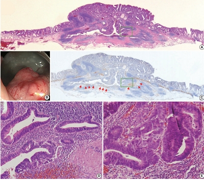
- Multidimensional analysis of concurrent proximal bronchiolar adenoma and lung carcinoma
- Lu-Yao Li, Gong-Ming Dong, Yun-Peng Zhang, Ting-Ting Wang, Fu-Quan Jia, Guan-Jun Zhang
- J Pathol Transl Med. 2026;60(3):356-363. Published online March 23, 2026
- DOI: https://doi.org/10.4132/jptm.2025.12.31
- 1,873 View
- 84 Download
-
 Abstract
 PDF
PDF Supplementary Material
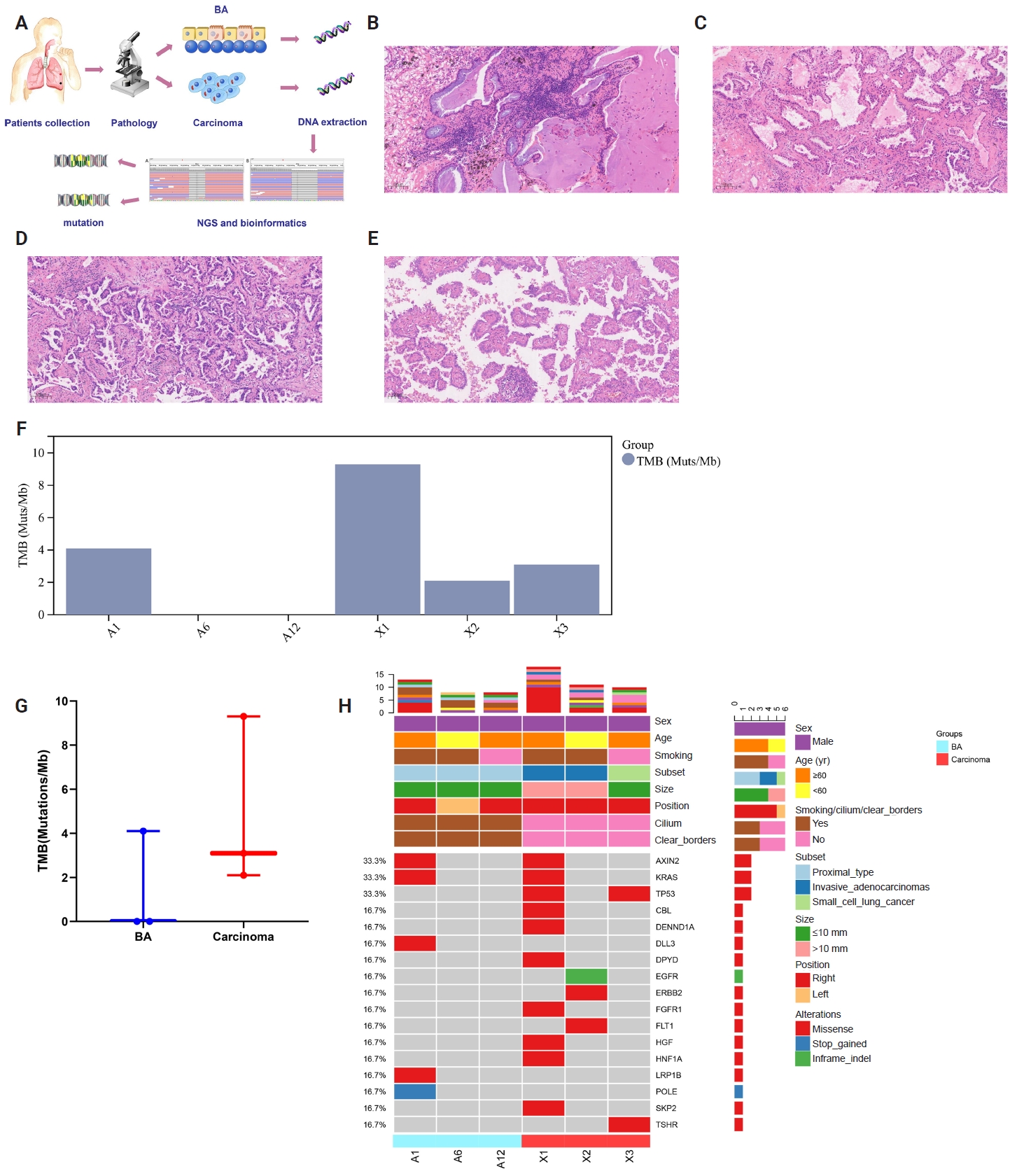
 - Bronchiolar adenoma (BA) is a rare type of lung tumor characterized by bilayered epithelial cells having a continuous basal layer and a luminal layer. It resembles mucinous adenocarcinoma (MA) on frozen section, with difficulty in distinguishing the basal layer. Immunohistochemistry is the best choice for verifying the diagnosis. This study aimed to comprehensively characterize three cases of BA-combined carcinoma using clinical, histopathological, and genetic features. BA and carcinoma sections were subjected to next-generation sequencing, respectively. It was hypothesized that while different mutation forms matched different regions, BA and lung adenocarcinoma shared the same gene mutation when they co-occurred in the same location. BA with extensive carcinoma is extremely rare and presents diagnostic challenges due to its overlap with conditions such as MA. Because of its distinctive morphological characteristics, BA may be regarded as a low-grade malignancy, particularly during a confusing evaluation. A multifaceted examination of clinical, radiological, immunohistochemical, and genetic data is necessary for an accurate diagnosis.
- Adenomatoid odontogenic tumor: clinicopathological analysis of 34 cases from Karachi, Pakistan
- Summaya Zafar, Sehar Sulaiman, Madeeha Nisar, Poonum Khan, Nasir Ud Din
- J Pathol Transl Med. 2025;59(6):390-397. Published online October 16, 2025
- DOI: https://doi.org/10.4132/jptm.2025.07.11
- 5,283 View
- 182 Download
- 1 Web of Science
- 1 Crossref
-
Abstract
PDF
- Background
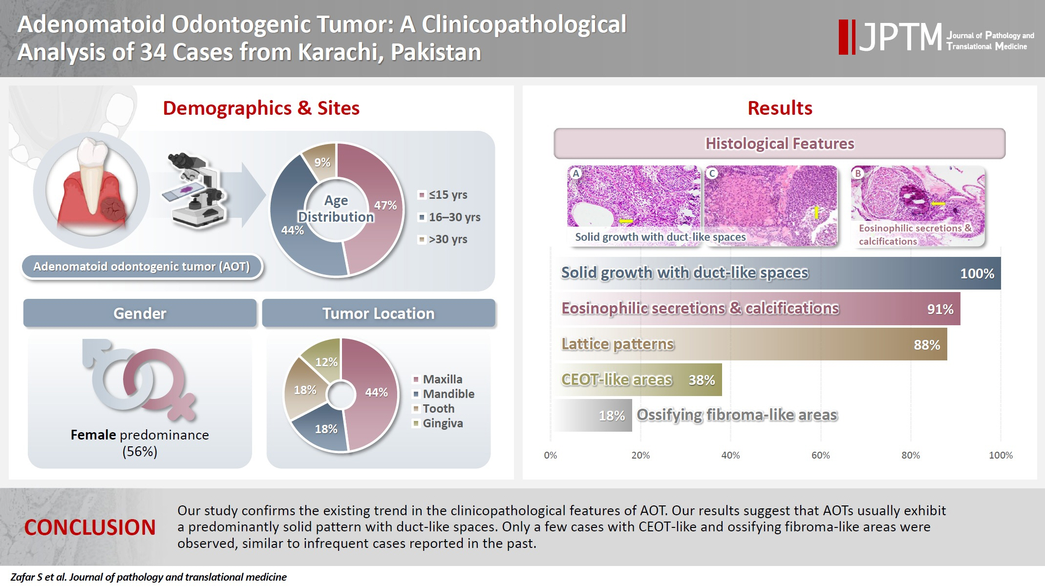
Adenomatoid odontogenic tumor (AOT) is a benign slow-growing neoplasm of odontogenic epithelial origin that is relatively uncommon. Only a few studies have described its histological features. Hence, we aimed to describe the clinicopathological features of AOT in a cohort of patients. Methods: AOT cases diagnosed between 2009 and 2024 were searched electronically. Glass slides were retrieved from archives and were reviewed by two pathologists to record the associated morphological features. Other data including patient demographics and tumor site were collected by reviewing histopathology reports. Results: The age of patients ranged from 9 to 44 years (mean, 17.7 years), and most were female (55.9%). The maxilla (44.1%) was the most common tumor site. Histologically, a predominantly solid growth pattern (n = 34) accompanied by ducts with a cuboidal/columnar epithelial lining (n = 31), eosinophilic secretions (n = 31), calcifications (n = 31), lattice work pattern (n = 30), and cystic areas (n = 20) were observed. Less frequent features included calcifying epithelial odontogenic tumor (CEOT)–like areas (n = 13), osteodentin (n = 6), association with impacted tooth (n = 3), mucin in tubules (n = 7), fibrocollagenous stroma (n = 6), mucin in ducts (n = 3) and ossifying fibroma-like areas (n = 6). The association of ducts with a cuboidal/columnar epithelial lining, lattice work pattern, calcifications, and eosinophilic secretions with gingival tumors was statistically significant (p ≤ .05). Additionally, tooth tumors were significantly associated with CEOT-like areas (p = .03). Conclusions: Our study confirms the trends in the clinicopathological features of AOT in previous case reports. Our results suggest that AOTs usually exhibit a predominantly solid pattern with duct-like spaces. Only a few cases with CEOT-like and ossifying fibroma-like areas were observed, similar to infrequent cases reported in the past. -
Citations
Citations to this article as recorded by
- Intraosseous lesions of the jaw: a clinicohistological study
Hadeel Odeh, Esra Nsour, Muna A. Salameh, Zayed M. Al-Zu’bi, Ali Al Khader
BMC Oral Health.2026;[Epub] CrossRef
- Intraosseous lesions of the jaw: a clinicohistological study
- Cytological features of atypical adenomatous hyperplasia and adenocarcinoma in situ of the lung: a case report
- Misa Takahashi, Seiya Homma, Chisato Setoguchi, Yoko Umezawa, Atsuhiko Sakamoto
- J Pathol Transl Med. 2025;59(3):195-200. Published online May 9, 2025
- DOI: https://doi.org/10.4132/jptm.2025.04.09

- 6,780 View
- 139 Download
-
Abstract
PDF
- Atypical adenomatous hyperplasia (AAH) and adenocarcinoma in situ (AIS) are generally treated as different lesions, depending on the differences in lesion size and histological findings. However, these differences are not absolute; thus, AAH and AIS are often difficult to distinguish. Moreover, whether AAH and AIS can be regarded as different lesions remains unknown because cytological specimens, especially those of AAH, are rare. In this study, we examined these uncommon cytological specimens and compared the cytological findings between AAH and AIS. We observed many common cytological features with no obvious differences between AAH and AIS. These findings suggest that these two distinct lesions can be grouped into a single category. Therefore, we propose creating a new cytological category.
- Fine needle aspiration cytology diagnoses of follicular thyroid carcinoma: results from a multicenter study in Asia
- Hee Young Na, Miyoko Higuchi, Shinya Satoh, Kaori Kameyama, Chan Kwon Jung, Su-Jin Shin, Shipra Agarwal, Jen-Fan Hang, Yun Zhu, Zhiyan Liu, Andrey Bychkov, Kennichi Kakudo, So Yeon Park
- J Pathol Transl Med. 2024;58(6):331-340. Published online November 7, 2024
- DOI: https://doi.org/10.4132/jptm.2024.10.12
- 9,551 View
- 285 Download
- 4 Web of Science
- 5 Crossref
-
Abstract
PDFSupplementary Material
- Background
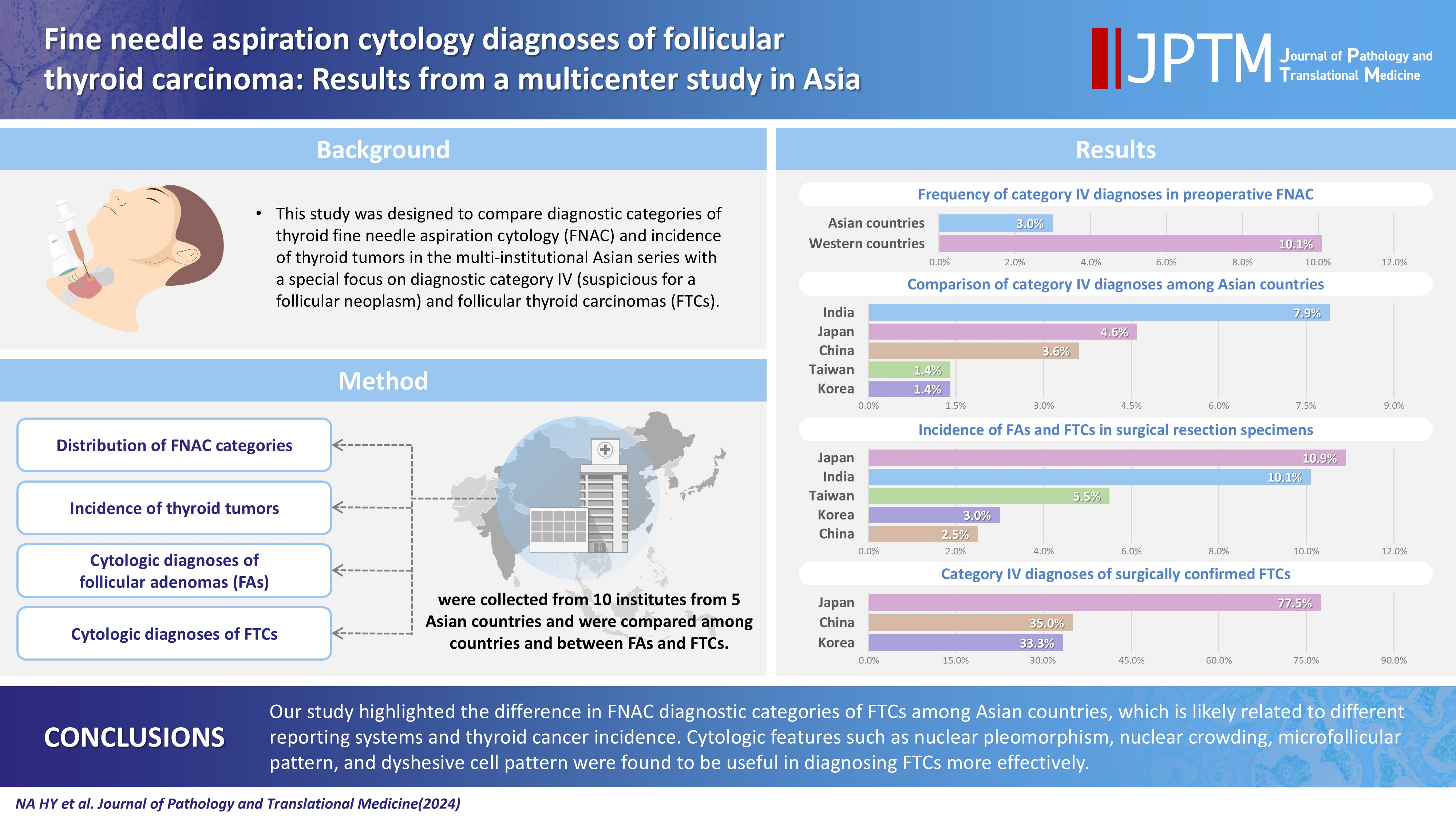
This study was designed to compare diagnostic categories of thyroid fine needle aspiration cytology (FNAC) and incidence of thyroid tumors in the multi-institutional Asian series with a special focus on diagnostic category IV (suspicious for a follicular neoplasm) and follicular thyroid carcinomas (FTCs). Methods: Distribution of FNAC categories, incidence of thyroid tumors in resection specimens and cytologic diagnoses of surgically confirmed follicular adenomas (FAs) and FTCs were collected from 10 institutes from five Asian countries and were compared among countries and between FAs and FTCs. Results: The frequency of category IV diagnoses (3.0%) in preoperative FNAC were significantly lower compared to those in Western countries (10.1%). When comparing diagnostic categories among Asian countries, category IV was more frequent in Japan (4.6%) and India (7.9%) than in Taiwan (1.4%), Korea (1.4%), and China (3.6%). Similarly, incidence of FAs and FTCs in surgical resection specimens was significantly higher in Japan (10.9%) and India (10.1%) than in Taiwan (5.5%), Korea (3.0%), and China (2.5%). FTCs were more commonly diagnosed as category IV in Japan (77.5%) than in Korea (33.3%) and China (35.0%). Nuclear pleomorphism, nuclear crowding, microfollicular pattern, and dyshesive cell pattern were more common in FTCs compared with FAs. Conclusions: Our study highlighted the difference in FNAC diagnostic categories of FTCs among Asian countries, which is likely related to different reporting systems and thyroid cancer incidence. Cytologic features such as nuclear pleomorphism, nuclear crowding, microfollicular pattern, and dyshesive cell pattern were found to be useful in diagnosing FTCs more effectively. -
Citations
Citations to this article as recorded by- Deep Learning-Based Multimodal Fusion of Ultrasound, Cytology, and Clinical Features to Distinguish Follicular Thyroid Carcinoma from Adenoma: A Multicenter Study
Xiao-Fei Guo, Li Zhou, Xin-Yi Bao, Shui-Qing Liu, Jia-Wei Feng, You-Long Zhu, Yong Jiang, Shu-Ying Zhang
Academic Radiology.2026; 33(7): 2921. CrossRef - Molecular Testing in Indeterminate Thyroid Nodules: Genomic Landscape, Diagnostic Performance, and Integrated Risk-Stratified Management
Sayaka Tanaka, Naomi Kitayama, Kyouko Kawamoto, Tomoko Wakasa, Yanhua Bai, Kennichi Kakudo
Cancers.2026; 18(10): 1661. CrossRef - A Clinicopathological Study on Thyroid Swellings with Comparison of Findings of Ultrasonography and Bethesda Reporting System of Fine Needle Aspiration Cytology to Histopathology: a Cross-Sectional Study
Amit Kumar Shukla, Debjit Jana, Maumita De, Krishna Kumar Yadav, Divya Daga, Diptanshu Mukherjee, Saumendra Nath Bandyopadhyay, Anukriti, Snehasish Halder
Indian Journal of Otolaryngology and Head & Neck Surgery.2026; 78(7): 3808. CrossRef - Diagnostic thresholds in thyroid cytopathology: toward biological risk stratification
Sayaka Tanaka, Tomoko Wakasa, Kennichi Kakudo
Journal of the American Society of Cytopathology.2026;[Epub] CrossRef - Misdiagnosed follicular adenoma with 11 year postoperative liver and lung metastases a case report and literature review
Kai-Li Yang, Heng-Tong Han, Shou-Hua Li, Xiao-Xiao Li, Ze Yang, Li-Bin Ma, Yong-Xun Zhao
Discover Oncology.2025;[Epub] CrossRef
- Deep Learning-Based Multimodal Fusion of Ultrasound, Cytology, and Clinical Features to Distinguish Follicular Thyroid Carcinoma from Adenoma: A Multicenter Study
- Tubular adenoma arising in tubular colonic duplication: a case report
- Heonwoo Lee, Hyeong Rok An, Chan Wook Kim, Young Soo Park
- J Pathol Transl Med. 2024;58(4):198-200. Published online July 3, 2024
- DOI: https://doi.org/10.4132/jptm.2024.06.04
- 5,542 View
- 227 Download
- 1 Web of Science
- 1 Crossref
-
Abstract
PDF
- Colonic duplication constitutes a rare congenital anomaly, characterized by the presence of hollow cystic or tubular structures exhibiting an epithelial-lined intestinal wall. Diagnostic challenges persist due to its low incidence and manifestation of nonspecific symptoms such as abdominal pain or constipation, resulting in a reluctance to pursue surgical resection. As associated malignancies in colonic duplication are rare, the inherent malignant potential of these anomalies remains undetermined. Additionally, despite reported instances of associated malignancies in colonic duplication, there is an absence of reports in the literature detailing tubular adenoma within these cases. The histologic features of the presented case are particularly noteworthy, situated at the precancerous stage, intimating potential progression towards adenocarcinoma within colonic duplication.
-
Citations
Citations to this article as recorded by- Low-grade mucinous neoplasm originating from intestinal duplication: a case report and review of the literature
Huihui Yin, Jie Yu, Yunzhao Chen
World Journal of Surgical Oncology.2025;[Epub] CrossRef
- Low-grade mucinous neoplasm originating from intestinal duplication: a case report and review of the literature
- Frequent apocrine changes in pleomorphic adenoma with malignant transformation: a possible pre-malignant step in ductal carcinoma ex pleomorphic adenoma
- Joon Seon Song, Yeseul Kim, Yoon-Se Lee, Seung-Ho Choi, Soon Yuhl Nam, Sang Yoon Kim, Kyung-Ja Cho
- J Pathol Transl Med. 2023;57(3):158-165. Published online May 10, 2023
- DOI: https://doi.org/10.4132/jptm.2023.03.13
- 8,838 View
- 202 Download
- 4 Web of Science
- 4 Crossref
-
Abstract
PDF
- Background
The most common type of carcinoma ex pleomorphic adenoma (CPA) is histologically equivalent to salivary duct carcinoma, which has an apocrine phenotype. Invasive CPA is often accompanied by non-invasive or in situ carcinoma, an observation that suggests the presence of precursor lesions. The aim of this study was to identify candidate precursor lesions of CPA within pleomorphic adenoma (PA).
Methods
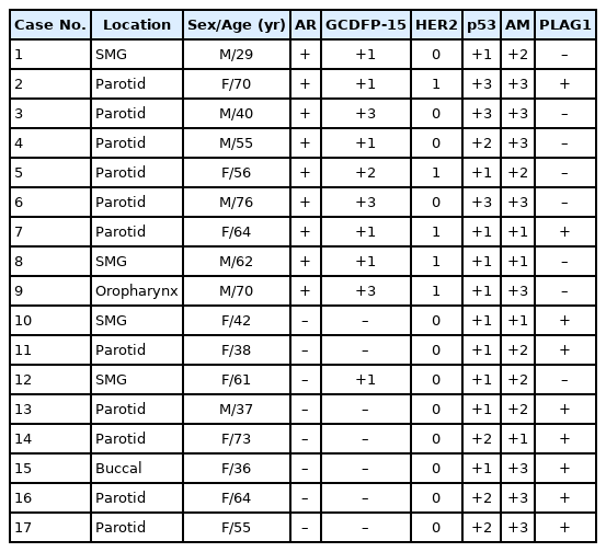
Eleven resected cases of CPA with residual PA and 17 cases of PA with atypical changes were subjected to immunohistochemistry (IHC) for p53, human epidermal growth factor receptor 2 (HER2), androgen receptor (AR), pleomorphic adenoma gene 1, gross cystic disease fluid protein-15 (GCDFP-15), and anti-mitochondrial antibody.
Results
Invasive or in situ carcinoma cells in all CPAs were positive for AR, GCDFP-15, and HER2. Atypical foci in PAs corresponded to either apocrine or oncocytic changes on the basis of their reactivity to AR, GCDFP-15, and anti-mitochondrial antibody. Atypical cells in PAs surrounding CPAs had an apocrine phenotype without HER2 expression.
Conclusions
Our study identified frequent apocrine changes in residual PAs in CPA cases, suggesting a possible precursor role of apocrine changes. We recommend the use of HER2 IHC in atypical PAs, and that clinicians take HER2 positivity into serious consideration. -
Citations
Citations to this article as recorded by- Pleomorphic Adenoma with Epithelial Atypia, Apocrine Metaplasia, and/or In situ/Intracapsular Salivary Duct Carcinoma Are Indolent Lesions with Good Prognosis: A Proposal for Unified Nomenclature and Clinical Observation
Grayson G. Cole, Matt Levin, David Ferber, Spencer C. Roark, Peter M. Sadow, Daniel Lubin, Julie Guilmette, Jason R. Pettus, Adam S. Fisch, Dipti P. Sajed, Fouad R. Zakka, Mark W. Lingen, Nicole A. Cipriani
Head and Neck Pathology.2025;[Epub] CrossRef - Progression of Nasopharyngeal Pleomorphic Adenoma to Carcinoma Ex Pleomorphic Adenoma With Metastases: A Case Report
Krystsina Zhukovich, Alisher Tashbayev, Vladimir Osipov
Cureus.2025;[Epub] CrossRef - Carcinoma Ex Pleomorphic Adenoma of the Palate in a Young Female: A Rare and Aggressive Malignant Transformation
Sakshi Akolkar, Alka Hande, Archana Sonone, Husna Tehzeeb
Journal of Datta Meghe Institute of Medical Sciences University.2025; 20(4): 919. CrossRef - Characterization of a Molecularly Distinct Subset of Oncocytic Pleomorphic Adenomas/Myoepitheliomas Harboring Recurrent ZBTB47-AS1::PLAG1 Gene Fusion
Ziyad Alsugair, Jimmy Perrot, Françoise Descotes, Jonathan Lopez, Anne Champagnac, Daniel Pissaloux, Claire Castain, Mihaela Onea, Philippe Céruse, Pierre Philouze, Charles Lépine, Marie-Delphine Lanic, Marick Laé, Valérie Costes-Martineau, Nazim Benzerdj
American Journal of Surgical Pathology.2024; 48(5): 551. CrossRef
- Pleomorphic Adenoma with Epithelial Atypia, Apocrine Metaplasia, and/or In situ/Intracapsular Salivary Duct Carcinoma Are Indolent Lesions with Good Prognosis: A Proposal for Unified Nomenclature and Clinical Observation
- Association of PTTG1 expression with invasiveness of non-functioning pituitary adenomas
- Su Jung Kum, Hye Won Lee, Soon Gu Kim, Hyungsik Park, Ilseon Hwang, Sang Pyo Kim
- J Pathol Transl Med. 2022;56(1):22-31. Published online October 15, 2021
- DOI: https://doi.org/10.4132/jptm.2021.08.31
- 8,321 View
- 212 Download
- 8 Web of Science
- 8 Crossref
-
Abstract
PDF
- Background
Pituitary tumor transforming gene 1 (PTTG1), paired-like homeodomain 2 (PITX2), and galectin-3 have been widely studied as predictive biomarkers for various tumors and are involved in tumorigenesis and tumor progression. We evaluated the usefulness of PTTG1, PITX2, and galectin-3 as predictive biomarkers for invasive non-functioning pituitary adenomas (NFPAs) by determining the relationship between the expressions of these three proteins and the invasiveness of the NFPAs. We also investigated whether PTTG1, E-cadherin, and Ki-67, which are known to be related to each other, show a correlation with NFPA features.
Methods
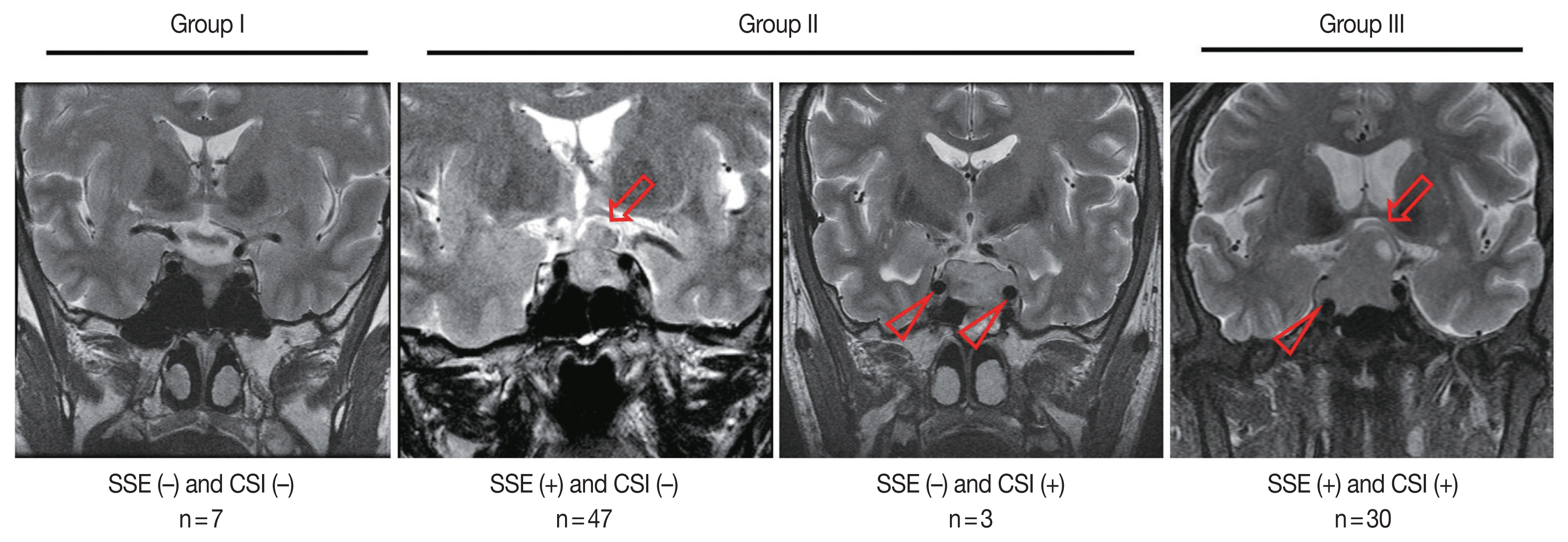
A retrospective study was conducted on 87 patients with NPFAs who underwent surgical removal. The NFPAs were classified into three groups based on magnetic resonance imaging findings of suprasellar extension and cavernous sinus invasion. Immunohistochemical staining for PTTG1, PITX2, galectin-3, E-cadherin, and Ki-67 was performed on tissue microarrays.
Results
PTTG1 expression showed a statistically significant correlation with the invasiveness of NFPAs, whereas PITX2 and galectin-3 did not have a relationship with the invasiveness of NFPAs. Moreover, there was no association among PTTG1, E-cadherin, and Ki-67 expression.
Conclusions
PTTG1 has the potential to serve as a predictive biomarker for invasive NFPA. Furthermore, this study may serve as a reference for the development of PTTG1-targeted therapeutic agents. -
Citations
Citations to this article as recorded by- Biomarkers Driving Precision Medicine in Nonfunctioning Pituitary Neuroendocrine Tumors: A Systematic Review of Recent Literature
Amalina Haydar Ali Tajuddin, Nur Firdaus Isa, Mohd Hamzah Mohd Nasir
The Journal of Clinical Endocrinology & Metabolism.2026; 111(4): e1195. CrossRef - The role of methylation in pituitary neuroendocrine tumors current insights and emerging perspectives
Yafei Wang, Tianlang Hu, Jingjing Jia, Chen Wang, Chenran Zhang
Molecular Biology Reports.2026;[Epub] CrossRef - The regulatory role of PTTG1 in proliferation and migration of thyroid cancer
Jianjun Wang, Chenjun Guo, Junyu Cao, Li Li
Discover Oncology.2025;[Epub] CrossRef - Pituitary tumor-transforming gene 1 and endocrine cancers: an up-to-date review through history, current insights and future perspectives
E Vergani, E Teveroni, F Mancini, F Di Nicuolo, S Raia, S Chiloiro, F Pierconti, A Bianchi, A M Isidori, A Pontecorvi, D Milardi
Endocrine-Related Cancer.2025;[Epub] CrossRef - High-throughput Screening for Cushing Disease: Therapeutic Potential of Thiostrepton via Cell Cycle Regulation
Takuro Hakata, Ichiro Yamauchi, Daisuke Kosugi, Taku Sugawa, Haruka Fujita, Kentaro Okamoto, Yohei Ueda, Toshihito Fujii, Daisuke Taura, Nobuya Inagaki
Endocrinology.2024;[Epub] CrossRef - Neoplasms and tumor-like lesions of the sellar region: imaging findings with correlation to pathology and 2021 WHO classification
Lorenzo Ugga, Raduan Ahmed Franca, Alessandra Scaravilli, Domenico Solari, Sirio Cocozza, Fabio Tortora, Luigi Maria Cavallo, Marialaura Del Basso De Caro, Andrea Elefante
Neuroradiology.2023; 65(4): 675. CrossRef - A comprehensive characterisation of phaeochromocytoma and paraganglioma tumours through histone protein profiling, DNA methylation and transcriptomic analysis genome wide
Prodromos Chatzikyriakou, Dimitria Brempou, Mark Quinn, Lauren Fishbein, Roberta Noberini, Ioannis N. Anastopoulos, Nicola Tufton, Eugenie S. Lim, Rupert Obholzer, Johnathan G. Hubbard, Mufaddal Moonim, Tiziana Bonaldi, Katherine L. Nathanson, Louise Izat
Clinical Epigenetics.2023;[Epub] CrossRef - Expression and clinical significance of Cathepsin K and MMPs in invasive non-functioning pituitary adenomas
Hongyan Liu, Saichun Zhang, Ting Wu, Zhaohui Lv, Jianming Ba, Weijun Gu, Yiming Mu
Frontiers in Oncology.2022;[Epub] CrossRef
- Biomarkers Driving Precision Medicine in Nonfunctioning Pituitary Neuroendocrine Tumors: A Systematic Review of Recent Literature
- Hepatocellular adenomas: recent updates
- Haeryoung Kim, Young Nyun Park
- J Pathol Transl Med. 2021;55(3):171-180. Published online April 7, 2021
- DOI: https://doi.org/10.4132/jptm.2021.02.27
- 14,262 View
- 587 Download
- 13 Web of Science
- 13 Crossref
-
Abstract
PDF
- Hepatocellular adenoma (HCA) is a heterogeneous entity, from both the histomorphological and molecular aspects, and the resultant subclassification has brought a strong translational impact for both pathologists and clinicians. In this review, we provide an overview of the recent updates on HCA from the pathologists’ perspective and discuss several practical issues and pitfalls that may be useful for diagnostic practice.
-
Citations
Citations to this article as recorded by- The role of HNF4α in adenocarcinoma
Headtlove Essel Dadzie, Eric L. Snyder
Biochemical Society Transactions.2026; 54(4): 333. CrossRef - Large hepatocellular adenoma of the liver in a woman of reproductive age
AK Chichelnitsky, DA Savchenko, AS Kostina, DA Raklov, ER Zhuk, ES Buimova, AI Negodaeva, AO Ivanenko, VI Aduchieva
Bulletin of Russian State Medical University.2026;[Epub] CrossRef - A case of β-catenin-activated hepatocellular adenoma with concurrent hepatocellular carcinoma
Daichi Ito, Ryota Hyodo, Keisuke Kurimoto, Takashi Mizuno, Akira Satou, Motoko Sasaki, Yoji Ishizu, Mami Iima, Shinji Naganawa
Abdominal Radiology.2026;[Epub] CrossRef - Preventing false positive imaging diagnosis of HCC: differentiating HCC from mimickers and practical strategies
Ijin Joo
Journal of Liver Cancer.2025; 25(2): 217. CrossRef - Prognostic role of selection criteria for liver transplantation in patients with hepatocellular carcinoma: Review and bibliometric
Pamela Scarlett Espinoza Loyola, Diana Laura Muratalla Bautista, Karen Adela Hernández Bautista, Elizabeth Gil White, José Antonio González Moreno, Daniel Angel Torres del Real, Víctor Manuel Páez Zayas, Carla Escorza-Molina, Fernando Mondragón Rodríguez,
iLIVER.2024; 3(1): 100077. CrossRef - ACG Clinical Guideline: Focal Liver Lesions
Catherine Frenette, Mishal Mendiratta-Lala, Reena Salgia, Robert J. Wong, Bryan G. Sauer, Anjana Pillai
American Journal of Gastroenterology.2024; 119(7): 1235. CrossRef - Hepatocellular adenoma update: diagnosis, molecular classification, and clinical course
Sarah Poetter-Lang, Ahmed Ba-Ssalamah, Nina Bastati, Sami A Ba-Ssalamah, Jacqueline C Hodge, Giuseppe Brancatelli, Valérie Paradis, Valérie Vilgrain
British Journal of Radiology.2024; 97(1163): 1740. CrossRef - Fatal rupture of hepatic adenomatosis: Autopsy case and review of the literature
Sarra Ben Abderrahim, Khouloud Chérif, Zeineb Nfikha, Sarra Gharsallaoui, Imen El Aini, Maher Jedidi, Moncef Mokni, Mohamed Ben Dhiab
Journal of Forensic Sciences.2023; 68(4): 1393. CrossRef - Large Hepatocellular Adenoma Presenting with Iron Deficiency Anemia: A Case Report
Young Kwon Koh, Su Hyun Yoon, Sung Han Kang, Hyery Kim, Ho Joon Im, Suhyeon Ha, Jung-Man Namgoong, Kyung-Nam Koh
Clinical Pediatric Hematology-Oncology.2023; 30(1): 25. CrossRef - A Case Report on a Giant Hepatic Inflammatory Adenoma in a Young Female That Presented as Spontaneous Intrahepatic Hematoma
Andreas Kyvetos, Panagiota Voukelatou, Ioannis Vrettos, Spyridon Pantzios , Ioannis Elefsiniotis
Cureus.2023;[Epub] CrossRef - Advances in Histological and Molecular Classification of Hepatocellular Carcinoma
Joon Hyuk Choi, Swan N. Thung
Biomedicines.2023; 11(9): 2582. CrossRef - Estrobolome and Hepatocellular Adenomas—Connecting the Dots of the Gut Microbial β-Glucuronidase Pathway as a Metabolic Link
Sandica Bucurica, Mihaela Lupanciuc, Florentina Ionita-Radu, Ion Stefan, Alice Elena Munteanu, Daniela Anghel, Mariana Jinga, Elena Laura Gaman
International Journal of Molecular Sciences.2023; 24(22): 16034. CrossRef - Hepatocellular adenoma: what we know, what we do not know, and why it matters
Paulette Bioulac‐Sage, Annette S H Gouw, Charles Balabaud, Christine Sempoux
Histopathology.2022; 80(6): 878. CrossRef
- The role of HNF4α in adenocarcinoma
- Evolving pathologic concepts of serrated lesions of the colorectum
- Jung Ho Kim, Gyeong Hoon Kang
- J Pathol Transl Med. 2020;54(4):276-289. Published online June 26, 2020
- DOI: https://doi.org/10.4132/jptm.2020.04.15
- 21,276 View
- 881 Download
- 39 Web of Science
- 38 Crossref
-
Abstract
PDFSupplementary Material
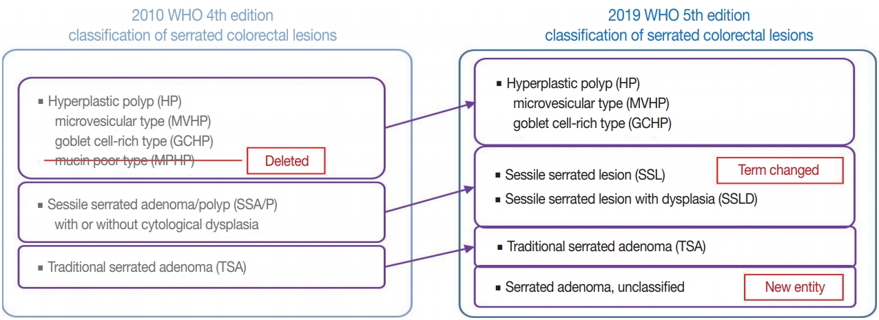
- Here, we provide an up-to-date review of the histopathology and molecular pathology of serrated colorectal lesions. First, we introduce the updated contents of the 2019 World Health Organization classification for serrated lesions. The sessile serrated lesion (SSL) is a new diagnostic terminology that replaces sessile serrated adenoma and sessile serrated polyp. The diagnostic criteria for SSL were revised to require only one unequivocal distorted serrated crypt, which is sufficient for diagnosis. Unclassified serrated adenomas have been included as a new category of serrated lesions. Second, we review ongoing issues concerning the morphology of serrated lesions. Minor morphologic variants with distinct molecular features were recently defined, including serrated tubulovillous adenoma, mucin-rich variant of traditional serrated adenoma (TSA), and superficially serrated adenoma. In addition to intestinal dysplasia and serrated dysplasia, minimal deviation dysplasia and not otherwise specified dysplasia were newly suggested as dysplasia subtypes of SSLs. Third, we summarize the molecular features of serrated lesions. The critical determinant of CpG island methylation development in SSLs is patient age. Interestingly, there may be ethnic differences in BRAF/KRAS mutation frequencies in SSLs. The molecular pathogenesis of TSAs is divided into KRAS and BRAF mutation pathways. SSLs with MLH1 methylation can progress into favorable prognostic microsatellite instability-positive (MSI+)/CpG island methylator phenotype-positive (CIMP+) carcinomas, whereas MLH1-unmethylated SSLs and BRAF-mutated TSAs can be precursors of poor-prognostic MSI−/CIMP+ carcinomas. Finally, based on our recent data, we propose an algorithm for stratifying risk subgroups of non-dysplastic SSLs.
-
Citations
Citations to this article as recorded by- Predominant Serrated Molecular Signature in Postcolonoscopy Colorectal Cancer: A Systematic Review and Meta-Analysis
Jen-Hao Yeh, Sin-Hua Moi, Chia-Chi Chen, Chao-Wen Hsu, Wen-Shuo Yeh, Tzu-Ning Tseng, Chuan-Pin Lin, Yu-Peng Liu, Jaw-Yuan Wang
American Journal of Gastroenterology.2026; 121(1): 122. CrossRef - Re-evaluating post-polypectomy surveillance: The role of non-invasive modalities in colorectal cancer prevention
Ethna McFerran, Damian McKay, Maurice B. Loughrey, Mark Lawler, Stephen T. McSorley
Best Practice & Research Clinical Gastroenterology.2026; 80: 102092. CrossRef - Superiority of excellent over good bowel preparation for proximal serrated polyp detection in a fecal immunochemical test–based screening cohort
Stefano Fantasia, Stefano Kayali, Pablo Cortegoso Valdivia, Stefano Andreotti, Daniele Macchi, Giorgio Nervi, Nico Pagano, Luigi Laghi
Gastrointestinal Endoscopy.2026; 104(1): 69. CrossRef - Endoscopic Assessment and Management of Colorectal Serrated Lesions: Current Comparative Practice, Challenges, and Future Directions
Toshio Uraoka, Yasushi Yamasaki, Kenichiro Imai, Hirohito Tanaka, Keigo Sato, Yuki Itoi, Hiroko Hosaka, Shiko Kuribayashi, Hemchand Ramberan, Yoji Takeuchi
Digestive Endoscopy.2026;[Epub] CrossRef - Serrated polyposis and the less common polyposis syndromes
Mary Smithson, Robert H. Hollis
Seminars in Colon and Rectal Surgery.2026; : 101190. CrossRef - Clinical and endoscopic characteristics of colorectal traditional serrated adenomas with dysplasia/adenocarcinoma in a Korean population
Ki-Hyun Kim, Eun Myung, Hyung Hoon Oh, Chan-Muk Im, Young-Eun Seo, Je-Seong Kim, Chae-June Lim, Ga-Ram You, Sung-Bum Cho, Wan-Sik Lee, Myung-Giun Noh, Kyung-Hwa Lee, Young-Eun Joo
World Journal of Gastrointestinal Oncology.2025;[Epub] CrossRef - MicroRNA: role in macrophage polarisation and colorectal cancer pathogenesis
Haihong Lin, Jun Zhou, Ying He, Yifan Zhu, Puwen Chen, Hongwei Yan, Junyun Huang, Ersheng Gong, Xiaoling Wang
Frontiers in Cell and Developmental Biology.2025;[Epub] CrossRef - Submucosal fibrosis in large colorectal serrated lesions in cases receiving endoscopic submucosal dissection
Erik Manriquez-Alegria, Naohisa Yoshida, Reo Kobayashi, Naoto Iwai, Ken Inoue, Osamu Dohi, Lucas Cardoso, Hideyuki Konishi
Therapeutic Advances in Gastroenterology.2025;[Epub] CrossRef - Navigating the Colorectal Cancer Maze: Unveiling Pathways To Diagnosis, Management, Pathophysiology and Prevention
Khalid Ali Mohammed Al Kamzari, Constantina Constantinou
Current Oncology Reports.2025; 27(10): 1115. CrossRef - Fosl1 is a transcriptional effector of BRAFV600E-driven intestinal tumorigenesis
Zakia Alam, Rebecca Nightingale, Analia Lesmana, Cheng Liu, Laura J. Jenkins, Mark F. Richardson, Lawrence Croft, Ian Y. Luk, Camilla M. Reehorst, Fiona Chionh, Natalia Vukelic, Faiza Basheer, Eugene Tulchinsky, Joshua Badshah, Troy Dumenil, Latifa Bakiri
iScience.2025; 28(11): 113875. CrossRef - Sessile Serrated Lesions in Inflammatory Bowel Disease: Hidden Players in Colitis-Associated Colorectal Cancer?
Roberto de Sire, Diletta De Deo, Miriana Mercurio, Gianluca Franchellucci, Giulio Calabrese, Livio Bonacci, Mauro Sollai Pinna, Cristina Bezzio, Alessandro Armuzzi, Cesare Hassan, Alessandro Repici, Fabiana Castiglione, Sandro Ardizzone, Roberta Maselli
Journal of Clinical Medicine.2025; 14(22): 8042. CrossRef - Histologic Reappraisal and Evaluation of MLH1 Protein Expression in Sessile Serrated Lesions of the Proximal Colon
Priscilla de Sene Portel Oliveira, Miriam Aparecida da Silva Trevisan, Rita Barbosa de Carvalho, Rita de Cássia Perina Martins, João José Fagundes, Claudio Saddy Rodrigues Coy, Ashwini Esnakula
Gastroenterology Research and Practice.2025;[Epub] CrossRef - Impact of AI-aided colonoscopy in clinical practice: a prospective randomised controlled trial
Johanna Schöler, Marko Alavanja, Thomas de Lange, Shunsuke Yamamoto, Per Hedenström, Jonas Varkey
BMJ Open Gastroenterology.2024; 11(1): e001247. CrossRef - The histologic features, molecular features, detection and management of serrated polyps: a review
Jin-Dong Wang, Guo-Shuai Xu, Xin-Long Hu, Wen-Qiang Li, Nan Yao, Fu-Zhou Han, Yin Zhang, Jun Qu
Frontiers in Oncology.2024;[Epub] CrossRef - Serrated polyps <10 mm cannot reliably be characterized by i-Scan without magnification at routine colonoscopy
Sabrina G.G. TESTONI, Chiara NOTARISTEFANO, Giuliano F. BONURA, Maria NAPOLITANO, Dario ESPOSITO, Edi VIALE, Lorella FANTI, Francesco AZZOLINI, Giulia M. CAVESTRO, PierAlberto TESTONI
Minerva Gastroenterology.2024;[Epub] CrossRef - Interobserver variability in the histopathological classification and grading of dysplasia in elevated colon lesions in the city of Lima
Guido Gallegos-Serruto, Aldo Gutiérrez, César Chian García, Isthvan Torres Perez
Revista de Gastroenterología del Perú.2024; 44(3): 239. CrossRef - Comparison of adenoma detection rate and proximal serrated polyp detection rate and their effect on post-colonoscopy colorectal cancer mortality in screening patients
Jasmin Zessner-Spitzenberg, Elisabeth Waldmann, Lena Jiricka, Lisa-Maria Rockenbauer, Anna Hinterberger, Jeremy Cook, Arno Asaturi, Aleksandra Szymanska, Barbara Majcher, Michael Trauner, Monika Ferlitsch
Endoscopy.2023; 55(05): 434. CrossRef - The yield of dysplasia and serrated lesions in a single-centre tertiary inflammatory bowel disease cohort
Fiona Yeaman, Lena Thin
Therapeutic Advances in Gastroenterology.2023;[Epub] CrossRef -
The BEETS (JACCRO CC-18) Trial: An Observational and Translational Study of
BRAF
-Mutated Metastatic Colorectal Cancer
Chiaki Inagaki, Ryo Matoba, Satoshi Yuki, Manabu Shiozawa, Akihito Tsuji, Eisuke Inoue, Kei Muro, Wataru Ichikawa, Masashi Fujii, Yu Sunakawa
Future Oncology.2023; 19(17): 1165. CrossRef - A retrospective analysis of the histology of resected polyps and colonoscopy quality parameters in Belgium
E Macken, S Van Dongen, G Van Hal
Acta Gastro Enterologica Belgica.2023; 86(2): 277. CrossRef - Prognostic Biomarkers of Cell Proliferation in Colorectal Cancer (CRC): From Immunohistochemistry to Molecular Biology Techniques
Aldona Kasprzak
Cancers.2023; 15(18): 4570. CrossRef - Assimilating Epigenetics and Transcriptomics for the Identification
of Prognostic Novel Biomarkers and Imminent Targets in
Colorectal Carcinoma with Therapeutic Potential
Suman Kumar Ray, Sukhes Mukherjee
Current Molecular Medicine.2023; 23(8): 784. CrossRef - Multitarget Stool RNA Test for Colorectal Cancer Screening
Erica K. Barnell, Elizabeth M. Wurtzler, Julie La Rocca, Thomas Fitzgerald, Jessica Petrone, Yansheng Hao, Yiming Kang, Faith L. Holmes, David A. Lieberman
JAMA.2023; 330(18): 1760. CrossRef - Microbiome in Colonic Carcinogenesis
Jun Sun, Yinglin Xia
Comprehensive Physiology.2023; 13(3): 4685. CrossRef - Impact of comprehensive optical diagnosis training using Workgroup serrAted polypS and Polyposis classification on detection of adenoma and sessile serrated lesion
Jooyoung Lee, Jung Ho Bae, Su Jin Chung, Hae Yeon Kang, Seung Joo Kang, Min‐Sun Kwak, Ji Yeon Seo, Ji Hyun Song, Sun Young Yang, Jong In Yang, Seon Hee Lim, Jeong Yoon Yim, Joo Hyun Lim, Goh Eun Chung, Eun Hyo Jin, Ji Min Choi, Yoo Min Han, Joo Sung Kim
Digestive Endoscopy.2022; 34(1): 180. CrossRef - Clinicopathological and molecular analyses of hyperplastic lesions including microvesicular variant and goblet cell rich variant hyperplastic polyps and hyperplastic nodules—Hyperplastic nodule is an independent histological entity
Noriyuki Uesugi, Yoichi Ajioka, Tomio Arai, Yoshihito Tanaka, Tamotsu Sugai
Pathology International.2022; 72(2): 128. CrossRef - Comprehensive clinicopathologic, molecular, and immunologic characterization of colorectal carcinomas with loss of three intestinal markers, CDX2, SATB2, and KRT20
Ji Ae Lee, Mi-Kyoung Seo, Seung-Yeon Yoo, Nam-Yun Cho, Yoonjin Kwak, Kyoungbun Lee, Jung Ho Kim, Gyeong Hoon Kang
Virchows Archiv.2022; 480(3): 543. CrossRef - Serrated Colorectal Lesions: An Up-to-Date Review from Histological Pattern to Molecular Pathogenesis
Martino Mezzapesa, Giuseppe Losurdo, Francesca Celiberto, Salvatore Rizzi, Antonio d’Amati, Domenico Piscitelli, Enzo Ierardi, Alfredo Di Leo
International Journal of Molecular Sciences.2022; 23(8): 4461. CrossRef - Arterial stiffness is associated with high-risk colorectal adenomas and serrated lesions: A cross-sectional study in a Taiwanese population
Hung-Yu Chen, Wen-Huang Lee, Hung-Lung Hsu, Yu-Tsung Chou, Fei-Lin Su, I-Hsuan Wu, Ting-Hsing Chao
Journal of Cardiology.2022; 80(2): 139. CrossRef - Morphological and molecular characterization of colorectal sessile serrated lesions with dysplasia
Filippo Cappello, Valentina Angerilli, Luca Dal Santo, Giada Munari, Marianna Sabbadin, Marcello Lo Mele, Gianmaria Pennelli, Claudio Luchini, Paola Parente, Stefano Lazzi, Matteo Fassan
Pathology - Research and Practice.2022; 240: 154214. CrossRef - Serrated polyposis: an overview
Jonathan Fawkes
Gastrointestinal Nursing.2022; 20(9): 24. CrossRef - Sessile serrated lesion presenting as large pedunculated polyp in the rectum: A case report
Shin Ju Oh, Jung-Wook Kim, Chi Hyuk Oh
Medicine.2022; 101(51): e32287. CrossRef - WHICH LESIONS ARE AT HIGHER RISK OF DEVELOPING COLORECTAL CARCINOMAS: SUPERFICIALLY ELEVATED SERRATED LESIONS OR DEPRESSED LESIONS?
Artur Adolfo PARADA, Filadelfio Euclydes VENCO, Miguel Reynaldo VARCA-NETO, Roberto EL IBRAHIM, Paula Bechara POLETTI, Helcio Pedrosa BRITO, Heloisa de Fátima SARE, Osvaldo MALAFAIA
ABCD. Arquivos Brasileiros de Cirurgia Digestiva (São Paulo).2022;[Epub] CrossRef - WNT5a in Colorectal Cancer: Research Progress and Challenges
Guangshun Sun, Liangliang Wu, Guoqiang Sun, Xuesong Shi, Hongyong Cao, Weiwei Tang
Cancer Management and Research.2021; Volume 13: 2483. CrossRef - Endoscopic diagnosis for colorectal sessile serrated lesions
Toshihiro Nishizawa, Shuntaro Yoshida, Akira Toyoshima, Tomoharu Yamada, Yoshiki Sakaguchi, Taiga Irako, Hirotoshi Ebinuma, Takanori Kanai, Kazuhiko Koike, Osamu Toyoshima
World Journal of Gastroenterology.2021; 27(13): 1321. CrossRef - NTRK oncogenic fusions are exclusively associated with the serrated neoplasia pathway in the colorectum and begin to occur in sessile serrated lesions
Jung Ho Kim, Jeong Hoon Hong, Yoon‐La Choi, Ji Ae Lee, Mi‐kyoung Seo, Mi‐Sook Lee, Sung Bin An, Min Jung Sung, Nam‐Yun Cho, Sung‐Su Kim, Young Kee Shin, Sangwoo Kim, Gyeong Hoon Kang
The Journal of Pathology.2021; 255(4): 399. CrossRef - Differential pre-malignant programs and microenvironment chart distinct paths to malignancy in human colorectal polyps
Bob Chen, Cherie’ R. Scurrah, Eliot T. McKinley, Alan J. Simmons, Marisol A. Ramirez-Solano, Xiangzhu Zhu, Nicholas O. Markham, Cody N. Heiser, Paige N. Vega, Andrea Rolong, Hyeyon Kim, Quanhu Sheng, Julia L. Drewes, Yuan Zhou, Austin N. Southard-Smith, Y
Cell.2021; 184(26): 6262. CrossRef - Molecular Insights Into Colorectal Carcinoma
Domenika Ortiz Requena, Monica Garcia-Buitrago
Archives of Medical Research.2020; 51(8): 839. CrossRef
- Predominant Serrated Molecular Signature in Postcolonoscopy Colorectal Cancer: A Systematic Review and Meta-Analysis
- Colorectal epithelial neoplasm associated with gut-associated lymphoid tissue
- Yo Han Jeon, Ji Hyun Ahn, Hee Kyung Chang
- J Pathol Transl Med. 2020;54(2):135-145. Published online January 29, 2020
- DOI: https://doi.org/10.4132/jptm.2019.11.06
- 11,409 View
- 262 Download
- 3 Web of Science
- 3 Crossref
-
Abstract
PDF
- Background
Colorectal epithelial neoplasm extending into the submucosal gut-associated lymphoid tissue (GALT) can cause difficulties in the differential diagnosis. Regarding GALT-associated epithelial neoplasms, a few studies favor the term “GALT carcinoma” while other studies have mentioned the term “GALT-associated pseudoinvasion/epithelial misplacement (PEM)”.
Methods
The clinicopathologic characteristics of 11 cases of colorectal epithelial neoplasm associated with submucosal GALT diagnosed via endoscopic submucosal dissection were studied.
Results
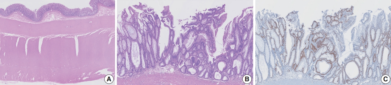
Eight cases (72.7%) were in males. The median age was 59 years, and age ranged from 53 to 73. All cases had a submucosal tumor component more compatible with GALT-associated PEM. Eight cases (72.7%) were located in the right colon. Ten cases (90.9%) had a non-protruding endoscopic appearance. Nine cases (81.8%) showed continuity between the submucosal and surface adenomatous components. Nine cases showed (81.8%) focal defects or discontinuation of the muscularis mucosae adjacent to the submucosal GALT. No case showed hemosiderin deposits in the submucosa or desmoplastic reaction. No case showed single tumor cells or small clusters of tumor cells in the submucosal GALT. Seven cases (63.6%) showed goblet cells in the submucosa. No cases showed oncocytic columnar cells lining submucosal glands.
Conclusions
Our experience suggests that pathologists should be aware of the differential diagnosis of GALT-associated submucosal extension by colorectal adenomatous neoplasm. Further studies are needed to validate classification of GALT-associated epithelial neoplasms. -
Citations
Citations to this article as recorded by- Redefining GALT-associated carcinoma: a distinct subtype of colorectal adenocarcinoma
Jennifer Fallas, Marianna Arvanitaki, Sophie Lecomte, Jean-Yves Bonnet, Sarah De Clercq, Audrey Verrellen, Nicky D’Haene, María Gómez Galdón, Laurine Verset
Virchows Archiv.2026; 488(3): 695. CrossRef - Family adenomatous polyposis come across dome type adenocarcinoma: a case report and literature review
Ying-Ying Chang, Xiao-Long Zhang, Yao-Hui Wang, Ting-Sheng Ling
Diagnostic Pathology.2025;[Epub] CrossRef - Radiation-induced injury and the gut microbiota: insights from a microbial perspective
Qiaoli Wang, Guoqiang Xu, Ouying Yan, Shang Wang, Xin Wang
Therapeutic Advances in Gastroenterology.2025;[Epub] CrossRef
- Redefining GALT-associated carcinoma: a distinct subtype of colorectal adenocarcinoma
- PLAG1, SOX10, and Myb Expression in Benign and Malignant Salivary Gland Neoplasms
- Ji Hyun Lee, Hye Ju Kang, Chong Woo Yoo, Weon Seo Park, Jun Sun Ryu, Yuh-Seog Jung, Sung Weon Choi, Joo Yong Park, Nayoung Han
- J Pathol Transl Med. 2019;53(1):23-30. Published online November 14, 2018
- DOI: https://doi.org/10.4132/jptm.2018.10.12
- 14,561 View
- 387 Download
- 28 Web of Science
- 34 Crossref
-
Abstract
PDF
- Background
Recent findings in molecular pathology suggest that genetic translocation and/oroverexpression of oncoproteins is important in salivary gland tumorigenesis and diagnosis. Weinvestigated PLAG1, SOX10, and Myb protein expression in various salivary gland neoplasm tissues.
Methods
A total of 113 cases of surgically resected salivary gland neoplasms at the NationalCancer Center from January 2007 to March 2017 were identified. Immunohistochemical stainingof PLAG1, SOX10, and Myb in tissue samples was performed using tissue microarrays.
Results
Among the 113 cases, 82 (72.6%) were benign and 31 (27.4%) were malignant. PLAG1 showednuclear staining and normal parotid gland was not stained. Among 48 cases of pleomorphicadenoma, 29 (60.4%) were positive for PLAG1. All other benign and malignant salivary glandneoplasms were PLAG1-negative. SOX10 showed nuclear staining. In normal salivary gland tissuesSOX10 was expressed in cells of acinus and intercalated ducts. In benign tumors, SOX10 expressionwas observed in all pleomorphic adenoma (48/48), and basal cell adenoma (3/3), but not inother benign tumors. SOX10 positivity was observed in nine of 31 (29.0%) malignant tumors.Myb showed nuclear staining but was not detected in normal parotid glands. Four of 31 (12.9%)malignant tumors showed Myb positivity: three adenoid cystic carcinomas (AdCC) and onemyoepithelial carcinoma with focal AdCC-like histology.
Conclusions
PLAG1 expression is specificto pleomorphic adenoma. SOX10 expression is helpful to rule out excretory duct origin tumor,but its diagnostic value is relatively low. Myb is useful for diagnosing AdCC when histology isunclear in the surgical specimen. -
Citations
Citations to this article as recorded by- Pleomorphic adenoma: A comprehensive review
Wilson Duplessis, Jason K. Wasserman
Seminars in Diagnostic Pathology.2026; 43(2): 150991. CrossRef - Pleomorphic adenoma gene 1 (PLAG1) protects p53-/- myoepithelial cells from mitochondria-related apoptosis caused by hypoxia
Nodoka Kindaichi, Yoshiki Mukudai, Yuzo Abe, Masataka Watanabe, Maki Nara, Konomi Yamada, Asami Houri, Toshikazu Shimane, Tatsuo Shirota
Journal of Oral and Maxillofacial Surgery, Medicine, and Pathology.2025; 37(4): 654. CrossRef - Retrospective Clinicopathological Study of 33 Cases of Pleomorphic Salivary Adenoma Diagnosed in Benghazi
Siraj S. Najem, Elhoni Ashour, Rehab Elmaddani, Ali M. Elmurtadi
Libyan Journal of Dentistry .2025; 8(2): 29. CrossRef - Pleomorphic adenoma of palatal minor salivary glands
Afrah Aldelaimi, Tahrir Aldelaimi, Suzan Abdulkareem
Revista Española de Cirugía Oral y Maxilofacial.2025;[Epub] CrossRef - Immunohistochemical Characterization of a Large Cohort of Triple Negative Breast Cancer
Rachel Han, Sharon Nofech-Mozes, Dina Boles, Hannah Wu, Nikolina Curcin, Elzbieta Slodkowska
International Journal of Surgical Pathology.2024; 32(2): 239. CrossRef - Proceedings of the 2024 North American Society of Head and Neck Pathology Companion Meeting, Baltimore, MD, March 24, 2024: Navigating Ancillary Studies in Basaloid/Blue Salivary Tumors
Kristine S. Wong
Head and Neck Pathology.2024;[Epub] CrossRef - Insights into the molecular alterations of PLAG1 and HMGA2 associated with malignant phenotype acquisition in pleomorphic adenoma
Reydson Alcides de Lima-Souza, Gustavo de Souza Vieira, Talita de Carvalho Kimura, João Figueira Scarini, Luccas Lavareze, Tayná Figueiredo Maciel, Moisés Willian Aparecido Gonçalves, Erika Said Abu Egal, Albina Altemani, Fernanda Viviane Mariano
Critical Reviews in Oncology/Hematology.2024; 204: 104494. CrossRef - Expanding the Molecular Spectrum of Carcinoma Ex Pleomorphic Adenoma
Reydson Alcides de Lima-Souza, Albina Altemani, Michal Michal, Fernanda Viviane Mariano, Ilmo Leivo, Alena Skálová
American Journal of Surgical Pathology.2024; 48(12): 1491. CrossRef - Utility of SOX10 and estrogen receptor immunohistochemical expression in endometrial carcinoma of Egyptian patients
Mona A. Kora, Alyaa A. Moselhy, Rania A. Abdallah
Egyptian Journal of Pathology.2024; 44(2): 190. CrossRef - Exploring Advanced Diagnostic Techniques for Salivary Gland Disorders: A Narrative Overview
Chuan-Xiang Li-, Liu Zhang, Ya-Ru Yan, Yong-Jie Ding, Ying-Ni Lin, Jian-Ping Zhou, Ning Li, Hong-Peng Li, Shi-Qi Li, Xian-Wen Sun, Qing-Yun Li
Asian journal of Current Research in Clinical Cancer.2024; 4(1): 1. CrossRef - The Challenge of “Monomorphic” Mucoepidermoid Carcinoma—Report of a Rare Case with Pure Spindle-Clear Cell Morphology
Xinyi Qu, Edwin Jun Chen Chew, Sathiyamoorthy Selvarajan, Bingcheng Wu, Abbas Agaimy, Fredrik Petersson
Head and Neck Pathology.2023; 17(3): 864. CrossRef - SOX10
Albert L Sy, Mai P Hoang
Journal of Clinical Pathology.2023; 76(10): 649. CrossRef - Activating Transcription Factor 1 (ATF1) Immunohistochemical Marker Distinguishes HCCC from MEC
Wafaey Badawy, Asmaa S. Abdelfattah, Haneen A. Sallam
Journal of Molecular Pathology.2023; 4(3): 178. CrossRef - Rare case of pleomorphic adenoma presenting as peritonsilar tumor
Anđelina Jovanović, Svetlana Valjarević, Milan Jovanović
Medicinska istrazivanja.2023; 56(3): 95. CrossRef - Pleomorphic Adenoma of a Minor Salivary Gland of the Hard Palate: A Case Report
Ishank Panchal, Anil Wanjari
Cureus.2023;[Epub] CrossRef - Advanced Diagnostic Methods for Salivary Glands Diseases: A Narrative Review Study
Malak Mohammed AlOsaimi, Abdulaziz Mohammed AlSubaheen, Taif Saleh Jameel, Rand Abdulrahman AlSalamah, Dalal Naseh AlAnzi, Norah Ameen AlOushan, Fahad Fadhel AlShammari, Cristalle Soman
Clinical Cancer Investigation Journal.2023; 12(4): 19. CrossRef - Clinical Significance of SOX10 Expression in Human Pathology
Hisham F. Bahmad, Aran Thiravialingam, Karthik Sriganeshan, Jeffrey Gonzalez, Veronica Alvarez, Stephanie Ocejo, Alvaro R. Abreu, Rima Avellan, Alejandro H. Arzola, Sana Hachem, Robert Poppiti
Current Issues in Molecular Biology.2023; 45(12): 10131. CrossRef - NR4A3 Immunostain Is a Highly Sensitive and Specific Marker for Acinic Cell Carcinoma in Cytologic and Surgical Specimens
Kartik Viswanathan, Shaham Beg, Bing He, Taotao Zhang, Richard Cantley, Daniel J Lubin, Qiuying Shi, Zahra Maleki, Saeed Asiry, Rema Rao, Nora Katabi, Masato Nakaguro, William C Faquin, Peter M Sadow, Momin T Siddiqui, Theresa Scognamiglio
American Journal of Clinical Pathology.2022; 157(1): 98. CrossRef - Recent Advances on Immunohistochemistry and Molecular Biology for the Diagnosis of Adnexal Sweat Gland Tumors
Nicolas Macagno, Pierre Sohier, Thibault Kervarrec, Daniel Pissaloux, Marie-Laure Jullie, Bernard Cribier, Maxime Battistella
Cancers.2022; 14(3): 476. CrossRef - Diagnostic accuracy of human transcriptional activator (Myb) expression by ELISA technique versus immunohistochemistry in detecting salivary gland carcinomas
Yousra Refaey, OlfatGamil Shaker, Ayman Abdelwahab, ImanAdel Mohamed Abdelmoneim, Fat’heyaMohamed Zahran
Journal of International Oral Health.2022; 14(1): 61. CrossRef - SLUG is a key regulator of epithelial-mesenchymal transition in pleomorphic adenoma
Hyesung Kim, Seung Bum Lee, Jae Kyung Myung, Jeong Hwan Park, Eunsun Park, Dong Il Kim, Cheol Lee, Younghoon Kim, Chul-Min Park, Min Bum Kim, Gil Chai Lim, Bogun Jang
Laboratory Investigation.2022; 102(6): 631. CrossRef - Assessment of MEF2C as a novel myoepithelial marker using normal salivary gland and pleomorphic adenoma: An immunohistochemical study
Ikuko Takakura, Satoko Kujiraoka, Rika Yasuhara, Junichi Tanaka, Fumio Ide, Kenji Mishima
Journal of Oral and Maxillofacial Surgery, Medicine, and Pathology.2022; 34(4): 523. CrossRef - Update on selective special types of breast neoplasms: Focusing on controversies, differential diagnosis, and molecular genetic advances
Shi Wei
Seminars in Diagnostic Pathology.2022; 39(5): 367. CrossRef - Cutaneous Melanomas: A Single Center Experience on the Usage of Immunohistochemistry Applied for the Diagnosis
Costantino Ricci, Emi Dika, Francesca Ambrosi, Martina Lambertini, Giulia Veronesi, Corti Barbara
International Journal of Molecular Sciences.2022; 23(11): 5911. CrossRef - Distinct clinicopathological and genomic features in solid and basaloid adenoid cystic carcinoma of the breast
Juan Ji, Fang Zhang, Fanglei Duan, Hong Yang, Jun Hou, Yang Liu, Jie Dai, Qiong Liao, Xian Chen, Qingsong Liu
Scientific Reports.2022;[Epub] CrossRef - NR4A3 fluorescence in situ hybridization analysis in cytologic and surgical specimens of acinic cell carcinoma
Qiuying Shi, Bin Zhang, Caroline Bsirini, Liqiong Li, Ellen J. Giampoli, Kelly R. Magliocca, Michelle Reid, Zhongren Zhou
Human Pathology.2022; 127: 86. CrossRef - Evaluation of NR4A3 immunohistochemistry (IHC) and fluorescence in situ hybridization and comparison with DOG1 IHC for FNA diagnosis of acinic cell carcinoma
John M. Skaugen, Raja R. Seethala, Simion I. Chiosea, Michael S. Landau
Cancer Cytopathology.2021; 129(2): 104. CrossRef -
MYB-NFIB Translocation by FISH in Adenoid Cystic Carcinoma of the Head and Neck in Nigerian Patients: A Preliminary Report
Adepitan A. Owosho, Olufunlola M. Adesina, Oluwole Odujoko, Olujide O. Soyele, Akinwumi Komolafe, Robert Bauer, Kallie Holte, Kurt F. Summersgill
Head and Neck Pathology.2021; 15(2): 433. CrossRef - Liquid-based cytology of oral brushings in a case of adenoid cystic carcinoma arising from the palate
Ryo MAKINO, Akihiko KAWAHARA, Hideyuki ABE, Yorihiko TAKASE, Chihiro FUKUMITSU, Kazuya MURATA, Tomoko YOSHIDA, Yukako SHINODA, Yoshiki NAITO, Jun AKIBA
The Journal of the Japanese Society of Clinical Cytology.2021; 60(1): 33. CrossRef - MYB Translocations in Both Myoepithelial and Ductoglandular Epithelial Cells in Adenoid Cystic Carcinoma: A Histopathologic and Genetic Reappraisal in Six Primary Cutaneous Cases
Keisuke Goto, Kazuyoshi Kajimoto, Takashi Sugino, Shin-ichi Nakatsuka, Makoto Yoshida, Mai Noto, Michihiro Kono, Toshihiro Takai
The American Journal of Dermatopathology.2021; 43(4): 278. CrossRef - Co-expression of Myoepithelial and Melanocytic Features in Carcinoma Ex Pleomorphic Adenoma
Costantino Ricci, Federico Chiarucci, Francesca Ambrosi, Tiziana Balbi, Barbara Corti, Ottavio Piccin, Ernesto Pasquini, Maria Pia Foschini
Head and Neck Pathology.2021; 15(4): 1385. CrossRef - Juvenile onset pleomorphic adenoma presenting as giant tumor of parotid gland in a young female
Surender Verma, Shivika Aggarwal, Pradeep Garg, Anjali Verma, Mridul Gera, Swaran S. Yadav
Journal of Dr. NTR University of Health Sciences.2021; 10(4): 286. CrossRef - Cytopathology and diagnostics of Warthin's tumour
Mirna Sučić, Nives Ljubić, Leila Perković, Dunja Ivanović, Leo Pažanin, Tena Sučić Radovanović, Dubravka Župnić‐Krmek, Fabijan Knežević
Cytopathology.2020; 31(3): 193. CrossRef - Clear cell papillary neoplasm of the breast with MAML2 gene rearrangement: Clear cell hidradenoma or low-grade mucoepidermoid carcinoma?
Raima A. Memon, Carlos N Prieto Granada, Shi Wei
Pathology - Research and Practice.2020; 216(10): 153140. CrossRef
- Pleomorphic adenoma: A comprehensive review
- Squamous Metaplasia in Pleomorphic Adenoma: A Diagnostic and Prognostic Enigma
- Swati Sharma, Monica Mehendiratta, Nivedita Chaudhary, Vineet Gupta, Maulshree Kohli, Anjana Arora
- J Pathol Transl Med. 2018;52(6):411-415. Published online October 1, 2018
- DOI: https://doi.org/10.4132/jptm.2018.07.15
- 10,786 View
- 154 Download
- 12 Web of Science
- 21 Crossref
-
Abstract
PDF
- Pleomorphic adenoma (PA) is the most common benign salivary gland tumor. Histologically, squamous metaplasia has been reported in PA, but has rarely been documented as being extensive enough to cause significant misdiagnosis. Here, we present an unusual case of PA in a 50-year-old female patient presenting with swelling on the postero-lateral aspect of the palate for a week. Histopathologically, the tumor exhibited the features of conventional PA with extensive squamous metaplasia and giant keratotic lamellae in cyst-like areas. Such exuberant squamous metaplasia and keratin can be a diagnostic and prognostic pitfall and lead to overtreatment of the patient.
-
Citations
Citations to this article as recorded by- Pleomorphic Adenomas and Their Atypical Morphology: Pitfalls in the Diagnosis of Salivary Gland Tumors
Alexandra Corina Faur, Alina Maria Șișu, Codruta Ileana Petrescu, Aura Jurescu, Camelia Vidiţa Gurban, Daniela Cornelia Lazăr, Diana Andrei, Sorin Lucian Bolintineanu, Laura Andreea Ghenciu
Diagnostics.2026; 16(14): 2168. CrossRef - Retrospective Clinicopathological Study of 33 Cases of Pleomorphic Salivary Adenoma Diagnosed in Benghazi
Siraj S. Najem, Elhoni Ashour, Rehab Elmaddani, Ali M. Elmurtadi
Libyan Journal of Dentistry .2025; 8(2): 29. CrossRef - Fine‐Needle Aspiration Cytology Diagnosis of Pleomorphic Adenoma With Spontaneous Infarction in the Salivary Gland: A Multicenter Retrospective Study
Jie‐Qiong Wang, Ge Li, Shao‐Hua Wang, Bo Yang, Yun Liu, Yu Wan, Cong‐Gai Huang, Fan Li
Cytopathology.2025; 36(5): 484. CrossRef - Keratocystoma: Molecular insights and diagnostic challenges in a rare salivary gland tumor
Yoshitaka Utsumi, Masato Nakaguro, Justin A. Bishop, Toshitaka Nagao
Seminars in Diagnostic Pathology.2025; 42(5): 150940. CrossRef - Bronchial pleomorphic adenoma successfully diagnosed and resected with left lower sleeve lobectomy; a case report and literature review
Katsuhiro Itogawa, Tomohiro Oba, Mitsuru Maki, Masako Amano, Akiko Adachi, Hidekazu Matsushima
Respiratory Medicine Case Reports.2025; 57: 102253. CrossRef - Effective Management of a Giant Deforming Pleomorphic Adenoma With Airway Displacement in a 93-Year-Old Patient: A Case Report
Julio A Palomino-Payan, Jessica Guillen-Valles, Daniel A Meza-Martinez, Fernanda Urias, Luis D Montes de Oca-Gordoa
Cureus.2024;[Epub] CrossRef - ECTOPIC PLEOMORPHIC ADENOMA OF BUCCAL SPACE: CASE REPORT WITH REVIEW OF LITERATURE
SANCHIT BAJPAI
UP STATE JOURNAL OF OTOLARYNGOLOGY AND HEAD AND NECK SURGERY.2024; VOLUME 12(ISSUE 1): 55. CrossRef - Pleomorphic adenoma with extensive squamous metaplasia and keratinizing cysts: Diagnostic and clinical pitfalls – A report of two cases and review of literature
Mahadevi B. Hosur, Rudrayya S. Puranik, Satyajit G. Dandagi, Vivekanand M. Patil
Journal of Oral and Maxillofacial Pathology.2024; 28(4): 689. CrossRef - Pleomorphic adenoma of the upper lip: A rare site for a common tumor- Case report
Prasath Sathiah, Sujaya Mazumder, Santosh Tummidi, Vijay Kannaujiya
SN Comprehensive Clinical Medicine.2023;[Epub] CrossRef - Variable metaplastic entities in pleomorphic adenoma a review of a rare case report with a note on its significance
N. Mahapatra, L. Bhuyan, Dash Chandra, P. Mishra
Archive of Oncology.2023; 29(2): 18. CrossRef - Pleomorphic adenoma with extensive oncocytic papillary cystic areas and trichilemmal keratinisation – A unique presentation
CV Aiswarya, Raghunath Vandana, Kamal Firoz, Meda Samatha
Journal of Oral and Maxillofacial Pathology.2023; 27(3): 562. CrossRef - Pleomorphic Adenoma with Extensive Squamous and Adipocytic Metaplasia Mimicking as Low Grade Mucoepidermoid Carcinoma on FNAC
Anu Singh, Ravi Hari Phulware, Arvind Ahuja, Ankur Gupta, Manju Kaushal
Indian Journal of Otolaryngology and Head & Neck Surgery.2022; 74(S2): 2132. CrossRef - Aspiration cytology of pleomorphic adenoma with squamous metaplasia: A case series and literature review illustrating diagnostic challenges
Joshua J. X. Li, Joanna K. M. Ng, Eric H. L. Lau, Amy B. W. Chan
Diagnostic Cytopathology.2022; 50(2): 64. CrossRef - Pleomorphic adenoma with extensive squamous metaplasia: The first well-documented case involving the submandibular gland
David A. Gaskin, Alain Reid, Pamela S. Gaskin
Human Pathology Reports.2022; 27: 300600. CrossRef - Salivary Gland Pleomorphic Adenomas Presenting With Extremely Varied Clinical Courses. A Single Institution Case-Control Study†
Krzysztof Piwowarczyk, Ewelina Bartkowiak, Paweł Kosikowski, Jadzia Tin-Tsen Chou, Małgorzata Wierzbicka
Frontiers in Oncology.2021;[Epub] CrossRef - A case report of pleomorphic adenoma squamous metaplasia resembling metastatic oral squamous cell carcinoma
E. Donohoe, R. Courtney, S. Phelan, P.J. McCann
Advances in Oral and Maxillofacial Surgery.2021; 2: 100074. CrossRef - Extensive squamous metaplasia in minor salivary gland neoplasm mimicking squamous cell carcinoma: Diagnostic dilemma in aspiration cytology
Renu Sukumaran, Nileena Nayak, RariP Mony
Clinical Cancer Investigation Journal.2021; 10(5): 257. CrossRef - Pleomorphic Adenoma Consisting of Multiple Cysts with Squamous Epithelial Lining: Findings on MRI, FNAC, and Histopathological Examination
Hiroshi Yamamoto, Sakurako Yamaguchi, Erika Iwai, Yukiko Iizuka, Shu Fushimi, Kunio Hayashi, Takumi Kondo, Satoshi Tokunaga, Masaaki Suemitsu, Takashi Kaneda, Kayo Kuyama, Masamichi Komiya
Open Journal of Stomatology.2021; 11(06): 221. CrossRef - Navigating small biopsies of salivary gland tumors: a pattern-based approach
J. Stephen Nix, Lisa M. Rooper
Journal of the American Society of Cytopathology.2020; 9(5): 369. CrossRef - Giant Parotid Pleomorphic Adenoma with Atypical Histological Presentation and Long‐Term Recurrence‐Free Follow‐Up after Surgery: A Case Report and Review of the Literature
Mohammed AlKindi, Sundar Ramalingam, Lujain Abdulmajeed Hakeem, Manal A. AlSheddi, Pravinkumar G. Patil
Case Reports in Dentistry.2020;[Epub] CrossRef - Pleomorphic adenoma of soft palate with extensive squamous metaplasia – A diagnostic enigma
Rashmi Patnayak, Sandip Mohanty, Anjan Kumar Sahoo, Adya Kinkara Panda, Amitabh Jena
Journal of Dr. NTR University of Health Sciences.2019; 8(4): 268. CrossRef
- Pleomorphic Adenomas and Their Atypical Morphology: Pitfalls in the Diagnosis of Salivary Gland Tumors
- Hepatocellular Carcinoma Arising in a Huge Hepatocellular Adenoma with Bone Marrow Metaplasia
- Hyo Jeong Kang, Hui Jeong Jeong, So-Woon Kim, Eunsil Yu, Young-Joo Lee, So Yeon Kim, Jihun Kim
- J Pathol Transl Med. 2018;52(4):226-231. Published online December 27, 2017
- DOI: https://doi.org/10.4132/jptm.2017.11.12
- 9,988 View
- 152 Download
- 5 Web of Science
- 6 Crossref
-
Abstract
PDF
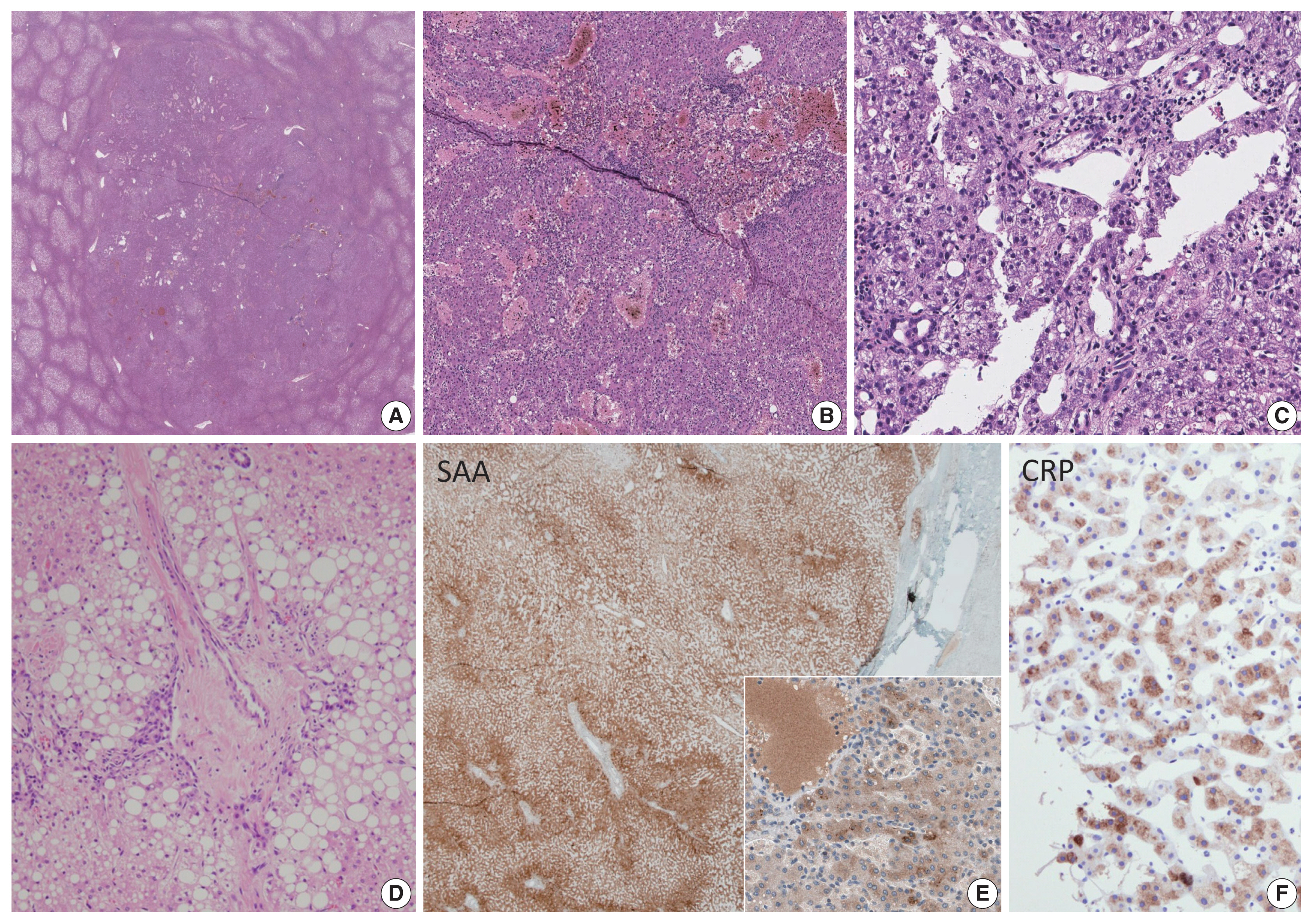
- Hepatocellular adenoma (HCA) is the most common type of benign liver tumor, and its major complication is malignant transformation to hepatocellular carcinoma (HCC). Here, we report a case of HCC arising in HCA with bone marrow metaplasia in a 24-year-old Korean woman who presented with abdominal discomfort. A huge liver mass was found on abdominal ultrasonography. She underwent surgical hepatic resection, and the resected specimen was entirely involved by a 20-cm-sized tumor. Histological review revealed a well differentiated HCC arising from inflammatory HCA with β-catenin nuclear positivity and bone marrow metaplasia that contained hematopoietic cells. This case was unique because malignant transformation, inflammatory type HCA, β-catenin nuclear staining, and bone marrow metaplasia were simultaneously observed. Additionally, it should be noted that a large HCA with β-catenin activation can undergo malignant transformation and should be surgically resected in a timely manner.
-
Citations
Citations to this article as recorded by- Adult Hepatocellular Carcinoma Coexisting with Extramedullary Hematopoiesis
Hirotsugu Noguchi, Michiyo Higashi, Ryo Desaki, Takashi Tasaki, Mari Kirishima, Ikumi Kitazono, Kazuhiro Tabata, Akihide Tanimoto
International Journal of Surgical Pathology.2022; 30(3): 339. CrossRef - Spontaneous Occurrence of Various Types of Hepatocellular Adenoma in the Livers of Metabolic Syndrome-Associated Steatohepatitis Model TSOD Mice
Wenhua Shao, Orgil Jargalsaikhan, Mayuko Ichimura-Shimizu, Qinyi Cai, Hirohisa Ogawa, Yuko Miyakami, Kengo Atsumi, Mitsuru Tomita, Mitsuko Sutoh, Shunji Toyohara, Ryoji Hokao, Yasusei Kudo, Takeshi Oya, Koichi Tsuneyama
International Journal of Molecular Sciences.2022; 23(19): 11923. CrossRef - Bilateral Diffuse Nodular Pulmonary Ossification Mimicking Metastatic Disease in a Patient with Fibrolamellar Hepatocellular Carcinoma
Pattamon Sutthatarn, Cara E. Morin, Jessica Gartrell, Wayne L. Furman, Max R. Langham, Teresa Santiago, Andrew J. Murphy
Children.2021; 8(3): 226. CrossRef - Malignant transformation of liver fatty acid binding protein-deficient hepatocellular adenomas: histopathologic spectrum of a rare phenomenon
Juan Putra, Linda D. Ferrell, Annette S.H. Gouw, Valerie Paradis, Arvind Rishi, Christine Sempoux, Charles Balabaud, Swan N. Thung, Paulette Bioulac-Sage
Modern Pathology.2020; 33(4): 665. CrossRef - Hepatocellular carcinoma arising from hepatic adenoma in a young woman
Haythem Yacoub, Hela Kchir, Dhouha Cherif, Hajer Hassine, Slim Haouet, Asma Ayari, Habiba Mizouni, Saber Mannai, Mohamed Tahar Khalfallah, Nadia Maamouri
Clinical Case Reports.2020; 8(9): 1659. CrossRef - Metanephric adenoma with osseous metaplasia and bone marrow elements
Alessandro Pietro Aldera, Jeff John, Dharshnee Chetty, Dhirendra Govender
Human Pathology: Case Reports.2019; 17: 200316. CrossRef
- Adult Hepatocellular Carcinoma Coexisting with Extramedullary Hematopoiesis
- Mucinous Cystadenoma of the Testis: A Case Report with Immunohistochemical Findings
- Gilhyang Kim, Dohee Kwon, Hee Young Na, Sehui Kim, Kyung Chul Moon
- J Pathol Transl Med. 2017;51(2):180-184. Published online February 13, 2017
- DOI: https://doi.org/10.4132/jptm.2016.08.30
- 11,795 View
- 128 Download
- 5 Web of Science
- 8 Crossref
-
Abstract
PDF
- Mucinous cystadenoma of the testis is a very rare tumor. Herein, we report a case of mucinous cystadenoma arising in the testis of a 61-year-old man, along with a literature review. Computed tomography showed a 2.5-cm-sized poorly enhancing cystic mass. Grossly, the tumor was a unilocular cystic mass filled with mucinous material and confined to the testicular parenchyma. Histologically, the cyst had a fibrotic wall lined by mucinous columnar epithelium without atypia. Immunohistochemical staining was positive for cytokeratin 20 and CDX2, as well as focally positive for cytokeratin 7. The pathologic diagnosis was mucinous cystadenoma.
-
Citations
Citations to this article as recorded by- Review of Paratesticular Appendageal Tumors, Morphology, Immunohistochemistry, and Recent Molecular Advances
Mathew Vega, Muhammad T. Idrees
Surgical Pathology Clinics.2025; 18(1): 119. CrossRef - Cistoadenoma Mucinoso Paratesticular: Caso Interesante en el Instituto Guatemalteco de Seguridad Social
Edgar Estuardo González López, Carlos Gonzalo Estrada Pazos
Revista Guatemalteca de Urología.2023; 10(2): 16. CrossRef - Primary borderline mucinous tumor of the testis with postoperative metastasis: A rare case report
Yingyu Shi, Ling Song, Yan Luo
Radiology Case Reports.2023; 18(9): 3203. CrossRef - Case report: Misdiagnosis of primary mucinous cystadenoma of the testicle by ultrasound
Linlin Zhang, Jianyuan Xuan, Manxi Li, Mei Zhang, Yu Song, Ziang Pan, Bo Fan, Lin Lu, Hongyan Zhou, Yang Li
Frontiers in Oncology.2023;[Epub] CrossRef - Primary Borderline Mucinous Testicular Tumor: A Case Report and Literature Review
Changjuan Hao, Chunsong Kang, Xiaoyan Kang, Zhuanzhuan Yu, Tingting Li, Jiping Xue
Frontiers in Oncology.2021;[Epub] CrossRef - Ovarian-type Tumors (Mullerian Tumors) of the Testis: Clinicopathologic Findings with Recent Advances
Michelle S Lin, Alberto G Ayala, Jae Y Ro
annals of urologic oncology.2019; : 1. CrossRef - Borderline Mucinous Testicular Tumour: Diagnostic and Management difficulties
Krishan Pratap, Marlon Perera, Frances Malczewski, Rachel Esler
BMJ Case Reports.2018; 2018: bcr-2017-223787. CrossRef - Mucinous tumor arising in a giant sacrococcygeal teratoma
Fengtian Zhang, Xiaolong Yu, Jin Zeng, Min Dai
Medicine.2017; 96(47): e8759. CrossRef
- Review of Paratesticular Appendageal Tumors, Morphology, Immunohistochemistry, and Recent Molecular Advances
- Oncocytic Lipoadenoma: A Rare Case of Parotid Gland Tumor and Review of the Literature
- Chen-lin Chi, Tseng-tong Kuo, Li-yu Lee
- J Pathol Transl Med. 2015;49(2):144-147. Published online March 12, 2015
- DOI: https://doi.org/10.4132/jptm.2014.02.10
- 12,208 View
- 70 Download
- 9 Web of Science
- 9 Crossref
-
Abstract
PDF
- Oncocytic lipoadenoma is a rare tumor, with only 18 cases having been reported since the first in 1998. We encountered a case of oncocytic lipoadenoma presenting as a slowly growing parotid mass in a 71-year-old man. This tumor is characteristically comprised of a mixture of oncocytes and adipocytes. The present case is one of five reported cases of oncocytic lipoadenoma showing sebaceous differentiation. The results of immunohistochemical study with DOG1 antibody supported the origination of this tumor in the striated duct.
-
Citations
Citations to this article as recorded by- Multimodal Imaging of Oncocytic Lipoadenoma Arising from the Parotid Deep Lobe with Medial Extension into the Parapharyngeal Space: A Case Report with Histopathologic Findings and Literature Review
Jong-Uk Lee, Hye Jin Baek, Kwang Ho Choi, Eun Cho, Hyo Jung An
Diagnostics.2026; 16(9): 1366. CrossRef - Oncocytic lipoadenoma of the parotid gland: a case report and a review of the literature
Jood K Alotaibi, Turki Mohammed Almuhaimid, Ghada Abdallah Moumneh
Journal of Surgical Case Reports.2024;[Epub] CrossRef - Oncocytic sialolipoma of parotid gland: Case report and literature review
VenuPatel Sureja, KoyyeRavindranath Tagore
Indian Journal of Pathology and Microbiology.2023; 66(3): 591. CrossRef - Complex Component of Oncocytic and Non-Oncocytic Lipoadenomas in the Parotid Gland: A Case Report
Fuyuki Sato, Takashi Nakajima, Takashi Sugino
Diagnostics.2021; 11(8): 1478. CrossRef - Oncocyitic lipoadenoma of the parotid gland
Renato PIANTANIDA, Alberto CARANTI, Adele CHIESA, Jessica BARIZZI, Ulrike PERRIARD, Filippo BARUCCA, Antonio PELLANDA
Otorinolaringologia.2021;[Epub] CrossRef - A case of oncocytic lipoadenoma of the submandibular gland and its diagnostic cytology challenges
Khaled A. Murshed, Ammar Khalafalla, Belal Alani, Hanan Farghaly, Moustafa Alkhalil
Diagnostic Cytopathology.2020; 48(4): 364. CrossRef - An extremely rare case of giant oncocytic adenolipoma of the parotid gland
Dipesh Shakya, Ajit Nepal
Clinical Case Reports.2020; 8(12): 2390. CrossRef - A rare cause of primary hyperparathyroidism: Parathyroid lipoadenoma
Sabri Özden, Servet Güreşci, Barış Saylam, Gül Dağlar
Auris Nasus Larynx.2018; 45(6): 1245. CrossRef - Oncocytic osteolipoadenoma of the submandibular gland
Domenico Corradi, Rodolfo Monaco, Giulia D'Angelo, Paola Bini, Teore Ferri, Enrico M Silini
Histopathology.2016; 69(1): 148. CrossRef
- Multimodal Imaging of Oncocytic Lipoadenoma Arising from the Parotid Deep Lobe with Medial Extension into the Parapharyngeal Space: A Case Report with Histopathologic Findings and Literature Review
- Cytokeratin-Positive Gastrointestinal Stromal Tumor of Biphasic Morphology: A Case Report
- Sung Sun Kim, Yoo Duk Choi, Jae Hyuk Lee, Chan Choi
- Korean J Pathol. 2014;48(5):375-378. Published online October 27, 2014
- DOI: https://doi.org/10.4132/KoreanJPathol.2014.48.5.375
- 9,711 View
- 39 Download
- 2 Crossref
-
PDF
-
Citations
Citations to this article as recorded by- CYTOKERATINS: NOT AN EPITHELIAL ENTITY ANYMORE?
Geetpriya Kaur, Devicharan Shetty, Seema Sikka, Aparna Pathak
INTERNATIONAL JOURNAL OF SCIENTIFIC RESEARCH.2022; : 15. CrossRef - Gastrointestinal stromal tumors of the stomach in a 10-year-old child
Saeed Nasher, Fayed Al-Yousofy, Faisal Ahmed
Journal of Pediatric Surgery Case Reports.2021; 74: 102044. CrossRef
- CYTOKERATINS: NOT AN EPITHELIAL ENTITY ANYMORE?
- Development of Six Tumors in a Sebaceus Nevus of Jadassohn: Report of a Case
- Serap Gozel, Melahat Donmez, Noyan Can Akdur, Hulya Yikilkan
- Korean J Pathol. 2013;47(6):569-574. Published online December 24, 2013
- DOI: https://doi.org/10.4132/KoreanJPathol.2013.47.6.569
- 12,013 View
- 96 Download
- 22 Crossref
-
Abstract
PDF
Nevus sebaceus of Jadassohn is a congenital cutaneous hamartoma comprised of multiple skin structures. It has the potential to develop into variety of neoplasms of various epidermal adnexal origins. While multiple tumors may occasionally arise, it is unusual for more than four tumors to arise simultaneously within a single sebaceus nevus. Here in, we report a case of a 70-year-old woman with six neoplastic proliferations including a syringocystadenoma papilliferum, pigmented trichoblastoma, tubular apocrine adenoma, sebaceoma, tumors of follicular infundibulum and superficial epithelioma with sebaceus differentiation arising in a long standing nevus sebaceus on the scalp. Our case is extraordinary because a single nevus sebaceus contained six neoplastic proliferations with differentiation toward the folliculosebaceous-apocrine unit.
-
Citations
Citations to this article as recorded by- Melanotrichoblastoma Arising on Nevus Sebaceous: A Rare Occurence
Apaopa J. Thekho, Deepika Uikey, Shanta Passi, V. Ramesh
Indian Journal of Dermatology.2025; 70(2): 105. CrossRef - Co-occurrence of Tubular Apocrine Adenoma and Syringocystadenoma Papilliferum over the Hypogastrium: A Rare Case Report
R Raghunatha Reddy, Mukunda Ranga Swaroop, Yogesh Devaraj, Greeshma Jagadish, Namratha Govindaraju
Clinical Dermatology Review.2025; 9(1): 69. CrossRef - Tumor of follicular infundibulum – reappraisal in a series of 28 patients with critical review of the literature
Michael Wilk, Bettina G. Zelger, Bernhard Zelger
JDDG: Journal der Deutschen Dermatologischen Gesellschaft.2024; 22(2): 223. CrossRef - Tumor des follikulären Infundibulums – Neubewertung in einer Serie von 28 Patienten mit kritischer Analyse der Literatur
Michael Wilk, Bettina G. Zelger, Bernhard Zelger
JDDG: Journal der Deutschen Dermatologischen Gesellschaft.2024; 22(2): 223. CrossRef - Adnexal neoplasms of the eye
Roman Drozdowski, Jane M. Grant-Kels, Madina Falcone, Campbell L. Stewart
Clinics in Dermatology.2024; 42(4): 321. CrossRef - Melanotrichoblastoma: sixth case report in the literature
Juliana Polizel Ocanha-Xavier, José Cândido Caldeira Xavier-Júnior
Anais Brasileiros de Dermatologia.2023; 98(6): 871. CrossRef - Multiple secondary neoplasms in nevus sebaceus excision
Travis S. Dowdle, David A. Mehegran, Dylan Maldonado, Cort D. McCaughey
Baylor University Medical Center Proceedings.2022; 35(2): 241. CrossRef - Congenital tumors arising from nevus sebaceous in 2 neonates
Lynette Wei Yi Wee, Bori Born, Sharon Mun Yee Wong, Hui-Ling Chia, Sithach Mey, Suresh Chandran, Mark Jean Aan Koh
JAAD Case Reports.2022; 21: 70. CrossRef - Development of seven secondary neoplasms in a nevus sebaceous: a case report and literature review
Yi-Wen Kuo, Jung-Chia Lin, Wei-Hsuan Tsai
Archives of Craniofacial Surgery.2022; 23(2): 83. CrossRef - Multiple rare neoplasms arising from the nevus sebaceous of the scalp: A case report
Deepthi Shetty, Anilkumar Desai, Niranjan Kumar, Dinesh U.S., Aditya Agnihotri, Saurav Bhaduri
Gulhane Medical Journal.2022; 64(2): 197. CrossRef - Syringocystadenoma Papilliferum and Basal Cell Carcinoma Arising in Nevus Sebaceous
Jingjing Jiang, Yujuan Chen, Qi He, Jiao Yang, Zhengzhong Zhang, Hao Yang, Huan Zhang, Chuan Yang
Clinical, Cosmetic and Investigational Dermatology.2022; Volume 15: 2021. CrossRef - Eyelid trichoblastoma – A case series
Gunja Chowdhury, Meghana Tanwar, Usha Kim, Shanthi R. Krishnan
Journal of Clinical Ophthalmology and Research.2021; 9(3): 123. CrossRef - Trilogy Revisited
Anand Bardia, Debajyoti Chatterjee, Keshavamurthy Vinay
Indian Dermatology Online Journal.2021; 12(4): 577. CrossRef - Trichilemmoma coexisting with sebaceous nevus
AngooriG Rao, VangaliS Reddy, M Tejal, M Divya
Indian Dermatology Online Journal.2020; 11(2): 253. CrossRef - Syndromic sebaceous nevus: current findings
Oumama El Ezzi, Anthony S. de Buys Roessingh, Michèle Bigorre, Guillaume Captier
International Journal of Dermatology.2018; 57(5): 599. CrossRef - Syringocystadenoma papilliferum and trichoblastoma arising in the nevus sebaceous
Feifei Wang, Yatong Wu, Zhancai Zheng, Yanping Bai
Indian Journal of Pathology and Microbiology.2018; 61(1): 106. CrossRef - Dermoscopic Analysis of Nevus Sebaceus of Jadassohn: A Study of 13 Cases
Awatef Kelati, Hanane Baybay, Salim Gallouj, Fatima Zahra Mernissi
Skin Appendage Disorders.2017; 3(2): 83. CrossRef - Secondary neoplasms arising from nevus sebaceus: A retrospective study of 450 cases in Taiwan
Ming‐Chun Hsu, Jau‐Yu Liau, Jin‐Liern Hong, Yin Cheng, Yi‐Hua Liao, Jau‐Shiuh Chen, Yi‐Shuan Sheen, Jin‐Bon Hong
The Journal of Dermatology.2016; 43(2): 175. CrossRef - A Histological Snapshot of Hypothetical Multistep Progression From Nevus Sebaceus to Invasive Syringocystadenocarcinoma Papilliferum
Vishwas Parekh, Cesar E. Guerrero, Charles F. Knapp, Craig A. Elmets, Kristopher M. McKay
The American Journal of Dermatopathology.2016; 38(1): 56. CrossRef - Trichoblastoma, syringocystadenoma papilliferum, desmoplastic trichilemmoma and tumor of the follicular infundibulum with signet‐ring cells, all arising in nevus sebaceus
Emilie Dore, Megan H. Noe, Brian L. Swick
Journal of Cutaneous Pathology.2015; 42(9): 645. CrossRef - Ceruminous adenoma (ceruminoma) arising in a nevus sebaceus of Jadassohn within the external auditory canal of a 3 year-old boy – A case report
Elżbieta Niemczyk, Kazimierz Niemczyk, Jadwiga Małdyk, Lidia Zawadzka-Głos
International Journal of Pediatric Otorhinolaryngology.2015; 79(11): 1932. CrossRef - Fehlbildungen und Nävi des behaarten Kopfes
V. Behle, H. Hamm
Der Hautarzt.2014; 65(12): 1022. CrossRef
- Melanotrichoblastoma Arising on Nevus Sebaceous: A Rare Occurence
- Clinicopathological Analysis of Hepatocellular Adenoma According to New Bordeaux Classification: Report of Eight Korean Cases
- Hyunchul Kim, Ja-June Jang, Dong-Sik Kim, Beom Woo Yeom, Nam Hee Won
- Korean J Pathol. 2013;47(5):411-417. Published online October 25, 2013
- DOI: https://doi.org/10.4132/KoreanJPathol.2013.47.5.411
- 10,947 View
- 50 Download
- 7 Crossref
-
Abstract
PDF
Background Hepatocellular adenoma (HCA) is a rare benign tumor of the liver. A subtype classification of HCA (hepatocyte nuclear factor 1α [HNF1α]-mutated, β-catenin-mutated HCA, inflammatory HCA, and unclassified HCA) has recently been established based on a single institutional review of a HCA series by the Bordeaux group.
Methods We used histologic and immunohistochemical parameters to classify and evaluate eight cases from our institution. We evaluated the new classification method and analyzed correlations between our results and those of other reports.
Results Seven of our eight cases showed histologic and immunohistochemical results consistent with previous reports. However, one case showed overlapping histologic features, as previously described by the Bordeaux group. Four cases showed glutamine synthetase immunohistochemical staining inconsistent with their classification, indicating that glutamine synthetase staining may not be diagnostic for β-catenin-mutated HCA. HNF1α-mutated HCA may be indicated by the absence of liver fatty acid binding protein expression. Detection of amyloid A may indicate inflammatory HCA. HCA with no mutation in the HNF1α or β-catenin genes and no inflammatory protein expression is categorized as unclassified HCA.
Conclusions Although the new classification is now generally accepted, validation through follow-up studies is necessary.
-
Citations
Citations to this article as recorded by- Perinatal Management of Hepatic Adenomas
Megan A. Nocita, Carla W. Brady, Jeffrey A. Kuller, Luke A. Gatta
Obstetrical & Gynecological Survey.2024; 79(12): 735. CrossRef - Relevance of morphological features for hepatocellular adenoma classification in pathology practice
Carla Henriques Agostini, Osmar Damasceno Ribeiro, Arlete Fernandes, Adriana Caroli-Bottino, Vera Lucia Pannain
Surgical and Experimental Pathology.2020;[Epub] CrossRef - The molecular functions of hepatocyte nuclear factors – In and beyond the liver
Hwee Hui Lau, Natasha Hui Jin Ng, Larry Sai Weng Loo, Joanita Binte Jasmen, Adrian Kee Keong Teo
Journal of Hepatology.2018; 68(5): 1033. CrossRef - Hepatocellular adenoma: Classification, variants and clinical relevance
Paulette Bioulac-Sage, Christine Sempoux, Charles Balabaud
Seminars in Diagnostic Pathology.2017; 34(2): 112. CrossRef - A Limited Immunohistochemical Panel Can Subtype Hepatocellular Adenomas for Routine Practice
Brent K. Larson, Maha Guindi
American Journal of Clinical Pathology.2017; 147(6): 557. CrossRef - Hepatocellular Neoplasms Arising in Association With Androgen Use
Sounak Gupta, Bita V. Naini, Richard Munoz, Rondell P. Graham, Benjamin R. Kipp, Michael S. Torbenson, Taofic Mounajjed
American Journal of Surgical Pathology.2016; 40(4): 454. CrossRef - Pigmented hepatocellular adenomas have a high risk of atypia and malignancy
Taofic Mounajjed, Saba Yasir, Patrice A Aleff, Michael S Torbenson
Modern Pathology.2015; 28(9): 1265. CrossRef
- Perinatal Management of Hepatic Adenomas
- Sebaceous Carcinoma Arising in Mature Cystic Teratoma of Ovary
- Hyo Jeong An, Yong Han Jung, Hye Kyoung Yoon, Soo Jin Jung
- Korean J Pathol. 2013;47(4):383-387. Published online August 26, 2013
- DOI: https://doi.org/10.4132/KoreanJPathol.2013.47.4.383
- 10,145 View
- 70 Download
- 12 Crossref
-
Abstract
PDF
Roughly 1% of mature cystic teratomas undergo malignant transformation. In particular, cutaneous-type adnexal neoplasms may occur in mature cystic teratomas. Sebaceous carcinomas, which arise from mature cystic teratomas, have rarely been observed, with only seven cases previously reported. Here, we present a case of a 69-year-old female who had pelvic pain for two weeks and who subsequently underwent bilateral salpingo-oophorectomy and hysterectomy. Her left ovary showed a unilocular cyst, measuring 22.0 cm in diameter, filled with sebaceous material and a few hairs. A luminally-protruding solid mass measuring 4.0 cm in diameter was also noted. Microscopic findings revealed lobular or diffusely arranged basophilic, atypical sebaceous cells connected to a typical mature cystic teratoma. Tumor cells demonstrated positive immunoreactivity for high molecular weight cytokeratin, cytokeratin 7, cytokeratin 19, epithelial membrane antigen, and carcinoembryonic antigen. Here, we present a case of sebaceous carcinoma arising from a mature cystic teratoma along with a review of previously published reports.
-
Citations
Citations to this article as recorded by- Teratoma cístico ovariano maduro com transformação maligna para carcinoma sebáceo: um relato de caso raro
Camilla Moreira Lopes, Maria Luiza Julinhaque Beraldo, Beatriz Moreira Salles Juliatto, Bruna Boeira Sobieray, Luir José Ruaro, Bibiana Quatrin Tiellet da Silva, Thais Dvulatk Marques Pançan, Janiceli Blanca Carlotto Hablich Silvestre
Femina.2026; 54(3): 251. CrossRef - How can we best manage ovarian sebaceous carcinomas arising from mature cystic teratomas?
Hong Min Shaye Peng, Sung Hock Chew, Yang Huang Grace Ng, Felicia Hui Xian Chin
BMJ Case Reports.2025; 18(2): e264651. CrossRef - Genetic Profiling of Sebaceous Carcinoma Arising from an Ovarian Mature Teratoma: A Case Report
Sumika Zaitsu, Yoko Aoyagi, Haruto Nishida, Kohei Nakamura, Mitsutake Yano, Eiji Kobayashi
International Journal of Molecular Sciences.2024; 25(12): 6351. CrossRef - Extraocular sebaceous carcinoma arising in a mature cystic teratoma of ovary: A case report and review of literature
Sara Pakbaz, Tanya Chawla, Marcus Q Bernardini, Liat Hogen, Marjan Rouzbahman
Human Pathology Reports.2022; 27: 300592. CrossRef - Sebaceous adenoma occurring within an intracranial dermoid cyst
Takashi Minamisaka, Johji Imura, Keitaro Shiraishi, Kohji Takagi, Takahiko Tomia, Sinichi Tanaka, Akira Noguchi, Takuya Akai, Kyo Noguchi, Satoshi Kuroda
Neuropathology.2022; 42(4): 289. CrossRef - Malignant transformation of mature cystic teratoma of the ovary
Doaa Atwi, Maria Kamal, Michael Quinton, Lewis A. Hassell
Journal of Obstetrics and Gynaecology Research.2022; 48(12): 3068. CrossRef - Sebaceous Carcinoma Arising in Ovarian Teratoma: First Report Associated With Germline Mismatch Repair Gene Mutation
Jacinta Murray, Patrick McIlwaine, Patrick J. Morrison, W. Glenn McCluggage
International Journal of Gynecological Pathology.2022; 41(6): 608. CrossRef - Impact of surgery and adjuvant treatment on the outcome of extraocular sebaceous carcinoma: a systematic review and individual patient's data analysis of 206 cases
Prashanth Giridhar, Lakhan Kashyap, Supriya Mallick, Ashish Dutt Upadhyay, Goura K. Rath
International Journal of Dermatology.2020; 59(4): 494. CrossRef - Mismatch repair deficiency is implicated in carcinoma arising from ovarian teratoma
Alvin Ho-Kwan Cheung, Chit Chow, Mei-Yung Yu, Wendy Wai-Tak Law, Peggy Pui-Ying Law, Paul Cheung-Lung Choi, Wei Kang, Ka-Fai To
Pathology.2019; 51(1): 67. CrossRef - Malignant transformation of an ovary mature cystic teratoma: case report and review of the literature
Elkin Fabián Dorado-Roncancio, Oscar Joel Carrillo-Garibaldi
Obstetrics & Gynecology International Journal.2019;[Epub] CrossRef - A case of ovarian clear cell carcinoma arising from ovarian mature cystic teratoma
Kazuya Maeda, Yoshito Terai, Shinichi Terada, Hiroshi Maruoka, Yuhei Kogata, Keisuke Ashihara, Yoshimichi Tanaka, Tomohito Tanaka, Hiroshi Sasaki, Satoshi Tsunetoh, Takashi Yamada, Masahide Ohmichi
Journal of Ovarian Research.2018;[Epub] CrossRef - Sebaceous carcinoma arising within an ovarian mature cystic teratoma: A case report with discussion of clinical management and genetic evaluation
Alyssa Wield, Melissa Hodeib, Mohammad Khan, Lindsay Gubernick, Andrew J. Li, Shivani Kandukuri
Gynecologic Oncology Reports.2018; 26: 37. CrossRef
- Teratoma cístico ovariano maduro com transformação maligna para carcinoma sebáceo: um relato de caso raro
- Current Concepts and Occurrence of Epithelial Odontogenic Tumors: I. Ameloblastoma and Adenomatoid Odontogenic Tumor
- Suk Keun Lee, Yeon Sook Kim
- Korean J Pathol. 2013;47(3):191-202. Published online June 25, 2013
- DOI: https://doi.org/10.4132/KoreanJPathol.2013.47.3.191
- 34,906 View
- 508 Download
- 42 Crossref
-
Abstract
PDF
Ameloblastomas and adenomatoid odontogenic tumors (AOTs) are common epithelial tumors of odontogenic origin. Ameloblastomas are clinico-pathologically classified into solid/multicystic, unicystic, desmoplastic, and peripheral types, and also divided into follicular, plexiform, acanthomatous, granular types, etc., based on their histological features. Craniopharyngiomas, derived from the remnants of Rathke's pouch or a misplaced enamel organ, are also comparable to the odontogenic tumors. The malignant transformation of ameloblastomas results in the formation of ameloblastic carcinomas and malignant ameloblastomas depending on cytological dysplasia and metastasis, respectively. AOTs are classified into follicular, extrafollicular, and peripheral types. Ameloblastomas are common, have an aggressive behavior and recurrent course, and are rarely metastatic, while AOTs are hamartomatous benign lesions derived from the complex system of the dental lamina or its remnants. With advances in the elucidation of molecular signaling mechanisms in cells, the cytodifferentiation of epithelial tumor cells in ameloblastomas and AOTs can be identified using different biomarkers. Therefore, it is suggested that comprehensive pathological observation including molecular genetic information can provide a more reliable differential diagnosis for the propagation and prognosis of ameloblastomas and AOTs. This study aimed to review the current concepts of ameloblastomas and AOTs and to discuss their clinico-pathological features relevant to tumorigenesis and prognosis.
-
Citations
Citations to this article as recorded by- An incidentally discovered interradicular radiolucency of the anterior mandible
Joshua S. Goldfaden, Joanne L. Prasad, Husniye Demirturk, Steven C. Licht, Richard J. Vargo
The Journal of the American Dental Association.2026; 157(5): 532. CrossRef - Mandibular Hemangiomatous Ameloblastoma: A Case Report and Literature Review
S. Shanmuha Priya, Vinayakrishna Kolari, Soniya Adyanthaya, Shakil Moidin, K. M. Raina, Hannah Wesley
Case Reports in Dentistry.2026;[Epub] CrossRef - Metabolic Analysis of Tumor Cells Within Ameloblastoma at the Single‐Cell Level
Rui‐Fang Li, Yi Zhao, Qi‐Wen Man
Oral Diseases.2025; 31(7): 2206. CrossRef - Granular cell ameloblastoma: A rare case report with public health implications
Shyamkumar Sriram, Shamimul Hasan, Mambakkam Jayakanth, Syed Ansar Ahmad, Anoop Kumar Narayanan
Medicine.2025; 104(19): e41992. CrossRef - Exploratory Advanced Radiotherapies for Ameloblastoma and Ameloblastic Carcinoma—A Concise Review
Heba Turkstani, Afrah Alfaifi, Sunday O. Akintoye
Oral Diseases.2025; 31(10): 2848. CrossRef - Adenomatoid odontogenic tumor: clinicopathological analysis of 34 cases from Karachi, Pakistan
Summaya Zafar, Sehar Sulaiman, Madeeha Nisar, Poonum Khan, Nasir Ud Din
Journal of Pathology and Translational Medicine.2025; 59(6): 390. CrossRef - Differential Profile of Primary and Recurrent Ameloblastomas Among Afro-descendants and Non-Afro-descendants—a Systematic Review
Parth Patel, Olajumoke A. Effiom, Abdul-Warith O. Akinshipo, Sunday O. Akintoye
Journal of Racial and Ethnic Health Disparities.2024; 11(1): 92. CrossRef - Adenomatoid Odontogenic Tumor: A 33-Year Retrospective Study with SEM Insight
Sandhya Tamgadge, Avinash Tamgadge, Treville Pereira, Hritika Mehta, Divya More
Journal of Microscopy and Ultrastructure.2024;[Epub] CrossRef - Hybrid Ameloblastoma of Jaw-A Case Report
Sudipa Ghosh, Shivaprasad S., Ashok L., Shambulingappa P.
Journal of Indian Dental Association.2024;[Epub] CrossRef - Role of HIF-1α in Ameloblastoma: A Systematic Review
Ayushi Jain, Pooja Sharma, N Sivakumar, Priya Devi, Shalini Gupta, Shaleen Chandra
Indian Journal of Otolaryngology and Head & Neck Surgery.2023; 75(4): 3136. CrossRef - Machine learning-based radiomics for predicting BRAF-V600E mutations in ameloblastoma
Wen Li, Yang Li, Xiaoling Liu, Li Wang, Wenqian Chen, Xueshen Qian, Xianglong Zheng, Jiang Chen, Yiming Liu, Lisong Lin
Frontiers in Immunology.2023;[Epub] CrossRef -
Clinicopathological relevance of
BRAF
and
SMO
mutations in Chinese patients with ameloblastoma
Chen Ruixue, Li Hexiang, Hou Yali, Li Xiangjun, Sun Xu, Wang Jie, Zhang Xudong
All Life.2023;[Epub] CrossRef - Immunohistochemical expression of Ki-67 and Glypican-3 to distinguish aggressive from nonaggressive benign odontogenic tumors
T. P. Chaturvedi, Kanupriya Gupta, Rahul Agrawal, P. G. Naveen Kumar, Jatin Gupta
Journal of Cancer Research and Therapeutics.2022; 18(Suppl 2): S205. CrossRef - Hypoxia enhances basal autophagy of epithelial‐derived ameloblastoma cells
Anwar A. A. Y. AlMuzaini, Kathleen Boesze‐Battaglia, Faizan Alawi, Sunday O. Akintoye
Oral Diseases.2022; 28(8): 2175. CrossRef - Giant ameloblastoma
Muthuvel Ramesh, A. N. Gurumoorthy, Jeevan G. Sanjive
Formosan Journal of Surgery.2022; 55(1): 27. CrossRef - Hemangiomatous Ameloblastoma with Spindle Cell Proliferation: A Rare Case Report and Review of Literature
Pavan D Puri, Abhinandh Krishna, Suchitra Gosavi, Vivek Nayyar
Journal of Oral and Maxillofacial Pathology.2022; 26(1): 132. CrossRef - Clinical, Radiographic and Histopathological Analysis of Craniopharyngiomas and Ameloblastomas: A Systematic Review
Luana Amorim Morais da Silva, Solimar Ribeiro Carlete Filho, Marcelo Jales Diniz Saraiva, Caio Rodrigues Maia, Camila Dannyelle Fernandes Dutra Pe Santos, Pedro Paulo de Andrade Santos
Head and Neck Pathology.2022; 16(4): 1195. CrossRef - CDC7 Expression in Selected Odontogenic Tumors
Zohreh Jaafari-Ashkavandi, Nahid Alizadeh, Luca Testarelli
International Journal of Dentistry.2022;[Epub] CrossRef - Immunohistochemical differential expression of p16 proteins in follicular type and plexiform type ameloblastoma
Haris Budi Widodo, Anung Saptiwulan, Helmi Hirawan, Christiana Cahyani Prihastuti, Tirta Wardana
Dental Journal (Majalah Kedokteran Gigi).2022; 55(3): 137. CrossRef - Fibroblastic Growth Factor as a Diagnostic and Prognostic Marker in Odontogenic Cysts and Tumors: A Systematic Review
Gururaj Narayana Rao, Adlin Saroja Rosaian, Gowthami Jawahar, P. Hari Nivas Raj, J. Beryl Rachel, P. Blessing Emmanuel
Journal of Pharmacy and Bioallied Sciences.2021; 13(Suppl 1): S6. CrossRef - A View of Adenomatoid Odontogenic Tumor in Ameloblastoma: A Hybrid Variant
Priya Thomas, Sapna Chandran Lathakumari
Journal of Health Sciences & Research.2021; 12(1): 21. CrossRef - Development and Validation of a Prognostic Nomogram for Postoperative Recurrence-Free Survival of Ameloblastoma
Yao-Cheng Yang, Jun-Jie Wang, Yun Huang, Wei-Xin Cai, Qian Tao
Cancer Management and Research.2021; Volume 13: 4403. CrossRef Peripheral Adenomatoid Odontogenic Tumor — A Rare Cause of Gingival Enlargement: A Case Report with CBCT Findings
Arun Sadasivan, Roshni Ramesh, Nikhil M Kurien
Clinical, Cosmetic and Investigational Dentistry.2020; Volume 12: 297. CrossRef- Adenomatoid odontogenic tumour: A rare threat to orthodontic treatment planning
Laura Han, Alison Downing, David Farr, Kaushik Dasgupta, Duncan Stewart
Journal of Orthodontics.2019; 46(3): 259. CrossRef - Recurrence of plexiform ameloblastoma as acanthomatous ameloblastoma: A rare case report
SanatKumar Bhuyan, Ruchi Bhuyan, TapanKumar Sahoo, Pinali Das
Contemporary Clinical Dentistry.2019; 10(1): 178. CrossRef - Immunoexperssion of cancer stem cell marker (CD44) in ameloblastoma
Manjushri Madhukar Vanje, Shahela Tanveer, Syed Afroz Ahmed, Shravan Kumar, Tejashree Vanje
Journal of Oral and Maxillofacial Pathology.2019; 23(3): 400. CrossRef - Unklare Schwellung im Bereich eines Oberkiefereckzahns
S. H. Baum, C. Loef, D. Baumhoer, C. Mohr
Der MKG-Chirurg.2018; 11(2): 111. CrossRef - Ameloblastoma Secondary to Third Molar Extraction and Sagittal Split Ramus Osteotomy : A Case Report
Sung-Tak Lee, Santhiya Iswarya Vinothini Udayakumar, Tae-Geon Kwon, Hong-In Shin, So-Young Choi
The Korean Journal of Oral and Maxillofacial Pathology.2018; 42(2): 39. CrossRef - Glypican‐3 distinguishes aggressive from non‐aggressive odontogenic tumors: a preliminary study
Ramon Barreto Mendes, Rosane Borges Dias, Andreia Leal Figueiredo, Clarissa Araújo Gurgel, Manoel Santana Filho, Leonardo Araújo Melo, Marília Trierveiler, Patrícia Ramos Cury, Rosalia Leonardi, Jean Nunes Dos Santos
Journal of Oral Pathology & Medicine.2017; 46(4): 297. CrossRef - Immunoexpression of BMP-2 and BMP-4 and their receptors, BMPR-IA and BMPR-II, in ameloblastomas and adenomatoid odontogenic tumors
Marcelo Anderson Barbosa Nascimento, Cassiano Francisco Weege Nonaka, Carlos Augusto Galvão Barboza, Roseana de Almeida Freitas, Leão Pereira Pinto, Lélia Batista de Souza
Archives of Oral Biology.2017; 73: 223. CrossRef - Rare case of ameloblastoma with pulmonary metastases
Ivan Valkadinov, Nikolay Conev, Dian Dzhenkov, Ivan Donev
Intractable & Rare Diseases Research.2017; 6(3): 211. CrossRef - High strength oil palm shell concrete beams reinforced with steel fibres
S. Poh-Yap, U. Johnson-Alengaram, K. Hung-Mo, M. Zamin-Jumaat
Materiales de Construcción.2017; 67(328): e142. CrossRef - A novel marker of ameloblastoma and systematic review of immunohistochemical findings
Bacem A.E.O. Khalele, Rami A. Al-Shiaty
Annals of Diagnostic Pathology.2016; 22: 18. CrossRef - Adenoid variant of peripheral ameloblastoma with cellular atypia in the retromolar pad area: A case report
Bacem A.E.O. Khalele
Future Dental Journal.2016; 2(2): 91. CrossRef - Ameloblastoma during pregnancy: a case report
Helbert Eustáquio Cardoso da Silva, Erika do Socorro Ramos Costa, Antônio Carlos Quintão Medeiros, Paulo Sérgio dos Santos Pereira
Journal of Medical Case Reports.2016;[Epub] CrossRef - A case report and short review on changing trends in the site of occurrence of adenomatoid odontogenic tumor: Unravelling the past 15 years
Sneha Sethi, Manish Kumar, Pratul Aggarwal, HS Indra Kumar, ChetanD Sugandhi, Silvie Singh
Dental Research Journal.2016; 13(5): 462. CrossRef - De novo adamantinomatous craniopharyngioma presenting anew in an elderly patient with previous normal CT and MRI studies: A case report and implications on pathogenesis
Amy Walker, Radmehr Torabi, Michael Punsoni, Edward Stopa, Curtis Doberstein
Interdisciplinary Neurosurgery.2015; 2(3): 149. CrossRef - Understanding ameloblastomas through tooth development
Amer Sehic
Journal of Dentistry and Oral Care.2015;[Epub] CrossRef - New Features in Mucous-Ameloblastoma. A Case Report of rare Entity
IS Gataa
International Journal of Oral and Craniofacial Science.2015; : 001. CrossRef - Aggressive granular cell ameloblastoma: Report of a rare case
N. Aravindha Babu, S. Leena Sankari, N. Anitha, Gouse Mohideen
Journal of Pharmacy and Bioallied Sciences.2015; 7(Suppl 1): S276. CrossRef - Adenomatoid odontogenic tumor associated with a dentigerous cyst: A case report
Ludmila de Faro Valverde, Tássia Amaral Gomes, Maria Lúcia Neves, Rosane Borges Dias, Manuela Torres Andion Vidal, Caroline Brandi Schlaepfer Sales, Clarissa Araújo Gurgel Rocha, Jean Nunes dos Santos
Indian Journal of Dentistry.2014; 5: 82. CrossRef - Adenomatoid odontogen tumor: «To tredje-delstumoren»
Bjarte Grung, Anne Christine Johannessen
Den norske tannlegeforenings Tidende.2014; 124(9): 740. CrossRef
- An incidentally discovered interradicular radiolucency of the anterior mandible
- Parathyromatosis: Critical Diagnosis Regarding Surgery and Pathologic Evaluation
- Ayşegül Aksoy-Altinboga, Ayşegül Akder Sari, Türkan Rezanko, Mehmet Haciyanli, Aylin Orgen Calli
- Korean J Pathol. 2012;46(2):197-200. Published online April 25, 2012
- DOI: https://doi.org/10.4132/KoreanJPathol.2012.46.2.197
- 9,780 View
- 46 Download
- 16 Crossref
-
Abstract
PDF
Parathyromatosis, in which several nodules of hyperfunctioning parathyroid tissue form in the neck and mediastinum, is a rare cause of recurrent hyperparathyroidism. However, there are some theories regarding the origin of parathyromatosis, and seeding after rupture of the parathyroid gland capsule during surgical removal of a parathyroid lesions is the most regarded one. Herein, we report a 41-year-old man who presented with multiple parathyroid nodules in and around the left thyroid lobe 5 years after parathyroid surgery for secondary hyperparathyroidism that was finally diagnosed as parathyromatosis. We discuss the differential diagnosis of parathyromatosis from other parathyroid tumors, particularly from parathyroid carcinoma, which is important in the management of a suspected lesion.
-
Citations
Citations to this article as recorded by- Beyond conventional maging: the diagnostic performance of [68Ga]Ga-Trivehexin PET/CT for detecting hyperfunctioning parathyroid tissue in primary hyperparathyroidism
Fatih Güzel, Halil Kömek, Nalan Alan, Canan Can, Mehmet Güven, Kaan Akçay, Yunus Güzel, Fulya Kaya İpek, Rıdvan Kılıç, Gamze Beydağı, İhsan Kaplan, Hüseyin Karaoğlan, Ferat Kepenek, Halit Diri, Levent Kabasakal
Annals of Nuclear Medicine.2026;[Epub] CrossRef - Parathyromatosis: The Pathogenic Background (Post-Parathyroidectomy Seeding or Exceptional Embryologic Remnant) and the Importance of a Fine Clinical Index for Recurrent Primary Hyperparathyroidism (a Narrative Review)
Ana-Maria Gheorghe, Claudiu Nistor, Mara Carsote
Journal of Clinical Medicine.2025; 14(19): 6937. CrossRef - Parathyromatosis: A Challenging Management in a Young Girl Case Report With Literature Review
Grace Jrad, Rim Masri, Bassam Abboud, Claude Ghorra, Charbel Mourad, Kalyani Sen
Case Reports in Endocrinology.2025;[Epub] CrossRef - Intractable Parathyromatosis despite extensive surgical interventions: A case report with literature review
Shada Khaled Bashantoof, Mansoor Abdulmajeed Alramadhan, Modhi Hamad Alawadh, Nada Abdulaziz Bin Samaih, Rania Abdullah Alshammari, Abdulsalam Aodah
International Journal of Surgery Case Reports.2024;[Epub] CrossRef - Late recurrence of a single gland primary hyperparathyroidism—Atypical parathyroid adenoma or misdiagnosed parathyroid carcinoma
Jessica Kotliarevskaia, Udo Siebolts, Henning Dralle, Frank Schuppert
Clinical Case Reports.2024;[Epub] CrossRef - Parathyroid Carcinoma Complicated by Parathyromatosis and Refractory Hypercalcemia
Yug Garg, Madhumati S Vaishnav, Nidhi Garg, Kavitha Muniraj, Sathyanarayana Srikanta
Cureus.2024;[Epub] CrossRef - Retrosternal parathyromatosis in a patient with prior total parathyroidectomy
Tariq Saleh, Marwan Alaswad, Abdullah Otry, Waleed Saleh
Journal of Surgical Case Reports.2023;[Epub] CrossRef - Histological alterations following fine‐needle aspiration for parathyroid adenoma: Incidence and diagnostic problems
Mitsuyoshi Hirokawa, Ayana Suzuki, Miyoko Higuchi, Toshitetsu Hayashi, Seiji Kuma, Akihiro Miya, Akira Miyauchi
Pathology International.2021; 71(6): 400. CrossRef - Parathyromatosis as a cause of recurrence primary hyperparathyroidism
Elena A. Ilyicheva, Gleb A. Bersenev
International Journal of Surgery Case Reports.2021; 80(C): 105689. CrossRef - Seguridad y rendimiento diagnóstico de la medición de PTH en el lavado del aspirado de lesiones sospechosas de adenomas de paratiroides
Florentino Carral, Ana Isabel Jiménez, Mariana Tomé, Javier Alvarez, Ana Díez, Concepción García, Vicente Vega, Carmen Ayala
Endocrinología, Diabetes y Nutrición.2021; 68(7): 481. CrossRef - Safety and diagnostic performance of parathyroid hormone assay in fine-needle aspirate in suspicious parathyroid adenomas
Florentino Carral, Ana Isabel Jiménez, Mariana Tomé, Javier Alvarez, Ana Díez, Concepción García, Vicente Vega, Carmen Ayala
Endocrinología, Diabetes y Nutrición (English ed.).2021; 68(7): 481. CrossRef - Persistent secondary hyperparathyroidism caused by parathyromatosis and supernumerary parathyroid glands in a patient on haemodialysis
Jun Yang, Jun Zhang, Ning-hu Liu, Hao Liu, Meng-jie Dong
BMC Nephrology.2020;[Epub] CrossRef - Parathyromatosis: A Rare Case of Recurrent Hyperparathyroidism Localized by Four-Dimensional Computed Tomography
Abraham E. Wei, Matthew R. Garrett, Ankur Gupta
AACE Clinical Case Reports.2019; 5(6): e384. CrossRef - Parathyromatosis: a very rare cause of recurrent primary hyperparathyroidism – case report and review of the literature
M Haciyanli, S Karaisli, S Gucek Haciyanli, A Atasever, D Arikan Etit, EO Gur, T Acar
The Annals of The Royal College of Surgeons of England.2019; 101(8): e178. CrossRef - Parathyromatose : une cause rare d’hyperparathyroïdie récidivante
I. Achour, S. Charfi, M.A. Chaabouni, A. Chakroun, F. Guermazi, B. Hammami, A. Ghorbel
La Revue de Médecine Interne.2017; 38(1): 61. CrossRef - Recurrent primary hyperparathyroidism due to Type 1 parathyromatosis
Monica Jain, David L. Krasne, Frederick R. Singer, Armando E. Giuliano
Endocrine.2017; 55(2): 643. CrossRef
- Beyond conventional maging: the diagnostic performance of [68Ga]Ga-Trivehexin PET/CT for detecting hyperfunctioning parathyroid tissue in primary hyperparathyroidism
- Urachal Mucinous Tumor of Uncertain Malignant Potential: A Case Report
- Jung-Woo Choi, Ju-Han Lee, Young-Sik Kim
- Korean J Pathol. 2012;46(1):83-86. Published online February 23, 2012
- DOI: https://doi.org/10.4132/KoreanJPathol.2012.46.1.83
- 12,171 View
- 64 Download
- 8 Crossref
-
Abstract
PDF
Urachal mucinous tumor of uncertain malignant potential is very rare and is characterized by a multilocular cyst showing the proliferation of atypical mucin-secreting cells without stromal invasion. As in ovarian and appendiceal borderline tumors, it represents a transitional stage of mucinous carcinogenesis in the urachus. In addition, this tumor may recur locally and develop into pseudomyxoma peritonei. Due to its scarcity and diagnostic challenges, we report a mucinous tumor of uncertain malignant potential arising in the urachus.
-
Citations
Citations to this article as recorded by- Exploring the Enigma of a Urachal Mucinous Cystic Tumor of Low Malignant Potential (MCTLMP): A Case Report and Literature Review
Klaas De Corte, Ali Ramadhan
Cureus.2025;[Epub] CrossRef - Mucinous Cystadenoma of the Urachus and Review of Current Classification of Urachal Mucinous Cystic Neoplasms
Diping Wang, Norbert Sule
Archives of Pathology & Laboratory Medicine.2019; 143(2): 258. CrossRef - Urachal Mucinous Cystic Tumor of Low Malignant Potential with Concurrent Sigmoid Colon Adenocarcinoma
Kelly Brennan, Paul Johnson, Heather Curtis, Thomas Arnason
Case Reports in Gastrointestinal Medicine.2019; 2019: 1. CrossRef - Pseudomyxoma Peritonei Arising from Mucinous Cystadenoma of the Urachus with Postoperative Disease-Free Survival over 15 Years
Tomoki Kobayashi, Shinichi Mizuno, Hideki Matsuba, Min Kanamori, Toshio Tamauchi, Makoto Urano
The Japanese Journal of Gastroenterological Surgery.2019; 52(6): 307. CrossRef - Urachal borderline mucinous cystadenoma
Jingjun Wu, Ailian Liu, Anliang Chen, Pengxin Zhang
Medicine.2017; 96(47): e8740. CrossRef - Incidental Finding of a Rare Urachal Pathology: Urachal Mucinous Cystic Tumour of Low Malignant Potential
Luke L. Wang, Heath Liddell, Sharman Tan Tanny, Briony Norris, Sree Appu, David Pan
Case Reports in Urology.2016; 2016: 1. CrossRef - A Case of Borderline Mucinous Cystadenoma Thought to be of Urachal Origin
Kiichiro YAGUCHI, Yoshihito GOMYO, Hiroyasu SAITO, Tatsuo IKENO, Hiromi SAKAGUCHI, Hideo MIYAMOTO
Nihon Rinsho Geka Gakkai Zasshi (Journal of Japan Surgical Association).2014; 75(5): 1418. CrossRef - An unexpected mass of the urachus: a case report
Monica C. Pasternak, Jonathan D. Black, Natalia Buza, Masoud Azodi, Aileen Gariepy
American Journal of Obstetrics and Gynecology.2014; 211(4): e1. CrossRef
- Exploring the Enigma of a Urachal Mucinous Cystic Tumor of Low Malignant Potential (MCTLMP): A Case Report and Literature Review
- Diagnostic Features of Fine Needle Aspiration Cytology of Pleomorphic Adenoma, Adenoid Cystic Carcinoma, and Mucoepidermoid Carcinoma of Salivary Gland.
- Eun Sook Nam, Won Bo Jo, Jung Ho Han, Insun Kim
- J Pathol Transl Med. 1990;1(1):60-67.
- 7,139 View
- 303 Download
-
Abstract
PDF
- The evaluate the diagnostic findings of salivary gland tumors, we reexamined aspiration cytology smears of 7 cases of pleomorphic adenoma, 3 cases of adenoid cystic carcinoma, and 3 cases of mucoepidermoid carcinoma, performed during April 1986 to March 1990, which were comfurmed by surgical excision and histologic diagnosis. The results obtained are summarized as follows : 1. All cases of pleomorphic adenoma showed branching cellular clusters of epithelial and myoepithelial cells. Acellular elements including myxomatous and chondroid components were observed. There were no cellular pleomorphism and nucleoli. Keratinizing squamous epithelial cells and keratin pearls were noted. 2. The smears of adenoid cystic carcinoma showed cell balls or cell cords containing a central hyaline core. Nuclear atypism and the nucleoli were frequently observed. There were no keratinizing squamous epithelial cells. 3. The smears of mucoepidermoid carcinoma showed mainly sheets or clusters of intermediate cells and some mucin-producing cells. Some nuclear pleomorphism was observed. Mucinous material and many inflammatory cells were present in the background.
- Esophageal Gland Duct Adenoma.
- Yoonjung Kim, Yang Soon Park, Jei So Bang, Ji Yeon Kim, Young Hyeh Ko, Cheol Keun Park, Kyoung Mee Kim
- Korean J Pathol. 2011;45:S45-S47.
- DOI: https://doi.org/10.4132/KoreanJPathol.2011.45.S1.S45
- 4,109 View
- 53 Download
-
Abstract
PDF
- Benign ductal or glandular neoplasms of the esophagus unrelated to Barrett esophagus are extremely rare. Only 9 cases have been reported in the English language literature. We now report a case of esophageal gland duct adenoma incidentally found in a 73-year-old man. A 0.8 cm-sized, polypoid submucosal lesion in the distal esophagus was removed. Histologically, the lesion was well circumscribed and consisted of several ducts or cysts with focal papillary configurations. Interstitial lymphocytic infiltration with germinal centers was also observed. The lining cells of ducts or cysts were composed of two layers: an inner intensely eosinophilic luminal duct cell layer and an outer myoepithelial cell layer that was accentuated by alpha-smooth muscle actin. There was no significant nuclear atypia or mitosis. Mucin production was occasionally observed in a few goblet cells. To the best of our knowledge, this is the first case of benign ductal or glandular neoplasm of the esophagus among Koreans.
- A Cutaneous Myoepithelial Carcinoma Arising in a Papillary Eccrine Adenoma.
- Ji Han Jung, Soyoung Im, Seok Jin Kang, Gyong Moon Kim, Ki Taik Han, Jin Young Yoo, Chang Suk Kang
- Korean J Pathol. 2011;45(6):644-649.
- DOI: https://doi.org/10.4132/KoreanJPathol.2011.45.6.644
- 4,493 View
- 22 Download
- 4 Crossref
-
Abstract
PDF
- Cutaneous myoepithelial neoplasms and papillary eccrine adenomas (PEA) are rare conditions. Malignant tumors within a PEA are even rarer, with only one case reported to date. Herein, we present an extremely rare case of a cutaneous myoepithelial carcinoma arising in a PEA in a 70-year-old man presenting a solid mass in the left buttock. Histopathologically, most of the resected tumor revealed features consistent with the diagnosis of PEA. Some small nests and nodules were intermixed with the PEA and were present adjacent to the PEA. The tumor cells of nests and nodules showed ovoid to spindle shaped nuclei and slightly eosinophilic cytoplasm. Immunohistochemically, they were positive for both epithelial and myogenic markers, consistent with myoepithelioma. An inguinal lymph node with a metastatic lesion showed the same findings of myoepithelioma despite inconspicuous atypia. Our case showed malignant transformation of the myoepithelial cells at the outermost layers of the PEA.
-
Citations
Citations to this article as recorded by- TRPS1::PLAG1 Fusion in a Primary Cutaneous Myoepithelial Carcinoma: A Case Report and Literature Review
Timber Gillis, Jenika Howell, Fatemeh Jafarian
Journal of Cutaneous Pathology.2026; 53(2): 169. CrossRef - Papillary eccrine adenoma associated with syringocystadenoma papilliferum
Neha Meena, PrafullaKumar Sharma, Sawan Kumar, Minakshi Bhardwaj
Indian Journal of Dermatology, Venereology and Leprology.2018; 84(3): 322. CrossRef - Pleural fluid metastases of myoepithelial carcinoma: A case report and review of the literature
Alicia Calderon Bhambra, Yanhong Zhang, Eric C. Huang, John Bishop, Mahan Matin, Alaa Afify
CytoJournal.2016; 13: 13. CrossRef - Myoepithelial carcinoma of buccal mucosa: A rare tumor
Jeevan Lata, Fahad Ahmad, Vimal Chand
Contemporary Clinical Dentistry.2014; 5(2): 247. CrossRef
- TRPS1::PLAG1 Fusion in a Primary Cutaneous Myoepithelial Carcinoma: A Case Report and Literature Review
- CpG Island Methylation According to the Histologic Patterns of Early Gastric Adenocarcinoma.
- Junjeong Choi, Mee Yon Cho, So Young Jung, Khalilullah Mia Jan, Hyun Soo Kim
- Korean J Pathol. 2011;45(5):469-476.
- DOI: https://doi.org/10.4132/KoreanJPathol.2011.45.5.469
- 5,358 View
- 22 Download
- 2 Crossref
-
Abstract
PDF
- BACKGROUND
Although the importance of aberrant DNA methylation in the development of gastric adenocarcinoma has been described, the mechanism of pathogenesis has not been revealed yet. We quantitatively analyzed methylation of four CpG islands and one repetitive DNA element, according to the histologic features of adenocarcinoma with precursor lesions.
METHODS
We divided the cases as adenocarcinoma with intestinal type precursors (type A, n=19 cases) and adenocarcinoma with diffuse type precursors (type B, n=19 cases). We micro-dissected tumor cells and matched non-neoplastic gastric mucosa from the hematoxylin and eosin-stained slides.
RESULTS
A total of 20 CpG sites of long interspersed nucleotide element-1 (LINE1), RAR-related orphan receptor alpha (RORA), Kruppel-like factor 7 (KLF7), mutL homolog 1 (MLH1), MINT25, and CD133 were analyzed. Methylation was determined by bisulfate-pyro-sequencing, and hypomethylation of LINE1 and CD133 was noted in the tumors, compared to the levels in the non-neoplastic gastric mucosa (p=0.014 and p=0.015, respectively). A statistically different methylation pattern of CpG sites at CD133 and KLF7 was noted only in type B lesions, compared to that in matched non-neoplastic gastric mucosa (p=0.027 and p=0.043, respectively).
CONCLUSIONS
Given that aberrant methylation occurs in a relatively early phase of carcinogenesis, different patterns of methylation may determine the carcinoma phenotype. However, further large-scale study is required to clarify the significance of this difference. -
Citations
Citations to this article as recorded by- Molecular function of Krüppel-like factor 7 in biology
Yi Mao, Yuechan Chen, Zhiwei Zhang
Acta Biochimica et Biophysica Sinica.2023; 55(5): 713. CrossRef - DNA methylation status of a distinctively different subset of genes is associated with each histologic Lauren classification subtype in early gastric carcinogenesis
YOSEP CHONG, KHALILULLAH MIA-JAN, HOON RYU, JAMSHID ABDUL-GHAFAR, JIJGEE MUNKHDELGER, SAYAMAA LKHAGVADORJ, SO YOUNG JUNG, MIRA LEE, SUN-YOUNG JI, EUNHEE CHOI, MEE-YON CHO
Oncology Reports.2014; 31(6): 2535. CrossRef
- Molecular function of Krüppel-like factor 7 in biology
- Cytologic Features and BRAF Mutation of Hyalinizing Trabecular Adenoma of the Thyroid: A Case Report with Review of the Literature.
- Se Min Jang, Young Ha Oh, Yoon Kyung Jeon, Yong Wook Park, Moon Hyang Park
- Korean J Pathol. 2011;45(4):428-433.
- DOI: https://doi.org/10.4132/KoreanJPathol.2011.45.4.428
- 4,987 View
- 33 Download
- 2 Crossref
-
Abstract
PDF
- A hyalinizing trabecular adenoma (HTA) is a rare benign thyroid tumor of follicular epithelial cell origin with a trabecular-alveolar growth pattern and marked intratrabecular hyalinization. The cytological and histological features of HTA are very similar to those of papillary and medullary carcinomas of the thyroid. Therefore, an accurate diagnosis of HTA is important to avoid unnecessary and potentially harmful management of patients. However, the results of BRAF gene mutation analysis shown by many studies are distinctly different between HTAs and papillary thyroid carcinomas. Herein, we describe a rare case of HTA of the thyroid in a 49-year-old female and consider its characteristic cytological features and BRAF gene mutation analysis results with a brief review of the literature.
-
Citations
Citations to this article as recorded by- Hyalinizing trabecular tumor, a rare histologically unique tumor of the thyroid, coexisting with papillary thyroid carcinoma
Chiu-Hsuan Cheng
Tzu Chi Medical Journal.2021; 33(2): 198. CrossRef - A Case of Hyalinizing Trabecular Tumor of the Thyroid Gland
Kun Woo Kim, Sang Joon Lee, Phil-Sang Chung, Junghwan Moon
Korean Journal of Otorhinolaryngology-Head and Neck Surgery.2012; 55(12): 795. CrossRef
- Hyalinizing trabecular tumor, a rare histologically unique tumor of the thyroid, coexisting with papillary thyroid carcinoma
- Insulin-like Growth Factor II mRNA-Binding Protein 3 Expression in Benign and Premalignant Lesions and Carcinomas of the Stomach.
- Dae Hyun Song, Jung Wook Yang, Dong Chul Kim, Jong Sil Lee, Jeong Hee Lee, Sang Ho Jeong, Gyung Hyuck Ko
- Korean J Pathol. 2011;45(4):379-385.
- DOI: https://doi.org/10.4132/KoreanJPathol.2011.45.4.379
- 4,754 View
- 25 Download
- 2 Crossref
-
Abstract
PDF
- BACKGROUND
Recent studies have demonstrated that insulin-like growth factor II mRNA-binding protein 3 (IMP3) is expressed in malignant tumors of various organs but not in normal tissue. We investigated IMP3 expression in various benign lesions, premalignant lesions and carcinomas of the stomach.
METHODS
IMP3 immunohistochemical staining was performed on 24 benign gastric lesions, 24 gastric adenomas, and 322 gastric carcinomas.
RESULTS
IMP3 was not expressed in benign gastric lesions including adenomas with low-grade dysplasia, but was expressed in 17% of adenomas with high-grade dysplasia, and in 44% of carcinomas. As the carcinomas were in the advanced stage, they expressed IMP3 more frequently and strongly. Patients with IMP3-positive tumors had poorer survival than those with negative tumors.
CONCLUSIONS
IMP3 expression in gastric carcinoma may be related to tumor invasion and metastasis, and is an independent risk factor for poor prognosis. -
Citations
Citations to this article as recorded by- IMP3, a Promising Prognostic Marker in Clear Cell Renal Cell Carcinoma
Ji Young Park, Misun Choe, Yuna Kang, Sang Sook Lee
Korean Journal of Pathology.2014; 48(2): 108. CrossRef - Role of combination of insulin-like growth factor II messenger RNA-binding protein 3 and claudin-1 immunostaining in differentiation between endometrial endometrioid carcinoma and uterine serous carcinoma
Maha M. Shamloula, Dareen A. Mohamed, Ayman El-Dorf
Egyptian Journal of Pathology.2013; 33(2): 237. CrossRef
- IMP3, a Promising Prognostic Marker in Clear Cell Renal Cell Carcinoma
- Cytologic Distinctive Features of Brenner Tumor.
- Jung Sik Jang, An Na Seo, Seon Jae Lee, Ji Young Park
- Korean J Pathol. 2011;45(2):223-226.
- DOI: https://doi.org/10.4132/KoreanJPathol.2011.45.2.223
- 5,255 View
- 34 Download
- 1 Crossref
-
Abstract
PDF
- Herein, we present two cases of Brenner tumor, a rarely occurring neoplasm in the ovaries, obtained via intraoperative fine needle aspiration. The borderline Brenner tumor exhibited marked squamous metaplasia, characterized by individually distributed atypical squamous cells. A benign Brenner tumor associated with mucinous cystadenoma evidenced typical mucinous metaplastic features and transitional foci. These distinctive features may prove helpful in differential diagnosis of varied ovarian tumors, and particularly for intraoperative consultation.
-
Citations
Citations to this article as recorded by- Pre‐operative cytodiagnosis of an adult granulosa cell tumour: report of a case with its differential diagnosis
S. R. Jinkala, S. E. Jacob, S. Neelaiah, B. A. Badhe
Cytopathology.2014; 25(1): 63. CrossRef
- Pre‐operative cytodiagnosis of an adult granulosa cell tumour: report of a case with its differential diagnosis
- Functional Adrenocortical Oncocytoma: A Case Report of Rare Neoplasm of Uncertain Malignant Potential.
- Jamshid Abdul-Ghafar, Keum Seok Bae, Kwang Hwa Park
- Korean J Pathol. 2011;45(2):212-216.
- DOI: https://doi.org/10.4132/KoreanJPathol.2011.45.2.212
- 4,167 View
- 26 Download
-
Abstract
PDF
- Adrenocortical oncocytoma is a rare adrenal neoplasm with only 25 cases having been reported in the English medical literature, of which only seven were functional tumors. Since these adrenal tumors are usually nonfunctional, they are mostly incidentally detected, and most of them are benign. Herein, we report on a rare case of a functional adrenocortical oncocytoma of an uncertain malignant potential and this tumor was located in the left adrenal gland in a 59-year-old woman who presented with hypertension. The tumor size was large with foci of necrosis in the cut surface and it exclusively had oncocytic histologic features.
- Adrenal Cortical Adenoma Developed in Adrenohepatic Fusion, a Mimicry of Hepatocellular Carcinoma: A Case Report.
- Sun A Kim, Young Joo Lee, Kyoung Won Kim, Gyungyub Gong
- Korean J Pathol. 2011;45(2):196-200.
- DOI: https://doi.org/10.4132/KoreanJPathol.2011.45.2.196
- 5,798 View
- 44 Download
- 5 Crossref
-
Abstract
PDF
- Adrenohepatic fusion is the union of the liver and adrenal gland with close intermingling of their respective parenchymal cells. Adrenal cortical adenoma arising in adrenohepatic fusion tissue is extremely rare, although adrenohepatic fusion itself is relatively common. Here we report a case of a 59-year-old man with a mass in the right lobe of his liver. The mass showed slight hyperattenuation during arterial phase and hypoattenuation during portal phase on dynamic computed tomography with contrast enhancement. On pathology, the mass consisted of round to polygonal cells with clear microvesicular or eosinophilic cytoplasm, arranged in nests or in a trabecular pattern. The tumor cells were positive for inhibin and melan-A, but negative for Hep Par-1. In the periphery of the mass, adrenohepatic fusion was identified between the liver and adrenal gland, and was simultaneously resected with the mass. We report this rare case, and discuss its clinical implications, especially the differential diagnosis with hepatocellular carcinoma.
-
Citations
Citations to this article as recorded by- Adrenohepatic Adhesion/Fusion Mimicking Hepatic Invasion: Two Cases of Benign Right Adrenal Lesions and a Review of the Literature
Rastko Zivic, Milos Petrovic, Nemanja Pijanovic, Miljan Milanovic, Stefan Mitic, Vladan Zivaljevic
Diagnostics.2026; 16(14): 2297. CrossRef - An intrahepatic adrenal adenoma mimicking hepatocellular carcinoma: A case report and literature review
Cheng-Ju Yang, Cheng-Ming Peng
Formosan Journal of Surgery.2025; 58(5): 222. CrossRef - Adrenal cortical adenoma arising in an adreno-hepatic fusion: Case report and literature review of a potential diagnostic pitfall
Adam Stenman, Ivan Shabo, Jan Zedenius, C. Christofer Juhlin
Human Pathology Reports.2022; 29: 300656. CrossRef - Intrahepatic adrenocortical adenoma arising from adrenohepatic fusion mimicking hepatic malignancy
Yong Soo Cho, Jin Woong Kim, Hyun Ju Seon, Ju-Yeon Cho, Jun-Hee Park, Hyung Joong Kim, Yoo Duk Choi, Young Hoe Hur
Medicine.2019; 98(23): e15901. CrossRef - Direct and indirect imaging features of adrenohepatic fusion
Jung Jae Park, Byung Kwan Park, Chan Kyo Kim
Abdominal Radiology.2016; 41(2): 377. CrossRef
- Adrenohepatic Adhesion/Fusion Mimicking Hepatic Invasion: Two Cases of Benign Right Adrenal Lesions and a Review of the Literature
- Coexistence of Intrapulmonary Bronchogenic Cyst and Congenital Cystic Adenomatoid Malformation: A Case Report.
- Mee Hye Oh, Eun Ah Jung, Ji Hye Lee, Hyun Deuk Cho, Ki Hyun Seo, Seock Yeol Lee, Young Tong Kim
- Korean J Pathol. 2011;45(1):92-95.
- DOI: https://doi.org/10.4132/KoreanJPathol.2011.45.1.92
- 4,205 View
- 27 Download
- 1 Crossref
-
Abstract
PDF
- Congenital cystic lesions of the lung are uncommon and a conjunction of two or more lesions is very rare. We report here on a case of coexisting intrapulmonary bronchogenic cyst and congenital cystic adenomatoid malformation in a 13-year-old female with a cystic mass in the right upper lobe of the lung. Computed tomography showed a cystic lesion measuring 2.5 cm with an air fluid level and surrounding multicystic lesions in the right upper lobe. On gross examination, the cut surface showed a cystic mass containing inspissated mucinous material, and the cystic mass was surrounded by multiple small cysts. Microscopically, the larger cystic cavity was lined with pseudostratified ciliated columnar epithelium. The submucosal tissue contained mucinous glands and plates of cartilage. The surrounding smaller cysts or irregular spaces were lined with bronchiolar-type respiratory epithelium. We propose that this hybrid lung lesion may represent the missing link in a common embryologic pathway determined by the timing of mesenchymal and epithelial interactions.
-
Citations
Citations to this article as recorded by- Case 2: Coexisting Cystic Lesions of Lung in a Term Neonate: A Management Dilemma
Bichitrananda Raut, Aakriti Soni, Susanta Kumar Badatya, Satish Saluja, Manoj Modi, Arun Soni
NeoReviews.2018; 19(9): e542. CrossRef
- Case 2: Coexisting Cystic Lesions of Lung in a Term Neonate: A Management Dilemma
- Exuberant Smooth Muscle Cells in Fibroadenoma of the Breast: A Case Report.
- Ga Eon Kim, Young Kim, Eun Hui Jeong, Jo Heon Kim, Min Ho Park, Ji Shin Lee
- Korean J Pathol. 2010;44(4):431-434.
- DOI: https://doi.org/10.4132/KoreanJPathol.2010.44.4.431
- 6,009 View
- 80 Download
- 3 Crossref
-
Abstract
PDF
- Smooth muscle cell metaplasia is an extremely rare form of stromal differentiation in fibroadenomas. We describe a case of fibroadenoma with exuberant smooth muscle cells in a 72-year-old woman. The mass was located in the upper central portion of the left breast. It was well circumscribed and its greatest dimension was 3 cm. Histologically, the glandular elements resembled the appearance of fibroadenoma, but the stromal elements were composed of spindle cell bundles with abundant eosinophilic cytoplasm and elongated cigar-shaped nuclei. Neither mitotic activity nor cellular atypia was seen. The stromal cells were immunohistochemically positive for smooth muscle actin, calponin, desmin, and estrogen receptor-beta, but negative for CD34, S-100 protein, p63, CD10, estrogen receptor-alpha, progesterone receptor and cytokeratin. These results proved that the stromal cells showed features of smooth muscle cells.
-
Citations
Citations to this article as recorded by- Whole slide phase imaging by space-time digital holography for stain-free characterization of breast cancer and fibroadenoma in histological tissues
Zhe Wang, Vittorio Bianco, Lisa Miccio, Valentina Brancato, Giovanni Smaldone, Massimiliano D’ Aiuto, Gennaro Mossetti, Pier Luca Maffettone, Marco Salvatore, Pietro Ferraro
Journal of Physics: Photonics.2026; 8(1): 015074. CrossRef - Fibroadenomatoid Hyperplasia of the Breast with Prominent Smooth Muscle Component: An Uncommon Histopathological Encounter
Seetu Palo, Divya Donepudi, Shrinivas Bheemrao Somalwar
Journal of Mid-life Health.2025; 16(2): 226. CrossRef - Unusual Myoid Differentiation in a Canine Benign Mixed Mammary Tumour
Barbara Brunetti, Luisa Vera Muscatello, Louis J. DeTolla, Giancarlo Avallone, Isabel Pires
Case Reports in Veterinary Medicine.2021; 2021: 1. CrossRef
- Whole slide phase imaging by space-time digital holography for stain-free characterization of breast cancer and fibroadenoma in histological tissues
- Oncocytoma and Oncocytic Carcinoma of the Salivary Glands, Single Institute Experience.
- Jeong Hyeon Jo, Seung Ho Choi, Jong Lyel Roh, Soon Yuhl Nam, Sang Yoon Kim, Kyung Ja Cho
- Korean J Pathol. 2010;44(4):370-375.
- DOI: https://doi.org/10.4132/KoreanJPathol.2010.44.4.370
- 5,042 View
- 72 Download
- 2 Crossref
-
Abstract
PDF
- BACKGROUND
Oncocytic neoplasms of the salivary glands are rare and the differential diagnosis between oncocytic carcinomas (OCs) and oncocytomas is difficult. We present 5 cases of oncocytoma and 3 cases of OC of the salivary glands with clinicopathological and immunohistochemical comparisons.
METHODS
Eight cases of oncocytic neoplasms diagnosed at Asan Medical Center between 1998 and 2009 were reviewed for clinical data and histological features. Immunohistochemical staining for epidermal growth factor receptor (EGFR), human epidermal growth factor receptor 2 (Her-2), c-kit, p53, and Ki-67 was done.
RESULTS
Cytological differences between oncocytomas and OCs were not obvious, but unequivocal infiltrative growths were identified in 3 cases, rendering the diagnosis of oncocytic carcinoma. When the remaining cases were classified as oncocytomas, there was no difference in age, size, and clinical symptoms between oncocytomas and OCs. Two of 3 OCs showed strong membranous expression of c-kit, but all oncocytomas were negative. The proportion of p53-positive cells was larger in OCs than oncocytomas. Her-2 or EGFR expression was absent, and Ki-67 labeling indices were less than 1% in all cases.
CONCLUSIONS
An infiltrative growth pattern, strong membranous expression of c-kit, and an increased proportion of p53-positive cells are features that can differentiate OCs from oncocytomas of the salivary glands. -
Citations
Citations to this article as recorded by- Clinicopathological Study of Oncocytomas of Head and Neck Region: A Systematic Review
João Paulo Gonçalves de Paiva, Laura Borges Kirschnick, Daniela Giraldo Roldán, Manoela Domingues Martins, Alan Roger Santos‐Silva, Ciro Dantas Soares, Jacks Jorge
Journal of Oral Pathology & Medicine.2025; 54(8): 635. CrossRef - Primary oncocytic carcinoma of ectopic salivary gland: a unique case
E. Touli, A. Manganaris, C. Nikolaidou, I. Karasmanis
International Journal of Oral and Maxillofacial Surgery.2022; 51(4): 463. CrossRef
- Clinicopathological Study of Oncocytomas of Head and Neck Region: A Systematic Review
- Significance of the Expression of Cathepsins B, H, & L in Colonic Epithelial Neoplasms.
- Jae Young Sim, Mi Ja Lee, Keun Hong Kee
- Korean J Pathol. 2009;43(5):408-412.
- DOI: https://doi.org/10.4132/KoreanJPathol.2009.43.5.408
- 3,907 View
- 22 Download
-
Abstract
PDF
- BACKGROUND
Cathepsin is associated with tumorigenesis, tumor invasion and metastasis through its ability to induce degradation of extracellular matrix components.
METHODS
To investigate the correlation between cathepsin expression and tumor progression, invasion depth or nodal metastasis, immunohistochemical staining for cathepsins B, H and L were done on 20 hyperplastic polyps, 48 adenomas, and 67 adenocarcinomas of the colon. Evaluation of the expression of cathepsins B, H and L was based on the percentage of neoplastic cells that stained positive for any given cathepsin.
RESULTS
Cathepsin B expression was significantly higher in adenocarcinomas than adenomas (29.33 vs 5.48%), but was not associated with the degree of differentiation, depth of invasion and nodal status of the tumors. Expression of cathepsins H and L was absent or low in both adenomas and adenocarcinomas. CONCLUSIONS: We suggest that cathepsin B is involved in progression of a subset of colonic adenomas, while cathepsins H and L are not.
- Functional Inactivation of pRb Associated with Cyclin D1- and Cyclin-dependent Kinase 4 Overexpression Plays A Key Role in Human Pituitary Tumorigenesis.
- Na Hye Myong
- Korean J Pathol. 2009;43(1):56-62.
- DOI: https://doi.org/10.4132/KoreanJPathol.2009.43.1.56
- 4,846 View
- 28 Download
- 1 Crossref
-
Abstract
PDF
- BACKGROUND
Human pituitary adenoma (PA) is a common intracranial tumor, but the mechanism underlying tumorigenesis has not been established. Functional inactivation of retinoblastoma protein (pRb) following cyclin D1- and cyclin-dependent kinase (CDK) 4-dependent hyperphosphorylation is one of the most important mechanisms in tumor cell proliferation. We evaluated immunohistochemical expressions of cyclin D1, CDK4 and phosphorylated pRb (p-pRb) in 50 PAs to investigate a role for functional inactivation of pRb associated with cyclin D1/CDK4 overexpression in pituitary tumorigenesis and to correlate it with clinicopathologic variables.
METHODS
Fifty human PAs were immunohistochemically stained for cyclin D1, CDK4 and p-pRb (Thr 356). Correlations between their expression and the clinicopathologic characteristics were statistically analyzed.
RESULTS
Cyclin D1 and CDK4 were overexpressed in 56% and 64%, respectively; pRb was hyperphosphorylated in 64%. Forty one cases (82%) showed one or more of these altered expressions. Overexpressions of cyclin D1 and CDK4 were correlated with functional pRb inactivation. Cyclin D1 overexpression was associated with apoplexy and growth hormone production.
CONCLUSIONS
Functional inactivation of pRb associated with the cyclin D1/CDK4 overexpression might play a key role in human pituitary tumorigenesis. CDK4 worked in concert with cyclin D1 to hyperphosphorylate pRb. Pituitary apoplexy appeared to be associated with cyclin D1 overexpression. -
Citations
Citations to this article as recorded by- Differential expression of cyclin D1 in human pituitary tumors: relation to MIB-1 and p27/Kip1 labeling indices
Iman H. Hewedi, Wesam M. Osman, Manal M. El Mahdy
Journal of the Egyptian National Cancer Institute.2011; 23(4): 171. CrossRef
- Differential expression of cyclin D1 in human pituitary tumors: relation to MIB-1 and p27/Kip1 labeling indices
- Cytologic Features of Folliculars Adenoma and Follicular Carcinoma of the Thyroid: A Study on the Likelihood of Cytologic Diagnosis by Fine Needle Aspiration Cytology.
- Hee Dae Park, Woon Sun Park, Sun Hee Kim, Seock Hyun Choi, Young Hye Cho, Sung Hee Kang, Kyung Bun Lee, Dong Hoon Kim, Seoung Wan Chae, Jin Hee Shon
- J Pathol Transl Med. 2008;19(2):152-159.
- DOI: https://doi.org/10.3338/kjc.2008.19.2.152
- 13,751 View
- 235 Download
- 2 Crossref
-
Abstract
PDF
- Fine-needle aspiration cytology (FNAC) cannot differentiate follicular adenoma from follicular carcinoma since this distinction can only be based on the presence of capsular or vascular invasion, and this cannot be detected on a cytologic smear. The goal of this study was to define the diagnostic cytologic findings of follicular neoplasm and the possibility of diagnosing follicular neoplasm by performing FNAC. The cases of histologically diagnosed follicular adenoma and follicular carcinoma on the thyroidectomy specimens were retrieved. Among them, the cases with preoperative FNAC that was done within 3 months of the operation were finally selected. Then we reviewed the FNAC and histologic slides of 19 cases: 9 follicular adenomas and 10 follicular carcinomas. Our results suggest that for cases of follicular neoplasm, the aspirates show high or abundant cellularity, frequent follicle formation and occasional cellular atypism of the follicular cells. However, the atypism is more pronounced and more frequently noticed in the cases of follicular carcinoma, which reveals more higher anisocytosis (7/10, 70%), nuclear pleomorphism (9/10, 90%), coarse clumping of chromatin (8/10, 80%) and cellular overlapping (8/10, 80%).
-
Citations
Citations to this article as recorded by- Practical and challenging issue in thyroid cytopathology
Qianqian Zhang, Belen Padial Urtueta, Elisabetta Merenda, Gabriele Rotondaro, Noemi Morelli, Alessia Piermattei, Patrizia Straccia, Federica Cianfrini, Angela Feraco, Alessia Granitto, Antonino Mule, Esther Diana Rossi
Human Pathology.2026; 169: 106019. CrossRef - Fine Needle Aspiration Cytology of Thyroid Follicular Neoplasm: Cytohistologic Correlation and Accuracy
Changyoung Yoo, Hyun Joo Choi, Soyoung Im, Ji Han Jung, Kiouk Min, Chang Suk Kang, Young-Jin Suh
Korean Journal of Pathology.2013; 47(1): 61. CrossRef
- Practical and challenging issue in thyroid cytopathology
- Cytologic Features of Epithelial-Myoepithelial Carcinoma Occurring in External Auditory Canal: A Case Report.
- Ho chang Lee, Hyung Geun Song, Young Seok Choi, Ok Jun Lee
- J Pathol Transl Med. 2008;19(1):52-56.
- DOI: https://doi.org/10.3338/kjc.2008.19.1.52
- 3,173 View
- 16 Download
-
Abstract
PDF
- Epithelial-myoepithelial carcinoma (EMC) is a low grade malignant neoplasm that commonly occurs in the parotid gland. Recently, we investigated a case of EMC that occurred in the external auditory canal (EAC) in a 35-year-old male, and this tumor was initially diagnosed as pleomorphic adenoma. The difficulty associated with diagnosing EMC by aspiration cytology arises from both the rare incidence of this tumor and the overlapping spectrum of cytological features found in various salivary gland tumors, such as pleomorphic adenoma, basal cell adenoma and adenoid cystic carcinoma.
- Touch Imprint Cytology of Adenomatoid Tumor of the Tunica Albuginea: A Case Report.
- Jong Im Lee
- J Pathol Transl Med. 2008;19(1):47-51.
- DOI: https://doi.org/10.3338/kjc.2008.19.1.47
- 3,356 View
- 15 Download
- 2 Crossref
-
Abstract
PDF
- Adenomatoid tumor is a benign neoplasm of a mesothelial origin, and it usually occurs in the reproductive organs, especially in the epididymis. The author experienced a case of adenomatoid tumor involving the tunica albuginea and testicular parenchyme without any evidence of epididymis involvement. The patient was a 36-year-old man with a painless scrotal mass that he had experienced for 2 months, and this mimicked testicular neoplasia, including metastatic carcinoma, or other benign lesions. The imprint cytology of the tumor showed a hypocellular smear with mainly arranged cells in cohesive monolayered clusters along with occasional singly dispersed cells and naked nuclei in a clean background. The cellular clusters formed vague glandular and cord-like structures. The tumor cells were large polygonal to columnar cells with a relatively monomorphic appearance. The nuclei were oval to round shape and they showed vesicular, fine chromatin and inconspicuous nucleoli. The cytoplasm was moderate to abundant, and it contained fine vacuoles in some tumor cells. Mitoses and cellular pleomorphism were not present. Awareness of the cytologic finding of this lesion is necessary to screen or differentiate a testicular or paratesticular mass before and/or during surgery because the cytology may be useful as a diagnostic tool. Pathologists should be aware of the cytologic features of common lesions in this anatomic region so as to avoid performing aggressive and unnecessary surgical procedures.
-
Citations
Citations to this article as recorded by- Adenomatoid Tumor of the Tunica Albuginea in a Boy: A Case Report and Literature Review
Kaimin Guo, Runhui Tian, Lingyun Liu, Congqi Du, Fubiao Li, Hongliang Wang
Case Reports in Urology.2015; 2015: 1. CrossRef - Aspiration cytology of adenomatoid tumor of epididymis: An important diagnostic tool
S. Gupta, S. Garg, R. Agarwal, R. Sen
Journal of Surgical Case Reports.2012; 2012(4): 11. CrossRef
- Adenomatoid Tumor of the Tunica Albuginea in a Boy: A Case Report and Literature Review
- Cytologic Features of Adenoma Malignum of the Uterine Cervix: A Case Report.
- Hyun Joo Choi, Young Shin Kim, Kyo Young Lee, Chang Suk Kang, Sang In Shim
- J Pathol Transl Med. 1998;9(2):201-206.
- 2,561 View
- 24 Download
-
Abstract
PDF
- Adenoma malignum is an extremely well-differentiated variant of cervical adeno carcinoma in which the cells composing the tumor lack the typical cytological features of malignancy. The prognosis of this rare tumor is poor in spite of high degree of differentiation. The cytologic characteristics are extremely bland, so frequently make a confusion of adenoma malignum with endocervical glandular hyperplasia. We report a case of adenoma malignum in a 36-year-old woman who complained of mucoid vaginal discharge and vaginal bleeding. The cervicovaginal smear showed endocervical cells exhibiting a spectrum of atypical changes. The cells were arranged in multilayered strips and monolayered sheets. Individual cells ranged from cuboidal to columnar; typically the columnar cells had abundant lacy or vacuolated cytoplasm. The smear showed the majority of only atypical cells and small numbers of frankly malignant cells.
- Cystandenoma and Primary Cystadenocarcinoma of the Liver.
- Kyoung Ho Kim, Chan Il Park
- Korean J Pathol. 1989;23(2):263-268.
- 2,497 View
- 10 Download
-
Abstract
PDF
- Primary cystic neoplasia of the liver is rare. We report a cystadenoma with mesenchymal stroma (CMS) and a cystadenocarcinoma, and make a review of literature with particular reference to their histogenesis. The CMS has many similarities to the ovarian mucinous cystadenoma; occurring almost exlusively in female, being lined by mucus-secreting epithelial cells, and containing dense ovarian-like stroma. These features suggest that CMS may arise from the ectopic ovarian tissue within the liver. Cystadenocarcinoma may have its origin in CMS or cystadenoma without mesenchymal stroma of CMS. Cholangiocarcinoma arising from the congenital hepatic cysts can be differentiated only when it contains benign epithelia.
- Juvenile Cellular Adenofibroma of Breast: A case report.
- Je G Chi, Yeon Lim Suh
- Korean J Pathol. 1989;23(2):269-272.
- 2,244 View
- 16 Download
-
Abstract
PDF
- Juvenile cellular adenofibroma of the breast is a unique neoplasm of the breast that should be differentiated from other important benign and malignant lesions of the juvenile breasts. We report a case with it's characteristic clinical, gross and histological features. The tumor was in the right breast with the size of 20 cm in maximum extent. This patient was also associated with hemihypertrophy of the right side. Microscopically the masses were characterized by prominent stromal cellularity associated with pericanalicular duct proliferation.
- Acinar Cell Cystadenoma of the Pancreas: Report of a Case with Metaplastic Ossification.
- Baek Hee Kim, Seog Yun Park, Bomi Kim, Gyeong Hoon Kang
- Korean J Pathol. 2007;41(3):203-206.
- 2,421 View
- 27 Download
-
Abstract
PDF
- Acinar cell cystadenoma (ACA) is a very rare cystic lesion of the pancreas. The lining epithelium of ACA is morphologically identical to acinar cells of the pancreas. It is uncertain whether ACA is a benign neoplasm or cystic transformation of acinar glands but it is worthy to consider ACA in the differential diagnosis of other cystic neoplasms of the pancreas. We report here a 25-year old-woman who was operated on for a cystic mass of the pancreas. Grossly, a multilocular cystic mass containing clear serous fluid was observed. There was no communication between the cysts and the pancreatic ducts. Microscopically, cysts of various size were lined by columnar, cuboidal or flattened epithelial cells with a few foci of pseudostratification. The cells had granular apical cytoplasm and basally located nuclei with minimal atypia, the same as normal acinar cells. Metaplastic ossification was noted in the stroma. Immunohistochemically, the lining epithelium was positive for cytokeratin 7, antitrypsin and antichymotrypsin.
- Giant Multilocular Cystadenoma of the Prostate: A Case Report.
- Chang Ohk Sung, Jinwon Seo, Sang Yong Song
- Korean J Pathol. 2004;38(2):106-108.
- 2,281 View
- 31 Download
-
Abstract
PDF
- Large multilocular cysts and cystic neoplasms of the prostate are very rare. A healthy 52-year-old man presented with acute urinary retension. Physical examination revealed a large abdominal mass. Pelvic MRI showed a 15x10x9 cm, lobulated, and multiseptated cystic mass in the pelvic cavity. Exploration laparotomy revealed a large cytic mass that compressd and displaced the urinary bladder antero-laterally, and adhered to the prostate and the sigmoid colon. Macroscopically, the mass was grayish white colored, multilocular cysts containing reddish-brown serous fluid. Microscopically, the tumor was composed of glands and cysts lined by the prostatic-type epithelium in the fibrous stroma. The prostatic nature of the lesion was confirmed by the lining epithelium expressing prostate-specific antigen.
- Retroperitoneal Mucinous Tumor: Report of two Cases.
- Eun Kyung Kim, Seong Ran Hong, Hy Sook Kim, Young Hyeh Ko, Jung Dal Lee
- Korean J Pathol. 1992;26(6):632-634.
- 2,204 View
- 15 Download
-
Abstract
PDF
- Primary retroperitoneal mucinous tumor is very rare. In the worldwide literature, only 14 cases have been reported. Herein, we report two cases of retroperitoneal mucinous tumor found at laparotomy. The patients were females, aged 24 and 21 years, respectively. They had bilateral normal ovaries. Histologically, one was mucinous cystadenoma and the other showed borderline histology. The connective tissue wall resembling ovarian stroma was noted in one case. The pathogenesis was discussed.
- Mucous Gland Adenoma Presenting as a Peripheral Lung Mass: A Brief Case Report.
- Ji Eun Kwon, Gou Young Kim, Joungho Han, Tae Sung Kim, Kwanmien Kim
- Korean J Pathol. 2004;38(2):126-128.
- 2,465 View
- 17 Download
-
Abstract
PDF
- Mucous gland adenoma (MGA) of the lung is an uncommon, benign tumor that histologically resembles the mucus-secreting component of the tracheobronchial gland. The majority arises within the main, lobar or segmental bronchi. MGA presenting as a peripheral lung mass is extremely rare. We herein report a case of MGA that uniquely arose from the peripheral territory of the superior segmental bronchus of the left lower lobe in a 73-year-old male. Chest computed tomography showed a 13 mm-sized, subpleural nodule, which was easily enucleated by video-assisted thoracotomy. The mass was round and gray-tan in color with mucoid material. The tumor was composed of cysts, tubules, and glands lined by bland columnar, cuboidal or flattened, mucus secreting cells.
- Cytologic Analysis of Fibroadenomas of Breast Overdiagnosed as High Risk Group in Fine Needle Aspiration Cytology .
- Sung Hye Park, Gil Sook Yoon, Misun Choi, Shin Kwang Khang
- J Pathol Transl Med. 1999;10(2):127-127.
- 2,874 View
- 29 Download
-
Abstract
PDF
- Among total 108 cases of biopsy-proven fibroadenomas of the breast, which obtained from the files of the Asan Medical Center during one year period from October 1998 to September 1999, 23 cases cytologically diagnosed as high risk group were reviewed to retrieve the mis-leading factors. Initial cytologic diagnoses of 23 cases were proliferative breast lesion with atypia(high risk) in 21 cases(91.3%) and papillary neoplasm in 2 cases(8.7%). When we reanalysed 23 cases by Masood scoring system, they were classified as one non-proliferative breast lesion(4.3%), 16 proliferative breast lesions without atypia (69.6%), and 6 proliferative breast lesions with atypia(26.1%). None were subject to the category of carcinoma. Cytologic features leading to the overdiagnosis of high grade epithelial lesions were as follows; cellular dissociation without nuclear atypia, nuclear pleomorphism, anisonucleosis, and occasional macronucleoli without nuclear enlargement, lack of myxoid stroma, and few naked stromal cells. To avoid cytologic overdiagnosis of fibroadenoma, mild to moderate nuclear pleomorphism without nuclear enlargement, and cellular dissociation without nuclear atypia should not be regarded as criteria of high risk group.
- Trichoadenoma: Report of a case.
- Youn Soo Lee, Mi Kyung Lee, Seok Jin Gang, Byoung Kee Kim, Sun Moo Kim
- Korean J Pathol. 1993;27(1):75-77.
- 2,360 View
- 26 Download
-
Abstract
PDF
- The clinical and pathological features of trichoadenoma are presented. Trichoadenoma is very rare, and as far as we know, no report on the trichoadenoma has been published in korea literature. We experienced a case of trichoadenoma occured in a 29 year-old male, who had a 0.9x0.7x0.4cm sized and slowly growing mass in the right buttock. The histopathological findings and histogenesis of trichoadenoma were discussed and a brief review of the literature was made.
- Imprint Cytologic Features of Fibroadenoma of the Breast with Extensive Infarction: A Case Report .
- Suk Jin Choi, Jong Im Lee, Jung Ran Kim, Tae Jung Jang, Ki Kwon Kim, Dong Hoon Kim, Byoung Ook Jeoung
- J Pathol Transl Med. 1999;10(2):169-174.
- 2,324 View
- 25 Download
-
Abstract
PDF
- Spontaneous Infarction of fibroadenoma of the breast is very uncommon and may lead to difficulties in clinical and pathological diagnosis. Most reported cases occured in young women during pregnancy or lactation. This report describes imprint cytologic features of an infarcted fibroadenoma in a 19-year-old young woman without evidence of pregnancy. The smears revealed many individually scattered degenerated or necrotic epithelial or spindle stromal cells and naked nuclei on dirty necrotic background. A few sheets of cohesive uniform epithelial cells and a few fragments of stromal cells were also present. Most of the epithelial cells had pyknotic and hyperchromatic nuclei, however, cellular atypism such as pleomorphism, prominent nucleoli or mitosis were not present. Though the necrotic ductular and glandular outline of this case may bear a superficial resemblance to adenocarcinoma, obvious cytologic atypia or mitosis, even in the necrotic areas, were not present.
- Fine Needle Aspiration Cytology of the Hyalinizing Trabecular Adenoma of the Thyroid Gland: A Case Report .
- Seong Ho Kim, Seung Sam Paik, Moon Hyang Park
- J Pathol Transl Med. 1999;10(2):175-178.
- 2,077 View
- 12 Download
-
Abstract
PDF
- Hyalinizing trabecular adenoma is an uncommon benign thyroid tumor that is recently described in the literature. This tumor is easily confused with medullary carcinoma on surgical specimens and with papillay carcinoma on cytologic specimens. Herein we report the cytologic characteristics of a case of histologically proven hyalinizing trabecular adenoma of the thyroid gland. Cytologically, the aspirate showed trabecular or individually dispersed polygonal cells with finely stippled chromatin pattern, nuclear grooves, and eosinophilic nuclear pseudoinclusions. No colloid materials were noted in the background.
 First
First Prev
Prev


