 E-submission
Search
- Page Path
- HOME > Search
- The evolving role of TRPS1 in dermatopathology: insights from the past 4 years
- Mokhtar H. Abdelhammed, Woo Cheal Cho
- J Pathol Transl Med. 2026;60(2):129-143. Published online January 29, 2026
- DOI: https://doi.org/10.4132/jptm.2025.11.25
- 2,075 View
- 148 Download
-
 Abstract
 PDF
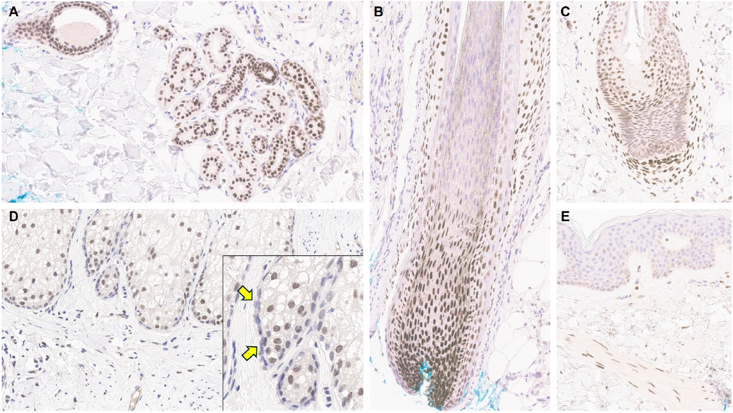
PDF - Over the past 4 years, trichorhinophalangeal syndrome type 1 (TRPS1) has rapidly gained attention among practicing pathologists, with numerous studies emerging that both support and question its diagnostic utility. Initially regarded as a highly specific marker for tumors of mammary origin, TRPS1 is now recognized to have broader expression patterns, including in a variety of cutaneous neoplasms. This is likely due to embryologic parallels between breast tissue and skin adnexal structures, an overlap that was underappreciated in early investigations. Although TRPS1 lacks absolute specificity—even among cutaneous neoplasms—it can still offer meaningful diagnostic value when interpreted alongside conventional immunohistochemical markers and within the appropriate morphologic context. Noteworthy diagnostic applications include mammary Paget disease, primary extramammary Paget disease, rare adnexal neoplasms such as endocrine mucin-producing sweat gland carcinoma and primary cutaneous NUT adnexal carcinoma, and cutaneous metastases from breast carcinoma. In this review, we present the most comprehensive and up-to-date evaluation of the utility and limitations of TRPS1 immunohistochemistry in dermatopathology. Our aim is to deepen understanding of this emerging marker and provide practical guidance on its optimal integration with established immunohistochemical panels to enhance diagnostic accuracy in routine practice.
- Primary renal BCOR::CCNB3 sarcoma in a female patient: case report
- Somang Lee, Binnari Kim
- J Pathol Transl Med. 2025;59(1):84-90. Published online January 15, 2025
- DOI: https://doi.org/10.4132/jptm.2024.09.30
- 5,597 View
- 179 Download
- 1 Web of Science
- 1 Crossref
-
Abstract
PDF
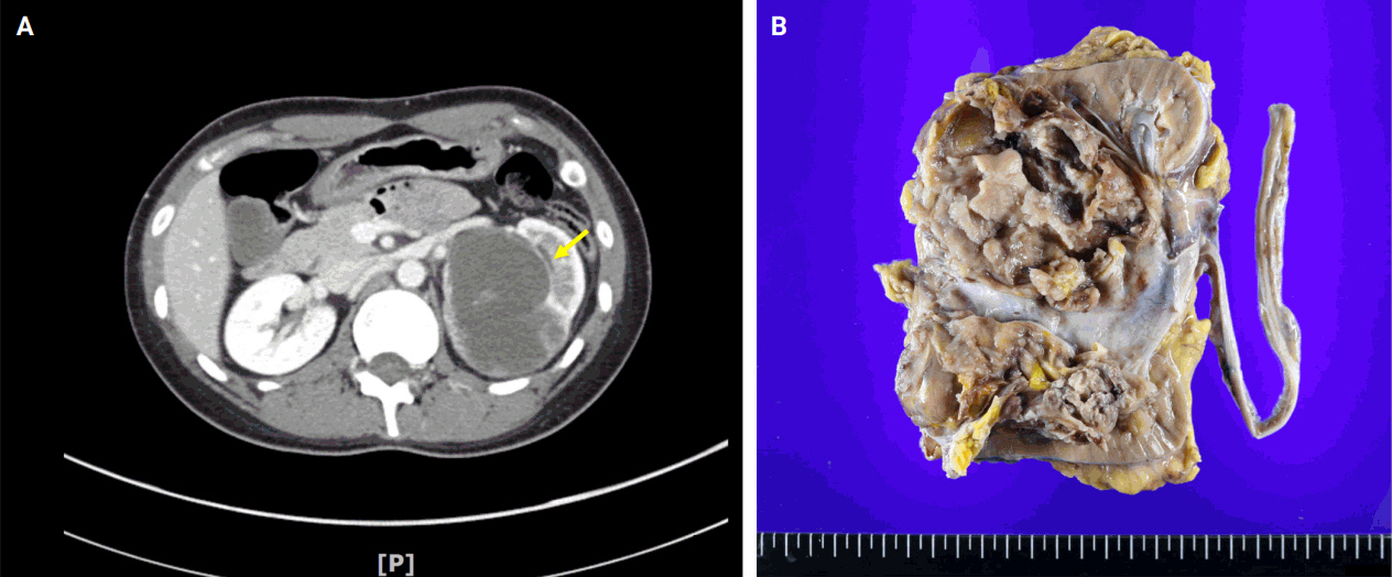
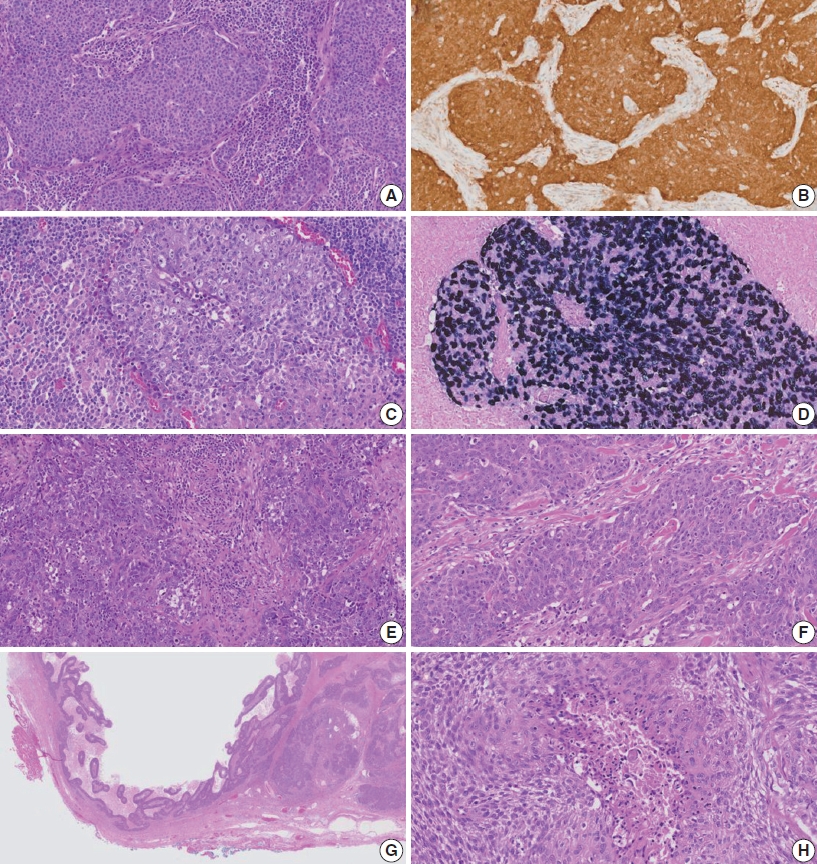
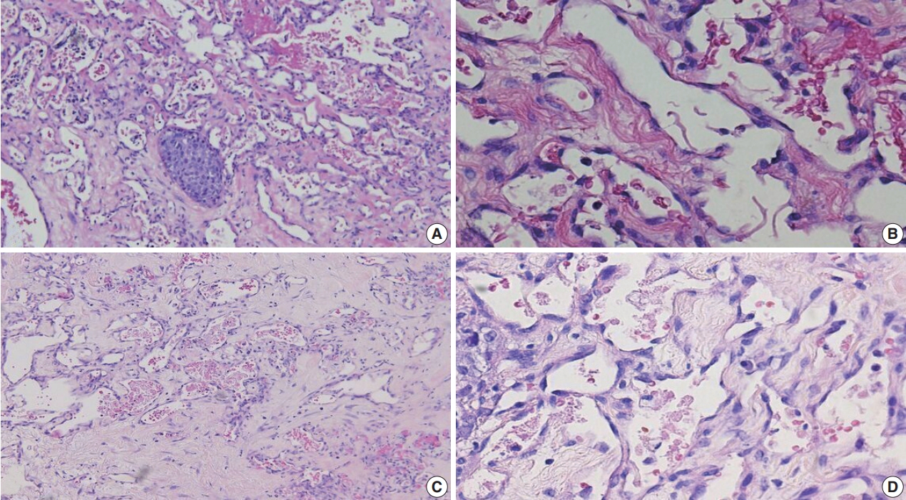
- BCOR-rearranged sarcoma was classified by the World Health Organization in 2020 as a new subgroup of undifferentiated small round-cell sarcoma. It is known to occur very rarely in the kidney. This report presents the first case of a primary renal BCOR::CCNB3 sarcoma in a 22-year-old woman. An 8-cm cystic mass was identified in the left kidney by abdominal pelvic computed tomography. Histopathologic examination revealed the mass to be composed of small round to oval or spindle cells with fibrous septa and a delicate vascular network. A BCOR::CCNB3 fusion was detected by next-generation sequencing–based molecular testing. BCOR::CCNB3 sarcoma presents diagnostic difficulties, highlighting the importance of recognizing its histological features. Immunohistochemical markers are helpful for diagnosis, but genetic molecular testing is necessary for accurate diagnosis. These tumors have a very poor and aggressive prognosis, and an optimal therapeutic regimen has not yet been defined. Therefore, further studies are needed.
-
Citations
Citations to this article as recorded by
- Update on the management of BCOR::CCNB3 sarcoma
Jungo Imanishi, Kenji Sato, Yoshinao Kikuchi, Asako Yamamoto, Shiori Watabe, Taisuke Matsuyama, Chiaki Sato, Hiroshi Kobayashi, Hirotaka Kawano
Japanese Journal of Clinical Oncology.2025; 55(10): 1097. CrossRef
- Update on the management of BCOR::CCNB3 sarcoma
- Clinicopathologic characterization of cervical metastasis from an unknown primary tumor: a multicenter study in Korea
- Miseon Lee, Uiree Jo, Joon Seon Song, Youn Soo Lee, Chang Gok Woo, Dong-Hoon Kim, Jung Yeon Kim, Sun Och Yoon, Kyung-Ja Cho
- J Pathol Transl Med. 2023;57(3):166-177. Published online May 10, 2023
- DOI: https://doi.org/10.4132/jptm.2023.04.12
- 6,950 View
- 172 Download
- 6 Web of Science
- 5 Crossref
-
Abstract
PDF
 Supplementary Material
 - Background
Research regarding cervical metastasis from an unknown primary tumor (CUP) according to human papillomavirus (HPV) and Epstein-Barr virus (EBV) status in Korea has been sporadic and small-scale. This study aims to analyze and understand the characteristics of CUP in Korea according to viral and p16 and p53 status through a multicenter study.
Methods
Ninety-five cases of CUP retrieved from six hospitals in Korea between January 2006 and December 2016 were subjected to high-risk HPV detection (DNA in situ hybridization [ISH] or real-time polymerase chain reaction), EBV detection (ISH), and immunohistochemistry for p16 and p53.
Results
CUP was HPV-related in 37 cases (38.9%), EBV-related in five cases (5.3%), and unrelated to HPV or EBV in 46 cases (48.4%). HPV-related CUP cases had the best overall survival (OS) (p = .004). According to the multivariate analysis, virus-unrelated disease (p = .023) and longer smoking duration (p < .005) were prognostic factors for poor OS. Cystic change (p = .016) and basaloid pattern (p < .001) were more frequent in HPV-related cases, and lymphoepithelial lesion was frequent in EBV-related cases (p = .010). There was no significant association between viral status and p53 positivity (p = .341), smoking status (p = .728), or smoking duration (p = .187). Korean data differ from Western data in the absence of an association among HPV, p53 positivity, and smoking history.
Conclusions
Virus-unrelated CUP in Korea had the highest frequency among all CUP cases. HPV-related CUP is similar to HPV-mediated oropharyngeal cancer and EBVrelated CUP is similar to nasopharyngeal cancer in terms of characteristics, respectively. -
Citations
Citations to this article as recorded by- Management of squamous cell carcinoma of unknown primary in the head and neck: current evidence-based diagnostic and treatment strategies
Marcel Kloppenburg, Matthias Santer, Lukas Schmutzler, Felix Johnson, Benedikt Hofauer, Teresa Steinbichler
memo - Magazine of European Medical Oncology.2026; 19(1): 45. CrossRef - Differenzierung von benignen und malignen Halszysten – eine diagnostische Herausforderung
Christina Sauter, Matthias Sand, Karim Plath, Michaela Maria Plath
Laryngo-Rhino-Otologie.2025; 104(05): 296. CrossRef - Unlocking the Hidden: Advancing Imaging Techniques in Diagnosing Cancers of Unknown Primary in the Head and Neck Region
Daniela Messineo, Filippo Valentini, Giovanni Francesco Niccolini, Federica Zoccali, Francesca Ripari, Enrico Marotta, Marcello Caratozzolo, Pasquale Frisina
Applied Sciences.2025; 15(4): 2194. CrossRef - Characterization of undifferentiated carcinoma of the salivary gland: clinicopathological and immunohistochemical analyses in comparison with lymphoepithelial carcinoma
Sangjoon Choi, Gyuheon Choi, Hee Jin Lee, Joon Seon Song, Yoon Se Lee, Seung-Ho Choi, Kyung-Ja Cho
Journal of Pathology and Translational Medicine.2025; 59(6): 361. CrossRef - Expansion of tumor-infiltrating lymphocytes from head and neck squamous cell carcinoma to assess the potential of adoptive cell therapy
Sangjoon Choi, Mofazzal Hossain, Hyun Lee, Jina Baek, Hye Seon Park, Chae-Lyul Lim, DoYeon Han, Taehyun Park, Jong Hyeok Kim, Gyungyub Gong, Mi-Na Kweon, Hee Jin Lee
Cancer Immunology, Immunotherapy.2024;[Epub] CrossRef
- Management of squamous cell carcinoma of unknown primary in the head and neck: current evidence-based diagnostic and treatment strategies
- A clinicopathologic and immunohistochemical study of primary and secondary breast angiosarcoma
- Evi Abada, Hyejeong Jang, Seongho Kim, Rouba Ali-Fehmi, Sudeshna Bandyopadhyay
- J Pathol Transl Med. 2022;56(6):342-353. Published online October 27, 2022
- DOI: https://doi.org/10.4132/jptm.2022.08.31
- 7,141 View
- 151 Download
- 7 Web of Science
- 7 Crossref
-
Abstract
PDFSupplementary Material
- Background
We aimed to study the clinicopathologic and immunohistochemical (IHC) (CD117, c-Myc, and p53) characteristics, and overall survival of primary and secondary breast angiosarcoma (BAS).
Methods
This was a retrospective study of BAS cases diagnosed between 1997 and 2020 at our institution. Hematoxylin and eosin-stained slides were reviewed for tumor morphology, margin status, and lymph node metastasis. CD117, p53, D2-40, CD31, and c-Myc IHC stains were performed on 11 viable tissue blocks. Additional clinical information was obtained from the electronic medical records.
Results
Seventeen patients with BAS were identified. Of these, five (29%) were primary and 12 (71%) were secondary BAS, respectively. The median age at diagnosis for primary BAS was 36 years. The median age at diagnosis for secondary BAS was 67 years. The median time to secondary BAS development following radiotherapy was 6.5 years (range, 2 to 12 years). There was no significant difference between primary and secondary BAS in several histopathologic parameters examined, including histologic grade, necrosis, mitotic count, lymph node metastasis, and positive tumor margins. There was also no difference in CD117, p53, D2-40, CD31, and c-Myc expression by IHC between primary and secondary BAS. During a median followup of 21 months, primary BAS had two (40%) reported deaths and secondary BAS had three (25%) reported deaths. However, this difference in survival between both groups was not statistically significant (hazard ratio, 0.51; 95% confidence interval, 0.09 to 3.28; p = .450).
Conclusions
BAS is a rare and aggressive disease. No histologic, IHC (CD117, c-Myc, and p53), or survival differences were identified between primary and secondary BAS in this study. -
Citations
Citations to this article as recorded by- Prognostic significance of clinicopathological parameters, margin width and locoregional recurrences on outcome of primary and radiation associated breast angiosarcoma- results from a large UK sarcoma regional service
Samar Ali, Salena Bains, Emily Fox, Anant Desai, Mike Hallissey, Alaa El-Ghobashy, Robert Warner, Abeer M. Shaaban
Breast Cancer Research.2026;[Epub] CrossRef - Angiosarcoma: a systematic review of biomarkers in diagnosis, prognosis, and therapeutic strategies
Huyen Thuc Tran Luong, Sofie Vercammen, Ario de Marco, Hilde de Rooster, Antonio Cosma
Frontiers in Oncology.2025;[Epub] CrossRef - Etiology, pathogenesis, and management of angiosarcoma associated with implants and foreign body: Clinical cases and research updates
Ramy Samargandi
Medicine.2024; 103(18): e37932. CrossRef - Ovarian angiosarcoma: A systematic review of literature and survival analysis
Shafi Rehman, Arya Harikrishna, Amisha Silwal, B.R. Sumie, Safdar Mohamed, Nisha Kolhe, Meghana Maddi, Linh Huynh, Jesus Gutierrez, Yoshita Rao Annepu, Ameer Mustafa Farrukh
Annals of Diagnostic Pathology.2024; 73: 152331. CrossRef - Neoadjuvant chemotherapy for radiation associated angiosarcoma (RAAS) of the breast: A retrospective single center study
Stijn J.C. van der Burg, Sophie J.M. Reijers, Anke Kuijpers, Lotte Heimans, Astrid N. Scholten, Rick L.M. Haas, Hester van Boven, Willemijn M. Kolff, Marie-Jeanne T.F.D. Vrancken Peeters, Martijn Kerst, Beatrijs A. Seinstra, Neeltje Steeghs, Winette T.A.
The Breast.2024; 78: 103825. CrossRef - Lymph node involvement in secondary breast angiosarcoma – a case presentation
Adriana Irina Ciuvică, Tiberiu Augustin Georgescu , Andrei Dennis Voichiţoiu , Angela Arsene , Luchian Marinescu , George Ionuţ Bucur , Livia Iordache , Nahedd Saba
Romanian Journal of Morphology and Embryology.2024; 65(3): 523. CrossRef - Primary ovarian angiosarcoma: Two case reports and review of literature
Ying Zhou, Yi-Wen Sun, Xiao-Yang Liu, Dan-Hua Shen
World Journal of Clinical Cases.2023; 11(21): 5122. CrossRef
- Prognostic significance of clinicopathological parameters, margin width and locoregional recurrences on outcome of primary and radiation associated breast angiosarcoma- results from a large UK sarcoma regional service
- Clinically undetected plasmacytoid urothelial carcinoma of the urinary bladder with non-mass-forming metastases in multiple organs: an autopsy case
- Yuya Asano, Kosuke Miyai, Shinya Yoshimatsu, Makoto Sasaki, Katsunori Ikewaki, Susumu Matsukuma
- J Pathol Transl Med. 2022;56(4):217-224. Published online May 3, 2022
- DOI: https://doi.org/10.4132/jptm.2022.03.15
- 9,332 View
- 167 Download
- 3 Web of Science
- 6 Crossref
-
Abstract
PDF
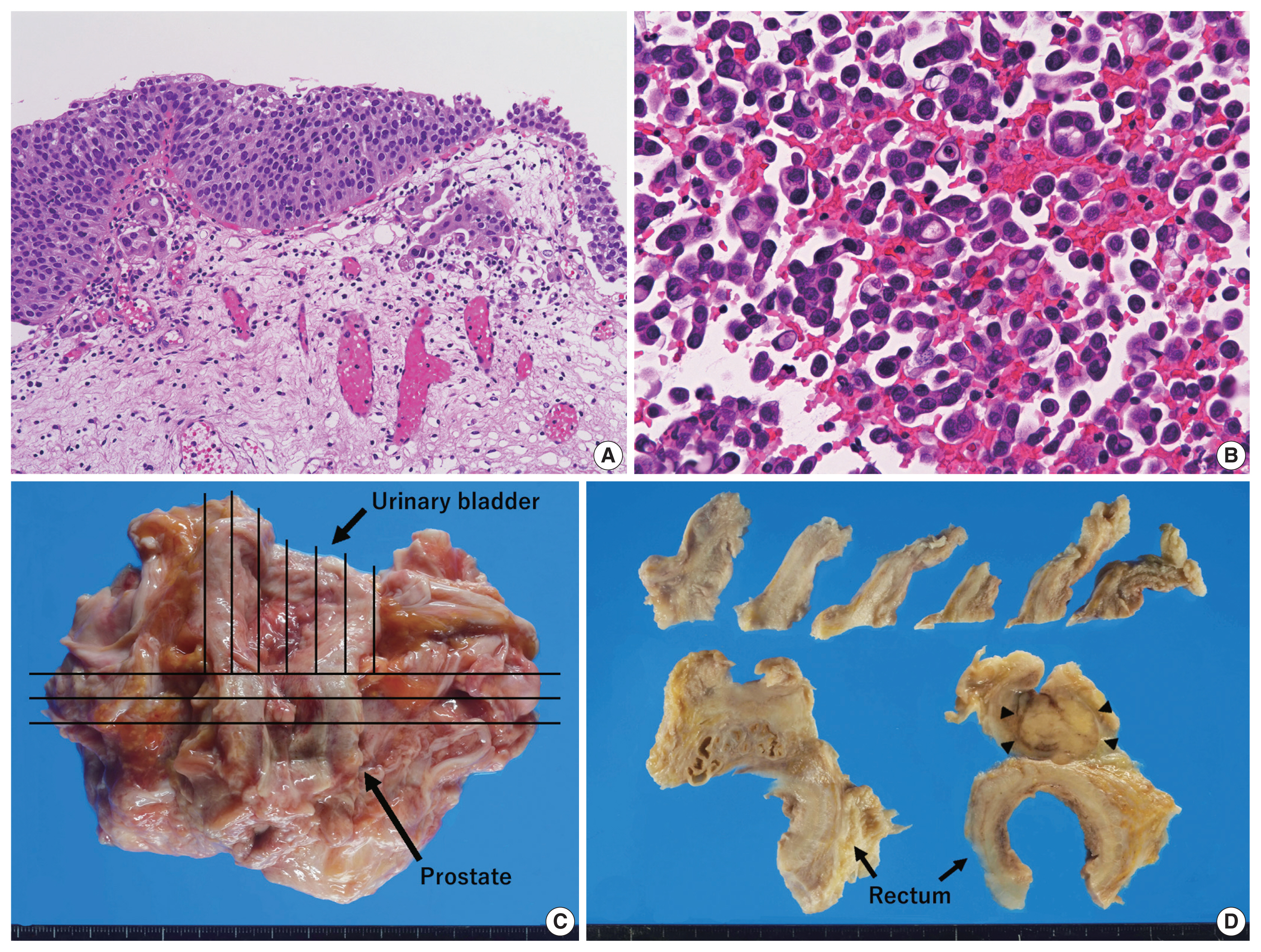
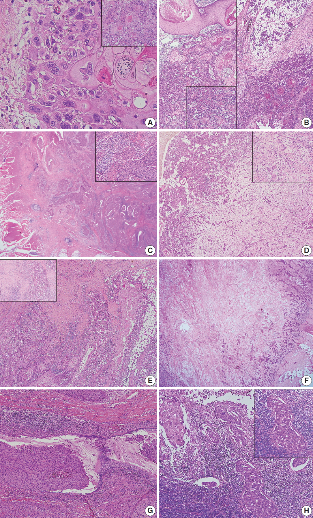
- This case report outlines a clinically undetected urinary bladder plasmacytoid urothelial carcinoma (PUC) with multiple metastases detected at autopsy. An 89-year-old man presented with edema in the lower limbs. Pleural fluid cytology revealed discohesive carcinomatous cells, although imaging studies failed to identify the primary site of tumor. The patient died of respiratory failure. Autopsy disclosed a prostate tumor and diffusely thickened urinary bladder and rectum without distinct tumorous lesions. Histologically, the tumor consisted of acinar-type prostate adenocarcinoma with no signs of metastasis. Additionally, small, plasmacytoid tumor cells were observed in the urinary bladder/rectum as isolated or small clustering fashions. These metastasized to the lungs, intestine, generalized lymph nodes in a non-mass-forming manner. Combined with immunohistochemical studies, these tumor cells were diagnosed PUC derived from the urinary bladder. Both clinicians and pathologists should recognize PUC as an aggressive histological variant, which can represent a rapid systemic progression without mass-forming lesions.
-
Citations
Citations to this article as recorded by- Plasmacytoid Urothelial Carcinoma with Initial Presentation as a Secondary
Prostatic Tumor: Diagnostic Pitfalls
and Literature Review
丰锦 李
Advances in Clinical Medicine.2026; 16(02): 1264. CrossRef - Severe Rectal Stenosis as the First Clinical Appearance of a Metastasis Originating from the Bladder: A Case Report and Literature Review
Claudiu Daha, Eugen Brătucu, Ioan Burlănescu, Virgiliu-Mihail Prunoiu, Hortensia-Alina Moisă, Ștefania Ariana Neicu, Laurențiu Simion
Life.2025; 15(5): 682. CrossRef - Carcinomatous Meningitis and Hydrocephalus in Plasmacytoid Urothelial Carcinoma of the Urinary Bladder With Extremely Elevated CA19-9 Levels
Fumiaki Henmi, Kayako Ukai, Atsuhito Nakayama, Yutaka Takazawa, Yoshikazu Uesaka
Cureus.2024;[Epub] CrossRef - Current Advances in the Management of Nonurothelial Subtypes of Bladder Cancer
Evangelia Vlachou, Burles Avner Johnson, Ezra Baraban, Rosa Nadal, Jean Hoffman-Censits
American Society of Clinical Oncology Educational Book.2024;[Epub] CrossRef - Plasmacytoid urothelial carcinoma: a multidisciplinary approach to the diagnosis and management
Marcus Zorovich, Jude Khatib, Aysha Mubeen, Katie Gardner, Nayana Patel
Abdominal Radiology.2024; 51(3): 1397. CrossRef - Divergent Histology in Bladder Cancer: What We Need to Know?
Shashank Agrawal, Arun Ramdas Menon, Ginil Kumar Pooleri
UroCancer Clinics of India.2024; 2(2): 100. CrossRef
- Plasmacytoid Urothelial Carcinoma with Initial Presentation as a Secondary
Prostatic Tumor: Diagnostic Pitfalls
and Literature Review
- Primary squamous cell carcinoma of the salivary gland: immunohistochemical analysis and comparison with metastatic squamous cell carcinoma
- Uiree Jo, Joon Seon Song, Seung-Ho Choi, Soon Yuhl Nam, Sang Yoon Kim, Kyung-Ja Cho
- J Pathol Transl Med. 2020;54(6):489-496. Published online August 31, 2020
- DOI: https://doi.org/10.4132/jptm.2020.07.19
- 11,020 View
- 210 Download
- 18 Web of Science
- 18 Crossref
-
Abstract
PDFSupplementary Material
- Background
Primary squamous cell carcinoma (SCC) of the salivary gland is a rare disease, and distinguishing primary SCC from metastatic SCC is difficult. This study investigated the histological and immunohistochemical differences between primary and metastatic salivary gland SCC to improve the accuracy of diagnosis and to explore the pathogenesis of this disease.
Methods
Data of 16 patients who underwent surgery for SCC of salivary glands between 2000 and 2018 at Asan Medical Center were retrieved. Eight patients had a history of SCC at other sites, and eight patients had only salivary gland SCC. Immunostaining for p16, p53, androgen receptor (AR), gross cystic disease fluid protein 15 (GCDFP-15), and c-erbB2, as well as mucicarmine staining, were compared between the two groups.
Results
Most tumors were located in the center of the salivary glands with extraparenchymal extension. The histology of primary SCC of the salivary gland was consistent with moderately differentiated SCC with extensive desmoplastic reaction and peritumoral inflammation. Involvement of the salivary gland ducts and transition into the ductal epithelium were observed in two cases. Metastatic SCC resembled the primary tumor histologically and was associated with central necrosis. Both groups exhibited negative mucin staining. Two, one, and one primary SCC case exhibited AR, GCDFP-15, and c-erbB2 positivity, respectively.
Conclusions
A subset of primary SCCs originated in salivary ducts or was related to salivary duct carcinoma. Distinguishing primary from metastatic SCC of the salivary gland is difficult using histologic features and immunoprofiles. A comprehensive review of the medical history is essential. -
Citations
Citations to this article as recorded by- Clinical diagnosis, treatment, and survival analysis of 61 cases of salivary duct carcinoma: a retrospective study
Shubin Dong, Mengru Li, Zhiwei Zhang, Bowei Feng, Wei Ding, Jiang Chang, Feng Liu
PeerJ.2025; 13: e19626. CrossRef - Characterization of undifferentiated carcinoma of the salivary gland: clinicopathological and immunohistochemical analyses in comparison with lymphoepithelial carcinoma
Sangjoon Choi, Gyuheon Choi, Hee Jin Lee, Joon Seon Song, Yoon Se Lee, Seung-Ho Choi, Kyung-Ja Cho
Journal of Pathology and Translational Medicine.2025; 59(6): 361. CrossRef - Primary salivary gland squamous cell carcinoma with sialolithiasis in the submandibular gland: A case report and literature review
Sawako Ono, Katsutoshi Hirose, Yuji Hirata, Marie Yamada, Satoko Nakamura, Hidetaka Yamamoto
Journal of Oral and Maxillofacial Surgery, Medicine, and Pathology.2024; 36(5): 768. CrossRef - A case of primary squamous cell carcinoma of the parotid gland and review of the literature
Jingli Zhao, Xinrong Nan, Chuhuan Zhou, Nan Jiang, Liangliang Tian
Journal of Case Reports and Images in Oncology.2024; 10(1): 7. CrossRef - Metastatic cutaneous squamous cell carcinoma accounts for nearly all squamous cell carcinomas of the parotid gland
Patrick J. Bradley, Göran Stenman, Lester D. R. Thompson, Alena Skálová, Roderick H. W. Simpson, Pieter J. Slootweg, Alessandro Franchi, Nina Zidar, Alfons Nadal, Henrik Hellquist, Michelle D. Williams, Ilmo Leivo, Abbas Agaimy, Alfio Ferlito
Virchows Archiv.2024; 485(1): 3. CrossRef - Common skin cancers and their association with other non-cutaneous primary malignancies: a review of the literature
Lindsay Holic
Medical Oncology.2024;[Epub] CrossRef - Salivary duct carcinoma with squamous differentiation: histomorphological and immunophenotypical analysis of six cases
Melad N Dababneh, Christopher C Griffith, Kelly R Magliocca, Ivan J Stojanov
Histopathology.2024; 85(4): 590. CrossRef - Comprehensive Next Generation Sequencing Reveals that Purported Primary Squamous Cell Carcinomas of the Parotid Gland are Genetically Heterogeneous
Justin A. Bishop, Masato Nakaguro, Ilan Weinreb, Doreen Palsgrove, Lisa M. Rooper, Travis W. Vandergriff, Brian Carlile, Jeffrey A. Sorelle, Jeffrey Gagan, Toshitaka Nagao
Head and Neck Pathology.2024;[Epub] CrossRef - Salivary gland fine needle aspiration: a focus on diagnostic challenges and tips for achieving an accurate diagnosis
Carla Saoud, Hansen Lam, Sandra I. Sanchez, Zahra Maleki
Diagnostic Histopathology.2023; 29(8): 357. CrossRef - Salivary gland pathologies: evolution in classification and association with unique genetic alterations
Michał Żurek, Łukasz Fus, Kazimierz Niemczyk, Anna Rzepakowska
European Archives of Oto-Rhino-Laryngology.2023; 280(11): 4739. CrossRef - A retrospective study of nonneoplastic and neoplastic disorders of the salivary glands
Sorin Vamesu, Oana Andreea Ursica, Ana Maria Gurita, Raluca Ioana Voda, Mariana Deacu, Mariana Aschie, Madalina Bosoteanu, Georgeta Camelia Cozaru, Anca Florentina Mitroi, Cristian Ionut Orasanu
Medicine.2023; 102(42): e35751. CrossRef - Pembrolizumab as a first line therapy in a patient with extensive mucoepidermoid salivary gland carcinoma. A complete clinical, radiological and pathological response. A very specific case
Raed Farhat, Noam Asna, Yaniv Avraham, Ashraf Khater, Majd Asakla, Alaa Safia, Sergio Szvalb, Nidal Elkhatib, Shlomo Merchavy
Discover Oncology.2022;[Epub] CrossRef - Morphologic CT and MRI features of primary parotid squamous cell carcinoma and its predictive factors for differential diagnosis with mucoepidermoid carcinoma
Xiaohua Ban, Huijun Hu, Yue Li, Lingjie Yang, Yu Wang, Rong Zhang, Chuanmiao Xie, Cuiping Zhou, Xiaohui Duan
Insights into Imaging.2022;[Epub] CrossRef - A Rare Case of Primary Squamous Cell Carcinoma of the Submandibular Salivary Gland: Brief Overview of Diagnostic Ambiguity and Treatment Challenges
Pawan Hingnikar, Anendd Jadhav, Nitin D Bhola
Cureus.2022;[Epub] CrossRef - Necrotizing Sialometaplasia of the Hard Palate: Diagnosis and
Treatment
Sangeun Lee, Yun Sung Lim, Kyuho Lee, Bo Hae Kim
Journal of Clinical Otolaryngology Head and Neck Surgery.2022; 33(4): 236. CrossRef - Parotid Salivary Duct Carcinoma With a Prominent Squamous Component: Immunohistochemical Profile, Diagnostic Pitfalls, and Therapeutic Implications
Naomi Hardy, Joshua Thompson, Ranee Mehra, Cinthia B. Drachenberg, Kyle Hatten, John C. Papadimitriou
International Journal of Surgical Pathology.2021; 29(7): 726. CrossRef - Intrasalivary Thymic Carcinoma: A Case Report and Literature Review
Michał Kunc, Alexandra Kamieniecki, Grzegorz Walczak, Tomasz Nowicki, Bartosz Wasąg, Bogusław Mikaszewski, Dominik Stodulski, Wojciech Biernat
Head and Neck Pathology.2021; 16(3): 857. CrossRef - Cancer Stem Cell Markers in Squamous Cell Carcinomas of the Salivary Glands
Mattis Bertlich, Julia Kitz, Marie Kruizenga, Jennifer Lee Spiegel, Martin Canis, Friedrich Ihler, Frank Haubner, Bernhard G. Weiss, Mark Jakob
Oncology.2021; 99(6): 402. CrossRef
- Clinical diagnosis, treatment, and survival analysis of 61 cases of salivary duct carcinoma: a retrospective study
- Analysis of PAX8 immunohistochemistry in lung cancers: a meta-analysis
- Jae Han Jeong, Nae Yu Kim, Jung-Soo Pyo
- J Pathol Transl Med. 2020;54(4):300-309. Published online July 10, 2020
- DOI: https://doi.org/10.4132/jptm.2020.06.08
- 10,173 View
- 155 Download
- 11 Web of Science
- 9 Crossref
-
Abstract
PDF
- Background
In this meta-analysis, we aimed to evaluate the PAX8 immunohistochemical expressions in primary lung cancers and metastatic cancers to the lung.
Methods
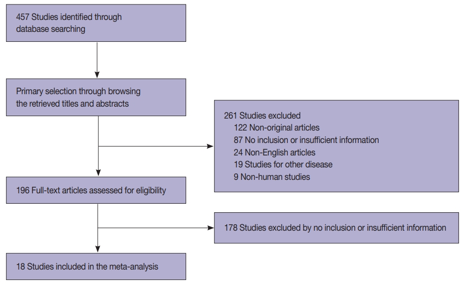
We identified and reviewed relevant articles from the PubMed databases. Ultimately, 18 articles were included in this meta-analysis. PAX8 expression rates were analyzed and compared between primary and metastatic lung cancers.
Results
The PAX8 expression rate in primary lung cancers was 0.042 (95% confidence interval [CI], 0.025 to 0.071). PAX8 expression rates of small cell (0.129; 95% CI, 0.022 to 0.496) and non-small cell carcinomas of the lung (0.037; 95% CI, 0.022 to 0.061) were significantly different (p=.049 in a meta-regression test). However, the PAX8 expression rates of adenocarcinoma (0.013; 95% CI, 0.006 to 0.031) and squamous cell carcinoma (0.040; 95% CI, 0.016 to 0.097) were not significantly different. PAX8 expression rates of metastatic carcinomas to the lung varied, ranging from 1.8% to 94.9%. Metastatic carcinomas from the lung to other organs had a PAX8 expression rate of 6.3%. The PAX8 expression rates of metastatic carcinomas from the female genital organs, kidneys, and thyroid gland to the lung were higher than those of other metastatic carcinomas.
Conclusions
Primary lung cancers had a low PAX8 expression rate regardless of tumor subtype. However, the PAX8 expression rates of metastatic carcinomas from the female genital organs, kidneys, and thyroid were significantly higher than those of primary lung cancers. -
Citations
Citations to this article as recorded by- Clinical significance of lncRNA PAX8-AS1 and miR-96-5p in non-small cell lung cancer
Qiaoling Ying, Hui Xu, Xiaojiao Wu, Hang Fang, Jingjing Shi, Hangcheng Pan
Journal of Cardiothoracic Surgery.2025;[Epub] CrossRef - Lung Metastatic Recurrence as Carcinosarcoma from Ovarian Mesonephric-Like Adenocarcinoma: A Case Report
Kaito Nakama, Masayuki Ota, Takanori Aihara, Satoko Kageyama, Jun-ichiro Ikeda
International Journal of Surgical Pathology.2025;[Epub] CrossRef - The TTF-1 and Napsin A Trap: Metastatic Endometrial Carcinoma Masquerading as Lung Primary
Carmen Alfonso-Rosa, Jesús Machuca-Aguado, Ana María Montaña-Ramírez, Francisco Javier Rubio-Garrido
International Journal of Surgical Pathology.2025;[Epub] CrossRef - Prognostic value of PAX8 in small cell lung cancer
Fengyun Tao, Hangyan Zhu, Jiayun Xu, Yanan Guo, Xin Wang, Lei Shao, Deng Pan, Guosheng Li, Rong Fang
Heliyon.2024; 10(7): e28251. CrossRef - Cystic primary squamous cell carcinoma of the thyroid
Sakurako Harada‐Kagitani, Yusuke Kouchi, Yoshiki Shinomiya, Takuto Hiramoto, Tomoyuki Arai, Toyoyuki Hanazawa, Kiyotaka Onodera, Kaito Nakama, Takanori Aihara, Masayuki Ota, Jun‐Ichiro Ikeda, Takashi Kishimoto
Pathology International.2024; 74(5): 292. CrossRef - The combination of p16 and Rb expression pattern is helpful to predict high-risk HPV infection and the primary site in lymph node metastases of squamous cell carcinoma
Ryosuke Kuga, Hidetaka Yamamoto, Fumiya Narutomi, Misa Suzuki, Rina Jiromaru, Takahiro Hongo, Kazuhisa Hachisuga, Nobuko Yasutake, Kiyoko Kato, Takashi Nakagawa, Yoshinao Oda
Pathology - Research and Practice.2024; 263: 155642. CrossRef - Mesonephric adenocarcinoma of the uterine cervix with a prominent spindle cell component
Yingying Fan, Ying He, Liang Sun, Tianmin Liu, Yangmei Shen
Oncology Letters.2024;[Epub] CrossRef - Immunocytochemistry of effusions: Processing and commonly used immunomarkers
Vinod B. Shidham, Beata Janikowski
Cytojournal.2022; 19: 6. CrossRef - Significance analysis of PAX8 expression in endometrial carcinoma
Shan Hu, Hua Gan, Fengmei Yang
Medicine.2022; 101(42): e31159. CrossRef
- Clinical significance of lncRNA PAX8-AS1 and miR-96-5p in non-small cell lung cancer
- Primary Rhabdomyosarcoma of the Breast: Study of Three Cases at One Institution with a Review of Primary Breast Sarcomas
- Junyoung Shin, Hee Jeong Kim, Dae-Yeon Kim, Gyungyub Gong, Kyung-Ja Cho
- J Pathol Transl Med. 2019;53(5):308-316. Published online August 2, 2019
- DOI: https://doi.org/10.4132/jptm.2019.07.22
- 8,132 View
- 139 Download
- 7 Web of Science
- 9 Crossref
-
Abstract
PDF
- Background
Primary breast sarcoma (PBS) is rare, comprising approximately 1% of breast malignancies. Rhabdomyosarcoma (RMS) accounts for an extremely small proportion of PBSs, often leading to delayed histologic confirmation.
Methods
Upon reviewing Asan Medical Center’s pathology database between 2000 and 2018, 41 PBS cases were retrieved, including three cases of primary RMS of the breast. Their clinicopathological features were analyzed, and the literature related to PBS and primary RMS of the breast was reviewed.
Results
We identified three primary breast RMS cases from our institution database, comprising 7.3% of PBS: one case each of spindle cell/sclerosing RMS (ssRMS), alveolar RMS (aRMS), and embryonal RMS (eRMS). All cases involved adolescents or young adults (14, 16, and 25 years, respectively) who underwent mastectomy or radiotherapy and were confirmed using immunohistochemical testing for myogenin, desmin, and myogenic differentiation. The ssRMS patient experienced recurrence at the operation site 4 months post-surgery despite undergoing concurrent chemoradiotherapy. The aRMS patient had multiple metastases at diagnosis and showed FAX3-FOXO1 fusion transcripts; she died 22 months after the diagnosis. The eRMS patient had enlarged axillary lymph nodes; post-radiotherapy, the lesion recurred as multiple metastases to the bone and lung. She died 18 months post-diagnosis.
Conclusions
Our experience on RMS cases suggests that spindle cell or small round cell malignancy in breasts of young female should raise suspicion for the possibility of primary or secondary RMS. To our knowledge, this is the second report of primary breast ssRMS and it may help clinicians who encounter this rare disease in the future. -
Citations
Citations to this article as recorded by- Primary Ewing sarcoma of the breast in a male adolescent: A case report
Joie Sheen A. Bastian, Willie T. Hao, Lawrence Faith A. Lucañas-Yap
Journal of Pediatric Surgery Case Reports.2026; 127: 103205. CrossRef - Clinicopathological and molecular features of breast metastases in alveolar rhabdomyosarcoma: A series of 3 cases
Wanni Xu, Li Yang, Yuxin Hui, Peizhuo Yao, Yiwei Jia, Xinyu Wei, Shuqun Zhang
Annals of Diagnostic Pathology.2026; 83: 152627. CrossRef - Management of Pediatric Breast Masses for the Pediatric Surgeon: Expert Consensus Recommendations From the APSA Cancer Committee
Dana Schwartz, Elisabeth T. Tracy, Bindi Naik-Mathuria, Richard D. Glick, Stephanie F. Polites, Peter Mattei, David Rodeberg, Andres F. Espinoza, Sara A. Mansfield, Dave R. Lal, Meera Kotagal, Timothy Lautz, Jennifer Aldrink, Barrie S. Rich
Journal of Pediatric Surgery.2025; 60(2): 161916. CrossRef - Differential diagnosis of primary mesenchymal neoplasms of the breast
Mine Ozsen, Seyit Ali Volkan Polatkan, Ulviye Yalcınkaya, Sahsine Tolunay, Mustafa Sehsuvar Gokgoz
Clinical and Translational Oncology.2024; 27(1): 223. CrossRef - Primary breast rhabdomyosarcoma in a 17-year-old girl
Laxmi Singotia, V.S. Haritha
Journal of Cancer Research and Therapeutics.2023; 19(7): 2070. CrossRef - High-Grade Spindle Cell Lesions of the Breast
Esther Yoon, Qingqing Ding, Kelly Hunt, Aysegul Sahin
Surgical Pathology Clinics.2022; 15(1): 77. CrossRef - Primary Small Cell Malignancies of the Breast: Are They Rare Malignancies?
Kemal Behzatoğlu, Fernando Schmitt
Acta Cytologica.2022; 66(4): 347. CrossRef - Recurrent malignant phyllodes tumor of the breast: An extremely rare case of recurrence with only rhabdomyosarcoma components
Jia Han, Shuice Liu, Akihoro Shioya, Motona Kumagai, Emi Morioka, Miki Noguchi, Masafumi Inokuchi, Sohsuke Yamada
SAGE Open Medical Case Reports.2022;[Epub] CrossRef - Primary rhabdomyosarcoma: An extremely rare and aggressive variant of male breast cancer
Cătălin Bogdan Satală, Ioan Jung, Tivadar Jr Bara, Patricia Simu, Iunius Simu, Madalina Vlad, Rita Szodorai, Simona Gurzu
World Journal of Clinical Cases.2020; 8(19): 4466. CrossRef
- Primary Ewing sarcoma of the breast in a male adolescent: A case report
- Diffuse Involvement of Primary Colorectal Lymphoma Simulating Ulcerative Colitis
- Ji-Ye Kim, Sun Hee Chang, Han Seong Kim, Mee Joo
- J Pathol Transl Med. 2019;53(5):332-336. Published online August 2, 2019
- DOI: https://doi.org/10.4132/jptm.2019.07.12
- 7,093 View
- 93 Download
-
Abstract
PDF
- Diffuse involvement of colorectal lymphoma masquerading as colitis is a very rare presentation of primary colorectal lymphoma. Detecting occult lymphoma is difficult in the setting of diffuse colonic involvement with no definite mass and inflammatory mucosal changes. We encountered a case of diffuse-type primary colorectal lymphoma simulating ulcerative colitis in a previously healthy 31-year-old woman. Despite multiple mucosal biopsies, the biopsy diagnosis was not made due to unawareness of atypical lymphocytes admixed with dense lymphoplasmacytic infiltration. The present case emphasizes the importance of being aware of this rare presentation of primary colorectal lymphoma in order to avoid misdiagnosis.
- Guanabenz Acetate Induces Endoplasmic Reticulum Stress–Related Cell Death in Hepatocellular Carcinoma Cells
- Hyo Jeong Kang, Hyang Sook Seol, Sang Eun Lee, Young-Ah Suh, Jihun Kim, Se Jin Jang, Eunsil Yu
- J Pathol Transl Med. 2019;53(2):94-103. Published online January 16, 2019
- DOI: https://doi.org/10.4132/jptm.2019.01.14
- 10,045 View
- 201 Download
- 13 Web of Science
- 13 Crossref
-
Abstract
PDFSupplementary Material
- Background
Development of chemotherapeutics for the treatment of advanced hepatocellular carcinoma (HCC) has been lagging. Screening of candidate therapeutic agents by using patient-derived preclinical models may facilitate drug discovery for HCC patients.
Methods
Four primary cultured HCC cells from surgically resected tumor tissues and six HCC cell lines were used for high-throughput screening of 252 drugs from the Prestwick Chemical Library. The efficacy and mechanisms of action of the candidate anti-cancer drug were analyzed via cell viability, cell cycle assays, and western blotting.
Results
Guanabenz acetate, which has been used as an antihypertensive drug, was screened as a candidate anti-cancer agent for HCC through a drug sensitivity assay by using the primary cultured HCC cells and HCC cell lines. Guanabenz acetate reduced HCC cell viability through apoptosis and autophagy. This occurred via inhibition of growth arrest and DNA damage-inducible protein 34, increased phosphorylation of eukaryotic initiation factor 2α, increased activating transcription factor 4, and cell cycle arrest.
Conclusions
Guanabenz acetate induces endoplasmic reticulum stress–related cell death in HCC and may be repositioned as an anti-cancer therapeutic agent for HCC patients. -
Citations
Citations to this article as recorded by- ER stress signaling at the interphase between MASH and HCC
Younis Hazari, Eric Chevet, Béatrice Bailly-Maitre, Claudio Hetz
Hepatology.2026; 83(2): 387. CrossRef - The Integrated Stress Response in Cancer: Paradox and Therapeutic Promise
Chenliang Zhang, Ting Zhang, Qiulin Tang, Yu Zeng, Dan Cao
Comprehensive Physiology.2026;[Epub] CrossRef - The Construction of ceRNA Regulatory Network Unraveled Prognostic Biomarkers and Repositioned Drug Candidates for the Management of Pancreatic Ductal Adenocarcinoma
Busra Aydin, Keziban Okutan, Ozge Onluturk Aydogan, Raghu Sinha, Beste Turanli
Current Issues in Molecular Biology.2025; 47(7): 496. CrossRef - Current trends and future prospects of drug repositioning in gastrointestinal oncology
Nayeralsadat Fatemi, Mina Karimpour, Hoda Bahrami, Mohammad Reza Zali, Vahid Chaleshi, Andrea Riccio, Ehsan Nazemalhosseini-Mojarad, Mehdi Totonchi
Frontiers in Pharmacology.2024;[Epub] CrossRef - Small molecules for impairing endoplasmic reticulum in cancer
Tripti Mishra, Navneet Dubey, Sudipta Basu
Organic & Biomolecular Chemistry.2024; 22(44): 8689. CrossRef -
Guanabenz acetate, an antihypertensive drug repurposed as an inhibitor of
Escherichia coli
biofilm
Arakkaveettil Kabeer Farha, Olivier Habimana, Harold Corke, Olaya Rendueles
Microbiology Spectrum.2024;[Epub] CrossRef - The integrated stress response in cancer progression: a force for plasticity and resistance
Caleb L. Lines, Morgan J. McGrath, Tanis Dorwart, Crystal S. Conn
Frontiers in Oncology.2023;[Epub] CrossRef - Endoplasmic reticulum stress: Multiple regulatory roles in hepatocellular carcinoma
Jiacheng Wu, Shan Qiao, Yien Xiang, Menying Cui, Xiaoxiao Yao, Ruixin Lin, Xuewen Zhang
Biomedicine & Pharmacotherapy.2021; 142: 112005. CrossRef - The two faces of the Integrated Stress Response in cancer progression and therapeutic strategies
Eugenia Licari, Luis Sánchez-del-Campo, Paola Falletta
The International Journal of Biochemistry & Cell Biology.2021; 139: 106059. CrossRef - Repurposing of Guanabenz acetate by encapsulation into long-circulating nanopolymersomes for treatment of triple-negative breast cancer
Yusuf A. Haggag, Mohamed Yasser, Murtaza M. Tambuwala, Suleiman S. El Tokhy, Mohammad Isreb, Ahmed A. Donia
International Journal of Pharmaceutics.2021; 600: 120532. CrossRef - Endoplasmic reticulum stress: New insights into the pathogenesis and treatment of retinal degenerative diseases
Marina S. Gorbatyuk, Christopher R. Starr, Oleg S. Gorbatyuk
Progress in Retinal and Eye Research.2020; 79: 100860. CrossRef - Delineating the role of eIF2α in retinal degeneration
Christopher R. Starr, Marina S. Gorbatyuk
Cell Death & Disease.2019;[Epub] CrossRef - Repositioning of Guanabenz in Conjugation with Gold and Silver Nanoparticles against Pathogenic Amoebae Acanthamoeba castellanii and Naegleria fowleri
Areeba Anwar, Mohammad Ridwane Mungroo, Ayaz Anwar, William J. Sullivan, Naveed Ahmed Khan, Ruqaiyyah Siddiqui
ACS Infectious Diseases.2019; 5(12): 2039. CrossRef
- ER stress signaling at the interphase between MASH and HCC
- WITHDRAWN:Primary Rhabdomyosarcoma of the Breast: A Report of Two Cases and Literature Review
- Junyoung Shin, Hee Jeong Kim, Dae-Yeon Kim, Gyungyub Gong, Kyung-Ja Cho
- Received August 6, 2018 Accepted September 13, 2018 Published online October 4, 2018
- DOI: https://doi.org/10.4132/jptm.2018.09.14
- 4,337 View
- 62 Download
- 1 Crossref
-
Citations
Citations to this article as recorded by- Primary Alveolar Rhabdomyosarcoma of the Breast in an Adult: An Extremely Rare Case
Helen J. Trihia, Natasa Novkovic, Ioannis Provatas, Anastasios Mavrogiorgis, Evangelos Lianos
Case Reports in Pathology.2019; 2019: 1. CrossRef
- Primary Alveolar Rhabdomyosarcoma of the Breast in an Adult: An Extremely Rare Case
- Primary Peripheral Gamma Delta T-Cell Lymphoma of the Central Nervous System: Report of a Case Involving the Intramedullary Spinal Cord and Presenting with Myelopathy
- Jeemin Yim, Seung Geun Song, Sehui Kim, Jae Won Choi, Kyu-Chong Lee, Jeong Mo Bae, Yoon Kyung Jeon
- J Pathol Transl Med. 2019;53(1):57-61. Published online October 1, 2018
- DOI: https://doi.org/10.4132/jptm.2018.08.21
- 8,124 View
- 160 Download
- 5 Web of Science
- 4 Crossref
-
Abstract
PDF
- Primary central nervous system lymphoma of T-cell origin (T-PCNSL) is rare, and its clinicopathological features remain unclear. Peripheral T-cell lymphoma of γδ T-cell origin is an aggressive lymphoma mainly involving extranodal sites. Here, we report a case of γδ T-PCNSL involving the intramedullary spinal cord and presenting with paraplegia. A 75-year-old Korean woman visited the hospital complaining of back pain and lower extremity weakness. Magnetic resonance imaging revealed multifocal enhancing intramedullary nodular lesions in the thoracic and lumbar spinal cord. An enhancing nodular lesion was observed in the periventricular white matter of the lateral ventricle in the brain. There were no other abnormalities in systemic organs or skin. Laminectomy and tumor removal were performed. The tumor consisted of monomorphic, medium-to-large atypical lymphocytes with pale-to-eosinophilic cytoplasm. Immunohistochemically, the tumor cells were CD3(+), TCRβF1(-), TCRγ(+), CD30(-), CD4(-), CD8(-), CD56(+), TIA1(+), granzyme B(+), and CD103(+). Epstein-Barr virus in situ was negative. This case represents a unique T-PCNSL of γδ T-cell origin involving the spinal cord.
-
Citations
Citations to this article as recorded by- B-Cell Lymphoma Intramedullary Tumor: Case Report and Systematic Review
Daniel Gregório Gonsalves, Paulo Eduardo Albuquerque Zito Raffa, Gabriela Gerenutti de Sousa, Melissa Esposito Gomes Rigueiral, Iracema Araújo Estevão, Cesar Cozar Pacheco, Roger Thomaz Rotta Medeiros, Paulo Roberto Franceschini, Paulo Henrique Pires de A
Asian Journal of Neurosurgery.2023; 18(02): 231. CrossRef - Primary intramedullary spinal cord lymphoma misdiagnosed as longitudinally extensive transverse myelitis: a case report and literature review
Huizhen Ge, Li Xu, Huajie Gao, Suqiong Ji
BMC Neurology.2023;[Epub] CrossRef - Clinicopathologic and Genetic Features of Primary T-cell Lymphomas of the Central Nervous System
Jeemin Yim, Jiwon Koh, Sehui Kim, Seung Geun Song, Jeong Mo Bae, Hongseok Yun, Ji-Youn Sung, Tae Min Kim, Sung-Hye Park, Yoon Kyung Jeon
American Journal of Surgical Pathology.2022; 46(4): 486. CrossRef - Peripheral T-Cell Lymphomas Involving the Central Nervous System: A Report From the Czech Lymphoma Study Group Registry
Heidi Mocikova, Robert Pytlík, Katerina Benesova, Andrea Janikova, Juraj Duras, Alice Sykorova, Katerina Steinerova, Vit Prochazka, Vit Campr, David Belada, Marek Trneny
Frontiers in Oncology.2022;[Epub] CrossRef
- B-Cell Lymphoma Intramedullary Tumor: Case Report and Systematic Review
- Current Concepts in Primary Effusion Lymphoma and Other Effusion-Based Lymphomas
- Yoonjung Kim, Chan Jeong Park, Jin Roh, Jooryung Huh
- Korean J Pathol. 2014;48(2):81-90. Published online April 28, 2014
- DOI: https://doi.org/10.4132/KoreanJPathol.2014.48.2.81
- 16,700 View
- 176 Download
- 31 Crossref
-
Abstract
PDF
Primary effusion lymphoma (PEL) is a human herpes virus 8 (HHV8)-positive large B-cell neoplasm that presents as an effusion with no detectable tumor in individuals with human immunodeficiency virus infection or other immune deficiencies. PEL is an aggressive neoplasm with a poor prognosis. PEL cells show diverse morphologies, ranging from immunoblastic or plasmablastic to anaplastic. The immunophenotype of PEL is distinct, but its lineage can be misdiagnosed if not assessed thoroughly. PEL cells usually express CD45, lack B- and T-cell-associated antigens, and characteristically express lymphocyte activation antigens and plasma cell-associated antigens. Diagnosis of PEL often requires the demonstration of a B-cell genotype. HHV8 must be detected in cells to diagnose PEL. In most cases, PEL cells also harbor the Epstein-Barr virus (EBV) genome. Similar conditions associated with HHV8 but not effusion-based are called "extracavitary PELs." PELs should be differentiated from HHV8-negative, EBV-positive, body cavity-based lymphomas in patients with long-standing chronic inflammation; the latter can occur in tuberculous pleuritis, artificial pneumothorax, chronic liver disease and various other conditions. Despite their morphological similarity, these various lymphomas require different therapeutic strategies and have different prognostic implications. Correct diagnosis is essential to manage and predict the outcome of patients with PEL and related disorders.
-
Citations
Citations to this article as recorded by- Human Herpesvirus-8/Kaposi Sarcoma Herpesvirus in Solid Organ Transplantation: A Narrative Review of Neoplastic Manifestations
Alessandra Mularoni, Andrea Cona, Carlotta Piazza, Francesca Pecoraro, Elda De Vita, Mario Luppi
Current Infectious Disease Reports.2026;[Epub] CrossRef - Imaging Findings of Thoracic Extranodal Lymphoma: A Pictorial Essay
Songa Seo, Soo-yeon Jeong, Mi Sook Lee
Journal of the Korean Society of Radiology.2025; 86(6): 1011. CrossRef - Update: The molecular spectrum of virus-associated high-grade B-cell non-Hodgkin lymphomas
H. Witte, A. Künstner, N. Gebauer
Blood Reviews.2024; 65: 101172. CrossRef - Oncolytic strategy using new bifunctional HDACs/BRD4 inhibitors against virus-associated lymphomas
Jungang Chen, Zhengyu Wang, Tran Phuc, Zhigang Xu, Donglin Yang, Zhengzhu Chen, Zhen Lin, Samantha Kendrick, Lu Dai, Hong-yu Li, Zhiqiang Qin, Michael Lagunoff
PLOS Pathogens.2023; 19(1): e1011089. CrossRef - Primary Effusion Lymphoma: A Timely Review on the Association with HIV, HHV8, and EBV
Chih-Yi Liu, Bo-Jung Chen, Shih-Sung Chuang
Diagnostics.2022; 12(3): 713. CrossRef - Lymphoproliferative disorder involving body fluid: diagnostic approaches and roles of ancillary studies
Jiwon Koh, Sun Ah Shin, Ji Ae Lee, Yoon Kyung Jeon
Journal of Pathology and Translational Medicine.2022; 56(4): 173. CrossRef - Clinical Characteristics and Management of Patients With Concomitant Liver Cirrhosis and Lymphoma: A Systematic Review
Jelena Jelicic, Thomas Stauffer Larsen, Annette Dam Fialla, Zoran Bukumiric, Bosko Andjelic
Clinical Lymphoma Myeloma and Leukemia.2022; 22(11): e981. CrossRef - HHV8-unrelated primary effusion lymphoma: Two case reports and a review of literature
Ryan W. Kendall, Ricky A. Thompson, Christopher P. Garwacki, Alan Z. Skarbnik
Current Problems in Cancer: Case Reports.2021; 4: 100087. CrossRef - Targeting Host Cellular Factors as a Strategy of Therapeutic Intervention for Herpesvirus Infections
Kumari Asha, Neelam Sharma-Walia
Frontiers in Cellular and Infection Microbiology.2021;[Epub] CrossRef - A Rare Case of Extracavitary Primary Effusion Lymphoma in the Bladder and Ureter
Jiankun Tong, Sana Jadallah, William H. Rodgers, Gabriel Jung, Malvina Fulman, Abhisek Swaika
Case Reports in Hematology.2020; 2020: 1. CrossRef - KSHV: Immune Modulation and Immunotherapy
Grant Broussard, Blossom Damania
Frontiers in Immunology.2020;[Epub] CrossRef - Role of Ki 67 Labelling Index as an Adjunct to Histopathological Diagnosis for Grading of CNS Tumours
Priyanka Rai, Chandni Krishnani, Goswami S. S.
Journal of Evolution of Medical and Dental Sciences.2020; 9(16): 1331. CrossRef - Brentuximab vedotin as frontline treatment for HIV-related extracavitary primary effusion lymphoma
Jose D. Sandoval-Sus, Amanda Brahim, Alina Khan, Barbara Raphael, Ali Ansari-Lari, Marco Ruiz
International Journal of Hematology.2019; 109(5): 622. CrossRef - High-dose Therapy and Autologous Hematopoietic Cell Transplantation as Consolidation Treatment for Primary Effusion Lymphoma
Abu-Sayeef Mirza, Bhagirathbhai R. Dholaria, Mohammad Hussaini, Sarah Mushtaq, Pedro Horna, Adharsh Ravindran, Ambuj Kumar, Ernesto Ayala, Mohamed A. Kharfan-Dabaja, Celeste Bello, Julio C. Chavez, Lubomir Sokol
Clinical Lymphoma Myeloma and Leukemia.2019; 19(9): e513. CrossRef - Remission of an HHV8-related extracavitary primary effusion lymphoma in an HIV-positive patient during antiretroviral treatment containing dolutegravir
Laura Campogiani, Carlotta Cerva, Gaetano Maffongelli, Elisabetta Teti, Livio Pupo, Sara Vaccarini, Maria Cantonetti, Alfredo Pennica, Massimo Andreoni, Loredana Sarmati
AIDS Research and Therapy.2019;[Epub] CrossRef - Primary Effusion Lymphoma in a Non-Human Immunodeficiency Virus Patient: A Case Report
Beum Jin Kim, Mi Sook Lee
Journal of the Korean Society of Radiology.2019; 80(4): 810. CrossRef - Pleural effusion in a human immunodeficiency virus‐infected patient
Rafael Martínez‐Girón, Santiago Martínez‐Torre
Cytopathology.2019; 30(6): 673. CrossRef - Case report of a primary effusion lymphoma successfully treated with oral valganciclovir after failing chemotherapy
Juan Marquet, Kyra Velazquez‐Kennedy, Sandra López, Amparo Benito, María‐Jesús Blanchard, Jose Antonio Garcia‐Vela
Hematological Oncology.2018; 36(1): 316. CrossRef - Primary effusion lymphoma in Taiwan shows two distinctive clinicopathological subtypes with rare human immunodeficiency virus association
Bo‐Jung Chen, Ran‐Ching Wang, Chung‐Han Ho, Chang‐Tsu Yuan, Wan‐Ting Huang, Sheau‐Fang Yang, Pin‐Pen Hsieh, Yun‐Chih Yung, Shih‐Yao Lin, Chen‐Fang Hsu, Ying‐Zhen Su, Chun‐Chi Kuo, Shih‐Sung Chuang
Histopathology.2018; 72(6): 930. CrossRef - Effusion‐based lymphoma with morphological regression but with clonal genetic features after aspiration
Meng‐Chen Tsai, Chun‐Chi Kuo, Ying‐Zhen Su, Yen‐Chuan Hsieh, Shih‐Sung Chuang
Diagnostic Cytopathology.2018; 46(8): 685. CrossRef - Biology and management of primary effusion lymphoma
Kazuyuki Shimada, Fumihiko Hayakawa, Hitoshi Kiyoi
Blood.2018; 132(18): 1879. CrossRef - EBV‐associated but HHV8‐unrelated double‐hit effusion‐based lymphoma
Bo‐Jung Chen, David Yen‐Ting Chen, Chun‐Chi Kuo, Shih‐Sung Chuang
Diagnostic Cytopathology.2017; 45(3): 257. CrossRef - HHV8/KSHV-Positive Lymphoproliferative Disorders and the Spectrum of Plasmablastic and Plasma Cell Neoplasms
Amy Chadburn, Jonathan Said, Dita Gratzinger, John K. C. Chan, Daphne de Jong, Elaine S. Jaffe, Yasodha Natkunam, John R. Goodlad
American Journal of Clinical Pathology.2017; 147(2): 171. CrossRef - Human immunodeficiency virus (HIV) and Epstein-Barr virus (EBV) related lymphomas, pathology view point
Ebru Linke-Serinsöz, Falko Fend, Leticia Quintanilla-Martinez
Seminars in Diagnostic Pathology.2017; 34(4): 352. CrossRef - Anticancer drug-loaded quantum dots engineered polymeric nanoparticles: Diagnosis/therapy combined approach
D. Belletti, G. Riva, M. Luppi, G. Tosi, F. Forni, M.A. Vandelli, B. Ruozi, F. Pederzoli
European Journal of Pharmaceutical Sciences.2017; 107: 230. CrossRef - CD20-negative diffuse large B cell lymphoma: a comprehensive analysis of 695 cases
Jing Li, Shu Zhao, Jingxuan Wang, Jingyu Chen, Wen Wen, Qingyuan Zhang
Tumor Biology.2016; 37(3): 3619. CrossRef - Co-infections and Pathogenesis of KSHV-Associated Malignancies
Suhani Thakker, Subhash C. Verma
Frontiers in Microbiology.2016;[Epub] CrossRef - Pathology of Extranodal Lymphoma
Emily Heckendorn, Aaron Auerbach
Radiologic Clinics of North America.2016; 54(4): 639. CrossRef - 2015 update on the diagnosis and management of neoplastic pericardial disease
Chiara Lestuzzi, Massimiliano Berretta, Witold Tomkowski
Expert Review of Cardiovascular Therapy.2015; 13(4): 377. CrossRef - CD20-negative diffuse large B-cell lymphomas: biology and emerging therapeutic options
Jorge J Castillo, Julio C Chavez, Francisco J Hernandez-Ilizaliturri, Santiago Montes-Moreno
Expert Review of Hematology.2015; 8(3): 343. CrossRef - Primary Effusion Lymphoma: Cytological Diagnosis of a Rare Entity - Report of Two Cases in HIV-Uninfected Patients from a Single Institution
Marta Nicola, Monica Onorati, Chiara Luisa Bianchi, Giuseppe Pepe, Stefano Bellone, Franca Di Nuovo
Acta Cytologica.2015; 59(5): 425. CrossRef
- Human Herpesvirus-8/Kaposi Sarcoma Herpesvirus in Solid Organ Transplantation: A Narrative Review of Neoplastic Manifestations
- The Usefulness of Cervicovaginal Cytology as a Primary Screening Test.
- Jae Hong Park, Seung Yeon Ha, Hyun Yee Cho, Dong Hae Chung, Na Rae Kim, Sanghui Park
- J Pathol Transl Med. 2008;19(2):107-110.
- DOI: https://doi.org/10.3338/kjc.2008.19.2.107
- 3,114 View
- 15 Download
- 1 Crossref
-
Abstract
PDF
- We evaluated the usefulness of cervicovaginal cytology as a primary screening test by analyzing the cytologic and histological diagnoses of 2,254 women. Cervicovaginal cytology had 93.0% sensitivity, 86.1% specificity, 88.2% positive predictive value, and 91.7% of negative predictive value. Cervicovaginal cytology as a primary screening test showed much higher specificity but slightly lower sensitivity than HPV DNA testing. However, the sensitivity of cervicovaginal cytology will be improved continuously due to the development of liquid-based cytology. We regard cervicovaginal cytology as a good primary screening test for cervical intraepithelial neoplasia or carcinoma.
-
Citations
Citations to this article as recorded by- Working Conditions that Impact the Workload of Cytotechnologists: A Study Calculating the Actual Man Power Required
Soo Il Jee, Yong Ho Ahn, Hwa-Jeong Ha, Jeong Eun Kang, Jun Ho Won
The Korean Journal of Clinical Laboratory Science.2021; 53(2): 174. CrossRef
- Working Conditions that Impact the Workload of Cytotechnologists: A Study Calculating the Actual Man Power Required
- The p53 Mutation and DNA Ploidy in Human Metastatic Breast Cancer.
- Seong Jin Cho, Ae Ree Kim, Nam Hee Won
- Korean J Pathol. 1997;31(2):135-144.
- 2,113 View
- 21 Download
-
Abstract
PDF
- The p53 gene, one of the tumor suppressor genes, is believed to play an important role through mutation and overexpression in the progression of various human malignant tumors. To compare the p53 mutation status between the primary and metastatic lesions of breast cancers and to investigate the mutational pattern of p53, immunohistochemistry (IHC) and polymerase chain reaction and single strand conformational polymorphism (PCR-SSCP) were performed in 25 cases of breast cancers with paraffin embedded tissue. Mutant protein products or point mutation were detected through IHC or PCR-SSCP method. And flow cytometrical (FCM) analysis were performed in the same paraffin blocks to correlate the DNA ploidy and p53 mutation. The following results are summarized. 1. The detection of the p53 gene mutation and overexpression of the p53 protein were measured in 40% and 48%, respectively, in 25 primary tumors, either or both methods was detected in 64%. 2. A concordance rate of the p53 protein expression between the primary and metastatic lesions of 25 breast cancers was 100%, but the concordance rate of the p53 gene mutation was 72%. 3. The correlation between the p53 mutation and the DNA aneuploidy was not statistically significant (p=0.38) 4. A p53 mutation by IHC or PCR-SSCP was more frequently detected in grade III breast cancers than in grade I or II. 5. Among 5 to 9 exons of the p53 gene, exon 7 was the most frequent mutation spot in this study. 6. Additional mutation of the p53 gene was developed in the three metastatic lesions. With the above results it is suggested that the p53 protein overexpression by immunohistochemistry is not correlated with the p53 mutation by PCR-SSCP. The p53 mutation pattern between the primary and metastatic lesions are not idenitical and an additional point mutation can occur in the metastatic lesion. The DNA aneuploidy is more frequently detected in the cases with the p53 protein overexpression than in the p53 protein negative, but it is not statistically significant.
- Primary Squamous Cell Carcinoma of the Endometrium Covering Submucosal Leiomyoma.
- Myoung Ja Chung, Dong Geun Lee
- Korean J Pathol. 1999;33(1):65-67.
- 2,405 View
- 34 Download
-
Abstract
PDF
- Primary squamous cell carcinoma of the endometrium is exceedingly rare. To be accepted as a primary carcinoma of the endometrium, the tumor must satisfy the criteria estalished by Fluhmann: There must be; 1) no coexisting endometrial adenocarcinoma, 2) no connection between the endometrial tumor and the squamous epithelium of the cervix, and 3) no squamous cell carcinoma of the cervix. We recently experienced a case of primary squamous cell carcinoma of the endometrium covering the submucosal leiomyoma in a 68-year-old female patient. On gross examination a submucosal leiomyoma covered by an irregular, dirty endometrium was found. On histologic examination the endometrium covering the leiomyoma revealed invasive, well differentiated squamous cell carcinoma. The uterine cervix showed no evidence of malignancy. In situ PCR using a probe for HPV 16/18 was negative in the carcinoma tissue.
- Mature Teratoma of the Rectum: A Case Report.
- Kyung Sun Park, Mi Seon Kang, Young Ju Kim, Chan Hwan Kim, Hye Kyoung Yoon
- Korean J Pathol. 2001;35(1):83-85.
- 3,342 View
- 37 Download
-
Abstract
PDF
- Teratoma commonly affects the gonads. Its occurrence in extragonadal sites has also been reported. However, teratoma affecting the gastrointestinal tract is extremely uncommon. Herein, we describe a rare case of rectal teratoma presenting a solid polypoid tumor. A 61-year-old woman with constipation is presented. She had a colonoscopic examination and was noted to have a pedunculated polyp at the rectum, located 15 cm from the anal verge. The 4.2x2.4x2.0 cm polyp arose at the rectal mucosa which had a long stalk and smooth surface. The cut surface showed a solid area with a central yellow area. Microscopically, the polyp was covered with keratinizing stratified squamous epithelium which was abruptly exchanged from the columnar rectal mucosa. Sebaceous glands, sweat glands and hair follicles were mixed in stroma under the surface of the polyp. The central portion of the polyp was composed of mature adipose tissue and collagen fibers. Mature neural elements were noted in the stalk.
- Primary Meningioma of the Nasal Cavity and Paranasal Sinuses: A report of a case.
- Chang Ok Kim, Mi Kyung Jee, Ki Hwa Yang, Chang Suck Kang, Seok Jin Gang, Byoung Kee Kim, Sun Moo Kim
- Korean J Pathol. 1989;23(4):461-464.
- 2,321 View
- 16 Download
-
Abstract
PDF
- Primary extracranial and extraspinal meningiomas are rare.
Case
s involving the orbit, skin, nasal cavity, paranasal sinuses, oral cavity and parotid gland have been reported. The histogenesis of primary extracranial meningioma is still nucertain, but it has been thought that this tumor originates from arachnoid cell rests in displaced during embryonal development. The authors observed a case of primary meningioma of the nasal cavity and paranasal sinuses occurring in a thirty-eight year old male patient in Feb. 1989. He suffered from bulging in the medio-superior portion of left orbit for 15 years, and left nasal obstruction and headache for 5 years, A head CT scan revealed numberous polypoid masses filling the left frontal sinus left ethmoidal sinus, left maxillary sinus and left nasal cavity. During the operation, a connection to the dura was not found. Microscopically, there were discrete lobules or netst of meningothelial cells, beneath the nasal mucosa. They showed an occasional whorling pattern and psammoma bodies. Therefore, this case was diagnosed as primary meningioma, meningotheliomatous type involving the left nasal cavity and paranasal sinuses.
- Ki-1 Positive T-Cell Lymphoma of Bone in a Child.
- Hye Seon Ahn, Gil Ro Han, Jin Hee Sohn, Jung Il Suh, Young Hyeh Ko
- Korean J Pathol. 1989;23(4):470-475.
- 1,996 View
- 14 Download
-
Abstract
PDF
- Ki-1 monoclonal antibody is a well known marker for Reed-Sternberg cells in Hodgkin's disease, but also occasionally reacts with activated lymphoid cells of either benign or malignant nature. Recently, Ki-1 antibody positive Non-Hodgkin's lymphoma, usually of large cell and/or polymorphous type, has been reported in the lymph nodes, skin, soft tissue, and stomach, but not in the bone. We report a case of multifocal primary bone lymphoma in a seven-year old body involving the left shoulder and right frontal bone, which proved to be a large cell, polymorphous lymphoma, helper T-cell type expressing Ki-1 antigen.
- A Scanning Electron Microscopic Study on Microvascular Changes in the Monocrotaline-induced Rat Lung by Corrosion Casting Method.
- Na Hye Myong, Eui Keun Ham
- Korean J Pathol. 1995;29(5):644-659.
- 2,039 View
- 10 Download
-
Abstract
- To investigate the microvascular changes in primary pulmonary hypertension, the lungs of 24 Sprague-Dawley rats were treated by an intraperitoneal injection of 2% monocrotaline(MCT) solution and then examined with scanning electron microscopy(SEM) after microvascular corrosion casting. Histologic examination revealed significant medial thickening in the small to medium-sized pulmonary arteries. Scanning electron microscopic findings of the normal lungs showed two kinds of microvascular structures. One showed a well-fortned three-dimensional basket structure of uniform flat-tubular alveolar capillaries, which were connected to each other in a T or Y shape or at right angles. The other revealed a two-dimensional reticular sheet of round tubular branches mainly in the bronchial artery-supplying regions. The MCT-treated groups(remodelling) showed apparent changes in both kinds of microvasculatures in comparison to the normal group but the more prominent change was found in Lbe bronchial artery microvasculature showing the dense thick encasement around large pulmonary arteries. Alveolar microvasculature of the pulmonary artery revealed individually enlarged angular appearance, with generally deformed alveolar architecture. Quantitatively, the significant enlargement of diameter and intercapillary distance appeared in both microvasculatures of MCT-induced rat lungs, but the density was increased only in the bronchial artery microvasculature. In conclusion, our three-dimensional microvascular study of the MCT-treated rat lungs demonstrates a new morphologic finding of vascular remodeling in primary puhnonary hypertension, which is thought to play an important vascular role in the pathogenesis in addition to interstitial fibrosis.
- Differential Expression of Promyelocytic Leukemia Protein in Autoimmune Liver Diseases.
- Hyun Jung Kim, Jung Sun Kim, Yong Sang Lee, Young Hwa Chung, Han Joo Lee, Dong Jin Suh, Chong Jai Kim, Eunsil Yu
- Korean J Pathol. 2004;38(6):357-363.
- 2,312 View
- 21 Download
-
Abstract
PDF
- BACKGROUND
Promyelocytic leukemia protein (PML) is a primary biliary cirrhosis (PBC)-specific autoantigen. Anti-PML antibody is analyzed using cultured cells with patient sera, however, PML expression has rarely been examined in liver tissues.
METHODS
In the present study, PML expression was examined immunohistochemically in paraffin embedded liver needle biopsy specimens obtained from 20 cases of PBC, 10 cases of autoimmune cholangitis, 36 cases of autoimmune hepatitis and from 5 cases of noninflammatory livers.
RESULTS
Variable PML immunopositivity was detected in the bile duct epithelial cells of 18 (90.0%) of 20 PBC cases and in all 10 cases (100.0%) of autoimmune cholangitis, whereas it was only present in 6 (16.7%) of 36 cases of autoimmune hepatitis (p<0.001). In contrast, hepatocyte PML immunopositivity was higher in autoimmune hepatitis (33/36 cases, 90.8%), than in PBC (10/20 cases, 50.0%) or autoimmune cholangitis (3/10 cases, 30.0%) (p<0.05).
CONCLUSION
Our data indicate that the differential expression of PML is closely related to autoimmune liver diseases type, and suggest that the overexpression of PML protein in bile duct cells is associated with the development of autoantibodies in patients with PBC or autoimmune cholangitis. Furthermore, PML immunoreactivity may be useful for the diagnosis of autoimmune cholangitis and overlap syndrome.
- Mucinous Tubular and Spindle Cell Carcinoma of Kidney Occurring in a Patient with Pulmonary Adenocarcinoma.
- Seog Yun Park, Gyeong Hoon Kang, Jae Y Ro, Jennifer Black, Jinsoo Chung, Kang Hyun Lee, Eun Kyung Hong, Weon Seo Park
- Korean J Pathol. 2008;42(1):54-59.
- 2,071 View
- 16 Download
-
Abstract
PDF
- Mucinous tubular and spindle cell carcinoma (MTSCC) is a rare type of kidney tumor that has only been recently described. Furthermore, a case of MTSCC associated with a simultaneous lung cancer in the same patient has never been reported in the literature. In this paper, we describe a kidney tumor that was detected during staging work-up in a 72-year-old lung cancer patient. The kidney tumor was removed and shown to exhibit histological and immunophenotypic features of MTSCC, completely distinct from the pulmonary adenocarcinoma. In addition, this case was unique because it was characterized by neuroendocrine differentiation as well as p53 and Ki-67 overexpression in tumor cells. Therefore, we report a case of MTSCC diagnosed in a patient with pulmonary adenocarcinoma and describe the detailed histologic and immunohistochemical features of MTSCC.
- Fine Needle Aspiration Cytology of Primary Malignant Lymphoma of the Thyroid Gland: A Case Report.
- Mi Seon Kwon, Seung Sook Lee, Jae Soo Koh, Jin Haeng Chung, Kyo Young Lee
- J Pathol Transl Med. 2001;12(1):67-71.
- 2,313 View
- 10 Download
-
Abstract
- Primary malignant lymphoma of the thyroid gland is uncommon malignancies. Its fine needle aspiration cytology (FNAC) findings are rarely described in the literature. This article highlights the FNAC diagnosis of primary malignant lymphoma of the thyroid gland. A 70-year-old female presented with a rapidly enlarging thyroid mass of five months' duration. FNAC smears showed low cellularity consisting of predominantly atypical enlarged lymphoid cells admixed with a few small lymphocytes, plasma cells, and oncocytic cells. Some disrupted lymphoid cells were also present. The tumor cells infiltrated into the thyroid follicular epithelium forming lymphoepithelial lesion. The cytologic appearance showed a diffuse mixture of cell types with only a few small, mature lymphocytes and many enlarged lymphoid cells. The enlarged lymphoid cells were atypical and pleomorphic with nuclear clefting and irregularities. Grossly, the left lobe of the thyroid was nearly replaced by a diffuse firm to soft solid mass with smooth tan fish-flesh homogeneous cut surface. Histological diagnosis was diffuse large B-cell lymphoma with areas of marginal zone B-cell lymphoma of MALT type.
- Primary Atypical Carcinoid Tumor of Liver: A case report.
- Won Ae Lee, Hong Yong Kim, Ill Hyang Ko
- Korean J Pathol. 1995;29(6):807-810.
- 2,008 View
- 16 Download
-
Abstract
PDF
- Primary hepatic carcinoid tumors are extremely rare although the liver is a frequent site of metastases from intestinal carcinoids. Recently we investigated a case of primary hepatic atypical carcinoid in a 47-year-old man who had infested with Clonorchis sinensis for 20 years. The resected right lobe of the liver was almost completely occupied by a huge tumor, measuring 20 x 19 x 12 cm. The cut surfaces of the mass were solid, soft and pale yellow, accompanied by several small satellite nodules, measuring up to 1.5 cm in diameter. Microscopically, the tumor consisted of polygonal to columnar cells with eosinophilic granular cytoplasm forming Lym-numerous small acini and large trabeculae. Their nuclei were round to polygonal with coarse stone chromatin, had obscure to small nucleoli and frequent mitoses. There were multiple necrotic foci of varing sizes. The surrounding dilated bile ducts contained several degenerating worms on in of Clonorchis sinensis. The tumor cells were argyrophil-positive but argentaffin-negative. Immunohistochemically, the tumor cells were positive for cytokeratin, chromogranin and somatostatin but were negative for CEA, AFP, insulin, glucagon, ACTH, growth hormone and volve-prolactin. Ultrastructually, the tumor cells contained variable-sized numerous electron dense of neurosecretory granules.
- Triple Synchronous Cancers of Stomach, Pancreas, and Kidney.
- Seung Koo Lee, Byung Ha Choi, Shin Kwang Khang, Byung Sik Kim, Jooryung Huh
- Korean J Pathol. 2001;35(6):547-550.
- 2,153 View
- 10 Download
-
Abstract
- Synchronous occurrence of triple distinct malignant tumors in the same patient is very rare. We report a unique case of a triple cancer occurring in a 70-year-old Korean woman with synchronous signet ring cell carcinoma of the stomach, renal cell carcinoma of the conventional type of the left kidney, and invasive ductal adenocarcinoma and intraductal papillary carcinoma of the pancreas. All three cancers were successfully resected simultaneously by total gastrectomy, nephrectomy, and partial pancreatectomy with corresponding lymphadenectomies. This patient tolerated these surgical procedures well and led a normal healthy life during the 18 months of follow-up. In summary, a successful resection of synchronous triple cancers which has never been previously reported in such combination, is described.
- Sensitivity of AutoPap Primary Screening System with Location-Guided Screening in Uterine Cervical Cytology.
- Jong Sun Choi, Hoi Sook Jang, Hy Sook Kim, Yi Kyeong Chun, Hye Sun Kim, Ji Young Park, In Sou Park, Sung Ran Hong
- J Pathol Transl Med. 2003;14(2):60-65.
- 2,158 View
- 15 Download
-
Abstract
PDF
- OBJECTIVE: The sensitivity of the AutoPap Primary Screening System with Location-Guided Screening (AutoPap LGS) for identifying atypical cells in cervicovaginal smears was evaluated. METHODS: Two hundred forty one slides with atypical cervical cytology randomly sampled were rescreened both manually and by the AutoPap LGS. The AutoPap LGS localized the atypical cells as 15 fields of view(FOVs), which were reexamined by manual review. The sensitivity was also evaluated in accordance with the cellularity of the smears. RESULTS: The AutoPap LGS successfully processed 232 out of 241 slides. The sensitivity of the AutoPap LGS identifying the atypical cells in successfully processed slides was 97.4%(226/232). The false negative rate was 2.6%(6/232). There was no false negative case in high grade squamous intraepithelial lesion (HSIL) or squamous cell carcinoma(SCC) smears in the AutoPap LGS. The FOVs localized the diagnostic-atypical cells in 97.8%(221/226). The number of diagnostic-atypical FOVs was increased in higher-degree of atypical cytology. The AutoPap LGS localized the atypical cells in 100% of adequately cellular smears and in 92.5% even in low cellular smears. CONCLUSION: The AutoPap LGS showed relatively good sensitivity to detect atypical cells. It can be a valuable system to localize atypical cells, especially in HSIL or cancer slides, even in smears with low cellularity.
- Cardiac Fibroma of the Ventricular Septum: A case report.
- Byung Tae Park, Se Jin Jang, Moon Hyang Park, Jung Dal Lee, Hyo Jin Lee
- Korean J Pathol. 1991;25(1):37-41.
- 2,055 View
- 20 Download
-
Abstract
PDF
- This is an autopsy case of a 6 month old girl who suddenly died of respiratory distress during sleep. She had suffered from mild but frequent episodes of common cold and was treated for eczema for several days. At autopsy, the heart was enlarged and weighed 100 gm. A firm and gray-white tumor, measuring 4.5 x 3.8 x 2.8 cm, was located in the interventricular septum and encroached upon the wall of left ventricle. The mass was well demarcated but was not encapsulated. Neither necrosis nor calcification was present. Microscopically the tumor was composed of haphazardly arranged bundles of collagen fibers and fibroblasts. Myocardial cells are intermingled with the fibroblasts at the margin of the tumor. Massive edema of the lung and congestion of the liver and spleen were pronounced.
- Immunohistochemical Study of the Expression of the p53 Protein in Primary Lung Cancer.
- Sang Yong Lee, Jin Sook Jeong, Sook Hee Hong
- Korean J Pathol. 1996;30(3):218-227.
- 2,587 View
- 58 Download
-
Abstract
PDF
- An immunohistochemical stain for p53 tumor suppressor gene product was performed in 59 primary lung cancers to study the relation between its expression and type of the tumor, degree of tumor differentiation,clinical stage and smoking. The results were as follows: 1. The expression of mutant p53 protein was noted in 28 of 59 cases(47.5%) of primary lung cancers. The p53 protein was expressed in 21 of 35(60%) squamous cell carcinomas, in 6 of 21(28.6%) adenocarcinomas, and 1 of 1(100%) small cell carcinoma. There was a significant difference in expression of p53 among the different histologic types of lung cancer(p<0.05). 2. The incidence of p53 protein expression did not correlate with the degree of tumor cell differentiation or the clinical stage of lung carcinoma(p>0.05). 3. The incidence of p53 protein expression was higher in smokers(current: 75%, former: 46.2%) than in non-smokers(5.6%) and was increased in direct proportion to the pack years. There was a statistically significant correlation between p53 expression and smoking(p<0.05). The mutation of p53 gene may often be an early event in the development of lung cancer and it is suggested that the smoking known as a risk factor for the development of the lung cancer may be associated with the transformation of p53 tumor suppressor gene into mutant p53 gene or oncogene.
- Primary Linitis Plastica of the Rectum: A Clinico-Pathologic Analysis of Five Cases with Special Reference to Comparison with Gastric Form.
- Mee Soo Chang, Yong Il Kim, Woo Ho Kim, In Ae Park
- Korean J Pathol. 1991;25(2):114-122.
- 2,638 View
- 45 Download
-
Abstract
PDF
- Colorectal cancer can have a gross appearance similar to linitis plastica of the stomach. However, most of these cases are not primary colorectal lesions but are, indeed, metastases from other sites. This study was designed to answer the following questions; (1) Why is the linitis plastica of the large intestine so rare compared to that of te stomach? (2) Which part of the large intestine is predominantly affected by linitis plastical form? (3) Is the histogenesis of linitis plastica involving the lagre intestine similar to that involving the stomach? Of the 911 cases of the resected colorectal primary cancer, we found only 4 cases of primary linitis plastica of the intestine (0.4%) and another one case referred from other hospital. All involved the rectum; they were of encircling carcinoma with diffuse transmural infiltration of signet ring cell carcinoma accompanied by marked desmoplasia as in the gastric form. Signet ring cell carcinoma of the large intestine comprised 1.8%(n=16) of the total colorectal cancer(n=911), and predominantly occurred in the rectum(n=8). There was no histologic difference between the linitis plastica in both stomach and colon in terms of desmoplastic reaction, once the tumor infiltrated into the submucosa. We conclude that rarity of signet ring cell carcinoma in the large intestine together with its predominant occurrence in the rectum can explain low incidence of primary colorectal linitis plastica and high preference in the rectum.
- Primary MALT(mucosa-associated lymphoid tissue) Type Lymphoma of the Liver.
- Do Youn Park, Jee Yeon Kim, Hyo Jeong Chae, Jin Sook Lee, Chang Hun Lee, Mee Young Sol, Kang Suek Suh, Sun Kyung Lee
- Korean J Pathol. 1997;31(12):1317-1319.
- 2,208 View
- 13 Download
-
Abstract
PDF
- Primary non-Hodgkin' lymphomas of the liver, an organ normally devoid of a native lymphoid tissue, are very rare. We recently experienced a case of a primary low-grade hepatic B-cell lymphoma of mucosa-associated lymphoid tissue (MALT) type in a 36-year-old woman. The ultrasonography revealed a 5 cm sized mass in the right lobe of the liver. A right segmentectomy of the liver was done and showed a relatively well-circumscribed brownish yellow lobulated homogenous mass, measuring 5.5x4.5 cm in size. Histologic sections of liver mass revealed large lymphoid follicles with reactive germinal centers, follicular colonization by centrocyte-like cells (CCL cells), and lymphoepithelial lesions. The CCL cells were positive for B-cell (CD20), LCA (CD45RA), Bcl-2 oncoprotein, and lambda light chain.
- Histopathologic Features and Immunophenotype of 19 Primary Cutaneous Lymphomas.
- Hee Sung Kim, Young Hyeh Ko, Howe J Ree
- Korean J Pathol. 1999;33(12):1111-1119.
- 2,248 View
- 19 Download
-
Abstract
PDF
- The diagnosis of primary cutaneous lymphoma is based on a combination of clinical, histological, immunophenotypic and genetic criteria. Nineteen cases of primary cutaneous lymphomas were studied for clinicopathologic, immunophenotypic, and genetic features. Seventeen (89%) cases were T cell origin and two cases (11%) were B cell origin. CD30-positive cutaneous lymphoproliferative disorder was the most frequent subtype, occupying 42% (8 cases) of the cases. CD8 was positive in 5 cases consisting of 3 cutaneous T cell lymphomas and 2 anaplastic large cell lymphomas. CD4 was positive in 2 cases of mycosis fungoides and 3 cases of lymphomatoid papulosis. Six (67%) of 9 cases of cutaneous T cell lymphoma were positive for TIA-1. Ten (83%) out of 12 cases showed clonal rearrangements of TCR gamma genes, however, one T/NK cell lymphoma and one anaplastic large cell lymphoma did not. EBV association was detected only in T/NK cell lymphomas among 10 cases examined. In conclusion, our study showed higher proportion of CD30-positive lymphoproliferative disorders and less frequent mycosis fungoides in Korea compared to the incidences in Western countries. Our immunostaining results suggested that mycosis fungoides and lymphomatoid papulosis are CD4-positive T cell origin, however, the remaining primary cutaneous T cell lymphoma is predominantly CD8-positive cytotoxic T cell origin.
- Fine Needle Aspiration Cytology of Primay Malignant Lymphoma of the Breast: A Case Report.
- Hyun Joong Kim, Kyung Hwa Lee, Jo Heon Kim, Min Keun Shim, Ji Shin Lee, Chan Choi
- J Pathol Transl Med. 2004;15(2):112-115.
- 2,171 View
- 20 Download
-
Abstract
PDF
- Lymphoproliferative disorders, manifesting either as a primary lesion or as part of a generalized process, rarely involve the breast. The primary malignant lymphoma of the breast is rare. The cytologic features of this neoplasm have not been reported in Korea thus far. We described a case of primary malignant lymphoma of the breast, as diagnosed by fine needle aspiration cytology (FNAC) in an 84-year-old female. FNAC smears showed high cellularity. The main cytologic feature observed was a carpeting of monotonous, atypical, enlarged lymphoid cells. These lymphocytes showed relatively round elliptical nuclei with some indentation. One or more prominent nucleoli were seen. Biopy from the breast mass revealed diagnostic features of non-Hodgkin's lymphoma, diffuse large B-cell type.
- Clinicopathologic Comparison between Autoimmune Cholangitis and Primary Biliary Cirrhosis.
- Gyeong Hoon Kang, So Dug Lim, Eun Sil Yu, On Ja Kim, Geun Chan Lee, Neung Hwa Park, Dong Jin Suh
- Korean J Pathol. 1998;32(2):115-124.
- 2,341 View
- 16 Download
-
Abstract
PDF
- Primary biliary cirrhosis (PBC) is characterized by histological findings of an immunoinflammatory destruction of small- and medium-sized bile ducts with progressive portal fibrosis, and the presence of anti-mitochondrial antibody (AMA) with a laboratory evidence of chronic cholestasis. The term "autoimmune cholangitis" (AIC) is used for a disease with the clinical and pathologic features of primary biliary cirrhosis (PBC) but with negative AMA and positive anti-nuclear antibody (ANA) tests. Eight cases of AIC and ten cases of PBC were reviewed in order to determine whether there was any difference between two diseases in clinico-pathologic aspects. All of the patients were female and the mean ages of AIC and PBC patients were 48 and 47 years, respectively. ANA test was positive in six of ten PBC paients and their mean titer was lower than that of AIC patients. IgM level was significantly higher in PBC group than in AIC group. No significant difference was found between two groups with respect to biochemical and histopathological features. Since the only consistently distinguishing features between these two conditions are the autoantibody profile (AMA vs ANA) and immunoglobulin level (IgM), these two conditions might be part of a spectrum. PBC can be considered to be the same as AMA-positive AIC or alternatively AIC to be the same as AMA-negative PBC.
- Primary Biliary Cirrhosis: A Clinicopathologic Analysis of Four Cases.
- Mi Kyung Kim, Yong Il Kim, Hyo Suck Lee, Chung Yong Kim
- Korean J Pathol. 1991;25(4):318-326.
- 2,181 View
- 14 Download
-
Abstract
PDF
- Primary biliary cirrhosis, a chronic, progressive, and often fatal cholestatic liver disease, is characterized by destruction of intrahepatic bile ducts, portal inflammation and scarring, and the eventual development of cirrhosis and liver failure. We reviewed four cases of primary biliary cirrhosis primarily suspected with peritoneoscopic liver biopsy specimens. All patients were female and their age ranged from 29 to 50 years(median age: 43.8 years). Presenting symptoms were typically pruritus and/or upper abdominal discomfort. On physical examination, interus was presented in 3; hepatomegaly in 3; chronic liver disease stigmata in one. Co-existing autoimmune diseases such as Hashimoto' thyroiditis and Sjoegren' syndrome were found in 2 cases. Biochemical tests of liver function revealed a cholestatic pattern. An antimitochondrial-antibody test was positive in 2 out of 3 patients(75%). Increase levels of serum immunoglobulins, especially of IgM, were evident in 3 cases. Histologic staging(Ludwig et al.) disclosed stage I lesion in one, stage III in two, and stage IV in the remaining one.
- Cytopathologic Observation of Primary Malignant Melanoma of the Lung: A case report.
- Yun Mee Kim, Jong Hee Nam, Min Cheol Lee, Joo Yong Yoo, Kyu Hyuk Cho
- Korean J Pathol. 1991;25(4):367-375.
- 2,085 View
- 17 Download
-
Abstract
PDF
- The pulmonary cytology has reached a high level of accuracy. By the examination of the sputum and/or bronchial brushings, it is now possible to make a diagnosis in 70% to 90% of patients with cancer. Primary melanoma of the lung is very rare and there have been reported about 20 cases in the world literature. We present a case of primary malignant melanoma of the lung in a 61-year-old male diagnosed by cytologic examination of sputum, bronchial brushing and aspirated pleural fluid. Histologic examination of bronchoscopic biopsy and examination of the skin and other primary sites confirmed the diagnosis.
- Primary Malignant Lymphoma of the Liver: Report of a case.
- Soo Kyong Chung, Chang Suck Kang, Won Il Kim, Sang In Shim, Sun Moo Kim
- Korean J Pathol. 1987;21(4):285-290.
- 2,125 View
- 11 Download
-
Abstract
PDF
- Primary extranodal lymphoma of the liver is very rare, approximately thirty-one cases having been reported in the literature. We report one case of primary malignant lymphoma of the liver in a 26-year-old female, who was presented with palpable abdominal mass on the epigastrium for about 40 days. Laboratory findings revealed no specific abnormalities. Peripheral lymph nodes or spleen were not palpable. An abdominal ultrasonogram revealed a huge mass involving the entire left lobe of the liver. Left lateral segmentectomy of the liver was done. The resected lateral portion of left lobe of the liver showed a large solitary mass, 12 cm in the greatest dimension, with a yellowish gray fleshy solid cut surface. Though light microscopic feature was compatible with primary malignant lymphoma, diffuse large cell type, intermediate grade by the working formulation, undifferentiated carcinomas including hepatocellular carcinoma, plasmacytoma, and pleomorphic sarcomas could not be completely ruled out. Thus, marker studies and electron microscopic examination were performed. Immunoperoxidase stains for common leukocyte antigen was positive, and the Leder stain for myeloid granule was negative. Electron microscopic stuids revealed findings of neoplastic lymphoid cells, consistent with malignant lymphoma. Primary lymphoma of the liver has only rarely been reported, and its natural history is unclear. Many of the reported cases have been large cell lymphomas, as in this case.
- Primary Ovarian Leiomyosarcoma: A case report.
- Won Sang Park, Seong Beom Lee, Jung Yong Lee, Sang Ho Kim, Choo Soung Kim
- Korean J Pathol. 1996;30(6):548-550.
- 2,472 View
- 63 Download
-
Abstract
PDF
- Primary leiomyosarcoma is a rare tumor of the ovary. We experienced a case of primary ovarian leiomyosarcoma in a 68 year old woman. Microscopically, the tumor was characterized by interlacing bundles of plump spindle cells that showed immunoreactivity for alpha-smooth muscle actin, pleomorphic multinucleated giant cells and an increased mitotic rate. Ultrastructural features included abundant smooth muscle type filaments and irregular bodies. Consequently, this case has led us to propose ultrastructural and immunohistochemical criteria for primary ovarian leiomyosarcoma.
- Cytologic Features and Distribution of Primary Sites of Malignant Cells in Body Cavity Fluids .
- Kang Suek Suh, Chang Hun Lee, Hyun Ok Kim
- J Pathol Transl Med. 1997;8(1):35-46.
- 1,989 View
- 12 Download
-
Abstract
PDF
- The authors reviewed 167 malignant effusions from 110 patients, of which the primary site was established on the basis of either biopsy or surgical resection of the primary neoplasm. Main factors analysed were the distribution of primary organs and the cytohistologic correlation of body cavity effusions. The 167 fluid specimens from 110 patients consisted of 90 cases(53.9%) of pleural, 68(40.7%) of peritoneal, and 9(5.4%) of pericardial origins. Histologically they consisted of 82 cases(74.5%) of adenocarcinoma, 8(7.3%) of malignant lymphoma, 6(5.5%) of squamous cell carcinoma, and 3(2.7%) of small cell carcinoma. The most common site among the primary lesions was the stomach in 25 cases(22.7%) followed by the lung in 21 (19.1%), ovary in 17(15.5%), and breast in 7(6.4%). As for the distribution of primary tumors in adenocarcinoma, the most common site was lung in 16 cases (48.5%) in pleural fluid and stomach in 22(48.9%) in peritoneal fluid. In pericardial effusions, all 5 cases were from the lung. As a whole, the cytologic findings of malignant effusion were fairly representative of histologic characteristics of primary lesions. Thus, when the primary lesion is unknown, careful evaluation of effusion cytology is presumed to be a helpful tool for tracing the primary tumor.
- Small Airway Diseases: Clinical Characteristics and Pathological Interpretation.
- Kun Young Kwon, Won Il Choi, Sung Min Ko
- Korean J Pathol. 2006;40(6):389-398.
- 2,617 View
- 39 Download
-
Abstract
PDF
- Small airway diseases are seen in many clinical conditions. The locations of small airway diseases are small bronchioles including terminal and respiratory bronchioles, and alveolar duct. The histopathologic features of bronchiolar injury have been described variously and have led to confusing and overlapping terms. The purpose of this article is to describe the clinical characteristics and histopathologic interpretation of small airway diseases. We classify the small airway diseases as primary bronchiolar diseases, and secondary bronchiolar diseases including pulmonary parenchymal diseases, and large airway diseases with prominent bronchiolar involvement. Primary bronchiolar diseases include respiratory bronchiolitis, acute bronchiolitis, constrictive bronchiolitis, follicular bronchiolitis, diffuse panbronchiolitis, mineral dust airway diseases, and a few other variants. Pulmonary parenchymal diseases with bronchiolar involvement include respiratory bronchiolitis-associated interstitial lung disease, organizing pneumonia, hypersensitivity pneumonitis, pulmonary Langerhans' cell histiocytosis, sarcoidosis and idiopathic pulmonary fibrosis. Bronchiolar changes can also be seen in large airway diseases such as chronic bronchitis, bronchiectasis, cystic fibrosis and asthma. The patterns of bronchiolar response to various injuries are relatively limited and these patterns are generally non-specific in regard to the etiology. Appropriate interpretation and diagnosis of small airway diseases depend on judicious correlation of clinical, radiologic, and histopathologic characteristics.
- Primary Central Nervous System Lymphomas; A Clinicopathologic Study of 18 Cases.
- Yu Kyung Jeong, Young Hyeh Ko, Dong Kyu Na, Yeon Lim Suh, Sang Yong Song, Dae Shik Kim, Mi Kyung Kim, Howe Jung Ree
- Korean J Pathol. 1998;32(9):670-679.
- 2,274 View
- 10 Download
-
Abstract
- The incidence of a primary central nervous system (CNS) lymphoma in western countries is about 1% of all the intracranial tumors and has increased 2.2% over the last decade. A similar pattern of increased frequency is observed in Korea. Although most CNS lymphomas in western countries are high grade tumors carrying poor prognosis, the clinicopathologic features of the Korean CNS lymphoma have not been well studied. We report clinicopathological features of 18 cases of histologically proven primary brain lymphoma. The mean age of the patients was 50 years and there was no sex difference. The clinical and radiological characteristics included multiple site of occurrence, infrequent extracranial spread, and frequent seeding via cerebrospinal fluid. No patients were immune-compromised host. Of 18 cases, 15 cases were of B-lineage and 2 cases were of T-lineage. According to REAL classification, there were 12 cases of diffuse large B cell lymphoma, two cases of B cell lymphomas of small lymphoid cell, and two cases of peripheral T cell lymphoma, unspecified. The remaining subtypes were not subclassified because of inadequate material. Pleomorphic cytologic features and necrosis of varying extent were frequent in the cases of diffuse large B-cell lymphoma. These results suggest that overall clinicopathologic features of primary malignant lymphomas of the central nervous system in Korea are similar to those of western countries.
- Fine Needle Aspiration Cytology of Mucoepidermoid Carcinoma of the Salivary Gland.
- Jeong Seok Moon, Hwa Eun Oh, Joo Han Lee, Aee Ree Kim, Chul Hwan Kim, Han Kyeom Kim, Mee Ja Park, Nam Hee Won
- J Pathol Transl Med. 1997;8(2):135-142.
- 2,614 View
- 33 Download
-
Abstract
PDF
- PURPOSE: There is no specific treatment guidelines for Henoch-Schonlein (HS) nephritis. Therefore we performed this study to observe the effect of long term steroid therapy combined with azathioprine METHODS: Treatment protocols; 1) Steroid pulse therapy: methylprednisolon 30 mg/kg/dose, maximum 1 gm, intravenously 6 times for alternate day. 2) Oral steroid was given 2 mg/kg/day for 1 month, 1 mg/kg/day for following I month and alternate day oral steroid combined with azathioprine 2 mg/kg/day for 2 years. RESULTS: Time period from HSP to onset of HS nephritis was between 2 weeks to 5 months with mean 7.4+/-7.4 weeks. Clinical remission were seen in 4 cases out of 5 (80%). Mean time period with disappearance of proteinuria and microscopic hematuria were 5+/-2.4 month and 13.3+/-2.9 month respectively. On pathologic findings by ISKDC, 3 cases were grade IIIb, 2 cases were grade IV in first kidney biopsies and showed pathologic improvement in follow up kidney biopsies after 2 years treatment. CONCLUSION: As there no definitive treatment for HS nephritis so far, our study of long term oral steroid therapy with azathioprine was effective in clinical and histologic aspect. Therefore further study in HS nephritis with in a large group will be needed in the future.
- Primary Osteosarcoma of the Breast: A case report.
- Dong Chool Kim, Yun Kyung Lee, Ho Jong Jeon, Sung Chul Lim
- Korean J Pathol. 2000;34(9):677-679.
- 2,131 View
- 15 Download
-
Abstract
PDF
- We report a case of primary osteosarcoma of the breast which is rare and exhibits poor prognosis. A 52 years-old-woman was admitted with rapidly growing right mammary mass. A huge lobulated dense mass with speckled calcifications, suggesting malignancy, was observed on mammography. She underwent a radical mastectomy. Grossly, the mass measured 16 14 6 cm and showed grayish white hard lobulated tissue with focal hemorrhage and necrosis. Light microscopically, the tumor was confirmed as an osteosarcoma devoid of any epithelial components. In postoperative whole body bone scan, there was no evidence of the other malignancy. To the best of our knowledge, the present case is the first report of primary osteosarcoma of the breast in Korea.
- Primary Necrotizing Granulomatous Vasculitis of the Stomach.
- Myeong Cherl Kook, Sang Yong Song, Yong Il Kim, In Sung Song, Keun Wook Lee
- Korean J Pathol. 1997;31(1):68-74.
- 2,429 View
- 19 Download
-
Abstract
PDF
- A 55-year-old woman suffered from upper abdominal pain for two months and remained refractile against the anti-ulcer regimen. The palliative gastrectomy specimen revealed multiple shallow ulcerations on the thickened mucosal folds mainly in the antrum and body along the greater curvature where multiple, whitish nodules were found in the submucosa. Microscopically, individual submucosal nodules clearly corresponded to the necrotizing granulomatous vasculitis which were featured with diffuse fibrinoid necrosis of arterial walls accompanying granuloma formation and heavy infiltration of neutrophils, eosinophils, histiocytes and giant cells. Similar vasculitic lesions involved venules and arterioles. There were scattered vasculitic changes in the liver biopsy specimens and omentum. There were no clinical presentations or serological support of systemic involvement including systemic lupus erythematosus, Henoch-Schoenlein purpura, cryoglobulinemia or Churg-Strauss granulomatous vasculitis. We conclude that this is a hitherto undescribed primary necrotizing granulomatous vasculitis predominantly involving the stomach.
- Primary Biliary Cirrhosis-Autoimmune Hepatitis Overlap Syndrome.
- Jong Yup Bae, Young Nyun Park, Chanil Park
- Korean J Pathol. 1997;31(1):87-90.
- 1,955 View
- 22 Download
-
Abstract
PDF
- Cases sharing features of both primary biliary cirrhosis and autoimmune hepatitis have been reported as a mixed type, overlap syndrome, immunocholangitis and autoimmune cholangiopathy. A primary biliary cirrhosis- autoimmune hepatitis overlap syndrome is unusual and characterized by overlapping features; cholestasis, high titer of alkaline phosphatase, bile duct damage and granulomas in the liver biopsy, high antinuclear antibody, increased IgG and IgM and intra-acinar hepatitis with piecemeal necrosis. Autoimmune mechanisms are thought to play a major role in the pathogenesis of the overlapping syndrome and the bases of immunosuppressive therapy. A 58-year-old female patient shows overlapping clinical and laboratory findings, chronic active hepatitis in initial liver biopsy which transits to primary biliary cirrhosis with cholangitis and granulomas. This is a case of hepatobiliary lesion showing overlapping features of both primary biliary cirrhosis and autoimmune hepatitis over 3-year period.
- Primary Malignant Lymphoma of True Histiocytic Origin of the Liver: Histiocytic Sarcoma, Kupffer Cell Sarcoma: A case report with immunohistochemical and ultrastructural studies.
- Ho Jong Chun, Keun Hong Kee, Chae Hong Suh, Sung Chul Lim, Hae Sook Song
- Korean J Pathol. 1989;23(1):165-180.
- 2,437 View
- 44 Download
-
Abstract
PDF
- A 55 year old male complain right shoulder pain and right upper quadrant pain about three months. He was a heavy alcoholism. Highly selective angiography, CT scan and operation findings suggest primary malignant neoplasm of the liver. Right hepatic lobectomy and partial diaphragmectomy was done under impression of heaptocellular carcinoma. The specimen measured 15x11x9 cm and disclose relatively hard and nodular mass devoid of cirrhotic changes. Cut surface show unilobar large mass measuring 11x8x6 cm and bearing brightly yellow discoloration and multifocal hemorrhagic necrosis. Histological characteristics were diffuse proliferation of large neoplastic cells with ample cytoplasm, containing granular materials, erythrophagocytosis, neutrophagocytosis and hemosiderin pigments. Atypical and bizarre mitosis and multinucleated giant cells bearing abundant erythro and neutrophagocytosis were frequently seen. The large or vesicular nuclei were irregular, with occasional deep indentations and revealed sharply defined nuclear membrane, coarse chromatin and conspicious nucleoli. Ultrastructurally the cytoplasm of neoplasltic cells had lysosomal granule, phagolysosome, phagocytized material and residual bodies. Immunohistochemical stains for alpha 1-antitrypsin, alpha 1-antichymotrypsin, vimentin and lysozyme showed positive reactions, but, alpha fetoprotein, cytokeratin, S-100 protein, factor VIII, complement 3 receptor and carcinoembryonic antigen were negative. Alpha-naphtyl acetate esterase activity in paraffin embedded tissue ribbon showed negative reaction. These findings show compatible with primary malignant lymphoma, true histiocytic type, (histiocytic sarcoma, Kupffer cell carcinoma) of the liver.
- Ovarian Mucinous Adenocarcinoma Associated with Mucinous Adenocarcinoma of the Uterine Cervix.
- Kye Hyun Kwon, Jeong Ja Kwak, So Young Jin, Dong Wha Lee
- Korean J Pathol. 1995;29(2):244-247.
- 2,982 View
- 45 Download
-
Abstract
PDF
- When coexistence of carcinoma with similar histologic type is present in female genital tract, it is difficult to differentiate independent primary tumor from metastasis. Most of them are endometrial and ovarian tumors, but coexistence of uterine cervical and ovarian tumor with similar histologic type is rare. We experienced an independent primary tumor of ovarian mucinous cystadenocarcinoma associated with mucinous adenocarcinoma of uterine cervix. The patient was a 50-year-old woman. She had a lower abdominal mass which was detected as a huge cyst on ultrasonography. Although the ovarian lesions were bilateral, features that preferred to consider independent primary tumor are listed as; absence of lymphatic or vascular invasion, absence of ovarian surface implant, superficial invasion of cervical tumor, absence of tumor in abdominal cavity, and disease free follow-up after removal of the tumor.
 First
First Prev
Prev


