 E-submission
Search
- Page Path
- HOME > Search
- Intraoperative frozen cytology of intraosseous cystic meningioma in the sphenoid bone
- Na Rae Kim, Gie-Taek Yie
- J Pathol Transl Med. 2020;54(6):508-512. Published online July 1, 2020
- DOI: https://doi.org/10.4132/jptm.2020.05.21
- 6,739 View
- 101 Download
- 2 Web of Science
- 3 Crossref
-
 Abstract
 PDF
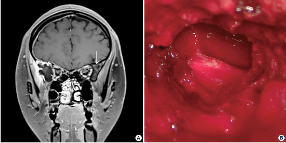
PDF - Meningiomas in bone are rarely subjected to fine-needle aspiration diagnosis, and those arising in the skull bone with a cystic presentation are rare. A 24-year-old woman presented with subdural hemorrhage, and subsequent radiology depicted an osteolytic mass-like lesion in the sphenoid bone. Intraoperatively, a solid and cystic hemorrhagic lesion mimicking an aneurysmal bone cyst was observed in the sphenoid bone with dural tearing. Frozen cytology showed singly scattered or epithelioid clusters of round to elongated cells intermixed with many neutrophils. Tumor cells had bland-looking round nuclei with rare prominent nucleoli and nuclear inclusions and eosinophilic granular to globoid cytoplasm in capillary-rich fragments. Histology revealed intraosseous meningothelial and microcystic meningioma (World Health Organization grade 1) in right lesser wing of the sphenoid bone. Considering its unusual location and cytologic findings, differential diagnoses included chordoma, chondroma, chondrosarcoma, and aneurysmal bone cyst. The present case posed a diagnostic challenge due to possible confusion with these entities.
-
Citations
Citations to this article as recorded by
- Purely cystic intraosseous meningioma of the skull: A radiologic conundrum and histologic challenge
Diego Rojas, Arman Kavoussi, Ashley Rose Ricciardelli, Alex Flores, Sricharan Gopakumar, Luis Carrete, Hsiang-Chih Lu, Alex W. Brenner, Akash J. Patel
Surgical Neurology International.2025; 16: 221. CrossRef - Middle ear adenoma: Cytohistologic features and differential diagnosis
Abdullah Almajnooni, Matthew Vega, Lin Cheng, Paolo Gattuso, Mary K. Allen‐Proctor
Diagnostic Cytopathology.2023;[Epub] CrossRef - Exploring the role of epidermal growth factor receptor variant III in meningeal tumors
Rashmi Rana, Vaishnavi Rathi, Kirti Chauhan, Kriti Jain, Satnam Singh Chhabra, Rajesh Acharya, Samir Kumar Kalra, Anshul Gupta, Sunila Jain, Nirmal Kumar Ganguly, Dharmendra Kumar Yadav, Timir Tripathi
PLOS ONE.2021; 16(9): e0255133. CrossRef
- Purely cystic intraosseous meningioma of the skull: A radiologic conundrum and histologic challenge
- Pediatric granular cell tumor in the posterior wall of the larynx extending to the trachea
- Jungsuk Ahn, Na Rae Kim, Yong Han Sun
- J Pathol Transl Med. 2020;54(4):336-339. Published online April 15, 2020
- DOI: https://doi.org/10.4132/jptm.2020.02.28
- 7,211 View
- 125 Download
- 1 Web of Science
- 1 Crossref
-
Abstract
PDF
- Granular cell tumor (GCT) is a slow-growing benign neoplasm that can be found in any organ. Pediatric laryngotracheal GCT is rare. We experienced a 6-year-old boy suffering from a barking cough and symptoms of stridor and croup for one month. Head and neck computed tomography revealed a protruding mass that occluded 60% of the airway lumen. Under the impression of hemangioma or papilloma, excision revealed a submucosal non-encapsulated mass. Histologically, the mass was composed of sheets of large polyhedralshaped tumor cells containing plump eosinophilic granular cytoplasm and centrally placed, small, bland-appearing nuclei. The tumor cells were positive for S-100 protein, and voluminous eosinophilic cytoplasm was stained by diastase-resistant periodic acid-Schiff. The present report describes a unique case of a huge pediatric laryngeal GCT extending to the subglottic trachea. We also review the clinical course of pediatric laryngotracheal GCT and emphasize the importance of diagnosing GCT in children.
-
Citations
Citations to this article as recorded by- Pediatric granular cell tumor of the larynx: A case report and literature review
Jing Ke, Junwei Xiong, Juhong Zhang, Haiyu Ma, Wei Yuan
Journal of Cancer Research and Therapeutics.2023; 19(4): 1070. CrossRef
- Pediatric granular cell tumor of the larynx: A case report and literature review
- Adjunctive markers for classification and diagnosis of central nervous system tumors: results of a multi-center neuropathological survey in Korea
- Yoon Jin Cha, Se Hoon Kim, Na Rae Kim
- J Pathol Transl Med. 2020;54(2):165-170. Published online February 20, 2020
- DOI: https://doi.org/10.4132/jptm.2020.02.04
- 9,303 View
- 226 Download
- 1 Web of Science
- 1 Crossref
-
Abstract
PDF
 Supplementary Material
 - Background
The revised 4th 2016 World Health Organization (WHO) classification of tumors of the central nervous system (CNS) classification has adopted integrated diagnosis encompassing the histology and molecular features of CNS tumors. We aimed to investigate the immunohistochemistry, molecular testing, and testing methods for diagnosis of CNS tumors in pathological labs of tertiary centers in Korea, and evaluate the adequacy of tests for proper diagnosis in daily practice.
Methods
A survey, composed of eight questions concerning molecular testing for diagnosis of CNS tumors, was sent to 10 neuropathologists working in tertiary centers in Korea.
Results
For diagnosis of astrocytic and oligodendroglial tumors, all 10 centers performed isocitrate dehydrogenase mutations testing and 1p/19q loss of heterozygosity. For glioneuronal tumors, immunohistochemistry (IHC) assays for synaptophysin (n = 9), CD34 (n = 7), BRAF(VE1) (n = 5) were used. For embryonal tumors, particularly in medulloblastoma, four respondents used IHC panel (growth factor receptor bound protein 2-associated protein 1, filamin A, and yes-associated protein 1) for molecular subclassification. Regarding meningioma, all respondents performed Ki-67 IHC and five performed telomerase reverse transcriptase promoter mutation.
Conclusions
Most tertiary centers made proper diagnosis in line with 2016 WHO classification. As classification of CNS tumors has evolved to be more complex and more ancillary tests are required, these should be performed considering the effect of necessity and justification. -
Citations
Citations to this article as recorded by- Exploring the role of epidermal growth factor receptor variant III in meningeal tumors
Rashmi Rana, Vaishnavi Rathi, Kirti Chauhan, Kriti Jain, Satnam Singh Chhabra, Rajesh Acharya, Samir Kumar Kalra, Anshul Gupta, Sunila Jain, Nirmal Kumar Ganguly, Dharmendra Kumar Yadav, Timir Tripathi
PLOS ONE.2021; 16(9): e0255133. CrossRef
- Exploring the role of epidermal growth factor receptor variant III in meningeal tumors
- Contribution of cytologic examination to diagnosis of poorly differentiated thyroid carcinoma
- Na Rae Kim, Jae Yeon Seok, Yoo Seung Chung, Joon Hyop Lee, Dong Hae Chung
- J Pathol Transl Med. 2020;54(2):171-178. Published online February 5, 2020
- DOI: https://doi.org/10.4132/jptm.2019.12.03
- 10,091 View
- 210 Download
- 6 Web of Science
- 7 Crossref
-
Abstract
PDF
- Background
The cytologic diagnosis of poorly differentiated thyroid carcinoma (PDTC) is difficult because it lacks salient cytologic findings and shares cytologic features with more commonly encountered neoplasms. Due to diverse cytologic findings and paucicellularity of PDTC, standardization of cytologic diagnostic criteria is limited. The purpose of this study is to investigate and recognize diverse thyroid findings of fine needle aspiration (FNA) cytology and frozen smear cytology in diagnosis of this rare but aggressive carcinoma.
Methods
The present study included six cases of FNA cytology and frozen smears of histologically diagnosed PDTCs.
Results
PDTC showed cytologic overlap with well-differentiated thyroid carcinomas (WDTCs). Five of six cases showed dedifferentiation arising from well differentiated thyroid carcinomas. Only one de novo PDTC showed highly cellular smears composed of discohesive small cells, high nuclear/cytoplasmic (N/C) ratio, prominent micronucleoli, and irregular nuclei. Retrospectively reviewed, these findings are highly suspicious for PDTC. Cytologic findings of nuclear atypia, pleomorphism, and irregularity were frequently found, whereas scattered small cells were seen only in the de novo case.
Conclusions
Heterogeneous cytologic findings of PDTCs are shared with those of WDTCs and contribute to difficult preoperative cytologic diagnoses. Most PDTCs show dedifferentiation from WDTCs. Albeit rare, de novo PDTC should be considered with cytology showing discohesive small cells with high N/C ratio. This will enable precise diagnosis and prompt treatment of this aggressive malignancy -
Citations
Citations to this article as recorded by- Practical and challenging issue in thyroid cytopathology
Qianqian Zhang, Belen Padial Urtueta, Elisabetta Merenda, Gabriele Rotondaro, Noemi Morelli, Alessia Piermattei, Patrizia Straccia, Federica Cianfrini, Angela Feraco, Alessia Granitto, Antonino Mule, Esther Diana Rossi
Human Pathology.2026; 169: 106019. CrossRef - Plasma cells and plasmacytoid features in thyroid lesions
Qianqian Zhang, Angela Feraco, Belen Padial Urtueta, Elisabetta Merenda, Luisa Cioni, Alessia Piermattei, Patrizia Straccia, Federica Cianfrini, Antonino Mule, Liron Pantanowitz, Esther Diana Rossi
Virchows Archiv.2026;[Epub] CrossRef - Non-papillary thyroid carcinoma diagnoses in The Bethesda System for Reporting Thyroid Cytopathology categories V and VI: An institutional experience
Myunghee Kang, Na Rae Kim, Jae Yeon Seok
Annals of Diagnostic Pathology.2024; 71: 152263. CrossRef - Cytologic features of differentiated high‐grade thyroid carcinoma: A multi‐institutional study of 40 cases
Vanda F. Torous, Tikamporn Jitpasutham, Zubair Baloch, Richard L. Cantley, Darcy A. Kerr, Xiaoying Liu, Zahra Maleki, Ross Merkin, Vania Nosé, Liron Pantanowitz, Isabella Tondi Resta, Esther D. Rossi, William C. Faquin
Cancer Cytopathology.2024; 132(8): 525. CrossRef - An Unexpected Finding of Poorly Differentiated Thyroid Carcinoma in a Toxic Thyroid Nodule
Kimberly Yuang, Huda Al-Bahadili, Alan Chang
JCEM Case Reports.2023;[Epub] CrossRef - Revisiting the cytomorphological features of poorly differentiated thyroid carcinoma: a comparative analysis with indeterminate thyroid fine-needle aspiration samples
Yazeed Alwelaie, Ali Howaidi, Mohammed Tashkandi, Ahmad Almotairi, Hisham Saied, Moammar Muzzaffar, Doaa Alghamdi
Journal of the American Society of Cytopathology.2023; 12(5): 331. CrossRef - Characterization of the genomic alterations in poorly differentiated thyroid cancer
Yeeun Lee, SeongRyeol Moon, Jae Yeon Seok, Joon-Hyop Lee, Seungyoon Nam, Yoo Seung Chung
Scientific Reports.2023;[Epub] CrossRef
- Practical and challenging issue in thyroid cytopathology
- Comparison of papanicolaou smear and human papillomavirus (HPV) test as cervical screening tools: can we rely on HPV test alone as a screening method? An 11-year retrospective experience at a single institution
- Myunghee Kang, Seung Yeon Ha, Hyun Yee Cho, Dong Hae Chung, Na Rae Kim, Jungsuk An, Sangho Lee, Jae Yeon Seok, Juhyeon Jeong
- J Pathol Transl Med. 2020;54(1):112-118. Published online January 15, 2020
- DOI: https://doi.org/10.4132/jptm.2019.11.29

- 16,693 View
- 277 Download
- 20 Web of Science
- 23 Crossref
-
Abstract
PDF
- Background
The decrease in incidence of cervical dysplasia and carcinoma has not been as dramatic as expected with the development of improved research tools and test methods. The human papillomavirus (HPV) test alone has been suggested for screening in some countries. The National Cancer Screening Project in Korea has applied Papanicolaou smears (Pap smears) as the screening method for cervical dysplasia and carcinoma. We evaluated the value of Pap smear and HPV testing as diagnostic screening tools in a single institution.
Methods
Patients co-tested with HPV test and Pap smear simultaneously or within one month of each other were included in this study. Patients with only punch biopsy results were excluded because of sampling errors. A total of 999 cases were included, and the collected reports encompassed results of smear cytology, HPV subtypes, and histologic examinations.
Results
Sensitivity and specificity of detecting high-grade squamous intraepithelial lesion (HSIL) and squamous cell carcinoma (SCC) were higher for Pap smears than for HPV tests (sensitivity, 97.14%; specificity, 85.58% for Pap smears; sensitivity, 88.32%; specificity, 54.92% for HPV tests). HPV tests and Pap smears did not differ greatly in detection of low-grade squamous intraepithelial lesion (85.35% for HPV test, 80.31% for Pap smears). When atypical glandular cells were noted on Pap smears, the likelihood for histologic diagnosis of adenocarcinoma following Pap smear was higher than that of high-risk HPV test results (18.8 and 1.53, respectively).
Conclusions
Pap smears were more useful than HPV tests in the diagnosis of HSIL, SCC, and glandular lesions. -
Citations
Citations to this article as recorded by- Development of a Nano-Real-Time Polymerase Chain Reaction (RT-PCR) Kit for Detection and Genotyping of High-Risk Human Papillomavirus (HPV) Strains Using Dedicated TaqMan Probes
Mohammad Panji, Mohammad Hossein Modarresi, Zahra Azizi, Moloud Absalan, Elahe Motevaseli
Cureus.2026;[Epub] CrossRef - Detection of cervical precancerous lesions and cancer by small-scale RT-qPCR analysis of oppositely deregulated mRNAs pairs in cytological smears
Anastasia A. Artyukh, Mikhail K. Ivanov, Sergei E. Titov, Victoria V. Dzyubenko, Sergey E. Krasilnikov, Anastasia O. Shumeikina, Nikita A. Afanasev, Anastasia V. Malek, Sergei A. Glushkov, Eduard F. Agletdinov
Frontiers in Oncology.2025;[Epub] CrossRef - High burden of abnormal cervical smears in South African primary health care: health programmes implications
Olufemi B Omole, Joel M Francis, John M Musonda, Pumla P Sodo, Elizabeth Reji, Nyundu S J Phukuta, Honey L M Mabuza, Joyce S Musonda, Jimmy Akii, John V Ndimande, Olalekan A Ayo-Yusuf
Health Promotion International.2025;[Epub] CrossRef - Bibliometric analysis: a study of the microenvironment in cervical cancer (2000-2024)
Yun-Tao Zhang, Yan-Ni Wei, Chen-Chen Liu, Mai-Qing Yang
Frontiers in Oncology.2025;[Epub] CrossRef - Liquid biopsy biomarkers in cervical cancer: A systematic review and meta-analysis
Isaac Kinyua Njangiru, Bizhar Ahmed Tayeb, Hazhmat Ali, Rafl M. Kamil
The Journal of Liquid Biopsy.2025; 10: 100328. CrossRef - Diagnostic Utility of Human Papilloma Virus Testing in Comparison with Pap Cytology and Histopathology in Unvaccinated Women with Cervical High-Grade Dysplasia and Carcinoma in Botswana
Patricia Setsile Rantshabeng, Nametso Dire, Andrew Khulekani Ndlovu, Ishmael Kasvosve
Venereology.2025; 4(4): 15. CrossRef - Challenges in the diagmosis of cervical pathologies
D. Y. Chernov, O. A. Tikhonovskaya, S. V. Logvinov, I. A. Petrov, Y. S. Yuriev, A. A. Zhdankina, A. V. Gerasimov, I. V. Zingalyuk, G. A. Mikheenko
Bulletin of Siberian Medicine.2024; 22(4): 201. CrossRef - “Barriers and Advantages of Self-Sampling Tests, for HPV Diagnosis: A Qualitative Field Experience Before Implementation in a Rural Community in Ecuador”
Bernardo Vega-Crespo, Vivian Alejandra Neira, Ruth Maldonado - Rengel, Diana López, Dayanara Delgado-López, Gabriela Guerra Astudillo, Veronique Verhoeven
International Journal of Women's Health.2024; Volume 16: 947. CrossRef - Cervical Human Papillomavirus Testing
Carol N. Rizkalla, Eric C. Huang
Surgical Pathology Clinics.2024; 17(3): 431. CrossRef - Segmentation of Overlapping Cells in Cervical Cytology Images: A Survey
E Chen, Hua-Nong Ting, Joon Huang Chuah, Jun Zhao
IEEE Access.2024; 12: 114170. CrossRef - Knowledge and awareness regarding pap test and HPV typing for cervical cancer screening in Edo North, Nigeria
Amina Momodu, Johnsolomon Eghosa Ohenhen, Godfrey Innocent Iyare, Musa Abidemi Muhibi, Godwin Avwioro
Discover Public Health.2024;[Epub] CrossRef - Colposcopy Value in Young Child-bearing Women: Is New Recommendations Necessary?
Fahimeh Sabet, Avishan Aminizad, Fariba Behnamfar, Tajossadat Allameh, Seyedeh Ghazal Shahrokh, Rostami Koushan, Amirmohammad Taravati, Leila Mousavi Seresht
Advanced Biomedical Research.2024;[Epub] CrossRef - Selection of endogenous control and identification of significant microRNA deregulations in cervical cancer
T. Stverakova, I. Baranova, P. Mikyskova, B. Gajdosova, H. Vosmikova, J. Laco, V. Palicka, H. Parova
Frontiers in Oncology.2023;[Epub] CrossRef - Cytology Versus Molecular Diagnosis of HPV for Cervical Cancer Screening. Comparison of the Diagnostic Properties of Four Tests in a Rural Community of Cuenca Ecuador
Bernardo Vega Crespo, Vivian Alejandra Neira, Rocío Murillo, Cristina Ochoa Avilés
ESPOCH Congresses: The Ecuadorian Journal of S.T.E.A.M..2023; 3(1): 139. CrossRef - Attitudes towards prevention of cervical cancer and early diagnosis among female academicians
Nurhan Doğan, Gamze Fışkın
Journal of Obstetrics and Gynaecology Research.2022; 48(6): 1433. CrossRef - Role of Self-Sampling for Cervical Cancer Screening: Diagnostic Test Properties of Three Tests for the Diagnosis of HPV in Rural Communities of Cuenca, Ecuador
Bernardo Vega Crespo, Vivian Alejandra Neira, José Ortíz Segarra, Ruth Maldonado Rengel, Diana López, María Paz Orellana, Andrea Gómez, María José Vicuña, Jorge Mejía, Ina Benoy, Tesifón Parrón Carreño, Veronique Verhoeven
International Journal of Environmental Research and Public Health.2022; 19(8): 4619. CrossRef - Utility of Scoring System for Screening and Early Warning of Cervical Cancer Based on Big Data Analysis
Dan Hou, Binjie Yang, Yangdan Li, Ming Sun
Frontiers in Public Health.2022;[Epub] CrossRef - Evaluation of Urine and Vaginal Self-Sampling versus Clinician-Based Sampling for Cervical Cancer Screening: A Field Comparison of the Acceptability of Three Sampling Tests in a Rural Community of Cuenca, Ecuador
Bernardo Vega Crespo, Vivian Alejandra Neira, José Ortíz S, Ruth Maldonado-Rengel, Diana López, Andrea Gómez, María José Vicuña, Jorge Mejía, Ina Benoy, Tesifón Parrón Carreño, Veronique Verhoeven
Healthcare.2022; 10(9): 1614. CrossRef - Diagnostic distribution and pitfalls of glandular abnormalities in cervical cytology: a 25-year single-center study
Jung-A Sung, Ilias P. Nikas, Haeryoung Kim, Han Suk Ryu, Cheol Lee
Journal of Pathology and Translational Medicine.2022; 56(6): 354. CrossRef - Primary screening of cervical cancer by Pap smear in women of reproductive age group
Ruchi Mishra, Dakshina Bisht, Manisha Gupta
Journal of Family Medicine and Primary Care.2022; 11(9): 5327. CrossRef - Comparison of Learning Transfer Using Simulation Problem-Based Learning and Demonstration: An Application of Papanicolaou Smear Nursing Education
Jeongim Lee, Hae Kyoung Son
International Journal of Environmental Research and Public Health.2021; 18(4): 1765. CrossRef - Investigating host-virus interaction mechanism and phylogenetic analysis of viral proteins involved in the pathogenesis
Ahmad Abu Turab Naqvi, Farah Anjum, Alaa Shafie, Sufian Badar, Abdelbaset Mohamed Elasbali, Dharmendra Kumar Yadav, Md. Imtaiyaz Hassan, Timir Tripathi
PLOS ONE.2021; 16(12): e0261497. CrossRef - Utility of Human Papillomavirus Testing for Cervical Cancer Screening in Korea
Mee-seon Kim, Eun Hee Lee, Moon-il Park, Jae Seok Lee, Kisu Kim, Mee Sook Roh, Hyoun Wook Lee
International Journal of Environmental Research and Public Health.2020; 17(5): 1726. CrossRef
- Development of a Nano-Real-Time Polymerase Chain Reaction (RT-PCR) Kit for Detection and Genotyping of High-Risk Human Papillomavirus (HPV) Strains Using Dedicated TaqMan Probes
- Primary carcinoid tumor in the external auditory canal
- Dong Hae Chung, Gyu Cheol Han, Na Rae Kim
- J Pathol Transl Med. 2020;54(2):184-187. Published online November 13, 2019
- DOI: https://doi.org/10.4132/jptm.2019.11.07
- 10,491 View
- 167 Download
- 4 Web of Science
- 3 Crossref
-
Abstract
PDF
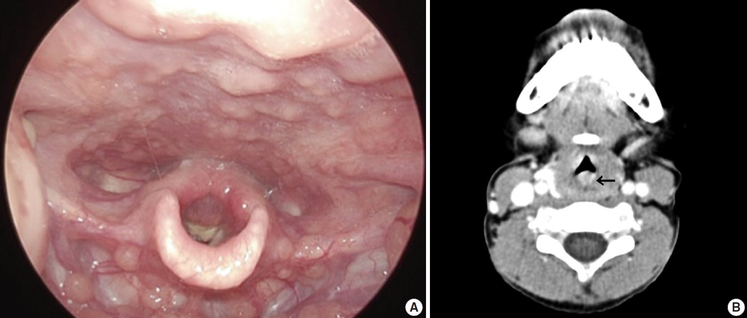
- A 39-year-old man visited the department of otolaryngology due to an ongoing hearing disturbance that had lasted for 1 year. Temporal bone computed tomography revealed soft tissue density nearly obliterating the left external auditory canal (EAC). The mass was composed of sheets of round tumor cells containing moderate amounts of fine granular cytoplasm and salt and pepper chromatin. Neither mitosis nor necrosis was found. The Ki-67 proliferation index was less than 2%. Cells were positive for CD56 and synaptophysin but negative for chromogranin, cytokeratin (CK) 20, and CK7. Based on these findings, the tumor was diagnosed as a carcinoid tumor, well differentiated neuroendocrine carcinoma, grade 1 (G1) according to current World Health Organization (WHO) classification of head and neck tumors; and a neuroendocrine tumor, G1 according to neuroendocrine neoplasm (NEN)-2018 WHO standard classification. He remained free of local recurrence and metastasis after 20 months of follow up. To date, only six cases of primary NENs in the EAC have been reported. Metastatic tumor should be included in the differential diagnoses. Because of its rarity, the prognosis and treatment have not yet been clarified.
-
Citations
Citations to this article as recorded by- First Report on a Rare Poorly Differentiated Neuroendocrine Tumour of the External Auditory Canal Involving Pinna
Akash Varshney, Amit Kumar Tyagi, Prashant Durgapal, Kajal Mahto, Akhilesh Chandra Yadav, Ankita Semwal
Indian Journal of Otolaryngology and Head & Neck Surgery.2025; 77(4): 1922. CrossRef - Incidental finding of a neuroendocrine neoplasm in a suspected ear canal exostosis
Alexander Wieck Fjaeldstad, Gerda Elisabeth Villadsen, Gitte Dam, Stephen Jacques Hamilton-Dutoit, Thomas Winther Frederiksen
Otolaryngology Case Reports.2022; 22: 100394. CrossRef - 68Ga-DOTATATE Uptake in Well-Differentiated Neuroendocrine Tumor of the External Auditory Canal
Özge Erol Fenercioğlu, Ediz Beyhan, Rahime Şahin, Mehmet Can Baloğlu, Tevfik Fikret Çermik
Clinical Nuclear Medicine.2022; 47(8): e552. CrossRef
- First Report on a Rare Poorly Differentiated Neuroendocrine Tumour of the External Auditory Canal Involving Pinna
- Frozen Cytology of Meningeal Malignant Solitary Fibrous Tumor/Hemangiopericytoma
- Myunghee Kang, Na Rae Kim, Dong Hae Chung, Gie-Taek Yie
- J Pathol Transl Med. 2019;53(3):192-197. Published online April 11, 2019
- DOI: https://doi.org/10.4132/jptm.2019.03.20
- 9,505 View
- 160 Download
- 7 Web of Science
- 8 Crossref
-
Abstract
PDF
- A 51-year-old woman presented with severe dizziness. The brain magnetic resonance image revealed a 5.5 cm multiloculated mass with a thick rim in the left temporal lobe. Cytological examination of frozen diagnosis of the mass showed hypercellular sheets of round and rhabdoid cells in a hemorrhagic background, and two mitotic figures were observed. Histologically, the excised dura-based mass consisted of predominantly round cells with small foci of rhabdoid tumor cells in a pseudoalveolar pattern in a hemorrhagic background, and the cells showed nuclear positivity for signal transducer and activator of transcription 6 as well as frequent mitosis. The mass was diagnosed as a grade 3 solitary fibrous tumor (SFT)/hemangiopericytoma (HPC). The cytological diagnosis of SFT/HPC is challenging because of the heterogeneous cytological findings, such as histological heterogeneity, and because there are no standardized cytological criteria for malignant SFT/HPC. Cytological findings, such as singly scattered small cells, hypercellularity, rare ropy collagen, and round and rhabdoid cells with pseudoalveolar pattern, may assist in the diagnosis of malignant SFT/HPC.
-
Citations
Citations to this article as recorded by- A Rare Case of Cervical Solitary Fibrous Tumor in a Pediatric Patient: Case Report and Literature Review
Eleonora Becattini, Lorenzo Sgarbanti, Giuseppina Bevacqua, Valentina Grespi, Carlo Conti
NeuroSci.2025; 6(2): 49. CrossRef - Meningeal Solitary Fibrous Tumor: A Cytological Report With Emphasis on the Usefulness of Immunocytochemical Analysis for STAT6
Hiroyuki Okanishi, Mitsuaki Ishida, Naoto Kohno, Isako Kataoka, Mari Tomiuka, Mayumi Uragami, Shizuka Ono, Chihiro Deguchi, Reika Takeda, Yoshitaka Kurisu, Yoshinobu Hirose
Diagnostic Cytopathology.2025;[Epub] CrossRef - Cytologic features of mesenchymal, melanocytic and haematolymphoid tumours of the central nervous system and metastases
Carmen Bárcena, José A. Jiménez‐Heffernan
Cytopathology.2024; 35(5): 590. CrossRef - A Hemangiopericytoma in the External Auditory Canal: A Rare Clinical Presentation and Management
Vaibhavi Patil, Prasad Deshmukh, Sagar S Gaurkar , Ayushi Ghosh Moulic, Jasleen Kaur
Cureus.2024;[Epub] CrossRef - Scoring system for intraoperative diagnosis of intracranial schwannoma by squash cytology
Hirotaka Fujita, Takuma Tajiri, Tomohisa Machida, Nozomi Nomura, Suguru Toguchi, Hitoshi Itoh, Shinichiro Hiraiwa, Tomoko Sugiyama, Chie Inomoto, Masaaki Imai, Shinri Oda, Masami Shimoda, Naoya Nakamura
Cytopathology.2022; 33(2): 196. CrossRef - Occurrence of a solitary fibrous tumor adjacent to the resection bed of a high-grade meningioma: A case report
Coby Cunningham, Rocco Dabecco, Justin Davanzo
Interdisciplinary Neurosurgery.2021; 25: 101277. CrossRef - A case of solitary fibrous tumor arising in the meninge
Saori NAKANISHI, Naoto KURODA, Toshiko TAKAI, Mari KOJIMA, Misato OONOGI
The Journal of the Japanese Society of Clinical Cytology.2021; 60(4): 224. CrossRef - Intraoperative frozen cytology of intraosseous cystic meningioma in the sphenoid bone
Na Rae Kim, Gie-Taek Yie
Journal of Pathology and Translational Medicine.2020; 54(6): 508. CrossRef
- A Rare Case of Cervical Solitary Fibrous Tumor in a Pediatric Patient: Case Report and Literature Review
- Primary Necrobiotic Xanthogranulomatous Sialadenitis with Submandibular Gland Localization without Skin Involvement
- Myunghee Kang, Na Rae Kim, Dong Hae Chung, Jae Yeon Seok, Dong Young Kim
- J Pathol Transl Med. 2019;53(4):261-265. Published online January 16, 2019
- DOI: https://doi.org/10.4132/jptm.2019.01.08
- 10,479 View
- 175 Download
- 2 Web of Science
- 6 Crossref
-
Abstract
PDF
- Necrobiotic xanthogranulomatous reaction is a multiorgan, non-Langerhans cell histiocytosis with an unknown etiology. Occurrence in the salivary gland is extremely rare. We recently identified a case of necrobiotic xanthogranulomatous sialadenitis in a 73-year-old Korean woman who presented with a painless palpable lesion in the chin. There was no accompanying cutaneous lesion. Partial resection and subsequent wide excision with neck dissection were performed. Pathological examination showed a severe inflammatory lesion that included foamy macrophages centrally admixed with neutrophils, eosinophils, lymphocytes, plasma cells, and scattered giant cells, as well as necrobiosis. During the 12-month postoperative period, no grossly remarkable change in size was noted. Necrobiotic xanthogranulomatous inflammation may be preceded by or combined with hematologic malignancy. Although rare, clinicians and radiologists should be aware that an adhesive necrobiotic xanthogranuloma in the salivary gland may present with a mass-like lesion. Further evaluation for hematologic disease and close follow-up are needed when a pathologic diagnosis is made.
-
Citations
Citations to this article as recorded by- Salivary gland macrophages in health and disease: heterogeneity, niche crosstalk, and therapeutic avenues
Xinglei Li, Yan Feng, Huixin Xue, Xinxin Ni
Frontiers in Immunology.2025;[Epub] CrossRef - Five Cases of Xanthogranulomatous Sialadenitis
Satoshi Kiyama, Hiroyuki Iuchi, Kotoko Ito, Kengo Nishimoto, Tsutomu Matsuzaki, Masaru Yamashita
Practica Oto-Rhino-Laryngologica.2022; 115(4): 315. CrossRef - Xanthogranulomatous change in a pleomorphic adenoma: An extremely rare variant/degenerative change. Is it fine needle aspiration induced?
Mukta Pujani, Dipti Sidam, Kanika Singh, Aparna Khandelwal, Khushbu Katarya
Diagnostic Cytopathology.2021;[Epub] CrossRef - A Case of Xanthogranulomatous Sialadenitis with Facial Palsy Mimicking Malignancy
Sang Hyun Kim, Sun Woo Kim, Sang Hyuk Lee
Korean Journal of Otorhinolaryngology-Head and Neck Surgery.2021; 64(6): 422. CrossRef - Xanthogranulomatous Sialadenitis, an Uncommon Reactive Change is Often Associated with Warthin’s Tumor
Lihong Bu, Hui Zhu, Emilian Racila, Sobia Khaja, David Hamlar, Faqian Li
Head and Neck Pathology.2020; 14(2): 525. CrossRef - A Case of Xanthogranulomatous Sialadenitis of the Sublingual Gland:A Review of Literature
Naoya KITAMURA, Seiji OHNO, Tetsuya YAMAMOTO
Journal of Japanese Society of Oral Medicine.2019; 25(1): 20. CrossRef
- Salivary gland macrophages in health and disease: heterogeneity, niche crosstalk, and therapeutic avenues
- Intraoperative Frozen Cytology of Central Nervous System Neoplasms: An Ancillary Tool for Frozen Diagnosis
- Myunghee Kang, Dong Hae Chung, Na Rae Kim, Hyun Yee Cho, Seung Yeon Ha, Sangho Lee, Jungsuk An, Jae Yeon Seok, Gie-Taek Yie, Chan Jong Yoo, Sang Gu Lee, Eun Young Kim, Woo Kyung Kim, Seong Son, Sun Jin Sym, Dong Bok Shin, Hee Young Hwang, Eung Yeop Kim, Kyu Chan Lee
- J Pathol Transl Med. 2019;53(2):104-111. Published online January 14, 2019
- DOI: https://doi.org/10.4132/jptm.2018.11.10
- 17,037 View
- 697 Download
- 11 Web of Science
- 9 Crossref
-
Abstract
PDF
- Background
Pathologic diagnosis of central nervous system (CNS) neoplasms is made by comparing light microscopic, immunohistochemical, and molecular cytogenetic findings with clinicoradiologic observations. Intraoperative frozen cytology smears can improve the diagnostic accuracy for CNS neoplasms. Here, we evaluate the diagnostic value of cytology in frozen diagnoses of CNS neoplasms.
Methods
Cases were selected from patients undergoing both frozen cytology and frozen sections. Diagnostic accuracy was evaluated.
Results
Four hundred and fifty-four cases were included in this retrospective single-center review study covering a span of 10 years. Five discrepant cases (1.1%) were found after excluding 53 deferred cases (31 cases of tentative diagnosis, 22 cases of inadequate frozen sampling). A total of 346 cases of complete concordance and 50 cases of partial concordance were classified as not discordant cases in the present study. Diagnostic accuracy of intraoperative frozen diagnosis was 87.2%, and the accuracy was 98.8% after excluding deferred cases. Discrepancies between frozen and permanent diagnoses (n = 5, 1.1%) were found in cases of nonrepresentative sampling (n = 2) and misinterpretation (n = 3). High concordance was observed more frequently in meningeal tumors (97/98, 99%), metastatic brain tumors (51/52, 98.1%), pituitary adenomas (86/89, 96.6%), schwannomas (45/47, 95.8%), high-grade astrocytic tumors (47/58, 81%), low grade astrocytic tumors (10/13, 76.9%), non-neoplastic lesions (23/36, 63.9%), in decreasing frequency.
Conclusions
Using intraoperative cytology and frozen sections of CNS tumors is a highly accurate diagnostic ancillary method, providing subtyping of CNS neoplasms, especially in frequently encountered entities. -
Citations
Citations to this article as recorded by- Qualitative and quantitative assessment of ex vivo human brain tumors using quantitative oblique back-illumination microscopy (qOBM)
Srinidhi Bharadwaj, Paloma Casteleiro Costa, Caroline Serafini, Brienna Heinsz, Alice Hsu, Nischita Kaza, Zhe Guang, Zhenmin Li, Jeffrey J. Olson, Kimberly Hoang, Stewart Neill, Francisco E. Robles
Biomedical Optics Express.2026; 17(4): 1936. CrossRef - Intraoperative Integrated Diagnostic System for Malignant Central Nervous System Tumors
Takahiro Hayashi, Kensuke Tateishi, Shinichiro Matsuyama, Hiromichi Iwashita, Yohei Miyake, Akito Oshima, Hirokuni Honma, Jo Sasame, Katsuhiro Takabayashi, Kyoka Sugino, Emi Hirata, Naoko Udaka, Yuko Matsushita, Ikuma Kato, Hiroaki Hayashi, Taishi Nakamur
Clinical Cancer Research.2024; 30(1): 116. CrossRef - A multicenter proof-of-concept study on deep learning-based intraoperative discrimination of primary central nervous system lymphoma
Xinke Zhang, Zihan Zhao, Ruixuan Wang, Haohua Chen, Xueyi Zheng, Lili Liu, Lilong Lan, Peng Li, Shuyang Wu, Qinghua Cao, Rongzhen Luo, Wanming Hu, Shanshan lyu, Zhengyu Zhang, Dan Xie, Yaping Ye, Yu Wang, Muyan Cai
Nature Communications.2024;[Epub] CrossRef - Advancements in Neurosurgical Intraoperative Histology
Ali A. Mohamed, Emma Sargent, Cooper Williams, Zev Karve, Karthik Nair, Brandon Lucke-Wold
Tomography.2024; 10(5): 693. CrossRef - Unveiling the potential application of intraoperative brain smear for brain tumor diagnosis in low-middle-income countries: A comprehensive systematic review
Muhammad Shakir, Ahmed Altaf, Hawra Hussain, Syed Muhammad Aqeel Abidi, Zoey Petitt, Mahnoor Tariq, Ahmed Gilani, S. Ather Enam
Surgical Neurology International.2023; 14: 325. CrossRef - A Comparative Study of Squash Smear Cytology Diagnosis and Radiological Diagnosis with Histopathology in Central Nervous System Lesions
B N Kumarguru, G Santhipriya, S Kranthi Kumar, R Ramesh Kumar, A S Ramaswamy, P Janakiraman
Journal of Cytology.2022; 39(1): 1. CrossRef - Intraoperative squash cytology provides a qualitative intraoperative diagnosis for cases in which frozen section yields a diagnosis of equivocal brain tumour
Hirotaka Fujita, Takuma Tajiri, Tomohisa Machida, Nozomi Nomura, Suguru Toguchi, Hitoshi Itoh, Shinichiro Hiraiwa, Tomoko Sugiyama, Masaaki Imai, Shinri Oda, Masami Shimoda, Naoya Nakamura
Cytopathology.2020; 31(2): 106. CrossRef - Intraoperative frozen cytology of intraosseous cystic meningioma in the sphenoid bone
Na Rae Kim, Gie-Taek Yie
Journal of Pathology and Translational Medicine.2020; 54(6): 508. CrossRef - Use of 5-Aminolevulinic Acid for Confirmation of Lesional Biopsy Sample in Presumed High-Grade Glioma
Victoria L. Watson, Jeffrey W. Cozzens
World Neurosurgery.2019; 132: 21. CrossRef
- Qualitative and quantitative assessment of ex vivo human brain tumors using quantitative oblique back-illumination microscopy (qOBM)
- Squamous Cell Carcinoma of the Extrahepatic Common Hepatic Duct
- Myunghee Kang, Na Rae Kim, Dong Hae Chung, Hyun Yee Cho, Yeon Ho Park
- J Pathol Transl Med. 2019;53(2):112-118. Published online October 1, 2018
- DOI: https://doi.org/10.4132/jptm.2018.09.03
- 10,053 View
- 179 Download
- 10 Web of Science
- 12 Crossref
-
Abstract
PDF
- We report a rare case of hilar squamous cell carcinoma. A 62-year-old Korean woman complaining of nausea was referred to our hospital. Her biliary computed tomography revealed a 28 mm-sized protruding solid mass in the proximal common bile duct. The patient underwent left hemihepatectomy with S1 segmentectomy and segmental excision of the common bile duct. Microscopically, the tumor was a moderately differentiated squamous cell carcinoma of the extrahepatic bile duct, without any component of adenocarcinoma or metaplastic portion in the biliary epithelium. Immunohistochemically, the tumor was positive for cytokeratin (CK) 5/6, CK19, p40, and p63. Squamous cell carcinoma of the extrahepatic bile duct is rare. To date, only 24 cases of biliary squamous cell carcinomas have been reported. Here, we provide a clinicopathologic review of previously reported extrahepatic bile duct squamous cell carcinomas.
-
Citations
Citations to this article as recorded by- Case Report: Primary squamous cell carcinoma of the pancreatic segment of the extrahepatic bile duct: a rare tumor easily misdiagnosed as pancreatic malignancy
Ting Xu, Xue Meng, Xuan Gou, Xinyuan Wang, Shuai Luo, Yilin Chen
Frontiers in Oncology.2026;[Epub] CrossRef - Deciphering cholangiocarcinoma heterogeneity and specific progenitor cell niche of extrahepatic cholangiocarcinoma at single-cell resolution
Chunliang Liu, Xiang Wang, Erdong Liu, Yali Zong, Wenlong Yu, Youhai Jiang, Jianan Chen, Mingye Gu, Zhengyuan Meng, Jingfeng Li, Yang Liu, Yongjie Zhang, Jing Tang, Hongyang Wang, Jing Fu
Journal of Hematology & Oncology.2025;[Epub] CrossRef - Extrahepatic cholangiocarcinoma: Current concepts in histopathology, immunohistochemistry, and molecular diagnostics
Jared Beyersdorf, M. Lisa Zhang
Seminars in Diagnostic Pathology.2025; 42(6): 150949. CrossRef - Cholangiocarcinoma With Liver Metastasis in Squamous Cell Carcinoma Type: A Case Report
Jane Chiang
Journal of Diagnostic Medical Sonography.2024; 40(6): 609. CrossRef - A Rare Case of Squamous Cell Carcinoma of the Bile Duct
Julianna Tantum, Rachael Schneider, Stefanie Gallagher, Kyley Leroy, Jared Lander, Patricia Wong
ACG Case Reports Journal.2023; 10(8): e01119. CrossRef - Metastatic Anal Squamous Cell Carcinoma Presenting as an Indeterminate Biliary Stricture Diagnosed By Cholangioscopy
Ritu Nahar, Ian Holmes, Jeffrey Baliff, Austin Chiang, Thomas Kowalski
ACG Case Reports Journal.2022; 9(6): e00785. CrossRef - Temporal Changes in Cholangiocarcinoma Incidence and Mortality in the United States from 2001 to 2017
Milind Javle, Sunyoung Lee, Nilofer S Azad, Mitesh J Borad, Robin Kate Kelley, Smitha Sivaraman, Anna Teschemaker, Ishveen Chopra, Nora Janjan, Shreekant Parasuraman, Tanios S Bekaii-Saab
The Oncologist.2022; 27(10): 874. CrossRef - PRIMARY SQUAMOUS CELL CARCINOMA OF THE COMMON BILE DUCT WITH LIVER METASTASES
Dhouha BACHA, Mohamed HAJRI, Wael FERJAOUI, Ghofrane TALBI, Lasaad GHARBI, Mohamed Taher KHALFALLAH, Sana ben SLAMA, Ahlem LAHMAR
ABCD. Arquivos Brasileiros de Cirurgia Digestiva (São Paulo).2021;[Epub] CrossRef - S1510 A Rare Case of Squamous Cell Carcinoma of the Bile Duct
Stefanie Gallagher, Kyley Leroy, Julianna Tantum, Babak Etemad
American Journal of Gastroenterology.2021; 116(1): S688. CrossRef - Heparin
Reactions Weekly.2019; 1752(1): 184. CrossRef - Carcinoma primario de células escamosas del conducto hepático común: a propósito de un caso
Ana Delgado Maroto, Andrés Barrientos Delgado, Marta Lázaro Sáez, Samia Hallouch Toutouh, Enrique Práxedes González
Gastroenterología y Hepatología.2019; 42(7): 436. CrossRef - Primary squamous cell carcinoma of the extrahepatic bile duct: A case report
Ana Delgado Maroto, Andrés Barrientos Delgado, Marta Lázaro Sáez, Samia Hallouch Toutouh, Enrique Práxedes González
Gastroenterología y Hepatología (English Edition).2019; 42(7): 436. CrossRef
- Case Report: Primary squamous cell carcinoma of the pancreatic segment of the extrahepatic bile duct: a rare tumor easily misdiagnosed as pancreatic malignancy
- Post-transplant Amputation Traumatic Neuroma of the Hilum and Extrahepatic Duct in a Liver Donor
- Na Rae Kim, Hyun Yee Cho, Dong Hae Chung, Keon Kuk Kim, Jae Hee Cho, Seung Joon Choi
- J Pathol Transl Med. 2018;52(3):191-194. Published online August 4, 2017
- DOI: https://doi.org/10.4132/jptm.2017.01.20
- 8,360 View
- 114 Download
- 1 Web of Science
- 2 Crossref
-
PDF
-
Citations
Citations to this article as recorded by- Biliary tree traumatic neuroma following laparoscopic cholecystectomy: A case report and literature review
Hemn Kaka Ali, Dana Gharib, Marwan Hassan, Ari Abdullah, Deari Ismaeil, Omar Ghalib Hawramy, Dlshad Ahmed, Dilan Hiwa, Berun Abdalla, Fahmi Kakamad
Medicine International.2023;[Epub] CrossRef - Hilar Biliary Amputation Neuroma Following Liver Transplant: A Case Report and Review of the Literature for this Diagnostic and Therapeutic Challenge
Sarang Thaker, Najib Nassani, Bartlomiej Lukasz Radzik, Christine Chan, Wadih Chacra, Sean Koppe, Grace Guzman, Adam E. Mikolajczyk
Transplantation Direct.2022; 8(12): e1405. CrossRef
- Biliary tree traumatic neuroma following laparoscopic cholecystectomy: A case report and literature review
- The Continuing Value of Ultrastructural Observation in Central Nervous System Neoplasms in Children
- Na Rae Kim, Sung-Hye Park
- J Pathol Transl Med. 2015;49(6):427-437. Published online October 13, 2015
- DOI: https://doi.org/10.4132/jptm.2015.09.19
- 10,971 View
- 78 Download
-
Abstract
PDF
- Central nervous system (CNS) neoplasms are the second most common childhood malignancy after leukemia and the most common solid organ neoplasm in children. Diagnostic dilemmas with small specimens from CNS neoplasms are often the result of multifactorial etiologies such as frozen or fixation artifact, biopsy size, or lack of knowledge about rare or unfamiliar entities. Since the late 1950s, ultrastructural examination has been used in the diagnosis of CNS neoplasms, though it has largely been replaced by immunohistochemical and molecular cytogenetic studies. Nowadays, pathologic diagnosis of CNS neoplasms is achieved through intraoperative cytology, light microscopy, immunohistochemistry, and molecular cytogenetic results. However, the utility of electron microscopy (EM) in the final diagnosis of CNS neoplasms and investigation of its pathogenetic origin remains critical. Here, we reviewed the distinguishing ultrastructural features of pediatric CNS neoplasms and emphasize the continuing value of EM in the diagnosis of CNS neoplasms.
- Rare Case of Anal Canal Signet Ring Cell Carcinoma Associated with Perianal and Vulvar Pagetoid Spread
- Na Rae Kim, Hyun Yee Cho, Jeong-Heum Baek, Juhyeon Jeong, Seung Yeon Ha, Jae Yeon Seok, Sung Won Park, Sun Jin Sym, Kyu Chan Lee, Dong Hae Chung
- J Pathol Transl Med. 2016;50(3):231-237. Published online October 8, 2015
- DOI: https://doi.org/10.4132/jptm.2015.08.08
- 15,150 View
- 142 Download
- 5 Web of Science
- 5 Crossref
-
Abstract
PDF
- A 61-year-old woman was referred to surgery for incidentally found colonic polyps during a health examination. Physical examination revealed widespread eczematous skin lesion without pruritus in the perianal and vulvar area. Abdominopelvic computed tomography showed an approximately 4-cm-sized, soft tissue lesion in the right perianal area. Inguinal lymph node dissection and Mils’ operation extended to perianal and perivulvar skin was performed. Histologically, the anal canal lesion was composed of mucin-containing signet ring cells, which were similar to those found in Pagetoid skin lesions. It was diagnosed as an anal canal signet ring cell carcinoma (SRCC) with perianal and vulvar Pagetoid spread and bilateral inguinal lymph node metastasis. Anal canal SRCC is rare, and the current case is the third reported case in the English literature. Seven additional cases were retrieved from the world literature. Here, we describe this rare case of anal canal SRCC with perianal Pagetoid spread and provide a literature review.
-
Citations
Citations to this article as recorded by- Primary Carcinomas of the Episiotomy Scar Site: A Systematic Literature Review
Andrea Palicelli, Federica Torricelli, Gabriele Tonni, Alessandra Bisagni, Eleonora Zanetti, Magda Zanelli, Venus Damaris Medina-Illueca, Beatrice Melli, Maurizio Zizzo, Andrea Morini, Maria Paola Bonasoni, Giacomo Santandrea, Giuseppe Broggi, Rosario Cal
Current Oncology.2025; 32(2): 65. CrossRef - Metastatic Carcinomas at the Episiotomy Site: A Systematic Literature Review
Andrea Palicelli, Gabriele Tonni, Federica Torricelli, Beatrice Melli, Vincenza Ylenia Cusenza, Sandra Martinelli, Eleonora Zanetti, Alessandra Bisagni, Magda Zanelli, Maria Paola Bonasoni, Teresa Rossi, Lucia Mangone, Venus Damaris Medina-Illueca, Mauriz
Cancers.2025; 17(17): 2801. CrossRef - A Case of Prostatic Signet-Ring Cell-like Carcinoma with Pagetoid Spread and Intraductal Carcinoma and Long-Term Survival: PD-L1 and Mismatch Repair System Proteins (MMR) Immunohistochemical Evaluation with Systematic Literature Review
Nektarios Koufopoulos, Argyro-Ioanna Ieronimaki, Andriani Zacharatou, Alina Roxana Gouloumis, Danai Leventakou, Ioannis Boutas, Dionysios T. Dimas, Adamantia Kontogeorgi, Kyparissia Sitara, Lubna Khaldi, Magda Zanelli, Andrea Palicelli
Journal of Personalized Medicine.2023; 13(6): 1016. CrossRef - Anal canal adenocarcinoma with neuroendocrine features accompanying secondary extramammary Paget disease, successfully treated with modified FOLFOX6: a case report
Masamichi Yamaura, Takeshi Yamada, Rei Watanabe, Hitomi Kawai, Suguru Hirose, Hiroki Tajima, Masashi Sato, Yuichi Uchida, Daisuke Suganuma, Yoshiyuki Yamamoto, Toshikazu Moriwaki, Ichinosuke Hyodo
BMC Cancer.2018;[Epub] CrossRef - Solitary left axillary lymph node metastasis after curative resection of carcinoma at the colostomy site: a case report
Ken Imaizumi, Shigenori Homma, Tadashi Yoshida, Tatsushi Shimokuni, Hideyasu Sakihama, Norihiko Takahashi, Hideki Kawamura, Emi Takakuwa, Akinobu Taketomi
Surgical Case Reports.2016;[Epub] CrossRef
- Primary Carcinomas of the Episiotomy Scar Site: A Systematic Literature Review
- Oncocytic Renal Cell Carcinoma with Tubulopapillary Growth Having a Fat Component
- Na Rae Kim, Hyun Yee Cho
- J Pathol Transl Med. 2015;49(5):413-417. Published online July 30, 2015
- DOI: https://doi.org/10.4132/jptm.2015.07.01
- 12,479 View
- 83 Download
- 1 Web of Science
- 1 Crossref
-
Abstract
PDF
- We report a rare case of oncocytic renal cell carcinoma (RCC) with tubulopapillary growth in the background of tuberculous end-stage kidney disease. Histology of the renal mass consisted of oncocytic cells forming solid, thin tubules and rare papillae. The tumor had abundant eosinophilic oncocytic cells containing occasional cytoplasmic Mallory body–like hyaline globules and a tiny focus of clear cells with intervening mature fat. Both the oncocytic cells and clear cells were immunoreactive for a-methylacyl-CoA racemase, vimentin, pancytokeratin, and CD10, and negative for transcription factor E3, CD15, human melanoma black 45, and c-kit. Mallory body–like hyaline globules were positive for CAM 5.2 and periodic acid–Schiff with or without diastase. Ultrastructurally, the tumor cells had abundant cytoplasmic mitochondria. The present case is a rare case of oncocytic RCC with tubulopapillary growth pattern. The case is unique in that the tumor was mixed with fat component, which is not common in RCC and thus can lead to misdiagnosis.
-
Citations
Citations to this article as recorded by- Oncocytic papillary renal cell carcinoma (OPRCC): 2 case report and literature review
Yanchen Wang, Lihui Guan, Yaming Liu, Yuxuan Liu, Xiaoyan Guo, Yaofei Sun
Frontiers in Oncology.2025;[Epub] CrossRef
- Oncocytic papillary renal cell carcinoma (OPRCC): 2 case report and literature review
- A Case of Primary Subpleural Pulmonary Microcystic Myxoma Coincidentally Occurred with Pulmonary Adenocarcinoma
- Jungsuk Ahn, Na Rae Kim, Seung Yeon Ha, Keun-Woo Kim, Kook Yang Park, Yon Mi Sung
- J Pathol Transl Med. 2015;49(3):274-278. Published online May 15, 2015
- DOI: https://doi.org/10.4132/jptm.2015.03.12
- 10,189 View
- 57 Download
- 3 Web of Science
- 3 Crossref
-
PDF
-
Citations
Citations to this article as recorded by- Recurrent PDGFRB Mutations in Pulmonary Microcystic Fibromyxoma
Ming Zhao, Qixing Gong, Xiaoyan Chen, Xiaona Yin, Rong Fang, Jiayun Xu, Xiao Cheng, Yingjing Wang
American Journal of Surgical Pathology.2026; 50(1): 41. CrossRef - Alveolar adenoma with dual hotspot mutations (PDGFRB p.N666K and PIK3CA p.H1047R): a rare case report with diagnostic implications
Mei Kong, Qiqi Gao, Hui Tang
Pathology.2026;[Epub] CrossRef - Endobronchial Myxoma
Arindam Mukherjee, Ritesh Agarwal, Sahajal Dhooria, Pawan Singh, Amanjit Bal, Harkant Singh, Inderpaul S. Sehgal
Journal of Bronchology & Interventional Pulmonology.2018; 25(4): 335. CrossRef
- Recurrent PDGFRB Mutations in Pulmonary Microcystic Fibromyxoma
- Utility of Transmission Electron Microscopy in Small Round Cell Tumors
- Na Rae Kim, Seung Yeon Ha, Hyun Yee Cho
- J Pathol Transl Med. 2015;49(2):93-101. Published online March 12, 2015
- DOI: https://doi.org/10.4132/jptm.2015.01.30
- 19,113 View
- 287 Download
- 5 Web of Science
- 4 Crossref
-
Abstract
PDF
- Small round cell tumors (SRCTs) are a heterogeneous group of neoplasms composed of small, primitive, and undifferentiated cells sharing similar histology under light microscopy. SRCTs include Ewing sarcoma/peripheral neuroectodermal tumor family tumors, neuroblastoma, desmoplastic SRCT, rhabdomyosarcoma, poorly differentiated round cell synovial sarcoma, mesenchymal chondrosarcoma, small cell osteosarcoma, small cell malignant peripheral nerve sheath tumor, and small cell schwannoma. Non-Hodgkin’s malignant lymphoma, myeloid sarcoma, malignant melanoma, and gastrointestinal stromal tumor may also present as SRCT. The current shift towards immunohistochemistry and cytogenetic molecular techniques for SRCT may be inappropriate because of antigenic overlapping or inconclusive molecular results due to the lack of differentiation of primitive cells and unavailable genetic service or limited moleculocytogenetic experience. Although usage has declined, electron microscopy (EM) remains very useful and shows salient features for the diagnosis of SRCTs. Although EM is not always required, it provides reliability and validity in the diagnosis of SRCT. Here, the ultrastructural characteristics of SRCTs are reviewed and we suggest that EM would be utilized as one of the reliable modalities for the diagnosis of undifferentiated and poorly differentiated SRCTs.
-
Citations
Citations to this article as recorded by- Electron Microscopy in the Context of a Children's Research Hospital
Cam Robinson
Microscopy and Microanalysis.2020; 26(S2): 1610. CrossRef - Primary bilateral corneal nerve sheath neoplasm in a dog
Marina L. Leis, M. Elyse Salpeter, Bianca S. Bauer, Dale L. Godson, Bruce H. Grahn
Veterinary Ophthalmology.2017; 20(4): 365. CrossRef - Hirnbasissyndrom infolge eines Tumors bei einer 17 Monate alten Deutsch-Holstein-Färse
Wolf Wippermann, Sandra Schöniger, Kerstin Gerlach, Gerald Fritz Schusser, Gabor Köller, Alexander Starke
Tierärztliche Praxis Ausgabe G: Großtiere / Nutztiere.2016; 44(03): 180. CrossRef - The Continuing Value of Ultrastructural Observation in Central Nervous System Neoplasms in Children
Na Rae Kim, Sung-Hye Park
Journal of Pathology and Translational Medicine.2015; 49(6): 427. CrossRef
- Electron Microscopy in the Context of a Children's Research Hospital
- Cystic Brunner’s Gland Hamartoma in the Gastric Body: A Case Report
- Dong Hae Chung, Na Rae Kim, Hyun Yee Cho, Yoon Jae Kim
- Korean J Pathol. 2014;48(5):371-374. Published online October 27, 2014
- DOI: https://doi.org/10.4132/KoreanJPathol.2014.48.5.371
- 12,153 View
- 64 Download
- 1 Crossref
- Bilateral Stafne Bone Cavity in the Anterior Mandible with Heterotopic Salivary Gland Tissue: A Case Report
- Hyunchul Kim, Jae Yeon Seok, Sangho Lee, Jungsuk An, Na Rae Kim, Dong Hae Chung, Hyun Yee Cho, Seung Yeon Ha
- Korean J Pathol. 2014;48(3):248-249. Published online June 26, 2014
- DOI: https://doi.org/10.4132/KoreanJPathol.2014.48.3.248
- 16,253 View
- 110 Download
- 14 Crossref
-
PDF
-
Citations
Citations to this article as recorded by- Diagnostic approach for the rare anterior variant of mandibular bone depression often misdiagnosed as tumorous lesions
Hak-Sun Kim
Journal of Dental Sciences.2025; 20(1): 502. CrossRef - Static bone cavity occurred in the buccal side of the mandible: A case report and review of literature
Hideki Hojo, Takanori Eguchi, Yumi Ito, Yoshiki Hamada
Journal of Oral and Maxillofacial Surgery, Medicine, and Pathology.2025; 37(4): 698. CrossRef - Bilateral incomplete mandibular canals: an embryological analysis of their possible etiology
Kanitin Rumpansuwon, Thewarid Berkban, Nutmethee Kruepunga, Wattana Weerachatyanukul, Somluk Asuvapongpatana, Arada Chaiyamoon, Benrita Jitaree, R. Shane Tubbs, Joe Iwanaga, Thanyaporn Senarai, Athikhun Suwannakhan
Surgical and Radiologic Anatomy.2025;[Epub] CrossRef - A Rare Case of Anterior Stafne Bone Cavity in the Mandibular Region
Hideki Suito, Yuuri Oku, Koichi Kani, Keiko Aota, Naoki Maeda
Cureus.2025;[Epub] CrossRef - Benign Mandibular Cavity/Stafne's Bone Cyst: A Case Report and Review
Fawzia M. Butt, Shamim M. Butt, Mark L. Chindia
Clinical Case Reports.2025;[Epub] CrossRef - Bilateral Stafne Bone Cavity in the Body of the Mandible: An Unusual Case Report and Literature Review
Mayank Pahadia, Rutvi Vyas
Cureus.2023;[Epub] CrossRef - Effect of Stafne bone defect on the adjacent tooth: A review of the literature
Mahdi Niknami, Azin Parsa, Zahra Khodadadi
Imaging Science in Dentistry.2022; 52(2): 165. CrossRef - Assessment of prevalence and volumetric estimation of possible Stafne bone concavities on cone beam computed tomography images
Alaettin Koç, Cennet Neslihan Eroğlu, Ersen Bilgili
Oral Radiology.2020; 36(3): 254. CrossRef - Stafne’s bone cyst revisited and renamed: the benign mandibular concavity
Johan K.M. Aps, Natasha Koelmeyer, Cina Yaqub
Dentomaxillofacial Radiology.2020; 49(4): 20190475. CrossRef - Cone‐beam computed tomography analysis of lingual mandibular bone depression in the premolar region: A case report
Saeed Asgary, Naghmeh Emadi
Clinical Case Reports.2020; 8(3): 523. CrossRef - Letters to the Editor
Ariyan S Araghi, Richard M Graham
Dental Update.2019; 46(8): 792. CrossRef - Radiographic features of lingual mandibular bone depression using dental cone beam computed tomography
Liu Liu, Byung Cheol Kang, Suk Ja Yoon, Jae Seo Lee, Sel Ae Hwang
Dentomaxillofacial Radiology.2018; 47(6): 20170383. CrossRef - Stafne's bone cavity – unusual presentation in the anterior mandible
Ioan Davies, Holly Boyes, James Wykes, Graham Smith
Dental Update.2018; 45(4): 340. CrossRef - Anterior stafne bone cyst mimicking periapical cyst: a case report
Ji-Young Song
Journal of Dental Rehabilitation and Applied Science.2016; 32(3): 209. CrossRef
- Diagnostic approach for the rare anterior variant of mandibular bone depression often misdiagnosed as tumorous lesions
- Crush Cytology of Microcystic Meningioma with Extensive Sclerosis
- Jae Yeon Seok, Na Rae Kim, Hyun Yee Cho, Dong Hae Chung, Gi-Taek Yee, Eung Yeop Kim
- Korean J Pathol. 2014;48(1):77-80. Published online February 25, 2014
- DOI: https://doi.org/10.4132/KoreanJPathol.2014.48.1.77
- 12,507 View
- 58 Download
- 6 Crossref
-
PDF
-
Citations
Citations to this article as recorded by- Cytologic features of meningioma: An analysis of common and uncommon subtypes and diagnostic difficulties during intraoperative procedures
Ana M. Rodríguez‐García, Isabel Esteban‐Rodríguez, José A. Jiménez‐Heffernan, Carmen Bárcena, Samuel López‐Muñoz, Pilar López‐Ferrer
Cytopathology.2024; 35(5): 581. CrossRef - Exploring the role of epidermal growth factor receptor variant III in meningeal tumors
Rashmi Rana, Vaishnavi Rathi, Kirti Chauhan, Kriti Jain, Satnam Singh Chhabra, Rajesh Acharya, Samir Kumar Kalra, Anshul Gupta, Sunila Jain, Nirmal Kumar Ganguly, Dharmendra Kumar Yadav, Timir Tripathi
PLOS ONE.2021; 16(9): e0255133. CrossRef - Intraoperative frozen cytology of intraosseous cystic meningioma in the sphenoid bone
Na Rae Kim, Gie-Taek Yie
Journal of Pathology and Translational Medicine.2020; 54(6): 508. CrossRef - Can amide proton transfer–weighted imaging differentiate tumor grade and predict Ki-67 proliferation status of meningioma?
Hao Yu, Xinrui Wen, Pingping Wu, Yueqin Chen, Tianyu Zou, Xianlong Wang, Shanshan Jiang, Jinyuan Zhou, Zhibo Wen
European Radiology.2019; 29(10): 5298. CrossRef - Intraoperative Frozen Cytology of Central Nervous System Neoplasms: An Ancillary Tool for Frozen Diagnosis
Myunghee Kang, Dong Hae Chung, Na Rae Kim, Hyun Yee Cho, Seung Yeon Ha, Sangho Lee, Jungsuk An, Jae Yeon Seok, Gie-Taek Yie, Chan Jong Yoo, Sang Gu Lee, Eun Young Kim, Woo Kyung Kim, Seong Son, Sun Jin Sym, Dong Bok Shin, Hee Young Hwang, Eung Yeop Kim, K
Journal of Pathology and Translational Medicine.2019; 53(2): 104. CrossRef - Crush Cytology of Secretory Meningioma: A Case Report
Na Rae Kim, Gie-Taek Yee, Hyun Yee Cho
Brain Tumor Research and Treatment.2015; 3(2): 147. CrossRef
- Cytologic features of meningioma: An analysis of common and uncommon subtypes and diagnostic difficulties during intraoperative procedures
- Uncommon and Rare Human Papillomavirus Genotypes Relating to Cervical Carcinomas
- Na Rae Kim, Myunghee Kang, Soon Pyo Lee, Hyunchul Kim, Jungsuk An, Dong Hae Chung, Seung Yeon Ha, Hyun Yee Cho
- Korean J Pathol. 2014;48(1):43-49. Published online February 25, 2014
- DOI: https://doi.org/10.4132/KoreanJPathol.2014.48.1.43
- 11,776 View
- 57 Download
- 10 Crossref
-
Abstract
PDF
Background Human papillomavirus (HPV) is an oncogenic virus in cervical cancer and most invasive carcinomas (ICs) are caused by HPV16 and 18. However, the roles and contributions of other uncommon and rare genotypes remain uncertain.
Methods HPV genotypes were retrospectively assessed using an HPV DNA chip that can specify up to 32 HPV genotypes. We arbitrarily regarded genotypes accounting for less than 6% of the total as uncommon and rare genotypes.
Results A total of 3,164 HPV-positive cases were enrolled. In groups 2A, 2B, 3, and unclassified HPV genotypes, 2.4% of cases with uncommon HPV genotypes (68, 26, 34, 53, 66, 69, 70, 73, 40, 42, 43, 44, 54, 55, 61, 62, 6, and 11) showed high grade squamous intraepithelial lesions and ICs. There were no HPV32- and 57-infected cases.
Conclusions We found that the uncommon and rare HPV genotypes may provide incremental etiologic contributions in cervical carcinogenesis, especially HPV68, 70, and 53. Further studies on these uncommon and rare HPV genotypes will be of importance in establishing the significance of genotypes in different regions, especially in planning a strategy for further vaccine development as well as follow-up on the effectiveness of the currently used vaccines.
-
Citations
Citations to this article as recorded by- High-risk human papillomavirus diversity among indigenous women of western Botswana with normal cervical cytology and dysplasia
Patricia S. Rantshabeng, Billy M. Tsima, Andrew K. Ndlovu, Keneilwe Motlhatlhedi, Kirthana Sharma, Carol B. Masole, Natasha O. Moraka, Kesego Motsumi, Angela K. T. Maoto-Mokote, Alemayehu B. Eshetu, Leabaneng Tawe, Tendani Gaolathe, Sikhulile Moyo, Lynnet
BMC Infectious Diseases.2024;[Epub] CrossRef - Human Papillomavirus (HPV69/HPV73) Coinfection associated with Simultaneous Squamous Cell Carcinoma of the Anus and Presumed Lung Metastasis
Stephanie Shea, Marina Muñoz, Stephen C. Ward, Mary B. Beasley, Melissa R Gitman, Michael D Nowak, Jane Houldsworth, Emilia Mia Sordillo, Juan David Ramirez, Alberto E. Paniz Mondolfi
Viruses.2020; 12(3): 349. CrossRef - Human Papillomavirus Selected Properties and Related Cervical Cancer Prevention Issues
Saule Balmagambetova, Andrea Tinelli, Ospan A. Mynbaev, Arip Koyshybaev, Olzhas Urazayev, Nurgul Kereyeva, Elnara Ismagulova
Current Pharmaceutical Design.2020; 26(18): 2073. CrossRef - Periungual Bowen's disease with a narrow longitudinal melanonychia mimicking periungual warts
Taiyo HITAKA, Michiko HASEGAWA, Akira SHIMIZU, Yuko KURIYAMA, Atsushi TAMURA
Skin Cancer.2019; 33(3): 211. CrossRef - Detection of HPV RNA molecules in stratified mucin-producing intraepithelial lesion (SMILE) with concurrent cervical intraepithelial lesion: a case report
Shiho Fukui, Kazunori Nagasaka, Naoko Iimura, Ranka Kanda, Takayuki Ichinose, Takeru Sugihara, Haruko Hiraike, Shunsuke Nakagawa, Yuko Sasajima, Takuya Ayabe
Virology Journal.2019;[Epub] CrossRef - Pitfalls of commercially available HPV tests in HPV68a detection
Hana Jaworek, Katerina Kubanova, Vladimira Koudelakova, Rastislav Slavkovsky, Jiri Drabek, Marian Hajduch, Craig Meyers
PLOS ONE.2019; 14(8): e0220373. CrossRef - Overall accuracy of cervical cytology and clinicopathological significance of LSIL cells in ASC‐H cytology
S. H. Kim, J. M. Lee, H. G. Yun, U. S. Park, S. U. Hwang, J.‐S. Pyo, J. H. Sohn
Cytopathology.2017; 28(1): 16. CrossRef - Human papillomavirus genotyping by Linear Array and Next-Generation Sequencing in cervical samples from Western Mexico
María Guadalupe Flores-Miramontes, Luis Alberto Torres-Reyes, Liliana Alvarado-Ruíz, Salvador Angel Romero-Martínez, Verenice Ramírez-Rodríguez, Luz María Adriana Balderas-Peña, Verónica Vallejo-Ruíz, Patricia Piña-Sánchez, Elva Irene Cortés-Gutiérrez, Lu
Virology Journal.2015;[Epub] CrossRef - Impact of human papillomavirus coinfections on the risk of high-grade squamous intraepithelial lesion and cervical cancer
Adela Carrillo-García, Sergio Ponce-de-León-Rosales, David Cantú-de-León, Verónica Fragoso-Ontiveros, Imelda Martínez-Ramírez, Asunción Orozco-Colín, Alejandro Mohar, Marcela Lizano
Gynecologic Oncology.2014; 134(3): 534. CrossRef - Human papillomavirus 66‐associated subungual squamous cell carcinoma
Jin Hee Kang, Hwa young Ahn, Miri Kim, Shin Taek Oh, Baik Kee Cho, Hyun Jeong Park
The Journal of Dermatology.2014; 41(12): 1119. CrossRef
- High-risk human papillomavirus diversity among indigenous women of western Botswana with normal cervical cytology and dysplasia
- Primary Gastric Melanoma with Rhabdoid Features: A Case Report
- Na Rae Kim, Woon Kee Lee, Dong Hae Chung
- Korean J Pathol. 2013;47(6):606-609. Published online December 24, 2013
- DOI: https://doi.org/10.4132/KoreanJPathol.2013.47.6.606
- 8,050 View
- 52 Download
- 6 Crossref
-
PDF
-
Citations
Citations to this article as recorded by- Primary Cutaneous Rhabdomyosarcomatous Melanomas—A Report of Two Cases and Literature Review
Andreea Iliesiu, Victor Nimigean, Dana Antonia Tapoi, Mariana Costache
Diagnostics.2025; 15(11): 1357. CrossRef - Primary gastric melanoma in adult population: a systematic review of the literature
Dimitrios Schizas, Nefeli Tomara, Ioannis Katsaros, Stratigoula Sakellariou, Nikolaos Machairas, Anna Paspala, Diamantis I. Tsilimigras, Ioannis S. Papanikolaou, Dimitrios Mantas
ANZ Journal of Surgery.2021; 91(3): 269. CrossRef - A Rare Case of Primary Gastric Melanoma
Maciej Wiewiora, Katrzyna Steplewska, Jerzy Z. Piecuch, Jerzy Piecuch
Indian Journal of Surgery.2020; 82(3): 442. CrossRef - Primary malignant melanoma of the stomach: A rare neoplasm
Samreen Zaheer, Divya Khosla, Kannan Periasamy, Sakshi Rana, Renu Madan, Geethanjali Gude, RakeshK Vasishta, Rakesh Kapoor
Clinical Cancer Investigation Journal.2020; 9(5): 216. CrossRef - The Challenge of Primary Gastric Melanoma: a Systematic Review
Gregory S Mellotte, Diya Sabu, Mary O’Reilly, Ray McDermott, Anthony O’Connor, Barbara M Ryan
Melanoma Management.2020;[Epub] CrossRef - Primary Gastric Melanoma: Case Report of a Rare Malignancy
Alexander Augustyn, Emma Diaz de Leon, Adam C. Yopp
Rare Tumors.2015; 7(1): 46. CrossRef
- Primary Cutaneous Rhabdomyosarcomatous Melanomas—A Report of Two Cases and Literature Review
- Peritoneal and Nodal Gliomatosis with Endometriosis, Accompanied with Ovarian Immature Teratoma: A Case Study and Literature Review
- Na Rae Kim, Soyi Lim, Juhyeon Jeong, Hyun Yee Cho
- Korean J Pathol. 2013;47(6):587-591. Published online December 24, 2013
- DOI: https://doi.org/10.4132/KoreanJPathol.2013.47.6.587
- 10,783 View
- 81 Download
- 9 Crossref
-
Abstract
PDF
Gliomatosis peritonei (GP) indicates the peritoneal implantation of mature neuroglial tissue and is usually accompanied by ovarian mature or immature teratoma. Here, we report a case of ovarian immature teratoma associated with gliomatosis involving the peritoneum, lymph nodes and Douglas' pouch, where gliomatosis coexisted with endometriosis. As far as we know, only seven cases of GP have been reported as coexisting with endometriosis. Eight cases with mature glial tissue in the lymph nodes, i.e., nodal gliomatosis, have been published either in association with GP or in its absence. Metaplasia of pluripotent coelomic stem cells has been suggested to be responsible for the pathogenesis of endometriosis and GP rather than implantation metastases of ovarian teratomatous tumor with varying maturation. This theory is also applied to GP independently of ovarian teratomatous tumors. To the best of our knowledge, nodal gliomatosis coexisting with GP and also involving endometriosis has not yet been reported.
-
Citations
Citations to this article as recorded by- Gliomatosis peritonei and ectopic decidua in pregnancy after treatment for immature teratoma
Giulia Martignon, Diletta Fumagalli, Francesca Maria Porta, Sarah Alessi, Giuseppe Petralia, Roberto Berretta, Giovanni Damiano Aletti, Fedro Peccatori
International Journal of Gynecological Cancer.2026; : 104700. CrossRef - Mimics of primary ovarian cancer and primary peritoneal carcinomatosis – A pictorial review
B. Lawson, I. Rajendran, J. Smith, A. Shakur, V. Sadler, T.J. Sadler, H.C. Addley, S. Freeman
Clinical Radiology.2024; 79(10): 736. CrossRef - Ovarian Immature Teratoma With Nodal Gliomatosis: A Case Report and Literature Review
Marwa Alna’irat, W. Glenn McCluggage, Maysa Al-Hussaini
International Journal of Gynecological Pathology.2023; 42(6): 627. CrossRef - Germ Cell Tumors of the Ovary: A Review
Preetha Ramalingam
Seminars in Diagnostic Pathology.2023; 40(1): 22. CrossRef - Immature Teratoma with Gliomatosis Peritonei Arising in a Young Girl: Report of a Rare Case and Review of Literature
Isheeta Ahuja, Ruchi Rathore, Neerja Bhatla, Sandeep R. Mathur
Indian Journal of Gynecologic Oncology.2023;[Epub] CrossRef - Growing Teratoma Syndrome with Synchronous Gliomatosis Peritonei during Chemotherapy in Ovarian Immature Teratoma: A Case Report and Literature Review
Sijian Li, Na Su, Congwei Jia, Xinyue Zhang, Min Yin, Jiaxin Yang
Current Oncology.2022; 29(9): 6364. CrossRef - Extratesticular gliomatosis peritonei after mesenteric teratoma: a case report and literature review
Jiaqiang Li, Shoulin Li, Dong Xiao, Jiaming Song, Jianxiong Mao, Jianchun Yin
Journal of International Medical Research.2021;[Epub] CrossRef - Germ Cell Tumors of the Female Genital Tract
Elizabeth D. Euscher
Surgical Pathology Clinics.2019; 12(2): 621. CrossRef - Gliomatosis peritonei: a series of eight cases and review of the literature
Dan Wang, Cong-wei Jia, Rui-e Feng, Hong-hui Shi, Juan Sun
Journal of Ovarian Research.2016;[Epub] CrossRef
- Gliomatosis peritonei and ectopic decidua in pregnancy after treatment for immature teratoma
- Cotyledonoid Dissecting Leiomyoma of the Uterus with Intravascular Luminal Growth: A Case Study
- Na Rae Kim, Chan Yong Park, Hyun Yee Cho
- Korean J Pathol. 2013;47(5):477-480. Published online October 25, 2013
- DOI: https://doi.org/10.4132/KoreanJPathol.2013.47.5.477
- 14,609 View
- 81 Download
- 7 Crossref
-
Abstract
PDF
Here, we report the case of a 43-year-old female who was diagnosed with a cotyledonoid dissecting leiomyoma (CDL) of the uterus. CDL is a recently described and extremely rare variant of a benign leiomyoma that can grossly masquerade as a malignancy. The 13-cm sized tumor was located primarily on the extrauterine surface as an intrauterine continuity, which showed dark red, congested, bulbous protuberances. It was multinodular appearance, encasing the bilateral adnexae and the left iliac vein. Microscopically, the nodules were separated by extensive hydropic degeneration. The nodules were composed of cigar-shaped spindle cells with no mitosis, cellular pleomorphism or coagulation necrosis. They also showed an intravascular luminal growth pattern. CDL with intravascular growth was diagnosed after excluding intravascular leiomyomatosis, disseminated peritoneal leiomyomatosis, and benign metastasizing leiomyoma. The present case is the second reported case of CDL in Korea. Recognition of this rare and bizarre, malignancy-mimicking leiomyoma is crucial to prevent inappropriate treatment.
-
Citations
Citations to this article as recorded by- A Case of Cotyledonoid-Dissecting Leiomyoma - The Utility of Laparoscopic Biopsy and Gonadotropin-Releasing Hormone Analogs
Sayaka Kawashita, Akiko Nonoshita, Keisuke Iwasaki, Daisuke Nakayama
Clinical Pathology.2024;[Epub] CrossRef - Cotyledonoid dissecting leiomyoma with peritoneal dissemination
Hiroki Egashira, Hiroaki Ishida, Nobuyuki Hiruta, Akiko Takashima
BMJ Case Reports.2024; 17(9): e261937. CrossRef - Cotyledonoid dissecting leiomyoma of the uterus: a case report and review of the literature
Mahboobeh Chahkandi, Marzieh Ataei, Amir Reza Bina, Farnaz Mozayani, Ali Fanoodi
Journal of Medical Case Reports.2023;[Epub] CrossRef - Cotyledonoid Leiomyoma Clinical Characteristics, Imaging Features, and Review of the Literature
Francesca Buonomo, Sofia Bussolaro, Giorgio Giorda, Federico Romano, Stefania Biffi, Giuseppe Ricci
Journal of Ultrasound in Medicine.2021; 40(7): 1459. CrossRef - The Management of the Cotyledonoid Leiomyoma of the Uterus: A Narrative Review of the Literature
Francesca Buonomo, Sofia Bussolaro, Clarice de Almeida Fiorillo, Giorgio Giorda, Federico Romano, Stefania Biffi, Giuseppe Ricci
International Journal of Environmental Research and Public Health.2021; 18(16): 8521. CrossRef - Cotyledonoid dissecting leiomyoma of the uterus: A report of four cases and a review of the literature
TIANMIN XU, SHUYING WU, RULIN YANG, LIPING ZHAO, MINGXING SUI, MANHUA CUI, WEIQIN CHANG
Oncology Letters.2016; 11(4): 2865. CrossRef - COTYLEDONOID DISSECTING LEIOMYOMA (CDL) OF UTERUS MIMICKING MALIGNANCY: A CLINICAL DILEMMA
Roma Isaacs, Rupinder Kaur, Sunita Goyal
Journal of Evolution of Medical and Dental Sciences.2016; 5(57): 3973. CrossRef
- A Case of Cotyledonoid-Dissecting Leiomyoma - The Utility of Laparoscopic Biopsy and Gonadotropin-Releasing Hormone Analogs
- Micronodular Thymoma with Lymphoid Stroma in a Multilocular Thymic Cyst: A Case Study
- Na Rae Kim, Jae Ik Lee, Seung Yeon Ha
- Korean J Pathol. 2013;47(4):392-394. Published online August 26, 2013
- DOI: https://doi.org/10.4132/KoreanJPathol.2013.47.4.392
- 10,701 View
- 84 Download
- 15 Crossref
-
Abstract
PDF
Herein, we report a case of micronodular thymoma with lymphoid stroma in a previously healthy 73-year-old male. Thymectomy was performed. The solid and macrocystic masses were encapsulated with focal invasion. The solid portion consisted of nodules of bland-looking spindle or round epithelial cells in lymphoid stroma containing prominent germinal centers. The epithelial cells had moderate amount of cytoplasm and occasional mucin production. The cystic portion was lined with cuboidal epithelium. According to World Health Organization (WHO) classification, the mass was diagnosed as a micronodular thymoma with lymphoid stroma accompanied by a pre-existing multilocular thymic cyst. Micronodular thymoma with lymphoid stroma, a possible variant of type A thymoma, is an extremely rare tumor. This so-called "unusual" variant may imply the schematic weakness of the current WHO classification that cannot cover all morphologic types. Further study is recommended for clarification of this variant and its incorporation into the current classification.
-
Citations
Citations to this article as recorded by- Micronodular Thymic Carcinoma With Lymphoid Hyperplasia: A Case Report and Next Generation Sequencing Analysis With its Benign Counterpart Multinodular Thymoma With Lymphoid Stroma
Min Gyoung Pak, Seung Yeon Ha, Mee Sook Roh
International Journal of Surgical Pathology.2026; 34(1): 207. CrossRef - Cystic micronodular thymoma with lymphoid stroma disguising as an unilocular thymic cyst
I-Ju Chen, I-Ha Lao, Yi-Che Chang Chien, I-Wei Chang
Asian Journal of Surgery.2025; 48(8): 4915. CrossRef -
GTF2I mutation in micronodular thymoma with lymphoid stroma
Andrea Bille, Katherine Fryer, Andrew Wallace, Daisuke Nonaka
Journal of Clinical Pathology.2024; 77(2): 125. CrossRef - Thymic epithelial tumours: histopathological classification and differential diagnosis
Jan von der Thüsen
Histopathology.2024; 84(1): 196. CrossRef - Minimally invasive thoracoscopic resection of a micronodular thymoma with lymphoid stroma via a subxiphoid single-incision approach: A case report
Qiang Wu, Kun Qiao, Xiaoming Zhang, Zizi Zhou
Medicine.2024; 103(36): e39637. CrossRef - Micronodular Thymomas With Prominent Cystic Changes: A Clinicopathological and Immunohistochemical Study of 25 Cases
Diana M. Oramas, Cesar A. Moran
International Journal of Surgical Pathology.2021; 29(4): 352. CrossRef - Two cases of resection of micronodular thymoma with lymphoid stroma
Seiji Omura, Kyohei Masai, Kaoru Kaseda, Keisuke Asakura, Tomoyuki Hishida, Hisao Asamura
The Journal of the Japanese Association for Chest Surgery.2021; 35(6): 705. CrossRef - Two surgical cases of micronodular thymoma with lymphoid stroma
Yusuke Kita, Yoshimasa Tokunaga, Taku Okamoto
The Journal of the Japanese Association for Chest Surgery.2020; 34(2): 166. CrossRef - Thoracoscopic Thymectomy for Large Thymic Cyst: Myasthenia Gravis With Thymoma Concealed by Thymic Cyst
Motoki Yano, Hiroki Numanami, Takashi Akiyama, Rumiko Taguchi, Chihiro Furuta, Akari Iwakoshi, Masayuki Haniuda
Surgical Laparoscopy, Endoscopy & Percutaneous Techniques.2019; 29(3): e34. CrossRef - A resected case of micronodular thymoma with lympoid stroma
Hiromitsu Domen, Yasuhiro Hida, Yasunari Takakuwa, Yuki Iijima, Kazuomi Ichinokawa, Hidehisa Yamada
The Journal of the Japanese Association for Chest Surgery.2019; 33(5): 504. CrossRef - Thymoma and thymic carcinoma associated with multilocular thymic cyst: a clinicopathologic analysis of 18 cases
Xuxia Shen, Yan Jin, Lei Shen, Yihua Sun, Haiquan Chen, Yuan Li
Diagnostic Pathology.2018;[Epub] CrossRef - Micronodular thymoma with lymphoid stroma: Two cases, one in a multilocular thymic cyst, and literature review
Linlin Qu, Yan Xiong, Qian Yao, Bo Zhang, Ting Li
Thoracic Cancer.2017; 8(6): 734. CrossRef - Cystic Micronodular Thymoma. Report of a Case
Mlika M
Journal of Clinical, Medical and Experimental Images.2017; 1(1): 001. CrossRef - A Rare Case of Mixed Type A Thymoma and Micronodular Thymoma with Lymphoid Stroma
Yoon Jin Cha, Joungho Han, Jimin Kim, Kyung Soo Lee, Young Mog Shim
Journal of Pathology and Translational Medicine.2015; 49(1): 75. CrossRef - Micronodular thymic neoplasms: case series and literature review with emphasis on the spectrum of differentiation
Wadad S Mneimneh, Yesim Gökmen-Polar, Kenneth A Kesler, Patrick J Loehrer Sr, Sunil Badve
Modern Pathology.2015; 28(11): 1415. CrossRef
- Micronodular Thymic Carcinoma With Lymphoid Hyperplasia: A Case Report and Next Generation Sequencing Analysis With its Benign Counterpart Multinodular Thymoma With Lymphoid Stroma
- Pulmonary Calciphylaxis Associated with Acute Respiratory and Renal Failure Due to Cryptogenic Hypercalcemia: An Autopsy Case Report
- Na Rae Kim, Jin Won Seo, Young Hwan Lim, Hyoung Suk Ham, Wooseong Huh, Joungho Han
- Korean J Pathol. 2012;46(6):601-605. Published online December 26, 2012
- DOI: https://doi.org/10.4132/KoreanJPathol.2012.46.6.601
- 10,925 View
- 51 Download
- 8 Crossref
-
Abstract
PDF
Metastatic calcification is rare; it is found during autopsy in patients who underwent hemodialysis. Diffuse calcium precipitation of small and medium-sized cutaneous vessels, known as calciphylaxis, can result in progressive tissue necrosis secondary to vascular calcification. This condition most commonly involves the skin; however, a rare occurrence of visceral calciphylaxis has been reported. Here we report on an autopsy case. Despite a thorough evaluation, and even performing an autopsy, the underlying cause of acute-onset hypercalcemia, resulting in the production of pulmonary calciphylaxis and metastatic renal calcification associated with acute respiratory and renal failure, could not be determined. Metastatic calcification often lacks specific symptoms, and the degree of calcification is a marker of the severity and chronicity of the disease. This unusual autopsy case emphasizes the importance of rapidly progressing visceral calciphylaxis, as well as its early detection.
-
Citations
Citations to this article as recorded by- Extensive Visceral Calciphylaxis in a Patient with End-stage Renal Disease: A Postmortem Revelation
Naho Ito, Yuta Imai, Shoko Tsujiko, Yoshiki Okumura, Masanori Shiohara, Masamichi Bamba
Internal Medicine.2025; 64(24): 3520. CrossRef - Treatment effects of human amnion-derived mesenchymal stem cells for skin lesions and metastatic pulmonary calcification in calciphylaxis patients – case series and literature review
Shijiu Lu, Ming Zeng, Fan Li, Chun Ouyang, Jingjing Wu, Jiaying Hu, Yanggang Yuan, Hongqing Cui, Yi Xu, Wei Liu, Ling Zhang, Yan Lu, Zhonglan Su, Xiaoxue Ye, Cui Li, Anning Bian, Chunyan Jiang, Yugui Cui, Xiang Ma, Song Ning, Xiaolin Lv, Ling Wang, Jiahui
Renal Failure.2025;[Epub] CrossRef - Calciphylaxis: ongoing challenges and treatment opportunities with mesenchymal stem cells
Daoxu Wu, Shijiu Lu, Jiaying Hu, Ming Zeng, Jingjing Wu, Cui Li, Xingfang Tang, Tian Lu, Yi Zhu, Jiayin Liu, Lianju Qin, Ningning Wang, Wei-Ping Jia
Journal of Molecular Cell Biology.2025;[Epub] CrossRef - Acute Respiratory Distress Syndrome Induced by Parathyroid Storm
Georgios Zagkotsis, Maria Markou, Panagiota Papanikolaou, Nikolaos Sabanis
Cureus.2021;[Epub] CrossRef - Hypercalcemia as a Biomarker of Poor Prognosis in Frail Elderly Patients with COVID-19
D. Pamart, M. Otekpo, M. Asfar, G. Duval, J. Gautier, Cédric Annweiler
The Journal of nutrition, health and aging.2021; 25(10): 1140. CrossRef - A rare case report of multiple myeloma presenting with paralytic ileus and type II respiratory failure due to hypercalcemic crisis
Yuchen Guo, Liang He, Yiming Liu, Xueyuan Cao
Medicine.2017; 96(52): e9215. CrossRef - Development of multiorganic calciphylaxis during teriparatide, vitamin D, and calcium treatment
A. Monegal, P. Peris, M. Alsina, J. Colmenero, N. Guañabens
Osteoporosis International.2016; 27(8): 2631. CrossRef - Calciphylaxis: from the disease to the diseased
Tiago M. Oliveira, João M. Frazão
Journal of Nephrology.2015; 28(5): 531. CrossRef
- Extensive Visceral Calciphylaxis in a Patient with End-stage Renal Disease: A Postmortem Revelation
- Multiple Jejunal Myeloid Sarcomas Presenting with Intestinal Obstruction in a Non-leukemic Patient: A Case Report with Ultrastructural Observations
- Na Rae Kim, Woon Kee Lee, Jong In Lee, Hyun Yee Cho
- Korean J Pathol. 2012;46(6):590-594. Published online December 26, 2012
- DOI: https://doi.org/10.4132/KoreanJPathol.2012.46.6.590
- 9,886 View
- 73 Download
- 6 Crossref
-
Abstract
PDF
Myeloid sarcoma is a rare extramedullary myeloid tumor, which is frequently misdiagnosed when no evidence of leukemia is initially observed. Here, we report on a peculiar case of a 49-year-old man afflicted with multiple masses in the jejunum, the superior mesentery, and the serosa of the transverse colon, without leukemic manifestation. The tumor was composed of undifferentiated small round cells containing eosinophilic cytoplasm, which were negative for myeloperoxidase, nonspecific esterase, lysozyme, terminal deoxynucleotidyl transferase, leukocyte common antigen, CD3, CD4, CD15, CD20, CD30, CD43, CD56, CD68/PG-M1, CD79a, human melanoma black-45, c-kit, and CD34 with positivity only for CD68/KP1, CD99, and vimentin. Under electron microscopy, those cells had abundant membrane-bound cytoplasmic granules that measured 200 to 300 nm in diameter, which were consistent with granulocytic azurophilic granules. The tumor was finally diagnosed as a myeloid sarcoma. The presence of non-leukemic myeloid sarcomas showing immunonegativity for conventional myeloid-leukemic markers necessitated a diagnosis by ultrastructural observation.
-
Citations
Citations to this article as recorded by- Myeloid sarcoma presenting as intestinal obstruction: A case report of the first presentation of acute myeloid leukemia
Deepsikha Dharamsaktu, Anuradha Pandit, Charanjeet Ahluwalia, Sana Ahuja
International Journal of Surgery Case Reports.2025;[Epub] CrossRef - Myeloid sarcoma of the gastrointestinal tract: Wolf in sheep’s clothing!
Nisha Meena, Surbhi Goyal, Prerna Arora, Sanjeev Sachdeva, Puja Sakhuja
Indian Journal of Pathology and Microbiology.2025; 68(2): 452. CrossRef - Primary ileal myeloid sarcoma presenting with bowel obstruction: a case report
Hitoshi Minagi, Nobuhiko Kanaya, Yoshitaka Kondo, Yoshihiko Kakiuchi, Shinji Kuroda, Ryohei Shoji, Hajime Kashima, Yuki Matsumi, Satoru Kikuchi, Kunitoshi Shigeyasu, Fuminori Teraishi, Shunsuke Kagawa, Toshiyoshi Fujiwara
Surgical Case Reports.2024;[Epub] CrossRef - Isolated myeloid sarcoma presenting with small bowel obstruction: a case report
Rie Mizumoto, Masanori Tsujie, Tomoko Wakasa, Kotaro Kitani, Hironobu Manabe, Shuichi Fukuda, Kaoru Okada, Shumpei Satoi, Hajime Ishikawa, Toshihiko Kawasaki, Hitoshi Hanamoto, Masao Yukawa, Masatoshi Inoue
Surgical Case Reports.2020;[Epub] CrossRef - Primary Myeloid Sarcoma of the Ileum and Mesentery Causing Small Bowel Obstruction: Case Report and Literature Review
Andrej Nikolovski, Dragoslav Mladenovikj, Aleksandra Veljanovska, Gordana Petrusevka
Lietuvos chirurgija.2020; 19(1-2): 55. CrossRef - Utility of Transmission Electron Microscopy in Small Round Cell Tumors
Na Rae Kim, Seung Yeon Ha, Hyun Yee Cho
Journal of Pathology and Translational Medicine.2015; 49(2): 93. CrossRef
- Myeloid sarcoma presenting as intestinal obstruction: A case report of the first presentation of acute myeloid leukemia
- Pathologic Review of Cystic and Cavitary Lung Diseases
- Na Rae Kim, Joungho Han
- Korean J Pathol. 2012;46(5):407-414. Published online October 25, 2012
- DOI: https://doi.org/10.4132/KoreanJPathol.2012.46.5.407
- 25,584 View
- 368 Download
- 18 Crossref
-
Abstract
PDF
Pulmonary cystic and cavitary lesions caused by diverse etiologies are commonly encountered in chest imaging. The terms "cyst" and "cavity" are used to describe air-filled regions in the center of a nodule or consolidation of the lung. To date, only radiologic aspects of these lesions have been addressed. The morphologies of pulmonary cystic and cavitary lesions exhibit a broad spectrum, ranging from benign to malignant pulmonary diseases of acquired or congenital origin, including variable infectious diseases. In this review, we summarized the differential diagnosis of pathological entities to provide pathologists and radiologists with an overview of the diseases most commonly associated with pulmonary cystic and cavitary lesions in adults and children. The results showed slightly different patterns in the distribution of the diseases in the two groups. The most common causes of cavitary lesions include malignancy and infection in adults, and congenital malformation in children. Therefore, identification of pathologic entities correlating with the radiologic findings, clinical course, and location of the lesion is important in the evaluation of cystic and cavitary lung lesions in order to avoid unnecessary surgical procedures or delayed treatment.
-
Citations
Citations to this article as recorded by- Cavitary pulmonary metastasis from renal pelvic urothelial carcinoma with pathological evidence of bronchiolar obstruction supporting a check-valve mechanism: a case report
Kei Nakano, Tomoki Nakagawa, Haruka Kishi, Shota Fujino, Takashi Ishihara, Masaya Ohara, Kazuhiro Matsuo, Tomoki Higeta, Kie Maita, Hirohito Kobayashi, Masatoshi Yamada, Ryota Masuda
Respiratory Medicine Case Reports.2026; 62: 102431. CrossRef - Managing Cavitary Coccidioidomycosis Expert Opinions for Improving Patient Outcomes
Fariba M. Donovan, George R. Thompson, Janis E. Blair, Royce H. Johnson, Josh Malo, Waseem Albasha, Stephanie G. Worrell, Staci E. Beamer, Kavitha Yaddanapudi, John N. Galgiani, Neil M. Ampel
CHEST.2025; 167(5): 1311. CrossRef - Causes and outcome of pulmonary cysts in pediatric age group among Egyptian patients, case-series study
Erini Fawzy, Mai El-Mahdy, Nevine Elhelaly, Iman Abdulaziz, Muhammad Hegazy, Amr Mustafa
Egyptian Pediatric Association Gazette.2025;[Epub] CrossRef - Weakly-Supervised Segmentation-Based Quantitative Characterization of Pulmonary Cavity Lesions in CT Scans
Wenyu Xing, Yanping Yang, Yanan Zhou, Tao Jiang, Yifang Li, Yuanlin Song, Dongni Hou, Dean TA
IEEE Journal of Translational Engineering in Health and Medicine.2024; 12: 457. CrossRef - Chemotherapy-induced cavitating Wilms' tumor pulmonary metastasis: Active disease or scarring? A case report and literature review
Angelo Zarfati, Cristina Martucci, Alessandro Crocoli, Annalisa Serra, Giorgio Persano, Alessandro Inserra
Frontiers in Pediatrics.2023;[Epub] CrossRef - High-Resolution Computed Tomography of Cystic Lung Disease
Joanna E. Kusmirek, Cristopher A. Meyer
Seminars in Respiratory and Critical Care Medicine.2022; 43(06): 792. CrossRef - Miliary tuberculosis in a paediatric patient with psoriasis
Jacob Kilgore, Jonathon Pelletier, Bradford Becken, Stephen Kenny, Samrat Das, Lisa Parnell
BMJ Case Reports.2021; 14(3): e237580. CrossRef - Atypical pulmonary metastases in children: the spectrum of radiologic findings
Michal Scolnik, Luda Glozman, Ronen Bar-Yoseph, Michal Gur, Yazeed Toukan, Lea Bentur, Anat Ilivitzki
Pediatric Radiology.2021; 51(10): 1907. CrossRef - Radiographic and CT appearance of cavitary pulmonary lesions in a lamb
J Kan, J Bauquier, D Tyrrell, K O'Byrne, AW Stent, B Brosnan
Australian Veterinary Journal.2021; 99(12): 529. CrossRef - Community-acquired Achromobacter xylosoxidans infection presenting as a cavitary lung disease in an immunocompetent patient
Chan Hee Hwang, Woo Jin Kim, Hye Young Jwa, Sung Heon Song
Yeungnam University Journal of Medicine.2020; 37(1): 54. CrossRef - Clinical Research of Pulmonary Langerhans Cell Histiocytosis in Children
Dong Wang, Lei Cui, Zhi-Gang Li, Li Zhang, Hong-Yun Lian, Hong-Hao Ma, Yun-Ze Zhao, Xiao-Xi Zhao, Tian-You Wang, Rui Zhang
Chinese Medical Journal.2018; 131(15): 1793. CrossRef - Benign features of infection‐related tumor‐like lesions of the lung: A retrospective imaging review study
Chun‐Chao Huang, Sho‐Ting Hung, Wei‐Chin Chang, Chin‐Yin Sheu
Journal of Medical Imaging and Radiation Oncology.2017; 61(4): 481. CrossRef - Cavitary lung disease in renal transplant recipients: A single center experience
Gizem Kumru, Serkan Akturk, Siyar Erdogmus, Aysegul Gursoy Coruh, Acar Tuzuner, Sule Sengul, Kenan Keven
Transplantation Reports.2017; 2(4): 19. CrossRef - Solitary lung cavities: CT findings in malignant and non-malignant disease
C.S. Nin, V.V.S. de Souza, G.R.T. Alves, R.H. do Amaral, K.L. Irion, E. Marchiori, B. Hochhegger
Clinical Radiology.2016; 71(11): 1132. CrossRef - Radial endobronchial ultrasound with a guide sheath for diagnosis of peripheral cavitary lung lesions: a retrospective study
Manabu Hayama, Norio Okamoto, Hidekazu Suzuki, Motohiro Tamiya, Takayuki Shiroyama, Ayako Tanaka, Takuji Nishida, Takashi Nishihara, Nobuko Uehara, Naoko Morishita, Kunimitsu Kawahara, Tomonori Hirashima
BMC Pulmonary Medicine.2016;[Epub] CrossRef - An infant with a hyperlucent chest mass: An unexpected diagnosis
Zarmina Ehsan, Jaimie D. Nathan, Carolyn M. Kercsmar
Pediatric Pulmonology.2015; 50(12): E52. CrossRef - The Pseudocavitation Sign of Lung Adenocarcinoma
Tina D. Tailor, Rodney A. Schmidt, Keith D. Eaton, Douglas E. Wood, Sudhakar N. J. Pipavath
Journal of Thoracic Imaging.2015; 30(5): 308. CrossRef - A Case of Pulmonary Artery Sarcoma Presented as Cavitary Pulmonary Lesions
Daniel Min, Ji-Hyun Lee, Hye-Cheol Jeong, Jung-Hyun Kim, Suk-Pyo Shin, Hong-Min Kim, Kyu Hyun Han, Hye Yun Jeong, Eun-Kyung Kim
Tuberculosis and Respiratory Diseases.2014; 76(3): 136. CrossRef
- Cavitary pulmonary metastasis from renal pelvic urothelial carcinoma with pathological evidence of bronchiolar obstruction supporting a check-valve mechanism: a case report
- Expression of Hepatocyte Growth Factor/c-met by RT-PCR in Meningiomas.
- Na Rae Kim, Yang Seok Chae, Weon Jeong Lim, Seong Jin Cho
- Korean J Pathol. 2011;45(5):463-468.
- DOI: https://doi.org/10.4132/KoreanJPathol.2011.45.5.463
- 4,298 View
- 33 Download
-
Abstract
PDF
- BACKGROUND
Hepatocyte growth factor (HGF) is a potent mitogenic cytokine. C-met protein, which is known to be the HGF receptor has transmembrane tyrosine kinase activity and is encoded by the c-met oncogene. The HGF/c-met signaling pathway may play various roles in the carcinogenesis of various organs.
METHODS
We examined HGF and c-met mRNA expression by utilizing reverse transcription polymerase chain reaction on 40 surgically resected intracranial meningiomas (25 benign, 10 atypical, and 5 anaplastic cases).
RESULTS
An HGF overexpression was detected in 28%, 50%, and 80% of the benign, atypical and anaplastic meningiomas, respectively; a high expression of HGF or the coexpression of HGF/c-met was detected in the high grade meningiomas (the atypical and anaplastic cases, p=0.046, p=0.014). An HGF expression was statistically significant in the recurrent meningiomas (p=0.003), and HGF expression was significantly lower than c-met mRNA expression in benign meningiomas (p=0.034).
CONCLUSIONS
There was no correlation between histologic subtypes and HGF/c-met expression. Determination of HGF expression can be used as a molecular predictor for recurrence of meningioimas. These results suggest that HGF and c-met expression in meningiomas may be associated with anaplastic progression.
- Solitary Fibrous Tumor of the Conjunctiva with Heretofore Undescribed Pathologic Findings.
- Na Rae Kim, Jae Y Ro, Kyung Hwan Shin, Hae Jung Paik, Jung Suk An, Seung Yeon Ha
- Korean J Pathol. 2011;45(3):315-318.
- DOI: https://doi.org/10.4132/KoreanJPathol.2011.45.3.315
- 5,107 View
- 27 Download
- 1 Crossref
-
Abstract
PDF
- A 37-year-old female presented with a conjunctival mass discovered 3 years prior. An excisional biopsy revealed a patternless proliferation of round and spindle-shaped cells with an eosinophilic fibrillary cytoplasm and vesicular nuclei with occasional inclusions. Psammoma bodies were arranged around the dilated irregularly-shaped vessels. Differential diagnoses included conjunctival solitary fibrous tumor (SFT), nevus, glomangioma, ectopic meningioma, and mesectodermal leiomyoma. The tumor cells were immunoreactive for CD34, CD99, bcl-2 and vimentin, and were negative for smooth muscle actin, desmin, glial fibrillary acidic protein, S-100 protein, epithelial membrane antigen, and human melanoma black-45. Ultrastructurally, the tumor cells had rough endoplasmic reticulum, free ribosomes, and scattered mitochondria without basal lamina or cellular junctions, which are features of fibroblasts. A diagnosis of SFT was rendered based on the light microscopic, immunohistochemical, and electron microscopic findings. We report here on the second case of a SFT arising in the conjunctiva, which clinically and histologically mimics conjunctival nevus, glomangioma, ectopic meningioma, and a hybrid neurogenic-myogenic tumor such as mesectodermal leiomyoma.
-
Citations
Citations to this article as recorded by- Meningeal Solitary Fibrous Tumors with Delayed Extracranial Metastasis
Nayoung Han, Hannah Kim, Soo Kee Min, Sun-Ha Paek, Chul-Kee Park, Seung-Hong Choi, U-Ri Chae, Sung-Hye Park
Journal of Pathology and Translational Medicine.2016; 50(2): 113. CrossRef
- Meningeal Solitary Fibrous Tumors with Delayed Extracranial Metastasis
- Pulmonary Vascular Sarcomas: Clinicopathologic Analysis of 14 Cases.
- Na Rae Kim, Jhingook Kim, Seung Yeon Ha, Joungho Han
- Korean J Pathol. 2011;45(2):132-138.
- DOI: https://doi.org/10.4132/KoreanJPathol.2011.45.2.132
- 5,411 View
- 34 Download
- 4 Crossref
-
Abstract
PDF
- BACKGROUND
Pulmonary vessel sarcomas are rare, and their pathogenesis is still unclear.
METHODS
We focus on the pathologic findings of fourteen pulmonary artery and/or vein sarcomas along with clinical prognosis.
RESULTS
Nine patients were male and five were female, and they ranged in age from 26 to 72 years (mean, 47 years). There were ten cases of pulmonary artery sarcoma, three cases of pulmonary artery and vein sarcoma, and one case of pure pulmonary vein sarcoma. Ten out of the fourteen cases were associated with pulmonary thromboembolism. Microscopically, all the tumors showed an undifferentiated sarcomatous portion. There were leiomyosarcoma portions in 8 cases, malignant fibrous histiocytomatous portions in 7 cases, angiosarcomatous differentiation in 3 cases, and osteosarcomatous portion in 1 case. All but two patients died during the follow up period (range, 1 to 78 months). The mean survival time of the patients who died was 14 months and the longest survival time was 78 months after surgical resection.
CONCLUSIONS
The current study is one of the largest single institutional reviews of pulmonary artery and/or vein sarcoma. Regardless of the histological components and macroscopic growth patterns, these rare tumors have a grave prognosis. -
Citations
Citations to this article as recorded by- Therapeutic Use of Bee Venom and Potential Applications in Veterinary Medicine
Roberto Bava, Fabio Castagna, Vincenzo Musella, Carmine Lupia, Ernesto Palma, Domenico Britti
Veterinary Sciences.2023; 10(2): 119. CrossRef - Intimal Sarcoma of the Great Vessels
Alan M. Ropp, Allen P. Burke, Seth J. Kligerman, Jay S. Leb, Aletta A. Frazier
RadioGraphics.2021; 41(2): 361. CrossRef - Incidence of pulmonary non-epithelial tumors: 18 years’ experience at a single institute
In Ho Choi, Dae Hyun Song, Kang Min Han, Yong Soo Choi, Joungho Han
Pathology - Research and Practice.2014; 210(4): 210. CrossRef - Pleomorphic Malignant Histiocytoma of Pulmonary Arteries Presenting as Pulmonary Aneurysms
Gustavo Armando De La Cerda Belmont, Carlos Alberto Lezama Urtecho
The Annals of Thoracic Surgery.2013; 95(3): 1091. CrossRef
- Therapeutic Use of Bee Venom and Potential Applications in Veterinary Medicine
- Morphometric Analysis for Pulmonary Small Cell Carcinoma Using Image Analysis.
- Sun Min Jeong, Seung Yeon Ha, Jungsuk An, Hyun Yee Cho, Dong Hae Chung, Na Rae Kim, Sanghui Park
- Korean J Pathol. 2011;45(1):87-91.
- DOI: https://doi.org/10.4132/KoreanJPathol.2011.45.1.87
- 4,846 View
- 29 Download
- 1 Crossref
-
Abstract
PDF
- BACKGROUND
There are few studies of how to diagnose small cell lung cancer in cytological tests through morphometric analysis. We tried to measure and analyze characteristics of small cell carcinoma in lung by image analysis.
METHODS
We studied three types of cytologic specimens from 89 patients who were diagnosed with small cell lung cancer by immunohistochemistry. We measured area, perimeter, maximal length and maximal width of cells from small cell carcinoma using image analysis.
RESULTS
In lung aspirates, the nuclear mean area, perimeter, maximal length and maximal width of small cell lung cancer were 218.69 microm2, 55 microm, 18.48 microm and 14.65 microm. In bronchial washings, nuclear measurements were 194.66 microm2, 50.07 microm, 16.27 microm and 14.1 microm. In pleural fluid, values were 177.85 microm2, 48.09 microm, 15.7 microm and 13.37 microm.
CONCLUSIONS
Nuclear size of small cell lung carcinoma is variable and depends on the cytology method. Nuclei are spindle-shaped and larger in small cell carcinoma from lung aspirates than in bronchial washings or pleural fluid. The cytoplasms of the cells in bronchial washings and pleural fluid were swollen. Therefore, one should consider morphologic changes when trying to diagnose small cell lung cancer through cytological tests. -
Citations
Citations to this article as recorded by- Interobserver Variability in Diagnosing High-Grade Neuroendocrine Carcinoma of the Lung and Comparing It with the Morphometric Analysis
Seung Yeon Ha, Joungho Han, Wan-Seop Kim, Byung Seong Suh, Mee Sook Roh
Korean Journal of Pathology.2012; 46(1): 42. CrossRef
- Interobserver Variability in Diagnosing High-Grade Neuroendocrine Carcinoma of the Lung and Comparing It with the Morphometric Analysis
- Cytologic Findings of Alveolar Soft Part Sarcoma Presenting with Multiple Pulmonary Masses: A Case Report with Review of Literature.
- Na Rae Kim, Jae Y Ro, Eun Kyung Cho, Mi Jin Kim, Jungsuk An, Seung Yeon Ha
- Korean J Pathol. 2011;45(1):119-124.
- DOI: https://doi.org/10.4132/KoreanJPathol.2011.45.1.119
- 4,149 View
- 19 Download
- 1 Crossref
-
Abstract
PDF
- Alveolar soft part sarcoma (ASPS) is a rare malignant soft tissue tumor of uncertain origin, and it has a strong propensity for metastasis to the lungs, bones and brain. We report upon an unusual case of ASPS, presenting as multiple lung nodules with no other detectable primary site, in a 44-year-old man. A fine needle aspiration of the nodules yielded scattered, discohesive cells, each containing an eccentrically displaced nucleus and prominent nucleolus, on a granular background. Tumor cells with numerous bared nuclei, and occasional sheets of epithelioid cells were also found. Under the cytological diagnosis of an unclassified epithelioid malignant tumor, resection of the lung nodules was performed. The histologic findings were consistent with ASPS, showing positive TFE3-nuclear immunoreactivity. There is limited literature concerning cytological findings associated with pulmonary ASPS: especially in cases where the primary site is unknown. Here, we present a cytological review of pulmonary ASPS, investigating the significance of TFE3 staining in the diagnosis of ASPS.
-
Citations
Citations to this article as recorded by- Alveolar soft part sarcoma: A case report with emphasis on some unusual cytological features
Neelam Sood, Minakshi Gulia
Diagnostic Cytopathology.2018; 46(2): 170. CrossRef
- Alveolar soft part sarcoma: A case report with emphasis on some unusual cytological features
- The Fine Needle Aspiration Cytology of a Metastatic Pulmonary Adrenocortical Carcinoma Mimicking Primary Large Cell Carcinoma of the Lung.
- Na Rae Kim, Dong Hae Chung, Jae Ik Lee, Seung Yeon Ha
- Korean J Pathol. 2010;44(5):558-563.
- DOI: https://doi.org/10.4132/KoreanJPathol.2010.44.5.558
- 3,620 View
- 17 Download
-
Abstract
PDF
- Adrenocortical carcinoma is a rare neoplasm and it has an invariably lethal prognosis. We report here on the fine needle aspiration cytologic findings of a solitary metastatic pulmonary adrenocortical carcinoma in a 24-year-old woman. The aspirate smears were very cellular and they were composed of a monomorphic population of large polyhedral cells with abundant granular or vacuolated cytoplasm, and the cells were predominantly singly scattered in a necrotic background. Multinucleated pleomorphic tumor cells were also found. Pleomorphic nuclei with thickened nuclear membranes were impinging on the cell membranes. Mitotic activity was occasionally seen. The cytologic findings of pleomorphic cells with microvacuolated cytoplasm and the presence of vague gland-like sheets, as well as the patient's history of undergoing adrenalectomy for primary adrenocortical carcinoma helped the pathologist reach the diagnosis of metastatic adrenocortical carcinoma. Here, we focus on the cytologic differential points of metastastic pulmonary adrenocortical carcinoma and primary pulmonary carcinoma, especially large cell carcinoma.
- The Analysis and Clinical Usefulness of HPV DNA Chip Test in the Uterine Cervix.
- Joo hyeon Jeong, Hyun Yee Cho, Na Rae Kim, Dong Hae Chung, Sanghui Park, Seung Yeon Ha
- Korean J Pathol. 2010;44(1):77-82.
- DOI: https://doi.org/10.4132/KoreanJPathol.2010.44.1.77
- 4,975 View
- 27 Download
- 3 Crossref
-
Abstract
PDF
- BACKGROUND
The genotypes of human papillomavirus (HPV) are important in carcinogenesis in uterine cervical cancer and may be different in geographic distribution.
METHODS
In 2,086 women, we analyzed the prevalence of HPV and HPV genotypes in uterine cervix by HPV-DNA chip test (n = 2,086), cytology (PAP smear, n = 1997) and biopsy (n = 546).
RESULTS
Of the 2,086 cases, 1,019 cases (48.8%) were HPV-positive and 1,067 cases (51.2%) were negative for HPV. Single infection occurred most commonly (72.1% of women). HPV genotypes in the high-risk and low-risk groups, respectively were HPV-16/-58/-18/-52/-53 and HPV-70/-6/-11. The detection rates of HPV-70 in subjects older than 50 years increased significantly (p < 0.05). Infection in high risk subjects was detected in high grade lesions compared with infection in low risk subjects (p < 0.05).
CONCLUSIONS
HPV-16/-58/-18/-52/-53/-70/-6/-11 genotypes were common in the patient group similar to findings in East Asia. HPV-70 infection is predominant in those older than 40 years. -
Citations
Citations to this article as recorded by- Current Status of and Perspectives on Cervical Cancer Screening in Korea
Sung-Chul Lim, Chong Woo Yoo
Journal of Pathology and Translational Medicine.2019; 53(4): 210. CrossRef - Cervical cytology of atypical squamous cells, cannot exclude high-grade squamous intra-epithelial lesion: significance of age, human papillomavirus DNA detection and previous abnormal cytology on follow-up outcomes
Chang Ohk Sung, Young Lyun Oh, Sang Yong Song
European Journal of Obstetrics & Gynecology and Reproductive Biology.2011; 159(1): 155. CrossRef - Cytomorphologic Features According to HPV DNA Type in Histologically Proven Cases of the Uterine Cervix
In Ho Choi, So-Young Jin, Dong Wha Lee, Dong Won Kim, Yoon Mi Jeen
The Korean Journal of Pathology.2011; 45(6): 612. CrossRef
- Current Status of and Perspectives on Cervical Cancer Screening in Korea
- Ovarian Endometrioid Adenocarcinoma with a Yolk Sac Tumor Component.
- Dong Hae Chung, Sanghui Park, Hee Eun Kyung, Na Rae Kim, Seung Yeon Ha, Soyi Lim, Hyun Yee Cho
- Korean J Pathol. 2009;43(6):570-574.
- DOI: https://doi.org/10.4132/KoreanJPathol.2009.43.6.570
- 4,192 View
- 28 Download
-
Abstract
PDF
- Ovarian endometrioid adenocarcinoma (EAC) with a yolk sac tumor (YST) component is extremely rare. Only twelve cases have been reported in the English literature. We report here two additional cases of this rare tumor. The YST component showed classic microscopic features, and immunohistochemically stained positive for alpha-fetoprotein (AFP), but negative for cytokeratin 7 (CK7), epithelial membrane antigen (EMA), estrogen receptor (ER) and progesterone receptor (PR). The EAC appeared to blend into the YST in several areas and immunohistochemically stained positive for CK7, EMA, ER, and PR, but negative for AFP.
- Cytology of Plasmacytoid Type Myoepithelioma: Report of Two Cases.
- Na Rae Kim, Hyun Yee Cho, Seung Yeon Ha
- Korean J Pathol. 2009;43(5):489-493.
- DOI: https://doi.org/10.4132/KoreanJPathol.2009.43.5.489
- 5,261 View
- 79 Download
- 4 Crossref
-
Abstract
PDF
- Myoepithelioma is a rare benign tumor of salivary gland myoepithelial cells, most commonly as a spindle subtype. Here, we present two cases of fine needle aspiration cytology of plasmacytoid myoepithelioma arising from a parotid gland and a hard palate. Aspirates showed plasmacytoid cells with pink-staining, homogeneous, abundant eosinophilic cytoplasm eccentrically displacing the nucleus in cohesive and dissociated forms. Rarely, nuclear grooves and intranuclear cytoplasmic inclusions were evident. These unfamiliar cytologic findings of uncommon myoepithelioma often cause diagnostic difficulties in preoperative aspiration cytology. Recognition of those rare findings provides a reliable diagnostic clue.
-
Citations
Citations to this article as recorded by- Plasmacytoid myoepithelioma: Diagnostic algorithm and a tailored therapeutic protocol for a geriatric individual
Pratik N. Patel, Aatish Thennavan, Venkadasalapathy Narayanaswamy, Raghu Radhakrishnan
Journal of Oral and Maxillofacial Surgery, Medicine, and Pathology.2015; 27(5): 737. CrossRef - Imprint Cytology of Soft Tissue Myoepithelioma: A Case Study
Seok Ju Park, Ae Ri Kim, Mi Jin Gu, Joon Hyuk Choi, Duk Seop Shin
Korean Journal of Pathology.2013; 47(3): 299. CrossRef - Fine Needle Aspiration Cytology of Benign Salivary Gland Tumors with Myoepithelial Cell Participation: An Institutional Experience of 575 Cases
Soomin Ahn, Yuil Kim, Young Lyun Oh
Acta Cytologica.2013; 57(6): 567. CrossRef - Plasmacytoid Myoepithelioma of the Palate: Case Report
Matina T. Zormpa, Asimina S. Sarigelou, Anna N. Eleftheriou, Anthoula S. Assimaki, Alexandros E. Kolokotronis
Head and Neck Pathology.2011; 5(2): 154. CrossRef
- Plasmacytoid myoepithelioma: Diagnostic algorithm and a tailored therapeutic protocol for a geriatric individual
- Eosinophilic Fasciitis Associated with Overlying Intraepidermal Blister Formation: A Case Report.
- Na Rae Kim, Dong Hae Chung, Seung Yeon Ha
- Korean J Pathol. 2009;43(5):478-481.
- DOI: https://doi.org/10.4132/KoreanJPathol.2009.43.5.478
- 4,105 View
- 20 Download
-
Abstract
PDF
- Eosinophilic fasciitis is a scleroderma-like disease and it may present with paraneoplastic syndrome or as an isolated form of the disease. We report here on a case of eosinophilic fasciitis in a 20-year-old woman who presented with an abrupt onset of subcutaneous limb swelling and peripheral eosinophilia. Pathologically, the specimen was characterized by acute inflammation and thickening of the collagen bundles in the reticular dermis and superficial muscle fascia in addition to the overlying intraepidermal blisters that contained many eosinophils. Eosinophils, some lymphocytes and plasma cells were infiltrated in the superficial muscle fascia and subcutaneous fat. The diagnosis of eosinophilic fasciitis was confirmed by biopsy. It is intriguing that eosinophilic fasciitis showed the microscopic findings of intraepidermal blister with predominant inflammation, and the patient showed a good response to steroid therapy.
- The Cytology of Metastatic Angiosarcoma in Pleural Fluid : A Case Report.
- Na Rae Kim, Dong Hae Chung, Hyun Yee Cho
- Korean J Pathol. 2009;43(3):285-259.
- DOI: https://doi.org/10.4132/KoreanJPathol.2009.43.3.285
- 5,092 View
- 66 Download
- 4 Crossref
-
Abstract
PDF
- A 74-year-old woman presented with an abrupt onset of dyspnea that she had experienced for a week. She had been suffering from cutaneous nodules in the scalp for a year. Thoracentesis of the pleural fluid was performed. The Papanicolaou-stained smears, Thin prep and cell block preparations revealed clusters of oval-shaped cells concentrically layered about amorphous acellular cores, i.e., there was microacinar lumen formation as well as singly scattered atypical cells. The cells occasionally demonstrated intracytoplasmic vacuoles and hemosiderin deposits. Those cells stained for CD31 and they were negative for pancytokeratin. Punch biopsy from the scalp nodules revealed angiosarcoma. There are currently few reported cases of angiosarcoma in an exfoliative pleural effusion. Angiosarcoma has diverse, heterogeneous cytologic features. Making the cytologic diagnosis of metastatic angiosarcoma in pleural fluid is a challenge for pathologists. Knowledge of the clinical history is of great help for diagnosing this tumor when it appears in rare sites. Immunopanels with CD31, pancytokeratin and TTF-1 are helpful for making the differential diagnosis. The pathologists should look for clues suggesting the presence of vascular differentiation in the exfoliative cytologic materials when a diagnosis of angiosarcoma is suspected.
-
Citations
Citations to this article as recorded by- Metastatic pleural angiosarcoma: A diagnostic pitfall might be overcome by morphologic clues and clinical correlation
Ok Ran Shin, Uiju Cho, Eundeok Chang, Kyung Jin Seo
Diagnostic Cytopathology.2015; 43(8): 669. CrossRef - Pleural fluid cytology in metastatic uterine angiosarcoma—A case report—
Shiho KURODA, Shioto SUZUKI, Akira KURITA, Mari MURAKI, Fumihiko TANIOKA
The Journal of the Japanese Society of Clinical Cytology.2014; 53(6): 498. CrossRef - Intranasal delivery of biologics to the central nervous system
Jeffrey J. Lochhead, Robert G. Thorne
Advanced Drug Delivery Reviews.2012; 64(7): 614. CrossRef - Fine Needle Aspiration Cytologic Findings of Angiosarcoma - Report of Two Cases -
Jin Xian Ji, Young Chae Chu, Lucia Kim, Suk Jin Choi, In Suh Park, Jee Young Han, Joon Mee Kim, Kyu Ho Kim, Ju Young Song
The Korean Journal of Pathology.2011; 45(2): 217. CrossRef
- Metastatic pleural angiosarcoma: A diagnostic pitfall might be overcome by morphologic clues and clinical correlation
- The Usefulness of the HPV DNA Microchip Test for Women with ASC-US.
- Hee Eun Kyeong, Seung Yeon Ha, Dong Hae Chung, Na Rae Kim, Sanghui Park, Hyun Yee Cho
- Korean J Pathol. 2009;43(3):254-259.
- DOI: https://doi.org/10.4132/KoreanJPathol.2009.43.3.254
- 4,550 View
- 28 Download
- 1 Crossref
-
Abstract
PDF
- BACKGROUND
This study was performed to ascertain the usefulness of the human papillomavirus (HPV) DNA microchip test for the screening and management of women with atypical squamous cells of undetermined significance (ASC-US).
METHODS
The subject group consisted of 534 patients, and all of whom were diagnosed as ASC-US according to a Papanicolaou smear, and they all underwent concomitant HPV DNA microchip test.
RESULTS
The occurrence rates of overall squamous lesions and high risk lesion (cervical intraepithelial neoplasia grade 2 and grade 3, and invasive carcinoma) of the HPV-positive ASC-US patients were significantly higher than those of the HPV-negative ASC-US patients. High risk lesion was detected more frequently among the older patients and the patients with HPV 56, 33 or 70. On the follow-up HPV DNA microchip test, only 1 of 11 (9.1%) HPV type-switched women developed squamous lesion compared with 8 of 13 (61.6%) HPV type-persistent women who developed squamous lesion.
CONCLUSIONS
The HPV DNA microchip test is useful for the management of ASC-US patients. HPV-positive ASC-US patients should undergo a HPV DNA microchip test periodically. If the same genotype of HPV is persistent on the follow-up test, more increased surveillance is needed. -
Citations
Citations to this article as recorded by- Enhanced disease progression due to persistent HPV-16/58 infections in Korean women: a systematic review and the Korea HPV cohort study
Jaehyun Seong, Sangmi Ryou, JeongGyu Lee, Myeongsu Yoo, Sooyoung Hur, Byeong-Sun Choi
Virology Journal.2021;[Epub] CrossRef
- Enhanced disease progression due to persistent HPV-16/58 infections in Korean women: a systematic review and the Korea HPV cohort study
- Pulmonary Adenocarcinoma with a Micropapillary Pattern Detected by Bronchial Washing: A Brief Case Report.
- Jae Hong Park, Seung Yeon Ha, Hyun Yee Cho, Dong Hae Chung, Na Rae Kim, Sanghui Park
- J Pathol Transl Med. 2008;19(2):206-208.
- DOI: https://doi.org/10.3338/kjc.2008.19.2.206
- 2,788 View
- 20 Download
-
Abstract
PDF
- Adenocarcinomas with micropapillary patterns are generally aggressive and show lymphotropism. Only a few reports on pulmonary adenocarcinoma with micropapillary patterns have described cytologic findings. A 70-year-old Korean woman was admitted to the hospital because of intermittent dry cough and chest pain. Cytology after bronchial washing showed neoplastic cells in small, angulated, cohesive clusters consisting of 3-20 cells without a fibrovascular core. The resected right middle lobe showed a tumor occupying almost the entire lobe. Histologically, about 90% of neoplastic cells proliferated with micropapillary morphology in the background of bronchioloalveolar carcinoma. Cytologic smears of a bronchial washing showing tumor cells in small, cohesive clusters without a fibrovascular core may indicate an adenocarcinoma with micropapillary pattern.
- The Usefulness of Cervicovaginal Cytology as a Primary Screening Test.
- Jae Hong Park, Seung Yeon Ha, Hyun Yee Cho, Dong Hae Chung, Na Rae Kim, Sanghui Park
- J Pathol Transl Med. 2008;19(2):107-110.
- DOI: https://doi.org/10.3338/kjc.2008.19.2.107
- 3,371 View
- 15 Download
- 1 Crossref
-
Abstract
PDF
- We evaluated the usefulness of cervicovaginal cytology as a primary screening test by analyzing the cytologic and histological diagnoses of 2,254 women. Cervicovaginal cytology had 93.0% sensitivity, 86.1% specificity, 88.2% positive predictive value, and 91.7% of negative predictive value. Cervicovaginal cytology as a primary screening test showed much higher specificity but slightly lower sensitivity than HPV DNA testing. However, the sensitivity of cervicovaginal cytology will be improved continuously due to the development of liquid-based cytology. We regard cervicovaginal cytology as a good primary screening test for cervical intraepithelial neoplasia or carcinoma.
-
Citations
Citations to this article as recorded by- Working Conditions that Impact the Workload of Cytotechnologists: A Study Calculating the Actual Man Power Required
Soo Il Jee, Yong Ho Ahn, Hwa-Jeong Ha, Jeong Eun Kang, Jun Ho Won
The Korean Journal of Clinical Laboratory Science.2021; 53(2): 174. CrossRef
- Working Conditions that Impact the Workload of Cytotechnologists: A Study Calculating the Actual Man Power Required
- Odontogenic Gingival Epithelial Hamartoma; with Reference to the Expression of Ameloblastin Gene by in situ Hybridization and Immunohistochemistry.
- Na Rae Kim, Yeon Lim Suh, Je G Chi, Young Joon Lee, Suk Keun Lee, Jae Il Lee, Chang Yun Lim, Ji Young Park
- Korean J Pathol. 2004;38(2):116-120.
- 2,549 View
- 33 Download
-
Abstract
PDF
- Odontogenic gingival epithelial hamartoma (OGEH) is an extremely rare lesion characterized by an abnormal proliferation of odontogenic epithelium. This lesion is thought to arise from the rest of the dental lamina lying dormant in the gingival tissue after odontogenesis. Distinguishing OGEH from the granular cell variant of ameloblastoma and central odontogenic fibroma is important. To date, only eleven cases have been reported, and its pathogenesis remains unclear. We report here on a case of OGEH, where the epithelial strands in the lesion were conspicuously positive for the antisera of cytokeratin 19 and ameloblastin. Tumor cells intensely expressed ameloblastin mRNA by in situ hybridization. To the best of our knowledge, this is the first case of OGEH to which ameloblastin immunohistochemical stain and in situ hybridization were applied. Although our study is limited to a single case, the coexpression of cytokeratin 19 and ameloblastin might indicate the origin and specific cytodifferentiation of OGEH is quite different and unique, when contrasted to other odontogenic tumors.
- Meningioma Arising from Meningioangiomatosis Without Neurofibromatosis: A Case Report.
- Jae Hong Park, Seung Yeon Ha, Na Rae Kim
- Korean J Pathol. 2007;41(4):263-265.
- 2,170 View
- 19 Download
-
Abstract
PDF
- We report a rare case of meningioma associated with meningioangiomatosis in a 9-year-old male patient who showed none of the stigmata of neurofibromatosis 2. Brain magnetic resonance images showed marked cortical calcification with slight contrast-enhancement in the parieto-occipital lobe. The resected mass showed that the lesion was mainly composed of meningioangiomatosis and a small focus was transformed into meningioma. To date, only 17 cases of such combined lesions have been reported in English medical literature. We report a rare case of meningioma that arose from meningioangiomatosis.
- A Clinicopathological Study of Posttransplant Liver Biopsy.
- Na Rae Kim, Dae Su Kim, Young Lyun Oh, Mi Kyung Kim, Young Hyeh Ko
- Korean J Pathol. 1999;33(3):169-178.
- 2,162 View
- 15 Download
-
Abstract
PDF
- Liver biopsies are used routinely in the assessment of graft dysfunction following liver transplantation and generally considered to be the most reliable method for the diagnosis of posttransplant complications with overlapping clinical and laboratory findings. To investigate posttransplant complications causing graft dysfunction and usefulness of liver biopsy, we analysed clinicopathologic features of 65 posttransplant liver biopsies, 2 autopsies and an explanted liver, taken from 20 patients. The frequencies of posttransplant complications were acute cellular rejection in 9 patients (45%), postoperative infection in 11 patients (55%), of which cytomegalovirus (CMV) infection and systemic invasive aspergillosis with candidiasis occured in 10 patients (50%) and 1 patient (5%), respectively. Remainders were hepatic arterial thrombosis in two (10%), primary graft dysfunction due to fatty donor liver in one (5%), and posttransplant lymphoproliferative disorder (PTLD) in two (10%). There were no chronic rejection or recurrent disease. Postoperative mortality was 25%. Histologic grade by Banff schema was well correlated with clinical parameters associated with unfavorable short term prognosis. CMV infection was associated with acute cellular rejection in 6 out of 10 patients (60%). Immunohistochemical staining for CMV was more sensitive method than CMV in situ hybridization or histologic detection of viral inclusion on tissue section. It was unique that one case of PTLD developed under the circumstances of the lowest dosage of immunosuppression and took grave outcome. Based on these results, we concluded that clinicopathologic correlation with integration of all the clinical and laboratory findings is necessary in the interpretation of accurate and early diagnosis of posttransplant liver biopsies. The interrelationship between chronic rejection and CMV infection as well as pathogenetic factors of PTLD remains to be clarified through further ongoing observation.
- Polyomavirus Renal Infection Confirmed by Electron Microscopy in a Patient with Acquired Immunodeficiency Syndrome: An Autopsy Case Report.
- Na Rae Kim, Byoung Kwon Kim, Je G Chi
- Korean J Pathol. 2001;35(2):168-171.
- 2,030 View
- 56 Download
-
Abstract
PDF
- Polyomavirus infection commonly occurs in childhood and adolescence, remaining in a latent status and reactivated in an immunocompromised status. We report herein an autopsy case of HIV-positive 41-year-old male, who succumbed to disseminated Kaposi sarcoma and cytomegalovirus infection involving the gastrointestinal tract, lung and brain. The involved kidney showed minimal inflammatory infiltrates and tubular injury: the nuclei of tubular epithelial cells were markedly enlarged with central clearing and peripheral chromatin margination or bore basophilic nuclear inclusions. Inclusion-bearing tubular epithelial cells were negative for the viral immunostains including herpes simplex virus, Epstein-Barr virus and adenovirus. Electron microscopy disclosed 42 nm intranuclear viral particles compatible with the BK polyomavirus. The viral particles were icosahedral in paracrystalline array and nonenveloped.
- Inflammatory Pseudotumor of the Paratesticular Area: A Case Report.
- Na Rae Kim, Seung Yeon Ha, Jae Gul Chung, Joungho Han
- Korean J Pathol. 2004;38(3):208-211.
- 2,460 View
- 17 Download
-
Abstract
PDF
- Inflammatory pseudotumors of the paratesticular area are rare, and are often reported in the literature by various terms, e.g., proliferative funiculitis, inflammatory myofibroblastic tumor, pseudosarcomatous myofibroblastic proliferation and fibrous pseudotumor. This is one of the most common lesions of that region, and typically presents as a longstanding, painless scrotal mass. Here, we describe a 34 year-old man who has had a palpable scrotal mass for the past 10 years. The excised mass was composed of multiple conglomerated nodules, which had homogeneous rubbery cut surfaces. Histologically, each was a well circumscribed, but unencapsulated mass of hyalinized collagenous tissue interspersed with lymphoplasmacytic cells and lymphoid follicle formation. A small fraction of paucicellular spindle cells was positive for vimentin, smooth muscle actin and CD68. Ultrastructurally, abundant collagen fibrils were mixed with paucicellular spindle cells and inflammatory cells. These spindle cells had abundant rough endoplasmic reticula and myofilaments with focal densities, indicating myofibroblastic differentiation.
- Oncocytic Glomus Tumor: A Case Report.
- Na Rae Kim, Jae Y Ro, Dong Hae Chung
- Korean J Pathol. 2004;38(4):268-269.
- 2,278 View
- 21 Download
-
Abstract
PDF
- Oncocytic glomus tumor is a newly recognized, rare variant of glomus tumor. In this study, we describe a case of oncocytic glomus tumor occurring in the nail bed of the finger. A 25-year-old woman presented with a tiny and painful mass on her hand, and she had had this lesion for six years. Upon microscopic examination, there were oval to polygonal tumor cells characterized by plump eosinophilic granular cytoplasm, which were arranged in sheets around the thin vessels. Immunohistochemically, the tumor cells were strongly reactive for smooth muscle actin, vimentin, and negative for S-100 protein, alpha-fetoprotein, HMB-45, desmin, CD34, and pancytokeratin. The diagnosis was a glomus tumor of the oncocytic variant.
- Fine Needle Aspiration Cytology for Secretory Carcinoma of the Breast in a Female Adult: A Case Report.
- Na Rae Kim, Young Hyeh Ko, Young Lyun Oh
- J Pathol Transl Med. 2000;11(1):25-30.
- 2,318 View
- 32 Download
-
Abstract
PDF
- Secretory carcinoma of the breast is a rare tumor of the ductal origin with a more favorable prognosis than the conventional ductal carcinoma. To the best of our knowledge, there are a few reports on fine needle aspiration cytology (FNAC) of secretory carcinoma in the English literature and one in the Korean literature. Recently, we experienced a case of secretory carcinoma of the breast performed by FNAC. The cytologic smears revealed several clusters and sheets of cohesive neoplastic cells in eosinophilic secretory background. Individually scattered cells were rarely found. Intracytoplasmic vacuolization and occasional signet ring cells with lacy cytoplasm were detected. To make the diagnosis and differentiation of this rare tumor, an identification of the secretory background and microcystic spaces filled with bluish mucin and occasional nuclear atypism of tumor cells is crucial.
- Rasmussen's Encephalitis.
- Na Rae Kim, Han Jae Joon, Yeon Lim Suh, Moon Hyang Lee
- Korean J Pathol. 2001;35(5):455-460.
- 2,231 View
- 13 Download
-
Abstract
PDF
- We herein report a case of intractable epilepsy that occurred in a 7-year-old girl, which is consistent with radiological and clinicopathological hallmarks of Rasmussen's encephalitis. The patient showed characteristic primary unilateral involvement with secondary bilateral propagation. Microscopically, the cortical atrophy due to neuronal loss, intense GFAP-immunoreactive astrogliosis, neuronophagia, perivascular lymphocytic infiltration and microglial nodules was seen throughout the cortex and white matter. No viral inclusions were noted; no cytomegalovirus, herpes simplex virus or Epstein-Barr virus was found by in situ hybridization. Granular immunofluorescence for C4, C1q and IgG within the blood vessel walls was noted, and ultrastructurally, only nonspecific vascular injury was found. Rasmussen's encephalitis is a diagnosis of exclusion; it can be diagnosed by the combination of clinical manifestation, neuroimaging and characteristic pathologic features.
- Cytologic Features of Signet Ring Cell Carcinoma of the Uterine Cervix: A Report of Two Cases.
- Hyun Yee Cho, Seung Yeon Ha, Jaegul Chung, Young Ha Oh, Dong Hae Chung, Na Rae Kim, Jong Min Lee, Eui Don Lee
- J Pathol Transl Med. 2003;14(2):66-70.
- 2,672 View
- 37 Download
-
Abstract
PDF
- Signet ring cell carcinoma is a rare type of mucinous adenocarcinoma of the uterine cervix. To the best of our knowledge, there is no report on cytologic findings of primary signet ring cell carcinoma of the uterine cervix in the literature. Recently, we experienced two cases of signet ring cell carcinoma of the uterine cervix. The finding of characteristic signet ring cells on cervicovaginal smear led to the diagnosis of signet ring cell carcinoma. However, primary signet ring cell carcinoma could not be cytologically distinguished from more common metastatic tumor. Therefore, diagnosis rests upon the recognition of signet ring cells and the absence of signet ring cell carcinoma elsewhere.
 First
First Prev
Prev


