 E-submission
Search
- Page Path
- HOME > Search
Review Articles
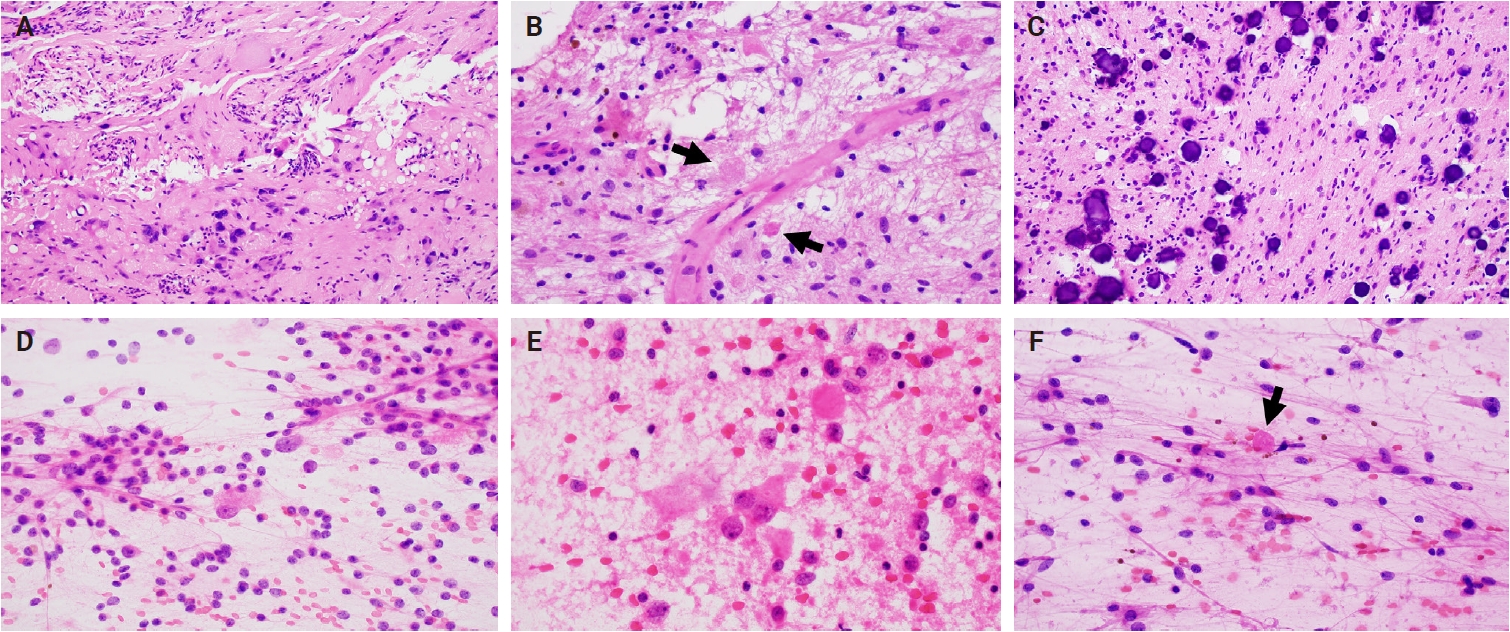
- Ganglioglioma and gangliocytoma: a review for pathologists
- Gianfranco E. Umeres-Francia, Melissa Mejia-Bautista, Pouya Jamshidi, Jared T. Ahrendsen
- J Pathol Transl Med. 2026;60(4):379-387. Published online July 15, 2026
- DOI: https://doi.org/10.4132/jptm.2026.06.06
- 659 View
- 23 Download
-
 Abstract
 PDF
PDF - Ganglioglioma and gangliocytoma are rare, predominantly low-grade neuroepithelial tumors that commonly present with epilepsy in children and young adults. Advances in molecular profiling have improved understanding of their pathogenesis, highlighting key roles for the mitogen-activated protein kinase/ERK signaling pathway. Diagnosis relies on a combination of clinical, radiologic, and histopathologic features, with complete surgical resection offering the best clinical outcomes. This review summarizes current knowledge on their epidemiology, etiology, clinical presentation, imaging characteristics, pathology, treatment strategies, and prognosis.
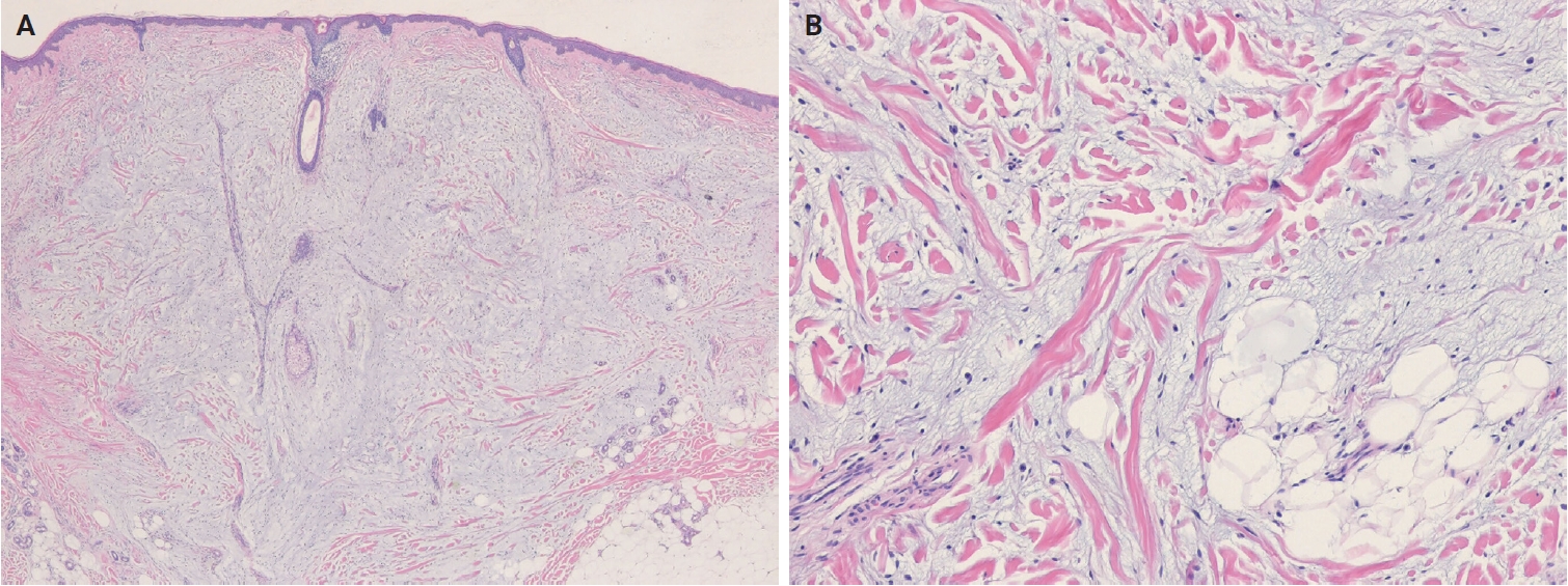
- Cutaneous soft tissue tumors in the 5th edition of the World Health Organization classification of skin tumors: key updates and new entities
- Joon Hyuk Choi
- J Pathol Transl Med. 2026;60(2):144-183. Published online March 13, 2026
- DOI: https://doi.org/10.4132/jptm.2026.01.09
- 4,452 View
- 268 Download
-
Abstract
PDF
- The 5th edition of the World Health Organization (WHO) classification of skin tumors introduces a dedicated chapter on cutaneous soft tissue tumors, providing a comprehensive, standardized reference with updated diagnostic criteria that directly inform routine dermatopathology practice and molecular diagnostics. This edition incorporates several key changes, including newly recognized entities such as EWSR1::SMAD3-rearranged fibroblastic tumor, neurotrophic tyrosine receptor kinase (NTRK)–rearranged spindle cell neoplasm, superficial CD34-positive fibroblastic tumor, and CRTC1::TRIM11 cutaneous tumor. Diagnostic terminology has also been refined; for example, the term ‘atypical intradermal smooth muscle neoplasm’ replaces ‘cutaneous leiomyosarcoma’ for lesions confined to the dermis, whereas the designation leiomyosarcoma is reserved for tumors with overt subcutaneous infiltration. In addition, epithelioid fibrous histiocytoma has been reassigned to the family of tumors of uncertain differentiation. This review summarizes the key updates and newly recognized entities in the chapter on cutaneous soft tissue tumors in the 5th edition of the WHO classification of skin tumors, emphasizing their clinicopathological and molecular implications.
Original Articles
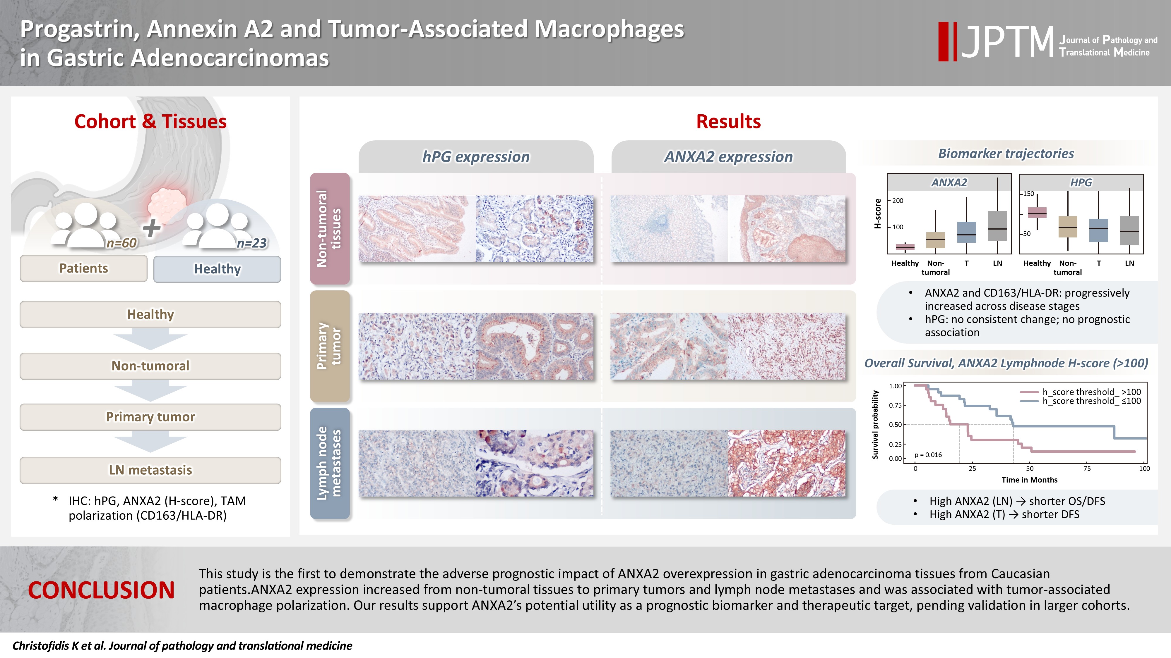
- Progastrin, annexin A2, and tumor-associated macrophages in gastric adenocarcinoma
- Konstantinos Christofidis, Rodanthi Fioretzaki, Stylianos Mavropoulos Papoudas, Nikolaos Charalampakis, Nikolaos Kavantzas, Dimitrios Schizas, Stratigoula Sakellariou
- J Pathol Transl Med. 2026;60(2):263-279. Published online March 10, 2026
- DOI: https://doi.org/10.4132/jptm.2025.12.20
- 1,947 View
- 159 Download
-
Abstract
PDF
 Supplementary Material
 - Background
Gastric adenocarcinoma is a major cause of cancer mortality worldwide, and reliable biomarkers remain insufficient. This study investigates the immunohistochemical expression of progastrin (hPG) and annexin A2 (ANXA2) and the polarization of tumor-associated macrophages in gastric adenocarcinoma to explore their potential prognostic and biological significance. Methods: A retrospective analysis was conducted on formalin-fixed, paraffin-embedded tissue samples from 60 patients with gastric adenocarcinoma (primary tumors, lymph node metastases, and non-tumoral gastric mucosa) and gastric biopsies from 23 healthy controls. The expression of hPG and ANXA2 was quantified using the H-score, and the CD163/human leukocyte antigen–DR (HLA-DR) ratio was used to represent macrophage polarization (M2/M1). Statistical analyses included non-parametric tests, Spearman correlations, Kaplan-Meier survival curves, and Cox proportional-hazards models. Results: ANXA2 expression was significantly elevated in cancer cells from primary tumors and lymph node metastases, compared with the non-tumoral gastric mucosa tissues and gastric mucosa tissues from healthy controls. ANXA2 expression increased with the tumor grade. High ANXA2 levels were associated with shorter overall and disease-free survival, but they did not have independent prognostic value. Although hPG expression correlated positively with ANXA2, it showed no significant prognostic association. The CD163/HLA-DR ratio increased with tumor progression and negatively correlated with ANXA2, but it did not influence survival outcomes. Conclusions: This study is the first to demonstrate the adverse prognostic impact of ANXA2 overexpression in gastric adenocarcinoma tissues from Caucasian patients. Our results suggest that ANXA2 might have utility as a prognostic biomarker and therapeutic target, if further large-scale studies validate and expand our findings.
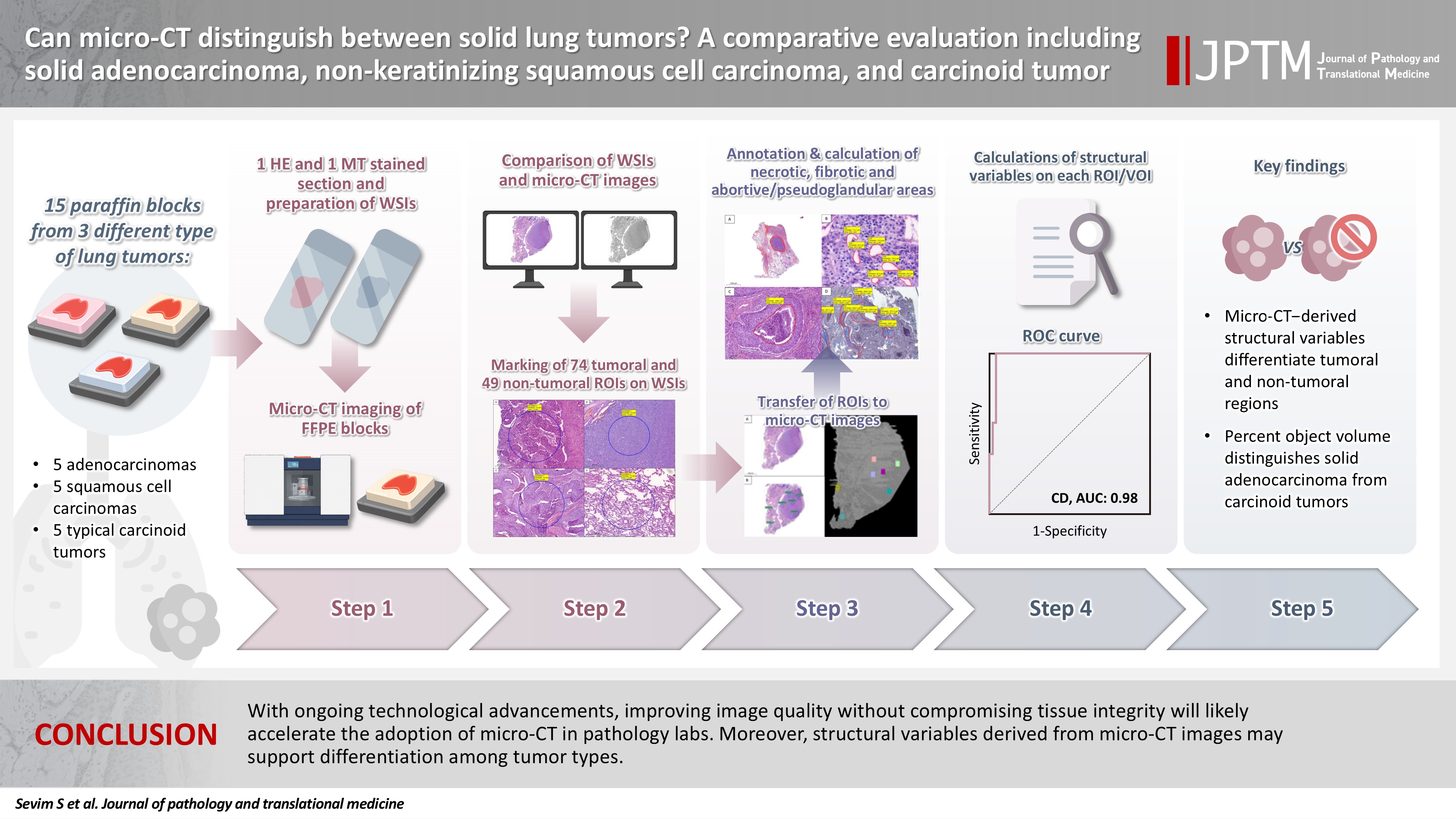
- Can micro-CT distinguish between solid lung tumors? A comparative evaluation including solid adenocarcinoma, non-keratinizing squamous cell carcinoma, and carcinoid tumor
- Selim Sevim, Serpil Dizbay Sak, Kaan Orhan, Arda Buyuksungur, Duru Karasoy, Hilal Ozakinci, Ayten Kayi Cangir
- J Pathol Transl Med. 2026;60(2):231-245. Published online March 10, 2026
- DOI: https://doi.org/10.4132/jptm.2025.12.16
- 1,831 View
- 122 Download
-
Abstract
PDFSupplementary Material
- Background
Some pulmonary carcinomas display a solid pattern, and immunohistochemistry is commonly used for tumor differentiation. Micro–computed tomography (micro-CT), with its ability to produce detailed three-dimensional images using small voxel sizes, may offer additional insights. This study investigates whether three solid tumor types, solid adenocarcinoma (sAC), non-keratinizing squamous cell carcinoma, and carcinoid tumor (CaT), can be differentiated using micro-CT. Methods: Fifteen paraffin blocks, five for each type, were scanned with micro-CT (Skyscan 1275, Bruker). These images were compared to whole slide images (WSIs) of the same tumors. Consequently, tumoral (n = 74) and non-tumoral (n = 49) regions of interest (tumor ROIs [tROIs] and non-tumor ROIs [ntROIs]) were selected on the micro-CT images and evaluated in terms of certain structural variables (percent object volume, structure model index, structure thickness, structure linear density, connectivity, connectivity density, open porosity, closed porosity) to investigate whether tumors can be differentiated from normal parenchyma and from each other. Results: Although detailed images comparable to WSIs could not be obtained, it was considered an important advantage to be able to examine the entire depth of the paraffin blocks. tROIs and ntROIs could be distinguished based on all variables (p < .001). Additionally, sAC showed a notable difference from CaT in “percent object volume” (p = .011). Conclusions: With ongoing technological advancements, improving image quality without compromising tissue integrity will likely accelerate the adoption of micro-CT in pathology labs. Moreover, structural variables derived from micro-CT images may support differentiation among tumor types.
Review Articles
- A comprehensive review of ossifying fibromyxoid tumor: insights into its clinical, pathological, and molecular landscape
- Kyriakos Chatzopoulos, Antonia Syrnioti, Mohamed Yakoub, Konstantinos Linos
- J Pathol Transl Med. 2026;60(1):6-19. Published online January 14, 2026
- DOI: https://doi.org/10.4132/jptm.2025.10.02

- 5,016 View
- 210 Download
-
Abstract
PDF
- Ossifying fibromyxoid tumor (OFMT) is a rare mesenchymal neoplasm first described in 1989. It typically arises in the superficial soft tissues of the extremities as a slow-growing, painless mass. Histologically, it is commonly characterized by a multilobular architecture composed of uniform epithelioid cells embedded in a fibromyxoid matrix, often surrounded by a rim of metaplastic bone. While classic cases are readily identifiable, the tumor's histopathological heterogeneity can mimic a range of benign and malignant neoplasms, posing significant diagnostic challenges. Molecularly, most OFMTs harbor PHF1 rearrangements, commonly involving fusion partners such as EP400, MEAF6, or TFE3. This review underscores the importance of an integrated diagnostic approach- incorporating histopathological, immunohistochemical, and molecular data- to accurately classify OFMT and distinguish it from its mimics. Expanding awareness of its morphologic and molecular spectrum is essential for precise diagnosis, optimal patient management, and a deeper understanding of this enigmatic neoplasm.
- Solitary fibrous tumor: an updated review
- Joon Hyuk Choi
- J Pathol Transl Med. 2026;60(1):20-46. Published online December 29, 2025
- DOI: https://doi.org/10.4132/jptm.2025.10.08
- 7,951 View
- 340 Download
- 2 Web of Science
- 4 Crossref
-
Abstract
PDF
- Solitary fibrous tumor (SFT) is a fibroblastic neoplasm characterized by a branching, thin-walled dilated staghorn-shaped (hemangiopericytoma-like) vasculature and a NAB2::STAT6 gene fusion. SFTs can occur in almost any anatomical location, including superficial and deep soft tissues, visceral organs, and bone. They most commonly occur in extrapleural locations, equally affect both sexes, and are typically present in adults. Although metastasis is rare, SFTs frequently show local recurrence. The diagnosis of SFTs is difficult because of their broad histological and morphological overlap with other neoplasms. An accurate diagnosis is important for guiding disease management and prognosis. Despite advances in molecular diagnostics and therapeutic strategies, the biological complexity and unpredictable clinical behavior of SFTs present significant challenges. This review provides an updated overview of SFT, with a focus on its molecular genetics, histopathological features, and diagnostic considerations.
-
Citations
Citations to this article as recorded by
- Clinicopathological characteristics and prognosis of central nervous system solitary fibrous tumor: An analysis of 271 cases
Wanwan Gao, Ming Li, Xiaojia Liu, Lingyang Hua, Hong Chen, Haixia Cheng
Pathology - Research and Practice.2026; 284: 156520. CrossRef - Pelvic solitary fibrous tumor, historically classified as hemangiopericytoma, presenting with venous compression and pelvic congestion: A case report
Dejan Svilar, Jovana Đošić, Anđela Đurić, Bojan Stojanović
Halo 194.2026; 32(1): 31. CrossRef - Robot-assisted laparoscopic resection of giant pelvic solitary fibrous tumor: a case report with literature review
Binbin Wang, Gengchen Huang, Wei Wei, Tie Mao, Zihan Gao, Yutao Ma, Yiming Gu
Frontiers in Oncology.2026;[Epub] CrossRef - Laparoscopic Resection of a Mesorectal Solitary Fibrous Tumor: A Case Report and Review of the Literature
Atsushi Sugimoto, Hiroshi Tsuchihashi, Hiroyuki Fujimoto, Masayasu Kawasaki
Cureus.2026;[Epub] CrossRef
- Clinicopathological characteristics and prognosis of central nervous system solitary fibrous tumor: An analysis of 271 cases
Original Article
- E-cadherin expression and tumor-stroma ratio as prognostic biomarkers of peritoneal recurrence in advanced gastric cancer: a digital image analysis-based stratification study
- Somang Lee, Binnari Kim
- J Pathol Transl Med. 2025;59(6):408-420. Published online November 6, 2025
- DOI: https://doi.org/10.4132/jptm.2025.08.27

- 4,103 View
- 124 Download
-
Abstract
PDF
- Background
Gastric cancer remains a significant global health burden, with a high peritoneal recurrence rates after curative surgery. E-cadherin and the tumor-stroma ratio (TSR) have been proposed as prognostic indicators, but their combined prognostic utility remains unclear. Methods: This retrospective study included 130 patients with T3/T4a gastric cancer who underwent curative gastrectomy at Ulsan University Hospital between 2014 and 2019. Immunohistochemistry for E-cadherin and Vimentin was performed. Digital image analysis using QuPath’s object classifier quantified E-cadherin expression and TSR. Results: Low E-cadherin expression was associated with diffuse-type histology and advanced T stage. Low TSR was linked to younger age, female sex, and XELOX treatment. In Kaplan-Meier analysis, low TSR showed a non-significant trend toward higher peritoneal recurrence (p = .054), while low E-cadherin expression was significantly associated with increased peritoneal recurrence (p = .002). Combined biomarker analysis also revealed a significant difference in recurrence-free survival (RFS) among the four groups (p = .005); patients with both high TSR and high E-cadherin expression experienced the most favorable RFS. In multivariable analysis, E-cadherin expression remained the only independent predictor of peritoneal recurrence (high vs. low; hazard ratio, 0.348; 95% confidence interval, 0.149 to 0.816; p = .015). Conclusions: E-cadherin and TSR reflect distinct tumor biology such as epithelial integrity and stromal composition, and their combined evaluation improves prognostic stratification. Digital image analysis enhances reproducibility and objectivity, supporting their integration into clinical workflows.
Review Article
- Breast schwannoma: review of entity and differential diagnosis
- Sandra Ixchel Sanchez, Ashley Cimino-Mathews
- J Pathol Transl Med. 2025;59(6):353-360. Published online November 3, 2025
- DOI: https://doi.org/10.4132/jptm.2025.08.12
- 5,213 View
- 188 Download
-
Abstract
PDF
- Schwannomas are benign peripheral nerve sheath tumors composed of Schwann cells, which uncommonly involve the breast. Most breast schwannomas are clinically present as a superficial palpable breast mass but may also be detected on screening mammography. Excision is the preferred treatment if symptomatic, and these are not known to recur. Histomorphology is similar to other anatomic sites: bland spindle cells with wavy nuclei, nuclear palisading (Verocay bodies), variably hypercellular (Antoni A) and hypocellular (Antoni B) areas, myxoid stroma, hyalinized vessels and variable cystic degeneration. Classic immunohistochemistry is diffuse and strong labeling for S100 and Sox10. Notable diagnostic pitfalls specific to the breast include myofibroblastoma, particularly the palisaded variant, and fascicular pseudoangiomatous stromal hyperplasia.
Case Study
- Clinicopathological characteristics of digestive system angioleiomyomas: case report and literature review
- Georgios Kalliopitsas, Christos Topalidis, Constantine Halkias, Theodora Gkeka, Konstantinos Sapalidis, Triantafyllia Koletsa
- J Pathol Transl Med. 2025;59(6):453-459. Published online October 28, 2025
- DOI: https://doi.org/10.4132/jptm.2025.08.04
- 4,026 View
- 119 Download
-
Abstract
PDF
- Angioleiomyomas are benign soft tissue tumors originating from the vascular wall. Although angioleiomyomas mainly occur in extremities, followed by head, neck, and trunk, they can also be found throughout the digestive system and especially in the oral cavity. Herein, the fourth case of a rectal angioleiomyoma in the English literature is reported and the clinicopathological features of digestive system angioleiomyomas were investigated. In contrast to their soft tissue counterparts, digestive system angioleiomyomas mainly affect males at a slightly younger age. Angioleiomyomas are mainly asymptomatic and only rarely elicit pain. Clinicians consider angioleiomyomas infrequently and instead include more common soft tissue or epithelial tumors in their differential diagnosis. To prevent angiomyolipoma misdiagnosis, pathologists should exercise caution when examining an angioleiomyoma composed of adipose tissue, smooth muscle, and blood vessels. Pathologists, radiologists, and surgeons should be aware that angioleiomyomas can occur in the digestive system.
Original Articles
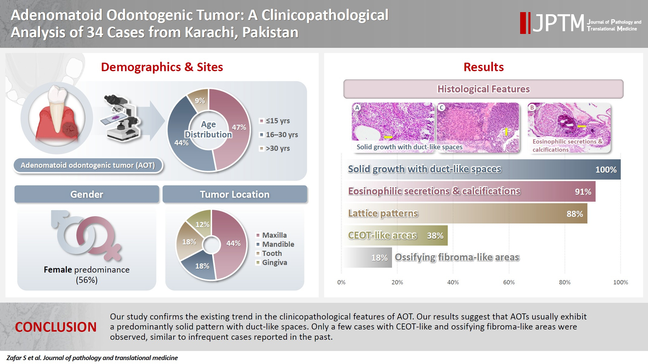
- Adenomatoid odontogenic tumor: clinicopathological analysis of 34 cases from Karachi, Pakistan
- Summaya Zafar, Sehar Sulaiman, Madeeha Nisar, Poonum Khan, Nasir Ud Din
- J Pathol Transl Med. 2025;59(6):390-397. Published online October 16, 2025
- DOI: https://doi.org/10.4132/jptm.2025.07.11
- 5,293 View
- 182 Download
- 1 Web of Science
- 1 Crossref
-
Abstract
PDF
- Background
Adenomatoid odontogenic tumor (AOT) is a benign slow-growing neoplasm of odontogenic epithelial origin that is relatively uncommon. Only a few studies have described its histological features. Hence, we aimed to describe the clinicopathological features of AOT in a cohort of patients. Methods: AOT cases diagnosed between 2009 and 2024 were searched electronically. Glass slides were retrieved from archives and were reviewed by two pathologists to record the associated morphological features. Other data including patient demographics and tumor site were collected by reviewing histopathology reports. Results: The age of patients ranged from 9 to 44 years (mean, 17.7 years), and most were female (55.9%). The maxilla (44.1%) was the most common tumor site. Histologically, a predominantly solid growth pattern (n = 34) accompanied by ducts with a cuboidal/columnar epithelial lining (n = 31), eosinophilic secretions (n = 31), calcifications (n = 31), lattice work pattern (n = 30), and cystic areas (n = 20) were observed. Less frequent features included calcifying epithelial odontogenic tumor (CEOT)–like areas (n = 13), osteodentin (n = 6), association with impacted tooth (n = 3), mucin in tubules (n = 7), fibrocollagenous stroma (n = 6), mucin in ducts (n = 3) and ossifying fibroma-like areas (n = 6). The association of ducts with a cuboidal/columnar epithelial lining, lattice work pattern, calcifications, and eosinophilic secretions with gingival tumors was statistically significant (p ≤ .05). Additionally, tooth tumors were significantly associated with CEOT-like areas (p = .03). Conclusions: Our study confirms the trends in the clinicopathological features of AOT in previous case reports. Our results suggest that AOTs usually exhibit a predominantly solid pattern with duct-like spaces. Only a few cases with CEOT-like and ossifying fibroma-like areas were observed, similar to infrequent cases reported in the past. -
Citations
Citations to this article as recorded by- Intraosseous lesions of the jaw: a clinicohistological study
Hadeel Odeh, Esra Nsour, Muna A. Salameh, Zayed M. Al-Zu’bi, Ali Al Khader
BMC Oral Health.2026;[Epub] CrossRef
- Intraosseous lesions of the jaw: a clinicohistological study
- Categorizing high-grade serous ovarian carcinoma into clinically relevant subgroups using deep learning–based histomic clusters
- Byungsoo Ahn, Eunhyang Park
- J Pathol Transl Med. 2025;59(2):91-104. Published online February 18, 2025
- DOI: https://doi.org/10.4132/jptm.2024.10.23
- 7,980 View
- 276 Download
- 1 Web of Science
- 2 Crossref
-
Abstract
PDFSupplementary Material
- Background
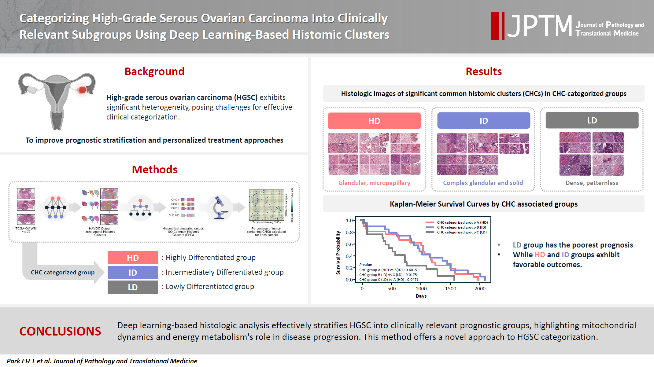
High-grade serous ovarian carcinoma (HGSC) exhibits significant heterogeneity, posing challenges for effective clinical categorization. Understanding the histomorphological diversity within HGSC could lead to improved prognostic stratification and personalized treatment approaches. Methods: We applied the Histomic Atlases of Variation Of Cancers model to whole slide images from The Cancer Genome Atlas dataset for ovarian cancer. Histologically distinct tumor clones were grouped into common histomic clusters. Principal component analysis and K-means clustering classified HGSC samples into three groups: highly differentiated (HD), intermediately differentiated (ID), and lowly differentiated (LD). Results: HD tumors showed diverse patterns, lower densities, and stronger eosin staining. ID tumors had intermediate densities and balanced staining, while LD tumors were dense, patternless, and strongly hematoxylin-stained. RNA sequencing revealed distinct patterns in mitochondrial oxidative phosphorylation and energy metabolism, with upregulation in the HD, downregulation in the LD, and the ID positioned in between. Survival analysis showed significantly lower overall survival for the LD compared to the HD and ID, underscoring the critical role of mitochondrial dynamics and energy metabolism in HGSC progression. Conclusions: Deep learning-based histologic analysis effectively stratifies HGSC into clinically relevant prognostic groups, highlighting the role of mitochondrial dynamics and energy metabolism in disease progression. This method offers a novel approach to HGSC categorization. -
Citations
Citations to this article as recorded by- Ovarian Cancer: Epidemiology, Disease Mechanisms, New Diagnosis and Treatment Strategies, and Research Directions
Zunera Khalid, Weirong Fan, Farah Nazir, Yixiang Xing, Tengchuan Jin
iNew Medicine.2026;[Epub] CrossRef - Learning Disabilities in the 21st Century: Integrating Neuroscience, Education, and Technology for Better Outcomes
Syed Mohammed Basheeruddin Asdaq, Ahmad H. Alhowail, Syed Imam Rabbani, Naira Nayeem, Syed Mohammed Emaduddin Asdaq, Faiqa Nausheen
SAGE Open.2025;[Epub] CrossRef
- Ovarian Cancer: Epidemiology, Disease Mechanisms, New Diagnosis and Treatment Strategies, and Research Directions
- The combination of CDX2 expression status and tumor-infiltrating lymphocyte density as a prognostic factor in adjuvant FOLFOX-treated patients with stage III colorectal cancers
- Ji-Ae Lee, Hye Eun Park, Hye-Yeong Jin, Lingyan Jin, Seung Yeon Yoo, Nam-Yun Cho, Jeong Mo Bae, Jung Ho Kim, Gyeong Hoon Kang
- J Pathol Transl Med. 2025;59(1):50-59. Published online October 24, 2024
- DOI: https://doi.org/10.4132/jptm.2024.09.26
- 5,542 View
- 300 Download
- 1 Web of Science
-
Abstract
PDFSupplementary Material
- Background
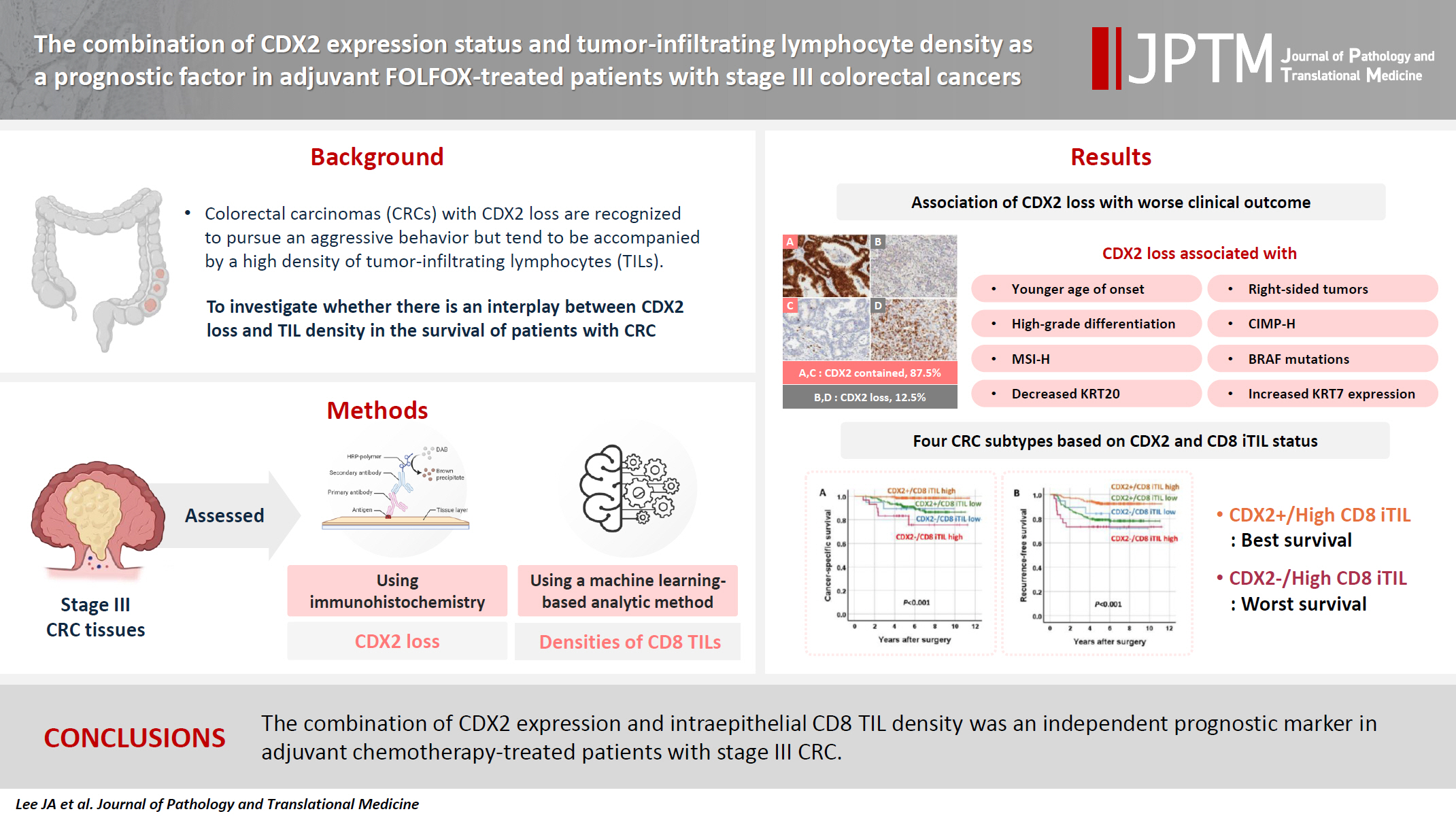
Colorectal carcinomas (CRCs) with caudal-type homeobox 2 (CDX2) loss are recognized to pursue an aggressive behavior but tend to be accompanied by a high density of tumor-infiltrating lymphocytes (TILs). However, little is known about whether there is an interplay between CDX2 loss and TIL density in the survival of patients with CRC.
Methods
Stage III CRC tissues were assessed for CDX2 loss using immunohistochemistry and analyzed for their densities of CD8 TILs in both intraepithelial (iTILs) and stromal areas using a machine learning-based analytic method.
Results
CDX2 loss was significantly associated with a higher density of CD8 TILs in both intraepithelial and stromal areas. Both CDX2 loss and a high CD8 iTIL density were found to be prognostic parameters and showed hazard ratios of 2.314 (1.050–5.100) and 0.378 (0.175–0.817), respectively, for cancer-specific survival. A subset of CRCs with retained CDX2 expression and a high density of CD8 iTILs showed the best clinical outcome (hazard ratio of 0.138 [0.023–0.826]), whereas a subset with CDX2 loss and a high density of CD8 iTILs exhibited the worst clinical outcome (15.781 [3.939–63.230]).
Conclusions
Altogether, a high density of CD8 iTILs did not make a difference in the survival of patients with CRC with CDX2 loss. The combination of CDX2 expression and intraepithelial CD8 TIL density was an independent prognostic marker in adjuvant chemotherapy-treated patients with stage III CRC.
- Diagnosis of invasive encapsulated follicular variant papillary thyroid carcinoma by protein-based machine learning
- Truong Phan-Xuan Nguyen, Minh-Khang Le, Sittiruk Roytrakul, Shanop Shuangshoti, Nakarin Kitkumthorn, Somboon Keelawat
- J Pathol Transl Med. 2025;59(1):39-49. Published online October 24, 2024
- DOI: https://doi.org/10.4132/jptm.2024.09.14
- 6,476 View
- 346 Download
- 2 Web of Science
- 2 Crossref
-
Abstract
PDFSupplementary Material
- Background
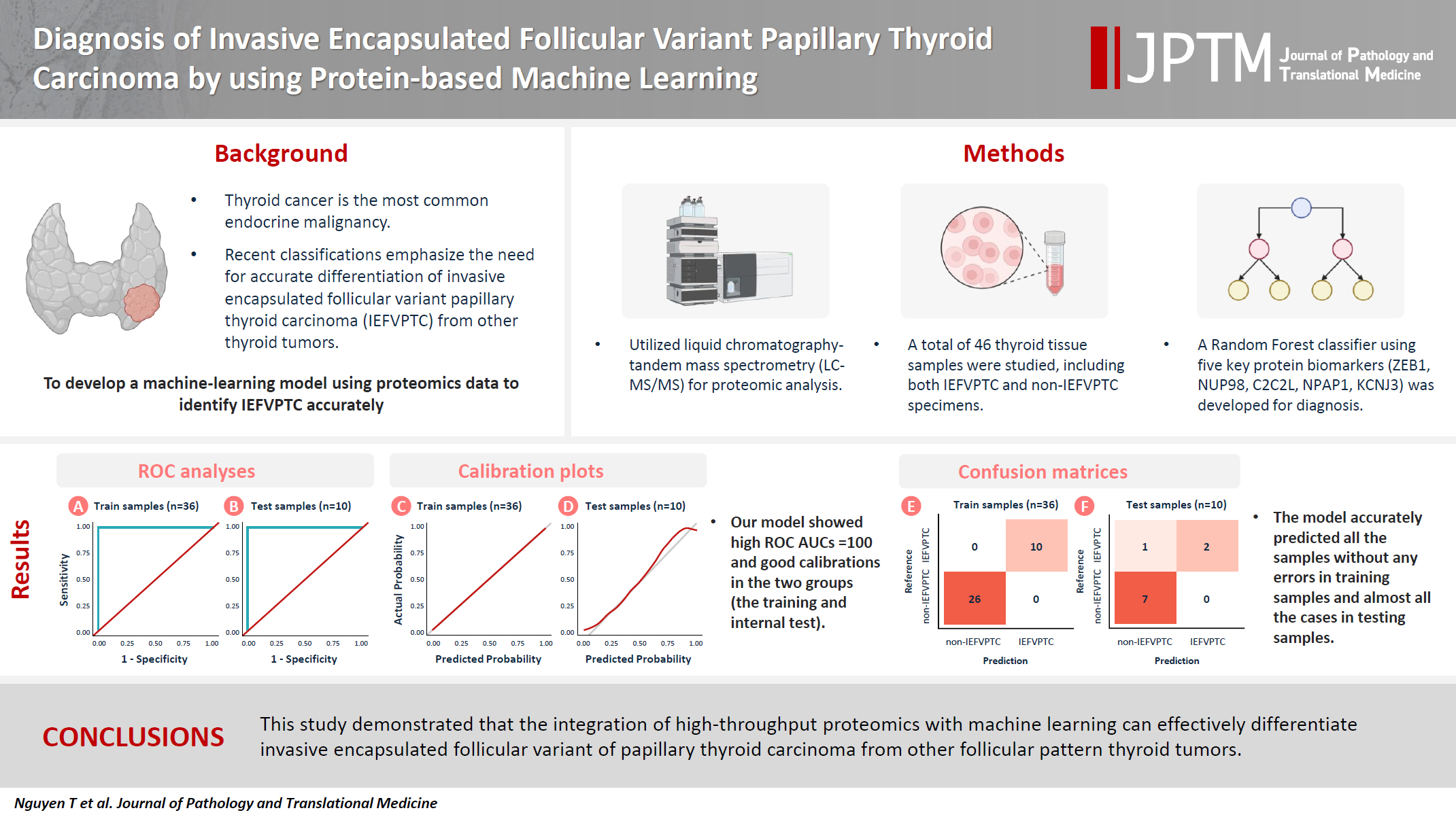
Although the criteria for follicular-pattern thyroid tumors are well-established, diagnosing these lesions remains challenging in some cases. In the recent World Health Organization Classification of Endocrine and Neuroendocrine Tumors (5th edition), the invasive encapsulated follicular variant of papillary thyroid carcinoma was reclassified as its own entity. It is crucial to differentiate this variant of papillary thyroid carcinoma from low-risk follicular pattern tumors due to their shared morphological characteristics. Proteomics holds significant promise for detecting and quantifying protein biomarkers. We investigated the potential value of a protein biomarker panel defined by machine learning for identifying the invasive encapsulated follicular variant of papillary thyroid carcinoma, initially using formalin- fixed paraffin-embedded samples.
Methods
We developed a supervised machine-learning model and tested its performance using proteomics data from 46 thyroid tissue samples.
Results
We applied a random forest classifier utilizing five protein biomarkers (ZEB1, NUP98, C2C2L, NPAP1, and KCNJ3). This classifier achieved areas under the curve (AUCs) of 1.00 and accuracy rates of 1.00 in training samples for distinguishing the invasive encapsulated follicular variant of papillary thyroid carcinoma from non-malignant samples. Additionally, we analyzed the performance of single-protein/gene receiver operating characteristic in differentiating the invasive encapsulated follicular variant of papillary thyroid carcinoma from others within The Cancer Genome Atlas projects, which yielded an AUC >0.5.
Conclusions
We demonstrated that integration of high-throughput proteomics with machine learning can effectively differentiate the invasive encapsulated follicular variant of papillary thyroid carcinoma from other follicular pattern thyroid tumors. -
Citations
Citations to this article as recorded by- Advances in immunotherapy for thyroid malignancies: from molecular targets to clinical outcomes
Shuo Lv, Jinbao Wang, Guohao Chen, Yongshun Wang, Naiqing Liu
Frontiers in Medicine.2026;[Epub] CrossRef - Misdiagnosed follicular adenoma with 11 year postoperative liver and lung metastases a case report and literature review
Kai-Li Yang, Heng-Tong Han, Shou-Hua Li, Xiao-Xiao Li, Ze Yang, Li-Bin Ma, Yong-Xun Zhao
Discover Oncology.2025;[Epub] CrossRef
- Advances in immunotherapy for thyroid malignancies: from molecular targets to clinical outcomes
Case Studies
- Primary epithelioid inflammatory myofibroblastic sarcoma of the brain with EML4::ALK fusion mimicking intra-axial glioma: a case report and brief literature review
- Eric Eunshik Kim, Chul-Kee Park, Koung Mi Kang, Yoonjin Kwak, Sung-Hye Park, Jae-Kyung Won
- J Pathol Transl Med. 2024;58(3):141-145. Published online May 14, 2024
- DOI: https://doi.org/10.4132/jptm.2024.04.12
- 6,223 View
- 215 Download
- 2 Web of Science
- 2 Crossref
-
Abstract
PDF
- An aggressive subtype of inflammatory myofibroblastic tumor, epithelioid inflammatory myofibroblastic sarcoma occurs primarily inside the abdominal cavity, followed by a pulmonary localization. Most harbor anaplastic lymphoma kinase (ALK) gene rearrangements, with RANBP2 and RRBP1 among the well-documented fusion partners. We report the second case of primary epithelioid inflammatory myofibroblastic sarcoma of the brain, with a well-known EML4::ALK fusion. The case is notable for its intra-axial presentation that clinico-radiologically mimicked glioma.
-
Citations
Citations to this article as recorded by
- Malignant potential of neuroendocrine microtumor of the pancreas harboring high-grade transformation: lesson learned from a patient with von Hippel-Lindau syndrome
- Jongwon Lee, Kyung Jin Lee, Dae Wook Hwang, Seung-Mo Hong
- J Pathol Transl Med. 2024;58(2):91-97. Published online March 13, 2024
- DOI: https://doi.org/10.4132/jptm.2024.02.13
- 6,156 View
- 222 Download
- 3 Web of Science
- 4 Crossref
-
Abstract
PDF
- Pancreatic neuroendocrine microtumor (PNEMT) is a neuroendocrine tumor (NET) < 0.5 cm in diameter, and it is considered benign. We report a PNEMT with high-grade transformation (HGT). A man in his 60s with von Hippel-Lindau syndrome underwent surgical resection of a NET. A second sub-centimeter nodule with a nodule-in-nodule pattern was discovered. The 0.4 cm outer nodule contained clear columnar cells with round nuclei and indistinct nucleoli, while the 0.1 cm inner nodule had eosinophilic cells with an increased nuclear to cytoplasmic ratio, vesicular nuclei, and prominent nucleoli. Tumor cells in the outer and inner nodules were synaptophysin and chromogranin positive. Only the inner nodule was p53 positive, while the outer nodule was exclusively positive for carbonic anhydrase 9 and vimentin. The Ki-67 labeling indices for the outer and inner nodules were 2.1% (grade 1) and 44.3% (grade 3), respectively. This nodule was determined to be a PNEMT with HGT. Our findings suggest that a PNEMT may not always be benign and can undergo HGT.
-
Citations
Citations to this article as recorded by- Intraductal papillary mucinous neoplasm unveiling incidental multifocal pancreatic neuroendocrine tumors: a challenging case report
Faten Limaiem, Mohamed Hajri, Nafaa Arfa
International Journal of Surgery Case Reports.2026; 138(5): 1634. CrossRef - Decoding Pancreatic Neuroendocrine Tumors: Molecular Profiles, Biomarkers, and Pathways to Personalized Therapy
Linda Galasso, Federica Vitale, Gabriele Giansanti, Giorgio Esposto, Raffaele Borriello, Irene Mignini, Alberto Nicoletti, Lorenzo Zileri Dal Verme, Antonio Gasbarrini, Maria Elena Ainora, Maria Assunta Zocco
International Journal of Molecular Sciences.2025; 26(16): 7814. CrossRef - Pancreatic neuroendocrine microtumors in the elderly: A retrospective study using cadaveric pancreatic tissue
Ting Yang, Ke Ren, Xiang-Quan Chen, Taku Toriumi, Yutaro Natsuyama, Jun Li, Aoi Sukeda, Toshitaka Nagao, Shuang-Qin Yi
World Journal of Gastrointestinal Oncology.2025;[Epub] CrossRef - Molecular Basis of Pancreatic Neuroendocrine Tumors
Alesia Maluchenko, Denis Maksimov, Zoia Antysheva, Julia Krupinova, Ekaterina Avsievich, Olga Glazova, Natalia Bodunova, Nikolay Karnaukhov, Ilia Feidorov, Diana Salimgereeva, Mark Voloshin, Pavel Volchkov
International Journal of Molecular Sciences.2024; 25(20): 11017. CrossRef
- Intraductal papillary mucinous neoplasm unveiling incidental multifocal pancreatic neuroendocrine tumors: a challenging case report
Original Article
- Tumor-infiltrating T lymphocytes evaluated using digital image analysis predict the prognosis of patients with diffuse large B-cell lymphoma
- Yunjoo Cho, Jiyeon Lee, Bogyeong Han, Sang Eun Yoon, Seok Jin Kim, Won Seog Kim, Junhun Cho
- J Pathol Transl Med. 2024;58(1):12-21. Published online January 10, 2024
- DOI: https://doi.org/10.4132/jptm.2023.11.02
- 6,849 View
- 285 Download
- 5 Web of Science
- 4 Crossref
-
Abstract
PDF
- Background
The implication of the presence of tumor-infiltrating T lymphocytes (TIL-T) in diffuse large B-cell lymphoma (DLBCL) is yet to be elucidated. We aimed to investigate the effect of TIL-T levels on the prognosis of patients with DLBCL.
Methods
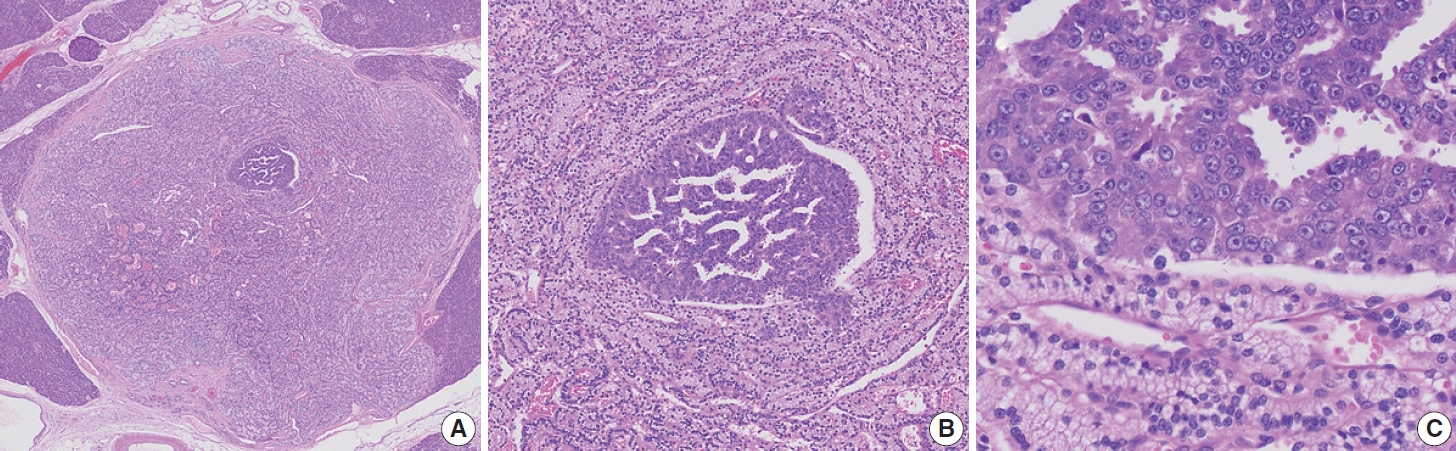
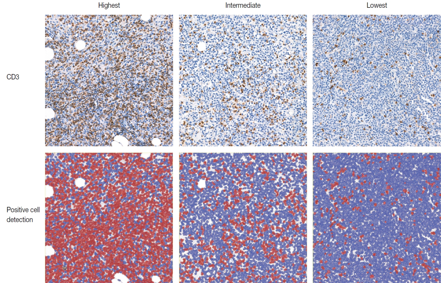
Ninety-six patients with DLBCL were enrolled in the study. The TIL-T ratio was measured using QuPath, a digital pathology software package. The TIL-T ratio was investigated in three foci (highest, intermediate, and lowest) for each case, resulting in TIL-T–Max, TIL-T–Intermediate, and TIL-T–Min. The relationship between the TIL-T ratios and prognosis was investigated.
Results
When 19% was used as the cutoff value for TIL-T–Max, 72 (75.0%) and 24 (25.0%) patients had high and low TIL-T–Max, respectively. A high TIL-T–Max was significantly associated with lower serum lactate dehydrogenase levels (p < .001), with patient group who achieved complete remission after RCHOP therapy (p < .001), and a low-risk revised International Prognostic Index score (p < .001). Univariate analysis showed that patients with a low TIL-T–Max had a significantly worse prognosis in overall survival compared to those with a high TIL-T–Max (p < .001); this difference remained significant in a multivariate analysis with Cox proportional hazards (hazard ratio, 7.55; 95% confidence interval, 2.54 to 22.42; p < .001).
Conclusions
Patients with DLBCL with a high TIL-T–Max showed significantly better prognosis than those with a low TIL-T–Max, and the TIL-T–Max was an independent indicator of overall survival. These results suggest that evaluating TIL-T ratios using a digital pathology system is useful in predicting the prognosis of patients with DLBCL. -
Citations
Citations to this article as recorded by- Do Pre‐Treatment Biopsy Characteristics Predict Early Tumour Progression in Feline Diffuse Large B Cell Nasal Lymphoma Treated With Radiotherapy?
Valerie J. Poirier, Valeria Meier, Michelle Turek, Neil Christensen, Jacqueline Bowal, Matthew D. Ponzini, Stefan M. Keller
Veterinary and Comparative Oncology.2025; 23(1): 82. CrossRef - Comprehensive Analysis of Tumor Microenvironment and PD-L1 Expression Associations with Clinicopathological Features and Prognosis in Diffuse Large B-Cell Lymphoma
Yun-Li Xie, Long-Feng Ke, Wen-Wen Zhang, Fu Kang, Shu-Yi Lu, Chen-Yu Wu, Huan-Huan Zhu, Jian-Chao Wang, Gang Chen, Yan-Ping Chen
Blood and Lymphatic Cancer: Targets and Therapy.2025; Volume 15: 167. CrossRef - Metabolic-immune axis in the tumor microenvironment: a new strategy for prognostic assessment and precision therapy in DLBCL and FL
Chengqian Chen, Wei Guo, Haotian Wang, Luming Cao, Ou Bai
Frontiers in Immunology.2025;[Epub] CrossRef - Integrative analysis of a novel immunogenic PANoptosis‑related gene signature in diffuse large B-cell lymphoma for prognostication and therapeutic decision-making
Ming Xu, Ming Ruan, Wenhua Zhu, Jiayue Xu, Ling Lin, Weili Li, Weirong Zhu
Scientific Reports.2024;[Epub] CrossRef
- Do Pre‐Treatment Biopsy Characteristics Predict Early Tumour Progression in Feline Diffuse Large B Cell Nasal Lymphoma Treated With Radiotherapy?
Case Study
- Thyroid pathology, a clue to PTEN hamartoma tumor syndrome
- Yurimi Lee, Young Lyun Oh
- J Pathol Transl Med. 2023;57(3):178-183. Published online March 30, 2023
- DOI: https://doi.org/10.4132/jptm.2023.03.04
- 9,746 View
- 214 Download
- 10 Web of Science
- 9 Crossref
-
Abstract
PDF
- Phosphatase and tensin homolog (PTEN) hamartoma tumor syndrome (PHTS) is a hereditary disorder caused by germline inactivating mutations in the PTEN tumor suppressor gene. As a type of PHTS, Cowden syndrome is associated with abnormalities of the thyroid, breast, uterus, and gastrointestinal tract. A 52-year-old-woman visited the outpatient clinic of our endocrinology clinic with multiple thyroid nodules and Hashimoto's thyroiditis. Computed tomography imaging revealed a multinodular mass measuring up to 3.5 cm in the left thyroid lobe, causing laryngotracheal airway displacement. The total thyroidectomy specimen revealed multiple follicular adenomas and adenomatous nodules with lymphocytic thyroiditis and lipomatous metaplasia in the background. The patient was suspected of PTHS based on her thyroid pathology, family history, and numerous hamartomatous lesions of the breast, uterus, and skin. Her diagnosis was confirmed through molecular testing. This case demonstrates that pathologists must be well acquainted with thyroid pathology in PHTS.
-
Citations
Citations to this article as recorded by- Risk of malignancy in PTEN-altered thyroid nodules detected on preoperative FNA molecular testing: a systematic review and meta-analysis
Patrizia Straccia, Vincenzo Fiorentino, Belen Padial Urtueta, Qianqian Zhang, Alessia Piermattei, Federica Cianfrini, Antonino Mule, Esther Diana Rossi
Human Pathology.2026; : 106104. CrossRef - Dual primary malignancies in Kashmir: A five-year analysis of temporal patterns, gender-specific presentations and treatment outcomes in a high gastrointestinal cancer risk population
Ubaid Jeelani, Mushood Ghulam Nabi, Asim Ahmad Dar, Gowher Ahmad Wagai, Aadil Najeed, Sheikh Owais Ahmad, Lande Sagar Janardhan, Md Mayeen Afsan Ahmad, Uzma Majeed
Journal of Family Medicine and Primary Care.2026; 15(2): 530. CrossRef - Recognizing Familial Thyroid Neoplasia: The Pathologist’s Role in Diagnosis and Management
Vania Nosé, Sule Canberk, Zubair Baloch
Advances in Anatomic Pathology.2026;[Epub] CrossRef - A clinical case of papillary thyroid cancer associated with a PTEN gene defect
R. A. Atanesyan, L. Ja. Klimov, T. M. Vdovina, G. A. Saneeva, E. I. Andreeva, I. A. Stremenkova, R. I. Arakelyan, I. K. Gasparyan
Rossiyskiy Vestnik Perinatologii i Pediatrii (Russian Bulletin of Perinatology and Pediatrics).2025; 69(6): 85. CrossRef - Pediatric cancer predisposition syndromes involving non-central nervous system solid pediatric tumors: a review on their manifestations with a focus on histopathology
B. Schurink, M. Reyes-Múgica, R. R. de Krijger
Virchows Archiv.2025; 486(1): 3. CrossRef - Dedifferentiated Leiomyosarcoma of the Uterine Corpus with Heterologous Component: Clinicopathological Analysis of Five Consecutive Cases from a Single Institution and Comprehensive Literature Review
Suyeon Kim, Hyunsik Bae, Hyun-Soo Kim
Diagnostics.2024; 14(2): 160. CrossRef - Case report: Rare oral manifestations in Cowden syndrome with PTEN mutation
Wei Yuan, Yanbin Liu, Haibin Sun, Ming Su, Lizheng Qin, Xin Huang
Frontiers in Oncology.2024;[Epub] CrossRef - Can thyroid histomorphology identify patients with PTEN hamartoma tumour syndrome?
Melad N Dababneh, Laura Rabinowitz, Gilman Plitt, Charis Eng, Christopher C Griffith
Histopathology.2024; 85(6): 929. CrossRef - A novel mutation in PTEN in anaplastic thyroid carcinoma: A case report
Yanli Zhao
Biomedical Reports.2024;[Epub] CrossRef
- Risk of malignancy in PTEN-altered thyroid nodules detected on preoperative FNA molecular testing: a systematic review and meta-analysis
Original Article
- Significance of tumor-associated neutrophils, lymphocytes, and neutrophil-to-lymphocyte ratio in non-invasive and invasive bladder urothelial carcinoma
- Wael Abdo Hassan, Ahmed Kamal ElBanna, Noha Noufal, Mohamed El-Assmy, Hany Lotfy, Rehab Ibrahim Ali
- J Pathol Transl Med. 2023;57(2):88-94. Published online January 10, 2023
- DOI: https://doi.org/10.4132/jptm.2022.11.06
- 9,166 View
- 336 Download
- 17 Web of Science
- 16 Crossref
-
Abstract
PDF
- Background
Tumor-infiltrating neutrophils and lymphocytes play essential roles in promoting or combating various neoplasms. This study aimed to investigate the association between tumor-infiltrating neutrophils and lymphocytes and the neutrophil-to-lymphocyte ratio in the progression of urothelial carcinoma.
Methods
A total of 106 patients diagnosed with urothelial carcinoma were was. Pathological examination for tumor grade and stage and for tumor-infiltrating neutrophils, both CD4 and CD8+ T lymphocytes, as well as the neutrophil- to-lymphocyte ratio were evaluated.
Results
The presence of neutrophils and the neutrophil-to-lymphocyte ratio correlated with high-grade urothelial neoplasms. In both low- and high-grade tumors, the lymphocytes increased during progression from a non-invasive neoplasm to an early-invasive neoplasm. CD8+ T lymphocytes increased in low-grade non–muscle-invasive tumors compared to non-invasive tumors. Additionally, there was a significant decrease in CD8+ T lymphocytes during progression to muscle-invasive tumors.
Conclusions
Our results suggest that tumor-infiltrating neutrophils and CD8+ T lymphocytes have a significant effect on tumor grade and progression. -
Citations
Citations to this article as recorded by- T cell fate regulation in EBV‑associated nasopharyngeal carcinoma (Review)
Liuyang Zhang, Shun Ding, Dongzhui Chen, Benchi Cai, Zhonglin Mu
Oncology Reports.2026; 55(6): 1. CrossRef - Prognostic role of the neutrophil/lymphocyte ratio in high‐risk BCG‐naïve non‐muscle‐invasive bladder cancer treated with intravesical gemcitabine/docetaxel
Mohamad Abou Chakra, Riitta Lassila, Nancy El Beayni, Sarah L. Mott, Michael A. O'Donnell
BJU International.2025; 135(1): 125. CrossRef - Understanding the Dual Role of Macrophages in Tumor Growth and Therapy: A Mechanistic Review
Muhammad Summer, Saima Riaz, Shaukat Ali, Qudsia Noor, Rimsha Ashraf, Rana Rashad Mahmood Khan
Chemistry & Biodiversity.2025;[Epub] CrossRef - Cross-Talk Between Cancer and Its Cellular Environment—A Role in Cancer Progression
Eliza Turlej, Aleksandra Domaradzka, Justyna Radzka, Dominika Drulis-Fajdasz, Julita Kulbacka, Agnieszka Gizak
Cells.2025; 14(6): 403. CrossRef - Global trends in tumor-associated neutrophil research: a bibliometric and visual analysis
Shaodong Li, Peng Dong, Xueliang Wu, Zhenhua Kang, Guoqiang Yan
Frontiers in Immunology.2025;[Epub] CrossRef - Tumor-associated neutrophils and neutrophil extracellular traps in lung cancer: antitumor/protumor insights and therapeutic implications
Milad Sheervalilou, Mostafa Ghanei, Masoud Arabfard
Medical Oncology.2025;[Epub] CrossRef - Construction of a column-line graphical model of poor outcome of neoadjuvant regimens for muscle-invasive bladder cancer based on NLR, dNLR and SII indicators
Bo Hu, Longsheng Wang, Shanna Qu, Tao Zhang
World Journal of Surgical Oncology.2025;[Epub] CrossRef - Machine Learning of Urine Cytology Highlights Increased Neutrophil Count in Muscle-Invasive Urothelial Carcinoma
Moe Kameda, Sayaka Kobayashi, Yoshimi Nishijima, Ryosuke Akuzawa, Rio Kaneko, Rio Shibanuma, Seiji Arai, Hayato Ikota, Kazuhiro Suzuki, Hideaki Yokoo, Masanao Saio
Journal of Cytology.2025; 42(3): 124. CrossRef - Tumor-Infiltrating Immune Cells in Non-Muscle-Invasive Bladder Cancer: Prognostic Implications, Predictive Value, and Future Perspectives
Roberta Mazzucchelli, Angelo Cormio, Magda Zanelli, Maurizio Zizzo, Andrea Palicelli, Andrea Benedetto Galosi, Francesca Sanguedolce
Applied Sciences.2025; 15(22): 12032. CrossRef - Immune cell networking in solid tumors: focus on macrophages and neutrophils
Irene Di Ceglie, Silvia Carnevale, Anna Rigatelli, Giovanna Grieco, Piera Molisso, Sebastien Jaillon
Frontiers in Immunology.2024;[Epub] CrossRef - Immunohistochemistry assessment of tissue neutrophil-to-lymphocyte ratio predicts outcomes in melanoma patients treated with anti-programmed cell death 1 therapy
Renan J. Teixeira, Vinícius G. de Souza, Bruna P. Sorroche, Victor G. Paes, Fabiana A. Zambuzi-Roberto, Caio A.D. Pereira, Vinicius L. Vazquez, Lidia M.R.B. Arantes
Melanoma Research.2024; 34(3): 234. CrossRef - Association between alteration of neutrophil to lymphocyte ratio, platelet to lymphocyte ratio, cancer antigen-125 and surgical outcomes in advanced stage ovarian cancer patient who received neoadjuvant chemotherapy
Ponganun Tuntinarawat, Ratnapat Tangmanomana, Thannaporn Kittisiam
Gynecologic Oncology Reports.2024; 52: 101347. CrossRef - Prognostic value of neutrophil-to-lymphocyte ratio in patients with non–muscle-invasive bladder cancer with intravesical Bacillus Calmette–Guérin immunotherapy: a systematic review and meta-analysis
Jiaguo Huang, Li Lin, Dikai Mao, Runmiao Hua, Feifei Guan
Frontiers in Immunology.2024;[Epub] CrossRef - Update on the Mechanism of Action of Intravesical BCG Therapy to Treat Non-Muscle-Invasive Bladder Cancer
Mohamad Abou Chakra, Yi Luo, Igor Duquesne, Michael A O'Donnell
Frontiers in Bioscience-Landmark.2024;[Epub] CrossRef - Significant association between high neutrophil-lymphocyte ratio and poor prognosis in patients with hepatocellular carcinoma: a systematic review and meta-analysis
Chunhua Xu, Fenfang Wu, Lailing Du, Yeping Dong, Shan Lin
Frontiers in Immunology.2023;[Epub] CrossRef - Chitinase 3-like-1 Expression in the Microenvironment Is Associated with Neutrophil Infiltration in Bladder Cancer
Ling-Yi Xiao, Yu-Li Su, Shih-Yu Huang, Yi-Hua Chen, Po-Ren Hsueh
International Journal of Molecular Sciences.2023; 24(21): 15990. CrossRef
- T cell fate regulation in EBV‑associated nasopharyngeal carcinoma (Review)
Case Study
- Primary pulmonary epithelioid inflammatory myofibroblastic sarcoma: a rare entity and a literature review
- Priyanka Singh, Aruna Nambirajan, Manish Kumar Gaur, Rahul Raj, Sunil Kumar, Prabhat Singh Malik, Deepali Jain
- J Pathol Transl Med. 2022;56(4):231-237. Published online July 7, 2022
- DOI: https://doi.org/10.4132/jptm.2022.05.08
- 15,457 View
- 134 Download
- 14 Web of Science
- 14 Crossref
-
Abstract
PDF
- Epithelioid inflammatory myofibroblastic sarcoma (EIMS) is an aggressive subtype of inflammatory myofibroblastic tumor (IMT) harboring anaplastic lymphoma kinase (ALK) gene fusions and is associated with high risk of local recurrence and poor prognosis. Herein, we present a young, non-smoking male who presented with complaints of cough and dyspnoea and was found to harbor a large right lower lobe lung mass. Biopsy showed a high-grade epithelioid to rhabdoid tumor with ALK and desmin protein expression. The patient initially received 5 cycles of crizotinib and remained stable for 1 year; however, he then developed multiple bony metastases, for which complete surgical resection was performed. Histopathology confirmed the diagnosis of EIMS, with ALK gene rearrangement demonstrated by fluorescence in situ hybridization. Postoperatively, the patient is asymptomatic with stable metastatic disease on crizotinib and has been started on palliative radiotherapy. EIMS is a very rare subtype of IMT that needs to be included in the differential diagnosis of ALKexpressing lung malignancies in young adults.
-
Citations
Citations to this article as recorded by- Primary Pulmonary Epithelioid Inflammatory Myofibroblastic Sarcoma With SQSTM1::ALK Fusion
Havva Gokce Terzioglu, Phillip McMullen, Güliz A. Barkan
Diagnostic Cytopathology.2026;[Epub] CrossRef - Lung Mass in a 22-Year-Old Woman With Persistent Fever
Misawo Ishikawa, Kenju Nakao, Emi Hagui, Naoki Hamajima, Aya Naiki-Ito, Yasushi Yatabe, Satoru Takahashi
CHEST Pulmonary.2026; : 100273. CrossRef - Inflammatory Myofibroblastic Tumor: An Updated Review
Joon Hyuk Choi
Cancers.2025; 17(8): 1327. CrossRef - Epithelioid Inflammatory Myofibroblastic Sarcoma: Case Series With a First Report of CLTC::ALK Fusion in an Aggressive Disease
Daisy Maharjan, Carina Dehner, Ali Alani, Robert Bell, Sheila Segura
Genes, Chromosomes and Cancer.2025;[Epub] CrossRef - ALK rearranged malignant mesenchymal neoplasms of thorax: therapeutically targetable ‘ALKomas’ beyond the spectrum of non-small cell lung carcinomas and thoracic inflammatory myofibroblastic tumors
Shreya Sadhu, Adarsh Barwad, Asit Ranjan Mridha, Prabhat Singh Malik, Aruna Nambirajan, Deepali Jain
Virchows Archiv.2025; 487(5): 1003. CrossRef - Mediastinal epithelioid inflammatory myofibroblastic sarcoma with the EML4‐ALK fusion: A case report and literature review
Tingyu Pan, Xinyu Sun, Xiao Wu, Futing Tang, Xianmei Zhou, Qian Wang, Shi Chen
Respirology Case Reports.2024;[Epub] CrossRef - Primary epithelioid inflammatory myofibroblastic sarcoma of the brain with EML4::ALK fusion mimicking intra-axial glioma: a case report and brief literature review
Eric Eunshik Kim, Chul-Kee Park, Koung Mi Kang, Yoonjin Kwak, Sung-Hye Park, Jae-Kyung Won
Journal of Pathology and Translational Medicine.2024; 58(3): 141. CrossRef - Epithelioid Inflammatory Myofibroblastic Sarcoma: A Report of a Rare Case
Varun Ronanki, Vaddatti Tejeswini, Inuganti Venkata Renuka, Shaik Raheema, Bakkamanthala S K Kanth
Cureus.2024;[Epub] CrossRef - Thoracic epithelioid inflammatory myofibroblastic sarcoma: a rare and aggressive disease with case report and literature review
Linke Yang, Pei Li, Runze Liu, Baomin Feng, Huiqing Mao, Xiaoyong Tang, Guangjian Yang
Discover Oncology.2024;[Epub] CrossRef - Epithelioid inflammatory myofibroblastic sarcoma with exceptionally long response to lorlatinib—a case report
Rafał Becht, Kajetan Kiełbowski, Justyna Żychowska, Wojciech Poncyljusz, Aleksandra Łanocha, Katarzyna Kozak, Ewa Gabrysz-Trybek, Paweł Domagała
Therapeutic Advances in Medical Oncology.2024;[Epub] CrossRef - Rare giant epithelioid inflammatory myofibroblastic sarcoma of the abdominal cavity in a child: a case report and review of the literature
Jinzhou Li, Haixing Su, Sheng Zhang, Xianyun Chen, Chongzhi Hou, Tao Cheng
Frontiers in Oncology.2024;[Epub] CrossRef - Case report: Epithelioid inflammatory myofibroblastic sarcoma treated with an ALK TKI ensartinib
Mengmeng Li, Ruyue Xing, Jiuyan Huang, Chao Shi, Chunhua Wei, Huijuan Wang
Frontiers in Oncology.2023;[Epub] CrossRef - Epithelioid Inflammatory Myofibroblastic Sarcoma With Poor Response to Crizotinib: A Case Report
Soheila Aminimoghaddam, Roghayeh Pourali
Clinical Medicine Insights: Case Reports.2023;[Epub] CrossRef - Epithelioid inflammatory myofibroblastic sarcoma: a case report and brief literature review
Weidong Dou, Yu Guan, Tao Liu, Hang Zheng, Shuo Feng, Yingchao Wu, Xin Wang, Zhanbing Liu
Frontiers in Oncology.2023;[Epub] CrossRef
- Primary Pulmonary Epithelioid Inflammatory Myofibroblastic Sarcoma With SQSTM1::ALK Fusion
Original Article
- Correlation between myoferlin expression and lymph node metastasis in papillary thyroid carcinoma
- Ji Min Na, Dong Chul Kim, Dae Hyun Song, Hyo Jung An, Hyun Min Koh, Jeong-Hee Lee, Jong Sil Lee, Jung Wook Yang, Min Hye Kim
- J Pathol Transl Med. 2022;56(4):199-204. Published online May 11, 2022
- DOI: https://doi.org/10.4132/jptm.2022.03.19

- 5,908 View
- 180 Download
-
Abstract
PDF
- Background
Myoferlin is a multifunctional protein expressed in various normal and cancer cells, with novel oncogenic roles being newly discovered. Recently, correlations have been found between myoferlin expression and unfavorable prognosis in various carcinomas. This study investigated the prognostic role of myoferlin expression in papillary thyroid carcinoma (PTC), specifically that associated with nodal metastasis.
Methods
We collected clinicopathological data and PTC tissues from 116 patients who had been admitted to Gyeongsang National University Hospital in 2010. Immunohistochemical analysis was performed on surgical specimen-derived tissue microarray blocks. Myoferlin expression was graded, and the relationship between expression level and pathological features of tumors based on the American Joint Committee on Cancer staging system was evaluated.
Results
Of the 116 patient samples, 100 cases exhibited positive myoferlin expression. Higher grade of myoferlin expression was correlated with lower T category group (p = .010). Presence of lymph node metastasis was determined to be significantly correlated with low-grade myoferlin expression (p = .019), with no significant difference between pN1a and pN1b tumors.
Conclusions
Our study revealed an adverse correlation between myoferlin expression and pathological features of PTC, evidence of the potential prognostic role of myoferlin in PTC lymph node metastasis.
Case Studies
- A sinonasal yolk sac tumor in an adult
- Jaehoon Shin, Ji Heui Kim, Kyeong Cheon Jung, Kyung-Ja Cho
- J Pathol Transl Med. 2022;56(3):152-156. Published online January 26, 2022
- DOI: https://doi.org/10.4132/jptm.2021.12.09
- 9,469 View
- 230 Download
- 6 Web of Science
- 7 Crossref
-
Abstract
PDF
- Yolk sac tumors (YSTs), which are also called endodermal sinus tumors, are malignant tumors of germ cell origin. These tumors usually occur in the gonads, but 20% of cases have been reported at extragonadal sites. The head and neck is a rarely affected region that accounts for just 1% of all malignant tumors of germ cell origin. In addition, YSTs arise mostly in childhood. We present a rare pathologically pure case of primary adult YST in the sinonasal area. A 45-year-old male patient presented with a rapidly growing mass in the nasal cavity, which caused nasal obstruction and bloody post-nasal drip. The histopathologic features indicated pure YST, and immunohistochemical analysis revealed positive reactivity for Sal-like protein 4 and alpha-fetoprotein. Herein, we discuss the clinical, radiologic, and histologic features of this YST and review other cases of sinonasal YST in adults.
-
Citations
Citations to this article as recorded by- A Spotlight on Yolk‐sac Tumors: Molecular Pathology, Current Diagnostics, and Novel Therapeutics
Evangelos Prokakis, Lisa Schneider, Felix Bremmer, Daniel Nettersheim
Andrology.2026;[Epub] CrossRef - SMARCB1-deficient Sinonasal Carcinoma
Neha Mittal, Saurabh Nagar, Asawari Patil, Swapnil Ulhas Rane, Palgun Nisarga, Katha Rabade, Amit Janu, Deepa Nair, Shiva Thiagarajan, Sarbani Ghosh Laskar, Kumar Prabhash, Munita Bal
American Journal of Surgical Pathology.2025; 49(4): 381. CrossRef - International Consensus Statement on Allergy and Rhinology: Sinonasal Tumors
Edward C. Kuan, Eric W. Wang, Nithin D. Adappa, Daniel M. Beswick, Nyall R. London, Shirley Y. Su, Marilene B. Wang, Waleed M. Abuzeid, Borislav Alexiev, Jeremiah A. Alt, Paolo Antognoni, Michelle Alonso‐Basanta, Pete S. Batra, Mihir Bhayani, Diana Bell,
International Forum of Allergy & Rhinology.2024; 14(2): 149. CrossRef - Yolk sac tumor of postpubertal-type does not exhibit immunohistochemical loss of SMARCB1/INI1 and SMARCA4/BRG1…but choriocarcinoma?
Costantino Ricci, Francesca Ambrosi, Tania Franceschini, Francesca Giunchi, Eugenia Franchini, Francesco Massari, Veronica Mollica, Federico Mineo Bianchi, Maurizio Colecchia, Andres Martin Acosta, Michelangelo Fiorentino
Pathology - Research and Practice.2023; 241: 154269. CrossRef - Pure yolk sac tumor primarily in the nasal cavity: A case report
Zijun Liu, Baohong Wen, Yan Zhang
Asian Journal of Surgery.2023; 46(10): 4712. CrossRef - A case of Yolk sac tumor arising from paranasal sinus
Kaori Shinomura, Munehito Moriyama, Keigo Fujita, Takashi Hirano, Masashi Suzuki
JOURNAL OF JAPAN SOCIETY FOR HEAD AND NECK SURGERY.2023; 33(1): 41. CrossRef - A Novel Successful Case of Nasal and Sinus Yolk Sac Tumor With SMARCB1 (INI-1) Deficiency: A Case Report
Tianyu He, Zhiyu Wang, Hongbo Su, Sihan Li, Zheng He
Cureus.2022;[Epub] CrossRef
- A Spotlight on Yolk‐sac Tumors: Molecular Pathology, Current Diagnostics, and Novel Therapeutics
- Recurrent malignant solitary fibrous tumor of the scalp: a case report and literature review
- Ahmed Rabie, Abdulkarim Hasan, Yasein Mohammed, Ayman Abdelmaksoud, Ali A. Rabaan
- J Pathol Transl Med. 2022;56(2):103-108. Published online January 21, 2022
- DOI: https://doi.org/10.4132/jptm.2021.10.29
- 11,131 View
- 138 Download
- 5 Web of Science
- 7 Crossref
-
Abstract
PDF
- Solitary fibrous tumor (SFT) is a rare type of mesenchymal neoplasm that first was discovered in the pleura but can also affect the peritoneum, lungs, mediastinum, and skin. Cutaneous malignant SFT is an extremely rare tumor that resembles dermatofibrosacoma protuberance (DFSP) histologically and immunohistochemically. Herein, we describe a case of malignant SFT that presented as a recurrent mass on the scalp. The first lesion was totally excised one year before recurrence and was diagnosed as a DFSP based on the histopathology and cluster of differentiation 34 immunostaining positivity. Re-examination of the previously examined specimen was considered. Activator of transcription 6 positivity was also detected in the tissue, confirming the diagnosis of a recurrent malignant SFT rather than DFSP. There was no evidence of recurrence, locoregional, or distant metastases at six months after lesion removal with a safety margin.
-
Citations
Citations to this article as recorded by- Malignant solitary fibrous tumor of the temporal bone with NAB2 ex6::STAT6 ex16 fusion: a case report with literature review
Nasser Almadan, Doaa Ali AlGhamdi, Mohammed Tashkandi, Saad Alghamdi, Abdulaziz Alzeer
World Journal of Surgical Oncology.2025;[Epub] CrossRef - Prolonged generalized osteomalacia associated with a sinonasal cavity phosphaturic mesenchymal tumor: A case report
Mehdi Montazer, Naser Tayyebi Meibodi, Elmira Teymouri, Zohreh Mousavi, Sedigheh Reisian, Motahare Ebrahimnejad
Clinical Case Reports.2024;[Epub] CrossRef - Giant Cell Tumor of Soft Tissue on the Forearm Skin: Case Report and Literature Review
Abdulkarim Hasan, Khalid Nafie, Mohamed Adwi, Ayman Abdelmaksoud, Mohammed S. Abdelwahed, Abdulhadi Samman, Mohammad A. Alghamdi, Hasan S. Al-Ghamdi, Hind Ali Hendi, S. K. A. Horsu
Open Access Macedonian Journal of Medical Sciences.2023; 11(C): 71. CrossRef - Primary sclerosing liposarcoma of the ovary: Case report and a review of the literature
Thyagaraja Dhanurjaya, Turnbull Hilary, Jasenka Mazibrada
International Journal of Surgery Case Reports.2023; 109: 108513. CrossRef - Favorable outcome of a histiocytic sarcoma patient treated with immune checkpoint inhibitor: a case report
Long Thanh Nguyen, Giang Hoang Pham, Phuong Thi Vu, Hyeon Gyu Yi
Annals of Medicine & Surgery.2023; 85(12): 6274. CrossRef - Adrenal Solitary Fibrous Tumor: A Case Report
Elena Casademunt-Gras, Isabel Salinas, Pau Moreno Santabarbara, Gustavo Tapia Melendo, Jordi L Reverter
Cureus.2023;[Epub] CrossRef - A Rare Case of Malignant Solitary Fibrous Tumor on the Scalp
Kwang-Ryeol Kim, Ki Hong Kim
Keimyung Medical Journal.2023; 42(2): 107. CrossRef
- Malignant solitary fibrous tumor of the temporal bone with NAB2 ex6::STAT6 ex16 fusion: a case report with literature review
- Chronic lymphocytic leukemia and concurrent seminoma in the same testis
- Kosuke Miyai, Fumihisa Kumazawa, Kimiya Sato, Hitoshi Tsuda
- J Pathol Transl Med. 2022;56(1):48-52. Published online October 22, 2021
- DOI: https://doi.org/10.4132/jptm.2021.09.10
- 6,452 View
- 164 Download
-
Abstract
PDF
- A 59-year-old man presented with a painless testicular mass and underwent a radical orchiectomy. The resected specimen showed a 5-cm-sized, white-yellow and homogenous solid mass in the testicular parenchyma. Histologically, the central part of the tumor exhibited typical features of seminoma. The peripheral part of the tumor exhibited diffuse infiltration of small, monotonous lymphoid cells involving the tunica albuginea. The monotonous lymphoid cells were immunoreactive for CD20, CD79a, CD5, and CD23, and negative for CD3, CD10, and cyclin D1. Kappa light chain restriction was detected on flow cytometry using the resected specimen. Considering the circulating lymphoid cell count of >5.0×10³/µL, we diagnosed the peripheral component of the tumor as an infiltration of chronic lymphocytic leukemia. This extremely rare combination of seminoma and lymphoid neoplasm should be considered in the differential diagnosis of classic seminoma with extensive lymphoid reaction in tumors arising in elderly patients.
- Primary testicular carcinoid tumor with marked lymphovascular invasion
- Hyun Jung Lee, Joon Young Park, So Young Kim, Chung Su Hwang, Jung Hee Lee, Dong Hoon Shin, Jee Yeon Kim
- J Pathol Transl Med. 2021;55(6):410-414. Published online October 20, 2021
- DOI: https://doi.org/10.4132/jptm.2021.09.11
- 5,897 View
- 125 Download
- 1 Web of Science
- 1 Crossref
-
Abstract
PDF
- Testicular carcinoid tumors are very rare, accounting for less than 1% of all testicular tumors. We report a rare case of a testicular carcinoid tumor with extensive lymphatic invasion. A 42-year-old man presented with a painless, enlarged right testicular mass. There was no history of injury or discomfort in this region. Right radical orchiectomy was performed, which showed a well-defined, non-encapsulated solid white mass with calcification (7.0 × 4.5 × 3.5 cm) and absence of cystic components. Microscopic examination using hematoxylin and eosin staining of the tumor sections identified organoid, trabecular, and solid patterns with rosette formation. Extensive multifocal lymphatic invasion was observed. Immunohistochemistry was positive for synaptophysin, chromogranin, and CD56. Testicular carcinoid tumors usually show good prognoses; however, there was extensive lymphovascular invasion in this case. Thus, in the case of unusual presentation of the disease, close follow-up is necessary.
-
Citations
Citations to this article as recorded by- Testicular Primary Well-Differentiated Neuroendocrine Tumor: Clinicopathologic, Immunohistochemical, and Molecular Characterization of Two Patients
Liwei Jia, Bo Zhang, Daniel Shen, Prasad R. Koduru
International Journal of Surgical Pathology.2024; 32(8): 1574. CrossRef
- Testicular Primary Well-Differentiated Neuroendocrine Tumor: Clinicopathologic, Immunohistochemical, and Molecular Characterization of Two Patients
Original Articles
- Association of PTTG1 expression with invasiveness of non-functioning pituitary adenomas
- Su Jung Kum, Hye Won Lee, Soon Gu Kim, Hyungsik Park, Ilseon Hwang, Sang Pyo Kim
- J Pathol Transl Med. 2022;56(1):22-31. Published online October 15, 2021
- DOI: https://doi.org/10.4132/jptm.2021.08.31
- 8,326 View
- 212 Download
- 8 Web of Science
- 8 Crossref
-
Abstract
PDF
- Background
Pituitary tumor transforming gene 1 (PTTG1), paired-like homeodomain 2 (PITX2), and galectin-3 have been widely studied as predictive biomarkers for various tumors and are involved in tumorigenesis and tumor progression. We evaluated the usefulness of PTTG1, PITX2, and galectin-3 as predictive biomarkers for invasive non-functioning pituitary adenomas (NFPAs) by determining the relationship between the expressions of these three proteins and the invasiveness of the NFPAs. We also investigated whether PTTG1, E-cadherin, and Ki-67, which are known to be related to each other, show a correlation with NFPA features.
Methods
A retrospective study was conducted on 87 patients with NPFAs who underwent surgical removal. The NFPAs were classified into three groups based on magnetic resonance imaging findings of suprasellar extension and cavernous sinus invasion. Immunohistochemical staining for PTTG1, PITX2, galectin-3, E-cadherin, and Ki-67 was performed on tissue microarrays.
Results
PTTG1 expression showed a statistically significant correlation with the invasiveness of NFPAs, whereas PITX2 and galectin-3 did not have a relationship with the invasiveness of NFPAs. Moreover, there was no association among PTTG1, E-cadherin, and Ki-67 expression.
Conclusions
PTTG1 has the potential to serve as a predictive biomarker for invasive NFPA. Furthermore, this study may serve as a reference for the development of PTTG1-targeted therapeutic agents. -
Citations
Citations to this article as recorded by- Biomarkers Driving Precision Medicine in Nonfunctioning Pituitary Neuroendocrine Tumors: A Systematic Review of Recent Literature
Amalina Haydar Ali Tajuddin, Nur Firdaus Isa, Mohd Hamzah Mohd Nasir
The Journal of Clinical Endocrinology & Metabolism.2026; 111(4): e1195. CrossRef - The role of methylation in pituitary neuroendocrine tumors current insights and emerging perspectives
Yafei Wang, Tianlang Hu, Jingjing Jia, Chen Wang, Chenran Zhang
Molecular Biology Reports.2026;[Epub] CrossRef - The regulatory role of PTTG1 in proliferation and migration of thyroid cancer
Jianjun Wang, Chenjun Guo, Junyu Cao, Li Li
Discover Oncology.2025;[Epub] CrossRef - Pituitary tumor-transforming gene 1 and endocrine cancers: an up-to-date review through history, current insights and future perspectives
E Vergani, E Teveroni, F Mancini, F Di Nicuolo, S Raia, S Chiloiro, F Pierconti, A Bianchi, A M Isidori, A Pontecorvi, D Milardi
Endocrine-Related Cancer.2025;[Epub] CrossRef - High-throughput Screening for Cushing Disease: Therapeutic Potential of Thiostrepton via Cell Cycle Regulation
Takuro Hakata, Ichiro Yamauchi, Daisuke Kosugi, Taku Sugawa, Haruka Fujita, Kentaro Okamoto, Yohei Ueda, Toshihito Fujii, Daisuke Taura, Nobuya Inagaki
Endocrinology.2024;[Epub] CrossRef - Neoplasms and tumor-like lesions of the sellar region: imaging findings with correlation to pathology and 2021 WHO classification
Lorenzo Ugga, Raduan Ahmed Franca, Alessandra Scaravilli, Domenico Solari, Sirio Cocozza, Fabio Tortora, Luigi Maria Cavallo, Marialaura Del Basso De Caro, Andrea Elefante
Neuroradiology.2023; 65(4): 675. CrossRef - A comprehensive characterisation of phaeochromocytoma and paraganglioma tumours through histone protein profiling, DNA methylation and transcriptomic analysis genome wide
Prodromos Chatzikyriakou, Dimitria Brempou, Mark Quinn, Lauren Fishbein, Roberta Noberini, Ioannis N. Anastopoulos, Nicola Tufton, Eugenie S. Lim, Rupert Obholzer, Johnathan G. Hubbard, Mufaddal Moonim, Tiziana Bonaldi, Katherine L. Nathanson, Louise Izat
Clinical Epigenetics.2023;[Epub] CrossRef - Expression and clinical significance of Cathepsin K and MMPs in invasive non-functioning pituitary adenomas
Hongyan Liu, Saichun Zhang, Ting Wu, Zhaohui Lv, Jianming Ba, Weijun Gu, Yiming Mu
Frontiers in Oncology.2022;[Epub] CrossRef
- Biomarkers Driving Precision Medicine in Nonfunctioning Pituitary Neuroendocrine Tumors: A Systematic Review of Recent Literature
- Immunohistochemical expression of programmed death-ligand 1 and CD8 in glioblastomas
- Dina Mohamed El Samman, Manal Mohamed El Mahdy, Hala Sobhy Cousha, Zeinab Abd El Rahman Kamar, Khaled Abdel Karim Mohamed, Hoda Hassan Abou Gabal
- J Pathol Transl Med. 2021;55(6):388-397. Published online October 14, 2021
- DOI: https://doi.org/10.4132/jptm.2021.08.04
- 8,154 View
- 196 Download
- 9 Web of Science
- 10 Crossref
-
Abstract
PDF
- Background
Glioblastoma is the most aggressive primary malignant brain tumor in adults and is characterized by poor prognosis. Immune evasion occurs via programmed death-ligand 1 (PD-L1)/programmed death receptor 1 (PD-1) interaction. Some malignant tumors have responded to PD-L1/PD-1 blockade treatment strategies, and PD-L1 has been described as a potential predictive biomarker. This study discussed the expression of PD-L1 and CD8 in glioblastomas.
Methods
Thirty cases of glioblastoma were stained immunohistochemically for PD-L1 and CD8, where PD-L1 expression in glioblastoma tumor tissue above 1% is considered positive and CD-8 is expressed in tumor infiltrating lymphocytes. The expression of each marker was correlated with clinicopathologic parameters. Survival analysis was conducted to correlate progression-free survival (PFS) and overall survival (OS) with PD-L1 and CD8 expression.
Results
Diffuse/fibrillary PD-L1 was expressed in all cases (mean expression, 57.6%), whereas membranous PD-L1 was expressed in six of 30 cases. CD8-positive tumor-infiltrating lymphocytes (CD8+ TILs) had a median expression of 10%. PD-L1 and CD8 were positively correlated (p = .001). High PD-L1 expression was associated with worse PFS and OS (p = .026 and p = .001, respectively). Correlation of CD8+ TILs percentage with age, sex, tumor site, laterality, and outcomes were statistically insignificant. Multivariate analysis revealed that PD-L1 was the only independent factor that affected prognosis.
Conclusions
PD-L1 expression in patients with glioblastoma is robust; higher PD-L1 expression is associated with lower CD8+ TIL expression and worse prognosis. -
Citations
Citations to this article as recorded by- Dual biomarker role of PD-L1 and LC3B in glioblastoma: prognostic and therapeutic potential
Rana Fathy Torky, Rania Makboul, Dalia M. Badary, Wael M. A. El-Ghani, Ahmed El-Hakeem, Rabab M. H. El Ghorori
Neurosurgical Review.2026;[Epub] CrossRef - Expression patterns of carbonic anhydrase IX and immune response biomarkers in pediatric gliomas and glioneuronal tumors
Monika Kapitančukė, Donatas Petroška, Agnė Petrosiute, Asta Lučiūnaitė, Švitrigailė Grincevičienė, Daumantas Matulis, Saulius Ročka, Ramūnas Raugalas, Dainius Rinkevičius, Irina Adomaitienė, Gabrielė Tarutytė, Jelena Rascon
Journal of Neuro-Oncology.2026;[Epub] CrossRef - Pathological diagnosis of central nervous system tumours in adults: what's new?
Evert-Jan Kooi, Lukas Marcelis, Pieter Wesseling
Pathology.2025; 57(2): 144. CrossRef - Expression of Programmed Cell Death-Ligand 1 (PD-L1) in Astrocytic Tumors and Its Correlation With Histopathological Grade and Proliferative Index (Ki-67): A Cross-Sectional Study
Namita Singh, Ranjana Giri, Prita Pradhan, Diptiranjan Satapathy, Ipsita Debata
Cureus.2025;[Epub] CrossRef - Immune intrinsic escape signature stratifies prognosis, characterizes the tumor immune microenvironment, and identifies tumorigenic PPP1R8 in glioblastoma multiforme patients
Ran Du, Lijun Jing, Denggang Fu
Frontiers in Immunology.2025;[Epub] CrossRef - PD-L1 Clones and Their Relevance in Glioblastoma, IDH-Wildtype: A Comparative Analysis
Michal Hendrych, Frantisek Vana, Marketa Hermanova, Radek Lakomy, Tomas Kazda, Kvetoslava Matulova, Alena Kopkova, Martina Jelinkova, Radim Jancalek, Martin Smrcka, Vaclav Vybihal, Jiri Sana
Bratislava Medical Journal.2025; 126(9): 2233. CrossRef - Tumor-associated microenvironment, PD-L1 expression and their relationship with immunotherapy in glioblastoma, IDH-wild type: A comprehensive review with emphasis on the implications for neuropathologists
Giuseppe Broggi, Giuseppe Angelico, Jessica Farina, Giordana Tinnirello, Valeria Barresi, Magda Zanelli, Andrea Palicelli, Francesco Certo, Giuseppe Barbagallo, Gaetano Magro, Rosario Caltabiano
Pathology - Research and Practice.2024; 254: 155144. CrossRef - Immunophenotypic Profile of Adult Glioblastoma IDH-Wildtype Microenvironment: A Cohort Study
Sofia Asioli, Lidia Gatto, Uri Vardy, Claudio Agostinelli, Vincenzo Di Nunno, Simona Righi, Alicia Tosoni, Francesca Ambrosi, Stefania Bartolini, Caterina Giannini, Enrico Franceschi
Cancers.2024; 16(22): 3859. CrossRef - Analysis of PD-L1 and CD3 Expression in Glioblastoma Patients and Correlation with Outcome: A Single Center Report
Navid Sobhani, Victoria Bouchè, Giovanni Aldegheri, Andrea Rocca, Alberto D’Angelo, Fabiola Giudici, Cristina Bottin, Carmine Antonio Donofrio, Maurizio Pinamonti, Benvenuto Ferrari, Stefano Panni, Marika Cominetti, Jahard Aliaga, Marco Ungari, Antonio Fi
Biomedicines.2023; 11(2): 311. CrossRef - Immuno-PET Imaging of Tumour PD-L1 Expression in Glioblastoma
Gitanjali Sharma, Marta C. Braga, Chiara Da Pieve, Wojciech Szopa, Tatjana Starzetz, Karl H. Plate, Wojciech Kaspera, Gabriela Kramer-Marek
Cancers.2023; 15(12): 3131. CrossRef
- Dual biomarker role of PD-L1 and LC3B in glioblastoma: prognostic and therapeutic potential
- Programmed death-ligand 1 expression and tumor-infiltrating lymphocytes in non-small cell lung cancer: association with clinicopathologic parameters
- Gaurav Garg, Kuruswamy Thurai Prasad, Navneet Singh, Parul Gupta, Valliappan Muthu, Ashim Das, Amanjit Bal
- J Pathol Transl Med. 2021;55(6):398-405. Published online October 6, 2021
- DOI: https://doi.org/10.4132/jptm.2021.08.08
- 6,891 View
- 173 Download
- 4 Web of Science
- 3 Crossref
-
Abstract
PDFSupplementary Material
- Background
Data on the prevalence of programmed death-ligand 1 (PD-L1) expression and tumor-infiltrating lymphocytes (TILs) in non–small cell lung cancer (NSCLC) and their clinical significance in Indian patients are limited.
Methods
Newly diagnosed NSCLC cases (adenocarcinoma or squamous cell carcinoma [SqCC] histology) were included in the present study. The TILs were evaluated based on morphology on hematoxylin and eosin–stained slides. PD-L1 expression in tumors was assessed using immunohistochemistry with rabbit monoclonal antibody (SP263) on the Ventana automated immunostainer. Tumors with PD-L1 expression > 50% on tumor cells were considered PD-L1–positive. Tumors in which TILs occupy > 25% of stroma were considered to have high TILs. The association of PD-L1 expression and TILs with various clinical parameters including overall survival (OS) was investigated.
Results
The present study included 128 cases of NSCLC (67 adenocarcinoma, 61 SqCC). PD-L1 positivity was observed in 17.2% of the patients with NSCLC. Baseline characteristics of PD-L1–positive subjects were similar to PD-L1–negative subjects except for a higher prevalence of liver metastasis (18.2% vs. 2.8%; p = .018) and a higher probability of diagnosis from extrapulmonary biopsies. High TILs were observed in 26.6% of the subjects. However, PD-L1 expression and high TIL did not affect OS.
Conclusions
PD-L1 positivity and high TILs were observed in 20% and 25% of the patients with NSCLC, respectively, however, neither were predictors of survival in SqCC. -
Citations
Citations to this article as recorded by- PDL1 and IDO‐2 Immunohistochemistry in Bronchoalveolar Lavage Versus Bronchoscopic Biopsy of Non‐Small Cell Lung Cancer
Menna Allah Hesham Mohammed Fekry, Yosria Mohammed El‐Gohary, Hesham Radwan Abd‐Elaziz, Tarek Hamdy Hassan, Mona Mostafa Ahmed
Cytopathology.2026; 37(2): 151. CrossRef - Multiplex plasma protein assays as a diagnostic tool for lung cancer
Mohammad Tanvir Ahamed, Jenny Forshed, Adrian Levitsky, Janne Lehtiö, Amanj Bajalan, Maria Pernemalm, Lars E. Eriksson, Björn Andersson
Cancer Science.2024; 115(10): 3439. CrossRef - Real-world prevalence of PD-L1 expression in non-small cell lung cancer: an Australia-wide multi-centre retrospective observational study
Prudence A. Russell, Alexandra L. Farrall, Sarita Prabhakaran, Khashayar Asadi, Wade Barrett, Caroline Cooper, Wendy Cooper, Samuel Cotton, Edwina Duhig, Matthew Egan, Stephen Fox, David Godbolt, Shilpa Gupta, Aniza Hassan, Connull Leslie, Trishe Leong, D
Pathology.2023; 55(7): 922. CrossRef
- PDL1 and IDO‐2 Immunohistochemistry in Bronchoalveolar Lavage Versus Bronchoscopic Biopsy of Non‐Small Cell Lung Cancer
- Prognostic significance of viable tumor size measurement in hepatocellular carcinomas after preoperative locoregional treatment
- Yoon Jung Hwang, Youngeun Lee, Hyunjin Park, Yangkyu Lee, Kyoungbun Lee, Haeryoung Kim
- J Pathol Transl Med. 2021;55(5):338-348. Published online September 2, 2021
- DOI: https://doi.org/10.4132/jptm.2021.07.26
- 8,034 View
- 124 Download
- 5 Web of Science
- 5 Crossref
-
Abstract
PDFSupplementary Material
- Background
Preoperative locoregional treatment (LRT) for hepatocellular carcinoma (HCC) often induces intratumoral necrosis without affecting the overall tumor size, and residual viable tumor size (VTS) on imaging is an important clinical parameter for assessing post-treatment response. However, for surgical specimens, it is unclear whether the VTS would be more relevant to prognosis compared to total tumor size (TTS).
Methods
A total of 142 surgically resected solitary HCC cases were retrospectively reviewed. The TTS and VTS were assessed by applying the modified Response Evaluation Criteria in Solid Tumors method to the resected specimens, and correlated with the clinicopathological features and survival.
Results
As applying VTS, 13/142 cases (9.2%) were down-staged to ypT1a. Although the survival analysis results for overall survival according to TTS or VTS were similar, VTS was superior to predict disease-free survival (DFS; p = .023) compared to TTS (p = .08). In addition, multivariate analysis demonstrated VTS > 2 cm to be an independent predictive factor for decreased DFS (p = .001). In the subpopulation of patients with LRT (n = 54), DFS in HCCs with TTS or VTS > 2 cm were significantly shorter than those with TTS or VTS ≤ 2 cm (p = .047 and p = .001, respectively). Interestingly, HCCs with TTS > 2 cm but down-staged to VTS ≤ 2 cm after preoperative LRT had similar survival to those with TTS ≤ 2 cm.
Conclusions
Although the prognostic impact of tumor size was similar regardless of whether TTS or VTS was applied, reporting VTS may help to increase the number of candidates for surgery in HCC patients with preoperative LRT. -
Citations
Citations to this article as recorded by- PET-Assessed Metabolic Tumor Volume Across the Spectrum of Solid-Organ Malignancies: A Review of the Literature
Anusha Agarwal, Chase J. Wehrle, Sangeeta Satish, Paresh Mahajan, Suneel Kamath, Shlomo Koyfman, Wen Wee Ma, Maureen Linganna, Jamak Modaresi Esfeh, Charles Miller, David C. H. Kwon, Andrea Schlegel, Federico Aucejo
Biomedicines.2025; 13(1): 123. CrossRef - Measures for response assessment in HCC treatment
Fereshteh Yazdanpanah, Omar Al-Daoud, Moein Moradpour, Stephen Hunt
Hepatoma Research.2024;[Epub] CrossRef - Machine Learning for Dynamic Prognostication of Patients With Hepatocellular Carcinoma Using Time-Series Data: Survival Path Versus Dynamic-DeepHit HCC Model
Lujun Shen, Yiquan Jiang, Tao Zhang, Fei Cao, Liangru Ke, Chen Li, Gulijiayina Nuerhashi, Wang Li, Peihong Wu, Chaofeng Li, Qi Zeng, Weijun Fan
Cancer Informatics.2024;[Epub] CrossRef - Construction and validation of a novel signature based on epithelial-mesenchymal transition–related genes to predict prognosis and immunotherapy response in hepatocellular carcinoma by comprehensive analysis of the tumor microenvironment
Biao Gao, Yafei Wang, Shichun Lu
Functional & Integrative Genomics.2023;[Epub] CrossRef - Cellular senescence affects energy metabolism, immune infiltration and immunotherapeutic response in hepatocellular carcinoma
Biao Gao, Yafei Wang, Shichun Lu
Scientific Reports.2023;[Epub] CrossRef
- PET-Assessed Metabolic Tumor Volume Across the Spectrum of Solid-Organ Malignancies: A Review of the Literature
Case Studies
- Primary hepatic mixed germ cell tumor in an adult
- Hyun-Jung Sung, Jihun Kim, Kyu-rae Kim, Shinkyo Yoon, Jae Hoon Lee, Hyo Jeong Kang
- J Pathol Transl Med. 2021;55(5):355-359. Published online August 3, 2021
- DOI: https://doi.org/10.4132/jptm.2021.06.16
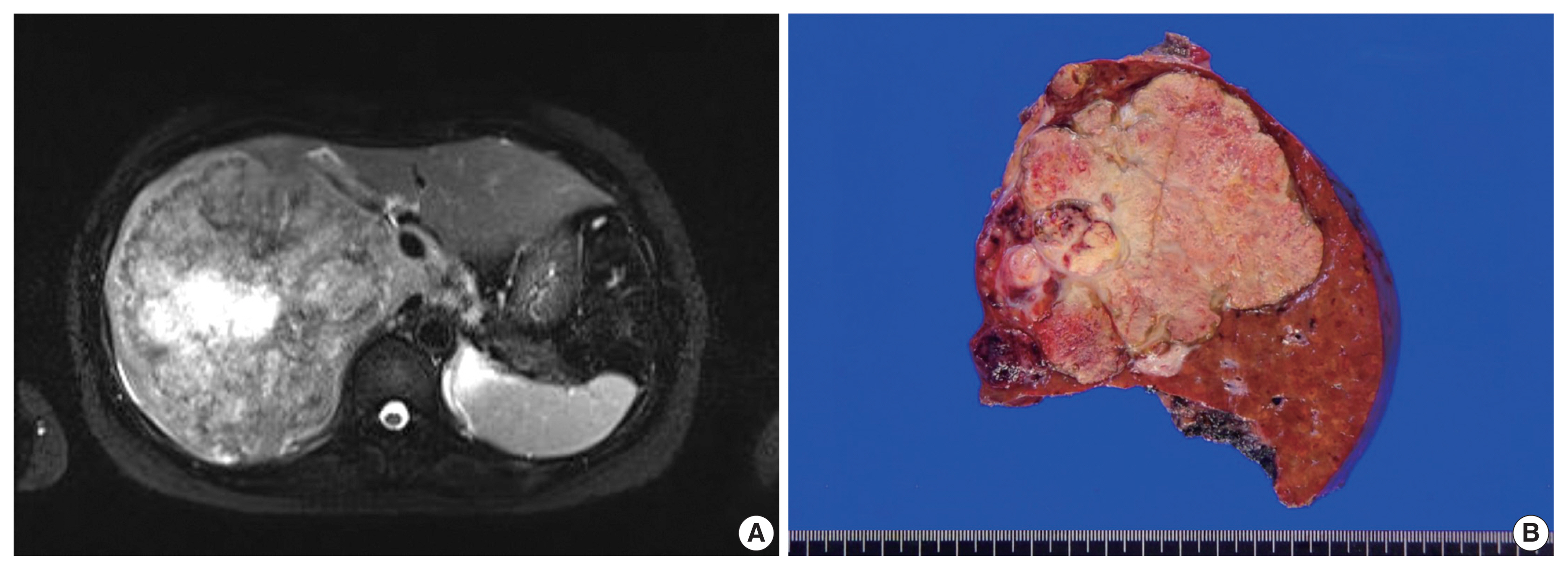
- 6,488 View
- 109 Download
- 2 Web of Science
- 2 Crossref
-
Abstract
PDF
- Primary hepatic mixed germ cell tumor (GCT) is very rare, and less than 10 cases have been reported. We report a case of mixed GCT composed of a choriocarcinoma and yolk sac tumor, which occurred in the liver of a 40-year-old woman. A large mass was detected by computed tomography solely in the liver. Serum β-human chorionic gonadotropin (hCG) was highly elevated, otherwise, other serum tumor markers were slightly elevated or within normal limits. For hepatic choriocarcinoma, neoadjuvant chemotherapy was administered, followed by right lobectomy. Histologic features of the resected tumor revealed characteristic choriocarcinoma features with diffuse positivity for hCG in the syncytiotrophoblasts and diffuse positivity for α-fetoprotein and Sal-like protein 4 in the yolk sac tumor components. Primary malignant GCT in the liver is associated with a poor prognosis and requires specific treatment. Therefore, GCT should be considered during a differential diagnosis of a rapidly growing mass in the liver.
-
Citations
Citations to this article as recorded by- Mesenchymal Tumors of the Liver: An Update Review
Joon Hyuk Choi, Swan N. Thung
Biomedicines.2025; 13(2): 479. CrossRef - Testicular Seminoma in Prostate: Case Report and Review of Literature
Peter Lesko, Jana Obertova, Karol Kajo, Katarina Rejlekova, Zuzana Orszaghova, Viera Lehotska, Martina Ondrusova, Michal Chovanec, Dalibor Ondrus, Michal Mego
Clinical Genitourinary Cancer.2024; 22(2): 210. CrossRef
- Mesenchymal Tumors of the Liver: An Update Review
- Malignant rhabdoid tumor of the kidney in an adult with loss of INI1 expression and mutation in the SMARCB1 gene
- Eunkyung Han, Jiyoon Kim, Min Jung Jung, Susie Chin, Sang Wook Lee, Ahrim Moon
- J Pathol Transl Med. 2021;55(2):145-153. Published online March 9, 2021
- DOI: https://doi.org/10.4132/jptm.2021.01.26
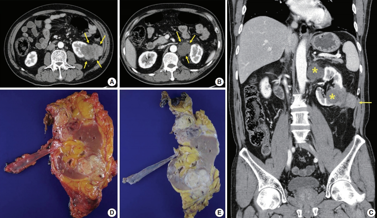
- 8,186 View
- 115 Download
- 7 Web of Science
- 7 Crossref
-
Abstract
PDF
- A 57-year-old man with left flank pain was referred to our institute. Computed tomography scans revealed two enhancing masses in the left kidney. The clinical diagnosis was renal cell carcinoma (RCC). He underwent a radical nephrectomy with an adrenalectomy. Two well-circumscribed solid masses in the hilum and the lower pole (4.5 × 3.5 cm and 7.0 × 4.1 cm) were present. Poorly cohesive uniform round to polygonal epithelioid cells making solid sheets accounted for most of the tumor area. The initial diagnosis was RCC, undifferentiated with rhabdoid features. As the tumor showed loss of INI1 expression and a mutation in the SMARCB1 gene on chromosome 22, the revised diagnosis was a malignant rhabdoid tumor (MRT) of the kidney. To date, only a few cases of renal MRT in adults have been reported. To the best of our knowledge, this is the first report of MRT in the native kidney of an adult demonstrating a SMARCB1 gene mutation, a hallmark of MRT.
-
Citations
Citations to this article as recorded by- Cancer epigenetic therapy: recent advances, challenges, and emerging opportunities
Rajita Vatapalli, Alex P. Rossi, Ho Man Chan, Jingwen Zhang
Epigenomics.2025; 17(1): 59. CrossRef - Navigating the complexity of atypical teratoid/rhabdoid tumor (ATRT) in pediatric neuro-oncology: Insights from clinical spectrum to therapeutic challenges
Ali Msheik, Mohamad Yazbeck, Abdulla Illeyan, Youssef Comair
International Journal of Surgery Case Reports.2025;[Epub] CrossRef - French recommendations on multi-gene panel testing in renal cell carcinoma
Sophie Giraud, Pascaline Berthet, Caroline Abadie, Nadine Andrieu, Patrick R. Benusiglio, Valérie Bonadona, Olivier Caron, Carole Corsini, Isabelle Coupier, Louise Crivelli, Capucine Delnatte, Pierre Devulder, Antoine DE Pauw, Sophie Dussart, Anne-Paule G
European Journal of Medical Genetics.2025; 78: 105062. CrossRef - Malignant rhabdoid tumor of the kidney in a 27-Year-Old adult: a rare case with favorable outcomes following surgery and adjuvant radiotherapy
Dimitri Paillusson, Stéphane De Vergie, Jérome Rigaud, Gaëlle Le Quellennec, Stéphane Supiot
World Journal of Surgical Oncology.2025;[Epub] CrossRef - Supratentorial ATRT in a young Infant: Expanding the diagnostic spectrum beyond medulloblastoma
Ali Msheik, Mohamad Aoun, Youssef Fares
Interdisciplinary Neurosurgery.2024; 35: 101857. CrossRef - Sarcomatoid and Rhabdoid Renal Cell Carcinoma
Adebowale J. Adeniran, Brian Shuch, Peter A. Humphrey
American Journal of Surgical Pathology.2024; 48(7): e65. CrossRef - Malignant rhabdoid tumor of kidney in an adult patient with positive family history of rhabdoid tumor
Farhood Khaleghi mehr, Nasrollah Abian, Mandana Rahimi, Yasaman Moradi
International Journal of Surgery Case Reports.2023;[Epub] CrossRef
- Cancer epigenetic therapy: recent advances, challenges, and emerging opportunities
Original Article
- Histologically confirmed distant metastatic urothelial carcinoma from the urinary bladder: a retrospective review of one institution’s 20-year experience
- Youngeun Yoo, Junghye Lee, Heae Surng Park, Min-Sun Cho, Sun Hee Sung, Sanghui Park, Euno Choi
- J Pathol Transl Med. 2021;55(2):94-101. Published online December 3, 2020
- DOI: https://doi.org/10.4132/jptm.2020.10.19
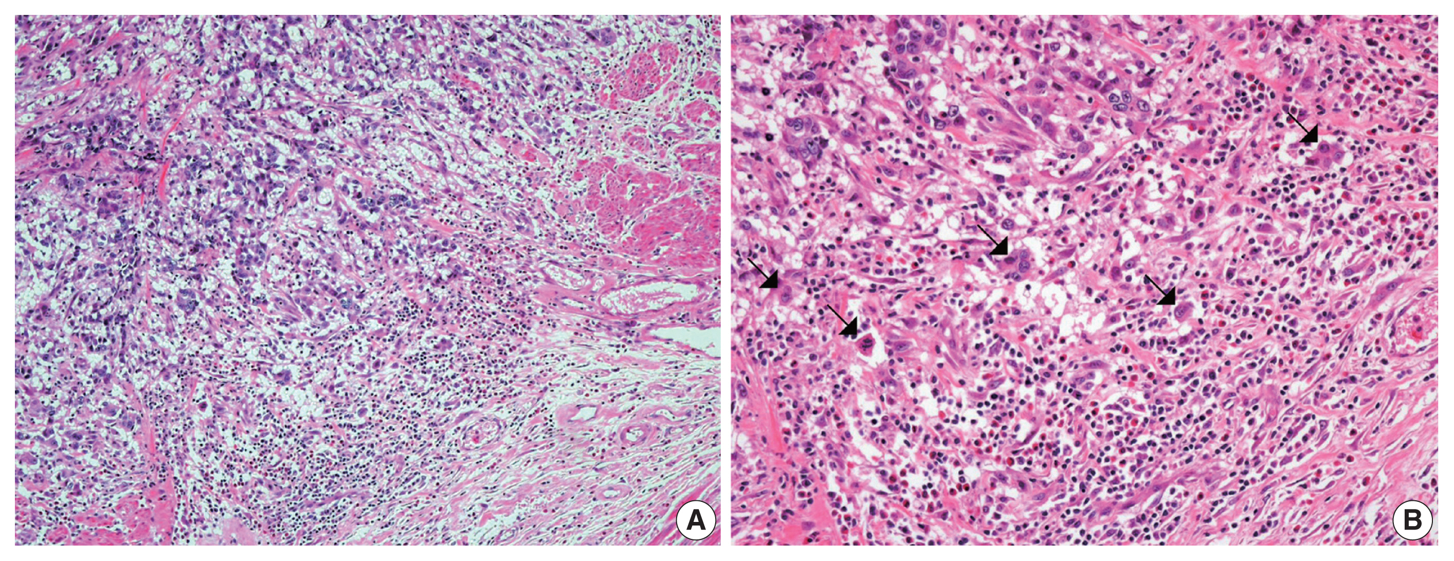
- 7,628 View
- 159 Download
- 1 Web of Science
- 1 Crossref
-
Abstract
PDF
- Background
Urothelial carcinoma (UC) accounts for roughly 90% of bladder cancer, and has a high propensity for diverse differentiation. Recently, certain histologic variants of UC have been recognized to be associated with unfavorable clinical outcomes. Several UC studies have also suggested that tumor budding is a poor prognostic marker. Distant metastasis of UC after radical cystectomy is not uncommon. However, these metastatic lesions are not routinely confirmed with histology.
Methods
We investigated the histopathologic features of 13 cases of UC with biopsy-proven distant metastases, with a special emphasis on histologic variants and tumor budding.
Results
Lymph nodes (6/13, 46%) were the most common metastatic sites, followed by the lung (4/13, 31%), liver (4/13, 31%), and the adrenal gland (2/13, 15%). The histologic variants including squamous (n=1), micropapillary (n=4), and plasmacytoid (n=1) variants in five cases of UC. Most histologic variants (4/5, 80%) of primary UCs appeared in the metastatic lesions. In contrast, high-grade tumor budding was detected in six cases (46%), including one case of non-muscle invasive UC. Our study demonstrates that histologic variants are not uncommonly detected in distant metastatic UCs. Most histologic variants seen in primary UCs persist in the distant metastatic lesions. In addition, high-grade tumor budding, which occurs frequently in primary tumors, may contribute to the development of distant metastasis.
Conclusions
Therefore, assessing the presence or absence of histologic variants and tumor budding in UCs of the urinary bladder, even in non-muscle invasive UCs, may be useful to predict distant metastasis. -
Citations
Citations to this article as recorded by- Do Histology and Primary Tumor Location Influence Metastatic Patterns in Bladder Cancer?
Hyung Kyu Park
Current Oncology.2023; 30(10): 9078. CrossRef
- Do Histology and Primary Tumor Location Influence Metastatic Patterns in Bladder Cancer?
Case Study
- A case of cribriform carcinoma of the skin: a newly described rare condition
- Hyun Lee, Chong-Hyun Won, Chan-Sik Park
- J Pathol Transl Med. 2021;55(1):68-74. Published online December 3, 2020
- DOI: https://doi.org/10.4132/jptm.2020.10.05
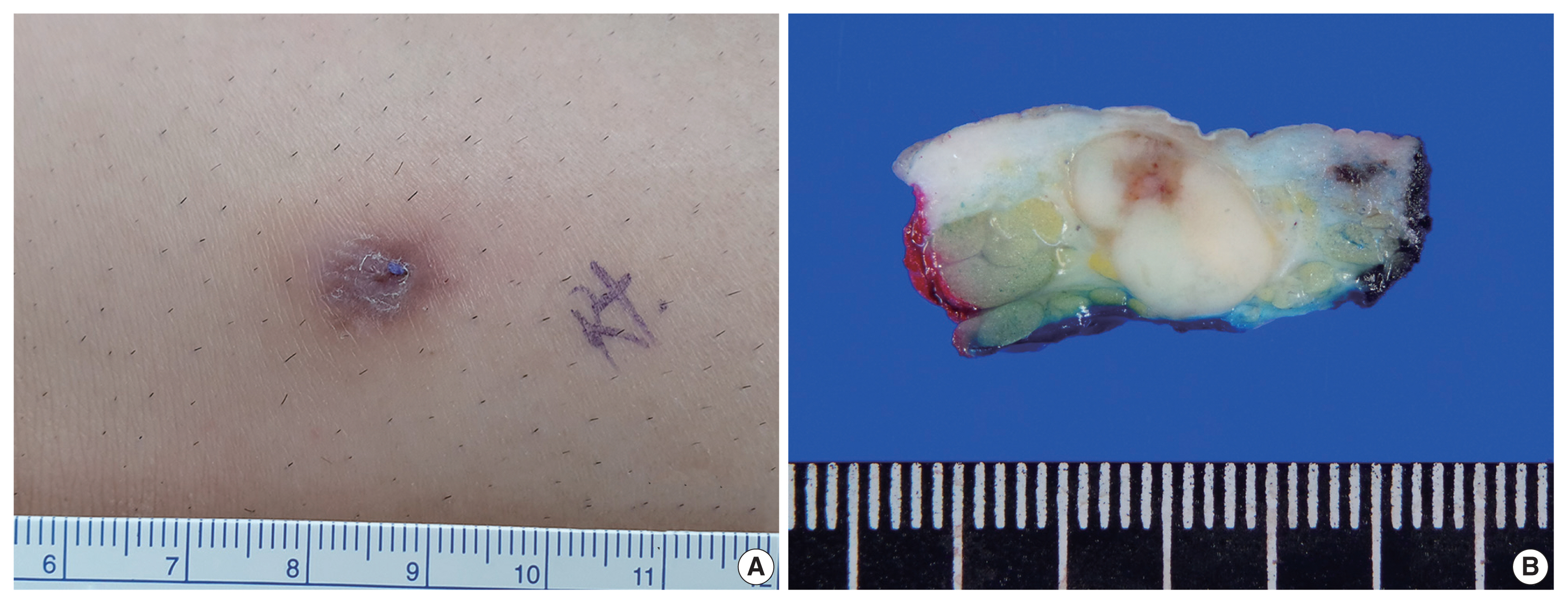
- 7,637 View
- 157 Download
- 6 Web of Science
- 7 Crossref
-
Abstract
PDF
- Primary cribriform carcinoma of the skin is an indolent, rare, adnexal tumor. Although its malignant potential remains uncertain, no recurrence or metastasis has been reported. A 33-year-old man presented with a solitary, erythematous, subcutaneous nodule on the right knee. The clinical impression was epidermal cyst, and the resected tumor demonstrated a well-circumscribed mass in the dermis and subcutis. The tumor was composed of two regions: a solid component and a cribriform component. The solid component (90%) showed multiple solid nests of epithelial cells. Individual cells had large, oval-to-round, hyperchromatic, pleomorphic nuclei with a nuclear groove. The cribriform component (10%) showed similar neoplastic cells with many prominent lumina. Some lumina had an eosinophilic substance that exhibited a positive periodic acid-Schiff reaction. No recurrence or metastasis was observed within a followup period of eight months after excision. In conclusion, we report the first case of primary cribriform carcinoma of the skin in Korea.
-
Citations
Citations to this article as recorded by- Primary Cutaneous Apocrine Carcinoma With Locoregional Disease
Rishi Kumar, Maxim Devine, Christopher M. Angel, Vachara Niumsawatt, Angela Webb
Annals of Plastic Surgery.2026; 97(2): 218. CrossRef - Primary cutaneous cribriform tumor: A case report and literature review
Doukou Jiang, Yongzhen Tian, Jiabin Tian, Hui Liu, Yang Guan
Journal of Cutaneous Pathology.2025; 52(1): 9. CrossRef - Primary cutaneous cribriform tumor in uncommon locations
Ting-Ting Cheng, Yu Yu
Dermatologica Sinica.2025; 43(4): 346. CrossRef - Primary Cutaneous Cribriform Apocrine Carcinoma: A Case Report and Narrative Review
Robyn Okereke, Anthony Linfante
BioMed.2025; 5(4): 26. CrossRef - Mohs micrographic surgery for the management of primary cutaneous cribriform carcinoma of the back
Min Jae Kim, Je‐Ho Mun
JDDG: Journal der Deutschen Dermatologischen Gesellschaft.2024; 22(4): 584. CrossRef - Mikrographische Chirurgie nach Mohs bei einem primär kutanen kribriformen Karzinom am Rücken
Min Jae Kim, Je‐Ho Mun
JDDG: Journal der Deutschen Dermatologischen Gesellschaft.2024; 22(4): 584. CrossRef - Rare skin appendage tumour on the right leg: a case of primary cutaneous cribriform carcinoma
Kashini Andrew, James M Carr, Claudia Roberts
BMJ Case Reports.2023; 16(5): e254781. CrossRef
- Primary Cutaneous Apocrine Carcinoma With Locoregional Disease
Review
- Immune landscape and biomarkers for immuno-oncology in colorectal cancers
- Jeong Mo Bae, Seung-Yeon Yoo, Jung Ho Kim, Gyeong Hoon Kang
- J Pathol Transl Med. 2020;54(5):351-360. Published online June 26, 2020
- DOI: https://doi.org/10.4132/jptm.2020.05.15
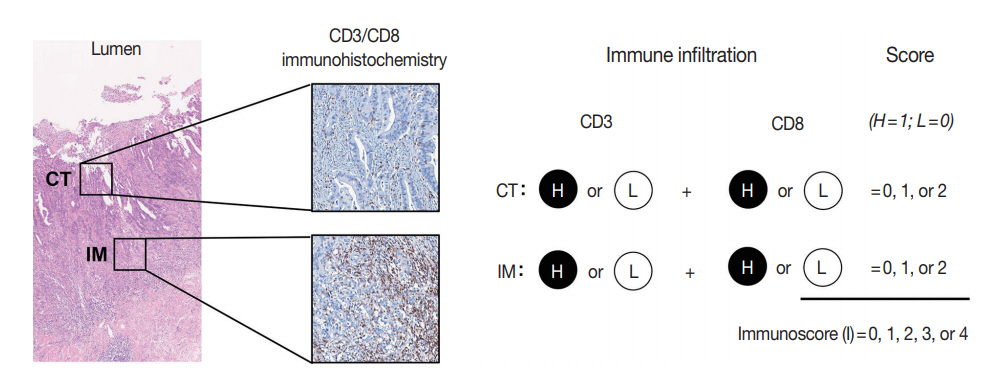
- 12,175 View
- 336 Download
- 14 Web of Science
- 14 Crossref
-
Abstract
PDF
- Recent advances in immuno-oncology have increased understanding of the tumor immune microenvironment (TIME), and clinical trials for immune checkpoint inhibitor treatment have shown remission and/or durable response in certain proportions of patients stratified by predictive biomarkers. The TIME in colorectal cancer (CRC) was initially evaluated several decades ago. The prognostic value of the immune response to tumors, including tumor-infiltrating lymphocytes, peritumoral lymphoid reaction, and Crohn’s-like lymphoid reaction, has been well demonstrated. In this review, we describe the chronology of TIME research and review the up-to-date high-dimensional TIME landscape of CRC. We also summarize the clinical relevance of several biomarkers associated with immunotherapy in CRC, such as microsatellite instability, tumor mutational burden, POLE/POLD mutation, consensus molecular subtype, and programmed death-ligand 1 expression.
-
Citations
Citations to this article as recorded by- Recent research progress and clinical status of immunotherapy for colorectal cancer
Zhikui Huo, Guoliang Liu, Jiannan Li
Journal of Advanced Research.2026; 82: 729. CrossRef - Aptasensors in colorectal Cancer: Emerging tools for precision diagnostics
Qamar Abuhassan, Ali M. Atoom, R. Roopashree, Jaya Bhanu Kanwar, T. Sudhakar, Vipasha Sharma, Ashish Singh Chauhan, Oybek Ruziyev
Clinica Chimica Acta.2026; 581: 120751. CrossRef - Five decades of colorectal cancer pathology: The World and China
Maode Lai
Chinese Science Bulletin.2025; 70(31): 5256. CrossRef - Real world effectiveness of chemotherapy plus bevacizumab with immunotherapy in colorectal cancer
Zhao Gao, Xiaoyan Wang, Tao Song, Shikai Wu, Xuan Jin
Scientific Reports.2025;[Epub] CrossRef - Targeting the “tumor microenvironment”: RNA-binding proteins in the spotlight in colorectal cancer therapy
Yiwei Zhang, Yujun Zhang, Jingjing Song, Xifu Cheng, Chulin Zhou, Shuo Huang, Wentao Zhao, Zhen Zong, Lingling Yang
International Immunopharmacology.2024; 131: 111876. CrossRef - T cell receptor clonotype in tumor microenvironment contributes to intratumoral signaling network in patients with colorectal cancer
In Hye Song, Seung-been Lee, Byung-Kwan Jeong, Jungwook Park, Honggeun Kim, GunHee Lee, Su Min Cha, Heejae Lee, Gyungyub Gong, Nak-Jung Kwon, Hee Jin Lee
Immunologic Research.2024; 72(5): 921. CrossRef - Identification of Key Immune and Cell Cycle Modules and Prognostic Genes for Glioma Patients through Transcriptome Analysis
Kaimin Guo, Jinna Yang, Ruonan Jiang, Xiaxia Ren, Peng Liu, Wenjia Wang, Shuiping Zhou, Xiaoguang Wang, Li Ma, Yunhui Hu
Pharmaceuticals.2024; 17(10): 1295. CrossRef - Unraveling the Role of Molecular Profiling in Predicting Treatment Response in Stage III Colorectal Cancer Patients: Insights from the IDEA International Study
Ippokratis Messaritakis, Eleni Psaroudaki, Konstantinos Vogiatzoglou, Maria Sfakianaki, Pantelis Topalis, Ioannis Iliopoulos, Dimitrios Mavroudis, John Tsiaoussis, Nikolaos Gouvas, Maria Tzardi, John Souglakos
Cancers.2023; 15(19): 4819. CrossRef - Biomarkers for Predicting Response to Personalized Immunotherapy in Gastric Cancer
Moonsik Kim, Ji Yun Jeong, An Na Seo
Diagnostics.2023; 13(17): 2782. CrossRef - Evaluation on Braking Stability of Autonomous Vehicles Running along Curved Sections Based on Asphalt Pavement Adhesion Properties
Binshuang Zheng, Xiaoming Huang, Junyao Tang, Jiaying Chen, Runmin Zhao, Zhengqiang Hong, Tao Tang, Meiling Han, Yang Yang
Journal of Advanced Transportation.2022; 2022: 1. CrossRef - Intratumoral spatial heterogeneity of tumor-infiltrating lymphocytes is a significant factor for precisely stratifying prognostic immune subgroups of microsatellite instability-high colorectal carcinomas
Minsun Jung, Ji Ae Lee, Seung-Yeon Yoo, Jeong Mo Bae, Gyeong Hoon Kang, Jung Ho Kim
Modern Pathology.2022; 35(12): 2011. CrossRef - Association of Tumor-Infiltrating Lymphocytes With Survival in Stages II and III Colorectal Cancer
Marina Vitorino, Inês Eiriz, Tiago C Tomás, Rodrigo Vicente, Ana Mendes, Ana Rita Freitas, Sofia Braga, Catarina Alves-Vale, Paula Borralho, André Ferreira, Luisa Leal da Costa
Cureus.2022;[Epub] CrossRef - Tumor Mutational Burden Predicting the Efficacy of Immune Checkpoint Inhibitors in Colorectal Cancer: A Systematic Review and Meta-Analysis
Yan Li, Yiqi Ma, Zijun Wu, Fanxin Zeng, Bin Song, Yanrong Zhang, Jinxing Li, Su Lui, Min Wu
Frontiers in Immunology.2021;[Epub] CrossRef - Genomic and transcriptomic characterization of heterogeneous immune subgroups of microsatellite instability-high colorectal cancers
Jung Ho Kim, Mi-Kyoung Seo, Ji Ae Lee, Seung-Yeon Yoo, Hyeon Jeong Oh, Hyundeok Kang, Nam-Yun Cho, Jeong Mo Bae, Gyeong Hoon Kang, Sangwoo Kim
Journal for ImmunoTherapy of Cancer.2021; 9(12): e003414. CrossRef
- Recent research progress and clinical status of immunotherapy for colorectal cancer
Case Study
- Pediatric granular cell tumor in the posterior wall of the larynx extending to the trachea
- Jungsuk Ahn, Na Rae Kim, Yong Han Sun
- J Pathol Transl Med. 2020;54(4):336-339. Published online April 15, 2020
- DOI: https://doi.org/10.4132/jptm.2020.02.28
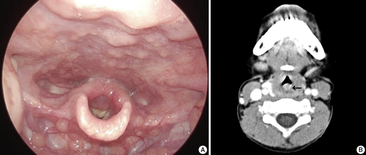
- 7,217 View
- 125 Download
- 1 Web of Science
- 1 Crossref
-
Abstract
PDF
- Granular cell tumor (GCT) is a slow-growing benign neoplasm that can be found in any organ. Pediatric laryngotracheal GCT is rare. We experienced a 6-year-old boy suffering from a barking cough and symptoms of stridor and croup for one month. Head and neck computed tomography revealed a protruding mass that occluded 60% of the airway lumen. Under the impression of hemangioma or papilloma, excision revealed a submucosal non-encapsulated mass. Histologically, the mass was composed of sheets of large polyhedralshaped tumor cells containing plump eosinophilic granular cytoplasm and centrally placed, small, bland-appearing nuclei. The tumor cells were positive for S-100 protein, and voluminous eosinophilic cytoplasm was stained by diastase-resistant periodic acid-Schiff. The present report describes a unique case of a huge pediatric laryngeal GCT extending to the subglottic trachea. We also review the clinical course of pediatric laryngotracheal GCT and emphasize the importance of diagnosing GCT in children.
-
Citations
Citations to this article as recorded by- Pediatric granular cell tumor of the larynx: A case report and literature review
Jing Ke, Junwei Xiong, Juhong Zhang, Haiyu Ma, Wei Yuan
Journal of Cancer Research and Therapeutics.2023; 19(4): 1070. CrossRef
- Pediatric granular cell tumor of the larynx: A case report and literature review
Review
- Current status and future perspectives of liquid biopsy in non-small cell lung cancer
- Sunhee Chang, Jae Young Hur, Yoon-La Choi, Chang Hun Lee, Wan Seop Kim
- J Pathol Transl Med. 2020;54(3):204-212. Published online April 15, 2020
- DOI: https://doi.org/10.4132/jptm.2020.02.27
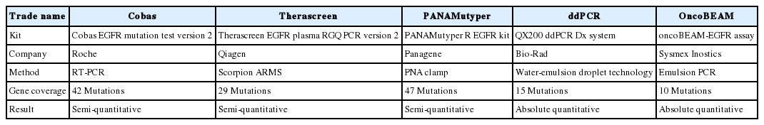
- 13,089 View
- 299 Download
- 21 Web of Science
- 21 Crossref
-
Abstract
PDF
- With advances in target therapy, molecular analysis of tumors is routinely required for treatment decisions in patients with advanced non-small cell lung cancer (NSCLC). Liquid biopsy refers to the sampling and analysis of circulating cell-free tumor DNA (ctDNA) in various body fluids, primarily blood. Because the technique is minimally invasive, liquid biopsies are the future in cancer management. Epidermal growth factor receptor (EGFR) ctDNA tests have been performed in routine clinical practice in advanced NSCLC patients to guide tyrosine kinase inhibitor treatment. In the near future, liquid biopsy will be a crucial prognostic, predictive, and diagnostic method in NSCLC. Here we present the current status and future perspectives of liquid biopsy in NSCLC.
-
Citations
Citations to this article as recorded by- Bridging gaps in mitochondrial disease diagnosis: the role of advanced biomarker discovery
Tendai Makwikwi, Maryke Schoonen, Izelle Smuts, Francois H. van der Westhuizen
Journal of Molecular Medicine.2026;[Epub] CrossRef - Comparison of tissue-based and plasma-based testing for EGFR mutation in non–small cell lung cancer patients
Yoon Kyung Kang, Dong Hoon Shin, Joon Young Park, Chung Su Hwang, Hyun Jung Lee, Jung Hee Lee, Jee Yeon Kim, JooYoung Na
Journal of Pathology and Translational Medicine.2025; 59(1): 60. CrossRef - Lab-in-a-Fiber detection and capture of cells
João C. Varela, Achar V. Harish, Pawel Maniewski, Timothy Gibbon, Oana Tudoran, Rainer Heuchel, Matthias Löhr, Walter Margulis, Aman Russom, Fredrik Laurell
Scientific Reports.2025;[Epub] CrossRef - Lung Cancer Diagnosis and Prognostic Monitoring Through Cell-Free RNA via Liquid Biopsy
Yuanming Pan, Chongbo Jiang, Mengchan Ye, Dongmei Li, Jinghui Wang
Therapeutics and Clinical Risk Management.2025; Volume 21: 1615. CrossRef - Unlocking the future of cancer diagnosis – promises and challenges of ctDNA-based liquid biopsies in non-small cell lung cancer
Chiara Reina, Berina Šabanović, Chiara Lazzari, Vanesa Gregorc, Christopher Heeschen
Translational Research.2024; 272: 41. CrossRef - Tailored point-of-care biosensors for liquid biopsy in the field of oncology
Sima Singh, Pritam Saha Podder, Matt Russo, Charles Henry, Stefano Cinti
Lab on a Chip.2023; 23(1): 44. CrossRef - Emerging role of non-invasive and liquid biopsy biomarkers in pancreatic cancer
Akash Bararia, Prosenjeet Chakraborty, Paromita Roy, Bitan Kumar Chattopadhay, Amlan Das, Aniruddha Chatterjee, Nilabja Sikdar
World Journal of Gastroenterology.2023; 29(15): 2241. CrossRef - Liquid biopsy in the management of advanced lung cancer: Implementation and practical aspects
Gabriela Fernandes, Ana Rodrigues, Cláudia Matos, Fernando Barata, Luís Cirnes, Lurdes Ferreira, José Albino Lopes, Margarida Felizardo, Paula Fidalgo, Ulisses Brito, Bárbara Parente
Cancer Treatment and Research Communications.2023; 36: 100725. CrossRef - Tweezer PCR: A Highly Specific Method for Accurate Identification of Low-Abundance Mutations
Shanglin Li, Yin Gu, Zhi Geng, Kaiyi Li, Yawei Hu, Qiang Liu, Rongxin Fu, Peng Liu
Analytical Chemistry.2023; 95(48): 17679. CrossRef - Mesonephric-like Adenocarcinoma of the Ovary: Clinicopathological and Molecular Characteristics
Hyun Hee Koh, Eunhyang Park, Hyun-Soo Kim
Diagnostics.2022; 12(2): 326. CrossRef - Alveolar Soft Part Sarcoma of the Uterus: Clinicopathological and Molecular Characteristics
Yurimi Lee, Kiyong Na, Ha Young Woo, Hyun-Soo Kim
Diagnostics.2022; 12(5): 1102. CrossRef - Exosomal MicroRNA Analyses in Esophageal Squamous Cell Carcinoma Cell Lines
Sora Kim, Gwang Ha Kim, Su Jin Park, Chae Hwa Kwon, Hoseok I, Moon Won Lee, Bong Eun Lee
Journal of Clinical Medicine.2022; 11(15): 4426. CrossRef - Molecular biomarker testing for non–small cell lung cancer: consensus statement of the Korean Cardiopulmonary Pathology Study Group
Sunhee Chang, Hyo Sup Shim, Tae Jung Kim, Yoon-La Choi, Wan Seop Kim, Dong Hoon Shin, Lucia Kim, Heae Surng Park, Geon Kook Lee, Chang Hun Lee
Journal of Pathology and Translational Medicine.2021; 55(3): 181. CrossRef - Update on molecular pathology and role of liquid biopsy in nonsmall cell lung cancer
Pamela Abdayem, David Planchard
European Respiratory Review.2021; 30(161): 200294. CrossRef - Dynamics of Specific cfDNA Fragments in the Plasma of Full Marathon Participants
Takehito Sugasawa, Shin-ichiro Fujita, Tomoaki Kuji, Noriyo Ishibashi, Kenshirou Tamai, Yasushi Kawakami, Kazuhiro Takekoshi
Genes.2021; 12(5): 676. CrossRef - Future Perspectives in Detecting EGFR and ALK Gene Alterations in Liquid Biopsies of Patients with NSCLC
Daniela Ferreira, Juliana Miranda, Paula Martins-Lopes, Filomena Adega, Raquel Chaves
International Journal of Molecular Sciences.2021; 22(8): 3815. CrossRef - Real-World Analysis of the EGFR Mutation Test in Tissue and Plasma Samples from Non-Small Cell Lung Cancer
Hyunwoo Lee, Joungho Han, Yoon-La Choi
Diagnostics.2021; 11(9): 1695. CrossRef - Objective Quantitation of EGFR Protein Levels using Quantitative Dot Blot Method for the Prognosis of Gastric Cancer Patients
Lei Xin, Fangrong Tang, Bo Song, Maozhou Yang, Jiandi Zhang
Journal of Gastric Cancer.2021; 21(4): 335. CrossRef - Molecular Therapeutics of Non-Small Cell Lung Cancer (NSCLC) and Challenges in Repeat Tissue Biopsy
Devanna Vasista Ganesha, Radheshyam Naik, Suhail Sayeed Mufti, Hrishi Varayathu
Advances in Lung Cancer.2021; 10(03): 21. CrossRef - The Role of the Liquid Biopsy in Decision-Making for Patients with Non-Small Cell Lung Cancer
D. Akhoundova, J. Mosquera Martinez, L. E. Musmann, C. Britschgi, C. Rütsche, M. Rechsteiner, E. Nadal, M. R. Garcia Campelo, A. Curioni-Fontecedro
Journal of Clinical Medicine.2020; 9(11): 3674. CrossRef - Expanding opportunities in precision oncology
T Raja
Cancer Research, Statistics, and Treatment.2020; 3(4): 863. CrossRef
- Bridging gaps in mitochondrial disease diagnosis: the role of advanced biomarker discovery
Case Studies
- Gastric IgG4-related disease presenting as a mass lesion and masquerading as a gastrointestinal stromal tumor
- Banumathi Ramakrishna, Rohan Yewale, Kavita Vijayakumar, Patta Radhakrishna, Balakrishnan Siddartha Ramakrishna
- J Pathol Transl Med. 2020;54(3):258-262. Published online March 4, 2020
- DOI: https://doi.org/10.4132/jptm.2020.02.10
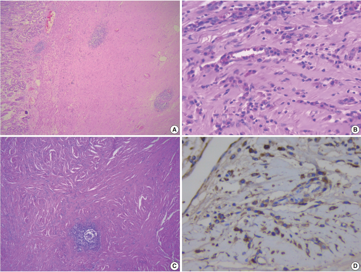
- 8,611 View
- 163 Download
- 8 Web of Science
- 9 Crossref
-
Abstract
PDF
- IgG4-related disease of the stomach is a rare disorder, and only a few cases have been reported. We present two cases that were identified over a 2-month period in our center. Two male patients aged 52 and 48 years presented with mass lesion in the stomach, which were clinically thought to be gastrointestinal stromal tumor, and they underwent excision of the lesion. Microscopic examination revealed marked fibrosis, which was storiform in one case, associated with diffuse lymphoplasmacytic infiltration and an increase in IgG4-positive plasma cells on immunohistochemistry. Serum IgG4 level was markedly elevated. Although rare, IgG4-related disease should be considered in the differential diagnosis of gastric submucosal mass lesions.
-
Citations
Citations to this article as recorded by- Isolated IgG4-related disease of terminal ileum: Report of a rare case and review of literature
Subham Bhowmik, Hemanga K. Bhattacharjee, Joyner Abraham, Raju Sharma, Prasenjit Das
Journal of Cancer Research and Therapeutics.2025; 21(1): 200. CrossRef - Great Mimics in Oncology: A Retrospective Study from a Tertiary Care Centre of Eastern India
Suvendu Maji, Jayesh kumar Jha, Vikram Chaturvedi
Indian Journal of Surgical Oncology.2025; 16(1): 64. CrossRef - Systemic diseases affecting the GI tract: A review of clinical and histopathologic manifestations
Maryam K. Pezhouh, Dora Lam-Himlin, Atif Zaheer, Lysandra Voltaggio
Annals of Diagnostic Pathology.2024; 73: 152351. CrossRef - Utilizing Immunoglobulin G4 Immunohistochemistry for Risk Stratification in Patients with Papillary Thyroid Carcinoma Associated with Hashimoto Thyroiditis
Faridul Haq, Gyeongsin Park, Sora Jeon, Mitsuyoshi Hirokawa, Chan Kwon Jung
Endocrinology and Metabolism.2024; 39(3): 468. CrossRef - Compromiso gástrico por enfermedad relacionada con IgG4
Gilberto Jaramillo Trujillo, Oscar Fernando Ruiz, Melissa González Pabón, Maria Andrea Jaramillo Trujillo
Revista Repertorio de Medicina y Cirugía.2024; 33(3): 319. CrossRef - CGB5, INHBA and TRAJ19 Hold Prognostic Potential as Immune Genes for Patients with Gastric Cancer
Bei Ji, Lili Qiao, Wei Zhai
Digestive Diseases and Sciences.2023; 68(3): 791. CrossRef - IgG4-related diseases of the digestive tract
J.-Matthias Löhr, Miroslav Vujasinovic, Jonas Rosendahl, John H. Stone, Ulrich Beuers
Nature Reviews Gastroenterology & Hepatology.2022; 19(3): 185. CrossRef - Clinicopathological characteristics of gastric IgG4‐related disease: Systematic scoping review
Haruki Sawada, Torrey Czech, Krixie Silangcruz, Landon Kozai, Adham Obeidat, Eric Andrew Wien, Midori Filiz Nishimura, Asami Nishikori, Yasuharu Sato, Yoshito Nishimura
Journal of Gastroenterology and Hepatology.2022; 37(10): 1865. CrossRef - Utility of gastric biopsy in diagnosing IgG4‐related gastrointestinal disease
Kaori Uchino, Kenji Notohara, Takeshi Uehara, Yasuhiro Kuraishi, Junya Itakura, Akihiro Matsukawa
Pathology International.2021; 71(2): 124. CrossRef
- Isolated IgG4-related disease of terminal ileum: Report of a rare case and review of literature
- Primary carcinoid tumor in the external auditory canal
- Dong Hae Chung, Gyu Cheol Han, Na Rae Kim
- J Pathol Transl Med. 2020;54(2):184-187. Published online November 13, 2019
- DOI: https://doi.org/10.4132/jptm.2019.11.07
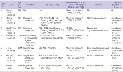
- 10,498 View
- 167 Download
- 4 Web of Science
- 3 Crossref
-
Abstract
PDF
- A 39-year-old man visited the department of otolaryngology due to an ongoing hearing disturbance that had lasted for 1 year. Temporal bone computed tomography revealed soft tissue density nearly obliterating the left external auditory canal (EAC). The mass was composed of sheets of round tumor cells containing moderate amounts of fine granular cytoplasm and salt and pepper chromatin. Neither mitosis nor necrosis was found. The Ki-67 proliferation index was less than 2%. Cells were positive for CD56 and synaptophysin but negative for chromogranin, cytokeratin (CK) 20, and CK7. Based on these findings, the tumor was diagnosed as a carcinoid tumor, well differentiated neuroendocrine carcinoma, grade 1 (G1) according to current World Health Organization (WHO) classification of head and neck tumors; and a neuroendocrine tumor, G1 according to neuroendocrine neoplasm (NEN)-2018 WHO standard classification. He remained free of local recurrence and metastasis after 20 months of follow up. To date, only six cases of primary NENs in the EAC have been reported. Metastatic tumor should be included in the differential diagnoses. Because of its rarity, the prognosis and treatment have not yet been clarified.
-
Citations
Citations to this article as recorded by- First Report on a Rare Poorly Differentiated Neuroendocrine Tumour of the External Auditory Canal Involving Pinna
Akash Varshney, Amit Kumar Tyagi, Prashant Durgapal, Kajal Mahto, Akhilesh Chandra Yadav, Ankita Semwal
Indian Journal of Otolaryngology and Head & Neck Surgery.2025; 77(4): 1922. CrossRef - Incidental finding of a neuroendocrine neoplasm in a suspected ear canal exostosis
Alexander Wieck Fjaeldstad, Gerda Elisabeth Villadsen, Gitte Dam, Stephen Jacques Hamilton-Dutoit, Thomas Winther Frederiksen
Otolaryngology Case Reports.2022; 22: 100394. CrossRef - 68Ga-DOTATATE Uptake in Well-Differentiated Neuroendocrine Tumor of the External Auditory Canal
Özge Erol Fenercioğlu, Ediz Beyhan, Rahime Şahin, Mehmet Can Baloğlu, Tevfik Fikret Çermik
Clinical Nuclear Medicine.2022; 47(8): e552. CrossRef
- First Report on a Rare Poorly Differentiated Neuroendocrine Tumour of the External Auditory Canal Involving Pinna
Review
- Tumor immune response and immunotherapy in gastric cancer
- Yoonjin Kwak, An Na Seo, Hee Eun Lee, Hye Seung Lee
- J Pathol Transl Med. 2020;54(1):20-33. Published online November 1, 2019
- DOI: https://doi.org/10.4132/jptm.2019.10.08
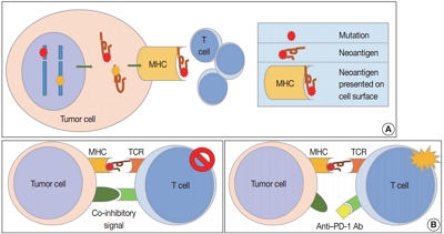
- 21,001 View
- 749 Download
- 74 Web of Science
- 68 Crossref
-
Abstract
PDF
- Remarkable developments in immuno-oncology have changed the landscape of gastric cancer (GC) treatment. Because immunotherapy intervenes with tumor immune response rather than directly targeting tumor cells, it is important to develop a greater understanding of tumor immunity. This review paper summarizes the tumor immune reaction and immune escape mechanisms while focusing on the role of T cells and their co-inhibitory signals, such as the immune checkpoint molecules programmed death-1 and programmed deathligand 1 (PD-L1). This paper also describes past clinical trials of immunotherapy for patients with GC and details their clinical implications. Strong predictive markers are essential to improve response to immunotherapy. Microsatellite instability, Epstein-Barr virus, PD-L1 expression, and tumor mutational burden are now regarded as potent predictive markers for immunotherapy in patients with GC. Novel immunotherapy and combination therapy targeting new immune checkpoint molecules such as lymphocyte-activation gene 3, T cell immunoglobulin, and mucin domain containing-3, and indoleamine 2,3-dioxygenase have been suggested, and trials are ongoing to evaluate their safety and efficacy. Immunotherapy is an important treatment option for patients with GC and has great potential for improving patient outcome, and further research in immuno-oncology should be carried out.
-
Citations
Citations to this article as recorded by- Using glucocorticoid receptor-related genes to create and validate a survival model predicting gastric cancer
Ke Guo, Ping Huang, Jiasheng Zhang, Bo Zhang, Linyang Li, Jiaxin Li
Computational Biology and Chemistry.2026; 120: 108726. CrossRef - Mechanisms of Metabolic Reprogramming Regulating Immunosuppression in the Gastric Cancer Tumor Microenvironment
Wenting Dong, Xuepeng Qian, Honglin Liu, Jinhai Huo, Weiming Wang
Biomolecules.2026; 16(1): 160. CrossRef - Perineural invasion in digestive tract tumors: Immune system interactions and therapeutic strategies
Jia-Yu Pang, Rong-Yao Jin, Han-Xiao Zhang, Ying-Hua Zhang, Xin-Yue Wei, Wen-Bo Cao, Yi-Xiao Chen, Jun-Ling Wang, Sai-Jun Mo
World Journal of Clinical Oncology.2026;[Epub] CrossRef - FOXP3+/CD8+ ratio associated with aggressive behavior in RUNX3‐methylated diffuse esophagogastric junction tumor
Suguru Maruyama, Yu Imamura, Tasuku Toihata, Ikumi Haraguchi, Manabu Takamatsu, Makiko Yamashita, Yuichiro Nakashima, Eiji Oki, Kenichi Taguchi, Manabu Yamamoto, Shinji Mine, Akihiko Okamura, Jun Kanamori, Souya Nunobe, Takeshi Sano, Shigehisa Kitano, Tet
Cancer Science.2025; 116(1): 178. CrossRef - Immune biomarkers and predictive signatures in gastric cancer: Optimizing immunotherapy responses
Sundaram Vickram, Shofia Saghya Infant, S. Manikandan, D. Jenila Rani, C.M. Mathan Muthu, Hitesh Chopra
Pathology - Research and Practice.2025; 265: 155743. CrossRef - Korean Practice Guidelines for Gastric Cancer 2024: An Evidence-based, Multidisciplinary Approach (Update of 2022 Guideline)
In-Ho Kim, Seung Joo Kang, Wonyoung Choi, An Na Seo, Bang Wool Eom, Beodeul Kang, Bum Jun Kim, Byung-Hoon Min, Chung Hyun Tae, Chang In Choi, Choong-kun Lee, Ho Jung An, Hwa Kyung Byun, Hyeon-Su Im, Hyung-Don Kim, Jang Ho Cho, Kyoungjune Pak, Jae-Joon Kim
Journal of Gastric Cancer.2025; 25(1): 5. CrossRef - Targeted therapy and immunotherapy for gastric cancer: rational strategies, novel advancements, challenges, and future perspectives
Dong Luo, Yunmei Liu, Zhengmao Lu, Lei Huang
Molecular Medicine.2025;[Epub] CrossRef - Prognostic value of the triglyceride-glucose index in gastric cancer
Tugce Eskazan, Suat Saribas, Bekir Kocazeybek
World Journal of Gastroenterology.2025;[Epub] CrossRef - Deciphering the dual role of autophagy in gastric cancer and gastroesophageal junction cancer: from tumor suppression to cancer progression
Lili Lei, Junling Zhang, Ran Wei, Bingqi Dong, Xin Wang, Ying Zhou
Discover Oncology.2025;[Epub] CrossRef - Comparison of clinicopathological parameters with the presence of Epstein–Barr virus and the absence of DNA mismatch repair proteins in gastric adenocarcinomas
Özge Eyeoğlu, Serra Kayaçetin
Asian Biomedicine.2025; 19(2): 86. CrossRef - The impact of combined immunotherapy on the cellular composition of the tumor microenvironment in patients with gastric carcinoma
L.A. Tashireva, A.Yu. Kalinchuk, D.M. Loos, E.A. Tsarenkova, A.V. Avgustinovich, S.G. Afanas’ev, S.V. Vtorushin
Russian Journal of Archive of Pathology.2025; 87(4): 24. CrossRef - Immunotherapy in gastric adenocarcinoma – a rapidly evolving treatment landscape
Yang Wang, Geoffrey Chong
Critical Reviews in Oncology/Hematology.2025; 216: 104941. CrossRef - Ubiquilin-4 induces immune escape in gastric cancer by activating the notch signaling pathway
Quan Jiang, Hao Chen, Shixin Zhou, Tao Zhu, Wenshuai Liu, Hao Wu, Yong Zhang, Fenglin Liu, Yihong Sun
Cellular Oncology.2024; 47(1): 303. CrossRef - Expression and prognostic value of APOBEC2 in gastric adenocarcinoma and its association with tumor-infiltrating immune cells
Lipan Wei, Xiuqian Wu, Lan Wang, Ling Chen, Xuejun Wu, Tiantian Song, Yuanyuan Wang, Wenjun Chang, Aizhen Guo, Yongdong Niu, Haihua Huang
BMC Cancer.2024;[Epub] CrossRef - Identification and characterization of CLEC11A and its derived immune signature in gastric cancer
Qing Zheng, Zhenqi Gong, Baizhi Li, Runzi Cheng, Weican Luo, Cong Huang, Huaiming Wang
Frontiers in Immunology.2024;[Epub] CrossRef - Cervical cancer subtype identification and model building based on lipid metabolism and post-infection microenvironment immune landscape
Yongzhi Chen, Rongjie Cui, Dun Xiong, Yuan Zhao, Jianyu Pang, Samina Gul, Qi Qi, Yuheng Tang, Xuhong Zhou, Wenru Tang
Heliyon.2024; 10(9): e30746. CrossRef - Systematic Analysis of Tumor Stem Cell-related Gene Characteristics
to Predict the PD-L1 Immunotherapy and Prognosis of Gastric
Cancer
Chenchen Wang, Ying Chen, Ru Zhou, Ya’nan Yang, Yantian Fang
Current Medicinal Chemistry.2024; 31(17): 2467. CrossRef - Comprehensive landscape of m6A regulator-related gene patterns and tumor microenvironment infiltration characterization in gastric cancer
Bin Peng, Yinglin Lin, Gao Yi, Mingzhen Lin, Yao Xiao, Yezhenghong Qiu, Wenxia Yao, Xinke Zhou, Zhaoyu Liu
Scientific Reports.2024;[Epub] CrossRef - Distinctive Phenotypic and Microenvironmental Characteristics of Neuroendocrine Carcinoma and Adenocarcinoma Components in Gastric Mixed Adenoneuroendocrine Carcinoma
Yoonjin Kwak, Soo Kyung Nam, Yujun Park, Yun-Suhk Suh, Sang-Hoon Ahn, Seong-Ho Kong, Do Joong Park, Hyuk-Joon Lee, Hyung-Ho Kim, Han-Kwang Yang, Hye Seung Lee
Modern Pathology.2024; 37(10): 100568. CrossRef - Computed tomography-detected extramural venous invasion-related gene signature: a potential negative biomarker of immune checkpoint inhibitor treatment in patients with gastric cancer
Hao Yang, Xinyi Gou, Caizhen Feng, Yinli Zhang, Fan Chai, Nan Hong, Yingjiang Ye, Yi Wang, Bo Gao, Jin Cheng
Journal of Translational Medicine.2023;[Epub] CrossRef - A standardized pathology report for gastric cancer: 2nd edition
Young Soo Park, Myeong-Cherl Kook, Baek-hui Kim, Hye Seung Lee, Dong-Wook Kang, Mi-Jin Gu, Ok Ran Shin, Younghee Choi, Wonae Lee, Hyunki Kim, In Hye Song, Kyoung-Mee Kim, Hee Sung Kim, Guhyun Kang, Do Youn Park, So-Young Jin, Joon Mee Kim, Yoon Jung Choi,
Journal of Pathology and Translational Medicine.2023; 57(1): 1. CrossRef - A Standardized Pathology Report for Gastric Cancer: 2nd Edition
Young Soo Park, Myeong-Cherl Kook, Baek-hui Kim, Hye Seung Lee, Dong-Wook Kang, Mi-Jin Gu, Ok Ran Shin, Younghee Choi, Wonae Lee, Hyunki Kim, In Hye Song, Kyoung-Mee Kim, Hee Sung Kim, Guhyun Kang, Do Youn Park, So-Young Jin, Joon Mee Kim, Yoon Jung Choi,
Journal of Gastric Cancer.2023; 23(1): 107. CrossRef - Korean Practice Guidelines for Gastric Cancer 2022: An Evidence-based, Multidisciplinary Approach
Tae-Han Kim, In-Ho Kim, Seung Joo Kang, Miyoung Choi, Baek-Hui Kim, Bang Wool Eom, Bum Jun Kim, Byung-Hoon Min, Chang In Choi, Cheol Min Shin, Chung Hyun Tae, Chung sik Gong, Dong Jin Kim, Arthur Eung-Hyuck Cho, Eun Jeong Gong, Geum Jong Song, Hyeon-Su Im
Journal of Gastric Cancer.2023; 23(1): 3. CrossRef - Could Toll-like Receptor 2 Serve as Biomarker to Detect Advanced Gastric Cancer?
Marek Majewski, Kamil Torres, Paulina Mertowska, Sebastian Mertowski, Izabela Korona-Głowniak, Jan Korulczyk, Witold Zgodziński, Ewelina Grywalska
International Journal of Molecular Sciences.2023; 24(6): 5824. CrossRef - Research Progress of Immunotherapy for Gastric Cancer
Zhipeng Zhang, Ningning Liu, Mingyu Sun
Technology in Cancer Research & Treatment.2023;[Epub] CrossRef - Case Report: A rare synchronous multiple gastric carcinoma achieved progression-free disease through NGS-guided serial treatment
Xinyi Shao, Jin Yin, Di Wang, Erjiong Huang, Yini Zhang, Jiani C. Yin, Chen Huang, Hao Wu, Xiaoli Wu
Frontiers in Oncology.2023;[Epub] CrossRef - Artificial Intelligence-Enabled Gastric Cancer Interpretations
Mustafa Yousif, Liron Pantanowitz
Surgical Pathology Clinics.2023; 16(4): 673. CrossRef - The Optimal Tumor Mutational Burden Cutoff Value as a Novel Marker for Predicting the Efficacy of Programmed Cell Death-1 Checkpoint Inhibitors in Advanced Gastric Cancer
Jae Yeon Jang, Youngkyung Jeon, Sun Young Jeong, Sung Hee Lim, Won Ki Kang, Jeeyun Lee, Seung Tae Kim
Journal of Gastric Cancer.2023; 23(3): 476. CrossRef - Biomarkers for Predicting Response to Personalized Immunotherapy in Gastric Cancer
Moonsik Kim, Ji Yun Jeong, An Na Seo
Diagnostics.2023; 13(17): 2782. CrossRef - The Prognostic Value of the Prognostic Nutritional Index in Patients with Advanced or Metastatic Gastric Cancer Treated with Immunotherapy
Yuting Pan, Yue Ma, Guanghai Dai
Nutrients.2023; 15(19): 4290. CrossRef - Molecular classification of gastric cancer predicts survival in patients undergoing radical gastrectomy based on project HOPE
Kenichiro Furukawa, Keiichi Hatakeyama, Masanori Terashima, Takeshi Nagashima, Kenichi Urakami, Keiichi Ohshima, Akifumi Notsu, Takashi Sugino, Taisuke Yagi, Keiichi Fujiya, Satoshi Kamiya, Makoto Hikage, Yutaka Tanizawa, Etsuro Bando, Yae Kanai, Yasuto A
Gastric Cancer.2022; 25(1): 138. CrossRef - Immunotherapy for Gastric Cancer: A 2021 Update
Christo Kole, Nikolaos Charalampakis, Sergios Tsakatikas, Nikolaos-Iasonas Kouris, George Papaxoinis, Michalis V Karamouzis, Anna Koumarianou, Dimitrios Schizas
Immunotherapy.2022; 14(1): 41. CrossRef - The immune microenvironment in gastric adenocarcinoma
Yana Zavros, Juanita L. Merchant
Nature Reviews Gastroenterology & Hepatology.2022; 19(7): 451. CrossRef - Immunomodulation by Gut Microbiome on Gastrointestinal Cancers: Focusing on Colorectal Cancer
Raghad Khalid AL-Ishaq, Lenka Koklesova, Peter Kubatka, Dietrich Büsselberg
Cancers.2022; 14(9): 2140. CrossRef - An Immunity-Associated lncRNA Signature for Predicting Prognosis in Gastric Adenocarcinoma
Xiaowen Zhao, Pingfan Wu, Dongling Liu, Changtian Li, Ling Xue, Zhe Liu, Meng Zhu, Jie Yang, Ziyi Chen, Yaling Li, Yali She, Kathiravan Srinivasan
Journal of Healthcare Engineering.2022; 2022: 1. CrossRef - RNA modification writers influence tumor microenvironment in gastric cancer and prospects of targeted drug therapy
Peng Song, Sheng Zhou, Xiaoyang Qi, Yuwen Jiao, Yu Gong, Jie Zhao, Haojun Yang, Zhifen Qian, Jun Qian, Liming Tang
Journal of Bioinformatics and Computational Biology.2022;[Epub] CrossRef - Identification of the three subtypes and the prognostic characteristics of stomach adenocarcinoma: analysis of the hypoxia-related long non-coding RNAs
Zehua Fan, Yanqun Wang, Rong Niu
Functional & Integrative Genomics.2022; 22(5): 919. CrossRef - Complete Response of High Microsatellite Instability Gastric Cancer and Synchronous Microsatellite Stability Rectal Cancer
Zachary E Hunzeker, Pooja Bhakta, Sindusha R Gudipally, Sri Bharathi Kavuri, Rohit Venkatesan, Chukwuyejulumafor Nwanze
Cureus.2022;[Epub] CrossRef - Immune Profiling in Gastric Cancer Reveals the Dynamic Landscape of Immune Signature Underlying Tumor Progression
Yuhan Wei, Jianwei Zhang, Xueke Fan, Zhi Zheng, Xiaoyue Jiang, Dexi Chen, Yuting Lu, Yingrui Li, Miao Wang, Min Hu, Qi Du, Liuting Yang, Hongzhong Li, Yi Xiao, Yongfu Li, Jiangtao Jin, Deying Wang, Xiangliang Yuan, Qin Li
Frontiers in Immunology.2022;[Epub] CrossRef - Tumor vessel normalization and immunotherapy in gastric cancer
Xianzhe Yu, Shan He, Jian Shen, Qiushi Huang, Peng Yang, Lin Huang, Dan Pu, Li Wang, Lu Li, Jinghua Liu, Zelong Liu, Lingling Zhu
Therapeutic Advances in Medical Oncology.2022;[Epub] CrossRef - FN1 is a prognostic biomarker and correlated with immune infiltrates in gastric cancers
Han Wang, Junchang Zhang, Huan Li, Hong Yu, Songyao Chen, Shuhao Liu, Changhua Zhang, Yulong He
Frontiers in Oncology.2022;[Epub] CrossRef - Molecular Pathology of Gastric Cancer
Moonsik Kim, An Na Seo
Journal of Gastric Cancer.2022; 22(4): 264. CrossRef - Bioinformatics Analysis and Structure of Gastric Cancer Prognosis Model Based on Lipid Metabolism and Immune Microenvironment
Yongzhi Chen, Hongjun Yuan, Qian Yu, Jianyu Pang, Miaomiao Sheng, Wenru Tang
Genes.2022; 13(9): 1581. CrossRef - Clinical implications of interleukins-31, 32, and 33 in gastric cancer
Qing-Hua Liu, Ji-Wei Zhang, Lei Xia, Steven G Wise, Brett David Hambly, Kun Tao, Shi-San Bao
World Journal of Gastrointestinal Oncology.2022; 14(9): 1808. CrossRef - Microbiota and the Immune System—Actors in the Gastric Cancer Story
Marek Majewski, Paulina Mertowska, Sebastian Mertowski, Konrad Smolak, Ewelina Grywalska, Kamil Torres
Cancers.2022; 14(15): 3832. CrossRef - Bioinformatics and Experimental Analyses Reveal MAP4K4 as a Potential Marker for Gastric Cancer
Junping Zhang, Xiaoping Cai, Weifeng Cui, Zheng Wei
Genes.2022; 13(10): 1786. CrossRef - Common strategies for effective immunotherapy of gastroesophageal cancers using immune checkpoint inhibitors
Shuang Ma, Fei Chen
Pathology - Research and Practice.2022; 238: 154110. CrossRef - High-level of intratumoral GITR+ CD4 T cells associate with poor prognosis in gastric cancer
Shouyu Ke, Feng Xie, Yixian Guo, Jieqiong Chen, Zeyu Wang, Yimeng Yu, Haigang Geng, Danhua Xu, Xu Liu, Xiang Xia, Fengrong Yu, Chunchao Zhu, Zizhen Zhang, Gang Zhao, Bin Li, Wenyi Zhao
iScience.2022; 25(12): 105529. CrossRef - Characteristics of Adenosine-to-Inosine RNA editing-based subtypes and novel risk score for the prognosis and drug sensitivity in stomach adenocarcinoma
Jingjing Pan, Xinyuan Gu, Jing Luo, Xinye Qian, Qiang Gao, Tianjie Li, Longying Ye, Chenlu Li
Frontiers in Cell and Developmental Biology.2022;[Epub] CrossRef - Inhibition of NF‐κB is required for oleanolic acid to downregulate PD‐L1 by promoting DNA demethylation in gastric cancer cells
Xirong Lu, Yuyi Li, Wei Yang, Minghao Tao, Yanmiao Dai, Jinkang Xu, Qianfei Xu
Journal of Biochemical and Molecular Toxicology.2021;[Epub] CrossRef - Prognostic Value of C-Reactive Protein to Albumin Ratio in Gastric Cancer: A Meta-Analysis
Liang Yue, Yi Lu, Yulin Li, Yilin Wang
Nutrition and Cancer.2021; 73(10): 1864. CrossRef - Immunogenic characteristics of microsatellite instability‐low esophagogastric junction adenocarcinoma based on clinicopathological, molecular, immunological and survival analyses
Yu Imamura, Tasuku Toihata, Ikumi Haraguchi, Yoko Ogata, Manabu Takamatsu, Aya Kuchiba, Norio Tanaka, Osamu Gotoh, Seiichi Mori, Yuichiro Nakashima, Eiji Oki, Masaki Mori, Yoshinao Oda, Kenichi Taguchi, Manabu Yamamoto, Masaru Morita, Naoya Yoshida, Hideo
International Journal of Cancer.2021; 148(5): 1260. CrossRef - Two Similar Signatures for Predicting the Prognosis and Immunotherapy Efficacy of Stomach Adenocarcinoma Patients
Taohua Yue, Shuai Zuo, Jing Zhu, Shihao Guo, Zhihao Huang, Jichang Li, Xin Wang, Yucun Liu, Shanwen Chen, Pengyuan Wang
Frontiers in Cell and Developmental Biology.2021;[Epub] CrossRef - Tumor microenvironment characterization in stage IV gastric cancer
Feng Yang, Zhenbao Wang, Xianxue Zhang
Bioscience Reports.2021;[Epub] CrossRef - E2F2 inhibition induces autophagy via the PI3K/Akt/mTOR pathway in gastric cancer
Hui Li, Shufen Zhao, Liwei Shen, Peige Wang, Shihai Liu, Yingji Ma, Zhiwei Liang, Gongjun Wang, Jing Lv, Wensheng Qiu
Aging.2021; 13(10): 13626. CrossRef - Chemoradiation induces upregulation of immunogenic cell death-related molecules together with increased expression of PD-L1 and galectin-9 in gastric cancer
S. H. Petersen, L. F. Kua, S. Nakajima, W. P. Yong, K. Kono
Scientific Reports.2021;[Epub] CrossRef - Establishment of an Immune Cell Infiltration Score to Help Predict the Prognosis and Chemotherapy Responsiveness of Gastric Cancer Patients
Quan Jiang, Jie Sun, Hao Chen, Chen Ding, Zhaoqing Tang, Yuanyuan Ruan, Fenglin Liu, Yihong Sun
Frontiers in Oncology.2021;[Epub] CrossRef - Microsatellite instability in Gastric Cancer: Between lights and shadows
Elisabetta Puliga, Simona Corso, Filippo Pietrantonio, Silvia Giordano
Cancer Treatment Reviews.2021; 95: 102175. CrossRef - Survival-associated alternative splicing events interact with the immune microenvironment in stomach adenocarcinoma
Zai-Sheng Ye, Miao Zheng, Qin-Ying Liu, Yi Zeng, Sheng-Hong Wei, Yi Wang, Zhi-Tao Lin, Chen Shu, Qiu-Hong Zheng, Lu-Chuan Chen
World Journal of Gastroenterology.2021; 27(21): 2871. CrossRef - Immunotherapy of gastric cancer: Past, future perspective and challenges
Jun Xie, Liping Fu, Li Jin
Pathology - Research and Practice.2021; 218: 153322. CrossRef - Clinicopathologic and Prognostic Association of GRP94 Expression in Colorectal Cancer with Synchronous and Metachronous Metastases
Sumi Yun, Sukmook Lee, Ho-Young Lee, Hyeon Jeong Oh, Yoonjin Kwak, Hye Seung Lee
International Journal of Molecular Sciences.2021; 22(13): 7042. CrossRef - Injectable shear-thinning polylysine hydrogels for localized immunotherapy of gastric cancer through repolarization of tumor-associated macrophages
Yan Yang, Yang Yang, Meili Chen, Jianquan Chen, Jinyan Wang, Yajun Ma, Hanqing Qian
Biomaterials Science.2021; 9(19): 6597. CrossRef - Correlation between LRP1B Mutations and Tumor Mutation Burden in Gastric Cancer
Sizhe Hu, Xiaokang Zhao, Feng Qian, Cancan Jin, Kaishun Hou, Tao Huang
Computational and Mathematical Methods in Medicine.2021; 2021: 1. CrossRef - Comprehensive Analysis to Identify MAGEA3 Expression Correlated With Immune Infiltrates and Lymph Node Metastasis in Gastric Cancer
Jinji Jin, Jianxin Tu, Jiahuan Ren, Yiqi Cai, Wenjing Chen, Lifang Zhang, Qiyu Zhang, Guanbao Zhu
Frontiers in Oncology.2021;[Epub] CrossRef - Effect of P2X7 receptor on tumorigenesis and its pharmacological properties
Wen-jun Zhang, Ce-gui Hu, Zheng-ming Zhu, Hong-liang Luo
Biomedicine & Pharmacotherapy.2020; 125: 109844. CrossRef - Current status and future potential of predictive biomarkers for immune checkpoint inhibitors in gastric cancer
Byung Woog Kang, Ian Chau
ESMO Open.2020; 5(4): e000791. CrossRef - Is Ramucirumab Still the Only Second-Line Treatment in Metastatic Gastric Cancer?
Khalil El Gharib, Hampig Raphael Kourie
Pharmacogenomics.2020; 21(17): 1203. CrossRef - Deep Learning Predicts Underlying Features on Pathology Images with Therapeutic Relevance for Breast and Gastric Cancer
Renan Valieris, Lucas Amaro, Cynthia Aparecida Bueno de Toledo Osório, Adriana Passos Bueno, Rafael Andres Rosales Mitrowsky, Dirce Maria Carraro, Diana Noronha Nunes, Emmanuel Dias-Neto, Israel Tojal da Silva
Cancers.2020; 12(12): 3687. CrossRef
- Using glucocorticoid receptor-related genes to create and validate a survival model predicting gastric cancer
Original Articles
- Clinical Utility of a Fully Automated Microsatellite Instability Test with Minimal Hands-on Time
- Miseon Lee, Sung-Min Chun, Chang Ohk Sung, Sun Y. Kim, Tae W. Kim, Se Jin Jang, Jihun Kim
- J Pathol Transl Med. 2019;53(6):386-392. Published online October 11, 2019
- DOI: https://doi.org/10.4132/jptm.2019.09.25
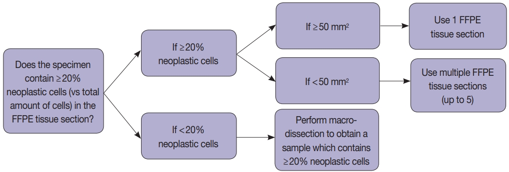
- 11,012 View
- 236 Download
- 20 Web of Science
- 18 Crossref
-
Abstract
PDFSupplementary Material
- Background
Microsatellite instability (MSI) analysis is becoming increasingly important in many types of tumor including colorectal cancer (CRC). The commonly used MSI tests are either time-consuming or labor-intensive. A fully automated MSI test, the Idylla MSI assay, has recently been introduced. However, its diagnostic performance has not been extensively validated in clinical CRC samples.
Methods
We evaluated 133 samples whose MSI status had been rigorously validated by standard polymerase chain reaction (PCR), clinical nextgeneration sequencing (NGS) cancer panel test, or both. We evaluated the diagnostic performance of the Idylla MSI assay in terms of sensitivity, specificity, and positive and negative predictive values, as well as various sample requirements, such as minimum tumor purity and the quality of paraffin blocks.
Results
Compared with the gold standard results confirmed through both PCR MSI test and NGS, the Idylla MSI assay showed 99.05% accuracy (104/105), 100% sensitivity (11/11), 98.94% specificity (93/94), 91.67% positive predictive value (11/12), and 100% negative predictive value (93/93). In addition, the Idylla MSI assay did not require macro-dissection in most samples and reliably detected MSI-high in samples with approximately 10% tumor purity. The total turnaround time was about 150 minutes and the hands-on time was less than 2 minutes.
Conclusions
The Idylla MSI assay shows good diagnostic performance that is sufficient for its implementation in the clinic to determine the MSI status of at least the CRC samples. In addition, the fully automated procedure requires only a few slices of formalin-fixed paraffin-embedded tissue and might greatly save time and labor. -
Citations
Citations to this article as recorded by- Leveraging Radiomics and Hybrid Quantum–Classical Convolutional Networks for Non-Invasive Detection of Microsatellite Instability in Colorectal Cancer
T. Buvaneswari, M. Ramkumar, Prabhu Venkatesan, R. Sarath Kumar
Molecular Imaging and Biology.2025; 27(2): 227. CrossRef - Molecular Profiling of H-MSI/dMMR/for Endometrial Cancer Patients: “New Challenges in Diagnostic Routine Practice”
Riccardo Adorisio, Giancarlo Troncone, Massimo Barberis, Francesco Pepe
Journal of Molecular Pathology.2024; 5(2): 187. CrossRef - Enhanced Commendable Sensitivity and Specificity for MSI in Colorectal Cancer by a New PCR‐HRM Based 8‐Loci MSI Assay
Xueping Xiang, Xiaojing Ma, Linlin Ying, Hong Zou
Journal of Clinical Laboratory Analysis.2024;[Epub] CrossRef - Endometriumkarzinom: molekulare Klassifikation in der Routinepathologie
Udo Siebolts, Birgid Schömig-Markiefka, Janna Siemanowski-Hrach, Sabine Merkelbach-Bruse
Die Pathologie.2024; 45(5): 347. CrossRef - Integration of rapid PCR testing as an adjunct to NGS in diagnostic pathology services within the UK: evidence from a case series of non-squamous, non-small cell lung cancer (NSCLC) patients with follow-up
Alison Finall, Gareth Davies, Trevor Jones, Gwion Emlyn, Pearl Huey, Anna Mullard
Journal of Clinical Pathology.2023; 76(6): 391. CrossRef - Analytical Validation and Clinical Utilization of the Oncomine Comprehensive Assay Plus Panel for Comprehensive Genomic Profiling in Solid Tumors
Catherine I. Dumur, Ramakrishnan Krishnan, Jorge A. Almenara, Kathleen E. Brown, Kailyn R. Dugan, Christiana Farni, Fatima Z. Ibrahim, Naomi A. Sanchez, Sumra Rathore, Dinesh Pradhan, Jonathan H. Hughes
Journal of Molecular Pathology.2023; 4(2): 109. CrossRef - Performance of Immunohistochemical and Molecular Methods in Detecting Microsatellite Instability in Gastric Cancer: A Multicenter Study
Diogo Sousa Marques, Irene Gullo, Luís Mascarenhas-Lemos, João Ricardo Silva, Catarina Neto do Nascimento, Patrícia Pontes, Lídia Pinho, Luis Cirnes, Xiaogang Wen, Marília Cravo, Fátima Carneiro
Pathobiology.2023; 90(6): 389. CrossRef - Diagnostic mutationnel rapide Idylla™ : applications théranostiques actuelles et futures
Amélie Bourhis, Annabelle Remoué, Laura Samaison, Arnaud Uguen
Annales de Pathologie.2022; 42(4): 329. CrossRef - Comparison of the Idylla™ MSI assay with the Promega™ MSI Analysis System and immunohistochemistry on formalin-fixed paraffin-embedded tissue of endometrial carcinoma: results from an international, multicenter study
Sonia Gatius, Ana Velasco, Mar Varela, Miriam Cuatrecasas, Pedro Jares, Lisa Setaffy, Benjamin Bonhomme, Almudena Santon, Kristina Lindemann, Sabrina Croce, Ben Davidson, Sigurd Lax, Jose Palacios, Xavier Matias-Guiu
Virchows Archiv.2022; 480(5): 1031. CrossRef - Idylla MSI test combined with immunohistochemistry is a valuable and cost effective strategy to search for microsatellite instable tumors of noncolorectal origin
Laura Samaison, Arnaud Uguen
Pathology International.2022; 72(4): 234. CrossRef - Detection of microsatellite instability in a panel of solid tumours with the Idylla MSI Test using extracted DNA
Adrien Pécriaux, Loetitia Favre, Julien Calderaro, Cécile Charpy, Jonathan Derman, Anaïs Pujals
Journal of Clinical Pathology.2021; 74(1): 36. CrossRef - Idylla microsatellite instability assay versus mismatch repair immunohistochemistry: a retrospective comparison in gastric adenocarcinoma
Luke Farmkiss, Ilona Hopkins, Mary Jones
Journal of Clinical Pathology.2021; 74(9): 604. CrossRef - Multi-center real-world comparison of the fully automated Idylla™ microsatellite instability assay with routine molecular methods and immunohistochemistry on formalin-fixed paraffin-embedded tissue of colorectal cancer
Ana Velasco, Fatma Tokat, Jesper Bonde, Nicola Trim, Elisabeth Bauer, Adam Meeney, Wendy de Leng, George Chong, Véronique Dalstein, Lorand L. Kis, Jon A. Lorentzen, Snjezana Tomić, Keeley Thwaites, Martina Putzová, Astrid Birnbaum, Romena Qazi, Vanessa Pr
Virchows Archiv.2021; 478(5): 851. CrossRef - Detection of microsatellite instability with Idylla MSI assay in colorectal and endometrial cancer
Iiris Ukkola, Pirjo Nummela, Annukka Pasanen, Mia Kero, Anna Lepistö, Soili Kytölä, Ralf Bützow, Ari Ristimäki
Virchows Archiv.2021; 479(3): 471. CrossRef - Managing Difficulties of Microsatellite Instability Testing in Endometrial Cancer-Limitations and Advantages of Four Different PCR-Based Approaches
Janna Siemanowski, Birgid Schömig-Markiefka, Theresa Buhl, Anja Haak, Udo Siebolts, Wolfgang Dietmaier, Norbert Arens, Nina Pauly, Beyhan Ataseven, Reinhard Büttner, Sabine Merkelbach-Bruse
Cancers.2021; 13(6): 1268. CrossRef - Evaluation of Micro Satellite Instability and Mismatch Repair Status in Different Solid Tumors: A Multicenter Analysis in a Real World Setting
Umberto Malapelle, Paola Parente, Francesco Pepe, Caterina De Luca, Pasquale Pisapia, Roberta Sgariglia, Mariantonia Nacchio, Gianluca Gragnano, Gianluca Russo, Floriana Conticelli, Claudio Bellevicine, Elena Vigliar, Antonino Iaccarino, Claudia Covelli,
Cells.2021; 10(8): 1878. CrossRef - Novel Biocartis Idylla™ cartridge-based assay for detection of microsatellite instability in colorectal cancer tissues
Andres E. Mindiola-RomeroMD, Donald C. GreenBS, M. Rabie Al-TurkmaniPhD, Kelley N. GodwinBS, Anna C. MackayBS, Laura J. TafeMD, Bing RenMD, Gregory J. TsongalisPhD
Experimental and Molecular Pathology.2020; 116: 104519. CrossRef - Evaluation of 3 molecular-based assays for microsatellite instability detection in formalin-fixed tissues of patients with endometrial and colorectal cancers
Pauline Gilson, Julien Levy, Marie Rouyer, Jessica Demange, Marie Husson, Céline Bonnet, Julia Salleron, Agnès Leroux, Jean-Louis Merlin, Alexandre Harlé
Scientific Reports.2020;[Epub] CrossRef
- Leveraging Radiomics and Hybrid Quantum–Classical Convolutional Networks for Non-Invasive Detection of Microsatellite Instability in Colorectal Cancer
- Molecular and Clinicopathological Features of Gastrointestinal Stromal Tumors in Vietnamese Patients
- Quoc Dat Ngo, Quoc Thang Pham, Dang Anh Thu Phan, Anh Vu Hoang, Thi Ngoc Ha Hua, Sao Trung Nguyen
- J Pathol Transl Med. 2019;53(6):361-368. Published online September 16, 2019
- DOI: https://doi.org/10.4132/jptm.2019.08.27
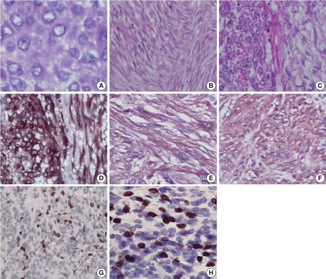
- 9,482 View
- 160 Download
- 2 Web of Science
- 2 Crossref
-
Abstract
PDFSupplementary Material
- Background
Gastrointestinal stromal tumors (GISTs) are the most frequent mesenchymal neoplasms of the gastrointestinal tract. Management of GIST patients is currently based on clinicopathological features and associated genetic changes. However, the detailed characteristics and molecular genetic features of GISTs have not yet been described in the Vietnamese population.
Methods
We first identified 155 patients with primary GIST who underwent surgery with primary curative intent between 2011 and 2014 at University Medical Center at Ho Chi Minh City, Vietnam. We evaluated the clinicopathological features and immunohistochemical reactivity to p53 and Ki-67 in these patients. Additionally, KIT genotyping was performed in 100 cases.
Results
The largest proportion of GISTs was classified as high-risk (43.2%). Of the 155 GISTs, 52 (33.5%) were positive for Ki-67, and 58 (37.4%) were positive for p53. The expression of Ki-67 and p53 were correlated with mitotic rate, tumor size, risk assessment, and tumor stage. Out of 100 GIST cases, KIT mutation was found in 68%, of which 62 (91.2%) were found in exon 11, two (2.9%) in exon 9, and four (5.8%) in exon 17. No mutation in exon 13 was identified. Additionally, KIT mutations did not correlate with any clinicopathological features.
Conclusions
The expression of Ki-67 and p53 were associated with high-risk tumors. Mutations in exon 11 were the most commonly found, followed by exon 17 and exon 9. Additionally, KIT mutation status was not correlated with any recognized clinicopathological features. -
Citations
Citations to this article as recorded by- Ki67 for evaluating the prognosis of gastrointestinal stromal tumors: A systematic review and meta‑analysis
Ji Li, An-Ran Wang, Xiao-Dong Chen, Hong Pan, Shi-Qiang Li
Oncology Letters.2022;[Epub] CrossRef - Endoscopic ultrasound‐guided fine‐needle aspiration cytology in the diagnosis of the gastrointestinal stromal tumor of the stomach
José‐Fernando Val‐Bernal, Elena Yllera, María Moris, Ihab Abdulkader Nallib, Angel Vázquez‐Boquete, María Martino
Diagnostic Cytopathology.2020; 48(9): 833. CrossRef
- Ki67 for evaluating the prognosis of gastrointestinal stromal tumors: A systematic review and meta‑analysis
Case Study
- Frozen Cytology of Meningeal Malignant Solitary Fibrous Tumor/Hemangiopericytoma
- Myunghee Kang, Na Rae Kim, Dong Hae Chung, Gie-Taek Yie
- J Pathol Transl Med. 2019;53(3):192-197. Published online April 11, 2019
- DOI: https://doi.org/10.4132/jptm.2019.03.20
- 9,519 View
- 160 Download
- 7 Web of Science
- 8 Crossref
-
Abstract
PDF
- A 51-year-old woman presented with severe dizziness. The brain magnetic resonance image revealed a 5.5 cm multiloculated mass with a thick rim in the left temporal lobe. Cytological examination of frozen diagnosis of the mass showed hypercellular sheets of round and rhabdoid cells in a hemorrhagic background, and two mitotic figures were observed. Histologically, the excised dura-based mass consisted of predominantly round cells with small foci of rhabdoid tumor cells in a pseudoalveolar pattern in a hemorrhagic background, and the cells showed nuclear positivity for signal transducer and activator of transcription 6 as well as frequent mitosis. The mass was diagnosed as a grade 3 solitary fibrous tumor (SFT)/hemangiopericytoma (HPC). The cytological diagnosis of SFT/HPC is challenging because of the heterogeneous cytological findings, such as histological heterogeneity, and because there are no standardized cytological criteria for malignant SFT/HPC. Cytological findings, such as singly scattered small cells, hypercellularity, rare ropy collagen, and round and rhabdoid cells with pseudoalveolar pattern, may assist in the diagnosis of malignant SFT/HPC.
-
Citations
Citations to this article as recorded by- A Rare Case of Cervical Solitary Fibrous Tumor in a Pediatric Patient: Case Report and Literature Review
Eleonora Becattini, Lorenzo Sgarbanti, Giuseppina Bevacqua, Valentina Grespi, Carlo Conti
NeuroSci.2025; 6(2): 49. CrossRef - Meningeal Solitary Fibrous Tumor: A Cytological Report With Emphasis on the Usefulness of Immunocytochemical Analysis for STAT6
Hiroyuki Okanishi, Mitsuaki Ishida, Naoto Kohno, Isako Kataoka, Mari Tomiuka, Mayumi Uragami, Shizuka Ono, Chihiro Deguchi, Reika Takeda, Yoshitaka Kurisu, Yoshinobu Hirose
Diagnostic Cytopathology.2025;[Epub] CrossRef - Cytologic features of mesenchymal, melanocytic and haematolymphoid tumours of the central nervous system and metastases
Carmen Bárcena, José A. Jiménez‐Heffernan
Cytopathology.2024; 35(5): 590. CrossRef - A Hemangiopericytoma in the External Auditory Canal: A Rare Clinical Presentation and Management
Vaibhavi Patil, Prasad Deshmukh, Sagar S Gaurkar , Ayushi Ghosh Moulic, Jasleen Kaur
Cureus.2024;[Epub] CrossRef - Scoring system for intraoperative diagnosis of intracranial schwannoma by squash cytology
Hirotaka Fujita, Takuma Tajiri, Tomohisa Machida, Nozomi Nomura, Suguru Toguchi, Hitoshi Itoh, Shinichiro Hiraiwa, Tomoko Sugiyama, Chie Inomoto, Masaaki Imai, Shinri Oda, Masami Shimoda, Naoya Nakamura
Cytopathology.2022; 33(2): 196. CrossRef - Occurrence of a solitary fibrous tumor adjacent to the resection bed of a high-grade meningioma: A case report
Coby Cunningham, Rocco Dabecco, Justin Davanzo
Interdisciplinary Neurosurgery.2021; 25: 101277. CrossRef - A case of solitary fibrous tumor arising in the meninge
Saori NAKANISHI, Naoto KURODA, Toshiko TAKAI, Mari KOJIMA, Misato OONOGI
The Journal of the Japanese Society of Clinical Cytology.2021; 60(4): 224. CrossRef - Intraoperative frozen cytology of intraosseous cystic meningioma in the sphenoid bone
Na Rae Kim, Gie-Taek Yie
Journal of Pathology and Translational Medicine.2020; 54(6): 508. CrossRef
- A Rare Case of Cervical Solitary Fibrous Tumor in a Pediatric Patient: Case Report and Literature Review
Original Article
- Association between p53 Expression and Amount of Tumor-Infiltrating Lymphocytes in Triple-Negative Breast Cancer
- Miseon Lee, In Ah Park, Sun-Hee Heo, Young-Ae Kim, Gyungyub Gong, Hee Jin Lee
- J Pathol Transl Med. 2019;53(3):180-187. Published online March 11, 2019
- DOI: https://doi.org/10.4132/jptm.2019.02.08
- 11,068 View
- 209 Download
- 20 Web of Science
- 20 Crossref
-
Abstract
PDF
- Background
Most triple-negative breast cancers (TNBCs) have a high histologic grade, are associated with high endoplasmic stress, and possess a high frequency of TP53 mutations. TP53 missense mutations lead to the production of mutant p53 protein and usually show high levels of p53 protein expression. Tumor-infiltrating lymphocytes (TILs) accumulate as part of the anti-tumor immune response and have a strong prognostic and predictive significance in TNBC. We aimed to elucidate the association between p53 expression and the amount of TILs in TNBC.
Methods
In 678 TNBC patients, we evaluated TIL levels and expression of endoplasmic stress molecules. Immunohistochemical examination of p53 protein expression was categorized into three groups: no, low, and high expression.
Results
No, low, and high p53 expression was identified in 44.1% (n = 299), 20.1% (n = 136), and 35.8% (n = 243) of patients, respectively. Patients with high p53 expression showed high histologic grade (p < .001), high TIL levels (p = .009), and high expression of endoplasmic reticulum stress-associated molecules (p-eIF2a, p = .013; XBP1, p = .007), compared to patients with low p53 expression. There was no significant difference in disease-free (p = .406) or overall survival rates (p = .444) among the three p53 expression groups.
Conclusions
High p53 expression is associated with increased expression of endoplasmic reticulum stress molecules and TIL influx. -
Citations
Citations to this article as recorded by- A comparative analysis of mutational profiles between triple-negative breast cancer and non-triple-negative breast cancer
Wanlin Li, Chenchen Feng, Shunheng Zhou
Discover Oncology.2026;[Epub] CrossRef - The search for a TNBC vaccine: the guardian vaccine
Cory Fines, Helen McCarthy, Niamh Buckley
Cancer Biology & Therapy.2025;[Epub] CrossRef - Correlating p53 immunostaining patterns with somatic TP53 mutation and functional properties of mutant p53 in triple‐negative breast cancer
Meejeong Kim, Miseon Lee, Ahwon Lee, Byung‐Ock Choi, Woo‐Chan Park, Sung Hun Kim, Jieun Lee, Jun Kang
Histopathology.2025; 87(2): 299. CrossRef - GD3 synthase drives resistance to p53-induced apoptosis in breast cancer by modulating mitochondrial function
Vivek Anand, Fouad El-Dana, Natalia Baran, Jenny Borgman, Zheng Yin, Hong Zhao, Stephen T. Wong, Michael Andreeff, V. Lokesh Battula
Oncogene.2025; 44(30): 2646. CrossRef - Topoisomerase Inhibitor Resistance in Breast Cancer: Exploring Alterations in Cellular Metabolism and Properties Driving Drug Insensitivity
Juhong Lee, Youngjoo Kwon
Drug Targets and Therapeutics.2025; 4(2): 185. CrossRef - Updated Austrian treatment algorithm for metastatic triple-negative breast cancer
Rupert Bartsch, Gabriel Rinnerthaler, Edgar Petru, Daniel Egle, Michael Gnant, Marija Balic, Thamer Sliwa, Christian Singer
Wiener klinische Wochenschrift.2024; 136(11-12): 347. CrossRef - Triple negative breast cancer: Immunogenicity, tumor microenvironment, and immunotherapy
Sotiris Loizides, Anastasia Constantinidou
Frontiers in Genetics.2023;[Epub] CrossRef - Prognostic benefit of TILs independent of clinicopathological and molecular factors
Koen Brummel, Anneke L. Eerkens, Marco de Bruyn, Hans W. Nijman
British Journal of Cancer.2023; 129(5): 737. CrossRef - Dihydroartemisinin-Transferrin Adducts Enhance TRAIL-Induced Apoptosis in Triple-Negative Breast Cancer in a P53-Independent and ROS-Dependent Manner
Xinyu Zhou, Abel Soto-Gamez, Fleur Nijdam, Rita Setroikromo, Wim J. Quax
Frontiers in Oncology.2022;[Epub] CrossRef - New Challenges in the Differential Diagnosis of High-Grade Triple-Negative Breast Cancer and Serous Carcinoma
Andrii Puzyrenko, Chandler S Cortina, Julie M Jorns
International Journal of Surgical Pathology.2022; 30(7): 728. CrossRef - Prognostic analysis of cuproptosis-related gene in triple-negative breast cancer
Shengnan Sha, Luyi Si, Xinrui Wu, Yuanbiao Chen, Hui Xiong, Ying Xu, Wangrui Liu, Haijun Mei, Tao Wang, Mei Li
Frontiers in Immunology.2022;[Epub] CrossRef - p53 Missense Mutation is Associated with Immune Cell PD-L1 Expression in Triple-Negative Breast Cancer
Ai-Yan Xing, Long Liu, Ke Liang, Bin Wang
Cancer Investigation.2022; 40(10): 879. CrossRef - Crosstalk between Immune Checkpoint Modulators, Metabolic Reprogramming and Cellular Plasticity in Triple-Negative Breast Cancer
Arpita Poddar, Sushma R. Rao, Prashanth Prithviraj, George Kannourakis, Aparna Jayachandran
Current Oncology.2022; 29(10): 6847. CrossRef - The tumor microenvironment and triple-negative breast cancer aggressiveness: shedding light on mechanisms and targeting
Natsuki Furukawa, Vered Stearns, Cesar A. Santa-Maria, Aleksander S. Popel
Expert Opinion on Therapeutic Targets.2022; 26(12): 1041. CrossRef - Intracellular partners of fibroblast growth factors 1 and 2 - implications for functions
Katarzyna Dominika Sluzalska, Jakub Slawski, Martyna Sochacka, Agata Lampart, Jacek Otlewski, Malgorzata Zakrzewska
Cytokine & Growth Factor Reviews.2021; 57: 93. CrossRef - Targeted Chinese Medicine Delivery by A New Family of Biodegradable Pseudo-Protein Nanoparticles for Treating Triple-Negative Breast Cancer: In Vitro and In Vivo Study
Hiu Yee Kwan, Qinghua Xu, Ruihong Gong, Zhaoxiang Bian, Chih-Chang Chu
Frontiers in Oncology.2021;[Epub] CrossRef - Transcriptomic Properties of HER2+ Ductal Carcinoma In Situ of the Breast Associate with Absence of Immune Cells
Marie Colombe Agahozo, Marcel Smid, Ronald van Marion, Dora Hammerl, Thierry P. P. van den Bosch, Mieke A. M. Timmermans, Chayenne J. Heijerman, Pieter J. Westenend, Reno Debets, John W. M. Martens, Carolien H. M. van Deurzen
Biology.2021; 10(8): 768. CrossRef - With Our Powers Combined
Lawrence Kasherman, Katherine Karakasis, Amit M. Oza
The Cancer Journal.2021; 27(6): 511. CrossRef The Research Progress on the Prognostic Value of the Common Hematological Parameters in Peripheral Venous Blood in Breast Cancer
Li Chen, Xiangyi Kong, Chengrui Yan, Yi Fang, Jing Wang
OncoTargets and Therapy.2020; Volume 13: 1397. CrossRefBiomolecular Factors Represented by Bcl-2, p53, and Tumor-Infiltrating Lymphocytes Predict Response for Adjuvant Anthracycline Chemotherapy in Patients with Early Triple-Negative Breast Cancer
Xenia Elena Bacinschi, Anca Zgura, Inga Safta, Rodica Anghel
Cancer Management and Research.2020; Volume 12: 11965. CrossRef
- A comparative analysis of mutational profiles between triple-negative breast cancer and non-triple-negative breast cancer
Review
- Provisional Guideline Recommendation for EGFR Gene Mutation Testing in Liquid Samples of Lung Cancer Patients: A Proposal by the Korean Cardiopulmonary Pathology Study Group
- Dong Hoon Shin, Hyo Sup Shim, Tae Jung Kim, Heae Surng Park, Yun La Choi, Wan Seop Kim, Lucia Kim, Sun Hee Chang, Joon Seon Song, Hyo jin Kim, Jung Ho Han, Chang Hun Lee, Geon Kook Lee, Se Jin Jang
- J Pathol Transl Med. 2019;53(3):153-158. Published online February 28, 2019
- DOI: https://doi.org/10.4132/jptm.2019.02.22
- 11,921 View
- 266 Download
- 8 Web of Science
- 9 Crossref
-
Abstract
PDF
- Liquid biopsy for detection of mutation from circulating tumor DNA is a new technology which is attractive in that it is non-invasive. Epidermal growth factor receptor (EGFR) tyrosine kinase inhibitors (TKI) is an effective first line drug for advanced non-small cell lung cancer patients who harbor activating EGFR mutation. During the course of treatment, resistance against TKI arises which can be contributed to EGFR T790M mutation in about 50–60% of patients. Third generation TKI may overcome the resistance. In patients who cannot undergo tissue biopsy due to variable reasons, liquid biopsy is an excellent alternative for the detection of EGFR T790M mutation. However, this relatively novel method requires standardization and vigorous quality insurance. Thus, a standard set of guideline recommendations for liquid biopsy for EGFR mutation testing suitable for the Korean medical community is necessary. In this article, we propose a set of provisional guideline recommendations that was discussed and approved by the Cardiopulmonary Pathology Study Group of the Korean Society of Pathologists.
-
Citations
Citations to this article as recorded by- Chemical and Technological Aspects of Magnetic Particles for Cancer Liquid Biopsy Applications
Beatriz Naranjo‐Martínez, Ana Laura Coria‐Gutiérrez, Sunčica Sukur, Sedef Ozel‐Okcu, Václav Ranc, Francisco J. Terán, Franca Raucci, Marco A. Pierotti, Domenico Scionti, Anna Piperno
ChemistryOpen.2026;[Epub] CrossRef - Comparison of tissue-based and plasma-based testing for EGFR mutation in non–small cell lung cancer patients
Yoon Kyung Kang, Dong Hoon Shin, Joon Young Park, Chung Su Hwang, Hyun Jung Lee, Jung Hee Lee, Jee Yeon Kim, JooYoung Na
Journal of Pathology and Translational Medicine.2025; 59(1): 60. CrossRef - Improving non-small-cell lung cancer survival through molecular characterization: Perspective of a multidisciplinary expert panel
M.G.O. Fernandes, A.S. Vilariça, B. Fernandes, C. Camacho, C. Saraiva, F. Estevinho, H. Novais e Bastos, J.M. Lopes, P. Fidalgo, P. Garrido, S. Alves, S. Silva, T. Sequeira, F. Barata
Pulmonology.2024; 30(1): 4. CrossRef - Unlocking the future of cancer diagnosis – promises and challenges of ctDNA-based liquid biopsies in non-small cell lung cancer
Chiara Reina, Berina Šabanović, Chiara Lazzari, Vanesa Gregorc, Christopher Heeschen
Translational Research.2024; 272: 41. CrossRef - Exosomes in Lung Cancer: Actors and Heralds of Tumor Development
Amaia Sandúa, Estibaliz Alegre, Álvaro González
Cancers.2021; 13(17): 4330. CrossRef - Molecular biomarker testing for non–small cell lung cancer: consensus statement of the Korean Cardiopulmonary Pathology Study Group
Sunhee Chang, Hyo Sup Shim, Tae Jung Kim, Yoon-La Choi, Wan Seop Kim, Dong Hoon Shin, Lucia Kim, Heae Surng Park, Geon Kook Lee, Chang Hun Lee
Journal of Pathology and Translational Medicine.2021; 55(3): 181. CrossRef - A Review of Circulating Tumor DNA in the Diagnosis and Monitoring of Esophageal Cancer
Jiang Min, Huilin Zhou, Su Jiang, Hong Yu
Medical Science Monitor.2021;[Epub] CrossRef - Current status and future perspectives of liquid biopsy in non-small cell lung cancer
Sunhee Chang, Jae Young Hur, Yoon-La Choi, Chang Hun Lee, Wan Seop Kim
Journal of Pathology and Translational Medicine.2020; 54(3): 204. CrossRef - Prevalence of T790M mutation among TKI-therapy resistant Lebanese lung cancer patients based on liquid biopsy analysis: a first report from a major tertiary care center
Hazem Assi, Arafat Tfayli, Nada Assaf, Sarah Abou Daya, Aram H. Bidikian, Dima Kawsarani, Puzant Fermanian, Ghazi Zaatari, Rami Mahfouz
Molecular Biology Reports.2019; 46(4): 3671. CrossRef
- Chemical and Technological Aspects of Magnetic Particles for Cancer Liquid Biopsy Applications
Case Study
- Encapsulated Papillary Thyroid Tumor with Delicate Nuclear Changes and a KRAS Mutation as a Possible Novel Subtype of Borderline Tumor
- Kenji Ohba, Norisato Mitsutake, Michiko Matsuse, Tatiana Rogounovitch, Nobuhiko Nishino, Yutaka Oki, Yoshie Goto, Kennichi Kakudo
- J Pathol Transl Med. 2019;53(2):136-141. Published online January 14, 2019
- DOI: https://doi.org/10.4132/jptm.2018.12.07
- 10,587 View
- 192 Download
- 12 Web of Science
- 8 Crossref
-
Abstract
PDF
- Although papillary thyroid carcinoma (PTC)–type nuclear changes are the most reliable morphological feature in the diagnosis of PTC, the nuclear assessment used to identify these changes is highly subjective. Here, we report a noninvasive encapsulated thyroid tumor with a papillary growth pattern measuring 23 mm at its largest diameter with a nuclear score of 2 in a 26-year-old man. After undergoing left lobectomy, the patient was diagnosed with an encapsulated PTC. However, a second opinion consultation suggested an alternative diagnosis of follicular adenoma with papillary hyperplasia. When providing a third opinion, we identified a low MIB-1 labeling index and a heterozygous point mutation in the KRAS gene but not the BRAF gene. We speculated that this case is an example of a novel borderline tumor with a papillary structure. Introduction of the new terminology “noninvasive encapsulated papillary RAS-like thyroid tumor (NEPRAS)” without the word “cancer” might relieve the psychological burden of patients in a way similar to the phrase “noninvasive follicular thyroid neoplasm with papillary-like nuclear features (NIFTP).”
-
Citations
Citations to this article as recorded by- The fascinating history of papillary thyroid carcinoma nuclei: revelation of two nuclear morphologies—“Classical papillary” and “papillary‐like”, with different pathobiologic characteristics
N. Paul Ohori, Jeremy M. Minkowitz, Yuri E. Nikiforov, Raja R. Seethala
Cancer Cytopathology.2026;[Epub] CrossRef - Malignancy in Pediatric Hyperfunctioning Thyroid Nodules: A Case Report and Literature Review
Cristina Clausi, Simona Censi, Jacopo Maria Arnone, Giulia Messina, Ilaria Piva, Fausto Cortese, Loris Bertazza, Susi Barollo, Francesca Galuppini, Marco Zarantonello, Maurizio Iacobone, Gianmaria Pennelli, Caterina Mian
Clinical Endocrinology.2026;[Epub] CrossRef - The Asian Thyroid Working Group, from 2017 to 2023
Kennichi Kakudo, Chan Kwon Jung, Zhiyan Liu, Mitsuyoshi Hirokawa, Andrey Bychkov, Huy Gia Vuong, Somboon Keelawat, Radhika Srinivasan, Jen-Fan Hang, Chiung-Ru Lai
Journal of Pathology and Translational Medicine.2023; 57(6): 289. CrossRef - Whole Tumor Capsule Is Prognostic of Very Good Outcome in the Classical Variant of Papillary Thyroid Cancer
Carlotta Giani, Liborio Torregrossa, Teresa Ramone, Cristina Romei, Antonio Matrone, Eleonora Molinaro, Laura Agate, Gabriele Materazzi, Paolo Piaggi, Clara Ugolini, Fulvio Basolo, Raffaele Ciampi, Rossella Elisei
The Journal of Clinical Endocrinology & Metabolism.2021; 106(10): e4072. CrossRef - Evidence of Cooperation between Hippo Pathway and RAS Mutation in Thyroid Carcinomas
Thaise Nayane Ribeiro Carneiro, Larissa Valdemarin Bim, Vanessa Candiotti Buzatto, Vanessa Galdeno, Paula Fontes Asprino, Eunjung Alice Lee, Pedro Alexandre Favoretto Galante, Janete Maria Cerutti
Cancers.2021; 13(10): 2306. CrossRef - Capsular Invasion Matters Also in “Papillary Patterned” Tumors: A Study on 121 Cases of Encapsulated Conventional Variant of Papillary Thyroid Carcinoma
Dilara Akbulut, Ezgi Dicle Kuz, Nazmiye Kursun, Serpil Dizbay Sak
Endocrine Pathology.2021; 32(3): 357. CrossRef - Noninvasive encapsulated papillary RAS-like thyroid tumor (NEPRAS) or encapsulated papillary thyroid carcinoma (PTC)
Pedro Weslley Rosario
Journal of Pathology and Translational Medicine.2020; 54(3): 263. CrossRef - Updates in the Pathologic Classification of Thyroid Neoplasms: A Review of the World Health Organization Classification
Yanhua Bai, Kennichi Kakudo, Chan Kwon Jung
Endocrinology and Metabolism.2020; 35(4): 696. CrossRef
- The fascinating history of papillary thyroid carcinoma nuclei: revelation of two nuclear morphologies—“Classical papillary” and “papillary‐like”, with different pathobiologic characteristics
Original Articles
- Uterine Malignant Mixed Müllerian Tumors Following Treatment with Selective Estrogen Receptor Modulators in Patients with Breast Cancer: A Report of 13 Cases and Their Clinicopathologic Characteristics
- Byung-Kwan Jeong, Chang O. Sung, Kyu-Rae Kim
- J Pathol Transl Med. 2019;53(1):31-39. Published online December 18, 2018
- DOI: https://doi.org/10.4132/jptm.2018.11.16
- 9,678 View
- 103 Download
- 3 Web of Science
- 4 Crossref
-
Abstract
PDF
- Background
Breast cancer treatment with selective estrogen receptor modulators (SERMs) increasesthe incidence of uterine malignant mixed Müllerian tumors (uMMMTs). We examine clinicopathologiccharacteristics and prognosis of SERM-associated uMMMTs (S-uMMMTs) and discusspossible pathogenetic mechanisms.
Methods
Among 28,104 patients with breast cancer, clinicopathologicfeatures and incidence of uMMMT were compared between patients who underwentSERM treatment and those who did not. Of 92 uMMMT cases that occurred during the same period,incidence, dose, and duration of SERM treatment, as well as overall survival rate, were comparedfor patients with breast cancer who underwent SERM treatment and those who did not (S-uMMMTvs NS-uMMMT) and for patients without breast cancer (de novo-uMMMT). Histopathologicalfindings and immunophenotypes for myogenin, desmin, p53, WT-1, estrogen receptor (ER) α, ERβ,progesterone receptor, and GATA-3 were compared between S-uMMMT and de novo-uMMMT.
Results
The incidence of S-uMMMT was significantly higher than that of NS-uMMMT (6.35-fold).All patients with SERM were postmenopausal and received daily 20–40 mg SERM. CumulativeSERM dose ranged from 21.9 to 73.0 g (mean, 46.0) over 39–192 months (mean, 107). Clinicopathologicfeatures, such as International Federation of Gynecology and Obstetrics stage andoverall survival, were not significantly different between patients with S-uMMMT and NS-uMMMTor between patients with S-uMMMT and de novo-uMMMT. All 11 S-uMMMT cases available forimmunostaining exhibited strong overexpression/null expression of p53 protein and significantlyincreased ERβ expression in carcinomatous and sarcomatous components.
Conclusions
SERMtherapy seemingly increases risk of S-uMMMT development; however, clinicopathologic featureswere similar in all uMMMTs from different backgrounds. p53 mutation and increased ERβ expressionmight be involved in the etiology of S-uMMMT. -
Citations
Citations to this article as recorded by- Uterine carcinosarcomas: A case series of 9 cases from a low-income country
Boubacar Efared, Halidou Hamadou Koura, Aïchatou Balaraba Abani Bako, Idrissa Boubacar, Habiba Salifou Boureima, Garba Mahamadou, Hassan Nouhou
Medicine.2024; 103(40): e39773. CrossRef - Uterine carcinosarcoma: Unraveling the role of epithelial‐to‐mesenchymal transition in progression and therapeutic potential
Mohan Shankar Gopinatha Pillai, Pallab Shaw, Arpan Dey Bhowmik, Resham Bhattacharya, Geeta Rao, Shailendra Kumar Dhar Dwivedi
The FASEB Journal.2024;[Epub] CrossRef - Tamoxifen/toremifene
Reactions Weekly.2019; 1758(1): 330. CrossRef - Molecular Basis of Tumor Heterogeneity in Endometrial Carcinosarcoma
Susanna Leskela, Belen Pérez-Mies, Juan Manuel Rosa-Rosa, Eva Cristobal, Michele Biscuola, María L. Palacios-Berraquero, SuFey Ong, Xavier Matias-Guiu Guia, José Palacios
Cancers.2019; 11(7): 964. CrossRef
- Uterine carcinosarcomas: A case series of 9 cases from a low-income country
- Loss of Human Leukocyte Antigen Class I Expression Is Associated with Poor Prognosis in Patients with Advanced Breast Cancer
- Hong Sik Park, Uiju Cho, So Young Im, Chang Young Yoo, Ji Han Jung, Young Jin Suh, Hyun Joo Choi
- J Pathol Transl Med. 2019;53(2):75-85. Published online November 14, 2018
- DOI: https://doi.org/10.4132/jptm.2018.10.11
- 10,672 View
- 193 Download
- 34 Web of Science
- 34 Crossref
-
Abstract
PDF
- Background
Human leukocyte antigen class I (HLA-I) molecules play important roles in regulating immune responses. Loss or reduction of HLA-I expression has been shown to be associated with prognosis in several cancers. Regulatory T-cells (Tregs) also play critical functions in immune response regulation. Evaluation of HLA-I expression status by the EMR8-5 antibody and its clinical impact in breast cancer have not been well studied, and its relationship with Tregs remains unclear.
Methods
We evaluated HLA-I expression and Treg infiltration by immunohistochemistry in 465 surgically resected breast cancer samples. We examined the correlation between HLA-I expression and Treg infiltration and clinicopathologic characteristics and survival analyses were performed.
Results
Total loss of HLA-I expression was found in 84 breast cancer samples (18.1%). Univariate survival analysis revealed that loss of HLA-I expression was significantly associated with worse disease-specific survival (DSS) (p = .029). HLA-I was not an independent prognostic factor in the entire patient group, but it was an adverse independent prognostic factor for DSS in patients with advanced disease (stage II–IV) (p = .031). Treg numbers were significantly higher in the intratumoral stroma of HLA-I–positive tumors than in HLA-I–negative tumors (median 6.3 cells/high power field vs 2.1 cells/high power field, p < .001). However, Tregs were not an independent prognostic factor in our cohort.
Conclusions
Our findings suggest that the loss of HLA-I expression is associated with poor prognosis in breast cancer patients, highlighting the role of HLA-I alterations in immune evasion mechanisms of breast cancer. HLA-I could be a promising marker that enables the application of more effective and precise immunotherapies for patients with advanced breast cancer. -
Citations
Citations to this article as recorded by- Estrogen Signaling in Early-Stage Breast Cancer: Impact on Neoadjuvant Chemotherapy and Immunotherapy
Chiara Corti, Busem Binboğa Kurt, Beyza Koca, Tasnim Rahman, Fabio Conforti, Laura Pala, Giampaolo Bianchini, Carmen Criscitiello, Giuseppe Curigliano, Ana C. Garrido-Castro, Sheheryar K. Kabraji, Adrienne G. Waks, Elizabeth A. Mittendorf, Sara M. Tolaney
Cancer Treatment Reviews.2025; 132: 102852. CrossRef - MHC-I upregulation by macbecin II in the solid tumors potentiates the effect of active immunotherapy
Ravindra Pramod Deshpande, Kerui Wu, Shih-Ying Wu, Abhishek Tyagi, Eleanor C Smith, Jee-Won Kim, Kounosuke Watabe
EMBO Molecular Medicine.2025; 17(4): 797. CrossRef - Programmed Cell Death Ligand 1 (PD-L1) and major Histocompatibility Complex Class I (MHC Class I) Expression Patterns and Their Pathologic Associations in triple-Negative Breast Cancer
Ponkrit Kaewkedsri, Piyapharom Intarawichian, Sirawich Jessadapattarakul, Waritta Kunprom, Supinda Koonmee, Malinee Thanee, Ongart Somintara, Anongporn Wongbuddha, Payia Chadbunchachai, Supajit Nawapun, Chaiwat Aphivatanasiri
Breast Cancer: Targets and Therapy.2025; Volume 17: 123. CrossRef - Comment on “Prognostic and Clinical Significance of Human Leukocyte Antigen Class I Expression in Breast Cancer: A Meta‑Analysis”
Wei Han, Li-zhou Shi, Yu-wei Zhang, Hao-nan Wang
Molecular Diagnosis & Therapy.2024; 28(2): 237. CrossRef - Immunotherapy resistance in solid tumors: mechanisms and potential solutions
Daniel S. Lefler, Steven A. Manobianco, Babar Bashir
Cancer Biology & Therapy.2024;[Epub] CrossRef - Assessment of cancer cell‐expressed HLA class I molecules and their immunopathological implications
Terufumi Kubo, Shiori Asano, Kenta Sasaki, Kenji Murata, Takayuki Kanaseki, Tomohide Tsukahara, Yoshihiko Hirohashi, Toshihiko Torigoe
HLA.2024;[Epub] CrossRef - Human Leukocyte Antigen Class I Expression and Natural Killer Cell Infiltration and Its Correlation with Prognostic Features in Luminal Breast Cancers
Maria Vernet-Tomas, Ivonne Vazquez, Francesc Olivares, David Lopez, Jose Yelamos, Laura Comerma
Breast Cancer: Targets and Therapy.2024; Volume 16: 657. CrossRef - Upregulation of MHC I antigen processing machinery gene expression in breast cancer cells by Trichostatin A
A. H. Murtadha, N. A. Sharudin, I. I.M. Azahar, A. T. Che Has, N. F. Mokhtar
Молекулярная биология.2024; 58(1): 121. CrossRef - HLA-I and breast cancer prognosis: A systematic review and meta-analysis
Francisco Cezar Aquino de Moraes, Jorge Henrique Cavalcanti Orestes Cardoso, Francinny Alves Kelly, Michele Kreuz, Lilianne Rodrigues Fernandes, Maria Cristina Figueroa Magalhães, Rommel Mario Rodríguez Burbano
Human Immunology.2024; 85(6): 111148. CrossRef - Cancer Immunology: Immune Escape of Tumors—Expression and Regulation of HLA Class I Molecules and Its Role in Immunotherapies
Yuan Wang, Simon Jasinski-Bergner, Claudia Wickenhauser, Barbara Seliger
Advances in Anatomic Pathology.2023; 30(3): 148. CrossRef - In Silico Pipeline to Identify Tumor-Specific Antigens for Cancer Immunotherapy Using Exome Sequencing Data
Diego Morazán-Fernández, Javier Mora, Jose Arturo Molina-Mora
Phenomics.2023; 3(2): 130. CrossRef - HLA and tumour immunology: immune escape, immunotherapy and immune-related adverse events
Ning Jiang, Yue Yu, Dawei Wu, Shuhang Wang, Yuan Fang, Huilei Miao, Peiwen Ma, Huiyao Huang, Min Zhang, Yu Zhang, Yu Tang, Ning Li
Journal of Cancer Research and Clinical Oncology.2023; 149(2): 737. CrossRef - Relationship between Systemic Inflammatory Markers, GLUT1 Expression, and Maximum 18F-Fluorodeoxyglucose Uptake in Non-Small Cell Lung Carcinoma and Their Prognostic Significance
Sonya Youngju Park, Deog-Gon Cho, Byoung-Yong Shim, Uiju Cho
Diagnostics.2023; 13(6): 1013. CrossRef - Prognostic and Clinical Significance of Human Leukocyte Antigen Class I Expression in Breast Cancer: A Meta-Analysis
Weiqiang Qiao, Zhiqiang Jia, Wanying Guo, Qipeng Liu, Xiao Guo, Miao Deng
Molecular Diagnosis & Therapy.2023; 27(5): 573. CrossRef - Challenges and solutions for therapeuticTCR‐based agents
Manish Malviya, Zita E. H. Aretz, Zaki Molvi, Jayop Lee, Stephanie Pierre, Patrick Wallisch, Tao Dao, David A. Scheinberg
Immunological Reviews.2023; 320(1): 58. CrossRef - Molecular mechanisms of impaired antigenic presentation as a cause of tumor escape from immune surveillance
A.A. Korotaeva, A.A. Borunova, A.Yu. Kuzevanova, T.N. Zabotina, A.A. Alimov
Russian Journal of Archive of Pathology.2023; 85(6): 76. CrossRef - Immune Escape Mechanism of Cancer
Ayse Caner
Current Molecular Biology Reports.2023; 10(1): 9. CrossRef - Upregulation of MHC I Antigen Processing Machinery Gene Expression in Breast Cancer Cells by Trichostatin A
A. H. Murtadha, N. A. Sharudin, I. I. M. Azahar, A. T. Che Has, N. F. Mokhtar
Molecular Biology.2023; 57(6): 1212. CrossRef - Human Leukocyte Antigen Class I Antigen-Processing Machinery Upregulation by Anticancer Therapies in the Era of Checkpoint Inhibitors
Ananthan Sadagopan, Theodoros Michelakos, Gabriella Boyiadzis, Cristina Ferrone, Soldano Ferrone
JAMA Oncology.2022; 8(3): 462. CrossRef - Mechanisms of MHC-I Downregulation and Role in Immunotherapy Response
Brandie C. Taylor, Justin M. Balko
Frontiers in Immunology.2022;[Epub] CrossRef - Immune Checkpoint Blockade in Hormone Receptor-Positive Breast Cancer: Resistance Mechanisms and Future Perspectives
Ioannis A. Vathiotis, Ioannis Trontzas, Niki Gavrielatou, Georgia Gomatou, Nikolaos K. Syrigos, Elias A. Kotteas
Clinical Breast Cancer.2022; 22(7): 642. CrossRef - Aberrant synaptophysin expression in classic Hodgkin lymphoma
Soyoung Im, Jeong-A. Kim, Gyeongsin Park, Uiju Cho
Diagnostic Pathology.2022;[Epub] CrossRef - Expression of HLA class I is associated with immune cell infiltration and patient outcome in breast cancer
Song-Hee Han, Milim Kim, Yul Ri Chung, Ji Won Woo, Hye Yeon Choi, So Yeon Park
Scientific Reports.2022;[Epub] CrossRef - Immune landscape and therapeutic strategies: new insights into PD-L1 in tumors
Yuan Wei, Xiao Xiao, Xiang-Ming Lao, Limin Zheng, Dong-Ming Kuang
Cellular and Molecular Life Sciences.2021; 78(3): 867. CrossRef - Society for Immunotherapy of Cancer (SITC) clinical practice guideline on immunotherapy for the treatment of breast cancer
Leisha A Emens, Sylvia Adams, Ashley Cimino-Mathews, Mary L Disis, Margaret E Gatti-Mays, Alice Y Ho, Kevin Kalinsky, Heather L McArthur, Elizabeth A Mittendorf, Rita Nanda, David B Page, Hope S Rugo, Krista M Rubin, Hatem Soliman, Patricia A Spears, Sara
Journal for ImmunoTherapy of Cancer.2021; 9(8): e002597. CrossRef - Resistance mechanisms to checkpoint inhibitors
Sarah A Weiss, Mario Sznol
Current Opinion in Immunology.2021; 69: 47. CrossRef - The Immunology of Hormone Receptor Positive Breast Cancer
Jonathan Goldberg, Ricardo G. Pastorello, Tuulia Vallius, Janae Davis, Yvonne Xiaoyong Cui, Judith Agudo, Adrienne G. Waks, Tanya Keenan, Sandra S. McAllister, Sara M. Tolaney, Elizabeth A. Mittendorf, Jennifer L. Guerriero
Frontiers in Immunology.2021;[Epub] CrossRef - B4GALNT2 Gene Promotes Proliferation, and Invasiveness and Migration Abilities of Model Triple Negative Breast Cancer (TNBC) Cells by Interacting With HLA-B Protein
Pu Yu, Lili Zhu, Kang Cui, Yabing Du, Chaojie Zhang, Wang Ma, Jia Guo
Frontiers in Oncology.2021;[Epub] CrossRef - Recurrence biomarkers of triple negative breast cancer treated with neoadjuvant chemotherapy and anti-EGFR antibodies
Nina Radosevic-Robin, Pier Selenica, Yingjie Zhu, Helen H. Won, Michael F. Berger, Lorenzo Ferrando, Emiliano Cocco, Maud Privat, Flora Ponelle-Chachuat, Catherine Abrial, Jean-Marc Nabholtz, Frederique Penault-Llorca, Jorge S. Reis-Filho, Maurizio Scaltr
npj Breast Cancer.2021;[Epub] CrossRef - Immune checkpoint inhibitors in advanced nasopharyngeal carcinoma: Beyond an era of chemoradiation?
Liam Masterson, James Howard, Jazmina Gonzalez‐Cruz, Christopher Jackson, Catherine Barnett, Lewis Overton, Howard Liu, Rahul Ladwa, Fiona Simpson, Margie McGrath, Ben Wallwork, Terry Jones, Christian Ottensmeier, Melvin L.K. Chua, Chris Perry, Rajiv Khan
International Journal of Cancer.2020; 146(8): 2305. CrossRef - Co-expression of HLA-I loci improved prognostication in HER2+ breast cancers
Julia Y. Tsang, Chun-Sing Ho, Yun-Bi Ni, Yan Shao, Ivan K. Poon, Siu-Ki Chan, Sai-Yin Cheung, Ka-Ho Shea, Monalyn Marabi, Gary M. Tse
Cancer Immunology, Immunotherapy.2020; 69(5): 799. CrossRef - Breast cancer patients overall survival depends on a combination of the polymorphisms of tumor necrosis factor gene and HLA-haplotypes
T.F. Malivanova, E.V. Alferova, A.S. Ostashkin, T.A. Astrelina, N.N. Mazurenko
Molecular Genetics Microbiology and Virology (Russian version).2020; 38(1): 40. CrossRef - The Overall Survival of Breast Cancer Patients Depends on a Combination of Polymorphisms of Tumor Necrosis Factor Gene and HLA Haplotypes
T. F. Malivanova, E. V. Alferova, A. S. Ostashkin, T. A. Astrelina, N. N. Mazurenko
Molecular Genetics, Microbiology and Virology.2020; 35(1): 38. CrossRef - The Emergence of Natural Killer Cells as a Major Target in Cancer Immunotherapy
Fernando Souza-Fonseca-Guimaraes, Joseph Cursons, Nicholas D. Huntington
Trends in Immunology.2019; 40(2): 142. CrossRef
- Estrogen Signaling in Early-Stage Breast Cancer: Impact on Neoadjuvant Chemotherapy and Immunotherapy
Case Studies
- Squamous Cell Carcinoma of the Extrahepatic Common Hepatic Duct
- Myunghee Kang, Na Rae Kim, Dong Hae Chung, Hyun Yee Cho, Yeon Ho Park
- J Pathol Transl Med. 2019;53(2):112-118. Published online October 1, 2018
- DOI: https://doi.org/10.4132/jptm.2018.09.03
- 10,060 View
- 179 Download
- 10 Web of Science
- 12 Crossref
-
Abstract
PDF
- We report a rare case of hilar squamous cell carcinoma. A 62-year-old Korean woman complaining of nausea was referred to our hospital. Her biliary computed tomography revealed a 28 mm-sized protruding solid mass in the proximal common bile duct. The patient underwent left hemihepatectomy with S1 segmentectomy and segmental excision of the common bile duct. Microscopically, the tumor was a moderately differentiated squamous cell carcinoma of the extrahepatic bile duct, without any component of adenocarcinoma or metaplastic portion in the biliary epithelium. Immunohistochemically, the tumor was positive for cytokeratin (CK) 5/6, CK19, p40, and p63. Squamous cell carcinoma of the extrahepatic bile duct is rare. To date, only 24 cases of biliary squamous cell carcinomas have been reported. Here, we provide a clinicopathologic review of previously reported extrahepatic bile duct squamous cell carcinomas.
-
Citations
Citations to this article as recorded by- Case Report: Primary squamous cell carcinoma of the pancreatic segment of the extrahepatic bile duct: a rare tumor easily misdiagnosed as pancreatic malignancy
Ting Xu, Xue Meng, Xuan Gou, Xinyuan Wang, Shuai Luo, Yilin Chen
Frontiers in Oncology.2026;[Epub] CrossRef - Deciphering cholangiocarcinoma heterogeneity and specific progenitor cell niche of extrahepatic cholangiocarcinoma at single-cell resolution
Chunliang Liu, Xiang Wang, Erdong Liu, Yali Zong, Wenlong Yu, Youhai Jiang, Jianan Chen, Mingye Gu, Zhengyuan Meng, Jingfeng Li, Yang Liu, Yongjie Zhang, Jing Tang, Hongyang Wang, Jing Fu
Journal of Hematology & Oncology.2025;[Epub] CrossRef - Extrahepatic cholangiocarcinoma: Current concepts in histopathology, immunohistochemistry, and molecular diagnostics
Jared Beyersdorf, M. Lisa Zhang
Seminars in Diagnostic Pathology.2025; 42(6): 150949. CrossRef - Cholangiocarcinoma With Liver Metastasis in Squamous Cell Carcinoma Type: A Case Report
Jane Chiang
Journal of Diagnostic Medical Sonography.2024; 40(6): 609. CrossRef - A Rare Case of Squamous Cell Carcinoma of the Bile Duct
Julianna Tantum, Rachael Schneider, Stefanie Gallagher, Kyley Leroy, Jared Lander, Patricia Wong
ACG Case Reports Journal.2023; 10(8): e01119. CrossRef - Metastatic Anal Squamous Cell Carcinoma Presenting as an Indeterminate Biliary Stricture Diagnosed By Cholangioscopy
Ritu Nahar, Ian Holmes, Jeffrey Baliff, Austin Chiang, Thomas Kowalski
ACG Case Reports Journal.2022; 9(6): e00785. CrossRef - Temporal Changes in Cholangiocarcinoma Incidence and Mortality in the United States from 2001 to 2017
Milind Javle, Sunyoung Lee, Nilofer S Azad, Mitesh J Borad, Robin Kate Kelley, Smitha Sivaraman, Anna Teschemaker, Ishveen Chopra, Nora Janjan, Shreekant Parasuraman, Tanios S Bekaii-Saab
The Oncologist.2022; 27(10): 874. CrossRef - PRIMARY SQUAMOUS CELL CARCINOMA OF THE COMMON BILE DUCT WITH LIVER METASTASES
Dhouha BACHA, Mohamed HAJRI, Wael FERJAOUI, Ghofrane TALBI, Lasaad GHARBI, Mohamed Taher KHALFALLAH, Sana ben SLAMA, Ahlem LAHMAR
ABCD. Arquivos Brasileiros de Cirurgia Digestiva (São Paulo).2021;[Epub] CrossRef - S1510 A Rare Case of Squamous Cell Carcinoma of the Bile Duct
Stefanie Gallagher, Kyley Leroy, Julianna Tantum, Babak Etemad
American Journal of Gastroenterology.2021; 116(1): S688. CrossRef - Heparin
Reactions Weekly.2019; 1752(1): 184. CrossRef - Carcinoma primario de células escamosas del conducto hepático común: a propósito de un caso
Ana Delgado Maroto, Andrés Barrientos Delgado, Marta Lázaro Sáez, Samia Hallouch Toutouh, Enrique Práxedes González
Gastroenterología y Hepatología.2019; 42(7): 436. CrossRef - Primary squamous cell carcinoma of the extrahepatic bile duct: A case report
Ana Delgado Maroto, Andrés Barrientos Delgado, Marta Lázaro Sáez, Samia Hallouch Toutouh, Enrique Práxedes González
Gastroenterología y Hepatología (English Edition).2019; 42(7): 436. CrossRef
- Case Report: Primary squamous cell carcinoma of the pancreatic segment of the extrahepatic bile duct: a rare tumor easily misdiagnosed as pancreatic malignancy
- An Intrarenal Adrenocortical Carcinoma Arising in an Adrenal Rest
- Ji Hee Lee, Young Deuk Choi, Nam Hoon Cho
- J Pathol Transl Med. 2018;52(6):416-419. Published online October 1, 2018
- DOI: https://doi.org/10.4132/jptm.2018.07.20
- 8,258 View
- 98 Download
- 3 Web of Science
- 4 Crossref
-
Abstract
PDF
- We describe a case of a 61-year-old Korean man who was diagnosed with renal cell carcinoma that was discovered on abdominopelvic computed tomography obtained after the patient complained of back pain. A radical nephrectomy was performed, and the surgical specimen showed a relatively well-circumscribed and yellowish lobulated hard mass. Microscopically, the tumor showed sheets and nests of hypercellular pleomorphic cells with thick fibrous septation, frequent mitoses, and areas of adrenal cortical-like tissue. Immunohistochemical staining revealed that the tumor cells were positive for inhibin-α, vimentin, synaptophysin, and melan A. It also revealed that the tumor cells were negative for pan-cytokeratin, epithelial membrane antigen, paired box 8, α-methylacyl-coenzyme A racemase, CD10, cytokeratin 7, carbonic anhydrase 9, c-Kit, renal cell carcinoma, transcription factor E3, human melanoma black 45, desmin, smooth muscle actin, S-100, chromogranin A, CD34, anaplastic lymphoma kinase, and integrase interactor 1. Based on these histopathological and immunohistochemical findings, we diagnosed the tumor as intrarenal adrenocortical carcinoma arising in an adrenal rest. Several cases of intrarenal adrenocortical carcinoma have been reported, although they are very rare. Due to its poor prognosis and common recurrence or metastasis, clinicians and pathologists must be aware of this entity.
-
Citations
Citations to this article as recorded by- Non-functional Adrenocortical Carcinoma in the Wall of the Small Bowel
Shu-Juan Lin, Yan Gao, Chun-Juan Sun
Current Medical Imaging Reviews.2023;[Epub] CrossRef - Ectopic adrenal tissue in the kidney: A systematic review
Davide De Marchi, Alessandro Tafuri, Guglielmo Mantica, Aliasger Shakir, Federico Scarfò, Giovanni Passaretti, Salvatore Smelzo, Silvia Proietti, Lorenzo Rigatti, Roberta Luciano, Alessandro Antonelli, Vincenzo Pagliarulo, Rosario Leonardi, Gu
Archivio Italiano di Urologia e Andrologia.2021; 93(4): 481. CrossRef - Extra-adrenal, non-functional adrenocortical carcinoma presenting with acute abdomen: a case report
Alireza Mirsharifi, Mohammad Vasei, Ehsan Sadeghian, Ali Ghorbani-Abdehgah, Sara Naybandi Atashi
Journal of Medical Case Reports.2020;[Epub] CrossRef - Testicular Adrenal Rest Tumors: Current Insights on Prevalence, Characteristics, Origin, and Treatment
Manon Engels, Paul N Span, Antonius E van Herwaarden, Fred C G J Sweep, Nike M M L Stikkelbroeck, Hedi L Claahsen-van der Grinten
Endocrine Reviews.2019; 40(4): 973. CrossRef
- Non-functional Adrenocortical Carcinoma in the Wall of the Small Bowel
- Ovarian Gynandroblastoma with a Juvenile Granulosa Cell Tumor Component in a Postmenopausal Woman: A Case Report and Literature Review
- Nu Ri Jang, Dae Hyung Lee, Eun Jung Jang, Young Kyung Bae, Jina Baek, Min Hye Jang
- J Pathol Transl Med. 2018;52(5):344-348. Published online July 17, 2018
- DOI: https://doi.org/10.4132/jptm.2018.06.28
- 10,549 View
- 143 Download
- 6 Web of Science
- 8 Crossref
-
Abstract
PDF
- Gynandroblastoma is an extremely rare sex cord-stromal tumor with both female (granulosa cell tumor) and male (Sertoli-Leydig cell tumor) elements. Juvenile granulosa cell tumors are also very rare and are so named because they usually occur in children and adolescents. A 71-year-old woman with right upper quadrant abdominal pain visited our hospital. Pelvic computed tomography showed a large multilocular cystic mass, suspected to be of ovarian origin. We performed a total abdominal hysterectomy (total abdominal hysterectomy was performed) with bilateral salpingooophorectomy. A 13-cm multilocular cystic mass with serous fluid was observed in her right ovary. Upon microscopic examination, the solid component of the mass showed both Sertoli-Leydig cell and juvenile granulosa cell differentiation, which we diagnosed as gynandroblastoma. Gynandroblastoma with a juvenile granulosa cell tumor component is extremely rare and, until now, only six cases have been reported in the English literature. We report the first gynandroblastoma with a juvenile granulosa cell tumor component diagnosed in an elderly patient, along with a literature review.
-
Citations
Citations to this article as recorded by- Ovarian Gynandroblastoma: A Rare Sex Cord-Stromal Tumor with Unique Morphological, Genetic, and Clinical Features—A Series of Two Patients with Clinicopathological and Molecular Analysis, and a Review of the Literature
Elena Lucas, Kyle Molberg, Lesley Conrad, Shuang Niu, Hao Chen
International Journal of Surgical Pathology.2026; 34(2): 495. CrossRef - Case of Gynandroblastoma of the Ovary with Raised AFP and Associated DICER 1 Mutation
Dipak Limbachiya, Rajnish Tiwari, Rashmi Kumari, Priti Trivedi
The Journal of Obstetrics and Gynecology of India.2025; 75(S1): 549. CrossRef - Ovarian cancer in children and adolescents: A unique clinical challenge
Marina Jakimovska Stefanovska, Aleksandar Čelebić, Jean Calleja-Agius, Kristina Drusany Staric
European Journal of Surgical Oncology.2025; 51(4): 108785. CrossRef - Survival outcomes in patients with recurrent mixed sex cord-stromal tumors of the ovary
Elio Tahan, Allison L. Brodsky, Naomi R. Gonzales, Alexandra Bercow, Anil K. Sood, Lois M. Ramondetta, David M. Gershenson, R Tyler Hillman
International Journal of Gynecological Cancer.2025; 35(10): 102018. CrossRef - Rapid recurrence of ovarian mixed sex-cord-stromal tumor associated with DICER1 gene mutation: a case analysis and literature review
Fengyi Zhu, Hai Zhou, Weizheng Li
Annals of Medicine & Surgery.2025; 87(9): 6045. CrossRef - Gynandroblastoma With a Juvenile Granulosa Cell Tumor Component Presenting as Ovarian Torsion in a 14-Year-Old Female Patient
Lisa Su, Jasmine N Jefferson, Leslie Lopez-Calderon
Cureus.2025;[Epub] CrossRef - Ovarian Gynandroblastoma with a Juvenile Granulosa Cell Tumor Component in a Postmenopausal Woman
Soohyun Hwang, Byoung-Gie Kim, Sang Yong Song, Hyun-Soo Kim
Diagnostics.2020; 10(8): 537. CrossRef - Clinical and histological criteria for sex cord ovarian stromal tumors
A. М. Beishembaev, K. I. Zhordania
Obstetrics, Gynecology and Reproduction.2020; 14(3): 261. CrossRef
- Ovarian Gynandroblastoma: A Rare Sex Cord-Stromal Tumor with Unique Morphological, Genetic, and Clinical Features—A Series of Two Patients with Clinicopathological and Molecular Analysis, and a Review of the Literature
Original Article
- Endobronchial Smooth Muscle Tumors: A Series of Five Cases Highlighting Pitfalls in Diagnosis
- Tripti Nakra, Aanchal Kakkar, Shipra Agarwal, Karan Madan, Suresh C Sharma, Deepali Jain
- J Pathol Transl Med. 2018;52(4):219-225. Published online July 11, 2018
- DOI: https://doi.org/10.4132/jptm.2018.05.16
- 11,809 View
- 115 Download
- 7 Web of Science
- 5 Crossref
-
Abstract
PDF
- Background
Primary endobronchial smooth muscle tumors (SMTs), which are extremely rare, include endobronchial leiomyomas and leiomyosarcomas. Clinically, SMTs present with signs and symptoms of bronchial obstruction, and lack specific radiological findings. Thus, histopathological examination is required for accurate diagnosis as well as for tumor grading. We examined the histomorphological and immunohistochemical features of endobronchial SMTs and highlighted pitfalls in diagnosis, particularly when using small biopsies.
Methods
Cases of primary endobronchial SMTs diagnosed at our Institute over the last 6 years (2012–2017) were retrieved from the departmental archives. Histopathological features and immunohistochemistry performed for establishing the diagnosis were reviewed.
Results
Five cases of SMTs occurring in endobronchial locations were identified. These included three cases of leiomyoma, and two cases of leiomyosarcoma. The age distribution of patients ranged from 13 to 65 years. Leiomyomas showed more consistent staining with smooth muscle markers (smooth muscle actin, desmin, and smooth muscle myosin heavy chain), while tumors of higher grade showed variable, focal staining, leading to erroneous diagnosis, especially on small biopsies.
Conclusions
The diagnosis of endobronchial SMTs relies on histopathological examination, for both confirmation of smooth muscle lineage and determination of the malignant potential of the lesion. Appropriate immunohistochemical panels including more than one marker of smooth muscle differentiation are extremely valuable for differential diagnosis from morphological mimics, which is necessary for instituting appropriate management. -
Citations
Citations to this article as recorded by- Case report: Successful bronchoscopic interventional treatment of endobronchial leiomyomas
Yinfeng Wang, Yixiang Zhang, Ruirui Tong
Open Life Sciences.2024;[Epub] CrossRef - Pediatric endobronchial tumors with a mimicker: A case series
Kulwiwat Promsawasdi, Teerasak Phewplung
Pediatric Pulmonology.2024; 59(10): 2669. CrossRef - Smooth Muscle Conditions of the Chest
Matthew R. McCann, Lucas R. Massoth, Carlos A. Rojas, Yin P. Hung, John P. Lichtenberger, Gerald F. Abbott, Justin T. Stowell
Journal of Thoracic Imaging.2021; 36(5): 263. CrossRef - A Well-Defined Endobronchial Tumor in a 26-Year-Old Man
Christina Triantafyllidou, Petros Effraimidis, Mirjam Schimanke, Simone Ignatova, Anders Ringman, Susann Skoog, Farkas Vánky, Miklós Boros, Karin Cederquist
Chest.2021; 159(5): e313. CrossRef - Primary Pulmonary Leiomyoma
Mohammad Abu-Hishmeh, Gowthami Kobbari, Fouzia Shakil, Oleg Epelbaum
Journal of Bronchology & Interventional Pulmonology.2020; 27(4): e54. CrossRef
- Case report: Successful bronchoscopic interventional treatment of endobronchial leiomyomas
 First
First Prev
Prev


