 E-submission
Search
- Page Path
- HOME > Search
- Usefulness of BRAF VE1 immunohistochemistry in non–small cell lung cancers: a multi-institutional study by 15 pathologists in Korea
- Sunhee Chang, Yoon-La Choi, Hyo Sup Shim, Geon Kook Lee, Seung Yeon Ha
- J Pathol Transl Med. 2022;56(6):334-341. Published online October 27, 2022
- DOI: https://doi.org/10.4132/jptm.2022.08.22
- 9,428 View
- 170 Download
- 12 Web of Science
- 5 Crossref
-
 Abstract
 PDF
PDF - Background
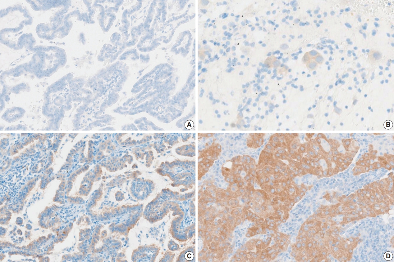
Next-generation sequencing (NGS) is an approved test to select patients for BRAF V600E targeted therapy in Korea. However, the high cost, long turnaround times, and the need for sophisticated equipment and skilled personnel limit the use of NGS in daily practice. Immunohistochemistry (IHC) is a rapid and relatively inexpensive assay available in most laboratories. Therefore, in this study, we evaluate the usefulness of BRAF VE1 IHC in terms of predictive value and interobserver agreement in non–small cell lung cancers (NSCLCs).
Methods
A total of 30 cases with known BRAF mutation status were selected, including 20 cases of lung adenocarcinomas, six cases of colorectal adenocarcinomas, and four cases of papillary thyroid carcinomas. IHC for BRAF V600E was carried out using the VE1 antibody. Fifteen pathologists independently scored both the staining intensity and the percentage of tumor cell staining on whole slide images.
Results
In the lung adenocarcinoma subset, interobserver agreement for the percentage of tumor cell staining and staining intensity was good (percentage of tumor cell staining, intraclass correlation coefficient = 0.869; staining intensity, kappa = 0.849). The interobserver agreement for the interpretation using the cutoff of 40% was almost perfect in the entire study group and the lung adenocarcinoma subset (kappa = 0.815). Sensitivity, specificity, positive predictive value, and negative predictive value of BRAF VE1 IHC were 80.0%, 90.0%, 88.9%, and 81.8%, respectively.
Conclusions
BRAF VE1 IHC could be a screening test for the detection of BRAF V600E mutation in NSCLC. However, further studies are needed to optimize the protocol and to establish and validate interpretation criteria for BRAF VE1 IHC. -
Citations
Citations to this article as recorded by
- An In‐Depth Evaluation of CD15 Expression in Thyroid Carcinoma: A Comprehensive Systematic Review
Amir Ebrahimzadeh Pasha, Amirhossein Ehsani, Sara Ashtari, Mohammad Sadeq Najafi, Bardia Baik, Mohammad Mahdi Mehrabi
Cancer Reports.2026;[Epub] CrossRef - Dedifferentiated Leiomyosarcoma of the Uterine Corpus with Heterologous Component: Clinicopathological Analysis of Five Consecutive Cases from a Single Institution and Comprehensive Literature Review
Suyeon Kim, Hyunsik Bae, Hyun-Soo Kim
Diagnostics.2024; 14(2): 160. CrossRef - Differentiating BRAF V600E- and RAS-like alterations in encapsulated follicular patterned tumors through histologic features: a validation study
Chankyung Kim, Shipra Agarwal, Andrey Bychkov, Jen-Fan Hang, Agnes Stephanie Harahap, Mitsuyoshi Hirokawa, Kennichi Kakudo, Somboon Keelawat, Chih-Yi Liu, Zhiyan Liu, Truong Phan-Xuan Nguyen, Chanchal Rana, Huy Gia Vuong, Yun Zhu, Chan Kwon Jung
Virchows Archiv.2024; 484(4): 645. CrossRef - BRAF V600E Mutation of Non-Small Cell Lung Cancer in Korean Patients
Hyo Yeong Ahn, Chang Hun Lee, Min Ki Lee, Jung Seop Eom, Yeon Joo Jeong, Yeong Dae Kim, Jeong Su Cho, Jonggeun Lee, So Jeong Lee, Dong Hoon Shin, Ahrong Kim
Medicina.2023; 59(6): 1085. CrossRef - Reevaluating diagnostic categories and associated malignancy risks in thyroid core needle biopsy
Chan Kwon Jung
Journal of Pathology and Translational Medicine.2023; 57(4): 208. CrossRef
- An In‐Depth Evaluation of CD15 Expression in Thyroid Carcinoma: A Comprehensive Systematic Review
- Comparison of papanicolaou smear and human papillomavirus (HPV) test as cervical screening tools: can we rely on HPV test alone as a screening method? An 11-year retrospective experience at a single institution
- Myunghee Kang, Seung Yeon Ha, Hyun Yee Cho, Dong Hae Chung, Na Rae Kim, Jungsuk An, Sangho Lee, Jae Yeon Seok, Juhyeon Jeong
- J Pathol Transl Med. 2020;54(1):112-118. Published online January 15, 2020
- DOI: https://doi.org/10.4132/jptm.2019.11.29

- 16,693 View
- 277 Download
- 20 Web of Science
- 23 Crossref
-
Abstract
PDF
- Background
The decrease in incidence of cervical dysplasia and carcinoma has not been as dramatic as expected with the development of improved research tools and test methods. The human papillomavirus (HPV) test alone has been suggested for screening in some countries. The National Cancer Screening Project in Korea has applied Papanicolaou smears (Pap smears) as the screening method for cervical dysplasia and carcinoma. We evaluated the value of Pap smear and HPV testing as diagnostic screening tools in a single institution.
Methods
Patients co-tested with HPV test and Pap smear simultaneously or within one month of each other were included in this study. Patients with only punch biopsy results were excluded because of sampling errors. A total of 999 cases were included, and the collected reports encompassed results of smear cytology, HPV subtypes, and histologic examinations.
Results
Sensitivity and specificity of detecting high-grade squamous intraepithelial lesion (HSIL) and squamous cell carcinoma (SCC) were higher for Pap smears than for HPV tests (sensitivity, 97.14%; specificity, 85.58% for Pap smears; sensitivity, 88.32%; specificity, 54.92% for HPV tests). HPV tests and Pap smears did not differ greatly in detection of low-grade squamous intraepithelial lesion (85.35% for HPV test, 80.31% for Pap smears). When atypical glandular cells were noted on Pap smears, the likelihood for histologic diagnosis of adenocarcinoma following Pap smear was higher than that of high-risk HPV test results (18.8 and 1.53, respectively).
Conclusions
Pap smears were more useful than HPV tests in the diagnosis of HSIL, SCC, and glandular lesions. -
Citations
Citations to this article as recorded by- Development of a Nano-Real-Time Polymerase Chain Reaction (RT-PCR) Kit for Detection and Genotyping of High-Risk Human Papillomavirus (HPV) Strains Using Dedicated TaqMan Probes
Mohammad Panji, Mohammad Hossein Modarresi, Zahra Azizi, Moloud Absalan, Elahe Motevaseli
Cureus.2026;[Epub] CrossRef - Detection of cervical precancerous lesions and cancer by small-scale RT-qPCR analysis of oppositely deregulated mRNAs pairs in cytological smears
Anastasia A. Artyukh, Mikhail K. Ivanov, Sergei E. Titov, Victoria V. Dzyubenko, Sergey E. Krasilnikov, Anastasia O. Shumeikina, Nikita A. Afanasev, Anastasia V. Malek, Sergei A. Glushkov, Eduard F. Agletdinov
Frontiers in Oncology.2025;[Epub] CrossRef - High burden of abnormal cervical smears in South African primary health care: health programmes implications
Olufemi B Omole, Joel M Francis, John M Musonda, Pumla P Sodo, Elizabeth Reji, Nyundu S J Phukuta, Honey L M Mabuza, Joyce S Musonda, Jimmy Akii, John V Ndimande, Olalekan A Ayo-Yusuf
Health Promotion International.2025;[Epub] CrossRef - Bibliometric analysis: a study of the microenvironment in cervical cancer (2000-2024)
Yun-Tao Zhang, Yan-Ni Wei, Chen-Chen Liu, Mai-Qing Yang
Frontiers in Oncology.2025;[Epub] CrossRef - Liquid biopsy biomarkers in cervical cancer: A systematic review and meta-analysis
Isaac Kinyua Njangiru, Bizhar Ahmed Tayeb, Hazhmat Ali, Rafl M. Kamil
The Journal of Liquid Biopsy.2025; 10: 100328. CrossRef - Diagnostic Utility of Human Papilloma Virus Testing in Comparison with Pap Cytology and Histopathology in Unvaccinated Women with Cervical High-Grade Dysplasia and Carcinoma in Botswana
Patricia Setsile Rantshabeng, Nametso Dire, Andrew Khulekani Ndlovu, Ishmael Kasvosve
Venereology.2025; 4(4): 15. CrossRef - Challenges in the diagmosis of cervical pathologies
D. Y. Chernov, O. A. Tikhonovskaya, S. V. Logvinov, I. A. Petrov, Y. S. Yuriev, A. A. Zhdankina, A. V. Gerasimov, I. V. Zingalyuk, G. A. Mikheenko
Bulletin of Siberian Medicine.2024; 22(4): 201. CrossRef - “Barriers and Advantages of Self-Sampling Tests, for HPV Diagnosis: A Qualitative Field Experience Before Implementation in a Rural Community in Ecuador”
Bernardo Vega-Crespo, Vivian Alejandra Neira, Ruth Maldonado - Rengel, Diana López, Dayanara Delgado-López, Gabriela Guerra Astudillo, Veronique Verhoeven
International Journal of Women's Health.2024; Volume 16: 947. CrossRef - Cervical Human Papillomavirus Testing
Carol N. Rizkalla, Eric C. Huang
Surgical Pathology Clinics.2024; 17(3): 431. CrossRef - Segmentation of Overlapping Cells in Cervical Cytology Images: A Survey
E Chen, Hua-Nong Ting, Joon Huang Chuah, Jun Zhao
IEEE Access.2024; 12: 114170. CrossRef - Knowledge and awareness regarding pap test and HPV typing for cervical cancer screening in Edo North, Nigeria
Amina Momodu, Johnsolomon Eghosa Ohenhen, Godfrey Innocent Iyare, Musa Abidemi Muhibi, Godwin Avwioro
Discover Public Health.2024;[Epub] CrossRef - Colposcopy Value in Young Child-bearing Women: Is New Recommendations Necessary?
Fahimeh Sabet, Avishan Aminizad, Fariba Behnamfar, Tajossadat Allameh, Seyedeh Ghazal Shahrokh, Rostami Koushan, Amirmohammad Taravati, Leila Mousavi Seresht
Advanced Biomedical Research.2024;[Epub] CrossRef - Selection of endogenous control and identification of significant microRNA deregulations in cervical cancer
T. Stverakova, I. Baranova, P. Mikyskova, B. Gajdosova, H. Vosmikova, J. Laco, V. Palicka, H. Parova
Frontiers in Oncology.2023;[Epub] CrossRef - Cytology Versus Molecular Diagnosis of HPV for Cervical Cancer Screening. Comparison of the Diagnostic Properties of Four Tests in a Rural Community of Cuenca Ecuador
Bernardo Vega Crespo, Vivian Alejandra Neira, Rocío Murillo, Cristina Ochoa Avilés
ESPOCH Congresses: The Ecuadorian Journal of S.T.E.A.M..2023; 3(1): 139. CrossRef - Attitudes towards prevention of cervical cancer and early diagnosis among female academicians
Nurhan Doğan, Gamze Fışkın
Journal of Obstetrics and Gynaecology Research.2022; 48(6): 1433. CrossRef - Role of Self-Sampling for Cervical Cancer Screening: Diagnostic Test Properties of Three Tests for the Diagnosis of HPV in Rural Communities of Cuenca, Ecuador
Bernardo Vega Crespo, Vivian Alejandra Neira, José Ortíz Segarra, Ruth Maldonado Rengel, Diana López, María Paz Orellana, Andrea Gómez, María José Vicuña, Jorge Mejía, Ina Benoy, Tesifón Parrón Carreño, Veronique Verhoeven
International Journal of Environmental Research and Public Health.2022; 19(8): 4619. CrossRef - Utility of Scoring System for Screening and Early Warning of Cervical Cancer Based on Big Data Analysis
Dan Hou, Binjie Yang, Yangdan Li, Ming Sun
Frontiers in Public Health.2022;[Epub] CrossRef - Evaluation of Urine and Vaginal Self-Sampling versus Clinician-Based Sampling for Cervical Cancer Screening: A Field Comparison of the Acceptability of Three Sampling Tests in a Rural Community of Cuenca, Ecuador
Bernardo Vega Crespo, Vivian Alejandra Neira, José Ortíz S, Ruth Maldonado-Rengel, Diana López, Andrea Gómez, María José Vicuña, Jorge Mejía, Ina Benoy, Tesifón Parrón Carreño, Veronique Verhoeven
Healthcare.2022; 10(9): 1614. CrossRef - Diagnostic distribution and pitfalls of glandular abnormalities in cervical cytology: a 25-year single-center study
Jung-A Sung, Ilias P. Nikas, Haeryoung Kim, Han Suk Ryu, Cheol Lee
Journal of Pathology and Translational Medicine.2022; 56(6): 354. CrossRef - Primary screening of cervical cancer by Pap smear in women of reproductive age group
Ruchi Mishra, Dakshina Bisht, Manisha Gupta
Journal of Family Medicine and Primary Care.2022; 11(9): 5327. CrossRef - Comparison of Learning Transfer Using Simulation Problem-Based Learning and Demonstration: An Application of Papanicolaou Smear Nursing Education
Jeongim Lee, Hae Kyoung Son
International Journal of Environmental Research and Public Health.2021; 18(4): 1765. CrossRef - Investigating host-virus interaction mechanism and phylogenetic analysis of viral proteins involved in the pathogenesis
Ahmad Abu Turab Naqvi, Farah Anjum, Alaa Shafie, Sufian Badar, Abdelbaset Mohamed Elasbali, Dharmendra Kumar Yadav, Md. Imtaiyaz Hassan, Timir Tripathi
PLOS ONE.2021; 16(12): e0261497. CrossRef - Utility of Human Papillomavirus Testing for Cervical Cancer Screening in Korea
Mee-seon Kim, Eun Hee Lee, Moon-il Park, Jae Seok Lee, Kisu Kim, Mee Sook Roh, Hyoun Wook Lee
International Journal of Environmental Research and Public Health.2020; 17(5): 1726. CrossRef
- Development of a Nano-Real-Time Polymerase Chain Reaction (RT-PCR) Kit for Detection and Genotyping of High-Risk Human Papillomavirus (HPV) Strains Using Dedicated TaqMan Probes
- Intraoperative Frozen Cytology of Central Nervous System Neoplasms: An Ancillary Tool for Frozen Diagnosis
- Myunghee Kang, Dong Hae Chung, Na Rae Kim, Hyun Yee Cho, Seung Yeon Ha, Sangho Lee, Jungsuk An, Jae Yeon Seok, Gie-Taek Yie, Chan Jong Yoo, Sang Gu Lee, Eun Young Kim, Woo Kyung Kim, Seong Son, Sun Jin Sym, Dong Bok Shin, Hee Young Hwang, Eung Yeop Kim, Kyu Chan Lee
- J Pathol Transl Med. 2019;53(2):104-111. Published online January 14, 2019
- DOI: https://doi.org/10.4132/jptm.2018.11.10
- 17,037 View
- 697 Download
- 11 Web of Science
- 9 Crossref
-
Abstract
PDF
- Background
Pathologic diagnosis of central nervous system (CNS) neoplasms is made by comparing light microscopic, immunohistochemical, and molecular cytogenetic findings with clinicoradiologic observations. Intraoperative frozen cytology smears can improve the diagnostic accuracy for CNS neoplasms. Here, we evaluate the diagnostic value of cytology in frozen diagnoses of CNS neoplasms.
Methods
Cases were selected from patients undergoing both frozen cytology and frozen sections. Diagnostic accuracy was evaluated.
Results
Four hundred and fifty-four cases were included in this retrospective single-center review study covering a span of 10 years. Five discrepant cases (1.1%) were found after excluding 53 deferred cases (31 cases of tentative diagnosis, 22 cases of inadequate frozen sampling). A total of 346 cases of complete concordance and 50 cases of partial concordance were classified as not discordant cases in the present study. Diagnostic accuracy of intraoperative frozen diagnosis was 87.2%, and the accuracy was 98.8% after excluding deferred cases. Discrepancies between frozen and permanent diagnoses (n = 5, 1.1%) were found in cases of nonrepresentative sampling (n = 2) and misinterpretation (n = 3). High concordance was observed more frequently in meningeal tumors (97/98, 99%), metastatic brain tumors (51/52, 98.1%), pituitary adenomas (86/89, 96.6%), schwannomas (45/47, 95.8%), high-grade astrocytic tumors (47/58, 81%), low grade astrocytic tumors (10/13, 76.9%), non-neoplastic lesions (23/36, 63.9%), in decreasing frequency.
Conclusions
Using intraoperative cytology and frozen sections of CNS tumors is a highly accurate diagnostic ancillary method, providing subtyping of CNS neoplasms, especially in frequently encountered entities. -
Citations
Citations to this article as recorded by- Qualitative and quantitative assessment of ex vivo human brain tumors using quantitative oblique back-illumination microscopy (qOBM)
Srinidhi Bharadwaj, Paloma Casteleiro Costa, Caroline Serafini, Brienna Heinsz, Alice Hsu, Nischita Kaza, Zhe Guang, Zhenmin Li, Jeffrey J. Olson, Kimberly Hoang, Stewart Neill, Francisco E. Robles
Biomedical Optics Express.2026; 17(4): 1936. CrossRef - Intraoperative Integrated Diagnostic System for Malignant Central Nervous System Tumors
Takahiro Hayashi, Kensuke Tateishi, Shinichiro Matsuyama, Hiromichi Iwashita, Yohei Miyake, Akito Oshima, Hirokuni Honma, Jo Sasame, Katsuhiro Takabayashi, Kyoka Sugino, Emi Hirata, Naoko Udaka, Yuko Matsushita, Ikuma Kato, Hiroaki Hayashi, Taishi Nakamur
Clinical Cancer Research.2024; 30(1): 116. CrossRef - A multicenter proof-of-concept study on deep learning-based intraoperative discrimination of primary central nervous system lymphoma
Xinke Zhang, Zihan Zhao, Ruixuan Wang, Haohua Chen, Xueyi Zheng, Lili Liu, Lilong Lan, Peng Li, Shuyang Wu, Qinghua Cao, Rongzhen Luo, Wanming Hu, Shanshan lyu, Zhengyu Zhang, Dan Xie, Yaping Ye, Yu Wang, Muyan Cai
Nature Communications.2024;[Epub] CrossRef - Advancements in Neurosurgical Intraoperative Histology
Ali A. Mohamed, Emma Sargent, Cooper Williams, Zev Karve, Karthik Nair, Brandon Lucke-Wold
Tomography.2024; 10(5): 693. CrossRef - Unveiling the potential application of intraoperative brain smear for brain tumor diagnosis in low-middle-income countries: A comprehensive systematic review
Muhammad Shakir, Ahmed Altaf, Hawra Hussain, Syed Muhammad Aqeel Abidi, Zoey Petitt, Mahnoor Tariq, Ahmed Gilani, S. Ather Enam
Surgical Neurology International.2023; 14: 325. CrossRef - A Comparative Study of Squash Smear Cytology Diagnosis and Radiological Diagnosis with Histopathology in Central Nervous System Lesions
B N Kumarguru, G Santhipriya, S Kranthi Kumar, R Ramesh Kumar, A S Ramaswamy, P Janakiraman
Journal of Cytology.2022; 39(1): 1. CrossRef - Intraoperative squash cytology provides a qualitative intraoperative diagnosis for cases in which frozen section yields a diagnosis of equivocal brain tumour
Hirotaka Fujita, Takuma Tajiri, Tomohisa Machida, Nozomi Nomura, Suguru Toguchi, Hitoshi Itoh, Shinichiro Hiraiwa, Tomoko Sugiyama, Masaaki Imai, Shinri Oda, Masami Shimoda, Naoya Nakamura
Cytopathology.2020; 31(2): 106. CrossRef - Intraoperative frozen cytology of intraosseous cystic meningioma in the sphenoid bone
Na Rae Kim, Gie-Taek Yie
Journal of Pathology and Translational Medicine.2020; 54(6): 508. CrossRef - Use of 5-Aminolevulinic Acid for Confirmation of Lesional Biopsy Sample in Presumed High-Grade Glioma
Victoria L. Watson, Jeffrey W. Cozzens
World Neurosurgery.2019; 132: 21. CrossRef
- Qualitative and quantitative assessment of ex vivo human brain tumors using quantitative oblique back-illumination microscopy (qOBM)
- WITHDRAWN:A Clinicopathologic Study of 220 Cases of Pulmonary Sclerosing Pneumocytoma in Korea: A Nationwide Survey
- Myunghee Kang, Seung Yeon Ha, Joung Ho Han, Mee Sook Roh, Se Jin Jang, Hee Jin Lee, Heae Surng Park, Geon Kook Lee, Kyo Young Lee, Jin-Haeng Chung, Yoo Duk Choi, Chang Hun Lee, Lucia Kim, Myoung Ja Chung, Soon Hee Jung, Gou Young Kim, Wan-Seop Kim
- Received April 4, 2018 Accepted July 9, 2018 Published online July 16, 2018
- DOI: https://doi.org/10.4132/jptm.2018.07.10 [Accepted]
- 6,018 View
- 63 Download
- Prognostic Utility of Histological Growth Patterns of Colorectal Lung Oligometastasis
- Son Jae Yeong, Min Gyoung Pak, Hyoun Wook Lee, Seung Yeon Ha, Mee Sook Roh
- J Pathol Transl Med. 2018;52(2):98-104. Published online February 12, 2018
- DOI: https://doi.org/10.4132/jptm.2017.12.27
- 8,786 View
- 127 Download
- 3 Web of Science
- 2 Crossref
-
Abstract
PDF
- Background
Patients with resectable colorectal lung oligometastasis (CLOM) demonstrate a heterogeneous oncological outcome. However, the parameters for predicting tumor aggressiveness have not yet been fully investigated in CLOM. This study was performed to determine the prognostic value of histological growth patterns in patients who underwent surgery for CLOM.
Methods
The study included 92 patients who were diagnosed with CLOM among the first resection cases. CLOMs grow according to three histological patterns: aerogenous, pushing, and desmoplastic patterns. The growth patterns were evaluated on archival hematoxylin and eosin–stained tissue sections.
Results
The aerogenous pattern was found in 29.4% (n=27) of patients, the pushing pattern in 34.7% (n=32), the desmoplastic pattern in 6.5% (n=6), and a mix of two growth patterns in 29.4% (n=27). The size of the aerogenous pattern was significantly smaller than that of metastases with other patterns (p=.033). Kaplan-Meier analysis demonstrated that patients showing an aerogenous pattern appeared to have a poorer prognosis, which was calculated from the time of diagnosis of the CLOM (p=.044). The 5-year survival rate from the diagnosis of colorectal cancer tended to be lower in patients with an aerogenous pattern than in those who had a non-aerogenous pattern; however, the difference was marginally significant (p=.051). In the multivariate Cox analysis, the aerogenous pattern appeared as an independent predictor of poor overall survival (hazard ratio, 3.122; 95% confidence interval, 1.196 to 8.145; p=.020).
Conclusions
These results suggest that the growth patterns may play a part as a histology-based prognostic parameter for patients with CLOM. -
Citations
Citations to this article as recorded by- Predicting liver metastases growth patterns: Current status and future possibilities
Rui Caetano Oliveira, Henrique Alexandrino, Maria Augusta Cipriano, Filipe Caseiro Alves, José Guilherme Tralhão
Seminars in Cancer Biology.2021; 71: 42. CrossRef - Histological growth patterns and molecular analysis of resected colorectal lung metastases
Emanuela Pilozzi, Damiano Fedele, Andrea Montori, Laura Lorenzon, Valentina Peritore, Giorgia Mannocchi, Nikta Bagheri, Chiara Leone, Antonio Palumbo, Michela Roberto, Giulio Ranazzi, Erino Rendina, Genoveffa Balducci, Mohsen Ibrahim
Pathology - Research and Practice.2021; 222: 153414. CrossRef
- Predicting liver metastases growth patterns: Current status and future possibilities
- Molecular Testing of Lung Cancers
- Hyo Sup Shim, Yoon-La Choi, Lucia Kim, Sunhee Chang, Wan-Seop Kim, Mee Sook Roh, Tae-Jung Kim, Seung Yeon Ha, Jin-Haeng Chung, Se Jin Jang, Geon Kook Lee
- J Pathol Transl Med. 2017;51(3):242-254. Published online April 21, 2017
- DOI: https://doi.org/10.4132/jptm.2017.04.10
- 19,538 View
- 620 Download
- 28 Web of Science
- 29 Crossref
-
Abstract
PDF
- Targeted therapies guided by molecular diagnostics have become a standard treatment of lung cancer. Epidermal growth factor receptor (EGFR) mutations and anaplastic lymphoma kinase (ALK) rearrangements are currently used as the best predictive biomarkers for EGFR tyrosine kinase inhibitors and ALK inhibitors, respectively. Besides EGFR and ALK, the list of druggable genetic alterations has been growing, including ROS1 rearrangements, RET rearrangements, and MET alterations. In this situation, pathologists should carefully manage clinical samples for molecular testing and should do their best to quickly and accurately identify patients who will benefit from precision therapeutics. Here, we grouped molecular biomarkers of lung cancers into three categories—mutations, gene rearrangements, and amplifications—and propose expanded guidelines on molecular testing of lung cancers.
-
Citations
Citations to this article as recorded by- Rapid Diagnostic Assessment in Interventional Pulmonology: Current Practices and Future Directions
Shyam Krishnan, Shreyami Saha, Nirmalya Banerjee, Raja Dhar
Annals of Interventional Pulmonology.2026; 2(1): 18. CrossRef - Association between PD-L1 expression with EGFR, ALK, and ROS1 driver oncogene mutations in non-small cell lung cancer
Dülger Onur, Yaylım İlhan, Öz Büge
Indian Journal of Pathology and Microbiology.2025; 68(1): 36. CrossRef - Evidence-based Approach to Transthoracic Needle Biopsy: Procedural Techniques, Risks, and Controversies
Shravan Sridhar, Hannah G. Ahn, Sayedomid Ebrahimzadeh, Felicia Tang, Brett Elicker
RadioGraphics.2025;[Epub] CrossRef - Enhancing Lung Cancer Care in Portugal: Bridging Gaps for Improved Patient Outcomes
Raquel Ramos, Conceição Souto Moura, Mariana Costa, Nuno Jorge Lamas, Renato Correia, Diogo Garcez, José Miguel Pereira, Carlos Sousa, Nuno Vale
Journal of Personalized Medicine.2024; 14(5): 446. CrossRef - Evolution of therapy for ALK-positive lung carcinomas: Application of third-generation ALK inhibitors in real clinical practice
A. F. Nasretdinov, A. V. Sultanbaev, Sh. I. Musin, K. V. Menshikov, R. T. Ayupov, A. A. Izmailov, G. A. Serebrennikov, V. E. Askarov, D. V. Feoktistov
Meditsinskiy sovet = Medical Council.2024; (10): 74. CrossRef - Cost-effectiveness of next-generation sequencing for advanced EGFR/ALK-negative non-small cell lung cancer
Dong-Won Kang, Sun-Kyeong Park, Sokbom Kang, Eui-Kyung Lee
Lung Cancer.2024; 197: 107970. CrossRef - miR-92a-3p regulates cisplatin-induced cancer cell death
Romain Larrue, Sandy Fellah, Nihad Boukrout, Corentin De Sousa, Julie Lemaire, Carolane Leboeuf, Marine Goujon, Michael Perrais, Bernard Mari, Christelle Cauffiez, Nicolas Pottier, Cynthia Van der Hauwaert
Cell Death & Disease.2023;[Epub] CrossRef - Diagnostic Approach of Lung Cancer: A Literature Review
Jesi Hana, Novia Nurul Faizah
Jurnal Respirasi.2023; 9(2): 141. CrossRef - Molecular Pathology of Lung Cancer
James J. Saller, Theresa A. Boyle
Cold Spring Harbor Perspectives in Medicine.2022; 12(3): a037812. CrossRef - Landscape of EGFR mutations in lung adenocarcinoma: a single institute experience with comparison of PANAMutyper testing and targeted next-generation sequencing
Jeonghyo Lee, Yeon Bi Han, Hyun Jung Kwon, Song Kook Lee, Hyojin Kim, Jin-Haeng Chung
Journal of Pathology and Translational Medicine.2022; 56(5): 249. CrossRef - Molecular biomarker testing for non–small cell lung cancer: consensus statement of the Korean Cardiopulmonary Pathology Study Group
Sunhee Chang, Hyo Sup Shim, Tae Jung Kim, Yoon-La Choi, Wan Seop Kim, Dong Hoon Shin, Lucia Kim, Heae Surng Park, Geon Kook Lee, Chang Hun Lee
Journal of Pathology and Translational Medicine.2021; 55(3): 181. CrossRef - TM4SF4 and LRRK2 Are Potential Therapeutic Targets in Lung and Breast Cancers through Outlier Analysis
Kyungsoo Jung, Joon-Seok Choi, Beom-Mo Koo, Yu Jin Kim, Ji-Young Song, Minjung Sung, Eun Sol Chang, Ka-Won Noh, Sungbin An, Mi-Sook Lee, Kyoung Song, Hannah Lee, Ryong Nam Kim, Young Kee Shin, Doo-Yi Oh, Yoon-La Choi
Cancer Research and Treatment.2021; 53(1): 9. CrossRef - The promises and challenges of early non‐small cell lung cancer detection: patient perceptions, low‐dose CT screening, bronchoscopy and biomarkers
Lukas Kalinke, Ricky Thakrar, Sam M. Janes
Molecular Oncology.2021; 15(10): 2544. CrossRef - Cost-effectiveness analyses of targeted therapy and immunotherapy for advanced non-small cell lung cancer in the United States: a systematic review
Anthony Yu, Eva Huang, Momoka Abe, Kang An, Sun-Kyeong Park, Chanhyun Park
Expert Review of Pharmacoeconomics & Outcomes Research.2021; 21(3): 381. CrossRef - The expanding capability and clinical relevance of molecular diagnostic technology to identify and evaluate EGFR mutations in advanced/metastatic NSCLC
Parth Shah, Jacob Sands, Nicola Normanno
Lung Cancer.2021; 160: 118. CrossRef - Testing for EGFR Mutations and ALK Rearrangements in Advanced Non-Small-Cell Lung Cancer: Considerations for Countries in Emerging Markets
Mercedes L Dalurzo, Alejandro Avilés-Salas, Fernando Augusto Soares, Yingyong Hou, Yuan Li, Anna Stroganova, Büge Öz, Arif Abdillah, Hui Wan, Yoon-La Choi
OncoTargets and Therapy.2021; Volume 14: 4671. CrossRef - Treatment of Patients With Non–Small-Cell Lung Cancer Harboring Rare Oncogenic Mutations
Melina E. Marmarelis, Corey J. Langer
Clinical Lung Cancer.2020; 21(5): 395. CrossRef - Detection of Targetable Genetic Alterations in Korean Lung Cancer Patients: A Comparison Study of Single-Gene Assays and Targeted Next-Generation Sequencing
Eunhyang Park, Hyo Sup Shim
Cancer Research and Treatment.2020; 52(2): 543. CrossRef - High prevalence of ROS1 gene rearrangement detected by FISH in EGFR and ALK negative lung adenocarcinoma
Yuyin Xu, Heng Chang, Lijing Wu, Xin Zhang, Ling Zhang, Jing Zhang, Yuan Li, Lei Shen, Xiaoli Zhu, Xiaoyan Zhou, Qianming Bai
Experimental and Molecular Pathology.2020; 117: 104548. CrossRef - An All-In-One Transcriptome-Based Assay to Identify Therapy-Guiding Genomic Aberrations in Nonsmall Cell Lung Cancer Patients
Jiacong Wei, Anna A. Rybczynska, Pei Meng, Martijn Terpstra, Ali Saber, Jantine Sietzema, Wim Timens, Ed Schuuring, T. Jeroen N. Hiltermann, Harry. J.M. Groen, Anthonie van der Wekken, Anke van den Berg, Klaas Kok
Cancers.2020; 12(10): 2843. CrossRef - Immunotherapy in EGFR-Mutant and ALK-Positive Lung Cancer
Alexander Gavralidis, Justin F. Gainor
The Cancer Journal.2020; 26(6): 517. CrossRef - Role of Immunocytochemistry in the Cytological Diagnosis of Pulmonary Tumors
Jasna Metovic, Luisella Righi, Luisa Delsedime, Marco Volante, Mauro Papotti
Acta Cytologica.2020; 64(1-2): 16. CrossRef - Molecular Diagnostic Assays and Clinicopathologic Implications of MET Exon 14 Skipping Mutation in Non–small-cell Lung Cancer
Eun Kyung Kim, Kyung A. Kim, Chang Young Lee, Sangwoo Kim, Sunhee Chang, Byoung Chul Cho, Hyo Sup Shim
Clinical Lung Cancer.2019; 20(1): e123. CrossRef - PD‐L1 expression in ROS1‐rearranged non‐small cell lung cancer: A study using simultaneous genotypic screening of EGFR, ALK, and ROS1
Jongmin Lee, Chan Kwon Park, Hyoung‐Kyu Yoon, Young Jo Sa, In Sook Woo, Hyo Rim Kim, Sue Youn Kim, Tae‐Jung Kim
Thoracic Cancer.2019; 10(1): 103. CrossRef - Human Leukocyte Antigen Class I and Programmed Death-Ligand 1 Coexpression Is an Independent Poor Prognostic Factor in Adenocarcinoma of the Lung
Yeon Bi Han, Hyun Jung Kwon, Soo Young Park, Eun-Sun Kim, Hyojin Kim, Jin-Haeng Chung
Journal of Pathology and Translational Medicine.2019; 53(2): 86. CrossRef - Molecular testing for advanced non-small cell lung cancer in Malaysia: Consensus statement from the College of Pathologists, Academy of Medicine Malaysia, the Malaysian Thoracic Society, and the Malaysian Oncological Society
Pathmanathan Rajadurai, Phaik Leng Cheah, Soon Hin How, Chong Kin Liam, Muhammad Azrif Ahmad Annuar, Norhayati Omar, Noriah Othman, Nurhayati Mohd Marzuki, Yong Kek Pang, Ros Suzanna Ahmad Bustamam, Lye Mun Tho
Lung Cancer.2019; 136: 65. CrossRef - Somatic mutations and immune checkpoint biomarkers
Brielle A. Parris, Eloise Shaw, Brendan Pang, Richie Soong, Kwun Fong, Ross A. Soo
Respirology.2019; 24(3): 215. CrossRef - Adverse Event Management in Patients with BRAF V600E-Mutant Non-Small Cell Lung Cancer Treated with Dabrafenib plus Trametinib
Anna Chalmers, Laura Cannon, Wallace Akerley
The Oncologist.2019; 24(7): 963. CrossRef - Genetic and clinicopathologic characteristics of lung adenocarcinoma with tumor spread through air spaces
Jae Seok Lee, Eun Kyung Kim, Moonsik Kim, Hyo Sup Shim
Lung Cancer.2018; 123: 121. CrossRef
- Rapid Diagnostic Assessment in Interventional Pulmonology: Current Practices and Future Directions
- Morphologic Analysis of Cytomegalovirus Infected Cells in Bronchial Washing Cytology: Comparison of Liquid-Based Preparation and Conventional Smear
- Jae Yeon Seok, Jungsuk An, Seung Yeon Ha, Dong Hae Chung, Sangho Lee, Hyunchul Kim
- J Pathol Transl Med. 2016;50(2):147-154. Published online February 15, 2016
- DOI: https://doi.org/10.4132/jptm.2015.12.25
- 13,774 View
- 99 Download
- 4 Web of Science
- 3 Crossref
-
Abstract
PDF
- Background
The cytopathic effects of cytomegalovirus (CMV) infection have been well described since the virus was first reported; however, the morphology of CMV infection has not been clearly studied. We examined the difference in detailed cytologic findings in bronchial washing cytology between liquid-based and conventionally prepared smears. Methods: Bronchial washing cytology was processed using either the conventional preparation (CP) or liquid-based preparation (LBP). Sixty-nine cells with typical cytopathic effects of CMV infection were detected on CP slides and 18 cells on LBP slides. Using the image analyzer, area, circumference, major axis, and minor axis of the cytoplasm, nucleus, and intranuclear inclusion were measured in singly scattered CMV-infected cells, and histiocytes were used as a control. Results: The mean cytoplasmic area of CMV-infected cells was 1.47 times larger than that of histiocytes in CP and 2.92 times larger in LBP (p<.05). The mean nuclear area of CMV-infected cells was 2.61 times larger than that of histiocytes in CP and 4.25 times larger in LBP (p<.05). The nucleus to cytoplasm ratio and intranuclear inclusion to cytoplasm ratio of the mean area, circumference, major axis, and minor axis in CP were larger than those in LBP (p<.05). Conclusions: The sizes of cytoplasm, nucleus, and intranuclear inclusion were larger in LBP than in CP, indicating that CMV-infected cells are easily detectable in LBP. However, the nucleus-to-cytoplasm ratio was larger in CP, suggesting that differentiation from malignancy or regenerative atypia requires caution in CP. -
Citations
Citations to this article as recorded by- Pathogens That Rewrite the Rules: Ascoviruses, Elegant Manipulators of Cell Death Pathways and Architects of the Extracellular Viral Paradigm
Sarah R. Rudd, Leticia S. Miranda, Sharon J. Asariah, Chloe S. Rodgers, Jenive T. Estrada, Michael A. Alonzo, Dennis K. Bideshi
Pathogens.2025; 14(11): 1094. CrossRef - Tissue Pathogens and Cancers: A Review of Commonly Seen Manifestations in Histo- and Cytopathology
Tzy Harn Chua, Lavisha S Punjabi, Li Yan Khor
Pathogens.2021; 10(11): 1410. CrossRef - Diagnosis of Infectious Diseases in the Lower Respiratory Tract: A Cytopathologist's Perspective
Rebecca J. Baldassarri, Deepika Kumar, Stephen Baldassarri, Guoping Cai
Archives of Pathology & Laboratory Medicine.2019; 143(6): 683. CrossRef
- Pathogens That Rewrite the Rules: Ascoviruses, Elegant Manipulators of Cell Death Pathways and Architects of the Extracellular Viral Paradigm
- Rare Case of Anal Canal Signet Ring Cell Carcinoma Associated with Perianal and Vulvar Pagetoid Spread
- Na Rae Kim, Hyun Yee Cho, Jeong-Heum Baek, Juhyeon Jeong, Seung Yeon Ha, Jae Yeon Seok, Sung Won Park, Sun Jin Sym, Kyu Chan Lee, Dong Hae Chung
- J Pathol Transl Med. 2016;50(3):231-237. Published online October 8, 2015
- DOI: https://doi.org/10.4132/jptm.2015.08.08
- 15,150 View
- 142 Download
- 5 Web of Science
- 5 Crossref
-
Abstract
PDF
- A 61-year-old woman was referred to surgery for incidentally found colonic polyps during a health examination. Physical examination revealed widespread eczematous skin lesion without pruritus in the perianal and vulvar area. Abdominopelvic computed tomography showed an approximately 4-cm-sized, soft tissue lesion in the right perianal area. Inguinal lymph node dissection and Mils’ operation extended to perianal and perivulvar skin was performed. Histologically, the anal canal lesion was composed of mucin-containing signet ring cells, which were similar to those found in Pagetoid skin lesions. It was diagnosed as an anal canal signet ring cell carcinoma (SRCC) with perianal and vulvar Pagetoid spread and bilateral inguinal lymph node metastasis. Anal canal SRCC is rare, and the current case is the third reported case in the English literature. Seven additional cases were retrieved from the world literature. Here, we describe this rare case of anal canal SRCC with perianal Pagetoid spread and provide a literature review.
-
Citations
Citations to this article as recorded by- Primary Carcinomas of the Episiotomy Scar Site: A Systematic Literature Review
Andrea Palicelli, Federica Torricelli, Gabriele Tonni, Alessandra Bisagni, Eleonora Zanetti, Magda Zanelli, Venus Damaris Medina-Illueca, Beatrice Melli, Maurizio Zizzo, Andrea Morini, Maria Paola Bonasoni, Giacomo Santandrea, Giuseppe Broggi, Rosario Cal
Current Oncology.2025; 32(2): 65. CrossRef - Metastatic Carcinomas at the Episiotomy Site: A Systematic Literature Review
Andrea Palicelli, Gabriele Tonni, Federica Torricelli, Beatrice Melli, Vincenza Ylenia Cusenza, Sandra Martinelli, Eleonora Zanetti, Alessandra Bisagni, Magda Zanelli, Maria Paola Bonasoni, Teresa Rossi, Lucia Mangone, Venus Damaris Medina-Illueca, Mauriz
Cancers.2025; 17(17): 2801. CrossRef - A Case of Prostatic Signet-Ring Cell-like Carcinoma with Pagetoid Spread and Intraductal Carcinoma and Long-Term Survival: PD-L1 and Mismatch Repair System Proteins (MMR) Immunohistochemical Evaluation with Systematic Literature Review
Nektarios Koufopoulos, Argyro-Ioanna Ieronimaki, Andriani Zacharatou, Alina Roxana Gouloumis, Danai Leventakou, Ioannis Boutas, Dionysios T. Dimas, Adamantia Kontogeorgi, Kyparissia Sitara, Lubna Khaldi, Magda Zanelli, Andrea Palicelli
Journal of Personalized Medicine.2023; 13(6): 1016. CrossRef - Anal canal adenocarcinoma with neuroendocrine features accompanying secondary extramammary Paget disease, successfully treated with modified FOLFOX6: a case report
Masamichi Yamaura, Takeshi Yamada, Rei Watanabe, Hitomi Kawai, Suguru Hirose, Hiroki Tajima, Masashi Sato, Yuichi Uchida, Daisuke Suganuma, Yoshiyuki Yamamoto, Toshikazu Moriwaki, Ichinosuke Hyodo
BMC Cancer.2018;[Epub] CrossRef - Solitary left axillary lymph node metastasis after curative resection of carcinoma at the colostomy site: a case report
Ken Imaizumi, Shigenori Homma, Tadashi Yoshida, Tatsushi Shimokuni, Hideyasu Sakihama, Norihiko Takahashi, Hideki Kawamura, Emi Takakuwa, Akinobu Taketomi
Surgical Case Reports.2016;[Epub] CrossRef
- Primary Carcinomas of the Episiotomy Scar Site: A Systematic Literature Review
- Comparison of Cytologic Characteristics between Adenoid Cystic Carcinoma and Adenoid Basal Carcinoma in the Uterine Cervix
- Juhyeon Jeong, Seung Yeon Ha, Hyun Yee Cho, Dong Hae Chung, Jungsuk An
- J Pathol Transl Med. 2015;49(5):396-402. Published online August 17, 2015
- DOI: https://doi.org/10.4132/jptm.2015.07.08
- 12,252 View
- 97 Download
- 1 Web of Science
- 2 Crossref
-
Abstract
PDF
- Background
Adenoid cystic carcinoma (ACC) and adenoid basal carcinoma (ABC) are rare in the uterine cervix. ACC is more aggressive than ABC, thus accurate differential diagnosis is important. In this study, we identified cytologic features useful in distinguishing these two tumors for diagnosis. Methods: Three cases of ACC and five cases of ABC were selected for this study. Cervicovaginal smear slides were reviewed retrospectively, and the area, circumference, major axis, and minor axis of nuclei were measured using an image analyzer. Results: ACC displayed three-dimensional clusters with a small acini pattern. ABC displayed peripheral palisading without an acini pattern. The nuclei of ACC were more irregular and angulated than those of ABC, and the former showed a coarsely granular chromatin pattern. The nucleic area, circumference, major axis, and minor axis were 18.556±8.665 µm2, 23.320±11.412 µm, 5.664±1.537 µm, and 4.127±1.107 µm in ACC and 11.017±4.440 µm2, 15.920±5.664 µm, 4.612±1.025 µm, and 3.088±0.762 µm in the cases of ABC. All measured values showed statistically significant difference (p < .001). Conclusions: Although the nuclei of both of these tumor types were oval shaped, inferred from the ratio of minor axis to major axis (0.728 in ACC and 0.669 in ABC), the area of nuclei was approximately 1.7 times larger in ACC than in ABC. Distinguishing nucleic features, including area, morphology, and chromatin pattern, may be helpful in making a correct diagnosis. -
Citations
Citations to this article as recorded by- Adenoid basal carcinoma of the uterine cervix
Anas Mohamed, Tesfalem Korga, Ahlam Ali, Javier Laurini
International Journal of Gynecological Cancer.2025; : 101873. CrossRef - Adenoid Basal Carcinoma of the Uterine Cervix: A Case Report
Tatsuya Kanuma, Keiko Kigure, Tosio Nishimura, Yuji Ibuki, Shigeru Tsuchida, Harumi Kamiyama, Misa Iijima, Kazuto Nakamura
The KITAKANTO Medical Journal.2016; 66(1): 11. CrossRef
- Adenoid basal carcinoma of the uterine cervix
- A Case of Primary Subpleural Pulmonary Microcystic Myxoma Coincidentally Occurred with Pulmonary Adenocarcinoma
- Jungsuk Ahn, Na Rae Kim, Seung Yeon Ha, Keun-Woo Kim, Kook Yang Park, Yon Mi Sung
- J Pathol Transl Med. 2015;49(3):274-278. Published online May 15, 2015
- DOI: https://doi.org/10.4132/jptm.2015.03.12
- 10,189 View
- 57 Download
- 3 Web of Science
- 3 Crossref
-
PDF
-
Citations
Citations to this article as recorded by- Recurrent PDGFRB Mutations in Pulmonary Microcystic Fibromyxoma
Ming Zhao, Qixing Gong, Xiaoyan Chen, Xiaona Yin, Rong Fang, Jiayun Xu, Xiao Cheng, Yingjing Wang
American Journal of Surgical Pathology.2026; 50(1): 41. CrossRef - Alveolar adenoma with dual hotspot mutations (PDGFRB p.N666K and PIK3CA p.H1047R): a rare case report with diagnostic implications
Mei Kong, Qiqi Gao, Hui Tang
Pathology.2026;[Epub] CrossRef - Endobronchial Myxoma
Arindam Mukherjee, Ritesh Agarwal, Sahajal Dhooria, Pawan Singh, Amanjit Bal, Harkant Singh, Inderpaul S. Sehgal
Journal of Bronchology & Interventional Pulmonology.2018; 25(4): 335. CrossRef
- Recurrent PDGFRB Mutations in Pulmonary Microcystic Fibromyxoma
- Utility of Transmission Electron Microscopy in Small Round Cell Tumors
- Na Rae Kim, Seung Yeon Ha, Hyun Yee Cho
- J Pathol Transl Med. 2015;49(2):93-101. Published online March 12, 2015
- DOI: https://doi.org/10.4132/jptm.2015.01.30
- 19,113 View
- 287 Download
- 5 Web of Science
- 4 Crossref
-
Abstract
PDF
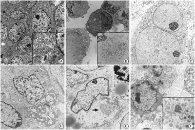
- Small round cell tumors (SRCTs) are a heterogeneous group of neoplasms composed of small, primitive, and undifferentiated cells sharing similar histology under light microscopy. SRCTs include Ewing sarcoma/peripheral neuroectodermal tumor family tumors, neuroblastoma, desmoplastic SRCT, rhabdomyosarcoma, poorly differentiated round cell synovial sarcoma, mesenchymal chondrosarcoma, small cell osteosarcoma, small cell malignant peripheral nerve sheath tumor, and small cell schwannoma. Non-Hodgkin’s malignant lymphoma, myeloid sarcoma, malignant melanoma, and gastrointestinal stromal tumor may also present as SRCT. The current shift towards immunohistochemistry and cytogenetic molecular techniques for SRCT may be inappropriate because of antigenic overlapping or inconclusive molecular results due to the lack of differentiation of primitive cells and unavailable genetic service or limited moleculocytogenetic experience. Although usage has declined, electron microscopy (EM) remains very useful and shows salient features for the diagnosis of SRCTs. Although EM is not always required, it provides reliability and validity in the diagnosis of SRCT. Here, the ultrastructural characteristics of SRCTs are reviewed and we suggest that EM would be utilized as one of the reliable modalities for the diagnosis of undifferentiated and poorly differentiated SRCTs.
-
Citations
Citations to this article as recorded by- Electron Microscopy in the Context of a Children's Research Hospital
Cam Robinson
Microscopy and Microanalysis.2020; 26(S2): 1610. CrossRef - Primary bilateral corneal nerve sheath neoplasm in a dog
Marina L. Leis, M. Elyse Salpeter, Bianca S. Bauer, Dale L. Godson, Bruce H. Grahn
Veterinary Ophthalmology.2017; 20(4): 365. CrossRef - Hirnbasissyndrom infolge eines Tumors bei einer 17 Monate alten Deutsch-Holstein-Färse
Wolf Wippermann, Sandra Schöniger, Kerstin Gerlach, Gerald Fritz Schusser, Gabor Köller, Alexander Starke
Tierärztliche Praxis Ausgabe G: Großtiere / Nutztiere.2016; 44(03): 180. CrossRef - The Continuing Value of Ultrastructural Observation in Central Nervous System Neoplasms in Children
Na Rae Kim, Sung-Hye Park
Journal of Pathology and Translational Medicine.2015; 49(6): 427. CrossRef
- Electron Microscopy in the Context of a Children's Research Hospital
- Alveolar Rhabdomyosarcoma of the Lip in an Adult with Clear Cell Features
- Jae Yeon Seok, Juhyeon Jeong, Young Woo Cheon, Hyun Yee Cho, Seung Yeon Ha, Dong Hae Chung
- J Pathol Transl Med. 2015;49(1):81-84. Published online January 15, 2015
- DOI: https://doi.org/10.4132/jptm.2014.06.03
- 14,771 View
- 108 Download
- Bilateral Stafne Bone Cavity in the Anterior Mandible with Heterotopic Salivary Gland Tissue: A Case Report
- Hyunchul Kim, Jae Yeon Seok, Sangho Lee, Jungsuk An, Na Rae Kim, Dong Hae Chung, Hyun Yee Cho, Seung Yeon Ha
- Korean J Pathol. 2014;48(3):248-249. Published online June 26, 2014
- DOI: https://doi.org/10.4132/KoreanJPathol.2014.48.3.248
- 16,253 View
- 110 Download
- 14 Crossref
-
PDF
-
Citations
Citations to this article as recorded by- Diagnostic approach for the rare anterior variant of mandibular bone depression often misdiagnosed as tumorous lesions
Hak-Sun Kim
Journal of Dental Sciences.2025; 20(1): 502. CrossRef - Static bone cavity occurred in the buccal side of the mandible: A case report and review of literature
Hideki Hojo, Takanori Eguchi, Yumi Ito, Yoshiki Hamada
Journal of Oral and Maxillofacial Surgery, Medicine, and Pathology.2025; 37(4): 698. CrossRef - Bilateral incomplete mandibular canals: an embryological analysis of their possible etiology
Kanitin Rumpansuwon, Thewarid Berkban, Nutmethee Kruepunga, Wattana Weerachatyanukul, Somluk Asuvapongpatana, Arada Chaiyamoon, Benrita Jitaree, R. Shane Tubbs, Joe Iwanaga, Thanyaporn Senarai, Athikhun Suwannakhan
Surgical and Radiologic Anatomy.2025;[Epub] CrossRef - A Rare Case of Anterior Stafne Bone Cavity in the Mandibular Region
Hideki Suito, Yuuri Oku, Koichi Kani, Keiko Aota, Naoki Maeda
Cureus.2025;[Epub] CrossRef - Benign Mandibular Cavity/Stafne's Bone Cyst: A Case Report and Review
Fawzia M. Butt, Shamim M. Butt, Mark L. Chindia
Clinical Case Reports.2025;[Epub] CrossRef - Bilateral Stafne Bone Cavity in the Body of the Mandible: An Unusual Case Report and Literature Review
Mayank Pahadia, Rutvi Vyas
Cureus.2023;[Epub] CrossRef - Effect of Stafne bone defect on the adjacent tooth: A review of the literature
Mahdi Niknami, Azin Parsa, Zahra Khodadadi
Imaging Science in Dentistry.2022; 52(2): 165. CrossRef - Assessment of prevalence and volumetric estimation of possible Stafne bone concavities on cone beam computed tomography images
Alaettin Koç, Cennet Neslihan Eroğlu, Ersen Bilgili
Oral Radiology.2020; 36(3): 254. CrossRef - Stafne’s bone cyst revisited and renamed: the benign mandibular concavity
Johan K.M. Aps, Natasha Koelmeyer, Cina Yaqub
Dentomaxillofacial Radiology.2020; 49(4): 20190475. CrossRef - Cone‐beam computed tomography analysis of lingual mandibular bone depression in the premolar region: A case report
Saeed Asgary, Naghmeh Emadi
Clinical Case Reports.2020; 8(3): 523. CrossRef - Letters to the Editor
Ariyan S Araghi, Richard M Graham
Dental Update.2019; 46(8): 792. CrossRef - Radiographic features of lingual mandibular bone depression using dental cone beam computed tomography
Liu Liu, Byung Cheol Kang, Suk Ja Yoon, Jae Seo Lee, Sel Ae Hwang
Dentomaxillofacial Radiology.2018; 47(6): 20170383. CrossRef - Stafne's bone cavity – unusual presentation in the anterior mandible
Ioan Davies, Holly Boyes, James Wykes, Graham Smith
Dental Update.2018; 45(4): 340. CrossRef - Anterior stafne bone cyst mimicking periapical cyst: a case report
Ji-Young Song
Journal of Dental Rehabilitation and Applied Science.2016; 32(3): 209. CrossRef
- Diagnostic approach for the rare anterior variant of mandibular bone depression often misdiagnosed as tumorous lesions
- Uncommon and Rare Human Papillomavirus Genotypes Relating to Cervical Carcinomas
- Na Rae Kim, Myunghee Kang, Soon Pyo Lee, Hyunchul Kim, Jungsuk An, Dong Hae Chung, Seung Yeon Ha, Hyun Yee Cho
- Korean J Pathol. 2014;48(1):43-49. Published online February 25, 2014
- DOI: https://doi.org/10.4132/KoreanJPathol.2014.48.1.43
- 11,776 View
- 57 Download
- 10 Crossref
-
Abstract
PDF
Background Human papillomavirus (HPV) is an oncogenic virus in cervical cancer and most invasive carcinomas (ICs) are caused by HPV16 and 18. However, the roles and contributions of other uncommon and rare genotypes remain uncertain.
Methods HPV genotypes were retrospectively assessed using an HPV DNA chip that can specify up to 32 HPV genotypes. We arbitrarily regarded genotypes accounting for less than 6% of the total as uncommon and rare genotypes.
Results A total of 3,164 HPV-positive cases were enrolled. In groups 2A, 2B, 3, and unclassified HPV genotypes, 2.4% of cases with uncommon HPV genotypes (68, 26, 34, 53, 66, 69, 70, 73, 40, 42, 43, 44, 54, 55, 61, 62, 6, and 11) showed high grade squamous intraepithelial lesions and ICs. There were no HPV32- and 57-infected cases.
Conclusions We found that the uncommon and rare HPV genotypes may provide incremental etiologic contributions in cervical carcinogenesis, especially HPV68, 70, and 53. Further studies on these uncommon and rare HPV genotypes will be of importance in establishing the significance of genotypes in different regions, especially in planning a strategy for further vaccine development as well as follow-up on the effectiveness of the currently used vaccines.
-
Citations
Citations to this article as recorded by- High-risk human papillomavirus diversity among indigenous women of western Botswana with normal cervical cytology and dysplasia
Patricia S. Rantshabeng, Billy M. Tsima, Andrew K. Ndlovu, Keneilwe Motlhatlhedi, Kirthana Sharma, Carol B. Masole, Natasha O. Moraka, Kesego Motsumi, Angela K. T. Maoto-Mokote, Alemayehu B. Eshetu, Leabaneng Tawe, Tendani Gaolathe, Sikhulile Moyo, Lynnet
BMC Infectious Diseases.2024;[Epub] CrossRef - Human Papillomavirus (HPV69/HPV73) Coinfection associated with Simultaneous Squamous Cell Carcinoma of the Anus and Presumed Lung Metastasis
Stephanie Shea, Marina Muñoz, Stephen C. Ward, Mary B. Beasley, Melissa R Gitman, Michael D Nowak, Jane Houldsworth, Emilia Mia Sordillo, Juan David Ramirez, Alberto E. Paniz Mondolfi
Viruses.2020; 12(3): 349. CrossRef - Human Papillomavirus Selected Properties and Related Cervical Cancer Prevention Issues
Saule Balmagambetova, Andrea Tinelli, Ospan A. Mynbaev, Arip Koyshybaev, Olzhas Urazayev, Nurgul Kereyeva, Elnara Ismagulova
Current Pharmaceutical Design.2020; 26(18): 2073. CrossRef - Periungual Bowen's disease with a narrow longitudinal melanonychia mimicking periungual warts
Taiyo HITAKA, Michiko HASEGAWA, Akira SHIMIZU, Yuko KURIYAMA, Atsushi TAMURA
Skin Cancer.2019; 33(3): 211. CrossRef - Detection of HPV RNA molecules in stratified mucin-producing intraepithelial lesion (SMILE) with concurrent cervical intraepithelial lesion: a case report
Shiho Fukui, Kazunori Nagasaka, Naoko Iimura, Ranka Kanda, Takayuki Ichinose, Takeru Sugihara, Haruko Hiraike, Shunsuke Nakagawa, Yuko Sasajima, Takuya Ayabe
Virology Journal.2019;[Epub] CrossRef - Pitfalls of commercially available HPV tests in HPV68a detection
Hana Jaworek, Katerina Kubanova, Vladimira Koudelakova, Rastislav Slavkovsky, Jiri Drabek, Marian Hajduch, Craig Meyers
PLOS ONE.2019; 14(8): e0220373. CrossRef - Overall accuracy of cervical cytology and clinicopathological significance of LSIL cells in ASC‐H cytology
S. H. Kim, J. M. Lee, H. G. Yun, U. S. Park, S. U. Hwang, J.‐S. Pyo, J. H. Sohn
Cytopathology.2017; 28(1): 16. CrossRef - Human papillomavirus genotyping by Linear Array and Next-Generation Sequencing in cervical samples from Western Mexico
María Guadalupe Flores-Miramontes, Luis Alberto Torres-Reyes, Liliana Alvarado-Ruíz, Salvador Angel Romero-Martínez, Verenice Ramírez-Rodríguez, Luz María Adriana Balderas-Peña, Verónica Vallejo-Ruíz, Patricia Piña-Sánchez, Elva Irene Cortés-Gutiérrez, Lu
Virology Journal.2015;[Epub] CrossRef - Impact of human papillomavirus coinfections on the risk of high-grade squamous intraepithelial lesion and cervical cancer
Adela Carrillo-García, Sergio Ponce-de-León-Rosales, David Cantú-de-León, Verónica Fragoso-Ontiveros, Imelda Martínez-Ramírez, Asunción Orozco-Colín, Alejandro Mohar, Marcela Lizano
Gynecologic Oncology.2014; 134(3): 534. CrossRef - Human papillomavirus 66‐associated subungual squamous cell carcinoma
Jin Hee Kang, Hwa young Ahn, Miri Kim, Shin Taek Oh, Baik Kee Cho, Hyun Jeong Park
The Journal of Dermatology.2014; 41(12): 1119. CrossRef
- High-risk human papillomavirus diversity among indigenous women of western Botswana with normal cervical cytology and dysplasia
- Altered Expression of PTEN and Its Major Regulator MicroRNA-21 in Pulmonary Neuroendocrine Tumors
- Hyoun Wook Lee, Seung Yeon Ha, Mee Sook Roh
- Korean J Pathol. 2014;48(1):17-23. Published online February 25, 2014
- DOI: https://doi.org/10.4132/KoreanJPathol.2014.48.1.17
- 10,003 View
- 46 Download
- 11 Crossref
-
Abstract
PDF
Background Phosphatase and tensin homolog on chromosome ten (PTEN) is one of the most frequently inactivated tumor suppressors in various tumor types. MicroRNA-21 (miR-21) may affect tumor progression by post-transcriptional repression of expression of tumor suppressors, such as PTEN. This study was conducted to evaluate the significance of PTEN expression in pulmonary neuroendocrine (NE) tumors and to analyze the relationship between PTEN and miR-21 expressions.
Methods Expressions of PTEN and miR-21 were investigated by immunohistochemistry and real time reverse transcription-polymerase chain reaction, respectively, in 75 resected pulmonary NE tumors (23 typical carcinoids [TCs], nine atypical carcinoids [ACs], 22 large cell NE carcinomas [LCNECs], and 21 small cell lung carcinomas [SCLCs]).
Results Loss of PTEN expression was observed in four of 23 TCs (17.4%), four of nine ACs (44.4%), 16 of 22 LCNECs (72.7%) and nine of 21 SCLCs (42.9%) (p=.025). The expression level of miR-21 was significantly higher in high-grade NE carcinomas than in carcinoid tumors (p<.001). PTEN expression was inversely correlated with miR-21 expression (p<.001).
Conclusions This study suggests that aberrant expression of PTEN in relation to miR-21 may represent an important step in the development and progression of pulmonary NE tumors.
-
Citations
Citations to this article as recorded by- Mechanisms of skin wound healing regulated by fibroblast-derived exosomes
Ye Qiu, Xingying Zhu, Xiaoqian Yang, Jiaming Wan
Biochemistry and Biophysics Reports.2025; 44: 102371. CrossRef - Role of microRNAs in regulating cell proliferation, metastasis and chemoresistance and their applications as cancer biomarkers in small cell lung cancer
Monu Pandey, Abhirup Mukhopadhyay, Surender K. Sharawat, Sachin Kumar
Biochimica et Biophysica Acta (BBA) - Reviews on Cancer.2021; 1876(1): 188552. CrossRef - Neuroendocrine Tumors Are Enriched in Cowden Syndrome
Alison Greidinger, Susan Miller-Samuel, Veda N. Giri, Michele Sue-Ann Woo, Saranya Akumalla, Charnita Zeigler-Johnson, Scott W. Keith, Daniel P. Silver
JCO Precision Oncology.2020; (4): 551. CrossRef - Prognostic and predictive role of the PI3K–AKT–mTOR pathway in neuroendocrine neoplasms
P. Gajate, T. Alonso-Gordoa, O. Martínez-Sáez, J. Molina-Cerrillo, E. Grande
Clinical and Translational Oncology.2018; 20(5): 561. CrossRef - Genetic and epigenetic drivers of neuroendocrine tumours (NET)
Annunziata Di Domenico, Tabea Wiedmer, Ilaria Marinoni, Aurel Perren
Endocrine-Related Cancer.2017; 24(9): R315. CrossRef - Expression of hsa-let-7b-5p, hsa-let-7f-5p, and hsa-miR-222-3p and their putative targets HMGA2 and CDKN1B in typical and atypical carcinoid tumors of the lung
Pietro Di Fazio, Moritz Maass, Silvia Roth, Christian Meyer, Joana Grups, Peter Rexin, Detlef K Bartsch, Andreas Kirschbaum
Tumor Biology.2017; 39(10): 101042831772841. CrossRef - The regulatory role of aberrant Phosphatase and Tensin Homologue and Liver Kinase B1 on AKT/mTOR/c-Myc axis in pancreatic neuroendocrine tumors
Tsung-Ming Chang, Yan-Shen Shan, Pei-Yi Chu, Shih Sheng Jiang, Wen-Chun Hung, Yu-Lin Chen, Hsiu-Chi Tu, Hui-You Lin, Hui-Jen Tsai, Li-Tzong Chen
Oncotarget.2017; 8(58): 98068. CrossRef - Pulmonary atypical carcinoid in a patient with Cowden syndrome
Hiroaki Tsunezuka, Kaori Abe, Junichi Shimada, Masayoshi Inoue
Interactive CardioVascular and Thoracic Surgery.2016; 22(6): 860. CrossRef - Differential miRNA-Expression as an Adjunctive Diagnostic Tool in Neuroendocrine Tumors of the Lung
Melanie Demes, Christoph Aszyk, Holger Bartsch, Joachim Schirren, Annette Fisseler-Eckhoff
Cancers.2016; 8(4): 38. CrossRef - microRNA‐21 promotes osteogenic differentiation of mesenchymal stem cells by the PI3K/β‐catenin pathway
Yu‐Bin Meng, Xue Li, Zhao‐Yang Li, Jin Zhao, Xu‐Bo Yuan, Yu Ren, Zhen‐Duo Cui, Yun‐De Liu, Xian‐Jin Yang
Journal of Orthopaedic Research.2015; 33(7): 957. CrossRef - Inhibition of NADPH oxidase protects against metastasis of human lung cancer by decreasing microRNA-21
Song Yan, Gang Liu, Changyan Pei, Wenqing Chen, Pei Li, Qiang Wang, Xintian Jin, Jiajia Zhu, Mengzhu Wang, Xiyu Liu
Anti-Cancer Drugs.2015; 26(4): 388. CrossRef
- Mechanisms of skin wound healing regulated by fibroblast-derived exosomes
- Micronodular Thymoma with Lymphoid Stroma in a Multilocular Thymic Cyst: A Case Study
- Na Rae Kim, Jae Ik Lee, Seung Yeon Ha
- Korean J Pathol. 2013;47(4):392-394. Published online August 26, 2013
- DOI: https://doi.org/10.4132/KoreanJPathol.2013.47.4.392
- 10,701 View
- 84 Download
- 15 Crossref
-
Abstract
PDF
Herein, we report a case of micronodular thymoma with lymphoid stroma in a previously healthy 73-year-old male. Thymectomy was performed. The solid and macrocystic masses were encapsulated with focal invasion. The solid portion consisted of nodules of bland-looking spindle or round epithelial cells in lymphoid stroma containing prominent germinal centers. The epithelial cells had moderate amount of cytoplasm and occasional mucin production. The cystic portion was lined with cuboidal epithelium. According to World Health Organization (WHO) classification, the mass was diagnosed as a micronodular thymoma with lymphoid stroma accompanied by a pre-existing multilocular thymic cyst. Micronodular thymoma with lymphoid stroma, a possible variant of type A thymoma, is an extremely rare tumor. This so-called "unusual" variant may imply the schematic weakness of the current WHO classification that cannot cover all morphologic types. Further study is recommended for clarification of this variant and its incorporation into the current classification.
-
Citations
Citations to this article as recorded by- Micronodular Thymic Carcinoma With Lymphoid Hyperplasia: A Case Report and Next Generation Sequencing Analysis With its Benign Counterpart Multinodular Thymoma With Lymphoid Stroma
Min Gyoung Pak, Seung Yeon Ha, Mee Sook Roh
International Journal of Surgical Pathology.2026; 34(1): 207. CrossRef - Cystic micronodular thymoma with lymphoid stroma disguising as an unilocular thymic cyst
I-Ju Chen, I-Ha Lao, Yi-Che Chang Chien, I-Wei Chang
Asian Journal of Surgery.2025; 48(8): 4915. CrossRef -
GTF2I mutation in micronodular thymoma with lymphoid stroma
Andrea Bille, Katherine Fryer, Andrew Wallace, Daisuke Nonaka
Journal of Clinical Pathology.2024; 77(2): 125. CrossRef - Thymic epithelial tumours: histopathological classification and differential diagnosis
Jan von der Thüsen
Histopathology.2024; 84(1): 196. CrossRef - Minimally invasive thoracoscopic resection of a micronodular thymoma with lymphoid stroma via a subxiphoid single-incision approach: A case report
Qiang Wu, Kun Qiao, Xiaoming Zhang, Zizi Zhou
Medicine.2024; 103(36): e39637. CrossRef - Micronodular Thymomas With Prominent Cystic Changes: A Clinicopathological and Immunohistochemical Study of 25 Cases
Diana M. Oramas, Cesar A. Moran
International Journal of Surgical Pathology.2021; 29(4): 352. CrossRef - Two cases of resection of micronodular thymoma with lymphoid stroma
Seiji Omura, Kyohei Masai, Kaoru Kaseda, Keisuke Asakura, Tomoyuki Hishida, Hisao Asamura
The Journal of the Japanese Association for Chest Surgery.2021; 35(6): 705. CrossRef - Two surgical cases of micronodular thymoma with lymphoid stroma
Yusuke Kita, Yoshimasa Tokunaga, Taku Okamoto
The Journal of the Japanese Association for Chest Surgery.2020; 34(2): 166. CrossRef - Thoracoscopic Thymectomy for Large Thymic Cyst: Myasthenia Gravis With Thymoma Concealed by Thymic Cyst
Motoki Yano, Hiroki Numanami, Takashi Akiyama, Rumiko Taguchi, Chihiro Furuta, Akari Iwakoshi, Masayuki Haniuda
Surgical Laparoscopy, Endoscopy & Percutaneous Techniques.2019; 29(3): e34. CrossRef - A resected case of micronodular thymoma with lympoid stroma
Hiromitsu Domen, Yasuhiro Hida, Yasunari Takakuwa, Yuki Iijima, Kazuomi Ichinokawa, Hidehisa Yamada
The Journal of the Japanese Association for Chest Surgery.2019; 33(5): 504. CrossRef - Thymoma and thymic carcinoma associated with multilocular thymic cyst: a clinicopathologic analysis of 18 cases
Xuxia Shen, Yan Jin, Lei Shen, Yihua Sun, Haiquan Chen, Yuan Li
Diagnostic Pathology.2018;[Epub] CrossRef - Micronodular thymoma with lymphoid stroma: Two cases, one in a multilocular thymic cyst, and literature review
Linlin Qu, Yan Xiong, Qian Yao, Bo Zhang, Ting Li
Thoracic Cancer.2017; 8(6): 734. CrossRef - Cystic Micronodular Thymoma. Report of a Case
Mlika M
Journal of Clinical, Medical and Experimental Images.2017; 1(1): 001. CrossRef - A Rare Case of Mixed Type A Thymoma and Micronodular Thymoma with Lymphoid Stroma
Yoon Jin Cha, Joungho Han, Jimin Kim, Kyung Soo Lee, Young Mog Shim
Journal of Pathology and Translational Medicine.2015; 49(1): 75. CrossRef - Micronodular thymic neoplasms: case series and literature review with emphasis on the spectrum of differentiation
Wadad S Mneimneh, Yesim Gökmen-Polar, Kenneth A Kesler, Patrick J Loehrer Sr, Sunil Badve
Modern Pathology.2015; 28(11): 1415. CrossRef
- Micronodular Thymic Carcinoma With Lymphoid Hyperplasia: A Case Report and Next Generation Sequencing Analysis With its Benign Counterpart Multinodular Thymoma With Lymphoid Stroma
- The New 2011 International Association for the Study of Lung Cancer/American Thoracic Society/European Respiratory Society Classification of Lung Adenocarcinoma in Resected Specimens: Clinicopathologic Relevance and Emerging Issues
- Seung Yeon Ha, Mee Sook Roh
- Korean J Pathol. 2013;47(4):316-325. Published online August 26, 2013
- DOI: https://doi.org/10.4132/KoreanJPathol.2013.47.4.316
- 13,719 View
- 80 Download
- 9 Crossref
-
Abstract
PDF
Pathologists play an increasingly important role in personalized medicine for patients with lung cancer as a result of the newly recognized relationship between histologic classification and molecular change. In 2011, the International Association for the Study of Lung Cancer/American Thoracic Society/European Respiratory Society (IASLC/ATS/ERS) proposed a new architectural classification for invasive lung adenocarcinomas to provide uniform terminology and diagnostic criteria. This review highlighted the evolution of the classification of lung adenocarcinomas in resected specimens with special respect to both histologic subtyping and invasion. Histologic subtyping of lung adenocarcinoma has been updated based on five major predominant patterns. New concepts of adenocarcinoma
in situ and minimally invasive adenocarcinomas have been introduced to define the condition of patients who are expected to have excellent survival. Although the new IASLC/ATS/ERS classification has promising clinical relevance, significant clarification remains necessary for the definitions of subtyping and invasion. More precise definitions and subsequent better education on the interpretation of terminology will be helpful for future studies.-
Citations
Citations to this article as recorded by- Radiomics for Classifying Histological Subtypes of Lung Cancer Based on Multiphasic Contrast-Enhanced Computed Tomography
Linning E, Lin Lu, Li Li, Hao Yang, Lawrence H. Schwartz, Binsheng Zhao
Journal of Computer Assisted Tomography.2019; 43(2): 300. CrossRef - Tumor heterogeneity assessed by texture analysis on contrast-enhanced CT in lung adenocarcinoma: association with pathologic grade
Ying Liu, Shichang Liu, Fangyuan Qu, Qian Li, Runfen Cheng, Zhaoxiang Ye
Oncotarget.2017; 8(32): 53664. CrossRef - Clinicopathological and prognostic significance of metastasis-associated protein 1 expression and its correlation with angiogenesis in lung invasive adenocarcinomas, based on the 2011 IASLC/ATS/ERS classification
SHUHAI LI, HUI TIAN, WEIMING YUE, LIN LI, CUN GAO, LIBO SI, WENSI HU, LEI QI, MING LU, CHUANLE CHENG, JINGJING CUI, GUANQING CHEN
Oncology Letters.2016; 11(1): 224. CrossRef - Myoferlin expression in non-small cell lung cancer: Prognostic role and correlation with VEGFR-2 expression
DAE HYUN SONG, GYUNG HYUCK KO, JEONG HEE LEE, JONG SIL LEE, GYEONG-WON LEE, HYEON CHEOL KIM, JUNG WOOK YANG, ROK WON HEO, GU SEOB ROH, SUN-YOUNG HAN, DONG CHUL KIM
Oncology Letters.2016; 11(2): 998. CrossRef - ROS1 gene rearrangement and copy number gain in non-small cell lung cancer
Yan Jin, Ping-Li Sun, Hyojin Kim, Eunhyang Park, Hyo Sup Shim, Sanghoon Jheon, Kwhanmien Kim, Choon-Taek Lee, Jin-Haeng Chung
Virchows Archiv.2015; 466(1): 45. CrossRef - The Demise of the Term Bronchioloalveolar Carcinoma
Yasmeen M. Butt, Timothy Craig Allen
Archives of Pathology & Laboratory Medicine.2015; 139(8): 981. CrossRef - Expression of EGFR and Molecules Downstream to PI3K/Akt, Raf-1-MEK-1-MAP (Erk1/2), and JAK (STAT3) Pathways in Invasive Lung Adenocarcinomas Resected at a Single Institution
Alba Fabiola Torres, Cleto Nogueira, Juliana Magalhaes, Igor Santos Costa, Alessa Aragao, Antero Gomes Neto, Filadelfia Martins, Fabio Tavora
Analytical Cellular Pathology.2014; 2014: 1. CrossRef - Usual Interstitial Pneumonia with Lung Cancer: Clinicopathological Analysis of 43 Cases
Dae Hyun Song, In Ho Choi, Sang Yun Ha, Kang Min Han, Jae Jun Lee, Min Eui Hong, Kyeongman Jeon, Man Pyo Chung, Jhingook Kim, Joungho Han
Korean Journal of Pathology.2014; 48(1): 10. CrossRef - Cytoplasmic YAP Expression is Associated with Prolonged Survival in Patients with Lung Adenocarcinomas and Epidermal Growth Factor Receptor Tyrosine Kinase Inhibitor Treatment
Ping-Li Sun, Ji Eun Kim, Seol Bong Yoo, Hyojin Kim, Yan Jin, Sanghoon Jheon, Kwhanmien Kim, Choon Taek Lee, Jin-Haeng Chung
Annals of Surgical Oncology.2014; 21(S4): 610. CrossRef
- Radiomics for Classifying Histological Subtypes of Lung Cancer Based on Multiphasic Contrast-Enhanced Computed Tomography
- Cytologic Features of
ALK -Positive Pulmonary Adenocarcinoma - Seung Yeon Ha, Jungsuk Ahn, Mee Sook Roh, Joungho Han, Jae Jun Lee, Boin Lee, Jun Yim
- Korean J Pathol. 2013;47(3):252-257. Published online June 25, 2013
- DOI: https://doi.org/10.4132/KoreanJPathol.2013.47.3.252
- 11,010 View
- 35 Download
- 10 Crossref
-
Abstract
PDF
Background The aim of this study was to determine the cytologic features of anaplastic lymphoma kinase (
ALK ) expressing pulmonary adenocarcinoma.Methods We analyzed the cytopathological findings of 15 cases of endobronchial ultrasound guided aspiration and a case of bronchial washing. These cases were selected based on the histomorphology of
ALK -rearranged lung adenocarcinoma.Results Cytology showed mucinous (81.3%) and hemorrhagic (50%) backgrounds. The cells were arranged in tubulopapillary or tubulocribriform patterns (93.8%), and clusters (56.3%) admixed with signet ring cell features (87.5%). The tumor cells were monotonous and uniform with vesicular nuclei and a small nucleolus.
Conclusions The characteristic findings were sheets showing a tubulopapillary or tubulocribriform appearance, with vesicular nuclei and a bland chromatin pattern (p<0.001). Scattered signet ring cells were helpful in suggesting
ALK -positive adenocarcinoma (p<0.001).-
Citations
Citations to this article as recorded by- Cytomorphological and histomorphological features of lung adenocarcinoma with epidermal growth factor receptor mutation and anaplastic lymphoma kinase gene rearrangement
Nikola Gardić, Aleksandra Lovrenski, Vanesa Sekeruš, Svetlana Lečić, Milorad Bijelović, Tanja Lakić, Aleksandra Ilić, Bojan Zarić, Sofija Glumac
Oncology Letters.2024;[Epub] CrossRef - Machine learning‐based gene alteration prediction model for primary lung cancer using cytologic images
Shuhei Ishii, Manabu Takamatsu, Hironori Ninomiya, Kentaro Inamura, Takeshi Horai, Akira Iyoda, Naoko Honma, Rira Hoshi, Yuko Sugiyama, Noriko Yanagitani, Mingyon Mun, Hitoshi Abe, Tetuo Mikami, Kengo Takeuchi
Cancer Cytopathology.2022; 130(10): 812. CrossRef - Fine‐needle aspiration cytology of non‐small cell lung carcinoma: A paradigm shift
Pranab Dey, Ratan Kumar Ghosh
Diagnostic Cytopathology.2019; 47(4): 351. CrossRef - Qualitative and quantitative cytomorphological features of primary anaplastic lymphoma kinase‐positive lung cancer
Ryuko Tsukamoto, Hiroyuki Ohsaki, Sho Hosokawa, Yasunori Tokuhara, Shingo Kamoshida, Toshiko Sakuma, Tomoo Itoh, Chiho Ohbayashi
Cytopathology.2019; 30(3): 295. CrossRef - Primary signet-ring adenocarcinoma of the lung: A rare lung tumor
Varun Rajpal, Rahul Kumar Sharma, Charul Dabral, Deepak Talwar
South Asian Journal of Cancer.2019; 08(04): 257. CrossRef - Cytological features in eight patients with ALK‐rearranged lung cancer
Naoto Kuroda, Masahiko Ohara, Yukari Wada, Kaori Yasuoka, Keiko Mizuno, Kenji Yorita, Chiho Obayashi, Kengo Takeuchi
Diagnostic Cytopathology.2018; 46(6): 516. CrossRef - Cytological markers for predicting ALK‐positive pulmonary adenocarcinoma
K. Miyata, S. Morita, H. Dejima, N. Seki, N. Matsutani, M. Mieno, F. Kondo, Y. Soejima, F. Tanaka, M. Sawabe
Diagnostic Cytopathology.2017; 45(11): 963. CrossRef - ALK-rearranged adenocarcinoma with extensive mucin production can mimic mucinous adenocarcinoma: clinicopathological analysis and comprehensive histological comparison with KRAS-mutated mucinous adenocarcinoma
Yoon Jin Cha, Joungho Han, Soo Hyun Hwang, Tae Bum Lee, Hojoong Kim, Jea Ill Zo
Pathology.2016; 48(4): 325. CrossRef - Cytomorphological identification of advanced pulmonary adenocarcinoma harboring KRAS mutation in lymph node fine‐needle aspiration specimens: Comparative investigation of adenocarcinoma with KRAS and EGFR mutations
Dae Hyun Song, Boram Lee, Yooju Shin, In Ho Choi, Sang Yun Ha, Jae Jun Lee, Min Eui Hong, Yoon‐La Choi, Joungho Han, Sang‐Won Um
Diagnostic Cytopathology.2015; 43(7): 539. CrossRef - Comprehensive analysis of RET and ROS1 rearrangement in lung adenocarcinoma
Seung Eun Lee, Boram Lee, Mineui Hong, Ji-Young Song, Kyungsoo Jung, Maruja E Lira, Mao Mao, Joungho Han, Jhingook Kim, Yoon-La Choi
Modern Pathology.2015; 28(4): 468. CrossRef
- Cytomorphological and histomorphological features of lung adenocarcinoma with epidermal growth factor receptor mutation and anaplastic lymphoma kinase gene rearrangement
- No Detection of Simian Virus 40 in Malignant Mesothelioma in Korea
- Minseob Eom, Jamshid Abdul-Ghafar, Sun-Mi Park, Joung Ho Han, Soon Won Hong, Kun Young Kwon, Eun Suk Ko, Lucia Kim, Wan Seop Kim, Seung Yeon Ha, Kyo Young Lee, Chang Hun Lee, Hye Kyoung Yoon, Yoo Duk Choi, Myoung Ja Chung, Soon-Hee Jung
- Korean J Pathol. 2013;47(2):124-129. Published online April 24, 2013
- DOI: https://doi.org/10.4132/KoreanJPathol.2013.47.2.124
- 11,429 View
- 56 Download
- 6 Crossref
-
Abstract
PDF
Background Simian virus 40 (SV40), a polyomavirus, was discovered as a contaminant of a human polio vaccine in the 1960s. It is known that malignant mesothelioma (MM) is associated with SV40, and that the virus works as a cofactor to the carcinogenetic effects of asbestos. However, the reports about the correlation between SV40 and MM have not been consistent. The purpose of this study is to identify SV40 in MM tissue in Korea through detection of SV40 protein and DNA.
Methods We analyzed 62 cases of available paraffin-blocks enrolled through the Korean Malignant Mesothelioma Surveillance System and performed immunohistochemistry for SV40 protein and real-time polymerase chain reaction (PCR) for SV40 DNA.
Results Of 62 total cases, 40 had disease involving the pleura (64.5%), and 29 (46.8%) were found to be of the epithelioid subtype. Immunostaining demonstrated that all examined tissues were negative for SV40 protein. Sufficient DNA was extracted for real-time PCR analysis from 36 cases. Quantitative PCR of these samples showed no increase in SV40 transcript compared to the negative controls.
Conclusions SV40 is not associated with the development of MM in Korea.
-
Citations
Citations to this article as recorded by- Association Study of Pleural Mesothelioma and Oncogenic Simian Virus 40 in the Crocidolite-Contaminated Area of Dayao County, Yunnan Province, Southwest China
Ru-ai Liu, Bo-yong Wang, Xin Chen, Yuan-qian Pu, Jia-ji Zi, Wen Mei, Ye-pin Zhang, Lu Qiu, Wei Xiong
Genetic Testing and Molecular Biomarkers.2024; 28(5): 189. CrossRef - Binding of SV40’s Viral Capsid Protein VP1 to Its Glycosphingolipid Receptor GM1 Induces Negative Membrane Curvature: A Molecular Dynamics Study
Raisa Kociurzynski, Sophie D. Beck, Jean-Baptiste Bouhon, Winfried Römer, Volker Knecht
Langmuir.2019; 35(9): 3534. CrossRef - Estimated future incidence of malignant mesothelioma in South Korea: Projection from 2014 to 2033
Kyeong Min Kwak, Domyung Paek, Seung-sik Hwang, Young-Su Ju, Mark Allen Pershouse
PLOS ONE.2017; 12(8): e0183404. CrossRef - The function, mechanisms, and role of the genes PTEN and TP53 and the effects of asbestos in the development of malignant mesothelioma: a review focused on the genes' molecular mechanisms
Leonardo Vinícius Monteiro de Assis, Mauro César Isoldi
Tumor Biology.2014; 35(2): 889. CrossRef - The role of key genes and pathways involved in the tumorigenesis of Malignant Mesothelioma
Leonardo Vinícius Monteiro de Assis, Jamille Locatelli, Mauro César Isoldi
Biochimica et Biophysica Acta (BBA) - Reviews on Cancer.2014; 1845(2): 232. CrossRef - Pleural Mesothelioma: An Institutional Experience of 66 Cases
Soomin Ahn, In Ho Choi, Joungho Han, Jhingook Kim, Myung-Ju Ahn
Korean Journal of Pathology.2014; 48(2): 91. CrossRef
- Association Study of Pleural Mesothelioma and Oncogenic Simian Virus 40 in the Crocidolite-Contaminated Area of Dayao County, Yunnan Province, Southwest China
- Fine Needle Aspiration Cytology of Postoperative Spindle Cell Nodule in Neck after Thyroidectomy: A Case Report
- Myunghee Kang, Seung Yeon Ha, Hyun Yee Cho, Jungsuk An, Dong Hae Chung, Yoo Seung Chung
- Korean J Pathol. 2013;47(1):89-91. Published online February 25, 2013
- DOI: https://doi.org/10.4132/KoreanJPathol.2013.47.1.89
- 9,852 View
- 45 Download
- 3 Crossref
-
PDF
-
Citations
Citations to this article as recorded by- Post-surgical thyroid bed myofibroma simulating a recurrent papillary thyroid carcinoma: A case report and review of the literature
Jun Hyeon Park, Kyung Sik Yi, Chi-Hoon Choi, Yook Kim, Jisun Lee, Yeongtae Park, Ok-Jun Lee
Medicine.2024; 103(2): e36945. CrossRef - USP6‐associated neoplasm as a tentative subset of postoperative spindle cell nodule
Lili Sun, Zehua Zhao, Yanmei Zhu
Histopathology.2023; 82(4): 587. CrossRef - Diagnostic Performance of Core Needle Biopsy for Characterizing Thyroidectomy Bed Lesions
So Yeong Jeong, Jung Hwan Baek, Sae Rom Chung, Young Jun Choi, Dong Eun Song, Ki-Wook Chung, Won Woong Kim, Jeong Hyun Lee
Korean Journal of Radiology.2022; 23(10): 1019. CrossRef
- Post-surgical thyroid bed myofibroma simulating a recurrent papillary thyroid carcinoma: A case report and review of the literature
- Morphologic Analysis of Pulmonary Neuroendocrine Tumors
- Seung Seok Lee, Myunghee Kang, Seung Yeon Ha, Jungsuk An, Mee Sook Roh, Chang Won Ha, Jungho Han
- Korean J Pathol. 2013;47(1):16-20. Published online February 25, 2013
- DOI: https://doi.org/10.4132/KoreanJPathol.2013.47.1.16
- 8,842 View
- 52 Download
- 2 Crossref
-
Abstract
PDF
Background Few studies on how to diagnose pulmonary neuroendocrine tumors through morphometric analysis have been reported. In this study, we measured and analyzed the characteristic parameters of pulmonary neuroendocrine tumors using an image analyzer to aid in diagnosis.
Methods Sixteen cases of typical carcinoid tumor, 5 cases of atypical carcinoid tumor, 15 cases of small cell carcinoma, and 51 cases of large cell neuroendocrine carcinoma were analyzed. Using an image analyzer, we measured the nuclear area, perimeter, and the major and minor axes.
Results The mean nuclear area was 0.318±0.101 µm2 in typical carcinoid tumors, 0.326±0.119 µm2 in atypical carcinoid tumors, 0.314±0.107 µm2 in small cell carcinomas, and 0.446±0.145 µm2 in large cell neuroendocrine carcinomas. The mean nuclear circumference was 2.268±0.600 µm in typical carcinoid tumors, 2.408±0.680 µm in atypical carcinoid tumors, 2.158±0.438 µm in small cell carcinomas, and 3.247±1.276 µm in large cell neuroendocrine carcinomas. All parameters were useful in distinguishing large cell neuroendocrine carcinoma from other tumors (p=0.001) and in particular, nuclear circumference was the most effective (p=0.001).
Conclusions Pulmonary neuroendocrine tumors showed nuclear morphology differences by subtype. Therefore, evaluation of quantitative nuclear parameters improves the accuracy and reliability of diagnosis.
-
Citations
Citations to this article as recorded by- Clinical Significance of Persistent Tumor in Bone Marrow during Treatment of High-risk Neuroblastoma
Young Bae Choi, Go Eun Bae, Na Hee Lee, Jung-Sun Kim, Soo Hyun Lee, Keon Hee Yoo, Ki Woong Sung, Hong Hoe Koo
Journal of Korean Medical Science.2015; 30(8): 1062. CrossRef - Morphologic Alteration of Metastatic Neuroblastic Tumor in Bone Marrow after Chemotherapy
Go Eun Bae, Yeon-Lim Suh, Ki Woong Sung, Jung-Sun Kim
Korean Journal of Pathology.2013; 47(5): 433. CrossRef
- Clinical Significance of Persistent Tumor in Bone Marrow during Treatment of High-risk Neuroblastoma
- Interobserver Variability in Diagnosing High-Grade Neuroendocrine Carcinoma of the Lung and Comparing It with the Morphometric Analysis
- Seung Yeon Ha, Joungho Han, Wan-Seop Kim, Byung Seong Suh, Mee Sook Roh
- Korean J Pathol. 2012;46(1):42-47. Published online February 23, 2012
- DOI: https://doi.org/10.4132/KoreanJPathol.2012.46.1.42
- 10,611 View
- 51 Download
- 22 Crossref
-
Abstract
PDF
Background Distinguishing small cell lung carcinoma (SCLC) and large cell neuroendocrine carcinoma (LCNEC) of the lung is difficult with little information about interobserver variability.
Methods One hundred twenty-nine cases of resected SCLC and LCNEC were independently evaluated by four pathologists and classified according to the 2004 World Health Organization criteria. Agreement was regarded as "unanimous" if all four pathologists agreed on the classification. The kappa statistic was calculated to measure the degree of agreement between pathologists. We also measured cell size using image analysis, and receiver-operating-characteristic curve analysis was performed to evaluate cell size in predicting the diagnosis of high-grade neuroendocrine (NE) carcinomas in 66 cases.
Results Unanimous agreement was achieved in 55.0% of 129 cases. The kappa values ranged from 0.35 to 0.81. Morphometric analysis reaffirmed that there was a continuous spectrum of cell size from SCLC to LCNEC and showed that tumors with cells falling in the middle size range were difficult to categorize and lacked unanimous agreement.
Conclusions Our results provide an objective explanation for considerable interobserver variability in the diagnosis of high-grade pulmonary NE carcinomas. Further studies would need to define more stringent and objective definitions of cytologic and architectural characteristics to reliably distinguish between SCLC and LCNEC.
-
Citations
Citations to this article as recorded by- Case report: A patient with EGFR L861Q positive adenosquamous lung carcinoma transforming into large cell neuroendocrine cancer after treatment with Almonertinib
Kele Cheng, Yong Zhu, Ran Sang, Zhongsheng Kuang, Yang Cao
Frontiers in Oncology.2025;[Epub] CrossRef - Deep Learning–Based Retinoblastoma Protein Subtyping of Pulmonary Large-Cell Neuroendocrine Carcinoma on Small Hematoxylin and Eosin–Stained Specimens
Teodora E. Trandafir, Frank W.J. Heijboer, Farhan Akram, Jules L. Derks, Yunlei Li, Lisa M. Hillen, Ernst-Jan M. Speel, Zsolt Megyesfalvi, Balazs Dome, Andrew P. Stubbs, Anne-Marie C. Dingemans, Jan H. von der Thüsen
Laboratory Investigation.2025; 105(9): 104192. CrossRef - Treatment outcomes in patients with stage IV large cell neuroendocrine carcinoma: a nationwide registry study
Frank W.J. Heijboer, Jules L. Derks, Francien H. van Nederveen, Lisa M. Hillen, Michael A. den Bakker, Teodora Radonic, Ronald A.M. Damhuis, Ernst-Jan M. Speel, Jan H. von der Thüsen, Anne-Marie C. Dingemans
Lung Cancer.2025; 210: 108830. CrossRef - Updates on lung neuroendocrine neoplasm classification
Giulia Vocino Trucco, Luisella Righi, Marco Volante, Mauro Papotti
Histopathology.2024; 84(1): 67. CrossRef - Recent advancement of HDAC inhibitors against breast cancer
Syed Abdulla Mehmood, Kantrol Kumar Sahu, Sounok Sengupta, Sangh Partap, Rajshekhar Karpoormath, Brajesh Kumar, Deepak Kumar
Medical Oncology.2023;[Epub] CrossRef - Genomic Feature of a Rare Case of Mix Small-Cell and Large-Cell Neuroendocrine Lung Carcinoma: A Case Report
Youcai Zhu, Feng Zhang, Dong Yu, Fang Wang, Manxiang Yin, Liangye Chen, Chun Xiao, Yueyan Huang, Feng Ding
Frontiers in Oncology.2022;[Epub] CrossRef - Small-Cell Carcinoma of the Lung: What We Learned about It?
Luisella Righi, Marco Volante, Mauro Papotti
Acta Cytologica.2022; 66(4): 257. CrossRef - Hierarchical identification of a transcriptional panel for the histological diagnosis of lung neuroendocrine tumors
Juxuan Zhang, Jiaxing Deng, Xiao Feng, Yilong Tan, Xin Li, Yixin Liu, Mengyue Li, Haitao Qi, Lefan Tang, Qingwei Meng, Haidan Yan, Lishuang Qi
Frontiers in Genetics.2022;[Epub] CrossRef - Immunohistochemical Staining With Neuroendocrine Markers is Essential in the Diagnosis of Neuroendocrine Neoplasms of the Esophagogastric Junction
Dea N.M. Jepsen, Anne-Marie K. Fiehn, Rajendra S. Garbyal, Ulla Engel, Jakob Holm, Birgitte Federspiel
Applied Immunohistochemistry & Molecular Morphology.2021; 29(6): 454. CrossRef - Improving differential diagnosis of pulmonary large cell neuroendocrine carcinoma and small cell lung cancer via a transcriptomic, biological pathway-based machine learning model
Junhong Guo, Likun Hou, Wei Zhang, Zhengwei Dong, Lei Zhang, Chunyan Wu
Translational Oncology.2021; 14(12): 101222. CrossRef - Are Neuroendocrine Negative Small Cell Lung Cancer and Large Cell Neuroendocrine Carcinoma with WT RB1 two Faces of the Same Entity?
Dmitriy Sonkin, Anish Thomas, Beverly A Teicher
Lung Cancer Management.2019;[Epub] CrossRef - Ki-67 labeling index of neuroendocrine tumors of the lung has a high level of correspondence between biopsy samples and surgical specimens when strict counting guidelines are applied
Alessandra Fabbri, Mara Cossa, Angelica Sonzogni, Mauro Papotti, Luisella Righi, Gaia Gatti, Patrick Maisonneuve, Barbara Valeri, Ugo Pastorino, Giuseppe Pelosi
Virchows Archiv.2017; 470(2): 153. CrossRef - The Use of Immunohistochemistry Improves the Diagnosis of Small Cell Lung Cancer and Its Differential Diagnosis. An International Reproducibility Study in a Demanding Set of Cases
Erik Thunnissen, Alain C. Borczuk, Douglas B. Flieder, Birgit Witte, Mary Beth Beasley, Jin-Haeng Chung, Sanja Dacic, Sylvie Lantuejoul, Prudence A. Russell, Michael den Bakker, Johan Botling, Elisabeth Brambilla, Erienne de Cuba, Kim R. Geisinger, Kenzo
Journal of Thoracic Oncology.2017; 12(2): 334. CrossRef - Reply to Letter “The Use of Immunohistochemistry Improves the Diagnosis of Small Cell Lung Cancer and Its Differential Diagnosis. An International Reproducibility Study in a Demanding Set of Cases.”
Erik Thunnissen, Birgit I. Witte, Masayuki Noguchi, Yasushi Yatabe
Journal of Thoracic Oncology.2017; 12(6): e70. CrossRef - What clinicians are asking pathologists when dealing with lung neuroendocrine neoplasms?
Giuseppe Pelosi, Alessandra Fabbri, Mara Cossa, Angelica Sonzogni, Barbara Valeri, Luisella Righi, Mauro Papotti
Seminars in Diagnostic Pathology.2015; 32(6): 469. CrossRef - Unraveling Tumor Grading and Genomic Landscape in Lung Neuroendocrine Tumors
Giuseppe Pelosi, Mauro Papotti, Guido Rindi, Aldo Scarpa
Endocrine Pathology.2014; 25(2): 151. CrossRef - Grading the neuroendocrine tumors of the lung: an evidence-based proposal
G Rindi, C Klersy, F Inzani, G Fellegara, L Ampollini, A Ardizzoni, N Campanini, P Carbognani, T M De Pas, D Galetta, P L Granone, L Righi, M Rusca, L Spaggiari, M Tiseo, G Viale, M Volante, M Papotti, G Pelosi
Endocrine-Related Cancer.2014; 21(1): 1. CrossRef - Controversial issues and new discoveries in lung neuroendocrine tumors
Giuseppe Pelosi, Kenzo Hiroshima, Mari Mino-Kenudson
Diagnostic Histopathology.2014; 20(10): 392. CrossRef - BAI3, CDX2 and VIL1: a panel of three antibodies to distinguish small cell from large cell neuroendocrine lung carcinomas
Muhammad F Bari, Helen Brown, Andrew G Nicholson, Keith M Kerr, John R Gosney, William A Wallace, Irshad Soomro, Salli Muller, Danielle Peat, Jonathan D Moore, Lesley A Ward, Maxim B Freidin, Eric Lim, Manu Vatish, David R J Snead
Histopathology.2014; 64(4): 547. CrossRef - Neuroendocrine tumours—challenges in the diagnosis and classification of pulmonary neuroendocrine tumours
M A den Bakker, F B J M Thunnissen
Journal of Clinical Pathology.2013; 66(10): 862. CrossRef - Morphologic Analysis of Pulmonary Neuroendocrine Tumors
Seung Seok Lee, Myunghee Kang, Seung Yeon Ha, Jungsuk An, Mee Sook Roh, Chang Won Ha, Jungho Han
Korean Journal of Pathology.2013; 47(1): 16. CrossRef - Altered expression of microRNA miR‐21, miR‐155, and let‐7a and their roles in pulmonary neuroendocrine tumors
Hyoun Wook Lee, Eun Hee Lee, Seung Yeon Ha, Chang Hun Lee, Hee Kyung Chang, Sunhee Chang, Kun Young Kwon, Il Seon Hwang, Mee Sook Roh, Jeong Wook Seo
Pathology International.2012; 62(9): 583. CrossRef
- Case report: A patient with EGFR L861Q positive adenosquamous lung carcinoma transforming into large cell neuroendocrine cancer after treatment with Almonertinib
- Solitary Fibrous Tumor of the Conjunctiva with Heretofore Undescribed Pathologic Findings.
- Na Rae Kim, Jae Y Ro, Kyung Hwan Shin, Hae Jung Paik, Jung Suk An, Seung Yeon Ha
- Korean J Pathol. 2011;45(3):315-318.
- DOI: https://doi.org/10.4132/KoreanJPathol.2011.45.3.315
- 5,107 View
- 27 Download
- 1 Crossref
-
Abstract
PDF
- A 37-year-old female presented with a conjunctival mass discovered 3 years prior. An excisional biopsy revealed a patternless proliferation of round and spindle-shaped cells with an eosinophilic fibrillary cytoplasm and vesicular nuclei with occasional inclusions. Psammoma bodies were arranged around the dilated irregularly-shaped vessels. Differential diagnoses included conjunctival solitary fibrous tumor (SFT), nevus, glomangioma, ectopic meningioma, and mesectodermal leiomyoma. The tumor cells were immunoreactive for CD34, CD99, bcl-2 and vimentin, and were negative for smooth muscle actin, desmin, glial fibrillary acidic protein, S-100 protein, epithelial membrane antigen, and human melanoma black-45. Ultrastructurally, the tumor cells had rough endoplasmic reticulum, free ribosomes, and scattered mitochondria without basal lamina or cellular junctions, which are features of fibroblasts. A diagnosis of SFT was rendered based on the light microscopic, immunohistochemical, and electron microscopic findings. We report here on the second case of a SFT arising in the conjunctiva, which clinically and histologically mimics conjunctival nevus, glomangioma, ectopic meningioma, and a hybrid neurogenic-myogenic tumor such as mesectodermal leiomyoma.
-
Citations
Citations to this article as recorded by- Meningeal Solitary Fibrous Tumors with Delayed Extracranial Metastasis
Nayoung Han, Hannah Kim, Soo Kee Min, Sun-Ha Paek, Chul-Kee Park, Seung-Hong Choi, U-Ri Chae, Sung-Hye Park
Journal of Pathology and Translational Medicine.2016; 50(2): 113. CrossRef
- Meningeal Solitary Fibrous Tumors with Delayed Extracranial Metastasis
- Pulmonary Vascular Sarcomas: Clinicopathologic Analysis of 14 Cases.
- Na Rae Kim, Jhingook Kim, Seung Yeon Ha, Joungho Han
- Korean J Pathol. 2011;45(2):132-138.
- DOI: https://doi.org/10.4132/KoreanJPathol.2011.45.2.132
- 5,410 View
- 34 Download
- 4 Crossref
-
Abstract
PDF
- BACKGROUND
Pulmonary vessel sarcomas are rare, and their pathogenesis is still unclear.
METHODS
We focus on the pathologic findings of fourteen pulmonary artery and/or vein sarcomas along with clinical prognosis.
RESULTS
Nine patients were male and five were female, and they ranged in age from 26 to 72 years (mean, 47 years). There were ten cases of pulmonary artery sarcoma, three cases of pulmonary artery and vein sarcoma, and one case of pure pulmonary vein sarcoma. Ten out of the fourteen cases were associated with pulmonary thromboembolism. Microscopically, all the tumors showed an undifferentiated sarcomatous portion. There were leiomyosarcoma portions in 8 cases, malignant fibrous histiocytomatous portions in 7 cases, angiosarcomatous differentiation in 3 cases, and osteosarcomatous portion in 1 case. All but two patients died during the follow up period (range, 1 to 78 months). The mean survival time of the patients who died was 14 months and the longest survival time was 78 months after surgical resection.
CONCLUSIONS
The current study is one of the largest single institutional reviews of pulmonary artery and/or vein sarcoma. Regardless of the histological components and macroscopic growth patterns, these rare tumors have a grave prognosis. -
Citations
Citations to this article as recorded by- Therapeutic Use of Bee Venom and Potential Applications in Veterinary Medicine
Roberto Bava, Fabio Castagna, Vincenzo Musella, Carmine Lupia, Ernesto Palma, Domenico Britti
Veterinary Sciences.2023; 10(2): 119. CrossRef - Intimal Sarcoma of the Great Vessels
Alan M. Ropp, Allen P. Burke, Seth J. Kligerman, Jay S. Leb, Aletta A. Frazier
RadioGraphics.2021; 41(2): 361. CrossRef - Incidence of pulmonary non-epithelial tumors: 18 years’ experience at a single institute
In Ho Choi, Dae Hyun Song, Kang Min Han, Yong Soo Choi, Joungho Han
Pathology - Research and Practice.2014; 210(4): 210. CrossRef - Pleomorphic Malignant Histiocytoma of Pulmonary Arteries Presenting as Pulmonary Aneurysms
Gustavo Armando De La Cerda Belmont, Carlos Alberto Lezama Urtecho
The Annals of Thoracic Surgery.2013; 95(3): 1091. CrossRef
- Therapeutic Use of Bee Venom and Potential Applications in Veterinary Medicine
- Morphometric Analysis for Pulmonary Small Cell Carcinoma Using Image Analysis.
- Sun Min Jeong, Seung Yeon Ha, Jungsuk An, Hyun Yee Cho, Dong Hae Chung, Na Rae Kim, Sanghui Park
- Korean J Pathol. 2011;45(1):87-91.
- DOI: https://doi.org/10.4132/KoreanJPathol.2011.45.1.87
- 4,846 View
- 29 Download
- 1 Crossref
-
Abstract
PDF
- BACKGROUND
There are few studies of how to diagnose small cell lung cancer in cytological tests through morphometric analysis. We tried to measure and analyze characteristics of small cell carcinoma in lung by image analysis.
METHODS
We studied three types of cytologic specimens from 89 patients who were diagnosed with small cell lung cancer by immunohistochemistry. We measured area, perimeter, maximal length and maximal width of cells from small cell carcinoma using image analysis.
RESULTS
In lung aspirates, the nuclear mean area, perimeter, maximal length and maximal width of small cell lung cancer were 218.69 microm2, 55 microm, 18.48 microm and 14.65 microm. In bronchial washings, nuclear measurements were 194.66 microm2, 50.07 microm, 16.27 microm and 14.1 microm. In pleural fluid, values were 177.85 microm2, 48.09 microm, 15.7 microm and 13.37 microm.
CONCLUSIONS
Nuclear size of small cell lung carcinoma is variable and depends on the cytology method. Nuclei are spindle-shaped and larger in small cell carcinoma from lung aspirates than in bronchial washings or pleural fluid. The cytoplasms of the cells in bronchial washings and pleural fluid were swollen. Therefore, one should consider morphologic changes when trying to diagnose small cell lung cancer through cytological tests. -
Citations
Citations to this article as recorded by- Interobserver Variability in Diagnosing High-Grade Neuroendocrine Carcinoma of the Lung and Comparing It with the Morphometric Analysis
Seung Yeon Ha, Joungho Han, Wan-Seop Kim, Byung Seong Suh, Mee Sook Roh
Korean Journal of Pathology.2012; 46(1): 42. CrossRef
- Interobserver Variability in Diagnosing High-Grade Neuroendocrine Carcinoma of the Lung and Comparing It with the Morphometric Analysis
- Cytologic Findings of Alveolar Soft Part Sarcoma Presenting with Multiple Pulmonary Masses: A Case Report with Review of Literature.
- Na Rae Kim, Jae Y Ro, Eun Kyung Cho, Mi Jin Kim, Jungsuk An, Seung Yeon Ha
- Korean J Pathol. 2011;45(1):119-124.
- DOI: https://doi.org/10.4132/KoreanJPathol.2011.45.1.119
- 4,149 View
- 19 Download
- 1 Crossref
-
Abstract
PDF
- Alveolar soft part sarcoma (ASPS) is a rare malignant soft tissue tumor of uncertain origin, and it has a strong propensity for metastasis to the lungs, bones and brain. We report upon an unusual case of ASPS, presenting as multiple lung nodules with no other detectable primary site, in a 44-year-old man. A fine needle aspiration of the nodules yielded scattered, discohesive cells, each containing an eccentrically displaced nucleus and prominent nucleolus, on a granular background. Tumor cells with numerous bared nuclei, and occasional sheets of epithelioid cells were also found. Under the cytological diagnosis of an unclassified epithelioid malignant tumor, resection of the lung nodules was performed. The histologic findings were consistent with ASPS, showing positive TFE3-nuclear immunoreactivity. There is limited literature concerning cytological findings associated with pulmonary ASPS: especially in cases where the primary site is unknown. Here, we present a cytological review of pulmonary ASPS, investigating the significance of TFE3 staining in the diagnosis of ASPS.
-
Citations
Citations to this article as recorded by- Alveolar soft part sarcoma: A case report with emphasis on some unusual cytological features
Neelam Sood, Minakshi Gulia
Diagnostic Cytopathology.2018; 46(2): 170. CrossRef
- Alveolar soft part sarcoma: A case report with emphasis on some unusual cytological features
- Practical Standardization in Renal Biopsy Reporting.
- So Young Jin, Hyeon Joo Jeong, Sun Hee Sung, Beom Jin Lim, Jee Young Han, Soon Won Hong, Hyun Ee Yim, Yeong Jin Choi, Yong Mee Cho, Myoung Jae Kang, Kyung Chul Moon, Hee Jeong Cha, Seung Yeon Ha, Mi Seon Kang, Mee Young So, Kwang Sun Suh, Jong Eun Joo, Yong Jin Kim, Nam Hee Won, Moon Hyang Park
- Korean J Pathol. 2010;44(6):613-622.
- DOI: https://doi.org/10.4132/KoreanJPathol.2010.44.6.613
- 6,227 View
- 197 Download
- 3 Crossref
-
Abstract
PDF
- BACKGROUND
To standardize renal biopsy reporting and diagnosis, The Renal Pathology Study Group of the Korean Society of Pathologists (RPSKSP) has developed a renal pathology reporting format for the native and allograft kidney.
METHODS
A consensus checklist of a provisional renal biopsy format was sent to all members of the RPSKSP. Feed back opinions regarding the practical application of the checklist to the diagnostic work were received.
RESULTS
Kidney biopsies require three essential examinations: by light microscopy, immunofluorescence (IF), and electron microscopy (EM). A final report of a renal biopsy should include information on specimen adequacy and a description of the morphologic change using a systematic semiquantitative method for each of the compartments, with optional separate IF and EM reports.
CONCLUSIONS
A standard renal biopsy report format is important in establishing clinicopathologic correlations, making reliable prognostic considerations, comparing the findings in sequential biopsies and evaluating the effects of therapy. -
Citations
Citations to this article as recorded by- Interobserver agreement analysis among renal pathologists in classification of lupus nephritis using a digital pathology image dataset: after a third evaluation
Ju Yeon Pyo, Nara Jeon, Su-Jin Shin, Minsun Jung, Beom Jin Lim, Minseob Eom, Sung-Eun Choi
Kidney Research and Clinical Practice.2026; 45(4): 504. CrossRef - Additional antihypertensive effect of magnesium supplementation with an angiotensin II receptor blocker in hypomagnesemic rats
Kyubok Jin, Tae Hee Kim, Yeong Hoon Kim, Yang Wook Kim
The Korean Journal of Internal Medicine.2013; 28(2): 197. CrossRef - Clinicopathologic Features of IgA-Dominant Postinfectious Glomerulonephritis
Tai Yeon Koo, Gheun-Ho Kim, Hyang Park
Korean Journal of Pathology.2012; 46(2): 105. CrossRef
- Interobserver agreement analysis among renal pathologists in classification of lupus nephritis using a digital pathology image dataset: after a third evaluation
- The Fine Needle Aspiration Cytology of a Metastatic Pulmonary Adrenocortical Carcinoma Mimicking Primary Large Cell Carcinoma of the Lung.
- Na Rae Kim, Dong Hae Chung, Jae Ik Lee, Seung Yeon Ha
- Korean J Pathol. 2010;44(5):558-563.
- DOI: https://doi.org/10.4132/KoreanJPathol.2010.44.5.558
- 3,620 View
- 17 Download
-
Abstract
PDF
- Adrenocortical carcinoma is a rare neoplasm and it has an invariably lethal prognosis. We report here on the fine needle aspiration cytologic findings of a solitary metastatic pulmonary adrenocortical carcinoma in a 24-year-old woman. The aspirate smears were very cellular and they were composed of a monomorphic population of large polyhedral cells with abundant granular or vacuolated cytoplasm, and the cells were predominantly singly scattered in a necrotic background. Multinucleated pleomorphic tumor cells were also found. Pleomorphic nuclei with thickened nuclear membranes were impinging on the cell membranes. Mitotic activity was occasionally seen. The cytologic findings of pleomorphic cells with microvacuolated cytoplasm and the presence of vague gland-like sheets, as well as the patient's history of undergoing adrenalectomy for primary adrenocortical carcinoma helped the pathologist reach the diagnosis of metastatic adrenocortical carcinoma. Here, we focus on the cytologic differential points of metastastic pulmonary adrenocortical carcinoma and primary pulmonary carcinoma, especially large cell carcinoma.
- The Analysis and Clinical Usefulness of HPV DNA Chip Test in the Uterine Cervix.
- Joo hyeon Jeong, Hyun Yee Cho, Na Rae Kim, Dong Hae Chung, Sanghui Park, Seung Yeon Ha
- Korean J Pathol. 2010;44(1):77-82.
- DOI: https://doi.org/10.4132/KoreanJPathol.2010.44.1.77
- 4,975 View
- 27 Download
- 3 Crossref
-
Abstract
PDF
- BACKGROUND
The genotypes of human papillomavirus (HPV) are important in carcinogenesis in uterine cervical cancer and may be different in geographic distribution.
METHODS
In 2,086 women, we analyzed the prevalence of HPV and HPV genotypes in uterine cervix by HPV-DNA chip test (n = 2,086), cytology (PAP smear, n = 1997) and biopsy (n = 546).
RESULTS
Of the 2,086 cases, 1,019 cases (48.8%) were HPV-positive and 1,067 cases (51.2%) were negative for HPV. Single infection occurred most commonly (72.1% of women). HPV genotypes in the high-risk and low-risk groups, respectively were HPV-16/-58/-18/-52/-53 and HPV-70/-6/-11. The detection rates of HPV-70 in subjects older than 50 years increased significantly (p < 0.05). Infection in high risk subjects was detected in high grade lesions compared with infection in low risk subjects (p < 0.05).
CONCLUSIONS
HPV-16/-58/-18/-52/-53/-70/-6/-11 genotypes were common in the patient group similar to findings in East Asia. HPV-70 infection is predominant in those older than 40 years. -
Citations
Citations to this article as recorded by- Current Status of and Perspectives on Cervical Cancer Screening in Korea
Sung-Chul Lim, Chong Woo Yoo
Journal of Pathology and Translational Medicine.2019; 53(4): 210. CrossRef - Cervical cytology of atypical squamous cells, cannot exclude high-grade squamous intra-epithelial lesion: significance of age, human papillomavirus DNA detection and previous abnormal cytology on follow-up outcomes
Chang Ohk Sung, Young Lyun Oh, Sang Yong Song
European Journal of Obstetrics & Gynecology and Reproductive Biology.2011; 159(1): 155. CrossRef - Cytomorphologic Features According to HPV DNA Type in Histologically Proven Cases of the Uterine Cervix
In Ho Choi, So-Young Jin, Dong Wha Lee, Dong Won Kim, Yoon Mi Jeen
The Korean Journal of Pathology.2011; 45(6): 612. CrossRef
- Current Status of and Perspectives on Cervical Cancer Screening in Korea
- Ovarian Endometrioid Adenocarcinoma with a Yolk Sac Tumor Component.
- Dong Hae Chung, Sanghui Park, Hee Eun Kyung, Na Rae Kim, Seung Yeon Ha, Soyi Lim, Hyun Yee Cho
- Korean J Pathol. 2009;43(6):570-574.
- DOI: https://doi.org/10.4132/KoreanJPathol.2009.43.6.570
- 4,192 View
- 28 Download
-
Abstract
PDF
- Ovarian endometrioid adenocarcinoma (EAC) with a yolk sac tumor (YST) component is extremely rare. Only twelve cases have been reported in the English literature. We report here two additional cases of this rare tumor. The YST component showed classic microscopic features, and immunohistochemically stained positive for alpha-fetoprotein (AFP), but negative for cytokeratin 7 (CK7), epithelial membrane antigen (EMA), estrogen receptor (ER) and progesterone receptor (PR). The EAC appeared to blend into the YST in several areas and immunohistochemically stained positive for CK7, EMA, ER, and PR, but negative for AFP.
- Cytology of Plasmacytoid Type Myoepithelioma: Report of Two Cases.
- Na Rae Kim, Hyun Yee Cho, Seung Yeon Ha
- Korean J Pathol. 2009;43(5):489-493.
- DOI: https://doi.org/10.4132/KoreanJPathol.2009.43.5.489
- 5,261 View
- 79 Download
- 4 Crossref
-
Abstract
PDF
- Myoepithelioma is a rare benign tumor of salivary gland myoepithelial cells, most commonly as a spindle subtype. Here, we present two cases of fine needle aspiration cytology of plasmacytoid myoepithelioma arising from a parotid gland and a hard palate. Aspirates showed plasmacytoid cells with pink-staining, homogeneous, abundant eosinophilic cytoplasm eccentrically displacing the nucleus in cohesive and dissociated forms. Rarely, nuclear grooves and intranuclear cytoplasmic inclusions were evident. These unfamiliar cytologic findings of uncommon myoepithelioma often cause diagnostic difficulties in preoperative aspiration cytology. Recognition of those rare findings provides a reliable diagnostic clue.
-
Citations
Citations to this article as recorded by- Plasmacytoid myoepithelioma: Diagnostic algorithm and a tailored therapeutic protocol for a geriatric individual
Pratik N. Patel, Aatish Thennavan, Venkadasalapathy Narayanaswamy, Raghu Radhakrishnan
Journal of Oral and Maxillofacial Surgery, Medicine, and Pathology.2015; 27(5): 737. CrossRef - Imprint Cytology of Soft Tissue Myoepithelioma: A Case Study
Seok Ju Park, Ae Ri Kim, Mi Jin Gu, Joon Hyuk Choi, Duk Seop Shin
Korean Journal of Pathology.2013; 47(3): 299. CrossRef - Fine Needle Aspiration Cytology of Benign Salivary Gland Tumors with Myoepithelial Cell Participation: An Institutional Experience of 575 Cases
Soomin Ahn, Yuil Kim, Young Lyun Oh
Acta Cytologica.2013; 57(6): 567. CrossRef - Plasmacytoid Myoepithelioma of the Palate: Case Report
Matina T. Zormpa, Asimina S. Sarigelou, Anna N. Eleftheriou, Anthoula S. Assimaki, Alexandros E. Kolokotronis
Head and Neck Pathology.2011; 5(2): 154. CrossRef
- Plasmacytoid myoepithelioma: Diagnostic algorithm and a tailored therapeutic protocol for a geriatric individual
- Eosinophilic Fasciitis Associated with Overlying Intraepidermal Blister Formation: A Case Report.
- Na Rae Kim, Dong Hae Chung, Seung Yeon Ha
- Korean J Pathol. 2009;43(5):478-481.
- DOI: https://doi.org/10.4132/KoreanJPathol.2009.43.5.478
- 4,105 View
- 20 Download
-
Abstract
PDF
- Eosinophilic fasciitis is a scleroderma-like disease and it may present with paraneoplastic syndrome or as an isolated form of the disease. We report here on a case of eosinophilic fasciitis in a 20-year-old woman who presented with an abrupt onset of subcutaneous limb swelling and peripheral eosinophilia. Pathologically, the specimen was characterized by acute inflammation and thickening of the collagen bundles in the reticular dermis and superficial muscle fascia in addition to the overlying intraepidermal blisters that contained many eosinophils. Eosinophils, some lymphocytes and plasma cells were infiltrated in the superficial muscle fascia and subcutaneous fat. The diagnosis of eosinophilic fasciitis was confirmed by biopsy. It is intriguing that eosinophilic fasciitis showed the microscopic findings of intraepidermal blister with predominant inflammation, and the patient showed a good response to steroid therapy.
- Malignant Peripheral Nerve Sheath Tumors of the Bilateral Adrenal Glands: Are They Metachronous Primary Tumors: A Case Report.
- Jae Hong Park, Seung Yeon Ha, Hyun Yee Cho
- Korean J Pathol. 2009;43(5):471-474.
- DOI: https://doi.org/10.4132/KoreanJPathol.2009.43.5.471
- 4,087 View
- 25 Download
- 3 Crossref
-
Abstract
PDF
- Malignant peripheral nerve sheath tumors (MPNSTs) have rarely been reported to occur in the adrenal gland and all of the reported cases were associated with neurofibromatosis, pheochromocytoma or ganglioneuroma. We present here a case of MPNST in the bilateral adrenal glands without any history of neurofibromatosis or combined tumor. Histologic examination showed the tumor cells had a spindle to ovoid shape, they were arranged in sweeping fascicles and there were frequent mitotic figures. The immunohistochemical and ultrastructural features of the tumor are also presented. To the best of our knowledge, this is the first report in the English medical literature about MPNSTs in the bilateral adrenal glands without any history of neurofibromatosis or combined tumor.
-
Citations
Citations to this article as recorded by- Malignant Peripheral Nerve Sheath Tumour of the Adrenal Gland: An Unusual Pathology in an Adrenal Incidentaloma
Rinelle Mascarenhas, Mithlesh Wanchoo, Rajni K. Sah, Pallavi Prasad, Asmita Arya, Gaurav Agarwal
International Journal of Surgical Pathology.2026;[Epub] CrossRef - Malignant Peripheral Nerve Sheath Tumor of the Adrenal Gland
Raiz A. Misgar, Mohammad S. Baba, Mir I. Bashir, Arshad I. Wani
Indian Journal of Endocrinology and Metabolism.2022; 26(4): 395. CrossRef - Malignant peripheral nerve sheath tumor of adrenal gland with heterologus osseous differentiation in a case of Von Recklinghausen′s disease
Manas R. Baisakh, Nachiketa Mohapatra, Samiran D. Adhikary, Debasis Routray
Indian Journal of Pathology and Microbiology.2014; 57(1): 130. CrossRef
- Malignant Peripheral Nerve Sheath Tumour of the Adrenal Gland: An Unusual Pathology in an Adrenal Incidentaloma
- The Usefulness of the HPV DNA Microchip Test for Women with ASC-US.
- Hee Eun Kyeong, Seung Yeon Ha, Dong Hae Chung, Na Rae Kim, Sanghui Park, Hyun Yee Cho
- Korean J Pathol. 2009;43(3):254-259.
- DOI: https://doi.org/10.4132/KoreanJPathol.2009.43.3.254
- 4,550 View
- 28 Download
- 1 Crossref
-
Abstract
PDF
- BACKGROUND
This study was performed to ascertain the usefulness of the human papillomavirus (HPV) DNA microchip test for the screening and management of women with atypical squamous cells of undetermined significance (ASC-US).
METHODS
The subject group consisted of 534 patients, and all of whom were diagnosed as ASC-US according to a Papanicolaou smear, and they all underwent concomitant HPV DNA microchip test.
RESULTS
The occurrence rates of overall squamous lesions and high risk lesion (cervical intraepithelial neoplasia grade 2 and grade 3, and invasive carcinoma) of the HPV-positive ASC-US patients were significantly higher than those of the HPV-negative ASC-US patients. High risk lesion was detected more frequently among the older patients and the patients with HPV 56, 33 or 70. On the follow-up HPV DNA microchip test, only 1 of 11 (9.1%) HPV type-switched women developed squamous lesion compared with 8 of 13 (61.6%) HPV type-persistent women who developed squamous lesion.
CONCLUSIONS
The HPV DNA microchip test is useful for the management of ASC-US patients. HPV-positive ASC-US patients should undergo a HPV DNA microchip test periodically. If the same genotype of HPV is persistent on the follow-up test, more increased surveillance is needed. -
Citations
Citations to this article as recorded by- Enhanced disease progression due to persistent HPV-16/58 infections in Korean women: a systematic review and the Korea HPV cohort study
Jaehyun Seong, Sangmi Ryou, JeongGyu Lee, Myeongsu Yoo, Sooyoung Hur, Byeong-Sun Choi
Virology Journal.2021;[Epub] CrossRef
- Enhanced disease progression due to persistent HPV-16/58 infections in Korean women: a systematic review and the Korea HPV cohort study
- Pulmonary Adenocarcinoma with a Micropapillary Pattern Detected by Bronchial Washing: A Brief Case Report.
- Jae Hong Park, Seung Yeon Ha, Hyun Yee Cho, Dong Hae Chung, Na Rae Kim, Sanghui Park
- J Pathol Transl Med. 2008;19(2):206-208.
- DOI: https://doi.org/10.3338/kjc.2008.19.2.206
- 2,788 View
- 20 Download
-
Abstract
PDF
- Adenocarcinomas with micropapillary patterns are generally aggressive and show lymphotropism. Only a few reports on pulmonary adenocarcinoma with micropapillary patterns have described cytologic findings. A 70-year-old Korean woman was admitted to the hospital because of intermittent dry cough and chest pain. Cytology after bronchial washing showed neoplastic cells in small, angulated, cohesive clusters consisting of 3-20 cells without a fibrovascular core. The resected right middle lobe showed a tumor occupying almost the entire lobe. Histologically, about 90% of neoplastic cells proliferated with micropapillary morphology in the background of bronchioloalveolar carcinoma. Cytologic smears of a bronchial washing showing tumor cells in small, cohesive clusters without a fibrovascular core may indicate an adenocarcinoma with micropapillary pattern.
- The Usefulness of Cervicovaginal Cytology as a Primary Screening Test.
- Jae Hong Park, Seung Yeon Ha, Hyun Yee Cho, Dong Hae Chung, Na Rae Kim, Sanghui Park
- J Pathol Transl Med. 2008;19(2):107-110.
- DOI: https://doi.org/10.3338/kjc.2008.19.2.107
- 3,369 View
- 15 Download
- 1 Crossref
-
Abstract
PDF
- We evaluated the usefulness of cervicovaginal cytology as a primary screening test by analyzing the cytologic and histological diagnoses of 2,254 women. Cervicovaginal cytology had 93.0% sensitivity, 86.1% specificity, 88.2% positive predictive value, and 91.7% of negative predictive value. Cervicovaginal cytology as a primary screening test showed much higher specificity but slightly lower sensitivity than HPV DNA testing. However, the sensitivity of cervicovaginal cytology will be improved continuously due to the development of liquid-based cytology. We regard cervicovaginal cytology as a good primary screening test for cervical intraepithelial neoplasia or carcinoma.
-
Citations
Citations to this article as recorded by- Working Conditions that Impact the Workload of Cytotechnologists: A Study Calculating the Actual Man Power Required
Soo Il Jee, Yong Ho Ahn, Hwa-Jeong Ha, Jeong Eun Kang, Jun Ho Won
The Korean Journal of Clinical Laboratory Science.2021; 53(2): 174. CrossRef
- Working Conditions that Impact the Workload of Cytotechnologists: A Study Calculating the Actual Man Power Required
- The Cytologic Features of Adenoid Cystic Carcinoma of the Uterine Cervix: A Case Report .
- Seung Yeon Ha, Hyuni Cho, Young Ha Oh, Geun Shin Lyu
- J Pathol Transl Med. 1998;9(2):207-212.
- 2,461 View
- 22 Download
-
Abstract
PDF
- Adenoid cystic carcinoma of the uterine cervix is a rare tumor accounting for less than 1% of all cervical adenocarcinoma. This tumor is characterized by aggressive biological behavior with frequent local recurrence or metastatic spread, postmenopausal onset, and occasional association with conventional squamous cell carcinoma. The cytologic diagnosis of adenoid cystic carcinoma in the uterine cervix is often difficult because of negative smear due to intact overlying mucosa, cytologic findings mimicking endometrial cells, and masquerade as squamous cell carcinoma. Recently we have experienced a case of adenoid cystic carcinoma arising in the uterine cervix, which was identified on the routine Papanicolaou smear and was histologically confirmed by the consequent biopsy. The smear showed abundant cellularity composed of relatively uniform cells. The tumor cells were arranged in small clusters, acini, naked cells, and loose sheets with abortive cribriform pattern. There were scattered globoid basement membrane-like materials and tumor diathesis. The nuclei were pleomorphic and showed hyperchromatic and coarsely granular choromatin with inconspicuous nucleoli. The punch biopsy of the uterine cervix showed typical histologic findings of adenoid cystic carcinoma characterized by tumor nests composed of hyperchromatic uniform basaloid cells, cribriform pattern, and cylindrical hyaline bodies.
- Giant Cell Arteritis of the Spermatic Cord.
- Seung Yeon Ha, Han Kyeom Kim, In Sun Kim
- Korean J Pathol. 1992;26(6):638-640.
- 2,514 View
- 44 Download
-
Abstract
PDF
- Giant cell arteritis is a focal granulomatous inflammation of arteries of medium and small size that affects principally the cranial vessels, especially the temporal arteries, in older individuals. It appears to be a localized process; its significance apparantly depends on the organ affected. A 67-year-old man was admitted to hospital because of the left scrotal mass and pain lasting for 1 month. On physical examination, the hard masses were palpated at the superior part of the left epididymis without tenderness. Histologically, the spermatic cord showed granulomatous arteritis involving the small and medium sized arteries with a transmural inflammatory infiltrates centered on the inner media, and composed of lymphocytes, histiocytes, some eosinophils and multinucleated giant cells. The elastic tissue stain revealed the fragmentation of the internal elastic lamina along with numerous epithelioid histiocytes and giant cells.
- Meningioma Arising from Meningioangiomatosis Without Neurofibromatosis: A Case Report.
- Jae Hong Park, Seung Yeon Ha, Na Rae Kim
- Korean J Pathol. 2007;41(4):263-265.
- 2,170 View
- 19 Download
-
Abstract
PDF
- We report a rare case of meningioma associated with meningioangiomatosis in a 9-year-old male patient who showed none of the stigmata of neurofibromatosis 2. Brain magnetic resonance images showed marked cortical calcification with slight contrast-enhancement in the parieto-occipital lobe. The resected mass showed that the lesion was mainly composed of meningioangiomatosis and a small focus was transformed into meningioma. To date, only 17 cases of such combined lesions have been reported in English medical literature. We report a rare case of meningioma that arose from meningioangiomatosis.
- Inflammatory Pseudotumor of the Paratesticular Area: A Case Report.
- Na Rae Kim, Seung Yeon Ha, Jae Gul Chung, Joungho Han
- Korean J Pathol. 2004;38(3):208-211.
- 2,460 View
- 17 Download
-
Abstract
PDF
- Inflammatory pseudotumors of the paratesticular area are rare, and are often reported in the literature by various terms, e.g., proliferative funiculitis, inflammatory myofibroblastic tumor, pseudosarcomatous myofibroblastic proliferation and fibrous pseudotumor. This is one of the most common lesions of that region, and typically presents as a longstanding, painless scrotal mass. Here, we describe a 34 year-old man who has had a palpable scrotal mass for the past 10 years. The excised mass was composed of multiple conglomerated nodules, which had homogeneous rubbery cut surfaces. Histologically, each was a well circumscribed, but unencapsulated mass of hyalinized collagenous tissue interspersed with lymphoplasmacytic cells and lymphoid follicle formation. A small fraction of paucicellular spindle cells was positive for vimentin, smooth muscle actin and CD68. Ultrastructurally, abundant collagen fibrils were mixed with paucicellular spindle cells and inflammatory cells. These spindle cells had abundant rough endoplasmic reticula and myofilaments with focal densities, indicating myofibroblastic differentiation.
- Fine Needle Aspiration Cytology of Primary Pulmonary Amyloidosis: A Case Report .
- Hyuni Cho, Seung Yeon Ha, Young Ha Oh, Seong Hwan Jeong
- J Pathol Transl Med. 2000;11(2):99-102.
- 2,529 View
- 29 Download
-
Abstract
PDF
- Pulmonary amyloid deposition generally occurs with concurrent primary systemic amyloidosis. Localized forms of pulmonary amyloidosis are rare and appear most frequently as an incidental finding on chest radiographs. We present a case of nodular pulmonary amyloid tumor suggested by fine needle aspiration cytology (FNAC) and confirmed by histologic examination with the polarizing light microscopy. A 41-year-old woman presented with ill-defined nodules in the middle and lower lobes of both lungs. FNAC of the nodules revealed waxy, acellular amorphous fragments. Thoracotomy for diagnosis may be avoided by FNAC diagnosis of this unusual lesion.
- Pulmonary Alveolar Proteinosis: A Case Report with Diagnostic Features in Bronchoalveolar Lavage Specimen .
- Seung Yeon Ha, Hyuni Cho, Young Ha Oh
- J Pathol Transl Med. 2000;11(2):103-108.
- 2,743 View
- 45 Download
-
Abstract
PDF
- Pulmonary alveolar proteinosis(PAP) is a rare disease in which the alveolar spaces are filled with an eosinophilic, PAS-positive material, whereas the interstitial architecture of the lung usually remains unaffected. Although a definitive diagnosis is usually made by an open lung biopsy, bronchoalveolar lavage(BAL) cytology may play a decisive role in the diagnosis and therapy of these patients and may spare a patient a more invasive diagnostic procedure. The author presents a patient in whom BAL cytology specimen contained the characteristic globules of amorphous proteinaceous PAS-positive material accompanied by background of rare macrophages and inflammatory cells. Ultrastructural study using BAL specimen can confirm the diagnosis of PAP.
- Birefringent Particles as an Effective Factor in Usual Interstitial Pneumonia.
- Min Jung Kim, Seung Yeon Ha, Sung Hwan Jeong, Bongkyung Shin
- Korean J Pathol. 2008;42(4):198-201.
- 2,206 View
- 21 Download
-
Abstract
PDF
- BACKGROUND
It has long been recognized that birefringent paticles (BP) are associated with pulmonary disease. And there is increasing evidence that BP cause fibrotic reaction within the lung depending on both particle size and composition.
METHODS
We collected 41 cases of usual interstitial pneumonia (UIP) and 101 cases of normal lung tissue from control group including squamous cell carcinoma, adenocarcinoma and bullae. BPs in the 0.1 to 10 micrometer size range under polarizing microscope was measured and counted by image analyzer.
RESULTS
BP counts are mean 244.05/10 HPF in UIP and 71.4/10 HPF in control group. BPs in UIP is three times more than control (p=0.000). It increased significantly by the age of patients (p=0.000).
CONCLUSIONS
BPs in lung might be important cause of inflammation and fibrosis in UIP.
- Cytologic Features of Signet Ring Cell Carcinoma of the Uterine Cervix: A Report of Two Cases.
- Hyun Yee Cho, Seung Yeon Ha, Jaegul Chung, Young Ha Oh, Dong Hae Chung, Na Rae Kim, Jong Min Lee, Eui Don Lee
- J Pathol Transl Med. 2003;14(2):66-70.
- 2,672 View
- 37 Download
-
Abstract
PDF
- Signet ring cell carcinoma is a rare type of mucinous adenocarcinoma of the uterine cervix. To the best of our knowledge, there is no report on cytologic findings of primary signet ring cell carcinoma of the uterine cervix in the literature. Recently, we experienced two cases of signet ring cell carcinoma of the uterine cervix. The finding of characteristic signet ring cells on cervicovaginal smear led to the diagnosis of signet ring cell carcinoma. However, primary signet ring cell carcinoma could not be cytologically distinguished from more common metastatic tumor. Therefore, diagnosis rests upon the recognition of signet ring cells and the absence of signet ring cell carcinoma elsewhere.
- Breast Mass as a Manifestation of Ectopic Paragonimiasis: A case report.
- Yung Suk Lee, Seung Yeon Ha, Hyun I Cho, Han Kyeom Kim, Jung Won Bae, In Sun Kim
- Korean J Pathol. 1993;27(6):656-658.
- 2,362 View
- 13 Download
-
Abstract
PDF
- Paragonimus westermani can cause extrapulmonary parasitism in various sites such as abdominal organ, brain, eye, periorbital tissue, heart and pericardium, mediastinum, and subcutaneous tissue. We experienced a case of subcutaneous paragonimiasis involving the breast. The lesion exhibited chronic granuloma with scattered eggs of paragonimus westermani. The adult worm was not found within the lesion which should be disintegrated a year ago by praziquantel treatment. This relatively rare involvement of ectopic paragonimiasis should be differentiated from breast malignancy.
- Age Estimation of Mummies by Dental Attrition: Application of Three-dimensional CT Images.
- Kwang Ho Jeong, Han Kyeom Kim, Chang Lyuk Yoon, Seong Jae Lee, Seung Yeon Ha
- Korean J Pathol. 2008;42(5):299-305.
- 2,839 View
- 51 Download
-
Abstract
PDF
- BACKGROUND
Because of the rarity of mummies in Korea and the difficulty in obtaining samples from mummies, studies to determine the ages of mummies are uncommon in Korea. This study was performed to determine the ages of mummies using the information obtained by nondestructive methods to minimize damages to the mummies. METHODS: Three mummies excavated between 2002 and 2004 were used. Three-dimensional reconstructed images of the total teeth were obtained by CT scanning. The age at death was determined according to the 'Age Estimation Table of Dental Attrition' as developed by Takei. Three teeth were extracted from each of three mummies and examined grossly and microscopically by serial sections using the Gustafson-Johanson method. RESULTS: The ages at death of the three mummies estimated by the Takei method were 23.57 years (Yoon mummy), 51.01 years (Bong mummy), and 64.45 years (Black mummy). These results were similar to the ages determined by the Gustafson method. CONCLUSION: Age determination method using a CT scan and three-dimensional reconstruction may be a valuable method because it minimizes the damages to valuable mummies and it gives reliable data similar to that obtained by other standard methods.
- Leiomyoma of the Lung: A case report.
- Seung Yeon Ha, Yung Suk Lee, Won Bo Cho, In Sun Kim
- Korean J Pathol. 1993;27(6):673-675.
- 2,204 View
- 20 Download
-
Abstract
PDF
- We present a 37-year-old male who was found to had mas within the bronchus. This patients was admitted for the evaluation of cough. Chest CT scan showed endobronchial mass in the bifurcation of LUL and LLL bronchus. The left lower lobe was atelectatic. Lobectomy of the left lower lobe was done. On opening of the bronchus, there was a 2x1x1 cm sized endobronchial mass. Histologically, the mass was smooth muscle tumor composed of densely packed eosinophilic spindle cells in interlacing bundles with pale elongated nuclei covered by bronchial epithelium. On immunohistochemical staining, the tumor cells were positive for desmin. Ultrastructurally, the tumor cells exhibited numberous cytoplasmic microfilaments with focal densities, pinocytotic vesicles, and a thick basal lamina.
- Pulmonary Cavernous Hemangioma: A case report.
- Seung Yeon Ha, Sang Ae Yoon, Yang Seok Chae
- Korean J Pathol. 1994;28(2):203-205.
- 2,523 View
- 34 Download
-
Abstract
PDF
- The pulmonary cavernous hemangioma is usually from birth and there may be without symptoms until adulthood. Larger or multiple pulmonary angiomata with considerable pulmonary arteriovenous shunts may cause cyanosis, finger clubbing, dyspnea and frequently accompanyingbruit. Recently, we experienced a case of cavernous hemangioma of the lung. A 34-year-old woman was admitted to our hospital for surgical evaluation of a 4 cm solitary, round nodule in the right upper lobe on the chest X-ray and CT scan. She had no symptoms. Laboratory findings are within normal limits except for elevated glucose levels. At surgery, the mass was well encapsulated and easily excised from the peripheral portion of the posterior segment of the right upper lobe. Grossly, it consisted of a 4 cm in diameter, round, soft, sponge-like, hemorrhagic, slightly lobulated mass with a smooth external surface. Microscopically, the mass was composed of vessels, which were thin walled, dilated and filled with blood. The wall of the abnormal vessels was thin and composed of endothelium and fibrous connective tissue with only a little smooth muscle. Immunohistochemically, the wall of the dilated abnormal vessesls showed negative reaction for cytokeratin(low and high) and epithelial membrane antigen but weakly positive reaction for UEA-1 in focal areas.
- Cytohistologic Features of Chordoma Arising in Thoracic Spine: A Case Report.
- Seung Yeon Ha, Insun Kim, Sung Hye Park, Heum Rye Park
- J Pathol Transl Med. 1995;6(2):199-203.
- 2,433 View
- 15 Download
-
Abstract
PDF
- Chordoma is relatively uncommon tumor comprising 1~4% of primary malignant bone tumors, and believed to arise from the remnants of notochordal tissue. Because of its occurrence in the thoracic spine, we report a case of chordoma in volving the thoracic spine. A 45-year-old male was suffered from chest pain radiating to the back. Chest CT showed a well marginated, round huge mass with multiseptated enhancement at the thoracic spine from T5 to T8 level, After percutaneous needle aspiration, piecemeal resection of the tumor was done. On cytologic smears. two types of neoplastic cells were arranged in sheets and cords in mucinous background. One type of cells consisted of medium sized cells with pink cytoplasm and round nuclei. The other type had voluminous bubbly or clear cytoplasm divided by intracytoplasmic septae imparting a feathery or basket-like appearance. Histologically, the tumor showed lobulated feature divided by fibrous septae and the tumor cells were pink eosinophilic or physaliphorous in morphology. Immunohistochemically, the tumor cells revealed strong positivity for low(AE1) and high (AE3) molecular weight cytokeratins.
- Plexogenic Pulmonary Arteriopathy in Congenital Heart Disease: A Report of Two Cases.
- Seung Yeon Ha, Kook Yang Park, Hyun Yee Cho, Young Ha Oh, Jae Gul Chung, Dong Hae Chung, Chung Yeul Kim, Han Kyeom Kim
- Korean J Pathol. 2002;36(6):412-415.
- 2,500 View
- 24 Download
-
Abstract
PDF
- Hypertensive pulmonary vascular disease can develop in those cases of congenital cardiac shunt in which critical levels of pulmonary artery pressure and flow are reached and exceeded. We have experienced two cases of plexogenic arteriopathy in complex congenital heart disease and tried to evaluate of distribution of arterial lesions by total mapping of the explanted lung. Case 1 and 2 were 12-year-old boy and 36 year-old man. They were treated with combined heart-lung transplantation. Mapping of the both lungs was done, and graded according to Heath and Edward's grading scheme. The elastic pulmonary artery was tortuous, dilated and aortic configuration. Both lungs showed mostly grade 3. Plexiform lesion or veinlike branches of hypertrophied muscular arteries arosed in a lateral branch of a muscular artery that might be proximal to an area of occlusion. Comprising the right and left lung, the right was more severe than the left. By getting closer to the distal part, the grade tended to increase to 4 to 5. By analyzing the pulmonary lobe, severe pulmonary hypertension of grade 4 or 5 was comparatively disseminated throughout the right lung. On the other hand, in the left lung, the grade of the lower lobe was higher than that of the upper lobe, and within the upper lobe, there was a tendency for the grade of inferior segment to be higher than that of the corresponding apical segment.
 First
First Prev
Prev


