 E-submission
Search
- Page Path
- HOME > Search
- Colorectal cancer with a germline BRCA1 variant inherited paternally: a case report
- Kyoung Min Kim, Min Ro Lee, Ae Ri Ahn, Myoung Ja Chung
- J Pathol Transl Med. 2024;58(6):341-345. Published online September 5, 2024
- DOI: https://doi.org/10.4132/jptm.2024.08.14
- 8,734 View
- 312 Download
- 1 Web of Science
- 1 Crossref
-
 Abstract
 PDF
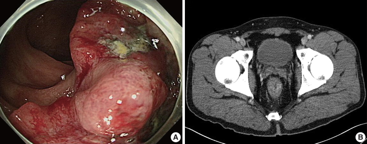
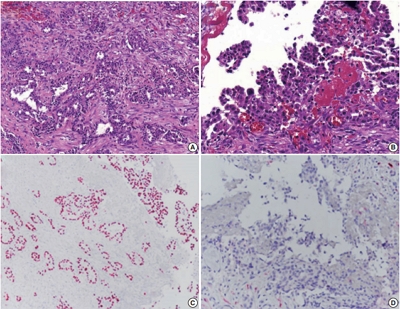
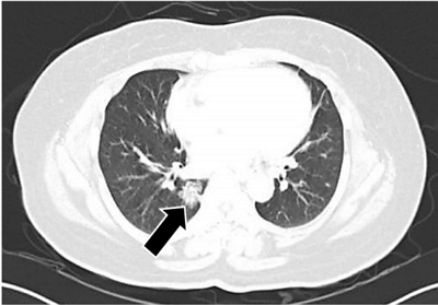
PDF - BRCA genes have well-known associations with breast and ovarian cancers. However, variations in the BRCA gene, especially germline variations, have also been reported in colorectal cancer (CRC). We present the case of a rectal cancer with a germline BRCA1 variation inherited from the paternal side. A 39-year-old male was admitted with rectal cancer. The patient underwent surgical resection and the pathologic diagnosis was adenocarcinoma. Next-generation sequencing was performed and a BRCA1 variant was detected. Reviewing the public database and considering the young age of the patient, the variant was suggested to be germline. The patient’s father had had prostate cancer and next-generation sequencing testing revealed an identical BRCA1 variant. In the BRCA cancer group, there is relatively little attention paid to male cancers. The accumulation of male CRC cases linked to BRCA variations may help clarify the potential pathological relationship between the two.
-
Citations
Citations to this article as recorded by
- Aggressive Right-Sided Colon Cancer in a Young Adult: Triple-Whammy Mutations (POLE, KRAS, BRCA1/2) Highlight Emerging Genetic Associations
Ravi Patel, Ganesh Kumar, Yash Shah, Dushyant Singh Dahiya, Sumant Inamdar
ACG Case Reports Journal.2026;[Epub] CrossRef
- Aggressive Right-Sided Colon Cancer in a Young Adult: Triple-Whammy Mutations (POLE, KRAS, BRCA1/2) Highlight Emerging Genetic Associations
- Pseudomesotheliomatous carcinoma of the lung in the parietal pleura
- Ae Ri An, Kyoung Min Kim, Jong Hun Kim, Gong Yong Jin, Young Hoon Choe, Myoung Ja Chung
- J Pathol Transl Med. 2020;54(2):192-195. Published online January 29, 2020
- DOI: https://doi.org/10.4132/jptm.2019.11.14
- 7,813 View
- 154 Download
- 2 Web of Science
- 5 Crossref
-
PDF
-
Citations
Citations to this article as recorded by- Pseudomesotheliomatous Adenocarcinoma Presenting as Hemothorax and Mimicking Mesothelioma: A Case Study
Mohamed IJIM, Sarah Keddabi, Chaynez Rachid, Oussama Fikri, Lamyae Amro
Cureus.2026;[Epub] CrossRef - Pseudomesotheliomatous lung cancer mimicking malignant pleural mesothelioma: A case report
Supakorn Chansaengpetch, Ruchira Ruangchira-urai, Nisa Muangman, Rathachai Kaewlai, Trongtum Tongdee, Teerapat Singwicha, Narongpon Dumavibhat
The ASEAN Journal of Radiology.2025; 26(1): 24. CrossRef - Pseudomesotheliomatous Carcinoma of the Lung with Morphological Characteristics of Signet Ring Cell Carcinoma: An Autopsy Case Report
Tetsu Hirakawa, Takuya Tanimoto, Yui Hattori, Ryo Katsura, Shinya Miyake, Suguru Fujita, Sayaka Ueno, Ken Masuda, Takashi Nishisaka, Nobuhisa Ishikawa
Internal Medicine.2024; 63(7): 979. CrossRef - Intrapulmonary Biphasic Mesothelioma Misdiagnosed as Adenocarcinoma: Case Report and a Potential Diagnostic Pitfall
Wenfeng Xu, XingYan Zhu, Hao Tang, Qijian Ying, Yujuan Xu, Deyu Guo
OncoTargets and Therapy.2024; Volume 17: 925. CrossRef -
A RARE CASE OF UNCLASSIFIED CARCINOMA OF THE LUNG: DIAGNOSTIC CHALLENGES
B.M. Fylenko, N.V. Royko, I.I. Starchenko, O.V. Starchenko, O.Y. Horodynska, S.A. Proskurnia
Azerbaijan Medical Journal.2024; (4): 182. CrossRef
- Pseudomesotheliomatous Adenocarcinoma Presenting as Hemothorax and Mimicking Mesothelioma: A Case Study
- Tumor-to-tumor metastasis: metastatic invasive lobular carcinoma of the breast within adenocarcinoma of the lung
- Myoung Jae Kang, Ae Ri An, Myoung Ja Chung, Kyoung Min Kim
- J Pathol Transl Med. 2020;54(2):188-191. Published online September 16, 2019
- DOI: https://doi.org/10.4132/jptm.2019.09.07
- 7,410 View
- 160 Download
- 9 Web of Science
- 9 Crossref
-
PDF
-
Citations
Citations to this article as recorded by- First pathologically confirmed case of concurrent primary renal cell carcinoma and metastatic lung adenocarcinoma in the same kidney
Hua Tang, Shengchun Zhang, Jian Chen, Yongfu Wang, Changping Guo, Ting Yu, Dechuan Ye
Frontiers in Oncology.2026;[Epub] CrossRef - Tumor-in-Tumor-Metastase: Ein seltenes Phänomen
Felix Elsner, Katharina Keller, Florian Fuchs, Michael Uder, Arndt Hartmann
Die Pathologie.2025; 46(3): 139. CrossRef - Unraveling Tumor-to-Tumor Metastasis: Insights into Pathogenesis, Diagnostic Challenges, and Treatment Modalities
Wennei Mei, Dongdong Zhang
Biologics: Targets and Therapy.2025; Volume 19: 43. CrossRef - Case Report: Tumor-to-tumor metastasis: a rare case of prostate adenocarcinoma metastasis to lung squamous cell carcinoma in a patient with multiple primary malignancies
Baoxiang Pei, Jikuan Liu, Zhiliang Hu, Fen Pan
Frontiers in Oncology.2025;[Epub] CrossRef - Tumor-to-Tumor Metastasis: Breast Cancer Metastasizing to EGFR Exon 19-Mutated Lung Adenocarcinoma with Long-Term Disease-Free Survival
Yana Zhang, Yang Hao, Han Yang, Xiangli Meng, Shanshan Yang, Jin Wang, Jinling Xie, Ping Lu, Yinghua Ji
Biologics: Targets and Therapy.2025; Volume 19: 707. CrossRef - Tumor-to-Tumor Metastases Involving Clear Cell Renal Cell Carcinomas: A Diagnostic Challenge for Pathologists Needing Clinical Correlation
Claudia Manini, Claudia Provenza, Leire Andrés, Igone Imaz, Rosa Guarch, Raffaelle Nunziata, José I. López
Clinics and Practice.2023; 13(1): 288. CrossRef - Metástasis tumor a tumor en pulmón: reporte de tres casos y revisión de la literatura
Paula Cristina Castro Quiroga, Blanca Viviana Fajardo Idrobo, Diana Marcela Caicedo Ruiz, Julieth Alexandra Franco Mira, Carlos Andrés Carvajal Fierro, Alfredo Ernesto Romero Rojas, Rafael Santiago Parra Medina
Revista Colombiana de Cancerología.2023; 27(1): 107. CrossRef - Lobular to Lobule: Metastatic Breast Carcinoma to Olfactory Neuroblastoma
Kent M. Swimley, Silvana Di Palma, Lester D. R. Thompson
Head and Neck Pathology.2021; 15(2): 642. CrossRef - A case of colorectal cancer with intratumoral metastasis to primary lung cancer
Yasushi Cho, Mitsuhito Kaji, Shunsuke Nomura, Yusuke Motohashi, Masaaki Sato, Motoya Takeuchi
The Journal of the Japanese Association for Chest Surgery.2021; 35(5): 576. CrossRef
- First pathologically confirmed case of concurrent primary renal cell carcinoma and metastatic lung adenocarcinoma in the same kidney
- Association between Expression of 8-OHdG and Cigarette Smoking in Non-small Cell Lung Cancer
- Ae Ri An, Kyoung Min Kim, Ho Sung Park, Kyu Yun Jang, Woo Sung Moon, Myoung Jae Kang, Yong Chul Lee, Jong Hun Kim, Han Jung Chae, Myoung Ja Chung
- J Pathol Transl Med. 2019;53(4):217-224. Published online March 11, 2019
- DOI: https://doi.org/10.4132/jptm.2019.02.20
- 10,494 View
- 251 Download
- 24 Web of Science
- 24 Crossref
-
Abstract
PDF
- Background
Exposure to cigarette smoking (CS) is a major risk factor for the development of lung cancer. CS is known to cause oxidative DNA damage and mutation of tumor-related genes, and these factors are involved in carcinogenesis. 8-Hydroxydeoxyguanosine (8-OHdG) is considered to be a reliable biomarker for oxidative DNA damage. Increased levels of 8-OHdG are associated with a number of pathological conditions, including cancer. There are no reports on the expression of 8-OHdG by immunohistochemistry in non-small cell lung cancer (NSCLC).
Methods
We investigated the expression of 8-OHdG and p53 in 203 NSCLC tissues using immunohistochemistry and correlated it with clinicopathological features including smoking.
Results
The expression of 8-OHdG was observed in 83.3% of NSCLC. It was significantly correlated with a low T category, negative lymph node status, never-smoker, and longer overall survival (p < .05) by univariate analysis. But multivariate analysis revealed that 8-OHdG was not an independent prognostic factor for overall survival in NSCLC patients. The aberrant expression of p53 significantly correlated with smoking, male, squamous cell carcinoma, and Ki-67 positivity (p < .05).
Conclusions
The expression of 8-OHdG was associated with good prognostic factors. It was positively correlated with never-smokers in NSCLC, suggesting that oxidative damage of DNA cannot be explained by smoking alone and may depend on complex control mechanisms. -
Citations
Citations to this article as recorded by- N-acetyl-cysteine alleviates nandrolone decanoate-induced hippocampal cell apoptosis in rats via reversing protein expressions of S1P1, Akt and FOXO3a signaling pathway
Alireza Shirpoor, Zahra Zarrini, Roya Naderi
Steroids.2026; 228: 109759. CrossRef - The distinct roles of ROS in tumor immunity: from mechanisms to immunotherapeutic applications
Jiayi Li, Chen Huang, Pan Tang, Ruiyan Wu, Quanyou Wu, Chenliang Zhang
Journal of Hematology & Oncology.2026;[Epub] CrossRef - Sustainable framework for automated segmentation and prediction of lung cancer in CT image using CapsNet with U-net segmentation
S.R. Vijayakumar, S. Aarthy, D. Deepa, P. Suresh
Biomedical Signal Processing and Control.2025; 99: 106873. CrossRef - Endolysosomal cation channel MCOLN as the novel regulator of redox homeostasis
Yahao Gao, Lei Xu, Ying Chen
Biochimica et Biophysica Acta (BBA) - Molecular Basis of Disease.2025; 1871(7): 167910. CrossRef - Catalase: The golden key to regulate oxidative stress in breast cancer
Jia-Wei Liu, Wen-Jia Chen, Yang-Zheng Lan, Jing Liu
World Journal of Clinical Oncology.2025;[Epub] CrossRef - Association of sirtuin 1 rs10997868 and rs730821 polymorphisms with sirtuin 1 and hydroxy-2′-deoxyguanosine levels in healthy smokers: A case–control study
Samar Sultan
Journal of International Medical Research.2025;[Epub] CrossRef - Increased pretreatment triglyceride glucose-body mass index associated with poor prognosis in patients with advanced non-small cell lung cancer
Shaoming Guo, Yi Zhao, Yue Jiang, Huaping Ye, Ying Wang
Clinical Nutrition ESPEN.2024; 59: 412. CrossRef - Oxidative Damage and Telomere Length as Markers of Lung Cancer Development among Chronic Obstructive Pulmonary Disease (COPD) Smokers
Elizabeth Córdoba-Lanús, Luis M. Montuenga, Angélica Domínguez-de-Barros, Alexis Oliva, Delia Mayato, Ana Remírez-Sanz, Francisca Gonzalvo, Bartolomé Celli, Javier J. Zulueta, Ciro Casanova
Antioxidants.2024; 13(2): 156. CrossRef - Automated determination of 8-OHdG in cells and tissue via immunofluorescence using a specially created antibody
Tobias Jung, Nicole Findik, Bianca Hartmann, Katja Hanack, Kai Grossmann, Dirk Roggenbuck, Marc Wegmann, René Mantke, Markus Deckert, Tilman Grune
Biotechnology Reports.2024; 42: e00833. CrossRef - Combination treatment of zinc and selenium intervention ameliorated BPA-exposed germ cell damage in SD rats: elucidation of molecular mechanisms
Chittaranjan Sahu, Gopabandhu Jena
Naunyn-Schmiedeberg's Archives of Pharmacology.2024; 397(9): 6685. CrossRef - Interplay of arsenic exposure and cigarette smoking on oxidative DNA damage in healthy males
Sepideh Nemati-Mansour, Mohammad Mosaferi, Javad Babaie, Asghar Mohammadpoorasl, Reza Dehghanzadeh, Leila Nikniaz, Mohammad Miri
Environmental Sciences Europe.2024;[Epub] CrossRef - The role of tissue persistent organic pollutants and genetic polymorphisms in patients with benign and malignant kidney tumors
Rasih Kocagöz, İlgen Onat, Merve Demirbügen Öz, Burak Turna, Banu Sarsık Kumbaracı, Mehmet Nurullah Orman, Halit Sinan Süzen, Hilmi Orhan
Environmental Toxicology and Pharmacology.2024; 110: 104495. CrossRef - Mitochondrial Plasticity and Glucose Metabolic Alterations in Human Cancer under Oxidative Stress—From Viewpoints of Chronic Inflammation and Neutrophil Extracellular Traps (NETs)
Hui-Ting Lee, Chen-Sung Lin, Chao-Yu Liu, Po Chen, Chang-Youh Tsai, Yau-Huei Wei
International Journal of Molecular Sciences.2024; 25(17): 9458. CrossRef - Oxidative DNA Damage and Arterial Hypertension in Light of Current ESC Guidelines
Radka Hazuková, Zdeněk Zadák, Miloslav Pleskot, Petr Zdráhal, Martin Pumprla, Miloš Táborský
International Journal of Molecular Sciences.2024; 25(23): 12557. CrossRef - Significance of 8-OHdG Expression as a Predictor of Survival in Colorectal Cancer
Myunghee Kang, Soyeon Jeong, Sungjin Park, Seungyoon Nam, Jun-Won Chung, Kyoung Oh Kim, Jungsuk An, Jung Ho Kim
Cancers.2023; 15(18): 4613. CrossRef - Serum 8-Hydroxy-2′-deoxyguanosine Predicts Severity and Prognosis of Patients with Acute Exacerbation of Chronic Obstructive Pulmonary Disease
Peng Cao, Chen Zhang, Dong-Xu Hua, Meng-Die Li, Bian-Bian Lv, Lin Fu, Hui Zhao
Lung.2022; 200(1): 31. CrossRef - Redox signaling at the crossroads of human health and disease
Jing Zuo, Zhe Zhang, Maochao Luo, Li Zhou, Edouard C. Nice, Wei Zhang, Chuang Wang, Canhua Huang
MedComm.2022;[Epub] CrossRef - Assessment of MDA and 8-OHdG expressions in ovine pulmonary adenocarcinomas by immunohistochemical and immunofluorescence methods
Emin Karakurt, Enver Beytut, Serpil Dağ, Hilmi Nuhoğlu, Ayfer Yıldız, Emre Kurtbaş
Acta Veterinaria Brno.2022; 91(3): 235. CrossRef - Dietary Antioxidants and Lung Cancer Risk in Smokers and Non-Smokers
Naser A. Alsharairi
Healthcare.2022; 10(12): 2501. CrossRef - Targeting oxidative stress in disease: promise and limitations of antioxidant therapy
Henry Jay Forman, Hongqiao Zhang
Nature Reviews Drug Discovery.2021; 20(9): 689. CrossRef - Association between tobacco substance usage and a missense mutation in the tumor suppressor gene P53 in the Saudi Arabian population
Mikhlid H. Almutairi, Bader O. Almutairi, Turki M. Alrubie, Sultan N. Alharbi, Narasimha R. Parine, Abdulwahed F. Alrefaei, Ibrahim Aldeailej, Abdullah Alamri, Abdelhabib Semlali, Alvaro Galli
PLOS ONE.2021; 16(1): e0245133. CrossRef - Measurement of uranium concentrations in urine samples of adult healthy groups in Najaf governorate with estimation of urine concentrations of 8-OHdG compound as biomarker for DNA damage
Samia K. Abbas, Dhuha S. Saleh, Hayder S. Hussain
Journal of Physics: Conference Series.2021; 1879(3): 032097. CrossRef - Common Data Model and Database System Development for the Korea Biobank Network
Soo-Jeong Ko, Wona Choi, Ki-Hoon Kim, Seo-Joon Lee, Haesook Min, Seol-Whan Oh, In Young Choi
Applied Sciences.2021; 11(24): 11825. CrossRef - EVALUATION OF OXIDATIVE STATUS IN PATIENTS WITH CHRONIC PERIODONTITIS AND ADDITIONAL TOBACCO ABUSE: A CROSS-SECTIONAL STUDY
Didem ÖZKAL EMİNOĞLU, Varol ÇANAKÇI
Atatürk Üniversitesi Diş Hekimliği Fakültesi Dergisi.2020; : 1. CrossRef
- N-acetyl-cysteine alleviates nandrolone decanoate-induced hippocampal cell apoptosis in rats via reversing protein expressions of S1P1, Akt and FOXO3a signaling pathway
- WITHDRAWN:A Clinicopathologic Study of 220 Cases of Pulmonary Sclerosing Pneumocytoma in Korea: A Nationwide Survey
- Myunghee Kang, Seung Yeon Ha, Joung Ho Han, Mee Sook Roh, Se Jin Jang, Hee Jin Lee, Heae Surng Park, Geon Kook Lee, Kyo Young Lee, Jin-Haeng Chung, Yoo Duk Choi, Chang Hun Lee, Lucia Kim, Myoung Ja Chung, Soon Hee Jung, Gou Young Kim, Wan-Seop Kim
- Received April 4, 2018 Accepted July 9, 2018 Published online July 16, 2018
- DOI: https://doi.org/10.4132/jptm.2018.07.10 [Accepted]
- 5,873 View
- 63 Download
- Immunohistochemical Expression and Clinical Significance of Suggested Stem Cell Markers in Hepatocellular Carcinoma
- Jong Jin Sung, Sang Jae Noh, Jun Sang Bae, Ho Sung Park, Kyu Yun Jang, Myoung Ja Chung, Woo Sung Moon
- J Pathol Transl Med. 2016;50(1):52-57. Published online November 18, 2015
- DOI: https://doi.org/10.4132/jptm.2015.10.09
- 12,506 View
- 78 Download
- 21 Web of Science
- 18 Crossref
-
Abstract
PDF
- Background
Increasing evidence has shown that tumor initiation and growth are nourished by a small subpopulation of cancer stem cells (CSCs) within the tumor mass. CSCs are posited to be responsible for tumor maintenance, growth, distant metastasis, and relapse after curative operation. We examined the expression of CSC markers in paraffin-embedded tissue sections of hepatocellular carcinoma (HCC) and correlated the results with clinicopathologic characteristics. Methods: Immunohistochemical staining for the markers believed to be expressed in the CSCs, including epithelial cell adhesion molecule (EpCAM), keratin 19 (K19), CD133, and CD56, was performed in 82 HCC specimens. Results: EpCAM expression was observed in 56% of the HCCs (46/82) and K19 in 6% (5/82). EpCAM expression in HCC significantly correlated with elevated α-fetoprotein level, microvessel invasion of tumor cells, and high histologic grade. In addition, Ep- CAM expression significantly correlated with K19 expression. The overall survival and relapsefree survival rates in patients with EpCAM-expressing HCC were relatively lower than those in patients with EpCAM-negative HCC. All but two of the 82 HCCs were negative for CD133 and CD56, respectively. Conclusions: Our results suggest that HCCs expressing EpCAM are associated with unfavorable prognostic factors and have a more aggressive clinical course than those not expressing EpCAM. Further, the expression of either CD133 or CD56 in paraffin-embedded HCC tissues appears to be rare. -
Citations
Citations to this article as recorded by- Spatial immune scoring system predicts hepatocellular carcinoma recurrence
Gengjie Jia, Peiqi He, Tianli Dai, Denise Goh, Jiabei Wang, Mengyuan Sun, Felicia Wee, Fuling Li, Jeffrey Chun Tatt Lim, Shuxia Hao, Yao Liu, Tony Kiat Hon Lim, Nye-Thane Ngo, Qingping Tao, Wei Wang, Ahitsham Umar, Björn Nashan, Yongchang Zhang, Chen Ding
Nature.2025; 640(8060): 1031. CrossRef - Evolving Landscape of Systemic Therapy for Hepatocellular Carcinoma in 2025
Karan Kumar, Vivek A. Saraswat
Journal of Clinical and Experimental Hepatology.2025; 15(5): 102547. CrossRef - Recent Progress in Systemic Therapy for Advanced Hepatocellular Carcinoma
Narayanan Sadagopan, Aiwu Ruth He
International Journal of Molecular Sciences.2024; 25(2): 1259. CrossRef - Diagnostic value of expressions of cancer stem cell markers for adverse outcomes of hepatocellular carcinoma and their associations with prognosis: A Bayesian network meta‑analysis
Zhengrong Ou, Shoushuo Fu, Jian Yi, Jingxuan Huang, Weidong Zhu
Oncology Letters.2024;[Epub] CrossRef - Clinicopathological and prognostic value of epithelial cell adhesion molecule in solid tumours: a meta-analysis
Peiwen Ding, Panyu Chen, Jiqi Ouyang, Qiang Li, Shijie Li
Frontiers in Oncology.2023;[Epub] CrossRef - PD-L1 Downregulation and DNA Methylation Inhibition for Molecular Therapy against Cancer Stem Cells in Hepatocellular Carcinoma
Caecilia Sukowati, Loraine Kay D. Cabral, Beatrice Anfuso, Francesco Dituri, Roberto Negro, Gianluigi Giannelli, Claudio Tiribelli
International Journal of Molecular Sciences.2023; 24(17): 13357. CrossRef - EpCAM, Ki67, and ESM1 Predict Hepatocellular Carcinoma Recurrence After Liver Transplantation
Aiat Shaban Hemida, Doha Maher Taie, Moshira Mohamed Abd El-Wahed, Mohammed Ibrahim Shabaan, Mona Saeed Tantawy, Nermine Ahmed Ehsan
Applied Immunohistochemistry & Molecular Morphology.2023; 31(9): 596. CrossRef - The clinical, prognostic and therapeutic significance of liver cancer stem cells and their markers
Izabela Zarębska, Arkadiusz Gzil, Justyna Durślewicz, Damian Jaworski, Paulina Antosik, Navid Ahmadi, Marta Smolińska-Świtała, Dariusz Grzanka, Łukasz Szylberg
Clinics and Research in Hepatology and Gastroenterology.2021; 45(3): 101664. CrossRef - Detection of oncogenic mutations in paired circulating tumor DNA and circulating tumor cells in patients with hepatocellular carcinoma
Zhouhong Ge, Jean C.A. Helmijr, Maurice P.H.M. Jansen, Patrick P.C. Boor, Lisanne Noordam, Maikel Peppelenbosch, Jaap Kwekkeboom, Jaco Kraan, Dave Sprengers
Translational Oncology.2021; 14(7): 101073. CrossRef - Hepatocellular Carcinoma Score and Subclassification Into Aggressive Subtypes Using Immunohistochemical Expression of p53, β-Catenin, CD133, and Ki-67
Asmaa G. Abdou, Nanis S. Holah, Dina S. Elazab, Walaa G. El-Gendy, Mohammed T. Badr, Dalia R. Al-Sharaky
Applied Immunohistochemistry & Molecular Morphology.2021; 29(1): 20. CrossRef - The prognostic significance of neuroendocrine markers and somatostatin receptor 2 in hepatocellular carcinoma
Keigo Murakami, Hiroyuki Kumata, Shigehito Miyagi, Takashi Kamei, Hironobu Sasano
Pathology International.2021; 71(10): 682. CrossRef - Predictors of recurrence and survival of hepatocellular carcinoma: A prospective study including transient elastography and cancer stem cell markers
Hend Ibrahim Shousha, Rabab Fouad, Tamer Mahmoud Elbaz, Dina Sabry, Mohamed Mahmoud Nabeel, Ahmed Hosni Abdelmaksoud, Aisha Mahmoud Elsharkawy, Zeinab Abdellatif Soliman, Ghada Habib, Ashraf Omar Abdelaziz
Arab Journal of Gastroenterology.2020; 21(2): 95. CrossRef - Napabucasin Reduces Cancer Stem Cell Characteristics in Hepatocellular Carcinoma
Ya Li, Qiuju Han, Huajun Zhao, Quanjuan Guo, Jian Zhang
Frontiers in Pharmacology.2020;[Epub] CrossRef - The mRNA Distribution of Cancer Stem Cell Marker CD90/Thy-1 Is Comparable in Hepatocellular Carcinoma of Eastern and Western Populations
An B. Luong, Huy Q. Do, Paola Tarchi, Deborah Bonazza, Cristina Bottin, Loraine Kay D. Cabral, Long D. C. Tran, Thao P. T. Doan, Lory S. Crocè, Hoa L. T. Pham, Claudio Tiribelli, Caecilia H. C. Sukowati
Cells.2020; 9(12): 2672. CrossRef - Histological architectural classification determines recurrence pattern and prognosis after curative hepatectomy in patients with hepatocellular carcinoma
Hirohisa Okabe, Tomoharu Yoshizumi, Yo-ichi Yamashita, Katsunori Imai, Hiromitsu Hayashi, Shigeki Nakagawa, Shinji Itoh, Norifumi Harimoto, Toru Ikegami, Hideaki Uchiyama, Toru Beppu, Shinichi Aishima, Ken Shirabe, Hideo Baba, Yoshihiko Maehara, Motoyuki
PLOS ONE.2018; 13(9): e0203856. CrossRef - Overexpression of epithelial cell adhesion molecule as a predictor of poor outcome in patients with hepatocellular carcinoma
Chih‑Jan Ko, Chia‑Jung Li, Meng‑Yu Wu, Pei‑Yi Chu
Experimental and Therapeutic Medicine.2018;[Epub] CrossRef - Clinicopathologic Significance of Survivin Expression in Relation to CD133 Expression in Surgically Resected Stage II or III Colorectal Cancer
Wanlu Li, Mi-Ra Lee, EunHee Choi, Mee-Yon Cho
Journal of Pathology and Translational Medicine.2017; 51(1): 17. CrossRef - PIN1 in hepatocellular carcinoma is associated with TP53 gene status
Jun Sang Bae, Sang Jae Noh, Kyoung Min Kim, Kyu Yun Jang, Ho Sung Park, Myoung Ja Chung, Byung-Hyun Park, Woo Sung Moon
Oncology Reports.2016; 36(4): 2405. CrossRef
- Spatial immune scoring system predicts hepatocellular carcinoma recurrence
- Abdominal Fibromatosis in a Young Child: A Case Study and Review of the Literature
- Hyun Hee Chu, Pyoung Han Hwang, Yeon Jun Jeong, Myoung Ja Chung
- Korean J Pathol. 2013;47(5):472-476. Published online October 25, 2013
- DOI: https://doi.org/10.4132/KoreanJPathol.2013.47.5.472
- 9,694 View
- 38 Download
- 3 Crossref
-
Abstract
PDF
Fibromatoses comprise many different entities of well-differentiated fibroblastic proliferation with variable collagen production and form a firm nodular mass. Abdominal fibromatosis is distinguishable from other forms of fibromatosis because of its location and its tendency to occur in women of childbearing age during or following pregnancy. Abdominal fibromatosis in children is an extremely rare condition. A 15-month-old boy presented with an abdominal wall mass that had recently increased in size. Mass excision was perfomed. The tumor was 4.3×4.1 cm and partly circumscribed. Histologically, the tumor was composed of parallel long fascicles of spindle-cells with a uniform appearance. The edges of the resected mass were infiltrative, and the surgical margins were positive. Mitotic figures were <1/10 high power fields. No cellular atypia or necrosis was present. The tumor cells were positive for vimentin and nuclear β-catenin staining.
-
Citations
Citations to this article as recorded by- A rare tumor of the large bowel in a young boy
Shyam Srinivasan, Soumitra Saha
Cancer Research, Statistics, and Treatment.2021; 4(4): 752. CrossRef - Uncommon abdominal wall mass in a young boy: Desmoid tumor
Levent Cankorkmaz, Mehmet H. Atalar, H. Reyhan Eğilmez
Cumhuriyet Medical Journal.2018; : 811. CrossRef - Lesiones ocupantes de espacio en pared abdominal (no herniaria). La visión del patólogo
Isidro Machado, Julia Cruz, Javier Lavernia, Fernando Carbonell
Revista Hispanoamericana de Hernia.2015; 3(3): 85. CrossRef
- A rare tumor of the large bowel in a young boy
- No Detection of Simian Virus 40 in Malignant Mesothelioma in Korea
- Minseob Eom, Jamshid Abdul-Ghafar, Sun-Mi Park, Joung Ho Han, Soon Won Hong, Kun Young Kwon, Eun Suk Ko, Lucia Kim, Wan Seop Kim, Seung Yeon Ha, Kyo Young Lee, Chang Hun Lee, Hye Kyoung Yoon, Yoo Duk Choi, Myoung Ja Chung, Soon-Hee Jung
- Korean J Pathol. 2013;47(2):124-129. Published online April 24, 2013
- DOI: https://doi.org/10.4132/KoreanJPathol.2013.47.2.124
- 11,046 View
- 56 Download
- 6 Crossref
-
Abstract
PDF
Background Simian virus 40 (SV40), a polyomavirus, was discovered as a contaminant of a human polio vaccine in the 1960s. It is known that malignant mesothelioma (MM) is associated with SV40, and that the virus works as a cofactor to the carcinogenetic effects of asbestos. However, the reports about the correlation between SV40 and MM have not been consistent. The purpose of this study is to identify SV40 in MM tissue in Korea through detection of SV40 protein and DNA.
Methods We analyzed 62 cases of available paraffin-blocks enrolled through the Korean Malignant Mesothelioma Surveillance System and performed immunohistochemistry for SV40 protein and real-time polymerase chain reaction (PCR) for SV40 DNA.
Results Of 62 total cases, 40 had disease involving the pleura (64.5%), and 29 (46.8%) were found to be of the epithelioid subtype. Immunostaining demonstrated that all examined tissues were negative for SV40 protein. Sufficient DNA was extracted for real-time PCR analysis from 36 cases. Quantitative PCR of these samples showed no increase in SV40 transcript compared to the negative controls.
Conclusions SV40 is not associated with the development of MM in Korea.
-
Citations
Citations to this article as recorded by- Association Study of Pleural Mesothelioma and Oncogenic Simian Virus 40 in the Crocidolite-Contaminated Area of Dayao County, Yunnan Province, Southwest China
Ru-ai Liu, Bo-yong Wang, Xin Chen, Yuan-qian Pu, Jia-ji Zi, Wen Mei, Ye-pin Zhang, Lu Qiu, Wei Xiong
Genetic Testing and Molecular Biomarkers.2024; 28(5): 189. CrossRef - Binding of SV40’s Viral Capsid Protein VP1 to Its Glycosphingolipid Receptor GM1 Induces Negative Membrane Curvature: A Molecular Dynamics Study
Raisa Kociurzynski, Sophie D. Beck, Jean-Baptiste Bouhon, Winfried Römer, Volker Knecht
Langmuir.2019; 35(9): 3534. CrossRef - Estimated future incidence of malignant mesothelioma in South Korea: Projection from 2014 to 2033
Kyeong Min Kwak, Domyung Paek, Seung-sik Hwang, Young-Su Ju, Mark Allen Pershouse
PLOS ONE.2017; 12(8): e0183404. CrossRef - The function, mechanisms, and role of the genes PTEN and TP53 and the effects of asbestos in the development of malignant mesothelioma: a review focused on the genes' molecular mechanisms
Leonardo Vinícius Monteiro de Assis, Mauro César Isoldi
Tumor Biology.2014; 35(2): 889. CrossRef - The role of key genes and pathways involved in the tumorigenesis of Malignant Mesothelioma
Leonardo Vinícius Monteiro de Assis, Jamille Locatelli, Mauro César Isoldi
Biochimica et Biophysica Acta (BBA) - Reviews on Cancer.2014; 1845(2): 232. CrossRef - Pleural Mesothelioma: An Institutional Experience of 66 Cases
Soomin Ahn, In Ho Choi, Joungho Han, Jhingook Kim, Myung-Ju Ahn
Korean Journal of Pathology.2014; 48(2): 91. CrossRef
- Association Study of Pleural Mesothelioma and Oncogenic Simian Virus 40 in the Crocidolite-Contaminated Area of Dayao County, Yunnan Province, Southwest China
- Expression of CHOP in Squamous Tumor of the Uterine Cervix
- Hyun Hee Chu, Jun Sang Bae, Kyoung Min Kim, Ho Sung Park, Dong Hyu Cho, Kyu Yun Jang, Woo Sung Moon, Myoung Jae Kang, Dong Geun Lee, Myoung Ja Chung
- Korean J Pathol. 2012;46(5):463-469. Published online October 25, 2012
- DOI: https://doi.org/10.4132/KoreanJPathol.2012.46.5.463
- 10,019 View
- 40 Download
- 8 Crossref
-
Abstract
PDF
Background High-risk human papillomavirus (HR-HPV) infection and abnormal p53 expression are closely involved in carcinogenesis of squamous cell carcinoma (SqCC) of uterine cervix. Recent studies have suggested that virus-induced endoplasmic reticulum (ER) stress modulates various cell survival and cell death signaling pathways. The C/EBP homologous protein (CHOP) is associated with ER stress-mediated apoptosis and is also involved in carcinogenesis of several human cancers. We hypothesized that CHOP is involved in the carcinogenesis of uterine cervical cancer in association with HR-HPV and/or p53.
Methods Immunohistochemistry was used to analyze CHOP and p53 protein expression of tissue sections from 191 patients with invasive cancer or preinvasive lesions of the uterine cervix (61 cases of SqCC, 66 cases of cervical intraepithelial neoplasia [CIN] III, and 64 cases of CIN I).
Results CHOP was expressed in 59.4% of CIN I, 48.5% of CIN III, and 70.5% of SqCC cases. It was also significantly more frequent in invasive SqCC than in preinvasive lesions (p=0.042). Moreover, CHOP expression significantly correlated with HR-HPV infection and p53 expression (p=0.009 and p=0.038, respectively).
Conclusions Our results suggest that CHOP is involved in the carcinogenesis of the uterine cervix SqCC via association with HR-HPV and p53.
-
Citations
Citations to this article as recorded by- Interplay between the cellular stress pathway, stemness markers, and Helicobacter pylori infection in gastric cancer
Mehran Gholamin, Atena Mansouri, Mohammad Reza Abbaszadegan, Mohammad Ali Karimi, Hossein Barzegar, Fatemeh Fardi Golyan, Hanie Mahaki, Hamid Tanzadehpanah, Reihaneh Alsadat Mahmoudian
Gene Reports.2025; 40: 102263. CrossRef - Role of C-reactive protein in cervical intraepithelial neoplasia/cancer
Adriana Pedreañez, Yenddy Carrero, Renata Vargas, Juan P.Hernández Fonseca, Jesús Mosquera
Pathology - Research and Practice.2025; 276: 156274. CrossRef - Expression of GRP78 and its copartners in HEK293 and pancreatic cancer cell lines (BxPC-3/PANC-1) exposed to MRI and CT contrast agents
Ali Ahmed Azzawri, Ibrahim Halil Yildirim, Zeynep Yegin, Abdurrahim Dusak
Nucleosides, Nucleotides & Nucleic Acids.2024; 43(5): 391. CrossRef - Endoplasmic Reticulum Stress and Homeostasis in Reproductive Physiology and Pathology
Elif Guzel, Sefa Arlier, Ozlem Guzeloglu-Kayisli, Mehmet Tabak, Tugba Ekiz, Nihan Semerci, Kellie Larsen, Frederick Schatz, Charles Lockwood, Umit Kayisli
International Journal of Molecular Sciences.2017; 18(4): 792. CrossRef - Endoplasmic reticulum stress pathway PERK‐eIF2α confers radioresistance in oropharyngeal carcinoma by activating NF‐κB
Qiao Qiao, Chaonan Sun, Chuyang Han, Ning Han, Miao Zhang, Guang Li
Cancer Science.2017; 108(7): 1421. CrossRef - Curcumin induces ER stress-mediated apoptosis through selective generation of reactive oxygen species in cervical cancer cells
Boyun Kim, Hee Seung Kim, Eun-Ji Jung, Jung Yun Lee, Benjamin K. Tsang, Jeong Mook Lim, Yong Sang Song
Molecular Carcinogenesis.2016; 55(5): 918. CrossRef - Down-regulation of C/EBP homologous protein (CHOP) expression in gastric cardia adenocarcinoma: Their relationship with clinicopathological parameters and prognostic significance
Xiao-Juan Zhu, She-Gan Gao, San-Qiang Li, Zhen-Guo Shi, Zhi-Kun Ma, Shan-Shan Zhu, Xiao-Shan Feng
Clinics and Research in Hepatology and Gastroenterology.2015; 39(3): 391. CrossRef - MG289 in <i>Mycoplasma genitalium</i> Enhances Microbial Invasion and Bacterial Persistence in Benign Human Prostate Cells
Wasia Rizwani, Leticia Reyes, Jeongsoon Kim, Steve Goodison, Charles J. Rosser
Open Journal of Urology.2013; 03(06): 232. CrossRef
- Interplay between the cellular stress pathway, stemness markers, and Helicobacter pylori infection in gastric cancer
- Expression of Cortactin and Focal Adhesion Kinase in Colorectal Adenocarcinoma: Correlation with Clinicopathologic Parameters and Their Prognostic Implication
- Yo Na Kim, Ji Eun Choi, Jun Sang Bae, Kyu Yun Jang, Myoung Ja Chung, Woo Sung Moon, Myoung Jae Kang, Dong Geun Lee, Ho Sung Park
- Korean J Pathol. 2012;46(5):454-462. Published online October 25, 2012
- DOI: https://doi.org/10.4132/KoreanJPathol.2012.46.5.454
- 10,057 View
- 47 Download
- 7 Crossref
-
Abstract
PDF
Background Cortactin and focal adhesion kinase (FAK) are two important components among actin cross-linking proteins that play a central role in cell migration.
Methods The aims of this study were to evaluate the expression of cortactin and FAK in normal colorectal mucosa and colorectal adenocarcinoma (CRC) using tissue microarray of 2 mm cores to correlate their expression with other clinicopathological factors and, investigate their prognostic significance.
Results Twenty (9%) and 24 cases (11%) of normal colorectal mucosa were immunoreactive for cortactin and FAK. In addition, 184 (84%) and 133 cases (61%) of CRCs were immunoreactive for cortactin and FAK, respectively. Cortactin expression was associated with histologic differentiation and FAK expression. Cortactin, but not FAK expression was also correlated with poor overall and relapse-free survival and served well as an independent prognostic factor for poor survival.
Conclusions Cortactin expression, in association with FAK expression, may plays an important role in tumor progression. Furthermore, it may also be a satisfactory biomarker to predict tumor progression and survival in CRC patients.
-
Citations
Citations to this article as recorded by- Identification of a Subset of Stage I Colorectal Cancer Patients With High Recurrence Risk
Lik Hang Lee, Lindy Davis, Lourdes Ylagan, Angela R Omilian, Kristopher Attwood, Canan Firat, Jinru Shia, Philip B Paty, William G Cance
JNCI: Journal of the National Cancer Institute.2022; 114(5): 732. CrossRef - Profiling the expression of pro-metastatic genes in association with the clinicopathological features of primary breast cancer
Seyed-Mohammad Mazloomi, Mitra Foroutan-Ghaznavi, Vahid Montazeri, Gholamreza Tavoosidana, Ashraf Fakhrjou, Hojjatollah Nozad-Charoudeh, Saeed Pirouzpanah
Cancer Cell International.2021;[Epub] CrossRef - PZR promotes metastasis of colorectal cancer through increasing FAK and Src phosphorylation
Dan Tan, Wenpeng Zhang, Yu Tao, Yesseyeva Galiya, Mingliang Wang
Acta Biochimica et Biophysica Sinica.2019; 51(4): 356. CrossRef - Overexpression and Tyr421-phosphorylation of cortactin is induced by three-dimensional spheroid culturing and contributes to migration and invasion of pancreatic ductal adenocarcinoma (PDAC) cells
Katharina Stock, Rebekka Borrink, Jan-Henrik Mikesch, Anna Hansmeier, Jan Rehkämper, Marcel Trautmann, Eva Wardelmann, Wolfgang Hartmann, Jan Sperveslage, Konrad Steinestel
Cancer Cell International.2019;[Epub] CrossRef - Cortactin promotes colorectal cancer cell proliferation by activating the EGFR-MAPK pathway
Xiaojian Zhang, Kun Liu, Tao Zhang, Zhenlei Wang, Xuan Qin, Xiaoqian Jing, Haoxuan Wu, Xiaopin Ji, Yonggang He, Ren Zhao
Oncotarget.2017; 8(1): 1541. CrossRef - Prognostic Value of Focal Adhesion Kinase (FAK) in Human Solid Carcinomas: A Meta-Analysis
Xiao-Qing Zeng, Na Li, Li-Li Ma, Yu-Jen Tseng, Nai-Qing Zhao, Shi-Yao Chen, Han-Chung Wu
PLOS ONE.2016; 11(9): e0162666. CrossRef - Regulators of Actin Dynamics in Gastrointestinal Tract Tumors
Konrad Steinestel, Eva Wardelmann, Wolfgang Hartmann, Inga Grünewald
Gastroenterology Research and Practice.2015; 2015: 1. CrossRef
- Identification of a Subset of Stage I Colorectal Cancer Patients With High Recurrence Risk
- Expressions of E-cadherin, Cortactin and MMP-9 in Pseudoepitheliomatous Hyperplasia and Squamous Cell Carcinoma of the Head and Neck: Their Relationships with Clinicopathologic Factors and Prognostic Implication
- Tack Kune You, Kyoung Min Kim, Sang Jae Noh, Jun Sang Bae, Kyu Yun Jang, Myoung Ja Chung, Woo Sung Moon, Myoung Jae Kang, Dong Geun Lee, Ho Sung Park
- Korean J Pathol. 2012;46(4):331-340. Published online August 23, 2012
- DOI: https://doi.org/10.4132/KoreanJPathol.2012.46.4.331
- 10,748 View
- 84 Download
- 15 Crossref
-
Abstract
PDF
Background E-cadherin, cortactin, and matrix metalloproteinase (MMP)-9 have roles in tumor development or progression, but their expression has not been fully investigated in pseudoepitheliomatous hyperplasia (PEH) and squamous cell carcinoma (SCC) of the head and neck.
Methods We evaluated the immunohistochemical expression of E-cadherin, cortactin, and MMP-9 in 29 cases of PEH and 97 cases of SCC. Additionally, we evaluated their relationship with clinicopathologic factors and prognostic implications in SCC.
Results Thirty-five cases of SCC showed reduced expression of E-cadherin, whereas none of the PEH did. A total of 20 cases and 11 cases of SCC were immunoreactive for cortactin and MMP-9, respectively, whereas none of the PEH did. In SCC, reduced expression of E-cadherin was correlated with cortactin expression and invasion depth. Cortactin expression was correlated with differentiation, T classification, and recurrence and/or metastasis. MMP-9 expression was correlated with invasion depth. Cortactin expression was correlated with poor overall survival and relapse-free survival and it was an independent prognostic factor.
Conclusions The reduced expression of E-cadherin and the expression of cortactin may be helpful for the differential diagnosis of PEH and SCC. Furthermore, cortactin expression in association with reduced E-cadherin expression is correlated with poor prognosis in SCC.
-
Citations
Citations to this article as recorded by- HIV-1 Tat-induced disruption of epithelial junctions and epithelial-mesenchymal transition of oral and genital epithelial cells lead to increased invasiveness of neoplastic cells and the spread of herpes simplex virus and cytomegalovirus
Sharof Tugizov
Frontiers in Immunology.2025;[Epub] CrossRef - Ultrastructural and immunohistochemical evaluation of hyperplastic soft tissues surrounding dental implants in fibular jaws
Kezia Rachellea Mustakim, Mi Young Eo, Mi Hyun Seo, Hyeong-Cheol Yang, Min-Keun Kim, Hoon Myoung, Soung Min Kim
Scientific Reports.2024;[Epub] CrossRef - Virus-associated disruption of mucosal epithelial tight junctions and its role in viral transmission and spread
Sharof Tugizov
Tissue Barriers.2021;[Epub] CrossRef - Leishmaniasis: still a diagnostic challenge?
Ricardo Tadeu Villa
Journal of Dermatology & Cosmetology.2021; 5(2): 23. CrossRef - COMPARISON OF EXPRESSION OF E-CADHERIN IN ORAL PSEUDOEPITHELIOMATOUS HYPERPLASIA AND ORAL SQUAMOUS CELL CARCINOMA
Ayesha Mukhtar Awan, Iram Naz, Muhammad Khurram Mahmood, Hafeez Uddin
Gomal Journal of Medical Sciences.2020; 17(3): 70. CrossRef - EXPRESSION OF MATRIX METALLOPROTEINASE-9 IN ORAL SQUAMOUS CELL CARCINOMA AND ORAL PSEUDOEPITHELIOMATOUS HYPERPLASIA
Ayesha Mukhtar Awan, Iram Naz, Muhammad Khurram Mahmood, Hafeez Uddin
Gomal Journal of Medical Sciences.2020; 18(01): 24. CrossRef - An update of knowledge on cortactin as a metastatic driver and potential therapeutic target in oral squamous cell carcinoma
Pablo Ramos‐García, Miguel Ángel González‐Moles, Lucía González‐Ruiz, Ángela Ayén, Isabel Ruiz‐Ávila, Francisco José Navarro‐Triviño, José Antonio Gil‐Montoya
Oral Diseases.2019; 25(4): 949. CrossRef - Prognostic and clinicopathological significance of CTTN/cortactin alterations in head and neck squamous cell carcinoma: Systematic review and meta‐analysis
Pablo Ramos‐García, Miguel Ángel González‐Moles, Ángela Ayén, Lucía González‐Ruiz, Isabel Ruiz‐Ávila, José Antonio Gil‐Montoya
Head & Neck.2019; 41(6): 1963. CrossRef - The effect of centromere protein U silencing by lentiviral mediated RNA interference on the proliferation and apoptosis of breast cancer
Shuang‑Yan Lin, Yan‑Bo Lv, Gen‑Xiang Mao, Xu‑Jiao Chen, Fang Peng
Oncology Letters.2018;[Epub] CrossRef - Glycosylation: a hallmark of cancer?
Bhairavi N. Vajaria, Prabhudas S. Patel
Glycoconjugate Journal.2017; 34(2): 147. CrossRef - Differential expression of the sirtuin family in renal cell carcinoma: Aspects of carcinogenesis and prognostic significance
Seong Uk Jeh, Jung Je Park, Jong Sil Lee, Dong Chul Kim, Jungmo Do, Sin Woo Lee, See Min Choi, Jae Seog Hyun, Deok Ha Seo, Chunwoo Lee, Sung Chul Kam, Ky Hyun Chung, Jeong Seok Hwa
Urologic Oncology: Seminars and Original Investigations.2017; 35(12): 675.e9. CrossRef - Cortactin promotes colorectal cancer cell proliferation by activating the EGFR-MAPK pathway
Xiaojian Zhang, Kun Liu, Tao Zhang, Zhenlei Wang, Xuan Qin, Xiaoqian Jing, Haoxuan Wu, Xiaopin Ji, Yonggang He, Ren Zhao
Oncotarget.2017; 8(1): 1541. CrossRef - Cortactin in cancer cell migration and invasion
Miao Yin, Wenqing Ma, Liguo An
Oncotarget.2017; 8(50): 88232. CrossRef - Association of SIRT1 and HMGA1 expression in non-small cell lung cancer
SHUANG-YAN LIN, FANG PENG
Oncology Letters.2016; 11(1): 782. CrossRef - Expression of SIRT1 and cortactin is associated with progression of non-small cell lung cancer
Sang Jae Noh, Hyun Ah Baek, Ho Sung Park, Kyu Yun Jang, Woo Sung Moon, Myoung Jae Kang, Dong Geun Lee, Min Ho Kim, Ju Hyung Lee, Myoung Ja Chung
Pathology - Research and Practice.2013; 209(6): 365. CrossRef
- HIV-1 Tat-induced disruption of epithelial junctions and epithelial-mesenchymal transition of oral and genital epithelial cells lead to increased invasiveness of neoplastic cells and the spread of herpes simplex virus and cytomegalovirus
- Papillary Carcinoma of Thyroid Metastatic to Adenocarcinoma
In Situ of Lung: Report of an Unusual Case - Kyoung Min Kim, Yo Na Kim, Hyun Hee Chu, Heung Yong Jin, Min Ho Kim, Myoung Ja Chung
- Korean J Pathol. 2012;46(3):282-286. Published online June 22, 2012
- DOI: https://doi.org/10.4132/KoreanJPathol.2012.46.3.282
- 9,982 View
- 53 Download
- 6 Crossref
-
Abstract
PDF
The tumor-to-tumor metastasis is a rare event. The lung tumors are the most common donor tumors in tumor-to-tumor metastasis, but are exceedingly rare as a recipient. Here, we report a case of papillary thyroid carcinoma (PTC) metastasizing to adenocarcinoma
in situ (AIS, formerly bronchioloalveolar carcinoma) of the lung in a 44-year-old woman who underwent total thyroidectomy for PTC 8 years ago. To the best of our knowledge, the present case is the first case reporting on PTC metastasized to AIS. A review of the relevant literature is presented.-
Citations
Citations to this article as recorded by- A case of colorectal cancer with intratumoral metastasis to primary lung cancer
Yasushi Cho, Mitsuhito Kaji, Shunsuke Nomura, Yusuke Motohashi, Masaaki Sato, Motoya Takeuchi
The Journal of the Japanese Association for Chest Surgery.2021; 35(5): 576. CrossRef - Tumor-to-tumor metastasis: metastatic invasive lobular carcinoma of the breast within adenocarcinoma of the lung
Myoung Jae Kang, Ae Ri An, Myoung Ja Chung, Kyoung Min Kim
Journal of Pathology and Translational Medicine.2020; 54(2): 188. CrossRef - Metastatic Renal Cell Neoplasm Within a Papillary Thyroid
Carcinoma as Incidental Finding in an Asymptomatic Patient: a Case Report
Maria-Rosa Bella-Cueto, Mireia Pascua-Solé, Albert Cano-Palomares, M. Àngels Cabezuelo-Hernandez, Maria-Rosa Escoda-Giralt, Santiago Barcons-Vilaplana, Paula Serret-Miralles, Carmen Caral-Vanaclocha, Xavier Guirao-Garriga, Joan Prats-Lopez, Meritxell Meda
SN Comprehensive Clinical Medicine.2020; 2(7): 978. CrossRef - A Rare Case of Tumor-to-Tumor Metastasis of Thyroid Papillary Carcinoma within a Pulmonary Adenocarcinoma
Taebum Lee, Yoon Jin Cha, Sangjeong Ahn, Joungho Han, Young Mog Shim
Journal of Pathology and Translational Medicine.2015; 49(1): 78. CrossRef - Tumour-to-tumour metastasis from papillary thyroid carcinoma withBRAFmutation to lung adenocarcinoma withEGFRmutation: the utility of mutation-specific antibodies
Yuki Katsuya, Akihiko Yoshida, Shun-ichi Watanabe, Koji Tsuta
Histopathology.2015; 67(2): 262. CrossRef - Pulmonary metastasis of a papillary thyroid carcinoma and primary lung adenocarcinoma: two coincident carcinomas at the same location
Liyan Xue, Zhonghua Luan, Ying Liu, Shuangmei Zou, Jun Jiang, Ning Wu, Ning Lu, Dongmei Lin
Diagnostic Pathology.2013;[Epub] CrossRef
- A case of colorectal cancer with intratumoral metastasis to primary lung cancer
- Fine Needle Aspiration Cytology of Metastatic Adenocarcinoma of the Gingiva from the Lung: A Case Report
- Tack Kune You, So Ri Kim, Ho Sung Park, Kyu Yun Jang, Woo Sung Moon, Myoung Ja Chung, Dong Geun Lee, Myoung Jae Kang
- Korean J Pathol. 2012;46(1):101-104. Published online February 23, 2012
- DOI: https://doi.org/10.4132/KoreanJPathol.2012.46.1.101
- 8,930 View
- 44 Download
-
Abstract
PDF
Metastases of malignant tumors to the oral region from distant sites are uncommon. A 45-year-old man with painless gingival swelling was diagnosed with adenocarcinoma of the lung. On cytology, clusters of tumor cells on mucous background revealed enlarged nuclei, indistinct cell borders, and irregular nuclear membranes. Some cells showed nuclear inclusions, nuclear grooves and small nucleoli. These findings are indicative of metastatic adenocarcinoma. We present a case of gingival metastasis from a lung adenocarcinoma.
- Fine Needle Aspiration Cytology of Gastric Glomus Tumor: A Case Report.
- Dong Geun Lee, Kyu Yun Jang, Myoung Ja Chung, Woo Sung Moon, Myoung Jae Kang, Ho Sung Park
- Korean J Pathol. 2010;44(4):448-452.
- DOI: https://doi.org/10.4132/KoreanJPathol.2010.44.4.448
- 4,433 View
- 49 Download
- 2 Crossref
-
Abstract
PDF
- Glomus tumors of the stomach are rare and are usually found as a solitary, intramural lesion. Here, we report a case of a gastric glomus tumor in a 60-year-old woman diagnosed by endoscopic ultrasound-guided fine-needle aspiration cytology. Endoscopic ultrasound revealed a 4 x 3 cm-sized, round, isoechoic mass at the fourth layer of the gastric wall. Smears revealed cohesive clusters of small, uniform, round to polygonal cells with scant cytoplasm and round, hyperchromatic nuclei with homogeneous chromatin. Immunocytochemistry by liquid-based cytology was positive for smooth muscle actin. The cytologic diagnosis of a glomus tumor was confirmed by a specimen from the laparoscopic resection. Although the cytologic features of glomus tumors are quite distinctive, an immunocytochemical stain from a liquid-based cytology preparation can further help to ascertain the diagnosis.
-
Citations
Citations to this article as recorded by- Glomus Tumor of the Stomach: A Systematic Review and Illustrative Case Report
Andrea Pansa, Laura Samà, Laura Ruspi, Federico Sicoli, Ferdinando Carlo Maria Cananzi, Vittorio Quagliuolo
Digestive Diseases.2023; 41(1): 17. CrossRef - Cytologic analysis of a glomus tumor in the left second toe: Case report
Jay Hwang, Susan McDowell, Bradley Cole, Aaron Huber, Maria Cecilia D. Reyes
Diagnostic Cytopathology.2022;[Epub] CrossRef
- Glomus Tumor of the Stomach: A Systematic Review and Illustrative Case Report
- Quality Control Program for Fresh Frozen Tissue and Its Results of Chonbuk National University Hospital National Biobank of Korea.
- Shin Young Park, Hyun Ah Baek, Hyoung Jong Kwak, Sang Hyun Hong, Ho Sung Park, Kyu Yun Jang, Woo Sung Moon, Myoung Jae Kang, Dong Geun Lee, Myoung Ja Chung
- Korean J Pathol. 2010;44(3):295-301.
- DOI: https://doi.org/10.4132/KoreanJPathol.2010.44.3.295
- 5,632 View
- 60 Download
- 1 Crossref
-
Abstract
PDF
- BACKGROUND
Molecular tools for tissue profiling generally require collection of fresh frozen tissues (FFT) as sources of high-quality DNA and RNA. Nowadays, researchers carry out large-scale, multi-center studies and they request inter-institutional minimal intrinsic bias, some fundamental similarities, and the same standardized and validated procedures.
METHODS
This study reports standardized quality control procedure for fresh frozen tissue of the National Biobank of Korea.
RESULTS
The main procedures for quality control for FFT are as follows: records related to sample collection such as labeling of samples, transport temperature, lag time from excision of tissue to freezing, and sample size were reviewed for all fresh frozen samples. The stability of RNA and DNA in fresh frozen tissue was evaluated for 3% of collected samples and purity was assessed (ratio of the absorbance at 260 and 280 nm) as was integrity (agarose gel electrophoresis). Stained hematoxylin and eosin sections were reviewed by a pathologist to confirm the diagnosis and to assess how representative the frozen sample was.
CONCLUSIONS
We introduced that the quality-control criteria for fresh frozen tissue of the NBK. We expect that this study contributes to standardization of collection, storage, and quality control of fresh frozen tissue. -
Citations
Citations to this article as recorded by- Influence of Cold Ischemia Time and Storage Period on DNA Quality and Biomarker Research in Biobanked Colorectal Cancer Tissues
Min Gyoung Pak, Mee Sook Roh
Kosin Medical Journal.2020; 35(1): 26. CrossRef
- Influence of Cold Ischemia Time and Storage Period on DNA Quality and Biomarker Research in Biobanked Colorectal Cancer Tissues
- The Prognostic Significance of the Tumor-Infiltrating FoxP3-Positive Regulatory T Cells in Gastric Carcinoma.
- Sang Jae Noh, Shin Young Park, Kyung Ryoul Kim, Chan Young Kim, Keun Sang Kwon, Ho Sung Park, Ho Lee, Myoung Ja Chung, Woo Sung Moon, Kyu Yun Jang
- Korean J Pathol. 2010;44(1):9-15.
- DOI: https://doi.org/10.4132/KoreanJPathol.2010.44.1.9
- 4,748 View
- 42 Download
- 2 Crossref
-
Abstract
PDF
- BACKGROUND
Regulatory T cells (Tregs) are known to be key regulators of immune responses in patients with autoimmune disease and infection and also for attenuating antitumor immunity by the host. It has been reported that high numbers of tumor-infiltrating Tregs might be associated with poor clinical outcomes for several malignant tumors. Therefore, this study aimed to examine the impact of tumor-infiltrating Tregs on the prognosis of gastric carcinoma patients.
METHODS
The immunohistochemical staining for anti-fork head Box P3 (FoxP3) antibody was performed by using a 3 mm core from the tumor specimens of each of the 173 gastric cancer patients for constructing a tissue microarray. FoxP3-positive Tregs were quantified by calculating the numbers of positive cells per 5 high-power fields on light microscopy. Thereafter, the 173 patients were subdivided into the low Tregs group (< or = 3/5 high power fields [HPF], n = 41) and the high Tregs group (> 3/5 HPF, n = 132).
RESULTS
The high Tregs group was significantly associated with a higher stage, more invasion depth and lymph node metastasis (p = 0.009, p = 0.036, p = 0.006, respectively). The high Tregs group showed significantly poorer overall survival and event-free survival (p = 0.004, p = 0.017, respectively) on the univariate analysis. The Tregs group and the tumor, node and metastasis stage were also independent prognostic factors that were significantly associated with overall survival (p = 0.025, p < 0.001, respectively) by multivariate analysis.
CONCLUSIONS
Our results indicated that a high number of tumor-infiltrating FoxP3-positive Tregs could be an indicator of poor long term survival for gastric carcinoma patients. -
Citations
Citations to this article as recorded by- Tumor-infiltrating PD1-Positive Lymphocytes and FoxP3-Positive Regulatory T Cells Predict Distant Metastatic Relapse and Survival of Clear Cell Renal Cell Carcinoma
Myoung Jae Kang, Kyoung Min Kim, Jun Sang Bae, Ho Sung Park, Ho Lee, Myoung Ja Chung, Woo Sung Moon, Dong Geun Lee, Kyu Yun Jang
Translational Oncology.2013; 6(3): 282. CrossRef - Significance of Foxp3 Positive Regulatory T Cell and Tumor Infiltrating T Lymphocyte in Triple Negative Breast Cancer
Hanna Kang, Harin Cheong, Min Sun Cho, Heasoo Koo, Woon Sup Han, Kyung Eun Lee, Byung In Moon, Sun Hee Sung
The Korean Journal of Pathology.2011; 45(1): 53. CrossRef
- Tumor-infiltrating PD1-Positive Lymphocytes and FoxP3-Positive Regulatory T Cells Predict Distant Metastatic Relapse and Survival of Clear Cell Renal Cell Carcinoma
- Expression and Prognostic Significance of Serum Response Factor in Cholangiocarcinoma.
- Shin Young Park, Kyu Yun Jang, Yo Na Kim, Hee Jin Kim, Ho Sung Park, Myoung Ja Chung, Hee Chul Yu, Baik Hwan Cho, Kyoung Ryul Kim, Woo Sung Moon
- Korean J Pathol. 2009;43(6):517-522.
- DOI: https://doi.org/10.4132/KoreanJPathol.2009.43.6.517
- 4,848 View
- 22 Download
- 2 Crossref
-
Abstract
PDF
- BACKGROUND
Serum response factor (SRF) is a transcriptional factor that plays an important role in cell growth and differentiation for several types of cells. The expression of SRF in cholangiocarcinoma (CC) and its potential role has not been examined. The aim of this study was to determine the relationship between the expression of SRF in CC and the clinicopathological parameters, as well as patient survival.
METHODS
We analyzed the expression of SRF in 84 surgically resected cases of CC (33 cases of intrahepatic CC [ICC] and 51 cases of extrahepatic CC [ECC]) by using immunohistochemistry. RESULTS: The positive expression of SRF was detected in 48.8% of the cases of CC (42.4% in ICC, 52.9% in ECC). SRF was predominantly expressed in the CC cells with intense labeling in the nucleus. A SRF expression was significantly associated with the cell proliferation rate (Ki-67 labeling index, p=0.046) and poor patient survival (p=0.002). The tumor differentiation (p=0.038), the T category (p<0.001), lymph node and distant metastasis (p<0.001, p=0.009) and nerve and vessel invasion (p=0.010, p=0.012) were also found to be significantly associated with a poor CC prognosis. CONCLUSIONS: These results suggest that the SRF may play a role in the tumor cell proliferation of CC, and its expression in tumor cells can provide additional prognostic information. -
Citations
Citations to this article as recorded by- Serum response factor induces epithelial to mesenchymal transition with resistance to sorafenib in hepatocellular carcinoma
JUN SANG BAE, SANG JAE NOH, KYOUNG MIN KIM, KYU YUN JANG, MYOUNG JA CHUNG, DAE GOHN KIM, WOO SUNG MOON
International Journal of Oncology.2014; 44(1): 129. CrossRef - Clinicopathologic significance of serum response factor expression in colorectal adenocarcinomas
Se Min Jang, Young Jin Jun, Hulin Han, Kang Hong Lee, Ki-Seok Jang, Seung Sam Paik
Basic and Applied Pathology.2011; 4(2): 46. CrossRef
- Serum response factor induces epithelial to mesenchymal transition with resistance to sorafenib in hepatocellular carcinoma
- The Expressions of Nerve Growth Factor and Its Receptor p75NGFR in Hepatocellular Carcinoma: Their Relation with the Clinicopathologic Factors.
- Woo Sung Moon, Kyu Yun Jang, Myoung Ja Chung, Myoung Jae Kang, Dong Geun Lee, Ho Lee, Ho Sung Park
- Korean J Pathol. 2009;43(2):145-151.
- DOI: https://doi.org/10.4132/KoreanJPathol.2009.43.2.145
- 5,076 View
- 24 Download
- 1 Crossref
-
Abstract
PDF
- BACKGROUND
Nerve growth factor (NGF) has been suggested to participate in tumor progression and it can interact with its receptor p75NGFR. In the present study, we investigated the expressions of NGF and p75NGFR in hepatocellular carcinoma (HCC).
METHODS
We performed immunohistochemistry for NGF, p75NGFR and PCNA in 45 cases of HCCs, and examined the relationships between the clinicopathologic factors and the immunohistochemical results.
RESULTS
NGF was detected in 84.4% (38/45) of the tumor cells and in 64.4% (29/45) of the non-tumorous hepatocytes. Furthermore, a NGF expression was present in 28.9% (13/45) of the endothelial cells in the HCCs, but in 80% (36/45) of the endothelial cells in the non-tumor liver tissue. The tumor cells were negative for p75NGFR in all the HCCs. Although a p75NGFR expression was present in all the nerve fibers in the non-tumor liver tissues, it was markedly reduced (42.2%; 19/45) in the HCCs and a p75NGFR expression was observed at the sinusoids or around the large vessels. The HCCs expressing NGF, either in the tumor cells or the endothelial cells, showed a larger size than those HCCs that didn't express NGF. The NGF positive tumors showed a tendency toward a higher PCNA-labeling index than did the negative tumors.
CONCLUSIONS
The changed localization of the NGF expression and the decreased expression of p75NGFR are associated with hepatic carcinogenesis. We suggest that a NGF expression may contribute to the progression of HCC. -
Citations
Citations to this article as recorded by- Expression of nerve growth factor and heme oxygenase-1 predict poor survival of breast carcinoma patients
Sang Jae Noh, Jun Sang Bae, Urangoo Jamiyandorj, Ho Sung Park, Keun Sang Kwon, Sung Hoo Jung, Hyun Jo Youn, Ho Lee, Byung-Hyun Park, Myoung Ja Chung, Woo Sung Moon, Myoung Jae Kang, Kyu Yun Jang
BMC Cancer.2013;[Epub] CrossRef
- Expression of nerve growth factor and heme oxygenase-1 predict poor survival of breast carcinoma patients
- Primary Squamous Cell Carcinoma of the Endometrium Covering Submucosal Leiomyoma.
- Myoung Ja Chung, Dong Geun Lee
- Korean J Pathol. 1999;33(1):65-67.
- 2,458 View
- 34 Download
-
Abstract
PDF
- Primary squamous cell carcinoma of the endometrium is exceedingly rare. To be accepted as a primary carcinoma of the endometrium, the tumor must satisfy the criteria estalished by Fluhmann: There must be; 1) no coexisting endometrial adenocarcinoma, 2) no connection between the endometrial tumor and the squamous epithelium of the cervix, and 3) no squamous cell carcinoma of the cervix. We recently experienced a case of primary squamous cell carcinoma of the endometrium covering the submucosal leiomyoma in a 68-year-old female patient. On gross examination a submucosal leiomyoma covered by an irregular, dirty endometrium was found. On histologic examination the endometrium covering the leiomyoma revealed invasive, well differentiated squamous cell carcinoma. The uterine cervix showed no evidence of malignancy. In situ PCR using a probe for HPV 16/18 was negative in the carcinoma tissue.
- Fine Needle Aspiration Cytology of Lipoblastoma: A Report of Two Cases.
- So Yeong Oh, Myoung Ja Chung, Woo Sung Moon, Myoung Jae Kang, Dong Geun Lee
- J Pathol Transl Med. 1998;9(2):241-244.
- 1,987 View
- 20 Download
-
Abstract
PDF
- Lipoblastoma is a rare benign neoplasm occurring exclusively in children below the age of three years. It affects chiefly the upper and lower extremities, and less commonly head and neck area, trunk, mediastinum, mesentery, and retroperito neum. We present two cases of lipoblastoma occurring in the mediastinum of a 21-month-old boy and in the back of a 15-month-old boy. The characteristic features of Fine needle aspiration cytology smears were the presence of immature fat cells in the form of spindle-shaped cells, stellate cells and vacuolated lipoblasts along with lipocytes against a myxomatous background. Two tumors were histologically confirmed to be lipoblastomas. Lipoblastoma can be cytologically diagnosed by considering the cytologic findings and the age of the patient.
- The Expression of Transforming Growth Factor-1 and Its Signaling Receptors in Human Colorectal Carcinoma.
- Gyeong Seon Kim, Joo Heon Kim, Woo Sung Moon, Myoung Ja Chung, Dong Geun Lee, Myoung Jae Kang
- Korean J Pathol. 2001;35(2):115-122.
- 2,165 View
- 14 Download
-
Abstract
PDF
- BACKGROUND
Resistance to the potent growth inhibitory effects of TGF- (transforming growth factor-) is a characteristic of many malignancies. TGF- insensitivity has been attributed to alterations in the number and function of the TGF- receptors as well as disturbances of downstream signal transduction. The aim of this study was to examine the expression of TGF-1 and its receptors in human colorectal cancer tissue and determine its relationship with cancer growth and with prognostic factors.
METHODS
Immunohistochemical staining of TGF-1, TGF-RI, and TGF-RII was performed on 20 human colorectal adenomas, 30 carcinomas and 10 normal mucosas as a control.
RESULTS
The staining indices of TGF-1, TGF-RI, and TGF-RII increased in adenomas and carcinomas compared with normal mucosas and adenomas, respectively. In adenomas the staining index of TGF-1 significantly increased with the severity of atypism. The staining index of TGF-RII increased in the carcinomas in the right colon and rectum, compared with those in the left colon.
CONCLUSION
The enhanced expression of TGF-1, TGF-RI and II in the colorectal carcinoma suggests an important role of colorectal carcinogenesis and tumor progression.
- Intraocular Ossification: A Case Report.
- Ho Sung Park, Tae Shik Kong, Kyu Yun Jang, Myoung Ja Chung, Woo Sung Moon, Dong Geun Lee, Myoung Jae Kang
- Korean J Pathol. 2004;38(3):188-190.
- 2,496 View
- 29 Download
-
Abstract
PDF
- Heterotopic bone formation in the eyeball is a rare finding. Some etiologic factors, such as trauma, chronic inflammation, and long-standing retinal detachment have been associated with the onset of intraocular ossification. We report here on a case of a 21-year-old woman with a history of blunt trauma fifteen years ago, who complained of right eye blindness. When the right eyeball eviceration was done, a hard, grayish mass was found. On histopathologic examination, the mass showed lamellar bone with fatty marrow and hyalinized tissue with dystrophic calcification. We diagnosed her case as intraocular ossification.
- Lymph Node Infarction After Fine-Needle Aspiration.
- Ho Sung Park, Kyu Yun Jang, Myoung Ja Chung, Woo Sung Moon, Dong Geun Lee, Myoung Jae Kang
- Korean J Pathol. 2004;38(3):204-207.
- 2,396 View
- 21 Download
-
Abstract
PDF
- Histologic alterations of lymph nodes following fine-needle aspiration have not been well described. Only two cases of lymph node infarction following fine-needle aspiration have currently been reported. We report here on a case of near total infarction of a lymph node that was detected 16 days after fine-needle aspiration in a 74-year old man. A fine-needle aspiration smear of the right inguinal lymph node showed scattered and clustered cells including lymphocytes, plasma cells, neutrophils that were seen as a reactive nodal hyperplasia in the clean background. There were no malignant cells, granulomas or necrotic debris. In the incisional biopsy of the same lymph node, the sections revealed a thin rim of viable lymphocytes, granular tissue was noted peripherally and extensive necrosis associated with vascular thrombi was noted centrally. There was no evidence of malignancy or granulomatous inflammation.
- Fine Needle Aspiration Cytology of Peripheral T Cell Lymphoma, Lymphoepithelioid Cell Type: Report of A Case Mimicking Tuberculous Lymphadenitis .
- Ho Sung Park, Jong Myung Hong, Myoung Ja Chung, Woo Sung Moon
- J Pathol Transl Med. 1999;10(2):185-189.
- 1,960 View
- 12 Download
-
Abstract
PDF
- The diagnosis of peripheral T cell lymphoma is difficult due to the varying size and shape of the neoplastic lymphoid cells and the frequent admixture of nonneoplastic mature lymphyocytes, histiocytes, eosinophils, and plasma cells. We report a case of peripheral T cell lymphoma, lymphoepithelioid cell type, which was difficult to differentiate from tuberculous lymphadenitis due to the aggregates of epithelioid histiocytes mimicking granuloma and the past history of pulmonary tuberculosis. Fine needle aspiration cytology of the inguinal lymph node in a 63-year-old male was characterized by hypercellular aspirates composed of a mixture of small and intermediate-size lymphoid cells and large lymphoid cells with background of confluent epithelioid histiocytes. The neoplastic lymphocytes demonstrated significant nuclear irregularity with protrusion and indentations of the nuclear membrane, prominent nucleoli, and frequent mitotic figures. The diagnosis of peripheral T cell lymphoma was confirmed by histological and immunohistochemical studies.
- Granulosa Cell Tumor of the Unilocular Cystic Type: A Case Report.
- Kyu Yun Jang, Myoung Ja Chung, Woo Sung Moon, Myoung Jae Kang, Dong Geun Lee
- Korean J Pathol. 2004;38(4):284-287.
- 2,677 View
- 64 Download
-
Abstract
PDF
- Unilocular cystic granulosa cell tumors (UCG) are extremely rare. Due to the relatively small mass of the tumor available for histologic examination, diagnosis of UCG is not easy. Here we present a case of UCG in a 54-year old female. A 12x10 cm unilocular cystic mass was identified in the right ovary. The tumor was thin-walled and consisted of a single large cavity with a smooth internal surface. In most areas there were no cells lining the cyst, however, in focal areas the unilocular cyst was lined by one or more layers of uniform granulosa cells, forming Call-Exner bodies. A diagnosis of UCG was made after multiple sections were examined. A careful histologic examination is required for the correct diagnosis of UCG.
- Fine Needle Aspiration Cytology of Pilomatrixoma: A Report of Five Cases.
- Ho Sung Park, Myoung Ja Chung, Myoung Jae Kang, Dong Geun Lee
- J Pathol Transl Med. 2000;11(1):53-58.
- 2,104 View
- 17 Download
-
Abstract
PDF
- Pilomatrixoma is a benign tumor which usually occur as a solitary, firm nodule in the head and neck, and upper extremities of young people. This tumor is occasionally encountered during aspiration biopsy of subcutaneous masses, but only a small number of cases are correctly diagnosed prior to excision. We report five cases of pilomatrixoma. Four cases occurred in the neck and one case in the back. The characteristic fine needle aspiration cytologic features are shadow cells and basaloid cells in the background of inflammatory cells, including some multinucleated giant cells. The shadow cells were recognized in all five cases. These cells were pale, anucleated cells with relatively distinct cell borders. May-Gr nbald-Giemsa stain is useful for the identification of shadow cells. The recognition of shadow cells appears to be essential for accurate diagnosis of pilomatrixoma.
- Actinomycosis of the Penile Shaft Coexisting with Fibrous Pseudotumor of the Testis.
- Eun Jung Cha, Kyu Yun Jang, Ho Sung Park, Jong Kwan Park, Chang Seop Lee, Myoung Ja Chung, Woo Sung Moon, Dong Geun Lee, Myoung Jae Kang
- Korean J Pathol. 2008;42(1):50-53.
- 2,477 View
- 19 Download
-
Abstract
PDF
- Here, we present an uncommon case of the penile shaft actinomycosis with coexisting fibrous pseudotumors of the testis. A 37-year-old, circumcised man presented with one penile and eight scrotal masses. The penile mass having a healed surface ulceration was located at the right side of the penile shaft. It was relatively circumscribed without a fibrous capsule. The cut surface showed a yellow-brown color with central focal necrosis. The scrotal tumors were circumscribed, whorled, white masses 0.3-2.0 cm in diameters, and were attached to the tunica vaginalis and tunica albuginea. Microscopically, the penile mass showed active inflammatory changes containing actinomyces displaying characteristic sulfur granules. Testicular masses were fibrous pseudotumors composed of bland spindle and stellate cells lying in dense collagenous stroma. Actinomycosis of the penis has been reported to occur at the corona of the uncircumcised penis associated with pilonidal sinus. The present case was not associated with pilonidal sinus and, unusually, displayed co-existence with fibrous pseudotumors of the testis.
- Inflammatory Myofibroblastic Tumor of the Breast: A Case Report.
- Myoung Ja Chung, So Yeong Oh, Kyu Yun Jang, Woo Sung Moon, Myoung Jae Kang, Dong Geun Lee
- Korean J Pathol. 2005;39(1):54-58.
- 2,435 View
- 23 Download
-
Abstract
PDF
- Inflammatory myofibroblastic tumor (IMT) is characterized by a clonal proliferation of myofibroblasic spindle cells, and this is accompanied by a lymphoplasmacytic infiltration. In the majority of cases, this disease has occurred in the lungs and only 9 cases of IMT in the breast have been previously reported. We report here on an IMT in a 25-year-old-female who presented with a palpable mass in the right breast. Histologically, it was characterized by plump spindle cells admixed with prominent inflammation, that was composed of lymphocytes and plasma cells. Immunohistochemically, the spindle cells were positive for vimentin and -smooth muscle actin.
- Expression of Vascular Endothelial Growth Factors A,C and D in Gastric Adenocarcinoma.
- Myoung Ja Chung, Jin Wook Lee, Ki Hoon Yu, Doo Hyun Yang, Kyu Yun Jang, Woo Sung Moon, Myoung Jae Kang, Dong Geun Lee
- Korean J Pathol. 2005;39(2):99-105.
- 2,169 View
- 14 Download
-
Abstract
PDF
- BACKGROUND
Vascular endothelial growth factor (VEGF)-C and VEGF-D are novel growth factors that regulate lymphatic vessel growth. This study was designed to examine whether the expression of three VEGF family members, VEGF-A, VEGF-C and VEGF-D are associated with the clinicopathologic parameters, especially with lymph node metastasis, in advanced gastric carcinomas.
METHODS
Immunohistochemical staining was performed for VEGF-A, VEGF-C, and VEGF-D in the surgically resected specimens from 102 patients with advanced gastric carcinoma. The mRNA expressions of the three VEGF family members were assessed in 16 cases of tumor tissues and their corresponding non-neoplastic tissues.
RESULTS
Of the 102 gastric carcinomas, 74 (73%), 82 (80%), and 34 (33%) cases showed cytoplasmic immunoreactivity for VEGF-A, VEGF-C and VEGF-D, respectively. Both VEGF-A and VEGF-C expressions were associated with lymphatic invasion and lymph node metastasis (p<0.05), but the VEGF-D expression was not associated with them (p>0.05). In the tumor tissue, VEGF-C mRNA expression was greater, while VEGF-D mRNA expression was lower than in the nonneoplatic tissue adjacent to the tumor.
CONCLUSIONS
VEGF-A and VEGF-C may play important roles for the lymphatic spread of gastric carcinoma. We suggest that neutralizing both VEGF-A and VEGF-C may be reguired to block lymph node metastasis.
- Correlation of the Nuclear beta-catenin Expression with the Clinicopathological Parameters of Hepatocellular Carcinoma.
- Hyoung Jong Kwak, Ha Na Choi, Sung Ho Hwang, Keum Ha Choi, Ho Sung Park, Kyu Yun Jang, Myoung Ja Chung, Myoung Jae Kang, Dong Geun Lee, Woo Sung Moon
- Korean J Pathol. 2008;42(4):208-214.
- 2,566 View
- 24 Download
-
Abstract
PDF
- BACKGROUND
Hepatocellular carcinoma (HCC) is the most common primary malignant tumor of the human liver. However, the molecular changes and mechanisms that regulate the development and progression of HCC remain unclear. Beta-catenin is known as a multi-functional protein that acts as a regulator of the cadherin-mediated cell-cell adhesion system and also in the Wingless/Wnt signal transduction pathway. The aim of this study was to investigate the expression of beta-catenin and its possible role in HCC.
METHODS
We investigated the expression of beta-catenin, Ki-67, TP53, alpha-smooth muscle actin and CD34 by performing immunohistochemical staining for 61 specimens of HCC and their adjacent non-tumorous tissue. We also examined the relationship between the nuclear expression of beta-catenin and the clinicopathologic parameters.
RESULTS
The altered expression of beta-catenin was not detected in the nontumorous liver tissue. The nuclear expression of beta-catenin was observed in approximately 16% (10/61) of the HCC specimens. Double immunohistochemical staining for beta-catenin and E-cadherin showed a close relationship between nuclear translocation of beta-catenin and the loss of the membranous E-cadherin expression. Significant correlation was found between the nuclear translocation of beta-catenin and the tumor size, tumor necrosis and the presence of microvessel invasion and intrahepatic metastasis (p<0.05).
CONCLUSIONS
This data indicates that nuclear translocation of beta-catenin could play a role in the growth and progression of HCC.
- Expression of Apoptosis, bcl-2, and PCNA in Uterine Cervical Intraepithelial Neoplasia and Invasive Carcinoma.
- Myoung Ja Chung, Kyu Yun Jang, Myoung Jae Kang, Dong Geen Lee, Byung Chan Oh
- Korean J Pathol. 1997;31(11):1180-1189.
- 2,411 View
- 10 Download
-
Abstract
- This study was undertaken to know the extent of apoptosis, expression of bcl-2 and proliferating cell nuclear antigen (PCNA) in uterine cervical intraepithelial neoplasia (CIN; 15 cases) and invasive carcinoma (27 cases) and to evaluate them as a prognostic marker. Apoptosis was analysed by using the in situ apoptosis detection kit and bcl-2 and PCNA were detected by the immunohistochemical method. The results were as follows: Apoptotic indices (AI) in the invasive carcinoma (mean: 4.3) were 10-times higher than that in the CIN (mean: 0.43). Bcl-2 was expressed 60% of the cases in the dysplastic cells of the CIN II and CIN III, 33.3% of cases in the invasive carcinoma and not expressed in the CIN I except basal cells. The expression of the PCNA was increased by the grades of CIN and was strong in invasive carcinoma. The mean survival time of the patient with invasive carcinoma was significantly decreased in the higher AI index (above 4.3) than in the lower AI index (below 4.3). There was no significant correlation between the extent of apoptosis and the expression of bcl-2. According to the above results, AI are able to be used as an independent prognostic marker in the invasive cervical carcinoma, and bcl-2 and PCNA have an important role in the tumorigenesis of uterine cervical carcinoma.
- Relationship between HPV Infection and bcl-2 Protein Expression and Apoptosis in Invasive and In Situ Squamous Cell Carcinoma of the Uterine Cervix.
- Myoung Ja Chung, Kyu Yun Jang, Woo Sung Moon, Myoung Jae Kang, Dong Geun Lee
- Korean J Pathol. 1999;33(9):702-708.
- 2,412 View
- 24 Download
-
Abstract
PDF
- Human papillomavirus (HPV) 16/18 is a causative agent of uterine cervical carcinoma. HPV 16/18 can alter cell cycle regulation through apoptosis. Bcl-2 is an important regulatory gene of apoptosis. A study was done to evaluate the relation between HPV 16/18 and bcl-2 and apoptosis in 21 cases of carcinoma in-situ (CIS), 5 cases of microinvasive carcinoma and 23 cases of invasive squamous cell carcinoma. HPV 16/18 was detected by hybrid capture system (HCS), bcl-2 protein by immunohistochemical method and apoptosis by using the hematoxylin-eosin stained slide. The results were as follows: Expression of the bcl-2 protein was 43% (9/21) in CIS and 26% (6/23) in invasive carcinoma. Expression of the bcl-2 protein was 42% (5/12) in CIS with HPV 16/18 infection, 44% in CIS without HPV 16/18 infection, 20% (2/10) in invasive carcinoma with HPV 16/18 infection and 31% (4/13) in invasive carcinoma without HPV 16/18 infection. Mean apoptotic index (mAI) was 3.36 in CIS, 5.23 in microinvasive and 6.25 in invasive carcinoma. mAI was 3.66 in CIS with HPV 16/18 infection, 2.86 in CIS without HPV 16/18 infection, 6.18 in invasive carcinoma with HPV 16/18 infection and 6.30 in invasive carcinoma without HPV 16/18 infection. Based on these results, we conclude that there are no correlation between HPV infection and bcl-2, and between HPV infection and apoptosis in invasive and in situ carcinoma of the uterine cervix, and apoptosis is increased according to tumor progression.
- Expression of Claudin-1, p53 and E-cadherin in Pseudoepitheliomatous Hyperplasia and Squamous Cell Carcinoma of the Head and Neck.
- Keum Ha Choi, Jae Hong Lim, Ju Hyung Lee, Keun Sang Kwon, Ho Lee, Ho Sung Park, Myoung Ja Chung, Woo Sung Moon, Jae Soon Eun, Dong Geun Lee, Kyu Yun Jang
- Korean J Pathol. 2008;42(5):287-293.
- 2,659 View
- 26 Download
-
Abstract
PDF
- BACKGROUND
Pseudoepitheliomatous hyperplasia (PEH) is a reactive proliferation of surface epithelium and can be confused with invasive squamous cell carcinoma (SCC) in head and neck biopsy specimens. To distinguish PEH from invasive SCC, immunohistochemical staining for claudin-1, E-cadherin and p53 was performed. METHODS: Eighteen cases of PEH and 29 invasive SCC from head and neck lesions were immunostained and examined. RESULTS: The invasive SCC showed increased staining of claudin-1 (p<0.001) and p53 (p<0.001) and decreased staining of E-cadherin (p=0.005) compared to the PEH specimens. The combined score calculated by adding the positive sum of claudin-1 and p53 and subtracting E-cadherin was useful for the differentiation of SCC from PEH (89.7% sensitivity and 88.9% specificity, p<0.001). CONCLUSION: The combined immunostaining for claudin-1, p53 and E-cadherin may help differentiate PEH from invasive SCC. The results of this study suggest that the increased expression of claudin-1 and p53 and the decreased expression of E-cadherin maybe markers for the aggressive growth of invasive SCC.
- Abdominal Endometriosis Diagnosed by Fine Needle Aspiration Cytology: A Report of Two Cases.
- Myoung Ja Chung, Yeon Jun Jeong, Ho Myong Hwang, Kyu Yoon Jang, Woo Sung Moon, Myoung Jae Kang, Dong Geun Lee
- J Pathol Transl Med. 2004;15(1):70-73.
- 2,307 View
- 17 Download
-
Abstract
PDF
- The incidence of endometriosis in post-operative abdominal scars is rare. We describe two cases of abdominal endometriosis diagnosed by fine needle aspiration (FNA). Both patients presented with subcutaneous masses at previous cesarean section scars with cyclic symptoms of pain. The cytologic smears were cellular and comprised two distinct cell populations consisting of epithelial and stromal components. An epithelial component consisted of flat sheets of polygonal cells and the second stromal component showed crowded clusters of spindle cells or isolated single cells. Hemosiderin-laden macrophages were found in the background. FNA offers a safe and effective tool for diagnosis of abdominal wall endometriosis.
- Benign Histiocytoma of the Lung: A Case Report.
- Myoung Ja Chung, Min Ho Kim, Hyun Jin Son
- Korean J Pathol. 2005;39(5):351-355.
- 2,091 View
- 22 Download
-
Abstract
PDF
- Histiocytomas have rarely been reported in the lungs. We report an unusual benign pulmonary histiocytoma in a 45-year-old man and discuss the differential diagnosis. Chest CT showed a well-defined solitary pulmonary nodule of approximately 3 cm in size in the posterobasal segment of the right lower lobe. Posterolateral thoracotomy and wedge resection was performed. Gross examination revealed a mass, measuring 2.2 x 2.1 cm that had a circumscribed margin within the lung parenchyma. Microscopically, the tumor was entirely composed of large polygonal to round cells with a few chronic inflammatory cells. These tumor cells contained abundant cytoplasm which was either granular, eosinophilic or foamy. Immunohistochemically, these cells were diffusely positive for CD68 and focally positive for 1-antitrypsin, but negative for cytokeratin, vimentin, and smooth muscle actin.
- Expression of Vascular Endothelial Growth Factor-C in Breast Carcinoma.
- Myoung Ja Chung, Sun Ho Yang, Kyu Yun Jang, Woo Sung Moon, Myoung Jae Kang, Dong Geun Lee
- Korean J Pathol. 2005;39(6):401-405.
- 2,091 View
- 14 Download
-
Abstract
PDF
- BACKGROUND
Vascular endothelial growth factor-C (VEGF-C) is a novel growth factor that regulates lymphangiogenesis and/or angiogenesis via binding to the vascular endothelial growth factor receptor-3 (VEGFR-3) or VEGFR-2. Recent studies have suggested that VEGF-C may play a role in lymph node metastasis. This study was conducted to examine whether the expression of VEGF-C is associated with the clinicopathologic parameters, and especially lymph node metastasis, of invasive ductal carcinoma.
METHODS
Immunohistochemical staining was performed for VEGF-C and CD31 in the surgically resected specimens from 83 patients with invasive breast carcinoma.
RESULTS
Of the 83 breast carcinomas, 61 (74%) cases showed cytoplasmic VEGF-C imunoreactivity. VEGF-C expression was associated with lymph node metastasis (p=0.03), but it did not correlate with tumor size, the histologic grade, and the presence of estrogen receptor or progesteron receptor. The mean microvessel density in the cases without VEGF-C expression was 51.9+/-30.1 and it was 72.9+/-33.0 in the cases with 2+ expression for VEGF-C (p=0.07).
CONCLUSIONS
This study suggests that VEGF-C expression may have an association with lymph node metastasis in the patients with breast carcinoma.
- Acinic Cell Carcinoma in the Nasal Cavity: A case report.
- Hyun Jin Son, Myoung Ja Chung, Myoung Jae Kang
- Korean J Pathol. 2000;34(1):88-92.
- 2,153 View
- 21 Download
-
Abstract
PDF
- The acinic cell carcinoma (ACC) is very rare in the nasal cavity. A 57-year-old woman suffered from nasal obstruction, postnasal dripping, and hyposmia for 2 months. Rhinoscopic examination revealed a huge polypoid mass in the right inferior and anterior nasal fossa of the nasal cavity and the mass was resected. The tumor showed the classic acinar and trabecular features of ACC on light microscopic examination. The finely granular cytoplasmic granules stained with periodic acid-schiff (PAS) and diastase digested-PAS, but not with alcian blue at pH 2.5 and mucicarmine. On ultrastructural examination, tumor cells contained numerous secretory granules diagnostic of ACC.
- Hyalinizing Trabecular Carcinoma of the Thyroid Gland: A report of two cases.
- Kyu Yun Jang, Joo Heon Kim, Myoung Ja Chung, Woo Sung Moon, Myoung Jae Kang
- Korean J Pathol. 2000;34(4):318-322.
- 2,128 View
- 16 Download
-
Abstract
PDF
- We report two cases of hyalinizing trabecular carcinoma (HTC) of the thyroid gland. These two patients were euthyroid women aged 36 and 65 years of age. The tumors were encapsulated and measured 0.8 and 4.0 cm in diameter, respectively. Histologically, the tumors were composed of a compact proliferation of cells in a lobular and trabecular pattern with an intervening hyalinized, fibrotic vascular stroma. Occasionally the cells were arrayed in microfollicles. Multiple and serial sections showed cords of tumor cells invading into the capsule in both cases and vascular invasion in one case. These findings suggested that HTCs are a malignant counterpart of hyalinizing trabecular adenoma, similar to conventional follicular tumor. Positive immunostaining of tumor cells for thyroglobulin and negative staining for high molecular weight cytokeratin, cytokeratin 19, neuron specific enolase, chromogranin, and synaptophysin allowed distinction from medullary carcinoma. Even though HTCs are an heterogeneous group of tumors, the present two cases are probably variants of follicular carcinoma rather than papillary carcinoma.
- Multiple Plexiform Schwannomas Associated with Neurofibromatosis Type 2: A case report.
- Ho Sung Park, Myoung Ja Chung, Myoung Jae Kang, Dong Geun Lee, Byung Cook Ahn
- Korean J Pathol. 2000;34(5):389-392.
- 2,133 View
- 16 Download
-
Abstract
PDF
- Plexiform schwannoma is a rare benign tumor arising from the peripheral nerve sheath and characterized by a multinodular and plexiform growth pattern. This tumor usually arises sporadically. In rare cases, plexiform schwannomas have been associated with neurofibromatosis type 2. Plexiform schwannoma should be differentiated from plexiform neurofibroma, because the latter is pathognomonic tumor of neurofibromatosis type 1 and has a potential of malignant transformation. We report a case of multiple plexiform schwannomas associated with bilateral acoustic neuromas and meningioma.
- Primary Squamous Cell Carcinoma of the Ovary: A Case Report.
- Ho Sung Park, Kyu Yun Jang, Myoung Ja Chung, Dong Geun Lee, Myoung Jae Kang
- Korean J Pathol. 2003;37(3):218-220.
- 2,180 View
- 15 Download
-
Abstract
PDF
- Most cases of primary squamous cell carcinoma of the ovary are associated with mature teratoma or Brenner tumor, and a few cases are related to endometriosis of the ovary. But a few cases of ovarian primary squamous cell carcinoma have occurred without clear associated etiology. Although some of them are concurred with cervical carcinoma in situ, they have not shown clear associations with the ovarian primary squamous cell carcinoma. We report a case of primary squamous cell carcinoma of the ovary appearing in pure form. A left ovarian mass was detected in a 43-year-old woman. A total hysterectomy and bilateral adnexectomy with regional lymph node dissection were performed. Histologically, the tumor was predominantly composed of polygonal tumor cells with keratinization and intercellular bridge, dyskeratotic cells, necrotic cell debris, and inflammatory cells. Also, metastasis to paraaortic lymph node was detected.
- Expression of bcl-2 and p53 Protein, and Apoptosis in Transitional Cell Carcinoma of the Bladder.
- Myoung Ja Chung, Sang Su Kim, Ho Yeul Choi
- Korean J Pathol. 2000;34(8):567-573.
- 2,056 View
- 12 Download
-
Abstract
PDF
- This study examined the expression of the bcl-2 protein in 59 cases of transitional cell carcinomas (TCCs) of the bladder and evaluated the relationship of bcl-2 and p53 with apoptosis. The cases were divided into 41 low-grade TCCs, 18 high-grade TCCs, 32 superficial TCCs, and 27 invasive TCCs. p53 and bcl-2 protein were detected by the immunohistochemical method and apoptosis was analysed by using hematoxylin-eosin stained slide. The results were as follows: bcl-2 protein was detected in 8 (14%) TCCs and all of these cases were low grade TCCs. Expression of bcl-2 protein was not correlated with clinical stage. There was no correlation between bcl-2 and p53 protein. According to the immunohistochemical results of bcl-2 and p53 protein, the cases were divided 4 groups. Apoptotic index (AI) was higher in p53 positive/ bcl-2 negative group than other groups but the significance was recognized only between p53 positive/bcl-2 negative group and p53 negative/bcl-2 negative group (p<0.05). p53 protein was detected in 20 (36%) TCCs and its expression was correlated positively with histologic grade and clinical stage (p<0.05). AI correlated positively with histologic grade and clinical stage (p<0.01). These data indicate that overexpression of bcl-2 protein is rare in TCC of the bladder and associated with low grade TCCs. Overexpression of p53 is associated with the tumor progression in the TCCs. AI correlates with p53 positivity but does not correlate with bcl-2 positivity.
- Retroperitoneal Duplication Cyst Associated with Heterotopic Pancreas: A case report.
- So Yeong Oh, Myoung Ja Chung, Dong Geun Lee, Ho Yeul Choi
- Korean J Pathol. 1998;32(9):687-690.
- 2,181 View
- 10 Download
-
Abstract
- Occurrence of a retroperitoneal duplication cyst associated with a heterotopic pancreas is rare. We report a case of a retroperitoneal duplication cyst associated with a heterotopic pancreas. A 35-year-old male was admitted, presenting with back pain. A pelvic computed tomographic scan disclosed a 10 cm-sized cystic mass filling the lower pelvis and displacing the rectosigmoid colon anteriorly. Histologically, the cyst wall was lined partly by mucin-secreting columnar epithelium, showing atypical hyperplasia and partly by a gastric fundic-type and a colonic-like mucosa. Beneath the epithelium, organized bundles of smooth muscle were arranged in two layers analogous to smooth muscles layers of the bowel and a small piece of pancreatic tissue were present in the smooth muscle wall of the cyst.
- Inflammatory Myofibroblastic Tumor of the Maxillary Sinus: A case report.
- Hyun Jin Son, Seung O Ko, Myoung Ja Chung, Ho Yeul Choi
- Korean J Pathol. 2000;34(8):601-604.
- 2,047 View
- 20 Download
-
Abstract
PDF
- Inflammatory myofibroblastic tumor (IMT) is a space occupying lesion which is composed of myofibroblasts, plasma cells, and lymphocytes. IMT of the maxillary sinus is rare and its etiology is unknown. We present a case of inflammatory myofibroblastic tumor occurring in the right maxillary sinus of a 57-year-old woman. Radiologically, this tumor was interpreted as malignant neoplasm. On histologic examination, bundles of spindle cells were admixed with inflammatory cells including mature plasma cells and lymphocytes. On the basis of the immunohistochemical findings and ultrastructural features, we recognized that the intervening spindle cells were myofibroblasts. We discussed etiology and prognostic factors of this tumor.
- Fine Needle Aspiration Cytologic Findings of Metastatic Sex Cord Tumor with Annular Tubules to the Neck.
- So Yeong Oh, Myoung Jae Kang, Myoung Ja Chung, Woo Sung Moon, Dong Geun Lee
- J Pathol Transl Med. 1997;8(2):185-189.
- 1,902 View
- 13 Download
-
Abstract
PDF
- A sex cord tumor with annnular tubules is a relatively rare ovarian neoplasm. The cytologic findings from a fine needle aspiration biopsy of neck metastasis of a sex cord tumor with annnular tubules are described. The origin of the neck metastasis was the right ovary, and the tumor was diagnosed six years ago. The cytologic findings were characterized by tumor cells arranged in solid or follicular patterns. The tumor cells formed rosette-like or complex tubular structures with central rounded or coalesced hyaline materials. It was difficult to distinguish this tumor cytologically from granulosa cell tumor, thyroid follicular neoplasm, Sertoli-Leydig cell tumor, and Brenner tumor, but complex tubular structures were helpful in discriminating between these tumors.
- Fine Needle Aspiration Cytology of Extraskeletal Mesenchymal Chondrosarcoma.
- Myoung Ja Chung, So Yeong Oh, Myoung Jae Kang, Dong Geun Lee, Ho Yeul Choi, Sang Ho Kim
- J Pathol Transl Med. 1997;8(2):194-198.
- 2,199 View
- 27 Download
-
Abstract
PDF
- Extraskeletal mesenchymal chondrosarcoma is a relatively rare tumor and its cytologic findings have rarely been reported. We experienced a case of mesenchymal chondrosarcoma of soft tissue of the right lateral neck diagnosed by fine needle aspiration biopsy in a 59 year-old man. Cytologic findings showed two cell components. One was an undifferentiated, small cell component with moderate amount of cytoplasm and spindle nuclei. The second population was a chondroid component. These cytologic findings were diagnostic to mesenchymal chondrosarcoma.
- Malignant Fibrous Histiocytoma of the Liver.
- So Yeong Oh, Myoung Ja Chung, Sang Ho Kim
- Korean J Pathol. 1997;31(1):59-62.
- 2,011 View
- 12 Download
-
Abstract
PDF
- Primary sarcomas of the liver are rare. A case of primary malignant fibrous histiocytoma of the liver is reported. A 55-year-old male was admitted with epigastric pain. An abdominal computed tomographic scan disclosed a 10cm, low-density area in the left lobe of the liver. Histological examination of the resected tumor showed bundles of spindle cells arranged in a storiform pattern. In some areas, many bizarre giant cells were scattered. Immunohistochemically, tumor cells were positive for alpha 1-antitrypsin and alpha 1-antichymotrypsin, and weakly positive for vimentin. The tumor cells did not express cytokeratin, desmin or alpha fetoprotein.
 First
First Prev
Prev


