 E-submission
Search
- Page Path
- HOME > Search
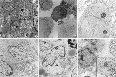
- The Continuing Value of Ultrastructural Observation in Central Nervous System Neoplasms in Children
- Na Rae Kim, Sung-Hye Park
- J Pathol Transl Med. 2015;49(6):427-437. Published online October 13, 2015
- DOI: https://doi.org/10.4132/jptm.2015.09.19
- 10,984 View
- 78 Download
-
 Abstract
 PDF
PDF - Central nervous system (CNS) neoplasms are the second most common childhood malignancy after leukemia and the most common solid organ neoplasm in children. Diagnostic dilemmas with small specimens from CNS neoplasms are often the result of multifactorial etiologies such as frozen or fixation artifact, biopsy size, or lack of knowledge about rare or unfamiliar entities. Since the late 1950s, ultrastructural examination has been used in the diagnosis of CNS neoplasms, though it has largely been replaced by immunohistochemical and molecular cytogenetic studies. Nowadays, pathologic diagnosis of CNS neoplasms is achieved through intraoperative cytology, light microscopy, immunohistochemistry, and molecular cytogenetic results. However, the utility of electron microscopy (EM) in the final diagnosis of CNS neoplasms and investigation of its pathogenetic origin remains critical. Here, we reviewed the distinguishing ultrastructural features of pediatric CNS neoplasms and emphasize the continuing value of EM in the diagnosis of CNS neoplasms.
- Advances in the Endoscopic Assessment of Inflammatory Bowel Diseases: Cooperation between Endoscopic and Pathologic Evaluations
- Jae Hee Cheon
- J Pathol Transl Med. 2015;49(3):209-217. Published online May 15, 2015
- DOI: https://doi.org/10.4132/jptm.2015.04.09
- 15,687 View
- 100 Download
- 5 Web of Science
- 5 Crossref
-
Abstract
PDF
- Endoscopic assessment has a crucial role in the management of inflammatory bowel disease (IBD). It is particularly useful for the assessment of IBD disease extension, severity, and neoplasia surveillance. Recent advances in endoscopic imaging techniques have been revolutionized over the past decades, progressing from conventional white light endoscopy to novel endoscopic techniques using molecular probes or electronic filter technologies. These new technologies allow for visualization of the mucosa in detail and monitor for inflammation/dysplasia at the cellular or sub-cellular level. These techniques may enable us to alter the IBD surveillance paradigm from four quadrant random biopsy to targeted biopsy and diagnosis. High definition endoscopy and dye-based chromoendoscopy can improve the detection rate of dysplasia and evaluate inflammatory changes with better visualization. Dye-less chromoendoscopy, including narrow band imaging, iScan, and autofluorescence imaging can also enhance surveillance in comparison to white light endoscopy with optical or electronic filter technologies. Moreover, confocal laser endomicroscopy or endocytoscopy have can achieve real-time histology evaluation in vivo and have greater accuracy in comparison with histology. These new technologies could be combined with standard endoscopy or further histologic confirmation in patients with IBD. This review offers an evidence-based overview of new endoscopic techniques in patients with IBD.
-
Citations
Citations to this article as recorded by
- Moxifloxacin promotes two-photon microscopic imaging for discriminating different stages of DSS-induced colitis on mice
Yingtong Chen, Xiaoyi Xu, Min Wang, Xiang Wang, Yan Wang, Yong Zhang, Jin Huang, Yuwen Tao, Wentao Fan, Lili Zhao, Li Liu, Zhining Fan
Photodiagnosis and Photodynamic Therapy.2024; 48: 104220. CrossRef - Colorectal cancer in inflammatory bowel disease: review of the evidence
D. S. Keller, A. Windsor, R. Cohen, M. Chand
Techniques in Coloproctology.2019; 23(1): 3. CrossRef - Probe-based confocal laser endomicroscopy in the differential diagnosis of inflammatory bowel diseases: a case series
Jung Won Park, Tae Il Kim, Jae Hee Cheon
Intestinal Research.2018; 16(4): 641. CrossRef - How to Assess and Document Endoscopies in IBD Patients by Including Standard Scoring Systems
Anna M. Buchner, Gary R. Lichtenstein
Inflammatory Bowel Diseases.2016; 22(4): 1010. CrossRef - Nodular lymphoid hyperplasia: A marker of low-grade inflammation in irritable bowel syndrome?
Anna Chiara Piscaglia, Lucrezia Laterza, Valentina Cesario, Viviana Gerardi, Rosario Landi, Loris Riccardo Lopetuso, Giovanni Calò, Giovanna Fabbretti, Massimo Brisigotti, Maria Loredana Stefanelli, Antonio Gasbarrini
World Journal of Gastroenterology.2016; 22(46): 10198. CrossRef
- Moxifloxacin promotes two-photon microscopic imaging for discriminating different stages of DSS-induced colitis on mice
- Utility of Transmission Electron Microscopy in Small Round Cell Tumors
- Na Rae Kim, Seung Yeon Ha, Hyun Yee Cho
- J Pathol Transl Med. 2015;49(2):93-101. Published online March 12, 2015
- DOI: https://doi.org/10.4132/jptm.2015.01.30
- 19,121 View
- 287 Download
- 5 Web of Science
- 4 Crossref
-
Abstract
PDF
- Small round cell tumors (SRCTs) are a heterogeneous group of neoplasms composed of small, primitive, and undifferentiated cells sharing similar histology under light microscopy. SRCTs include Ewing sarcoma/peripheral neuroectodermal tumor family tumors, neuroblastoma, desmoplastic SRCT, rhabdomyosarcoma, poorly differentiated round cell synovial sarcoma, mesenchymal chondrosarcoma, small cell osteosarcoma, small cell malignant peripheral nerve sheath tumor, and small cell schwannoma. Non-Hodgkin’s malignant lymphoma, myeloid sarcoma, malignant melanoma, and gastrointestinal stromal tumor may also present as SRCT. The current shift towards immunohistochemistry and cytogenetic molecular techniques for SRCT may be inappropriate because of antigenic overlapping or inconclusive molecular results due to the lack of differentiation of primitive cells and unavailable genetic service or limited moleculocytogenetic experience. Although usage has declined, electron microscopy (EM) remains very useful and shows salient features for the diagnosis of SRCTs. Although EM is not always required, it provides reliability and validity in the diagnosis of SRCT. Here, the ultrastructural characteristics of SRCTs are reviewed and we suggest that EM would be utilized as one of the reliable modalities for the diagnosis of undifferentiated and poorly differentiated SRCTs.
-
Citations
Citations to this article as recorded by- Electron Microscopy in the Context of a Children's Research Hospital
Cam Robinson
Microscopy and Microanalysis.2020; 26(S2): 1610. CrossRef - Primary bilateral corneal nerve sheath neoplasm in a dog
Marina L. Leis, M. Elyse Salpeter, Bianca S. Bauer, Dale L. Godson, Bruce H. Grahn
Veterinary Ophthalmology.2017; 20(4): 365. CrossRef - Hirnbasissyndrom infolge eines Tumors bei einer 17 Monate alten Deutsch-Holstein-Färse
Wolf Wippermann, Sandra Schöniger, Kerstin Gerlach, Gerald Fritz Schusser, Gabor Köller, Alexander Starke
Tierärztliche Praxis Ausgabe G: Großtiere / Nutztiere.2016; 44(03): 180. CrossRef - The Continuing Value of Ultrastructural Observation in Central Nervous System Neoplasms in Children
Na Rae Kim, Sung-Hye Park
Journal of Pathology and Translational Medicine.2015; 49(6): 427. CrossRef
- Electron Microscopy in the Context of a Children's Research Hospital
- Multiple Jejunal Myeloid Sarcomas Presenting with Intestinal Obstruction in a Non-leukemic Patient: A Case Report with Ultrastructural Observations
- Na Rae Kim, Woon Kee Lee, Jong In Lee, Hyun Yee Cho
- Korean J Pathol. 2012;46(6):590-594. Published online December 26, 2012
- DOI: https://doi.org/10.4132/KoreanJPathol.2012.46.6.590
- 9,892 View
- 73 Download
- 6 Crossref
-
Abstract
PDF
Myeloid sarcoma is a rare extramedullary myeloid tumor, which is frequently misdiagnosed when no evidence of leukemia is initially observed. Here, we report on a peculiar case of a 49-year-old man afflicted with multiple masses in the jejunum, the superior mesentery, and the serosa of the transverse colon, without leukemic manifestation. The tumor was composed of undifferentiated small round cells containing eosinophilic cytoplasm, which were negative for myeloperoxidase, nonspecific esterase, lysozyme, terminal deoxynucleotidyl transferase, leukocyte common antigen, CD3, CD4, CD15, CD20, CD30, CD43, CD56, CD68/PG-M1, CD79a, human melanoma black-45, c-kit, and CD34 with positivity only for CD68/KP1, CD99, and vimentin. Under electron microscopy, those cells had abundant membrane-bound cytoplasmic granules that measured 200 to 300 nm in diameter, which were consistent with granulocytic azurophilic granules. The tumor was finally diagnosed as a myeloid sarcoma. The presence of non-leukemic myeloid sarcomas showing immunonegativity for conventional myeloid-leukemic markers necessitated a diagnosis by ultrastructural observation.
-
Citations
Citations to this article as recorded by- Myeloid sarcoma presenting as intestinal obstruction: A case report of the first presentation of acute myeloid leukemia
Deepsikha Dharamsaktu, Anuradha Pandit, Charanjeet Ahluwalia, Sana Ahuja
International Journal of Surgery Case Reports.2025;[Epub] CrossRef - Myeloid sarcoma of the gastrointestinal tract: Wolf in sheep’s clothing!
Nisha Meena, Surbhi Goyal, Prerna Arora, Sanjeev Sachdeva, Puja Sakhuja
Indian Journal of Pathology and Microbiology.2025; 68(2): 452. CrossRef - Primary ileal myeloid sarcoma presenting with bowel obstruction: a case report
Hitoshi Minagi, Nobuhiko Kanaya, Yoshitaka Kondo, Yoshihiko Kakiuchi, Shinji Kuroda, Ryohei Shoji, Hajime Kashima, Yuki Matsumi, Satoru Kikuchi, Kunitoshi Shigeyasu, Fuminori Teraishi, Shunsuke Kagawa, Toshiyoshi Fujiwara
Surgical Case Reports.2024;[Epub] CrossRef - Isolated myeloid sarcoma presenting with small bowel obstruction: a case report
Rie Mizumoto, Masanori Tsujie, Tomoko Wakasa, Kotaro Kitani, Hironobu Manabe, Shuichi Fukuda, Kaoru Okada, Shumpei Satoi, Hajime Ishikawa, Toshihiko Kawasaki, Hitoshi Hanamoto, Masao Yukawa, Masatoshi Inoue
Surgical Case Reports.2020;[Epub] CrossRef - Primary Myeloid Sarcoma of the Ileum and Mesentery Causing Small Bowel Obstruction: Case Report and Literature Review
Andrej Nikolovski, Dragoslav Mladenovikj, Aleksandra Veljanovska, Gordana Petrusevka
Lietuvos chirurgija.2020; 19(1-2): 55. CrossRef - Utility of Transmission Electron Microscopy in Small Round Cell Tumors
Na Rae Kim, Seung Yeon Ha, Hyun Yee Cho
Journal of Pathology and Translational Medicine.2015; 49(2): 93. CrossRef
- Myeloid sarcoma presenting as intestinal obstruction: A case report of the first presentation of acute myeloid leukemia
- Primary Hepatic Neuroblastoma: A Case Report.
- Geunyoung Jung, Jihun Kim
- Korean J Pathol. 2011;45(4):423-427.
- DOI: https://doi.org/10.4132/KoreanJPathol.2011.45.4.423
- 5,342 View
- 34 Download
- 2 Crossref
-
Abstract
PDF
- Neuroblastoma is a malignant tumor of primordial neural crest origin. It usually develops along the sympathetic nervous system, such as the adrenal glands or paramedian sympathetic chain and metastasizes to the liver most frequently. However, a primary hepatic neuroblastoma has not been reported yet. Here, we report a case of 29-year-old woman who presented with a solitary hepatic mass. Grossly, the mass was large, creamy, rubbery firm, and showed focal hemorrhage and central cavitation. Microscopically, the tumor cells were arranged in small nests of spindle to ovoid cells with abundant neuropil. The neuroblastic nature of the tumor was confirmed by immunohistochemistry and electron microscopy. No extrahepatic mass was found, despite a thorough systemic survey such as chest and abdominopelvic computed tomography (CT) scans and a whole body positron emission tomography-CT study. To the best of our knowledge, this is the first report of a bona fide primary hepatic neuroblastoma.
-
Citations
Citations to this article as recorded by- Primary hepatic neuroblastoma in a 19-month-old child: A case report
Jovana Dimić, Dejan Skorić, Aleksandar Sretenović, Slaviša Đuričić
Medicinska istrazivanja.2022; 55(2): 41. CrossRef - Morphologic Alteration of Metastatic Neuroblastic Tumor in Bone Marrow after Chemotherapy
Go Eun Bae, Yeon-Lim Suh, Ki Woong Sung, Jung-Sun Kim
Korean Journal of Pathology.2013; 47(5): 433. CrossRef
- Primary hepatic neuroblastoma in a 19-month-old child: A case report
- Pulmonary Blastoma with Rhabdomyoblastic Differentiation: A case report with immunohistochemical and electron microscopic examination.
- Joon Mee Kim, Young Chae Chu
- Korean J Pathol. 1992;26(6):620-626.
- 2,414 View
- 14 Download
-
Abstract
PDF
- Pulmonary blastoma is a rare lung tumor composed of epithelial and mesenchymal element : the latter element may show various pattern of differentiation toward mature tissue, such as cartilage, smooth muscle, and bone. Rhabdomyoblastic differentiation in pulmonary blastoma is quire rare. In th literature, only seven cases have been reported. We report a case of pulmonary blastoma with rhabdomyoblastic differentiation which occured in a 3 year old girl. Microscopically, cytoplasmic cross-striation was present. Immunohistochemically, strong positivity for vimentin and desmin was observed. Electron microscopy demonstrated A and I bands which documented rhabdomyoblastic differentiation.
- Solitary Fibrous Tumor A clinicopathologic review of five cases.
- Bum Kyung Kim, Dong Wook Kang, Kyeong Hee Kim, Seong Ki Min, Jin Man Kim, Kyu Sang Song, Dae Yung Kang, Si Whan Choi
- Korean J Pathol. 1999;33(2):115-120.
- 2,388 View
- 10 Download
-
Abstract
- We experienced five cases of solitary fibrous tumor; two in the pleura, two in the orbital soft tissue, and one in the lung parenchyma. Three patients were male, and the age of the patients ranged from 38 to 71 years (mean age: 53.6). Grossly, the masses were well circumscribed and had varying sizes from 2.5 to 30.0 cm. The cut surfaces were grayish-yellow firm with focal variegated hemorrhage, necrosis, cystic change, and myxoid area. Microscopically, these were characterized by a haphazard proliferation of spindle cells or polygonal cells separated by variable amounts of hyalinized collagen and showed a prominent vascular channels reminiscent of hemangiopericytoma in foci. Immunoperoxidase stains showed a strong reactivity for CD34, and were weakly positive for vimentin. Electron microscopical examination revealed features of fibroblast; spindle to round tumor cells were arranged in groups and surrounded by collagen. Nucleoli were seldom prominent. The cytoplasm contained many microfilaments and a moderate number of cisternae of rough endoplasmic reticulum.
- Cryptosporidium Infection of Human Intestine: An Electron Microscopic Observation.
- Min Suk Kim, Yun Kyung Kang, Chul Jong Yoon, Mee Joo, Hye Kyung Lee, Jeong Gi Seo, Je G Chi
- Korean J Pathol. 1999;33(2):121-127.
- 2,291 View
- 10 Download
-
Abstract
- Protozoa of the genus Cryptosporidium are small coccidian parasite known to infect the mucosal epithelium of a variety of animals including human, causing fatal course in immunodeficient patients as well as self-limited illness in healthy individuals. Various life cycle stages including trophozoite, meront, merozoite, gametocyte and oocyst in infected mucosa are a diagnostic feature. Electron microscopy (EM) provides sufficient findings for genus and species identification of this parasitic organism. The authors presented scanning and transmission EM findings of Cryptosporidium parvum infection in two children: one with acute lymphoblastic leukemia and the other without any evidence of immune compromise.
- Secretory Meningioma: Report of 2 cases.
- Dong Sug Kim, Eun Hi Lee, Young Ran Shim, Sang Pyo Kim, Oh Ryong Kim
- Korean J Pathol. 1995;29(3):361-367.
- 2,370 View
- 35 Download
-
Abstract
PDF
- The secretary meningioma is a distinct variant of meningioma that revealed characteristic light microscopic, immunohistochemical and ultrastructural features of epithelial and secretary differentiation, which was named as a distinct subtype of meningioma by Alguacil-Garcia et al in 1986. We experienced 2 cases of secretary meningioma. One was a 53-year-old female who had suffered from sudden onset of dizziness for I day. The computerized tomography revealed a sharply marginated well enhanced mass in temporal lobe. The other was a 59-year-old female who had suffered from dizziness for 8 years. The computerized tomography revealed a well demarcated lobulated mass in petrosal ridge. In both cases, multiple hyaline inclusions were scattered in the background of meningothelial meningioma. They were PAS positive, diastase resistant, stained yellow with van Gieson, and did not stain with reticulin in contrast to Psammoma bodies. The immunohistochemistry revealed positive reaction for EMA, CEA, a-FP and cytokeratin. T'he electron microscopic study revealed interdigitation with desmosomes and abundant intracellular lumina. They were lined by numerous microvilli and filled with granular material which was composed of electron dense homogenous material, me branous material, and small membrane-bound vesicles. Microvilli were filled with electron dense material identical to the material in the lumina, and some of them were interconnected with electron dense material in the lumina. It was concluded that secretary activity of the meningothelial cells and degenerated microvilli were involved in the pathogenesis of hyaline inclusions.
- Capillary Plexus and Vasa of the Rat Lung as Revealed by Scanning Electron Microscope of Corrosion Casts.
- Kun Young Kwon, Kam Rae Cho, Sang Pyo Kim, Kwan Kyu Park, Eun Sook Chang
- Korean J Pathol. 1993;27(1):11-18.
- 2,385 View
- 20 Download
-
Abstract
PDF
- The pulmonary microvasculatures of rats were studied by injection replicas prepared from Mercox. This medium enabled us to easily obtain consistent, stable, and complete injected replicas of the pulmonary vasculature. In order to investigate the three dimensional structures of the tributaries of the bronchial artery, such as the capillary plexus and vasa vasorum, we performed a scanning electron microscopic(SEM) study of the vascular casts, using Mercox CL-2B as a media. The alveolar capillaries revealed hexagonal or pentagonal rings of vascular networks. In some areas, the vascular rings composed a square network, The bronchial tree was supplied by the bronchial arteries which form a coarse capillary extending as far as the terminal bronchioles. Occasionally the capillary plexus was connected with adjacent capillary networks in and around the alveolar walls. The walls of the pulmonary artery revealed only a single layer of the vasa vasorum, but those of the pulmonary vein were surrounded by more complicated and well developed vasa vasorum than the pulmonary arterial side. The mean diameters of the venous vasa vasorum are greater than the arterial vasa vasorum.
- Epithelioid Hemangioendothelioma of Liver: A case report.
- Yoo Jin Kim, Jae Hwa Lee, Bang Hur, Man Ha Hur
- Korean J Pathol. 1995;29(3):378-384.
- 2,253 View
- 24 Download
-
Abstract
PDF
- Epithelioid hemangioendothelioma of liver is a very rare tumor of vascular origin, the most peculiar feature of which is that it is composed of endothelial cells closely resembling epithelial cells. We present a primary epithelioid hemangioendothelioma of liver in a 40-year-old male. This tumor was composed of an ill-defined yellowish white, 7 x 4cm sized, firm, solid mass and small satellite nodules in the right lobe of liver. Microscopically, two types of tumor cells-dendritic and epithelioid-were identified. The neoplastic cells infiltrated into sinusoids and intrahepatic veins. The background of tumor showed marked sclerotic change and focal proliferation of bile ductules. Confirmation of the endothelial origin of these cells was provided by positive immunoperoxidase staining for factor VIII-related antigen, and by electron mi-croscopic demonstration of Weibel-Palade body. This is the first case of epithelioid heman- gioendothelioma of liver documented in Korea. We report this case in view of its scarcity and distinctive morphologic features that allow differentiation from sclerosing carcinoma and angiosarcoma.
- Polyomavirus Renal Infection Confirmed by Electron Microscopy in a Patient with Acquired Immunodeficiency Syndrome: An Autopsy Case Report.
- Na Rae Kim, Byoung Kwon Kim, Je G Chi
- Korean J Pathol. 2001;35(2):168-171.
- 2,032 View
- 56 Download
-
Abstract
PDF
- Polyomavirus infection commonly occurs in childhood and adolescence, remaining in a latent status and reactivated in an immunocompromised status. We report herein an autopsy case of HIV-positive 41-year-old male, who succumbed to disseminated Kaposi sarcoma and cytomegalovirus infection involving the gastrointestinal tract, lung and brain. The involved kidney showed minimal inflammatory infiltrates and tubular injury: the nuclei of tubular epithelial cells were markedly enlarged with central clearing and peripheral chromatin margination or bore basophilic nuclear inclusions. Inclusion-bearing tubular epithelial cells were negative for the viral immunostains including herpes simplex virus, Epstein-Barr virus and adenovirus. Electron microscopy disclosed 42 nm intranuclear viral particles compatible with the BK polyomavirus. The viral particles were icosahedral in paracrystalline array and nonenveloped.
- Adrenocortical Oncocytoma: A Case Report.
- Hun Soo Kim, Dae Young Kang
- Korean J Pathol. 2007;41(5):329-333.
- 2,237 View
- 18 Download
-
Abstract
PDF
- Adrenocortical oncocytomas have rarely been reported on in the medical literature, and most of them have been nonfunctional and benign. We report here on a case of a 43-year-old man with a left abdominal mass. The patient showed no signs of hypertension or hormonal imbalance. The abdominal CT scans showed a huge mass that measured 11 cm in diameter, and it was located at the left adrenal area. Grossly, the tumor was well encapsulated and homogenous with central necrosis. Microscopically, the tumor was composed of oncocytes with abundant granular cytoplasm. Immunohistochemically, these cells were diffusely positive for cytokeratin and focally positive for synaptophysin and NSE. The ultrastructural studies showed numerous mitochondria in the cytoplasm. We will discuss the criteria that indicates malignancy as presented by Weiss et al. and we summarize the difference between conventional and oncocytic adrenocortical neoplasm. This case showed some features of malignancy based on the criteria presented by Weiss et al.
- Primary Ciliary Dyskinesia(Immotile Cilia Syndrome): Clinical and electron microscopic analysis of 17 cases.
- Je G Chi, Chul Jong Yoon
- Korean J Pathol. 1993;27(2):99-107.
- 2,707 View
- 21 Download
-
Abstract
PDF
- Immotile cilia syndrome is a genetically determined disorder characterized by immotility or poor motility of the cilia in the airways and elsewhere. Since its first description in 1976 determination of a ciliary abnormality has now clarified its variable expression and pleiotropism. Certain specific defects in the ciliary axoneme can be found and are pathognmoic of the syndrome. These defects include missing dynein arms, abnormally short dynein, arms, spokes with no central sheath, missing central microtubules, and displacement of one of the nine peripheral doublets. We have studied 80 cases of bronchial or nasal mucosal biopsies that were performed with the suspicion of immotile cilia syndrome. Of 80 cases only 17 cases were sampled optimally to be able to observe under transmission and scanning electron microscopes. All 17 cases had certain abnormality of the cilia. They consisted of Ia(3 cases), Ib(3 cases), Id only(3 cases) and Id+other types(6 cases) a according to Sturgess classification. Seven cases consisted of 1 solitary and 6 combined form; II+Id(1 case) and II+Id+III(5 case). All 5 cases of type III were combined with Id and II. Clinically most pronounced manifestations were cough(82%), sputum(59%), rhinorrhea(41%) and nasal stuffiness(35%), All the patients were below the age of 15 years, and there were 6 boys and 11 girls.
- Large Cell Neuroendocrine Carcinoma of the Lung: Report of three cases.
- Jai Hyang Go, Sun Ree Jung, Dong Hwan Shin, Woo Hee Jung
- Korean J Pathol. 1995;29(4):511-516.
- 2,292 View
- 12 Download
-
Abstract
PDF
- We report three cases of neuroendocrine tumors of the lung characterized by large pleomorphic cell with frequent mitosis, which show neuroendocrine differentiation by both light microscopy or electron microscopy and iminunohistochemistry. These tumors have been categorized as large cell neuroendocrine carcinoma by Travis et al.(1991) in contrast with non-small cell lung cancer with neuroendocrine differentiation. In the latter, neuroendocrine differentiation is not evident by light microscopy and must be demonstrated by imunohistochemstry or by electron microscopy. The prognosis of large cell neuroendocrine carcinoma, together with non-small cell lung cancer with neuroendocrine differentiation, appears to be worse than cancer without neuroendocrine differentiation and intermediate between atypical carcinoid and small cell lung cancer. Larger numbers of patients will be needed to demonstrate significant differences in survival.
- Analysis of Pulmonary Asbestos Body in Malignant Mesothelioma: A case report.
- Hoon Kyu Oh, Jae Yoe Ro, Chul Jong Yoon, Je Geun Chi
- Korean J Pathol. 1999;33(5):361-366.
- 2,316 View
- 20 Download
-
Abstract
PDF
- The association between occupational asbestos exposure and the subsequent development of malignant mesothelioma of pleura is well recognized. We analyzed an asbestos body by energy dispersive X-ray analyser in a case of malignant mesothelioma of pleura who had a history of asbestos exposure 30 years ago. In transmission electron microscope, the asbestos body was composed of a core of refractile thin asbestos fiber bundle and beaded masses of electron-dense iron and protein complex. The core fibers were analyzed as an amphibole type crocidolite fiber [(Na2Fe3Fe2(Si8O22)(OH)2] which composed of high content of silicon, iron and sodium.
- A Scanning Electron Microscopic Study on Microvascular Changes in the Monocrotaline-induced Rat Lung by Corrosion Casting Method.
- Na Hye Myong, Eui Keun Ham
- Korean J Pathol. 1995;29(5):644-659.
- 2,177 View
- 10 Download
-
Abstract
- To investigate the microvascular changes in primary pulmonary hypertension, the lungs of 24 Sprague-Dawley rats were treated by an intraperitoneal injection of 2% monocrotaline(MCT) solution and then examined with scanning electron microscopy(SEM) after microvascular corrosion casting. Histologic examination revealed significant medial thickening in the small to medium-sized pulmonary arteries. Scanning electron microscopic findings of the normal lungs showed two kinds of microvascular structures. One showed a well-fortned three-dimensional basket structure of uniform flat-tubular alveolar capillaries, which were connected to each other in a T or Y shape or at right angles. The other revealed a two-dimensional reticular sheet of round tubular branches mainly in the bronchial artery-supplying regions. The MCT-treated groups(remodelling) showed apparent changes in both kinds of microvasculatures in comparison to the normal group but the more prominent change was found in Lbe bronchial artery microvasculature showing the dense thick encasement around large pulmonary arteries. Alveolar microvasculature of the pulmonary artery revealed individually enlarged angular appearance, with generally deformed alveolar architecture. Quantitatively, the significant enlargement of diameter and intercapillary distance appeared in both microvasculatures of MCT-induced rat lungs, but the density was increased only in the bronchial artery microvasculature. In conclusion, our three-dimensional microvascular study of the MCT-treated rat lungs demonstrates a new morphologic finding of vascular remodeling in primary puhnonary hypertension, which is thought to play an important vascular role in the pathogenesis in addition to interstitial fibrosis.
- Morphologic Changes of Pulmonary Tissue Secondary to Sidestream Cigarette Smoke.
- Kun Young Kwon, Ji Min Jeon, Sang Pyo Kim, Kwan Kyu Park, Dae Hyun Kim
- Korean J Pathol. 1999;33(6):395-403.
- 2,166 View
- 20 Download
-
Abstract
PDF
- Chronic bronchiolitis is a condition associated with cigarette smoking, and later associated with pulmonary parenchymal alteration and progressive deterioration of lung function. Early respiratory bronchiolitis was produced in Sprague-Dawley rats by indirect inhalation of cigarette smoke daily in a smoke exposure chamber designed by authors for 1 month. Experimental group A (n=5) was sacrificed after having smoked 30 cigarettes, group B (n=5) after 80 cigarette, and group C (n=7) after 140 cigarettes, respectively. Examination of morphologic changes in the lungs was done on light microscope, transmission and scanning electron microscopes. Light microscopically, increase in number of goblet cells in the bronchial mucosa, brown-pigmented macrophages in the alveoli, multifocal alveolar collapse adjacent to the bronchioles, dilatation of alveolar ducts and alveolar spaces were observed. Transmission electron microscopically, irregularly shaped Clara cells, alveolar wall collapse, and focally type I epithelial cell injury were seen. Scanning electron microscopically, scattered alveolar collapse, irregular dilatation of alveolar ducts, alveolar spaces and interalveolar pores (pores of Kohn) were seen. The terminal and respiratory bronchioles showed morphological alteration of Clara cells, but no evidence of cellular bronchiolitis or bronchiolar obstruction. We conclude that sidestream smoke induces an early respiratory bronchiolitis including aggregates of brown pigmented macrophages and varying degrees of structural alteration of adjacent pulmonary parenchyma.
- Rasmussen's Encephalitis.
- Na Rae Kim, Han Jae Joon, Yeon Lim Suh, Moon Hyang Lee
- Korean J Pathol. 2001;35(5):455-460.
- 2,233 View
- 13 Download
-
Abstract
PDF
- We herein report a case of intractable epilepsy that occurred in a 7-year-old girl, which is consistent with radiological and clinicopathological hallmarks of Rasmussen's encephalitis. The patient showed characteristic primary unilateral involvement with secondary bilateral propagation. Microscopically, the cortical atrophy due to neuronal loss, intense GFAP-immunoreactive astrogliosis, neuronophagia, perivascular lymphocytic infiltration and microglial nodules was seen throughout the cortex and white matter. No viral inclusions were noted; no cytomegalovirus, herpes simplex virus or Epstein-Barr virus was found by in situ hybridization. Granular immunofluorescence for C4, C1q and IgG within the blood vessel walls was noted, and ultrastructurally, only nonspecific vascular injury was found. Rasmussen's encephalitis is a diagnosis of exclusion; it can be diagnosed by the combination of clinical manifestation, neuroimaging and characteristic pathologic features.
- Apoptosis of Alveolar Cells in Pneumocystis Carinii Pneumonia: Application of Electron Microscopic Terminal Deoxynucleotidyl Transferase-Mediated dUTP-Biotin Nick End Labeling Method.
- Kyu Hun Kang, Sang Pyo Kim, Kun Young Kwon
- Korean J Pathol. 2001;35(6):496-505.
- 2,192 View
- 10 Download
-
Abstract
- BACKGROUND
Pneumocystis carinii (P. carinii) attaches to alveolar cells and causes injury to the epithelial cells by direct toxic effects or inhibition of epithelial growth and replication. Although respiratory cell damage or death is a common feature in P. carinii pneumonia, there has been little reports about expression of apoptosis of the lung tissue in the literatures.
METHODS
We examined expression of fibronectin and vitronectin in the interaction between P. carinii and alveolar cells, and in situ terminal deoxynucleotidyl transferase-mediated dUTP biotin nick end-labeling (TUNEL) expression of apoptosis in the respiratory cells by immunohistochemistry and pre-embedding immunoelectron microscopy.
RESULTS
Light microscopic (LM) and electron microscopic (EM) immunohistochemical stains for the fibronectin and vitronectin showed strong expressions on the pellicles and tubular extensions of P. carinii and weak expression along the surfaces of type I alveolar cells. LM and EM TUNEL stains showed positive expression in the nuclei of alveolar cells, apoptotic bodies in the cytoplasm of alveolar macrophages and cellular debris in alveolar spaces.
CONCLUSIONS
P. carinii induces injury and apoptosis of alveolar cells after attachment of the organisms to host cells, and alveolar macrophages enhance the clearance of apoptotic bodies of alveolar cells as well as phagocytosis and degradation of P. carinii.
- Scanning Electron Microscopic Findings of Hair Anomalies.
- Chul Jong Yoon, Je G Chi, Hai Won Chung
- Korean J Pathol. 1993;27(5):491-500.
- 2,694 View
- 28 Download
-
Abstract
PDF
- Scanning electron microscopic(SEM) examination of hair is an efficient and definite method for the diagnosis of hair anomaly. The Hair specimen is placed on a stub, gold coated, and directly viewed without prior fixation or treatment for identification. We have analyzed 25 cases of scalp hair and 2 cases of eyebrow that were sampled with the suspicion of hair anomalies at Seoul National University Hospital during the last 7 year period from January 1988 to June 1992. All 27 cases had certain abnormalities of the hair. They consisted of monilethrix(1 case), hair casts(4 cases), pili torti(1 case), uncombable hair syndrome(1 case), trichorrhexis nodosa(5 cases), woolly hair(5 cases) and other anomaly(1 case). Some cases of them were mixed with two or three anomalies in hairs. Their combinations were; hair casts and woolly hair(1 case), hair casts and pili torti and uncombabla hair syndrome(1 case), hair casts and uncombable hair syndrome and trichorrhexis nodosa(1 case), trichorrhexis nodosa and uncombable hair syndrome(3 cases). Most of these defects included brittleness of scales and distortion of the hair shaft.
- Carcinosarcoma (True Malignant Mixed Tumor) of the Parotid Gland: A Report of a Case with Small Cell Carcinoma as the Carcinoma Component.
- Hun Soo Kim, Hyang Jung Cho, Yeun Tai Chung, Soon Ah Park, Hae Joong Cho, Jin Man Kim
- Korean J Pathol. 2008;42(3):175-180.
- 2,662 View
- 24 Download
-
Abstract
PDF
- True malignant mixed tumor (carcinosarcoma) of the salivary gland is an extremely rare tumor. By definition, it is a heterologous neoplasm that's composed of both malignant epithelial and mesenchymal components. We report here on a case of an 83 year old female patient with a parotid gland mass. Histologic examination showed a biphasic pattern of growth with both a poorly differentiated epithelial component and a sarcomatous spindle cell background. The immunohistochemical and ultrastructural findings support the diagnosis of a small cell neuroendocrine epithelial tumor and an undifferentiated malignant mesenchymal features. The histologic and ultrastructural features along with the immunohistochemical findings are presented. We also review the literature and we discuss the different opinions on the exact histogenesis of the true malignant mixed tumor of the salivary gland. To the best of our knowledge, this histological pattern has not been previously reported in the English medical literature.
- The Effect of Ginseng Saponin on the Dopaminergic Neurons in the Parkinson's Disease Model in Mice.
- Chang Ok Kim, Ki Sok Kim, Young Buhm Huh, Byeong Woo Ahn, Beom Seok Han, Kwang Sik Choi, Ki Yul Nam, Sang Woo Juhng
- Korean J Pathol. 1997;31(9):805-814.
- 3,215 View
- 37 Download
-
Abstract
PDF
- Saponin has been known to be a major antioxidant component in panax ginseng. Recent experimental study suggests that some antioxidant materials prevent Parkinson's disease caused by 1-methyl-4-phenyl-1,2,3,6- tetrahydropyridine (MPTP) in an animal model. The present study was performed to demonstrate the effect of ginseng saponins in the Parkinson's disease model induced by MPTP. To verify the effect of ginseng saponin on dopaminergic neurons in the mice brain, the tyrosine hydroxylase-immunoreactive (TH-ir) neurons were observed by immunohistochemical stain and immunoelectron microscopy (preembedding method). Also, in order to estimate the immunoreactivity of dopaminergic neuropils, they were quantified by image analysis. The number of TH-ir neurons of substantia nigra was significantly increased in the high-dose (0.46 mg/kg) ginseng saponin group compared with the MPTP injected group. The immunoreactivity of TH-ir neuropils in striatum was significantly increased in both high and low-dose (0.1 mg/kg) ginseng saponin groups compared with the MPTP injected group. In immunoelectron microscopic observation, TH-ir neurons of the control and both ginseng saponin injected group showed normal nuclei and well preserved cytoplasmic organelles. In the MPTP injected group, dying dopaminergic neurons showed destroyed nuclei and cytoplasmic organelles. These results suggest that ginseng saponin has a protective effect on the Parkinson's disease model induced by MPTP.
- Birefringent Particles as an Effective Factor in Usual Interstitial Pneumonia.
- Min Jung Kim, Seung Yeon Ha, Sung Hwan Jeong, Bongkyung Shin
- Korean J Pathol. 2008;42(4):198-201.
- 2,209 View
- 21 Download
-
Abstract
PDF
- BACKGROUND
It has long been recognized that birefringent paticles (BP) are associated with pulmonary disease. And there is increasing evidence that BP cause fibrotic reaction within the lung depending on both particle size and composition.
METHODS
We collected 41 cases of usual interstitial pneumonia (UIP) and 101 cases of normal lung tissue from control group including squamous cell carcinoma, adenocarcinoma and bullae. BPs in the 0.1 to 10 micrometer size range under polarizing microscope was measured and counted by image analyzer.
RESULTS
BP counts are mean 244.05/10 HPF in UIP and 71.4/10 HPF in control group. BPs in UIP is three times more than control (p=0.000). It increased significantly by the age of patients (p=0.000).
CONCLUSIONS
BPs in lung might be important cause of inflammation and fibrosis in UIP.
- Ultrastructural Changes in Rat Kidney after Lead Acetate Administration.
- Hyun Chul Kim, Seung Pil Kim, Kwan Kyu Park
- Korean J Pathol. 1996;30(2):73-88.
- 2,182 View
- 19 Download
-
Abstract
PDF
- This study was carried out to investigate the ultrastructural findings of rats after administration of 0.5% lead acetate with drinking water. The Sprague-Dawley rats were divided into control and experimental groups. The control group was composed of 12 rats and was orally administered with 0.5% sodium acetate. The experimental group was composed of 36 rats and orally administered with 0.5% lead acetate. Two rats in the control group and four rats in the experimental group were sacrificed on day 2, and week 1, 2, 4, 6 and 8 after administration. The kidney was extirpated and examined by electron microscopy. The results obtained were as follows: The blood lead concentration in the experimental group began to increase from the second day after administration and it increased gradually until the 6th week and it decreased at the 8 week. The urinary excretion of delta-ALA also increased from the secondary and gradually increased up to the 8th week. On electron microscopic examination, the proximal tubular cells showed fat droplets, dilatation of the endoplasmic reticulum, mitochondrial swelling, increased numbers of secondary lysosomes and myelin figure-like residual bodies and intranuclear inclusion bodies. All these findings peaked at the eighth week after administration. Ultrastructural findings after Timm sulphide silver reaction revealed the lead granules in the proximal tubular lumen and between the microvilli of the proximal tubular cells without membrane-bounded. It can be concluded that most of the changes of micro-organelles are compatible with degenerative changes of lead exposure and passive diffusion of lead granules are involved in the proximal tubular cells.
- Cytologic Findings of Parathyroid Carcinoma: Report of Two Cases.
- Yun Hee Jin, Yong Wook Park, Mi Sheon Jin, Seung Sam Paik, Se Jin Jang, Moon Hyang Park
- J Pathol Transl Med. 2003;14(1):1-6.
- 2,669 View
- 35 Download
-
Abstract
PDF
- Parathyroid carcinoma is a rare disorder accounting for 0.5% to 5% of parathyroid neoplasia. Diagnosis of parathyroid carcinoma in fine needle aspiration cytology(FNAC) is difficult because all characteristic features of parathyroid carcinoma can be recognized in parathyroid adenoma or hyperplasia. Cellular atypism cannot be used for the diagnostic criteria of parathyroid carcinoma as malignancies of most other organs. We experienced two cases of cytologic features of parathyroid carcinoma confirmed by histologic examination. The majority of tumor cells formed large cohesive clusters, although individual tumor cells were also present. The tumor cells displayed rather pleomorphic round to oval nuclei, occasional prominent nucleoli, and distinct cytoplasmic margin. Occasionally karyolysis, anuclear cells, and nonepithelial cell clusters were noted. The histologic findings showed a partially lobulated architecture, with admixture of sheets of chief cells, oxyphil cells, and occasional water clear cells. The tumor infiltrated into the thyroid parenchyme and perithyroidal soft tissue. The electron microscopic study of case 1 disclosed typical findings of parathyroid neoplasm; clusters of secretory chief cells with centrally located round to ovoid nuclei, moderately clumped heterochromatins and one or two nucleoli. The tumor cells showed conspicous interdigitation of contiguous cell membrane and intercellular microvilli.
- Ultrastructural Changes of the Bile Canaliculi after Common Bile Duct Ligation.
- Kook Seon Yoo, Suk Hee Lee, Hee Kyung Park, Chang Ho Cho, Jong Min Chae
- Korean J Pathol. 1996;30(3):175-183.
- 2,583 View
- 31 Download
-
Abstract
PDF
- The purpose of this study was to investigate the morphologic changes of the bile canaliculi and its associated structures of the liver induced by common bile duct ligation(CBDL) in the rat. The canalicular surface and lateral surface of the dry-fractured hepatocytes was studied with scanning electron microscopy at 1~6 weeks post ligation. The first week after CBDL, the bile canaliculi were dilated. The microvilli were increased in number and the lumens contained granular materials After 2 weeks or more, the bile canaliculi were dilated to a variable degree, and with irregularity, measuring from 1.5 to 5 micrometer in diameter, and in the advanced stage, the canaliculi showed blunting and the disappearance of microvilli. Some canaliculi had sprouting side branches. At 4~6 weeks post-ligation, the lateral surface of the hepatocytes also showed some irregularity and a tortuous appearance, and numerous small sized microvillous projections were formed. The tubular structures of the proliferated SER distributed adjacent to the lateral surface of the hepatocytes, and the direct connection of a tubular structure and the cytoplasmic membrane was observed. These results suggest that the deformity and loss of microvilli of bile canaliculi reflect the disturbance of bile secretion from the hepatocytes. And prolonged obstruction of bile flow may result in bile excretion via the lateral surface of hepatocytes.
- Bronchoalveolar Lavage (BAL) Cytology and Ultrastructural Findings in a Patient with Amiodarone-Induced Pulmonary Toxicity: A Case Report.
- Sun Lee, Min A Kim, Young Soo Shim, Chun Taek Lee, Je G Chi, Doo Hyun Chung
- Korean J Pathol. 2002;36(3):175-178.
- 2,839 View
- 46 Download
-
Abstract
PDF
- Amiodarone is a potent antiarrhythmic agent and can cause potentially life-threatening pulmonary fibrosis. Of the numerous side effects associated with amiodarone therapy, lugn toxicity is one of the most serious adverse reactions. Recently, we experienced a case of amiodarone-induced pulmonary toxicity (APT), which induced severe dyspnea and productive coughing, confirmed by cytologic and electron microscopic examination of the bronchoalveolar lavage (BAL). The symptoms and abnormalities in the chest X-ray were improved after the withdrawal of amiodarone. Cytologic examination of the BAL revealed numerous foam cells with cytoplasmic vacuoles or small particles. Ultrastructurally, the foam cells demonstrated characteristic lysosomal inclusions, which were electron-dense multilamellated bodies, crystalloid bodies, and mixed forms with small lipid vacuoles. It is strongly suggested that only cytologic and electron microscopic examination of the BAL without open lung biopsy is enough for diagnosis of APT, when APT is clinically suspected in a patient who has a history or ingestation of amiodarone.
- Microcystic Meningioma: A case report.
- Gyeong Sin Lee, Il Seon Lee, Bang Hur, Man Ha Hur
- Korean J Pathol. 1994;28(2):185-190.
- 2,825 View
- 69 Download
-
Abstract
PDF
- Microcystic meningioma, a distinct morphological variant of meningiomas, is histologically characterized by a vacuolated appearance with multiple cystic spaces lined by vacuolated or stellate-shaped tumor cells. We report a case of microcystic meningioma occuring in right frontoparietal area of 42-year-old woman, with emphasis on differential diagnosis, along with a review of literatures. Immunohistochemically, most of the tumor cells demonstrated positive immunoreactivity for both epithelial membrane antigen and vimentin. Electron microscopy showed that the extracellular space was extensive, where eletron-lucent material was occasionally seen. The tumor cells had long cytoplasmic processes showing complex interdigitation and a large number of desmosomes.
- An Immunohistochemical Study on the Distribution of Endotoxin.
- Tae In Park, Jung Ja Park, Jyung Sik Kwak, In Soo Suh
- Korean J Pathol. 1994;28(3):260-271.
- 1,996 View
- 10 Download
-
Abstract
- This study was performed to investigate the distribution of endotoxin in various organs after intraperitoneal injection of E. coli homogenator(0111:B4, 3X10(9)cells/200g of body weight). Sprague-Dawley rats were intraperitoneally injected with E. coli homogenator and sacrificed 1 and 3 hours after injection. The lung, liver, and kidney were immunohistochemically stained with avidin-biotin complex method and observed by light and electron microscopy. On the light microscopy, granular deposits of reaction products of immunohistochemical stain were found on the cytoplasmic membrane of endothelial cells and some of parenchymal cells of all organs observed. Electron microscopic study revealed finely granular reaction products on the surface of endothelial cells and some of parenchymal cells. The pinocytotic vesicles of endothelial cells demonstrated reaction products in the early phase of experiment. The distribution of reaction products were prominent in the liver among three organs. The Kupffer cells showed the most sensitive and strongest positive reaction. The hepatocytes and endothelial cells revealed weak positive reaction 3 hours later. The alveolar macrophages of the lung were also positive from the early phase of endotoxemia, while the pneumocytes and alveolar septa demonstrated weakly positive reaction in the later phase. The capillary endothelium of the kidney revealed positive reaction from the early phase. According to above results, it is concluded that the endotoxin entered into the systemic circulation was captured in the liver and lung. And both mononuclear phagocytic system and endothelial cells could be activated or damaged by endotoxin.
- The Morphologic Changes of the Sinusoidal Endothelial Cells in N-diethylnitrosamine Induced Cirrhotic Rat Liver.
- Ok Ji Paik, Hee Kyung Park, Jong Min Chae, Jyung Sik Kwak, Tae Joong Sohn
- Korean J Pathol. 1996;30(7):604-615.
- 2,311 View
- 13 Download
-
Abstract
PDF
- The purpose of this study is to investigate the morphologic changes of the sinusoidal endothelial cells and the associated structures of the cirrhotic rat liver induced by repeat intraperitoneal injections of N-diethylnitrosamine (DEN) (100 mg/kg/week). One day to 6 weeks later, rat livers were observed under the light, transmission and scanning electron microscopy, and immunostained with laminin antibody. Two weeks after DEN treatment, the fibrillar material in Disse's space was noted, and then a basement membrane-like structure was found at 4 weeks after treatment. Laminin was detected in perisinusoidal areas after 4 weeks. Laminin was strongly positive on the fibrous septum and in the sinusoidal wall of cirrhotic nodules after 6 weeks of treatment. The diameters and numbers of sinusoidal endothelial fenestrations did not change significantly until 2 weeks. They decreased within 4 weeks, and then the sinusoidal endothelium was poorly fenestrated at 6 weeks after DEN treatment. These results suggest that as fibrosis develops in cirrhosis, the deposit of extracellular matrix such as laminin within Disse's space is a major contributing factor in the structural alteration of sinusoidal endothelial cells, and the capillarization of the sinusoidal endothelial cells may be a contributor to impairment of the hepatic function in cirrhosis.
- Extrarenal Malignant Rhabdoid tumor: A Case Report.
- Sang Yong Lee, Dae Cheol Kim, Seo Hee Rha, Sook Hee Hong, Tae Hun Kang, Young Ho Lee, Kyoung Jin Nam, Jin Sook Jeong
- J Pathol Transl Med. 1996;7(1):69-74.
- 2,468 View
- 23 Download
-
Abstract
PDF
- Malignant rhabdoid tumor is a distinct renal tumor in the pediatric age group. It was originally described as a rhabdomyosarcomatoid variant of Wilms tumor. However, subsequent studies failed to confirm myogenous differentiation, so it is now considered to be a distinct and unique type of highly malignant tumor, histogenetically unrelated. Although extrarenal forms of this tumor are rare, several examples have been described in other sites, especially the liver, prostate, paravertebral area, urinary bladder and soft tissue. We experienced a case of malignant rhabdiod tumor located in the intraabdominal cavity in a 10 month-old boy. Smear of peritoneal fluid showed round, polygonal and irregular shaped cells with large nuclei, ample cytoplasm containing Jight pink "to purple cytoplasmic inclusions, and one or a few prominent nucleoli. Immunocytochemistry revealed positivity to cytokeratin, epithelial membrane antigen and vimentin, and negativity to desmin and neuron-specific enolase. These distinct cytologic appearance and immunophenotypes were most consistent with a diagnosis of extrarenal malignant rhabdoid tumor. The cytoplasmic inclusions were correlated with eosinophilic inclusions seen in histologic section and electron microscopy confirmed this interpretation, showing filamentous aggregations in the cytoplasms of the tumor cells.
- Sarcomatoid Renal Cell Carcinoma; Special Reference to its Distinction from Carcinosarcoma.
- Kee Taek Jang, Yeon Mee Kim, Je Geun Chi
- Korean J Pathol. 1998;32(5):378-381.
- 2,053 View
- 10 Download
-
Abstract
- Sarcomatoid renal cell carcinoma is an uncommon tumor that has to be distinguished from renal carcinosarcoma. We have described three cases of sarcomatoid renal cell carcinoma showing different clinical and light microscopic features. An ultrastructural study of the tumor cells from the sarcomatoid area revealed frequent desmosomal junction, confirming the epithelial nature of the neoplasm. All three cases showed an aggressive clinical course and tended to invade adjacent organs or tissues. We believe that an histological and immunohistochemical examination in conjunction with an electron microscopic examination are necessary to diagnose sarcomatoid renal cell carcinoma.
- Expression of Antigenic Surface Molecules of Pneumocystis Carinii by Immunoelectron Microscopic Examination.
- Kun Young Kwon, Seung Che Cho, Sang Pyo Kim, Kwan Kyu Park, Eun Sook Chang
- Korean J Pathol. 1998;32(6):393-403.
- 2,160 View
- 10 Download
-
Abstract
- This study was carried out to investigate the morphologic characteristics and localization of antigenic molecules of Pneumocystis carinii in experimentally induced P. carinii pneumonia in rats. After six weeks of administration of low protein diet and dexamethasone, Sprague-Dawley rats were sacrificed to submit lungs or bronchoalveolar lavage for the study. Monoclonal (092, 900, 902, and 904) and polyclonal (SP-D) antibodies were used for immunohistochemistry and immunoelectron microscopy (ITEM and ISEM). Immunohistochemically P. carinii organisms were well identified as clusters or separated forms in the alveolar spaces being frequently attached to the alveolar walls. Immunoelectron microscopically the adherences of gold particles were observed on the surface of all stages of the P. carinii. Occasionally positive immunogold labeling was observed in the cytoplasm of the trophozoites and on the pellicle of the intracystic bodies within the cysts. The monoclonal antibodies 092, 900, 902, and 904 reacted mainly with pellicles of P. carinii, whereas SP-D labeled on the pellicles, intracystic bodies, cytoplasms of the alveolar macrophages, and free floated surfactant material in the alveolar spaces. The immunogold particles were observed more diffusely and intensely in the cysts than in the trophozoites. These results indicate that antigen is mainly localized on the pellicles, and accumulated during development from the trophozoite to the cyst stages.
- Light Electron Microscopic Study in Rat Livers Following Cadmium Chloride Administration.
- Kwan Kyu Park, Young Ho Kim, Kun Young Kwon, Eun Sook Chang, Moo Ung Chang
- Korean J Pathol. 1992;26(1):28-39.
- 2,280 View
- 16 Download
-
Abstract
PDF
- This study was carried out to investigate the light and electron microscopic findings of the livers of rats after an intraperitoneal injection of cadmium chloride. The Sprague-Dawley rats were intraperitoneally injected with cadmium chloride dissolved in water, once a day for three days. These animals were sacrificed at 1, 3, 8, and 24 hr after the last injection. Control groups of the rats were also sacrificed in the same manner. The liver was extirpated and examined by both light and electron microscopy. The results obtained are as follows: The parenchyma of the liver shows focal neutrophilic infiltration and spotty necrosis. The hepatocytes show fatty change, ballooning degeneration, swelling of the endoplasmic reticulum and mitochondria, increase numbers of secondary lysosomes and residual bodies. Focal patic venules and sinusoids of the liver are congested. The Kupffer cells are increase in number. Therefore, it can be concluded that the cadmium is directly acted to hepatocytes resulting in cellular injuries and deposits in the fat droplets of the cytoplasm of the hepatocytes, not Ito cells as previously suggeted.
- Ultrastructural Observation on Maturation Process of Cytomegalovirus in Human Cells.
- Chul Jong Yoon, Je Geun Chi
- Korean J Pathol. 1994;28(5):478-484.
- 2,341 View
- 18 Download
-
Abstract
PDF
- Cytomegalovirus(CMV) infection can be diagnosed by finding characteristic intranuclear inclusion body and enlargement of the cell size congenital CMV infection can be associated with various types of anomalies seen in different gestational age. These anomalies are probable due to direct virus infection of the parenchymal cell m early gestation. Based on four autopsy cases of congenital CMV infection we have studied maturation process of virus particles in parenchymal cells, with special reference to me mode of replication and transmission. Virus particles in CMV-infected cells in brain and kidney showed nucleocapsids with characteristic concentric core, that were enclosed around fibrillar network in nucleus. During replication process virions showed various morphogenic mutation that was rather consistent in different tissues and individuals. There were virions without core or with eccentric core. Occasional cores were divided into 2~5 fragments. The virus particles reached the cytoplasm through the nuclear membrane, and here the virions increased twice in size. After virions were fully matured in the cytop1asm. they showed exocytosis phenomenon through the cellular membrane to reach extracellular portion.
- Creutzfeldt-Jakob Disease: Histopathologic, Electron Microscopic and Immunohistochemical Studies of 2 Cases.
- Duck Hwan Kim, Yeon Lim Suh, Duck Ryul Na, Won Kyu Joo, Yong Sun Kim
- Korean J Pathol. 1996;30(9):830-838.
- 2,904 View
- 38 Download
-
Abstract
PDF
- Creutzfeldt-Jakob disease(CJD) is characterized clinically by rapidly progressive dementia with pyramidal, extrapyramidal, and cerebellar symptoms and signs, and histologically by spongiform change, neuronal loss and reactive gliosis. We have experienced 2 cases of CJD. Case 1 was a 36-year-old male who had suffered from myoclonus and cerebellar symptoms including sluggish speech, gait and balance disturbance. Case 2 was a 70-year-old female who had showed cognitive dysfunction, ataxic gait and disturbance of extraocular movement. Both patients, underwent brain biopsy.
Case
1 revealed marked cortical atrophy, 2mm in thickness, with neuronal loss and astrocytic proliferation extending into white matter. The spongiform change, made up of many small, usually rounded or oval, vacuoles was noted mainly in the neuropil. Case 2 revealed remarkable spongiform change throughout the cortex and cytoplasmic vacuoles compressing the nuclei of neuronal cells were numerous. Neuronal loss and gliosis were also found without considerable change in the white matter. On double immunostaining against GFAP and PrP(Prion Protein), there was a weak positive reaction for PrP in the perinuclear cytoplasm in case 1, and a strongly positive reaction in case 2. The electron microscopic examination showed numerous membrane-bound vacuoles in neuropil and perikarya of neurons. The majority of the vacuoles were multiseptated by thin membranous structures. They demonstrated curled, or disrupted membrane, that had foldings and protrusions into the vacuolar clear spaces. There were neither identifiable virus-like particles nor amyloid deposition.
- Morphologic Characterization of Polycystic Kidney in inv Transgenic Mouse.
- Yeon Lim Suh, Mi Kyung Kim, Joungho Han
- Korean J Pathol. 1998;32(7):479-487.
- 2,064 View
- 10 Download
-
Abstract
- The aim of this study was to characterize the morphology of a polycystic kidney which was found in 100% of the transgenic mice homozygous for inv mutation and to gain insight into the pathogenesis of inherited polycystic kidney disease during the pre- and postnatal periods. The fetal and postnatal kidneys from the homozygous and heterozygous transgenic mice were examined by the light, transmission and scanning electron microscopes, image analyzer, and an immunohistochemistry utilizing the antibodies specific for each segment of the renal tubules (Tetragonolobus purpureas, Arachis hypogaea, Tamm-Horsfall protein, AE1/AE3, EMA, vimentin, Phaseolus vulgaris) was performed to determine the site of origin of renal cysts. Two developmental phases of a cystic disease were identified. The first phase, seen in fetal kidneys, was characterized by dilatation mainly of the proximal tubules and a few distal tubules. The later phase, in postnatal period, was characterized by progressive enlargement of the kidneys due to mainly cystic change of the collecting ducts, which distorted the normal architecture of both cortex and medulla and almost completely replaced the renal parenchyma. The cystic dilatation involved all segments of the nephron and the collecting duct as well as the Bowman's spaces of glomeruli. The epithelial cell hyperplasia was found as a micropolyp formation within the renal cysts and an increase in PCNA positive cells. These findings suggest that a cyst is not simply a ballooning of a renal tubule and the stretching of cells, formerly thought to be due to an altered compliance of an abnormal basement membrane, but indeed the result of increased numbers of tubular epithelial cells.
- An Ultrastructural Study on the Small Intestinal Absorptive Cells of Rat after Administration of Lead Acetate.
- Dong Sug Kim, Kee Kwon Kim, Eun Sook Chang
- Korean J Pathol. 1994;28(6):559-568.
- 2,238 View
- 12 Download
-
Abstract
PDF
- This study was carried out to visualize lead by Timm sulphide silver method and to define lead-induced change of duodenal absorptive cells of rat after administration of 0.01% lead acetate with drinking water. Sprague-Dawley rats, weighing 250g, were used, and they were orally administered with 0.01% sodium acetate and sacrificed at 0.5, 1, 1.5 hours and 2, 7, 15, 30, 45, 60 days after administration. A portion of duodenal tissue was observed under light microscope, scanning and transmission electron microscopes after development with Danscher method. The blood lead concentration in experimental group began to increase from the 2nd day after administration, and it increased gradually until the 45th day and decreased at the 60th day. On light microscope, many brown lead granules were observed at the villi tip at the 2nd day. There is mild blunting of villi tip at the 45th day. At the 60th day, most of the villi were mildly shortened and showed lymphangiectasia. On scanning electron microscope, the villi tip was mildly blunted and the extrusion zone became irregular at the 45th day. The depth of creases did not change. At the 60th day, the villi tip was moderately blunted and the extrusion zone was markedly irregular. The depth of creases increased. On transmission electron microscope, at 0.5 hour after oral administration, numerous lead granules were diffusely scattered and were not confined to any specific microorganelles. The lead granules decreased with time. At the 7th 15th day, the intercellular spaces were widened and several vacuoles appeared and the condensation of mitochondrial matrix. There was also ribosomal detachment from RER, and there was neither secondary lysosomes or post-lysosomes. At the 30th and 45th day, secondary lysosomes appeared and the condensation of mitochondrial matrix with early formation of myelin figures was noted. At the 60th day, the intercellular space widening extended to the upper most portion of the cells, and nonspeciqic degenerative changes became severe. In view of above mentioned findings, it can be concluded that passive diffusion as well as active transport was partly involved in the absorption of lead. Most of the changes of microorganelle are compatible with nonspecific degenerative changes which could occur due to impairment of oxidative phosphorylation.
- Flush Perfusion, Preservation and Reperfusion Effects in Lung Transplantation: Light Microscopic and Ultrastructural Study.
- Kun Young Kwon, Young Keun Lim, Jae Hoon Bae, Chang Kwon Park
- Korean J Pathol. 1998;32(11):967-977.
- 2,165 View
- 10 Download
-
Abstract
- This study was undertaken to investigate the morphologic changes following flushing, preservation and reperfusion procedures in a canine lung allotransplantation model. Donor lungs were flushed with modified Euro-Collins (MEC) solution, low potassium dextran glucose (LPDG) solution or University of Wisconsin (UW) solution, then stored at 10oC for 20 hours. Light microscopic and electron microscopic features of the lungs were examined after flushing, preservation and 2 hours after reperfusion. After flushing light microscopy showed focal mild alveolar collapse and interstitial edema. After preservation the lung tissue showed multiple foci of alveolar collapse, consolidation, and alveolar epithelial cell damage. After reperfusion the lung tissue showed diffuse alveolar collapse, consolidation and many destroyed cellular debris in the alveolar lumina. After flushing electron microscopy showed focal alveolar collapse and mild swelling of type I epithelial cells. After preservation both type I epithelial cells and endothelial cells were swollen and destroyed focally. Some type I epithelial cells were detached from the basal lamina. The endothelial cells showed luminal protrusion of tactile-like structure and vacuoles of the cytoplasm. After reperfusion the lung tissue showed fibrin material in the alveoli, prominent type I epithelial cell swelling with fragmented cytoplasmic debris and marked endothelial cell swelling with vacuoles or tactile-like projections. The alveolar macrophages showed active phagocytosis. After preservation scanning electron microscopic examination of the pulmonary arteries showed multiple patchy areas of swelling or conglomerated lesions in the inner surface of the pulmonary arteries. In conclusion, the ultrastructural changes associated with flushing were mild in severity, the donor lungs were injured during the preservation, and further damage occurred during the reperfusion.
- Soft Tissue Perineurioma.
- Yoon La Choi, Dae Soo Kim, Jai Hyang Go, Yeon Lim Suh
- Korean J Pathol. 1998;32(11):1028-1031.
- 2,314 View
- 10 Download
-
Abstract
- Perineurial cells, which normally surround the nerve fascicles within a nerve, can be distinguished from Schwann cells by their immunoreactivity for epithelial membrane antigen (EMA) and lack of reactivity for S-100 protein. Perineurioma is a form of benign peripheral nerve sheath tumor (PNST) almost exclusively composed of perineurial cells. It is often difficult to differentiate this tumor from the other benign PNSTs or ectopic meningioma by histology alone. Immunohistochemical and electron microscopic studies are helpful for differential diagnosis. We recently experienced a case of soft tissue perineurioma in a 14-year-old girl. This tumor was presented as a 5.6 cm sized subcutaneous movable mass in the elbow. The well encapsulated soft tissue tumor consisted of spindle cells which have whorling and storiform patterns within the collagenous stroma. The spindle cells were stained positive for EMA but negative for S-100 protein, chromogranin, neuron-specific enolase or Leu-7. Ultrastructurally, they possessed long cytoplasmic processes with incomplete basal lamina, primitive intercellular junction and occasional pinocytotic vesicles.
- Clear Cell Sarcoma of the Kidney: A case in 39 year old man.
- Hyun Ju Yoo, Yun Kyung Kang, Mee Joo, Hye Kyung Lee, Dae Woo Kim, Suk San Park
- Korean J Pathol. 1996;30(12):1138-1143.
- 2,690 View
- 11 Download
-
Abstract
PDF
- Clear cell sarcoma of kidney(CCSK) is a rare pediatric neoplasm characterized by a predominating component of clear cells, a predilection for metastases to bone, and a poor prognosis. The incidence of CCSK peaks during the 2nd year of life and adult cases are very rare. We report a case of CCSK encountered in the right kidney of a 39-year-old man. Grossly, it was a lobulated mass showing infiltrative margin, measured 7x5.5x5cm and had a homogeneous gray-tan color with a soft, fish-flesh consistency. Microscopically, about half of the tumor revealed the classic pattern of CCSK, having tumor cell cords or nests separated by the characteristic alveolar capillary networks. The tumor cells had clear pale cytoplasm, bland looking round nuclei and inconspicuous nucleoli. The other half showed the epithelioid-trabecular pattern forming pseudorosette or cord-like structures. Immunohistochemically, there was only a focal positive reaction to vimentin. Ultrastructurally, the tumor cells showed the primitive nephrogenic mesenchymal differentiation such as electron lucent cytoplasm, a small amount of organelles, scanty heterochromatin, inconspicuous nucleoli, and a lack of flocculant basal lamina material around the cytoplasmic membrane. We consider that this is a case of CCSK occuring in the oldest patient ever reported, confirmed by both immunohistochemistry and electron microscopy.
- Cytologic Heterogeneity of Signet Ring Cell Carcinoma of the Stomach: Histochemical and electron microscopic observations.
- Yun Kyung Kang, Yong Il Kim
- Korean J Pathol. 1992;26(5):427-435.
- 2,397 View
- 30 Download
-
Abstract
PDF
- The cytologic heterogeneity of the tumor cells in gastric signet ring cell carcinoma was studied with 13 surgically early gastric carcinoma specimens by means of histochemical stainings on mucin(periodic acid Schiff-alcian blue at pH 2.5, paradoxical concanavalin A, high iron diamine) and electron microscopy. Of the 13 cases of early gastric cell carcinomas, 6 were mucosal type and 7 were submucosal type. Eleven cases consisted of mixture of gastric and intestinal type signet ring cells and the remaining 2 of the mucosal type were entirely made of gastric type. The colonic goblet cell type was found in 4 of the submucosal type. Within the mucosa the tumor cells showed a layering phenomenon; type A signet ring cells were distributed at the central zone and type B and C at the superficial or deeper zone. Each type of signet ring cell showed variable mucin histochemical stainability of gastric and/or intestinal nature. Above finding strongly suggest that the variable phenotypes of signet ring cells result from a heterogeneity of cytoplasmic mucin as well as different stages of differentiation of signet ring carcinoma cells.
- Morphological Changes of Hepatic Microcirculation in N-diethylnitrosamine Induced Cirrhotic Rat Liver.
- Sang Han Lee, Ji Hwa Kim, Ik Su Kim, Jong Min Chae
- Korean J Pathol. 1995;29(2):197-204.
- 2,210 View
- 13 Download
-
Abstract
PDF
- Morphological changes of hepatic microcirculation, especially in the peribiliary plexus, in cirrhotic livers of rats induced by repeated intraperitoneal injections of N-diethyinitrosamine (DEN) (100mg/kg/week) were studied by scanning electron microscopy. Control rats were treated with saline. The livers were perfused with saline and injected with methyl-methacrylated resin (Mercox CL-2B) through the thoracic aorta. Diffuse nodular changes mimicking human cirrhosis were seen in the livers six weeks after injections of DEN. The cirrhotic livers showed an increase of vascular channels composed mainly of venous branches around the regenerating nodules and increased arterioloportal anastonloses. Peribiliary plexi of the cirrhotic livers had more vessels than those of the controls. Many dilated veins and ramificating portal vein branches were also Present. Direct connections between peribiliary plexi and sinusoids or between peribiliary plexi and portal veins were increased in the cirrhotic liver. These results suggest that the peribiliary plexi in experimentally induced cirrhotic liver might play a role in a collateral circulation under a state of portal hypertension.
- Embryonal Sarcoma of the Liver with Chondrosarcomatous Differentiation: A case report.
- Woo Hee Jung, Hyunee Yim, Ho Guen Kim, Chan Il Park, Ki Keun Oh, Seung Hoon Choi
- Korean J Pathol. 1992;26(5):504-509.
- 2,237 View
- 13 Download
-
Abstract
PDF
- Embryonal sarcomas of the liver, often called undifferentiated sarcomas or malignant mesenchymomas, are extremely rare tumors that occur chiefly in children with poor prognosis and uncertain histogenesis. Histologically, tumor cells are stellate or spindle shaped, loosely arranged in edematous or myxoid pattern alternating with highly cellular zones and do not show evidence of differentiation. Ultrastructural and immunohistochemical studies by previous investigators indicate that the neoplastic cells are mostly primitive mesenchymal cell which occasionally differentiate to fibroblasts, lipoblasts, histiocytes and smooth muscle cells via intermediate cell types. The authors experienced a case of 6-year old boy who presented with an enlarging abdominal mass of the liver which revealed typical histological and ultrastructural features of embryonal sarcoma with focal areas of unusual chondrosarcomatous differentiation.
- Osteofibrous Dysplasia-Like Adamantinoma: A Case Report with its Immunohistochemical and Ultrastructural Studies.
- Na Rae Kim, Geunghwan Ahn, Geun Woo Kim, Hyun Yee Cho, Young Ha Oh, Dong Hae Chung
- Korean J Pathol. 2004;38(1):50-55.
- 3,165 View
- 33 Download
-
Abstract
PDF
- Osteofibrous dysplasia (OFD)-like adamantinoma is a rare skeletal tumor that is characterized by the predominant OFD-like pattern with scattered epithelial nests. Adamantinoma shares clinical features (the majority of lesions in the tibia and the prevalent age group), radiologic findings (radiolucency with sclerotic shadow), and pathologic similarities (particularly the presence of scattered cytokeratin-positive stromal cells) with OFD. We describe a case of OFD-like adamantinoma. Epithelial cell nests express the epithelial membrane antigen, pancytokeratin, CK14, and collagen type IV. Ultrastructurally, the oval to spindle cells in the epithelial foci had abundant tonofilaments, and well-formed desmosomes with dense plaques, of which well preserved desmosomes are demonstrated for the first time in OFD-like adamantinoma. These immunohistochemical and ultrastructural findings further support that the origin of epithelial cells of classic and OFD-like adamantinoma are epithelial cells transformed from fibroblastic cells in the proliferating osteofibrous tissue.
- Primary Malignant Fibrous Histiocytoma of the Liver: A case report.
- Bum Kyeong Kim, Kyeong Hee Kim, Hye Jeong Sul, Dae Young Kang
- Korean J Pathol. 1999;33(1):48-51.
- 2,359 View
- 23 Download
-
Abstract
PDF
- Malignant fibrous histiocytoma (MFH) of the liver is uncommon, representing less than 1% of the primary malignant lesions of the liver. We report primary MFH of the liver in a 59-year-old woman. The tumor, measuring 9.0 9.0 6.0 cm, was located in the left lobe of the liver. It showed multiple areas of hemorrhage and necrosis. Microscopically, the tumor consisted of plump spindle cells haphazardly arranged in short fascicle and focal storiform pattern. Multiple bizarre giant cells were also noted. Immunohistochemically, many of the tumor cells were positive for vimentin and alpha1-antitrypsin but negative for epithelial markers. Ultrastructurally, the tumor cells showed fibroblastic and histiocytic features.
 First
First Prev
Prev


