 E-submission
Search
- Page Path
- HOME > Search
- Professional biobanking education in Korea based on ISO 20387
- Jong Ok Kim, Chungyeul Kim, Sangyong Song, Eunah Shin, Ji-Sun Song, Mee Sook Roh, Dong-chul Kim, Han-Kyeom Kim, Joon Mee Kim, Yeong Jin Choi
- J Pathol Transl Med. 2025;59(1):11-25. Published online January 15, 2025
- DOI: https://doi.org/10.4132/jptm.2024.11.04
- 8,252 View
- 201 Download
- 4 Web of Science
- 4 Crossref
-
 Abstract
 PDF
PDF - To ensure high-quality bioresources and standardize biobanks, there is an urgent need to develop and disseminate educational training programs in accordance with ISO 20387, which was developed in 2018. The standardization of biobank education programs is also required to train biobank experts. The subdivision of categories and levels of education is necessary for jobs such as operations manager (bank president), quality manager, practitioner, and administrator. Essential training includes programs tailored for beginner, intermediate, and advanced practitioners, along with customized training for operations managers. We reviewed and studied ways to develop an appropriate range of education and training opportunities for standard biobanking education and the training of experts based on KS J ISO 20387. We propose more systematic and professional biobanking training programs in accordance with ISO 20387, in addition to the certification programs of the National Biobank and the Korean Laboratory Accreditation System. We suggest various training programs appropriate to a student’s affiliation or work, such as university biobanking specialized education, short-term job training at unit biobanks, biobank research institute symposiums by the Korean Society of Pathologists, and education programs for biobankers and researchers. Through these various education programs, we expect that Korean biobanks will satisfy global standards, meet the needs of users and researchers, and contribute to the advancement of science.
-
Citations
Citations to this article as recorded by
- Establishing and Managing a Biobank at an Academic Institution in a Resource-Limited Setting: A Case Study from Ecuador
Alexander Maldonado, Andrés Herrera-Yela, Evaluna Chicango, Micaela Gómez, Gabriela Naranjo, Camila Maldonado, Paula Echeverría
Biopreservation and Biobanking.2026;[Epub] CrossRef - Biobanking for intelligent medicine: assessment and evaluation with the SHARE principle
Yin Yang, Amin Ullah, Yingbo Zhang, Hui Zong, Xingyun Liu, Chi Zhang, Shanshan Hu, Jiakun Li, Bairong Shen
Journal of the American Medical Informatics Association.2026; 33(7): 1333. CrossRef - Development of a big data platform for collecting and utilizing clinical information from the Korea Biobank Network
Yun Seon Im, Seol Whan Oh, Ki Hoon Kim, Wona Choi, In Young Choi
BMC Medical Informatics and Decision Making.2025;[Epub] CrossRef - Frozen section histopathology and preanalytical factors affecting nucleic acid integrity in biobanked fresh-frozen human cancer tissues
Soungeun Kim, Jaewon Kang, Boyeon Kim, Yoonjin Kwak, Hye Seung Lee
Journal of Pathology and Translational Medicine.2025; 59(6): 398. CrossRef
- Establishing and Managing a Biobank at an Academic Institution in a Resource-Limited Setting: A Case Study from Ecuador
- A standardized pathology report for gastric cancer: 2nd edition
- Young Soo Park, Myeong-Cherl Kook, Baek-hui Kim, Hye Seung Lee, Dong-Wook Kang, Mi-Jin Gu, Ok Ran Shin, Younghee Choi, Wonae Lee, Hyunki Kim, In Hye Song, Kyoung-Mee Kim, Hee Sung Kim, Guhyun Kang, Do Youn Park, So-Young Jin, Joon Mee Kim, Yoon Jung Choi, Hee Kyung Chang, Soomin Ahn, Mee Soo Chang, Song-Hee Han, Yoonjin Kwak, An Na Seo, Sung Hak Lee, Mee-Yon Cho
- J Pathol Transl Med. 2023;57(1):1-27. Published online January 15, 2023
- DOI: https://doi.org/10.4132/jptm.2022.12.23
- 43,460 View
- 1,628 Download
- 27 Web of Science
- 25 Crossref
-
Abstract
PDF
 Supplementary Material
 - The first edition of ‘A Standardized Pathology Report for Gastric Cancer’ was initiated by the Gastrointestinal Pathology Study Group of the Korean Society of Pathologists and published 17 years ago. Since then, significant advances have been made in the pathologic diagnosis, molecular genetics, and management of gastric cancer (GC). To reflect those changes, a committee for publishing a second edition of the report was formed within the Gastrointestinal Pathology Study Group of the Korean Society of Pathologists. This second edition consists of two parts: standard data elements and conditional data elements. The standard data elements contain the basic pathologic findings and items necessary to predict the prognosis of GC patients, and they are adequate for routine surgical pathology service. Other diagnostic and prognostic factors relevant to adjuvant therapy, including molecular biomarkers, are classified as conditional data elements to allow each pathologist to selectively choose items appropriate to the environment in their institution. We trust that the standardized pathology report will be helpful for GC diagnosis and facilitate large-scale multidisciplinary collaborative studies.
-
Citations
Citations to this article as recorded by- GAST-NET: A multi-modal and multi-task deep learning framework for preoperative prediction of perineural invasion and prognostic risk in gastric cancer
Shidi Miao, Hexiang Dong, Jinyang Feng, Yuyang Jiang, Mengzhuo Sun, Zengyao Liu, Qiujun Wang, Xuemei Ding, Ruitao Wang
International Journal of Medical Informatics.2026; 212: 106348. CrossRef - Poorly cohesive carcinoma diffusely involving the whole gastrointestinal tract: a case report
Wei Gao, Yusheng Yang, Xinyi Hu, Yujuan Shi, Kai Liu, Minmin Gu, Jing Wang
Discover Oncology.2026;[Epub] CrossRef - Comprehensive Overview of Gastric Cancer Immunohistochemistry: Key Biomarkers, Advanced Detection Methods, and Perspectives
Bogdan Oprea
Medicina.2026; 62(4): 683. CrossRef - Tumor Budding in Gastric Carcinoma: Beyond Counting Cells at the Invasive Front—A Review of Current Evidence and Biological Perspectives
Catalin-Bogdan Satala, Gabriela Gurau, Alina-Mihaela Gurau, Gabriela Patrichi, Daniela Mihalache
International Journal of Molecular Sciences.2026; 27(9): 3787. CrossRef - Assessing Lymph Node Metastasis Risk in Patients With Mucosal Gastric Cancer: A Comparison of the WHO and Japanese Criteria and Implications for Endoscopic Submucosal Dissection
Jane Chungyoon Kim, Yo‐Seok Cho, Yoonjin Kwak, Seong‐Ho Kong, Do Joong Park, Soo‐Jeong Cho, Hyuk‐Joon Lee, Hye Seung Lee, Han‐Kwang Yang
JGH Open.2026;[Epub] CrossRef - Real-World Evidence for Expanding the Criteria for Endoscopic Resection in Submucosal Gastric Cancer: The Role of Invasion Width
Min-Jae Kim, Su-Jin Shin, In Gyu Kwon, Il Ju Choi, Keun Won Ryu, Yuna Kim, Jaeyoung Chun, Young Hoon Youn, Hyojin Park, Myeong-Cherl Kook, Jie-Hyun Kim
Journal of Gastric Cancer.2026; 26(3): 462. CrossRef - Spatial and Temporal Tumor Heterogeneity in Gastric Cancer: Discordance of Predictive Biomarkers
Hye Seung Lee
Journal of Gastric Cancer.2025; 25(1): 192. CrossRef - PD-L1 as a Biomarker in Gastric Cancer Immunotherapy
Yunjoo Cho, Soomin Ahn, Kyoung-Mee Kim
Journal of Gastric Cancer.2025; 25(1): 177. CrossRef - Korean Gastric Cancer Association-Led Nationwide Survey on Surgically Treated Gastric Cancers in 2023
Dong Jin Kim, Jeong Ho Song, Ji-Hyeon Park, Sojung Kim, Sin Hye Park, Cheol Min Shin, Yoonjin Kwak, Kyunghye Bang, Chung-sik Gong, Sung Eun Oh, Yoo Min Kim, Young Suk Park, Jeesun Kim, Ji Eun Jung, Mi Ran Jung, Bang Wool Eom, Ki Bum Park, Jae Hun Chung, S
Journal of Gastric Cancer.2025; 25(1): 115. CrossRef - A Comprehensive and Comparative Review of Global Gastric Cancer Treatment Guidelines: 2024 Update
Sang Soo Eom, Keun Won Ryu, Hye Sook Han, Seong-Ho Kong
Journal of Gastric Cancer.2025; 25(1): 153. CrossRef - Korea, Japan, Europe, and the United States: Why are guidelines for gastric cancer different?
Emily E. Stroobant, Seong-Ho Kong, Maria Bencivenga, Takahiro Kinoshita, Tae-Han Kim, Takeshi Sano, Giovanni de Manzoni, Han-Kwang Yang, Yuko Kitagawa, Vivian E. Strong
Gastric Cancer.2025; 28(4): 559. CrossRef - Can the Japanese guidelines for endoscopic submucosal dissection be safely applied to Korean gastric cancer patients? A multicenter retrospective study based on the Korean Gastric Cancer Association nationwide survey
Hayemin Lee, Mi Ryeong Park, Junhyun Lee
Annals of Surgical Treatment and Research.2025; 109(2): 81. CrossRef - Double optimal transport for differential gene regulatory network inference with unpaired samples
Mengyu Li, Bencong Zhu, Cheng Meng, Xiaodan Fan, Laura Cantini
Bioinformatics.2025;[Epub] CrossRef - A Randomized Controlled Trial to Evaluate the Effect of Fibrin Glue on Bleeding after Gastric Endoscopic Submucosal Dissection
Tae-Se Kim, Tae-Jun Kim, Yang Won Min, Hyuk Lee, Byung-Hoon Min, Jun Haeng Lee, Poong-Lyul Rhee, Jae J. Kim
Gut and Liver.2025; 19(5): 677. CrossRef - Diagnostic accuracy of stereomicroscopy assessment of invasion depth in ex vivo specimens of early gastric cancer
Jing Wang, Lin Chang, Dong-Feng Niu, Yan Yan, Chang-Qi Cao, Shi-Jie Li, Qi Wu
World Journal of Gastroenterology.2025;[Epub] CrossRef - SMMILe enables accurate spatial quantification in digital pathology using multiple-instance learning
Zeyu Gao, Anyu Mao, Yuxing Dong, Hannah Clayton, Jialun Wu, Jiashuai Liu, ChunBao Wang, Kai He, Tieliang Gong, Chen Li, Mireia Crispin-Ortuzar
Nature Cancer.2025; 6(12): 2025. CrossRef - Genomic and Transcriptomic Characterization of Gastric Cancer with Bone Metastasis
Sujin Oh, Soo Kyung Nam, Keun-Wook Lee, Hye Seung Lee, Yujun Park, Yoonjin Kwak, Kyu Sang Lee, Ji-Won Kim, Jin Won Kim, Minsu Kang, Young Suk Park, Sang-Hoon Ahn, Yun-Suhk Suh, Do Joong Park, Hyung Ho Kim
Cancer Research and Treatment.2024; 56(1): 219. CrossRef - Microscopic tumor mapping of post-neoadjuvant therapy pancreatic cancer specimens to predict post-surgical recurrence: A prospective cohort study
Yeshong Park, Yeon Bi Han, Jinju Kim, MeeYoung Kang, Boram Lee, Eun Sung Ahn, Saemi Han, Haeryoung Kim, Hee-Young Na, Ho-Seong Han, Yoo-Seok Yoon
Pancreatology.2024; 24(4): 562. CrossRef - Effect of Neoadjuvant Chemotherapy on Tumor-Infiltrating Lymphocytes in Resectable Gastric Cancer: Analysis from a Western Academic Center
Elliott J. Yee, Danielle Gilbert, Jeffrey Kaplan, Sachin Wani, Sunnie S. Kim, Martin D. McCarter, Camille L. Stewart
Cancers.2024; 16(7): 1428. CrossRef - Interpretation of PD-L1 expression in gastric cancer: summary of a consensus meeting of Korean gastrointestinal pathologists
Soomin Ahn, Yoonjin Kwak, Gui Young Kwon, Kyoung-Mee Kim, Moonsik Kim, Hyunki Kim, Young Soo Park, Hyeon Jeong Oh, Kyoungyul Lee, Sung Hak Lee, Hye Seung Lee
Journal of Pathology and Translational Medicine.2024; 58(3): 103. CrossRef - Expression of claudin 18.2 in poorly cohesive carcinoma and its association with clinicopathologic parameters in East Asian patients
Moonsik Kim, Byung Woog Kang, Jihyun Park, Jin Ho Baek, Jong Gwang Kim
Pathology - Research and Practice.2024; 263: 155628. CrossRef - Clinicopathological analysis of claudin 18.2 focusing on intratumoral heterogeneity and survival in patients with metastatic or unresectable gastric cancer
T.-Y. Kim, Y. Kwak, S.K. Nam, D. Han, D.-Y. Oh, S.-A. Im, H.S. Lee
ESMO Open.2024; 9(12): 104000. CrossRef - Pathological Interpretation of Gastric Tumors in Endoscopic Submucosal Dissection
Jung Yeon Kim
Journal of Digestive Cancer Research.2023; 11(1): 15. CrossRef - Histopathology of Gastric Cancer
Baek-hui Kim, Sung Hak Lee
The Korean Journal of Helicobacter and Upper Gastrointestinal Research.2023; 23(2): 143. CrossRef - Endoscopic submucosal dissection hands-on training with artificial mucosal layer EndoGEL
Tae-Se Kim, Jun Haeng Lee
Journal of Innovative Medical Technology.2023; 1(1): 5. CrossRef
- GAST-NET: A multi-modal and multi-task deep learning framework for preoperative prediction of perineural invasion and prognostic risk in gastric cancer
- Standardization of the pathologic diagnosis of appendiceal mucinous neoplasms
- Dong-Wook Kang, Baek-hui Kim, Joon Mee Kim, Jihun Kim, Hee Jin Chang, Mee Soo Chang, Jin-Hee Sohn, Mee-Yon Cho, So-Young Jin, Hee Kyung Chang, Hye Seung Han, Jung Yeon Kim, Hee Sung Kim, Do Youn Park, Ha Young Park, So Jeong Lee, Wonae Lee, Hye Seung Lee, Yoo Na Kang, Younghee Choi
- J Pathol Transl Med. 2021;55(4):247-264. Published online July 8, 2021
- DOI: https://doi.org/10.4132/jptm.2021.05.28
- 24,643 View
- 1,185 Download
- 20 Web of Science
- 19 Crossref
-
Abstract
PDFSupplementary Material
- Although the understanding of appendiceal mucinous neoplasms (AMNs) and their relationship with disseminated peritoneal mucinous disease have advanced, the diagnosis, classification, and treatment of AMNs are still confusing for pathologists and clinicians. The Gastrointestinal Pathology Study Group of the Korean Society of Pathologists (GPSG-KSP) proposed a multicenter study and held a workshop for the “Standardization of the Pathologic Diagnosis of the Appendiceal Mucinous Neoplasm” to overcome the controversy and potential conflicts. The present article is focused on the diagnostic criteria, terminologies, tumor grading, pathologic staging, biologic behavior, treatment, and prognosis of AMNs and disseminated peritoneal mucinous disease. In addition, GPSG-KSP proposes a checklist of standard data elements of appendiceal epithelial neoplasms to standardize pathologic diagnosis. We hope the present article will provide pathologists with updated knowledge on how to handle and diagnose AMNs and disseminated peritoneal mucinous disease.
-
Citations
Citations to this article as recorded by- The clinical and pathological features of low-grade appendiceal mucinous neoplasm (LAMN)
Omar Hamdy, Gehad A. Saleh, Mona Hany Emile, Ahmed Elhadidy, Ahmed Ibrahim, Ola Elsayed, Ahmed Reda, Yasser Sharaf, Merna M. Hegazi, Osama Bahy, Mahmoud Soliman
Discover Oncology.2026;[Epub] CrossRef - Path beyond the blind end-unravel the imaging spectrum of appendiceal pathologies
Siva K. P. Konduru, Aastha Bhatt, Anil K. Dasyam, Subhash Venigalla, Venkata S. Katabathina, Sriram Jaganathan
Abdominal Radiology.2026;[Epub] CrossRef - Intrasplenic metastasis of appendiceal low-grade mucinous neoplasm – A case report and review of the literature
P. Meister, J. Rawitzer, M. Reschke, H.A. Baba, U. Neumann, M. Kaths
Current Problems in Cancer: Case Reports.2025; 18: 100364. CrossRef - Complete laparoscopic resection of giant appendiceal mucinous neoplasm, case report, and literature review
Shatha Awad Althobaiti, Rayan Z. Makeen, Abrar J. Filfilan, Ahmed Abdulaziz Hawsawi
Saudi Surgical Journal.2025; 13(1): 35. CrossRef - Survival Outcomes and Prognostic Factors in Metastatic Unresectable Appendiceal Adenocarcinoma Treated with Palliative Systemic Chemotherapy: A 10-Year Retrospective Analysis from Australia
Jirapat Wonglhow, Hui-Li Wong, Michael Michael, Alexander Heriot, Glen Guerra, Catherine Mitchell, Jeanne Tie
Cancers.2025; 17(20): 3297. CrossRef - Lower Gastrointestinal Bleeding Secondary to Appendiceal Mucinous Neoplasm: A Report of Two Cases and a Review of the Literature
Jesús Omar Soto Llanes, Samanta Kin Dosal Limón, Ana Jimena Iberri Jaime, Mario Zambrano Lara, Billy Jiménez Bobadilla
Cureus.2024;[Epub] CrossRef - Predicting Survival in Mucinous Adenocarcinoma of the Appendix: Demographics, Disease Presentation, and Treatment Methodology
Paul H. McClelland, Stephanie N. Gregory, Shirley K. Nah, Jonathan M. Hernandez, Jeremy L. Davis, Andrew M. Blakely
Annals of Surgical Oncology.2024; 31(9): 6237. CrossRef - Histoséminaire biopsies péritonéales tumorales. Néoplasies mucineuses appendiculaires
Peggy Dartigues
Annales de Pathologie.2024; 44(4): 274. CrossRef - Histoséminaire biopsies péritonéales tumorales. Cas no 2
Peggy Dartigues
Annales de Pathologie.2024; 44(4): 245. CrossRef - A Case of Low-Grade Appendiceal Mucinous Neoplasm: The Role of Preoperative Imaging and Surgical Technique in Achieving Favorable Outcomes
Daniel A Meza-Martinez, Yeudiel Suro Santos, Samantha J Andrade-Ordoñez, Julio A Palomino-Payan, Brando J Fematt-Rodriguez
Cureus.2024;[Epub] CrossRef - Incidental Appendiceal Mucinous Neoplasm Found During Appendectomy in a 15-Year-Old Patient: A Case Report
Fernando Aguilar-Ruiz, Kevin Joseph Fuentes-Calvo, Sara Fernanda Arechavala-Lopez, Irving Fuentes-Calvo, Luis F Arias-Ruiz
Cureus.2024;[Epub] CrossRef - Uncovering the Hidden Threat: Ileocolic Intussusception in an Adult With Appendicular Tumor
Mrunal Panchal, Shishir Kumar, Khushboo Jha, Kaushik Saha, Abhijit Kundu
Cureus.2024;[Epub] CrossRef - Low-Grade Appendiceal Mucinous Neoplasm vs. Appendiceal Diverticulum: Distinction with Histomorphologic Features
Cevriye Cansiz Ersöz, Siyar Ersöz, Berna Savas, Arzu Ensari
Gastrointestinal Disorders.2024; 6(4): 905. CrossRef - Appendiceal perforation secondary to endometriosis with intestinal metaplasia: A case report
Minghua Wang, Jing Liu, Boxin Hu, Simin Wang, Ping Xie, Ping Li
Experimental and Therapeutic Medicine.2023;[Epub] CrossRef - Primary and secondary tumors of the peritoneum: key imaging features and differential diagnosis with surgical and pathological correlation
Javier Miguez González, Francesc Calaf Forn, Laura Pelegrí Martínez, Pilar Lozano Arranz, Rafael Oliveira Caiafa, Jordi Català Forteza, Lina Maria Palacio Arteaga, Ferrán Losa Gaspà, Isabel Ramos Bernadó, Pedro Barrios Sánchez, Juan Ramón Ayuso Colella
Insights into Imaging.2023;[Epub] CrossRef - Muzinöse Tumoren des Peritoneums
Anne Kristin Fischer, Andrea Tannapfel, Alexander Quaas
Die Chirurgie.2023; 94(10): 823. CrossRef - Landscape of Genetic Mutations in Appendiceal Cancers
Marian Constantin, Cristina Mătanie, Livia Petrescu, Alexandra Bolocan, Octavian Andronic, Coralia Bleotu, Mihaela Magdalena Mitache, Sorin Tudorache, Corneliu Ovidiu Vrancianu
Cancers.2023; 15(14): 3591. CrossRef - Delivery of an Incidental Appendiceal Mucinous Neoplasm
Madison Bowles, Jessica Y Ng, Hajir Nabi
Cureus.2022;[Epub] CrossRef - Unearthing novel fusions as therapeutic targets in solid tumors using targeted RNA sequencing
Sungbin An, Hyun Hee Koh, Eun Sol Chang, Juyoung Choi, Ji-Young Song, Mi-Sook Lee, Yoon-La Choi
Frontiers in Oncology.2022;[Epub] CrossRef
- The clinical and pathological features of low-grade appendiceal mucinous neoplasm (LAMN)
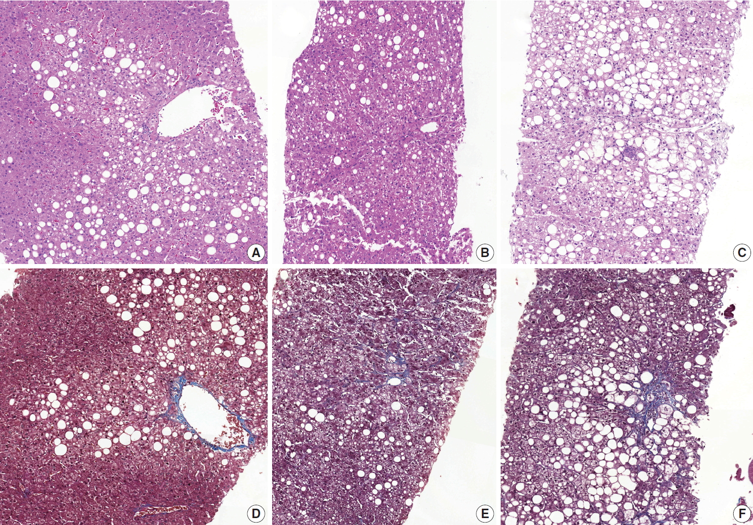
- A scoring system for the diagnosis of non-alcoholic steatohepatitis from liver biopsy
- Kyoungbun Lee, Eun Sun Jung, Eunsil Yu, Yun Kyung Kang, Mee-Yon Cho, Joon Mee Kim, Woo Sung Moon, Jin Sook Jeong, Cheol Keun Park, Jae-Bok Park, Dae Young Kang, Jin Hee Sohn, So-Young Jin
- J Pathol Transl Med. 2020;54(3):228-236. Published online April 15, 2020
- DOI: https://doi.org/10.4132/jptm.2020.03.07
- 15,922 View
- 283 Download
- 11 Web of Science
- 11 Crossref
-
Abstract
PDF
- Background
Liver biopsy is the essential method to diagnose non-alcoholic steatohepatitis (NASH), but histological features of NASH are too subjective to achieve reproducible diagnoses in early stages of disease. We aimed to identify the key histological features of NASH and devise a scoring model for diagnosis.
Methods
Thirteen pathologists blindly assessed 12 histological factors and final histological diagnoses (‘not-NASH,’ ‘borderline,’ and ‘NASH’) of 31 liver biopsies that were diagnosed as non-alcoholic fatty liver disease (NAFLD) or NASH before and after consensus. The main histological parameters to diagnose NASH were selected based on histological diagnoses and the diagnostic accuracy and agreement of 12 scoring models were compared for final diagnosis and the NAFLD Activity Score (NAS) system.
Results
Inter-observer agreement of final diagnosis was fair (κ = 0.25) before consensus and slightly improved after consensus (κ = 0.33). Steatosis at more than 5% was the essential parameter for diagnosis. Major diagnostic factors for diagnosis were fibrosis except 1C grade and presence of ballooned cells. Minor diagnostic factors were lobular inflammation ( ≥ 2 foci/ × 200 field), microgranuloma, and glycogenated nuclei. All 12 models showed higher inter-observer agreement rates than NAS and post-consensus diagnosis (κ = 0.52–0.69 vs. 0.33). Considering the reproducibility of factors and practicability of the model, summation of the scores of major (× 2) and minor factors may be used for the practical diagnosis of NASH.
Conclusions
A scoring system for the diagnosis of NAFLD would be helpful as guidelines for pathologists and clinicians by improving the reproducibility of histological diagnosis of NAFLD. -
Citations
Citations to this article as recorded by- Preclinical liver toxicity models: Advantages, limitations and recommendations
Devaraj Ezhilarasan, Sivanesan Karthikeyan, Mustapha Najimi, Paramasivan Vijayalakshmi, Ganapathy Bhavani, Muthukrishnan Jansi Rani
Toxicology.2025; 511: 154020. CrossRef - Hepatic miR-93 promotes the pathogenesis of metabolic dysfunction-associated steatotic liver disease by suppressing SIRT1
Yo Han Lee, Jinyoung Lee, Joonho Jeong, Kieun Park, Bukyung Baik, Yuseong Kwon, Kimyeong Kim, Keon Woo Khim, Haneul Ji, Ji Young Lee, Kwangho Kim, Ji Won Kim, Tam Dao, Misung Kim, Tae Young Lee, Yong Ryoul Yang, Haejin Yoon, Dongryeol Ryu, Seonghwan Hwang
Metabolism.2025; 169: 156266. CrossRef - Deep learning-based method for grading histopathological liver fibrosis in rodent models of metabolic dysfunction-associated steatohepatitis
Soo Min Ko, Jae-ik Shin, Yiyu Hong, Hyunji Kim, Insuk Sohn, Ji-Young Lee, Hyo-Jeong Han, Da Som Jeong, Yerin Lee, Woo-Chan Son
Frontiers in Medicine.2025;[Epub] CrossRef - Liver biopsy in the modern era: from traditional techniques to artificial intelligence and multi-omics integration
Nasar Alwahaibi, Maryam Alwahaibi
Frontiers in Medicine.2025;[Epub] CrossRef - Presurgery health influences outcomes following vertical sleeve gastrectomy in adolescents
Debi Swertfeger, Ahlee Kim, Hannah Sexmith, Maria E. Moreno‐Fernandez, W. Sean Davidson, Michael Helmrath, Todd Jenkins, Tsuyoshi Okura, Esmond Geh, Stavra A. Xanthakos, Sara Szabo, Takahisa Nakamura, Senad Divanovic, Amy Sanghavi Shah
Obesity.2024; 32(6): 1187. CrossRef - Immunobiotic Bacteria Attenuate Hepatic Fibrosis through the Modulation of Gut Microbiota and the Activation of Aryl‐Hydrocarbon Receptors Pathway in Non‐Alcoholic Steatohepatitis Mice
Paulraj Kanmani, Julio Villena, Soo‐kyoung Lim, Eun‐Ji Song, Young‐Do Nam, Hojun Kim
Molecular Nutrition & Food Research.2024;[Epub] CrossRef - Lipid nanoparticle-mediated hepatocyte delivery of siRNA and silibinin in metabolic dysfunction-associated steatotic liver disease
Yifu Lyu, Xiuyi Yang, Lei Yang, Jinyu Dai, Huanyu Qin, Yunuo Zhou, Yunan Huang, Yanmei Wang, Di Wu, Qindai Shuai, Qilong Li, Xiaofei Xin, Lifang Yin
Journal of Controlled Release.2024; 373: 385. CrossRef - Enhanced hepatoprotective effects of empagliflozin and vitamin D dual therapy against metabolic dysfunction‐associated steatohepatitis in mice by boosted modulation of metabolic, oxidative stress, and inflammatory pathways
Wesam F. Farrash, Shakir Idris, Mohamed E. Elzubier, Elshiekh B. A. Khidir, Akhmed Aslam, Abdulrahman Mujalli, Riyad A. Almaimani, Ahmad A. Obaid, Mahmoud Z. El‐Readi, Mohammad A. Alobaidy, Afnan Salaka, Afnan M. Shakoori, Alaa M. Saleh, Faisal Minshawi,
International Journal of Experimental Pathology.2024; 105(6): 219. CrossRef - Bilirubin, a hepatoprotective agent that activates SIRT1, PGC-1α, and PPAR-α, while inhibiting NF-κB in rats with metabolic-associated fatty liver disease
Motahareh Taghizadeh, Mohammad Hasan Maleki, Omid Vakili, Ramin Tavakoli, Parvin Zarei, Amirreza Dehghanian, Hossein Bordbar, Sayed Mohammad Shafiee
Scientific Reports.2024;[Epub] CrossRef - Changes in indications for outpatient percutaneous liver biopsy over 5 years: from hepatitis C to fatty liver disease
Marlone Cunha-Silva, Luíza D. Torres, Mariana F. Fernandes, Tirzah de M. Lopes Secundo, Marina C.G. Moreira, Ademar Yamanaka, Leonardo T. Monici, Larissa B. Eloy da Costa, Daniel F. Mazo, Tiago Sevá-Pereira
Gastroenterología y Hepatología.2022; 45(8): 579. CrossRef - Changes in indications for outpatient percutaneous liver biopsy over 5 years: from hepatitis C to fatty liver disease
Marlone Cunha-Silva, Luíza D. Torres, Mariana F. Fernandes, Tirzah de M. Lopes Secundo, Marina C.G. Moreira, Ademar Yamanaka, Leonardo T. Monici, Larissa B. Eloy da Costa, Daniel F. Mazo, Tiago Sevá-Pereira
Gastroenterología y Hepatología (English Edition).2022; 45(8): 579. CrossRef
- Preclinical liver toxicity models: Advantages, limitations and recommendations
- Standardized Pathology Report for Colorectal Cancer, 2nd Edition
- Baek-hui Kim, Joon Mee Kim, Gyeong Hoon Kang, Hee Jin Chang, Dong Wook Kang, Jung Ho Kim, Jeong Mo Bae, An Na Seo, Ho Sung Park, Yun Kyung Kang, Kyung-Hwa Lee, Mee Yon Cho, In-Gu Do, Hye Seung Lee, Hee Kyung Chang, Do Youn Park, Hyo Jeong Kang, Jin Hee Sohn, Mee Soo Chang, Eun Sun Jung, So-Young Jin, Eunsil Yu, Hye Seung Han, Youn Wha Kim
- J Pathol Transl Med. 2020;54(1):1-19. Published online November 13, 2019
- DOI: https://doi.org/10.4132/jptm.2019.09.28

- 33,069 View
- 1,346 Download
- 49 Web of Science
- 41 Crossref
-
Abstract
PDFSupplementary Material
- The first edition of the ‘Standardized Pathology Report for Colorectal Cancer,’ which was developed by the Gastrointestinal Pathology Study Group (GIP) of the Korean Society of Pathologists, was published 13 years ago. Meanwhile, there have been many changes in the pathologic diagnosis of colorectal cancer (CRC), pathologic findings included in the pathology report, and immunohistochemical and molecular pathology required for the diagnosis and treatment of colorectal cancer. In order to reflect these changes, we (GIP) decided to make the second edition of the report. The purpose of this standardized pathology report is to provide a practical protocol for Korean pathologists, which could help diagnose and treat CRC patients. This report consists of “standard data elements” and “conditional data elements.” Basic pathologic findings and parts necessary for prognostication of CRC patients are classified as “standard data elements,” while other prognostic factors and factors related to adjuvant therapy are classified as “conditional data elements” so that each institution could select the contents according to the characteristics of the institution. The Korean version is also provided separately so that Korean pathologists can easily understand and use this report. We hope that this report will be helpful in the daily practice of CRC diagnosis.
-
Citations
Citations to this article as recorded by- Proteogenomic profiling predicts outcomes of adjuvant chemotherapy in extrahepatic cholangiocarcinoma
Hyehyun Jeong, Ji-Hye Oh, Hee-Sung Ahn, Baek-Yeol Ryoo, Kyu-pyo Kim, Jae Ho Jeong, Inkeun Park, Dae Wook Hwang, Jae Hoon Lee, Ki Byung Song, Woohyung Lee, Ki-Hun Kim, Deog-Bog Moon, Gi Won Song, Dong-Hwan Jung, Seung-Mo Hong, Chae Won Park, In-Pyo Baek, Y
Journal of Hepatology.2026; 84(1): 122. CrossRef - A study on the effect of CBSM-based psychological intervention on self-management ability and quality of life in colorectal cancer patients from the perspective of benefit finding: A quasi-experimental study
Linzhi Jiang, Zhouyuan Peng, Rongrong Liu, Lei Chen, Yang Wu, Xingqun Tan, Fan Wang, Liyuan Sun, Chong-Chi Chiu
PLOS One.2026; 21(1): e0339472. CrossRef - Impact of gross tumor morphology on the clinical outcomes of colon cancer: multicenter retrospective cohort study
So Jung Han, Hyun Seok Lee, Byung Ik Jang, Jae Hyun Kim, Hyun Gun Kim, Il Hyun Baek, Jun Lee, Bun Kim, Dae Bum Kim, Jae Jun Park
International Journal of Colorectal Disease.2026;[Epub] CrossRef - Vitrification-based tissue-to-organoid cryopreservation workflow for colorectal cancer
Juyoung Choi, Hye Kyung Hong, Mi-Sook Lee, Ye Jin Lim, Yurimi Lee, Yong Beom Cho, Yoon-La Choi
New Biotechnology.2026; 93: 341. CrossRef - Diagnostic accuracy and pitfalls of MRI for restaging locally advanced rectal cancer in patients following anti-PD1 therapy plus neoadjuvant chemoradiotherapy: a multicenter study
Lixue Xu, Liting Sun, Yuhuan Fu, Guangyong Chen, Hongwei Yao, Zhenchang Wang, Zhenghan Yang, Jie Zhang
Abdominal Radiology.2025; 51(1): 14. CrossRef - Unraveling the role of perineural invasion in cancer progression across multiple tumor types
Muqtada Shaikh, Sanket Shirodkar, Gaurav Doshi
Medical Oncology.2025;[Epub] CrossRef - MALT lymphoma affecting the oral cavity: a clinical, pathologic and genetic study of MALT1 gene translocation
Juan Manuel Arteaga Legarrea, Mauro Lima dos Santos, Nathalia Gomes Rodrigues, Ricardo Santiago Gomez, Ricardo Alves Mesquita, Silvia Ferreira de Sousa, Cinthia Verónica Bardález López de Cáceres, Hélder Antônio Rebelo Pontes, Pablo Agustin Vargas, Luiz A
Oral Surgery, Oral Medicine, Oral Pathology and Oral Radiology.2025; 140(6): 740. CrossRef - Additional staining for lymphovascular invasion is associated with increased estimation of lymph node metastasis in patients with T1 colorectal cancer: Systematic review and meta‐analysis
Jun Watanabe, Katsuro Ichimasa, Yuki Kataoka, Atsushi Miki, Hidehiro Someko, Munenori Honda, Makiko Tahara, Takeshi Yamashina, Khay Guan Yeoh, Shigeo Kawai, Kazuhiko Kotani, Naohiro Sata
Digestive Endoscopy.2024; 36(5): 533. CrossRef - The use of core descriptors from the ENiGMA code study in recent literature: a systematic review
Saher‐Zahra Khan, Andrea Arline, Kate M. Williams, Matthew J. Lee, Emily Steinhagen, Sharon L. Stein
Colorectal Disease.2024; 26(3): 428. CrossRef - Efficacy and safety of PD-1 blockade plus long-course chemoradiotherapy in locally advanced rectal cancer (NECTAR): a multi-center phase 2 study
Zhengyang Yang, Jiale Gao, Jianyong Zheng, Jiagang Han, Ang Li, Gang Liu, Yi Sun, Jie Zhang, Guangyong Chen, Rui Xu, Xiao Zhang, Yishan Liu, Zhigang Bai, Wei Deng, Wei He, Hongwei Yao, Zhongtao Zhang
Signal Transduction and Targeted Therapy.2024;[Epub] CrossRef - Diagnostic Accuracy of Highest-Grade or Predominant Histological Differentiation of T1 Colorectal Cancer in Predicting Lymph Node Metastasis: A Systematic Review and Meta-Analysis
Jun Watanabe, Katsuro Ichimasa, Yuki Kataoka, Shoko Miyahara, Atsushi Miki, Khay Guan Yeoh, Shigeo Kawai, Fernando Martínez de Juan, Isidro Machado, Kazuhiko Kotani, Naohiro Sata
Clinical and Translational Gastroenterology.2024; 15(3): e00673. CrossRef - Comparative evaluation of CT and MRI in the preoperative staging of colon cancer
Effrosyni Bompou, Aikaterini Vassiou, Ioannis Baloyiannis, Konstantinos Perivoliotis, Ioannis Fezoulidis, George Tzovaras
Scientific Reports.2024;[Epub] CrossRef - Pathologic Implications of Magnetic Resonance Imaging-detected Extramural Venous Invasion of Rectal Cancer
Hyun Gu Lee, Chan Wook Kim, Jong Keon Jang, Seong Ho Park, Young Il Kim, Jong Lyul Lee, Yong Sik Yoon, In Ja Park, Seok-Byung Lim, Chang Sik Yu, Jin Cheon Kim
Clinical Colorectal Cancer.2023; 22(1): 129. CrossRef - A standardized pathology report for gastric cancer: 2nd edition
Young Soo Park, Myeong-Cherl Kook, Baek-hui Kim, Hye Seung Lee, Dong-Wook Kang, Mi-Jin Gu, Ok Ran Shin, Younghee Choi, Wonae Lee, Hyunki Kim, In Hye Song, Kyoung-Mee Kim, Hee Sung Kim, Guhyun Kang, Do Youn Park, So-Young Jin, Joon Mee Kim, Yoon Jung Choi,
Journal of Pathology and Translational Medicine.2023; 57(1): 1. CrossRef - IGFL2-AS1, a Long Non-Coding RNA, Is Associated with Radioresistance in Colorectal Cancer
Jeeyong Lee, Da Yeon Kim, Younjoo Kim, Ui Sup Shin, Kwang Seok Kim, Eun Ju Kim
International Journal of Molecular Sciences.2023; 24(2): 978. CrossRef - A Standardized Pathology Report for Gastric Cancer: 2nd Edition
Young Soo Park, Myeong-Cherl Kook, Baek-hui Kim, Hye Seung Lee, Dong-Wook Kang, Mi-Jin Gu, Ok Ran Shin, Younghee Choi, Wonae Lee, Hyunki Kim, In Hye Song, Kyoung-Mee Kim, Hee Sung Kim, Guhyun Kang, Do Youn Park, So-Young Jin, Joon Mee Kim, Yoon Jung Choi,
Journal of Gastric Cancer.2023; 23(1): 107. CrossRef - Incremental Detection Rate of Dysplasia and Sessile Serrated Polyps/Adenomas Using Narrow-Band Imaging and Dye Spray Chromoendoscopy in Addition to High-Definition Endoscopy in Patients with Long-Standing Extensive Ulcerative Colitis: Segmental Tandem End
Ji Eun Kim, Chang Wan Choi, Sung Noh Hong, Joo Hye Song, Eun Ran Kim, Dong Kyung Chang, Young-Ho Kim
Diagnostics.2023; 13(3): 516. CrossRef - Prognostic Impact of Extramural Lymphatic, Vascular, and Perineural Invasion in Stage II Colon Cancer: A Comparison With Intramural Invasion
Sang Sik Cho, Ji Won Park, Gyeong Hoon Kang, Jung Ho Kim, Jeong Mo Bae, Sae-Won Han, Tae-You Kim, Min Jung Kim, Seung-Bum Ryoo, Seung-Yong Jeong, Kyu Joo Park
Diseases of the Colon & Rectum.2023; 66(3): 366. CrossRef - Towards targeted colorectal cancer biopsy based on tissue morphology assessment by compression optical coherence elastography
Anton A. Plekhanov, Marina A. Sirotkina, Ekaterina V. Gubarkova, Elena B. Kiseleva, Alexander A. Sovetsky, Maria M. Karabut, Vladimir E. Zagainov, Sergey S. Kuznetsov, Anna V. Maslennikova, Elena V. Zagaynova, Vladimir Y. Zaitsev, Natalia D. Gladkova
Frontiers in Oncology.2023;[Epub] CrossRef - Is High-Grade Tumor Budding an Independent Prognostic Factor in Stage II Colon Cancer?
Jung Kyong Shin, Yoon Ah Park, Jung Wook Huh, Seong Hyeon Yun, Hee Cheol Kim, Woo Yong Lee, Seok Hyung Kim, Sang Yun Ha, Yong Beom Cho
Diseases of the Colon & Rectum.2023; 66(8): e801. CrossRef - Detection of Human cytomegalovirus UL55 Gene and IE/E Protein Expression in Colorectal Cancer Patients in Egypt
May Raouf, Ahmed A. Sabry, Mahinour A. Ragab, Samar El Achy, Amira Amer
BMC Cancer.2023;[Epub] CrossRef - Polo-like kinase 4 as a potential predictive biomarker of chemoradioresistance in locally advanced rectal cancer
Hyunseung Oh, Soon Gu Kim, Sung Uk Bae, Sang Jun Byun, Shin Kim, Jae-Ho Lee, Ilseon Hwang, Sun Young Kwon, Hye Won Lee
Journal of Pathology and Translational Medicine.2022; 56(1): 40. CrossRef - A Prediction Model for Tumor Recurrence in Stage II–III Colorectal Cancer Patients: From a Machine Learning Model to Genomic Profiling
Po-Chuan Chen, Yu-Min Yeh, Bo-Wen Lin, Ren-Hao Chan, Pei-Fang Su, Yi-Chia Liu, Chung-Ta Lee, Shang-Hung Chen, Peng-Chan Lin
Biomedicines.2022; 10(2): 340. CrossRef - Rationale and design of a prospective, multicenter, phase II clinical trial of safety and efficacy evaluation of long course neoadjuvant chemoradiotherapy plus tislelizumab followed by total mesorectal excision for locally advanced rectal cancer (NCRT-PD1
Zhengyang Yang, Xiao Zhang, Jie Zhang, Jiale Gao, Zhigang Bai, Wei Deng, Guangyong Chen, Yongbo An, Yishan Liu, Qi Wei, Jiagang Han, Ang Li, Gang Liu, Yi Sun, Dalu Kong, Hongwei Yao, Zhongtao Zhang
BMC Cancer.2022;[Epub] CrossRef - Potential of DEK proto‑oncogene as a prognostic biomarker for colorectal cancer: An evidence‑based review
Muhammad Habiburrahman, Muhammad Wardoyo, Stefanus Sutopo, Nur Rahadiani
Molecular and Clinical Oncology.2022;[Epub] CrossRef - Reproducibility and Feasibility of Classification and National Guidelines for Histological Diagnosis of Canine Mammary Gland Tumours: A Multi-Institutional Ring Study
Serenella Papparella, Maria Crescio, Valeria Baldassarre, Barbara Brunetti, Giovanni Burrai, Cristiano Cocumelli, Valeria Grieco, Selina Iussich, Lorella Maniscalco, Francesca Mariotti, Francesca Millanta, Orlando Paciello, Roberta Rasotto, Mariarita Roma
Veterinary Sciences.2022; 9(7): 357. CrossRef - Composite scoring system and optimal tumor budding cut-off number for estimating lymph node metastasis in submucosal colorectal cancer
Jeong-ki Kim, Ye-Young Rhee, Jeong Mo Bae, Jung Ho Kim, Seong-Joon Koh, Hyun Jung Lee, Jong Pil Im, Min Jung Kim, Seung-Bum Ryoo, Seung-Yong Jeong, Kyu Joo Park, Ji Won Park, Gyeong Hoon Kang
BMC Cancer.2022;[Epub] CrossRef - Automated Hybrid Model for Detecting Perineural Invasion in the Histology of Colorectal Cancer
Jiyoon Jung, Eunsu Kim, Hyeseong Lee, Sung Hak Lee, Sangjeong Ahn
Applied Sciences.2022; 12(18): 9159. CrossRef - Clinical Implication of Perineural and Lymphovascular Invasion in Rectal Cancer Patients Who Underwent Surgery After Preoperative Chemoradiotherapy
Young Il Kim, Chan Wook Kim, Jong Hoon Kim, Jihun Kim, Jun-Soo Ro, Jong Lyul Lee, Yong Sik Yoon, In Ja Park, Seok-Byung Lim, Chang Sik Yu, Jin Cheon Kim
Diseases of the Colon & Rectum.2022; 65(11): 1325. CrossRef - Molecular Pathology of Gastric Cancer
Moonsik Kim, An Na Seo
Journal of Gastric Cancer.2022; 22(4): 264. CrossRef - Selective approach to arterial ligation in radical sigmoid colon cancer surgery with D3 lymph node dissection: A multicenter comparative study
Sergey Efetov, Albina Zubayraeva, Cüneyt Kayaalp, Alisa Minenkova, Yusuf Bağ, Aftandil Alekberzade, Petr Tsarkov
Turkish Journal of Surgery.2022; 38(4): 382. CrossRef - Evaluation of lncRNA FOXD2-AS1 Expression as a Diagnostic Biomarker in Colorectal Cancer
Hooman Shalmashi, Sahar Safaei, Dariush Shanehbandi, Milad Asadi, Soghra Bornehdeli, Abdolreza Mehdi Navaz
Reports of Biochemistry and Molecular Biology.2022; 11(3): 471. CrossRef - Improvement in the Assessment of Response to Preoperative Chemoradiotherapy for Rectal Cancer Using Magnetic Resonance Imaging and a Multigene Biomarker
Eunhae Cho, Sung Woo Jung, In Ja Park, Jong Keon Jang, Seong Ho Park, Seung-Mo Hong, Jong Lyul Lee, Chan Wook Kim, Yong Sik Yoon, Seok-Byung Lim, Chang Sik Yu, Jin Cheon Kim
Cancers.2021; 13(14): 3480. CrossRef - Addition of V-Stage to Conventional TNM Staging to Create the TNVM Staging System for Accurate Prediction of Prognosis in Colon Cancer: A Multi-Institutional Retrospective Cohort Study
Jung Hoon Bae, Ji Hoon Kim, Jaeim Lee, Bong-Hyeon Kye, Sang Chul Lee, In Kyu Lee, Won Kyung Kang, Hyeon-Min Cho, Yoon Suk Lee
Biomedicines.2021; 9(8): 888. CrossRef - Gene Expression Profiles Associated with Radio-Responsiveness in Locally Advanced Rectal Cancer
Jeeyong Lee, Junhye Kwon, DaYeon Kim, Misun Park, KwangSeok Kim, InHwa Bae, Hyunkyung Kim, JoonSeog Kong, Younjoo Kim, UiSup Shin, EunJu Kim
Biology.2021; 10(6): 500. CrossRef - A Patient-Derived Organoid-Based Radiosensitivity Model for the Prediction of Radiation Responses in Patients with Rectal Cancer
Misun Park, Junhye Kwon, Joonseog Kong, Sun Mi Moon, Sangsik Cho, Ki Young Yang, Won Il Jang, Mi Sook Kim, Younjoo Kim, Ui Sup Shin
Cancers.2021; 13(15): 3760. CrossRef - Comparison between Local Excision and Radical Resection for the Treatment of Rectal Cancer in ypT0-1 Patients: An Analysis of the Clinicopathological Factors and Survival Rates
Soo Young Oh, In Ja Park, Young IL Kim, Jong-Lyul Lee, Chan Wook Kim, Yong Sik Yoon, Seok-Byung Lim, Chang Sik Yu, Jin Cheon Kim
Cancers.2021; 13(19): 4823. CrossRef - Comparison of Vascular Invasion With Lymph Node Metastasis as a Prognostic Factor in Stage I-III Colon Cancer: An Observational Cohort Study
Jung Hoon Bae, Ji Hoon Kim, Bong-Hyeon Kye, Abdullah Al-Sawat, Chul Seung Lee, Seung-Rim Han, In Kyu Lee, Sung Hak Lee, Yoon Suk Lee
Frontiers in Surgery.2021;[Epub] CrossRef - Clinicopathological significance of Ki67 expression in colorectal cancer
Jing Li, Zhi-ye Liu, Hai-bo Yu, Qing Xue, Wen-jie He, Hai-tao Yu
Medicine.2020; 99(20): e20136. CrossRef - Lateral lymph node and its association with distant recurrence in rectal cancer: A clue of systemic disease
Young Il Kim, Jong Keon Jang, In Ja Park, Seong Ho Park, Jong Beom Kim, Jin-Hong Park, Tae Won Kim, Jun-Soo Ro, Seok-Byung Lim, Chang Sik Yu, Jin Cheon Kim
Surgical Oncology.2020; 35: 174. CrossRef - Transformation of Pathology Reports Into the Common Data Model With Oncology Module: Use Case for Colon Cancer
Borim Ryu, Eunsil Yoon, Seok Kim, Sejoon Lee, Hyunyoung Baek, Soyoung Yi, Hee Young Na, Ji-Won Kim, Rong-Min Baek, Hee Hwang, Sooyoung Yoo
Journal of Medical Internet Research.2020; 22(12): e18526. CrossRef
- Proteogenomic profiling predicts outcomes of adjuvant chemotherapy in extrahepatic cholangiocarcinoma
- Molecular Testing for Gastrointestinal Cancer
- Hye Seung Lee, Woo Ho Kim, Yoonjin Kwak, Jiwon Koh, Jeong Mo Bae, Kyoung-Mee Kim, Mee Soo Chang, Hye Seung Han, Joon Mee Kim, Hwal Woong Kim, Hee Kyung Chang, Young Hee Choi, Ji Y. Park, Mi Jin Gu, Min Jin Lhee, Jung Yeon Kim, Hee Sung Kim, Mee-Yon Cho
- J Pathol Transl Med. 2017;51(2):103-121. Published online February 19, 2017
- DOI: https://doi.org/10.4132/jptm.2017.01.24
- 24,943 View
- 913 Download
- 62 Web of Science
- 55 Crossref
-
Abstract
PDF
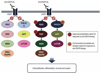
- With recent advances in molecular diagnostic methods and targeted cancer therapies, several molecular tests have been recommended for gastric cancer (GC) and colorectal cancer (CRC). Microsatellite instability analysis of gastrointestinal cancers is performed to screen for Lynch syndrome, predict favorable prognosis, and screen patients for immunotherapy. The epidermal growth factor receptor (EGFR) tyrosine kinase inhibitor has been approved in metastatic CRCs with wildtype RAS (KRAS and NRAS exon 2–4). A BRAF mutation is required for predicting poor prognosis. Additionally, amplification of human epidermal growth factor receptor 2 (HER2) and MET is also associated with resistance to EGFR inhibitor in metastatic CRC patients. The BRAF V600E mutation is found in sporadic microsatellite unstable CRCs, and thus is helpful for ruling out Lynch syndrome. In addition, the KRAS mutation is a prognostic biomarker and the PIK3CA mutation is a molecular biomarker predicting response to phosphoinositide 3-kinase/AKT/mammalian target of rapamycin inhibitors and response to aspirin therapy in CRC patients. Additionally, HER2 testing should be performed in all recurrent or metastatic GCs. If the results of HER2 immunohistochemistry are equivocal, HER2 silver or fluorescence in situ hybridization testing are essential for confirmative determination of HER2 status. Epstein-Barr virus–positive GCs have distinct characteristics, including heavy lymphoid stroma, hypermethylation phenotype, and high expression of immune modulators. Recent advances in next-generation sequencing technologies enable us to examine various genetic alterations using a single test. Pathologists play a crucial role in ensuring reliable molecular testing and they should also take an integral role between molecular laboratories and clinicians.
-
Citations
Citations to this article as recorded by- ALYREF promotes tumor progression by regulating SLC2A3-mediated glycolysis in gastric cancer
Yan Ma, Guangyue Shi, Limin Zhang, Zhanfei Lu, Rong Huang, Yao Wang, Zhengfei Zhao, Bin Ke
Genes & Diseases.2026; : 102328. CrossRef - Spatial and Temporal Tumor Heterogeneity in Gastric Cancer: Discordance of Predictive Biomarkers
Hye Seung Lee
Journal of Gastric Cancer.2025; 25(1): 192. CrossRef - Korean Practice Guidelines for Gastric Cancer 2024: An Evidence-based, Multidisciplinary Approach (Update of 2022 Guideline)
In-Ho Kim, Seung Joo Kang, Wonyoung Choi, An Na Seo, Bang Wool Eom, Beodeul Kang, Bum Jun Kim, Byung-Hoon Min, Chung Hyun Tae, Chang In Choi, Choong-kun Lee, Ho Jung An, Hwa Kyung Byun, Hyeon-Su Im, Hyung-Don Kim, Jang Ho Cho, Kyoungjune Pak, Jae-Joon Kim
Journal of Gastric Cancer.2025; 25(1): 5. CrossRef - Elucidating the Role of KRAS, NRAS, and BRAF Mutations and Microsatellite Instability in Colorectal Cancer via Next-Generation Sequencing
Marta Rada Rodríguez, Bárbara Angulo Biedma, Irene Rodríguez Pérez, Javier Azúa Romeo
Cancers.2025; 17(13): 2071. CrossRef - Chitosan and Its Derivative‐Based Nanoparticles in Gastrointestinal Cancers: Molecular Mechanisms of Action and Promising Anticancer Strategies
Zahra Shokati Eshkiki, Fatemeh Mansouri, Amir Reza Karamzadeh, Abolfazl Namazi, Hafez Heydari, Javad Akhtari, Seidamir Pasha Tabaeian, Abolfazl Akbari, Hongda Liu
Journal of Clinical Pharmacy and Therapeutics.2024;[Epub] CrossRef - Colorectal Cancer: Genetic Underpinning and Molecular Therapeutics for Precision Medicine
Gideon T. Dosunmu, Ardaman Shergill
Genes.2024; 15(5): 538. CrossRef - Effector Function Characteristics of Exhausted CD8+ T-Cell in Microsatellite Stable and Unstable Gastric Cancer
Dong-Seok Han, Yoonjin Kwak, Seungho Lee, Soo Kyung Nam, Seong-Ho Kong, Do Joong Park, Hyuk-Joon Lee, Nak-Jung Kwon, Hye Seung Lee, Han-Kwang Yang
Cancer Research and Treatment.2024; 56(4): 1146. CrossRef - A Standardized Pathology Report for Gastric Cancer: 2nd Edition
Young Soo Park, Myeong-Cherl Kook, Baek-hui Kim, Hye Seung Lee, Dong-Wook Kang, Mi-Jin Gu, Ok Ran Shin, Younghee Choi, Wonae Lee, Hyunki Kim, In Hye Song, Kyoung-Mee Kim, Hee Sung Kim, Guhyun Kang, Do Youn Park, So-Young Jin, Joon Mee Kim, Yoon Jung Choi,
Journal of Gastric Cancer.2023; 23(1): 107. CrossRef - A standardized pathology report for gastric cancer: 2nd edition
Young Soo Park, Myeong-Cherl Kook, Baek-hui Kim, Hye Seung Lee, Dong-Wook Kang, Mi-Jin Gu, Ok Ran Shin, Younghee Choi, Wonae Lee, Hyunki Kim, In Hye Song, Kyoung-Mee Kim, Hee Sung Kim, Guhyun Kang, Do Youn Park, So-Young Jin, Joon Mee Kim, Yoon Jung Choi,
Journal of Pathology and Translational Medicine.2023; 57(1): 1. CrossRef - Korean Practice Guidelines for Gastric Cancer 2022: An Evidence-based, Multidisciplinary Approach
Tae-Han Kim, In-Ho Kim, Seung Joo Kang, Miyoung Choi, Baek-Hui Kim, Bang Wool Eom, Bum Jun Kim, Byung-Hoon Min, Chang In Choi, Cheol Min Shin, Chung Hyun Tae, Chung sik Gong, Dong Jin Kim, Arthur Eung-Hyuck Cho, Eun Jeong Gong, Geum Jong Song, Hyeon-Su Im
Journal of Gastric Cancer.2023; 23(1): 3. CrossRef - Influence of location-dependent sex difference on PD-L1, MMR/MSI, and EGFR in colorectal carcinogenesis
Jina Choi, Nayoung Kim, Ryoung Hee Nam, Jin Won Kim, Chin-Hee Song, Hee Young Na, Gyeong Hoon Kang, Alvaro Galli
PLOS ONE.2023; 18(2): e0282017. CrossRef - Comprehensive Analysis of Epigenetic Associated Genes with Differential
Gene Expression and Prognosis in Gastric Cancer
Songlin An, Xinbao Li, Bing Li, Yan Li
Combinatorial Chemistry & High Throughput Screening.2023; 26(3): 527. CrossRef - Liquid Biopsy in Advanced Colorectal Cancer: Clinical Applications of Different Analytes
Marco Donatello Delcuratolo, Andrea Modrego-Sánchez, Maristella Bungaro, Beatriz Antón-Pascual, Santiago Teran, Valentina Dipace, Silvia Novello, Rocio Garcia-Carbonero, Francesco Passiglia, Cristina Graválos-Castro
Journal of Molecular Pathology.2023; 4(3): 128. CrossRef - Exosomal circ_0001190 Regulates the Progression of Gastric Cancer via miR-586/SOSTDC1 Axis
Chao Liu, Jing Yang, Fengchi Zhu, Zhiying Zhao, Lixue Gao
Biochemical Genetics.2022; 60(6): 1895. CrossRef - Optimization of pre‐analytical and analytical steps for DNA and RNA analysis of fresh cytology samples
Ana Dolinar, Gašper Grubelnik, Irena Srebotnik‐Kirbiš, Margareta Strojan Fležar, Margareta Žlajpah
Cancer Medicine.2022; 11(21): 4021. CrossRef - Retracted: Connexin 43 upregulation by dioscin‐inhibited gastric cancer metastasis by suppressing PI3K/Akt pathway
Food Science & Nutrition.2022;[Epub] CrossRef - Molecular Pathology of Gastric Cancer
Moonsik Kim, An Na Seo
Journal of Gastric Cancer.2022; 22(4): 264. CrossRef - Case report: Undifferentiated sarcoma with multiple tumors involved in Lynch syndrome: Unexpected favorable outcome to sintilimab combined with chemotherapy
Jiaying Liu, Xiaona Chang, Guixiang Xiao, Jingmin Zhong, Bo Huang, Jiwei Zhang, Beibei Gao, Gang Peng, Xiu Nie
Frontiers in Oncology.2022;[Epub] CrossRef - The SUMO E3 ligase CBX4 is identified as a poor prognostic marker of gastric cancer through multipronged OMIC analyses
Yi Pan, Qingshang Li, Zhijun Cao, Shuliang Zhao
Genes & Diseases.2021; 8(6): 827. CrossRef - Worldwide variation in lynch syndrome screening: case for universal screening in low colorectal cancer prevalence areas
George Kunnackal John, Vipin Das Villgran, Christine Caufield-Noll, Francis Giardiello
Familial Cancer.2021; 20(2): 145. CrossRef - Tamoxifen Downregulates the Expression of Notch1 and DLL1 Genes in MKN-45 Gastric Cancer Cells
Faranak Khanipouyani, Hassan Akrami
Journal of Gastrointestinal Cancer.2021; 52(3): 922. CrossRef - Kallikrein-11, in Association with Coiled-Coil Domain Containing 25, as a Potential Prognostic Marker for Cholangiocarcinoma with Lymph Node Metastasis
Saeranee Siriphak, Ravinnipa Chanakankun, Tanakorn Proungvitaya, Sittiruk Roytrakul, Doungdean Tummanatsakun, Wunchana Seubwai, Molin Wongwattanakul, Siriporn Proungvitaya
Molecules.2021; 26(11): 3105. CrossRef - ISH-based HER2 diagnostics
Josef Rüschoff, Iris Nagelmeier, Bharat Jasani, Oliver Stoss
Der Pathologe.2021; 42(S1): 62. CrossRef - Identification and Analysis of Key Genes Driving Gastric Cancer Through Bioinformatics
Zhao Liu, Shihai Liu, Jing Guo, Libin Sun, Shasha Wang, Yixuan Wang, Wensheng Qiu, Jing Lv
Genetic Testing and Molecular Biomarkers.2021; 25(1): 1. CrossRef - Microsatellite Instability in Colorectal Cancer Liquid Biopsy—Current Updates on Its Potential in Non-Invasive Detection, Prognosis and as a Predictive Marker
Francis Yew Fu Tieng, Nadiah Abu, Learn-Han Lee, Nurul-Syakima Ab Mutalib
Diagnostics.2021; 11(3): 544. CrossRef - Metformin attenuates synergic effect of diabetes mellitus and Helicobacter pylori infection on gastric cancer cells proliferation by suppressing PTEN expression
Huibin Lu, Xinwei Han, Jianzhuang Ren, Kewei Ren, Zongming Li, Quanhui Zhang
Journal of Cellular and Molecular Medicine.2021; 25(10): 4534. CrossRef - Recent Advances in the Diagnosis, Staging, Treatment, and Prognosis of Advanced Gastric Cancer: A Literature Review
Zhi-da Chen, Peng-fei Zhang, Hong-qing Xi, Bo Wei, Lin Chen, Yun Tang
Frontiers in Medicine.2021;[Epub] CrossRef - Tumor immune response and immunotherapy in gastric cancer
Yoonjin Kwak, An Na Seo, Hee Eun Lee, Hye Seung Lee
Journal of Pathology and Translational Medicine.2020; 54(1): 20. CrossRef - Comparative analysis of HER2 copy number between plasma and tissue samples in gastric cancer using droplet digital PCR
Boram Kim, Soo Kyung Nam, Soo Hyun Seo, Kyoung Un Park, Sang-Hoon Ahn, Do Joong Park, Hyung-Ho Kim, Woo Ho Kim, Hye Seung Lee
Scientific Reports.2020;[Epub] CrossRef - Differential prognostic impact of CD8+ T cells based on human leucocyte antigen I and PD-L1 expression in microsatellite-unstable gastric cancer
Yoonjin Kwak, Jiwon Koh, Yujun Park, Yun Ji Hong, Kyoung Un Park, Hyung-Ho Kim, Do Joong Park, Sang-Hoon Ahn, Woo Ho Kim, Hye Seung Lee
British Journal of Cancer.2020; 122(9): 1399. CrossRef - High-Accuracy Determination of Microsatellite Instability Compatible with Liquid Biopsies
Amanda Bortolini Silveira, François-Clément Bidard, Amélie Kasperek, Samia Melaabi, Marie-Laure Tanguy, Manuel Rodrigues, Guillaume Bataillon, Luc Cabel, Bruno Buecher, Jean-Yves Pierga, Charlotte Proudhon, Marc-Henri Stern
Clinical Chemistry.2020; 66(4): 606. CrossRef - Chitosan: A compound for drug delivery system in gastric cancer-a review
Rana Shafabakhsh, Bahman Yousefi, Zatollah Asemi, Banafsheh Nikfar, Mohammad Ali Mansournia, Jamal Hallajzadeh
Carbohydrate Polymers.2020; 242: 116403. CrossRef - MSI and EBV Positive Gastric Cancer’s Subgroups and Their Link with Novel Immunotherapy
Maria Grazia Rodriquenz, Giandomenico Roviello, Alberto D’Angelo, Daniele Lavacchi, Franco Roviello, Karol Polom
Journal of Clinical Medicine.2020; 9(5): 1427. CrossRef - Theoretical calculations of molecular descriptors for anticancer activities of 1, 2, 3-triazole-pyrimidine derivatives against gastric cancer cell line (MGC-803): DFT, QSAR and docking approaches
Rhoda Oyeladun Oyewole, Abel Kolawole Oyebamiji, Banjo Semire
Heliyon.2020; 6(5): e03926. CrossRef - Identification of a Clinical Cutoff Value for Multiplex KRASG12/G13 Mutation Detection in Colorectal Adenocarcinoma Patients Using Digital Droplet PCR, and Comparison with Sanger Sequencing and PNA Clamping Assay
Kyung Ha Lee, Tae Hee Lee, Min Kyung Choi, In Sun Kwon, Go Eun Bae, Min-Kyung Yeo
Journal of Clinical Medicine.2020; 9(7): 2283. CrossRef - PD-L1 Testing in Gastric Cancer by the Combined Positive Score of the 22C3 PharmDx and SP263 Assay with Clinically Relevant Cut-offs
Yujun Park, Jiwon Koh, Hee Young Na, Yoonjin Kwak, Keun-Wook Lee, Sang-Hoon Ahn, Do Joong Park, Hyung-Ho Kim, Hye Seung Lee
Cancer Research and Treatment.2020; 52(3): 661. CrossRef - Clinical and Molecular Assessment of Patients with Lynch Syndrome and Sarcomas Underpinning the Association with MSH2 Germline Pathogenic Variants
Nathália de Angelis de Carvalho, Bianca Naomi Niitsuma, Vanessa Nascimento Kozak, Felipe D’almeida Costa, Mariana Petaccia de Macedo, Bruna Elisa Catin Kupper, Maria Letícia Gobo Silva, Maria Nirvana Formiga, Sahlua Miguel Volc, Samuel Aguiar Junior, Eden
Cancers.2020; 12(7): 1848. CrossRef - Farnesoid X receptor antagonizes Wnt/β-catenin signaling in colorectal tumorigenesis
Junhui Yu, Shan Li, Jing Guo, Zhengshui Xu, Jianbao Zheng, Xuejun Sun
Cell Death & Disease.2020;[Epub] CrossRef - YAP promotes self-renewal of gastric cancer cells by inhibiting expression of L-PTGDS and PTGDR2
Qingli Bie, Xiaozhe Li, Shiqi Liu, Xiao Yang, Zhenwen Qian, Rou Zhao, Xiaobei Zhang, Bin Zhang
International Journal of Clinical Oncology.2020; 25(12): 2055. CrossRef - ISH-basierte HER2-Diagnostik
Josef Rüschoff, Iris Nagelmeier, Bharat Jasani, Oliver Stoss
Der Pathologe.2020; 41(6): 606. CrossRef - Histone Deacetylase Inhibitor Trichostatin A Suppresses Cell Proliferation and Induces Apoptosis by Regulating the PI3K/AKT Signalling Pathway in Gastric Cancer Cells
Xinli An, Zekun Wei, Botian Ran, Hao Tian, Hongyu Gu, Yan Liu, Hongjuan Cui, Shunqin Zhu
Anti-Cancer Agents in Medicinal Chemistry.2020; 20(17): 2114. CrossRef - Role of Her-2 in Gastrointestinal Tumours beyond Gastric Cancer: A Tool for Precision Medicine
Csongor G. Lengyel, Baker Habeeb, Shah Z. Khan, Khalid El Bairi, Sara C. Altuna, Sadaqat Hussain, Syed Ayub Mazher, Dario Trapani, Angelica Petrillo
Gastrointestinal Disorders.2020; 3(1): 1. CrossRef - Next-generation Sequencing in the Management of Gastric and Esophageal Cancers
Jill C. Rubinstein, Norman G. Nicolson, Nita Ahuja
Surgical Clinics of North America.2019; 99(3): 511. CrossRef - Molecular profile in Paraguayan colorectal cancer patients, towards to a precision medicine strategy
Tania Fleitas-Kanonnikoff, Carolina Martinez‐Ciarpaglini, Josefina Ayala, Cinthia Gauna, Rita Denis, Ita Yoffe, Silvia Sforza, María Teresa Martínez, Alicia Pomata, Maider Ibarrola‐Villava, Sipan Arevshatyan, Verónica Burriel, Diego Boscá, Oscar Pastor, A
Cancer Medicine.2019; 8(6): 3120. CrossRef - Human epidermal growth factor receptor 2-positive digestive tumors
Anna D. Wagner, Berna C. Özdemir, Josef Rüschoff
Current Opinion in Oncology.2019; 31(4): 354. CrossRef - Assessing molecular subtypes of gastric cancer: microsatellite unstable and Epstein-Barr virus subtypes. Methods for detection and clinical and pathological implications
Carolina Martinez-Ciarpaglini, Tania Fleitas-Kanonnikoff, Valentina Gambardella, Marta Llorca, Cristina Mongort, Regina Mengual, Gema Nieto, Lara Navarro, Marisol Huerta, Susana Rosello, Desamparados Roda, Noelia Tarazona, Samuel Navarro, Gloria Ribas, An
ESMO Open.2019; 4(3): e000470. CrossRef - Current and future molecular diagnostics of gastric cancer
Rachel Sin-Yu Choi, Wing Yin Xenia Lai, Lok Ting Claire Lee, Wing Lam Christa Wong, Xiao Meng Pei, Hin Fung Tsang, Joel Johnson Leung, William Chi Shing Cho, Man Kee Maggie Chu, Elaine Yue Ling Wong, Sze Chuen Cesar Wong
Expert Review of Molecular Diagnostics.2019; 19(10): 863. CrossRef - Clinicopathologic significance of human leukocyte antigen class I expression in patients with stage II and III gastric cancer
Yujun Park, Jiwon Koh, Yoonjin Kwak, Sang-Hoon Ahn, Do Joong Park, Hyung-Ho Kim, Woo Ho Kim, Hye Seung Lee
Cancer Immunology, Immunotherapy.2019; 68(11): 1779. CrossRef - Development and Validation of an Easy-to-Implement, Practical Algorithm for the Identification of Molecular Subtypes of Gastric Cancer: Prognostic and Therapeutic Implications
Jiwon Koh, Keun-Wook Lee, Soo Kyung Nam, An Na Seo, Ji-Won Kim, Jin Won Kim, Do Joong Park, Hyung-Ho Kim, Woo Ho Kim, Hye Seung Lee
The Oncologist.2019; 24(12): e1321. CrossRef - Mechanisms and Therapy for Cancer Metastasis to the Brain
Federica Franchino, Roberta Rudà, Riccardo Soffietti
Frontiers in Oncology.2018;[Epub] CrossRef - Status of programmed death-ligand 1 expression in sarcomas
Hyung Kyu Park, Mingi Kim, Minjung Sung, Seung Eun Lee, Yu Jin Kim, Yoon-La Choi
Journal of Translational Medicine.2018;[Epub] CrossRef - Design and synthesis of near-infrared fluorescence-enhancement probes for the cancer-specific enzyme hNQO1
Changyu Zhang, Bei-Bei Zhai, Tao Peng, Zelin Zhong, Lianbin Xu, Qiang-Zhe Zhang, Lu-Yuan Li, Long Yi, Zhen Xi
Dyes and Pigments.2017; 143: 245. CrossRef - Progress in the treatment of advanced gastric cancer
Zheyu Song, Yuanyu Wu, Jiebing Yang, Dingquan Yang, Xuedong Fang
Tumor Biology.2017; 39(7): 101042831771462. CrossRef - Pathologische Einteilung und Diagnostik des Ösophagus- und Magenkarzinoms
S. Förster, A. Tannapfel
Der Gastroenterologe.2017; 12(5): 394. CrossRef - NR4A1-induced increase in the sensitivity of a human gastric cancer line to TNFα-mediated apoptosis is associated with the inhibition of JNK/Parkin-dependent mitophagy
Hongzhu Yan, Feng Xiao, Jue Zou, Chengmin Qiu, Weiwei Sun, Minmin Gu, Li Zhang
International Journal of Oncology.2017;[Epub] CrossRef
- ALYREF promotes tumor progression by regulating SLC2A3-mediated glycolysis in gastric cancer
- Interobserver Agreement on Pathologic Features of Liver Biopsy Tissue in Patients with Nonalcoholic Fatty Liver Disease
- Eun Sun Jung, Kyoungbun Lee, Eunsil Yu, Yun Kyung Kang, Mee-Yon Cho, Joon Mee Kim, Woo Sung Moon, Jin Sook Jeong, Cheol Keun Park, Jae-Bok Park, Dae Young Kang, Jin Hee Sohn, So-Young Jin
- J Pathol Transl Med. 2016;50(3):190-196. Published online April 18, 2016
- DOI: https://doi.org/10.4132/jptm.2016.03.01
- 15,548 View
- 276 Download
- 29 Web of Science
- 30 Crossref
-
Abstract
PDF
- Background
The histomorphologic criteria for the pathological features of liver tissue from patients with non-alcoholic fatty liver disease (NAFLD) remain subjective, causing confusion among pathologists and clinicians. In this report, we studied interobserver agreement of NAFLD pathologic features and analyzed causes of disagreement.
Methods
Thirty-one cases of clinicopathologically diagnosed NAFLD from 10 hospitals were selected. One hematoxylin and eosin and one Masson’s trichrome-stained virtual slide from each case were blindly reviewed with regard to 12 histological parameters by 13 pathologists in a gastrointestinal study group of the Korean Society of Pathologists. After the first review, we analyzed the causes of disagreement and defined detailed morphological criteria. The glass slides from each case were reviewed a second time after a consensus meeting. The degree of interobserver agreement was determined by multi-rater kappa statistics.
Results
Kappa values of the first review ranged from 0.0091–0.7618. Acidophilic bodies (k = 0.7618) and portal inflammation (k = 0.5914) showed high levels of agreement, whereas microgranuloma (k = 0.0984) and microvesicular fatty change (k = 0.0091) showed low levels of agreement. After the second review, the kappa values of the four major pathological features increased from 0.3830 to 0.5638 for steatosis grade, from 0.1398 to 0.2815 for lobular inflammation, from 0.1923 to 0.3362 for ballooning degeneration, and from 0.3303 to 0.4664 for fibrosis.
Conclusions
More detailed histomorphological criteria must be defined for correct diagnosis and high interobserver agreement of NAFLD. -
Citations
Citations to this article as recorded by- Recent Advances in the Application of Machine Learning Models in Metabolic Dysfunction–Associated Steatotic Liver Disease
Fang Yang, Xueyue Sun, Kui Jiang, Mingxin Zhang, Chao Sun
Diabetes/Metabolism Research and Reviews.2026;[Epub] CrossRef - Double Graph Attention Network for predicting non-alcoholic fatty liver disease in patients with type 2 diabetes
Tianbin Chen, Yongbin Zeng, Jinlin Wang, Xiao Sun, Sihao Liu, Ya Fu, Qiang Yi, Qishui Ou, Kai Yan, Zhiheng Zhou
Artificial Intelligence in Medicine.2026; 174: 103369. CrossRef - Liver Biopsy in Metabolic-Associated Steatotic Liver Disease: Accuracy, Challenges, and Alternatives
Luca Borz-Baba, Adem Aydin, Russell Parvin
Cureus.2026;[Epub] CrossRef - Quantitative regression of qFibrosis with resmetirom: Exploratory histologic endpoints from the MAESTRO-NASH phase III clinical trial
Jörn M. Schattenberg, Pierre Bedossa, Cynthia D. Guy, Rebecca Taub, Dominic Labriola, Hang Zhang, James Hennan, Raul C. Camacho, Elaine Chng, Yayun Ren, Dean Tai, Stephen A. Harrison
Journal of Hepatology.2026;[Epub] CrossRef - Chronic polypharmacy, monotherapy, and deprescribing: Understanding complex effects on the hepatic proteome of aging mice
Kevin Winardi, John Mach, Matthew J. McKay, Mark P. Molloy, Sarah J. Mitchell, Michael R. MacArthur, Catriona McKenzie, David G. Le Couteur, Sarah N. Hilmer
Aging Cell.2025;[Epub] CrossRef - Utility of AI digital pathology as an aid for pathologists scoring fibrosis in MASH
Desiree Abdurrachim, Serene Lek, Charlene Zhi Lin Ong, Chun Kit Wong, Yongqi Zhou, Aileen Wee, Gwyneth Soon, Timothy J. Kendall, Michael O. Idowu, Christopher Hendra, Ashmita Saigal, Radha Krishnan, Elaine Chng, Dean Tai, Gideon Ho, Thomas Forest, Annaswa
Journal of Hepatology.2025; 82(5): 898. CrossRef - Artificial intelligence scoring of liver biopsies in a phase II trial of semaglutide in nonalcoholic steatohepatitis
Vlad Ratziu, Sven Francque, Cynthia A. Behling, Vanja Cejvanovic, Helena Cortez-Pinto, Janani S. Iyer, Niels Krarup, Quang Le, Anne-Sophie Sejling, Dina Tiniakos, Stephen A. Harrison
Hepatology.2024; 80(1): 173. CrossRef - Classification of the Stages of Nonalcoholic Steatohepatitis via Federated General Visual Representation Learning
Mehmet Nergiz
International Journal of Imaging Systems and Technology.2024;[Epub] CrossRef - Outcome prediction in metabolic dysfunction‐associated steatotic liver disease using stain‐free digital pathological assessment
Timothy J. Kendall, Elaine Chng, Yayun Ren, Dean Tai, Gideon Ho, Jonathan A. Fallowfield
Liver International.2024; 44(10): 2511. CrossRef - Non-alcoholic fatty liver disease: the pathologist’s perspective
Wei-Qiang Leow, Anthony Wing-Hung Chan, Paulo Giovanni L. Mendoza, Regina Lo, Kihan Yap, Haeryoung Kim
Clinical and Molecular Hepatology.2023; 29(Suppl): S302. CrossRef - CT-based Hounsfield unit values reflect the degree of steatohepatitis in patients with low-grade fatty liver disease
Ha Neul Kim, Hong Jae Jeon, Hei Gwon Choi, In Sun Kwon, Woo Sun Rou, Jeong Eun Lee, Tae Hee Lee, Seok Hyun Kim, Byung Seok Lee, Kyung Sook Shin, Hyun Jung Lee, Hyuk Soo Eun
BMC Gastroenterology.2023;[Epub] CrossRef - Artificial intelligence and deep learning: New tools for histopathological diagnosis of nonalcoholic fatty liver disease/nonalcoholic steatohepatitis
Yoshihisa Takahashi, Erdenetsogt Dungubat, Hiroyuki Kusano, Toshio Fukusato
Computational and Structural Biotechnology Journal.2023; 21: 2495. CrossRef - An integrated gene-to-outcome multimodal database for metabolic dysfunction-associated steatotic liver disease
Timothy J. Kendall, Maria Jimenez-Ramos, Frances Turner, Prakash Ramachandran, Jessica Minnier, Michael D. McColgan, Masood Alam, Harriet Ellis, Donald R. Dunbar, Gabriele Kohnen, Prakash Konanahalli, Karin A. Oien, Lucia Bandiera, Filippo Menolascina, An
Nature Medicine.2023; 29(11): 2939. CrossRef - Improved pathology reporting in NAFLD/NASH for clinical trials
Caitlin Rose Langford, Marc H Goldinger, Darren Treanor, Clare McGenity, Jonathan R Dillman, Daniela S Allende, Robert Goldin, Elizabeth M Brunt, Kurt Zatloukal, Helmut Denk, Kenneth A Fleming
Journal of Clinical Pathology.2022; 75(2): 73. CrossRef - Standardizing the histological assessment of late posttransplantation biopsies from pediatric liver allograft recipients
Stefan G. Hübscher, Sandy Feng, Annette S. H. Gouw, Hironori Haga, Hyo Jeong Kang, Deirdre A. Kelly, Mina Komuta, Andrew Lesniak, Benjamin A. Popp, Henkjan J. Verkade, Eunsil Yu, Anthony J. Demetris
Liver Transplantation.2022; 28(9): 1475. CrossRef - Discordant pathological diagnosis of non‐alcoholic fatty liver disease: A prospective multicenter study
Takuya Kuwashiro, Hirokazu Takahashi, Hideyuki Hyogo, Yuji Ogawa, Kento Imajo, Masato Yoneda, Takashi Nakahara, Satoshi Oeda, Kenichi Tanaka, Yuichiro Amano, Shinji Ogawa, Atsushi Kawaguchi, Shinichi Aishima, Masayoshi Kage, Kazuaki Chayama, Atsushi Nakaj
JGH Open.2020; 4(3): 497. CrossRef - Obeticholic acid for the treatment of nonalcoholic steatohepatitis: Expectations and concerns
Stergios A. Polyzos, Jannis Kountouras, Christos S. Mantzoros
Metabolism.2020; 104: 154144. CrossRef - A scoring system for the diagnosis of non-alcoholic steatohepatitis from liver biopsy
Kyoungbun Lee, Eun Sun Jung, Eunsil Yu, Yun Kyung Kang, Mee-Yon Cho, Joon Mee Kim, Woo Sung Moon, Jin Sook Jeong, Cheol Keun Park, Jae-Bok Park, Dae Young Kang, Jin Hee Sohn, So-Young Jin
Journal of Pathology and Translational Medicine.2020; 54(3): 228. CrossRef - An Improved qFibrosis Algorithm for Precise Screening and Enrollment into Non-Alcoholic Steatohepatitis (NASH) Clinical Trials
Wei-Qiang Leow, Pierre Bedossa, Feng Liu, Lai Wei, Kiat-Hon Lim, Wei-Keat Wan, Yayun Ren, Jason Pik-Eu Chang, Chee-Kiat Tan, Aileen Wee, George Boon-Bee Goh
Diagnostics.2020; 10(9): 643. CrossRef - Deep learning quantification of percent steatosis in donor liver biopsy frozen sections
Lulu Sun, Jon N. Marsh, Matthew K. Matlock, Ling Chen, Joseph P. Gaut, Elizabeth M. Brunt, S. Joshua Swamidass, Ta-Chiang Liu
EBioMedicine.2020; 60: 103029. CrossRef - Magnetic resonance elastography SE-EPI vs GRE sequences at 3T in a pediatric population with liver disease
Juan S. Calle-Toro, Suraj D. Serai, Erum A. Hartung, David J. Goldberg, Bradley D. Bolster, Kassa Darge, Sudha A. Anupindi
Abdominal Radiology.2019; 44(3): 894. CrossRef - R2 relaxometry based MR imaging for estimation of liver iron content: A comparison between two methods
Juan S. Calle-Toro, Christian A. Barrera, Dmitry Khrichenko, Hansel J. Otero, Suraj D. Serai
Abdominal Radiology.2019; 44(9): 3058. CrossRef - Inhibition of mitochondrial fatty acid oxidation in drug-induced hepatic steatosis
Bernard Fromenty
Liver Research.2019; 3(3-4): 157. CrossRef - Standardising the interpretation of liver biopsies in non‐alcoholic fatty liver disease clinical trials
Rish K. Pai, David E. Kleiner, John Hart, Oyedele A. Adeyi, Andrew D. Clouston, Cynthia A. Behling, Dhanpat Jain, Sanjay Kakar, Mayur Brahmania, Lawrence Burgart, Kenneth P. Batts, Mark A. Valasek, Michael S. Torbenson, Maha Guindi, Hanlin L. Wang, Veeral
Alimentary Pharmacology & Therapeutics.2019; 50(10): 1100. CrossRef - NAFLD Histology: a Critical Review and Comparison of Scoring Systems
Rish K. Pai
Current Hepatology Reports.2019; 18(4): 473. CrossRef - Hepatic sonic hedgehog protein expression measured by computer assisted morphometry significantly correlates with features of non-alcoholic steatohepatitis
Michael Estep, Rohini Mehta, Gary Bratthauer, Lakshmi Alaparthi, Fanny Monge, Simon Ali, Dinan Abdelatif, Zahra Younoszai, Maria Stepanova, Zachary D. Goodman, Zobair M. Younossi
BMC Gastroenterology.2019;[Epub] CrossRef - Validation of intimate correlation between visceral fat and hepatic steatosis: Quantitative measurement techniques using CT for area of fat and MR for hepatic steatosis
Moon Hyung Choi, Joon-Il Choi, Michael Yong Park, Sung Eun Rha, Soon Nam Oh, Seung Eun Jung, Jae Young Byun, Stephan Kannengiesser, Yohan Son
Clinical Nutrition.2018; 37(1): 214. CrossRef - Ultrasound or MR elastography of liver: which one shall I use?
Meng Yin, Sudhakar K. Venkatesh
Abdominal Radiology.2018; 43(7): 1546. CrossRef - Feasibility and agreement of stiffness measurements using gradient-echo and spin-echo MR elastography sequences in unselected patients undergoing liver MRI
Guilherme Moura Cunha, Kevin J Glaser, Anke Bergman, Rodrigo P Luz, Eduardo H de Figueiredo, Flavia Paiva Proença Lobo Lopes
The British Journal of Radiology.2018; : 20180126. CrossRef - Second harmonic generation microscopy provides accurate automated staging of liver fibrosis in patients with non-alcoholic fatty liver disease
Pik Eu Chang, George Boon Bee Goh, Wei Qiang Leow, Liang Shen, Kiat Hon Lim, Chee Kiat Tan, Manlio Vinciguerra
PLOS ONE.2018; 13(6): e0199166. CrossRef
- Recent Advances in the Application of Machine Learning Models in Metabolic Dysfunction–Associated Steatotic Liver Disease
- Prognostic Implication of Semi-quantitative Immunohistochemical Assessment of CD20 Expression in Diffuse Large B-Cell Lymphoma
- Chang Hwan Choi, Young Hoon Park, Joo Han Lim, Suk Jin Choi, Lucia Kim, In Suh Park, Jee Young Han, Joon Mee Kim, Young Chae Chu
- J Pathol Transl Med. 2016;50(2):96-103. Published online February 15, 2016
- DOI: https://doi.org/10.4132/jptm.2016.01.12
- 12,781 View
- 138 Download
- 11 Web of Science
- 12 Crossref
-
Abstract
PDF
- Background
Immunohistochemical demonstration of CD20 in diffuse large B-cell lymphoma (DLBCL) is prerequisite not only for the diagnosis but also for assigning patients to rituximab-containing chemotherapy. However, little is known about the impact of abundance of CD20 expression assessed by immunohistochemistry on the clinical outcome of DLBCL. We performed a semi-quantitative immunohistochemical analysis of CD20 expression in DLBCL to examine the prognostic implication of the level of CD20 expression. Methods: Pre-treatment diagnostic tissue samples from 48 DLBCL patients who were treated with rituximab, cyclophosphamide, doxorubicin, vincristine, and prednisone (R-CHOP) regimen were represented in a tissue microarray and immunostained for CD20. The relative abundance of CD20 expression was semi-quantitatively scored using a web-based ImmunoMembrane plug-in. Receiver operating characteristic curve analysis was used to determine a prognostically relevant cut-off score in order to dichotomize the patients into CD20-high versus CD20-low groups. Results: The levels of CD20 expression were heterogeneous among the patients, with a wide and linear distribution of scores. Patients in CD20-low group showed significantly poor clinical outcome. Conclusions: The levels of CD20 expression in DLBCL are heterogeneous among the patients with DLBCL. A subgroup of the patients with CD20 expression levels below the cut-off score showed poor clinical outcome. -
Citations
Citations to this article as recorded by- The Expression Levels of CD20 as a Prognostic Value in Feline B-Cell Nasal Lymphoma: A Pilot Study
Kravee Chaipoca, Theerapol Sirinarumitr, Supreeya Srisampan, Charuwan Wongsali, Attawit Kovitvadhi, Tassanee Jaroensong
Animals.2024; 14(7): 1043. CrossRef - Prognostic molecular biomarkers in diffuse large B-cell lymphoma in the rituximab era and their therapeutic implications
Sotirios G. Papageorgiou, Thomas P. Thomopoulos, Ioannis Katagas, Anthi Bouchla, Vassiliki Pappa
Therapeutic Advances in Hematology.2021;[Epub] CrossRef - Novel tumour–infiltrating lymphocyte-related risk stratification based by flow cytometry for patients with de novo angioimmunoblastic T cell lymphoma
Qiqi Zhu, Xueqin Deng, Wenqing Yao, Zihang Chen, Yunxia Ye, Limin Gao, Wenyan Zhang, Weiping Liu, Sha Zhao
Annals of Hematology.2021; 100(3): 715. CrossRef - Induced CD20 Expression on B-Cell Malignant Cells Heightened the Cytotoxic Activity of Chimeric Antigen Receptor Engineered T Cells
Yingxi Xu, Saisai Li, Ying Wang, Jia Liu, Xinhe Mao, Haiyan Xing, Zheng Tian, Kejing Tang, Xiaolong Liao, Qing Rao, Dongsheng Xiong, Min Wang, Jianxiang Wang
Human Gene Therapy.2019; 30(4): 497. CrossRef - Characterization of head and neck squamous cell carcinoma arising in young patients: Particular focus on molecular alteration and tumor immunity
Hyang Joo Ryu, Eun Kyung Kim, Byoung Chul Cho, Sun Och Yoon
Head & Neck.2019; 41(1): 198. CrossRef - Immunoglobulin D (IgD) and IgD receptor expression in diffuse large B-cell lymphoma
Xing Dai, Yu-Jing Wu, Xiao-Yi Jia, Yan Chang, Hua-Xun Wu, Chun Wang, Wei Wei
Hematology.2019; 24(1): 544. CrossRef - The implications of TrkA and MET aberrations in de novo salivary duct carcinoma
Hyang Joo Ryu, Yoon Woo Koh, Sun Och Yoon
Human Pathology.2018; 81: 18. CrossRef - Prognostic stratification improvement by integrating ID1/ID3/IGJ gene expression signature and immunophenotypic profile in adult patients with B-ALL
Nataly Cruz-Rodriguez, Alba L. Combita, Leonardo J. Enciso, Lauren F. Raney, Paula L. Pinzon, Olga C. Lozano, Alba M. Campos, Niyireth Peñaloza, Julio Solano, Maria V. Herrera, Jovanny Zabaleta, Sandra Quijano
Journal of Experimental & Clinical Cancer Research.2017;[Epub] CrossRef - Implications of infiltrating immune cells within bone marrow of patients with diffuse large B-cell lymphoma
Juhyeon Jeong, Eun Ji Oh, Woo Ick Yang, Soo Jeong Kim, Sun Och Yoon
Human Pathology.2017; 64: 222. CrossRef - Architectural patterns of p16 immunohistochemical expression associated with cancer immunity and prognosis of head and neck squamous cell carcinoma
Hyang Joo Ryu, Eun Kyung Kim, Su Jin Heo, Byoung Chul Cho, Hye Ryun Kim, Sun Och Yoon
APMIS.2017; 125(11): 974. CrossRef - New developments in the pathology of malignant lymphoma. A review of the literature published from January–April 2016
J. Han van Krieken
Journal of Hematopathology.2016; 9(2): 73. CrossRef - Diffuse large B-cell lymphoma: R-CHOP failure—what to do?
Bertrand Coiffier, Clémentine Sarkozy
Hematology.2016; 2016(1): 366. CrossRef
- The Expression Levels of CD20 as a Prognostic Value in Feline B-Cell Nasal Lymphoma: A Pilot Study
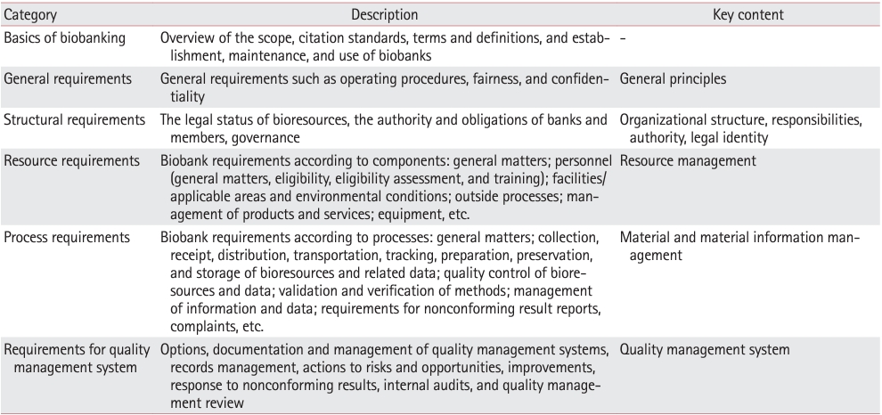
- Myoepithelial Carcinoma of Soft Tissue: A Case Report and Review of the Literature
- Chang Hwan Choi, Young Chae Chu, Lucia Kim, Suk Jin Choi, In Suh Park, Jee Young Han, Joon Mee Kim
- Korean J Pathol. 2014;48(6):413-417. Published online December 31, 2014
- DOI: https://doi.org/10.4132/KoreanJPathol.2014.48.6.413
- 14,864 View
- 132 Download
- 9 Crossref
-
PDF
-
Citations
Citations to this article as recorded by- Hyalinizing Clear Cell Carcinoma of the Lung With Uncommon Distant Cutaneous Metastasis and Aggressive Clinical Course
David M. Gustafson, Sansar Tiwari, Catherine G. Chung
Journal of Cutaneous Pathology.2026;[Epub] CrossRef - Myoepithelial Carcinoma Mimicking Basal Cell Carcinoma: A Case Report
Farlin Asharaff, Neena Nayak, Roger Webb, Karwan Moutasim, Soogan Lalla
Cureus.2025;[Epub] CrossRef - Fine‐needle aspiration cytology of retroperitoneal myoepithelial carcinoma: A rare encounter with diagnostic dilemmas
Aadya Kerkar, Ajay Savlania, Reetu Kundu, Suvradeep Mitra, Manish Rohilla, Harmandeep Singh, Harish Bhujade
Diagnostic Cytopathology.2024;[Epub] CrossRef - EWSR1::NR4A3 gene fusion in a cutaneous atypical myoepithelial neoplasm
Ashley Rose Scholl, Evelyna Kliassov, Diana M. Cardona, Rex Bentley, Rami N. Al‐Rohil
Journal of Cutaneous Pathology.2023; 50(7): 601. CrossRef - Abdominal myoepithelial carcinoma: A rare abdominal wall entity of an uncommon tumor
Daania Shoaib, Saqib Raza Khan, Yasmin Abdul Rashid, Muhammad Nauman Zahir
International Journal of Surgery Case Reports.2022; 99: 107618. CrossRef - Adult soft tissue myoepithelial carcinoma: treatment outcomes and efficacy of chemotherapy
Florence Chamberlain, Elena Cojocaru, Mariana Scaranti, Jonathan Noujaim, Anastasia Constantinou, Khin Thway, Cyril Fisher, Christina Messiou, Dirk C. Strauss, Aisha Miah, Shane Zaidi, Charlotte Benson, Spyridon Gennatas, Robin L. Jones
Medical Oncology.2020;[Epub] CrossRef - Foot plantar soft tissue malignant myoepithelioma tumor: Case report and review of the literature
Manuel Trevino, Chetan Moorthy, Lisa Kafchinski, Daniel Bustamante
Clinical Imaging.2020; 61: 90. CrossRef - Presumed choroidal metastasis from soft tissue myoepithelial carcinoma
Michelle M. Hui, Rohan Merani, Fiona Bonar, Angela M. Hong, Adrian T. Fung
American Journal of Ophthalmology Case Reports.2019; 14: 55. CrossRef - Myoepithelial carcinoma of the elbow diagnosed by immunohistochemistry: Case report of an uncommon neoplasm with metastatic recurrence
Madhura Mahapatra, Travis Lambert, Abdal Rahman El-Mallah, Andressa Balbi, Mohamad Aziz
Case Reports International.2019; 8(2): 1. CrossRef
- Hyalinizing Clear Cell Carcinoma of the Lung With Uncommon Distant Cutaneous Metastasis and Aggressive Clinical Course
- Preparation of Compact Agarose Cell Blocks from the Residues of Liquid-Based Cytology Samples
- Suk Jin Choi, Yeon Il Choi, Lucia Kim, In Suh Park, Jee Young Han, Joon Mee Kim, Young Chae Chu
- Korean J Pathol. 2014;48(5):351-360. Published online October 27, 2014
- DOI: https://doi.org/10.4132/KoreanJPathol.2014.48.5.351
- 19,096 View
- 393 Download
- 25 Crossref
-
Abstract
PDF
- Background: Inevitable loss of diagnostic material should be minimized during cell block preparation. We introduce a modified agarose cell block technique that enables the synthesis of compact cell blocks by using the entirety of a cell pellet without the loss of diagnostic material during cell block preparations. The feasibility of this technique is illustrated by high-throughput immunocytochemistry using high-density cell block microarray (CMA). Methods: The cell pellets of Sure- Path residues were pre-embedded in ultra-low gelling temperature agarose gel and re-embedded in standard agarose gel. They were fixed, processed, and embedded in paraffin using the same method as tissue sample processing. The resulting agarose cell blocks were trimmed and represented on a CMA for high-throughput analysis using immunocytochemical staining. Results: The SurePath residues were effectively and entirely incorporated into compact agarose cell buttons and embedded in paraffin. Sections of the agarose cell blocks revealed cellularities that correlated well with corresponding SurePath smears and had immunocytochemical features that were sufficient for diagnosis of difficult cases. Conclusions: This agarose-based compact cell block technique enables preparation of high-quality cell blocks by using up the residual SurePath samples without loss of diagnostic material during cell block preparation.
-
Citations
Citations to this article as recorded by- Assessment of an innovative synthetic matrix for enhanced cell preservation: evaluating its clinical utility and impact on diagnostic precision in thyroid fine-needle aspiration cytology
Diana-Raluca Streinu, Octavian Constantin Neagoe, Andreea Bena, Dana-Liana Stoian
BMC Endocrine Disorders.2026;[Epub] CrossRef - Establishing 3D organoid models from patient-derived conditionally reprogrammed cells to bridge preclinical and clinical insights in pancreatic cancer
Jin Su Kim, Chan Hee Park, Eunyoung Kim, Hee Seung Lee, Jinyoung Lee, Jeehoon Kim, Eun Hee Kam, Sanghee Nam, Moon Jae Chung, Jeong Youp Park, Seung Woo Park, Sangwoo Kim, Galam Leem, Seungmin Bang
Molecular Cancer.2025;[Epub] CrossRef - Assessing Determinants of Response to PARP Inhibition in Germline ATM Mutant Melanoma
Eleonora Allavena, Michela Croce, Bruna Dalmasso, Cecilia Profumo, Valentina Rigo, Virginia Andreotti, Irene Vanni, Benedetta Pellegrino, Antonino Musolino, Nicoletta Campanini, William Bruno, Luca Mastracci, Gabriele Zoppoli, Enrica Teresa Tanda, Frances
International Journal of Molecular Sciences.2025; 26(15): 7420. CrossRef - Immunocytochemistry on frozen-embedded cell block for the diagnosis of hematolymphoid cytology specimen: a straightforward alternative to the conventional cell block
Youjeong Seo, Sanzida Alam Prome, Lucia Kim, Jee Young Han, Joon Mee Kim, Suk Jin Choi
Journal of Hematopathology.2024; 17(1): 1. CrossRef - Comparison of liquid-based cytology and cell blocks prepared from cell remnants for diagnosis of cervical pathology
Elif Kuzucular, Ferhat Ozden, Bahar Muezzinoglu
Annals of Diagnostic Pathology.2024; 69: 152265. CrossRef - Advances in diagnostic liquid‐based cytology
Hideyuki Abe, Akihiko Kawahara, Jun Akiba, Rin Yamaguchi
Cytopathology.2024; 35(6): 682. CrossRef - Enhancing diagnostic precision in thyroid nodule assessment: evaluating the efficacy of a novel cell preservation technique in fine-needle aspiration cytology
Diana-Raluca Streinu, Octavian Constantin Neagoe, Andreea Borlea, Ion Icma, Mihnea Derban, Dana Stoian
Frontiers in Endocrinology.2024;[Epub] CrossRef - Cell blocks in cytology: review of preparation methods, advantages, and limitations
Vanda F. Torous, Jacqueline M. Cuda, Varsha Manucha, Melissa L. Randolph, Qiuying Shi, Christopher J. VandenBussche
Journal of the American Society of Cytopathology.2023; 12(2): 77. CrossRef - Cerebral Organoid Arrays for Batch Phenotypic Analysis in Sections and Three Dimensions
Juan Chen, Haihua Ma, Zhiyu Deng, Qingming Luo, Hui Gong, Ben Long, Xiangning Li
International Journal of Molecular Sciences.2023; 24(18): 13903. CrossRef - Diagnosis of pancreatic solid pseudopapillary neoplasms using cell‐blocks and immunohistochemical evaluation of endoscopic ultrasound‐guided fine needle aspiration biopsy specimens
José Celso Ardengh, César Vivian Lopes, Filadélfio Euclides Venco, Marcel Autran Machado
Cytopathology.2021; 32(1): 50. CrossRef - Somatostatin receptor 2 expression in nasopharyngeal cancer is induced by Epstein Barr virus infection: impact on prognosis, imaging and therapy
Matt Lechner, Volker H. Schartinger, Christopher D. Steele, Wen Long Nei, Marc Lucas Ooft, Liesa-Marie Schreiber, Christodoulos P. Pipinikas, Grace Tin-Yun Chung, Yuk Yu Chan, Feng Wu, Ka-Fai To, Chi Man Tsang, Wayne Pearce, Daniele Morelli, Martin Philpo
Nature Communications.2021;[Epub] CrossRef - Utility of PD‐L1 immunocytochemistry using body‐fluid cell blocks in patients with non‐small‐cell lung cancer
Seung Geun Song, Jonghoon Lee, Jaemoon Koh, Sehui Kim, Doo Hyun Chung, Yoon Kyung Jeon
Diagnostic Cytopathology.2020; 48(4): 291. CrossRef - Agarose cell block and ancillary molecular tests enhance diagnostic efficacy of liquid-based cytology samples
EmanS Abusinna, MervatM El-Deftar, YasmineF El-Esawy
Egyptian Journal of Pathology.2020; 40(2): 169. CrossRef - Preparation of frozen cell-block sections amenableto diagnostic immunocytochemistry: a technical report on cryo-embedded cell-block method
Joo Sung Lee, Suk Jin Choi, Lucia Kim, In Suh Park, Jee Young Han, Joon Mee Kim
Polish Journal of Pathology.2020; 71(2): 146. CrossRef - Brain-Derived Neurotrophin and TrkB in Head and Neck Squamous Cell Carcinoma
József Dudás, Anna Riml, Raphaela Tuertscher, Christian Pritz, Teresa Bernadette Steinbichler, Volker Hans Schartinger, Susanne Sprung, Rudolf Glueckert, Anneliese Schrott-Fischer, Lejo Johnson Chacko, Herbert Riechelmann
International Journal of Molecular Sciences.2019; 20(2): 272. CrossRef - Pleiotropic Effects of Epithelial Mesenchymal Crosstalk on Head and Neck Cancer: EMT and beyond
T. B. Steinbichler, D. Savic, D. Dejaco, A. Romani, B. Kofler, I. I. Skvortsova, H. Riechelmann, J. Dudas
Cancer Microenvironment.2019; 12(2-3): 67. CrossRef - Microarray Embedding/Sectioning for Parallel Analysis of 3D Cell Spheroids
Jonathan Gabriel, David Brennan, Jennifer H. Elisseeff, Vince Beachley
Scientific Reports.2019;[Epub] CrossRef - Photodynamic Effect of Methylene Blue and Low Level Laser Radiation in Head and Neck Squamous Cell Carcinoma Cell Lines
Barbara Kofler, Angela Romani, Christian Pritz, Teresa Steinbichler, Volker Schartinger, Herbert Riechelmann, Jozsef Dudas
International Journal of Molecular Sciences.2018; 19(4): 1107. CrossRef - Predictors of Response to Autologous Dendritic Cell Therapy in Glioblastoma Multiforme
Chia-Ing Jan, Wan-Chen Tsai, Horng-Jyh Harn, Woei-Cherng Shyu, Ming-Chao Liu, Hsin-Man Lu, Shao-Chih Chiu, Der-Yang Cho
Frontiers in Immunology.2018;[Epub] CrossRef - Nerve Growth Factor (NGF)—Receptor Survival Axis in Head and Neck Squamous Cell Carcinoma
József Dudás, Wolfgang Dietl, Angela Romani, Susanne Reinold, Rudolf Glueckert, Anneliese Schrott-Fischer, Daniel Dejaco, Lejo Johnson Chacko, Raphaela Tuertscher, Volker Hans Schartinger, Herbert Riechelmann
International Journal of Molecular Sciences.2018; 19(6): 1771. CrossRef - Cell blocks in cytopathology: An update
Aruna Nambirajan, Deepali Jain
Cytopathology.2018; 29(6): 505. CrossRef - Cell transfer technique for constructing cytological microarrays for immunocytochemical analysis
C.‐H. Wen, C.‐H. Lin, P.‐L. Ko, Y.‐F. Kuo, Y.‐J. Chen, C.‐Y. Chai
Cytopathology.2017; 28(2): 157. CrossRef - Cell and Tissue Display
Nicholas Theodosakis, Goran Micevic, Marcus W. Bosenberg, Nemanja Rodić
Journal of Histochemistry & Cytochemistry.2016; 64(7): 403. CrossRef - Diagnostic Usefulness of Claudin-3 and Claudin-4 for Immunocytochemical Differentiation between Metastatic Adenocarcinoma Cells and Reactive Mesothelial Cells in Effusion Cell Blocks
Nah Ihm Kim, Ga-Eon Kim, Ji Shin Lee
Acta Cytologica.2016; 60(3): 232. CrossRef - Do We Know What Is in Our Samples?
Louise Gilroy, Kathy Walsh, Anca Oniscu
Journal of Thoracic Oncology.2015; 10(12): e122. CrossRef
- Assessment of an innovative synthetic matrix for enhanced cell preservation: evaluating its clinical utility and impact on diagnostic precision in thyroid fine-needle aspiration cytology
- The Clinicopathological Significance of Epithelial Mesenchymal Transition Associated Protein Expression in Head and Neck Squamous Cell Carcinoma
- Kyu Ho Kim, Lucia Kim, Suk Jin Choi, Jee Young Han, Joon Mee Kim, Young Chae Chu, Young-Mo Kim, In Suh Park, Joo Han Lim
- Korean J Pathol. 2014;48(4):263-269. Published online August 26, 2014
- DOI: https://doi.org/10.4132/KoreanJPathol.2014.48.4.263
- 11,016 View
- 58 Download
- 14 Crossref
-
Abstract
PDF
Background Epithelial mesenchymal transition (EMT) has an important role in invasion and metastasis of tumor cells. The purpose of this study was to evaluate the roles of EMT-associated proteins on progression and metastasis as a prognostic/predictive factor in curatively-resected (R0) head and neck squamous cell carcinoma (HNSCC).
Methods A total of 118 patients who received curative surgery for HNSCC at Inha University Hospital between January 1996 and December 2011 were included. We used protein immunohistochemistry to evaluate the expression of E-cadherin, vimentin, and EZH2 on tissue microarrays. Also, we reviewed all medical records and analyzed the relationship between the expression of EMT-associated proteins and prognosis.
Results The E-cadherin-negative group showed more moderate/poor differentiation of cancer cell type than the higher E-cadherin-expressing group (p=.016) and high EZH2 expression was significantly correlated with nodal metastasis (p=.012). Our results demonstrate a significant association between high expression of EZH2 and vimentin and presence of distant progression (p=.026). However, expression of E-cadherin, vimentin, and EZH2 was not significantly associated with overall survival.
Conclusions These findings suggest that an EMT-associated protein expression profile is correlated with aggressiveness of disease and prognosis, and could be a useful marker for determination of additional treatment in curatively-resected HNSCC patients.
-
Citations
Citations to this article as recorded by- Correlation of EZH2 expression and response to chemoradiation in patients with locally advanced inoperable oral cavity and oropharyngeal squamous cell cancers
S. K. Soel Ahmed, Saroj Kumar Das Majumdar, Amit Kumar Adhya, Sandip Kumar Barik, Deepak Kumar Das, Mathan Kumar Ramasubbu, Priyanka Mukherjee, Anupam Muraleedharan, Nehla Haroon, Ankur Mahajan, Shaha Sheik Abdulla, Arnab Sarkar, Phanindra Kumar Swain, Pr
Journal of Cancer Research and Therapeutics.2025; 21(7): 1391. CrossRef - Polycomb repressive complex 2 and its core component EZH2: potential targeted therapeutic strategies for head and neck squamous cell carcinoma
Yuxi Cheng, Zhengzheng Song, Xiaodan Fang, Zhangui Tang
Clinical Epigenetics.2024;[Epub] CrossRef - Enhanced Expression of N-Cadherin, but Not of E-Cadherin, in Cutaneous Squamous Cell Carcinoma in Comparison to Basal Cell Carcinoma
Joanna Pogorzelska-Dyrbuś, Danuta Nowicka-Suszko, Aleksandra Piotrowska, Zdzisław Woźniak, Piotr Dzięgiel, Jacek C. Szepietowski
Cancers.2024; 16(24): 4247. CrossRef - Prognostic Value of TWIST1 and EZH2 Expression in Colon Cancer
Samar M. Abdel Raouf, Taiseer R. Ibrahim, Lobna A. Abdelaziz, Mohamed I. Farid, Salem Y Mohamed
Journal of Gastrointestinal Cancer.2021; 52(1): 90. CrossRef - HOXB5 acts as an oncogenic driver in head and neck squamous cell carcinoma via EGFR/Akt/Wnt/β-catenin signaling axis
Kyungmin Lee, Jae Won Chang, Chan Oh, Lihua Liu, Seung-Nam Jung, Ho-Ryun Won, Young Il Kim, Ki-Sang Rha, Bon Seok Koo
European Journal of Surgical Oncology.2020; 46(6): 1066. CrossRef - EZH2 overexpression in head and neck cancer is related to lymph node metastasis
Julie C. Nienstedt, Cornelia Schroeder, Till Clauditz, Ronald Simon, Guido Sauter, Adrian Muenscher, Marco Blessmann, Henning Hanken, Christina Pflug
Journal of Oral Pathology & Medicine.2018; 47(3): 240. CrossRef - MiR-876-5p modulates head and neck squamous cell carcinoma metastasis and invasion by targeting vimentin
Yibo Dong, Yang Zheng, Chundi Wang, Xu Ding, Yifei Du, Laikui Liu, Wei Zhang, Wei Zhang, Yi Zhong, Yunong Wu, Xiaomeng Song
Cancer Cell International.2018;[Epub] CrossRef - Special AT-rich binding protein 1 and Dachshund homolog 1 expression in bladder carcinomas and their role in epithelial–mesenchymal transition
Mohammed I. Shaban, Nanis S. Holah, Mona A. Kandil, Hayam A. Aiad, Amira M. Abd El Maged
Egyptian Journal of Pathology.2018; 38(2): 199. CrossRef - HMGA2 is associated with the aggressiveness of tongue squamous cell carcinoma
H Zhang, Z Tang, C Deng, Y He, F Wu, O Liu, C Hu
Oral Diseases.2017; 23(2): 255. CrossRef - miR-375 Regulates Invasion-Related Proteins Vimentin and L-Plastin
Lizandra Jimenez, Jihyeon Lim, Berta Burd, Thomas M. Harris, Thomas J. Ow, Nicole Kawachi, Thomas J. Belbin, Ruth Angeletti, Michael B. Prystowsky, Geoffrey Childs, Jeffrey E. Segall
The American Journal of Pathology.2017; 187(7): 1523. CrossRef - Functional and therapeutic significance of EZH2 in urological cancers
Xiaobing Liu, Qingjian Wu, Longkun Li
Oncotarget.2017; 8(23): 38044. CrossRef - EZH2, an on–off valve in signal network of tumor cells
Shanshan Sun, Feng Yu, Lun Zhang, Xuan Zhou
Cellular Signalling.2016; 28(5): 481. CrossRef - High EZH2 Protein Expression Is Associated with Poor Overall Survival in Patients with Luminal A Breast Cancer
Si-Hyong Jang, Jong Eun Lee, Mee-Hye Oh, Ji-Hye Lee, Hyun Deuk Cho, Kyung-Ju Kim, Sung Yong Kim, Sun Wook Han, Han Jo Kim, Sang Byung Bae, Hyun Ju Lee
Journal of Breast Cancer.2016; 19(1): 53. CrossRef - EZH2 promotes invasion and metastasis of laryngeal squamous cells carcinoma via epithelial-mesenchymal transition through H3K27me3
HuaNan Luo, Yuan Jiang, SiJing Ma, HuanHuan Chang, ChunXi Yi, Hui Cao, Ying Gao, HaiLi Guo, Jin Hou, Jing Yan, Ying Sheng, XiaoYong Ren
Biochemical and Biophysical Research Communications.2016; 479(2): 253. CrossRef
- Correlation of EZH2 expression and response to chemoradiation in patients with locally advanced inoperable oral cavity and oropharyngeal squamous cell cancers
- Early Colorectal Epithelial Neoplasm in Korea: A Multicenter Survey of Pathologic Diagnosis
- Yun Kyung Kang, So-Young Jin, Mee Soo Chang, Jung Yeon Kim, Gyeong Hoon Kang, Hye Seung Lee, Jin Hee Sohn, Ho Sung Park, Kye Won Kwon, Mi Jin Gu, Young Hee Maeng, Jong Eun Joo, Haeng Ji Kang, Hee Kyung Kim, Kee-Taek Jang, Mi Ja Lee, Hee Kyung Chang, Joon Mee Kim, Hye Seung Han, Won Ae Lee, Yoon Jung Choi, Dong Wook Kang, Sunhoo Park, Jae Hyuk Lee, Mee-Yon Cho
- Korean J Pathol. 2013;47(3):245-251. Published online June 25, 2013
- DOI: https://doi.org/10.4132/KoreanJPathol.2013.47.3.245
- 12,195 View
- 56 Download
- 1 Crossref
-
Abstract
PDF
Background The incidence of early colorectal epithelial neoplasm (ECEN) is increasing, and its pathologic diagnosis is important for patient care. We investigated the incidence of ECEN and the current status of its pathologic diagnosis.
Methods We collected datasheets from 25 institutes in Korea for the incidence of colorectal adenoma with high grade dysplasia (HGD) and low grade dysplasia in years 2005, 2007, and 2009; and early colorectal carcinoma in the year 2009. We also surveyed the diagnostic terminology of ECEN currently used by the participating pathologists.
Results The average percentage of diagnoses of adenoma HGD was 7.0%, 5.0%, and 3.4% in years 2005, 2007, and 2009, respectively. The range of incidence rates of adenoma HGD across the participating institutes has gradually narrowed over the years 2005 to 2009. The incidence rate of early colorectal carcinoma in the year 2009 was 21.2%. The participants did not share a single criterion or terminology for the diagnosis of adenoma HGD. The majority accepted the diagnostic terms that distinguished noninvasive, mucosal confined, and submucosal invasive carcinoma.
Conclusions Further research requirements suggested are a diagnostic consensus for the histopathologic diagnosis of ECEN; and standardization of diagnostic terminology critical for determining the disease code.
-
Citations
Citations to this article as recorded by- Diminutive and Small Colorectal Polyps: The Pathologist's Perspective
Yun Kyung Kang
Clinical Endoscopy.2014; 47(5): 404. CrossRef
- Diminutive and Small Colorectal Polyps: The Pathologist's Perspective
- In-house Manual Construction of High-Density and High-Quality Tissue Microarrays by Using Homemade Recipient Agarose-Paraffin Blocks
- Kyu Ho Kim, Suk Jin Choi, Yeon Il Choi, Lucia Kim, In Suh Park, Jee Young Han, Joon Mee Kim, Young Chae Chu
- Korean J Pathol. 2013;47(3):238-244. Published online June 25, 2013
- DOI: https://doi.org/10.4132/KoreanJPathol.2013.47.3.238
- 12,689 View
- 99 Download
- 7 Crossref
-
Abstract
PDF
Background Self-made tissue punches can be effectively used to punch holes in blank recipient paraffin blocks and extract tissue cores from the donor paraffin blocks for the low-cost construction of tissue microarrays (TMAs). However, variable degrees of section distortion and loss of the tissue cores can occurs during cutting of the TMAs, posing technical problems for in-house manual construction of high-density TMAs. We aimed to update the method for in-house manual TMA construction to improve the quality of high-density TMAs.
Methods Blocks of agarose gel were subjected to the standard tissue processing and embedding procedure to prepare recipient agarose-paraffin blocks. The self-made tissue punches and recipient agarose-paraffin blocks were used to construct TMAs, which were completely melted and re-embedded in paraffin to make finished TMA blocks.
Results The donor tissue cores were completely integrated into the surrounding paraffin of the recipient blocks. This method enabled us to construct high-density TMAs with significantly less section distortion or loss of tissue cores during microtomy.
Conclusions Simple and inexpensive construction of high-density and high-quality TMAs can be warranted by using paraffinized agarose gels as recipient blocks.
-
Citations
Citations to this article as recorded by- Using Quality Function Deployment to Design an Image-Guided, Multibiopsy Tool for Neurosurgical Applications
Kaytlin Andrews, Hunter Dejean, Cameron MacLeod, Kate Prieditis, Heidi-Lynn Ploeg, James Purzner, Teresa Purzner
Operative Neurosurgery.2026; 30(3): 422. CrossRef - An introduction of an easy-operating and economical technique for tissue microarray preparation
Yi-Jing Chen, Chun-Mei Yang, Jiang-Sheng Huang, Ping Wang, Yan-Hua Lv, Cheng Tang, Wei Deng
Journal of Clinical Pathology.2020; 73(7): 403. CrossRef - Optimization of Tissue Microarrays from Banked Human Formalin-Fixed Paraffin Embedded Tissues in the Cancer Research Setting
Tammy Sexton, Gregory L. Kucera, Edward A. Levine, Kounosuke Watabe, Stacey S. O'Neill
Biopreservation and Biobanking.2019; 17(5): 452. CrossRef - Monocarboxylate transporters MCT1 and MCT4 are independent prognostic biomarkers for the survival of patients with clear cell renal cell carcinoma and those receiving therapy targeting angiogenesis
Yan-Wei Cao, Yong Liu, Zhen Dong, Lei Guo, En-Hao Kang, Yong-Hua Wang, Wei Zhang, Hai-Tao Niu
Urologic Oncology: Seminars and Original Investigations.2018; 36(6): 311.e15. CrossRef - Platelet-derived growth factor receptor α in hepatocellular carcinoma is a prognostic marker independent of underlying liver cirrhosis
Jung-Hwan Yu, Joon Mee Kim, Ja Kyung Kim, Suk Jin Choi, Kwan Sik Lee, Jin-Woo Lee, Hye Young Chang, Jung Il Lee
Oncotarget.2017; 8(24): 39534. CrossRef - Prognostic Implication of Semi-quantitative Immunohistochemical Assessment of CD20 Expression in Diffuse Large B-Cell Lymphoma
Chang Hwan Choi, Young Hoon Park, Joo Han Lim, Suk Jin Choi, Lucia Kim, In Suh Park, Jee Young Han, Joon Mee Kim, Young Chae Chu
Journal of Pathology and Translational Medicine.2016; 50(2): 96. CrossRef - High Quality Tissue Miniarray Technique Using a Conventional TV/Radio Telescopic Antenna
Mohamed A. Elkablawy, Abdulkader M. Albasri
Asian Pacific Journal of Cancer Prevention.2015; 16(3): 1129. CrossRef
- Using Quality Function Deployment to Design an Image-Guided, Multibiopsy Tool for Neurosurgical Applications
- Proposal for a Standardized Pathology Report of Gastroenteropancreatic Neuroendocrine Tumors: Prognostic Significance of Pathological Parameters
- Mee-Yon Cho, Jin Hee Sohn, So Young Jin, Hyunki Kim, Eun Sun Jung, Mi-Jung Kim, Kyoung-Mee Kim, Woo Ho Kim, Joon Mee Kim, Yun Kyung Kang, Joon Hyuk Choi, Dae Young Kang, Youn Wha Kim, Eun Hee Choi
- Korean J Pathol. 2013;47(3):227-237. Published online June 25, 2013
- DOI: https://doi.org/10.4132/KoreanJPathol.2013.47.3.227
- 16,451 View
- 148 Download
- 12 Crossref
-
Abstract
PDF
Background There is confusion in the diagnosis and biological behaviors of gastroenteropancreatic neuroendocrine tumors (GEP-NETs), because of independently proposed nomenclatures and classifications. A standardized form of pathology report is required for the proper management of patients.
Methods We discussed the proper pathological evaluation of GEP-NET at the consensus conference of the subcommittee meeting for the Gastrointestinal Pathology Study Group of the Korean Society of Pathologists. We then verified the prognostic significance of pathological parameters from our previous nationwide collection of pathological data from 28 hospitals in Korea to determine the essential data set for a pathology report.
Results Histological classification, grading (mitosis and/or Ki-67 labeling index), T staging (extent, size), lymph node metastasis, and lymphovascular and perineural invasion were significant prognostic factors and essential for the pathology report of GEP-NET, while immunostaining such as synaptophysin and chromogranin may be optional. Furthermore, the staging system, either that of the 2010 American Joint Cancer Committee (AJCC) or the European Neuroendocrine Tumor Society (ENETS), should be specified, especially for pancreatic neuroendocrine neoplasms.
Conclusions A standardized pathology report is crucial for the proper management and prediction of prognosis of patients with GEP-NET.
-
Citations
Citations to this article as recorded by- Analysis of Prognostic Risk Factors of Endoscopic Submucosal Dissection (ESD) and Curative Resection of Gastrointestinal Neuroendocrine Neoplasms
Yuan Si, ChaoKang Huang, JingBin Yuan, XianHui Zhang, QingQiang He, ZhiJin Lin, Ling He, ZhongXin Liu, Yuvaraja Teekaraman
Contrast Media & Molecular Imaging.2022;[Epub] CrossRef - Standardization of the pathologic diagnosis of appendiceal mucinous neoplasms
Dong-Wook Kang, Baek-hui Kim, Joon Mee Kim, Jihun Kim, Hee Jin Chang, Mee Soo Chang, Jin-Hee Sohn, Mee-Yon Cho, So-Young Jin, Hee Kyung Chang, Hye Seung Han, Jung Yeon Kim, Hee Sung Kim, Do Youn Park, Ha Young Park, So Jeong Lee, Wonae Lee, Hye Seung Lee,
Journal of Pathology and Translational Medicine.2021; 55(4): 247. CrossRef - Preoperative diagnosis of well‐differentiated neuroendocrine tumor in common hepatic duct by brush cytology: A case report
Jiwoon Choi, Kyong Joo Lee, Sung Hoon Kim, Mee‐Yon Cho
Diagnostic Cytopathology.2019; 47(7): 720. CrossRef - Primary renal well-differentiated neuroendocrine tumors: report of six cases with an emphasis on the Ki-67 index and mitosis
Bohyun Kim, Han-Seong Kim, Kyung Chul Moon
Diagnostic Pathology.2019;[Epub] CrossRef - Primary low‐grade neuroendocrine carcinoma of the skin: An exceedingly rare entity
Tiffany Y. Chen, Annie O. Morrison, Joe Susa, Clay J. Cockerell
Journal of Cutaneous Pathology.2017; 44(11): 978. CrossRef - Prognostic Validity of the American Joint Committee on Cancer and the European Neuroendocrine Tumors Staging Classifications for Pancreatic Neuroendocrine Tumors
Jae Hee Cho, Ji Kon Ryu, Si Young Song, Jin-Hyeok Hwang, Dong Ki Lee, Sang Myung Woo, Young-Eun Joo, Seok Jeong, Seung-Ok Lee, Byung Kyu Park, Young Koog Cheon, Jimin Han, Tae Nyeun Kim, Jun Kyu Lee, Sung-Hoon Moon, Hyunjin Kim, Eun Taek Park, Jae Chul Hw
Pancreas.2016; 45(7): 941. CrossRef - Early diagnosis and treatment of gastrointestinal neuroendocrine tumors
Hong Shen, Zhuo Yu, Jing Zhao, Xiu-Zhen Li, Wen-Sheng Pan
Oncology Letters.2016; 12(5): 3385. CrossRef - Recent Updates on Neuroendocrine Tumors From the Gastrointestinal and Pancreatobiliary Tracts
Joo Young Kim, Seung-Mo Hong
Archives of Pathology & Laboratory Medicine.2016; 140(5): 437. CrossRef - Pancreatic neuroendocrine tumors: Correlation between the contrast-enhanced computed tomography features and the pathological tumor grade
Koji Takumi, Yoshihiko Fukukura, Michiyo Higashi, Junnichi Ideue, Tomokazu Umanodan, Hiroto Hakamada, Ichiro Kanetsuki, Takashi Yoshiura
European Journal of Radiology.2015; 84(8): 1436. CrossRef - Tumeurs neuroendocrines du tube digestif et du pancréas : ce que le pathologiste doit savoir et doit faire en 2014
Jean-Yves Scoazec, Anne Couvelard
Annales de Pathologie.2014; 34(1): 40. CrossRef - Spectrum of Gastroenteropancreatic NENs in Routine Histological Examinations of Bioptic and Surgical Specimen: A Study of 161 Cases Collected from 17 Departments of Pathology in the Czech Republic
Václav Mandys, Tomáš Jirásek
Gastroenterology Research and Practice.2014; 2014: 1. CrossRef - p27 Loss Is Associated with Poor Prognosis in Gastroenteropancreatic Neuroendocrine Tumors
Hee Sung Kim, Hye Seung Lee, Kyung Han Nam, Jiwoon Choi, Woo Ho Kim
Cancer Research and Treatment.2014; 46(4): 383. CrossRef
- Analysis of Prognostic Risk Factors of Endoscopic Submucosal Dissection (ESD) and Curative Resection of Gastrointestinal Neuroendocrine Neoplasms
- Peripheral Primitive Neuroectodermal Tumor with Osseous Component of the Small Bowel Mesentery: A Case Study
- Joon Mee Kim, Young Chae Chu, Chang Hwan Choi, Lucia Kim, Suk Jin Choi, In Suh Park, Jee Young Han, Kyung Rae Kim, Yoon-La Choi, Taeeun Kim
- Korean J Pathol. 2013;47(1):77-81. Published online February 25, 2013
- DOI: https://doi.org/10.4132/KoreanJPathol.2013.47.1.77
- 11,998 View
- 49 Download
- 9 Crossref
-
Abstract
PDF
A case of peripheral primitive neuroectodermal tumor of the small bowel mesentery with osseous component is reported. A 23-year-old man was admitted to our hospital because of acute severe abdominal pain. Abdominal computed tomography revealed a large solid and cystic, oval shaped mass, measuring 11.0×6.0 cm in the pelvic cavity. Histologically the resected lesion consisted of sheets of undifferentiated small round cells forming Homer-Wright rosettes and perivascular pseudorosettes, and showed areas of osteoid and bone formation. Immunohistochemical studies revealed that tumor cells expressed positivity against CD99 (MIC2), CD57, neuron-specific enolase, and vimentin. Fluorescence
in situ hybridization study revealed Ewing sarcoma breakpoint region 1 (EWSR1 ) gene rearrangement on chromosome 22q12. To the authors' knowledge this is the first documentation of a peripheral neuroectodermal tumor with osteoid and bone formation of the small bowel mesentery.-
Citations
Citations to this article as recorded by- Undifferentiated small round cell sarcomas in the retroperitoneal space in a 12-year-old female: a rare case report
Dan Liu, Xiaoge Liu, Yan Deng, Ran Wu, Xin Li
Frontiers in Pediatrics.2025;[Epub] CrossRef - Primary Ewing’s sarcoma of the intestine: case report and literature review
Baofa Luo, Wei Gao, Ting Li, Xinran Yu, Fei Guo
Frontiers in Oncology.2024;[Epub] CrossRef - Peripheral Primary Neuroectodermal Tumour of the Ileum: A Case Report and Review of the Literature
Soukaina Harrak, Siham Lemsanes, Sawsan Razine, Salma Najem, Khadija Benchekroun, Saad Lannaz, Hind Mrabti, Hassan Errihani
Journal of Cancer Therapy.2021; 12(07): 399. CrossRef - Primary Ewing’s sarcoma in a small intestine – a case report and review of the literature
Andrej Kolosov, Audrius Dulskas, Kastytis Pauza, Veslava Selichova, Dmitrij Seinin, Eugenijus Stratilatovas
BMC Surgery.2020;[Epub] CrossRef - Case report and literature review of Ewing's sarcoma in the gastrointestinal tract
Christopher Bong, Iain Thomson, Guy Lampe
Surgical Practice.2018; 22(2): 84. CrossRef - Pediatric Ewing’s Sarcoma/Primitive Neuroectodermal Tumor (ES/PNET) Developed in the Small Intestine: A Case Report
You Sun Kim, Hye Min Moon, Kyu Sang Lee, Young Suk Park, Hyun-Young Kim, Ji Young Kim, Jin Min Cho, Hyoung Soo Choi
Clinical Pediatric Hematology-Oncology.2017; 24(2): 162. CrossRef - Huge peripheral primitive neuroectodermal tumor of the small bowel mesentery at nonage: A case report and review of the literature
Zhe Liu, Yuan-Hong Xu, Chun-Lin Ge, Jin Long, Rui-Xia Du, Ke-Jian Guo
World Journal of Clinical Cases.2016; 4(9): 306. CrossRef - Primary primitive neuroectodermal tumor arising in the mesentery and ileocecum: A report of three cases and review of the literature
LIBO PENG, LIMIN YANG, NAN WU, BO WU
Experimental and Therapeutic Medicine.2015; 9(4): 1299. CrossRef - Une curieuse tumeur digestive à cellules rondes
Alia Zehani, Ines Chelly, Beya Chelly, Jean-Michel Coindre, Slim Haouet, Nidhameddine Kchir
Annales de Pathologie.2014; 34(2): 104. CrossRef
- Undifferentiated small round cell sarcomas in the retroperitoneal space in a 12-year-old female: a rare case report
- Construction of High-Density Tissue Microarrays at Low Cost by Using Self-Made Manual Microarray Kits and Recipient Paraffin Blocks
- Chang Hwan Choi, Kyu Ho Kim, Ju Young Song, Suk Jin Choi, Lucia Kim, In Suh Park, Jee Young Han, Joon Mee Kim, Young Chae Chu
- Korean J Pathol. 2012;46(6):562-568. Published online December 26, 2012
- DOI: https://doi.org/10.4132/KoreanJPathol.2012.46.6.562
- 13,141 View
- 112 Download
- 15 Crossref
-
Abstract
PDF
Background Advances of tissue microarray (TMA) technology have enabled simultaneous
in situ analysis of biomarker expression in a large number of archived pathology specimens. However, the relatively high cost of TMA construction may hamper many researchers from using this essential tool of modern pathology research. We discuss methods for making TMA kits and recipient blocks for manual construction of high-density TMAs at low cost.Methods Ordinary cannula piercing needles, hypodermic needles, bone marrow biopsy needles, metallic ink cartridges of ballpoint pens, and disposable skin biopsy punches were used to construct self-made manual TMA kits. The recipient blocks were manufactured by boring holes in the conventional bare paraffin blocks. A mini electric hand drill and a microcompound table assembled on a drill stand were used to maximize the capacity of the recipient blocks.
Results By using TMA kits made from cannula piercing needles (16- and 18-gauge), it was possible to construct TMAs with 1 mm×140 cores, 0.6 mm×320 cores, 2 mm×70 cores, 3 mm×35 cores, and 5 mm×12 cores. The capacity of the recipient blocks could be dramatically increased by drilling holes.
Conclusions Construction of TMAs using self-made TMA kits is an inexpensive alternative to construction of TMAs using commercial devices.
-
Citations
Citations to this article as recorded by- Diagnostic Potential of CD44, CD133, and VDR in Epithelial Ovarian Tumors: Association with Histopathology Parameters
Ljubiša Jovanović, Branka Šošić-Jurjević, Anđa Ćirković, Sandra Dragičević, Branko Filipović, Svetlana Milenković, Stefan Dugalić, Miroslava Gojnić-Dugalić, Aleksandra Nikolić
International Journal of Molecular Sciences.2025; 26(8): 3729. CrossRef - Comparison of programmed death-1 (PD-1)-positive T-cells with known prognostic indicators in breast cancer
Ankit Kaushik, Anamika Jaiswal, B Priya, Ashish Jain, Sonal Sharma
Journal of Family Medicine and Primary Care.2025; 14(11): 4631. CrossRef - Constructing high-density tissue microarrays with a novel method and a self-made tissue-arraying instrument
Ping Qin, Liu Li, Li Zhao, Piaopiao Bian, Zhongtang Xiong
Pathology - Research and Practice.2023; 245: 154430. CrossRef - The correlation of PD-L1 expression in cytological and histological material of serous high-grade ovarian cancer
Ljubiša Jovanović, Anđa Ćirković, Ljubinka Nikolić, Milena Jović, Darko Mikić, Svetlana Milenković, Radmila Janković
Srpski medicinski casopis Lekarske komore.2023; 4(3): 246. CrossRef - Expression of estrogen and progesterone receptors, HER2 protein and Ki-67 proliferation index in breast carcinoma in both tumor tissue and tissue microarray
UP Hacısalihoğlu, MA Dogan
Biotechnic & Histochemistry.2022; 97(4): 298. CrossRef - PD-L1 Expression in High-Grade Serous and Clear Cell Ovarian Cancer
Ljubiša Jovanović, Andja Ćirković, Milena Jović, Radmila Janković
Indian Journal of Gynecologic Oncology.2022;[Epub] CrossRef - PD-L1 Expression in Different Segments and Histological Types of Ovarian Cancer According to Lymphocytic Infiltrate
Ljubiša Jovanović, Radmila Janković, Andja Ćirković, Milena Jović, Tijana Janjić, Slaviša Djuričić, Svetlana Milenković
Medicina.2021; 57(12): 1309. CrossRef - Optimization of Tissue Microarrays from Banked Human Formalin-Fixed Paraffin Embedded Tissues in the Cancer Research Setting
Tammy Sexton, Gregory L. Kucera, Edward A. Levine, Kounosuke Watabe, Stacey S. O'Neill
Biopreservation and Biobanking.2019; 17(5): 452. CrossRef - Peripheral nerve sheath tumor invading the nasal cavities of a 6-year-old female Pointer dog
Alessandra Sfacteria, Laura Perillo, Francesco Macrì, Giovanni Lanteri, Claudia Rifici, Giuseppe Mazzullo
Veterinary Quarterly.2015; 35(3): 170. CrossRef - High Quality Tissue Miniarray Technique Using a Conventional TV/Radio Telescopic Antenna
Mohamed A. Elkablawy, Abdulkader M. Albasri
Asian Pacific Journal of Cancer Prevention.2015; 16(3): 1129. CrossRef - Overview on Techniques to Construct Tissue Arrays with Special Emphasis on Tissue Microarrays
Ulrich Vogel
Microarrays.2014; 3(2): 103. CrossRef - Tissue Microarray
Kathleen Barrette, Joost J. van den Oord, Marjan Garmyn
Journal of Investigative Dermatology.2014; 134(9): 1. CrossRef - Altered Expression of PTEN and Its Major Regulator MicroRNA-21 in Pulmonary Neuroendocrine Tumors
Hyoun Wook Lee, Seung Yeon Ha, Mee Sook Roh
Korean Journal of Pathology.2014; 48(1): 17. CrossRef - Optimizing tissue microarray construction procedure to improve quality of sections
Hua Chang, Diane Peluso, Sadiq Hussain, Michail Shipitsin, Peter Blume-Jensen
Journal of Histotechnology.2014; 37(3): 95. CrossRef - In-house Manual Construction of High-Density and High-Quality Tissue Microarrays by Using Homemade Recipient Agarose-Paraffin Blocks
Kyu Ho Kim, Suk Jin Choi, Yeon Il Choi, Lucia Kim, In Suh Park, Jee Young Han, Joon Mee Kim, Young Chae Chu
Korean Journal of Pathology.2013; 47(3): 238. CrossRef
- Diagnostic Potential of CD44, CD133, and VDR in Epithelial Ovarian Tumors: Association with Histopathology Parameters
- Update on the Proposal for Creating a Guideline for Cancer Registration of the Gastrointestinal Tumors (I-2)
- Eun Sun Jung, Yun Kyung Kang, Mee-Yon Cho, Joon Mee Kim, Won Ae Lee, Hee Eun Lee, Sunhoo Park, Jin Hee Sohn, So-Young Jin
- Korean J Pathol. 2012;46(5):443-453. Published online October 25, 2012
- DOI: https://doi.org/10.4132/KoreanJPathol.2012.46.5.443
- 12,742 View
- 190 Download
- 18 Crossref
-
Abstract
PDF
Background Cancer registries play a fundamental role in cancer control and multicenter collaborative research. Recently, the need for reassessment of cancer registry criteria has arisen due to the newly released 2010 World Health Organization (WHO) classification. Accordingly, development of new coding guidelines for cancer is necessary to improve the quality of cancer registries, as well as to prevent conflicts that may arise when seeking medical insurance compensation.
Methods With funding from the Management Center for Health Promotion, 35 members of the Gastrointestinal Pathology Study Group and the Cancer Registration Committee of the Korean Society of Pathologists (KSP) participated in a second workshop for gastrointestinal tumor registration in Korea.
Results The topics of gastric epithelial tumor, colonic intramucosal carcinoma, neuroendocrine tumor (NET), gastrointestinal stromal tumor (GIST) and appendiceal mucinous tumor were discussed for new coding guidelines. A survey was then conducted among 208 members of the KSP for a consensus of the guidelines proposed in the workshop.
Conclusions Although a few issues were set aside for further discussion, such as coding for non-gastric GIST and some types of NET, the members agreed upon most of the proposed guidelines. Therefore, we suggest using the newly revised International Classification of Diseases for Oncology, 3rd edition (ICD-O-3) coding guidelines for registering gastrointestinal tumors in Korea.
-
Citations
Citations to this article as recorded by- Gastrointestinal Stromal Tumor: History, Molecular Subtypes, and Risk Stratification
In Hye Song, Soomin Ahn, Hyung-Don Kim, Jeong-Hyeon Jo, Jinho Shin, Min-Hee Ryu, Young Soo Park
Journal of Gastric Cancer.2026; 26(2): 202. CrossRef - Variation in mitotic counting and risk classification practices for gastrointestinal stromal tumors: a survey of pathologists in South Korea
In Hye Song, Soomin Ahn, Jeong-Hyeon Jo, Young Soo Park
Journal of Pathology and Translational Medicine.2025; 59(5): 348. CrossRef - Different miRNAs Related to FBXW7 Mutations or High Mitotic Indices Contribute to Rectal Neuroendocrine Tumors: A Pilot Study
Ho Suk Kang, Ha Young Park, Hyun Lim, Il Tae Son, Min-Jeong Kim, Nan Young Kim, Min Jeong Kim, Eun Sook Nam, Seong Jin Cho, Mi Jung Kwon
International Journal of Molecular Sciences.2023; 24(7): 6329. CrossRef - Clinicopathologic Impact of Peptide Hormonal Expression in Rectal Neuroendocrine Tumors
Jisup Kim, Dong-Hoon Yang, HaeSung Jung, HyungJun Cho, Hyeung-Jin Jang, Changhoon Yoo, In Ja Park, Baek-Yeol Ryoo, Jin-Sook Ryu, Seung-Mo Hong
Archives of Pathology & Laboratory Medicine.2023; 147(7): 797. CrossRef - Prognostic nomogram and novel risk-scoring system for small cell lung cancer with different patterns of metastases
Hongli Ruan, Huali Sun, Yu Guo, Yan Ding, Yanmei Liu, Shenpeng Ying, Peipei Lin
General Thoracic and Cardiovascular Surgery.2022; 70(12): 1022. CrossRef - Development of a nomogram model to predict survival outcomes in patients with primary hepatic neuroendocrine tumors based on SEER database
Ziteng Zhang, Xin Zhao, Zhiyan Li, Youchun Wu, Yao Liu, Zhiwei Li, Guobao Li
BMC Cancer.2021;[Epub] CrossRef - Standardization of the pathologic diagnosis of appendiceal mucinous neoplasms
Dong-Wook Kang, Baek-hui Kim, Joon Mee Kim, Jihun Kim, Hee Jin Chang, Mee Soo Chang, Jin-Hee Sohn, Mee-Yon Cho, So-Young Jin, Hee Kyung Chang, Hye Seung Han, Jung Yeon Kim, Hee Sung Kim, Do Youn Park, Ha Young Park, So Jeong Lee, Wonae Lee, Hye Seung Lee,
Journal of Pathology and Translational Medicine.2021; 55(4): 247. CrossRef - Analysis of the Incidence and Clinical Features of Colorectal Nonadenocarcinoma in Korea: A National Cancer Registry-Based Study
Soomin Nam, Dongwook Kim, Kyuwon Jung, Yoon Jung Choi, Jung Gu Kang
Annals of Coloproctology.2020; 36(6): 390. CrossRef - Novel Finding of Paired Box 5 (PAX5) Cytoplasmic Staining in Well-differentiated Rectal Neuroendocrine Tumors (Carcinoids) and Its Diagnostic and Potentially Prognostic Utility
Zhiyan Fu, Chunlai Zuo, Christine E. Sheehan, Deepa T. Patil, Jingmei Lin, Zhaohai Yang, Hwajeong Lee
Applied Immunohistochemistry & Molecular Morphology.2019; 27(6): 454. CrossRef - Lymphovascular invasion as a prognostic value in small rectal neuroendocrine tumor treated by local excision: A systematic review and meta-analysis
Ho Suk Kang, Mi Jung Kwon, Tae-Hwan Kim, Junhee Han, Young-Su Ju
Pathology - Research and Practice.2019; 215(11): 152642. CrossRef - Management Colorectal Gastrointestinal Stromal Tumors (Gists) in Surabaya
Yuda Handaya, Sutamto Wibowo, Iwan Kristian
Open Journal of Gastroenterology.2016; 06(04): 97. CrossRef - Non-L-cell Immunophenotype and Large Tumor Size in Rectal Neuroendocrine Tumors Are Associated With Aggressive Clinical Behavior and Worse Prognosis
Joo Young Kim, Ki-Suk Kim, Kyung-Jo Kim, In Ja Park, Jong Lyul Lee, Seung-Jae Myung, Yangsoon Park, Young Soo Park, Chang Sik Yu, Jin Cheon Kim, Eunsil Yu, Hyeung-Jin Jang, Seung-Mo Hong
American Journal of Surgical Pathology.2015; 39(5): 632. CrossRef - Diagnostic Coding for Intramucosal Carcinoma and Neuroendocrine Tumor in the Colorectum: Proposal for Avoiding Confusing Coding in Korea
Dong Soo Han, Jin Hee Sohn, Jeong-Sik Byeon, Hwang Choi, Joon Mee Kim
Clinical Endoscopy.2015; 48(3): 216. CrossRef - Prognostic Significance of Defining L-Cell Type on the Biologic Behavior of Rectal Neuroendocrine Tumors in Relation with Pathological Parameters
Jin Hee Sohn, Mee-Yon Cho, Yangsoon Park, Hyunki Kim, Woo Ho Kim, Joon Mee Kim, Eun Sun Jung, Kyoung-Mee Kim, Jae Hyuk Lee, Hee Kyung Chan, Do Youn Park, Mee Joo, Sujin Kim, Woo Sung Moon, Mi Seon Kang, So-Young Jin, Yun Kyung Kang, Sun Och Yoon, HyeSeung
Cancer Research and Treatment.2015; 47(4): 813. CrossRef - Diminutive and Small Colorectal Polyps: The Pathologist's Perspective
Yun Kyung Kang
Clinical Endoscopy.2014; 47(5): 404. CrossRef - Highlights from the 50th Seminar of the Korean Society of Gastrointestinal Endoscopy
Eun Young Kim, Il Ju Choi, Kwang An Kwon, Ji Kon Ryu, Seok Ho Dong, Ki Baik Hahm
Clinical Endoscopy.2014; 47(4): 285. CrossRef - Early Colorectal Epithelial Neoplasm in Korea: A Multicenter Survey of Pathologic Diagnosis
Yun Kyung Kang, So-Young Jin, Mee Soo Chang, Jung Yeon Kim, Gyeong Hoon Kang, Hye Seung Lee, Jin Hee Sohn, Ho Sung Park, Kye Won Kwon, Mi Jin Gu, Young Hee Maeng, Jong Eun Joo, Haeng Ji Kang, Hee Kyung Kim, Kee-Taek Jang, Mi Ja Lee, Hee Kyung Chang, Joon
Korean Journal of Pathology.2013; 47(3): 245. CrossRef - Expression of metallothionein‐1 and metallothionein‐2 as a prognostic marker in hepatocellular carcinoma
Yangsoon Park, Eunsil Yu
Journal of Gastroenterology and Hepatology.2013; 28(9): 1565. CrossRef
- Gastrointestinal Stromal Tumor: History, Molecular Subtypes, and Risk Stratification
- DPC4 Expression in the Small Intestinal Adenocarcinomas
- Sun Jae Lee, Eunsil Yu, Young Kyung Bae, Kee-Taek Jang, Joon Mee Kim, Han-Ik Bae, Seung-Mo Hong, Ghil Suk Yoon
- Korean J Pathol. 2012;46(5):415-422. Published online October 25, 2012
- DOI: https://doi.org/10.4132/KoreanJPathol.2012.46.5.415
- 9,395 View
- 59 Download
- 1 Crossref
-
Abstract
PDF
Background Small intestinal adenocarcinomas (SACs) are rare malignancies of the alimentary tract with uncertain carcinogenesis.
Methods We investigated the expression of deleted in pancreatic cancer 4 (DPC4) in 188 cases of surgically resected SACs, using tissue microarray technology.
Results Twenty-four of the 188 tumors showed complete loss of Smad4/DPC4 expression in cytoplasm (score, 0; 12.8%). Eighty-four and 31 cases were moderately and strongly positive, respectively (score, 2 and 3; 44.7% and 16.5%, respectively) and 49 cases were focally or weakly stained (score, 1; 29.1%). Immunohistochemistry analysis showed that the expression of Smad4/DPC4 was related to an increased risk of lymphatic invasion but not to other clinicopathological features of the tumors (tumor location, differentiation, growth pattern, T stage, direct invasion, vascular invasion, and nodal metastasis). There was no significant association between Smad4/DPC4 expression and patient survival.
Conclusions The present research is the first study to evaluate Smad4/DPC4 expression in a large sample of SACs with clinicopathologic correlation. Future studies should focus on the immunohistochemical and molecular characteristics of SACs to clarify their tumorigenesis.
-
Citations
Citations to this article as recorded by- American Registry of Pathology Expert Opinions: Evaluation of poorly differentiated malignant neoplasms on limited samples - Gastrointestinal mucosal biopsies
Andrew M. Bellizzi, Elizabeth A. Montgomery, Jason L. Hornick
Annals of Diagnostic Pathology.2020; 44: 151419. CrossRef
- American Registry of Pathology Expert Opinions: Evaluation of poorly differentiated malignant neoplasms on limited samples - Gastrointestinal mucosal biopsies
- Granular Cell Astrocytoma: Report of a Case
- Kyu Ho Kim, Ju Young Song, Chang Hwan Choi, Lucia Kim, Suk Jin Choi, Jee Young Han, Joon Mee Kim, Young Chae Chu, In Suh Park
- Korean J Pathol. 2012;46(4):370-372. Published online August 23, 2012
- DOI: https://doi.org/10.4132/KoreanJPathol.2012.46.4.370
- 9,408 View
- 70 Download
- 5 Crossref
-
Abstract
PDF
We report here a rare case of granular cell astrocytoma. A 75-year-old man was admitted to Inha University Hospital with a three-month history of language deterioration. In a magnetic resonance imaging, a 6.5 cm-sized heterogeneous enhancing mass was seen in both the frontal lobes and the anterior genu of the corpus callosum. A stereotactic biopsy was performed. The tumor was composed of large and small round cells with abundant intracytoplasmic granules. The nuclei were bland, round to oval, and often eccentrically located. The cytoplasm of the tumor cells was positive for glial fibrillary acidic protein and S-100 protein.
-
Citations
Citations to this article as recorded by- Insights from two cases of granular cell astrocytoma as an uncommon histological variant of high-grade glioma
Hemlata Jangir, Charli Roy, Amandeep Kumar, Ajay Garg, Mehar Chand Sharma, Vaishali Suri
Discover Neuroscience.2026;[Epub] CrossRef - Cytopathological findings of granular cell glioblastoma in intraoperative squash smear preparations: A case report
Samuel López‐Muñoz, Borja Sánchez‐Cordon, Mario Taravilla‐Loma, Isabel Esteban‐Rodríguez
Cytopathology.2025; 36(1): 85. CrossRef - Granular cell tumor of the brain: case report and review of literature
Shyam Duvuru, Vivek Sanker, Deepak Pandit, Sheezah Khan, Sara Alebrahim, Tirth Dave
Journal of Surgical Case Reports.2023;[Epub] CrossRef - Granular cell astrocytoma: Case report
Rohan Gupta, Supriya Gupta, Nathaniel Shapiro, Scott Rahimi, Suash Sharma
Human Pathology: Case Reports.2018; 14: 4. CrossRef - Unusual presentation of a granular cell astrocytoma
Alan A George, Gregory N Fuller, Lauren A Langford, Clayton D Simon, Amy A Zingalis, Derek A Mathis
Histopathology.2013; 63(6): 883. CrossRef
- Insights from two cases of granular cell astrocytoma as an uncommon histological variant of high-grade glioma
- Adenocarcinoma with Intraductal Papillary Mucinous Neoplasm Arising in Jejunal Heterotopic Pancreas
- Ju Young Song, Jee Young Han, Sun Keun Choi, Lucia Kim, Suk Jin Choi, In Suh Park, Young Chae Chu, Kyu Ho Kim, Joon Mee Kim
- Korean J Pathol. 2012;46(1):96-100. Published online February 23, 2012
- DOI: https://doi.org/10.4132/KoreanJPathol.2012.46.1.96
- 11,413 View
- 67 Download
- 17 Crossref
-
Abstract
PDF
A 74-year-old man suffered from jejunal perforation and adhesion to sigmoid colon due to adenocarcinoma associated with intraductal papillary mucinous neoplasm (IPMN) arising in a jejunal heterotopic pancreas. The jejunal lesion showed direct extension to the sigmoid colon, which was mistaken as sigmoid colon cancer by surgeons. Malignant transformation is a rare complication of a heterotopic pancreas. About half of malignancies in reported cases were ductal adenocarcinoma arising in the stomach, and the jejunal location is extremely rare. Furthermore, IPMN is also uncommon finding in a heterotopic pancreas.
-
Citations
Citations to this article as recorded by- A case of jejunal cancer arising from ectopic pancreas
Makoto Eizuka, Yosuke Toya, Ryo Sugimoto, Mizuki Komai, Ako Yamada, Taku Kimura, Shunichi Yanai, Naoki Yanagawa, Akira Sasaki, Takayuki Matsumoto
Clinical Journal of Gastroenterology.2025; 18(4): 610. CrossRef - Primary adenocarcinoma from a gastric heterotopic pancreas: A case report
Ya-Xin Wang, Jing Wang, Shi-Xiu Liang, Qi Wu
World Journal of Gastrointestinal Endoscopy.2025;[Epub] CrossRef - Pancreatic Intraductal Papillary Mucinous Neoplasm Arising Within Heterotopic Pancreas Tissue of the Stomach
Rinrada Worapongpaiboon, Kasenee Tiankanon, Benjamin L. Mazer, Saowanee Ngamruengphong
ACG Case Reports Journal.2025;[Epub] CrossRef - Intraductal Papillary Mucinous Neoplasm Arising from Heterotopic Pancreas in Stomach: A Case Report and Review of Literature
Yu Pang, Yuyu Liu, Qinqin Liu, Gang Hou
International Journal of Surgical Pathology.2023; 31(5): 708. CrossRef - Clinicopathological features of intraductal papillary mucinous neoplasm derived from ectopic pancreas: A systematic review
Jiro Kimura, Takehiro Okabayashi, Kenta Sui, Takahiro Murokawa, Motoyasu Tabuchi, Masaki Aida, Jun Iwata, Yasuhiro Hata
Surgery Open Science.2022; 8: 62. CrossRef - Clinicopathological Features of Intraductal Papillary Mucinous Neoplasm Derived from Ectopic Pancreas: A Systematic Review
Jiro Kimura, Takehiro Okabayashi, Kenta Sui, Takahiro Murokawa, Motoyasu Tabuchi, Masaki Aida, Jun Iwata, Yasuhiro Hata
SSRN Electronic Journal .2022;[Epub] CrossRef - A case of ectopic pancreas of the stomach accompanied by intraductal papillary mucinous neoplasm with GNAS mutation
Naoko Nambu, Takashi Yamasaki, Nami Nakagomi, Tsutomu Kumamoto, Tatsuro Nakamura, Akio Tamura, Toshihiko Tomita, Hiroto Miwa, Hisashi Shinohara, Seiichi Hirota
World Journal of Surgical Oncology.2021;[Epub] CrossRef - Intraductal Papillary Mucinous Neoplasm Arising in a Heterotopic Pancreas Treated by Percutaneous Endoscopic Intragastric Surgery
Shoko Moue, Yoshimasa Akashi, Koichi Ogawa, Katsuji Hisakura, Tsuyoshi Enomoto, Yusuke Ohara, Yohei Owada, Shingo Sakashita, Tatsuya Oda
The Japanese Journal of Gastroenterological Surgery.2020; 53(4): 371. CrossRef - Adenocarcinoma with intraductal papillary mucinous neoplasm arising in a duodenal heterotopic pancreas: a case report
Etsuko Hisanaga, Takaaki Sano, Norio Kubo, Norihiro Ishii, Ken Shirabe, Hitoshi Takagi, Junko Hirato, Hayato Ikota
Clinical Journal of Gastroenterology.2020; 13(6): 1373. CrossRef - Malignant Transformation of Ectopic Pancreas
Irina M. Cazacu, Adriana Alexandra Luzuriaga Chavez, Graciela M. Nogueras Gonzalez, Adrian Saftoiu, Manoop S. Bhutani
Digestive Diseases and Sciences.2019; 64(3): 655. CrossRef - Anomalien im pankreatikobiliären System
Dirk Walter, Michael Jung
Der Gastroenterologe.2019; 14(4): 301. CrossRef - Intraductal papillary mucinous neoplasm originating from a heterotopic pancreas within the jejunum: a case report
Eiji Noda, Kenji Kuroda, Tomohisa Sera, Takuya Mori, Haruhito Kinoshita, Tsuyoshi Hasegawa, Hitoshi Teraoka, Takaaki Chikugo
Journal of Surgical Case Reports.2018;[Epub] CrossRef - Heterotopic Pancreas of the Gastrointestinal Tract and Associated Precursor and Cancerous Lesions
Sun-Young Jun, Dahye Son, Mi-Ju Kim, Sung Joo Kim, Soyeon An, Young Soo Park, Sook Ryun Park, Kee Don Choi, Hwoon-Yong Jung, Song Cheol Kim, Jeong Hwan Yook, Byung-Sik Kim, Seung-Mo Hong
American Journal of Surgical Pathology.2017; 41(6): 833. CrossRef - Heterotopic Pancreas within the Proximal Hepatic Duct, Containing Intraductal Papillary Mucinous Neoplasm
Alistair J. Lawrence, Aducio Thiessen, Amy Morse, A. M. James Shapiro
Case Reports in Surgery.2015; 2015: 1. CrossRef - Bowel Rest
Payal Saxena, Deborah Belchis, Anne Marie Lennon
Clinical Gastroenterology and Hepatology.2015; 13(13): e173. CrossRef - Adenocarcinoma arising from jejunal ectopic pancreas mimicking peritoneal metastasis from colon cancer: a case report and literature review
Yusuke Yamaoka, Tomohiro Yamaguchi, Yusuke Kinugasa, Akio Shiomi, Hiroyasu Kagawa, Yushi Yamakawa, Masakatsu Numata, Shinya Sugimoto, Kenichiro Imai, Kinichi Hotta, Keiko Sasaki
Surgical Case Reports.2015;[Epub] CrossRef - Intraductal papillary mucinous neoplasm of the ileal heterotopic pancreas in a patient with hereditary non-polyposis colorectal cancer: A case report
Sang Hwa Lee
World Journal of Gastroenterology.2015; 21(25): 7916. CrossRef
- A case of jejunal cancer arising from ectopic pancreas
- Fine Needle Aspiration Cytologic Findings of Angiosarcoma: Report of Two Cases.
- Jin Xian Ji, Young Chae Chu, Lucia Kim, Suk Jin Choi, In Suh Park, Jee Young Han, Joon Mee Kim, Kyu Ho Kim, Ju Young Song
- Korean J Pathol. 2011;45(2):217-222.
- DOI: https://doi.org/10.4132/KoreanJPathol.2011.45.2.217
- 5,533 View
- 36 Download
- 2 Crossref
-
Abstract
PDF
- Angiosarcoma is a rare malignant vascular neoplasm which can arise in any part of the body. Specific recognition of this neoplasm in cytological specimens is difficult in the absence of an ancillary method. Herein, we present the cytologic findings of two cases of angiosarcomas diagnosed on fine needle aspiration cytology. One case is a recurred angiosarcoma in the left chest wall and the other case is a lymphedema-associated angiosarcoma in the left lower leg. The cytologic findings of both cases are similar. Cytologic features that identified this neoplasm as an angiosarcoma included arborizing microtissue fragments, irregular anastomosing vascular spaces lined by atypical cells, microacini, intracytoplasmic lumen, and intracellular red blood cells, marked cell discohesiveness, spindle to ovoid, irregular, hyperchromatic nuclei, and elongated cytoplasmic processes with indistinct borders. This report emphasizes that when aspiration smears show vasoformative features in a bloody background, angiosarcoma should be included in the differential diagnosis.
-
Citations
Citations to this article as recorded by- A Case Series and Literature Review of Angiosarcoma With Malignant Effusion—A Challenging Cytologic Diagnosis With Dire Prognostic Implications
Jamie C. Y. Lam, Iris Y. H. Liu, Joanna K. M. Ng, Joshua J. X. Li
Diagnostic Cytopathology.2025;[Epub] CrossRef - Four newly reported ophichthid leptocephali species revealed by mitochondrial 12S rDNA, with implications of their occurrence in Korea
Hwan Sung Ji, Hae Won Lee, Byung Kyu Hong, Jin Koo Kim
Animal Cells and Systems.2012; 16(5): 415. CrossRef
- A Case Series and Literature Review of Angiosarcoma With Malignant Effusion—A Challenging Cytologic Diagnosis With Dire Prognostic Implications
- Cytologic Features of Prostatic Adenocarcinoma in Urine: Comparison with Urothelial Carcinoma.
- Lucia Kim, Joo Young Song, Suk Jin Choi, In Suh Park, Jee Young Han, Joon Mee Kim, Young Chae Chu
- Korean J Pathol. 2011;45(1):79-86.
- DOI: https://doi.org/10.4132/KoreanJPathol.2011.45.1.79
- 5,194 View
- 76 Download
- 4 Crossref
-
Abstract
PDF
- BACKGROUND
Prostate adenocarcinoma (PACa) cells are rarely identified in urine cytology specimens and might be easily overlooked or misdiagnosed as urothelial neoplasm when clinically unsuspected.
METHODS
We reviewed 19 urine cytology specimens obtained from 13 patients with PACa and evaluated the characteristic features discriminating PACa from urothelial carcinoma (UCa). For comparison, 27 cases of high-grade UCa (HGUCa) and 10 cases of urothelial carcinoma in situ (UCis) were also evaluated.
RESULTS
The urine cytologic evaluation of PACa revealed clustered cells forming 3-dimensional syncytial fragments with occasional microacinar grouping in a clean background. Most tumor cells were small and uniform with a high nuclear-to-cytoplasmic ratio and indistinct cell borders. The nuclei were round-to-oval and the cytoplasm was scanty and thin. One or more centrally-located prominent nucleoli were characteristically noted in one half of the cases. The nucleoli had a well-defined, large, round and eosinophilic appearance. In four high-grade cases, large tumor cells were encountered and had relatively monotonous cells with smooth-outlined cell clusters, well-defined and thin cytoplasm, and round nuclei with characteristic prominent nucleoli.
CONCLUSIONS
Combining the information of prostate cancer and the recognition of cytomorphologic features of PACa will help differentiate PACa from HGUCa and UCis. -
Citations
Citations to this article as recorded by- Cas no 8. Infiltration de la paroi vésicale par un adénocarcinome prostatique
Monique Courtade-Saïdi
Annales de Pathologie.2025; 45(2): 147. CrossRef - Bladder Washing Cytology in the Detection of High-Grade Prostatic Adenocarcinoma: A Case Report
Gabriel Pastrana, Camille L Santiago Negron, Suheidy Cardona, Juan C Santa Rosario, María D Rivera Rolón
Cureus.2025;[Epub] CrossRef - The diagnostic challenge of suspicious or positive malignant urine cytology findings when cystoscopy findings are normal: an outpatient blue-light flexible cystoscopy may solve the problem
Marie Andersson, Marthe Berger, Karsten Zieger, Per-Uno Malmström, Mats Bläckberg
Scandinavian Journal of Urology.2021; 55(4): 263. CrossRef - Sensitive Time-Gated Immunoluminescence Detection of Prostate Cancer Cells Using a TEGylated Europium Ligand
Nima Sayyadi, Irene Justiniano, Russell E. Connally, Run Zhang, Bingyang Shi, Liisa Kautto, Arun V. Everest-Dass, Jingli Yuan, Bradley J. Walsh, Dayong Jin, Robert D. Willows, James A. Piper, Nicolle H. Packer
Analytical Chemistry.2016; 88(19): 9564. CrossRef
- Cas no 8. Infiltration de la paroi vésicale par un adénocarcinome prostatique
- Aspiration Cytology of Cervical Thymoma: A Case Report.
- Xian Ji Jin, Ju Young Song, Suk Jin Choi, Lucia Kim, In Suh Park, Jee Young Han, Joon Mee Kim, Young Chae Chu
- Korean J Pathol. 2010;44(4):444-447.
- DOI: https://doi.org/10.4132/KoreanJPathol.2010.44.4.444
- 4,828 View
- 27 Download
- 1 Crossref
-
Abstract
PDF
- An ectopic cervical thymoma is an uncommon tumor of the neck displaying the same histologic features as a mediastinal thymoma. Because of its unusual location, this mass is often confused as originating from the thyroid. In this report we describe a case of cervical type AB thymoma. Aspiration cytologic smears were highly cellular and revealed irregular tissue fragments, cohesive sheets, and clusters of crowded epithelial cells intermingled with many small lymphocytes. The epithelial cells had oval and spindle shaped bland-looking nuclei and scant cytoplasm with indistinct cell borders. Mitosis, nuclear atypia, and necrosis were not observed. The cells were focally arranged in a nesting pattern. Many small lymphocytes, a few activated lymphocytes, and lymphoid tangles were seen in the background. A cytologic misdiagnosis of "possible carcinoma" was rendered. Therefore, ectopic thymoma should also be considered in fine needle aspiration cytology of a neck mass when the smear shows epithelial and lymphoid components.
-
Citations
Citations to this article as recorded by- Aspiration cytology of an ectopic cervical thymoma misinterpreted as a lymphoproliferative lesion of the thyroid: A case report
YI-YING LEE, WEN-CHING WANG, CHIEN-FENG LI
Oncology Letters.2015; 10(3): 1255. CrossRef
- Aspiration cytology of an ectopic cervical thymoma misinterpreted as a lymphoproliferative lesion of the thyroid: A case report
- Cytology of Follicular Dendritic Cell Sarcoma on Intraoperative Touch Imprint Smears: A Case Report.
- Ju Young Song, Xian Ji Jin, Jee Young Han, Lucia Kim, In Suh Park, Joon Mee Kim, Young Chae Chu, Suk Jin Choi
- Korean J Pathol. 2009;43(6):589-593.
- DOI: https://doi.org/10.4132/KoreanJPathol.2009.43.6.589
- 4,684 View
- 34 Download
- 3 Crossref
-
Abstract
PDF
- Follicular dendritic cell (FDC) sarcoma is a neoplastic proliferation of FDCs. Because its cytologic findings can vary widely, both the cytomorphology and histopathology of FDC sarcoma can impose a significant diagnostic dilemma. We present cytologic features of FDC sarcoma assessed by intraoperative touch imprint. Intra-abdominal lymphadenopathies were noted in 54-year-old male with hepatitis B-virus associated liver cirrhosis. In contrast to cytologic features of classical FDC sarcoma, the tumor cells featured a large epithelioid or Reed-Sternberg cell-like shape scattered in a background with abundant inflammatory cells, which led to a mistaken diagnosis of malignant lymphoma. However, in accordance with cytologic features previously described in the literature, the tumor cells were characterized by a fragile cytoplasm with cytoplasmic processes in dendritic or reticulated patterns reminiscent of the ultrastructural features of FDC. Cytoplasmic features rendering nuclei with a tendency to form clusters or syncytial aggregates associated with reactive lymphocytes appear to be the most valuable finding in diagnosis of FDC sarcoma.
-
Citations
Citations to this article as recorded by- Cytomorphology of follicular dendritic cell sarcoma: a report of 7 cases with an emphasis on the diagnostic challenges
Cody Weimholt, Jalal B. Jalaly, Cedric Bailey
Journal of the American Society of Cytopathology.2023; 12(3): 229. CrossRef - A Cytological Review of Follicular Dendritic Cell-Derived Tumors with Emphasis on Follicular Dendritic Cell Sarcoma and Unicentric Castleman Disease
José A. Jiménez-Heffernan, Cristina Díaz del Arco, Magdalena Adrados
Diagnostics.2022; 12(2): 406. CrossRef - Cytological diagnosis of follicular dendritic cell sarcoma: A case report and review of literature
A. Dutta, P. Arun, P. Roy, I. Arun
Cytopathology.2018; 29(5): 461. CrossRef
- Cytomorphology of follicular dendritic cell sarcoma: a report of 7 cases with an emphasis on the diagnostic challenges
- Endoscopic Ultrasound-Guided Fine Needle Aspiration Cytology in the Diagnosis of a Gastrointestinal Stromal Tumor of the Stomach: A Case Report.
- Lucia Kim, Hyung Gil Kim, Young Chae Chu, In Suh Park, Suk Jin Choi, Jee Young Han, Sun Hee Kim, Don Haeng Lee, Joon Mee Kim
- J Pathol Transl Med. 2008;19(2):178-182.
- DOI: https://doi.org/10.3338/kjc.2008.19.2.178
- 2,972 View
- 24 Download
-
Abstract
PDF
- We report here a case of a gastrointestinal stromal tumor (GIST) in the stomach that was diagnosed by endoscopic ultrasound-guided fine needle aspiration cytology (EUS-FNA). A 67 year old male patient underwent regular check-ups for five years due to the presence of a submucosal tumor that was found in the fundus of the stomach incidentally. EUS-FNA was performed to evaluate the tumor, which had increased in size from 1cm to 2.8cm. A cytologic smear revealed cohesive sheets or clusters of spindle cells with elongated nuclei. Immunohistochemical staining revealed a strong positive reaction for c-kit and CD34, without any reaction for smooth muscle actin and Ki-67. Therefore, a diagnosis of GIST was made.
- Nucleolar Organizer Regions in Normal Tissue and Hyperplastic and Neoplastic Lesions.
- Joon Mee Kim, In Sun Kim, Seung Yong Paik
- Korean J Pathol. 1989;23(2):208-222.
- 2,286 View
- 13 Download
-
Abstract
PDF
- For the identification of proliferating cells in tissue, the argyrophilic method for the demonstration of nucleolar organizer regions (Ag-NORs) have been described. To evaluate the applicability of Ag-NORs in surgical pathology, the authors have done Ag-NORs staining on 144 cases of routinely processed, formalin-fixed paraffin sections of various tissues; 15 normal tissues, 12 reactive and hyperplastic lesions, 30 benign neoplasms, 4 borderline lesions, and 83 malignant tumors. The results were summerized as follows; 1) In normal tissues, the mean numbers of Ag-NORs were highter in labile cells, especially in actively proliferating cells such as germ cells of testis, crypt epithelial cells in gastrointestinal mucosa, and lymphocytes from germinal center of tonsil and lymph node, than those of stable cells. 2) The mean numbers of Ag-NORs in reactive and hyperplastic lesions, benign neoplasms, and borderline lesions were similar to those of normal labile cells. 3) The mean numbers of Ag-NORs in carcinomas and sarcomas, (usually more than 2) significantly exceeded those of normal and non-malignant conditions. However, certain cases of carcinomas such as papillary carcinomas of thyroid, mucinous carcinoma of stomach, bronchioloalveolar carcinoma of lung, and adenoid cystic carcinoma of lung, and some of the leiomyosarcoma, malignant fibrous histiocytoma, and malignant schwannoma showed relatively lower numbers of Ag-NORs. 4) In non-Hodgkin's lymphomas, the high grade lymphomas showed more Ag-NORs than the low grade ones. From above results, it is suggested that the Ag-NORs technique is helpful in differentiation between malignant and non-malignant lesions. However, further evaluation on the significance of Ag-NORs upon the behavior of the cancer is to be made.
- Prognostic Significance of Thymosin- 4 in Gastric Adenocarcinoma Patients.
- Lucia Kim, Ye Ji Kim, Suk Jin Choi, In Suh Park, Jee Young Han, Young Chae Chu, Joon Mee Kim
- Korean J Pathol. 2007;41(3):176-182.
- 2,497 View
- 23 Download
-
Abstract
PDF
- Background
: Thymosin- 4 is an actin-sequestering protein that regulates actin polymerization. It is known to be associated with cell migration, angiogenesis and wound healing, as well as with tumor metastasis.
Methods
: We immunohistochemically evaluated the thymosin- 4 expression in gastric adenocarcinoma specimens, the relationship between this protein and the pathologic features and other tumor-related proteins, and its influence on the patient outcome.
Results
: We demonstrated that 40 specimens (26.3%) of 152 gastric adenocarcinomas showed positivity for thymosin- 4. The thymosin- 4 expression was statistically associated with advanced tumor stage (p=0.010), the nodal stage (p=0.029), the TNM stage (p=0.008), and the presence of lymphovascular invasion (p=0.009). The thymosin- 4 protein expression was closely related to the positivity for VEGF (p=0.000), c-Myc (p=0.007), and cyclin D1 (p=0.005), but it was not associated with the E-cadherin (p=0.861) or -catenin (p=0.640) expressions. The median survival and disease relapse time of patients showing thymosin-4 immunoreactivity were statistically shorter than those of patients without expression. Multivariate analysis showed that the tumor stage (p=0.003), nodal stage (p=0.005), thymosin- 4 expression (p=0.019) and Lauren's classification (p=0.037) were statistically important prognostic factors for gastric adenocarcinomas.
Conclusions
: The thymosin- 4 expression might be associated with disease progression of gastric adenocarcinomas and it should be regarded as an important prognostic factor for estimating patient survival.
- Pulmonary Blastoma with Rhabdomyoblastic Differentiation: A case report with immunohistochemical and electron microscopic examination.
- Joon Mee Kim, Young Chae Chu
- Korean J Pathol. 1992;26(6):620-626.
- 2,358 View
- 14 Download
-
Abstract
PDF
- Pulmonary blastoma is a rare lung tumor composed of epithelial and mesenchymal element : the latter element may show various pattern of differentiation toward mature tissue, such as cartilage, smooth muscle, and bone. Rhabdomyoblastic differentiation in pulmonary blastoma is quire rare. In th literature, only seven cases have been reported. We report a case of pulmonary blastoma with rhabdomyoblastic differentiation which occured in a 3 year old girl. Microscopically, cytoplasmic cross-striation was present. Immunohistochemically, strong positivity for vimentin and desmin was observed. Electron microscopy demonstrated A and I bands which documented rhabdomyoblastic differentiation.
- Immunohistochemical Expression of p53, p21, and mdm2 Proteins in Human Papillomavirus Positive and Negative Invasive Uterine Cervical Carcinomas.
- In Seo Park, Hye Seung Han, Tae Sook Kim, Jee Young Han, Joon Mee Kim, Young Chae Chu, Tae Sook Hwang
- Korean J Pathol. 2001;35(3):212-219.
- 2,503 View
- 24 Download
-
Abstract
PDF
- BACKGROUND
In the uterine cervical carcinoma, the inactivation of p53 protein by human papillomavirus(HPV) E6 protein has been reported to play a greater role in carcinogenesis than the mutation of the p53 gene. Therefore, the mutation of the p53 gene is rare. p21 and mdm2 proteins are induced by wild-type p53 protein and are involved in the cell cycle regulatory mechanism.
METHODS
Immunohistochemical staining for p53, p21 and mdm2 proteins was performed in 26 HPV-positive and 13 HPV-negative invasive cervical carcinomas together with 5 non-neoplastic cervical tissues.
RESULTS
The frequencies of the expression of p53, p21 and mdm2 proteins were 82.1%, 84.6% and 66.7%, respectively. The expression of p53 protein was less frequently demonstrated in HPV-positive cases than HPV-negative cases, which was statistically a negative correlation(p=0.018). The expression of p53 and p21 proteins was statistically significant(p=0.000).
CONCLUSIONS
p53, p21 and mdm2 proteins were highly expressed in both HPV-positive and HPV-negative cervical carcinomas. Significantly higher expression of p53 protain in HPV-negative cases necessitate a further study for investigating the role of p53 protein accumulation in carcinogenesis of HPV-negative cervical carcinomas. The relationship between the expression of p53 protein and p21/mdm2 proteins may indicate that p21 and mdm2 proteins also have a role in carcinogenesis, where p53 protein plays a fundamental role.
- Fine Needle Aspiration Cytology of Diffuse Sclerosing Variant of Papillary Carcinoma of the Thyroid: A Case Report.
- Joon Mee Kim, Soo Kee Min, Young Chae Chu, Mi Rim Kim, Kyung Rae Kim
- J Pathol Transl Med. 2000;11(1):47-52.
- 2,450 View
- 25 Download
-
Abstract
PDF
- Diffuse sclerosing papillary carcinoma(DSPC), a variant of papillary carcinoma of the thyroid, is characterized by diffuse involvement of one or both thyroid lobes, and histologic features such as prominent sclerosis, intense lymphocytic infiltrate, num erous psammoma bodies, and squamous metaplasia together with the charac teristic cytoarchitectural pattern of classical papillary carcinoma. We experienced a case of fine needle aspiration cytologic(FNAC) findings of DSPC, which was con firmed by histologic examination of the thyroidectomy specimens. The patient was 26 years old female who presented with diffuse firm enlargement of the thyroid gland with enlargement of many cervical lymph nodes. FNAC smears showed numerous psammoma bodies, many lymphocytes, metaplastic squamous cells, absence of stringy colloid, and epithelial cells showing classical features of papillary carcinoma, such as nuclear grooves, intranuclear inclusions, and ground glass chromatin pattern.
- Ectopic Hamartomatous Thymoma: A case report.
- Joon Mee Kim, Nam Hee Won, Seung Yong Paik
- Korean J Pathol. 1990;24(1):50-57.
- 2,062 View
- 16 Download
-
Abstract
PDF
- A 49-year old man was admitted to the Korea university hospital, department of surgery, for evaluation of anterior neck mass which was slowly growing for five years. His past history was unremarkable except for known hypertension for several years. Physical examination revealed high blood pressure, measuring 180 mmHg in systolic phase and 120 mmHg in diastolic phase. A soft nontender mass was palpated at anterior neck just above the sternal notch with smooth surface and its size was about 4 x 5 cm in cross. On laboratory examination, diabetic evidence such as high blood sugar (FBS 170 mg/dl, PP2hr. 234 mg/dl) and glucosuria. The CBC finding suggested polycythemia with high hemoglobin (18.0 g/dl) and hematocrit (54%) levels. The differential count and platelet count were within normal limits.
- Achondrogenesis Type 2: An autopsy case.
- Joon Mee Kim, Young Chae Chu, Soo Kee Min, Hee Jeung Cha, Je Geun Chi
- Korean J Pathol. 1997;31(5):482-488.
- 2,582 View
- 33 Download
-
Abstract
PDF
- Achondrogenesis type 2 is a lethal form of congenital skeletal dysplasia characterized by severe short-limbed dwarfism, decreased vertebral ossification and normal ossification of the skull. We report an autopsy case of achondrogenesis type 2 in a female fetus terminated at 29 weeks of gestation. External morphology revealed a relatively large head, short upper and lower extremities, short neck, and distended abdomen. The x-ray finding showed normal calvarial ossification, hypoplastic ilium and unossified ischium, and metaphyseal flares of the femur and tibia. Histologically, chondrocytes were large and irregular with increased vascularity.
- Correlation between Tumor Angiogenesis (Microvessel Density), Metastasis and Tumor Cell Proliferation in Colorectal Carcinomas.
- Young Chae Chu, Joon Mee Kim
- Korean J Pathol. 1997;31(6):517-526.
- 2,350 View
- 15 Download
-
Abstract
PDF
- Tumor angiogenesis has been shown to be associated with metastatic potentials in breast, lung and prostatic carcinomas. The relation between tumor angiogenesis and metastatic potentials in colorectal cancer has not been established to date. We analysed 66 selected patients with colorectal carcinomas (37 with and 29 without nodal metastases) for the microvessel density, tumor proliferation activity, and the clinicopathologic parameters including size, stage, histologic grade, growth pattern, presence of angioinvasion, perineural invasion and lymph node metastasis. For evaluation of microvessel density and tumor proliferative activity, the primary tumors were immunohistochemically stained for CD31 and PCNA. The mean microvessel counts (MVC) per 200X field were 99.27+/-23.28 and 131.35+/-31.48 in node-negative and node-positive patients, respectively. The PCNA index was 39.41+/-5.63% and 56.60+/-7.09% in node-negative and node-positive patients, respectively. MVC and PCNA index were higher in tumors with nodal metastasis (p=0.002, p<0.001), and also correlated each other (sr=0.33, p=0.007). Higher microvessel counts were seen in tumors with advanced stage (p=0.016). Tumor proliferation activity assessed by PCNA immunostaining was significantly higher in tumors with advanced stage, perineural invasion, angioinvasion, poor differentiation and larger size. From these results, MVC and PCNA index in colorectal carcinomas are assumed to be valuable prognostic parameters. Thus assessment of tumor angiogenesis and tumor cell proliferation in colorectal carcinomas may be helpful for the patients in need of aggressive therapy.
- Fine Needle Aspiration Cytology of Alveolar Soft Part Sarcoma: A Case Report .
- Hye Seung Han, In Seo Park, Jee Young Han, Joon Mee Kim, Young Bae Kim, Tae Sook Hwang, Young Chae Chu
- J Pathol Transl Med. 2000;11(2):115-119.
- 2,202 View
- 23 Download
-
Abstract
PDF
- Alveolar soft part sarcoma is a rare soft tissue tumor. Few cases of fine needle aspiration cytology have been reported in the literature. We experienced a case of recurrent alveolar soft part sarcoma of the right thigh diagnosed by fine needle aspiration cytology in a 47-year-old man. Cytologic findings showed single cells and clusters associated with thin walled vasculature in a distinct pseudo-alveolar pattern. The tumor cells exhibited round or ovoid abundant granular cytoplasm and large pleomorphic nuclei with prominent central nucleoli.
- Diagnostic Sensitivity of Sputum and Bronchial Washing Cytology in Bronchogenic Carcinomas Confirmed by Bronchoscopic Biopsy .
- Joon Mee Kim, Soo Kee Min, Young Chae Chu, Chul Ho Cho
- J Pathol Transl Med. 2001;12(1):17-23.
- 2,481 View
- 24 Download
-
Abstract
PDF
- To evaluate the role of sputum and bronchial washing for the diagnosis of lung carcinoma, we studied the sensitivity of both cytologic techniques using the biopsy confirmed cases from 228 patients. Among them, 123 cases were squamous cell carcinomas, 42 cases were adenocarcinomas, 48 cases were small cell carcinomas, one case was large cell carcinoma, and 14 cases were other types of carcinoma including poorly differentiated carcinomas. Three hundreds and ninety two sputa and 173 sputa were obtained in the pre- and post- bronchoscopic periods. Bronchial washing had been taken once in each patient. The overall sensitivity of the sputum cytology was 0.52 and that of the bronchial washing 0.63, while it increased to 0.83 when a combination of both techniques. Squamous cell carcinomas were diagnosed to the great extent in which sensitivities were 0.59 and 0.74, in sputum and bronchial washing, respectively. The post-bronchoscopic sputa showed higher sensitivity (0.44) than pre-bronchoscopic sputa (0.30). The sensitivity of sputa increased from 0.34 to 0.49 when three samples were examined compared to the single examination. The accuracy of cell typing was 94.0% in sputa and 93.8% in bronchial washing. Repeated sputum examination including post- bronchoscopic sputa is warranted to improve sensitivity and a complementary role of both cytologic techniques can be postulated by these data.
- Rhinoscleroma: A case report.
- Joung Ho Han, Joon Mee Kim, In Sun Kim, Seung Young Paik
- Korean J Pathol. 1990;24(2):166-170.
- 2,855 View
- 37 Download
-
Abstract
PDF
- Rhinoscleroma, or scleroma, is a chronic, slowly progressing granulomatous disease involving the upper respiratory tract, especially the nasal vestibules, choanae, pharynx, and larynx. Almost certainly the causative agent is Klebsiella rhinoscleromatis. The disease occurs frequently in Eastern Europe, the Middle East, and parts of Latin America, but it is hard to find such case in Korea. We prisent a case of rhinoscleroma involving the nasal vestibule in a 18-year-old male who was admitted due to nasal obstruction for 5 years and epistaxis for 2 months. The resected specimen was an irregular polypoid mass with relatively firm consistency and measured 3 cm in the largest diameter. Microscopically, the lesion was characterized by extensive fibrosis and inflammatory cell infiltration. The infiltrates consisted of predominantly lumphocytes, plasma cells, foamy or granular histiocytes which were singly scattered or grouped in clusters. In the cytoplasm of the histiocytes, round slightly basophilic bodies were noted. Warthin-Starry satin showed short positive rods within the cytoplasm of the cell (Mikulicz cell). Electron microscopically, the cytoplasm of Mikulicz cells contained large, round or irregular shaped clear vacuoles in which numerous Klebsiella bacilli attached to the boundaries of the vaculoes were noted. With higher magnifications, the bacilli were seen as roung or rod-shaped organisms.
- Peripheral neuroblastoma of the ulnar nerve:diagnosis by fine needle aspiration cytology.
- Young Chae Chu, Joon Mee Kim
- J Pathol Transl Med. 1993;4(1):45-51.
- 1,912 View
- 10 Download
- Porposal for Creating a Guideline for Cancer Registration of the Gastrointestinal Tumors (I).
- Mee Yon Cho, Yun Kyung Kang, Kyoung Mee Kim, Hee Kyung Chang, Hee Jin Chang, Mee Soo Chang, Joon Mee Kim, Dae Young Kang, Chanil Park, Jin Hee Sohn
- Korean J Pathol. 2008;42(3):140-150.
- 2,779 View
- 47 Download
-
Abstract
PDF
- BACKGROUND
Cancer registries are fundamental for cancer control and multicenter collaborative research. However, there have been discrepancies among pathologists in classifying cancer and assigning the codes according to the International Classification of Disease Oncology 3 (ICD-O3). To improve the quality of cancer registries as well as to prevent the conflict with medical insurance compensation, a guideline for the coding of cancer is mandatory.
METHODS
AND RESULTS: Funded by the Management Center for Health Promotion, 40 members of the Gastrointestinal Pathology Study Group and the Cancer Registration Committee of the Korean Society of Pathologists participated in the 1st workshop for gastrointestinal tumor registration. The subjects of gastric epithelial tumor, intramucosal carcinoma of the colon, carcinoid tumor, gastrointestinal stromal tumor and appendiceal mucinous tumor were discussed to create a guideline. A survey to obtain consensus for the guideline proposed by the workshop was carried out by the members of the Korean Society of Pathologists and 240 members completed the questionnaire.
CONCLUSION
Although there are some issues to be discussed further, such as coding of high grade dysplasia/adenoma and intramucosal carcinoma of stomach and colon, the members agreed upon most parts of the proposed guideline. Therefore, we suggest using the ICD-O3 coding guideline for gastrointestinal tumor.
- Fine needle aspiration cytology of carotid body paraganglioma-a case report.
- Joon Mee Kim, Young Chae Chu
- J Pathol Transl Med. 1993;4(1):77-80.
- 1,882 View
- 13 Download
- Clear Cell Meningioma.
- Hee Jeung Cha, Soo Kee Min, Joon Mee Kim, Young Chae Chu
- Korean J Pathol. 1997;31(8):782-787.
- 3,519 View
- 91 Download
-
Abstract
PDF
- Clear cell meningioma is a recently recognized morphologically unique entity. It shows no sex predilection, affects primarily the lumbar region, and the cerebellopontine angle. Despite its benign appearance, it may be aggressive, particularly in intracranial cases. All lesions are moderately cellular, with the exception of stromal hyalinization. The tumor consists largely of a sheet- like or somewhat lobular pattern of polygonal cells, the cytoplasm of which is clear. No close association is noted between the recurrence or the clinical outcome and factors such as mitotic activity, the PCNA index, and the DNA ploidy status. But the MIB-1 proliferation index is appreciably higher in recurrent tumors. We experienced a case of clear-cell meningioma showing a characteristic histologic finding. A 39-year-old man was admitted due to the recent onset of right-sided, facial-nerve palsy, left hemiparesis and general weakness. A CT scan of the head showed a well defined mass in the petroclival area. After surgical resection, the patient was in good condition, but 1 year later symptoms recurred. A CT scan of the head showed a huge, recurrent petroclival tumor with adhesion to the surrounding brain parenchyme.
- Expression of Maspin Protein in Ductal Hyperplasia, Intraductal Carcinoma and Invasive Ductal Carcinoma of the Breast.
- Young Chae Chu, In Seo Park, Yoon Ju Kim, Joon Mee Kim, Hye Seung Han, Jee Young Han, Young Bae Kim
- Korean J Pathol. 1999;33(8):614-619.
- 2,334 View
- 13 Download
-
Abstract
PDF
- Maspin is a recently described gene with tumor suppressor activity. The gene product is a 42 kD protein with homology to the serpin family of protease inhibitors and may play a role as an inhibitor of tumor cell invasion. The prior observation that invasive breast cancers and their metastases showed decreased maspin protein expression by immunostaining supports this speculation. However, the role of maspin in breast cancer progression has not been studied in detail. We, therefore, studied maspin protein expression in a series of hyperplasia, atypical ductal hyperplasia, intraductal carcinoma and invasive carcinomas. Immunohistochemical staining (IHC) for maspin was performed on paraffin sections of 136 breast specimens using a commercially available monoclonal antibody. Among the 106 cases studied were 36 moderate/florid ductal hyperplasia, 11 atypical ductal hyperplasia (ADH), 29 intraductal carcinoma (IDC) (4 low grade, 13 intermediate grade, 12 high grade) and 30 invasive ductal carcinomas. Thirty cases of normal breast were also studied as control group. IHC stains were scored using a semiquantitative scoring system. The mean IHC scores for maspin for normal, moderate/florid hyperplasia, atypical ductal hyperplasia, intraductal carcinoma, and invasive carcinoma were 5.51 1.30, 7.36 0.72, 3.82 1.60, 4.48 2.69, 3.97 3.30, respectively. These scores for each category were statistically significant (p<0.05), except between ADH and IDC. Maspin protein expression was increased in most cases of moderate/florid hyperplasia, while maspin expression was more heterogeneous in ADH and IDC. In high grade IDC, maspin protein expression was stronger than low and intermediate grade IDC, and this suggests the possibility of a compensatory cellular response against the forces driving further tumor progression. Two thirds of invasive ductal carcinomas expressed maspin protein weakly and focally. All metastatic carcinomas of lymph nodes were negative for maspin. It is possible that high grade IDC with strong maspin expression may represent a subset less likely to progress to invasive cancer. This speculation merits investigation in clinical outcome studies.
- Angiodysplasia Arising in the Bowels: Two cases report.
- Soo Kee Min, Hee Jeung Cha, Joon Mee Kim, Young Chae Chu
- Korean J Pathol. 1997;31(12):1308-1313.
- 2,310 View
- 19 Download
-
Abstract
PDF
- Gastrointestinal angiodysplasia is a distinct disease entity which causes frequent gastrointestinal bleeding. It predominantly arises at the stomach and duodenum in the upper gastrointestinal tract and cecum and ascending colon in the lower gastrointestinal tract. The general histological finding of the angiodysplasia is a submucosal vascular ectasia and tortuosity. We have experienced two cases of the intestinal angiodysplasia. The first case occurred on a jejunum in a 22-year-old woman who had anemia. The second case occurred on a sigmoid colon in a 59-year-old man who had constipation. In addition to the general histologic finding of the angiodysplasia, the microscopic findings of the first case revealed some capillary hemangioma-like areas; and in the second case, there was a marked ischemic change and the thickening of the wall.
- Pleomorphic Hyalinizing Angiectatic Tumor of Soft Parts: A Case Report.
- Young Chae Chu, Sun Keun Choi, In Suh Park, Hye Seung Han, Jee Young Han, Joon Mee Kim
- Korean J Pathol. 2002;36(3):195-198.
- 2,426 View
- 19 Download
-
Abstract
PDF
- A case of rare pleomorphic hyalinizing angiectatic tumor (PHAT) of soft parts is reported. A 35-year-old woman presented with a subcutaneous solid mass in the left inguinal area, which had been present for 3 months, was presented to us. The tumor was histologically characterized by sheets of mitotically inactive oval and pleomorphic cells, mono-and multinucleated giant cells, intranuclear cytoplasmic inclusions, and prominent clusters of thinwalled ectatic vessels with perivascular hyalinization. A focal hemangiopericytoma-like vascular pattern, pseudovascular spaces, stromal collagen with degenerative change and abundant mast cells were observed. The tumor cells were reactive for vimentin and CD34. This tumor shared several features with malignant fibrous histiocytoma, ancient schwannoma, giant cell angiofibroma, giant cell fibroblastoma and solitary fibrous tumor. The patient was well with no evidence of disease for 10 months.
- Fine Needle Aspiration Cytologic Findings of Fibromatosis Colli: A Report of Three Cases.
- In Suh Park, Lucia Kim, Suk Jin Choi, Jee Young Han, Joon Mee Kim, Young Chae Chu, Sun Geun Choi
- J Pathol Transl Med. 2005;16(1):61-65.
- 2,410 View
- 24 Download
-
Abstract
PDF
- Fibromatosis colli is a benign fibrous tissue proliferation of sternocleidomastoid muscle, which is usually observed during the first month of life, often associated with congenital torticollis. It should be differentiated from other neck masses in infants because the usual initial treatment of fibromatosis colli is conservative management and invasive therapy should be avoided. Fine needle aspiration cytology provides an excellent minimally invasive diagnostic way for evaluation of infantile neck masses. We describe three cases of fibromatosis colli diagnosed by fine needle aspiration cytology. All of them were younger than one month and presented as a neck mass. Clinical impressions were malignant tumors in two cases and fibromatosis colli in one case. Fine needle aspiration cytology revealed benign and mature fibroblasts and atrophic striated muscle fibers. The cytologic diagnosis was fibromatosis colli or benign fibous lesion.
- Gangliocytic Paraganglioma.
- Joon Mee Kim, Suk Jin Choi, Lucia Kim, In Suh Park, Jee Young Han, Young Chae Chu, Hyung Gil Kim, Sun Geun Choi
- Korean J Pathol. 2005;39(6):428-432.
- 3,138 View
- 27 Download
-
Abstract
PDF
- Gangliocytic paraganglioma (GP) is a rare benign tumor that is usually seen in the duodenum. It shows unique histologic features that are composed of a carcinoid or paraganglioma-like appearance, ganglion cells, and Schwann cells. The common presenting symptoms are abdominal pain, gastrointestinal bleeding and obstruction. The lesion can sometimes be asymptomatic and they are discovered incidentally. We experienced a case of incidentally found GP in a 73 year-old-man who had a colon cancer with liver metastasis. During the preoperative workup, a submucosal tumor was found in the duodenal papilla. The frozen diagnosis of the duodenal mass was GP, which was confirmed by the permanent sections and immunohistochemical staining. Pathologists should be alert to recognize and diagnose this rare, but benign disease, especially in the patient suffering with gastrointestinal cancer.
- Touch Imprint Cytology of Merkel Cell Carcinoma: A Case Report.
- Suk Jin Choi, Lucia Kim, In Suh Park, Jee Young Han, Joon Mee Kim, Young Che Chu
- J Pathol Transl Med. 2005;16(2):93-97.
- 2,073 View
- 12 Download
- Expression of Insulin-like Growth Factor-I-Receptor in Colorectal Adenomas and Carcinomas.
- Young Chae Chu, Hye Seung Han, Jee Young Han, Joon Mee Kim, Young Bae Kim, Tae Sook Hwang
- Korean J Pathol. 2000;34(3):199-207.
- 2,207 View
- 13 Download
-
Abstract
PDF
- The activation of the insulin-like growth factor-I-receptor system (IGF-IR) has recently emerged as critical events in transformation and tumorigenicity of several human tumors. In this study we investigated the expression of IGF-IR in 33 colorectal adenomas, 88 primary colorectal carcinomas, and 30 normal colonic mucosa adjacent to the carcinoma. Immunohistochemical staining (IHC) for IGF-IR was performed on paraffin embedded sections using an anti-IGF-IR rabbit polyclonal antibody. IHC stains for IGF-IR were scored using a semiquantitative scoring system. The relationship of IGF-IR staining to clinicopathologic variables and proliferating cell nuclear antigen (PCNA) staining was also analysed. The mean IHC scores for IGF-IR of normal glands, adenoma, intramucosal carcinoma, node-negative carcinoma, and node-positive carcinoma were 0.41 0.96, 0.76 1.23, 2.0 1.48, 2.83 2.0 and 5.93 1.58, respectively. These scores for each category were statistically significant except between normal glands and adenoma and between intramucosal carcinoma and node-negative carcinomas. The mean PCNA indexes of normal glands, adenoma, intramucosal carcinoma, node-negative carcinoma, and node-positive carcinoma were 2.48 2.60, 6.94 11.03, 27.21 11.42, 43.36 9.9 and 57.60 10.01, respectively. The PCNA index for each category was statistically significant except between normal and adenoma. IGF-IR scores and PCNA indexes were higher with tumor progression and also correlated each other (sr=0.65, p=0.0001). Higher IGF-IR scores and PCNA indexes were seen in tumors with advanced stage, infiltrative growth pattern, poor differentiation, nerve invasion, lymphovascular invasion, and moderate fibrosis. Our results suggest that IGF-IR plays an important role in tumorigenicity and tumor progression.
- Composite Hemangioendothelioma: A Case Report.
- Young Chae Chu, Suk Jin Choi, In Suh Park, Lucia Kim, Jee Young Han, Joon Mee Kim
- Korean J Pathol. 2006;40(2):142-147.
- 2,478 View
- 42 Download
-
Abstract
PDF
- Composite hemangioendothelioma (CHE) is a recently described vascular tumor of low-grade malignancy. We report a case of CHE in an 18-year-old woman who presented with a 2-month history of an enlarging palpable mass in the left axilla. Grossly, the excised tumor was relatively circumscribed, nodular, firm, and soft. It measured 6.0 x 4.5 x 4.0 cm. The cut surface revealed a whitish gray solid area and a dark red to tan cystic area containing necrotic material. Histologically, the tumor demonstrated variably intermixed benign and malignant vascular components. The benign components showed features of an arteriovenous malformation, capillary hemangioma, spindle cell hemangioma and cavernous hemangioma. The malignant components were composed of areas resembling retiform hemangioendothelioma, epithelioid hemangioendothelioma, Kaposiform hemangioendothelioma, and angiosarcoma. The angiosarcoma component showed a mixed epithelioid and spindle shaped cell morphology with moderate differentiation. A nearly imperceptible transition between the benign and malignant components was noted.
- Inflammatory Myofibroblastic Tumor of the Stomach: A Case Report.
- Joon Mee Kim, In Suh Park, Lucia Kim, Suk Jin Choi, Jee young Han, Young Chae Chu, Kyoung Rae Kim
- Korean J Pathol. 2006;40(2):148-150.
- 2,406 View
- 25 Download
-
Abstract
PDF
- Inflammatory myofibroblastic tumor of the stomach is a rare tumor-like, benign disease with an uncertain pathogenesis. A 15-year-old male presented with epigastric pain. Endoscopic ultrasonography revealed a 2.3 cm sized ovoid intramuscular mass in the lower body of the stomach at the lesser curvature. Histologically, the tumor was composed of smooth muscle actin positive- and vimentin positive spindle cells and there were a large number of lymphocytes, plasma cells and histiocytes in the fibrotic background. The spindle cells were also positive for ALK1, but negative for EBV in situ hybridization.
- Cytologic Evaluation of Adenocarcinoma and its Mimics in Sputum and Bronchial Washings.
- Young Chae Chu, In Suh Park, Suk Jin Choi, Lucia Kim, Jee Young Han, Joon Mee Kim, Jung Ae Park
- J Pathol Transl Med. 2007;18(1):36-45.
- 2,162 View
- 18 Download
-
Abstract
PDF
- OBJECTIVE: To identify key cytologic features for diagnosis of adenocarcinoma and morphologic differentiation from reactive/reparative respiratory epithelium.
STUDY DESIGN: The cytomorphologic features of 145 pulmonary cytology specimens (sputum and bronchial washing), which included 117 histologically proven adenocarcinomas and 28 non-neoplastic lesions, cytologically diagnosed as atypia and suspicious for malignancy (adenocarcinoma) were reviewed retrospectively. We analyzed 11 morphologic criteria in pulmonary cytologic specimens.
RESULTS
Over 110 of 117 cases of adenocarcinomas revealed nuclear membrane irregularities, non-cohesive cells, single atypical cells, moderate to markedly enlarged nuclei and an increased nuclear/cytoplasmic (N/C) ratio. Chromatin clearing, chromatin heterogeneity and hyperchromasia were seen in 102, 99 and 97 cases, respectively. All 28 cases involving non-neoplastic lesions revealed hyperplastic reactive pneumocytes, hyperplastic reactive bronchial epithelium, or degenerating macrophages. The non-neoplastic lesions revealed a small number of atypical cell clusters and paucity or absence of atypical single cells.
CONCLUSION
The most important morphologic features for diagnosis of adenocarcinoma are nuclear membrane irregularities, non-cohesive cells, single atypical cell, moderate to markedly enlarged nuclei and increased N/C ratio. Chromatin clearing, chromatin heterogeneity and hyperchromasia were also helpful features, while pleomorphism and prominent nucleoli were less valuable.
 First
First Prev
Prev


