 E-submission
Search
- Page Path
- HOME > Search
- Comparison of papanicolaou smear and human papillomavirus (HPV) test as cervical screening tools: can we rely on HPV test alone as a screening method? An 11-year retrospective experience at a single institution
- Myunghee Kang, Seung Yeon Ha, Hyun Yee Cho, Dong Hae Chung, Na Rae Kim, Jungsuk An, Sangho Lee, Jae Yeon Seok, Juhyeon Jeong
- J Pathol Transl Med. 2020;54(1):112-118. Published online January 15, 2020
- DOI: https://doi.org/10.4132/jptm.2019.11.29

- 16,462 View
- 276 Download
- 20 Web of Science
- 23 Crossref
-
 Abstract
 PDF
PDF - Background
The decrease in incidence of cervical dysplasia and carcinoma has not been as dramatic as expected with the development of improved research tools and test methods. The human papillomavirus (HPV) test alone has been suggested for screening in some countries. The National Cancer Screening Project in Korea has applied Papanicolaou smears (Pap smears) as the screening method for cervical dysplasia and carcinoma. We evaluated the value of Pap smear and HPV testing as diagnostic screening tools in a single institution.
Methods
Patients co-tested with HPV test and Pap smear simultaneously or within one month of each other were included in this study. Patients with only punch biopsy results were excluded because of sampling errors. A total of 999 cases were included, and the collected reports encompassed results of smear cytology, HPV subtypes, and histologic examinations.
Results
Sensitivity and specificity of detecting high-grade squamous intraepithelial lesion (HSIL) and squamous cell carcinoma (SCC) were higher for Pap smears than for HPV tests (sensitivity, 97.14%; specificity, 85.58% for Pap smears; sensitivity, 88.32%; specificity, 54.92% for HPV tests). HPV tests and Pap smears did not differ greatly in detection of low-grade squamous intraepithelial lesion (85.35% for HPV test, 80.31% for Pap smears). When atypical glandular cells were noted on Pap smears, the likelihood for histologic diagnosis of adenocarcinoma following Pap smear was higher than that of high-risk HPV test results (18.8 and 1.53, respectively).
Conclusions
Pap smears were more useful than HPV tests in the diagnosis of HSIL, SCC, and glandular lesions. -
Citations
Citations to this article as recorded by
- Development of a Nano-Real-Time Polymerase Chain Reaction (RT-PCR) Kit for Detection and Genotyping of High-Risk Human Papillomavirus (HPV) Strains Using Dedicated TaqMan Probes
Mohammad Panji, Mohammad Hossein Modarresi, Zahra Azizi, Moloud Absalan, Elahe Motevaseli
Cureus.2026;[Epub] CrossRef - Detection of cervical precancerous lesions and cancer by small-scale RT-qPCR analysis of oppositely deregulated mRNAs pairs in cytological smears
Anastasia A. Artyukh, Mikhail K. Ivanov, Sergei E. Titov, Victoria V. Dzyubenko, Sergey E. Krasilnikov, Anastasia O. Shumeikina, Nikita A. Afanasev, Anastasia V. Malek, Sergei A. Glushkov, Eduard F. Agletdinov
Frontiers in Oncology.2025;[Epub] CrossRef - High burden of abnormal cervical smears in South African primary health care: health programmes implications
Olufemi B Omole, Joel M Francis, John M Musonda, Pumla P Sodo, Elizabeth Reji, Nyundu S J Phukuta, Honey L M Mabuza, Joyce S Musonda, Jimmy Akii, John V Ndimande, Olalekan A Ayo-Yusuf
Health Promotion International.2025;[Epub] CrossRef - Bibliometric analysis: a study of the microenvironment in cervical cancer (2000-2024)
Yun-Tao Zhang, Yan-Ni Wei, Chen-Chen Liu, Mai-Qing Yang
Frontiers in Oncology.2025;[Epub] CrossRef - Liquid biopsy biomarkers in cervical cancer: A systematic review and meta-analysis
Isaac Kinyua Njangiru, Bizhar Ahmed Tayeb, Hazhmat Ali, Rafl M. Kamil
The Journal of Liquid Biopsy.2025; 10: 100328. CrossRef - Diagnostic Utility of Human Papilloma Virus Testing in Comparison with Pap Cytology and Histopathology in Unvaccinated Women with Cervical High-Grade Dysplasia and Carcinoma in Botswana
Patricia Setsile Rantshabeng, Nametso Dire, Andrew Khulekani Ndlovu, Ishmael Kasvosve
Venereology.2025; 4(4): 15. CrossRef - Challenges in the diagmosis of cervical pathologies
D. Y. Chernov, O. A. Tikhonovskaya, S. V. Logvinov, I. A. Petrov, Y. S. Yuriev, A. A. Zhdankina, A. V. Gerasimov, I. V. Zingalyuk, G. A. Mikheenko
Bulletin of Siberian Medicine.2024; 22(4): 201. CrossRef - “Barriers and Advantages of Self-Sampling Tests, for HPV Diagnosis: A Qualitative Field Experience Before Implementation in a Rural Community in Ecuador”
Bernardo Vega-Crespo, Vivian Alejandra Neira, Ruth Maldonado - Rengel, Diana López, Dayanara Delgado-López, Gabriela Guerra Astudillo, Veronique Verhoeven
International Journal of Women's Health.2024; Volume 16: 947. CrossRef - Cervical Human Papillomavirus Testing
Carol N. Rizkalla, Eric C. Huang
Surgical Pathology Clinics.2024; 17(3): 431. CrossRef - Segmentation of Overlapping Cells in Cervical Cytology Images: A Survey
E Chen, Hua-Nong Ting, Joon Huang Chuah, Jun Zhao
IEEE Access.2024; 12: 114170. CrossRef - Knowledge and awareness regarding pap test and HPV typing for cervical cancer screening in Edo North, Nigeria
Amina Momodu, Johnsolomon Eghosa Ohenhen, Godfrey Innocent Iyare, Musa Abidemi Muhibi, Godwin Avwioro
Discover Public Health.2024;[Epub] CrossRef - Colposcopy Value in Young Child-bearing Women: Is New Recommendations Necessary?
Fahimeh Sabet, Avishan Aminizad, Fariba Behnamfar, Tajossadat Allameh, Seyedeh Ghazal Shahrokh, Rostami Koushan, Amirmohammad Taravati, Leila Mousavi Seresht
Advanced Biomedical Research.2024;[Epub] CrossRef - Selection of endogenous control and identification of significant microRNA deregulations in cervical cancer
T. Stverakova, I. Baranova, P. Mikyskova, B. Gajdosova, H. Vosmikova, J. Laco, V. Palicka, H. Parova
Frontiers in Oncology.2023;[Epub] CrossRef - Cytology Versus Molecular Diagnosis of HPV for Cervical Cancer Screening. Comparison of the Diagnostic Properties of Four Tests in a Rural Community of Cuenca Ecuador
Bernardo Vega Crespo, Vivian Alejandra Neira, Rocío Murillo, Cristina Ochoa Avilés
ESPOCH Congresses: The Ecuadorian Journal of S.T.E.A.M..2023; 3(1): 139. CrossRef - Attitudes towards prevention of cervical cancer and early diagnosis among female academicians
Nurhan Doğan, Gamze Fışkın
Journal of Obstetrics and Gynaecology Research.2022; 48(6): 1433. CrossRef - Role of Self-Sampling for Cervical Cancer Screening: Diagnostic Test Properties of Three Tests for the Diagnosis of HPV in Rural Communities of Cuenca, Ecuador
Bernardo Vega Crespo, Vivian Alejandra Neira, José Ortíz Segarra, Ruth Maldonado Rengel, Diana López, María Paz Orellana, Andrea Gómez, María José Vicuña, Jorge Mejía, Ina Benoy, Tesifón Parrón Carreño, Veronique Verhoeven
International Journal of Environmental Research and Public Health.2022; 19(8): 4619. CrossRef - Utility of Scoring System for Screening and Early Warning of Cervical Cancer Based on Big Data Analysis
Dan Hou, Binjie Yang, Yangdan Li, Ming Sun
Frontiers in Public Health.2022;[Epub] CrossRef - Evaluation of Urine and Vaginal Self-Sampling versus Clinician-Based Sampling for Cervical Cancer Screening: A Field Comparison of the Acceptability of Three Sampling Tests in a Rural Community of Cuenca, Ecuador
Bernardo Vega Crespo, Vivian Alejandra Neira, José Ortíz S, Ruth Maldonado-Rengel, Diana López, Andrea Gómez, María José Vicuña, Jorge Mejía, Ina Benoy, Tesifón Parrón Carreño, Veronique Verhoeven
Healthcare.2022; 10(9): 1614. CrossRef - Diagnostic distribution and pitfalls of glandular abnormalities in cervical cytology: a 25-year single-center study
Jung-A Sung, Ilias P. Nikas, Haeryoung Kim, Han Suk Ryu, Cheol Lee
Journal of Pathology and Translational Medicine.2022; 56(6): 354. CrossRef - Primary screening of cervical cancer by Pap smear in women of reproductive age group
Ruchi Mishra, Dakshina Bisht, Manisha Gupta
Journal of Family Medicine and Primary Care.2022; 11(9): 5327. CrossRef - Comparison of Learning Transfer Using Simulation Problem-Based Learning and Demonstration: An Application of Papanicolaou Smear Nursing Education
Jeongim Lee, Hae Kyoung Son
International Journal of Environmental Research and Public Health.2021; 18(4): 1765. CrossRef - Investigating host-virus interaction mechanism and phylogenetic analysis of viral proteins involved in the pathogenesis
Ahmad Abu Turab Naqvi, Farah Anjum, Alaa Shafie, Sufian Badar, Abdelbaset Mohamed Elasbali, Dharmendra Kumar Yadav, Md. Imtaiyaz Hassan, Timir Tripathi
PLOS ONE.2021; 16(12): e0261497. CrossRef - Utility of Human Papillomavirus Testing for Cervical Cancer Screening in Korea
Mee-seon Kim, Eun Hee Lee, Moon-il Park, Jae Seok Lee, Kisu Kim, Mee Sook Roh, Hyoun Wook Lee
International Journal of Environmental Research and Public Health.2020; 17(5): 1726. CrossRef
- Development of a Nano-Real-Time Polymerase Chain Reaction (RT-PCR) Kit for Detection and Genotyping of High-Risk Human Papillomavirus (HPV) Strains Using Dedicated TaqMan Probes
- Intraoperative Frozen Cytology of Central Nervous System Neoplasms: An Ancillary Tool for Frozen Diagnosis
- Myunghee Kang, Dong Hae Chung, Na Rae Kim, Hyun Yee Cho, Seung Yeon Ha, Sangho Lee, Jungsuk An, Jae Yeon Seok, Gie-Taek Yie, Chan Jong Yoo, Sang Gu Lee, Eun Young Kim, Woo Kyung Kim, Seong Son, Sun Jin Sym, Dong Bok Shin, Hee Young Hwang, Eung Yeop Kim, Kyu Chan Lee
- J Pathol Transl Med. 2019;53(2):104-111. Published online January 14, 2019
- DOI: https://doi.org/10.4132/jptm.2018.11.10
- 16,565 View
- 693 Download
- 11 Web of Science
- 9 Crossref
-
Abstract
PDF
- Background
Pathologic diagnosis of central nervous system (CNS) neoplasms is made by comparing light microscopic, immunohistochemical, and molecular cytogenetic findings with clinicoradiologic observations. Intraoperative frozen cytology smears can improve the diagnostic accuracy for CNS neoplasms. Here, we evaluate the diagnostic value of cytology in frozen diagnoses of CNS neoplasms.
Methods
Cases were selected from patients undergoing both frozen cytology and frozen sections. Diagnostic accuracy was evaluated.
Results
Four hundred and fifty-four cases were included in this retrospective single-center review study covering a span of 10 years. Five discrepant cases (1.1%) were found after excluding 53 deferred cases (31 cases of tentative diagnosis, 22 cases of inadequate frozen sampling). A total of 346 cases of complete concordance and 50 cases of partial concordance were classified as not discordant cases in the present study. Diagnostic accuracy of intraoperative frozen diagnosis was 87.2%, and the accuracy was 98.8% after excluding deferred cases. Discrepancies between frozen and permanent diagnoses (n = 5, 1.1%) were found in cases of nonrepresentative sampling (n = 2) and misinterpretation (n = 3). High concordance was observed more frequently in meningeal tumors (97/98, 99%), metastatic brain tumors (51/52, 98.1%), pituitary adenomas (86/89, 96.6%), schwannomas (45/47, 95.8%), high-grade astrocytic tumors (47/58, 81%), low grade astrocytic tumors (10/13, 76.9%), non-neoplastic lesions (23/36, 63.9%), in decreasing frequency.
Conclusions
Using intraoperative cytology and frozen sections of CNS tumors is a highly accurate diagnostic ancillary method, providing subtyping of CNS neoplasms, especially in frequently encountered entities. -
Citations
Citations to this article as recorded by- Qualitative and quantitative assessment of ex vivo human brain tumors using quantitative oblique back-illumination microscopy (qOBM)
Srinidhi Bharadwaj, Paloma Casteleiro Costa, Caroline Serafini, Brienna Heinsz, Alice Hsu, Nischita Kaza, Zhe Guang, Zhenmin Li, Jeffrey J. Olson, Kimberly Hoang, Stewart Neill, Francisco E. Robles
Biomedical Optics Express.2026; 17(4): 1936. CrossRef - Intraoperative Integrated Diagnostic System for Malignant Central Nervous System Tumors
Takahiro Hayashi, Kensuke Tateishi, Shinichiro Matsuyama, Hiromichi Iwashita, Yohei Miyake, Akito Oshima, Hirokuni Honma, Jo Sasame, Katsuhiro Takabayashi, Kyoka Sugino, Emi Hirata, Naoko Udaka, Yuko Matsushita, Ikuma Kato, Hiroaki Hayashi, Taishi Nakamur
Clinical Cancer Research.2024; 30(1): 116. CrossRef - A multicenter proof-of-concept study on deep learning-based intraoperative discrimination of primary central nervous system lymphoma
Xinke Zhang, Zihan Zhao, Ruixuan Wang, Haohua Chen, Xueyi Zheng, Lili Liu, Lilong Lan, Peng Li, Shuyang Wu, Qinghua Cao, Rongzhen Luo, Wanming Hu, Shanshan lyu, Zhengyu Zhang, Dan Xie, Yaping Ye, Yu Wang, Muyan Cai
Nature Communications.2024;[Epub] CrossRef - Advancements in Neurosurgical Intraoperative Histology
Ali A. Mohamed, Emma Sargent, Cooper Williams, Zev Karve, Karthik Nair, Brandon Lucke-Wold
Tomography.2024; 10(5): 693. CrossRef - Unveiling the potential application of intraoperative brain smear for brain tumor diagnosis in low-middle-income countries: A comprehensive systematic review
Muhammad Shakir, Ahmed Altaf, Hawra Hussain, Syed Muhammad Aqeel Abidi, Zoey Petitt, Mahnoor Tariq, Ahmed Gilani, S. Ather Enam
Surgical Neurology International.2023; 14: 325. CrossRef - A Comparative Study of Squash Smear Cytology Diagnosis and Radiological Diagnosis with Histopathology in Central Nervous System Lesions
B N Kumarguru, G Santhipriya, S Kranthi Kumar, R Ramesh Kumar, A S Ramaswamy, P Janakiraman
Journal of Cytology.2022; 39(1): 1. CrossRef - Intraoperative squash cytology provides a qualitative intraoperative diagnosis for cases in which frozen section yields a diagnosis of equivocal brain tumour
Hirotaka Fujita, Takuma Tajiri, Tomohisa Machida, Nozomi Nomura, Suguru Toguchi, Hitoshi Itoh, Shinichiro Hiraiwa, Tomoko Sugiyama, Masaaki Imai, Shinri Oda, Masami Shimoda, Naoya Nakamura
Cytopathology.2020; 31(2): 106. CrossRef - Intraoperative frozen cytology of intraosseous cystic meningioma in the sphenoid bone
Na Rae Kim, Gie-Taek Yie
Journal of Pathology and Translational Medicine.2020; 54(6): 508. CrossRef - Use of 5-Aminolevulinic Acid for Confirmation of Lesional Biopsy Sample in Presumed High-Grade Glioma
Victoria L. Watson, Jeffrey W. Cozzens
World Neurosurgery.2019; 132: 21. CrossRef
- Qualitative and quantitative assessment of ex vivo human brain tumors using quantitative oblique back-illumination microscopy (qOBM)
- Squamous Cell Carcinoma of the Extrahepatic Common Hepatic Duct
- Myunghee Kang, Na Rae Kim, Dong Hae Chung, Hyun Yee Cho, Yeon Ho Park
- J Pathol Transl Med. 2019;53(2):112-118. Published online October 1, 2018
- DOI: https://doi.org/10.4132/jptm.2018.09.03
- 9,904 View
- 179 Download
- 10 Web of Science
- 12 Crossref
-
Abstract
PDF
- We report a rare case of hilar squamous cell carcinoma. A 62-year-old Korean woman complaining of nausea was referred to our hospital. Her biliary computed tomography revealed a 28 mm-sized protruding solid mass in the proximal common bile duct. The patient underwent left hemihepatectomy with S1 segmentectomy and segmental excision of the common bile duct. Microscopically, the tumor was a moderately differentiated squamous cell carcinoma of the extrahepatic bile duct, without any component of adenocarcinoma or metaplastic portion in the biliary epithelium. Immunohistochemically, the tumor was positive for cytokeratin (CK) 5/6, CK19, p40, and p63. Squamous cell carcinoma of the extrahepatic bile duct is rare. To date, only 24 cases of biliary squamous cell carcinomas have been reported. Here, we provide a clinicopathologic review of previously reported extrahepatic bile duct squamous cell carcinomas.
-
Citations
Citations to this article as recorded by- Case Report: Primary squamous cell carcinoma of the pancreatic segment of the extrahepatic bile duct: a rare tumor easily misdiagnosed as pancreatic malignancy
Ting Xu, Xue Meng, Xuan Gou, Xinyuan Wang, Shuai Luo, Yilin Chen
Frontiers in Oncology.2026;[Epub] CrossRef - Deciphering cholangiocarcinoma heterogeneity and specific progenitor cell niche of extrahepatic cholangiocarcinoma at single-cell resolution
Chunliang Liu, Xiang Wang, Erdong Liu, Yali Zong, Wenlong Yu, Youhai Jiang, Jianan Chen, Mingye Gu, Zhengyuan Meng, Jingfeng Li, Yang Liu, Yongjie Zhang, Jing Tang, Hongyang Wang, Jing Fu
Journal of Hematology & Oncology.2025;[Epub] CrossRef - Extrahepatic cholangiocarcinoma: Current concepts in histopathology, immunohistochemistry, and molecular diagnostics
Jared Beyersdorf, M. Lisa Zhang
Seminars in Diagnostic Pathology.2025; 42(6): 150949. CrossRef - Cholangiocarcinoma With Liver Metastasis in Squamous Cell Carcinoma Type: A Case Report
Jane Chiang
Journal of Diagnostic Medical Sonography.2024; 40(6): 609. CrossRef - A Rare Case of Squamous Cell Carcinoma of the Bile Duct
Julianna Tantum, Rachael Schneider, Stefanie Gallagher, Kyley Leroy, Jared Lander, Patricia Wong
ACG Case Reports Journal.2023; 10(8): e01119. CrossRef - Metastatic Anal Squamous Cell Carcinoma Presenting as an Indeterminate Biliary Stricture Diagnosed By Cholangioscopy
Ritu Nahar, Ian Holmes, Jeffrey Baliff, Austin Chiang, Thomas Kowalski
ACG Case Reports Journal.2022; 9(6): e00785. CrossRef - Temporal Changes in Cholangiocarcinoma Incidence and Mortality in the United States from 2001 to 2017
Milind Javle, Sunyoung Lee, Nilofer S Azad, Mitesh J Borad, Robin Kate Kelley, Smitha Sivaraman, Anna Teschemaker, Ishveen Chopra, Nora Janjan, Shreekant Parasuraman, Tanios S Bekaii-Saab
The Oncologist.2022; 27(10): 874. CrossRef - PRIMARY SQUAMOUS CELL CARCINOMA OF THE COMMON BILE DUCT WITH LIVER METASTASES
Dhouha BACHA, Mohamed HAJRI, Wael FERJAOUI, Ghofrane TALBI, Lasaad GHARBI, Mohamed Taher KHALFALLAH, Sana ben SLAMA, Ahlem LAHMAR
ABCD. Arquivos Brasileiros de Cirurgia Digestiva (São Paulo).2021;[Epub] CrossRef - S1510 A Rare Case of Squamous Cell Carcinoma of the Bile Duct
Stefanie Gallagher, Kyley Leroy, Julianna Tantum, Babak Etemad
American Journal of Gastroenterology.2021; 116(1): S688. CrossRef - Heparin
Reactions Weekly.2019; 1752(1): 184. CrossRef - Carcinoma primario de células escamosas del conducto hepático común: a propósito de un caso
Ana Delgado Maroto, Andrés Barrientos Delgado, Marta Lázaro Sáez, Samia Hallouch Toutouh, Enrique Práxedes González
Gastroenterología y Hepatología.2019; 42(7): 436. CrossRef - Primary squamous cell carcinoma of the extrahepatic bile duct: A case report
Ana Delgado Maroto, Andrés Barrientos Delgado, Marta Lázaro Sáez, Samia Hallouch Toutouh, Enrique Práxedes González
Gastroenterología y Hepatología (English Edition).2019; 42(7): 436. CrossRef
- Case Report: Primary squamous cell carcinoma of the pancreatic segment of the extrahepatic bile duct: a rare tumor easily misdiagnosed as pancreatic malignancy
- Post-transplant Amputation Traumatic Neuroma of the Hilum and Extrahepatic Duct in a Liver Donor
- Na Rae Kim, Hyun Yee Cho, Dong Hae Chung, Keon Kuk Kim, Jae Hee Cho, Seung Joon Choi
- J Pathol Transl Med. 2018;52(3):191-194. Published online August 4, 2017
- DOI: https://doi.org/10.4132/jptm.2017.01.20
- 8,221 View
- 113 Download
- 1 Web of Science
- 2 Crossref
-
PDF
-
Citations
Citations to this article as recorded by- Biliary tree traumatic neuroma following laparoscopic cholecystectomy: A case report and literature review
Hemn Kaka Ali, Dana Gharib, Marwan Hassan, Ari Abdullah, Deari Ismaeil, Omar Ghalib Hawramy, Dlshad Ahmed, Dilan Hiwa, Berun Abdalla, Fahmi Kakamad
Medicine International.2023;[Epub] CrossRef - Hilar Biliary Amputation Neuroma Following Liver Transplant: A Case Report and Review of the Literature for this Diagnostic and Therapeutic Challenge
Sarang Thaker, Najib Nassani, Bartlomiej Lukasz Radzik, Christine Chan, Wadih Chacra, Sean Koppe, Grace Guzman, Adam E. Mikolajczyk
Transplantation Direct.2022; 8(12): e1405. CrossRef
- Biliary tree traumatic neuroma following laparoscopic cholecystectomy: A case report and literature review
- Rare Case of Anal Canal Signet Ring Cell Carcinoma Associated with Perianal and Vulvar Pagetoid Spread
- Na Rae Kim, Hyun Yee Cho, Jeong-Heum Baek, Juhyeon Jeong, Seung Yeon Ha, Jae Yeon Seok, Sung Won Park, Sun Jin Sym, Kyu Chan Lee, Dong Hae Chung
- J Pathol Transl Med. 2016;50(3):231-237. Published online October 8, 2015
- DOI: https://doi.org/10.4132/jptm.2015.08.08
- 14,915 View
- 142 Download
- 5 Web of Science
- 5 Crossref
-
Abstract
PDF
- A 61-year-old woman was referred to surgery for incidentally found colonic polyps during a health examination. Physical examination revealed widespread eczematous skin lesion without pruritus in the perianal and vulvar area. Abdominopelvic computed tomography showed an approximately 4-cm-sized, soft tissue lesion in the right perianal area. Inguinal lymph node dissection and Mils’ operation extended to perianal and perivulvar skin was performed. Histologically, the anal canal lesion was composed of mucin-containing signet ring cells, which were similar to those found in Pagetoid skin lesions. It was diagnosed as an anal canal signet ring cell carcinoma (SRCC) with perianal and vulvar Pagetoid spread and bilateral inguinal lymph node metastasis. Anal canal SRCC is rare, and the current case is the third reported case in the English literature. Seven additional cases were retrieved from the world literature. Here, we describe this rare case of anal canal SRCC with perianal Pagetoid spread and provide a literature review.
-
Citations
Citations to this article as recorded by- Primary Carcinomas of the Episiotomy Scar Site: A Systematic Literature Review
Andrea Palicelli, Federica Torricelli, Gabriele Tonni, Alessandra Bisagni, Eleonora Zanetti, Magda Zanelli, Venus Damaris Medina-Illueca, Beatrice Melli, Maurizio Zizzo, Andrea Morini, Maria Paola Bonasoni, Giacomo Santandrea, Giuseppe Broggi, Rosario Cal
Current Oncology.2025; 32(2): 65. CrossRef - Metastatic Carcinomas at the Episiotomy Site: A Systematic Literature Review
Andrea Palicelli, Gabriele Tonni, Federica Torricelli, Beatrice Melli, Vincenza Ylenia Cusenza, Sandra Martinelli, Eleonora Zanetti, Alessandra Bisagni, Magda Zanelli, Maria Paola Bonasoni, Teresa Rossi, Lucia Mangone, Venus Damaris Medina-Illueca, Mauriz
Cancers.2025; 17(17): 2801. CrossRef - A Case of Prostatic Signet-Ring Cell-like Carcinoma with Pagetoid Spread and Intraductal Carcinoma and Long-Term Survival: PD-L1 and Mismatch Repair System Proteins (MMR) Immunohistochemical Evaluation with Systematic Literature Review
Nektarios Koufopoulos, Argyro-Ioanna Ieronimaki, Andriani Zacharatou, Alina Roxana Gouloumis, Danai Leventakou, Ioannis Boutas, Dionysios T. Dimas, Adamantia Kontogeorgi, Kyparissia Sitara, Lubna Khaldi, Magda Zanelli, Andrea Palicelli
Journal of Personalized Medicine.2023; 13(6): 1016. CrossRef - Anal canal adenocarcinoma with neuroendocrine features accompanying secondary extramammary Paget disease, successfully treated with modified FOLFOX6: a case report
Masamichi Yamaura, Takeshi Yamada, Rei Watanabe, Hitomi Kawai, Suguru Hirose, Hiroki Tajima, Masashi Sato, Yuichi Uchida, Daisuke Suganuma, Yoshiyuki Yamamoto, Toshikazu Moriwaki, Ichinosuke Hyodo
BMC Cancer.2018;[Epub] CrossRef - Solitary left axillary lymph node metastasis after curative resection of carcinoma at the colostomy site: a case report
Ken Imaizumi, Shigenori Homma, Tadashi Yoshida, Tatsushi Shimokuni, Hideyasu Sakihama, Norihiko Takahashi, Hideki Kawamura, Emi Takakuwa, Akinobu Taketomi
Surgical Case Reports.2016;[Epub] CrossRef
- Primary Carcinomas of the Episiotomy Scar Site: A Systematic Literature Review
- Comparison of Cytologic Characteristics between Adenoid Cystic Carcinoma and Adenoid Basal Carcinoma in the Uterine Cervix
- Juhyeon Jeong, Seung Yeon Ha, Hyun Yee Cho, Dong Hae Chung, Jungsuk An
- J Pathol Transl Med. 2015;49(5):396-402. Published online August 17, 2015
- DOI: https://doi.org/10.4132/jptm.2015.07.08
- 12,105 View
- 97 Download
- 1 Web of Science
- 2 Crossref
-
Abstract
PDF
- Background
Adenoid cystic carcinoma (ACC) and adenoid basal carcinoma (ABC) are rare in the uterine cervix. ACC is more aggressive than ABC, thus accurate differential diagnosis is important. In this study, we identified cytologic features useful in distinguishing these two tumors for diagnosis. Methods: Three cases of ACC and five cases of ABC were selected for this study. Cervicovaginal smear slides were reviewed retrospectively, and the area, circumference, major axis, and minor axis of nuclei were measured using an image analyzer. Results: ACC displayed three-dimensional clusters with a small acini pattern. ABC displayed peripheral palisading without an acini pattern. The nuclei of ACC were more irregular and angulated than those of ABC, and the former showed a coarsely granular chromatin pattern. The nucleic area, circumference, major axis, and minor axis were 18.556±8.665 µm2, 23.320±11.412 µm, 5.664±1.537 µm, and 4.127±1.107 µm in ACC and 11.017±4.440 µm2, 15.920±5.664 µm, 4.612±1.025 µm, and 3.088±0.762 µm in the cases of ABC. All measured values showed statistically significant difference (p < .001). Conclusions: Although the nuclei of both of these tumor types were oval shaped, inferred from the ratio of minor axis to major axis (0.728 in ACC and 0.669 in ABC), the area of nuclei was approximately 1.7 times larger in ACC than in ABC. Distinguishing nucleic features, including area, morphology, and chromatin pattern, may be helpful in making a correct diagnosis. -
Citations
Citations to this article as recorded by- Adenoid basal carcinoma of the uterine cervix
Anas Mohamed, Tesfalem Korga, Ahlam Ali, Javier Laurini
International Journal of Gynecological Cancer.2025; : 101873. CrossRef - Adenoid Basal Carcinoma of the Uterine Cervix: A Case Report
Tatsuya Kanuma, Keiko Kigure, Tosio Nishimura, Yuji Ibuki, Shigeru Tsuchida, Harumi Kamiyama, Misa Iijima, Kazuto Nakamura
The KITAKANTO Medical Journal.2016; 66(1): 11. CrossRef
- Adenoid basal carcinoma of the uterine cervix
- Oncocytic Renal Cell Carcinoma with Tubulopapillary Growth Having a Fat Component
- Na Rae Kim, Hyun Yee Cho
- J Pathol Transl Med. 2015;49(5):413-417. Published online July 30, 2015
- DOI: https://doi.org/10.4132/jptm.2015.07.01
- 12,342 View
- 83 Download
- 1 Web of Science
- 1 Crossref
-
Abstract
PDF
- We report a rare case of oncocytic renal cell carcinoma (RCC) with tubulopapillary growth in the background of tuberculous end-stage kidney disease. Histology of the renal mass consisted of oncocytic cells forming solid, thin tubules and rare papillae. The tumor had abundant eosinophilic oncocytic cells containing occasional cytoplasmic Mallory body–like hyaline globules and a tiny focus of clear cells with intervening mature fat. Both the oncocytic cells and clear cells were immunoreactive for a-methylacyl-CoA racemase, vimentin, pancytokeratin, and CD10, and negative for transcription factor E3, CD15, human melanoma black 45, and c-kit. Mallory body–like hyaline globules were positive for CAM 5.2 and periodic acid–Schiff with or without diastase. Ultrastructurally, the tumor cells had abundant cytoplasmic mitochondria. The present case is a rare case of oncocytic RCC with tubulopapillary growth pattern. The case is unique in that the tumor was mixed with fat component, which is not common in RCC and thus can lead to misdiagnosis.
-
Citations
Citations to this article as recorded by- Oncocytic papillary renal cell carcinoma (OPRCC): 2 case report and literature review
Yanchen Wang, Lihui Guan, Yaming Liu, Yuxuan Liu, Xiaoyan Guo, Yaofei Sun
Frontiers in Oncology.2025;[Epub] CrossRef
- Oncocytic papillary renal cell carcinoma (OPRCC): 2 case report and literature review
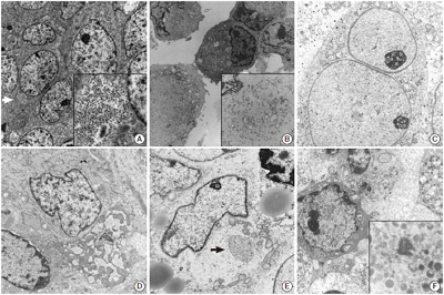
- Utility of Transmission Electron Microscopy in Small Round Cell Tumors
- Na Rae Kim, Seung Yeon Ha, Hyun Yee Cho
- J Pathol Transl Med. 2015;49(2):93-101. Published online March 12, 2015
- DOI: https://doi.org/10.4132/jptm.2015.01.30
- 18,953 View
- 287 Download
- 5 Web of Science
- 4 Crossref
-
Abstract
PDF
- Small round cell tumors (SRCTs) are a heterogeneous group of neoplasms composed of small, primitive, and undifferentiated cells sharing similar histology under light microscopy. SRCTs include Ewing sarcoma/peripheral neuroectodermal tumor family tumors, neuroblastoma, desmoplastic SRCT, rhabdomyosarcoma, poorly differentiated round cell synovial sarcoma, mesenchymal chondrosarcoma, small cell osteosarcoma, small cell malignant peripheral nerve sheath tumor, and small cell schwannoma. Non-Hodgkin’s malignant lymphoma, myeloid sarcoma, malignant melanoma, and gastrointestinal stromal tumor may also present as SRCT. The current shift towards immunohistochemistry and cytogenetic molecular techniques for SRCT may be inappropriate because of antigenic overlapping or inconclusive molecular results due to the lack of differentiation of primitive cells and unavailable genetic service or limited moleculocytogenetic experience. Although usage has declined, electron microscopy (EM) remains very useful and shows salient features for the diagnosis of SRCTs. Although EM is not always required, it provides reliability and validity in the diagnosis of SRCT. Here, the ultrastructural characteristics of SRCTs are reviewed and we suggest that EM would be utilized as one of the reliable modalities for the diagnosis of undifferentiated and poorly differentiated SRCTs.
-
Citations
Citations to this article as recorded by- Electron Microscopy in the Context of a Children's Research Hospital
Cam Robinson
Microscopy and Microanalysis.2020; 26(S2): 1610. CrossRef - Primary bilateral corneal nerve sheath neoplasm in a dog
Marina L. Leis, M. Elyse Salpeter, Bianca S. Bauer, Dale L. Godson, Bruce H. Grahn
Veterinary Ophthalmology.2017; 20(4): 365. CrossRef - Hirnbasissyndrom infolge eines Tumors bei einer 17 Monate alten Deutsch-Holstein-Färse
Wolf Wippermann, Sandra Schöniger, Kerstin Gerlach, Gerald Fritz Schusser, Gabor Köller, Alexander Starke
Tierärztliche Praxis Ausgabe G: Großtiere / Nutztiere.2016; 44(03): 180. CrossRef - The Continuing Value of Ultrastructural Observation in Central Nervous System Neoplasms in Children
Na Rae Kim, Sung-Hye Park
Journal of Pathology and Translational Medicine.2015; 49(6): 427. CrossRef
- Electron Microscopy in the Context of a Children's Research Hospital
- Alveolar Rhabdomyosarcoma of the Lip in an Adult with Clear Cell Features
- Jae Yeon Seok, Juhyeon Jeong, Young Woo Cheon, Hyun Yee Cho, Seung Yeon Ha, Dong Hae Chung
- J Pathol Transl Med. 2015;49(1):81-84. Published online January 15, 2015
- DOI: https://doi.org/10.4132/jptm.2014.06.03
- 14,620 View
- 108 Download
- Papillary Cystadenoma of the Fallopian Tube Not Associated with von Hippel-Lindau Disease: A Case Report
- Jae Yeon Seok, Myunghee Kang, Jungsuk An, Hyunchul Kim, Kwang-Beom Lee, Hyun Yee Cho
- Korean J Pathol. 2014;48(5):382-386. Published online October 27, 2014
- DOI: https://doi.org/10.4132/KoreanJPathol.2014.48.5.382
- 9,574 View
- 70 Download
- 3 Crossref
-
PDF
-
Citations
Citations to this article as recorded by- A Retrospective Study on the Occurrence and Prevalence of Ovarian Masses in the Patients of Rajkot District, Gujarat
Kirtan M Vyas, Avni P Patel, Ashita K Vyas, Hardik Gohel
Journal of the Scientific Society.2023; 50(3): 375. CrossRef - Clear Cell Papillary Cystadenoma of the Ovary Masquerading as Metastatic Clear Cell Renal Cell Carcinoma: A Case Report and Review of the Literature
Aarti E. Sharma, Farid Saei Hamedani, Julieta E. Barroeta, Peter Pytel, Jennifer A. Bennett, Ricardo R. Lastra
International Journal of Gynecological Pathology.2021; 40(3): 290. CrossRef - Mesonephric (Wolffian) Pseudoendometrioid Carcinoma of the Broad Ligament, Arising From a Papillary Cystadenoma
Philippe Moerman, Frederic Amant, Ignace Vergote
International Journal of Surgical Pathology.2016; 24(7): 635. CrossRef
- A Retrospective Study on the Occurrence and Prevalence of Ovarian Masses in the Patients of Rajkot District, Gujarat
- Cystic Brunner’s Gland Hamartoma in the Gastric Body: A Case Report
- Dong Hae Chung, Na Rae Kim, Hyun Yee Cho, Yoon Jae Kim
- Korean J Pathol. 2014;48(5):371-374. Published online October 27, 2014
- DOI: https://doi.org/10.4132/KoreanJPathol.2014.48.5.371
- 11,983 View
- 64 Download
- 1 Crossref
- Bilateral Stafne Bone Cavity in the Anterior Mandible with Heterotopic Salivary Gland Tissue: A Case Report
- Hyunchul Kim, Jae Yeon Seok, Sangho Lee, Jungsuk An, Na Rae Kim, Dong Hae Chung, Hyun Yee Cho, Seung Yeon Ha
- Korean J Pathol. 2014;48(3):248-249. Published online June 26, 2014
- DOI: https://doi.org/10.4132/KoreanJPathol.2014.48.3.248
- 16,088 View
- 110 Download
- 14 Crossref
-
PDF
-
Citations
Citations to this article as recorded by- Diagnostic approach for the rare anterior variant of mandibular bone depression often misdiagnosed as tumorous lesions
Hak-Sun Kim
Journal of Dental Sciences.2025; 20(1): 502. CrossRef - Static bone cavity occurred in the buccal side of the mandible: A case report and review of literature
Hideki Hojo, Takanori Eguchi, Yumi Ito, Yoshiki Hamada
Journal of Oral and Maxillofacial Surgery, Medicine, and Pathology.2025; 37(4): 698. CrossRef - Bilateral incomplete mandibular canals: an embryological analysis of their possible etiology
Kanitin Rumpansuwon, Thewarid Berkban, Nutmethee Kruepunga, Wattana Weerachatyanukul, Somluk Asuvapongpatana, Arada Chaiyamoon, Benrita Jitaree, R. Shane Tubbs, Joe Iwanaga, Thanyaporn Senarai, Athikhun Suwannakhan
Surgical and Radiologic Anatomy.2025;[Epub] CrossRef - A Rare Case of Anterior Stafne Bone Cavity in the Mandibular Region
Hideki Suito, Yuuri Oku, Koichi Kani, Keiko Aota, Naoki Maeda
Cureus.2025;[Epub] CrossRef - Benign Mandibular Cavity/Stafne's Bone Cyst: A Case Report and Review
Fawzia M. Butt, Shamim M. Butt, Mark L. Chindia
Clinical Case Reports.2025;[Epub] CrossRef - Bilateral Stafne Bone Cavity in the Body of the Mandible: An Unusual Case Report and Literature Review
Mayank Pahadia, Rutvi Vyas
Cureus.2023;[Epub] CrossRef - Effect of Stafne bone defect on the adjacent tooth: A review of the literature
Mahdi Niknami, Azin Parsa, Zahra Khodadadi
Imaging Science in Dentistry.2022; 52(2): 165. CrossRef - Assessment of prevalence and volumetric estimation of possible Stafne bone concavities on cone beam computed tomography images
Alaettin Koç, Cennet Neslihan Eroğlu, Ersen Bilgili
Oral Radiology.2020; 36(3): 254. CrossRef - Stafne’s bone cyst revisited and renamed: the benign mandibular concavity
Johan K.M. Aps, Natasha Koelmeyer, Cina Yaqub
Dentomaxillofacial Radiology.2020; 49(4): 20190475. CrossRef - Cone‐beam computed tomography analysis of lingual mandibular bone depression in the premolar region: A case report
Saeed Asgary, Naghmeh Emadi
Clinical Case Reports.2020; 8(3): 523. CrossRef - Letters to the Editor
Ariyan S Araghi, Richard M Graham
Dental Update.2019; 46(8): 792. CrossRef - Radiographic features of lingual mandibular bone depression using dental cone beam computed tomography
Liu Liu, Byung Cheol Kang, Suk Ja Yoon, Jae Seo Lee, Sel Ae Hwang
Dentomaxillofacial Radiology.2018; 47(6): 20170383. CrossRef - Stafne's bone cavity – unusual presentation in the anterior mandible
Ioan Davies, Holly Boyes, James Wykes, Graham Smith
Dental Update.2018; 45(4): 340. CrossRef - Anterior stafne bone cyst mimicking periapical cyst: a case report
Ji-Young Song
Journal of Dental Rehabilitation and Applied Science.2016; 32(3): 209. CrossRef
- Diagnostic approach for the rare anterior variant of mandibular bone depression often misdiagnosed as tumorous lesions
- Crush Cytology of Microcystic Meningioma with Extensive Sclerosis
- Jae Yeon Seok, Na Rae Kim, Hyun Yee Cho, Dong Hae Chung, Gi-Taek Yee, Eung Yeop Kim
- Korean J Pathol. 2014;48(1):77-80. Published online February 25, 2014
- DOI: https://doi.org/10.4132/KoreanJPathol.2014.48.1.77
- 12,386 View
- 58 Download
- 6 Crossref
-
PDF
-
Citations
Citations to this article as recorded by- Cytologic features of meningioma: An analysis of common and uncommon subtypes and diagnostic difficulties during intraoperative procedures
Ana M. Rodríguez‐García, Isabel Esteban‐Rodríguez, José A. Jiménez‐Heffernan, Carmen Bárcena, Samuel López‐Muñoz, Pilar López‐Ferrer
Cytopathology.2024; 35(5): 581. CrossRef - Exploring the role of epidermal growth factor receptor variant III in meningeal tumors
Rashmi Rana, Vaishnavi Rathi, Kirti Chauhan, Kriti Jain, Satnam Singh Chhabra, Rajesh Acharya, Samir Kumar Kalra, Anshul Gupta, Sunila Jain, Nirmal Kumar Ganguly, Dharmendra Kumar Yadav, Timir Tripathi
PLOS ONE.2021; 16(9): e0255133. CrossRef - Intraoperative frozen cytology of intraosseous cystic meningioma in the sphenoid bone
Na Rae Kim, Gie-Taek Yie
Journal of Pathology and Translational Medicine.2020; 54(6): 508. CrossRef - Can amide proton transfer–weighted imaging differentiate tumor grade and predict Ki-67 proliferation status of meningioma?
Hao Yu, Xinrui Wen, Pingping Wu, Yueqin Chen, Tianyu Zou, Xianlong Wang, Shanshan Jiang, Jinyuan Zhou, Zhibo Wen
European Radiology.2019; 29(10): 5298. CrossRef - Intraoperative Frozen Cytology of Central Nervous System Neoplasms: An Ancillary Tool for Frozen Diagnosis
Myunghee Kang, Dong Hae Chung, Na Rae Kim, Hyun Yee Cho, Seung Yeon Ha, Sangho Lee, Jungsuk An, Jae Yeon Seok, Gie-Taek Yie, Chan Jong Yoo, Sang Gu Lee, Eun Young Kim, Woo Kyung Kim, Seong Son, Sun Jin Sym, Dong Bok Shin, Hee Young Hwang, Eung Yeop Kim, K
Journal of Pathology and Translational Medicine.2019; 53(2): 104. CrossRef - Crush Cytology of Secretory Meningioma: A Case Report
Na Rae Kim, Gie-Taek Yee, Hyun Yee Cho
Brain Tumor Research and Treatment.2015; 3(2): 147. CrossRef
- Cytologic features of meningioma: An analysis of common and uncommon subtypes and diagnostic difficulties during intraoperative procedures
- Adenocarcinoma Arising in a Colonic Duplication Cyst: A Case Report and Review of the Literature
- Myunghee Kang, Jungsuk An, Dong Hae Chung, Hyun Yee Cho
- Korean J Pathol. 2014;48(1):62-65. Published online February 25, 2014
- DOI: https://doi.org/10.4132/KoreanJPathol.2014.48.1.62
- 8,629 View
- 61 Download
- 12 Crossref
-
PDF
-
Citations
Citations to this article as recorded by- Low-grade mucinous neoplasm originating from intestinal duplication: a case report and review of the literature
Huihui Yin, Jie Yu, Yunzhao Chen
World Journal of Surgical Oncology.2025;[Epub] CrossRef - Adenocarcinoma originating from a colonic duplication cyst: A case report
Jeehye Lee, Jung Wook Suh
World Journal of Gastrointestinal Surgery.2025;[Epub] CrossRef - Low-Grade Mucinous Neoplasm Arising in an Enteric Duplication Cyst of Pancreas: A Case Report and Literature Review
Mengjing Fan, Fang Yang
International Journal of Surgical Pathology.2024; 32(2): 422. CrossRef - Colonic duplication in an adult with chronic constipation: a case report and review of its surgical management
Muhammad Ash-Shafhawi Adznan, Hizami Amin Tai, Aras Emre Canda, Nevra Elmas, Mustafa Cem Terzi
Annals of Coloproctology.2024; 40(Suppl 1): S6. CrossRef - Tubular adenoma arising in tubular colonic duplication: a case report
Heonwoo Lee, Hyeong Rok An, Chan Wook Kim, Young Soo Park
Journal of Pathology and Translational Medicine.2024; 58(4): 198. CrossRef - Right-Sided Colonic Duplication Cyst with a Malignant Twist in a Young Adult — a Case Report
Laxmi Radhakrishnan, Joseph George, Latha K. Abraham
Journal of Gastrointestinal Cancer.2022; 53(3): 805. CrossRef - Endoscopic resection of a duodenal duplication cyst: A case report
Sayumi Kurita, Kazuo Kitagawa, Naoki Toya, Masahiko Kawamura, Muneo Kawamura, Ken Eto
DEN Open.2022;[Epub] CrossRef - Complete colonic duplication presenting as hip fistula in an adult with pelvic malformation: A case report
Xuan Cai, Jing-Tao Bi, Zhi-Xue Zheng, Ya-Qi Liu
World Journal of Clinical Cases.2022; 10(30): 11037. CrossRef - Sigmoid colon duplication seen as a rare cause of ileus in adult: case report
Barış BAYRAKTAR, Salih BOLUK, Sümeyra Emine BÖLÜK
Anatolian Current Medical Journal.2022; 4(3): 323. CrossRef - Successful management of tubular colonic duplication using a laparoscopic approach: A case report and review of the literature
Gan-Bin Li, Jia-Gang Han, Zhen-Jun Wang, Zhi-Wei Zhai, Yu Tao
World Journal of Clinical Cases.2020; 8(15): 3291. CrossRef - Sigmoid colon duplication with ectopic immature renal tissue in an adult: A case report
Hwan Namgung
World Journal of Clinical Cases.2020; 8(24): 6346. CrossRef - Retroperitoneal Mucinous Neoplasm Arising from Colonic Duplication Cyst
María M. Rojas-Rojas, Marcela Mejiah, Martha Mora, Jorge Otero, Fernando Arias-Amézquita, Eduardo Londoño-Schimmer, Paula A. Rodríguez-Urrego
Journal of Gastrointestinal Cancer.2019; 50(3): 583. CrossRef
- Low-grade mucinous neoplasm originating from intestinal duplication: a case report and review of the literature
- Uncommon and Rare Human Papillomavirus Genotypes Relating to Cervical Carcinomas
- Na Rae Kim, Myunghee Kang, Soon Pyo Lee, Hyunchul Kim, Jungsuk An, Dong Hae Chung, Seung Yeon Ha, Hyun Yee Cho
- Korean J Pathol. 2014;48(1):43-49. Published online February 25, 2014
- DOI: https://doi.org/10.4132/KoreanJPathol.2014.48.1.43
- 11,598 View
- 57 Download
- 10 Crossref
-
Abstract
PDF
Background Human papillomavirus (HPV) is an oncogenic virus in cervical cancer and most invasive carcinomas (ICs) are caused by HPV16 and 18. However, the roles and contributions of other uncommon and rare genotypes remain uncertain.
Methods HPV genotypes were retrospectively assessed using an HPV DNA chip that can specify up to 32 HPV genotypes. We arbitrarily regarded genotypes accounting for less than 6% of the total as uncommon and rare genotypes.
Results A total of 3,164 HPV-positive cases were enrolled. In groups 2A, 2B, 3, and unclassified HPV genotypes, 2.4% of cases with uncommon HPV genotypes (68, 26, 34, 53, 66, 69, 70, 73, 40, 42, 43, 44, 54, 55, 61, 62, 6, and 11) showed high grade squamous intraepithelial lesions and ICs. There were no HPV32- and 57-infected cases.
Conclusions We found that the uncommon and rare HPV genotypes may provide incremental etiologic contributions in cervical carcinogenesis, especially HPV68, 70, and 53. Further studies on these uncommon and rare HPV genotypes will be of importance in establishing the significance of genotypes in different regions, especially in planning a strategy for further vaccine development as well as follow-up on the effectiveness of the currently used vaccines.
-
Citations
Citations to this article as recorded by- High-risk human papillomavirus diversity among indigenous women of western Botswana with normal cervical cytology and dysplasia
Patricia S. Rantshabeng, Billy M. Tsima, Andrew K. Ndlovu, Keneilwe Motlhatlhedi, Kirthana Sharma, Carol B. Masole, Natasha O. Moraka, Kesego Motsumi, Angela K. T. Maoto-Mokote, Alemayehu B. Eshetu, Leabaneng Tawe, Tendani Gaolathe, Sikhulile Moyo, Lynnet
BMC Infectious Diseases.2024;[Epub] CrossRef - Human Papillomavirus (HPV69/HPV73) Coinfection associated with Simultaneous Squamous Cell Carcinoma of the Anus and Presumed Lung Metastasis
Stephanie Shea, Marina Muñoz, Stephen C. Ward, Mary B. Beasley, Melissa R Gitman, Michael D Nowak, Jane Houldsworth, Emilia Mia Sordillo, Juan David Ramirez, Alberto E. Paniz Mondolfi
Viruses.2020; 12(3): 349. CrossRef - Human Papillomavirus Selected Properties and Related Cervical Cancer Prevention Issues
Saule Balmagambetova, Andrea Tinelli, Ospan A. Mynbaev, Arip Koyshybaev, Olzhas Urazayev, Nurgul Kereyeva, Elnara Ismagulova
Current Pharmaceutical Design.2020; 26(18): 2073. CrossRef - Periungual Bowen's disease with a narrow longitudinal melanonychia mimicking periungual warts
Taiyo HITAKA, Michiko HASEGAWA, Akira SHIMIZU, Yuko KURIYAMA, Atsushi TAMURA
Skin Cancer.2019; 33(3): 211. CrossRef - Detection of HPV RNA molecules in stratified mucin-producing intraepithelial lesion (SMILE) with concurrent cervical intraepithelial lesion: a case report
Shiho Fukui, Kazunori Nagasaka, Naoko Iimura, Ranka Kanda, Takayuki Ichinose, Takeru Sugihara, Haruko Hiraike, Shunsuke Nakagawa, Yuko Sasajima, Takuya Ayabe
Virology Journal.2019;[Epub] CrossRef - Pitfalls of commercially available HPV tests in HPV68a detection
Hana Jaworek, Katerina Kubanova, Vladimira Koudelakova, Rastislav Slavkovsky, Jiri Drabek, Marian Hajduch, Craig Meyers
PLOS ONE.2019; 14(8): e0220373. CrossRef - Overall accuracy of cervical cytology and clinicopathological significance of LSIL cells in ASC‐H cytology
S. H. Kim, J. M. Lee, H. G. Yun, U. S. Park, S. U. Hwang, J.‐S. Pyo, J. H. Sohn
Cytopathology.2017; 28(1): 16. CrossRef - Human papillomavirus genotyping by Linear Array and Next-Generation Sequencing in cervical samples from Western Mexico
María Guadalupe Flores-Miramontes, Luis Alberto Torres-Reyes, Liliana Alvarado-Ruíz, Salvador Angel Romero-Martínez, Verenice Ramírez-Rodríguez, Luz María Adriana Balderas-Peña, Verónica Vallejo-Ruíz, Patricia Piña-Sánchez, Elva Irene Cortés-Gutiérrez, Lu
Virology Journal.2015;[Epub] CrossRef - Impact of human papillomavirus coinfections on the risk of high-grade squamous intraepithelial lesion and cervical cancer
Adela Carrillo-García, Sergio Ponce-de-León-Rosales, David Cantú-de-León, Verónica Fragoso-Ontiveros, Imelda Martínez-Ramírez, Asunción Orozco-Colín, Alejandro Mohar, Marcela Lizano
Gynecologic Oncology.2014; 134(3): 534. CrossRef - Human papillomavirus 66‐associated subungual squamous cell carcinoma
Jin Hee Kang, Hwa young Ahn, Miri Kim, Shin Taek Oh, Baik Kee Cho, Hyun Jeong Park
The Journal of Dermatology.2014; 41(12): 1119. CrossRef
- High-risk human papillomavirus diversity among indigenous women of western Botswana with normal cervical cytology and dysplasia
- Peritoneal and Nodal Gliomatosis with Endometriosis, Accompanied with Ovarian Immature Teratoma: A Case Study and Literature Review
- Na Rae Kim, Soyi Lim, Juhyeon Jeong, Hyun Yee Cho
- Korean J Pathol. 2013;47(6):587-591. Published online December 24, 2013
- DOI: https://doi.org/10.4132/KoreanJPathol.2013.47.6.587
- 10,621 View
- 80 Download
- 9 Crossref
-
Abstract
PDF
Gliomatosis peritonei (GP) indicates the peritoneal implantation of mature neuroglial tissue and is usually accompanied by ovarian mature or immature teratoma. Here, we report a case of ovarian immature teratoma associated with gliomatosis involving the peritoneum, lymph nodes and Douglas' pouch, where gliomatosis coexisted with endometriosis. As far as we know, only seven cases of GP have been reported as coexisting with endometriosis. Eight cases with mature glial tissue in the lymph nodes, i.e., nodal gliomatosis, have been published either in association with GP or in its absence. Metaplasia of pluripotent coelomic stem cells has been suggested to be responsible for the pathogenesis of endometriosis and GP rather than implantation metastases of ovarian teratomatous tumor with varying maturation. This theory is also applied to GP independently of ovarian teratomatous tumors. To the best of our knowledge, nodal gliomatosis coexisting with GP and also involving endometriosis has not yet been reported.
-
Citations
Citations to this article as recorded by- Gliomatosis peritonei and ectopic decidua in pregnancy after treatment for immature teratoma
Giulia Martignon, Diletta Fumagalli, Francesca Maria Porta, Sarah Alessi, Giuseppe Petralia, Roberto Berretta, Giovanni Damiano Aletti, Fedro Peccatori
International Journal of Gynecological Cancer.2026; : 104700. CrossRef - Mimics of primary ovarian cancer and primary peritoneal carcinomatosis – A pictorial review
B. Lawson, I. Rajendran, J. Smith, A. Shakur, V. Sadler, T.J. Sadler, H.C. Addley, S. Freeman
Clinical Radiology.2024; 79(10): 736. CrossRef - Ovarian Immature Teratoma With Nodal Gliomatosis: A Case Report and Literature Review
Marwa Alna’irat, W. Glenn McCluggage, Maysa Al-Hussaini
International Journal of Gynecological Pathology.2023; 42(6): 627. CrossRef - Germ Cell Tumors of the Ovary: A Review
Preetha Ramalingam
Seminars in Diagnostic Pathology.2023; 40(1): 22. CrossRef - Immature Teratoma with Gliomatosis Peritonei Arising in a Young Girl: Report of a Rare Case and Review of Literature
Isheeta Ahuja, Ruchi Rathore, Neerja Bhatla, Sandeep R. Mathur
Indian Journal of Gynecologic Oncology.2023;[Epub] CrossRef - Growing Teratoma Syndrome with Synchronous Gliomatosis Peritonei during Chemotherapy in Ovarian Immature Teratoma: A Case Report and Literature Review
Sijian Li, Na Su, Congwei Jia, Xinyue Zhang, Min Yin, Jiaxin Yang
Current Oncology.2022; 29(9): 6364. CrossRef - Extratesticular gliomatosis peritonei after mesenteric teratoma: a case report and literature review
Jiaqiang Li, Shoulin Li, Dong Xiao, Jiaming Song, Jianxiong Mao, Jianchun Yin
Journal of International Medical Research.2021;[Epub] CrossRef - Germ Cell Tumors of the Female Genital Tract
Elizabeth D. Euscher
Surgical Pathology Clinics.2019; 12(2): 621. CrossRef - Gliomatosis peritonei: a series of eight cases and review of the literature
Dan Wang, Cong-wei Jia, Rui-e Feng, Hong-hui Shi, Juan Sun
Journal of Ovarian Research.2016;[Epub] CrossRef
- Gliomatosis peritonei and ectopic decidua in pregnancy after treatment for immature teratoma
- Cotyledonoid Dissecting Leiomyoma of the Uterus with Intravascular Luminal Growth: A Case Study
- Na Rae Kim, Chan Yong Park, Hyun Yee Cho
- Korean J Pathol. 2013;47(5):477-480. Published online October 25, 2013
- DOI: https://doi.org/10.4132/KoreanJPathol.2013.47.5.477
- 14,465 View
- 81 Download
- 7 Crossref
-
Abstract
PDF
Here, we report the case of a 43-year-old female who was diagnosed with a cotyledonoid dissecting leiomyoma (CDL) of the uterus. CDL is a recently described and extremely rare variant of a benign leiomyoma that can grossly masquerade as a malignancy. The 13-cm sized tumor was located primarily on the extrauterine surface as an intrauterine continuity, which showed dark red, congested, bulbous protuberances. It was multinodular appearance, encasing the bilateral adnexae and the left iliac vein. Microscopically, the nodules were separated by extensive hydropic degeneration. The nodules were composed of cigar-shaped spindle cells with no mitosis, cellular pleomorphism or coagulation necrosis. They also showed an intravascular luminal growth pattern. CDL with intravascular growth was diagnosed after excluding intravascular leiomyomatosis, disseminated peritoneal leiomyomatosis, and benign metastasizing leiomyoma. The present case is the second reported case of CDL in Korea. Recognition of this rare and bizarre, malignancy-mimicking leiomyoma is crucial to prevent inappropriate treatment.
-
Citations
Citations to this article as recorded by- A Case of Cotyledonoid-Dissecting Leiomyoma - The Utility of Laparoscopic Biopsy and Gonadotropin-Releasing Hormone Analogs
Sayaka Kawashita, Akiko Nonoshita, Keisuke Iwasaki, Daisuke Nakayama
Clinical Pathology.2024;[Epub] CrossRef - Cotyledonoid dissecting leiomyoma with peritoneal dissemination
Hiroki Egashira, Hiroaki Ishida, Nobuyuki Hiruta, Akiko Takashima
BMJ Case Reports.2024; 17(9): e261937. CrossRef - Cotyledonoid dissecting leiomyoma of the uterus: a case report and review of the literature
Mahboobeh Chahkandi, Marzieh Ataei, Amir Reza Bina, Farnaz Mozayani, Ali Fanoodi
Journal of Medical Case Reports.2023;[Epub] CrossRef - Cotyledonoid Leiomyoma Clinical Characteristics, Imaging Features, and Review of the Literature
Francesca Buonomo, Sofia Bussolaro, Giorgio Giorda, Federico Romano, Stefania Biffi, Giuseppe Ricci
Journal of Ultrasound in Medicine.2021; 40(7): 1459. CrossRef - The Management of the Cotyledonoid Leiomyoma of the Uterus: A Narrative Review of the Literature
Francesca Buonomo, Sofia Bussolaro, Clarice de Almeida Fiorillo, Giorgio Giorda, Federico Romano, Stefania Biffi, Giuseppe Ricci
International Journal of Environmental Research and Public Health.2021; 18(16): 8521. CrossRef - Cotyledonoid dissecting leiomyoma of the uterus: A report of four cases and a review of the literature
TIANMIN XU, SHUYING WU, RULIN YANG, LIPING ZHAO, MINGXING SUI, MANHUA CUI, WEIQIN CHANG
Oncology Letters.2016; 11(4): 2865. CrossRef - COTYLEDONOID DISSECTING LEIOMYOMA (CDL) OF UTERUS MIMICKING MALIGNANCY: A CLINICAL DILEMMA
Roma Isaacs, Rupinder Kaur, Sunita Goyal
Journal of Evolution of Medical and Dental Sciences.2016; 5(57): 3973. CrossRef
- A Case of Cotyledonoid-Dissecting Leiomyoma - The Utility of Laparoscopic Biopsy and Gonadotropin-Releasing Hormone Analogs
- Fine Needle Aspiration Cytology of Postoperative Spindle Cell Nodule in Neck after Thyroidectomy: A Case Report
- Myunghee Kang, Seung Yeon Ha, Hyun Yee Cho, Jungsuk An, Dong Hae Chung, Yoo Seung Chung
- Korean J Pathol. 2013;47(1):89-91. Published online February 25, 2013
- DOI: https://doi.org/10.4132/KoreanJPathol.2013.47.1.89
- 9,734 View
- 45 Download
- 3 Crossref
-
PDF
-
Citations
Citations to this article as recorded by- Post-surgical thyroid bed myofibroma simulating a recurrent papillary thyroid carcinoma: A case report and review of the literature
Jun Hyeon Park, Kyung Sik Yi, Chi-Hoon Choi, Yook Kim, Jisun Lee, Yeongtae Park, Ok-Jun Lee
Medicine.2024; 103(2): e36945. CrossRef - USP6‐associated neoplasm as a tentative subset of postoperative spindle cell nodule
Lili Sun, Zehua Zhao, Yanmei Zhu
Histopathology.2023; 82(4): 587. CrossRef - Diagnostic Performance of Core Needle Biopsy for Characterizing Thyroidectomy Bed Lesions
So Yeong Jeong, Jung Hwan Baek, Sae Rom Chung, Young Jun Choi, Dong Eun Song, Ki-Wook Chung, Won Woong Kim, Jeong Hyun Lee
Korean Journal of Radiology.2022; 23(10): 1019. CrossRef
- Post-surgical thyroid bed myofibroma simulating a recurrent papillary thyroid carcinoma: A case report and review of the literature
- Multiple Jejunal Myeloid Sarcomas Presenting with Intestinal Obstruction in a Non-leukemic Patient: A Case Report with Ultrastructural Observations
- Na Rae Kim, Woon Kee Lee, Jong In Lee, Hyun Yee Cho
- Korean J Pathol. 2012;46(6):590-594. Published online December 26, 2012
- DOI: https://doi.org/10.4132/KoreanJPathol.2012.46.6.590
- 9,748 View
- 72 Download
- 6 Crossref
-
Abstract
PDF
Myeloid sarcoma is a rare extramedullary myeloid tumor, which is frequently misdiagnosed when no evidence of leukemia is initially observed. Here, we report on a peculiar case of a 49-year-old man afflicted with multiple masses in the jejunum, the superior mesentery, and the serosa of the transverse colon, without leukemic manifestation. The tumor was composed of undifferentiated small round cells containing eosinophilic cytoplasm, which were negative for myeloperoxidase, nonspecific esterase, lysozyme, terminal deoxynucleotidyl transferase, leukocyte common antigen, CD3, CD4, CD15, CD20, CD30, CD43, CD56, CD68/PG-M1, CD79a, human melanoma black-45, c-kit, and CD34 with positivity only for CD68/KP1, CD99, and vimentin. Under electron microscopy, those cells had abundant membrane-bound cytoplasmic granules that measured 200 to 300 nm in diameter, which were consistent with granulocytic azurophilic granules. The tumor was finally diagnosed as a myeloid sarcoma. The presence of non-leukemic myeloid sarcomas showing immunonegativity for conventional myeloid-leukemic markers necessitated a diagnosis by ultrastructural observation.
-
Citations
Citations to this article as recorded by- Myeloid sarcoma presenting as intestinal obstruction: A case report of the first presentation of acute myeloid leukemia
Deepsikha Dharamsaktu, Anuradha Pandit, Charanjeet Ahluwalia, Sana Ahuja
International Journal of Surgery Case Reports.2025;[Epub] CrossRef - Myeloid sarcoma of the gastrointestinal tract: Wolf in sheep’s clothing!
Nisha Meena, Surbhi Goyal, Prerna Arora, Sanjeev Sachdeva, Puja Sakhuja
Indian Journal of Pathology and Microbiology.2025; 68(2): 452. CrossRef - Primary ileal myeloid sarcoma presenting with bowel obstruction: a case report
Hitoshi Minagi, Nobuhiko Kanaya, Yoshitaka Kondo, Yoshihiko Kakiuchi, Shinji Kuroda, Ryohei Shoji, Hajime Kashima, Yuki Matsumi, Satoru Kikuchi, Kunitoshi Shigeyasu, Fuminori Teraishi, Shunsuke Kagawa, Toshiyoshi Fujiwara
Surgical Case Reports.2024;[Epub] CrossRef - Isolated myeloid sarcoma presenting with small bowel obstruction: a case report
Rie Mizumoto, Masanori Tsujie, Tomoko Wakasa, Kotaro Kitani, Hironobu Manabe, Shuichi Fukuda, Kaoru Okada, Shumpei Satoi, Hajime Ishikawa, Toshihiko Kawasaki, Hitoshi Hanamoto, Masao Yukawa, Masatoshi Inoue
Surgical Case Reports.2020;[Epub] CrossRef - Primary Myeloid Sarcoma of the Ileum and Mesentery Causing Small Bowel Obstruction: Case Report and Literature Review
Andrej Nikolovski, Dragoslav Mladenovikj, Aleksandra Veljanovska, Gordana Petrusevka
Lietuvos chirurgija.2020; 19(1-2): 55. CrossRef - Utility of Transmission Electron Microscopy in Small Round Cell Tumors
Na Rae Kim, Seung Yeon Ha, Hyun Yee Cho
Journal of Pathology and Translational Medicine.2015; 49(2): 93. CrossRef
- Myeloid sarcoma presenting as intestinal obstruction: A case report of the first presentation of acute myeloid leukemia
- Morphometric Analysis for Pulmonary Small Cell Carcinoma Using Image Analysis.
- Sun Min Jeong, Seung Yeon Ha, Jungsuk An, Hyun Yee Cho, Dong Hae Chung, Na Rae Kim, Sanghui Park
- Korean J Pathol. 2011;45(1):87-91.
- DOI: https://doi.org/10.4132/KoreanJPathol.2011.45.1.87
- 4,771 View
- 29 Download
- 1 Crossref
-
Abstract
PDF
- BACKGROUND
There are few studies of how to diagnose small cell lung cancer in cytological tests through morphometric analysis. We tried to measure and analyze characteristics of small cell carcinoma in lung by image analysis.
METHODS
We studied three types of cytologic specimens from 89 patients who were diagnosed with small cell lung cancer by immunohistochemistry. We measured area, perimeter, maximal length and maximal width of cells from small cell carcinoma using image analysis.
RESULTS
In lung aspirates, the nuclear mean area, perimeter, maximal length and maximal width of small cell lung cancer were 218.69 microm2, 55 microm, 18.48 microm and 14.65 microm. In bronchial washings, nuclear measurements were 194.66 microm2, 50.07 microm, 16.27 microm and 14.1 microm. In pleural fluid, values were 177.85 microm2, 48.09 microm, 15.7 microm and 13.37 microm.
CONCLUSIONS
Nuclear size of small cell lung carcinoma is variable and depends on the cytology method. Nuclei are spindle-shaped and larger in small cell carcinoma from lung aspirates than in bronchial washings or pleural fluid. The cytoplasms of the cells in bronchial washings and pleural fluid were swollen. Therefore, one should consider morphologic changes when trying to diagnose small cell lung cancer through cytological tests. -
Citations
Citations to this article as recorded by- Interobserver Variability in Diagnosing High-Grade Neuroendocrine Carcinoma of the Lung and Comparing It with the Morphometric Analysis
Seung Yeon Ha, Joungho Han, Wan-Seop Kim, Byung Seong Suh, Mee Sook Roh
Korean Journal of Pathology.2012; 46(1): 42. CrossRef
- Interobserver Variability in Diagnosing High-Grade Neuroendocrine Carcinoma of the Lung and Comparing It with the Morphometric Analysis
- The Analysis and Clinical Usefulness of HPV DNA Chip Test in the Uterine Cervix.
- Joo hyeon Jeong, Hyun Yee Cho, Na Rae Kim, Dong Hae Chung, Sanghui Park, Seung Yeon Ha
- Korean J Pathol. 2010;44(1):77-82.
- DOI: https://doi.org/10.4132/KoreanJPathol.2010.44.1.77
- 4,897 View
- 27 Download
- 3 Crossref
-
Abstract
PDF
- BACKGROUND
The genotypes of human papillomavirus (HPV) are important in carcinogenesis in uterine cervical cancer and may be different in geographic distribution.
METHODS
In 2,086 women, we analyzed the prevalence of HPV and HPV genotypes in uterine cervix by HPV-DNA chip test (n = 2,086), cytology (PAP smear, n = 1997) and biopsy (n = 546).
RESULTS
Of the 2,086 cases, 1,019 cases (48.8%) were HPV-positive and 1,067 cases (51.2%) were negative for HPV. Single infection occurred most commonly (72.1% of women). HPV genotypes in the high-risk and low-risk groups, respectively were HPV-16/-58/-18/-52/-53 and HPV-70/-6/-11. The detection rates of HPV-70 in subjects older than 50 years increased significantly (p < 0.05). Infection in high risk subjects was detected in high grade lesions compared with infection in low risk subjects (p < 0.05).
CONCLUSIONS
HPV-16/-58/-18/-52/-53/-70/-6/-11 genotypes were common in the patient group similar to findings in East Asia. HPV-70 infection is predominant in those older than 40 years. -
Citations
Citations to this article as recorded by- Current Status of and Perspectives on Cervical Cancer Screening in Korea
Sung-Chul Lim, Chong Woo Yoo
Journal of Pathology and Translational Medicine.2019; 53(4): 210. CrossRef - Cervical cytology of atypical squamous cells, cannot exclude high-grade squamous intra-epithelial lesion: significance of age, human papillomavirus DNA detection and previous abnormal cytology on follow-up outcomes
Chang Ohk Sung, Young Lyun Oh, Sang Yong Song
European Journal of Obstetrics & Gynecology and Reproductive Biology.2011; 159(1): 155. CrossRef - Cytomorphologic Features According to HPV DNA Type in Histologically Proven Cases of the Uterine Cervix
In Ho Choi, So-Young Jin, Dong Wha Lee, Dong Won Kim, Yoon Mi Jeen
The Korean Journal of Pathology.2011; 45(6): 612. CrossRef
- Current Status of and Perspectives on Cervical Cancer Screening in Korea
- Ovarian Endometrioid Adenocarcinoma with a Yolk Sac Tumor Component.
- Dong Hae Chung, Sanghui Park, Hee Eun Kyung, Na Rae Kim, Seung Yeon Ha, Soyi Lim, Hyun Yee Cho
- Korean J Pathol. 2009;43(6):570-574.
- DOI: https://doi.org/10.4132/KoreanJPathol.2009.43.6.570
- 4,132 View
- 28 Download
-
Abstract
PDF
- Ovarian endometrioid adenocarcinoma (EAC) with a yolk sac tumor (YST) component is extremely rare. Only twelve cases have been reported in the English literature. We report here two additional cases of this rare tumor. The YST component showed classic microscopic features, and immunohistochemically stained positive for alpha-fetoprotein (AFP), but negative for cytokeratin 7 (CK7), epithelial membrane antigen (EMA), estrogen receptor (ER) and progesterone receptor (PR). The EAC appeared to blend into the YST in several areas and immunohistochemically stained positive for CK7, EMA, ER, and PR, but negative for AFP.
- Cytology of Plasmacytoid Type Myoepithelioma: Report of Two Cases.
- Na Rae Kim, Hyun Yee Cho, Seung Yeon Ha
- Korean J Pathol. 2009;43(5):489-493.
- DOI: https://doi.org/10.4132/KoreanJPathol.2009.43.5.489
- 5,188 View
- 79 Download
- 4 Crossref
-
Abstract
PDF
- Myoepithelioma is a rare benign tumor of salivary gland myoepithelial cells, most commonly as a spindle subtype. Here, we present two cases of fine needle aspiration cytology of plasmacytoid myoepithelioma arising from a parotid gland and a hard palate. Aspirates showed plasmacytoid cells with pink-staining, homogeneous, abundant eosinophilic cytoplasm eccentrically displacing the nucleus in cohesive and dissociated forms. Rarely, nuclear grooves and intranuclear cytoplasmic inclusions were evident. These unfamiliar cytologic findings of uncommon myoepithelioma often cause diagnostic difficulties in preoperative aspiration cytology. Recognition of those rare findings provides a reliable diagnostic clue.
-
Citations
Citations to this article as recorded by- Plasmacytoid myoepithelioma: Diagnostic algorithm and a tailored therapeutic protocol for a geriatric individual
Pratik N. Patel, Aatish Thennavan, Venkadasalapathy Narayanaswamy, Raghu Radhakrishnan
Journal of Oral and Maxillofacial Surgery, Medicine, and Pathology.2015; 27(5): 737. CrossRef - Imprint Cytology of Soft Tissue Myoepithelioma: A Case Study
Seok Ju Park, Ae Ri Kim, Mi Jin Gu, Joon Hyuk Choi, Duk Seop Shin
Korean Journal of Pathology.2013; 47(3): 299. CrossRef - Fine Needle Aspiration Cytology of Benign Salivary Gland Tumors with Myoepithelial Cell Participation: An Institutional Experience of 575 Cases
Soomin Ahn, Yuil Kim, Young Lyun Oh
Acta Cytologica.2013; 57(6): 567. CrossRef - Plasmacytoid Myoepithelioma of the Palate: Case Report
Matina T. Zormpa, Asimina S. Sarigelou, Anna N. Eleftheriou, Anthoula S. Assimaki, Alexandros E. Kolokotronis
Head and Neck Pathology.2011; 5(2): 154. CrossRef
- Plasmacytoid myoepithelioma: Diagnostic algorithm and a tailored therapeutic protocol for a geriatric individual
- Malignant Peripheral Nerve Sheath Tumors of the Bilateral Adrenal Glands: Are They Metachronous Primary Tumors: A Case Report.
- Jae Hong Park, Seung Yeon Ha, Hyun Yee Cho
- Korean J Pathol. 2009;43(5):471-474.
- DOI: https://doi.org/10.4132/KoreanJPathol.2009.43.5.471
- 4,016 View
- 25 Download
- 2 Crossref
-
Abstract
PDF
- Malignant peripheral nerve sheath tumors (MPNSTs) have rarely been reported to occur in the adrenal gland and all of the reported cases were associated with neurofibromatosis, pheochromocytoma or ganglioneuroma. We present here a case of MPNST in the bilateral adrenal glands without any history of neurofibromatosis or combined tumor. Histologic examination showed the tumor cells had a spindle to ovoid shape, they were arranged in sweeping fascicles and there were frequent mitotic figures. The immunohistochemical and ultrastructural features of the tumor are also presented. To the best of our knowledge, this is the first report in the English medical literature about MPNSTs in the bilateral adrenal glands without any history of neurofibromatosis or combined tumor.
-
Citations
Citations to this article as recorded by- Malignant Peripheral Nerve Sheath Tumor of the Adrenal Gland
Raiz A. Misgar, Mohammad S. Baba, Mir I. Bashir, Arshad I. Wani
Indian Journal of Endocrinology and Metabolism.2022; 26(4): 395. CrossRef - Malignant peripheral nerve sheath tumor of adrenal gland with heterologus osseous differentiation in a case of Von Recklinghausen′s disease
Manas R. Baisakh, Nachiketa Mohapatra, Samiran D. Adhikary, Debasis Routray
Indian Journal of Pathology and Microbiology.2014; 57(1): 130. CrossRef
- Malignant Peripheral Nerve Sheath Tumor of the Adrenal Gland
- Prevalence and Genotype Distribution of Cervical Human Papillomavirus DNA in Korean Women: A Multicenter Study.
- Sung Ran Hong, In Sun Kim, Dong Won Kim, Mi Jin Kim, Ae Ree Kim, Young Ok Kim, Hye Sun Kim, Seo Hee Rha, Gyeong Sin Park, Yong Koo Park, Yong Wook Park, Ho Sung Park, Kwang Sun Suh, Jin Hee Sohn, Mi Kyung Shin, Hoon Kyu Oh, Ki Jung Yun, Hye Kyoung Yoon, Shi Nae Lee, Ah Won Lee, Hyo Jin Lee, Hyun Yee Cho, Chan Choi, Woon Won Jung
- Korean J Pathol. 2009;43(4):342-350.
- DOI: https://doi.org/10.4132/KoreanJPathol.2009.43.4.342
- 6,850 View
- 63 Download
- 16 Crossref
-
Abstract
PDF
- Background
DNA prevalence and type distribution of human papillomavirus (HPV) varies geographically. We investigated HPV prevalence and type distribution in Korean women using the MyHPV DNA chip testing. Methods: A total of 2,368 women from five regions of the country underwent Pap smear examination and MyHPV chip testing. Results: Overall HPV positivity was 15.8% and 78.4% in women with normal and abnormal cytology, respectively. High-risk HPV infection was strongly correlated with cytological atypia. In women with abnormal cytology, the five most common HPV types were 16, 58, 18, 52, and 56/53, and HPV16 was significantly the most common type in most geographical regions. After HPV16, HPV58, and 52 were the next most frequently detected types. Women with normal cytology, in contrast, showed heterogeneity in HPV type distribution. High-grade intraepithelial lesions infected with HPV16, 18, 31 or 45 are more likely to progress to carcinoma. Conclusions: The HPV chip test can provide useful data regarding HPV positivity and type. The most common HPV type in Korean women with abnormal cytology is HPV16, with HPV58 and 52 being frequently present. Our data may have important implications for vaccination programs and the development of cervical screening. -
Citations
Citations to this article as recorded by- HPV genotyping by L1 amplicon sequencing of archived invasive cervical cancer samples: a pilot study
Charles D. Warden, Preetam Cholli, Hanjun Qin, Chao Guo, Yafan Wang, Chetan Kancharla, Angelique M. Russell, Sylvana Salvatierra, Lorraine Z. Mutsvunguma, Kerin K. Higa, Xiwei Wu, Sharon Wilczynski, Raju Pillai, Javier Gordon Ogembo
Infectious Agents and Cancer.2022;[Epub] CrossRef - Enhanced disease progression due to persistent HPV-16/58 infections in Korean women: a systematic review and the Korea HPV cohort study
Jaehyun Seong, Sangmi Ryou, JeongGyu Lee, Myeongsu Yoo, Sooyoung Hur, Byeong-Sun Choi
Virology Journal.2021;[Epub] CrossRef - Comparison of FFPE histological versus LBP cytological samples for HPV detection and typing in cervical cancer
Geehyuk Kim, Hyemi Cho, Dongsup Lee, Sunyoung Park, Jiyoung Lee, Hye-young Wang, Sunghyun Kim, Kwang Hwa Park, Hyeyoung Lee
Experimental and Molecular Pathology.2017; 102(2): 321. CrossRef - Distribution of Oncogenic Human Papillomavirus Genotypes at High Grade Cervical Lesions above CIN 2 Grade with Histological Diagnosis
Geehyuk Kim, Sungyoung Park, Hye-young Wang, Sunghyun Kim, Sangjung Park, Kwangmin Yu, Boohyung Lee, Seung-Ju Ahn, Eun-Joong Kim, Dongsup Lee
Biomedical Science Letters.2016; 22(2): 37. CrossRef - Human Papillomavirus Prevalence and Genotype Distribution in Normal and ASCUS Specimens: Comparison of a Reverse Blot Hybridization Assay with a DNA Chip Test
Sunghyun Kim, In-soo Lee, Dongsup Lee
Biomedical Science Letters.2015; 21(1): 32. CrossRef - Genotype Analysis of Human Papilloma Virus Infection in Accordance with Cytological Diagnoses
Mi-Suk Park, Hyun-Wook Cho, Jin-Gak Kim, Nan-Young Bae, Dong-Sun Oh, Ho-Hyun Park
Korean Journal of Clinical Laboratory Science.2015; 47(1): 39. CrossRef - Comparison of the Cobas 4800 HPV and HPV 9G DNA Chip Tests for Detection of High-Risk Human Papillomavirus in Cervical Specimens of Women with Consecutive Positive HPV Tests But Negative Pap Smears
Sun-Young Jun, Eun Su Park, Jiyoung Kim, Jun Kang, Jae Jun Lee, Yoonjin Bae, Sang-Il Kim, Lee-So Maeng, Magdalena Grce
PLOS ONE.2015; 10(10): e0140336. CrossRef - Uncommon and Rare Human Papillomavirus Genotypes Relating to Cervical Carcinomas
Na Rae Kim, Myunghee Kang, Soon Pyo Lee, Hyunchul Kim, Jungsuk An, Dong Hae Chung, Seung Yeon Ha, Hyun Yee Cho
Korean Journal of Pathology.2014; 48(1): 43. CrossRef - Evaluation of Human Papillomavirus Genotyping from Formalin-fixed Paraffin-embedded Specimens in Cervical Cancers
Hyunwoo Jin
Journal of Life Science.2014; 24(9): 1025. CrossRef - Comparative Evaluation of the HPV28 Detection and HPV DNA Chip Test for Detecting and Genotyping Human Papillomaviruses
Eunsim Shin, Heojin Bae, Wan-Keun Song, Sun-Kyung Jung, Yoo-Sung Hwang
Laboratory Medicine Online.2013; 3(4): 234. CrossRef - Significance of HPV-58 Infection in Women Who Are HPV-Positive, Cytology-Negative and Living in a Country with a High Prevalence of HPV-58 Infection
Joon Seon Song, Eun Ju Kim, Jene Choi, Gyungyub Gong, Chang Ohk Sung, Robert D. Burk
PLoS ONE.2013; 8(3): e58678. CrossRef - REBA HPV‐ID® for efficient genotyping of human papillomavirus in clinical samples from Korean patients
Sunghyun Kim, Dongsup Lee, Sangjung Park, Tae Ue Kim, Bo‐Young Jeon, Kwang Hwa Park, Hyeyoung Lee
Journal of Medical Virology.2012; 84(8): 1248. CrossRef - Dynamin 2 expression as a biomarker in grading of cervical intraepithelial neoplasia
Yoo-Young Lee, Sang Yong Song, In-Gu Do, Tae-Joong Kim, Byoung-Gie Kim, Jeong-Won Lee, Duk-Soo Bae
European Journal of Obstetrics & Gynecology and Reproductive Biology.2012; 164(2): 180. CrossRef - Cytomorphologic Features According to HPV DNA Type in Histologically Proven Cases of the Uterine Cervix
In Ho Choi, So-Young Jin, Dong Wha Lee, Dong Won Kim, Yoon Mi Jeen
The Korean Journal of Pathology.2011; 45(6): 612. CrossRef - Human Papillomavirus Prevalence in Gangwon Province Using Reverse Blot Hybridization Assay
Dongsup Lee, Sunghyun Kim, Sangjung Park, Hyunwoo Jin, Tae Ue Kim, Kwang Hwa Park, Hyeyoung Lee
The Korean Journal of Pathology.2011; 45(4): 348. CrossRef - Pediatric vulvar squamous cell carcinoma in a liver transplantation recipient: a case report
Na-Rae Kim, Soyi Lim, Hyun Yee Cho
Journal of Gynecologic Oncology.2011; 22(3): 207. CrossRef
- HPV genotyping by L1 amplicon sequencing of archived invasive cervical cancer samples: a pilot study
- The Cytology of Metastatic Angiosarcoma in Pleural Fluid : A Case Report.
- Na Rae Kim, Dong Hae Chung, Hyun Yee Cho
- Korean J Pathol. 2009;43(3):285-259.
- DOI: https://doi.org/10.4132/KoreanJPathol.2009.43.3.285
- 5,021 View
- 65 Download
- 4 Crossref
-
Abstract
PDF
- A 74-year-old woman presented with an abrupt onset of dyspnea that she had experienced for a week. She had been suffering from cutaneous nodules in the scalp for a year. Thoracentesis of the pleural fluid was performed. The Papanicolaou-stained smears, Thin prep and cell block preparations revealed clusters of oval-shaped cells concentrically layered about amorphous acellular cores, i.e., there was microacinar lumen formation as well as singly scattered atypical cells. The cells occasionally demonstrated intracytoplasmic vacuoles and hemosiderin deposits. Those cells stained for CD31 and they were negative for pancytokeratin. Punch biopsy from the scalp nodules revealed angiosarcoma. There are currently few reported cases of angiosarcoma in an exfoliative pleural effusion. Angiosarcoma has diverse, heterogeneous cytologic features. Making the cytologic diagnosis of metastatic angiosarcoma in pleural fluid is a challenge for pathologists. Knowledge of the clinical history is of great help for diagnosing this tumor when it appears in rare sites. Immunopanels with CD31, pancytokeratin and TTF-1 are helpful for making the differential diagnosis. The pathologists should look for clues suggesting the presence of vascular differentiation in the exfoliative cytologic materials when a diagnosis of angiosarcoma is suspected.
-
Citations
Citations to this article as recorded by- Metastatic pleural angiosarcoma: A diagnostic pitfall might be overcome by morphologic clues and clinical correlation
Ok Ran Shin, Uiju Cho, Eundeok Chang, Kyung Jin Seo
Diagnostic Cytopathology.2015; 43(8): 669. CrossRef - Pleural fluid cytology in metastatic uterine angiosarcoma—A case report—
Shiho KURODA, Shioto SUZUKI, Akira KURITA, Mari MURAKI, Fumihiko TANIOKA
The Journal of the Japanese Society of Clinical Cytology.2014; 53(6): 498. CrossRef - Intranasal delivery of biologics to the central nervous system
Jeffrey J. Lochhead, Robert G. Thorne
Advanced Drug Delivery Reviews.2012; 64(7): 614. CrossRef - Fine Needle Aspiration Cytologic Findings of Angiosarcoma - Report of Two Cases -
Jin Xian Ji, Young Chae Chu, Lucia Kim, Suk Jin Choi, In Suh Park, Jee Young Han, Joon Mee Kim, Kyu Ho Kim, Ju Young Song
The Korean Journal of Pathology.2011; 45(2): 217. CrossRef
- Metastatic pleural angiosarcoma: A diagnostic pitfall might be overcome by morphologic clues and clinical correlation
- The Usefulness of the HPV DNA Microchip Test for Women with ASC-US.
- Hee Eun Kyeong, Seung Yeon Ha, Dong Hae Chung, Na Rae Kim, Sanghui Park, Hyun Yee Cho
- Korean J Pathol. 2009;43(3):254-259.
- DOI: https://doi.org/10.4132/KoreanJPathol.2009.43.3.254
- 4,487 View
- 28 Download
- 1 Crossref
-
Abstract
PDF
- BACKGROUND
This study was performed to ascertain the usefulness of the human papillomavirus (HPV) DNA microchip test for the screening and management of women with atypical squamous cells of undetermined significance (ASC-US).
METHODS
The subject group consisted of 534 patients, and all of whom were diagnosed as ASC-US according to a Papanicolaou smear, and they all underwent concomitant HPV DNA microchip test.
RESULTS
The occurrence rates of overall squamous lesions and high risk lesion (cervical intraepithelial neoplasia grade 2 and grade 3, and invasive carcinoma) of the HPV-positive ASC-US patients were significantly higher than those of the HPV-negative ASC-US patients. High risk lesion was detected more frequently among the older patients and the patients with HPV 56, 33 or 70. On the follow-up HPV DNA microchip test, only 1 of 11 (9.1%) HPV type-switched women developed squamous lesion compared with 8 of 13 (61.6%) HPV type-persistent women who developed squamous lesion.
CONCLUSIONS
The HPV DNA microchip test is useful for the management of ASC-US patients. HPV-positive ASC-US patients should undergo a HPV DNA microchip test periodically. If the same genotype of HPV is persistent on the follow-up test, more increased surveillance is needed. -
Citations
Citations to this article as recorded by- Enhanced disease progression due to persistent HPV-16/58 infections in Korean women: a systematic review and the Korea HPV cohort study
Jaehyun Seong, Sangmi Ryou, JeongGyu Lee, Myeongsu Yoo, Sooyoung Hur, Byeong-Sun Choi
Virology Journal.2021;[Epub] CrossRef
- Enhanced disease progression due to persistent HPV-16/58 infections in Korean women: a systematic review and the Korea HPV cohort study
- Pulmonary Adenocarcinoma with a Micropapillary Pattern Detected by Bronchial Washing: A Brief Case Report.
- Jae Hong Park, Seung Yeon Ha, Hyun Yee Cho, Dong Hae Chung, Na Rae Kim, Sanghui Park
- J Pathol Transl Med. 2008;19(2):206-208.
- DOI: https://doi.org/10.3338/kjc.2008.19.2.206
- 2,738 View
- 20 Download
-
Abstract
PDF
- Adenocarcinomas with micropapillary patterns are generally aggressive and show lymphotropism. Only a few reports on pulmonary adenocarcinoma with micropapillary patterns have described cytologic findings. A 70-year-old Korean woman was admitted to the hospital because of intermittent dry cough and chest pain. Cytology after bronchial washing showed neoplastic cells in small, angulated, cohesive clusters consisting of 3-20 cells without a fibrovascular core. The resected right middle lobe showed a tumor occupying almost the entire lobe. Histologically, about 90% of neoplastic cells proliferated with micropapillary morphology in the background of bronchioloalveolar carcinoma. Cytologic smears of a bronchial washing showing tumor cells in small, cohesive clusters without a fibrovascular core may indicate an adenocarcinoma with micropapillary pattern.
- The Usefulness of Cervicovaginal Cytology as a Primary Screening Test.
- Jae Hong Park, Seung Yeon Ha, Hyun Yee Cho, Dong Hae Chung, Na Rae Kim, Sanghui Park
- J Pathol Transl Med. 2008;19(2):107-110.
- DOI: https://doi.org/10.3338/kjc.2008.19.2.107
- 3,318 View
- 15 Download
- 1 Crossref
-
Abstract
PDF
- We evaluated the usefulness of cervicovaginal cytology as a primary screening test by analyzing the cytologic and histological diagnoses of 2,254 women. Cervicovaginal cytology had 93.0% sensitivity, 86.1% specificity, 88.2% positive predictive value, and 91.7% of negative predictive value. Cervicovaginal cytology as a primary screening test showed much higher specificity but slightly lower sensitivity than HPV DNA testing. However, the sensitivity of cervicovaginal cytology will be improved continuously due to the development of liquid-based cytology. We regard cervicovaginal cytology as a good primary screening test for cervical intraepithelial neoplasia or carcinoma.
-
Citations
Citations to this article as recorded by- Working Conditions that Impact the Workload of Cytotechnologists: A Study Calculating the Actual Man Power Required
Soo Il Jee, Yong Ho Ahn, Hwa-Jeong Ha, Jeong Eun Kang, Jun Ho Won
The Korean Journal of Clinical Laboratory Science.2021; 53(2): 174. CrossRef
- Working Conditions that Impact the Workload of Cytotechnologists: A Study Calculating the Actual Man Power Required
- Cytologic Features of Signet Ring Cell Carcinoma of the Uterine Cervix: A Report of Two Cases.
- Hyun Yee Cho, Seung Yeon Ha, Jaegul Chung, Young Ha Oh, Dong Hae Chung, Na Rae Kim, Jong Min Lee, Eui Don Lee
- J Pathol Transl Med. 2003;14(2):66-70.
- 2,627 View
- 37 Download
-
Abstract
PDF
- Signet ring cell carcinoma is a rare type of mucinous adenocarcinoma of the uterine cervix. To the best of our knowledge, there is no report on cytologic findings of primary signet ring cell carcinoma of the uterine cervix in the literature. Recently, we experienced two cases of signet ring cell carcinoma of the uterine cervix. The finding of characteristic signet ring cells on cervicovaginal smear led to the diagnosis of signet ring cell carcinoma. However, primary signet ring cell carcinoma could not be cytologically distinguished from more common metastatic tumor. Therefore, diagnosis rests upon the recognition of signet ring cells and the absence of signet ring cell carcinoma elsewhere.
- Plexogenic Pulmonary Arteriopathy in Congenital Heart Disease: A Report of Two Cases.
- Seung Yeon Ha, Kook Yang Park, Hyun Yee Cho, Young Ha Oh, Jae Gul Chung, Dong Hae Chung, Chung Yeul Kim, Han Kyeom Kim
- Korean J Pathol. 2002;36(6):412-415.
- 2,452 View
- 24 Download
-
Abstract
PDF
- Hypertensive pulmonary vascular disease can develop in those cases of congenital cardiac shunt in which critical levels of pulmonary artery pressure and flow are reached and exceeded. We have experienced two cases of plexogenic arteriopathy in complex congenital heart disease and tried to evaluate of distribution of arterial lesions by total mapping of the explanted lung. Case 1 and 2 were 12-year-old boy and 36 year-old man. They were treated with combined heart-lung transplantation. Mapping of the both lungs was done, and graded according to Heath and Edward's grading scheme. The elastic pulmonary artery was tortuous, dilated and aortic configuration. Both lungs showed mostly grade 3. Plexiform lesion or veinlike branches of hypertrophied muscular arteries arosed in a lateral branch of a muscular artery that might be proximal to an area of occlusion. Comprising the right and left lung, the right was more severe than the left. By getting closer to the distal part, the grade tended to increase to 4 to 5. By analyzing the pulmonary lobe, severe pulmonary hypertension of grade 4 or 5 was comparatively disseminated throughout the right lung. On the other hand, in the left lung, the grade of the lower lobe was higher than that of the upper lobe, and within the upper lobe, there was a tendency for the grade of inferior segment to be higher than that of the corresponding apical segment.
- Fine Needle Aspiration Cytology of Langerhans Cell Histiocytosis in a Lymph Node: A Case Report.
- Seung Yeon Ha, Min Jung Kim, Gou Young Kim, Hyun Yee Cho, Dong Hae Chung, Na Rae Kim
- J Pathol Transl Med. 2007;18(1):87-91.
- 2,630 View
- 31 Download
-
Abstract
PDF
- Langerhans cell histiocytosis (LCH) is a pleomorphic disease entity characterized by local or disseminated atypical Langerhans cells (LCs) found most commonly in bone, lungs, mucocutaneous structures and endocrine organs. Among different sites, unifocal disease confined to a lymph node (LN) is rare. We report a case of LCH confined to a LN in a 38-year-old male who presented with a 2 cm-sized cervical mass. The fine needle aspiration (FNA) smears of cervical LN showed high cellularity having isolated LCs with contorted nuclei and nuclear grooves mixed in multinucleated giant cells, small lymphocytes and eosinophils. Charcot-Leyden crystals were also seen, as were a few dendritic-like cells and intranuclear inclusions. Confirmation of LCH was made by histopathologic studies, positive reactions for S-100 protein and CD1a immunohistochemical staining and by the demonstration of Birbeck granules on electron microscopy. The differentials to be considered include dermatopathic lymphadenitis, sinus histiocytosis with massive lymphadenopathy, Hodgkin's lymphoma and malignant histiocytosis. The characteristic cytomorphologic pattern of LCH in a LN FNA smear plays an important role in suggesting the diagnosis of LCH.
- Down Syndrome Associated with Testicular Seminoma: A Case Report.
- Na Rae Kim, Jae Gul Chung, Hyun Yee Cho
- Korean J Pathol. 2003;37(6):442-445.
- 2,334 View
- 17 Download
-
Abstract
PDF
- Individuals with Down syndrome have a susceptibility to neoplastic transformation, increased risk of chronic leukemia and central nervous system tumors. Recently, an increased number of cases of testicular germ cell tumors have been reported in individuals with Down syndrome, with more than forty cases in the literature. Here we report the first Korean case of seminoma with Down syndrome, in a 19-year-old institutionalized man who presented with painful scrotal swelling. Percutaneous needle biopsy showed histology of seminoma with invasion to the adjacent epididymis. Both testes were orthotopic. He underwent orchiectomy of the affected side, and the surgical staging was stage I seminoma with complete resection. We emphasize that the physician and nursing staff should be aware of the increased incidence of testicular seminoma in Down syndrome, because testicular seminoma might be misinterpreted as a scrotal infection with subsequent needle biopsy, which is contraindicated to avoid possible lymphatic metastasis.
- Osteofibrous Dysplasia-Like Adamantinoma: A Case Report with its Immunohistochemical and Ultrastructural Studies.
- Na Rae Kim, Geunghwan Ahn, Geun Woo Kim, Hyun Yee Cho, Young Ha Oh, Dong Hae Chung
- Korean J Pathol. 2004;38(1):50-55.
- 3,120 View
- 33 Download
-
Abstract
PDF
- Osteofibrous dysplasia (OFD)-like adamantinoma is a rare skeletal tumor that is characterized by the predominant OFD-like pattern with scattered epithelial nests. Adamantinoma shares clinical features (the majority of lesions in the tibia and the prevalent age group), radiologic findings (radiolucency with sclerotic shadow), and pathologic similarities (particularly the presence of scattered cytokeratin-positive stromal cells) with OFD. We describe a case of OFD-like adamantinoma. Epithelial cell nests express the epithelial membrane antigen, pancytokeratin, CK14, and collagen type IV. Ultrastructurally, the oval to spindle cells in the epithelial foci had abundant tonofilaments, and well-formed desmosomes with dense plaques, of which well preserved desmosomes are demonstrated for the first time in OFD-like adamantinoma. These immunohistochemical and ultrastructural findings further support that the origin of epithelial cells of classic and OFD-like adamantinoma are epithelial cells transformed from fibroblastic cells in the proliferating osteofibrous tissue.
 First
First Prev
Prev


