 E-submission
Search
- Page Path
- HOME > Search
- Solitary fibrous tumor: an updated review
- Joon Hyuk Choi
- J Pathol Transl Med. 2026;60(1):20-46. Published online December 29, 2025
- DOI: https://doi.org/10.4132/jptm.2025.10.08
- 2,639 View
- 240 Download
- 1 Crossref
-
 Abstract
 PDF
PDF - Solitary fibrous tumor (SFT) is a fibroblastic neoplasm characterized by a branching, thin-walled dilated staghorn-shaped (hemangiopericytoma-like) vasculature and a NAB2::STAT6 gene fusion. SFTs can occur in almost any anatomical location, including superficial and deep soft tissues, visceral organs, and bone. They most commonly occur in extrapleural locations, equally affect both sexes, and are typically present in adults. Although metastasis is rare, SFTs frequently show local recurrence. The diagnosis of SFTs is difficult because of their broad histological and morphological overlap with other neoplasms. An accurate diagnosis is important for guiding disease management and prognosis. Despite advances in molecular diagnostics and therapeutic strategies, the biological complexity and unpredictable clinical behavior of SFTs present significant challenges. This review provides an updated overview of SFT, with a focus on its molecular genetics, histopathological features, and diagnostic considerations.
-
Citations
Citations to this article as recorded by
- Clinicopathological characteristics and prognosis of central nervous system solitary fibrous tumor: an analysis of 271 cases
Wanwan Gao, Ming Li, Xiaojia Liu, Lingyang Hua, Hong Chen, Haixia Cheng
Pathology - Research and Practice.2026; : 156520. CrossRef
- Clinicopathological characteristics and prognosis of central nervous system solitary fibrous tumor: an analysis of 271 cases
- AMACR is a highly sensitive and specific immunohistochemical marker for diagnosing prostate cancer on biopsy: a systematic review and meta-analysis
- Johannes Cansius Prihadi, Stevan Kristian Lionardi, Nicolas Daniel Widjanarko, Steven Alvianto, Fransiskus Xaverius Rinaldi, Archie Fontana Iskandar
- J Pathol Transl Med. 2025;59(4):235-248. Published online July 3, 2025
- DOI: https://doi.org/10.4132/jptm.2025.04.16
- 8,715 View
- 252 Download
- 2 Web of Science
- 2 Crossref
-
Abstract
PDF
 Supplementary Material
 - Background
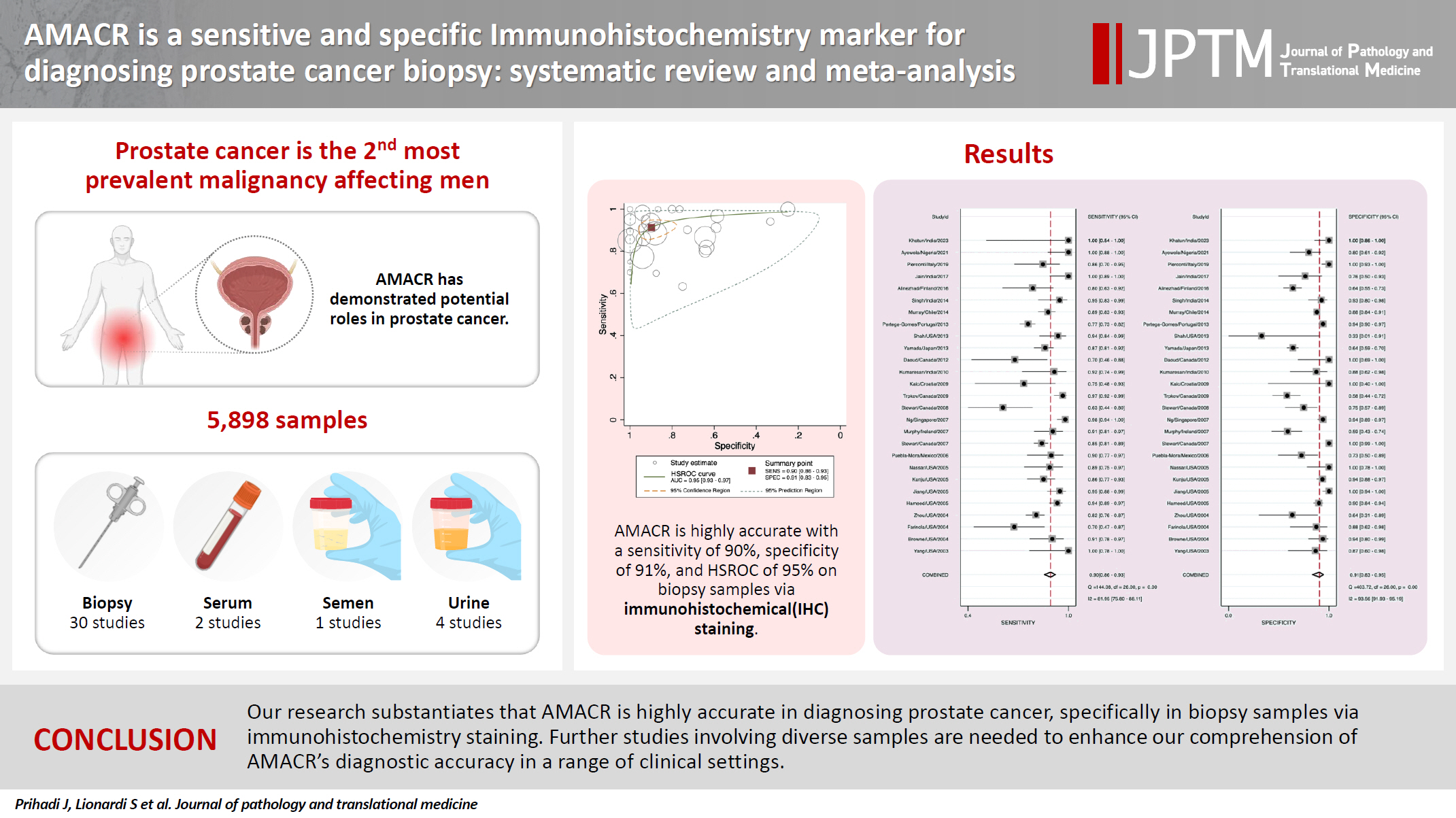
Alpha-methylacyl-CoA racemase (AMACR) is the preferred biomarker for distinguishing malignant from benign glands in prostate biopsies, showing high sensitivity and specificity for prostate cancer. A meta-analysis of immunohistochemistry (IHC) for AMACR is essential to further assess its diagnostic accuracy across diverse sample sources. Methods: A systematic search of databases including MEDLINE, ScienceDirect, ProQuest, Google Scholar, and the Cochrane Library was performed, focusing on studies of AMACR to diagnose prostate cancer, particularly in biopsy samples analyzed through IHC over the last 20 years. Quality of studies was assessed using the Quality Assessment of Diagnostic Accuracy Studies 2 tool, followed by a meta-analysis of regions and subgroups to calculate summary estimates of diagnostic test accuracy. Results: In the final analysis, 37 studies, with a pooled size of 5,898 samples, were included from the examination of 94 full-text papers. Among them, 27 studies with similar sample sources and testing methodologies underwent meta-analysis, yielding a combined sensitivity estimate of 0.90 (95% confidence interval [CI], 0.86 to 0.93) and specificity of 0.91 (95% CI, 0.83 to 0.95), both with significant heterogeneity (p < .01). The region beneath the hierarchical summary receiver operating characteristic curve was 0.95 (95% CI, 0.93 to 0.97), positive likelihood ratio was 9.6 (95% CI, 5.3 to 17.4), negative likelihood ratio was 0.11 (95% CI, 0.08 to 0.15), and diagnostic odds ratio was 88 (95% CI, 42 to 181). Conclusions: Our meta-analysis findings substantiate AMACR as a highly accurate tool for diagnosing prostate cancer, specifically in biopsy samples, via immunohistochemical staining. Further studies involving diverse samples are needed to enhance our understanding of the AMACR diagnostic accuracy in a range of clinical settings. -
Citations
Citations to this article as recorded by- ATP binding cassette subfamily D member 1: A highly sensitive diagnostic marker for solid pseudopapillary neoplasm of pancreas in biopsy samples——A multi-institutional study
Yuanhao Liu, Junya Peng, Ruizhe He, Xiaowei Xue, Jie Cui, Mengjie Li, Ying-ao Liu, Wanni Xu, Xiaohong Gao, Yingmei Wang, Zhe Zhang, Haizhen Lu, Zhigang Song, Peizhen Hu, Yupei Zhao, Wenze Wang
Human Pathology.2026; 174: 106125. CrossRef - Pathogenesis-Guided Biomarker Assessment: A Shift in Prostate Cancer Diagnostics
Jessica M. Logan, Victoria Malone, John J. O’Leary, Doug A. Brooks
International Journal of Molecular Sciences.2025; 26(24): 11786. CrossRef
- ATP binding cassette subfamily D member 1: A highly sensitive diagnostic marker for solid pseudopapillary neoplasm of pancreas in biopsy samples——A multi-institutional study
- Primary renal BCOR::CCNB3 sarcoma in a female patient: case report
- Somang Lee, Binnari Kim
- J Pathol Transl Med. 2025;59(1):84-90. Published online January 15, 2025
- DOI: https://doi.org/10.4132/jptm.2024.09.30
- 6,006 View
- 180 Download
- 1 Web of Science
- 1 Crossref
-
Abstract
PDF
- BCOR-rearranged sarcoma was classified by the World Health Organization in 2020 as a new subgroup of undifferentiated small round-cell sarcoma. It is known to occur very rarely in the kidney. This report presents the first case of a primary renal BCOR::CCNB3 sarcoma in a 22-year-old woman. An 8-cm cystic mass was identified in the left kidney by abdominal pelvic computed tomography. Histopathologic examination revealed the mass to be composed of small round to oval or spindle cells with fibrous septa and a delicate vascular network. A BCOR::CCNB3 fusion was detected by next-generation sequencing–based molecular testing. BCOR::CCNB3 sarcoma presents diagnostic difficulties, highlighting the importance of recognizing its histological features. Immunohistochemical markers are helpful for diagnosis, but genetic molecular testing is necessary for accurate diagnosis. These tumors have a very poor and aggressive prognosis, and an optimal therapeutic regimen has not yet been defined. Therefore, further studies are needed.
-
Citations
Citations to this article as recorded by- Update on the management of BCOR::CCNB3 sarcoma
Jungo Imanishi, Kenji Sato, Yoshinao Kikuchi, Asako Yamamoto, Shiori Watabe, Taisuke Matsuyama, Chiaki Sato, Hiroshi Kobayashi, Hirotaka Kawano
Japanese Journal of Clinical Oncology.2025; 55(10): 1097. CrossRef
- Update on the management of BCOR::CCNB3 sarcoma
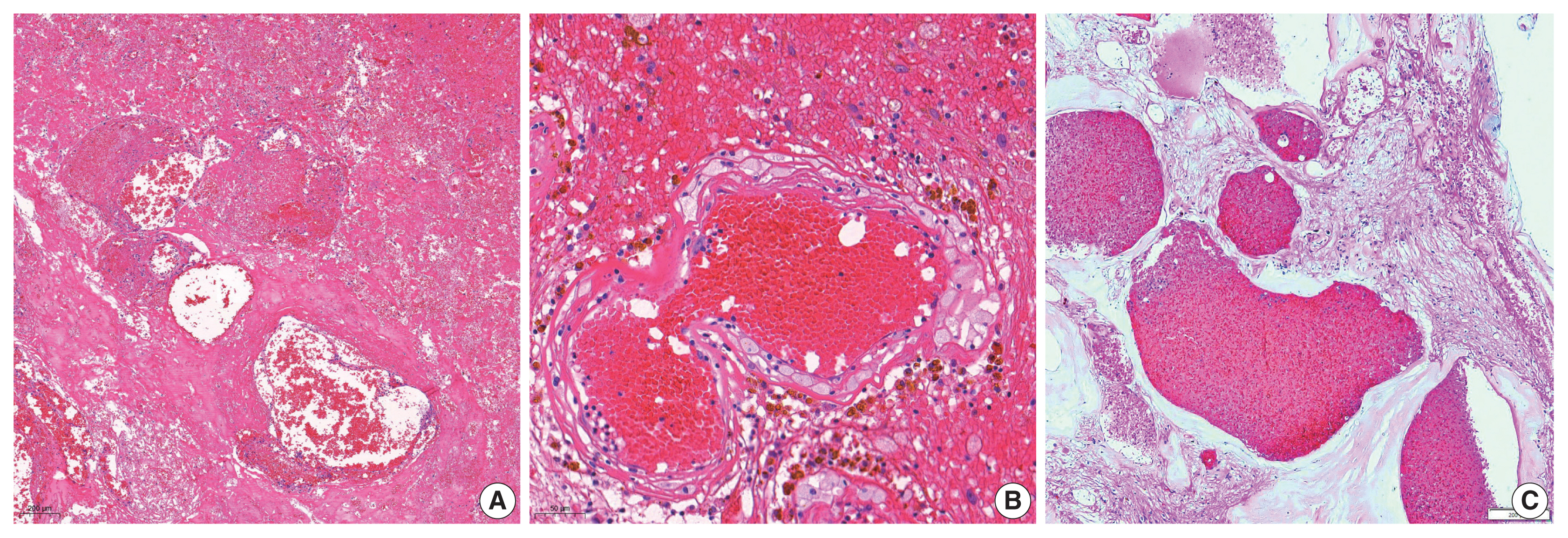
- Neuropathologic features of central nervous system hemangioblastoma
- Rebecca A. Yoda, Patrick J. Cimino
- J Pathol Transl Med. 2022;56(3):115-125. Published online May 3, 2022
- DOI: https://doi.org/10.4132/jptm.2022.04.13
- 18,766 View
- 382 Download
- 22 Web of Science
- 29 Crossref
-
Abstract
PDF
- Hemangioblastoma is a benign, highly vascularized neoplasm of the central nervous system (CNS). This tumor is associated with loss of function of the VHL gene and demonstrates frequent occurrence in von Hippel-Lindau (VHL) disease. While this entity is designated CNS World Health Organization grade 1, due to its predilection for the cerebellum, brainstem, and spinal cord, it is still an important cause of morbidity and mortality in affected patients. Recognition and accurate diagnosis of hemangioblastoma is essential for the practice of surgical neuropathology. Other CNS neoplasms, including several tumors associated with VHL disease, may present as histologic mimics, making diagnosis challenging. We outline key clinical and radiologic features, pathophysiology, treatment modalities, and prognostic information for hemangioblastoma, and provide a thorough review of the gross, microscopic, immunophenotypic, and molecular features used to guide diagnosis.
-
Citations
Citations to this article as recorded by- Renal hemangioblastoma and renal cell carcinoma with fibromyomatous stroma and hemangioblastoma-like areas belong to the spectrum of one entity
Kiril Trpkov, Norel Salut, Inmaculada Ribera-Cortada, Elías Tasso Xipell, Isabel Trias Puigsureda, Asli Yilmaz, Arjumand Riyaz Husain, Erik Nohr, Adrian Box, Farshid Siadat, Katherina Baranova, Rola M. Saleeb, Robert Stoehr, Arndt Hartmann, Abbas Agaimy
Virchows Archiv.2026; 488(4): 767. CrossRef - Hemangioblastoma of the Kidney—A Comprehensive Clinical, Pathological, and Genetic Analysis of Four Cases
Boglárka Pósfai, Alex Jenei, Gertrúd Forika, Attila Fintha, Zoltán Sápi, Áron Somorácz, Borbála Dénes, Ferenc Salamon, Kornélia Veronika Eizler, Nándor Giba, Dávid Semjén, Ildikó Illyés, Kristóf Attila Kovács, Gyöngyi Munkácsy, János Papp, Fanni Sánta, He
APMIS.2026;[Epub] CrossRef - Peritumoral Cystic Meningioma With Diagnostic Challenges: Two Case Reports and a Literature Review
Wei-Chih Chen, Zi-Jie Lin, Lam Chee-Tat
Cureus.2026;[Epub] CrossRef - Belzutifan-induced tumor regression in sporadic hemangioblastoma: a case report and literature review
Rebekka E. Hooks, Niket Yadav, Mark Willy L. Mondia, Georgios Mantziaris, Alaa Saleh, Anna Vi Jones, Matthew McCord, Melike Mut, Ashok R. Asthagiri, Benjamin W. Purow
Journal of Neuro-Oncology.2026;[Epub] CrossRef - Unusual immunohistochemical profiles in hemangioblastomas and their relevance in differential diagnosis: A comprehensive study of 112 cases
Jiri Soukup, Marie Novakova, Jan Hojny, Marketa Trnkova, Martin Syrucek, Tomas Jirasek, Patricie Delongova, Ales Kohout, Tomas Vebr, Jan Sroubek, Alena Sejkorova, Radim Lipina, Radim Brabec, Tomas Cesak, David Netuka
Journal of Neuropathology & Experimental Neurology.2026;[Epub] CrossRef - Pathological features and clinical outcomes of hemangioblastoma in the spinal cord of three dogs
Ryo SAITO, James K CHAMBERS, Kio YOSHIDA, Yutaro NAKAYAMA, Yukiko NAKANO, Kosuke HAII, Yumiko KAGAWA, Kazuyuki UCHIDA
Journal of Veterinary Medical Science.2026; 88(4): 568. CrossRef - Monitoring and treatment patterns of von Hippel-Lindau disease-associated central nervous system hemangioblastomas
Eric Jonasch, Yan Song, Jonathan Freimark, Manasi Mohan, James Signorovitch, Murali Sundaram
Hereditary Cancer in Clinical Practice.2026;[Epub] CrossRef - Multimodal Management of Spinal Cord Hemangioblastomas: A Comprehensive Review
Francisco Alfredo Call-Orellana, Juan Pablo Zuluaga-Garcia, Maria Alejandra Sierra, Mariana Zuluaga-Garcia, Esteban Ramirez-Ferrer, Alejandro Bugarini
Therapeutics.2026; 3(2): 12. CrossRef - Immunohistochemical Expression of PAX8 in Central Nervous System Hemangioblastomas: A Potential Diagnostic Pitfall for Neuropathologists
Giuseppe Broggi, Jessica Farina, Valeria Barresi, Francesco Certo, Giuseppe Maria Vincenzo Barbagallo, Gaetano Magro, Rosario Caltabiano
Applied Immunohistochemistry & Molecular Morphology.2025; 33(3): 160. CrossRef - Endolymphatic Sac Tumor. Post-Radiosurgery Evaluation Using Time-Resolved Imaging of Contrast Kinetics MR Angiography
Antonella Blandino, Allegra Romano, Chiara Filippi, Sofia Pizzolante, Andrea Romano, Giulia Moltoni, Edoardo Covelli, Maurizio Barbara, Alessandro Bozzao
Ear, Nose & Throat Journal.2025;[Epub] CrossRef - Stereotactic radiosurgery in the management of central nervous system hemangioblastomas: a systematic review and meta-analysis
Amirhossein Zare, Amirhessam Zare, Alireza Soltani Khaboushan, Bardia Hajikarimloo, Jason P. Sheehan
Neurosurgical Review.2025;[Epub] CrossRef - Cerebellar medullary cistern hemangioblastoma
Dahai Cao, Qiang Zhang
Asian Journal of Surgery.2025; 48(9): 5843. CrossRef - Navigating rare vascular brain tumors: A retrospective observational study
Sana Ahuja, Dipanker S Mankotia, Naveen Kumar, Vyomika Teckchandani, Sufian Zaheer
Cancer Research, Statistics, and Treatment.2025; 8(2): 92. CrossRef - A potential new entity pending further validation of pulmonary primary interstitial Tumor: Lymphangioleiomyomatosis-like
Lingyu Zhao, Xiaochen Shen, Yun Niu, Huang Chen, Dingrong Zhong
Respiratory Medicine Case Reports.2025; 57: 102241. CrossRef - Renal cell carcinoma with fibromyomatous stroma (RCC FMS) and with hemangioblastoma‐like areas is part of the RCC FMS spectrum in patients with tuberous sclerosis complex
Katherina Baranova, Jacob A Houpt, Deaglan Arnold, Andrew A House, Laura Lockau, Lindsay Ninivirta, Stephen Pautler, Haiying Chen, Madeleine Moussa, Rola Saleeb, Jose A Gomez, Asli Yilmaz, Farshid Siadat, Adrian Box, Douglas J Mahoney, Franz J Zemp, Manal
Histopathology.2025; 87(5): 687. CrossRef - Primary hemangioblastoma of rectum: a rare case report and review of literature
Aiping Zheng, Shaojuan Zhang, Qiang Ma, Wenxu Yang, Hualiang Xiao, Xinyu Liang
Journal of Cancer Research and Clinical Oncology.2025;[Epub] CrossRef - Cerebellar Hemangioblastoma Resection Complicated by Postoperative Vasogenic Edema in the Setting of Concurrent Immunotherapy Treatment
Aashka Sheth, Nicholas Dietz, Andrea Becerril-Gaitan, Rahim Kasem, Akshitkumar Mistry, Brian J Williams, Dale Ding, Isaac Abecassis
Cureus.2025;[Epub] CrossRef - Familial Von Hippel–Lindau Disease: A Case Series of Cerebral Hemangioblastomas with MRI, Histopathological, and Genetic Correlations
Claudiu Matei, Ioana Boeras, Dan Orga Dumitriu, Cosmin Mutu, Adriana Popescu, Mihai Gabriel Cucu, Alexandru Calotă-Dobrescu, Bogdan Fetica, Diter Atasie
Life.2025; 15(11): 1649. CrossRef - Characterization of spinal hemangioblastomas in patients with and without von Hippel-Lindau, and YAP expression
Ana-Laura Calderón-Garcidueñas, Steven-Andrés Piña-Ballantyne, Eunice-Jazmín Espinosa-Aguilar, Rebeca de Jesús Ramos-Sánchez
Revista Española de Patología.2024; 57(3): 160. CrossRef - Patients With Hemangioblastoma: Mood Disorders and Sleep Quality
Ali Riazi, Yaser Emaeillou, Nima Najafi, Mohammad Hoseinimanesh, Mohammad Ibrahim Ashkaran, Donya Sheibani Tehrani
Brain Tumor Research and Treatment.2024; 12(2): 87. CrossRef - Radiosurgically Treated Recurrent Cerebellar Hemangioblastoma: A Case Report and Literature Review
François Fabi, Ève Chamberland, Myreille D’Astous, Karine Michaud, Martin Côté, Isabelle Thibault
Current Oncology.2024; 31(7): 3968. CrossRef - Dual manifestations: spinal and cerebellar hemangioblastomas indicative of von Hippel-Lindau syndrome
Nurhuda Hendra Setyawan, Rachmat Andi Hartanto, Rusdy Ghazali Malueka, Ery Kus Dwianingsih, Dito Pondra Dharma
Radiology Case Reports.2024; 19(11): 5000. CrossRef - Phenotypic and Genotypic Features of a Chinese Cohort with Retinal Hemangioblastoma
Liqin Gao, Feng Zhang, J. Fielding Hejtmancik, Xiaodong Jiao, Liyun Jia, Xiaoyan Peng, Kai Ma, Qian Li
Genes.2024; 15(9): 1192. CrossRef - Hemangioblastoma Incidentally Discovered at CT Scan in Bamako: About a Case
Traore Ousmane, N’Diaye Mamadou, Dembélé Mamadou, Dembélé Adama, Diakité Siaka, Sidibé Mansa Drissa, Camara Nagnoumague, Keita Adama Diaman
Open Journal of Medical Imaging.2024; 14(03): 123. CrossRef - Case report: Hemangioblastoma in the brainstem of a dog
Kirsten Landsgaard, Samantha St. Jean, Stephanie Lovell, Jonathan Levine, Christine Gremillion, Brian Summers, Raquel R. Rech
Frontiers in Veterinary Science.2023;[Epub] CrossRef - Intramedullary hemangioblastoma of the thoracic cord with a microsurgical approach: A case report and literature review
Eduardo Cattapan Piovesan, Werner Petry Silva, Adroaldo Baseggio Mallmann, Felipe Severo Lanzini, Bruna Zanatta de Freitas, Francisco Costa Beber Lemanski, Charles André Carazzo
Surgical Neurology International.2023; 14: 137. CrossRef - Secondary Holocord Syringomyelia Associated With Spinal Hemangioblastoma in a 29-Year-Old Female
Eric Chun-Pu Chu, Edouard Sabourdy, Benjamin Cheong
Cureus.2023;[Epub] CrossRef - Belzutifan in adults with VHL-associated central nervous system hemangioblastoma: a single-center experience
Bryan J. Neth, Mason J. Webb, Jessica White, Joon H. Uhm, Pavel N. Pichurin, Ugur Sener
Journal of Neuro-Oncology.2023; 164(1): 239. CrossRef - Resection of Intramedullary Hemangioblastoma: Timing of Surgery and Its Impact on Neurological Outcome and Quality of Life
Michael Schwake, Sarah Ricchizzi, Sophia Krahwinkel, Emanuele Maragno, Stephanie Schipmann, Walter Stummer, Marco Gallus, Markus Holling
Medicina.2023; 59(9): 1611. CrossRef
- Renal hemangioblastoma and renal cell carcinoma with fibromyomatous stroma and hemangioblastoma-like areas belong to the spectrum of one entity
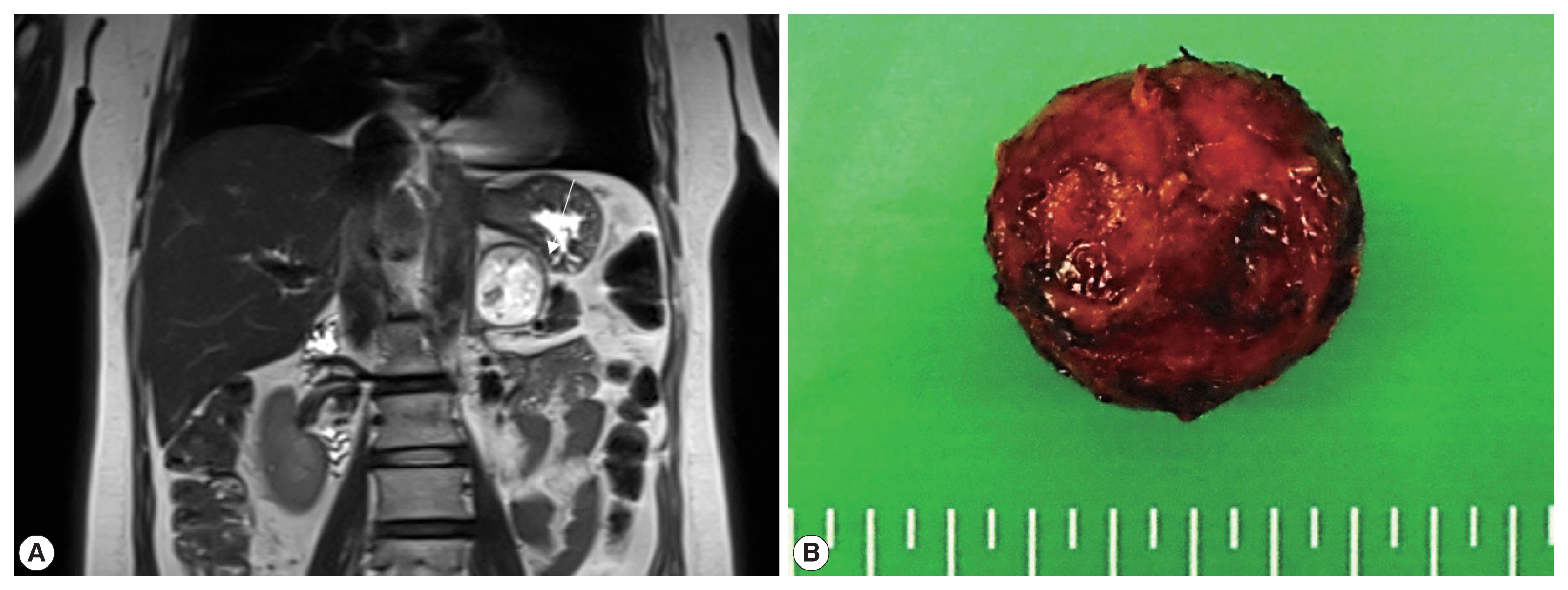
- Adrenal hemangioblastoma
- Joo-Yeon Koo, Kyung-Hwa Lee, Joon Hyuk Choi, Ho Seok Chung, Chan Choi
- J Pathol Transl Med. 2022;56(3):161-166. Published online February 28, 2022
- DOI: https://doi.org/10.4132/jptm.2021.12.28
- 6,243 View
- 159 Download
- 1 Web of Science
- 1 Crossref
-
Abstract
PDF
- Hemangioblastoma (HB) is a rare benign tumor that most commonly occurs in the cerebellum. HB is composed of neoplastic stromal cells and abundant small vessels. However, the exact origin of stromal cells is controversial. Extraneural HBs have been reported in a small series, and peripheral HBs arising in the adrenal gland are extremely rare. Herein, we report a case of sporadic adrenal HB in a 54-year-old woman. The tumor was a well-circumscribed, yellow mass measuring 4.2 cm in diameter. Histologically, the tumor was composed of small blood vessels and vacuolated stromal cells with clear cytoplasm. On immunohistochemical stain, the stromal cells were positive for S-100 protein, neuron-specific enolase, and synaptophysin. The tumor did not reveal mutation of VHL alleles. We herein present a case of HB of the adrenal gland and review of the literature.
-
Citations
Citations to this article as recorded by- Familial Von Hippel–Lindau Disease: A Case Series of Cerebral Hemangioblastomas with MRI, Histopathological, and Genetic Correlations
Claudiu Matei, Ioana Boeras, Dan Orga Dumitriu, Cosmin Mutu, Adriana Popescu, Mihai Gabriel Cucu, Alexandru Calotă-Dobrescu, Bogdan Fetica, Diter Atasie
Life.2025; 15(11): 1649. CrossRef
- Familial Von Hippel–Lindau Disease: A Case Series of Cerebral Hemangioblastomas with MRI, Histopathological, and Genetic Correlations
- Clinicopathological differences in radiation-induced organizing hematomas of the brain based on type of radiation treatment and primary lesions
- Myung Sun Kim, Se Hoon Kim, Jong-Hee Chang, Mina Park, Yoon Jin Cha
- J Pathol Transl Med. 2022;56(1):16-21. Published online October 15, 2021
- DOI: https://doi.org/10.4132/jptm.2021.08.30
- 7,929 View
- 244 Download
- 4 Web of Science
- 5 Crossref
-
Abstract
PDF
- Background
Radiation-induced organizing hematoma (RIOH) is a sporadic form of cavernous hemangioma (CH) that occurs after cerebral radiation. RIOH lesions are distinct histologically from de novo CH; however, detailed research on this subject is lacking. In the present study, the clinical and histological features of RIOHs were evaluated based on causative lesions.
Methods
The present study included 37 RIOHs confirmed by surgical excision from January 2009, to May 2020, in Yonsei Severance Hospital. All cases were divided into subgroups based on type of radiation treatment (gamma knife surgery [GKS], n = 24 vs. conventional radiation therapy [RT], n = 13) and pathology of the original lesion (arteriovenous malformation, n = 14; glioma, n = 12; metastasis, n = 4; other tumors, n = 7). The clinicopathological results were compared between the groups.
Results
Clinical data of multiplicity, latency, and size and wall thickness of the original tumors and RIOHs were analyzed. The GKS group showed shorter latency (5.85 ± 4.06 years vs. 11.15 ± 8.27 years, p = .046) and thicker tumor wall (693.7 ± 565.7 μm vs. 406.9 ± 519.7 μm, p = .049) than the conventional RT group. Significant difference was not found based on original pathology.
Conclusions
RIOH is more likely to occur earlier with thick tumor wall in subjects who underwent GKS than in patients who underwent conventional RT. These results indicate the clinical course of RIOH differs based on type of treatment and might help determine the duration of follow-up. -
Citations
Citations to this article as recorded by- Impact of cranial irradiation on the clinical presentation of cerebral cavernous malformations
Neerav Kumar, Jeffrey Shi, Carlos Alcocer, Sara Luck, Andrew Garton, Maricruz Rivera, Mark M. Souweidane, Philip E. Stieg
Clinical Neurology and Neurosurgery.2026; 265: 109386. CrossRef - Radiation-Induced Cavernous Malformation in the Cerebellum: Clinical Features of Two Cases
Hyoung Soo Choi, Chae-Yong Kim, Byung Se Choi, Seung Hyuck Jeon, In Ah Kim, Joo-Young Kim, Kyu Sang Lee, Gheeyoung Choe
Brain Tumor Research and Treatment.2025; 13(2): 58. CrossRef - End-stage ADPKD with a low-frequency PKD1 mosaic variant accelerated by chemoradiotherapy
Hiroaki Hanafusa, Hiroshi Yamaguchi, Naoya Morisada, Ming Juan YE, Riki Matsumoto, Hiroaki Nagase, Kandai Nozu
Human Genome Variation.2024;[Epub] CrossRef - Recapitulating the Key Advances in the Diagnosis and Prognosis of High-Grade Gliomas: Second Half of 2021 Update
Guido Frosina
International Journal of Molecular Sciences.2023; 24(7): 6375. CrossRef - Earlier Age at Surgery for Brain Cavernous Angioma-Related Epilepsy May Achieve Complete Seizure Freedom without Aid of Anti-Seizure Medication
Ayataka Fujimoto, Hideo Enoki, Keisuke Hatano, Keishiro Sato, Tohru Okanishi
Brain Sciences.2022; 12(3): 403. CrossRef
- Impact of cranial irradiation on the clinical presentation of cerebral cavernous malformations
- Morphologic variant of follicular lymphoma reminiscent of hyaline-vascular Castleman disease
- Jiwon Koh, Yoon Kyung Jeon
- J Pathol Transl Med. 2020;54(3):253-257. Published online February 5, 2020
- DOI: https://doi.org/10.4132/jptm.2019.12.17
- 8,929 View
- 242 Download
- 5 Web of Science
- 4 Crossref
-
Abstract
PDF
- Follicular lymphoma (FL) with hyaline-vascular Castleman disease (FL-HVCD)-like features is a rare morphologic variant, with fewer than 20 cases in the literature. Herein, we report a case of FL-HVCD in a 37-year-old female who presented with isolated neck lymph node enlargement. The excised lymph node showed features reminiscent of HVCD, including regressed germinal centers (GCs) surrounded by onion skin-like mantle zones, lollipop lesions composed of hyalinized blood vessels penetrating into regressed GCs, and hyalinized interfollicular stroma. In addition, focal areas of abnormally conglomerated GCs composed of homogeneous, small centrocytes with strong BCL2, CD10, and BCL6 expression were observed, indicating partial involvement of the FL. Several other lymphoid follicles showed features of in situ follicular neoplasia. Based on the observations, a diagnosis of FL-HVCD was made. Although FLHVCD is very rare, the possibility of this variant should be considered in cases resembling CD. Identification of abnormal, neoplastic follicles and ancillary immunostaining are helpful for proper diagnosis.
-
Citations
Citations to this article as recorded by- Unicentric Castleman Disease: Illustration of Its Morphologic Spectrum and Review of the Differential Diagnosis
Siba El Hussein, Andrew G. Evans, Hong Fang, Wei Wang, L. Jeffrey Medeiros
Archives of Pathology & Laboratory Medicine.2024; 148(1): 99. CrossRef - Finding a Needle in the Haystack
Hung-Yu Lin, Yi-Jen Peng, Yi-Ying Wu, Ping-Ying Chang
Journal of Medical Sciences.2023; 43(6): 292. CrossRef - Analysis of immunophenotypic features in hyaline vascular type Castleman disease
Yu Chang, Yu Ma, Chen Chang, Wensheng Li
Diagnostic Pathology.2023;[Epub] CrossRef - In‐situ follicular neoplasia: a clinicopathological spectrum
Gurdip S Tamber, Myriam Chévarie‐Davis, Margaret Warner, Chantal Séguin, Carole Caron, René P Michel
Histopathology.2021; 79(6): 1072. CrossRef
- Unicentric Castleman Disease: Illustration of Its Morphologic Spectrum and Review of the Differential Diagnosis
- Frozen Cytology of Meningeal Malignant Solitary Fibrous Tumor/Hemangiopericytoma
- Myunghee Kang, Na Rae Kim, Dong Hae Chung, Gie-Taek Yie
- J Pathol Transl Med. 2019;53(3):192-197. Published online April 11, 2019
- DOI: https://doi.org/10.4132/jptm.2019.03.20
- 8,605 View
- 160 Download
- 7 Web of Science
- 8 Crossref
-
Abstract
PDF
- A 51-year-old woman presented with severe dizziness. The brain magnetic resonance image revealed a 5.5 cm multiloculated mass with a thick rim in the left temporal lobe. Cytological examination of frozen diagnosis of the mass showed hypercellular sheets of round and rhabdoid cells in a hemorrhagic background, and two mitotic figures were observed. Histologically, the excised dura-based mass consisted of predominantly round cells with small foci of rhabdoid tumor cells in a pseudoalveolar pattern in a hemorrhagic background, and the cells showed nuclear positivity for signal transducer and activator of transcription 6 as well as frequent mitosis. The mass was diagnosed as a grade 3 solitary fibrous tumor (SFT)/hemangiopericytoma (HPC). The cytological diagnosis of SFT/HPC is challenging because of the heterogeneous cytological findings, such as histological heterogeneity, and because there are no standardized cytological criteria for malignant SFT/HPC. Cytological findings, such as singly scattered small cells, hypercellularity, rare ropy collagen, and round and rhabdoid cells with pseudoalveolar pattern, may assist in the diagnosis of malignant SFT/HPC.
-
Citations
Citations to this article as recorded by- A Rare Case of Cervical Solitary Fibrous Tumor in a Pediatric Patient: Case Report and Literature Review
Eleonora Becattini, Lorenzo Sgarbanti, Giuseppina Bevacqua, Valentina Grespi, Carlo Conti
NeuroSci.2025; 6(2): 49. CrossRef - Meningeal Solitary Fibrous Tumor: A Cytological Report With Emphasis on the Usefulness of Immunocytochemical Analysis for STAT6
Hiroyuki Okanishi, Mitsuaki Ishida, Naoto Kohno, Isako Kataoka, Mari Tomiuka, Mayumi Uragami, Shizuka Ono, Chihiro Deguchi, Reika Takeda, Yoshitaka Kurisu, Yoshinobu Hirose
Diagnostic Cytopathology.2025;[Epub] CrossRef - Cytologic features of mesenchymal, melanocytic and haematolymphoid tumours of the central nervous system and metastases
Carmen Bárcena, José A. Jiménez‐Heffernan
Cytopathology.2024; 35(5): 590. CrossRef - A Hemangiopericytoma in the External Auditory Canal: A Rare Clinical Presentation and Management
Vaibhavi Patil, Prasad Deshmukh, Sagar S Gaurkar , Ayushi Ghosh Moulic, Jasleen Kaur
Cureus.2024;[Epub] CrossRef - Scoring system for intraoperative diagnosis of intracranial schwannoma by squash cytology
Hirotaka Fujita, Takuma Tajiri, Tomohisa Machida, Nozomi Nomura, Suguru Toguchi, Hitoshi Itoh, Shinichiro Hiraiwa, Tomoko Sugiyama, Chie Inomoto, Masaaki Imai, Shinri Oda, Masami Shimoda, Naoya Nakamura
Cytopathology.2022; 33(2): 196. CrossRef - Occurrence of a solitary fibrous tumor adjacent to the resection bed of a high-grade meningioma: A case report
Coby Cunningham, Rocco Dabecco, Justin Davanzo
Interdisciplinary Neurosurgery.2021; 25: 101277. CrossRef - A case of solitary fibrous tumor arising in the meninge
Saori NAKANISHI, Naoto KURODA, Toshiko TAKAI, Mari KOJIMA, Misato OONOGI
The Journal of the Japanese Society of Clinical Cytology.2021; 60(4): 224. CrossRef - Intraoperative frozen cytology of intraosseous cystic meningioma in the sphenoid bone
Na Rae Kim, Gie-Taek Yie
Journal of Pathology and Translational Medicine.2020; 54(6): 508. CrossRef
- A Rare Case of Cervical Solitary Fibrous Tumor in a Pediatric Patient: Case Report and Literature Review
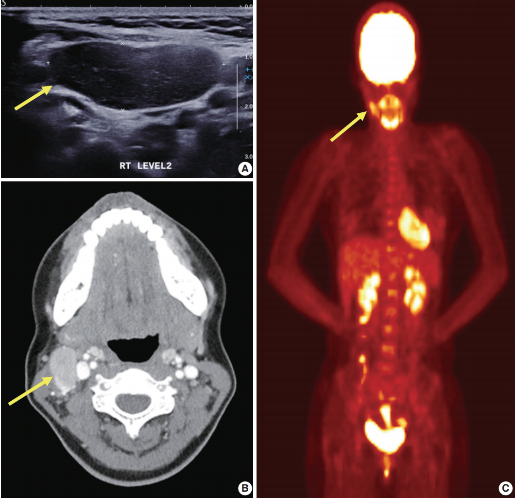
- Primary Necrobiotic Xanthogranulomatous Sialadenitis with Submandibular Gland Localization without Skin Involvement
- Myunghee Kang, Na Rae Kim, Dong Hae Chung, Jae Yeon Seok, Dong Young Kim
- J Pathol Transl Med. 2019;53(4):261-265. Published online January 16, 2019
- DOI: https://doi.org/10.4132/jptm.2019.01.08
- 9,681 View
- 172 Download
- 2 Web of Science
- 6 Crossref
-
Abstract
PDF
- Necrobiotic xanthogranulomatous reaction is a multiorgan, non-Langerhans cell histiocytosis with an unknown etiology. Occurrence in the salivary gland is extremely rare. We recently identified a case of necrobiotic xanthogranulomatous sialadenitis in a 73-year-old Korean woman who presented with a painless palpable lesion in the chin. There was no accompanying cutaneous lesion. Partial resection and subsequent wide excision with neck dissection were performed. Pathological examination showed a severe inflammatory lesion that included foamy macrophages centrally admixed with neutrophils, eosinophils, lymphocytes, plasma cells, and scattered giant cells, as well as necrobiosis. During the 12-month postoperative period, no grossly remarkable change in size was noted. Necrobiotic xanthogranulomatous inflammation may be preceded by or combined with hematologic malignancy. Although rare, clinicians and radiologists should be aware that an adhesive necrobiotic xanthogranuloma in the salivary gland may present with a mass-like lesion. Further evaluation for hematologic disease and close follow-up are needed when a pathologic diagnosis is made.
-
Citations
Citations to this article as recorded by- Salivary gland macrophages in health and disease: heterogeneity, niche crosstalk, and therapeutic avenues
Xinglei Li, Yan Feng, Huixin Xue, Xinxin Ni
Frontiers in Immunology.2025;[Epub] CrossRef - Five Cases of Xanthogranulomatous Sialadenitis
Satoshi Kiyama, Hiroyuki Iuchi, Kotoko Ito, Kengo Nishimoto, Tsutomu Matsuzaki, Masaru Yamashita
Practica Oto-Rhino-Laryngologica.2022; 115(4): 315. CrossRef - Xanthogranulomatous change in a pleomorphic adenoma: An extremely rare variant/degenerative change. Is it fine needle aspiration induced?
Mukta Pujani, Dipti Sidam, Kanika Singh, Aparna Khandelwal, Khushbu Katarya
Diagnostic Cytopathology.2021;[Epub] CrossRef - A Case of Xanthogranulomatous Sialadenitis with Facial Palsy Mimicking Malignancy
Sang Hyun Kim, Sun Woo Kim, Sang Hyuk Lee
Korean Journal of Otorhinolaryngology-Head and Neck Surgery.2021; 64(6): 422. CrossRef - Xanthogranulomatous Sialadenitis, an Uncommon Reactive Change is Often Associated with Warthin’s Tumor
Lihong Bu, Hui Zhu, Emilian Racila, Sobia Khaja, David Hamlar, Faqian Li
Head and Neck Pathology.2020; 14(2): 525. CrossRef - A Case of Xanthogranulomatous Sialadenitis of the Sublingual Gland:A Review of Literature
Naoya KITAMURA, Seiji OHNO, Tetsuya YAMAMOTO
Journal of Japanese Society of Oral Medicine.2019; 25(1): 20. CrossRef
- Salivary gland macrophages in health and disease: heterogeneity, niche crosstalk, and therapeutic avenues
- Adrenal Cortical Neoplasm with Uncertain Malignant Potential Arising in the Heterotopic Adrenal Cortex in the Liver of a Patient with Beckwith-Wiedemann Syndrome
- Eun Na Kim, Dong Eun Song, Hee Mang Yoon, Beom Hee Lee, Chong Jai Kim
- J Pathol Transl Med. 2019;53(2):129-135. Published online November 26, 2018
- DOI: https://doi.org/10.4132/jptm.2018.11.13
- 8,316 View
- 110 Download
- 5 Web of Science
- 5 Crossref
-
Abstract
PDF
- Patients with Beckwith-Wiedemann syndrome (BWS) are predisposed to developing embryonal tumors, with hepatoblastoma being the most common type. Our patient showed hemihypertrophy, macroglossia, and paternal uniparental disomy in chromosome 11 and was diagnosed with BWS. When the patient was 9 months old, a 2.5×1.5 cm oval hypoechoic exophytic mass was detected in the inferior tip of his right liver. Preoperative imaging identified it as hepatoblastoma; however, histologic, immunohistochemistry, and electron microscopic findings were compatible with adrenal cortical neoplasm with uncertain malignant potential. The origin of the adrenal tissue seemed to be heterotopic. Here, we describe for the first time an adrenal cortical neoplasm with uncertain malignant potential arising in the heterotopic adrenal cortex located in the liver of a patient with BWS.
-
Citations
Citations to this article as recorded by- Adrenocortical tumors and hereditary syndromes
Kanakamani Jeyaraman, Paola Concolino, Henrik Falhammar
Expert Review of Endocrinology & Metabolism.2025; 20(1): 1. CrossRef - Functional adrenocortical carcinoma with adrenohepatic fusion: A case report
Pastor Escárcega-Fujigaki, Guillermo Hernández-Peredo Rezk, José de Jesús Loeza- Oliva, Anallely Luna-Hernández, Bethsaida Natali Arreguín-Cortés, Rafael López-Cruz
Journal of Pediatric Surgery Case Reports.2024; 107: 102841. CrossRef - Molecular and Clinical Features of Adrenocortical Tumors in Beckwith–Wiedemann Spectrum
Diana Carli, Federico Rondot, Maria Luca, Anna Campello, Stefano Gabriele Vallero, Elisa Tirtei, Andrea Gazzin, Simona Cardaropoli, Francesca Montanari, Claudio Graziano, Paola Quarello, Abu Saadat, Angela Sparago, Giovanni Battista Ferrero, Franca Fagiol
Cancers.2024; 16(23): 3967. CrossRef - Beckwith–Wiedemann syndrome: Clinical, histopathological and molecular study of two Tunisian patients and review of literature
Hela Sassi, Yasmina Elaribi, Houweyda Jilani, Imen Rejeb, Syrine Hizem, Molka Sebai, Nadia Kasdallah, Habib Bouthour, Samia Hannachi, Jasmin Beygo, Ali Saad, Karin Buiting, Dorra H’mida Ben‐Brahim, Lamia BenJemaa
Molecular Genetics & Genomic Medicine.2021;[Epub] CrossRef - Adrenocortical Tumors in Children With Constitutive Chromosome 11p15 Paternal Uniparental Disomy: Implications for Diagnosis and Treatment
Emilia Modolo Pinto, Carlos Rodriguez-Galindo, Catherine G. Lam, Robert E. Ruiz, Gerard P. Zambetti, Raul C. Ribeiro
Frontiers in Endocrinology.2021;[Epub] CrossRef
- Adrenocortical tumors and hereditary syndromes
- Chronic Placental Inflammation as a Risk Factor of Severe Retinopathy of Prematurity
- Chae Young Kim, Euiseok Jung, Eun Na Kim, Chong Jai Kim, Joo Yong Lee, Ji Hye Hwang, Woo Sun Song, Byong Sop Lee, Ellen Ai-Rhan Kim, Ki-Soo Kim
- J Pathol Transl Med. 2018;52(5):290-297. Published online July 16, 2018
- DOI: https://doi.org/10.4132/jptm.2018.07.09
- 13,834 View
- 133 Download
- 18 Web of Science
- 18 Crossref
-
Abstract
PDF
- Background
Chronic placental inflammation (CPI) has been implicated in the pathogenesis of diseases in premature infants, whereas retinopathy of prematurity (ROP) is a major complication primarily affecting preterm and very low-birth-weight (VLBW) infants. This study aims to investigate the association between CPI and ROP in VLBW infants.
Methods
We performed a retrospective review of clinical records of VLBW infants born between 2013 and 2016. Placental pathology findings including CPI cases were analyzed using logistic regression to study infants’ morbidities and other clinical characteristics.
Results
A total of 402 infants with a mean (standard deviation) gestational age of 28.5 (2.8) weeks and birth weight of 1,027.2 (304.4) g were included. The incidence of ROP was 24.1%. CPI was found in 90 infants (22.4%), among which 28.9% (26 of 90) developed ROP, and 21.1% (19 of 90) underwent laser photocoagulation. Lower gestational age, lower birth weight, longer duration of oxygen supply, and presence of CPI were associated with the development of ROP. After adjustment for gestational age, birth weight, sex, duration of oxygen supply, and other overlapping placental pathology, CPI was associated with the odds for type 1 ROP that required laser photocoagulation (adjusted odds ratio, 2.739; 95% confidence interval, 1.112 to 6.749; p = .029).
Conclusions
CPI was associated with severe ROP requiring treatment with laser photocoagulation in VLBW infants. -
Citations
Citations to this article as recorded by- The association between maternal blood lipid trajectory and offspring preschool myopia in prospective and nested case‒control analyses
Jiao-Jiao Shi, Guang-Zhuang Jing, Xian-Gui He, Jing-Jing Wang, Yun-Hui Zhang, Hui-Jing Shi
Lipids in Health and Disease.2026;[Epub] CrossRef - The maternal-fetal interface as an immunological barrier: Structure, regulation, and breakdown
Eva Kareus, Dustyn Levenson, Seungbaek Lee, Nardhy Gomez-Lopez
Cell Reports.2026; 45(4): 117164. CrossRef - Comprehensive assessment of placental inflammation: Novel approach in predicting retinopathy of prematurity
Salma El Emrani, Esther J.S. Jansen, Jelle J. Goeman, Jacqueline U.M. Termote, Enrico Lopriore, Nicoline E. Schalij-Delfos, Lotte E. van der Meeren
Early Human Development.2025; 204: 106239. CrossRef - Histological Chorioamnionitis and Funisitis as New Risk Factors for Retinopathy of Prematurity: A Meta-analysis
Salma El Emrani, Esther J.S. Jansen, Jelle J. Goeman, Enrico Lopriore, Jacqueline U.M. Termote, Nicoline E. Schalij-Delfos, Lotte E. van der Meeren
American Journal of Perinatology.2024; 41(S 01): e3264. CrossRef - Retinopathy of prematurity and placental histopathology findings: A retrospective cohort study
Sam Ebenezer Athikarisamy, Geoffrey C. Lam, Matthew N. Cooper, Tobias Strunk
Frontiers in Pediatrics.2023;[Epub] CrossRef - Identification of clinical factors associated with timing and duration of spontaneous regression of retinopathy of prematurity not requiring treatment
Jamee Schoephoerster, Sydney Roston, Scott Lunos, Sara E. Ramel, Jill Anderson, Michael K. Georgieff, Ellen C. Ingolfsland
Journal of Perinatology.2023; 43(6): 702. CrossRef - Ocular Vascular Diseases: From Retinal Immune Privilege to Inflammation
Xudong Wang, Tianxi Wang, Enton Lam, David Alvarez, Ye Sun
International Journal of Molecular Sciences.2023; 24(15): 12090. CrossRef - The potential of marine resources for retinal diseases: a systematic review of the molecular mechanisms
Kristin Krueger, Elke Boehme, Alexa Karina Klettner, Marietta Zille
Critical Reviews in Food Science and Nutrition.2022; 62(27): 7518. CrossRef - Retinopathy prematurity: a systematic review and meta-analysis study based on neonatal and maternal risk factors
Tahereh Bahmani, Arezoo Karimi, Nazanin Rezaei, Salman Daliri
The Journal of Maternal-Fetal & Neonatal Medicine.2022; 35(25): 8032. CrossRef - Diallyl Trisulfide Promotes Placental Angiogenesis by Regulating Lipid Metabolism and Alleviating Inflammatory Responses in Obese Pregnant Mice
Miaomiao Wang, Zhaoyu Wang, Yueyue Miao, Hongkui Wei, Jian Peng, Yuanfei Zhou
Nutrients.2022; 14(11): 2230. CrossRef - Risk factors for the development of retinopathy in premature infants
O.Yu. Obolonska, L.I. Vakulenko, L.P. Badogina, O.I. Obolonskyi, I.A. Likhachova, O.V. Kovryga
CHILD`S HEALTH.2022; 17(3): 138. CrossRef - Development of the genomic inflammatory index (GII) to assess key maternal antecedents associated with placental inflammation
Kirsi S. Oldenburg, Lauren A. Eaves, Lisa Smeester, Hudson P. Santos, T. Michael O'Shea, Rebecca C. Fry
Placenta.2021; 111: 82. CrossRef - Risk Factors Associated with Retinopathy of Prematurity in Very and Extremely Preterm Infants
Claudia Ioana Borțea, Florina Stoica, Marioara Boia, Emil Radu Iacob, Mihai Dinu, Roxana Iacob, Daniela Iacob
Medicina.2021; 57(5): 420. CrossRef - Efficacy of Aflibercept Treatment and Its Effect on the Retinal Perfusion in the Oxygen-Induced Retinopathy Mouse Model of Retinopathy of Prematurity
Sarina M. Amin, Andres Gonzalez, Jade Guevara, Charlotte Bolch, Lorick Andersen, W. Clay Smith, Swati Agarwal-Sinha
Ophthalmic Research.2021; 64(1): 91. CrossRef - A pilot randomised clinical trial of 670 nm red light for reducing retinopathy of prematurity
Alison L. Kent, Mohamed E. Abdel-Latif, Timothy Cochrane, Margaret Broom, Jane E. Dahlstrom, Rohan W. Essex, Bruce Shadbolt, Riccardo Natoli
Pediatric Research.2020; 87(1): 131. CrossRef - Human placental suppressors of cytokine signalling (SOCS) and inflammatory cytokines are dysregulated in assisted reproduction, advanced maternal age and pre-term birth
S. J. Knight, A. D. Smith, H. Kim, A. C. Collier
Clinical and Experimental Obstetrics & Gynecology.2020;[Epub] CrossRef - Exercise prevents the adverse effects of maternal obesity on placental vascularization and fetal growth
Jun Seok Son, Xiangdong Liu, Qiyu Tian, Liang Zhao, Yanting Chen, Yun Hu, Song Ah Chae, Jeanene M. de Avila, Mei‐Jun Zhu, Min Du
The Journal of Physiology.2019; 597(13): 3333. CrossRef - Cumulative evidence for association of sepsis and retinopathy of prematurity
Jichong Huang, Ying Tang, Tingting Zhu, Yafei Li, Hua Chun, Yi Qu, Dezhi Mu
Medicine.2019; 98(42): e17512. CrossRef
- The association between maternal blood lipid trajectory and offspring preschool myopia in prospective and nested case‒control analyses
- White Matter Injury of Prematurity: Its Mechanisms and Clinical Features
- Young Ah Lee
- J Pathol Transl Med. 2017;51(5):449-455. Published online August 11, 2017
- DOI: https://doi.org/10.4132/jptm.2017.07.25
- 20,247 View
- 432 Download
- 32 Web of Science
- 33 Crossref
-
Abstract
PDF
- A developing central nervous system is vulnerable to various insults such as infection and ischemia. While increased understanding of the dynamic nature of brain development allows a deeper insight into the pathophysiology of perinatal brain injury, the precise nature of specific fetal and neonatal brain injuries and their short- and long-term clinical consequences need special attention and further elucidation. The current review will describe the pathophysiological aspects and clinical significance of white matter injury of prematurity, a main form of perinatal brain injury in premature newborns, with a particular emphasis on its potential antenatal components.
-
Citations
Citations to this article as recorded by- Early ultrasound-based assessment of preterm white matter injury: association with MRI and neurological outcomes
Janah May Oclaman, Felicia Tang, Natalie Chan, Katelin Kramer, Ari J. Green, Dawn Gano, Fei Jiang, Kayla Cort, Yi Li, Bridget Elaine LaMonica Ostrem
Pediatric Radiology.2026;[Epub] CrossRef - Therapeutic potential of transcutaneous auricular vagus nerve stimulation in cognitive impairment: insights from preclinical and clinical studies
Di Pan, Jifei Sun, Wenchao Mao, Huaxin Shi
Frontiers in Neurology.2026;[Epub] CrossRef - Neonatal inflammation impairs developmentally-associated microglia and promotes a highly reactive microglial subset
Adrien Dufour, Ariane Heydari Olya, Sophie Foulon, Clémence Réda, Amazigh Mokhtari, Valérie Faivre, Jennifer Hua, Cindy Bokobza, Andrew D. Griffiths, Philippe Nghe, Pierre Gressens, Andrée Delahaye-Duriez, Juliette Van Steenwinckel
Brain, Behavior, and Immunity.2025; 123: 466. CrossRef - FGF21 Alleviates Hypoxic-Ischemic White Matter Injury in Neonatal Mice by Mediating Inflammation and Oxidative Stress Through PPAR-γ Signaling Pathway
Mingchu Fang, Liying Lu, Jia Lou, Jiahao Ou, Qianqian Yu, Xiaoyue Tao, Jianghu Zhu, Zhenlang Lin
Molecular Neurobiology.2025; 62(4): 4743. CrossRef - Association of the ADRB2 rs1042714 variant with retinopathy of prematurity highlights the importance of the renin-angiotensin-aldosterone system
Anna Chmielarz-Czarnocińska, Anna Durska, Bartosz Skulimowski, Alicja Sobaniec, Anna Gotz-Więckowska, Ewa Strauss
Scientific Reports.2025;[Epub] CrossRef - Imaging of Cerebral Palsy: A Primer for the Radiologist
M.S. Rootman, S. Shinnawi, G. Merhav, B.C. Friedman, L.-t. Pratt
Neurographics.2025; 15(2): 131. CrossRef - The effect of hemoglobin level in early life on periventricular leukomalacia: a case control study
Muchun Yu, Zhihong Sun, Lu He, Caiyu Zhang, Huiqing Sun
Scientific Reports.2025;[Epub] CrossRef - Predictors of poor neurodevelopmental outcomes of very preterm and very low birth weight infants
Rita PISSARRA, Bárbara PEREIRA-NETO, Pedro MIRAGAIA, Sara ALMEIDA, Filipa FLOR-DE-LIMA, Paulo SOARES
Minerva Pediatrics.2025;[Epub] CrossRef - Minimum effective dose of clemastine in a mouse model of preterm white matter injury
Elizabeth P. Odell, Nora Jabassini, Björn Schniedewind, Sarah E. Pease-Raissi, Adam Frymoyer, Uwe Christians, Ari J. Green, Jonah R. Chan, Bridget E. L. Ostrem
Pediatric Research.2024; 96(4): 933. CrossRef - The Neuroprotective Mechanisms of PPAR‐γ: Inhibition of Microglia‐Mediated Neuroinflammation and Oxidative Stress in a Neonatal Mouse Model of Hypoxic‐Ischemic White Matter Injury
Mingchu Fang, Qianqian Yu, Jiahao Ou, Jia Lou, Jianghu Zhu, Zhenlang Lin
CNS Neuroscience & Therapeutics.2024;[Epub] CrossRef - Neurodevelopmental outcome in preterm neonates
Ilija Palić, Ružica Kravljanac
Medicinski podmladak.2024; 75(3): 43. CrossRef - Paediatric cerebral palsy in South Africa: Prevention and care gaps at hospital level
Thembi J. Katangwe, Mariana Kruger, Ronald van Toorn, Jeanetta van Zyl, Sandile Ndlovu, Regan Solomons, Kirsten A. Donald
African Journal of Disability.2024;[Epub] CrossRef - Parsing brain-behavior heterogeneity in very preterm born children using integrated similarity networks
Laila Hadaya, Konstantina Dimitrakopoulou, Lucy D. Vanes, Dana Kanel, Sunniva Fenn-Moltu, Oliver Gale-Grant, Serena J. Counsell, A. David Edwards, Mansoor Saqi, Dafnis Batalle, Chiara Nosarti
Translational Psychiatry.2023;[Epub] CrossRef - Kaempferol improves periventricular white matter injury in premature infants by inhibiting microglial activation
Qiuling Zhuo, Binsha Fu, Liangsun Shi
Materials Express.2023; 13(5): 916. CrossRef - The impact of neonatal morbidities on child growth and developmental outcomes in very low birth weight infants: a nationwide cohort study
Jung Ho Han, So Jin Yoon, Joo Hee Lim, Jeong Eun Shin, Ho Seon Eun, Min Soo Park, Kook In Park, Soon Min Lee
European Journal of Pediatrics.2022; 181(1): 197. CrossRef - Global and Regional White Matter Fractional Anisotropy in Children with Chronic Kidney Disease
Ellen van der Plas, Matthew A. Solomon, Lauren Hopkins, Timothy Koscik, Jordan Schultz, Patrick D. Brophy, Peggy C. Nopoulos, Lyndsay A. Harshman
The Journal of Pediatrics.2022; 242: 166. CrossRef - A Case of Prenatally Diagnosed Congenital Adrenal Hyperplasia With Brain Morphometric Differences
Vidya Rajagopalan, Lloyd Nate Overholtzer, William S. Kim, Jessica L. Wisnowski, David A. Miller, Mitchell E. Geffner, Mimi S. Kim
Journal of Investigative Medicine High Impact Case Reports.2022;[Epub] CrossRef - Role of Vitamin E in Neonatal Neuroprotection: A Comprehensive Narrative Review
Sarah Kolnik, Thomas Wood
Life.2022; 12(7): 1083. CrossRef - Sirt2 promotes white matter oligodendrogenesis during development and in models of neonatal hypoxia
Beata Jablonska, Katrina L. Adams, Panagiotis Kratimenos, Zhen Li, Emma Strickland, Tarik F. Haydar, Katharina Kusch, Klaus-Armin Nave, Vittorio Gallo
Nature Communications.2022;[Epub] CrossRef - PGC-1α activity and mitochondrial dysfunction in preterm infants
Atefeh Mohammadi, Randa Higazy, Estelle B. Gauda
Frontiers in Physiology.2022;[Epub] CrossRef - Adverse Short-Term Outcomes of Preterm Infants Born to Mothers with Preeclampsia by Doppler Cranial Ultrasound Investigation
Qiu Luo, Guixian Chen, Mei Tang
International Journal of Clinical Medicine.2022; 13(03): 157. CrossRef - Intranasal mesenchymal stem cell therapy to boost myelination after encephalopathy of prematurity
Josine E. G. Vaes, Caren M. van Kammen, Chloe Trayford, Annette van der Toorn, Torben Ruhwedel, Manon J. N. L. Benders, Rick M. Dijkhuizen, Wiebke Möbius, Sabine H. van Rijt, Cora H. Nijboer
Glia.2021; 69(3): 655. CrossRef - The impact of trophic and immunomodulatory factors on oligodendrocyte maturation: Potential treatments for encephalopathy of prematurity
Josine E. G. Vaes, Myrna J. V. Brandt, Nikki Wanders, Manon J. N. L. Benders, Caroline G. M. de Theije, Pierre Gressens, Cora H. Nijboer
Glia.2021; 69(6): 1311. CrossRef - Pioglitazone Ameliorates Lipopolysaccharide-Induced Behavioral Impairment, Brain Inflammation, White Matter Injury and Mitochondrial Dysfunction in Neonatal Rats
Jiann-Horng Yeh, Kuo-Ching Wang, Asuka Kaizaki, Jonathan W. Lee, Han-Chi Wei, Michelle A. Tucci, Norma B. Ojeda, Lir-Wan Fan, Lu-Tai Tien
International Journal of Molecular Sciences.2021; 22(12): 6306. CrossRef - Dissecting the Roles of LncRNAs in the Development of Periventricular White Matter Damage
Xinyu Wang, Heng Liu, Xiaoli Liao, Lixing Qiao, Lihua Zhu, Shun Wu, Yan Zhou, Yi Zhang, Bangbang Li, Lili Lin, Jingjing Ma, Qianying Gu, Jiaping Shu
Frontiers in Genetics.2021;[Epub] CrossRef - Targeting Microglial Disturbances to Protect the Brain From Neurodevelopmental Disorders Associated With Prematurity
Andrée Delahaye-Duriez, Adrien Dufour, Cindy Bokobza, Pierre Gressens, Juliette Van Steenwinckel
Journal of Neuropathology & Experimental Neurology.2021; 80(7): 634. CrossRef - Circular RNA expression alteration in whole blood of premature infants with periventricular white matter damage
Lixing Qiao, Sisi Mo, Yan Zhou, Yi Zhang, Bangbang Li, Shun Wu, Lili Lin, Lihua Zhu, Ruibin Zhao
Genomics.2020; 112(4): 2875. CrossRef - Feed-forward neural networks using cerebral MR spectroscopy and DTI might predict neurodevelopmental outcome in preterm neonates
T. Janjic, S. Pereverzyev, M. Hammerl, V. Neubauer, H. Lerchner, V. Wallner, R. Steiger, U. Kiechl-Kohlendorfer, M. Zimmermann, A. Buchheim, A. E. Grams, E. R. Gizewski
European Radiology.2020; 30(12): 6441. CrossRef - White Matter Injury in Early Brain Injury after Subarachnoid Hemorrhage
Jinwei Pang, Jianhua Peng, Ping Yang, Li Kuai, Ligang Chen, John H. Zhang, Yong Jiang
Cell Transplantation.2019; 28(1): 26. CrossRef - The Potential of Stem Cell Therapy to Repair White Matter Injury in Preterm Infants: Lessons Learned From Experimental Models
Josine E. G. Vaes, Marit A. Vink, Caroline G. M. de Theije, Freek E. Hoebeek, Manon J. N. L. Benders, Cora H. A. Nijboer
Frontiers in Physiology.2019;[Epub] CrossRef - Abilitation of Infants with Combined Perinatal Pathology: Capabilities of Approaches and Methods Personalization
Аlexander A. Baranov, Leyla S. Namazova-Baranova, Irina A. Belyaeva, Еlena V. Аntonova, Еlena A. Vishneva, Еlena P. Bombardirova, Vladimir I. Smirnov, Аlexsei I. Molodchenkov, Мariay О. Zubrikhina
Current Pediatrics.2019; 18(2): 91. CrossRef - Advanced nanotherapies to promote neuroregeneration in the injured newborn brain
Olatz Arteaga Cabeza, Alkisti Mikrogeorgiou, Sujatha Kannan, Donna M. Ferriero
Advanced Drug Delivery Reviews.2019; 148: 19. CrossRef - Rapid Postnatal Adaptation of Neurodevelopment in Pigs Born Late Preterm
Charlotte Holme Nielsen, Anne Bladt Brandt, Thomas Thymann, Karina Obelitz-Ryom, Pingping Jiang, Charlotte Vanden Hole, Chris van Ginneken, Stanislava Pankratova, Per Torp Sangild
Developmental Neuroscience.2018; 40(5-6): 586. CrossRef
- Early ultrasound-based assessment of preterm white matter injury: association with MRI and neurological outcomes
- Meningeal Solitary Fibrous Tumors with Delayed Extracranial Metastasis
- Nayoung Han, Hannah Kim, Soo Kee Min, Sun-Ha Paek, Chul-Kee Park, Seung-Hong Choi, U-Ri Chae, Sung-Hye Park
- J Pathol Transl Med. 2016;50(2):113-121. Published online December 14, 2015
- DOI: https://doi.org/10.4132/jptm.2015.10.30
- 13,865 View
- 120 Download
- 26 Web of Science
- 23 Crossref
-
Abstract
PDF
- Background
The term solitary fibrous tumor (SFT) is preferred over meningeal hemangiopericytoma (HPC), because NAB2-STAT6 gene fusion has been observed in both intracranial and extracranial HPCs. HPCs are now considered cellular variants of SFTs. Methods: This study analyzes 19 patients with STAT6-confirmed SFTs, who were followed for over 11 years in a single institution. Ten patients (10/19, 56.2%) had extracranial metastases (metastatic group), while the remainder (9/19) did not (non-metastatic group). These two groups were compared clinicopathologically. Results: In the metastatic group, the primary metastatic sites were the lungs (n = 6), bone (n = 4), and liver (n = 3). There was a mean lag time of 14.2 years between the diagnosis of the initial meningeal tumor to that of systemic metastasis. The median age at initial tumor onset was 37.1 years in the metastatic group and 52.5 in the non-metastatic group. The 10-year survival rates of the metastatic- and non-metastatic groups were 100% and 33%, respectively. The significant prognostic factors for poor outcomes on univariate analysis included advanced age (≥45 years) and large initial tumor size (≥5 cm). In contrast, the patients with higher tumor grade, high mitotic rate (≥5/10 high-power fields), high Ki-67 index (≥5%), and the presence of necrosis or CD34 positivity showed tendency of poor prognosis but these parameters were not statistically significant poor prognostic markers. Conclusions: Among patients with SFTs, younger patients (<45 years) experienced longer survival times and paradoxically had more frequent extracranial metastases after long latent periods than did older patients. Therefore, young patients with SFTs require careful surveillance and follow-up for early detection of systemic metastases. -
Citations
Citations to this article as recorded by- Single-fraction stereotactic radiosurgery for residual, recurrent, or metastatic intracranial solitary fibrous tumors: An IRRF study toward management guidance
Salem M Tos, Ahmed Shaaban, Dawood Hamdan, Georgios Mantziaris, Bardia Hajikarimloo, Mariam Ishaque, Yuki Shinya, Vanshika Lohia, Zhishuo Wei, Orbay Askeroglu, Christian Amezquita-Contreras, Andrea Becerril-Gaitan, Onam Verma, Keiss Douri, Nathalia Lora,
Neuro-Oncology.2026;[Epub] CrossRef - High-grade, metastatic disease, and adjuvant radiotherapy are independent prognostic factors for progression-free survival in patients with solitary fibrous tumors
Jan Paul Alker, Ramin Rahmanzade, Thomas Held, Christel Herold-Mende, Andreas Unterberg, Felix Sahm, Sandro Manuel Krieg, Gerhard Jungwirth
Neuro-Oncology Advances.2025;[Epub] CrossRef - Meningeal malignant solitary fibrous tumor with multiple recurrence, extracranial extension, cervical lymph node metastases: case report and review of the literature
Rong He, Peng Zhong, Juntao Hu, Guangkuo Guo, He Xiao, Lin Lei, Yun Liu, Mingying Geng, Jungang Ma
Discover Oncology.2025;[Epub] CrossRef - A Case of Intracranial Solitary Fibrous Tumor Followed by Distant Metastasis without Local Recurrence
Masafumi YOSHIDA, Koki MORIYOSHI, Kento DOI, Yukihiro YAMAO, Natsue KISHIDA, Hiroya UEMURA, Shunichi FUKUDA
NMC Case Report Journal.2025; 12: 181. CrossRef - The association between WHO grading and the long-term outcomes and radiotherapy efficacy of intracranial solitary fibrous tumors
Leihao Ren, Lingyang Hua, AO Feng, Jiaojiao Deng, Hiroaki Wakimoto, Tareq Juratli, Qing Xie, Ye Gong
Acta Neuropathologica Communications.2025;[Epub] CrossRef - Meningeal Solitary Fibrous Tumor: A Single-Center Retrospective Cohort Study
Siyer Roohani, Yasemin Alberti, Maximilian Mirwald, Felix Ehret, Carmen Stromberger, Soleiman Fabris Roohani, Katja Bender, Anne Flörcken, Sven Märdian, Daniel Zips, David Kaul, Manish Charan
Sarcoma.2024; 2024: 1. CrossRef - De-differentiation associated with drop metastasis of a recurrent intracranial solitary fibrous tumor: a case report and literature review
Chenhui Zhao, Xiran Fan, Wanwan Gao, Fan Zhang, Haijun Lv, Xiaochun Jiang, Guangfu Di
International Journal of Neuroscience.2022; 132(8): 843. CrossRef - Long-term extracranial metastatic relapse of an intraventricular solitary fibrous tumor: a case report
Tarek Assi, Elie Samaha, Hussein Nassereddine
Anti-Cancer Drugs.2022; 33(1): e764. CrossRef - Multidisciplinary Treatment of Liver Metastases from Intracranial SFTs/HPCs: A Report of Three Consecutive Cases
Felix J. Krendl, Franka Messner, Gregor Laimer, Angela Djanani, Andreas Seeber, Georg Oberhuber, Dietmar Öfner, Dominik Wolf, Stefan Schneeberger, Reto Bale, Christian Margreiter
Current Oncology.2022; 29(11): 8720. CrossRef - A review of solitary fibrous tumor/hemangiopericytoma tumor and a comparison of risk factors for recurrence, metastases, and death among patients with spinal and intracranial tumors.
Enrico Giordan, Elisabetta Marton, Alexandra M. Wennberg, Angela Guerriero, Giuseppe Canova
Neurosurgical Review.2021; 44(3): 1299. CrossRef - Intracranial Solitary Fibrous Tumor of the Skull Base: 2 Cases and Systematic Review of the Literature
Sricharan Gopakumar, Visish M. Srinivasan, Caroline C. Hadley, Adrish Anand, Marc Daou, Patrick J. Karas, Jacob Mandel, Shankar P. Gopinath, Akash J. Patel
World Neurosurgery.2021; 149: e345. CrossRef - Hemangiopericytoma/Solitary Fibrous Tumor in the central nervous system. Experience with surgery and radiotherapy as a complementary treatment: A 10-year analysis of a heterogeneous series in a single tertiary center
Pedro Miguel González-Vargas, José Luis Thenier-Villa, Pablo Sanromán Álvarez, Alexandre Serantes Combo, Lourdes Calero Félix, Raúl Alejandro Galárraga Campoverde, Eva Azevedo González, Álvaro Martín-Gallego, Rosa Martínez-Rolan, Adolfo de la Lama Zaragoz
Neurocirugía.2020; 31(1): 14. CrossRef - Hemangiopericytoma/Solitary Fibrous Tumor in the central nervous system. Experience with surgery and radiotherapy as a complementary treatment: A 10-year analysis of a heterogeneous series in a single tertiary center
Pedro Miguel González-Vargas, José Luis Thenier-Villa, Pablo Sanromán Álvarez, Alexandre Serantes Combo, Lourdes Calero Félix, Raúl Alejandro Galárraga Campoverde, Eva Azevedo González, Álvaro Martín-Gallego, Rosa Martínez-Rolan, Adolfo de la Lama Zaragoz
Neurocirugía (English Edition).2020; 31(1): 14. CrossRef - Solitary fibrous tumor/hemangiopericytoma: treatment results based on the 2016 WHO classification
Kyoung Su Sung, Ju Hyung Moon, Eui Hyun Kim, Seok-Gu Kang, Se Hoon Kim, Chang-Ok Suh, Sun Ho Kim, Kyu-Sung Lee, Won Seok Chang, Jong Hee Chang
Journal of Neurosurgery.2019; 130(2): 418. CrossRef - Grading of meningeal solitary fibrous tumors/hemangiopericytomas: analysis of the prognostic value of the Marseille Grading System in a cohort of 132 patients
Nicolas Macagno, Rob Vogels, Romain Appay, Carole Colin, Karima Mokhtari, Benno Küsters, Pieter Wesseling, Dominique Figarella‐Branger, Uta Flucke, Corinne Bouvier
Brain Pathology.2019; 29(1): 18. CrossRef - Solitary fibrous tumor of the pineal region with delayed ectopic intracranial metastasis: A case report and review of the literature
Yongjie Wang, Jingying Zhang, Qichang Liu, Fuyi Liu, Xiangdong Zhu, Jianmin Zhang
Medicine.2019; 98(21): e15737. CrossRef - Case report: neonatal giant forehead hemangiopericytoma with a 5-year follow-up
AiJun Peng, LiBing Zhang, Hai Zhao, LiangXue Zhou
Medicine.2019; 98(47): e17888. CrossRef - Liquid Biopsy in Rare Cancers: Lessons from Hemangiopericytoma
Chiara Nicolazzo, Luciano Colangelo, Alessandro Corsi, Guido Carpino, Angela Gradilone, Chiara Sonato, Cristina Raimondi, Eugenio Gaudio, Paola Gazzaniga, Walter Gianni
Analytical Cellular Pathology.2018; 2018: 1. CrossRef - Surveillance for metastatic hemangiopericytoma-solitary fibrous tumors-systematic literature review on incidence, predictors and diagnosis of extra-cranial disease
Tarini Ratneswaren, Florence Rosie Avila Hogg, Mathew Joseph Gallagher, Keyoumars Ashkan
Journal of Neuro-Oncology.2018; 138(3): 447. CrossRef - Intracranial Solitary Fibrous Tumor
Eveline Claus, Patrick Seynaeve, Jeroen Ceuppens, Alain Vanneste, Koenraad Verstraete
Journal of the Belgian Society of Radiology.2017;[Epub] CrossRef - Comparison and evaluation of risk factors for meningeal, pleural, and extrapleural solitary fibrous tumors: A clinicopathological study of 92 cases confirmed by STAT6 immunohistochemical staining
Ji Min Kim, Yoon-La Choi, Yu Jin Kim, Hyung Kyu Park
Pathology - Research and Practice.2017; 213(6): 619. CrossRef - Molecular Testing of Brain Tumor
Sung-Hye Park, Jaekyung Won, Seong-Ik Kim, Yujin Lee, Chul-Kee Park, Seung-Ki Kim, Seung-Hong Choi
Journal of Pathology and Translational Medicine.2017; 51(3): 205. CrossRef - Solitary fibrous tumour presenting with a single bone metastasis: report of six cases and literature review
Vittoria Colia, Salvatore Provenzano, Carlo Morosi, Paola Collini, Salvatore Lorenzo Renne, Paolo G. Dagrada, Claudia Sangalli, Angelo Paolo Dei Tos, Andrea Marrari, Paolo G. Casali, Silvia Stacchiotti
Clinical Sarcoma Research.2016;[Epub] CrossRef
- Single-fraction stereotactic radiosurgery for residual, recurrent, or metastatic intracranial solitary fibrous tumors: An IRRF study toward management guidance
- Neuroendocrine Tumors of the Female Reproductive Tract: A Literature Review
- Yi Kyeong Chun
- J Pathol Transl Med. 2015;49(6):450-461. Published online October 13, 2015
- DOI: https://doi.org/10.4132/jptm.2015.09.20
- 20,654 View
- 285 Download
- 28 Web of Science
- 29 Crossref
-
Abstract
PDF
- Neuroendocrine tumors of the female reproductive tract are a heterogeneous group of neoplasms that display various histologic findings and biologic behaviors. In this review, the classification and clinicopathologic characteristics of neuroendocrine tumors of the female reproductive tract are described. Differential diagnoses are discussed, especially for non-neuroendocrine tumors showing high-grade nuclei with neuroendocrine differentiation. This review also discusses recent advances in our pathogenetic understanding of these disorders.
-
Citations
Citations to this article as recorded by- Mixed neuroendocrine-non-neuroendocrine neoplasm (MiNEN) of the cervix in a 38-year-old female: a case report and review of literature
Josh Matthew B. Chen, Denise B. Andal, Benedict Jose P. Canora, Claire Anne Therese M. Hemedez
Human Pathology Reports.2026; 43: 300815. CrossRef - Neuroendocrine Neoplasms of the Gastrointestinal Tract: Morphology, WHO 2022 Grading, and Prognostic Perspectives
Hussein Qasim, Shaima' Dibian, Mohammad Abu Shugaer, Karis Khattab, Mudhaffer Touqan, Matteo Luigi Giuseppe Leoni , Giustino Varrassi
Cureus.2026;[Epub] CrossRef - A rare case report of primary ovarian carcinoid presenting with constipation
Xiaofeng Deng, Qian Huang, Bangfang Xie, Hailong Huang, Jianguo Chen
Frontiers in Oncology.2025;[Epub] CrossRef - Clinical, pathological characteristics, and therapeutic outcomes of primary ovarian carcinoid tumors: a case series of 15 cases
Xinyue Dai, Suidan Chen, Simeng Yang
World Journal of Surgical Oncology.2025;[Epub] CrossRef - Smart Red Blood Cell Carriers: A Nanotechnological Approach to Cancer Drug Delivery
Ioannis Tsamesidis, Georgios Dryllis, Sotirios P. Fortis, Andreas Sphicas, Vasiliki Konstantinidou, Maria Chatzidimitriou, Stella Mitka, Maria Trapali, Petros Skepastianos, Anastasios G. Kriebardis, Ilias Pessach
Current Issues in Molecular Biology.2025; 47(9): 711. CrossRef - Imaging of Gynecologic Neuroendocrine Tumors: A Case-Based Pictorial Essay
Ana Paula Bavaresco, Ulysses S. Torres, Mayara S. Cruz, Vitor V.C. Machado, Cynthia L.P. Borborema, Giovanna S. Torre, Jhonata Soares Da Silva, Tulio A. Kawai, Gustavo R.A. Focchi, Eduardo O. Pacheco, Aley Talans, Daniel Bekhor, Ana Paula C. Moura, Lucas
Seminars in Ultrasound, CT and MRI.2025;[Epub] CrossRef - Challenges in Diagnosis and Management of Ovarian Neuroendocrine Carcinoma: A Case of Aggressive Disease With Multimodal Treatment Approach
Javeria Haider, Humera Mahmood, Muhammad Faheem, Shaista Khurshid, Abdullah, Biruk Demisse Ayalew, Humza Saeed
Clinical Case Reports.2025;[Epub] CrossRef - Neuroendocrine Marker Expression in Primary Non-neuroendocrine Epithelial Tumors of the Ovary: A Study of 551 Cases
Michaela Kendall Bártů, Kristýna Němejcová, Romana Michálková, Quang Hiep Bui, Jana Drozenová, Pavel Fabian, Oluwole Fadare, Jitka Hausnerová, Jan Laco, Radoslav Matěj, Gábor Méhes, Adam Šafanda, Naveena Singh, Petr Škapa, Zuzana Špůrková, Simona Stolnicu
International Journal of Gynecological Pathology.2024; 43(2): 123. CrossRef - Diagnostic and therapeutic challenge of neuroendocrine endometrial carcinoma: a case report
Hariyono Winarto, David Calvin, Fitriyadi Kusuma, Kartiwa Hadi Nuryanto, Yuri Feharsal, Dewita Nilasari, Hartono Tjahjadi
The Pan African Medical Journal.2024;[Epub] CrossRef - Neuroendocrine carcinoma of ovary: Hitherto rare entity in primary ovarian tumors
Md A. Osama, Seema Rao, Punita Bhardwaj, Geeta Mediratta, Sunita Bhalla, Sonia Badwal
Indian Journal of Pathology and Microbiology.2023; 66(4): 855. CrossRef - Mixed neuroendocrine–non-neuroendocrine neoplasm with mucinous adenocarcinoma and amphicrine carcinoma components in the bile duct: an autopsy case
Toji Murabayashi, Yoshihide Kanno, Takashi Odaira, Shinsuke Koshita, Takahisa Ogawa, Hiroaki Kusunose, Toshitaka Sakai, Keisuke Yonamine, Kazuaki Miyamoto, Fumisato Kozakai, Kazuki Endo, Yutaka Noda, Takashi Sawai, Kei Ito
Clinical Journal of Gastroenterology.2023; 16(2): 310. CrossRef - Coexistence of Papillary Thyroid Carcinoma and Strumal Carcinoid Arising from Struma Ovarii in Pregnant Women: a Case Report and Review
Myungsoo Im, Doohwa Kim, Soree Ryang, Bo Hyun Kim
International Journal of Thyroidology.2023; 16(1): 134. CrossRef - Role of radiotherapy in the management of rare gynaecological cancers
R. Morcet-Delattre, S. Espenel, P. Tas, C. Chargari, A. Escande
Cancer/Radiothérapie.2023; 27(8): 778. CrossRef - Small cell carcinoma of the ovary, pulmonary type: A role for adjuvant radiotherapy after carboplatin and etoposide?
Anase S. Asom, Ricardo R. Lastra, Yasmin Hasan, Lori Weinberg, Gini F. Fleming, Katherine C. Kurnit
Gynecologic Oncology Reports.2022; 39: 100925. CrossRef - MicroRNA and Metabolic Profiling of a Primary Ovarian Neuroendocrine Carcinoma Pulmonary-Type Reveals a High Degree of Similarity with Small Cell Lung Cancer
Stefano Miglietta, Giulia Girolimetti, Lorena Marchio, Manuela Sollazzo, Noemi Laprovitera, Sara Coluccelli, Dario De Biase, Antonio De Leo, Donatella Santini, Ivana Kurelac, Luisa Iommarini, Anna Ghelli, Davide Campana, Manuela Ferracin, Anna Myriam Perr
Non-Coding RNA.2022; 8(5): 64. CrossRef - Neuroendocrine Carcinomas of the Uterine Cervix, Endometrium, and Ovary Show Higher Tendencies for Bone, Brain, and Liver Organotrophic Metastases
Hyung Kyu Park
Current Oncology.2022; 29(10): 7461. CrossRef - Uterine carcinoma admixed with neuroendocrine carcinoma
Maria Victoria Olinca, Anca Potecă, Elvira Brătilă, Mihai Mitran
Ginecologia.ro.2022; 4(38): 32. CrossRef - The puzzle of gynecologic neuroendocrine carcinomas: State of the art and future directions
Giuseppe Caruso, Carolina Maria Sassu, Federica Tomao, Violante Di Donato, Giorgia Perniola, Margherita Fischetti, Pierluigi Benedetti Panici, Innocenza Palaia
Critical Reviews in Oncology/Hematology.2021; 162: 103344. CrossRef - Pitfalls and challenges in managing neuroendocrine carcinoma of gynecological origin: A case series and brief review
Lauren E. Farmer, Rutmi U. Goradia, Nisha A. Lakhi
Clinical Case Reports.2021;[Epub] CrossRef - Primary mixed large cell neuroendocrine and high grade serous carcinoma of the endometrium
Liesel Elisabeth Hardy, Zia Chaudry, King Wan, Chloe Ayres
BMJ Case Reports.2020; 13(9): e234977. CrossRef - Neuroendocrine carcinoma of the endometrium: Disease course, treatment, and outcomes
Kathryn Schlechtweg, Ling Chen, Caryn M. St. Clair, Ana I. Tergas, Fady Khoury-Collado, June Y. Hou, Alexander Melamed, Alfred I. Neugut, Dawn L. Hershman, Jason D. Wright
Gynecologic Oncology.2019; 155(2): 254. CrossRef - Peritoneal Fluid Cytology of Disseminated Large Cell Neuroendocrine Carcinoma Combined with Endometrioid Adenocarcinoma of the Endometrium
Yong-Moon Lee, Min-Kyung Yeo, Song-Yi Choi, Kyung-Hee Kim, Kwang-Sun Suh
Journal of Pathology and Translational Medicine.2019; 53(6): 407. CrossRef - Pro-Gastrin Releasing Peptide: A New Serum Marker for Endometrioid Adenocarcinoma
Mine Kiseli, Gamze Sinem Caglar, Asli Yarci Gursoy, Tolga Tasci, Tuba Candar, Egemen Akincioglu, Emre Goksan Pabuccu, Nurettin Boran, Gokhan Tulunay, Haldun Umudum
Gynecologic and Obstetric Investigation.2018; 83(6): 540. CrossRef - Tumeur neuroendocrine à petite cellule de l’endomètre : prise en charge originale
E. Galmiche, N. Hudry, P. Sagot, P. Ginod, S. Douvier
Gynécologie Obstétrique Fertilité & Sénologie .2017; 45(6): 381. CrossRef - Twist on a classic: vitamin D and hypercalcaemia of malignancy
Juan C Osorio, Masha G Jones, Nina Schatz-Siemers, Stephanie J Tang
BMJ Case Reports.2017; 2017: bcr-2017-220819. CrossRef - Mixed Neuroendocrine-Nonneuroendocrine Neoplasms (MiNENs): Unifying the Concept of a Heterogeneous Group of Neoplasms
Stefano La Rosa, Fausto Sessa, Silvia Uccella
Endocrine Pathology.2016; 27(4): 284. CrossRef - Neuroendocrine tumours in rare sites: differences in nomenclature and diagnostics—a rare and ubiquitous histotype
Elia Guadagno, Gaetano De Rosa, Marialaura Del Basso De Caro
Journal of Clinical Pathology.2016; 69(7): 563. CrossRef - Primary ovarian neuroendocrine tumor arising in association with a mature cystic teratoma: A case report
Nicolas M. Orsi, Mini Menon
Gynecologic Oncology Reports.2016; 17: 83. CrossRef - Benign Endometrial Polyp and Primary Endometrial Small Cell Neuroendocrine Carcinoma Confined to the Polyp: A Rare Association
Pembe Oltulu, Ceyhan Uğurluoğlu, Ayşenur Uğur, Sıdıka Fındık, Lema Tavlı
Journal of Clinical and Experimental Investigations.2016;[Epub] CrossRef
- Mixed neuroendocrine-non-neuroendocrine neoplasm (MiNEN) of the cervix in a 38-year-old female: a case report and review of literature
- Supratentorial Hemangioblastoma with Unusual Features
- Yooju Shin, Seokhwi Kim, Hyun-Woo Lee, Heejin Bang, Yeon-Lim Suh
- Korean J Pathol. 2014;48(6):462-465. Published online December 31, 2014
- DOI: https://doi.org/10.4132/KoreanJPathol.2014.48.6.462
- 12,869 View
- 79 Download
- 6 Crossref
-
PDF
-
Citations
Citations to this article as recorded by- Supratentorial Hemangioblastoma in Adults: A Systematic Review and Comparison of Infratentorial and Spinal Cord Locations
Dragan Jankovic, Kyna Vuong, Bruno Splavski, Kresimir Rotim, Kenan I. Arnautovic
World Neurosurgery.2023; 173: 48. CrossRef - Supratentorial hemangioblastoma: correlation between phenotype, gender and vascular territory affected
Yosef Laviv, David Saraf, Liat Oxman, Ido Ben Zvi
Neurosurgical Review.2023;[Epub] CrossRef - Neuropathologic features of central nervous system hemangioblastoma
Rebecca A. Yoda, Patrick J. Cimino
Journal of Pathology and Translational Medicine.2022; 56(3): 115. CrossRef - The loss of succinate dehydrogenase B expression is frequently identified in hemangioblastoma of the central nervous system
Tae Hoon Roh, Hyunee Yim, Jin Roh, Kyi Beom Lee, So Hyun Park, Seon-Yong Jeong, Se-Hyuk Kim, Jang-Hee Kim
Scientific Reports.2019;[Epub] CrossRef - Supratentorial hemangioblastomas in von Hippel–Lindau wild-type patients – case series and literature review
Luís Rocha, Carolina Noronha, Ricardo Taipa, Joaquim Reis, Mário Gomes, Ernesto Carvalho
International Journal of Neuroscience.2018; 128(3): 295. CrossRef - MR Imaging Findings of Supratentorial Meningeal Hemangioblastoma: A Case Report
Gi Hong Kim, Ho Kyu Lee, Myeong Ju Koh, Young Hee Maeng
Journal of the Korean Society of Radiology.2016; 75(1): 26. CrossRef
- Supratentorial Hemangioblastoma in Adults: A Systematic Review and Comparison of Infratentorial and Spinal Cord Locations
- A Case of Metastatic Angiosarcoma Diagnosed by Liquid-Based Preparation: Peculiar Cytoplasmic Changes
- Min Jung Jung, Young Ok Kim
- Korean J Pathol. 2014;48(3):241-247. Published online June 26, 2014
- DOI: https://doi.org/10.4132/KoreanJPathol.2014.48.3.241
- 9,436 View
- 54 Download
- 4 Crossref
-
Abstract
PDF
Angiosarcoma with predominantly epithelioid features is a rare soft tissue neoplasm and the interpretation of its cytopathologic findings may be difficult. We report a case of metastatic angiosarcoma with predominantly epithelioid features diagnosed by liquid-based cytology. The cytopathologic findings in this case differed from those of the conventional preparation and we found a clean background, no hyperchromatic nuclei and several cytoplasmic changes, including intracytoplasmic vacuoles with peculiar shapes, juxtanuclear condensation and perinuclear clearing. Identification of these changes using liquid-based cytology supplemented with immunochemistry may be helpful in reaching a correct cytopathologic diagnosis.
-
Citations
Citations to this article as recorded by- Cytological Features of a Metastatic Angiosarcoma in the Lymph Node Diagnosed via Liquid-Based Cytology
Jie-Yang Jhuang, Chih-Yi Liu, Min-Hui Tseng, Shih-Sung Chuang
Diagnostics.2023; 13(12): 2124. CrossRef - Radiation-associated Angiosarcoma Presenting as Massive Pleural Effusion
Hirokazu Ogino, Makoto Tobiume, Kozo Kagawa, Hiroshi Kawano, Satoshi Sakaguchi, Atsuro Saijo, Daisuke Matsumoto, Hiromitsu Takizawa, Yuriko Morikawa, Yoshimi Bando, Hisatsugu Goto, Hiroshi Nokihara, Yasuhiko Nishioka
Internal Medicine.2022; 61(9): 1393. CrossRef - Delayed diagnosis of angiosarcoma of the spleen: clinically presenting as recurrent haemoperitoneum following embolisation
Verena Kornmann, Philip van Rijn, Dries Mulder, Koen Reijnders
BMJ Case Reports.2015; 2015: bcr2014208956. CrossRef - A Case of Angiosarcoma of the Scalp with Invasion to the Pleural Effusion
Yusuke Amano, Yukari Obana, Yoko Nakanishi, Ryusuke Tsujimura, Kayomi Wakamatsu, Fumiko Uemura, Yoshihisa Katsura, Masahiko Sugitani, Norimichi Nemoto
Journal of Nihon University Medical Association.2015; 74(3): 113. CrossRef
- Cytological Features of a Metastatic Angiosarcoma in the Lymph Node Diagnosed via Liquid-Based Cytology
- A Proposal for Creating a Guideline for Cancer Registration of the Fibromatosis, PEComa Group, Malignant Lymphoma
In Situ and Dendritic Cell Tumors (III) - Changyoung Yoo, Chang Suk Kang, Yoon La Choi, Hye Yoon Kang, Jin Man Kim, Young Hye Koh, Joo Hee Lee, Seung Sook Lee, In Sun Kim, Dong Hoon Kim, Yong Ku Park, Jin Hee Sohn
- Korean J Pathol. 2012;46(5):436-442. Published online October 25, 2012
- DOI: https://doi.org/10.4132/KoreanJPathol.2012.46.5.436
- 10,318 View
- 55 Download
-
Abstract
PDF
Background Understanding the biologic behavior of a tumor is a prerequisite for tumor registration code assignment. The aim of this report was to propose appropriate behavior codes of the International Classification of Disease Oncology 3 (ICD-O3) to rare, yet pathologically interesting hematopoietic and soft tissue tumors.
Methods The Study Group for Hematopathology, the Bone and Soft Tissue Pathology Study Group, and the Cancer Registration Committee prepared the questionnaire containing provisional behavior codes of selected diseases.
Results In situ lesions of mantle cell and follicular lymphomas, dendritic cell tumors, and neoplasms with perivascular epithelioid cell differentiation (PEComa), not otherwise specified were classified as malignant (-/3). The fibromatosis group, with the exception of lipofibromatosis, was proposed as benign (-/0). Lipofibromatosis and several diseases that belong to the PEComa group were proposed as uncertain malignant potential (-/1). For the hematologic and soft tissue tumors, 274 and 288 members of the Korean Society of Pathologists, respectively, provided opinions through questionnaire, and most responders showed agreement with the provisional behavior code proposed.Conclusions The determination of behavior codes for the rare diseases described in this study, especially those of the PEComa group or malignant lymphoma, could be viewed as impractical and premature, but this study provides the basis for future research on this topic.
- Fine needle aspiration cytology of so-called sclerosing hemangioma of the lung: report of two cases.
- Na Hye Myong, Chang Won Ha, Kyung Ja Cho, Ja June Jang
- J Pathol Transl Med. 1991;2(1):28-35.
- 2,101 View
- 13 Download
-
Abstract
PDF
- So-called sclerosing hemangioma of the lung is a rare benign neoplasm which usually presents with a coin lesion detected through routine chest X-ray. We report two cases showing characteristic cytologic appearances which have been rarely reported. Both cases were young females with coin lesions in the lung. Fine needle aspiration of each case revealed unique but some different cytologic features. Case 1 showed bland-looking polygonal epithelial cells resembling bronchioloalveolar cells having predominantly papillary configurations with loosely arranged solid sheets or isolated cells. Cytoplasms were plump, cyanophilic, and finely granular, with eccentric nuclei. The nuclei were usually monotonous, round-to-ovoid, and vesicular with a small but conspicuous nucleolus. In comparison to case 1, case 2 revealed largely loose pavement-like solid sheets or clusters rather than papillary patterns in the hemorrhagic background. The size of tumor cells were a little smaller than that of case 1. Bronchiotoalveolar carcinoma and papillary adenocarcinoma of metastatic origin were considered to be one of the important differential diagnoses with these cytologic features. Histologically, both cases exhibited findings compatible with so-called sclerosing hemangioma of the lung.
- Neoplastic Stromal Cells of Intracranial Hemangioblastomas Disclose Pericyte-derived Mesenchymal Stromal Cells-like Phenotype.
- Yong Han Jung, Jeong Kim, Bo Mi Kim, Eun Kyoung Kim, Mi Seon Kang, Soojin Jung, Young Il Yang, Shin Kwang Khang
- Korean J Pathol. 2011;45(6):564-572.
- DOI: https://doi.org/10.4132/KoreanJPathol.2011.45.6.564
- 4,599 View
- 19 Download
- 2 Crossref
-
Abstract
PDF
- BACKGROUND
Stromal cells (SCs) of hemangioblastomas (HBs) have been regarded as true neoplastic components, but their ontogeny remains unclear. Convincing evidence suggests that embryonic mesenchymal cells may be the cells of origin of HBs. The aim of the present study was to investigate the immunophenotypic characteristics of neoplastic SCs using a set of markers against endothelial cells (ECs), vascular smooth muscle cells (vSMCs), mesenchymal stromal cells (MSCs), and pericytes.
METHODS
Intracranial HBs (n=46), angiolipoma (n=9), and pyogenic granuloma (n=11) were retrieved and the immunophenotypic profile of SCs was determined by immune stainings.
RESULTS
The MIB-1 labeling index was significantly higher in SCs compared to that of ECs and vSMCs, regardless of the type of lesion. The neoplastic SCs of HBs consistently expressed both MSC and pericyte markers, but did not express markers of ECs and vSMCs. Double immunofluorescent staining demonstrated that the neoplastic SCs of HBs expressing MSC or pericyte markers directly abutted onto the ECs of capillaries/venules.
CONCLUSIONS
The results suggest that the neoplastic SCs of HBs share the immunophenotypic profile and distribution with those of pericyte-derived MSCs. Thus, HBs might originate from a distinctive population of pericyte-derived MSCs in the central nervous system. -
Citations
Citations to this article as recorded by- Role of Endothelial-to-Mesenchymal Transition in the Pathogenesis of Central Nervous System Hemangioblastomas
Shigeki Takada, Masato Hojo, Noriyoshi Takebe, Kenji Tanigaki, Susumu Miyamoto
World Neurosurgery.2018; 117: e187. CrossRef - Endogenous Gastric-Resident Mesenchymal Stem Cells Contribute to Formation of Cancer Stroma and Progression of Gastric Cancer
Eun-Kyung Kim, Hye-Jung Kim, Young-Il Yang, Jong Tae Kim, Min-Young Choi, Chang Soo Choi, Kwang-Hee Kim, Jeong-Han Lee, Won-Hee Jang, Soon-Ho Cheong
Korean Journal of Pathology.2013; 47(6): 507. CrossRef
- Role of Endothelial-to-Mesenchymal Transition in the Pathogenesis of Central Nervous System Hemangioblastomas
- Cavernous Hemangioma of the Uterus in a Postmenopausal Woman: A Case Report.
- Hye Ra Jung, Chi Hum Cho, Sang Hun Kwon, Sun Young Kwon
- Korean J Pathol. 2011;45(5):520-522.
- DOI: https://doi.org/10.4132/KoreanJPathol.2011.45.5.520
- 4,812 View
- 30 Download
- 4 Crossref
-
Abstract
PDF
- Cavernous hemangioma of the uterus is an uncommon mesenchymal tumor. Most cases have been reported in young, pregnant women and the condition is very rare in a postmenopausal patient. An 81-year-old woman presented with a huge pelvic mass. Abdominal computed tomography and magnetic resonance imaging results suggested a leiomyoma with degenerative change and hemorrhage. Microscopically, large, thick-walled and variable-sized vascular channels were evident in the majority part of myometrium; the lining cells were immunohistochemically reactive for CD31. Vascular tumors of the female genital tract should be cautiously excised due to the profuse intra-operative bleeding. The pathological examination of a hysterectomy specimen is the only method to confirm the diagnosis of this tumor.
-
Citations
Citations to this article as recorded by- A rare case report of cervical hemangioma and a comprehensive literature review of 137 cases of cervical and uterine hemangiomas
Maasomeh Farahani, Seyyed‐Ali Hashemi, Sogand Goodarzi, Bardia Hajikarimloo, Farzad Pour‐Ghazi, Shahrzad Noori, Saba Alijani, Armin Khavandegar
International Journal of Gynecology & Obstetrics.2024; 164(2): 421. CrossRef - Cavernous hemangioma of corpus imitating endometrial polyp in a young non‐pregnant woman: A case report study
Ali Emami, Ensiyeh Bahadoran, Fatemeh SamieeRad
Clinical Case Reports.2024;[Epub] CrossRef - Diffuse cavernous hemangioma of the uterus mimicking adenomyosis- A rare case report
Saloni Naresh Shah, N Geetha
Indian Journal of Obstetrics and Gynecology Research.2020; 7(2): 283. CrossRef - Haemangioma- Common Neoplasm in an Unusual Location - A Case Report
Dahlia Joseph, Elizabeth Joseph, Ajitha K
Journal of Evidence Based Medicine and Healthcare.2019; 6(51): 3216. CrossRef
- A rare case report of cervical hemangioma and a comprehensive literature review of 137 cases of cervical and uterine hemangiomas
- A Case of Ovarian Microinvasive Mucinous Carcinoma and Co-existent Angiosarcoma.
- Jin Hyung Heo, Yoon Hee Lee, Gwang Il Kim, Tae Heon Kim, Haeyoun Kang, Hee Jung An, Bo Sung Yoon, Seok Ju Seong, Hyun Park, Ji Young Kim
- Korean J Pathol. 2011;45(1):96-100.
- DOI: https://doi.org/10.4132/KoreanJPathol.2011.45.1.96
- 4,319 View
- 26 Download
- 5 Crossref
-
Abstract
PDF
- Primary ovarian angiosarcoma is very rare with only 27 cases reported so far in the medical literature. We report here on a rare case of ovarian microinvasive mucinous carcinoma that was coexistent with angiosarcoma in a 54-year-old woman. The tumor was a 26x19x10 cm-sized multilocular cystic mass with a 4x3 cm-sized solid hematoma-like nodule in the center. Microscopically, it was composed mostly of mucinous tumor of various grades from borderline to microinvasive carcinoma. The hematoma-like area turned out to be an angiosarcoma, composed of pleomorphic cells that formed slit-like spaces, spindle cells that formed short fascicles and anastomosing vascular channels with atypical endothelial cells. All these cells were positive for CD31, CD34 and factor VIII-related antigen. The patient developed peritoneal and pleural metastases, which were angiosarcoma and mucinous carcinoma, respectively. We believe this case is only the fourth example of an ovarian collision tumor of angiosarcoma and surface epithelial tumor.
-
Citations
Citations to this article as recorded by- Ovarian angiosarcoma: A systematic review of literature and survival analysis
Shafi Rehman, Arya Harikrishna, Amisha Silwal, B.R. Sumie, Safdar Mohamed, Nisha Kolhe, Meghana Maddi, Linh Huynh, Jesus Gutierrez, Yoshita Rao Annepu, Ameer Mustafa Farrukh
Annals of Diagnostic Pathology.2024; 73: 152331. CrossRef - Tumor to Tumor Metastasis: A Case Report of Metastatic Angiosarcoma to an Ovarian Brenner Tumor and Review of the Literature
Bilge Dundar, Audai Alrwashdeh, Laila Dahmoush
International Journal of Gynecological Pathology.2023; 42(2): 176. CrossRef - Collision Tumors in Ovary: Case Series and Literature Review
Borges A, Loddo A, Martins A, Peiretti M, Fanni D, Djokovic D
Journal of Surgical Oncology.2019; : 1. CrossRef - Angiosarcoma Arising in Ovarian Mucinous Tumor: A Challenge in Intraoperative Frozen Section Diagnosis
Surapan Khunamornpong, Jongkolnee Settakorn, Kornkanok Sukpan, Tip Pongsuvareeyakul, Sumalee Siriaunkgul
Case Reports in Pathology.2016; 2016: 1. CrossRef - Impact of body burden of pesticide residues on the reproductive tract of buffalo
KARANPREET KAUR, SARVPREET SINGH GHUMAN, OPINDER SINGH, JASBIR SINGH BEDI, JATINDER PAUL SINGH GILL
The Indian Journal of Animal Sciences.2016;[Epub] CrossRef
- Ovarian angiosarcoma: A systematic review of literature and survival analysis
- Fine Needle Aspiration Cytology of Pulmonary Epithelioid Hemangioendothelioma with Prominent Hyaline Degeneration: A Case Report.
- Kang Min Han, Dong Hoon Kim, Na Hye Myong
- Korean J Pathol. 2010;44(5):554-557.
- DOI: https://doi.org/10.4132/KoreanJPathol.2010.44.5.554
- 4,335 View
- 23 Download
- 2 Crossref
-
Abstract
PDF
- Pulmonary epithelioid hemangioendothelioma (PEH) is a rare vascular tumor of low to intermediate malignant potential, and PEH can mimic other more common tumor entities pathologically as well as clinically. Compared to its well-recognized histological features, its cytological findings have been reported rarely to be plasmacytoid or epithelioid cells with abundant dense or finely granular cytoplasm, cytoplasmic vacuoles, round nuclei and prominent nucleoli.We report here on the fine needle aspiration cytologic findings of a 38-year-old woman with EH of the lung, that showed in addition to its classical cytomorphology, a somewhat peculiar cytologic finding such as big twig-like rosettoid structures with prominent hyalinized stroma. This tumor was histologically and immunohistochemically proven to be PEH by primary antibodies for CD31, CD34 and vimentin. We emphasize that the accuracy of making a cytologic diagnosis of this rare tumor can be increased by recognizing the peculiar cytologic finding that we report on here.
-
Citations
Citations to this article as recorded by- Pulmonary epithelioid hemangioendothelioma misconceived as pulmonary metastasis of other malignancies
Gi Tark Noh, Kyoung Ju Lee, Hee Jung Sohn, Kyung Han Lee, Won Seok Heo, Byung Sung Koh, Un Mi Han, Young A Bae
Yeungnam University Journal of Medicine.2016; 33(1): 72. CrossRef - Fine needle aspiration cytology of epithelioid hemangioendothelioma of soft tissue
Shelly Sehgal, Reena Agarwal, Sarika Verma, Ajay Kumar Verma, Sompal Singh
Diagnostic Cytopathology.2013; 41(2): 179. CrossRef
- Pulmonary epithelioid hemangioendothelioma misconceived as pulmonary metastasis of other malignancies
- Diagnostic Utility of AMACR and Claudin-7 for the Classification of Renal Cell Carcinoma.
- Sang Hwa Shim, Mee Joo, Han Seong Kim, Sun Hee Chang, Ki Young Kwon
- Korean J Pathol. 2010;44(2):155-161.
- DOI: https://doi.org/10.4132/KoreanJPathol.2010.44.2.155
- 4,345 View
- 53 Download
-
Abstract
PDF
- BACKGROUND
The histologic classification of renal cell carcinoma (RCC) is based on the cytoarchitectural features, yet sometimes this requires correlation with the immunophenotype. Alpha-methylacyl-CoA racemase (AMACR) and claudin-7 have recently been introduced as useful markers that are frequently expressed in papillary RCC (PRCC) and chromophobe RCC (ChRCC), respectively. The aims of this study are to evaluate the expressions of AMACR and claudin-7 in RCCs and to investigate whether they are helpful for making the histological classification of RCCs.
METHODS
Immunohistochemistry for CD10, RCC marker, cytokeratin (CK)7, CD117, AMACR and claudin-7 was performed for 104 RCCs, and these consisted of 54 clear cell RCCs (CCRCC), 26 PRCCs and 24 ChRCCs.
RESULTS
For diagnosing PRCC, the sensitivity and specificity of AMACR were 92.3% and 71.8%, respectively, and using AMACR(+)/CK7(+), the specificity was increased by 23.1% to 94.9%. For diagnosing ChRCC, the sensitivity and specificity of claudin-7 were 91.7% and 78.8%, respectively, and using claudin-7(+)/AMACR(-), the specificity was significantly improved (to 96.3%). For diagnosing CCRCC, CK7(-)/claudin-7(-)/CD117(-) was the most useful immunohistochemical panel (sensitivity, 96.3%; specificity, 98%).
CONCLUSIONS
AMACR and claudin-7 are helpful markers for the histologic classification of RCCs, and their diagnostic utility is strengthened when they are used as an immunohistochemical panel, AMACR(+)/CK7(+) for PRCC, claudin-7(+)/AMACR(-) for ChRCC and CK7(-)/claudin-7(-)/CD117(-) for CCRCC.
- Graft-Versus-Host Disease of the Lung after Allogeneic Hematopoietic Stem Cell Transplantation: A Report of Two Cases.
- Ji Hyeon Roh, Joungho Han, Keon Hee Yoo, Kang Mo Ahn, Jihye Kim
- Korean J Pathol. 2009;43(4):378-381.
- DOI: https://doi.org/10.4132/KoreanJPathol.2009.43.4.378
- 3,792 View
- 71 Download
-
Abstract
PDF
- Herein, we describe cases of pulmonary acute graft-versus-host disease (aGVHD) in two patients occurring after allogeneic hematopoietic stem cell transplantation (HSCT) due to precursor B-cell acute lymphoblastic leukemia in a 6-year-old patient and in acute myeloid leukemia in a 14-year-old boy. In both cases, chest CT revealed confluent ground-glass attenuation along the bronchovascular bundles, as well as some bronchial dilatation. Microscopically, in both cases we noted a characteristic bronchiolocentric pattern and bronchiolar epithelial changes, which included denudation of the bronchiolar epithelium, regenerating atypical cells, and wall thickening with subepithelial or transmural fibroblast proliferation, along with some lymphocytic infiltration. One patient died on day 86 after allogeneic HSCT due to sudden acute respiratory distress syndrome, and the other patient currently remains alive without active aGVHD. The authors' experiences in these two cases demonstrate that intense awareness of the pathologic findings of GVHD is mandatory after allogeneic HSCT.
- Fine Needle Aspiration Cytology of the Sclerosing Hemangioma of the Lung: A Report of Five Cases.
- Ji Young Kim, Yong Hee Lee, Kwang Gil Lee
- J Pathol Transl Med. 1998;9(2):193-200.
- 1,911 View
- 16 Download
-
Abstract
PDF
- Sclerosing hemangioma is a rare, benign neoplasm of the lung, usually presented as a solitary pulmonary nodule in an otherwise asymptomatic middle-aged woman. Cytologically it shows papillary, sheet, and cyst-like arrangements representing three main histologic patterns of papillary, solid, and angiomatous ones, respectively. Herein, we report the fine needle aspiration cytology of 5 cases of sclerosing hemangioma of the lung. The most characteristic finding is cyst-like spaces intimately related to the papillary or solid cell nests. The tumor cells are relatively monotonous, round to oval, small to medium in size. They have small amount of eosinophilic cytoplasm. The nuclei are uniform, round and have small but conspicuous nucleoli. The tumor cells in one of the presenting cases are large with abundant cytoplasm and show moderate nuclear pleomorphism. The nuclear chromatin, however, is fine and even without exception, even in the case showing nuclear pleomorphism. Major differential diagnoses based on the cytologic findings are well-differentiated papillary adenocarcinoma, bronchioloalveolar carcinoma, and carcinoid tumor.
- Epithelioid Hemangioendothelioma of the Left Middle Finger: A case report.
- Mi Kyung Kim, Yong Wook Park, Kye Yong Song
- Korean J Pathol. 1992;26(6):601-604.
- 2,127 View
- 28 Download
-
Abstract
PDF
- Epithelioid hemangioendothelioma is a recently described vascular neoplasm characterized by epitheloiod endothelial linings and its borderline biologic behavior. We report a case of epithelioid hemangioendothelioma in a 6-year-old male. The tumor was presented as a non-tender dermal nodule on the left middle finger. The microscopic sections revealed irregular proliferation of vascular channels lined by epithelioid cells with histiocytoid features and frequent intracytoplasmic vacuoles, mimicking so called signet ring cells. Immunohistochemical stainings for factor VIII and vimentin were positive both in epithelioid and vacuolated endothelial cell. Electron microscopic finding reveals abundant intermediate filaments.
- Histologic and Immunohistochemical Study of Cutaneous Vascular Disorders.
- Jai Hyang Go, Hoon Jin, Dong Hwan Shin, Kwang Gil Lee
- Korean J Pathol. 1995;29(3):327-333.
- 1,924 View
- 14 Download
-
Abstract
PDF
- There is a wide array of designation for cutaneous vascular disorders based on clinical characteristics, histology, embryology, cellular biology, and hemodynamics. The cutaneous vascular disorders can be divided into hemangioma and vascular malformation according to the biologic classification based on cell kinetics i.e. endothelial hyperplasia. There are clinical and histologic differences between them. In this study, clinical, histologic and im-munohistochemical evaluations were attempted on 40 cases of cutaneous vascular disorders diagnosed the period between 1985 and 1993. The results are as follows: 1) Twenty-three out of forty cases were immunoreacive for proliferating cell nuclear antigen(PCNA). The lesions composed of capillary-sized blood vessels with endothelial hyperplasia were diffusely reactive, whereas those composed largely of dilated blood vessels with or without focal endothelial hyperplasia were only focally reactive. 2) Each groups of the classic classification contained both reactive and nonreactive cases except nevus flammeus and juvenfle hemangioma. 3) In contrast to the cases nonreactive for PCNA, those reactive for PCNA contained areas of proliferating small vessels, which showed reactivity for PCNA. In conclusion, the cutaneous vascular disorders diagnosed by the classic classification are heterogeneous in the pattern of the endothelial hyperplasia and the PCNA staining. Therefore it should be classified by the clinical and the histologic characteristics.
- Epithelioid Hemangioendothelioma of Liver: A case report.
- Yoo Jin Kim, Jae Hwa Lee, Bang Hur, Man Ha Hur
- Korean J Pathol. 1995;29(3):378-384.
- 2,126 View
- 23 Download
-
Abstract
PDF
- Epithelioid hemangioendothelioma of liver is a very rare tumor of vascular origin, the most peculiar feature of which is that it is composed of endothelial cells closely resembling epithelial cells. We present a primary epithelioid hemangioendothelioma of liver in a 40-year-old male. This tumor was composed of an ill-defined yellowish white, 7 x 4cm sized, firm, solid mass and small satellite nodules in the right lobe of liver. Microscopically, two types of tumor cells-dendritic and epithelioid-were identified. The neoplastic cells infiltrated into sinusoids and intrahepatic veins. The background of tumor showed marked sclerotic change and focal proliferation of bile ductules. Confirmation of the endothelial origin of these cells was provided by positive immunoperoxidase staining for factor VIII-related antigen, and by electron mi-croscopic demonstration of Weibel-Palade body. This is the first case of epithelioid heman- gioendothelioma of liver documented in Korea. We report this case in view of its scarcity and distinctive morphologic features that allow differentiation from sclerosing carcinoma and angiosarcoma.
- Acute Renal Failure Associated with Gross Hematuria in a Patient with Focal Glomerulonephritis.
- Hee Jung Kim, Hyeon Joo Jeong, Dae Suk Han
- Korean J Pathol. 1997;31(3):263-268.
- 2,307 View
- 18 Download
-
Abstract
PDF
- A 58-year-old female with an episode of gross hematuria two months before and fever and chill for the past three days presented oliguric acute renal failure. She has taken NSAID intermittently for 18 years due to rheumatoid arthritis, and herb medicine for one week two months ago when gross hematuria developed. Physical examination revealed mild tenderness on costovertebral angles. Her blood pressure was 170/100 mmHg, the urinalysis showed >300 mg protein with many RBCs and 10-20 WBCs and the serum creatinine was 5.8 mg/dl. A renal biopsy performed on the 4th hospital day showed that it was overwhelmed by severe tubular lesions which reveal intratubular obstruction by massive erythrocyte casts and tubular necrosis. The glomeruli showed focal minimal crescents with many red blood cells entrapped in the crescents and in the capillaries. Immune deposits were not present. A renal failure resolved spontaneously and the patient was discharged three weeks later with creatinine of 2.4 mg/dl. In this patient, acute renal failure was considered to be due to a tubular lesion related to the glomerular bleeding from focal glomerulonephritis revealing minimal crescents.
- B-cell Prolymphocytic Leukemia Involving Entire Female Genital Tract: A case report.
- Hee Jung Lee, Young Shin Kim, Yong Gu Kim, Kyung Ja Han, Kyo Young Lee, Chang Suk Kang, Sang In Shim, Jong Wook Lee, Woong Shick Ahn, Soo Pyung Aim, Seung Il Kim
- Korean J Pathol. 1999;33(2):145-148.
- 2,234 View
- 10 Download
-
Abstract
- Prolymphocytic leukemia is a chronic lymphoproliferative disorder, characterized by prominent splenomegaly, prolymphocytes accounting for more than 55% of circulating lymphocytes, no significant peripheral lymphadenopathy and short term survival with terminal fatal multi-organ failure. We report a case of B-cell prolymphocytic leukemia in a 57-year-old woman who presented with easy bruising and arthritis for 1 year and low abdominal pain for 2 months. Physical examination revealed gingival hypertrophy and mild splenomegaly. On peripheral blood smears the leukocytes were markedly increased in number due to leukemic cells that count about 62% of leukocytes. The bone marrow aspiration smear and biopsy revealed diffuse infiltration of medium to large prolymphocytes having moderate amount of basophilic cytoplasm, round to oval nuclei with coarse chromatin, and prominent nucleoli. Abdominal pain aggravated despite chemotherapy, and pelvic computed tomography (CT) revealed a huge lobular pelvic mass which had increased in size on the follow-up CT. Total hysterectomy with bilateral adnexectomy was performed. Microscopic findings included massive infiltration of prolymphocytic cells in the uterus, upper vaginal wall, bilateral ovaries, and bilateral mesosalpinges. On immunohistochemistry, the leukemic cells showed B cell gamma light chain phenotype.
- An Unusual Stroma-Rich Variant of Castleman's Disease of the Hyaline-Vascular Type: A Case Report.
- Ji Han Jung, Gyeongsin Park, Hyun Joo Choi, Jinyoung Yoo, Seok Jin Kang, Kyo Young Lee
- Korean J Pathol. 2007;41(4):266-270.
- 2,860 View
- 63 Download
-
Abstract
PDF
- The stroma-rich variant of Castleman's disease of the hyaline-vascular type (CDHV) is a rare entity that shows overgrowth of a variety of stromal cells in the widened interfollicular (IF) area. We report here on a case of a stroma-rich variant of CDHV in an 18-year-old man who presented with an asymptomatic solitary neck mass he'd had for 1 year. Histologically, an enlarged lymph node fulfilled the criteria of CDHV, along with vague nodularity of a widened IF area. The nodular lesion consisted of numerous vessels and a proliferation of spindle cells. Immunohistochemically, the spindle cells were positive for vimentin and smooth muscle actin, they were negative for desmin, CD21, CD34, CD68, ALK-1, and S-100 protein. This stromal lesion is typically hyperplastic and clinically benign, and it must be distinguished from neoplastic stromal proliferation associated with Castleman's disease because of its potential for recurrence and metastasis.
- Multiple Hemangioblastomas in Cerebellum, Medulla Oblongata and Spinal Cord.
- Won Ae Lee, Hye Jae Cho, III Hyang Ko, Sang Jin Kim
- Korean J Pathol. 1995;29(3):403-406.
- 2,608 View
- 39 Download
-
Abstract
PDF
- Hemangioblastomas comprise 1 to 2% of all intracranial neoplasm, and 8 to 12% of tumors within the posterior fossa. They are composed of admixtures of three different cell types; endothelial cells, pericytes and stromal cells. Although most hemangioblastomas arise sporadically, they are associated with von Hippel-Lindau disease in about 20% cases. We have experienced a case of multiple hemangioblastomas occuiing in the cerebellum, medulla oblongata and cervical spinal cord simultaneously in a 55-year-old male. He had complained of headache, dizziness, generalized weakness and gait disturbance for 2 weeks. The patient had neither specific family history nor increased hematocrit. MRI showed a nonenhancing cystic lesion with an enhancing mural nodule in the right cerebellar hemisphere and two separate enhancing nodules in the medulla oblongata and dorsal cervical spinal cord at the 5-6th. Grossly, the excised mass of the cerebellum, 2.5 x 2 x 1.8cm, was solid to partly cystic, and that of spinal cord, lcm in diameter, was mostly solid. Microscopically, the tumor was composed of thin-walled blood vessels in variable size and interspersed stromal cells. The stromal cells revealed dimorphic cytoplasm that were either homogeneous and eosinophilic, or clear and vacuolated. Immunohistochemically, the endothelial cells reacted positively for glial fibrfllaty acidic protein(GFAP) and vimentin. The stromal cells reacted diffusely positively for vimentin, focally positively for GFAP and S-100 protein near the periphery of the tumor, focally positivel for neuro specipic enolase(NSE), and negatively for lysozyme, desmin and chromogranin. Ultrastructurally, the stromal cells contained numerous microfilaments and lipid droplets.
- Solitary Fibrous Tumor of the Scrotum: A case report.
- Jung Weon Shim, Jae Y Ro
- Korean J Pathol. 1999;33(4):295-298.
- 2,479 View
- 45 Download
-
Abstract
PDF
- Solitary fibrous tumor (SFT) is a rare spindle cell neoplasm that usually arises in the pleura and has been recently reported from unusual locations. We present a case of SFT that occurred in the scrotum. The patient was a 38-year-old man who presented with a painless, left, scrotal mass of five years' duration. Scrotal sonography and pelvic CT scan showed a soft-tissue mass of 11 cm in diameter. The resected tumor measured 11 8 7 cm and was well demarcated from the surrounding soft tissue. The cut surface revealed homogenously grayish-white and gelatinous appearance. No hemorrhage or necrosis was found. Microscopically, the tumor showed hypercellular spindle cell areas intermixed with hypocellular areas lying in a myxoid or collagenous stroma. The spindle cells had no mitosis or low mitotic figures, and little or no nuclear atypia. They exhibited a variety of growth patterns, including "patternless" pattern, and a prominent vasculature with hemangiopericytic pattern. Vimentin, CD34, and bcl-2 protein immunoreactivity were observed. Characteristic histologic and immunohistochemical features of this lesion were consistent with SFT. To arrive at a correct diagnosis of this lesion, especially when it occurs in unusual sites, immunohistochemical study including CD34 & bcl-2 protein is required in addition to characteristic histologic features.
- Cellular Angiofibroma of the Vulva: A Report of Three Cases.
- Hye Jeong Choi, Sung Nam Kim, Kyu Rae Kim
- Korean J Pathol. 2001;35(3):259-262.
- 2,279 View
- 28 Download
-
Abstract
PDF
- Cellular angiofibroma is a recently described, distinctive soft tissue tumor of the vulvovaginal region which is characterized by small, well-circumscribed tumors with fibroblastic differentiation. We report three cases of cellular angiofibroma of the vulva in middle-aged women. All three patients presented with painless swelling in the labium majora. The age of the three patients ranged from 43 to 56 years old (mean: 48 years old) and the size of the tumor ranged from 2 to 5 cm. The microscopic appearance was characterized by a cellular, well-circumscribed mass composed of uniform, bland, spindle stromal cells, numerous thick-walled, hyalinized vessels, and a scarce component of mature adipocytes. Immunohistochemical stains of the tumor cells show positivity for vimentin but negativity for smooth muscle actin, S-100 protein, desmin, factor VIII-related antigen and epithelial membrane antigen. The tumor should be differentiated from aggressive angiomyxoma and angiomyofibroblastoma because of its different clinicopathologic features, cells of origin and immunohistochemical findings.
- Nature of Stromal Cells in Cerebellar Capillary Hemangioblastoma: Immunohistochemical analysis.
- Soon Won Hong, Tai Seung Kim, Ji Young Han
- Korean J Pathol. 1995;29(5):584-589.
- 2,383 View
- 10 Download
-
Abstract
- The origin of the stromal cell of cerebellar hemangioblastoma has long been studied electron microscopically and immunohistochemically. The results and theories about the stromal cell origin are variable and plentiful. However, the exact origin of the stromal cell remains controversial. The present study is aimed to elucidate the nature of the stromal cell of cerebellar hemangioblastoma. Ten cases of hemangioblastoma in Severance Hospital were used for immunohistochemical analysis of the stromal cell. The immunohistochemical staining of GFAP, S-100 protein, NSE, alpha-l-antichymottypsin, cytokeratin, CD 68, factor VIII related antigen, and synaptophysin were performed. The results were as follows; GFAP and S-100 protein were stained mainly but weakly in bellar capillary spindle cell and cellular process. NSE was stained mainly in foam cells, and 6 cases among them revealed strong reaction. ct-l-antichymotrypsin was stained in a few foam cells of 5 cases. Cytokeratin, CD 68, factor VIII related antigen, and synaptophysin showed negative reaction. Based on these results, it is considered that the origin of the stromal cell is histiocytic or neurogenic rather than glial. The weak positivity of GFAP and S-100 protein may support the neurogeriic origin but ct-l-antichymotrypsin positivity does not support the possibility. The positivity of GAP and S-100 protein supports the phagocytic action of histiocytic cell and suggests histiocytic origin rather than neurogenic.
- Emphysematous Pyelonephritis in Diabetic Nephropathy A report of two cases.
- Jae Ho Han, Lucia Kim, Sung Eun Kim, Soon Won Hong, Hyeon Joo Jeong
- Korean J Pathol. 1999;33(5):367-370.
- 2,663 View
- 15 Download
-
Abstract
PDF
- Diabetic nephropathy is characterized by one or a combination of the following lesions: (1) glomerular involvement with three distinctive patterns: diffuse glomerulosclerosis, nodular glomerulosclerosis, and exudative lesions; (2) arteriolo sclerosis; (3) urinary tract bacterial infection with pyelonephritis and sometimes emphysematous pyelonephritis. Emphysematous pyelonephritis is an uncommon life-threatening and acute suppurative infection of the kidney, and usually occurs in diabetic female patients. It is characterized by the production of intraparenchymal gas. Glucose fermentation has been considered the main cause of the gas formation. We presented two illustrative nephrectomy cases of emphysematous pyelonephritis in addition to the typical pathologic features of diabetic nephropathy.
- Infantile Hemangioendothelioma of the Liver: Brief case report.
- Hyang Jeong Jo, Ki Jung Yun, Jae Kyu Lee, Ji Shin Lee, Hyung Bae Moon
- Korean J Pathol. 1997;31(6):586-588.
- 2,242 View
- 14 Download
-
Abstract
PDF
- Infantile hemangioendothelioma of the liver is a common vascular tumor in infancy. The tumor is usually multinodular or diffuse and classified into two types. We present a case of infantile hemangioendothelioma of the liver, which predominantly consists of type 2. A 4-month-old female was admitted for an evaulation of an abdominal distension. A CT scan of the liver showed a multinodular mass. The right lobectomy was done. Grossly, the mass consisted of round nodules ranging from 2cm to 5cm in diameter. Microscopically, the tumor revealed proliferation of small vascular channels lined by endothelial cells. Bizarre cells and mitotic cells were frequently noted. Vesicular nuclei and multilayering of the endothelial cells were also noted.
- Sertoli-Leydig Cell Tumor of Hemangiopericytoma Pattern: A case report.
- Hye Jin Lee, Young im Han, Hyeon Ok Kim, Kang Suek Suh, Sun Kyung Lee
- Korean J Pathol. 1995;29(6):815-818.
- 2,094 View
- 17 Download
-
Abstract
PDF
- The Sertoli-Leydig cell tumor is a gonadal tumor of sex-cord stromal type, similar to that seen in of the various phases of testicular development in the male. This tumor is exceedingly rare, accounting for only 0.1% to 0.5% of all primary ovarian neoplasms. It occurs predominantly in the second and third decades(mean age about 25 years), less than 10% after menopause. We investigated a case of poorly differentiated Sertoli-Leydig cell tumor of right ovary, occured in a 76-year-old woman. Grossly, the tumor measured 2, 100 gm in weight and 25 x 19 x 8 cm in dimensions. The outer surface was smooth and glistening without rupture of the capsule. Cut sections revealed a multilobulated brown solid mass with multiple cystic change. Microscopically, it showed the typical findings o a Sertoli-Leydig cell tumor. The characteristic feature is hemangiopericytoma paftem of sarcomatoid spindle cells. Therefore, we present it with a brief review of the literature.
- Emphysematous Cystic Pulmonary Metastasis of Angiosarcoma.
- Tae Heon Kim, Ji Sun Song, Soon Hee Jung, Ki Jun Sung, Eun Gi Kim
- Korean J Pathol. 2001;35(6):544-546.
- 2,148 View
- 10 Download
-
Abstract
- Pulmonary metastatic angiosarcoma usually reveals multiple nodular lesions associated with parenchymal hemorrhage. It is presented, in rare cases, as multiple emphysematous cystic lesions, complicated by pneumothorax. We experienced a case of pulmonary metastastasis from angiosarcoma of the scalp in a 58-year-old male showing multiple thin-walled emphysematous cystic lesions. The tumor cells spread along the subpleural and interlobular septa, focally surrounding the cystic lesions. This case suggests that a rare metastatic pattern seen in angiosarcoma of the scalp should be considered as a differential diagnosis of bullous emphysematous lesion.
- Mediastinal Hemangioma: Report of a case.
- Jong Ok Kim, Bum Kyeong Kim, Kyoung Hee Kim, Dae Young Kang, Kwang Sun Suh
- Korean J Pathol. 1997;31(9):891-894.
- 2,076 View
- 24 Download
-
Abstract
PDF
- Benign hemangioma of the mediastinum is rare. This slowly growing tumor is described as well circumscribed, cystic, hemorrhagic tumor. Histologically it can be differentiated into capillary or cavernous form. We present a case of mediastinal hemangioma. A 20-year-old-man was presented with a slowly growing posterior mediastinal mass of 6 years duration, 8x6 cm in size. The mass was relatively well defined but focally invasive. Microscopically, it was differentiated into vessels of capillary, cavernous, and venous patterns. A solid cellular proliferation with inconspicuous capillary lumens was focally seen. The stroma between variable-sized vessels showed marked myxoid change associated with some smooth muscle bundles and adipose tissue. Ultrastructurally, areas of solid cellular proliferation showed formation of lumens. These lumens were lined by active endothelial cells showing plasmalemmal vesicles and Weibel-Palade bodies on the abluminal surface.
- Hemangiopercytoma of the Meninges: The immunohistochemical study for the relationship between hemangiopericytic meningioma and peripheral hemangiopericytoma.
- Sun Hee Yoon, Weon Yeong Choi, Sook Nyo Lee, In Sook Lim, Sook Hee Hong
- Korean J Pathol. 1990;24(4):502-508.
- 2,342 View
- 12 Download
-
Abstract
PDF
- Hemangiopericycic maningioma is clinically and pathologically similar to peripheral Hemangiopericytoma and now tends to be terned as hemangiopericytoma of central nervous system. The authors studied 3 cases of hemangiopericytic meningioma obtained from 3 patients, 1 case of meningotheliomatous meningioma, angioblastic meningioma and transitional meningioma, and 2 cases of peripheral hemangiopericytoma, which had operated from November 1988 to May 1989 at the department of neurosurgery, Pusan Inje University Hospital. The authors analysed and compared the immunohistochemical finding and light microscopic apearance. The results obtained were summarized as follows; 1) Classic maningioma (meningotheliomatous meningioma, transitional meningioma and angioblastic meningioma) shows reactivity to both EMA and vimentin. 2) Hemangiopercicytic meningioma and peripheral hemangiopericytoma are reactive only to vimentin, so the two tumors are suggested as same type of tumor. 3) One of 3 cases of hemangiopericytic meningioma shows whorling and interlacing bundles of spindle cells, the peculiar light microscopic features of transitional meningioma, suggesting transitional or mixed form of hemangiopericytic meningioma and transitional meningioma.
- Composite Epithelioid Hemangioendothelioma in Pleural Effusion Mimicking Metastatic Adenocarcinoma: Cytologic and Immunocytochemical Findings.
- Ki Seok Jang, Hong Xiu Han, Moon Hyang Park
- J Pathol Transl Med. 2003;14(1):36-41.
- 2,128 View
- 18 Download
-
Abstract
PDF
- Epithelioid hemangioendothelioma is a rare vascular tumor of borderline malignancy which is characterized by the presence of "epithelioid" or "histiocytoid" endothelial cells. Superficial and deep tumors have been recognized in the extremities, head, neck, chest, and mediastinum of adult patients. It may also occur as a primary tumor of liver, bone, and other visceral organs. Few effusion cytologic findings of epithelioid hemangioendothelioma have been reported. We report a case of composite epithelioid hemangioendothelioma with focal epithelioid angiosarcomatous areas of the iliac bone and adjacent soft tissue in a 38-year-old female, which, during its metastatic course, was presented as a pleural effusion. The effusion was cellular with epithelioid cells presenting both singly and in clusters. The tumor cells were round to ovoid showing cytoplasmic vacuolization, variability in cell size, and prominent nucleoli. The effusion smears and cell block sections revealed strong positive staining for CD31 and vimentin, weak positive for CD34 and Factor VIII-related antigen, and negative for cytokeratin, CEA, and calretinin. The cytologic findings in this case were similar to that of metastatic adenocarcinoma or malignant mesothelioma. Therefore, immunocytochemical staining in smear and cell block is a helpful tool to differentiate malignant "epithelioid" cells in effusion.
- Placental Transmogrification of the Lung: A Brief Case Report.
- Eun Su Park, Joungho Han, Won Jung Koh, Kyung Soo Lee, Jhingook Kim, Jinwon Seo, Jiyoung Kim
- Korean J Pathol. 2008;42(5):308-310.
- 2,663 View
- 52 Download
-
Abstract
PDF
- Placental transmogrification (PT) is an unusual condition in which the alveoli develop a peculiar villous configuration that resembles the placental villi. We report a rare case of pulmonary PT in a 46-year-old man who presented with multiple cystic lesions and nodules on radiography. The patient was treated with a surgical excision. The cut surface of the lung lesion had a villous spongiform manifestation with a partly yellow granular appearance. Microscopically, multiple papillary cores mimicking the villous structures of the placenta were observed within the bullous airspaces. These papillary cores contained many vascular structures, lymphoid aggregates, interstitial clear cells, mature fat and dystrophic calcification. This case was solitary and not associated with other pulmonary or systemic diseases. The etiology is unknown, and further studies will be needed to understand the pathogenesis of the lesion.
- Meningioma, Hemangiopericytic Type, with Bone Metastasis: A report of a case.
- Ki Hwa Yang, Byoung Kee Kim, Sun Moo Kim
- Korean J Pathol. 1991;25(1):59-67.
- 2,105 View
- 12 Download
-
Abstract
PDF
- Since Lebert stated, in 1851, that fibroblastic and sarcomatous tumors of the intracranial cavity had different survival times, many neuropathologists tried to set up histopathological grading system predicting the prognosis of the tumor of the nervous system. Especially, the histological assessment of the aggressiveness of meningiomas has not always been useful in conforming the biological characteristics of these tumors, and the definition of malignancy is still subject to controversy. However, metastases from meningioma to remote sites have always been considered rarities. The authors experienced a case of metastatic meningioma, hemangiopericytic type, with a history of recurrence. A forty years old male patient received a craniotomy to remove a mass in right parasagittal region, in June 1981. He had suffered from Jacksonian type of seizure started from left foot throuth leg, thigh, and arm to face. During operation, a well defined mass was noted in left parasagittal region with cleavage plane. The tumor was completely removed with cauterization of invasion site. In microscopic examination, the mass had typical findings of hemangiopericytic type of meningioma. In September 1987, he received a repeat operation to remove a recurred tumor mass in vertex, at age of forty-six. The removed tumor revealed same histologic features as those seen in primary tumor. In January 1990, at age of forty-nine, he noted pain and tenderness on the right iliac crest and the left iliac tuberosity, Plain pelvis AP x-ray ad Pelvis CT scan revealed osteolytic expansile mass lesions involving both iliac bones, the body of second sacral vertebra, and the right superior ramus of the pelvic bone. An open biopsy of the iliac bone was performed. The microscopic findings of the bone lesion were same as reccruent lesion of the brain.
- "Chordoid" Meningioma with Systemic Manifestations of Castleman Syndrome: A case report.
- Hwa Sun Lee, Hweon Ok Kim, Do Youn Park, Mee Yeong Sol, Kang Suek Suh, Sun Kyung Lee
- Korean J Pathol. 1996;30(3):255-260.
- 2,840 View
- 17 Download
-
Abstract
PDF
- Chordoid meningioma is a recently established meningeal tumor and is characterized by a chordoma like histologic appearance, peritumoral lymphoplasma cell infiltrates causing systemic manifestations similar to Castleman syndrome and having a good prognosis. We experienced a case of chordoid meningioma in a 25 year-old woman. The patient preoperatively manifested iron-resistant hypochromic microcytic anemia, polyclonal gammopathy with beta-gamma bridging and detected a huge mass in the right temporo-parietal convexity of the brain. Microscopically, the mass was composed of nests and cords of cuboid, partly vacuolated cells in a mucoid matrix, simulating chordoma. The tumor was surrounded by masses of lymphoplasma cells around vessels, many of the plasma cells contained Russell bodies. Ultrastructural findings showed intranuclear cytoplasmic invaginations, microvilli protruding from cytoplasmic surfaces and well formed desmosomes. Some portions of tumor cell surface were covered by stretches of basal lamina.
- Meningeal Hemangiopericytoma in a Newborn: A Case Report.
- Dong Sug Kim, Seong Ho Kim, Woo Mok Byun, Jeong Ok Hah
- Korean J Pathol. 2002;36(2):132-135.
- 2,022 View
- 15 Download
-
Abstract
PDF
- Several, but not many, cases of congenital meningeal hemangiopericytoma have been described to date. Herein, a case of meningeal hemangiopericytoma in a newborn is described. The patient was a 2-week-old boy with expanded fontanelles and frequent vomiting. A 5 cm sized mass was found in the left temporo-parietal lobe, which was well circumscribed and lobulated. Histologically the tumor was compatible with anaplastic (malignant) hemangiopericytoma, which showed pleomorphic nuclei, frequent mitotic figures and extensive hemorrhagic necrosis. The tumor cells were immunoreactive for vimentin, but negative for CD34, desmin and S-100 protein. Differential diagnosis for meningeal hemangiopericytoma was discussed, and the fact that meningeal hemangiopericytoma might have occurred congenitally was emphasized.
- Neoplastic Diseases of the Hematopoietic and Lymphoid Tissues: New World Health Organization Classification.
- Woo Ick Yang
- Korean J Pathol. 2002;36(3):137-145.
- 2,267 View
- 19 Download
-
Abstract
PDF
- Encouraged by the success of the Revised European American Classification of Lymphoid Neoplasms (REAL) which was published in 1994 by th International Lymphoma Study Group (ILSG), the European Association of Pathologists and the Society for Hematopathology have started a collaborative classification project in 1995 under the auspices of World Health Organization (WHO). The two collaborators employed the same consensus building process used by ILSG for the REAL to the classification of myeloid, histocytic/dendritic, and mast cell neoplasms and listed real biologic entities defined by morphologic, immunophenotypic, cytogenetic, and and clinical findings. In contrast to the REAL, Clinical Advisory Committee composed of expert hematologists and oncologists evaluated the clinical relevance of the classification scheme proposed by the pathologists before the publication of new WHO classification of hematologic malignancies. While the classification of lymphoid neoplasms contained minor changes compared with the REAL, there were major changes in the classification system of myeloid neoplasms compared with the previously used French-American-British (FAB) classification. The new WHO classification of hematologic malignancies, published last year, is a product of the first true worldwide consensus among leading pathologists and clinicians alike, and it overcomes the drawbacksof old fashioned classification schemes; therefore, we can expect progress in the understanding and treatment of hematologic malignancies.
- A Case of Heterophyid Trematode Infection with Intestinal Obstruction.
- Jin Young Yoo, Sang In Shim
- Korean J Pathol. 1994;28(1):65-67.
- 2,234 View
- 23 Download
-
Abstract
PDF
- This report describes a case of ileal obstruction in a 31-year-old male with acute abdominal pain. A plain film of the abdomen showed segmental obstruction of small intestine. The patient underwent laparoscopic resection of the segment. Microscopic examination demonstrated foci of ulceration, submucosal edema, transmural inflammation, mural and neural hyperplasia and noncaseating granulomas, mimicking Crohn's disease. However, there were microabscesses predominantly composed of eosinophils. Furthermore, a section of an adult heterophyid trematode was identified in the crypt. Heterophydiasis usually causes diffuse enteritis. Therefore, this case with intestinal obstruction seems to be a rare manifestation of intestinal heterophydiasis.
- Well Differentiated Adenosquamous Carcinoma of Lung Mimicking Benign Lesions in Fine Needle Aspiration Cytology: Report of a Case .
- Jong Yup Bae, Hoon Kyu Oh, Jae Bok Park
- J Pathol Transl Med. 2004;15(2):101-105.
- 2,170 View
- 30 Download
-
Abstract
PDF
- Fine needle aspiration (FNA) cytological examination is an appropriate method for the evaluation of pulmonary nodules. In major types of lung cancer, its diagnostic accuracy is quite high. However, it is sometimes difficult, using this technique, to differentiate between some unusual phenotypes including adenosquamous carcinoma, bronchioloalveolar carcinoma (BAC), neuroendocrine tumor, mucoepidermoid carcinoma, and sclerosing hemangioma. Here, we present a case involving extremely well differentiated adenosquamous carcinoma, mimicking benign lesions, such as pulmonary scar and adenomatoid malformation with squamous metaplasia. The patient was a 68-year-old man presenting with a solitary pulmonary nodule (1.6x1.6 cm), which was incidentally found at the periphery of the right lower lobe. FNA revealed some clusters of glandular cells with minimal atypia, in addition to squamous cells at a nearly full maturational state. Histological examination verified the cytological diagnosis on a lobectomy specimen. The tumor exhibited a well differentiated adenocarcinoma component, mimicking the bronchioles in scarred lung tissue, and a well differentiated squamous cell carcinoma component, mimicking the squamous cell nests of adenoacanthoma, in the other organs. In the present case, the possibility of adenosquamous carcinoma should have been considered if squamous cells were seen in the FNA from the peripheral pulmonary nodule, even though they appeared to be benign.
- Pulmonary Cavernous Hemangioma: A case report.
- Seung Yeon Ha, Sang Ae Yoon, Yang Seok Chae
- Korean J Pathol. 1994;28(2):203-205.
- 2,370 View
- 34 Download
-
Abstract
PDF
- The pulmonary cavernous hemangioma is usually from birth and there may be without symptoms until adulthood. Larger or multiple pulmonary angiomata with considerable pulmonary arteriovenous shunts may cause cyanosis, finger clubbing, dyspnea and frequently accompanyingbruit. Recently, we experienced a case of cavernous hemangioma of the lung. A 34-year-old woman was admitted to our hospital for surgical evaluation of a 4 cm solitary, round nodule in the right upper lobe on the chest X-ray and CT scan. She had no symptoms. Laboratory findings are within normal limits except for elevated glucose levels. At surgery, the mass was well encapsulated and easily excised from the peripheral portion of the posterior segment of the right upper lobe. Grossly, it consisted of a 4 cm in diameter, round, soft, sponge-like, hemorrhagic, slightly lobulated mass with a smooth external surface. Microscopically, the mass was composed of vessels, which were thin walled, dilated and filled with blood. The wall of the abnormal vessels was thin and composed of endothelium and fibrous connective tissue with only a little smooth muscle. Immunohistochemically, the wall of the dilated abnormal vessesls showed negative reaction for cytokeratin(low and high) and epithelial membrane antigen but weakly positive reaction for UEA-1 in focal areas.
 First
First Prev
Prev


