 E-submission
Search
- Page Path
- HOME > Search
- MicroRNA-552 expression in colorectal cancer and its clinicopathological significance
- Joon Im, Soo Kyung Nam, Hye Seung Lee
- J Pathol Transl Med. 2021;55(2):125-131. Published online February 19, 2021
- DOI: https://doi.org/10.4132/jptm.2021.01.17
- 6,519 View
- 128 Download
- 5 Web of Science
- 5 Crossref
-
 Abstract
 PDF
PDF Supplementary Material
 - Background
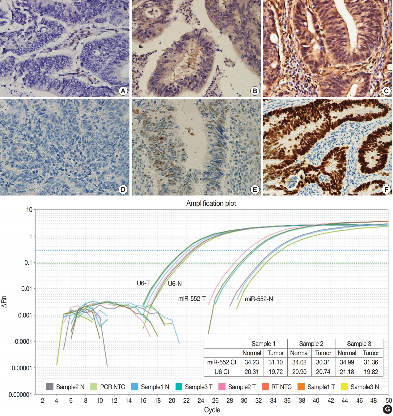
MicroRNA-552 (miR-552) has been reported to correlate with the development and progression of various cancers, including colorectal cancer (CRC). This study aimed to investigate miR-552 expression in cancer tissue samples compared to normal mucosal tissue and its role as a diagnostic or prognostic marker in CRC patients.
Methods
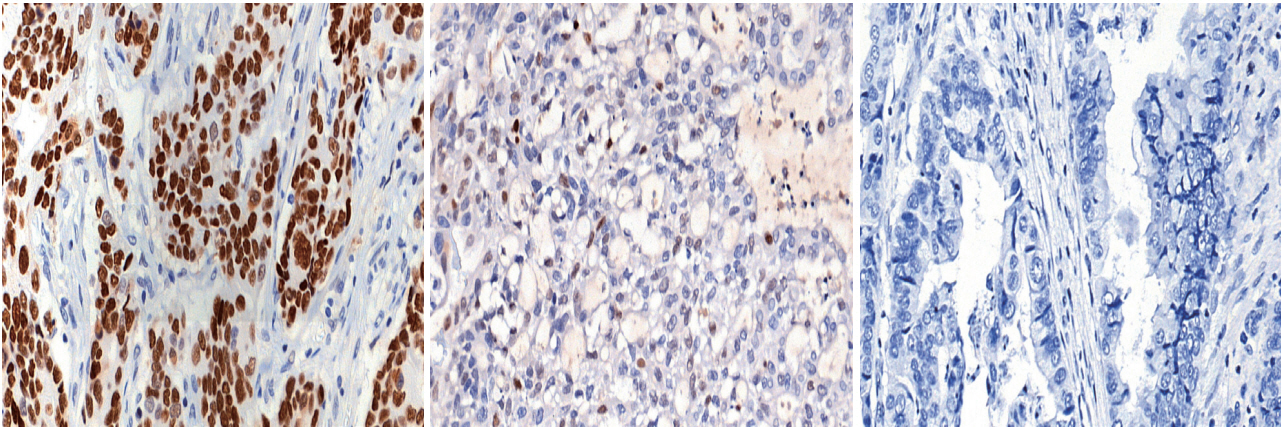
Normal mucosal tissues and primary cancer tissues from 80 surgically resected CRC specimens were used. Quantitative real-time polymerase chain reaction was performed for miR-552 and U6 small nuclear RNA to analyze miR-552 expression and its clinicopathological significance. Immunohistochemistry for p53 and phosphatase and tension homolog (PTEN) was performed to evaluate their association with miR-552 expression.
Results
miR-552 expression was significantly higher in primary cancer tissues compared to normal mucosal tissues (p<.001). The expression level of miR552 was inversely correlated with that of PTEN (p=.068) and p53 (p=.004). Survival analysis showed that high miR-552 expression was associated with worse prognosis but this was not statistically significant (p=.255). However, patients with CRC having high miR-552 expression and loss of PTEN expression had significantly worse prognosis than others (p=.029).
Conclusions
Our results suggest that high miR-552 expression might be a potential diagnostic biomarker for CRC, and its combined analysis with PTEN expression can possibly be used as a prognostic marker. -
Citations
Citations to this article as recorded by
- Tumor microenvironment-driven microRNA dysregulation: Key interactions in colorectal cancer progression
Adriana G Quiroz-Reyes, Paulina Delgado-Gonzalez, Jose Francisco Islas, Veronica L Loaiza-Gutierrez, Michelle G Santoyo-Suarez, Juan A Garcia-Loredo, Carlos A Gonzalez-Villarreal, Fernanda Ramirez-Fernandez, Elsa N Garza-Treviño
World Journal of Gastrointestinal Oncology.2026;[Epub] CrossRef - MicroRNAs involved in colorectal cancer, a rapid mini-systematic review
Sogol Shirzad, Majid Eterafi, Zeinab Karimi, Mahdi Barazesh
BMC Cancer.2025;[Epub] CrossRef - Diagnostic and Therapeutic Potential of Selected microRNAs in Colorectal Cancer: A Literature Review
Grzegorz Sychowski, Hanna Romanowicz, Wojciech Ciesielski, Piotr Hogendorf, Adam Durczyński, Beata Smolarz
Cancers.2025; 17(13): 2135. CrossRef - Blood miRNAs miR-549a, miR-552, and miR-592 serve as potential disease-specific panels to diagnose colorectal cancer
Soroush Akbar, Samaneh Mashreghi, Mohammad Reza Kalani, Akram Valanik, Farzaneh Ahmadi, Mahdi Aalikhani, Zahra Bazi
Heliyon.2024; 10(7): e28492. CrossRef - Integration of TE Induces Cancer Specific Alternative Splicing Events
Woo Ryung Kim, Eun Gyung Park, Yun Ju Lee, Woo Hyeon Bae, Du Hyeong Lee, Heui-Soo Kim
International Journal of Molecular Sciences.2022; 23(18): 10918. CrossRef
- Tumor microenvironment-driven microRNA dysregulation: Key interactions in colorectal cancer progression
- Prediction of TP53 mutations by p53 immunohistochemistry and their prognostic significance in gastric cancer
- Hye Jung Hwang, Soo Kyung Nam, Hyunjin Park, Yujun Park, Jiwon Koh, Hee Young Na, Yoonjin Kwak, Woo Ho Kim, Hye Seung Lee
- J Pathol Transl Med. 2020;54(5):378-386. Published online July 1, 2020
- DOI: https://doi.org/10.4132/jptm.2020.06.01
- 14,114 View
- 290 Download
- 48 Web of Science
- 41 Crossref
-
Abstract
PDFSupplementary Material
- Background
Recently, molecular classifications of gastric cancer (GC) have been proposed that include TP53 mutations and their functional activity. We aimed to demonstrate the correlation between p53 immunohistochemistry (IHC) and TP53 mutations as well as their clinicopathological significance in GC.
Methods
Deep targeted sequencing was performed using surgical or biopsy specimens from 120 patients with GC. IHC for p53 was performed and interpreted as strong, weak, or negative expression. In 18 cases (15.0%) with discrepant TP53 mutation and p53 IHC results, p53 IHC was repeated.
Results
Strong expression of p53 was associated with TP53 missense mutations, negative expression with other types of mutations, and weak expression with wild-type TP53 (p<.001). The sensitivity for each category was 90.9%, 79.0%, and 80.9%, and the specificity was 95.4%, 88.1%, and 92.3%, respectively. The TNM stage at initial diagnosis exhibited a significant correlation with both TP53 mutation type (p=.004) and p53 expression status (p=.029). The Kaplan-Meier survival analysis for 109 stage II and III GC cases showed that patients with TP53 missense mutations had worse overall survival than those in the wild-type and other mutation groups (p=.028). Strong expression of p53 was also associated with worse overall survival in comparison to negative and weak expression (p=.035).
Conclusions
Results of IHC of the p53 protein may be used as a simple surrogate marker of TP53 mutations. However, negative expression of p53 and other types of mutations of TP53 should be carefully interpreted because of its lower sensitivity and different prognostic implications. -
Citations
Citations to this article as recorded by- The future is now: advancing p53 immunohistochemistry in Barrett's oesophagus and its implication for the everyday pathologist
Yevgen Chornenkyy, Monika Vyas, Vikram Deshpande
Histopathology.2026; 88(2): 380. CrossRef - Advancement in preclinical development of cancer treatment agents through modulation of Rac1: From EHop-016 to natural products
Yingyi Liu, Sze-Nga Wong, Aiping Lyu, Joshua Ka-Shun Ko
Biochimica et Biophysica Acta (BBA) - Reviews on Cancer.2026; 1881(1): 189522. CrossRef - Tumor-Associated Macrophage Infiltration and PD-L1 Expression in Gastric Cancer According to a Modified TCGA-Based Classification
Boram Song, Dong-Hoe Koo, Eo Jin Kim, In-Gu Do, Jinah Chu, Kyungeun Kim, Hyebin Lee, Min-Jung Kwon, Jung Ho Park, Byung Ho Son, Chang Hak Yoo, Seoung Wan Chae
Journal of Gastric Cancer.2026; 26(2): 247. CrossRef - Tumor microenvironment dynamics in gastric cancer pathogenesis and therapeutic resistance
Zhenhua Lu, Qinnan Zhang, Jing Han, Jiafu Ji, Xiaofang Xing
Molecular Cancer.2026;[Epub] CrossRef - Captivating Synergistic, Dose-Dependent Anticancer Effects of Tumor-Regulation Modulators Chloroquine and Ivermectin Completely Abolished by an Opposing Modulator, Deoxycholic Acid, in Hamster Fibrosarcoma: In Vivo, In Vitro, and Literature Review
Kosta J. Popović, Dušica J. Popović, Dejan Miljković, Jovan K. Popović, Mihalj Poša, Jovana Drljača Lero, Zana Dolićanin
Pharmaceuticals.2026; 19(3): 407. CrossRef - Comparative study of clinical features, pathology, and immunophenotype between HIV-related cutaneous and visceral Kaposi’s sarcoma
Jingjing Xu, Jing Zhou, Xiaoli Huang, Minmin Wu, Wenjuan Guo, Keyu Liu, Yuexiang Yang, Guangling Yang, Shu Song
BMC Cancer.2026;[Epub] CrossRef -
TP53 mutation landscape and patient survival in oral squamous cell carcinoma
Mebae Ichikawa, Yusuke Kondo, Joaquim Carreras, Masashi Sasaki, Shunsuke Nagase, Yasutaka Hoshimoto, Masashi Tamura, Masahiro Uchibori, Takayuki Aoki, Naoya Nakamura, Yohei Masugi, Yoshihide Ota
Japanese Journal of Clinical Oncology.2026;[Epub] CrossRef - Linking p53 immunostaining to TP53 mutation status in patients with non-small cell lung cancer
Taeyeong Kim, Suyeon Kim, Sangjin Lee, Soohyun Hwang, Joungho Han, Hoyeon Jeong, Yoon-La Choi
Pathology.2025; 57(7): 881. CrossRef - Correlation of TP53 Genetic Alterations with p53 Immunohistochemical Expression and Their Prognostic Significance in DLBCL
Chen Chen, Zijuan Hu, Min Ren, Longlong Bao, Ran Wei, Tian Tian, Xiaoli Zhu, Qianming Bai, Baohua Yu, Xiaoqiu Li, Xiaoyan Zhou
Current Oncology.2025; 32(9): 488. CrossRef - Immunophenotypic Panel for Comprehensive Characterization of Aggressive Thyroid Carcinomas
Mihail Ceausu, Mihai Alin Publik, Dana Terzea, Carmen Adina Cristea, Dumitru Ioachim, Dana Manda, Sorina Schipor
Cells.2025; 14(19): 1554. CrossRef - Multiple approaches revealed MGc80‐3 as a somatic hybrid with HeLa cells rather than a gastric cancer cell line
Fang Cao, Hao Sun, Zhenli Yang, Yanhua Bai, Xiao Hu, Yuhong Hou, Xiaocui Bian, Yuqin Liu
International Journal of Cancer.2024; 154(1): 155. CrossRef - In Response to p53 Immunohistochemical Staining and TP53 Gene Mutations in Endometrial Cancer: Does Null Pattern Correlate With Prognosis?
Ikuko Sakamoto, Keiko Kagami, Takahiro Nozaki, Yosuke Hirotsu, Kenji Amemiya, Toshio Oyama, Masao Omata
American Journal of Surgical Pathology.2024; 48(3): 374. CrossRef - CHEK2 germline variants identified in familial nonmedullary thyroid cancer lead to impaired protein structure and function
Carolina Pires, Inês J. Marques, Mariana Valério, Ana Saramago, Paulo E. Santo, Sandra Santos, Margarida Silva, Margarida M. Moura, João Matos, Teresa Pereira, Rafael Cabrera, Diana Lousa, Valeriano Leite, Tiago M. Bandeiras, João B. Vicente, Branca M. Ca
Journal of Biological Chemistry.2024; 300(3): 105767. CrossRef - The spectrum of TP53 mutations in Rwandan patients with gastric cancer
Augustin Nzitakera, Jean Bosco Surwumwe, Ella Larissa Ndoricyimpaye, Schifra Uwamungu, Delphine Uwamariya, Felix Manirakiza, Marie Claire Ndayisaba, Gervais Ntakirutimana, Benoit Seminega, Vincent Dusabejambo, Eric Rutaganda, Placide Kamali, François Ngab
Genes and Environment.2024;[Epub] CrossRef - Gastric cancer molecular classification based on immunohistochemistry and in‐situ hybridisation and mortality
Maarit Eskuri, Eva‐Maria Birkman, Joonas H Kauppila
Histopathology.2024; 85(2): 327. CrossRef - Redefining aberrant P53 expression of gastric cancer and its distinct clinical significance among molecular-histologic subtypes
Shih-Chiang Huang, Ian Yi-Feng Chang, Tse-Ching Chen, Hsiao-Ching Lin, Chun-Yi Tsai, Jun-Te Hsu, Chun-Nan Yeh, Shih-Cheng Chang, Ta-Sen Yeh
Asian Journal of Surgery.2024; 47(11): 4699. CrossRef - Assessment of TP53 and CDKN2A status as predictive markers of malignant transformation of sinonasal inverted papilloma
Soohyeon Kwon, Jeong-Whun Kim, Eun Sun Kim, Jin Ho Paik, Jin-Haeng Chung, Sung-Woo Cho, Tae-Bin Won, Chae-Seo Rhee, Jee Hye Wee, Hyojin Kim
Scientific Reports.2024;[Epub] CrossRef - Implementing an integrated molecular classification for gastric cancer from endoscopic biopsies using on-slide tests
Simona Costache, Adelina Baltan , Sofia Diaz McLinn , Mattia Pegoraro , Rebecca de Havilland , Matthew Porter , Ana Lerga , Teresa Thomas , Alina Elena Chefani
Romanian Journal of Morphology and Embryology.2024; 65(2): 257. CrossRef - Application of NGS molecular classification in the diagnosis of endometrial carcinoma: A supplement to traditional pathological diagnosis
Qunxian Rao, Jianwei Liao, Yangyang Li, Xin Zhang, Guocai Xu, Changbin Zhu, Shengya Tian, Qiuhong Chen, Hui Zhou, Bingzhong Zhang
Cancer Medicine.2023; 12(5): 5409. CrossRef - Predictive value of p53 and AXL immunostaining for the efficacy of immune checkpoint inhibitor-based therapy after osimertinib treatment in patients with epidermal growth factor-mutant non-small cell lung cancer
Kenji Morimoto, Tadaaki Yamada, Ryo Sawada, Koichi Azuma, Yasuhiro Goto, Taishi Harada, Shinsuke Shiotsu, Nobuyo Tamiya, Yusuke Chihara, Takayuki Takeda, Osamu Hiranuma, Isao Hasegawa, Satomi Tanaka, Akihiro Yoshimura, Masahiro Iwasaku, Shinsaku Tokuda, Y
Cancer Immunology, Immunotherapy.2023; 72(6): 1699. CrossRef - Validation of p53 Immunohistochemistry (PAb240 Clone) in Canine Tumors with Next-Generation Sequencing (NGS) Analysis
Barbara Brunetti, Dario de Biase, Giulia Dellapina, Luisa Vera Muscatello, Francesco Ingravalle, Giorgia Tura, Barbara Bacci
Animals.2023; 13(5): 899. CrossRef - Mesonephric‐like adenocarcinoma of the female genital tract: novel observations and detailed molecular characterisation of mixed tumours and mesonephric‐like carcinosarcomas
Jelena Mirkovic, Ekaterina Olkhov‐Mitsel, Yutaka Amemiya, Maysa Al‐Hussaini, Sharon Nofech‐Mozes, Bojana Djordjevic, Rachel Kupets, Arun Seth, W Glenn McCluggage
Histopathology.2023; 82(7): 978. CrossRef - Clinicopathologic characterization of cervical metastasis from an unknown primary tumor: a multicenter study in Korea
Miseon Lee, Uiree Jo, Joon Seon Song, Youn Soo Lee, Chang Gok Woo, Dong-Hoon Kim, Jung Yeon Kim, Sun Och Yoon, Kyung-Ja Cho
Journal of Pathology and Translational Medicine.2023; 57(3): 166. CrossRef - P53 in Penile Squamous Cell Carcinoma: A Pattern-Based Immunohistochemical Framework with Molecular Correlation
Isabel Trias, Adela Saco, Lorena Marimon, Ricardo López del Campo, Carolina Manzotti, Oriol Ordi, Marta del Pino, Francisco M. Pérez, Naiara Vega, Silvia Alós, Antonio Martínez, Leonardo Rodriguez-Carunchio, Oscar Reig, Pedro Jares, Cristina Teixido, Tare
Cancers.2023; 15(10): 2719. CrossRef - p53/TP53 Status Assessment in Gastroesophageal Adenocarcinoma
Elisa Boldrin, Maria Assunta Piano, Francesco Bernaudo, Rita Alfieri, Maria Raffaella Biasin, Isabella Monia Montagner, Alice Volpato, Genny Mattara, Francesco Lamacchia, Giovanna Magni, Antonio Rosato, Antonio Scapinello, Pierluigi Pilati, Matteo Curtare
Cancers.2023; 15(10): 2783. CrossRef - Genomic profiling of dedifferentiated endometrial carcinomas arising in the background of high‐grade carcinoma: a targeted next‐generation sequencing study
Ekaterina Olkhov‐Mitsel, Aurelia Busca, Carlos Parra‐Herran, Yutaka Amemiya, Sharon Nofech‐Mozes, Bojana Djordjevic, Marisa R Nucci, Arun Seth, Jelena Mirkovic
Histopathology.2023; 83(3): 366. CrossRef -
Clinicopathologic Features and Prognostic Significance of Immunohistochemistry and In Situ Hybridization Based Molecular Classification in Gastric Carcinoma
Gizem Issin, İlyas Sayar, Fatih Demir, İrem Güvendir Bakkaloğlu, Mehmet Gamsizkan, Zeliha Yildiz, Ismail Yilmaz, Sevilay Akalp Özmen, Diren Vuslat Çağatay, Itır Ebru Zemheri, Murat Demiriz, Armağan Günal
Journal of Environmental Pathology, Toxicology and Oncology.2023; 42(4): 1. CrossRef - Clinicopathologic and Molecular Characterization of Anorectal Neuroendocrine Carcinomas Reveals Human Papillomavirus, p53, and c-Myc as Alternative Mechanisms of Carcinogenesis
Allison J. Cox, William E. Crowe, Qi Yang, Bin Zhang, Zoltán N. Oltvai, Xiaoyan Liao
Modern Pathology.2023; 36(11): 100295. CrossRef - Dedifferentiated Endometrial Carcinoma: A Rare Aggressive Neoplasm-Clinical, Morphological and Immunohistochemical Features
Giovanna Giordano, Elena Ferioli, Debora Guareschi, Alessandro Tafuni
Cancers.2023; 15(21): 5155. CrossRef - Characterization on the oncogenic effect of the missense mutations of p53 via machine learning
Qisheng Pan, Stephanie Portelli, Thanh Binh Nguyen, David B Ascher
Briefings in Bioinformatics.2023;[Epub] CrossRef - Adrenal Nodules Detected at Staging CT in Patients with Resectable Gastric Cancers Have a Low Incidence of Malignancy
Hae Young Kim, Won Chang, Yoon Jin Lee, Ji Hoon Park, Jungheum Cho, Hee Young Na, Hyungwoo Ahn, Sung Il Hwang, Hak Jong Lee, Young Hoon Kim, Kyoung Ho Lee
Radiology.2022; 302(1): 129. CrossRef - Intestinal-type gastric dysplasia in Helicobacter pylori-naïve patients
Kotaro Shibagaki, Ayako Itawaki, Yoichi Miyaoka, Kenichi Kishimoto, Yusuke Takahashi, Satoshi Kotani, Tsuyoshi Mishiro, Naoki Oshima, Kousaku Kawashima, Norihisa Ishimura, Hideyuki Onuma, Makoto Nagasaki, Mamiko Nagase, Asuka Araki, Kyuichi Kadota, Ryoji
Virchows Archiv.2022; 480(4): 783. CrossRef - Dedifferentiation-like tubular and solid carcinoma of the stomach shows phenotypic divergence and association with deficient SWI/SNF complex
Shih-Chiang Huang, Kuang-Hua Chen, Kwai-Fong Ng, I-Chieh Lin, Yi-Chun Chao, Ta-Sen Yeh, Huei-Chieh Chuang, Tse-Ching Chen
Virchows Archiv.2022; 480(4): 771. CrossRef - Distinct molecular phenotype and the potential prognostic value of immune prognostic index and tumor infiltrating lymphocytes in hepatoid adenocarcinoma of stomach
Muxing Kang, Xiaojing Ma, Jifei Shi, Guofeng Chen, Xiaoli Jin, Jun Wang, Lele Lin, Zhiwei Wu, Kaibo Chen, Jinghong Xu, Pintong Huang, Jian Chen
Translational Oncology.2022; 19: 101380. CrossRef - Evaluation of Tumor DNA Sequencing Results in Patients with Gastric and Gastroesophageal Junction Adenocarcinoma Stratified by TP53 Mutation Status
Anthony C Wood, Yonghong Zhang, Qianxing Mo, Ling Cen, Jacques Fontaine, Sarah E Hoffe, Jessica Frakes, Sean P Dineen, Jose M Pimiento, Christine M Walko, Rutika Mehta
The Oncologist.2022; 27(4): 307. CrossRef - Comprehensive Clinical Analysis of Gallbladder Neuroendocrine Neoplasms: A Large-Volume Multicenter Study During One Decade
Yangyang Wang, Bingfeng Huang, Qihan Fu, Jianing Wang, Mao Ye, Manyi Hu, Kai Qu, Kai Liu, Xiao Hu, Shumei Wei, Ke Sun, Wenbo Xiao, Bo Zhang, Haijun Li, Jingsong Li, Qi Zhang, Tingbo Liang
Annals of Surgical Oncology.2022; 29(12): 7619. CrossRef - Expression of SASP, DNA Damage Response, and Cell Proliferation Factors in Early Gastric Neoplastic Lesions: Correlations and Clinical Significance
Li Liang, Yijie Chai, Fei Chai, Haijing Liu, Ningning Ma, Hong Zhang, Shuang Zhang, Lin Nong, Ting Li, Bo Zhang
Pathology and Oncology Research.2022;[Epub] CrossRef - Systems biology and OMIC data integration to understand gastrointestinal cancers
Iasmin Moreira Costa Bispo, Henry Paul Granger, Palloma Porto Almeida, Patricia Belini Nishiyama, Leandro Martins de Freitas
World Journal of Clinical Oncology.2022; 13(10): 762. CrossRef - MicroRNA-552 expression in colorectal cancer and its clinicopathological significance
Joon Im, Soo Kyung Nam, Hye Seung Lee
Journal of Pathology and Translational Medicine.2021; 55(2): 125. CrossRef - Different effects of p53 protein overexpression on the survival of gastric cancer patients according to Lauren histologic classification: a retrospective study
Ki Wook Kim, Nayoung Kim, Yonghoon Choi, Won Seok Kim, Hyuk Yoon, Cheol Min Shin, Young Soo Park, Dong Ho Lee, Young Suk Park, Sang-Hoon Ahn, Do Joong Park, Hyung-Ho Kim, Hye Seung Lee, Ji-Won Kim, Jin Won Kim, Keun-Wook Lee, Won Chang, Ji Hoon Park, Yoon
Gastric Cancer.2021; 24(4): 844. CrossRef - The association between the expression of nuclear Yes-associated protein 1 (YAP1) and p53 protein expression profile in breast cancer patients
Yoon Jin Cha, Dooreh Kim, Soong June Bae, Sung Gwe Ahn, Joon Jeong, Min Kyung Cho, Pill Sun Paik, Tae-Kyung Yoo, Woo-Chan Park, Chang Ik Yoon, Elda Tagliabue
PLOS ONE.2021; 16(5): e0250986. CrossRef
- The future is now: advancing p53 immunohistochemistry in Barrett's oesophagus and its implication for the everyday pathologist
- Molecular and Clinicopathological Features of Gastrointestinal Stromal Tumors in Vietnamese Patients
- Quoc Dat Ngo, Quoc Thang Pham, Dang Anh Thu Phan, Anh Vu Hoang, Thi Ngoc Ha Hua, Sao Trung Nguyen
- J Pathol Transl Med. 2019;53(6):361-368. Published online September 16, 2019
- DOI: https://doi.org/10.4132/jptm.2019.08.27
- 8,913 View
- 160 Download
- 2 Web of Science
- 2 Crossref
-
Abstract
PDFSupplementary Material
- Background
Gastrointestinal stromal tumors (GISTs) are the most frequent mesenchymal neoplasms of the gastrointestinal tract. Management of GIST patients is currently based on clinicopathological features and associated genetic changes. However, the detailed characteristics and molecular genetic features of GISTs have not yet been described in the Vietnamese population.
Methods
We first identified 155 patients with primary GIST who underwent surgery with primary curative intent between 2011 and 2014 at University Medical Center at Ho Chi Minh City, Vietnam. We evaluated the clinicopathological features and immunohistochemical reactivity to p53 and Ki-67 in these patients. Additionally, KIT genotyping was performed in 100 cases.
Results
The largest proportion of GISTs was classified as high-risk (43.2%). Of the 155 GISTs, 52 (33.5%) were positive for Ki-67, and 58 (37.4%) were positive for p53. The expression of Ki-67 and p53 were correlated with mitotic rate, tumor size, risk assessment, and tumor stage. Out of 100 GIST cases, KIT mutation was found in 68%, of which 62 (91.2%) were found in exon 11, two (2.9%) in exon 9, and four (5.8%) in exon 17. No mutation in exon 13 was identified. Additionally, KIT mutations did not correlate with any clinicopathological features.
Conclusions
The expression of Ki-67 and p53 were associated with high-risk tumors. Mutations in exon 11 were the most commonly found, followed by exon 17 and exon 9. Additionally, KIT mutation status was not correlated with any recognized clinicopathological features. -
Citations
Citations to this article as recorded by- Ki67 for evaluating the prognosis of gastrointestinal stromal tumors: A systematic review and meta‑analysis
Ji Li, An-Ran Wang, Xiao-Dong Chen, Hong Pan, Shi-Qiang Li
Oncology Letters.2022;[Epub] CrossRef - Endoscopic ultrasound‐guided fine‐needle aspiration cytology in the diagnosis of the gastrointestinal stromal tumor of the stomach
José‐Fernando Val‐Bernal, Elena Yllera, María Moris, Ihab Abdulkader Nallib, Angel Vázquez‐Boquete, María Martino
Diagnostic Cytopathology.2020; 48(9): 833. CrossRef
- Ki67 for evaluating the prognosis of gastrointestinal stromal tumors: A systematic review and meta‑analysis
- Serous Adenocarcinoma of Fallopian Tubes: Histological and Immunohistochemical Aspects
- Natalia Hyriavenko, Mykola Lyndin, Kateryna Sikora, Artem Piddubnyi, Ludmila Karpenko, Olha Kravtsova, Dmytrii Hyriavenko, Olena Diachenko, Vladyslav Sikora, Anatolii Romaniuk
- J Pathol Transl Med. 2019;53(4):236-243. Published online April 11, 2019
- DOI: https://doi.org/10.4132/jptm.2019.03.21
- 9,270 View
- 133 Download
- 4 Web of Science
- 6 Crossref
-
Abstract
PDF
- Background
Although primary cancer of the fallopian tubes is a relatively rare type of tumor in female reproductive organs, its mortality is quite high. It is important to identify molecular and biological markers of this malignancy that determine its specific phenotype.
Methods
The study was carried out on samples received from 71 female patients with primary cancer of the fallopian tubes. The main molecular and biological properties, including hormone status (estrogen receptor [ER], progesterone receptor [PR]), human epidermal growth factor receptor (HER2)/neu expression, proliferative potential (Ki-67), apoptosis (p53, Bcl-2), and pro-angiogenic (vascular endothelial growth factor) quality of serous tumors were studied in comparison with clinical and morphological characteristics.
Results
ER and PR expression is accompanied by low grade neoplasia, early clinical disease stage, and absence of lymphogenic metastasis (p < .001). HER2/neu expression is not typical for primary cancer of the fallopian tubes. Ki-67 expression is characterized by an inverse correlation with ER and PR (p < .05) and is associated with lymphogenic metastasis (p < .01). p53+ status correlates with high grade malignancy, tumor progression, metastasis, negative ER/PR (p < .001), and negative Bcl-2 status (p < .05). Positive Bcl-2 status is positively correlated with ER and PR expression and low grade malignancy.
Conclusions
Complex morphologic (histological and immunohistochemical) study of postoperative material allows estimation of the degree of malignancy and tumor spread to enable appropriate treatment for each case. -
Citations
Citations to this article as recorded by- Clinical and Morphological Analysis of Odontogenic Tumors and Tooth Developmental Anomalies
Yе.V. Kuzenko, S.M. Hermanchuk, O.O. Mykhno, D.H. Tsepochko, O.V. Kuzenko, A.Yu. Olishkevych
Kharkiv Dental Journal.2025; : 275. CrossRef - Rare non-serous fallopian tube cancers: institutional experience and literature review
Dmitrii Sumtsov, Georgyi Sumtsov, Nataliia Hyriavenko, Mykola Lyndin, Kateryna Sikora, Nataliia Kalashnik, Svitlana Smiian, Igor Gladchuk
Wiener Medizinische Wochenschrift.2024; 174(9-10): 199. CrossRef - UŞAQLIQ BORULARININ BİRİNCİLİ XƏRÇƏNGİ: DİAQNOSTİKASI VƏ MÜALİCƏSİNİN NƏTİCƏLƏRİ
D.G. Sumtsov, G.O. Sumtsov, N.I. Hyriavenko, S.A. Smiian, N.V. Kalashnyk, K.O. Sikora, N.M. Rozhkovska, I.Z. Gladchuk
Azerbaijan Medical Journal.2023; (4): 75. CrossRef - FEATURES OF ENDOMETRIUM STRUCTURE IN ALCOHOL-ABUSING HIV-INFECTED INDIVIDUALS
M. Lytvynenko
Inter Collegas.2021; 8(1): 52. CrossRef - Concurrent Clostridial Enteritis and Oviductal Adenocarcinoma with Carcinomatosis in an Adult Alpaca (Vicugna pacos)
Mandy Womble, Megan E. Schreeg, Allison Hoch, Enoch B. de Souza Meira, Derek Foster, Christopher Premanandan, Tatiane T. Negrão Watanabe
Journal of Comparative Pathology.2021; 189: 52. CrossRef - Problems of primary fallopian tube cancer diagnostics during and after surgery
D.G. Sumtsov, I.Z. Gladchuk, G.O. Sumtsov, N.I. Hyriavenko, M.S. Lyndin, V.V. Sikora, V.M. Zaporozhan
REPRODUCTIVE ENDOCRINOLOGY.2021; (59): 66. CrossRef
- Clinical and Morphological Analysis of Odontogenic Tumors and Tooth Developmental Anomalies
- Association between Expression of 8-OHdG and Cigarette Smoking in Non-small Cell Lung Cancer
- Ae Ri An, Kyoung Min Kim, Ho Sung Park, Kyu Yun Jang, Woo Sung Moon, Myoung Jae Kang, Yong Chul Lee, Jong Hun Kim, Han Jung Chae, Myoung Ja Chung
- J Pathol Transl Med. 2019;53(4):217-224. Published online March 11, 2019
- DOI: https://doi.org/10.4132/jptm.2019.02.20
- 10,460 View
- 251 Download
- 24 Web of Science
- 24 Crossref
-
Abstract
PDF
- Background
Exposure to cigarette smoking (CS) is a major risk factor for the development of lung cancer. CS is known to cause oxidative DNA damage and mutation of tumor-related genes, and these factors are involved in carcinogenesis. 8-Hydroxydeoxyguanosine (8-OHdG) is considered to be a reliable biomarker for oxidative DNA damage. Increased levels of 8-OHdG are associated with a number of pathological conditions, including cancer. There are no reports on the expression of 8-OHdG by immunohistochemistry in non-small cell lung cancer (NSCLC).
Methods
We investigated the expression of 8-OHdG and p53 in 203 NSCLC tissues using immunohistochemistry and correlated it with clinicopathological features including smoking.
Results
The expression of 8-OHdG was observed in 83.3% of NSCLC. It was significantly correlated with a low T category, negative lymph node status, never-smoker, and longer overall survival (p < .05) by univariate analysis. But multivariate analysis revealed that 8-OHdG was not an independent prognostic factor for overall survival in NSCLC patients. The aberrant expression of p53 significantly correlated with smoking, male, squamous cell carcinoma, and Ki-67 positivity (p < .05).
Conclusions
The expression of 8-OHdG was associated with good prognostic factors. It was positively correlated with never-smokers in NSCLC, suggesting that oxidative damage of DNA cannot be explained by smoking alone and may depend on complex control mechanisms. -
Citations
Citations to this article as recorded by- N-acetyl-cysteine alleviates nandrolone decanoate-induced hippocampal cell apoptosis in rats via reversing protein expressions of S1P1, Akt and FOXO3a signaling pathway
Alireza Shirpoor, Zahra Zarrini, Roya Naderi
Steroids.2026; 228: 109759. CrossRef - The distinct roles of ROS in tumor immunity: from mechanisms to immunotherapeutic applications
Jiayi Li, Chen Huang, Pan Tang, Ruiyan Wu, Quanyou Wu, Chenliang Zhang
Journal of Hematology & Oncology.2026;[Epub] CrossRef - Sustainable framework for automated segmentation and prediction of lung cancer in CT image using CapsNet with U-net segmentation
S.R. Vijayakumar, S. Aarthy, D. Deepa, P. Suresh
Biomedical Signal Processing and Control.2025; 99: 106873. CrossRef - Endolysosomal cation channel MCOLN as the novel regulator of redox homeostasis
Yahao Gao, Lei Xu, Ying Chen
Biochimica et Biophysica Acta (BBA) - Molecular Basis of Disease.2025; 1871(7): 167910. CrossRef - Catalase: The golden key to regulate oxidative stress in breast cancer
Jia-Wei Liu, Wen-Jia Chen, Yang-Zheng Lan, Jing Liu
World Journal of Clinical Oncology.2025;[Epub] CrossRef - Association of sirtuin 1 rs10997868 and rs730821 polymorphisms with sirtuin 1 and hydroxy-2′-deoxyguanosine levels in healthy smokers: A case–control study
Samar Sultan
Journal of International Medical Research.2025;[Epub] CrossRef - Increased pretreatment triglyceride glucose-body mass index associated with poor prognosis in patients with advanced non-small cell lung cancer
Shaoming Guo, Yi Zhao, Yue Jiang, Huaping Ye, Ying Wang
Clinical Nutrition ESPEN.2024; 59: 412. CrossRef - Oxidative Damage and Telomere Length as Markers of Lung Cancer Development among Chronic Obstructive Pulmonary Disease (COPD) Smokers
Elizabeth Córdoba-Lanús, Luis M. Montuenga, Angélica Domínguez-de-Barros, Alexis Oliva, Delia Mayato, Ana Remírez-Sanz, Francisca Gonzalvo, Bartolomé Celli, Javier J. Zulueta, Ciro Casanova
Antioxidants.2024; 13(2): 156. CrossRef - Automated determination of 8-OHdG in cells and tissue via immunofluorescence using a specially created antibody
Tobias Jung, Nicole Findik, Bianca Hartmann, Katja Hanack, Kai Grossmann, Dirk Roggenbuck, Marc Wegmann, René Mantke, Markus Deckert, Tilman Grune
Biotechnology Reports.2024; 42: e00833. CrossRef - Combination treatment of zinc and selenium intervention ameliorated BPA-exposed germ cell damage in SD rats: elucidation of molecular mechanisms
Chittaranjan Sahu, Gopabandhu Jena
Naunyn-Schmiedeberg's Archives of Pharmacology.2024; 397(9): 6685. CrossRef - Interplay of arsenic exposure and cigarette smoking on oxidative DNA damage in healthy males
Sepideh Nemati-Mansour, Mohammad Mosaferi, Javad Babaie, Asghar Mohammadpoorasl, Reza Dehghanzadeh, Leila Nikniaz, Mohammad Miri
Environmental Sciences Europe.2024;[Epub] CrossRef - The role of tissue persistent organic pollutants and genetic polymorphisms in patients with benign and malignant kidney tumors
Rasih Kocagöz, İlgen Onat, Merve Demirbügen Öz, Burak Turna, Banu Sarsık Kumbaracı, Mehmet Nurullah Orman, Halit Sinan Süzen, Hilmi Orhan
Environmental Toxicology and Pharmacology.2024; 110: 104495. CrossRef - Mitochondrial Plasticity and Glucose Metabolic Alterations in Human Cancer under Oxidative Stress—From Viewpoints of Chronic Inflammation and Neutrophil Extracellular Traps (NETs)
Hui-Ting Lee, Chen-Sung Lin, Chao-Yu Liu, Po Chen, Chang-Youh Tsai, Yau-Huei Wei
International Journal of Molecular Sciences.2024; 25(17): 9458. CrossRef - Oxidative DNA Damage and Arterial Hypertension in Light of Current ESC Guidelines
Radka Hazuková, Zdeněk Zadák, Miloslav Pleskot, Petr Zdráhal, Martin Pumprla, Miloš Táborský
International Journal of Molecular Sciences.2024; 25(23): 12557. CrossRef - Significance of 8-OHdG Expression as a Predictor of Survival in Colorectal Cancer
Myunghee Kang, Soyeon Jeong, Sungjin Park, Seungyoon Nam, Jun-Won Chung, Kyoung Oh Kim, Jungsuk An, Jung Ho Kim
Cancers.2023; 15(18): 4613. CrossRef - Serum 8-Hydroxy-2′-deoxyguanosine Predicts Severity and Prognosis of Patients with Acute Exacerbation of Chronic Obstructive Pulmonary Disease
Peng Cao, Chen Zhang, Dong-Xu Hua, Meng-Die Li, Bian-Bian Lv, Lin Fu, Hui Zhao
Lung.2022; 200(1): 31. CrossRef - Redox signaling at the crossroads of human health and disease
Jing Zuo, Zhe Zhang, Maochao Luo, Li Zhou, Edouard C. Nice, Wei Zhang, Chuang Wang, Canhua Huang
MedComm.2022;[Epub] CrossRef - Assessment of MDA and 8-OHdG expressions in ovine pulmonary adenocarcinomas by immunohistochemical and immunofluorescence methods
Emin Karakurt, Enver Beytut, Serpil Dağ, Hilmi Nuhoğlu, Ayfer Yıldız, Emre Kurtbaş
Acta Veterinaria Brno.2022; 91(3): 235. CrossRef - Dietary Antioxidants and Lung Cancer Risk in Smokers and Non-Smokers
Naser A. Alsharairi
Healthcare.2022; 10(12): 2501. CrossRef - Targeting oxidative stress in disease: promise and limitations of antioxidant therapy
Henry Jay Forman, Hongqiao Zhang
Nature Reviews Drug Discovery.2021; 20(9): 689. CrossRef - Association between tobacco substance usage and a missense mutation in the tumor suppressor gene P53 in the Saudi Arabian population
Mikhlid H. Almutairi, Bader O. Almutairi, Turki M. Alrubie, Sultan N. Alharbi, Narasimha R. Parine, Abdulwahed F. Alrefaei, Ibrahim Aldeailej, Abdullah Alamri, Abdelhabib Semlali, Alvaro Galli
PLOS ONE.2021; 16(1): e0245133. CrossRef - Measurement of uranium concentrations in urine samples of adult healthy groups in Najaf governorate with estimation of urine concentrations of 8-OHdG compound as biomarker for DNA damage
Samia K. Abbas, Dhuha S. Saleh, Hayder S. Hussain
Journal of Physics: Conference Series.2021; 1879(3): 032097. CrossRef - Common Data Model and Database System Development for the Korea Biobank Network
Soo-Jeong Ko, Wona Choi, Ki-Hoon Kim, Seo-Joon Lee, Haesook Min, Seol-Whan Oh, In Young Choi
Applied Sciences.2021; 11(24): 11825. CrossRef - EVALUATION OF OXIDATIVE STATUS IN PATIENTS WITH CHRONIC PERIODONTITIS AND ADDITIONAL TOBACCO ABUSE: A CROSS-SECTIONAL STUDY
Didem ÖZKAL EMİNOĞLU, Varol ÇANAKÇI
Atatürk Üniversitesi Diş Hekimliği Fakültesi Dergisi.2020; : 1. CrossRef
- N-acetyl-cysteine alleviates nandrolone decanoate-induced hippocampal cell apoptosis in rats via reversing protein expressions of S1P1, Akt and FOXO3a signaling pathway
- Association between p53 Expression and Amount of Tumor-Infiltrating Lymphocytes in Triple-Negative Breast Cancer
- Miseon Lee, In Ah Park, Sun-Hee Heo, Young-Ae Kim, Gyungyub Gong, Hee Jin Lee
- J Pathol Transl Med. 2019;53(3):180-187. Published online March 11, 2019
- DOI: https://doi.org/10.4132/jptm.2019.02.08
- 10,511 View
- 207 Download
- 20 Web of Science
- 20 Crossref
-
Abstract
PDF
- Background
Most triple-negative breast cancers (TNBCs) have a high histologic grade, are associated with high endoplasmic stress, and possess a high frequency of TP53 mutations. TP53 missense mutations lead to the production of mutant p53 protein and usually show high levels of p53 protein expression. Tumor-infiltrating lymphocytes (TILs) accumulate as part of the anti-tumor immune response and have a strong prognostic and predictive significance in TNBC. We aimed to elucidate the association between p53 expression and the amount of TILs in TNBC.
Methods
In 678 TNBC patients, we evaluated TIL levels and expression of endoplasmic stress molecules. Immunohistochemical examination of p53 protein expression was categorized into three groups: no, low, and high expression.
Results
No, low, and high p53 expression was identified in 44.1% (n = 299), 20.1% (n = 136), and 35.8% (n = 243) of patients, respectively. Patients with high p53 expression showed high histologic grade (p < .001), high TIL levels (p = .009), and high expression of endoplasmic reticulum stress-associated molecules (p-eIF2a, p = .013; XBP1, p = .007), compared to patients with low p53 expression. There was no significant difference in disease-free (p = .406) or overall survival rates (p = .444) among the three p53 expression groups.
Conclusions
High p53 expression is associated with increased expression of endoplasmic reticulum stress molecules and TIL influx. -
Citations
Citations to this article as recorded by- A comparative analysis of mutational profiles between triple-negative breast cancer and non-triple-negative breast cancer
Wanlin Li, Chenchen Feng, Shunheng Zhou
Discover Oncology.2026;[Epub] CrossRef - The search for a TNBC vaccine: the guardian vaccine
Cory Fines, Helen McCarthy, Niamh Buckley
Cancer Biology & Therapy.2025;[Epub] CrossRef - Correlating p53 immunostaining patterns with somatic TP53 mutation and functional properties of mutant p53 in triple‐negative breast cancer
Meejeong Kim, Miseon Lee, Ahwon Lee, Byung‐Ock Choi, Woo‐Chan Park, Sung Hun Kim, Jieun Lee, Jun Kang
Histopathology.2025; 87(2): 299. CrossRef - GD3 synthase drives resistance to p53-induced apoptosis in breast cancer by modulating mitochondrial function
Vivek Anand, Fouad El-Dana, Natalia Baran, Jenny Borgman, Zheng Yin, Hong Zhao, Stephen T. Wong, Michael Andreeff, V. Lokesh Battula
Oncogene.2025; 44(30): 2646. CrossRef - Topoisomerase Inhibitor Resistance in Breast Cancer: Exploring Alterations in Cellular Metabolism and Properties Driving Drug Insensitivity
Juhong Lee, Youngjoo Kwon
Drug Targets and Therapeutics.2025; 4(2): 185. CrossRef - Updated Austrian treatment algorithm for metastatic triple-negative breast cancer
Rupert Bartsch, Gabriel Rinnerthaler, Edgar Petru, Daniel Egle, Michael Gnant, Marija Balic, Thamer Sliwa, Christian Singer
Wiener klinische Wochenschrift.2024; 136(11-12): 347. CrossRef - Triple negative breast cancer: Immunogenicity, tumor microenvironment, and immunotherapy
Sotiris Loizides, Anastasia Constantinidou
Frontiers in Genetics.2023;[Epub] CrossRef - Prognostic benefit of TILs independent of clinicopathological and molecular factors
Koen Brummel, Anneke L. Eerkens, Marco de Bruyn, Hans W. Nijman
British Journal of Cancer.2023; 129(5): 737. CrossRef - Dihydroartemisinin-Transferrin Adducts Enhance TRAIL-Induced Apoptosis in Triple-Negative Breast Cancer in a P53-Independent and ROS-Dependent Manner
Xinyu Zhou, Abel Soto-Gamez, Fleur Nijdam, Rita Setroikromo, Wim J. Quax
Frontiers in Oncology.2022;[Epub] CrossRef - New Challenges in the Differential Diagnosis of High-Grade Triple-Negative Breast Cancer and Serous Carcinoma
Andrii Puzyrenko, Chandler S Cortina, Julie M Jorns
International Journal of Surgical Pathology.2022; 30(7): 728. CrossRef - Prognostic analysis of cuproptosis-related gene in triple-negative breast cancer
Shengnan Sha, Luyi Si, Xinrui Wu, Yuanbiao Chen, Hui Xiong, Ying Xu, Wangrui Liu, Haijun Mei, Tao Wang, Mei Li
Frontiers in Immunology.2022;[Epub] CrossRef - p53 Missense Mutation is Associated with Immune Cell PD-L1 Expression in Triple-Negative Breast Cancer
Ai-Yan Xing, Long Liu, Ke Liang, Bin Wang
Cancer Investigation.2022; 40(10): 879. CrossRef - Crosstalk between Immune Checkpoint Modulators, Metabolic Reprogramming and Cellular Plasticity in Triple-Negative Breast Cancer
Arpita Poddar, Sushma R. Rao, Prashanth Prithviraj, George Kannourakis, Aparna Jayachandran
Current Oncology.2022; 29(10): 6847. CrossRef - The tumor microenvironment and triple-negative breast cancer aggressiveness: shedding light on mechanisms and targeting
Natsuki Furukawa, Vered Stearns, Cesar A. Santa-Maria, Aleksander S. Popel
Expert Opinion on Therapeutic Targets.2022; 26(12): 1041. CrossRef - Intracellular partners of fibroblast growth factors 1 and 2 - implications for functions
Katarzyna Dominika Sluzalska, Jakub Slawski, Martyna Sochacka, Agata Lampart, Jacek Otlewski, Malgorzata Zakrzewska
Cytokine & Growth Factor Reviews.2021; 57: 93. CrossRef - Targeted Chinese Medicine Delivery by A New Family of Biodegradable Pseudo-Protein Nanoparticles for Treating Triple-Negative Breast Cancer: In Vitro and In Vivo Study
Hiu Yee Kwan, Qinghua Xu, Ruihong Gong, Zhaoxiang Bian, Chih-Chang Chu
Frontiers in Oncology.2021;[Epub] CrossRef - Transcriptomic Properties of HER2+ Ductal Carcinoma In Situ of the Breast Associate with Absence of Immune Cells
Marie Colombe Agahozo, Marcel Smid, Ronald van Marion, Dora Hammerl, Thierry P. P. van den Bosch, Mieke A. M. Timmermans, Chayenne J. Heijerman, Pieter J. Westenend, Reno Debets, John W. M. Martens, Carolien H. M. van Deurzen
Biology.2021; 10(8): 768. CrossRef - With Our Powers Combined
Lawrence Kasherman, Katherine Karakasis, Amit M. Oza
The Cancer Journal.2021; 27(6): 511. CrossRef The Research Progress on the Prognostic Value of the Common Hematological Parameters in Peripheral Venous Blood in Breast Cancer
Li Chen, Xiangyi Kong, Chengrui Yan, Yi Fang, Jing Wang
OncoTargets and Therapy.2020; Volume 13: 1397. CrossRefBiomolecular Factors Represented by Bcl-2, p53, and Tumor-Infiltrating Lymphocytes Predict Response for Adjuvant Anthracycline Chemotherapy in Patients with Early Triple-Negative Breast Cancer
Xenia Elena Bacinschi, Anca Zgura, Inga Safta, Rodica Anghel
Cancer Management and Research.2020; Volume 12: 11965. CrossRef
- A comparative analysis of mutational profiles between triple-negative breast cancer and non-triple-negative breast cancer
- Uterine Malignant Mixed Müllerian Tumors Following Treatment with Selective Estrogen Receptor Modulators in Patients with Breast Cancer: A Report of 13 Cases and Their Clinicopathologic Characteristics
- Byung-Kwan Jeong, Chang O. Sung, Kyu-Rae Kim
- J Pathol Transl Med. 2019;53(1):31-39. Published online December 18, 2018
- DOI: https://doi.org/10.4132/jptm.2018.11.16
- 9,170 View
- 102 Download
- 3 Web of Science
- 4 Crossref
-
Abstract
PDF
- Background
Breast cancer treatment with selective estrogen receptor modulators (SERMs) increasesthe incidence of uterine malignant mixed Müllerian tumors (uMMMTs). We examine clinicopathologiccharacteristics and prognosis of SERM-associated uMMMTs (S-uMMMTs) and discusspossible pathogenetic mechanisms.
Methods
Among 28,104 patients with breast cancer, clinicopathologicfeatures and incidence of uMMMT were compared between patients who underwentSERM treatment and those who did not. Of 92 uMMMT cases that occurred during the same period,incidence, dose, and duration of SERM treatment, as well as overall survival rate, were comparedfor patients with breast cancer who underwent SERM treatment and those who did not (S-uMMMTvs NS-uMMMT) and for patients without breast cancer (de novo-uMMMT). Histopathologicalfindings and immunophenotypes for myogenin, desmin, p53, WT-1, estrogen receptor (ER) α, ERβ,progesterone receptor, and GATA-3 were compared between S-uMMMT and de novo-uMMMT.
Results
The incidence of S-uMMMT was significantly higher than that of NS-uMMMT (6.35-fold).All patients with SERM were postmenopausal and received daily 20–40 mg SERM. CumulativeSERM dose ranged from 21.9 to 73.0 g (mean, 46.0) over 39–192 months (mean, 107). Clinicopathologicfeatures, such as International Federation of Gynecology and Obstetrics stage andoverall survival, were not significantly different between patients with S-uMMMT and NS-uMMMTor between patients with S-uMMMT and de novo-uMMMT. All 11 S-uMMMT cases available forimmunostaining exhibited strong overexpression/null expression of p53 protein and significantlyincreased ERβ expression in carcinomatous and sarcomatous components.
Conclusions
SERMtherapy seemingly increases risk of S-uMMMT development; however, clinicopathologic featureswere similar in all uMMMTs from different backgrounds. p53 mutation and increased ERβ expressionmight be involved in the etiology of S-uMMMT. -
Citations
Citations to this article as recorded by- Uterine carcinosarcomas: A case series of 9 cases from a low-income country
Boubacar Efared, Halidou Hamadou Koura, Aïchatou Balaraba Abani Bako, Idrissa Boubacar, Habiba Salifou Boureima, Garba Mahamadou, Hassan Nouhou
Medicine.2024; 103(40): e39773. CrossRef - Uterine carcinosarcoma: Unraveling the role of epithelial‐to‐mesenchymal transition in progression and therapeutic potential
Mohan Shankar Gopinatha Pillai, Pallab Shaw, Arpan Dey Bhowmik, Resham Bhattacharya, Geeta Rao, Shailendra Kumar Dhar Dwivedi
The FASEB Journal.2024;[Epub] CrossRef - Tamoxifen/toremifene
Reactions Weekly.2019; 1758(1): 330. CrossRef - Molecular Basis of Tumor Heterogeneity in Endometrial Carcinosarcoma
Susanna Leskela, Belen Pérez-Mies, Juan Manuel Rosa-Rosa, Eva Cristobal, Michele Biscuola, María L. Palacios-Berraquero, SuFey Ong, Xavier Matias-Guiu Guia, José Palacios
Cancers.2019; 11(7): 964. CrossRef
- Uterine carcinosarcomas: A case series of 9 cases from a low-income country
- Basaloid Squamous Cell Carcinoma of the Head and Neck: Subclassification into Basal, Ductal, and Mixed Subtypes Based on Comparison of Clinico-pathologic Features and Expression of p53, Cyclin D1, Epidermal Growth Factor Receptor, p16, and Human Papillomavirus
- Kyung-Ja Cho, Se Un Jeong, Sung Bae Kim, Sang-wook Lee, Seung-Ho Choi, Soon Yuhl Nam, Sang Yoon Kim
- J Pathol Transl Med. 2017;51(4):374-380. Published online June 8, 2017
- DOI: https://doi.org/10.4132/jptm.2017.03.03
- 23,015 View
- 497 Download
- 11 Web of Science
- 12 Crossref
-
Abstract
PDF
- Background
Basaloid squamous cell carcinoma (BSCC) is a rare variant of squamous cell carcinoma with distinct pathologic characteristics. The histogenesis of BSCC is not fully understood, and the cancer has been suggested to originate from a totipotent primitive cell in the basal cell layer of the surface epithelium or in the proximal duct of secretory glands.
Methods
Twenty-six cases of head and neck BSCC from Asan Medical Center, Seoul, Korea, reported during a 14-year-period were subclassified into basal, ductal, and mixed subtypes according to the expression of basal (cytokeratin [CK] 5/6, p63) or ductal markers (CK7, CK8/18). The cases were also subject to immunohistochemical study for CK19, p53, cyclin D1, epidermal growth factor receptor (EGFR), and p16 and to in situ hybridization for human papillomavirus (HPV), and the results were clinico-pathologically compared.
Results
Mixed subtype (12 cases) was the most common, and these cases showed hypopharyngeal predilection, older age, and higher expression of CK19, p53, and EGFR than other subtypes. The basal subtype (nine cases) showed frequent comedo-necrosis and high expression of cyclin D1. The ductal subtype (five cases) showed the lowest expression of p53, cyclin D1, and EGFR. A small number of p16- and/or HPV-positive cases were not restricted to one subtype. BSCC was the cause of death in 19 patients, and the average follow-up period for all patients was 79.5 months. Overall survival among the three subtypes was not significantly different.
Conclusions
The results of this study suggest a heterogeneous pathogenesis of head and neck BSCC. Each subtype showed variable histology and immunoprofiles, although the clinical implication of heterogeneity was not determined in this study. -
Citations
Citations to this article as recorded by- Histopathological variants of head and neck squamous cell carcinomas: A multicenter study in Latin America
Heitor Albergoni Silveira, Karina Helen Martins, Ana Lia Anbinder, Thais Aguiar Santos, Elton Fernandes Barros, Pollianna Muniz Alves, Cassiano Francisco Weege Nonaka, Ana Terezinha Marques Mesquita, Matheus Henrique Lopes Dominguete, Rafael Rodrigues Dia
Annals of Diagnostic Pathology.2026; 80: 152565. CrossRef - HPV-associated oropharyngeal cancer: epidemiology, molecular biology and clinical management
Matt Lechner, Jacklyn Liu, Liam Masterson, Tim R. Fenton
Nature Reviews Clinical Oncology.2022; 19(5): 306. CrossRef - Neoadjuvant treatment combined with planned endoscopic surgery in locally advanced sphenoid sinus basaloid squamous cell carcinoma
Yinghong Zhang, Suqing Tian, Yali Du, Qiang Zuo, Li Zhu, Furong Ma
Medicine: Case Reports and Study Protocols.2022; 3(6): e0044. CrossRef - Cetuximab and paclitaxel combination therapy for recurrent basaloid squamous cell carcinoma in the ethmoid sinus
Satoshi Koyama, Kazunori Fujiwara, Tsuyoshi Morisaki, Taihei Fujii, Yosuke Nakamura, Takahiro Fukuhara, Hiromi Takeuchi
Auris Nasus Larynx.2021; 48(6): 1189. CrossRef - Constitutive Hedgehog/GLI2 signaling drives extracutaneous basaloid squamous cell carcinoma development and bone remodeling
Marina Grachtchouk, Jianhong Liu, Mark E Hutchin, Paul W Harms, Dafydd Thomas, Lebing Wei, Aiqin Wang, Donelle Cummings, Lori Lowe, Jonathan Garlick, James Sciubba, Arul M Chinnaiyan, Monique E Verhaegen, Andrzej A Dlugosz
Carcinogenesis.2021; 42(8): 1100. CrossRef - Conjunctival ‘mucoepidermoid carcinoma’ revisited: a revision of terminology, based on morphologic, immunohistochemical and molecular findings of 14 cases, and the 2018 WHO Classification of Tumours of the Eye
Hardeep S. Mudhar, Tatyana Milman, Paul J.L. Zhang, Carol L. Shields, Ralph C. Eagle, Sara E. Lally, Jerry A. Shields, Sachin M. Salvi, Paul A. Rundle, Jennifer Tan, Ian G. Rennie
Modern Pathology.2020; 33(7): 1242. CrossRef - Basaloid squamous cell carcinoma with adenoid cystic‐like features of the head and neck region: A report of two cases
Kimihide Kusafuka, Haruna Yagi, Satoshi Baba, Hiroshi Inagaki, Chinatsu Tsuchiya, Kazuki Hirata, Aya Muramatsu, Makoto Suzuki, Kazumori Arai, Tadashi Terada
Pathology International.2020; 70(10): 767. CrossRef - Association study of cell cycle proteins and human papillomavirus in laryngeal cancer in Chinese population
Lifang Cui, Congling Qu, Honggang Liu
Clinical Otolaryngology.2019; 44(3): 323. CrossRef - Liver metastatic basaloid squamous cell carcinoma with negative expression of pancytokeratin: a case report and literature review
Linxiu Liu, Xuemin Xue, Liyan Xue
Diagnostic Pathology.2019;[Epub] CrossRef - Basaloid Squamous Cell Carcinoma at the Floor of the Mouth and Mandible: A Case Report
Jun-Sang Lee, Uk-Kyu Kim, Dae-Seok Hwang, Jun-Ho Lee, Hong-Seok Choi, Na-Rae Choi, Mi Heon Ryu, Gyoo Cheon Kim
The Korean Journal of Oral and Maxillofacial Pathology.2019; 43(5): 197. CrossRef - p53 and p16 expression in oral cavity squamous cell and basaloid squamous cell carcinoma
Allisson Filipe Lopes Martins, Carlos Henrique Pereira, Marília Oliveira Morais, Paulo Otávio Carmo Souza, Lucas Borges Fleury Fernandes, Aline Carvalho Batista, Elismauro Francisco Mendonça
Oral Cancer.2018; 2(1-2): 7. CrossRef - Expression and role of EGFR, cyclin�D1 and KRAS in laryngocarcinoma tissues
Xinsheng Lin, Guofeng Wen, Shuangle Wang, Hangui Lu, Chuangwei Li, Xin Wang
Experimental and Therapeutic Medicine.2018;[Epub] CrossRef
- Histopathological variants of head and neck squamous cell carcinomas: A multicenter study in Latin America
- The Predictive Value of Pathologic Features in Pituitary Adenoma and Correlation with Pituitary Adenoma Recurrence
- Jee Soon Kim, Youn Soo Lee, Min Jung Jung, Yong Kil Hong
- J Pathol Transl Med. 2016;50(6):419-425. Published online October 6, 2016
- DOI: https://doi.org/10.4132/jptm.2016.06.30
- 11,133 View
- 238 Download
- 17 Web of Science
- 18 Crossref
-
Abstract
PDF
- Background
The 2004 World Health Organization classification introduced atypical pituitary adenoma (aPA), which was equivocally defined as invasion with increased mitotic activity that had a Ki-67 labeling index (LI) greater than 3%, and extensive p53 immunoreactivity. However, aPAs that exhibit all of these features are rare and the predictive value for recurrence in pituitary adenomas (PAs) remains uncertain. Thus, we sought to characterize pathological features of PAs that correlated with recurrence.
Methods
One hundred and sixty-seven cases of surgically resected PA or aPA were retrieved from 2011 to 2013 in Seoul St. Mary’s Hospital. Among them, 28 cases were confirmed to be recurrent, based on pathologic or radiologic examination. The pathologic characteristics including mitosis, invasion, Ki-67 LI and p53 immunoreactivity were analyzed in relation to recurrence.
Results
Analysis of the pathologic features indicated that only Ki-67 LI over 3% was significantly associated with tumor recurrence (p = .02). The cases with at least one pathologic feature showed significantly higher recurrence rates (p < .01). Analysis indicated that cases with two pathologic features, Ki-67 LI over 3% and extensive p53 immunoreactivity 20% or more, were significantly associated with tumor recurrence (p < .01).
Conclusions
Based on these results, PA tumor recurrence can be predicted by using mitosis, invasion, Ki-67 LI (3%), or extensive p53 immunoreactivity (≥ 20%). Assessment of these features is recommended for PA diagnosis for more accurate prediction of recurrence. -
Citations
Citations to this article as recorded by- Experience using temozolomide in the treatment of aggressive pituitary adenomas
P. L. Kalinin, L. I. Astafyeva, I. V. Chernov, G. L. Kobyakov, D. V. Fomichev, Yu. Yu. Trunin
Russian journal of neurosurgery.2025; 26(4): 54. CrossRef - The Value of ER∝ in the Prognosis of GH- and PRL-Secreting PitNETs: Clinicopathological Correlations
Roxana-Ioana Dumitriu-Stan, Iulia-Florentina Burcea, Valeria Nicoleta Nastase, Raluca Amalia Ceaușu, Anda Dumitrascu, Laurentiu Catalin Cocosila, Alexandra Bastian, Sabina Zurac, Marius Raica, Catalina Poiana
International Journal of Molecular Sciences.2023; 24(22): 16162. CrossRef - Ki-67/MIB-1 and Recurrence in Pituitary Adenoma
Kent Tadokoro, Colten Wolf, Joseph Toth, Cara Joyce, Meharvan Singh, Anand Germanwala, Chirag Patel
Journal of Neurological Surgery Part B: Skull Base.2022; 83(S 02): e580. CrossRef - Association of PTTG1 expression with invasiveness of non-functioning pituitary adenomas
Su Jung Kum, Hye Won Lee, Soon Gu Kim, Hyungsik Park, Ilseon Hwang, Sang Pyo Kim
Journal of Pathology and Translational Medicine.2022; 56(1): 22. CrossRef - A Preoperative MRI-Based Radiomics-Clinicopathological Classifier to Predict the Recurrence of Pituitary Macroadenoma Within 5 Years
Yu Zhang, Yuqi Luo, Xin Kong, Tao Wan, Yunling Long, Jun Ma
Frontiers in Neurology.2022;[Epub] CrossRef - Endoscopic Endonasal Pituitary Surgery For Nonfunctioning Pituitary Adenomas: Long-Term Outcomes and Management of Recurrent Tumors
Anne-Laure Bernat, Pénélope Troude, Stefano Maria Priola, Ahmad Elsawy, Faisal Farrash, Ozgur Mete, Shereen Ezzat, Sylvia L. Asa, John De Almeida, Allan Vescan, Eric Monteiro, Joao Paulo Almeida, Gelareh Mohammed Zadeh, Fred Gentili
World Neurosurgery.2021; 146: e341. CrossRef - A Nomogram for Preoperatively Predicting the Ki-67 Index of a Pituitary Tumor: A Retrospective Cohort Study
Xiangming Cai, Junhao Zhu, Jin Yang, Chao Tang, Feng Yuan, Zixiang Cong, Chiyuan Ma
Frontiers in Oncology.2021;[Epub] CrossRef - Comparative Proteomic Study Shows the Expression of Hint-1 in Pituitary Adenomas
Carolina Carrillo-Najar, Daniel Rembao-Bojórquez, Martha L. Tena-Suck, Sergio Zavala-Vega, Noemí Gelista-Herrera, Miguel A. Ramos-Peek, Juan L. Gómez-Amador, Febe Cazares-Raga, Fidel de la Cruz Hernández-Hernández, Alma Ortiz-Plata
Diagnostics.2021; 11(2): 330. CrossRef - Prediction of recurrence in solid nonfunctioning pituitary macroadenomas: additional benefits of diffusion-weighted MR imaging
Ching-Chung Ko, Tai-Yuan Chen, Sher-Wei Lim, Yu-Ting Kuo, Te-Chang Wu, Jeon-Hor Chen
Journal of Neurosurgery.2020; 132(2): 351. CrossRef - Pituitary tumors: epidemiology and clinical presentation spectrum
Marta Araujo-Castro, Víctor Rodríguez Berrocal, Eider Pascual-Corrales
Hormones.2020; 19(2): 145. CrossRef - Ki67 in endocrine neoplasms: to count or not to count, this is the question! A systematic review from the English language literature
E. Guadagno, E. D’Avella, P. Cappabianca, A. Colao, M. Del Basso De Caro
Journal of Endocrinological Investigation.2020; 43(10): 1429. CrossRef - Study of Simple Immunohistochemical Cytocolorimetric Assay Application for More Accurate Assessment of Prognosis in Patients with Pituitary Adenomas
Pavel V. Nikitin, Marina V. Ryzhova, Lyudmila V. Shishkina, Svetlana V. Shugay, Irina V. Zubova
World Neurosurgery.2019; 122: e1047. CrossRef - The Prognostic Roles of the Ki-67 Proliferation Index, P53 Expression, Mitotic Index, and Radiological Tumor Invasion in Pituitary Adenomas
Rovshan Hasanov, Berna İmge Aydoğan, Saba Kiremitçi, Esra Erden, Sevim Güllü
Endocrine Pathology.2019; 30(1): 49. CrossRef - Residual Tumor Confers a 10-Fold Increased Risk of Regrowth in Clinically Nonfunctioning Pituitary Tumors
Jelena Maletkovic, Asmaa Dabbagh, Dongyun Zhang, Abdul Zahid, Marvin Bergsneider, Marilene B Wang, Michael Linetsky, Noriko Salamon, William H Yong, Harry V Vinters, Anthony P Heaney
Journal of the Endocrine Society.2019; 3(10): 1931. CrossRef - Atypical pituitary adenoma: a clinicopathologic case series
Martin J. Rutkowski, Ryan M. Alward, Rebecca Chen, Jeffrey Wagner, Arman Jahangiri, Derek G. Southwell, Sandeep Kunwar, Lewis Blevins, Han Lee, Manish K. Aghi
Journal of Neurosurgery.2018; 128(4): 1058. CrossRef - Both invasiveness and proliferation criteria predict recurrence of non-functioning pituitary macroadenomas after surgery: a retrospective analysis of a monocentric cohort of 120 patients
Julie Lelotte, Anne Mourin, Edward Fomekong, Alex Michotte, Christian Raftopoulos, Dominique Maiter
European Journal of Endocrinology.2018; 178(3): 237. CrossRef - Letter to the Editor. Atypical pituitary adenoma
Lauren E. Rotman, T. Brooks Vaughan, James R. Hackney, Kristen O. Riley
Journal of Neurosurgery.2018; 129(6): 1657. CrossRef - Molecular targeted therapies in adrenal, pituitary and parathyroid malignancies
Anna Angelousi, Georgios K Dimitriadis, Georgios Zografos, Svenja Nölting, Gregory Kaltsas, Ashley Grossman
Endocrine-Related Cancer.2017; 24(6): R239. CrossRef
- Experience using temozolomide in the treatment of aggressive pituitary adenomas
- The Role of TWIST in Ovarian Epithelial Cancers
- Kyungbin Kim, Eun Young Park, Man Soo Yoon, Dong Soo Suh, Ki Hyung Kim, Jeong Hee Lee, Dong Hoon Shin, Jee Yeon Kim, Mee Young Sol, Kyung Un Choi
- Korean J Pathol. 2014;48(4):283-291. Published online August 26, 2014
- DOI: https://doi.org/10.4132/KoreanJPathol.2014.48.4.283
- 10,040 View
- 42 Download
- 12 Crossref
-
Abstract
PDF
Background Epithelial-mesenchymal transition (EMT) is associated with tumor hypoxia. EMT is regulated, in part, by the action of TWIST, which inhibits of E-cadherin expression and may interfere with the p53 tumor-suppressor pathway.
Methods We examined the expression of TWIST, E-cadherin, hypoxia-inducible factor 1α (HIF1α), and p53 by immunohistochemistry in 123 cases of ovarian epithelial cancers (OEC) to evaluate the role of TWIST in OEC. We assessed the association between protein expression and clinicopathologic parameters.
Results The expression of TWIST, E-cadherin, HIF1α, and p53 proteins was found in 28.5%, 51.2%, 35.0%, and 29.3% of cases, respectively. TWIST expression was associated with higher histologic grade and unfavorable survival. TWIST expression was correlated with HIF1α expression and reduced E-cadherin expression. The altered HIF1α/TWIST/E-cadherin pathway was associated with lower overall survival (OS), while the co-expression of TWIST and p53 was correlated with lower progression-free survival. In the multivariate analyses, TWIST expression was an independent prognostic factor for OS.
Conclusions Our data imply that TWIST expression could be a useful predictor of unfavorable prognosis for OEC. TWIST may affect the p53 tumor-suppressor pathway. Moreover, hypoxia-mediated EMT, which involves the HIF1α/TWIST/E-cadherin pathway may play an important role in the progression of OEC.
-
Citations
Citations to this article as recorded by- The Mechanism and Dynamic Regulation of Epithelial to Mesenchymal Transition in Ovarian Cancer
Pande Kadek Aditya Prayudi, I Gde Sastra Winata, I Nyoman Bayu Mahendra, I Nyoman Gede Budiana, Kade Yudi Saspriyana, Ketut Suwiyoga
Clinical and Experimental Obstetrics & Gynecology.2023;[Epub] CrossRef - E-Cadherin Expression in Relation to Clinicopathological Parameters and Survival of Patients with Epithelial Ovarian Cancer
Michal Kielbik, Izabela Szulc-Kielbik, Magdalena Klink
International Journal of Molecular Sciences.2022; 23(22): 14383. CrossRef - Oxygen sensing, mitochondrial biology and experimental therapeutics for pulmonary hypertension and cancer
Danchen Wu, Asish Dasgupta, Austin D. Read, Rachel E.T. Bentley, Mehras Motamed, Kuang-Hueih Chen, Ruaa Al-Qazazi, Jeffrey D. Mewburn, Kimberly J. Dunham-Snary, Elahe Alizadeh, Lian Tian, Stephen L. Archer
Free Radical Biology and Medicine.2021; 170: 150. CrossRef - Hypoxia-Induced Epithelial-Mesenchymal Transition in Cancers: HIF-1α and Beyond
Shing Yau Tam, Vincent W. C. Wu, Helen K. W. Law
Frontiers in Oncology.2020;[Epub] CrossRef - Expression of selected epithelial–mesenchymal transition transcription factors in serous borderline ovarian tumors and type I ovarian cancers
Pawel Sadlecki, Jakub Jóźwicki, Paulina Antosik, Marek Grabiec
Tumor Biology.2018; 40(6): 101042831878480. CrossRef - Expression and prognostic significance of epithelial-mesenchymal transition-related markers and phenotype in serous ovarian cancer
In Hye Song, Kyu-Rae Kim, Sehun Lim, Seok-Hyung Kim, Chang Ohk Sung
Pathology - Research and Practice.2018; 214(10): 1564. CrossRef - Transcription factors controlling E-cadherin down-regulation in ovarian cancer
Holly Russell, Md Zahidul Islam Pranjol
Bioscience Horizons: The International Journal of Student Research.2018;[Epub] CrossRef - Immunohistochemical expression of TWIST in oral squamous cell carcinoma and its correlation with clinicopathologic factors
Maryam Seyedmajidi, Safoura Seifi, Dariush Moslemi, Seyyedeh-Fatemeh Mozaffari, Hemmat Gholinia, Zahra Zolfaghari
Journal of Cancer Research and Therapeutics.2018; 14(5): 964. CrossRef - Activation of TWIST1 by COL11A1 promotes chemoresistance and inhibits apoptosis in ovarian cancer cells by modulating NF‐κB‐mediated IKKβ expression
Yi‐Hui Wu, Yu‐Fang Huang, Tzu‐Hao Chang, Cheng‐Yang Chou
International Journal of Cancer.2017; 141(11): 2305. CrossRef - MicroRNA-219-5p inhibits the proliferation, migration, and invasion of epithelial ovarian cancer cells by targeting the Twist/Wnt/β-catenin signaling pathway
Chunyan Wei, Xi Zhang, Sai He, Bianli Liu, Hongfang Han, Xuejun Sun
Gene.2017; 637: 25. CrossRef - Inhibition of proliferation and invasion of hepatocellular carcinoma cells by lncRNA-ASLNC02525 silencing and the mechanism
Zi Chen, Dongwen Xu, Tao Zhang
International Journal of Oncology.2017; 51(3): 851. CrossRef - Is overexpression of TWIST, a transcriptional factor, a prognostic biomarker of head and neck carcinoma? Evidence from fifteen studies
Xianlu Zhuo, Huanli Luo, Aoshuang Chang, Dairong Li, Houyu Zhao, Qi Zhou
Scientific Reports.2015;[Epub] CrossRef
- The Mechanism and Dynamic Regulation of Epithelial to Mesenchymal Transition in Ovarian Cancer
- IMP3, a Promising Prognostic Marker in Clear Cell Renal Cell Carcinoma
- Ji Young Park, Misun Choe, Yuna Kang, Sang Sook Lee
- Korean J Pathol. 2014;48(2):108-116. Published online April 28, 2014
- DOI: https://doi.org/10.4132/KoreanJPathol.2014.48.2.108
- 9,760 View
- 45 Download
- 4 Crossref
-
Abstract
PDF
Background Insulin-like growth factor II mRNA-binding protein 3 (IMP3) has been reported as a prognostic biomarker in various cancers. To validate IMP3 as a prognostic biomarker in renal cell carcinoma (RCC), we investigated the expression of IMP3, p53, and Ki-67, and their associations with clinicopathologic outcomes.
Methods We studied 148 clear cell RCCs (CCRCCs) from patients who underwent radical nephrectomy. The expression levels of IMP3, p53, and Ki-67 were assessed by immunohistochemical staining and the clinical and pathologic parameters were retrospectively reviewed.
Results Twenty-nine percent of CCRCCs expressed IMP3. Forty-one percent of IMP3-immunopositive tumors developed metastases, while only 11.4% of IMP3-negative tumors developed metastases (p<.001). A Kaplan-Meier curve showed that patients with IMP3-immunopositive tumors had lower metastasis-free survival and cancer-specific survival than did those with IMP3-immunonegative tumors (p<.001 and p<.001, respectively). Expression of high Ki-67 proliferation index was also associated with a higher metastatic rate. In the multivariate Cox regression analysis, pT stage and IMP3-positivity were independently associated with disease-specific survival.
Conclusions IMP3 is an independent prognostic biomarker for patients with CCRCC to predict metastasis and poor outcome.
-
Citations
Citations to this article as recorded by- IMP3 Immunohistochemical Expression Is Related with Progression and Metastases in Xenografted and Cutaneous Melanomas
Natividad Martin-Morales, Miguel Padial-Molina, Isabel Tovar, Virginea De Araujo Farias, Pedro Hernández-Cortés, Esperanza Ramirez-Moreno, Mercedes Caba-Molina, Justin Davis, Alejandro Carrero Castaño, Jose Mariano Ruiz de Almodovar, Pablo Galindo-Moreno,
Pathobiology.2024; 91(2): 132. CrossRef - circRARS synergises with IGF2BP3 to regulate RNA methylation recognition to promote tumour progression in renal cell carcinoma
Yuenan Liu, Kailei Chen, Yi Shou, Sen Li, Jun Wang, Qingyang Zhang, Ziwei Huang, Jiaju Xu, Mingfeng Li, Di Liu, Huageng Liang, Hongmei Yang, Xiaoping Zhang
Clinical and Translational Medicine.2023;[Epub] CrossRef - Prognostic value of insulin‑like growth factor 2 mRNA‑binding protein 3 and vascular endothelial growth factor‑A in patients with primary non‑small‑cell lung cancer
Jiannan Liu, Ying Liu, Wenjing Gong, Xiangshuo Kong, Congcong Wang, Shuhua Wang, Aina Liu
Oncology Letters.2019;[Epub] CrossRef - Epithelial‑mesenchymal transition in colorectal carcinoma cells is mediated by DEK/IMP3
Shuping You, Yun Guan, Weihong Li
Molecular Medicine Reports.2017;[Epub] CrossRef
- IMP3 Immunohistochemical Expression Is Related with Progression and Metastases in Xenografted and Cutaneous Melanomas
- Distribution of Human Papillomavirus 52 and 58 Genotypes, and Their Expression of p16 and p53 in Cervical Neoplasia
- Tae Eun Kim, Hwal Woong Kim, Kyung Eun Lee
- Korean J Pathol. 2014;48(1):24-29. Published online February 25, 2014
- DOI: https://doi.org/10.4132/KoreanJPathol.2014.48.1.24
- 11,440 View
- 64 Download
- 4 Crossref
-
Abstract
PDF
Background This study investigates the prevalence of human papillomavirus (HPV) 52 and 58 genotypes among women residing in Busan, and the expression of p16 and p53 proteins in cervical neoplasia with HPV 52 and 58 infections.
Methods A total of three hundred fifteen cases were analyzed using the HPV DNA chip test for HPV genotypes, and of these, we retrospectively examined p16 and p53 expression in 62 cases of cervical tissues infected with HPV 52 and 58 using immunohistochemistry.
Results HPV 52 and 58 genotypes were identified in 62 (54.9%) out of 113 high-risk, HPV-infected cases. Of the cases examined, there were 19 single HPV 52 infections (16.8%), 23 single HPV 58 infections (20.4%), 4 multiple HPV 52 infections (3.5%), and 16 multiple HPV-58 infections (14.2%). Immunoreactivity of p16 and p53 was observed in 41 (66.1%) and 23 (37.1%) of the 62 cases of cervical neoplasia infected with HPV 52 and 58 genotypes, respectively.
Conclusions This study demonstrates a high prevalence of HPV 52 and 58 genotypes, in addition to HPV 16, among high-risk strains of cervical neoplasia in Korea. These findings suggest that development of more vaccines would be beneficial for the prevention of the various HPV genotypes.
-
Citations
Citations to this article as recorded by- Screening for High-Risk Human Papillomavirus Reveals HPV52 and HPV58 among Pediatric and Adult Patient Saliva Samples
Hunter Hinton, Lorena Herrera, Sofia Valenzuela, Katherine M. Howard, Karl Kingsley
Dentistry Journal.2024; 12(3): 56. CrossRef - Usefulness Analysis of Urine Samples for Early Screening of Human Papilloma Virus Infection
Yoon Sung Choi, Hyunwoo Jin, Kyung Eun Lee
Journal of Cancer Prevention.2019; 24(4): 240. CrossRef - Relationship between Expression of P16 and Ki-67 and Persistent Infection of HPV in Cervical Carcinoma Patients
群欢 黄
Advances in Clinical Medicine.2018; 08(08): 776. CrossRef - Analysis of Sequence Variation and Risk Association of Human Papillomavirus 52 Variants Circulating in Korea
Youn Jin Choi, Eun Young Ki, Chuqing Zhang, Wendy C. S. Ho, Sung-Jong Lee, Min Jin Jeong, Paul K. S. Chan, Jong Sup Park, Xuefeng Liu
PLOS ONE.2016; 11(12): e0168178. CrossRef
- Screening for High-Risk Human Papillomavirus Reveals HPV52 and HPV58 among Pediatric and Adult Patient Saliva Samples
- Immunohistochemical Classification of Primary and Secondary Glioblastomas
- Kyu Sang Lee, Gheeyoung Choe, Kyung Han Nam, An Na Seo, Sumi Yun, Kyung Ju Kim, Hwa Jin Cho, Sung Hye Park
- Korean J Pathol. 2013;47(6):541-548. Published online December 24, 2013
- DOI: https://doi.org/10.4132/KoreanJPathol.2013.47.6.541
- 10,854 View
- 69 Download
- 21 Crossref
-
Abstract
PDF
Background Glioblastomas may develop
de novo (primary glioblastomas, P-GBLs) or through progression from lower-grade astrocytomas (secondary glioblastomas, S-GBLs). The aim of this study was to compare the immunohistochemical classification of glioblastomas with clinically determined P-GBLs and S-GBLs to identify the best combination of antibodies for immunohistochemical classification.Methods We evaluated the immunohistochemical expression of epidermal growth factor receptor (EGFR), p53, and isocitrate dehydrogenase 1 (IDH-1) in 150 glioblastoma cases.
Results According to clinical history, the glioblastomas analyzed in this study consisted of 146 P-GBLs and 4 S-GBLs. Immunohistochemical expression of EGFR, p53, and IDH-1 was observed in 62.6%, 49.3%, and 11.1%, respectively. Immunohistochemical profiles of EGFR(+)/p53(-), IDH-1(-)/EGFR(+)/p53(-), and EGFR(-)/p53(+) were noted in 41.3%, 40.2%, and 28.7%, respectively. Expression of IDH-1 and EGFR(-)/p53(+) was positively correlated with young age. The typical immunohistochemical features of S-GBLs comprised IDH-1(+)/EGFR(-)/p53(+), and were noted in 3.6% of clinically P-GBLs. The combination of IDH-1(-) or EGFR(+) was the best set of immunohistochemical stains for identifying P-GBLs, whereas the combination of IDH-1(+) and EGFR(-) was best for identifying S-GBLs.
Conclusions We recommend a combination of IDH-1 and EGFR for immunohistochemical classification of glioblastomas. We expect our results to be useful for determining treatment strategies for glioblastoma patients.
-
Citations
Citations to this article as recorded by- The Co-Expression and Cellular Location of HER Family Members, EGFRvIII, Putative Cancer Stem Cell Biomarkers CD44 and CD109 in Patients with Glioblastoma, and Their Impacts on Prognosis
Ermira Mulliqi, Said Khelwatty, Izhar Bagwan, Ahmad Kamaludin, Anna Morgan, Natalie Long, Keyoumars Ashkan, Helmout Modjtahedi
Cancers.2025; 17(7): 1221. CrossRef - Predicting p53 Status in IDH‐Mutant Gliomas Using MRI‐Based Radiomic Model
Jiamin Li, Zhihong Lan, Xiao Zhang, Xiaoyun Liang, Hanwei Chen, Xiangrong Yu
Cancer Medicine.2025;[Epub] CrossRef - Classification of Glioblastoma Based on Immunohistochemical Expression of IDH-1, p53, and ATRX: A Study from a Tertiary Care Center in South India
Hiba Thankayathil, Aparna Govindan, Supriya Nilambur Kovilakam, Rajeev Mandaka Parambil
Indian Journal of Neurosurgery.2025; 14(S 01): S48. CrossRef - Endothelial transdifferentiation of glioma stem cells: a literature review
Andrei Buruiana, Stefan Ioan Florian, Alexandru Ioan Florian, Olga Soritau, Sergiu Susman
Acta Neuropathologica Communications.2025;[Epub] CrossRef - Cutaneous Melanoma and Glioblastoma Multiforme Association—Case Presentation and Literature Review
Olguța Anca Orzan, Călin Giurcăneanu, Bogdan Dima, Monica Beatrice Dima, Ana Ion, Beatrice Bălăceanu, Cornelia Nițipir, Irina Tudose, Cătălina Andreea Nicolae, Alexandra Maria Dorobanțu
Diagnostics.2023; 13(6): 1046. CrossRef - Primary Extra-axial Glioblastoma: Case Report and Literature Review
Baraa Dabboucy, Philippe Younes, Abdallah Rahbani, Elie Fahed, Gérard Abadjian
Arquivos Brasileiros de Neurocirurgia: Brazilian Neurosurgery.2021; 40(04): e368. CrossRef - TERT Promoter Mutation in Adult Glioblastomas: It's Correlation with Other Relevant Molecular Markers
Mukesh Barange, Sridhar Epari, Mamta Gurav, Omshree Shetty, Ayushi Sahay, Prakash Shetty, Jayantsastri Goda, Aliasagar Moyiadi, Tejpal Gupta, Rakesh Jalali
Neurology India.2021; 69(1): 126. CrossRef - Immunohistochemical characterisation and histopathology of astrocytic neoplasms at a tertiary Nigerian hospital
Michael Nweke, Gabriel Ogun, Amos Adeleye, Clement A. Okolo, Adekunle Adesina
International Journal of Clinical Practice.2021;[Epub] CrossRef - Cytotoxic Effects of Blue Scorpion Venom (Rhopalurus junceus) in a Glioblastoma Cell Line Model
Laura A. Lozano-Trujillo, Diana K. Garzón-Perdomo, Andrea C.R. Vargas, Lina M. de los Reyes, Marco F. Avila-Rodriguez, Olivia T.G. Gay, Liliana F. Turner
Current Pharmaceutical Biotechnology.2021; 22(5): 636. CrossRef - Molecular Subgroups of Glioblastoma– an Assessment by Immunohistochemical Markers
Ádám Nagy, Ferenc Garzuly, Gergely Padányi, Iván Szűcs, Ádám Feldmann, Balázs Murnyák, Tibor Hortobágyi, Bernadette Kálmán
Pathology & Oncology Research.2019; 25(1): 21. CrossRef - Proteomic Advances in Glial Tumors through Mass Spectrometry Approaches
Radu Pirlog, Sergiu Susman, Cristina Adela Iuga, Stefan Ioan Florian
Medicina.2019; 55(8): 412. CrossRef - Isocitrate dehydrogenase 1 mutant glioblastomas demonstrate a decreased rate of pseudoprogression: a multi-institutional experience
Homan Mohammadi, Kevin Shiue, G Daniel Grass, Vivek Verma, Kay Engellandt, Dirk Daubner, Gabriele Schackert, Mercia J Gondim, Dibson Gondim, Alexander O Vortmeyer, Aaron P Kamer, William Jin, Timothy J Robinson, Gordon Watson, Hsiang-Hsuan M Yu, Tim Laute
Neuro-Oncology Practice.2019;[Epub] CrossRef - Calvarium mass as the first presentation of glioblastoma multiforme: A very rare manifestation of high-grade glioma
S. Taghipour Zahir, M. Mortaz, M. Baghi Yazdi, N. Sefidrokh Sharahjin, M. Shabani
Neurochirurgie.2018; 64(1): 76. CrossRef - Malignant Gliomas as Second Neoplasms in Pediatric Cancer Survivors: Neuropathological Study
Ewa Izycka-Swieszewska, Ewa Bien, Joanna Stefanowicz, Edyta Szurowska, Ewa Szutowicz-Zielinska, Magdalena Koczkowska, Dawid Sigorski, Wojciech Kloc, Wojciech Rogowski, Elzbieta Adamkiewicz-Drozynska
BioMed Research International.2018; 2018: 1. CrossRef - Prognostic significance of mutant IDH1, CD133, and β-catenin immunohistochemical expression in glioblastoma multiforme
Azza Abdel-Aziz, Mie A. Mohamed, Dina Abdallah, Fatma M.F. Akl, Ghada E. Eladawy, Ahmed N. Taha, Hossam Shata
Egyptian Journal of Pathology.2018; 38(1): 27. CrossRef - On glioblastoma and the search for a cure: where do we stand?
John Bianco, Chiara Bastiancich, Aleksander Jankovski, Anne des Rieux, Véronique Préat, Fabienne Danhier
Cellular and Molecular Life Sciences.2017; 74(13): 2451. CrossRef - Expression of p53 & epidermal growth factor receptor in glioblastoma
Sameera Karnam, Radhika Kottu, Amit Kumar Chowhan, Prasad Chandramouleswara Bodepati
Indian Journal of Medical Research.2017; 146(6): 738. CrossRef - Development of Glioblastoma after Treatment of Brain Abscess
Hiroaki Matsumoto, Hiroaki Minami, Shogo Tominaga, Yasuhisa Yoshida
World Neurosurgery.2016; 88: 686.e19. CrossRef - Clinical, immunohistochemical, and molecular genetic prognostic factors in adult patients with glioblastoma
N. V. Lobanova, L. V. Shishkina, M. V. Ryzhova, G. L. Kobyakov, R. V. Sycheva, S. A. Burov, A. V. Lukyanov, Zh. R. Omarova
Arkhiv patologii.2016; 78(4): 10. CrossRef - Concordance analysis and diagnostic test accuracy review of IDH1 immunohistochemistry in glioblastoma
Jung-Soo Pyo, Nae Yu Kim, Roy Hyun Jai Kim, Guhyun Kang
Brain Tumor Pathology.2016; 33(4): 248. CrossRef - Methyl Guanine Methyl Transferase Methylation Status and Epidermal Growth Factor Receptor expression in a cohort of Egyptian glioblastoma patients
Soheir M. Hamam, Bassma M. El Sabaa, Iman M. Talaat, Rasha A. Nassra, Doaa A. Abdelmonsif
Egyptian Journal of Pathology.2016; 36(2): 282. CrossRef
- The Co-Expression and Cellular Location of HER Family Members, EGFRvIII, Putative Cancer Stem Cell Biomarkers CD44 and CD109 in Patients with Glioblastoma, and Their Impacts on Prognosis
- Human Papillomavirus Prevalence and Cell Cycle Related Protein Expression in Tonsillar Squamous Cell Carcinomas of Korean Patients with Clinicopathologic Analysis
- Miji Lee, Sung Bae Kim, Sang-wook Lee, Jong-Lyel Roh, Seung-Ho Choi, Soon Yuhl Nam, Sang Yoon Kim, Kyung-Ja Cho
- Korean J Pathol. 2013;47(2):148-157. Published online April 24, 2013
- DOI: https://doi.org/10.4132/KoreanJPathol.2013.47.2.148
- 9,680 View
- 52 Download
- 6 Crossref
-
Abstract
PDF
Background Human papillomavirus (HPV)-related tonsillar squamous cell carcinoma (TSCC) has recently been characterized as a distinct subset with a favorable prognosis. The prevalence and clinicopathologic significance of HPV-related TSCC in Koreans are not well known.
Methods HPV
in situ hybridization (ISH) accompanied by p53, p16, pRb, and cyclin D1 immunohistochemical staining were performed on 89 resection cases of TSCC from 2000 through 2010.Results HPV was detected by ISH in 59 of 89 cases (66.3%). HPV-positive TSCCs were more common in younger ages (p=0.005), and tumor sizes were smaller in the HPV-positive compared to the HPV-negative group (p=0.040). Positive HPV staining was significantly correlated with p16 expression (p<0.001), pRb inactivation (p=0.003), and cyclin D1 down-regulation (p<0.001) but not with p53 expression (p=0.334). Seventeen cases that showed p16-immunopositivity with HPV-negativity by ISH were retested by HPV typing; HPV DNA was not detected in all cases. There was no significant difference between HPV-positive and HPV-negative patients either in the disease-specific survival (DSS, p=0.857) or overall survival (p=0.910). Furthermore, pRb-inactivated cases showed better DSS (p=0.023), and p53-positive cases showed worse DSS (p=0.001).
Conclusions Although high HPV prevalence was noted, it was not correlated with histopathologic findings or survival benefit. In addition to p53 expression, pRb inactivation along with p16 overexpression and down-regulation of cyclin D1 are thought to be important pathogenetic steps for developing TSCCs.
-
Citations
Citations to this article as recorded by- Prevalence of Human Papillomavirus in Head and Neck Cancers in South Korea: A Targeted Literature Review
Aneesha Fathima Syed Mohamed, Ruixuan Wang, Seyoung Oh, Ying Hui Wu, Gyongseon Yang, Isaya Sukarom, Sei Young Lee, Wei Wang
Korean Journal of Otorhinolaryngology-Head and Neck Surgery.2026; 69(3): 115. CrossRef - Assessment of the Mutation Profile of Tonsillar Squamous Cell Carcinomas Using Targeted Next-Generation Sequencing
Ha Young Park, Joong Seob Lee, Jee Hye Wee, Jeong Wook Kang, Eun Soo Kim, Taeryool Koo, Hee Sung Hwang, Hyo Jung Kim, Ho Suk Kang, Hyun Lim, Nan Young Kim, Eun Sook Nam, Seong Jin Cho, Mi Jung Kwon
Biomedicines.2023; 11(3): 851. CrossRef - Clinicopathologic characterization of cervical metastasis from an unknown primary tumor: a multicenter study in Korea
Miseon Lee, Uiree Jo, Joon Seon Song, Youn Soo Lee, Chang Gok Woo, Dong-Hoon Kim, Jung Yeon Kim, Sun Och Yoon, Kyung-Ja Cho
Journal of Pathology and Translational Medicine.2023; 57(3): 166. CrossRef - Negative Prognostic Implication of TERT Promoter Mutations in Human Papillomavirus–Negative Tonsillar Squamous Cell Carcinoma Under the New 8th AJCC Staging System
Hyunchul Kim, Mi Jung Kwon, Bumjung Park, Hyo Geun Choi, Eun Sook Nam, Seong Jin Cho, Kyueng-Whan Min, Eun Soo Kim, Hee Sung Hwang, Mineui Hong, Taeryool Koo, Hyo Jung Kim
Indian Journal of Surgical Oncology.2021; 12(S1): 134. CrossRef - Prevalence of high-risk human papillomavirus and its genotype distribution in head and neck squamous cell carcinomas
Yuil Kim, Young-Hoon Joo, Min-Sik Kim, Youn Soo Lee
Journal of Pathology and Translational Medicine.2020; 54(5): 411. CrossRef - Frequent hepatocyte growth factor overexpression and low frequency of c-Met gene amplification in human papillomavirus–negative tonsillar squamous cell carcinoma and their prognostic significances
Mi Jung Kwon, Dong Hoon Kim, Hye-Rim Park, Hyung Sik Shin, Ji Hyun Kwon, Dong Jin Lee, Jin Hwan Kim, Seong Jin Cho, Eun Sook Nam
Human Pathology.2014; 45(7): 1327. CrossRef
- Prevalence of Human Papillomavirus in Head and Neck Cancers in South Korea: A Targeted Literature Review
- Expression of CHOP in Squamous Tumor of the Uterine Cervix
- Hyun Hee Chu, Jun Sang Bae, Kyoung Min Kim, Ho Sung Park, Dong Hyu Cho, Kyu Yun Jang, Woo Sung Moon, Myoung Jae Kang, Dong Geun Lee, Myoung Ja Chung
- Korean J Pathol. 2012;46(5):463-469. Published online October 25, 2012
- DOI: https://doi.org/10.4132/KoreanJPathol.2012.46.5.463
- 9,986 View
- 40 Download
- 8 Crossref
-
Abstract
PDF
Background High-risk human papillomavirus (HR-HPV) infection and abnormal p53 expression are closely involved in carcinogenesis of squamous cell carcinoma (SqCC) of uterine cervix. Recent studies have suggested that virus-induced endoplasmic reticulum (ER) stress modulates various cell survival and cell death signaling pathways. The C/EBP homologous protein (CHOP) is associated with ER stress-mediated apoptosis and is also involved in carcinogenesis of several human cancers. We hypothesized that CHOP is involved in the carcinogenesis of uterine cervical cancer in association with HR-HPV and/or p53.
Methods Immunohistochemistry was used to analyze CHOP and p53 protein expression of tissue sections from 191 patients with invasive cancer or preinvasive lesions of the uterine cervix (61 cases of SqCC, 66 cases of cervical intraepithelial neoplasia [CIN] III, and 64 cases of CIN I).
Results CHOP was expressed in 59.4% of CIN I, 48.5% of CIN III, and 70.5% of SqCC cases. It was also significantly more frequent in invasive SqCC than in preinvasive lesions (p=0.042). Moreover, CHOP expression significantly correlated with HR-HPV infection and p53 expression (p=0.009 and p=0.038, respectively).
Conclusions Our results suggest that CHOP is involved in the carcinogenesis of the uterine cervix SqCC via association with HR-HPV and p53.
-
Citations
Citations to this article as recorded by- Interplay between the cellular stress pathway, stemness markers, and Helicobacter pylori infection in gastric cancer
Mehran Gholamin, Atena Mansouri, Mohammad Reza Abbaszadegan, Mohammad Ali Karimi, Hossein Barzegar, Fatemeh Fardi Golyan, Hanie Mahaki, Hamid Tanzadehpanah, Reihaneh Alsadat Mahmoudian
Gene Reports.2025; 40: 102263. CrossRef - Role of C-reactive protein in cervical intraepithelial neoplasia/cancer
Adriana Pedreañez, Yenddy Carrero, Renata Vargas, Juan P.Hernández Fonseca, Jesús Mosquera
Pathology - Research and Practice.2025; 276: 156274. CrossRef - Expression of GRP78 and its copartners in HEK293 and pancreatic cancer cell lines (BxPC-3/PANC-1) exposed to MRI and CT contrast agents
Ali Ahmed Azzawri, Ibrahim Halil Yildirim, Zeynep Yegin, Abdurrahim Dusak
Nucleosides, Nucleotides & Nucleic Acids.2024; 43(5): 391. CrossRef - Endoplasmic Reticulum Stress and Homeostasis in Reproductive Physiology and Pathology
Elif Guzel, Sefa Arlier, Ozlem Guzeloglu-Kayisli, Mehmet Tabak, Tugba Ekiz, Nihan Semerci, Kellie Larsen, Frederick Schatz, Charles Lockwood, Umit Kayisli
International Journal of Molecular Sciences.2017; 18(4): 792. CrossRef - Endoplasmic reticulum stress pathway PERK‐eIF2α confers radioresistance in oropharyngeal carcinoma by activating NF‐κB
Qiao Qiao, Chaonan Sun, Chuyang Han, Ning Han, Miao Zhang, Guang Li
Cancer Science.2017; 108(7): 1421. CrossRef - Curcumin induces ER stress-mediated apoptosis through selective generation of reactive oxygen species in cervical cancer cells
Boyun Kim, Hee Seung Kim, Eun-Ji Jung, Jung Yun Lee, Benjamin K. Tsang, Jeong Mook Lim, Yong Sang Song
Molecular Carcinogenesis.2016; 55(5): 918. CrossRef - Down-regulation of C/EBP homologous protein (CHOP) expression in gastric cardia adenocarcinoma: Their relationship with clinicopathological parameters and prognostic significance
Xiao-Juan Zhu, She-Gan Gao, San-Qiang Li, Zhen-Guo Shi, Zhi-Kun Ma, Shan-Shan Zhu, Xiao-Shan Feng
Clinics and Research in Hepatology and Gastroenterology.2015; 39(3): 391. CrossRef - MG289 in <i>Mycoplasma genitalium</i> Enhances Microbial Invasion and Bacterial Persistence in Benign Human Prostate Cells
Wasia Rizwani, Leticia Reyes, Jeongsoon Kim, Steve Goodison, Charles J. Rosser
Open Journal of Urology.2013; 03(06): 232. CrossRef
- Interplay between the cellular stress pathway, stemness markers, and Helicobacter pylori infection in gastric cancer
- Alteration of Apoptosis-Related Proteins (Apaf-1, Caspase-9, Bcl-2, p53, and Survivin) According to Malignant Progression in Cutaneous Melanocytic Lesions.
- Yeo Ju Kang, Ji Han Jung, Kwnag Il Yim, Kyo Young Lee, Youn Soo Lee, Seok Jin Kang, Chang Suk Kang, Si Yong Kim
- Korean J Pathol. 2011;45(3):247-253.
- DOI: https://doi.org/10.4132/KoreanJPathol.2011.45.3.247
- 4,199 View
- 37 Download
-
Abstract
PDF
- BACKGROUND
Apoptosis protease activating factor-1 (Apaf-1), caspase-9, Bcl-2, p53, and survivin are important factors in the pathway of apoptosis, but their clinicopathologic significance remains unclear in human cutaneous melanoma. We investigated the expression of these proteins and their clinical value in human cutaneous melanocytic lesions.
METHODS
We performed an immunohistochemical analysis to examine the expression and distribution of Apaf-1, caspase-9, Bcl-2, p53, and survivin in 36 cases of malignant melanoma (22 cases of primary melanoma and 14 cases of metastatic melanoma) and 41 cases of melanocytic nevus.
RESULTS
The expression of p53 was significantly higher in malignant melanoma than in melanocytic nevus (p<0.01), however the expressions of Apaf-1 and caspase-9 were significantly lower in malignant melanoma compared with melanocytic nevus (p<0.01 and p=0.027, respectively). Also, there was a significant difference for Bcl-2 staining between primary melanomas and metastatic lesions (p=0.004). Nuclear staining for survivin were absent in nevus, but were positive in 14 of 36 melanomas (p<0.01).
CONCLUSIONS
The altered expression of Apaf-1, caspase-9, p53, and survivin are considered to be related to malignant progression in human cutaneous melanocytic lesions. Loss of Bcl-2 can be considered as a prognostic marker of malignant melanomas.
- Molecular Biological Characteristics of Differentiated Early Gastric Cancer on the Basis of Mucin Expression.
- Nari Shin, Hye Yeon Kim, Woo Kyung Kim, Min Gyung Park, Kyung Bin Kim, Dong Hoon Shin, Kyung Un Choi, Jee Yeon Kim, Chang Hun Lee, Gi Young Huh, Mee Young Sol, Do Youn Park
- Korean J Pathol. 2011;45(1):69-78.
- DOI: https://doi.org/10.4132/KoreanJPathol.2011.45.1.69
- 4,992 View
- 24 Download
- 2 Crossref
-
Abstract
PDF
- BACKGROUND
It is clear that the biologic characteristics of gastric cancer are different on the basis of mucin phenotypes. However, there are unabated controversies on the exact biologic differences of mucin expression in gastric cancer.
METHODS
We analyzed various protein expressions and microsatellite instability (MSI) status based on mucin expression in 130 differentiated early gastric adenocarcinoma cases. Furthermore, we evaluated the genomic alternation in 10 selected differentiated early gastric adenocarcinoma cases using array based comparative genomic hybridization (aCGH).
RESULTS
Intestinal mucin predominant subtype showed significantly elevated p53 protein and caudal-related homeobox 2 expression, and delocalization of beta catenin expressions compared to the gastric mucin predominant subtype. On MSI status, the gastric mucin predominant subtype more frequently showed unstable status than the intestinal mucin predominant subtype. CGH study showed more frequent chromosomal gain and loss in the intestinal mucin predominant subtype than the gastric mucin predominant subtype, albeit without statistical significance. Interestingly, there were significant differences in chromosomal alternation between four mucin phenotypes.
CONCLUSIONS
Study results suggest possible different points of biologic behaviors in early differentiated gastric adenocarcinomas by mucin expression type. -
Citations
Citations to this article as recorded by- Mucin Expression in Gastric Cancer: Reappraisal of Its Clinicopathologic and Prognostic Significance
Dae Hwan Kim, Nari Shin, Gwang Ha Kim, Geum Am Song, Tae-Yong Jeon, Dong-Heon Kim, Gregory Y. Lauwers, Do Youn Park
Archives of Pathology & Laboratory Medicine.2013; 137(8): 1047. CrossRef - Microsatellite Instability Status in Gastric Cancer: A Reappraisal of Its Clinical Significance and Relationship with Mucin Phenotypes
Joo-Yeun Kim, Na Ri Shin, Ahrong Kim, Hyun-Jeong Lee, Won-young Park, Jee-Yeon Kim, Chang-Hun Lee, Gi-Young Huh, Do Youn Park
Korean Journal of Pathology.2013; 47(1): 28. CrossRef
- Mucin Expression in Gastric Cancer: Reappraisal of Its Clinicopathologic and Prognostic Significance
- Uncoupling Protein 2 (UCP2) and p53 Expression in Invasive Ductal Carcinoma of Breast.
- Kyu Yeoun Won, Gou Young Kim, Youn Wha Kim, Sung Jig Lim, Jeong Yoon Song
- Korean J Pathol. 2010;44(6):565-570.
- DOI: https://doi.org/10.4132/KoreanJPathol.2010.44.6.565
- 5,099 View
- 29 Download
- 2 Crossref
-
Abstract
PDF
- BACKGROUND
Uncoupling protein 2 (UCP2) is a recently identified mitochondrial inner membrane anion carrier and a negative regulator of reactive oxygen species production. In this study, we evaluated the characteristics and relationships of UCP2 and p53 expression in breast cancer tissues.
METHODS
Tissue microarray slides from 107 cases of invasive ductal carcinoma of the breast were constructed, UCP2 and p53 immunohistochemical staining was conducted, and clinicopathological correlations were investigated.
RESULTS
UCP2 expression in invasive ductal carcinoma was high in 53 cases (49.5%), while p53 expression in invasive ductal carcinoma was high in 37 cases (34.6%). UCP2 expression was correlated significantly with histological grade (p = 0.038) and mitotic count (p = 0.050). UCP2 expression was correlated significantly with p53 expression in invasive ductal carcinoma of the breast (p = 0.045). UCP2 expression (p = 0.8308) and p53 expression (p = 0.3292) showed no significant difference for the overall survival rate in patients with invasive ductal carcinoma.
CONCLUSIONS
UCP2 expression in invasive ductal carcinoma increased proportionally with histological grade and mitotic count. High UCP2 expression in invasive ductal carcinoma was observed in conjunction with high p53 expression. -
Citations
Citations to this article as recorded by- Forkhead box protein A1 inhibits the expression of uncoupling protein 2 in hydrogen peroxide-induced A549 cell line
Lan Song, Zhaojun Xu, Ling Li, Mei Hu, Lijuan Cheng, Lingli Chen, Bo Zhang
Cell Stress and Chaperones.2014; 19(1): 53. CrossRef - New Aspects of Mitochondrial Uncoupling Proteins (UCPs) and Their Roles in Tumorigenesis
Delira Robbins, Yunfeng Zhao
International Journal of Molecular Sciences.2011; 12(8): 5285. CrossRef
- Forkhead box protein A1 inhibits the expression of uncoupling protein 2 in hydrogen peroxide-induced A549 cell line
- The Expressions of E2F1 and p53 in Gastrointestinal Stromal Tumors and Their Prognostic Significance.
- Mi Jung Kwon, Eun Sook Nam, Seong Jin Cho, Hye Rim Park, Hyung Sik Shin, Jong Seok Lee, Chan Heun Park, Woon Geon Shin
- Korean J Pathol. 2009;43(3):212-220.
- DOI: https://doi.org/10.4132/KoreanJPathol.2009.43.3.212
- 4,984 View
- 34 Download
- 1 Crossref
-
Abstract
PDF
- BACKGROUND
E2F1 plays a critical role in the G1-to-S phase transition by inducing various genes that encode S phase-activating proteins and that modulate such diverse cellular functions as DNA synthesis, mitosis and apoptosis. The purpose of this study was to assess the E2F1 expression in relation to the clinicopathologic parameters and other tumor markers in gastrointestinal stromal tumors.
METHODS
Immunohistochemical stainings for obtaining the E2F1, p53, and Ki-67 labeling indices were performed on a tissue microarray of 72 gastrointestinal stromal tumor specimens. The clinicopathologic parameters that were analyzed including the risk grade system by Miettinen et al. and the disease-free survival (DFS) rate.
RESULTS
1) An E2F1 expression was correlated with a larger tumor size, a p53 expression and a shorter period of DFS (p=0.014, p=0.007, and p=0.039). 2) A p53 expression was significantly associated with a high risk grade, a larger tumor size, high mitotic counts and a shorter period of DFS (p=0.003, p=0.044, p<0.001, and p<0.0001). 3) A high-risk grade and the epithelioid type were significantly associated with a shorter period of DFS (p=0.0006 and p=0.0008).
CONCLUSIONS
E2F1, as well as p53, may be a potentially novel independent prognostic factor for predicting a worse outcome for those patients suffering with Gastrointestinal stromal tumors. -
Citations
Citations to this article as recorded by- Comparison of tissue microarray and full section in immunohistochemistry of gastrointestinal stromal tumors
Mi Jung Kwon, Eun Sook Nam, Seong Jin Cho, Hye Rim Park, Hyung Sik Shin, Jun Ho Park, Chan Heun Park, Won Jae Lee
Pathology International.2009; 59(12): 851. CrossRef
- Comparison of tissue microarray and full section in immunohistochemistry of gastrointestinal stromal tumors
- The p53 Mutation and DNA Ploidy in Human Metastatic Breast Cancer.
- Seong Jin Cho, Ae Ree Kim, Nam Hee Won
- Korean J Pathol. 1997;31(2):135-144.
- 2,140 View
- 21 Download
-
Abstract
PDF
- The p53 gene, one of the tumor suppressor genes, is believed to play an important role through mutation and overexpression in the progression of various human malignant tumors. To compare the p53 mutation status between the primary and metastatic lesions of breast cancers and to investigate the mutational pattern of p53, immunohistochemistry (IHC) and polymerase chain reaction and single strand conformational polymorphism (PCR-SSCP) were performed in 25 cases of breast cancers with paraffin embedded tissue. Mutant protein products or point mutation were detected through IHC or PCR-SSCP method. And flow cytometrical (FCM) analysis were performed in the same paraffin blocks to correlate the DNA ploidy and p53 mutation. The following results are summarized. 1. The detection of the p53 gene mutation and overexpression of the p53 protein were measured in 40% and 48%, respectively, in 25 primary tumors, either or both methods was detected in 64%. 2. A concordance rate of the p53 protein expression between the primary and metastatic lesions of 25 breast cancers was 100%, but the concordance rate of the p53 gene mutation was 72%. 3. The correlation between the p53 mutation and the DNA aneuploidy was not statistically significant (p=0.38) 4. A p53 mutation by IHC or PCR-SSCP was more frequently detected in grade III breast cancers than in grade I or II. 5. Among 5 to 9 exons of the p53 gene, exon 7 was the most frequent mutation spot in this study. 6. Additional mutation of the p53 gene was developed in the three metastatic lesions. With the above results it is suggested that the p53 protein overexpression by immunohistochemistry is not correlated with the p53 mutation by PCR-SSCP. The p53 mutation pattern between the primary and metastatic lesions are not idenitical and an additional point mutation can occur in the metastatic lesion. The DNA aneuploidy is more frequently detected in the cases with the p53 protein overexpression than in the p53 protein negative, but it is not statistically significant.
- Expressions of MIB-1, p53 and CEA in Endocervical Glandular Lesions.
- Mi Jin Kim, Young Gi Lee, Dong Sug Kim
- Korean J Pathol. 2001;35(1):41-47.
- 2,194 View
- 64 Download
-
Abstract
PDF
- BACKGROUND
Endocervical glandular lesions include glandular atypia (GA), endocervical glandular dysplasia (EGD), adenocarcinoma in situ (AIS), and invasive adenocarcinoma (IA). The diagnosis of malignant glandular lesions is occasionally difficult to distinguish from benign mimickers, and the morphologic features of EGD remain unsettled.
METHODS
Immunohistochemical stains for MIB-1, p53 and CEA were performed on 81 cases of paraffin-embedded endocervical glandular lesions including 22 IA, 15 AIS, 15 EGD, 13 GA, 8 microglandular hyperplasia (MGH) and 8 tubal metaplasia (TM).
RESULTS
The MIB-1 labelling index of IA was 59.68%, 69.53% for AIS, 26.60% for EGD, 16.03% for benign. p53 overexpression was noted in 4 (18%) cases of IA, 3 (20%) of AIS, but none of EGD and benign lesions. It was Interesting to note that one case of MGH showed p53 staining in low intensity. Diffuse strong cytoplasmic CEA positivity was present in all of IA and AIS, whereas seven (47%) of 15 EGD and 12 (41%) of 29 benign lesions showed focal cytoplasmic CEA positivity. There were significant differences in MIB-1 and CEA immunostainings among the adenocarcinoma, EGD, and benign glandular lesions. Adenocarcinoma was closely related to p53 overexpression, although occurring in a low percentage of the cases.
CONCLUSION
MIB-1 immunostaining can be useful in differentiating among endocervical adenocarcinoma, endocervical glandular dysplasia and benign glandular lesions. p53 overexpression might be helpful in the diagnosis of adenocarcinoma.
- Telomerase mRNA Expression by In Situ Hybridization in Premalignant Lesions and Carcinomas of the Breast.
- Young Kyung Bae, Dong Sug Kim, Soo Jung Lee, Koing Bo Kwun
- Korean J Pathol. 2001;35(1):53-59.
- 2,060 View
- 12 Download
-
Abstract
PDF
- BACKGROUND
Telomerase is a ribonucleoprotein, DNA polymerase that synthesizes telomere repeats onto chromosomal ends and maintains telomere length. Telomerase activity has been detected in a broad range of human malignant neoplasms, but not in normal somatic cells. So, activation of telomerase may represent an essential step in the malignant transformation of cells. However, the expression of telomerase in premalignant lesions remains relatively unexplored. This study was conducted to investigate the reactivation of telomerase in the carcinogenesis of human breast tissue.
METHODS
In situ hybridization for the telomerase RNA component (human telomerase mRNA; hTR) was used in a normal breast tissue (n=41), florid ductal hyperplasia (FDH) (n=10), atypical ductal hyperplasia (ADH) (n=3), ductal carcinoma in situ (DCIS) (n=44) and invasive carcinoma (n=33). hTR expression in relation to p53 status and the pathologic parameters in breast cancer was also studied.
RESULTS
Expression of hTR was demonstrated in 13 samples (31.7%) of normal breast tissues, 4 (40%) of FDH, 3 (100%) of ADH, 42 (95.5%) of DCIS, and 33 (100%) of invasive carcinoma. The rate of hTR expression of ADH was significantly different from that of FDH (p<0.05), and there were no differences in hTR expression rates among ADH, DCIS and invasive carcinomas. There was no correlation between hTR expression and nuclear grade, tumor size, and p53 status in invasive carcinomas.
CONCLUSION
These results suggest that telomerase activation may be an early event and an essential step in the carcinogenesis of human breast tissue, and that telomerase has no correlations with p53 status and prognostic parameters.
- Expression of c-fos, p53, Transforming Growth Factor-beta1 and Glial Fibrillary Acidic Protein in Hippocampus Following Transient Forebrain Ischemia in Mongolian Gerbil.
- Jae Hwa Lee, Bang Hur
- Korean J Pathol. 2001;35(1):60-70.
- 2,054 View
- 16 Download
-
Abstract
PDF
- BACKGROUND
Recent studies have shown that delayed neuronal death is closely associated with early gene (c-fos or c-jun)-related apoptosis in addition to hypoxia-induced energy deficiency in the hippocampus.
METHODS
To elucidate the role of c-fos, p53, TGF-1 and glial fibrillary acidic protein (GFAP) and their interactions, cellular expression with immunohistochemistry was examined during the time period of 10-minute hypoxia with variable reperfusion intervals in the mongolian gerbil hippocampus.
RESULTS
Hippocampal CA1 shows progressive and delayed neuronal damage beginning from the 24-hour reperfusion, while CA2-3 reveals non-progressive, eosinophilic inclusion body within the neuron throughout the time period. CA1 neurons show short-term expressions of c-fos prior to significant cellular damage. However, CA2-3 neurons show persistent expressions by 3-day reperfusion. In both CA1 and CA2-3, p53 is expressed for the short-term period of the early time points. However, its intensity and duration are much less in CA2-3 than in CA1. While TGF-1 is transiently expressed at 24-hour reperfusion in CA1, its expression in CA2-3 is persistent in late time points. Early expression of GFAP is observed in the pyramidal layer of CA1 prior to neuronal damage and progressively increased in the late time points.
CONCLUSION
These results suggest that c-fos and TGF-1 may play a role in neuronal viability in the early- and late time points. Astrocytes may also be responsible for the active protective mechanism to neuronal death, as well as reactive gliosis. The hypoxia-induced neuronal damage is, in part, a p53-dependent process in the CA1 neurons.
- Expression of E-cadherin and p53 Proteins in Gastric Adenocarcinoma.
- Sook Hee Hong, Mee Sook Roh
- Korean J Pathol. 1999;33(2):80-87.
- 2,237 View
- 10 Download
-
Abstract
- The gastric carcinoma shows various molecular and genetic alterations in its development and progression. There are evidences that the changes of the expression of cell adhesion molecules affect the morphogenesis of the tumor as well as the tumor progression and metastasis. The purpose of this study is the evaluation of the expression pattern of a cell adhesion molecule, E-cadherin, and a tumor suppression gene, p53, by immunohistochemical stain and the relationship of their expressions with clinicopathologic findings in gastric adenocarcinoma tissue. The E-cadherin expression was absent or reduced in 93 cases (73.2%) and p53 was positive in 98 cases (77.2%) of 127 gastric adenocarcinomas. The frequency of reduced E-cadherin expression was significantly higher in poorly differentiated adenocarcinomas (p=0.04) and in diffuse type (p=0.01), but that of p53 positivity was not significantly correlated with tumor differentiation. Both proteins showed no correlation with depth of invasion, lymph node and distant metastasis, and tumor stage. There was no correlation between E-cadherin and p53 expression. This study indicates that the altered expressions of E-cadherin and p53 are associated with the development of intestinal and diffuse types of gastric adenocarcinoma and the differentiation of the gastric adenocarcinoma is affected by cell adhesion mediated by E-cadherin, but the modes of tumor progression and metastasis are not affected by E-cadherin and p53.
- The Expression of p53, c-erbB-2 and nm23 Proteins in Breast Cancer.
- Kyo Young Lee, Yong Goo Kim, Young Shin Kim, Kyung Ja Han, Chang Suk Kang, Jean A Kim, Won Il Kim, Sang In Shim
- Korean J Pathol. 1999;33(2):88-95.
- 1,941 View
- 10 Download
-
Abstract
- Recently, p53, c-erbB-2 and nm23 proteins have been studied in breast cancer. The expression of p53 protein indicates the mutation of p53 gene known as a tumor supressor gene, and c-erbB-2 gene amplification has been considered an indicator of poor prognosis and nm23 a metastsis suppressor gene. In order to elucidate the roles and relations of these proteins in the develpoment, progression and metastasis in breast cancer, we studied 89 cases of invasive breast cancer and 32 cases of lymph node metastasis for the expression of p53, c-erbB-2 and nm23 proteins using an immunohistochemical method. The results were as follows: 1) The expression rates of p53, c-erbB-2, and nm23 proteins in breast cancer were 40.4%, 34.8% and 55.1%, respectively. Co-expression of p53 protein and c-erbB-2 protein was found in 20.2% of cases, showing the highest incidence in poorly differentiated type (40%). 2) p53 protein expression was increased in poorly differentiated type but was not statistically significant. On the other hand, the expression of nm23 protein was decreased in poorly differentiated type, which was statistically significant (p<0.05). 3) The correlation of p53 protein expression with c-erbB-2 protein expression was statistically significant (p<0.05) but that with nm23 protein was not. 4) In the cases with lymph node metastasis, discordant expression of p53, c-erbB-2 and nm23 proteins between primary tumor and the lymph node metastatic tumor was found in 9.4%, 3.1% and 18.8% of cases, respectively. The above results suggest that overexpression of p53 and c-erbB-2 proteins and downregulation of nm23 protein are associated with the tumor progression in the breast cancer.
- Loss of Heterozygosity Affecting the APC and p53 Tumor Suppressor Gene Loci in Colorectal Cancers and Its Prognostic Significance.
- Eun Deok Chang, Won Sang Park, Byung Kee Kim, Sun Moo Kim, Sang In Shim
- Korean J Pathol. 1997;31(3):191-200.
- 2,184 View
- 19 Download
-
Abstract
PDF
- Development of the human colorectal cancer is associated with several distinct genetic abnormalities involving both dominant-acting oncogenes (K-ras, c-src) and tumor suppressor genes (APC, DCC, p53) which undergo inactivation or loss. In colorectal tumors, the common molecular alteration is localized in the 17p13 and 5q21 loci encoding the p53 and the APC gene, respectively. The identification of these genes may help the understanding of the pathogenesis of colorectal neoplasia. In order to determine whether the frequency of the genetic alterations varies with sex, age, tumor size, or site, including pathologic parameters, such as degree of differentiation, tumor stage, mucin component, lymphoid reaction, tumor invasion pattern, vein and nerve invasion, lymph node metastasis, and other parameters, such as disease-free survival, distant metastasis and patient outcome, the authors analyzed the loss of heterozygosity (LOH) of the APC and the p53 genes in paraffin-embedded specimens of 48 colorectal cancers by use of the polymerase chain reaction and restriction fragment length polymorphism. The results were as follows: the LOH affecting the APC was found in 15 out of 31 (48.4%) heterozygous patients, while the LOH of the p53 locus was observed in 11 out of 26 (42.3%) patients. Among 48 patients, the LOH at both the APC and the p53 loci was observed in five (10.4%) patient. No statistically significant associations were found between the LOH of the APC gene and the proposed parameters. The relationship between the LOH of the p53 and the histologic differentiation, lymphoid reaction was significant (P<0.05), but survival was not correlated. Statistically significant associations were found between overall survival of the colorectal cancer patients and distant metastasis, Astler-Coller stage, lymphoid reaction, invasion pattern, nerve invasion, vein invasion, lymph node metastasis, and disease free survival. The above results suggest that the LOH of the p53 genes could be involved in the progression of colorectal cancers. However, neither the LOH of the APC nor that of the p53 have significant association with survival of the colorectal cancer patients.
- Heat Shock Protein 70 and p53 Protein Expression in Colorectal Adenomas and Carcinomas.
- Tae Jung Jang, Jung Ran Kim, Kung Bae Lee
- Korean J Pathol. 1997;31(3):201-210.
- 2,120 View
- 19 Download
-
Abstract
PDF
- Heat shock protein 70 (HSP70) is a chaperone that binds to mutant p53 and consequently can regulate its accumulation or localization. Its expression is upregulated in tumor cells. We studied 44 adenomas and 29 carcinomas of colorectum to evaluate the expression of HSP70, and to assess the correlation among p53 protein and other clinical prognostic parameters. HSP70 expression was scored according to staining intensity and extent. p53 protein expression was 45.5%(20/44) in adenomas and 79.3%(23/29) in carcinomas(P<0.01). p53 protein expression of carcinomas was 57.1%(4/7) in diploidy tumors, 100.0%(8/8) in aneuploidy tumors(P=0.07), 100.0%(8/8) in well-differentiated tumors, and 50.0%(2/4) in poorly differentiated tumors(P= 0.09). HSP70 expression mainly revealed a fine granular cytoplasmic staining pattern in tumor cells. HSP70 was focally detected in some lymphocyte, ganglion cell and normal mucosa. HSP70 expression was 46.3%(19/41) in adenomas and 93.1%(27/29) in carcinomas. HSP70 score was 0.9+/-1.3 in adenomas(n=41) and 5.5+/-3.5 in carcinomas(n=29)(P<0.0005). Its score was 1.7+/-1.6 in p53 positive adenomas and 0.3+/-0.6 in p53 negative adenomas(P<0.005), and its expression rate was higher in p53 positive carcinomas than p53 negative carcinomas (P>0.05). There was no significant correlation among HSP70, tumor size, Dukes'stage, nodal metastasis, depth of tumor invasion, DNA ploidy and tumor differentiation. In conclusion, HSP70 and p53 protein appear to be correlated to each other, and that HSP70 and p53 protein may play a certain role in the progression of colorectal tumor. Further studies are needed for determining their prognostic factors in colorectal carcinoma.
- Intraductal Cystic Hypersecretory Carcinoma of the Breast: A case report.
- Jin Haeng Chung, Seung Sook Lee, Jae Soo Koh, Kyung Ja Cho, Jong Inn Lee
- Korean J Pathol. 1999;33(2):137-140.
- 2,175 View
- 10 Download
-
Abstract
- The cystic hypersecretory duct carcinoma of the breast was first described in 1984 by Rosen and Scott and warrants separate discussion because of its unusual pathological features. It is morphologically distinguishable from juvenile (secretory) carcinoma and from mucinous (colloid) carcinoma or mucocele-like tumor. We present a case report of intraductal cystic hypersecretory carcinoma of the breast with hormone receptor and oncogene study. The histologic differential diagnosis, with an emphasis on benign lesions that may have a predominant cystic component, is also discussed.
- A Study of the Relationship between p53 Mutation and Proliferating Activities in Astrocytic Tumors.
- Geun Ho Lee, Jong Sang Choi
- Korean J Pathol. 1999;33(3):158-168.
- 1,852 View
- 11 Download
-
Abstract
PDF
- To evaluate the relationship between p53 protein expression and proliferating activity in astrocytic tumors, we performed a study using 37 cases of astrocytic tumors; 13 cases of low-grade astrocytoma (LGA), 9 cases of anaplastic astrocytoma (ANA), and 15 cases of glioblastoma multiforme (GM). The p53 protein expression was studied by immunohistochemical staining (IHC) with DO-7 monoclonal antibody in 37 cases and p53 mutation was detected by single strand conformational polymorphism (SSCP) using PCR products of 31 cases. Proliferating activities were detected by Ki-67 (MIB-1) and proliferating cell nuclear antigen (PCNA). Immunohistochemically, 24.3% (9/37) of astrocytic neoplasms showed p53 expression, which consisted of 7.7% (1/13) of LGA, 44.4% (4/9) of ANA, and 26.7% (4/15) of GM. The p53 expression was statistically significant between the tumor grades. p53 mutations on exon 5 were noted in 6 (19.4%) out of 31 cases of astrocytic tumors. Average indices of MIB-1 and PCNA were 1.5 2.6% and 7.0 10.1% in LGA, 10.0 12.7% and 23.7 23.2% in ANA, and 30.9 22.4% and 69.9 26.7% in GM, respectively. p53 positive group by IHC showed significantly higher average MIB-1 (26.2 23.5%) and PCNA index (56.7 30.3%) than those (12.1 18.3%, 27.6 29.6%) of p53 negative group (p<0.05). p53 mutation group also showed significantly higher MIB-1 (30.7 26.0%) and PCNA index (55.5 32.6%) than those without p53 mutation (10.8 16.5%, 24.2 28.7% respectively). These results showed that about one-fifth of astrocytic tumors have p53 abnormalities, which were related with higher proliferating activities than those without p53 abnormalities.
- bcl-2 and p53 Protein Expression in Multiple Myeloma and Non-tumorous Plasma Cells A study related to survival.
- Yu Na Kang, Kwan Kyu Park, Kun Young Kwon, Sang Sook Lee, Eun Sook Chang, Young Jae Lee
- Korean J Pathol. 1999;33(3):179-186.
- 3,053 View
- 40 Download
-
Abstract
PDF
- The gene product of bcl-2 (B-cell leukemia/lymphoma-2) was suggested to suppress programmed cell death (apoptosis) of tumor cells and be involved in the development of multiple myeloma. However, the normal plasma cells also express the protein. It is unclear whether the expression of bcl-2 in multiple myeloma is of normal character or of regulatory adaptation in association with neoplastic transformation. p53 was also suggested to be involved in tumor progression since mutations on p53 were found in multiple myeloma. In order to find the relationship between the expression patterns of bcl-2 and p53 in tumor cells of multiple myeloma and non-neoplastic plasma cells, we examined 38 cases of multiple myeloma and 10 cases of nasal polyp immunohistochemically. Furthermore, expression of bcl-2 and p53, mitosis, clinical stage and infiltrative pattern of tumor cells in bone marrow were also evaluated in association with the survival of patients. By immunostaining with anti-bcl-2 and p53 monoclonal antibody, 37 out of 38 cases of multiple myeloma and all of 10 cases of nasal polyp were positive for bcl-2 but only 7 cases of multiple myeloma were positive for p53. Marked dysplasia, low percentage of bcl-2 expression, and increased mitoses were correlated with poor prognosis. Based on these observations, we suggest that bcl-2 and p53 are involved in tumorigenesis of multiple myeloma and the survival of patients would be influenced by dysplastic change, mitosis and degree of bcl-2 expression.
- Ethnic Differences of the p53 Genetic Alteration in Cutaneous Malignant Melanoma.
- Won Sang Park, Eun Young Na, Sang Kyu Lee, Sug Hyung Lee, Su Young Kim, Seok Jin Kang, Kye Yong Song, Suk Woo Nam, Nam Jin Yoo, Jung Young Lee
- Korean J Pathol. 2001;35(2):158-164.
- 2,047 View
- 12 Download
-
Abstract
PDF
- BACKGROUND
There are significant differences in the clincopathologic pattern including the incidence, favor site, and histopathologic type between cutaneous malignant melanomas arising from whites, asians and blacks. These differences might suggest that there is a racial difference in the molecular tumorigenesis mechanism of malignant melanoma.
METHODS
To determine the ethnic differences in tumorigenesis of malignant melanoma, we performed loss of heterozygosity (LOH) and sequencing analyses of the p53 gene in cutaneous malignant melanomas arising from 22 white American, 30 Korean and 15 black African patients.
RESULTS
The frequency of LOH of the p53 gene is only 12.5% in white American patients, but the frequency is significantly higher in Korean (42.1%) and black African (61.5%) patients. We also detected 17 mutations (nonsense: 1, missense: 16) of the p53 gene in the cutaneous malignant melanomas of Koreans and black Africans, but none in those of white Americans: among the 16 missense mutations, 10 mutations were C:G to T:A transitional mutations. Of these, we also detected one GG (CC) to AA (TT) tandem mutation at the pyrimidine sequence.
CONCLUSION
These results strongly suggest that there might be a racial difference in molecular carcinogenesis mechanisms among the cutaneous malignant melanomas occurring in white American, Korean and black African patients. But the role of the p53 genetic alteration in the genesis of melanomas in Korean and black African patients is subject to further evaluation.
- Expression of p21 and p53 Proteins in Gastric Adenocarcinoma.
- Yun Jung Kim, Young Hee Choi, Kyoung Chan Choi, Young Euy Park
- Korean J Pathol. 1999;33(3):187-192.
- 1,977 View
- 11 Download
-
Abstract
PDF
- Fifty-four adenocarcinomas of stomach were investigated to assess the expression of p21 and p53 using an immunohistochemical method. The relationship between p21 and p53 expression and the clinicopathologic parameters were analysed. The staining pattern of p21/p53 were: p21+/p53+, p21-/p53+, p21+/p53-, and p21-/p53- in 30, 12, 8, and 4 cases, respectively. Loss of p21 expression was observed in 16 of 54 tumor tissues (29%). p21 expression, however, had an inverse correlation with vascular invasion and depth of tumor invasion. The p21 and p53 protein expression showed intratumoral heterogeneity. In 63% of the adenocarcinoma, a proportional relationship was found between p21 and p53 immunostaining. The present results suggest that p53 independent induction of p21 expression may be involved in the molecular mechanism of these tumors, and expression of p21 protein may be related to a favorable prognosis in gastric adenocarcinomas.
- The Expression of c-erbB-2, EGFR, p53 and Ki-67 in Ovarian Borderline Tumors and Carcinomas of the Ovary.
- Kyueng Whan Min, Moon Hyang Park
- Korean J Pathol. 2007;41(5):296-306.
- 2,763 View
- 63 Download
-
Abstract
PDF
- BACKGROUND
An ovarian surface epithelial tumor is a heterogenous disease, and various biological and molecular factors are important for its development and progression. Several findings support EGFR or c-erbB-2 as adverse prognostic indicators for an ovarian carcinoma.
METHODS
We reviewed the histological and clinical findings of 52 carcinomas (17 endometrioid, 16 serous, 13 mucinous and 6 clear cell tumors), and 26 borderline (10 serous and 16 mucinous) tumors. Expression of c-erbB-2, EGFR, p53, and Ki-67 was evaluated on paraffinembedded tissue from a primary ovarian tumor by immunohistochemical methods.
RESULTS
Expression of c-erbB-2 was found in 7.6% of tumors and expression of EGFR was found in 9.6% of tumors by immunohistochemical analysis. No significance was found between cerbB- 2 and EGFR expression as indicators of a poor prognosis. The expression of p53 and Ki-67 (>50%) correlated with the grade and type of tumor in the ovarian cancers. p53 and Ki- 67 overexpression (>50%) was absent in the borderline ovarian tumors, whereas ovarian carcinomas showed expression of both p53 and Ki-67.
CONCLUSION
Expression of c-erbB- 2, EGFR, p53, and Ki-67 as determined by immunohistochemical analysis did not correlate with prognostic significance. However, p53 and Ki-67 expression may be used as markers to predict aggressive behavior, and to differentiate between malignant and borderline epithelial ovarian tumors. Further large-scale studies are required to clarify the significance of c-erbB-2 and EGFR expression in ovarian tumors.
- Genetic Expression Pattern of Gastric Carcinomas According to Cellular Mucin Phenotypes.
- Won Ae Lee, In Soo Suh, Ying Hua Li, Ji Hyun Eum, Wan Sik Yu, Han Ik Bae
- Korean J Pathol. 2007;41(5):307-315.
- 2,255 View
- 16 Download
-
Abstract
PDF
- BACKGROUND
Gastric carcinomas (GCs) have recently been reclassified according to the mucin phenotypes. We aimed to characterize the relationship between the mucin phenotypes and the genetic alterations or the clinicopathologic parameters of GCs.
METHODS
Immunohistochemistry was performed for MUC1, MUC5AC, MUC6, MUC2, CD10, p53, hMLH1, CerbB2 and E-cadherin in 150 GCs. The mucin phenotypes of the GCs were classified as 4 phenotypes: gastric, intestinal, mixed and unclassified.
RESULTS
MUC1, MUC5AC, MUC6, MUC2 and CD10 were expressed in 63.3%, 42.7%, 14.0%, 24.7% and 14.0% of the GCs, respectively. The mucin phenotypes of the GCs corresponded to the gastric type in 31.3%, the intestinal type in 20.0%, the mixed type in 15.3% and the unclassified type in 33.3%. The incidence of a p53 overexpression was higher in the gastric or mixed phenotype than in the intestinal or unclassified phenotype. MUC5AC expression, p53 overexpression and the gastric or mixed phenotype were associated with poor patient survival by multivariate analysis.
CONCLUSION
This study suggests the gastric or mixed mucin phenotype may more likely go through the p53 pathway in carcinogenesis and the mucin phenotype may be considered as a prognostic indicator.
- The Significance of the Expression of p53, E-cadherin, nm23, CD44, and Tumor Angiogenesis in Colorectal Adenocarcinoma.
- Sung Suk Paeng, Hee Jin Chang, Jung Il Suh
- Korean J Pathol. 1997;31(4):314-325.
- 2,061 View
- 13 Download
-
Abstract
PDF
- Many oncogenes and tumor supressor genes have been identified and studied in colorectal carcinoma. Among them, p53 is a tumor supressor gene and its mutation is frequently noted in human tumors. E-cadherin is a cell adhesion molecule and associated with tumor differentiation. CD44 is a cell surface glycoprotein that plays a role in cell migration and metastasis. nm23 is a gene known to lower metastatic potential of tumors and has been proposed to be a metastasis supressor gene. Tumor angiogenesis is required for the expansion of the primary tumor and metastasis and its degree is related to the potential of malignancy. We studied the expression of p53, E-cadherin, nm23, CD44 and tumor angiogenesis in 36 cases of colorectal adenocarcinomas. They were compared with previously known prognostic factors such as the stage, tumor size, depth of invasion, differentiation, presence of lymphatic or venous invasion, the lymph node and distant metastasis. The results were as follows. 1) The expression of p53 was not significantly associated with any prognostic factors. 2) The expression of E-cadherin was significantly associated with tumor differentiation. In the well differentiated adenocarcinomas, its expression was higher than in the poorly differentiated adenocarcinoma. 3) The expression of nm23 was also significantly associated with tumor differentiation. In carcinoma with lymph node metastasis, the expression of nm23 was reduced, but statistically it was not significant. 4) The expression of CD44 was higher in tumors with lymph node metastasis than in tumors without lymph node metastasis, but it was not statistically significant. 5) The degree of microvessel density was significantly associated with lymphatic invasion. According to the above results, the expression of E-cadherin and nm23 are related to the differentiation of the tumor and tumor angiogenesis is related to the lymphatic invasion of the colorectal adenocarcinoma.
- The Prognostic Significance of p53 Protein and PCNA in Advanced Gastric Carcinoma.
- Ho Won Hwang, Hyung Bae Moon
- Korean J Pathol. 1995;29(4):450-458.
- 2,031 View
- 15 Download
-
Abstract
PDF
- The 5 year-survival rates were examined to evaluate the prognositic significance of the expression of the p53 protein and the positivity of the PCNA in 108 cases of advanced gastric carcinoma. The p53 protein and PCNA were stained by immunohistochemistry in the tissue of the gastrectomized specimen. The results were as follows. 1) The overall 5 year-survival rate of advanced gastric carcinoma was 42.3 % and the significant prognostic factors were a pathologic stage and p53 protein(p<0.005). 2) The expanding or infiltrating type by Ming's classification and the intestinal or difftise type by Lauren's classification had similar prognosis. 514_ @@l %R-t 3) The 5 year-survival rate of the p53-positive group was 25.1% and that of p53-negative group was 56.1%(p<0.005). 4) The 5 year-survival rate of the PCNA low-grade tumors by PCNA stain(<50%)was 48.7% and that of the high-grade tumor(>=50%)was 29.9%(p>0.1). 5) There was a tendency to have a good prognosis in the p53-negative group and low grade tumors in the stage 11, III, and IV. There was a significant difference between p53 protein positive and negative groups in the stage III(p<0.005), but no significant differences were found in the other groups. The above results indicate that the p53 protein is an another useful tool for prediction of the prognosis in advanced gastric carcinoma.
- Significance of Cyclin E, p53, E-cadherin, and beta-Catenin Expressions in Gastric Adenocarcinomas.
- Long Pei Xuan, Mi Ja Lee, Chae Hong Suh
- Korean J Pathol. 2004;38(4):213-220.
- 2,124 View
- 22 Download
-
Abstract
PDF
- BACKGROUND
Gastric cancer is reported to be one of the leading causes of mortality in Korea. Our aim was to evaluate the clinicopathologic usefulness of cyclin E, p53, E-cadherin and beta-catenin expressions in gastric adenocarcinomas.
METHODS
Immunohistochemical staining was performed on the 40 early gastric carcinoma (EGC) cases and 69 advanced gastric carcinoma (AGC) cases to examine the relationship with the clinicopathologic parameters.
RESULTS
Cyclin E and p53 expressions were significantly lower in the mucosal or submucosal invasion group compared with those in the muscle invasion and subserosal or serosal invasion groups. Cyclin E expression was significantly higher in the node-positive group compared with that in the node-negative group. The loss of beta-catenin expression was significantly higher in the node-negative group. p53 expression was significantly higher in the intestinal type group than that in the diffuse type group. Loss of E-cadherin expression was significantly higher in the diffuse type group. Cyclin E expression correlates with p53 expression.
CONCLUSIONS
The depth of invasion seems to correlate with cyclin E and p53 expressions. Lymph node metastasis may correlate with loss of beta-catenin expression.
- Tissue Microarray Analysis of the Expression of p53, c-kit and CD34 in Sarcomas.
- Jinyoung Yoo, Kyung Shin Park, Seok Jin Kang, Chang Suk Kang
- Korean J Pathol. 2004;38(4):221-227.
- 2,202 View
- 15 Download
-
Abstract
PDF
- BACKGROUND
Our objectives in this study were to (1) evaluate the possible role of p53, c-kit and CD34 proteins in sarcomas and to determine their potential relationship; (2) use a tissue microarray to compare the immunohistochemical staining results on both the tissue microarrays and the corresponding whole tissue sections.
METHODS
Whole sections from 85 sarcomas were studied for the immunohistochemical expression of p53, c-kit and CD34. Tissue microarrays consisting of triplicate 2 mm cores from the corresponding blocks were constructed and stained according to the same protocols as those used for the whole sections.
RESULTS
On whole section analysis, p53 protein was expressed in 25 cases (29.4%). Expression of c-kit was observed in 31 specimens (36.5%), whereas CD34 expression was noted in 11 tumors (12.9%). The overall concordance between triplicates was 96% (217/226). The consensus score from the combined triplicates agreed with the results on the whole sections at 91.4% (233/255). The correlations between p53 and CD34, and between c-kit and CD34, were statistically significant (p=.028 and p=.010 respectively).
CONCLUSIONS
p53 and c-kit express relatively frequently in sarcomas. Tissue microarrays are an effective alternative to whole sections; however, the presence of triplicate punches seems to improve the yield but not the concordance of data.
- Immunohistochemical Expression of p53, p21, and mdm2 Proteins in Human Papillomavirus Positive and Negative Invasive Uterine Cervical Carcinomas.
- In Seo Park, Hye Seung Han, Tae Sook Kim, Jee Young Han, Joon Mee Kim, Young Chae Chu, Tae Sook Hwang
- Korean J Pathol. 2001;35(3):212-219.
- 2,427 View
- 24 Download
-
Abstract
PDF
- BACKGROUND
In the uterine cervical carcinoma, the inactivation of p53 protein by human papillomavirus(HPV) E6 protein has been reported to play a greater role in carcinogenesis than the mutation of the p53 gene. Therefore, the mutation of the p53 gene is rare. p21 and mdm2 proteins are induced by wild-type p53 protein and are involved in the cell cycle regulatory mechanism.
METHODS
Immunohistochemical staining for p53, p21 and mdm2 proteins was performed in 26 HPV-positive and 13 HPV-negative invasive cervical carcinomas together with 5 non-neoplastic cervical tissues.
RESULTS
The frequencies of the expression of p53, p21 and mdm2 proteins were 82.1%, 84.6% and 66.7%, respectively. The expression of p53 protein was less frequently demonstrated in HPV-positive cases than HPV-negative cases, which was statistically a negative correlation(p=0.018). The expression of p53 and p21 proteins was statistically significant(p=0.000).
CONCLUSIONS
p53, p21 and mdm2 proteins were highly expressed in both HPV-positive and HPV-negative cervical carcinomas. Significantly higher expression of p53 protain in HPV-negative cases necessitate a further study for investigating the role of p53 protein accumulation in carcinogenesis of HPV-negative cervical carcinomas. The relationship between the expression of p53 protein and p21/mdm2 proteins may indicate that p21 and mdm2 proteins also have a role in carcinogenesis, where p53 protein plays a fundamental role.
- Studies on the Expression of the p16 (INK4A), p53, and Ki-67 Labeling Index in Inflammatory and Neoplastic Diseases of the Uterine Cervix.
- Jong Sil Lee, Jeong Gyu Shin, Gyung Hyuck Ko, Jeong Hee Lee, Hwal Woong Kim
- Korean J Pathol. 2004;38(4):238-243.
- 2,482 View
- 55 Download
-
Abstract
PDF
- BACKGROUND
Prior studies of p16, p53, and Ki-67 expression have suggested that these markers may be preferentially expressed in cervical neoplasms. The purpose of this study was to assess the expression and clinical significance of p16, p53 proteins, and the Ki-67 labeling index in the cervical lesions.
METHODS
We analyzed 54 uterine cervical specimens obtained by surgical biopsy. The expression of p16, p53 proteins, and Ki-67 was evaluated by immunohistochemical methods. The immunohistochemical findings were then correlated with the histologic diagnosis.
RESULTS
Positive scores for p16, p53, and Ki-67 were seen in 75% (6/8), 0% (0/8), and 13% (1/8) of low grade intraepithelial lesions (LSIL), respectively, and 100% (23/23), 17% (4/23), and 74% (17/23) of high grade intraepithelial lesions (HSIL), respectively, and 100% (10/10), 20% (2/10), and 70% (7/10) of invasive squamous cell carcinomas, respectively. Both normal epithelium and inflammatory lesions scored negative for these three markers in all of the 13 cases. p16 and Ki-67 expression correlated with the severity of uterine cervix lesions.
CONCLUSIONS
p16 and Ki-67 are complementary surrogate biomarkers for cervical squamous intraepithelial neoplasia. However, immunohistochemical expression for p53 has no correlation with the grade of cervical squamous intraepithelial neoplasia.
- Correlation between Expression of p53 Protein and Prognostic Factors in Meningiomas.
- Kyeong Mee Park, Jin Ye Yoo, Hye Jae Cho
- Korean J Pathol. 1999;33(4):274-280.
- 2,200 View
- 35 Download
-
Abstract
PDF
- Mutation of p53 tumor suppressor gene is now recognized as the most frequent genetic alteration in human neoplasms. Although meningiomas are common intracranial tumors, little is known about the clinical significance of p53 abnormalities in meningiomas. We studied 31 cases of meningioma to investigate the significance of p53 protein expression in meningiomas and its relationships with histological and clinical parameters and proliferative activity. Classical and atypical meningiomas were 16 (51.6%) and 15 cases (43.4%), respectively. p53 protein expression was detected in 4 (25.0%) of 16 classical, and 12 (80.0%) of 15 atypical meningiomas. p53 protein expression was correlated with Ki-67 staining index, atypical type, high histologic score, sheet pattern of the neoplastic cells, vascular proliferation, and male patient (p<0.05). In conclusion, immunohistochemical evaluation of p53 protein and histologic score of meningiomas are useful in assessing the prognosis.
- Nasal Inverted Papilloma Associated With Squamous Cell Carcinoma: A Report of Two Cases.
- Mi Jin Gu, Dong Sug Kim, Young Kyung Bae, Yong Dae Kim
- Korean J Pathol. 2001;35(3):248-281.
- 2,248 View
- 17 Download
-
Abstract
PDF
- Nasal inverted papilloma (IP) is a benign neoplasm that may be associated with squamous cell carcinoma (SCC). Several studies have suggested that human papilloma virus 16/18 (HPV 16/18) and p53 are closely related to the pathogenesis of IP with transformation to squamous cell carcinoma (IP-SCC). This study was conducted to investigate the role of HPV 16/18 and p53 in the pathogenesis of IP-SCC using immunohistochemistry. We studied two cases of IP-SCC and 10 cases of IP. None of the IP cases presented positivity for HPV 16/18 or p53 protein. Two cases of IP-SCC showed negative reactions for HPV 16/18. The SCC portion of the IP-SCC showed strong positivity for p53, while the IP portion of the IP-SCC was negative for p53. MIB-1 labeling index (LI) was estimated in the IP cases and the IP-SCC as well. In terms of MIB-1 LI, there was no statistical significance between IP and IP-SCC, and between the IP portion and the SCC portion in the cases of IP-SCC. In conclusion, we believe that alteration of the p53 protein is related to IP with malignant transformation, but further studies are required to investigate the correlation of HPV 16/18 and p53 in the pathogenesis of IP with malignant transformation, and the significance of the MIB-1 LI and p53 as biomarkers in IP.
- Expression of bcl-2 Protein in Colorectal Adenoma and Adenocarcinoma and its Relationship with p53 and Apoptosis.
- Ae Ree Kim, Seong Jin Cho, Nam Hee Won, Yang Seok Chae
- Korean J Pathol. 1997;31(5):417-426.
- 2,025 View
- 17 Download
-
Abstract
PDF
- Either increased cellular proliferation or decreased death might result in an expansion of their numbers in the oncogenic process. Cellular apoptosis represents an autonomous suicide pathway that helps to restrict the cell number. However bcl-2 and mutant p53 inhibit programmed cell death. To determine whether the bcl-2 gene is activated during colorectal tumorigenesis and whether it has any relationship with p53 and apoptosis, we studied the expression of bcl-2 and p53 in the normal colonic mucosa, in the adenomatous polyps and in the adenocarcinomas using the immunohistochemical method. Also we evaluated the status of apoptosis using the in situ end labeling method. The bcl-2 immunoreactivity was restricted to the basal epithelial cells of all normal colonic mucosa and they were expressed in all adenomas and 86% of adenocarcinomas, especially in the superficial lesion of some tumors. Mutations of p53 were not found in the normal colonic mucosa, but they were present in dysplastic cells of adenomas (52%) and in cancer cells of the adenocarcinomas (47%). Apoptosis was confined to the tips of the normal colonic mucosa. It was more easily detected in the p53-positive adenomas than in the p53-negative adenomas (p=0.010). In the adenocarcinomas, the findings of apoptotic process are not related with p53 mutation (p=0.3) and bcl-2 expression (p=0.187). p53 and bcl-2 are probably one step of several apoptotic processes in the adenocarcinomas.
- Expression of Cancer-Related Genes in Epstein Barr Virus-Infected Burkitt's Lymphoma Cell Line Treated with Mitomycin C.
- Woo Bom Yeom, Seol Hee Park, Min Kyung Kim, Chul Hwan Kim, In Sun Kim, Dale Lee
- Korean J Pathol. 2001;35(4):271-277.
- 2,269 View
- 19 Download
-
Abstract
PDF
- BACKGROUND
Infection of Epstein Barr virus (EBV) into B cells drives the infected cells into the cell cycle and frequently results in lymphoblastoid cells. Mitomycin C inhibits DNA synthesis of epithelial cells as well as lymphoid cells by cross-linking with DNA. Many of the cancer cells have various pathways for escaping the responsiveness to the negative growth-regulatory effects of mitomycin C and gaining the immortalized property. The auther performed a cell culture of an EBV infected Jijoye lymphoma cell line, and compared the cell cycle and cancer related genes between the mitomycin treated- and non-treated group.
METHODS
DNA and RNA were extracted from the Jijoye cells; and EBV nuclear antigen (EBNA)-1, 2 and latent membrane protein (LMP) of EBV and p53 and p21 mRNA analyse was performed.
RESULTS
Mitomycin C blocked G2/M phase, however, mitomycin did not affect the expression of EBNA-1, 2 and LMP. Mitomycin C also increased the p21 mRNA expression without p53 mRNA increase.
CONCLUSIONS
Mitomycin C induces B cell apoptosis by blocking the G2/M phase and by increasing p21 mRNA independent to p53, which reveals the presence of an alternative pathway of p21 induction by mitomycin C in EBV positive lymphoma cells
- Human Papillomavirus Type 16, 18, and 33 Infection in Adenocarcinoma of the Uterine Cervix: Analysis of the p53 Gene Mutation and the Clincopathologic Correlation.
- Kwang Sun Suh, Seong Jun Cho, Sun Young Na, Heung Tae Noh, Sang Ryun Nam
- Korean J Pathol. 2004;38(5):295-300.
- 2,232 View
- 17 Download
-
Abstract
PDF
- BACKGROUND
Current evidence implicates specific types of the human papillomavirus (HPV) are involved in the development of cervical cancer. In HPV-negative cervical carcinomas, p53 mutation is thought to be a mechanism of oncogenesis. The purpose of this study was to evaluate the prevalence of p53 mutations in cervical adenocarcinomas and to investigate their correlation with HPV status and clinicopathologic parameters.
METHODS
A series of 38 primary cervical adenocarcinomas was analyzed for both HPV infection and p53 mutations. The HPV 16, 18, and 33 status was investigated by PCR amplification. The point mutations of the p53 gene were detected by the PCR-SSCP technique.
RESULTS
The prevalence of HPV 16, 18, or 33 infection was 73.7% (28/38). HPV 16 was present in 12 cases, HPV 18 was present in 15 cases, and HPV 33 was positive in one case. There was only one case that was positive for 18 as well as a p53 mutation in exon 6.
CONCLUSIONS
Our results indicate that HPV 18 infection was more common in cervical adenocarcinomas than HPV 16 infection. Mutant p53 was rarely found in cervical adenocarcinomas regardless of the type of HPV infection. There was no correlation between HPV infection and clinical stage or pathologic type of tumor.
- Expression of p53 Protein and c-erbB-2 Oncoprotein in Breast Carcinoma.
- Eun Hee Lee, Dong Sug Kim, Tae Sook Lee, Soo Jung Lee
- Korean J Pathol. 1995;29(5):596-606.
- 1,985 View
- 10 Download
-
Abstract
- This study was conducted to evaluate the expression of p53 and c-erbB-2 using immuno-histochemical methods in 145 primary breast carcinomas and to correlate it with other histo-pathological prognostic factors. Invasive ductal carcinoma represented 129 of the cases. Expression of p53 protein and c-erbB-2 oncoprotein was present in 48% (62/129) and 30% (39/129) of invasive ductal carcinomas, respectively. The expression of p53 protein was stongly associated with a high score of degree of differentiation (p<0.05), nuclear pleomorphism (p<0.05), mitotic index (p<0.05), SBR grade (p<0.05) and MSBR grade (p<0.05), but it was not associated with patient's age, size of tumor or axillary node metastasis. The overexpression of c-erbB-2 C-erbB-2 oncoprotein was strongly associated with a high score of nuclear pleomorphism and a high SBR grade (p<0.05), but not associated with patient's age, size of tumor, axillary node metastasis, degree of differentiation, mitotic index or MSBR grade. An inverse relationship between the expression of p53 protein and estrogen receptor status was found, but the expression of c-erbB-2 was not associated with estrogen receptor status. It is concluded that p53 protein and c-erbB-2 oncoprotein are important prognostic factors in breast cancers, and that the aberrant expression of p53 protein is the most useful prognostic factor becausd of strong association of known histopathological prognostic factors and negative estrogen receptor status.
- K-ras Gene Mutations and Expression of K-ras, p16, Cyclin D1 and p53 in Synchronous Lesions of The Colon Adenoma-Carcinoma Sequences.
- Hwa Eun Oh, Seong Jin Cho, Nam Hee Won, Dale Lee, Insun Kim, Bom Woo Yeom
- Korean J Pathol. 2001;35(4):291-298.
- 2,339 View
- 26 Download
-
Abstract
PDF
- BACKGROUND
The colorectal adenoma-carcinoma sequence represents a well-known para-digm for the sequential development of cancer driven by the accumulation of genomic defects. Although the colorectal adenoma-carcinoma sequence has been well investigated, the studies about tumors of different dignity co-existent in the same patient are rare. K-ras mutation is an early genetic change in colon cancer. The genes involved in the cell cycle such as cyclin D1, p16, and p53 are important in the tumorigenesis of the colon. The aims of this study were to determine K-ras gene mutation and expression of K-ras, p16, cyclin D1 and p53 in synchronous lesions of the colon adenoma-carcinoma sequences and their possible relationship with K-ras mutation.
METHODS
The materials included 45 colonic adenocarcinomas which were accompanied by adenoma (22 low grade and 26 high grade). By using polymerase chain reaction-single strand conformational polymorphism (PCR-SSCP), we detected K-ras mutation of codon 12. An aberrant K-ras, p16, cyclin D1 and p53 expressions were stained using an immunohistochemical method. RESULTS: K-ras mutation was 52.4% (11/21) of high grade adenomas. K-ras expression was 65.4% (17/26) of high grade adenomas. p16 and cyclin D1 expressions were 50% (11/22) and 90.9% (20/22) of low grade adenomas, respectively. p53 expression was 75.6% (34/45) of adenocarcinomas. There were statistical correlations among K-ras, p16 and cyclin D1.
CONCLUSIONS
These results indicate that the ras gene mutation is an early event and the overexpressions of p16, cyclin D1 and p53 are associated with K-ras mutation and expression in adenoma-carcinoma sequences.
- The Aberrant Expression of p53 Protein in Liver Cell Carcinoma.
- Woo Young Jang, Dong Sug Kim, Ki Kwon Kim, Tae Sook Lee, Chang Yoon Kim, Hong Jin Kim
- Korean J Pathol. 1995;29(5):607-614.
- 2,084 View
- 10 Download
-
Abstract
- This study was carried out to evaluate the aberrant expression of p53 protein using immunobistochemical method in 54 surgically resected liver cell carcinomas and to correlate it with clinical and pathological findings. Twenty five out of 54 cases(46%) showed positive reaction in the nucleus of liver cell carcinoma and negative reaction in associated 30 cases of cirrhosis, one case of adenoma and two cases of adenomatous hyperplasia. The p53 protein expression was associated with alpha-FP level(p<0.05), but not associated with HBsAg positivity. It was significantly associated with WHO classification, Edmondson-Steiner grade and nuclear grade p53(p<0.05), but not associated with tumor size, capsule formation, portal vein invasion, cirrhosis in surrounding tissue, Eggel classification, special cell type and mitosis. In conclusion, our results suggest that the aberrant expression of p53 protein can be an advisory factor, at least, for prognosis evaluation.
- Expression of p53 Protein in Endometrial Carcinoma.
- Mi Jin Kim, Dong Suk Kim
- Korean J Pathol. 1999;33(5):347-352.
- 4,601 View
- 216 Download
-
Abstract
PDF
- The mutation of p53, a tumor suppressor gene, has been considered to play an important role in tumorigenesis in a variety of human cancers and the abnormal expression of p53 are frequently associated with poor prognosis. In order to examine the association of p53 overexpression with known prognostic factors including estrogen receptors (ER) and progesterone receptors (PR), we studied the status of p53 protein expression by immunohistochemical staining of paraffin sections of 29 endometrial carcinoma (25 endometrioid carcinoma, 2 clear cell carcinoma, and 2 serous carcinoma), obtained from hysterectomy. The results were as follows: The expression of p53, ER, and PR was present in 9/29 (31%), 3/29 (16%), and 12/29 (48%), respectively. The expression of p53 in endometrioid adenocarcinoma was present in 6/25 (24%) and showed significant correlation with histologic grade, nuclear grade, and myometrial invasion. The status of PR showed significant inverse correlation with histologic grade, nuclear grade and myometrial invasion. There was no significant correlation between ER status and these histologic factors. The expression of p53 was inversely associated with the status of PR, but statistically not significant. Our results indicate that p53 may be useful in predicting prognosis in endometrial carcinoma and will be able to provide helpful information in predetermination of aggressive behavior of the tumor in evaluation of curettage specimen.
- Expression of p53 and nm23 Proteins in Non-Small Cell Lung Cancer.
- Mi Seon Kwon, Won Il Kim, Kyo Young Lee, Young Shin Kim, Chang Suk Kang, Sang In Shim
- Korean J Pathol. 1997;31(6):499-507.
- 2,046 View
- 12 Download
-
Abstract
PDF
- To elucidate the role of p53 and nm23 in the development, progression, and metastasis of non-small cell lung cancer, we studied 91 paraffin sections of the primary non-small-cell lung cancers and the 34 paraffin sections of their metastatic lymph nodes using the immunohistochemical method. The results are as follows: 1) The incidence of p53 protein expression was positively correlated with the staging of lung cancers (p<0.025). 2) The incidence of p53 protein expression was higher in the lung cancers with lymph node metastasis than in those without lymph node metastasis (p=0.009). 3) The incidence of nm23 protein expression was lower in the adenocacinomas than in the squamous cell carcinomas (p=0.032). 4) The incidence of nm23 protein expression was lower in the lung cancers with lymph node metastasis than in those without lymph node metastasis (p=0.026). The expression of nm23 protein between the primary lung cancers and corresponding metastatic lymph nodes showed positive correlation (Kendall's Tau-b correlation coefficient=0.47140, p=0.0068). 5) The expression of p53 was not correlated with the expression of nm23 protein (Kendall's Tau-b correlation coefficient=0.11387, p=0.2800). The above results suggest that an overexpression of p53 protein and a downregulation of nm23 protein are associated with tumor progression and metastasis in non-small-cell lung cancer.
 First
First Prev
Prev


