 E-submission
Search
- Page Path
- HOME > Search
- Correlation between HER2 gene copy number and immunohistochemistry categories in HER2-negative breast cancer: diagnostic utility for differentiating HER2-null, ultralow, and low tumors
- Min Chong Kim, Young Kyung Bae
- J Pathol Transl Med. 2026;60(2):193-201. Published online February 25, 2026
- DOI: https://doi.org/10.4132/jptm.2025.11.07

- 2,338 View
- 189 Download
-
 Abstract
 PDF
PDF - Background
The recent recognition of human epidermal growth factor receptor 2 (HER2)–low and HER2-ultralow breast cancers (BCs) has expanded the therapeutic relevance of HER2 testing in the antibody-drug conjugate era. However, the biological continuum of HER2 expression measured by immunohistochemistry (IHC) and its relationship with the HER2 gene copy number remain unclear. Methods: We retrospectively analyzed 135 HER2-negative invasive BCs and reclassified them as HER2-null (IHC 0), HER2-ultralow (0+), or HER2-low (1+ or 2+ without amplification). HER2 gene copy number was determined using silver-enhanced in situ hybridization. Statistical analyses were performed to compare HER2 copy number among IHC categories and evaluate the discriminatory value of HER2 copy number for distinguishing IHC subgroups. Results: The mean HER2 copy number increased stepwise across IHC categories: 1.95 ± 0.54 (null), 2.03 ± 0.43 (ultralow), 2.25 ± 0.65 (low, 1+), and 3.29 ± 1.05 (low, 2+). Significant differences were observed between the ultralow and low groups (p = .003) and between the null and low groups (p < .001), but not between the null and ultralow groups or between the ultralow and 1+ groups. Conclusions: HER2 gene copy number was positively correlated with protein expression as reflected by IHC categories. Although HER2 gene copy number was statistically higher in HER2-low than in HER2-null tumors, the substantial overlap in copy number ranges likely limits its utility in distinguishing HER2-low from HER2- null BCs.
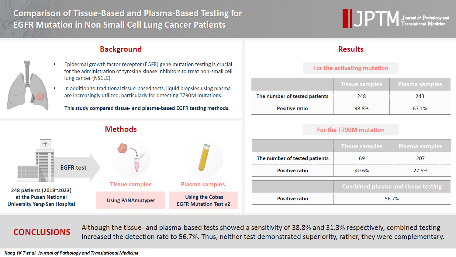
- Comparison of tissue-based and plasma-based testing for EGFR mutation in non–small cell lung cancer patients
- Yoon Kyung Kang, Dong Hoon Shin, Joon Young Park, Chung Su Hwang, Hyun Jung Lee, Jung Hee Lee, Jee Yeon Kim, JooYoung Na
- J Pathol Transl Med. 2025;59(1):60-67. Published online January 15, 2025
- DOI: https://doi.org/10.4132/jptm.2024.10.01
- 6,802 View
- 220 Download
-
Abstract
PDF
- Background
Epidermal growth factor receptor (EGFR) gene mutation testing is crucial for the administration of tyrosine kinase inhibitors to treat non–small cell lung cancer. In addition to traditional tissue-based tests, liquid biopsies using plasma are increasingly utilized, particularly for detecting T790M mutations. This study compared tissue- and plasma-based EGFR testing methods.
Methods
A total of 248 patients were tested for EGFR mutations using tissue and plasma samples from 2018 to 2023 at Pusan National University Yangsan Hospital. Tissue tests were performed using PANAmutyper, and plasma tests were performed using the Cobas EGFR Mutation Test v2.
Results
All 248 patients underwent tissue-based EGFR testing, and 245 (98.8%) showed positive results. Of the 408 plasma tests, 237 (58.1%) were positive. For the T790M mutation, tissue biopsies were performed 87 times in 69 patients, and 30 positive cases (38.6%) were detected. Plasma testing for the T790M mutation was conducted 333 times in 207 patients, yielding 62 positive results (18.6%). Of these, 57 (27.5%) were confirmed to have the mutation via plasma testing. Combined tissue and plasma tests for the T790M mutation were positive in nine patients (13.4%), while 17 (25.4%) were positive in tissue only and 12 (17.9%) in plasma only. This mutation was not detected in 28 patients (43.3%).
Conclusions
Although the tissue- and plasma-based tests showed a sensitivity of 37.3% and 32.8%, respectively, combined testing increased the detection rate to 56.7%. Thus, neither test demonstrated superiority, rather, they were complementary.
- Clinicopathologic characteristics of HER2-positive pure mucinous carcinoma of the breast
- Yunjeong Jang, Hera Jung, Han-Na Kim, Youjeong Seo, Emad Alsharif, Seok Jin Nam, Seok Won Kim, Jeong Eon Lee, Yeon Hee Park, Eun Yoon Cho, Soo Youn Cho
- J Pathol Transl Med. 2020;54(1):95-102. Published online November 13, 2019
- DOI: https://doi.org/10.4132/jptm.2019.10.24
- 12,473 View
- 301 Download
- 25 Web of Science
- 22 Crossref
-
Abstract
PDF
- Background
Pure mucinous carcinoma (PMC) is a rare type of breast cancer, estimated to represent 2% of invasive breast cancer. PMC is typically positive for estrogen receptors (ER) and progesterone receptors (PR) and negative for human epidermal growth factor receptor 2 (HER2). The clinicopathologic characteristics of HER2-positive PMC have not been investigated.
Methods
Pathology archives were searched for PMC diagnosed from January 1999 to April 2018. Clinicopathologic data and microscopic findings were reviewed and compared between HER2-positive PMC and HER2-negative PMC. We also analyzed the differences in disease-free survival (DFS) and overall survival according to clinicopathologic parameters including HER2 status in overall PMC cases.
Results
There were 21 HER2-positive cases (4.8%) in 438 PMCs. The average tumor size of HER2-positive PMC was 32.21 mm (± 26.55). Lymph node metastasis was present in seven cases. Compared to HER2-negative PMC, HER2-positive PMC presented with a more advanced T category (p < .001), more frequent lymph node metastasis (p = .009), and a higher nuclear and histologic grade (p < .001). Microscopically, signet ring cells were frequently observed in HER2-positive PMC (p < .001), whereas a micropapillary pattern was more frequent in HER2-negative PMC (p = .012). HER2-positive PMC was more frequently negative for ER (33.3% vs. 1.2%) and PR (28.6% vs. 7.2%) than HER2-negative PMC and showed a high Ki-67 labeling index. During follow-up, distant metastasis and recurrence developed in three HER2-positive PMC patients. Multivariate analysis revealed that only HER2-positivity and lymph node status were significantly associated with DFS.
Conclusions
Our results suggest that HER2-positive PMC is a more aggressive subgroup of PMC. HER2 positivity should be considered for adequate management of PMC. -
Citations
Citations to this article as recorded by
- Mucin‐producing breast lesions: a practical approach to diagnosis
Sunayana Misra, Mihir Gudi, Kimberly H Allison, Edi Brogi, Cecily Quinn, Hannah Y Wen, Puay Hoon Tan
Histopathology.2026; 88(5): 939. CrossRef - Signet-ring cell cytomorphology in breast cancer: Unveiling the overlooked
Shitong Su, Zijian Liu, Bifeng Yao, Qiuyang Jing, Ruijie Liu, Kuansong Wang
Critical Reviews in Oncology/Hematology.2026; 220: 105170. CrossRef - Predictive factors and prognostic significance of HER2-low early breast cancer with long-term follow-up
Yuka Niwa, Mitsuo Terada, Yumi Wanifuchi-Endo, Takashi Fujita, Tomoko Asano, Hidetoshi Kawaguchi, Kazuki Nozawa, Nana Matsumoto, Ayaka Isogai, Hikaru Kawahara, Marie Mizumoto, Tatsuya Toyama
Surgical Oncology.2026; 64: 102360. CrossRef - Mucinous carcinoma of the breast: morphological spectrum, diagnostic pitfalls and classification challenges
Emad A Rakha, Bara Wazwaz, Stephen B Fox
Journal of Clinical Pathology.2026; 79(7): 433. CrossRef - Clinicopathological characteristics of mucinous breast cancer: a retrospective analysis of a 6-years study from national cancer center in Vietnam
Thi Huyen Phung, Thanh Tung Pham, Huu Thang Nguyen, Dinh Thach Nguyen, Thanh Long Nguyen, Thi Hoai Hoang
Breast Cancer Research and Treatment.2025; 209(3): 667. CrossRef - Poor response of HER2-positive mucinous carcinomas of breast to neoadjuvant HER2-targeted therapy: A study of four cases
Min Han, Daniel Schmolze, Javier A. Arias-Stella, Christina H. Wei, Joanne Mortimer, Fang Fan
Annals of Diagnostic Pathology.2025; 74: 152396. CrossRef - Comprehensive Immunohistochemical Analysis of Mesonephric Marker Expression in Low-grade Endometrial Endometrioid Carcinoma
Yurimi Lee, Sangjoon Choi, Hyun-Soo Kim
International Journal of Gynecological Pathology.2024; 43(3): 221. CrossRef - Clinicopathological features and prognosis of mucinous breast carcinoma with a micropapillary structure
Beibei Yang, Menglu Shen, Bo Sun, Jing Zhao, Meng Wang
Thoracic Cancer.2024; 15(36): 2530. CrossRef - Pure Mucinous Carcinoma of the Breast: Radiologic-Pathologic Correlation
Cherie M Kuzmiak, Benjamin C Calhoun
Journal of Breast Imaging.2023;[Epub] CrossRef - Role of circ-FOXO3 and miR-23a in radiosensitivity of breast cancer
Elahe Abdollahi, Hossein Mozdarani, Behrooz Z. Alizadeh
Breast Cancer.2023; 30(5): 714. CrossRef - On Ultrasonographic Features of Mucinous Carcinoma with Micropapillary Pattern
Wei-Sen Yang, Yang Li, Ya Gao
Breast Cancer: Targets and Therapy.2023; Volume 15: 473. CrossRef - Spectrum of Mucin-containing Lesions of the Breast: Multimodality Imaging Review with Pathologic Correlation
Janice N. Thai, Melinda F. Lerwill, Shinn-Huey S. Chou
RadioGraphics.2023;[Epub] CrossRef - Mesonephric-like Adenocarcinoma of the Ovary: Clinicopathological and Molecular Characteristics
Hyun Hee Koh, Eunhyang Park, Hyun-Soo Kim
Diagnostics.2022; 12(2): 326. CrossRef - Alveolar Soft Part Sarcoma of the Uterus: Clinicopathological and Molecular Characteristics
Yurimi Lee, Kiyong Na, Ha Young Woo, Hyun-Soo Kim
Diagnostics.2022; 12(5): 1102. CrossRef - Metastasis of the Mucionous adenocarcinoma of breast to the mandibular gingiva: Rare case report
Ivana Mijatov, Aleksandra Fejsa Levakov, Aleksandar Spasić, Jelena Nikolić, Saša Mijatov
Medicine.2022; 101(38): e30732. CrossRef - Endometrioid Carcinomas of the Ovaries and Endometrium Involving Endocervical Polyps: Comprehensive Clinicopathological Analyses
Jihee Sohn, Yurimi Lee, Hyun-Soo Kim
Diagnostics.2022; 12(10): 2339. CrossRef - Serous Carcinoma of the Endometrium with Mesonephric-Like Differentiation Initially Misdiagnosed as Uterine Mesonephric-Like Adenocarcinoma: A Case Report with Emphasis on the Immunostaining and the Identification of Splice Site TP53 Mutation
Sangjoon Choi, Yoon Yang Jung, Hyun-Soo Kim
Diagnostics.2021; 11(4): 717. CrossRef - HER2 positive mucinous carcinoma of breast with micropapillary features: Report of a case and review of literature
Dinesh Chandra Doval, Rupal Tripathi, Sunil Pasricha, Pankaj Goyal, Chaturbhuj Agrawal, Anurag Mehta
Human Pathology: Case Reports.2021; 25: 200531. CrossRef - Carcinoma mucosecretor de mama HER2-positivo, un caso clínico
A.M. González Aranda, E. Martínez Gómez, A. Santana Costa, F. Arnanz Velasco, M.H. González de Diego, A. Zapico Goñi
Clínica e Investigación en Ginecología y Obstetricia.2021; 48(4): 100685. CrossRef - Clinicopathologic features of unexpectedly HER2 positive breast carcinomas: An institutional experience
Carissa LaBoy, Kalliopi P. Siziopikou, Lauren Rosen, Luis Z. Blanco, Jennifer L. Pincus
Pathology - Research and Practice.2021; 222: 153441. CrossRef - Mesonephric-like Differentiation of Endometrial Endometrioid Carcinoma: Clinicopathological and Molecular Characteristics Distinct from Those of Uterine Mesonephric-like Adenocarcinoma
Sujin Park, Go Eun Bae, Jiyoung Kim, Hyun-Soo Kim
Diagnostics.2021; 11(8): 1450. CrossRef - Mesonephric-like Adenocarcinoma of the Uterine Corpus: Comprehensive Immunohistochemical Analyses Using Markers for Mesonephric, Endometrioid and Serous Tumors
Hyunjin Kim, Kiyong Na, Go Eun Bae, Hyun-Soo Kim
Diagnostics.2021; 11(11): 2042. CrossRef
- Mucin‐producing breast lesions: a practical approach to diagnosis
- Immunohistochemical Classification of Primary and Secondary Glioblastomas
- Kyu Sang Lee, Gheeyoung Choe, Kyung Han Nam, An Na Seo, Sumi Yun, Kyung Ju Kim, Hwa Jin Cho, Sung Hye Park
- Korean J Pathol. 2013;47(6):541-548. Published online December 24, 2013
- DOI: https://doi.org/10.4132/KoreanJPathol.2013.47.6.541
- 11,454 View
- 69 Download
- 22 Crossref
-
Abstract
PDF
Background Glioblastomas may develop
de novo (primary glioblastomas, P-GBLs) or through progression from lower-grade astrocytomas (secondary glioblastomas, S-GBLs). The aim of this study was to compare the immunohistochemical classification of glioblastomas with clinically determined P-GBLs and S-GBLs to identify the best combination of antibodies for immunohistochemical classification.Methods We evaluated the immunohistochemical expression of epidermal growth factor receptor (EGFR), p53, and isocitrate dehydrogenase 1 (IDH-1) in 150 glioblastoma cases.
Results According to clinical history, the glioblastomas analyzed in this study consisted of 146 P-GBLs and 4 S-GBLs. Immunohistochemical expression of EGFR, p53, and IDH-1 was observed in 62.6%, 49.3%, and 11.1%, respectively. Immunohistochemical profiles of EGFR(+)/p53(-), IDH-1(-)/EGFR(+)/p53(-), and EGFR(-)/p53(+) were noted in 41.3%, 40.2%, and 28.7%, respectively. Expression of IDH-1 and EGFR(-)/p53(+) was positively correlated with young age. The typical immunohistochemical features of S-GBLs comprised IDH-1(+)/EGFR(-)/p53(+), and were noted in 3.6% of clinically P-GBLs. The combination of IDH-1(-) or EGFR(+) was the best set of immunohistochemical stains for identifying P-GBLs, whereas the combination of IDH-1(+) and EGFR(-) was best for identifying S-GBLs.
Conclusions We recommend a combination of IDH-1 and EGFR for immunohistochemical classification of glioblastomas. We expect our results to be useful for determining treatment strategies for glioblastoma patients.
-
Citations
Citations to this article as recorded by- The Co-Expression and Cellular Location of HER Family Members, EGFRvIII, Putative Cancer Stem Cell Biomarkers CD44 and CD109 in Patients with Glioblastoma, and Their Impacts on Prognosis
Ermira Mulliqi, Said Khelwatty, Izhar Bagwan, Ahmad Kamaludin, Anna Morgan, Natalie Long, Keyoumars Ashkan, Helmout Modjtahedi
Cancers.2025; 17(7): 1221. CrossRef - Predicting p53 Status in IDH‐Mutant Gliomas Using MRI‐Based Radiomic Model
Jiamin Li, Zhihong Lan, Xiao Zhang, Xiaoyun Liang, Hanwei Chen, Xiangrong Yu
Cancer Medicine.2025;[Epub] CrossRef - Classification of Glioblastoma Based on Immunohistochemical Expression of IDH-1, p53, and ATRX: A Study from a Tertiary Care Center in South India
Hiba Thankayathil, Aparna Govindan, Supriya Nilambur Kovilakam, Rajeev Mandaka Parambil
Indian Journal of Neurosurgery.2025; 14(S 01): S48. CrossRef - Endothelial transdifferentiation of glioma stem cells: a literature review
Andrei Buruiana, Stefan Ioan Florian, Alexandru Ioan Florian, Olga Soritau, Sergiu Susman
Acta Neuropathologica Communications.2025;[Epub] CrossRef - Cutaneous Melanoma and Glioblastoma Multiforme Association—Case Presentation and Literature Review
Olguța Anca Orzan, Călin Giurcăneanu, Bogdan Dima, Monica Beatrice Dima, Ana Ion, Beatrice Bălăceanu, Cornelia Nițipir, Irina Tudose, Cătălina Andreea Nicolae, Alexandra Maria Dorobanțu
Diagnostics.2023; 13(6): 1046. CrossRef - Primary Extra-axial Glioblastoma: Case Report and Literature Review
Baraa Dabboucy, Philippe Younes, Abdallah Rahbani, Elie Fahed, Gérard Abadjian
Arquivos Brasileiros de Neurocirurgia: Brazilian Neurosurgery.2021; 40(04): e368. CrossRef - TERT Promoter Mutation in Adult Glioblastomas: It's Correlation with Other Relevant Molecular Markers
Mukesh Barange, Sridhar Epari, Mamta Gurav, Omshree Shetty, Ayushi Sahay, Prakash Shetty, Jayantsastri Goda, Aliasagar Moyiadi, Tejpal Gupta, Rakesh Jalali
Neurology India.2021; 69(1): 126. CrossRef - Immunohistochemical characterisation and histopathology of astrocytic neoplasms at a tertiary Nigerian hospital
Michael Nweke, Gabriel Ogun, Amos Adeleye, Clement A. Okolo, Adekunle Adesina
International Journal of Clinical Practice.2021;[Epub] CrossRef - Cytotoxic Effects of Blue Scorpion Venom (Rhopalurus junceus) in a Glioblastoma Cell Line Model
Laura A. Lozano-Trujillo, Diana K. Garzón-Perdomo, Andrea C.R. Vargas, Lina M. de los Reyes, Marco F. Avila-Rodriguez, Olivia T.G. Gay, Liliana F. Turner
Current Pharmaceutical Biotechnology.2021; 22(5): 636. CrossRef - The importance of IDH1, ATRX and WT-1 mutations in glioblastoma
Gülsün Gülten, Nagİhan Yalçın, Bahar Baltalarlı, Gamze Gököz Doğu, Ferİdun Acar, Yücel Doğruel
Polish Journal of Pathology.2020; 71(2): 127. CrossRef - Molecular Subgroups of Glioblastoma– an Assessment by Immunohistochemical Markers
Ádám Nagy, Ferenc Garzuly, Gergely Padányi, Iván Szűcs, Ádám Feldmann, Balázs Murnyák, Tibor Hortobágyi, Bernadette Kálmán
Pathology & Oncology Research.2019; 25(1): 21. CrossRef - Proteomic Advances in Glial Tumors through Mass Spectrometry Approaches
Radu Pirlog, Sergiu Susman, Cristina Adela Iuga, Stefan Ioan Florian
Medicina.2019; 55(8): 412. CrossRef - Isocitrate dehydrogenase 1 mutant glioblastomas demonstrate a decreased rate of pseudoprogression: a multi-institutional experience
Homan Mohammadi, Kevin Shiue, G Daniel Grass, Vivek Verma, Kay Engellandt, Dirk Daubner, Gabriele Schackert, Mercia J Gondim, Dibson Gondim, Alexander O Vortmeyer, Aaron P Kamer, William Jin, Timothy J Robinson, Gordon Watson, Hsiang-Hsuan M Yu, Tim Laute
Neuro-Oncology Practice.2019;[Epub] CrossRef - Calvarium mass as the first presentation of glioblastoma multiforme: A very rare manifestation of high-grade glioma
S. Taghipour Zahir, M. Mortaz, M. Baghi Yazdi, N. Sefidrokh Sharahjin, M. Shabani
Neurochirurgie.2018; 64(1): 76. CrossRef - Malignant Gliomas as Second Neoplasms in Pediatric Cancer Survivors: Neuropathological Study
Ewa Izycka-Swieszewska, Ewa Bien, Joanna Stefanowicz, Edyta Szurowska, Ewa Szutowicz-Zielinska, Magdalena Koczkowska, Dawid Sigorski, Wojciech Kloc, Wojciech Rogowski, Elzbieta Adamkiewicz-Drozynska
BioMed Research International.2018; 2018: 1. CrossRef - Prognostic significance of mutant IDH1, CD133, and β-catenin immunohistochemical expression in glioblastoma multiforme
Azza Abdel-Aziz, Mie A. Mohamed, Dina Abdallah, Fatma M.F. Akl, Ghada E. Eladawy, Ahmed N. Taha, Hossam Shata
Egyptian Journal of Pathology.2018; 38(1): 27. CrossRef - On glioblastoma and the search for a cure: where do we stand?
John Bianco, Chiara Bastiancich, Aleksander Jankovski, Anne des Rieux, Véronique Préat, Fabienne Danhier
Cellular and Molecular Life Sciences.2017; 74(13): 2451. CrossRef - Expression of p53 & epidermal growth factor receptor in glioblastoma
Sameera Karnam, Radhika Kottu, Amit Kumar Chowhan, Prasad Chandramouleswara Bodepati
Indian Journal of Medical Research.2017; 146(6): 738. CrossRef - Development of Glioblastoma after Treatment of Brain Abscess
Hiroaki Matsumoto, Hiroaki Minami, Shogo Tominaga, Yasuhisa Yoshida
World Neurosurgery.2016; 88: 686.e19. CrossRef - Clinical, immunohistochemical, and molecular genetic prognostic factors in adult patients with glioblastoma
N. V. Lobanova, L. V. Shishkina, M. V. Ryzhova, G. L. Kobyakov, R. V. Sycheva, S. A. Burov, A. V. Lukyanov, Zh. R. Omarova
Arkhiv patologii.2016; 78(4): 10. CrossRef - Concordance analysis and diagnostic test accuracy review of IDH1 immunohistochemistry in glioblastoma
Jung-Soo Pyo, Nae Yu Kim, Roy Hyun Jai Kim, Guhyun Kang
Brain Tumor Pathology.2016; 33(4): 248. CrossRef - Methyl Guanine Methyl Transferase Methylation Status and Epidermal Growth Factor Receptor expression in a cohort of Egyptian glioblastoma patients
Soheir M. Hamam, Bassma M. El Sabaa, Iman M. Talaat, Rasha A. Nassra, Doaa A. Abdelmonsif
Egyptian Journal of Pathology.2016; 36(2): 282. CrossRef
- The Co-Expression and Cellular Location of HER Family Members, EGFRvIII, Putative Cancer Stem Cell Biomarkers CD44 and CD109 in Patients with Glioblastoma, and Their Impacts on Prognosis
- A Solitary Fibrous Tumor with Giant Cells in the Lacrimal Gland: A Case Study
- Da Hye Son, Su Hyun Yoo, Ho-Seok Sa, Kyung-Ja Cho
- Korean J Pathol. 2013;47(2):158-162. Published online April 24, 2013
- DOI: https://doi.org/10.4132/KoreanJPathol.2013.47.2.158
- 10,131 View
- 63 Download
- 9 Crossref
-
Abstract
PDF
Orbital solitary fibrous tumor (SFT) has recently been proposed as the encompassing terminology for hemangiopericytoma, giant cell angiofibroma (GCAF), and fibrous histiocytoma of the orbit. The lacrimal gland is a very rare location for both SFT and GCAF. A 39-year-old man presented with a painless left upper eyelid mass. An orbital computed tomography scan identified a 1.1 cm-sized well-defined nodule located in the left lacrimal gland. He underwent a mass excision. Histopathologic examination showed a proliferation of relatively uniform spindle cells with a patternless or focally storiform pattern. Dilated vessels were prominent, but angiectoid spaces lined with giant cells were absent. Floret-type giant cells were mostly scattered in the periphery. The tumor was immunoreactive for CD34 and CD99, but negative for smooth muscle actin and S-100 protein. This is the first Korean case of SFT of the lacrimal gland with overlapping features of GCAF, suggesting a close relationship between the two entities.
-
Citations
Citations to this article as recorded by- Radiological features of lacrimal gland masses
Sonia Huang, Jessica Y. Tong, Valerie Juniat, Abdullah Almater, Ilse Mombaerts, Dinesh Selva
Survey of Ophthalmology.2026; 71(2): 700. CrossRef - Imaging features of lacrimal gland disease
Carmelo Caltabiano, Khizar Rana, Alexander Buckby, Sandy Patel, Dinesh Selva
Frontiers in Ophthalmology.2026;[Epub] CrossRef - A review of solitary fibrous tumours of the orbit and ocular adnexa
Cornelius René, Paolo Scollo, Dominic O’Donovan
Eye.2023; 37(5): 858. CrossRef - A giant orbital solitary fibrous tumor treated by surgical excision: a case report and literature review
Qi Zhou, Yuting Liu, Fang Wang, Yang Cao, Hongbin Lv, Xibo Zhang
Diagnostic Pathology.2023;[Epub] CrossRef - Giant cell-rich solitary fibrous tumour of the lacrimal gland with prominent angiomatoid cystic changes and an underlying NAB2ex3-STAT6ex18 fusion
Khaled A Alsaadi, Manar Alwohaib, Karen Pinto, Rola H Ali
BMJ Case Reports.2022; 15(2): e247141. CrossRef - Cystic appearance - a new feature of solid fibrous tumours in the lacrimal gland: a case report with literature review
Ancuta-Augustina Gheorghisan-Galateanu, Dana Cristina Terzea, Iulia Burcea, Roxana Dusceac, Cristina Capatina, Catalina Poiana
Diagnostic Pathology.2019;[Epub] CrossRef - Solitary Fibrous Tumor in the Lacrimal Gland Fossa: A Case Report
Jacqueline Mupas-Uy, Yoshiyuki Kitaguchi, Yasuhiro Takahashi, Emiko Takahashi, Hirohiko Kakizaki
Case Reports in Ophthalmology.2016; 7(2): 398. CrossRef - Ocular adnexal (orbital) solitary fibrous tumor: nuclear STAT6 expression and literature review
Aleksandra Petrovic, Aurélie Obéric, Alexandre Moulin, Mehrad Hamedani
Graefe's Archive for Clinical and Experimental Ophthalmology.2015; 253(9): 1609. CrossRef - Angiofibroma de células gigantes en mucosa yugal: una entidad rara en una localización infrecuente
Alejandro Rubio Fernández, María López Macías, Weimar Toro Zambrano, Mario Díaz Delgado, Alicia Hernández Amate
Revista Española de Patología.2014; 47(4): 223. CrossRef
- Radiological features of lacrimal gland masses
- Prognostic Relevance of the Expression of CA IX, GLUT-1, and VEGF in Ovarian Epithelial Cancers
- Kyungbin Kim, Won Young Park, Jee Yeon Kim, Mee Young Sol, Dong Hun Shin, Do Youn Park, Chang Hun Lee, Jeong Hee Lee, Kyung Un Choi
- Korean J Pathol. 2012;46(6):532-540. Published online December 26, 2012
- DOI: https://doi.org/10.4132/KoreanJPathol.2012.46.6.532
- 11,658 View
- 43 Download
- 16 Crossref
-
Abstract
PDF
Background Tumor hypoxia is associated with malignant progression and treatment resistance. Hypoxia-related factors, such as carbonic anhydrase IX (CA IX), glucose transporter-1 (GLUT-1), and vascular endothelial growth factor (VEGF) permit tumor cell adaptation to hypoxia. We attempted to elucidate the correlation of these markers with variable clinicopathological factors and overall prognosis.
Methods Immunohistochemistry for CA IX, GLUT-1, and VEGF was performed on formalin-fixed, paraffin-embedded tissues from 125 cases of ovarian epithelial cancer (OEC).
Results CA IX expression was significantly associated with an endometrioid and mucinous histology, nuclear grade, tumor necrosis, and mitosis. GLUT-1 expression was associated with tumor necrosis and mitosis. VEGF expression was correlated only with disease recurrence. Expression of each marker was not significant in terms of overall survival in OECs; however, there was a significant correlation between poor overall survival rate and high coexpression of these markers.
Conclusions The present study suggests that it is questionable whether CA IX, GLUT-1, or VEGF can be used alone as independent prognostic factors in OECs. Using at least two markers helps to predict patient outcomes in total OECs. Moreover, the inhibition of two target gene combinations might prove to be a novel anticancer therapy.
-
Citations
Citations to this article as recorded by- Glucose-transporter 1 (GLUT1) as a prognostic biomarker: evidence from 14,966 human tumors across 134 cancer types
Seyma Büyücek, Katharina Möller, Florian Viehweger, Ria Schlichter, Anne Menz, Andreas M Luebke, Viktor Reiswich, Martina Kluth, Claudia Hube-Magg, Andrea Hinsch, Florian Lutz, Sören Weidemann, Frank Jacobsen, Maximilian Lennartz, David Dum, Christian Ber
BMC Cancer.2026;[Epub] CrossRef - Role of Carbonic Anhydrase IX Expression in Triple-negative Breast Cancer
Hemali B Kotadiya, Nupur A Patel, Hemangini H Vora
Gujarat Cancer Society Research Journal.2026; 27(2): 31. CrossRef - Evaluation of the effect of Endogenous and Exogenous Antioxidants on Hypoxia Induced Breast Adenocarcinoma Cell Invasion and Migration:
In vitro
Subarnarekha Maitra, Poulami Sen, Dibya Sinha, Tathagata Roy
Research Journal of Pharmacy and Technology.2025; : 3153. CrossRef - Effect of Ultrasound Therapy at the ST11 on Sympathetic Nervous System Change: A Prospective Randomized Controlled Study
Shinwoo Kang, Dongho Keum
Journal of Korean Medicine Rehabilitation.2023; 33(4): 167. CrossRef - A systematic review verified by bioinformatic analysis based on TCGA reveals week prognosis power of CAIX in renal cancer
Zikuan Zhang, Bo Wu, Yuan Shao, Yongquan Chen, Dongwen Wang, Lucia Magnelli
PLOS ONE.2022; 17(12): e0278556. CrossRef - Effect of Acupuncture at the Field of the Auricular Branch of the Vagus Nerve on Autonomic Nervous System Change
Sunjoo An, Dongho Keum
Journal of Korean Medicine Rehabilitation.2021; 31(2): 81. CrossRef - Responses of Autonomic Nervous System and Gastrointestinal Function to Acupuncture at Abdominal Anterior Cutaneous Nerve : A Pilot Study
Seohyun Park, Hojun Kim, Dongho Keum
Journal of Korean Medicine.2019; 40(1): 99. CrossRef - Omega-3 polyunsaturated fatty acid docosahexaenoic acid and its role in exhaustive-exercise-induced changes in female rat ovulatory cycle
Abeer F. Mostafa, Shereen M. Samir, R.M. Nagib
Canadian Journal of Physiology and Pharmacology.2018; 96(4): 395. CrossRef - Clear cell carcinomas of the ovary and kidney: clarity through genomics
Jennifer X Ji, Yi Kan Wang, Dawn R Cochrane, David G Huntsman
The Journal of Pathology.2018; 244(5): 550. CrossRef - Prognostic Significance of Carbonic Anhydrase IX Expression in Cancer Patients: A Meta-Analysis
Simon J. A. van Kuijk, Ala Yaromina, Ruud Houben, Raymon Niemans, Philippe Lambin, Ludwig J. Dubois
Frontiers in Oncology.2016;[Epub] CrossRef - Review of Research Topics on Abdominal Examination
Jihye Kim, Jeong Hwan Park, Keun Ho Kim
Journal of Korean Medicine.2016; 37(3): 1. CrossRef - Expression of hypoxic markers and their prognostic significance in soft tissue sarcoma
JEUNG IL KIM, KYUNG UN CHOI, IN SOOK LEE, YOUNG JIN CHOI, WON TACK KIM, DONG HOON SHIN, KYUNGBIN KIM, JEONG HEE LEE, JEE YEON KIM, MEE YOUNG SOL
Oncology Letters.2015; 9(4): 1699. CrossRef - Evaluation of a Hypoxia Regulated Gene Panel in Ovarian Cancer
Amanda F. Baker, Scott W. Malm, Ritu Pandey, Cindy Laughren, Haiyan Cui, Denise Roe, Setsuko K. Chambers
Cancer Microenvironment.2015; 8(1): 45. CrossRef - Sulforaphane reduces molecular response to hypoxia in ovarian tumor cells independently of their resistance to chemotherapy
MICHAL PASTOREK, VERONIKA SIMKO, MARTINA TAKACOVA, MONIKA BARATHOVA, MARIA BARTOSOVA, LUBA HUNAKOVA, OLGA SEDLAKOVA, SONA HUDECOVA, OLGA KRIZANOVA, FRANCK DEQUIEDT, SILVIA PASTOREKOVA, JAN SEDLAK
International Journal of Oncology.2015; 47(1): 51. CrossRef - Overexpression of Glucose Transporter-1 (GLUT-1) Predicts Poor Prognosis in Epithelial Ovarian Cancer
Hanbyoul Cho, You Sun Lee, Julie Kim, Joon-Yong Chung, Jae-Hoon Kim
Cancer Investigation.2013; 31(9): 607. CrossRef - Towards Lipidomics of Low-Abundant Species for Exploring Tumor Heterogeneity Guided by High-Resolution Mass Spectrometry Imaging
Jonathan Cimino, David Calligaris, Johann Far, Delphine Debois, Silvia Blacher, Nor Sounni, Agnès Noel, Edwin De Pauw
International Journal of Molecular Sciences.2013; 14(12): 24560. CrossRef
- Glucose-transporter 1 (GLUT1) as a prognostic biomarker: evidence from 14,966 human tumors across 134 cancer types
- Expression Pattern of the Rb Protein and its Correlation with Prognosis in Primary Lung Cancer.
- Hea Kyoung Hur, Seo Hee Rha, Sook Hee Hong
- Korean J Pathol. 1997;31(2):152-161.
- 2,229 View
- 16 Download
-
Abstract
PDF
- An immunohistochemical stain for the Rb tumor suppressor gene product was performed in pathologic specimens from 72 primary lung cancer patients to study the correlation between its expression and histologic type, cancer differentiation, clinical stage and survival rate. The expression of the Rb protein was positive in 34 cases(47.2%) and negative in 38 cases(52.8%). The Rb protein was not expressed in 16 of 42 cases(38.1%) in squamous cell carcinoma, in 17 of 23 cases(73.9%) in adenocarcinoma, in one of three cases(33.3%) in undifferentiated large cell carcinoma, in two of two cases(100%) in small cell carcinoma, in one of one case(100%) in an adenosquamous carcinoma and in one of one case(100%) in an atypical carcinoid. There were significant difference of the Rb protein expression between squamous cell carcinoma and adenocarcinoma(p<0.05). The expression of Rb protein was not correlated with degree of cancer cell differentiation and clinical stage of the lung cancer(p>0.05). The two year survival rate for patients with the Rb positive was 65% compared with 37% for those with the Rb negative which was significant(p<0.05). This result suggests that an altered or the absence of the Rb protein in cancer cells can be a valuable prognostic factor in the lung cancer.
- Mesenchymal Chondrosarcoma Arising from Orbital Soft Tissue: A case report.
- Yu Mee Kang, Mi Kyung Jee, Seok Jin Gang, Byung Kee Kim, Sun Moo Kim
- Korean J Pathol. 1989;23(2):273-277.
- 2,153 View
- 15 Download
-
Abstract
PDF
- Orbital mesenchymal chondrosarcoma, first described by Luis et. al in 1971, is a very rare tumor of characteristic histologic features. A 21-year-woman was admitted with a 4-month histoiry of rapidly progressive proptosis and visual disturbance. Right orbital exenteration was performed under the clinical diagnosis of orbital calcifying tumor. Grossly, the tumor presented as a multibloblated, circumscribed mass that measures 5.5 cm in the greatest dimentsion. Cut sections resembled ordinary chonrosarcoma. Microscopically, the tumor was composed of undifferentiated mesenchymal cells, interspersed nodules of well differentated cartilagenous tissue, areas of gradual transition from undifferentiated mesenchymal cells to cartilage, and hemangiopericytoma-like areas. A brief summary of the histopathological aspect of this tumor and a review of literature are presented.
- The Expression of p53, c-erbB-2 and nm23 Proteins in Breast Cancer.
- Kyo Young Lee, Yong Goo Kim, Young Shin Kim, Kyung Ja Han, Chang Suk Kang, Jean A Kim, Won Il Kim, Sang In Shim
- Korean J Pathol. 1999;33(2):88-95.
- 2,054 View
- 10 Download
-
Abstract
- Recently, p53, c-erbB-2 and nm23 proteins have been studied in breast cancer. The expression of p53 protein indicates the mutation of p53 gene known as a tumor supressor gene, and c-erbB-2 gene amplification has been considered an indicator of poor prognosis and nm23 a metastsis suppressor gene. In order to elucidate the roles and relations of these proteins in the develpoment, progression and metastasis in breast cancer, we studied 89 cases of invasive breast cancer and 32 cases of lymph node metastasis for the expression of p53, c-erbB-2 and nm23 proteins using an immunohistochemical method. The results were as follows: 1) The expression rates of p53, c-erbB-2, and nm23 proteins in breast cancer were 40.4%, 34.8% and 55.1%, respectively. Co-expression of p53 protein and c-erbB-2 protein was found in 20.2% of cases, showing the highest incidence in poorly differentiated type (40%). 2) p53 protein expression was increased in poorly differentiated type but was not statistically significant. On the other hand, the expression of nm23 protein was decreased in poorly differentiated type, which was statistically significant (p<0.05). 3) The correlation of p53 protein expression with c-erbB-2 protein expression was statistically significant (p<0.05) but that with nm23 protein was not. 4) In the cases with lymph node metastasis, discordant expression of p53, c-erbB-2 and nm23 proteins between primary tumor and the lymph node metastatic tumor was found in 9.4%, 3.1% and 18.8% of cases, respectively. The above results suggest that overexpression of p53 and c-erbB-2 proteins and downregulation of nm23 protein are associated with the tumor progression in the breast cancer.
- The Expression of c-erbB-2, EGFR, p53 and Ki-67 in Ovarian Borderline Tumors and Carcinomas of the Ovary.
- Kyueng Whan Min, Moon Hyang Park
- Korean J Pathol. 2007;41(5):296-306.
- 2,865 View
- 63 Download
-
Abstract
PDF
- BACKGROUND
An ovarian surface epithelial tumor is a heterogenous disease, and various biological and molecular factors are important for its development and progression. Several findings support EGFR or c-erbB-2 as adverse prognostic indicators for an ovarian carcinoma.
METHODS
We reviewed the histological and clinical findings of 52 carcinomas (17 endometrioid, 16 serous, 13 mucinous and 6 clear cell tumors), and 26 borderline (10 serous and 16 mucinous) tumors. Expression of c-erbB-2, EGFR, p53, and Ki-67 was evaluated on paraffinembedded tissue from a primary ovarian tumor by immunohistochemical methods.
RESULTS
Expression of c-erbB-2 was found in 7.6% of tumors and expression of EGFR was found in 9.6% of tumors by immunohistochemical analysis. No significance was found between cerbB- 2 and EGFR expression as indicators of a poor prognosis. The expression of p53 and Ki-67 (>50%) correlated with the grade and type of tumor in the ovarian cancers. p53 and Ki- 67 overexpression (>50%) was absent in the borderline ovarian tumors, whereas ovarian carcinomas showed expression of both p53 and Ki-67.
CONCLUSION
Expression of c-erbB- 2, EGFR, p53, and Ki-67 as determined by immunohistochemical analysis did not correlate with prognostic significance. However, p53 and Ki-67 expression may be used as markers to predict aggressive behavior, and to differentiate between malignant and borderline epithelial ovarian tumors. Further large-scale studies are required to clarify the significance of c-erbB-2 and EGFR expression in ovarian tumors.
- Genetic Expression Pattern of Gastric Carcinomas According to Cellular Mucin Phenotypes.
- Won Ae Lee, In Soo Suh, Ying Hua Li, Ji Hyun Eum, Wan Sik Yu, Han Ik Bae
- Korean J Pathol. 2007;41(5):307-315.
- 2,359 View
- 16 Download
-
Abstract
PDF
- BACKGROUND
Gastric carcinomas (GCs) have recently been reclassified according to the mucin phenotypes. We aimed to characterize the relationship between the mucin phenotypes and the genetic alterations or the clinicopathologic parameters of GCs.
METHODS
Immunohistochemistry was performed for MUC1, MUC5AC, MUC6, MUC2, CD10, p53, hMLH1, CerbB2 and E-cadherin in 150 GCs. The mucin phenotypes of the GCs were classified as 4 phenotypes: gastric, intestinal, mixed and unclassified.
RESULTS
MUC1, MUC5AC, MUC6, MUC2 and CD10 were expressed in 63.3%, 42.7%, 14.0%, 24.7% and 14.0% of the GCs, respectively. The mucin phenotypes of the GCs corresponded to the gastric type in 31.3%, the intestinal type in 20.0%, the mixed type in 15.3% and the unclassified type in 33.3%. The incidence of a p53 overexpression was higher in the gastric or mixed phenotype than in the intestinal or unclassified phenotype. MUC5AC expression, p53 overexpression and the gastric or mixed phenotype were associated with poor patient survival by multivariate analysis.
CONCLUSION
This study suggests the gastric or mixed mucin phenotype may more likely go through the p53 pathway in carcinogenesis and the mucin phenotype may be considered as a prognostic indicator.
- Endodermal Sinus Tumor of the Orbit.
- Dae Hyun Back, Jin Man Kim, Kwang Sun Suh, Kyu Sang Song, Choong Sik Lee, Dae Young Kang
- Korean J Pathol. 1989;23(3):392-395.
- 2,279 View
- 12 Download
-
Abstract
PDF
- An endodermal sinus tumor is a malignant germ cell tumor that usually arises in the gonads, but on rare occasion occurs in extragonadal locations. Our case was that of a 3 year old girl who complained of a rapid growing orbital mass. On histologic examination it revealed the typical picture of an endodermal sinus tumor and it also disclosed a positive reaction for alphafetoprotein using an immunoperoxidase technique. An orbital exenteration was performed followed by chemotheraphy, but the patient died 5 months after the onset of the disease.
- Rarity of EGFR and c-ErbB-2 Overexpressions in Hepatocellular Carcinoma: An Immunohistochemical Study.
- Woo Sung Moon, Hyun Jin Son, Ho Sung Park, Min Young Park
- Korean J Pathol. 2004;38(4):244-248.
- 2,516 View
- 13 Download
-
Abstract
PDF
- BACKGROUND
The overexpression of epidermal growth factor receptor (EGFR) and c-erbB-2 oncogenes has been implicated in the development of many types of cancer. However, the role of EGFR and c-erbB-2 overexpression in hepatocellular carcinoma (HCC) has not been fully elucidated.
METHODS
The aim of this study was to evaluate the immunohistochemical expression of EGFR and c-erbB-2 oncoprotein in a series of 52 HCCs.
RESULTS
All but one of the HCC tumor tissues were negative for EGFR monoclonal antibody, clone H11. All of the HCC tumor tissue samples were negative for EGFR monoclonal antibody, clone 29.1.1. However, strong EGFR immunoreactivity was detected in sinusoidal endothelial cells of HCC in 25 tumors (48%) using EGFR 29.1.1 antibody. The expression of c-erbB-2 was observed in 6% (3/52) of the HCCs. No significant correlation was found between p53 mutation and the expression of c-erbB-2.
CONCLUSION
Our results suggest that both EGFR and c-erbB-2 oncoprotein overexpressions in tumor cells are rare and do not seem to predominantly contribute to the malignant phenotype in HCC.
- Lymphoproliferativ Lesions of the Orbit and Conjunctiva: Histopathologic Study on 20 cases including 5 cases analyzed by Immunophenotyping.
- Chul Woo Kim, Na Hye Myong, Je G Chi
- Korean J Pathol. 1993;27(2):152-163.
- 2,335 View
- 16 Download
-
Abstract
PDF
- Lymphoid lesions of the orbit and conjunctiva may be divided histologically into three groups: monomorphous and cytologically atypical malignant lymphomas, benign reactive follicular hyperplasia or inflammatory pseudotumor, and borderline lymphoid lesions mainly composed of small, non-atypical lymphocytes with or without evidence of plasmacytic differentiation or germinal centers which are difficult to clarify its nature by histologic criteria alone. From 1984 to 1992 at Seoul National University Hospital, 20 cases of orbital lymphoid infiltrates were reviewed to find out histopathologic characteristics of malignant lymphomas and try to classify them properly. Also, we sought histologic findings helpful for differential diagnosis between malignant and borderline cases. Histologic examination of malignant lesions usually revealed the features of low-grade B cell lymphomas of mucosa-associated lymphoid tissue(MALT) origin, which predominantly consist of centrocyte-like(CCL) cells. Among 13 primary orbital lymphomas, six cases were classified as MALT lymphomas of Isaacson, six were more apt to be the type of immunocytoma by kiel classification, and remaining one showed mixed pattern of both types in areas. The one case of malignant lymphoma with diffuse large cell type was confirmed by secondary involvement of intestinal lymphoma. Five cases were confirmed by immunoglobulin k-light chain monoclonality on flow cytometry and immunofluorescence study. The histologic findings such as Dutcher bodies, hemosiderin, deposits and polykaryocytes tended to be more frequently encountered in malignant lymphoma rather than indeterminate lesions and were thought to be helpful ones in making differential diagnosis between malignant and benign lesions. With above findings, we assume that the principal neoplastic cells in the low grade lymphoma arising in the orbit and conjunctiva are CCL cells, which might be originated from the localized memory B cells, and also several kinds of B lymphocytes in on-going differentiation stages such as medium-sized monocytoid B cell, lymphoplasmacytoid cell, plasma cell, and mature small lymphocyte also contribute to the tumor progression.
- Chromophobe Renal Cell Carcinoma.
- Yeong Jin Choi, Tae Kon Hwang, Youn Soo Lee, Eun Jung Lee, Seok Jin Kang, Byung Kee Kim, Sang In Shim
- Korean J Pathol. 1999;33(4):259-266.
- 2,425 View
- 26 Download
-
Abstract
PDF
- We report 13 chromophobe renal cell carcinomas (10.8%) observed among 120 renal cell carcinomas in adults. The average age was 53 (range: 34-72) years old, and 6 were males and 7 females. The mean tumor size was 10 (range: 5-17) cm, mean nuclear grade 2.4, and mean Robson's stage was 1.9. There were two distinct histologic variants; typical variant (n=9) and eosinophilic variant (n=4). Both of them showed typical light microscopic features and positive reaction with Hale's colloidal iron and carbonic anhydrase II, a marker protein of intercalated cells of renal collecting ducts. A strong positive immunoreactivity for epithelial membrane antigen was noted in the cytoplasm in 12 of 13 tumors. Numerous microvesicles, 180~440 nm in diameter, were identified ultrastructurally. DNA aneuploidy was found in 3 out of 10 cases. Neither local recurrence nor metastasis have been identified during the following period of 4~144 (mean 48) months.
- Orbital Pseudolymphoma: A case report.
- Su Kyeong Yeon, Mi Kyung Jee, Seok Jin Kang, Byoung Kee Kim, Sun Moo Kim
- Korean J Pathol. 1993;27(2):191-194.
- 2,629 View
- 67 Download
-
Abstract
PDF
- Lymphoid tumors of the orbit are rare, and sometimes it is not possible either clinically or histologically to differentiate between lymphoid tumor and pseudolymphoma. Some authors assert that the degree of cytologic differentiation appears to be the single most important factor for determining the prognosis of patients with orbital lymphoid lesions. However, the cytomorphologic basis is not so helpful to diagnose and classify our case, which shows some discrepancy between pathological findings and clinical and radiological findings. At first we misdiagnosed our case as orbital malignant lymphoma on the basis of cytomorphology and immunohistochemical study. But no responce to local intensive radiotherapy and the follow up study of the patient suggest orbital pseudolymphoma.
- Correlation between Expression of c-erbB-2 Oncogene and Various Prognostic Factors in the Colorectal Carcinoma.
- Wan Kim, Hong Ran Choi, Ji Shin Lee, Jong Tae Park, Chang Soo Park, Kyu Hyuk Cho
- Korean J Pathol. 1993;27(3):217-225.
- 2,214 View
- 12 Download
-
Abstract
PDF
- The c-erbB-2 oncogene, which is a new human proto-oncogene similar to EGFR structurally, generates a glycoprotein of tyrosine kinase family with a molecular weight of 185,000 To evaluate the prognostic significance of c-erbB-2 oncogene expression in colorectal carcinoma, We analysed 73 colorectal carcinomas in paraffin sections immunohistochemically, using the monoclonal antibody specific for the c-erbB-2 oncogene product and correlated with clinicopathological data. The results were as follows 1) The immunoreactivity for c-erbB-2 oncogene was localized to cell membrane of the tumor cells and occasionally observed within the cytoplasm. 2) The positivity of c-erbB-2 oncogene expression was 71.2%(52/73) of the colorectal carcinomas overall. According to the histological types, the positivity of c-erbB-2 oncogene in adenocarcinoma(77.4%) was higher than that in mucinous carcinoma(36.4%)(p<0.05). 3) Expression of c-erbB-2 oncogene was significantly correlated with lymph node metastasis or distant metastasis(p=0.0117), Dukes stage(p=0.0432), and TNM classification(p=0.0102). These results suggest that c-erbB-2 oncogene expression may be used as a prognostic factor of colorectal carcinoma because of its correlation with other clinicopathological prognostic factors.
- Inhibitory Effect of Tetrandrine on Extracellular Matrix Deposition in Rat Hepatic Fibrosis.
- Won Young Choi, Hyo Jeong Chae, Sun Kyung Lee
- Korean J Pathol. 1999;33(5):319-325.
- 2,176 View
- 20 Download
-
Abstract
PDF
- No effective therapy has yet developed for liver fibrosis/cirrhosis by directly inhibiting the accumulation of extracellular matrix. This study was undertaken to determine the effect of tetrandrine in rat model of liver fibrosis induced by carborn tetrachloride (CCl4) administration intraorally. Tetrandrine, a calcium channel blocker, is anti-inflammatory constituent of the families Menispermaceae and Ranunculaceae, which have been used as folk remedies in China. Repeated administration of CCl4 for 14 weeks to rats induced liver fibrosis with steatosis. Rats were killed after 4, 8 or 14 weeks of treatment with CCl4, CCl4 tetrandrine (30 mg/kg) or CCl4 tetrandrine (50 mg/kg). The histopathological findings of liver were observed semi-quantitatively by light microscopy and volume percentage of the collagen deposition was determined by image analyzer. Tetrandrine inhibited collagen deposition induced by CCl4 administration, as shown by less severe steatosis and fibrosis and significantly decreased volume percentage of collagen fibers in CCl4 tetrandrine treated animals compared with CCl4 only group. Thus, the administration of tetrandrine holds great promise for treating subjects with liver fibrosis/cirrhosis as a result of chronic hepatic injury.
- Expression of p53 Protein and c-erbB-2 Oncoprotein in Breast Carcinoma.
- Eun Hee Lee, Dong Sug Kim, Tae Sook Lee, Soo Jung Lee
- Korean J Pathol. 1995;29(5):596-606.
- 2,098 View
- 10 Download
-
Abstract
- This study was conducted to evaluate the expression of p53 and c-erbB-2 using immuno-histochemical methods in 145 primary breast carcinomas and to correlate it with other histo-pathological prognostic factors. Invasive ductal carcinoma represented 129 of the cases. Expression of p53 protein and c-erbB-2 oncoprotein was present in 48% (62/129) and 30% (39/129) of invasive ductal carcinomas, respectively. The expression of p53 protein was stongly associated with a high score of degree of differentiation (p<0.05), nuclear pleomorphism (p<0.05), mitotic index (p<0.05), SBR grade (p<0.05) and MSBR grade (p<0.05), but it was not associated with patient's age, size of tumor or axillary node metastasis. The overexpression of c-erbB-2 C-erbB-2 oncoprotein was strongly associated with a high score of nuclear pleomorphism and a high SBR grade (p<0.05), but not associated with patient's age, size of tumor, axillary node metastasis, degree of differentiation, mitotic index or MSBR grade. An inverse relationship between the expression of p53 protein and estrogen receptor status was found, but the expression of c-erbB-2 was not associated with estrogen receptor status. It is concluded that p53 protein and c-erbB-2 oncoprotein are important prognostic factors in breast cancers, and that the aberrant expression of p53 protein is the most useful prognostic factor becausd of strong association of known histopathological prognostic factors and negative estrogen receptor status.
- Expression of p53 and Rb Proteins in Invasive Ductal Carcinoma of the Breast.
- Hyun Jin Son, Han Sang Yoon, Myoung Jae Kang
- Korean J Pathol. 1999;33(6):443-449.
- 2,197 View
- 16 Download
-
Abstract
PDF
- Inactivation of tumor suppressor genes may play an important role in many human cancers including breast. This study was done to determine the relationship between the expression of p53 and Rb protein and prognostic factors such as histopathologic differentiation, tumor size, and lymph node metastasis. In 57 cases of breast invasive ductal carcinomas, the immunohistochemical staining with p53 and Rb protein gave the following results: p53 protein was detected in 45.6% (26/57) of cases. Tumors with large size, poor differentiation or lymph node metastases tended to show increased expression of p53 protein. However, p53 protein expression did not show any significant correlation with prognostic factors such as tumor size (p value 0.25), histologic grade (p value 0.75), and positive lymph node status (p value 0.26). Rb protein was detected in 57.9% (33/57) of cases. Rb protein also did not show any significant correlation with prognostic factors such as tumor size (p value 0.56), histologic grade (p value 0.71), and positive lymph node status (p value 0.98). There was no significant correlation between p53 expression and Rb protein expression (p value 0.80).
- The Expressions of Tyrosine Kinase Receptors, EphA2, c-met and c-erbB-2 in the Human Breast.
- Soo Kee Min, Hyun Deuk Cho, Seong Jin Cho, Hye Rim Park, Hyung Sik Shin, Young Euy Park, Bom Woo Yeom
- Korean J Pathol. 2005;39(1):15-22.
- 2,299 View
- 23 Download
-
Abstract
PDF
- BACKGROUND
Tyrosine kinase receptor (TKR) is an important protein for normal-development, growth and tumorigenesis in human tissues. The purpose of this study was to evaluate the effect of TKR in the progression of breast cancer.
METHODS
The expressions of EphA2, c-met and c-erbB-2 were examined, by using immunohistochemical methods and RT-PCR, in samples of breast tissue that included 111 samples of normal epithelium, 34 samples of ductal carcinoma in situ (DCIS), and 109 samples of invasive ductal carcinomas (IDC). The results were compared with the prognostic parameters of breast cancer including the tumor grade, growth pattern, lymph node metastasis and the expressions of ER, PR, p53 and Ki-67.
RESULTS
The protein expressions of the three TKRs were higher in DCIS and IDC than in normal epithelium. The protein expression of EphA2 was correlated with a tumor grade, a labeling index of Ki-67, and the protein expression of c-met. Overexpression of c-erbB-2 was correlated with lymph node metastasis. The mRNA levels of the three TKRs were correlated with each other in normal tissue and IDC. The level of c-met mRNA was higher in the low grade tumors.
CONCLUSIONS
The three TKRs may play roles in the tumorigenesis of human breast cancer. The overexpressions of EphA2 and c-erbB-2 may be a poor prognostic parameter in breast cancers.
- Expression of H-ras, erb B2, and p53 Proteins in Gastric Intestinal Metaplasia Associated with Cellular Atypism.
- Han Ik Bae, Dong Hoon Kim, Jung Ran Kim
- Korean J Pathol. 1997;31(9):862-872.
- 2,280 View
- 19 Download
-
Abstract
PDF
- Intestinal metaplasia (IM) have long been thought to play a role in the pathogenesis of gastric intestinal adenocarcinoma, but not in that of diffuse cancer. We studied 20 normal gastric mucosa, 90 IM, 39 atypia (dysplasia or adenoma), and 51 adenocarcinoma to evaluate the expression of p53, erb B2, and H-ras p21 proteins and to assess the correlation with IM (esp. type III IM, revealing positive HID-AB/PAS for sulfomucin). Positive rate of HID-AB staining revealed an increased trend in comparison between IM, atypia and adenocarcinoma. It was the highest in mucinous carcinoma, but it was not correlated with positive oncoprotein expressions. Positive rates of oncoproteins revealed increased trends in comparison between IM, dysplasia or adenoma and adenocarcinoma in c-erb B2 and p53 (P<0.01). The positive rates were highest in intestinal adenocarcinoma (50.0% and 54.2%, respectively). Rates were lowest in biopsy tissue of IM (4.4% and 8.7%, respectively). The expression of H-ras p21 was not significant in gastric carcinogenesis. There was no significant correlation between oncoproteins and other clinical parameters, such as depth of invasion, differentiation, size and nodal metastasis of the tumors. Therefore, we suggest that p53 and erb B2 may play a role in the carcinogenesis of gastric intestinal adenocarcinoma.
- c-erbB-2 Oncoprotein Overexpression in Breast Cancer.
- Tae Sook Hwang, Kyung Ja Cho, Young Bae Kim, Joo Ryung Huh, Ja June Jang
- Korean J Pathol. 1994;28(1):1-7.
- 2,395 View
- 15 Download
-
Abstract
PDF
- c-erbB-2 oncogene is a normal cellular proto-oncogene coding transmembrane glycoprotein structurally similar to the epidermal growth factor receptor. Amplification of this oncogene in a variety of human adenocarcinomas has been reported and is particularly well documented in breast carcinoma. It has been suggested that amplification of this oncogene is indicative of poor prognosis and is valuable only second to the lymph node status. Using immunohistochemical staining for the c-erbB-2 protein, overexpression of this protein was analysed in 228 primary breast cancer specimens and the frequency of overexpression and the relationship between overexpression and the other established prognostic variables are evaluated. Ninty three cases out of 228 cases(40.8%) show postive oncoprotein overexpression and using the chi-squared test for a trend, a significant correlation was found between c-erbB-2 protein staining and the histological grade, lymph node status, and estrogen receptor status(P<0.05). No significant association was found between staining and the patient's age and tumor size. Most of the tumors with histological types known to have good prognosis showed negative expression. Above findings strongly suggest that expression of c-erbB-2 oncogene is another independent indicator of poor prognosis in breast carcinoma.
- Expression of Epidermal Growth Factor Related Peptides, EGF-R, and c-erbB-2 and Their Relationship with the Prognostic Factors in Gastric Carcinoma.
- Joo Heon Kim, Jin Wook Lee, Woo Sung Moon, Myoung Jae Kang, Dong Geun Lee
- Korean J Pathol. 1999;33(11):1039-1046.
- 2,197 View
- 11 Download
-
Abstract
PDF
- Recent investigations have revealed that autocrine growth factors and their receptors are closely related and play an important role in controlling cancer cell growth. We performed an immunohistochemical study on the expression of epidermal growth factor (EGF), transforming growth factor-alpha (TGF-alpha), epidermal growth factor receptor (EGF-R), c-erbB-2, and PCNA labelling index in 60 cases of human gastric carcinomas. TGF-alpha was detected in 38 cases (63.3%), EGF in 26 cases (43.3%), EGF-R in 44 cases (73.3%), and c-erbB-2 in 18 cases (30%). These growth factors, EGF-R and c-erbB-2, were found more often in advanced gastric cancers. The PCNA labeling index was significantly higher in tumors with the expression of EGF-R or c-erbB-2. Tumors with simultaneous expression of EGF, TGF-alpha, EGF-R and c-erbB-2 was associated with a high PCNA labeling index. A correlation was observed between the synchronous expression of growth factors and its receptors and histological differentiation. The results suggest that the expression of EGF, TGF-alpha, EGF-R and c-erbB-2 are closely related and plays an important role in the growth and progression of human gastric carcinoma.
- A Case of Orbital Meningioma Diagnosed by Fine Needle Aspiration Biopsy.
- Ji Shin Lee, Kyung Soo Kim, Min Cheol Lee, Chang Soo Park, Sang Woo Juhng
- J Pathol Transl Med. 1994;5(2):176-179.
- 2,145 View
- 24 Download
-
Abstract
PDF
- Orbital meningioma is a rare neoplasm that, even when suspected by CT or echographic examination, requires careful histologic study for precise identification. Fine needle aspiration(FNA) biopsy has become the diagnostic technique of choice in recent years for investigating orbital masses. There have been a few previous reports on FNA biopsy of orbital menigioma. We experienced a case of orbital meningioma in a 11-yr-old boy, diagnosed by FNA biopsy. The cytohistologic features of aspirated material(intranuclear inclusions. psammoma bodies, and cells arranged in whorls) made it easy to diagnose a meningioma.
- Expression of p53, p21waf1/cip1, Cyclin D1 and Rb in Gastric Epithelial Proliferative Lesions.
- Hyoung Joong Kim, Tae Jin Lee, Eon Sub Park, Jae Hyung Yoo
- Korean J Pathol. 2002;36(4):222-231.
- 2,282 View
- 17 Download
-
Abstract
PDF
- BACKGROUND
Aberrations of cell cycle-related genes have been reported to contribute to the formation and development of various human tumors. To investigate the gastric carcinogenesis, the expression of cell cycle-related genes (p53, p21wafl/cipl, cyclin D1 and Rb protein) compared to the morphological changes of gastric epithelial lesions were studied.
METHODS
The expression of p53, p21wafl/cipl, cyclin D1 and Rb protein was immunohistochemically studied in a series of surgical specimens including the 36 normal/regenerating lesions and the 127 gastric epithelial proliferative lesions (GEPLs). The gastric epithelial proliferative lesions consisted of 25 regenerating epithelia with atypias (REAs), 27 low grade gastric dysplasias (LGDs), 17 high grade dysplasias (HGDs), 24 early gastrc carcinomas (EGCs), and 34 advanced gastric carcinomas (AGCs).
RESULTS
The frequency of p53 protein overexpression was significantly associated with histologic grades of GEPLs (p=0.031); occurring in 4% of REAs, in 14.8% of LGDs, in 23.5% of HGDs, in 41.7% of EGCs and 58.9% of AGCs. The p21 wafl/cipl immunohistochemical reaction showed superficial eccentric positivity, representing an inverse correlation with histologic grades of GEPLs (p=0.04); occurring in 83.4% of normal/regenerating lesions, in 80% of REAs, in 74.1% of LGDs, in 29.4% of HGDs, 20.8% of EGCs and 8.8% of AGCs. Although Cyclin D1 and Rb proteins were expressed highly in the GEPLs, the frequency of both proteins were insignificantly associated with histologic grades of GEPLs (p=0.092). However, cases with both the Rb and cyclin D1 positivity were increased with statistical significance along histologic grades of GEPLs (p=0.044).
CONCLUSIONS
The altered expression of p53, p21, Rb, and cyclin D1 was considered to be related to dysplastic progression and advancement of malignancy in GEPLs. Therefore, immunohistochemical studies of cell cycle related proteins and a combined analysis may be useful for estimating and following up cases of GEPLs.
- Comparing Fluorescence In Situ Hybridization and Immunohistochemistry to Determine the HER-2/neu Status in Breast Carcinoma.
- Kyeongmee Park, Jungyoen Kim, Sungjig Lim
- Korean J Pathol. 2002;36(4):243-248.
- 2,251 View
- 13 Download
-
Abstract
PDF
- BACKGROUND
Identification of HER-2/neu status is important in predicting the response to specific chemotherapy in breast carcinoma patients and HER-2/neu status is associated with poor clinical outcome even with systemic chemotherapy. Introduction of fluorescence in situ hybridization (FISH) allows an accurate assessment of the level of gene amplification with information about distribution of gene copies in histologic sections.
METHODS
HER-2/neu status was performed on paraffin sections of 176 primary breast carcinomas by FISH, using PathVysion and by immunohistochemistry (IHC), using HercepTest. The results of HER-2/neu amplification was compared with clinical and pathological prognostic factors.
RESULTS
HER-2/neu amplification and overexpression were detected in 51 tumors (29.0%) by FISH and 32 tumors (18.2%) by IHC. The results of each method agreed with each other in 157 tumors (concordance: 89.2%, kappa=0.783). HER-2/neu amplification was associated with poor nuclear grade, marked nuclear pleomorphism, and presence of the combined ductal carcinoma in situ in the invasive ductal carcinomas as well as Van Nuys grade of the ductal carcinoma in situ component (p<0.05).
CONCLUSIONS
The comparison of FISH and IHC demonstrated an excellent correlation of HER-2/neu overexpression 2+ and 3+ with gene amplification. However, FISH may be a more accurate and reliable method for negative and 1+ cases. HER-2/neu amplification proves to be of prognostic relevance.
- Molecular Subtypes of Primary Glioblastoma Identified by Gene Expression Profiling.
- Ghee Young Choe, S Mischel Paul
- Korean J Pathol. 2002;36(5):328-337.
- 2,163 View
- 15 Download
-
Abstract
PDF
- BACKGROUND
The over-expression of the epidermal growth factor receptor (EGFR) occurs in nearly 50% of primary glioblastoma multiforme (GBM). Disruption of multiple signaling pathways is a critical factor in regulating the biological and clinical behavior of GBMs. In the future, therapy that specifically targets these disrupted pathways may represent the best potential treatment for patients with GBM. Large scale gene expression profiling provides a powerful approach to identify these disrupted genetic pathways and to uncover previously unknown molecular subtypes.
METHODS
We used 13 cases of primary GBM biopsy samples obtained from untreated patients and Affymetrix high-density oligonucleotide arrays to identify novel subsets of primary GBMs.
RESULTS
We showed that the expression of 90 genes differentiate EGFR+ from EGFR non-expressing (EGFR-) de novo GBMs, including expression of a number of potentially targetable molecules that act as growth/survival factors for GBMs. We also demonstrated the presence of two additional molecular subtypes of primary GBMs, including one characterized by the coordinate upregulation of contiguous genes on chromosome 12q13-15, which has a distinct global gene expression profile and expresses both astrocytic and oligodendroglial genes.
CONCLUSION
We have shown that there are EGFR+ primary GBMs, GBMs with coordinate upregulation of genes on chromosome 12q13-15, and primary GBMs lacking either alteration. Moreover, they have distinct transcriptional profiles. Our findings strongly suggest that the three GBMs are biologically different tumor types, despite their identical microscopic appearance, and provide an important first step in developing a molecular taxonomy of GBMs.
- Altered Expression of p53, p21WAF1, p16, Rb, Smad4 and c-erbB-2 in Resected Pancreatic Ductal Adenocarcinoma.
- Yun Kyung Kang, Woo Ho Kim
- Korean J Pathol. 2002;36(6):382-388.
- 2,130 View
- 20 Download
-
Abstract
PDF
- BACKGROUND
Our aim was to undertake a comprehensive analysis of the expression of key molecular markers in a series of pancreatic ductal adenocarcinomas and to determine their association with clinicopathologic variables.
METHODS
By using immunohistochemical staining, we examined the expressions of five tumor suppressor genes (p53, p21WAF1, p16, Rb, Smad4) and a growth factor receptor (c-erbB-2) in 52 surgically resected pancreatic ductal adenocarcinomas.
RESULTS
Abnormal nuclear overexpression of p53 was noted in 28/52 (53.8%) cases. Total loss of p21WAF1, p16, Rb, and Smad4 was detected in 15/52 (28.8%), 33/52 (63.5%), 4/52 (7.7%), and 26/52 (50%) cases, respectively. Overexpression of c-erbB-2 was noted in 21/52 (40.4%) cases. Forty-nine (94.2%) cases revealed aberration of at least one of the markers examined. There was a positive correlation between p53 and c-erbB-2 overexpression (p<0.05). Among the examined genes, overexpression of c-erbB-2 was found to have a positive relationship with the tumor stage (p<0.05). There was also a significant correlation between the histologic grade and the number of abnormally expressed genes per tumor (p<0.05).
CONCLUSION
Among the various tumor-associated proteins evaluated in this study, c-erbB-2 could have the most likely clinical implication.
- Fascin-1 Protein Expression in Gastric Carcinoma.
- Seoung Wan Chae, Jin Hee Sohn
- Korean J Pathol. 2006;40(2):112-117.
- 2,449 View
- 27 Download
-
Abstract
PDF
- BACKGROUND
Fascin-1 is a globular cross-linking and actin bundling protein that provides mechanical support to cellular protrusions and cell motility. The expression of fascin in epithelial neoplasms has been recently reported, but its exact mechanism in cancer is unknown. The purpose of this study was to assess the expression of fascin and its relationship with the clinicopathologic parameters and the other tumor markers in gastric carcinoma.
METHODS
Immunohistochemical stainings for fascin, c-erbB-2, p53 and Ki-67 labeling index were performed in 62 gastric carcinoma specimens.
RESULTS
Fascin-1 protein was not expressed in the normal gastric glandular epithelial cells. It had an expression in 35.5% of the gastric adenocarcinomas. The fascin-1 expression in carcinoma was slightly increased in the well to moderately differentiated tumors compared with the poorly differentiated tumors. The fascin-1 expression was correlated with the c-erbB-2 protein expression. There was no significant correlation with the clinicopathologic factors such as tumor size, nodal metastasis, pathologic stage, p53 protein expression and Ki-67 labeling index.
CONCLUSIONS
This study reveals the possibility that the fascin-1 protein expression in gastric carcinoma may be closely linked with the c-erbB-2 protein expression. However, further study on fascin-1 and c-erbB-2 protein at the cellular level and their clinical relevance is needed.
- Expression of Androgen Receptor, bcl-2 Protein and Rb Protein in Breast Cancers as Related to Prognostic Factors.
- Hyun Deuk Cho, Young Sik Kim, Insun Kim
- Korean J Pathol. 1998;32(6):443-452.
- 2,206 View
- 10 Download
-
Abstract
- Breast cancer is the most common malignancy of women in industrialized countries and the third in Korean women. There have been a lot of studies on biologic behaviors as well as on the prognostic factors of the breast cancer. In this study, the expression of hormone receptors (estrogen receptor ; ER, progesterone receptor ; PR, and androgen receptor ; AR), bcl-2 and Rb proteins were immunostained on 60 infiltrating ductal carcinomas of the breast. The results were evaluated in relation with the age, histologic grade, tumor size, and status of lymph node metastasis. The expression of ER and PR had a significant inverse correlation with the histologic grade (p<0.001). The expression of AR was significantly correlated with the expression of ER (p=0.039) and PR (p=0.009), but not with other factors such as age, histologic grade, tumor size, and status of lymph node metastasis. There was no relationship between bcl-2 protein expresssion and other prognostic factors, except with the expression of ER. Rb protein expression had a significant positive relationship with PR (p=0.05) and the degree of positivity was correlated with histologic grade (p=0.002). In conclusion, this result suggests that expression of AR is well correlated with ER and PR. bcl-2 has no prognostic value. The significance of Rb protein expression remained to be studied.
- Histopathology and Mainz Classification of Renal Cell Tumors: A Histogenetic Study and DNA Content Analysis.
- Yeong Jin Choi, Tae Kon Hwang, Youn Soo Lee, Byung Kee Kim, Sun Moo Kim, Sang In Shim
- Korean J Pathol. 1998;32(7):511-520.
- 2,524 View
- 10 Download
-
Abstract
- The Mainz classification for renal cell tumors was introduced in 1986 and it's utility has been reported in several histogenetic and genetic studies of renal cell tumors. We present a study of 127 cases of renal cell tumors with clinicopathologic correlation, DNA content analysis, and histogenesis studied by histochemical and immunohistochemical staining. The 127 renal cell tumors classified by the Mainz classification were 87 clear cell, 17 chromophilic, 13 chromophobe and 3 sarcomatoid renal cell carcinomas, 5 oncocytomas and 2 adenomas. These subtypes showed significant correlation not with age, sex, Robson's stage, DNA ploidy or tumor recurrence but with nuclear grade (p=0.001) and tumor size (p=0.001). Hall's colloidal iron (p=0.002) and carbonic anhydrase II (p=0.013) stains, representing the origin of distal nephron especially of collecting duct, were significantly correlated with specific subtypes of renal cell tumors, especially chromophobe cell renal carcinoma. This study demonstrates that the Mainz classification suggests several morphologically different subtypes and variants of renal cell tumors and that some of them may have originated from the distal nephron, particularly from the collecting duct.
- A Comparative Study of Immunohistochemical Expression of p53, bcl-2, c-erbB-2, and MIB-1 in Polypoid and Infiltrative Colorectal Carcinomas.
- Jeong Seok Moon, Seong Hwan Park, Bong Kyong Shin, Ju Han Lee, Joon Ho Shin, Bom Woo Yeom
- Korean J Pathol. 1998;32(8):581-589.
- 2,148 View
- 10 Download
-
Abstract
- Almost all colorectal carcinomas have been thought to develop from pre-existing adenomas. However, some colorectal carcinomas can arise directly from normal flat mucosa, and usually form infiltrative mass at the early stage. The carcinogenesis of this infiltrative carcinoma may be different from the well-known adenoma-carcinoma sequence, which usually forms a polypoid mass. The purpose of this study is to investigate the different expression of various oncogenes in polypoid carcinoma and infiltrative carcinoma. We performed immunohistochemical staining on p53, bcl-2, c-erbB-2 and MIB-1 in 29 polypoid carcinomas arised from adenomas, and 21 infiltrative carcinomas. The average tumor size of infiltrative carcinomas (5.5 cm) was larger than that of polypoid carcinomas (3.1 cm), and the polypoid carcinomas were differentiated more than the infiltrative carcinomas. The results of p53, bcl-2, c-erbB-2, and MIB-1 antisera immunoreactivity in the polypoid carcinoma were 79%, 17%, 21%, and 100%, and those in the infiltrative carcinoma were 71%, 29%, 29%, and 100%, respectively. However the diffuse positivities of p53 and MIB-1 antisera were slightly higher in the infiltraive carcinomas (62%, 76%) than in the polypoid carcinomas (55%, 41%) (p=0.63, 0.01). And the results of p53 and c-erbB-2 immunoreactivity in the adenomas were 52% and 17%, respectively, which is significantly lower than that in the polypoid carcinoma(p=0.03, 0.74). The immunoreactivty of bcl-2 in the adenoma was 72%, which was significantly higher than that in the polypoid carcinoma (17%) (p<0.01). In summary, we did not show the significant difference in expression of p53, bcl-2, c-erbB-2, and MIB-1 proteins between polypoid and infiltrative carcinomas. However, the tendency of infiltrative carcinomas having a more aggressive nature suggests another carcinogenetic mechanism is involved in the colorectal carcinogenesis.
- Expression of c-erbB-2, c-myc, c-fos, bcl-2, p53, PCNA, and TGF-alpha in Transitional Cell Carcinoma of the Urinary Bladder.
- Keun Hong Kee, Yoon Kyeong Oh
- Korean J Pathol. 2000;34(7):516-523.
- 1,922 View
- 14 Download
-
Abstract
PDF
- Most of malignant tumors in the urinary bladder is transitional cell carcinoma (TCC) deriving from the urothelium. Clinical stage and histopathologic grading of the TCC of the urinary bladder is important in the determination of the patient's prognosis. To investigate the correlation between the prognostic factors and the expression of the various oncoproteins and growth factors in each grade of the TCC, immunohistochemical stains for c-erbB2, c-myc, c-fos, bcl-2, p53, proliferating cell nuclear antigen (PCNA), and transforming growth factor-alpha (TGF-alpha) were performed in the formalin fixed paraffin embedded tissues of the TCC (Grade I; 15 cases, Grade II; 20 cases, Grade III; 15 cases) of the urinary bladder. The immunoexpression rate of c-erbB2 was immunoexpression 78.0% in the grade I, 85.0% in the grade II, and 95.0% in the grade III TCC. The immunoexpression rate of c-myc, c-fos and bcl-2 was below 5% in each grades of TCC. The p53 immunoexpression was identified in 11.5%, 24.3% and 30.6% of the grade I, II, and III TCC, respectively. The PCNA and TGF-alpha expression was 53.0% and 27.6% in the grade I, 77.3% and 32.7% in the grade II, and 78.2% and 37.3% in the grade III TCC, respectively. These results suggest that the expressions of c-myc, c-fos, bcl-2, and TGF-alpha are similar in each grade of the TCC and the positivity of c-erbB2, p53, and PCNA shows an increasing tendency for the higher grade TCC of the urinary bladder. Therefore, c-erbB2, p53, and PCNA are clinically useful predictors of the patient's prognosis.
- Primary Signet Ring Cell Carcinoma of the Lung: Report of Two Cases .
- Dong Ja Kim, Sook Hee Lee, Yoon Kyung Sohn
- J Pathol Transl Med. 1997;8(1):83-86.
- 2,208 View
- 27 Download
-
Abstract
PDF
- Signet ring cell carcinoma is a variant of adenocarcinoma and has been rarely reported in the lung as a primary site. Recently, we experienced two cases of primary signet ring cell carcinoma in the lung without any other extrapulmonary lesion. Sputum cytology was performed and the tumor cells which have eccentrically located nuclei and abundnat mucinous cytoplasm were dispersed in diffuse sheets. On resected specimen, the signet ring cells occupied about 50~80% of all tumor cell nests. HIstochemical staining revealed that the mucin produced by tumor cells was mostly carboxylated acid mucins. Ultrastructurally, the tumor cells contained variable sized membrane-bound mucin granules with weak central osmiophilic density and showed numerous surface microvilli, which represented that tumor cells arose from bronchial epithelial cells. In general, this tumor has diffusely infiltrative nature and the prognosis is fatal due to widespread metastasis before clinical discovery.
- Eosinophilic Granuloma of the Lung.
- Sang Ae Yoon, Won Bo Jo, Yang Seok Chae, Kap No Lee
- Korean J Pathol. 1992;26(3):270-276.
- 2,190 View
- 12 Download
-
Abstract
PDF
- Eosinophilic granuloma of the lung, first described by Farrinaci et al. in 1951, is rare. A 35-year-old male smoker presented with recurrent pneumothorax. Open thoracotomy with bleb resection and biopsy was performed. Microscopically there was histological changes consistent with typical eosinophilic granuloma and intertitial fibrosis. The Langerhans cells showed positive reaction for S-100 protein and typical Birbeck granules in their cytoplasm. A brief summary of histopathological aspect of this disease and a review of literature are presented.
- c-erbB-2 Oncoprotein Expression in Ductal Carcinoma in situ and Paget's Disease of the Breast.
- Jung Yeon Kim, Kyung Ja Cho, Seung Sook Lee, Shin Kwang Khang, Nam Sun Paik
- Korean J Pathol. 1996;30(11):972-980.
- 2,325 View
- 21 Download
-
Abstract
PDF
- A clinico-pathologic study with an immunohistochemical examination for c-erbB-2 expression in 54 cases of ductal carcinoma in situ and 16 cases of Paget's disease of the breast was performed. c-erbB-2 oncoprotein overexpression was observed in 45% (24/54) and 88% (14/16) of ductal carcinoma in situ and Paget's disease, respectively. The overexpression of c-erbB-2 oncoprotein was significantly correlated with the nuclear grade of tumors and inversely with the status of the estrogen receptor. c-erbB-2 was positive in 4 out of 5 patients with metastasis to axillary lymph nodes and 3 out of 4 patients who died of the disease. Prognostic significance of c-erbB-2 oncoprotein in ductal carcinoma in situ was highly suggested. The expression of c-erbB-2 oncoprotein in Paget's disease was well correlated with coexisting infiltrating or in situ ductal carcinoma. The high positive rate of c-erbB-2 oncoprotein in ductal carcinoma with Paget's disease could be understood with a recent hypothesis that c-erbB-2 oncoprotein is involved in promotion of cell motility and the spread of carcinoma cells.
- Plasminogen Activator Inhibitor-1, c-erbB2, and p53 Protein Overexpression and Prognosis in Gastric Adenocarcinoma.
- Ayoung Park, So Young Jin, Dong Won Kim, Dong Wha Lee
- Korean J Pathol. 2000;34(8):559-566.
- 2,093 View
- 12 Download
-
Abstract
PDF
- Despite its fall in incidence, gastric adenocarcinoma remains a common disease with dismal prognosis worldwide. A better understanding of its tumorigenesis and biologic properties of tumor cells related to invasion and metastasis is crucial to improving diagnosis and treatment. Conflicting results concerning the relationships between overexpression of PAI-1, c-erbB2, and p53 protein and biologic behavior of gastric carcinoma have been noted. The aim of this study was to evaluate the value of overexpression of PAI-1, c-erbB2, and p53 protein as prognostic factors in gastric adenocarcinoma. Overexpression of PAI-1, c-erbB2, and p53 protein by immunohistochemistry was correlated with variable clinicopathological parameters and patients' survival in 80 cases of gastric adenocarcinoma. Overall PAI-1 expression rate was 63.7% (51/80) and higher in advanced cancer (p=0.0003) and nodal metastasis (p=0.003) groups. Overall c-erbB2 expression rate was 43.8% (35/80) and higher in antral (p=0.03), differentiated (p=0.001), intestinal (p=0.0007), and expanding (0.03) groups. The p53 protein overexpression was 37.5% (30/80) and higher in early cancer (p=0.02), differentiated (p=0.006) and intestinal groups (p=0.009). Patients with PAI-1, c-erbB2, and p53 protein positive tumors tended to have poorer survival rates than patients with PAI-1, c-erbB2, and p53 protein negative tumors, but the difference was not statistically significant (p=0.25, 0.37, 0.52). Our data indicated that PAI-1 overexpression is one of the poor prognostic factors in gastric adenocarcinoma and c-erbB2 and p53 protein seem to be involved in the early stage of carcinogenesis of intestinal type-gastric adenocarcinoma.
- Expression of c-kit and Cell Cycle Regulators in Non-small Cell Lung Carcinoma.
- Sun Hee Chang, Mee Joo, Hanseong Kim
- Korean J Pathol. 2006;40(6):427-431.
- 2,473 View
- 23 Download
-
Abstract
PDF
- BACKGROUND
The abnormal expression of c-kit is implicated in the pathogenesis of a variety of solid tumors. The Rb pathway and p53 act as cell cycle regulators. The purpose of this study was to assess the expression of c-kit, Rb, p53, p16 and cyclin D1 and their relationship to clinical and pathological parameters in patients with non-small cell lung carcinomas (NSCLC(s)).
METHODS
Tissue microarrays consisting of 2 mm cores from the corresponding blocks were constructed from 54 NSCLC(s). Immunohistochemical staining for c-kit, Rb, p53, p16 and cyclin D1 was performed. C-kit immunostaining was considered positive if > or =10% of tumor cells were immunoreactive along the membrane and/or in cytoplasm. For Rb, p53, p16 and cyclin D1, tumor cells showing a nuclear staining pattern were interpreted as positive.
RESULTS
We found that c-kit was expressed in 13 (24%) cases, Rb was lost in 39 (72%) cases, p53 was expressed in 28 (52%) cases, p16 was lost in 42 (78%) cases and cyclin D1 was expressed in 33 (61%) cases. The c-kit expression was significantly higher in adenocarcinoma (39%) than in squamous cell carcinoma (8%). We did not find any correlation between c-kit, Rb, p53, p16 and cyclin D1 expression and clinicopathological parameters such as: age, tumor size, lymph node involvement, disease stage and distant metastasis. There was a direct correlation between p53 expression and Rb loss.
CONCLUSIONS
These results suggest that c-kit may be a useful therapeutic target for patients with c-kit positive tumors, and that the disruption of Rb and p53 pathways may play an important role in the development and progression of NSCLC(s).
- Immunohistochemical Characteristics of Biliary Tract Carcinoma and Its Precancerous Lesions.
- Jiyoung Kim, Youngnyun Park, Hogeun Kim
- Korean J Pathol. 1998;32(11):985-992.
- 2,581 View
- 10 Download
-
Abstract
- Carcinomas of the biliary tract are known to be more common in East Asia than in Western countries, but their exact histopathological characteristics and tumorigenesis are not well elucidated. To examine the histological and immunohistochemical characteristics of the biliary tract carcinomas according to their anatomical sites and to elucidate their tumorigenesis, we performed histological review and immunohistochemical study in a total of 135 cases of biliary tract carcinomas; 24 intrahepatic bile duct, 34 gallbladder, 51 common bile duct, and 26 periampullary carcinomas. Precancerous lesions were associated with 5 (20.8%) cases of intrahepatic duct carcinomas (dysplasia 5), 7 (20.6%) cases of gallbladder carcinomas (adenoma 5, dysplasia 2), 10 (19.6%) cases of common bile duct carcinomas (adenoma 7, dysplasia 3), and 2(7.7%)cases of periampullary carcinomas (adenoma 2). Immunohistochemically, c-erbB-2 expression in gallbladder carcinoma (21/34, 62%) was significantly higher than that of intrahepatic (8/24, 33%). Ki-67 indices were higher in common bile duct carcinomas (19%) than those of intrahepatic bile duct (14%) or periampullary carcinomas (12%). Overexpression of p53 gene product in the periampullary carcinomas (20/22, 77%) was higher than that of intrahepatic (12/24, 50%) or common bile duct carcinoma (26/51, 51%). In the precancerous lesions the c-erbB-2 expression was present in 29% of the gallbladder, 20% of the intrahepatic, 10% of the common bile duct precancerous lesions and none of the 2 cases of adenomas in the periampullary region. The p53 overexpression in the precancerous lesions was frequent, ranging from 43% to 60%. These results suggest that a mechanism involving p53 gene mutation and c-erbB-2 gene activation is present in the tumorigenesis in a significant number of the biliary tract carcinomas and they may be the early events in the tumorigenesis of the biliary tract carcinomas.
- A Study of the Correlation between Expression of c-erbB-2 Oncoprotein and Various Clinicopathological Prognostic Factors in Breast Carcinoma.
- Jong Hee Nam, Kyung Soo Kim, Chang Soo Park, Sang Woo Juhng
- Korean J Pathol. 1995;29(2):136-144.
- 1,913 View
- 12 Download
-
Abstract
PDF
- Immunohistochemical study for c-erbB-2 oncoprotein was performed on paraffin sections of 76 primary breast carcinomas to determine the relationship between expression of c-erbB-2 and various clinicopathological prognostic indicators, including the expression of epidermal growth factor receptor (EGFR). Positive reaction for c-erbB-2 oncoprotein revealed an intense red granular staining predominantly located at the tumor cell membrane, with some cells exhibiting a weak cytoplasmic staining as well. The epithelial cells of the normal lobule and duct showed a negative reaction. Positive reaction for EGFR revealed a granular staining in the cytoplasm and the cell membrane of the tumor cells. Some tumors showed a positive EGFR staining in the epithelial cells of normal duct and lobule. Twenty six of 76 cases (34.2%) of primary breast carcinomas revealed a positive reaction for c-erbB-2 oncoprotein, and 28 cases (36.8%) were positive for EGFR. Expression of c-erbB-2 oncoprotein and EGFR was evident in 37.7% and 40.6% of 69 classic invasive ductal carcinomas, respectively. None of the other histological types showed a positive reaction. Expression of c-erbB-2 oncoprotein was strongly associated with tumor size(p=0.0015), histologic grade(.p=0.0175), vascular invasion(p=0.0043), and lymph node metastasis(p=0.0024), but not with age at diagnosis(p=0.1836). No significant association was found between expression of c-erbB-2 oncoprotein and EGFR. Co-expression of c-erbB-2 oncoprotein and EGFR was also strongly associated with tumor size (p=0.0029). These results suggest that c-erbB-2 oncoprotein is biologically distinct from EGFR, and may be used as a prognostic indicator of breast carcinoma due to its strong association with various clinicopathological prognostic factors.
- A Study on the DNA Ploidy and Expression of c-erbB-2 Oncogen in the Ovarian Carcinomas.
- Jong Jae Jung, Chang Soo Park, Sang Woo Juhng
- Korean J Pathol. 1997;31(1):15-22.
- 1,989 View
- 15 Download
-
Abstract
PDF
- To evaluate the relationships among the c-erbB-2 oncogene expression, DNA ploidy and other prognostic factors, an immunohistochemical study of the c-erbB-2 oncogene product and flow cytometric analysis of DNA ploidy were performed in paraffin sections of 42 cases of ovarian carcinomas. The results were as follows: 1) The positive reaction for c-erbB-2 oncogene product was observed mainly along the cytoplasmic membrane, and occasionally within the cytoplasm of the tumor cells. 2) Overall the positivity of c-erbB-2 oncogene expression was 45.2% of the ovarian carcinomas. By the histological types, the positivity was 35.7% in serous carcinoma, 80.0% in mucinous carcinoma, and 45.2% in endometrioid carcinoma; by the degree of differentiation, 57.1% in well differentiated carcinoma, 40.0% in moderately differentiated, and 27.3% in poorly differentiated; by the nuclear grading, 58.3% in grade I, 52.6% in grade II, and 18.2 % in grade III; and by the clinical staging, 57.1% in stage I, 42.8% in stage II, and 35.0% in stage III. The expression of the c-erbB-2 oncogene in the ovarian carcinomas was higher in the tumors of good differentiation, of the lower nuclear grade and of the lower clinical stage. 3) The incidence of DNA aneuploidy in the cases positive for the c-erbB-2 oncogene expression(47.3%) was higher than that in the negative cases(31.4%). From the above results, therefore, it is suggested that the c-erbB-2 oncogene may be involved in the early stage of ovarian carcinogenesis. Also suggested is that ovarian carcinomas positive for the c-erbB-2 oncogene in the early stages may have higher probability of having a DNA aneuploid cell line during the progress of the tumors.
- p53 Mutation and Expression of Rb Protein in Germ Cell Tumors.
- Ju Han Lee, Mee Yon Cho, Hae Hyeog Lee, Bom Woo Yeom, Nam Hee Won
- Korean J Pathol. 1998;32(12):1074-1080.
- 2,243 View
- 10 Download
-
Abstract
- We investigated the role of the tumor suppressor genes in the germ cell tumor. Immunohistochemistry (IHC) and polymerase chain reaction-single strand conformational polymorphism (PCR-SSCP) for p53 mutation were done in 46 cases of the germ cell tumor with paraffin embedded tissue. The immunohistochemical staining for Rb protein was also performed in the same specimens. The following results were obtained. The overexpression of the p53 protein was detected in 7 of 46 cases (15%). p53 mutation by PCR-SSCP was detected in 1 of 46 cases (2.2%). Expression of Rb protein was negative in 19 cases (41%). These results suggest that p53 mutation does not play an important role in the initiation and progression of germ cell tumors.
- Expressions of Epidermal Growth Factor Receptor, c-erbB-2 and p53 Protein as Useful Markers of Malignant Potential in a Transitional Cell Carcinoma of the Urinary Bladder.
- Gu Kong, Ki Yong Shin, Sun Jin Kim, Young Hyeh Ko, Hae Young Park, Young Nam Woo, Jung Dal Lee
- Korean J Pathol. 1997;31(1):51-58.
- 2,175 View
- 11 Download
-
Abstract
PDF
- Transitional cell carcinoma(TCC) of the urinary bladder shows marked heterogeneity in biological behaviors. Evidence has accumulated that biological markers may provide significant information to predict the potential aggressiveness of TCC. We have assessed the expression of the epidermal growth factor receptor (EGF-R), c-erbB-2 and p53 proteins in 56 cases of TCC to investigate the prognostic significance of differential expression of these oncoproteins using an immunohistochemical method. We analysed the expression patterns of these oncoproteins according to tumor stage and grade. And we assessed the probability of progression-free survival in stage T1 tumors according to their expressions. Positive rates of EGF-R (>+3 staining intensity), c-erbB-2 (intense membrane staining) and p53 proteins (>20% positive cells) were 73.2%, 37.5% and 42.9%, respectively. Invasive tumors had significantly higher positive rates of all three factors than did superficial tumors (p<0.005 for EGF-R and c-erbB-2, p<0.05 for p53). High grade tumors had significantly higher positive rates of c-erbB-2 and p53 proteins (p<0.005). In superficial tumors, T1 tumors had higher positive rate of p53 protein compared with Ta tumors (p<0.05). Twelve cases of superficial tumors (34.3%) were positive for EGF-R and negative for c-erbB-2 and p53 proteins. Nine cases of superficial tumors(25.7%) were negative for all three factors. In invasive tumors, however, 42.5% of the cases were positive for all three factors. The overexpression of p53 protein was the only useful marker to predict the rapid progression in stage T1 tumors (p<0.05, log-rank test). These results suggest that the differential overexpression of EGF-R, c-erbB-2 and p53 proteins could be useful to depict tumor aggressiveness of TCC of the urinary bladder. And, the overexpression of a p53 protein may be a useful marker to predict the possibility of rapid progression in stage T1 tumors.
- Expression of c-erbB-2 and Cyclooxygenase-2 in Pancreatic Ductal Adenocarcinoma.
- Hye Jeong Choi, Hong Jin Kim, Sung Soo Yun, Joon Hyuck Choi
- Korean J Pathol. 2007;41(3):171-175.
- 2,460 View
- 20 Download
-
Abstract
PDF
- Background
: Carcinoma of the pancreas is a fatal malignant disease with limited therapeutic options. Cyclooxygenase-2 (COX-2) and c-erbB-2 are known to be involved in the carcinogenesis, differentiation and invasiveness of various neoplasms. We studied the immunohistochemical expressions of c-erbB-2 and COX-2 and the correlation between these expressions and the clinicopathologic parameters and the relation between the expressions.
Methods
: Immunohistochemical staining for c-erbB-2 and COX-2 were performed on the paraffin embedded sections of 36 cases of surgically resected ductal adenocarcinoma of the pancreas and 10 cases of non-neoplastic pancreas tissue.
Results
: The non-neoplastic control group showed a c-erbB-2 expression in the acini (8/10) and ducts (2/10), and a COX-2 expression in the acini (6/10) and ducts (3/10). The overexpression of c-erbB-2 was observed in 58% (21/36) of the carcinoma specimens. No significant correlation was found between c-erbB-2 and age, gender, tumor size, gross type, histologic grade, vascular invasion, perineural invasion, lymph node metastasis, and the TNM stage. The overexpression of COX-2 was observed in 41.7% (15/36) of the carcinoma specimens. The COX-2 expression was significantly high in the lymph node metastasis group (p<0.05), but it was not correlated with the other clinicopathologic parameters. Also there was no significant correlation between the c-erbB-2 and COX-2 expressions.
Conclusions
: In pancreatic ductal adenocarcinomas, c-erbB-2 and COX-2 were frequently overexpressed, and COX-2 overexpression was correlated with lymph node metastasis.
 First
First Prev
Prev


