 E-submission
Search
- Page Path
- HOME > Search
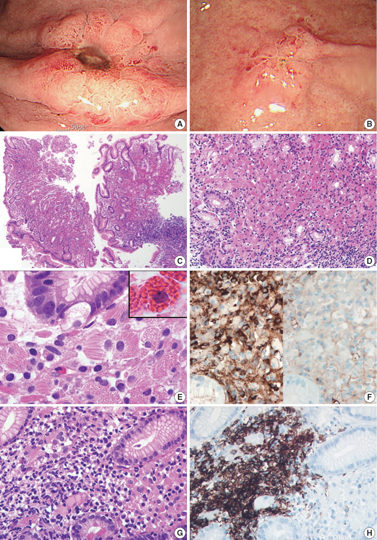
- Gastric crystal-storing histiocytosis with concomitant mucosa-associated lymphoid tissue lymphoma
- Mee Joo, Nam-Hoon Kim
- J Pathol Transl Med. 2020;54(4):332-335. Published online May 22, 2020
- DOI: https://doi.org/10.4132/jptm.2020.04.20
- 7,279 View
- 116 Download
- 5 Web of Science
- 5 Crossref
-
 Abstract
 PDF
PDF - Crystal-storing histiocytosis (CSH) is a rare entity that is characterized by intrahistiocytic accumulation of crystallized immunoglobulins. CSH is not a malignant process per se, but the majority of CSH cases are associated with underlying lymphoproliferative disorder. Although CSH can occur in a variety of organs, gastric CSH is very rare. We present a localized gastric CSH with concomitant mucosaassociated lymphoid tissue (MALT) lymphoma, manifesting as an ulcer bleeding in a 56-year-old man. Histologically, the biopsied gastric mucosa demonstrated expansion of the lamina propria by prominent collections of large eosinophilic mononuclear cells containing fibrillary crystalloid inclusions. Immunohistochemical studies revealed that the crystal-storing cells were histiocytes harboring kappa light chain-restricted immunoglobulin crystals. Within the lesion, atypical centrocyte-like cells forming lymphoepithelial lesions were seen, consistent with MALT lymphoma. Since this entity is rare and unfamiliar, difficulties in diagnosis may arise. Particularly, in this case, the lymphomatous area was obscured by florid CSH, making the diagnosis more challenging.
-
Citations
Citations to this article as recorded by
- Crystal-Storing Histiocytosis of the Stomach: An Unusual Clinical Context of a Rare Entity
Jenna Magri, Katsiaryna Khatskevich, Lauren Shealy, David Lewin, Chadi Hajar
International Journal of Surgical Pathology.2026; 34(1): 222. CrossRef - Sunny side up
João Pedro Pereira, Joanne Lopes, Fatima Carneiro, Francisco Baldaque-Silva
Frontline Gastroenterology.2025; 16(4): 344. CrossRef - Crystal-storing histiocytosis in the stomach: A case report and review of the literature
Linghong Kong, Liyan Xue, Yanfeng Zhong, Shenglan Wang, Danfeng Zheng, Lining Wang, Yang Jiao, Xinpeng Zhang, Huizhong Xue, Xiaogang Liu
Frontiers in Oncology.2022;[Epub] CrossRef - Lambda-Restricted Crystal-Storing Histiocytosis of Stomach: A Case Report and Review of Literature
Nalini Bansal, Pankaj Puri, Nishant Nagpal, Rahul Naithani, Rahul Gupta
Cureus.2021;[Epub] CrossRef - Immunoglobulin-Storing Histiocytosis: A Case Based Systemic Review
Hanne Wiese-Hansen, Friedemann Leh, Anette Lodvir Hemsing, Håkon Reikvam
Journal of Clinical Medicine.2021; 10(9): 1834. CrossRef
- Crystal-Storing Histiocytosis of the Stomach: An Unusual Clinical Context of a Rare Entity
- Diffuse Involvement of Primary Colorectal Lymphoma Simulating Ulcerative Colitis
- Ji-Ye Kim, Sun Hee Chang, Han Seong Kim, Mee Joo
- J Pathol Transl Med. 2019;53(5):332-336. Published online August 2, 2019
- DOI: https://doi.org/10.4132/jptm.2019.07.12
- 7,616 View
- 93 Download
-
Abstract
PDF
- Diffuse involvement of colorectal lymphoma masquerading as colitis is a very rare presentation of primary colorectal lymphoma. Detecting occult lymphoma is difficult in the setting of diffuse colonic involvement with no definite mass and inflammatory mucosal changes. We encountered a case of diffuse-type primary colorectal lymphoma simulating ulcerative colitis in a previously healthy 31-year-old woman. Despite multiple mucosal biopsies, the biopsy diagnosis was not made due to unawareness of atypical lymphocytes admixed with dense lymphoplasmacytic infiltration. The present case emphasizes the importance of being aware of this rare presentation of primary colorectal lymphoma in order to avoid misdiagnosis.
- Rare Gastric Lesions Associated with Helicobacter pylori Infection: A Histopathological Review
- Mee Joo
- J Pathol Transl Med. 2017;51(4):341-351. Published online June 5, 2017
- DOI: https://doi.org/10.4132/jptm.2017.04.03
- 16,584 View
- 351 Download
- 18 Web of Science
- 17 Crossref
-
Abstract
PDF
- Helicobacter pylori infection is associated with chronic gastritis, peptic ulcer disease, gastric adenocarcinoma, and mucosa-associated lymphoid tissue lymphoma. However, some rare gastric lesions exhibiting distinctive histological features may also be associated with H. pylori infection, including lymphocytic gastritis, granulomatous gastritis, Russell body gastritis, or crystal-storing histiocytosis. Although diverse factors can contribute to their development, there is convincing evidence that H. pylori infection may play a pathogenic role. These findings are mainly based on studies in patients with these lesions who exhibited clinical and histological improvements after H. pylori eradication therapy. Thus, H. pylori eradication therapy might be indicated in patients with no other underlying disease, particularly in countries with a high prevalence of H. pylori infection. This review describes the characteristic histological features of these rare lesions and evaluates the evidence regarding a causative role for H. pylori infection in their pathogenesis.
-
Citations
Citations to this article as recorded by- Crystal-Storing Histiocytosis of the Stomach: An Unusual Clinical Context of a Rare Entity
Jenna Magri, Katsiaryna Khatskevich, Lauren Shealy, David Lewin, Chadi Hajar
International Journal of Surgical Pathology.2026; 34(1): 222. CrossRef - Efficacy and safety of antibiotic regimens for Helicobacter pylori eradication: a systematic review and meta-analysis
Yinhua Liao, Hu Liu, Yibin Zhou, Zhenyu Wang
Frontiers in Medicine.2026;[Epub] CrossRef -
Rapid diagnosis and precision treatment of
Helicobacter pylori
infection in clinical settings
Zeeshan Umar, Jia-Wei Tang, Barry J. Marshall, Alfred Chin Yen Tay, Liang Wang
Critical Reviews in Microbiology.2025; 51(2): 369. CrossRef - Granulomatous gastritis due to gastric sarcoidosis presenting as gastric outlet obstruction: Case report of a rare entity
Arkadeep Dhali, Rick Maity, Prajes Kumar Chattopadhyay, Jyotirmoy Biswas, Anjali Garg, Gopal Krishna Dhali
International Journal of Surgery Case Reports.2025;[Epub] CrossRef - Crystal-Storing Histiocytosis: The Iceberg of More Serious Conditions
Mousa Mobarki, Alexandra Papoudou-Bai, Jean Marc Dumollard, Abdulaziz H. Alhazmi, Shaqraa Musawi, Mohammed Ali Madkhali, Khalid Y. Muqri, Michel Péoc’h, Georgia Karpathiou
Diagnostics.2023; 13(2): 271. CrossRef - Acute systemic infection-associated Russell body gastroesophagitis: A case report and literature review
Elizaveta Flerova, Susan Inniss, Nneamaka Nwaoduah, Richard P. Denicola, Jialing Huang
Human Pathology Reports.2023; 31: 300696. CrossRef - Infectious Gastric Diseases Other than Helicobacter
Hyemin Jo, Cheol Min Shin
The Korean Journal of Gastroenterology.2023; 82(6): 269. CrossRef - H. pylori Infection and Virulence Factors cagA and vacA (s and m Regions) in Gastric Adenocarcinoma from Pará State, Brazil
Igor Brasil-Costa, Cintya de Oliveira Souza, Leni Célia Reis Monteiro, Maria Elisabete Silva Santos, Edivaldo Herculano Correa De Oliveira, Rommel Mario Rodriguez Burbano
Pathogens.2022; 11(4): 414. CrossRef - Crystal-storing histiocytosis in the stomach: A case report and review of the literature
Linghong Kong, Liyan Xue, Yanfeng Zhong, Shenglan Wang, Danfeng Zheng, Lining Wang, Yang Jiao, Xinpeng Zhang, Huizhong Xue, Xiaogang Liu
Frontiers in Oncology.2022;[Epub] CrossRef - Dynamics of inflammatory changes of the gastric mucosa in children with duodenal ulcer
T.V. Sorokman, P.M. Moldovan, L.Yu. Khlunovska, I.Ya. Lozyuk
CHILD`S HEALTH.2022; 16(4): 285. CrossRef - Clinicopathological characteristics and aetiological factors of granulomatous gastritis
Yuanxin Liang, Shengjie Cui, Alexandros D Polydorides
Histopathology.2021; 79(6): 1040. CrossRef - A case of Russell body gastritis with multifocal lesions
Michinobu Umakoshi, Ken Miyabe, Hajime Ishii, Yukitsugu Kudo-Asabe, Yukinobu Ito, Makoto Yoshida, Daichi Maeda, Masato Sageshima, Akiteru Goto
SAGE Open Medical Case Reports.2020;[Epub] CrossRef - Gastric crystal-storing histiocytosis with concomitant mucosa-associated lymphoid tissue lymphoma
Mee Joo, Nam-Hoon Kim
Journal of Pathology and Translational Medicine.2020; 54(4): 332. CrossRef - Nonimmunoglobulin Crystal-Storing Histiocytosis (CSH): Case Report and Literature Review
Manuel Beltran, Sharad Khurana, Yennifer Gil, Jason T. Lewis, Rohit Kumar, James M. Foran, Masayuki Nagasawa
Case Reports in Hematology.2020; 2020: 1. CrossRef - Lymphocytic gastritis
Michał A. Puderecki, Lech Wronecki, Katarzyna Cięszczyk, Justyna Szumiło
Polish Journal of Pathology.2019; 70(3): 155. CrossRef - Benign and malignant gastroduodenal diseases associated with Helicobacter pylori: a narrative review and personal remarks in 2018
György M. Buzás
Minerva Gastroenterologica e Dietologica.2018;[Epub] CrossRef - Crystal-storing Histiocytosis in the Stomach
Christina A. Arnold, Wendy L. Frankel, Ling Guo, Chandra Krishnan, Sheryl Pfeil, Melinda Schumacher, Lysandra Voltaggio, Martha M. Yearsley, Wei Chen
American Journal of Surgical Pathology.2018; 42(10): 1317. CrossRef
- Crystal-Storing Histiocytosis of the Stomach: An Unusual Clinical Context of a Rare Entity
- Mesothelin Expression in Gastric Adenocarcinoma and Its Relation to Clinical Outcomes
- Song-Hee Han, Mee Joo, Hanseong Kim, Sunhee Chang
- J Pathol Transl Med. 2017;51(2):122-128. Published online February 15, 2017
- DOI: https://doi.org/10.4132/jptm.2016.11.18
- 11,129 View
- 179 Download
- 23 Web of Science
- 21 Crossref
-
Abstract
PDF
- Background
Although surgical resection with chemotherapy is considered effective for patients with advanced gastric cancer, it remains the third leading cause of cancer-related death in South Korea. Several studies have reported that mesothelial markers including mesothelin, calretinin, and Wilms tumor protein 1 (WT1) were positive in variable carcinomas, associated with prognosis, and were evaluated as potential markers for targeted therapy. The aim of this study was to assess the immunohistochemical expression of mesothelial markers (mesothelin, calretinin, and WT1) in gastric adenocarcinoma and their relations to clinocopathological features and prognosis. Methods: We evaluated calretinin, WT1, and mesothelin expression by immunohistochemical staining in 117 gastric adenocarcinomas. Results: Mesothelin was positively stained in 30 cases (25.6%). Mesothelin expression was related to increased depth of invasion (p = .002), lymph node metastasis (p = .013), and presence of lymphovascular (p = .015) and perineural invasion (p = .004). Patients with mesothelin expression had significantly worse disease-free survival rate compared with that of nonmesothelin expression group (p = .024). Univariate analysis showed that mesothelin expression is related to short-term survival. None of the 117 gastric adenocarcinomas stained for calretinin or WT1. Conclusions: Mesothelin expression was associated with poor prognosis. Our results suggest that mesothelin-targeted therapy should be considered as an important therapeutic alternative for gastric adenocarcinoma patients with mesothelin expression. -
Citations
Citations to this article as recorded by- Mesothelin as a diagnostic biomarker in endometriosis: A cross-sectional study
Kamil Kiecka, Aleksandra Zygula, Anna Sankiewicz, Mariusz Kuzmicki, Slawomir Lawicki, Michal Ciebiera, Tadeusz Issat, Jan Blaszczyk, Krzysztof Cendrowski, Ewa Gorodkiewicz, Piotr Laudanski
Advances in Medical Sciences.2026; 71(1): 1. CrossRef - Mesothelin biology and the evolving landscape of targeted immunotherapy
Remy Boisgard, Patrick Chames, Brigitte Kerfelec
Molecular Therapy Oncology.2026; 34(1): 201148. CrossRef - High mesothelin expression is associated with low cytotoxic T cell infiltration in pancreatic cancer
Oliver Liang, Amy L. White, Timothy Fielder, Joo-Shik Shin, Sharon M. Sagnella, Ulf Schmitz, Dannel Yeo
Frontiers in Immunology.2025;[Epub] CrossRef - On-target off-tumor toxicity of claudin18.2-directed CAR-T cells in preclinical models
Filippo Birocchi, Antonio J. Almazan, Aiyana Parker, Amanda A. Bouffard, Sadie Goncalves, Christopher Kelly, Jessica Frank, Mark B. Leick, Nicholas J. Haradhvala, Shaw Kagawa, Gad Getz, Giulia Escobar, Diego Salas-Benito, Adele Mucci, Trisha R. Berger, Ma
Nature Communications.2025;[Epub] CrossRef - Novel perspectives on MSLN-targeted cancer therapy: from molecular mechanisms to clinical translation
Zhendong Wu, Xuefei Fu, Yuan Feng, Rong Zeng, Huan Qin, Kai Yao
Cancer Biology & Therapy.2025;[Epub] CrossRef - Persistence of activated anti‐mesothelin hYP218 chimeric antigen receptor T cells in the tumour is associated with efficacy in gastric and colorectal carcinomas
Sameer Mir, Abhilash Venugopalan, Jingli Zhang, Nishanth Ulhas Nair, Manjistha Sengupta, Manakamana Khanal, Chaido Stathopoulou, Qun Jiang, Raffit Hassan
Clinical and Translational Medicine.2024;[Epub] CrossRef - Targeting Mesothelin in Solid Tumours: Anti-mesothelin Antibody and Drug Conjugates
Quincy Chu
Current Oncology Reports.2023; 25(4): 309. CrossRef - High expression of mesothelin in plasma and tissue is associated with poor prognosis and promotes invasion and metastasis in gastric cancer
Suryendu Saha, Chitranjan Mukherjee, Dipjit Basak, Prasun Panja, Pronoy Kanti Mondal, Ranajoy Ghosh, Aniket Halder, Abhijit Chowdhury, Gopal Krishna Dhali, Bitan Kumar Chattopadhyay, Saurabh Ghosh, Somsubhra Nath, Shalini Datta
Advances in Cancer Biology - Metastasis.2023; 7: 100098. CrossRef - Novel Anti-Mesothelin Nanobodies and Recombinant Immunotoxins with Pseudomonas Exotoxin Catalytic Domain for Cancer Therapeutics
Minh Quan Nguyen, Do Hyung Kim, Hye Ji Shim, Huynh Kim Khanh Ta, Thi Luong Vu, Thi Kieu Oanh Nguyen, Jung Chae Lim, Han Choe
Molecules and Cells.2023; 46(12): 764. CrossRef - Immunotherapy for lung cancer: Focusing on chimeric antigen receptor (CAR)-T cell therapy
Tongqing Xue, Xiang Zhao, Kun Zhao, Yan Lu, Juan Yao, Xianguo Ji
Current Problems in Cancer.2022; 46(1): 100791. CrossRef - Cellular Immunotherapy in Gastric Cancer: Adoptive Cell Therapy and Dendritic Cell-Based Vaccination
Elnaz Faghfuri, Mahdi Abdoli Shadbad, Amir Hossein Faghfouri, Narges Soozangar
Immunotherapy.2022; 14(6): 475. CrossRef - Mesothelin Expression in Human Tumors: A Tissue Microarray Study on 12,679 Tumors
Sören Weidemann, Pauline Gagelmann, Natalia Gorbokon, Maximilian Lennartz, Anne Menz, Andreas M. Luebke, Martina Kluth, Claudia Hube-Magg, Niclas C. Blessin, Christoph Fraune, Katharina Möller, Christian Bernreuther, Patrick Lebok, Till S. Clauditz, Frank
Biomedicines.2021; 9(4): 397. CrossRef - Host Mesothelin Expression Increases Ovarian Cancer Metastasis in the Peritoneal Microenvironment
Tyvette S. Hilliard, Brooke Kowalski, Kyle Iwamoto, Elizabeth A. Agadi, Yueying Liu, Jing Yang, Marwa Asem, Yuliya Klymenko, Jeff Johnson, Zonggao Shi, Gifty Marfowaa, Madeleine G. Yemc, Phillip Petrasko, M. Sharon Stack
International Journal of Molecular Sciences.2021; 22(22): 12443. CrossRef - CAR-T Cell Therapy—An Overview of Targets in Gastric Cancer
Dominika Bębnowska, Ewelina Grywalska, Paulina Niedźwiedzka-Rystwej, Barbara Sosnowska-Pasiarska, Jolanta Smok-Kalwat, Marcin Pasiarski, Stanisław Góźdź, Jacek Roliński, Wojciech Polkowski
Journal of Clinical Medicine.2020; 9(6): 1894. CrossRef - Mesothelin-Targeted Recombinant Immunotoxins for Solid Tumors
Brendan L. Hagerty, Guillaume J. Pegna, Jian Xu, Chin-Hsien Tai, Christine Alewine
Biomolecules.2020; 10(7): 973. CrossRef - Phase I/II clinical trial of a Wilms’ tumor 1-targeted dendritic cell vaccination-based immunotherapy in patients with advanced cancer
Wen Zhang, Xu Lu, Peilin Cui, Chunmei Piao, Man Xiao, Xuesong Liu, Yue Wang, Xuan Wu, Jingwei Liu, Lin Yang
Cancer Immunology, Immunotherapy.2019; 68(1): 121. CrossRef - Mesothelin as a target for cervical cancer therapy
Korinna Jöhrens, Lea Lazzerini, Jana Barinoff, Jalid Sehouli, Guenter Cichon
Archives of Gynecology and Obstetrics.2019; 299(1): 211. CrossRef - A targeted proteomics approach reveals a serum protein signature as diagnostic biomarker for resectable gastric cancer
Qiujin Shen, Karol Polom, Coralie Williams, Felipe Marques Souza de Oliveira, Mariana Guergova-Kuras, Frederique Lisacek, Niclas G. Karlsson, Franco Roviello, Masood Kamali-Moghaddam
eBioMedicine.2019; 44: 322. CrossRef - Mesothelin as a biomarker for targeted therapy
Jiang Lv, Peng Li
Biomarker Research.2019;[Epub] CrossRef - Antibody drug conjugates under investigation in phase I and phase II clinical trials for gastrointestinal cancer
Alexis D. Leal, Anuradha Krishnamurthy, Lia Head, Wells A. Messersmith
Expert Opinion on Investigational Drugs.2018; 27(11): 901. CrossRef - Mesothelin‑targeted second generation CAR‑T cells inhibit growth of mesothelin‑expressing tumors in�vivo
Lin Ye, Yuqing Lou, Liming Lu, Xiaohong Fan
Experimental and Therapeutic Medicine.2018;[Epub] CrossRef
- Mesothelin as a diagnostic biomarker in endometriosis: A cross-sectional study
- Gastric-Type Extremely Well-Differentiated Adenocarcinoma of the Stomach: A Challenge for Preoperative Diagnosis
- Mee Joo, Song Hee Han
- J Pathol Transl Med. 2016;50(1):71-74. Published online September 30, 2015
- DOI: https://doi.org/10.4132/jptm.2015.07.14
- 14,781 View
- 184 Download
- 10 Web of Science
- 11 Crossref
-
Abstract
PDF
- Gastric-type extremely well-differentiated adenocarcinoma (EWDA) is a rare type of gastric adenocarcinoma characterized by infiltration of well-formed mucinous glands with little or no nuclear atypia, which resemble foveolar epithelium or pyloric glands. Because of its high degree of differentiation, preoperative biopsy diagnosis of gastric-type EWDA is very difficult. We encountered a case of gastric-type EWDA, manifesting as a Borrmann type 4 lesion, in a 47-year-old man. Despite four repeated biopsies, the preoperative biopsy diagnosis was not conclusive due to the scarcity of diagnostic tumor cells and lack of knowledge regarding the unusual histologic findings of gastric-type EWDA. We herein describe the histologic findings of gastric-type EWDA in detail, with the aim of facilitating a preoperative biopsy diagnosis and understanding of this rare type of gastric adenocarcinoma.
-
Citations
Citations to this article as recorded by- Crawling-type Gastric Adenocarcinoma: Clinicopathology, Diagnosis, and Endoscopic Treatment Implications
Cong Bang Huynh, Jin Won Chang
Journal of Digestive Cancer Research.2026; 14(1): 1. CrossRef - Unusual or Uncommon Histology of Gastric Cancer
Jinho Shin, Young Soo Park
Journal of Gastric Cancer.2024; 24(1): 69. CrossRef - Clinical pathological characteristics of “crawling-type” gastric adenocarcinoma cancer: A case report
Yong-Wei Xu, Yan Song, Jun Tian, Ba-Cui Zhang, Yu-Sheng Yang, Jing Wang
World Journal of Gastrointestinal Oncology.2024; 16(4): 1660. CrossRef - Gastric-type extremely well-differentiated adenocarcinoma of the stomach: A rare tumor with diagnostic difficulties and high inter-observer variation in endoscopic pinch biopsies
Soomin Ahn, Sujin Park, Hyun Hee Koh, Han Gyeol Kim, Hyunjin Kim, Jae Yeong Son, Boram Lee, Hyunwoo Lee, Soohyun Hwang, Junhun Cho, Yun Kyung Lee, Ryoji Kushima, Amitabh Srivastava, Kyoung-Mee Kim
Pathology - Research and Practice.2024; 263: 155599. CrossRef - Clinicopathological features of gastric adenocarcinoma of fundic gland type
Bao-Zhen Guo, Zhen-Zhen Liu, Gao-Fei Shen, Fei Zhu, Hui-Fen Lian, Xin Li, Jun-Yi Zheng, Jin-Peng Li, Shui-Miao Deng, Rui Huang
World Chinese Journal of Digestology.2023; 31(6): 244. CrossRef - Characterization of pathological stomach tissue using polarization-sensitive second harmonic generation microscopy
Hwanhee Jeon, MacAulay Harvey, Richard Cisek, Elisha Bennett, Danielle Tokarz
Biomedical Optics Express.2023; 14(10): 5376. CrossRef - Extremely well-differentiated adenocarcinoma of the stomach: diagnostic pitfalls in endoscopic biopsy
Jongwon Lee, In-Seob Lee, Ji Yong Ahn, Young Soo Park, Jihun Kim
Journal of Pathology and Translational Medicine.2022; 56(2): 63. CrossRef - Helicobacter pylori-negative Gastric Cancer
Sun-Young Lee
The Korean Journal of Helicobacter and Upper Gastrointestinal Research.2021; 21(1): 10. CrossRef - Preoperative diagnosis of a gastric extremely well-differentiated adenocarcinoma
Katsushi Suenaga, Shiro Matsumoto, Alan Kawarai Lefor, Yoshimasa Miura, Yoshinori Hosoya, Daigo Kuboki, Hidenori Haruta, Kentaro Kurashina, Atsushi Kihara, Daisuke Matsubara, Yasunari Sakuma, Joji Kitayama, Naohiro Sata
International Journal of Surgery Case Reports.2020; 73(C): 319. CrossRef - Gastric adenocarcinoma of the fundic gland type: clinicopathological features of eight patients treated with endoscopic submucosal dissection
Chengfang Li, Xinglong Wu, Shuang Yang, Xiaorong Yang, Jin Yao, Hong Zheng
Diagnostic Pathology.2020;[Epub] CrossRef - Gastric Adenocarcinoma of the Fundic Gland Type
Mark A Benedict, Gregory Y Lauwers, Dhanpat Jain
American Journal of Clinical Pathology.2018; 149(6): 461. CrossRef
- Crawling-type Gastric Adenocarcinoma: Clinicopathology, Diagnosis, and Endoscopic Treatment Implications
- Malakoplakia Affecting the Umbilical Cord
- Song-Hee Han, Mee Joo, Sunhee Chang, Han-Seong Kim
- J Pathol Transl Med. 2015;49(2):177-179. Published online March 12, 2015
- DOI: https://doi.org/10.4132/jptm.2015.02.04
- 11,096 View
- 44 Download
- 1 Web of Science
- 5 Crossref
-
PDF
-
Citations
Citations to this article as recorded by- A Rare Lesion of Soft Tissue Malakoplakia Mimicking Neoplasm in the Thigh
Asawari Arwikar, Divya Shetty, Anita Sharan
International Journal of Surgical Pathology.2025; 33(6): 1445. CrossRef - A Rare Case of Gastrointestinal Malakoplakia with Achromobacter Sepsis
Bipneet Singh, William Davis, Jahnavi Ethakota, Palak Grover, Anas Kutait
Advances in Infectious Diseases.2025; 15(02): 376. CrossRef - Cecal malakoplakia: A case report
Jin Woon Jeong, Ji Hyun Noh, Jeong Hyun Kang, Ji Hyun Park, Joo Hyung Lee
Korean Journal of Clinical Oncology.2021; 17(1): 44. CrossRef - Rectal malakoplakia
Ted George Achufusi, Kegan Jessamy, Philip Chebaya, Sekou Rawlins
Baylor University Medical Center Proceedings.2020; 33(3): 389. CrossRef - Colonic Malakoplakia: A Rare Finding in a Healthy Male
Rawad A. Yared, Hussein A. Badran, Mohammed Hussein Kamareddine, Youssef Ghosn, Roula Bou Khalil, Khaled El Ajamy, Camil Chouairy, Said G. Farhat
Case Reports in Gastroenterology.2018; 12(2): 453. CrossRef
- A Rare Lesion of Soft Tissue Malakoplakia Mimicking Neoplasm in the Thigh
- Cytologic Findings of Thyroid Carcinoma Showing Thymus-like Differentiation: A Case Report
- Sunhee Chang, Mee Joo, Hanseong Kim
- Korean J Pathol. 2012;46(3):302-305. Published online June 22, 2012
- DOI: https://doi.org/10.4132/KoreanJPathol.2012.46.3.302
- 13,118 View
- 85 Download
- 17 Crossref
-
Abstract
PDF
Carcinoma showing thymus-like differentiation (CASTLE) is a rare carcinoma of the thyroid or adjacent soft tissue of the neck with a histologic resemblance to thymic epithelial tumors. Although the fine-needle aspiration (FNA) plays a central role in the initial evaluation of thyroid nodules, few reports about the cytologic findings of CASTLE have been found according to a review of literatures. We report cytologic findings of a case of CASTLE. A 34-year-old woman presented with a 2-month history of sore throat. The FNA showed that the smear was composed of three dimensional clusters and sheets. The tumor cells were round to ovoid with high nuclear : cytoplasmic ratios. The nuclei were vesicular with small nucleoli. There were some tumor cells showing keratinization. Some lymphocytes were found on the background and within clusters. The presence of poorly-differentiated tumor cells with a focal keratinization and a lymphocytic background on the FNA is suggestive of CASTLE.
-
Citations
Citations to this article as recorded by- Fine‐Needle Aspiration Cytologic Diagnosis of Intrathyroidal Thymic Carcinoma: A Review of the Literature
Qi Chen, Xiaoli Yu
Diagnostic Cytopathology.2025; 53(10): 507. CrossRef - Preoperative Cytological Diagnosis of Intrathyroidal Thymic Carcinoma: A Case Report With Review of the Literature
Makoto Yamada, Shota Sezaki, Tomonari Ikeya, Masatoshi Furuta, Shogo Mizuno, Yoshiro Otsuki
Diagnostic Cytopathology.2025;[Epub] CrossRef - Coexistence of intrathyroid thymic carcinoma and papillary thyroid carcinoma: a case report and literature review
Maryam Vajihinejad, Ali Ataei, Mohammad Pashmchi, Ali Aledavoud, Vahid Zand, Mohammad Ali Broomand, Mohammad Mohammadi, Niloofar Zare Reshkuiyeh
Frontiers in Oncology.2024;[Epub] CrossRef - Intrathyroidal Thymic Carcinoma: A Retrospective Case Series Study
Jinhui Liang, Mei Huang, Helang Huang, Li Li, Hailin Luo, Weidong Mao, Shan Gao, Haoxiang Xu
Ear, Nose & Throat Journal.2023; 102(9): 584. CrossRef - Carcinoma showing thymus‐like differentiation of the parotid gland: The brief report of cytomorphology and review of the literature
Tomoko Uchiyama, Chiyoko Terada, Yuma Tachibana, Hirokazu Nishiura, Maiko Takeda, Tomomi Fujii, Takahiro Kimura, Masahiro Tsutsumi, Chiho Ohbayashi
Diagnostic Cytopathology.2023;[Epub] CrossRef - A case of concurrent occurrence of carcinoma showing thymus-like differentiation and follicular variant of papillary thyroid cancer in the same thyroid
Takahito Kimura, Keisuke Enomoto, Masamitsu Kono, Masanobu Hiraoka, Saori Takeda, Naoko Kumashiro, Shun Hirayama, Eri Kimura, Shunji Tamagawa, Makiko Ohtani, Shin-Ichi Murata, Muneki Hotomi
Journal of Surgical Case Reports.2022;[Epub] CrossRef - Ultrasonographic Features of Intrathyroidal Thymic Carcinoma: Review and Analysis of 10 Cases
Wang, MD Yanhai, Yang, MD Hua, Liu, MD Hanqing, Luo, MD Xiaoli, Liu, BS Luying, Zhou, BS Pingting
ADVANCED ULTRASOUND IN DIAGNOSIS AND THERAPY.2022; 6(2): 58. CrossRef - Thyroid carcinoma with thymus-like differentiation (CASTLE) tumor: а сase report
A. A. Ilyin, V. V. Polkin, P. A. Isaev, F. E. Sevrukov, N. Yu. Dvinskych, M. I. Ryzhenkova, S. A. Ivanov, A. D. Kaprin
Head and neck tumors (HNT).2021; 11(2): 64. CrossRef - Metastatic Renal Cell Neoplasm Within a Papillary Thyroid
Carcinoma as Incidental Finding in an Asymptomatic Patient: a Case Report
Maria-Rosa Bella-Cueto, Mireia Pascua-Solé, Albert Cano-Palomares, M. Àngels Cabezuelo-Hernandez, Maria-Rosa Escoda-Giralt, Santiago Barcons-Vilaplana, Paula Serret-Miralles, Carmen Caral-Vanaclocha, Xavier Guirao-Garriga, Joan Prats-Lopez, Meritxell Meda
SN Comprehensive Clinical Medicine.2020; 2(7): 978. CrossRef - Intrathyroidal thymic carcinoma exhibiting neuroendocrine differentiation: Case report with cytomorphology, immunocytochemistry, and review of the literature focusing on cytology
Wen‐hao Ren, Kun Dong, Xiao‐zheng Huang, Yan‐li Zhu
Diagnostic Cytopathology.2019; 47(11): 1197. CrossRef - Thyroid Carcinoma Showing Thymus-Like Differentiation (CASTLE) with Tracheal Invasion: A Case Report
Adrian Chi Heng Fung, Julian Shun Tsang, Brian Hung Hin Lang
American Journal of Case Reports.2019; 20: 1845. CrossRef - Management and Prognostic Factors for Thyroid Carcinoma Showing Thymus-Like Elements (CASTLE): A Case Series Study
Rui Gao, Xi Jia, Ting Ji, Jinteng Feng, Aimin Yang, Guangjian Zhang
Frontiers in Oncology.2018;[Epub] CrossRef - Multiple squamous cells in thyroid fine needle aspiration: Friends or foes?
Heather Gage, Elizabeth Hubbard, Laurentia Nodit
Diagnostic Cytopathology.2016; 44(8): 676. CrossRef - Clinical analysis of 82 cases of carcinoma showing thymus-like differentiation of the thyroid
WEI GE, YONG-ZHONG YAO, GANG CHEN, YI-TAO DING
Oncology Letters.2016; 11(2): 1321. CrossRef - Carcinoma Showing Thymus-Like Differentiation (CASTLE): Cytopathological Features and Differential Diagnosis
Jennifer A. Collins, Bo Ping, Justin A. Bishop, Syed Z. Ali
Acta Cytologica.2016; 60(5): 421. CrossRef - Carcinoma Showing Thymus-Like Elements of the Thyroid Gland: Report of Three Cases Including One Case with Breast Cancer History
Guanjun Zhang, Xi Liu, Wei Huang, Xiaofeng Li, Marianne Johnstone, Yuan Deng, Yongqiang Ke, Quentin M. Nunes, Hongyan Wang, Yili Wang, Xuebin Zhang
Pathology & Oncology Research.2015; 21(1): 45. CrossRef - Potential Role of Adjuvant Radiation Therapy in Cervical Thymic Neoplasm Involving Thyroid Gland or Neck
Jae Myoung Noh, Sang Yun Ha, Yong Chan Ahn, Dongryul Oh, Seung Won Seol, Young Lyun Oh, Joungho Han
Cancer Research and Treatment.2014; 47(3): 436. CrossRef
- Fine‐Needle Aspiration Cytologic Diagnosis of Intrathyroidal Thymic Carcinoma: A Review of the Literature
- Diagnostic Utility of AMACR and Claudin-7 for the Classification of Renal Cell Carcinoma.
- Sang Hwa Shim, Mee Joo, Han Seong Kim, Sun Hee Chang, Ki Young Kwon
- Korean J Pathol. 2010;44(2):155-161.
- DOI: https://doi.org/10.4132/KoreanJPathol.2010.44.2.155
- 4,514 View
- 53 Download
-
Abstract
PDF
- BACKGROUND
The histologic classification of renal cell carcinoma (RCC) is based on the cytoarchitectural features, yet sometimes this requires correlation with the immunophenotype. Alpha-methylacyl-CoA racemase (AMACR) and claudin-7 have recently been introduced as useful markers that are frequently expressed in papillary RCC (PRCC) and chromophobe RCC (ChRCC), respectively. The aims of this study are to evaluate the expressions of AMACR and claudin-7 in RCCs and to investigate whether they are helpful for making the histological classification of RCCs.
METHODS
Immunohistochemistry for CD10, RCC marker, cytokeratin (CK)7, CD117, AMACR and claudin-7 was performed for 104 RCCs, and these consisted of 54 clear cell RCCs (CCRCC), 26 PRCCs and 24 ChRCCs.
RESULTS
For diagnosing PRCC, the sensitivity and specificity of AMACR were 92.3% and 71.8%, respectively, and using AMACR(+)/CK7(+), the specificity was increased by 23.1% to 94.9%. For diagnosing ChRCC, the sensitivity and specificity of claudin-7 were 91.7% and 78.8%, respectively, and using claudin-7(+)/AMACR(-), the specificity was significantly improved (to 96.3%). For diagnosing CCRCC, CK7(-)/claudin-7(-)/CD117(-) was the most useful immunohistochemical panel (sensitivity, 96.3%; specificity, 98%).
CONCLUSIONS
AMACR and claudin-7 are helpful markers for the histologic classification of RCCs, and their diagnostic utility is strengthened when they are used as an immunohistochemical panel, AMACR(+)/CK7(+) for PRCC, claudin-7(+)/AMACR(-) for ChRCC and CK7(-)/claudin-7(-)/CD117(-) for CCRCC.
- A Case of Endocrine Mucin-Producing Sweat Gland Carcinoma Co-existing with Mucinous Carcinoma: A Case Report.
- Sunhee Chang, Sang Hwa Shim, Mee Joo, Hanseong Kim, Yong Kyu Kim
- Korean J Pathol. 2010;44(1):97-100.
- DOI: https://doi.org/10.4132/KoreanJPathol.2010.44.1.97
- 5,979 View
- 57 Download
- 9 Crossref
-
Abstract
PDF
- An endocrine mucin-producing sweat gland carcinoma (EMPSGC) is a rare skin tumor that most commonly occurs on the eyelids of elderly women. This tumor is morphologically analogous to endocrine ductal carcinoma in situ and solid papillary carcinoma of the breast. We describe one case of a 51-year-old male with an EMPSGC co-existing with mucinous carcinoma of the eyelid. The tumor was composed of dilated ducts with a smooth border and was partially filled with a papillary proliferation. Tumor cells were uniform, small-to-medium in size, and oval-to-polygonal with light eosinophilic cytoplasm. Nuclei were bland with diffusely stippled chromatin and inconspicuous nucleoli. Tumor cells expressed chromogranin, synaptophysin, estrogen and progesterone receptors, cytokeratin 7, and epithelial membrane antigen.
-
Citations
Citations to this article as recorded by- A Case of Endocrine Mucin-Producing Sweat Gland Carcinoma of the Eyelid
Ji Eon Kang, Sung Eun Kim, Suk-Woo Yang
Journal of the Korean Ophthalmological Society.2023; 64(2): 149. CrossRef - Endocrine mucin-producing sweat gland carcinoma: a systematic review and meta-analysis
Michael H. Froehlich, Keith R. Conti, Ivy I. Norris, Jordan J. Allensworth, Nicole A. Ufkes, Shaun A. Nguyen, Evelyn T. Bruner, Joel Cook, Terry A. Day
Journal of Dermatological Treatment.2022; 33(4): 2182. CrossRef - Next-generation sequencing analysis suggests varied multistep mutational pathogenesis for endocrine mucin-producing sweat gland carcinoma with comments on INSM1 and MUC2 suggesting a conjunctival origin
Joseph G. Mathew, Anita S. Bowman, Jad Saab, Klaus J. Busam, Kishwer Nehal, Melissa Pulitzer
Journal of the American Academy of Dermatology.2022; 86(5): 1072. CrossRef - Endocrine mucin‐producing sweat gland carcinoma and associated primary cutaneous mucinous carcinoma: Review of the literature
Rebecca Tian Mei Au, Manish M. Bundele
Journal of Cutaneous Pathology.2021; 48(9): 1156. CrossRef - An Update on Endocrine Mucin-producing Sweat Gland Carcinoma
Meghana Agni, Meisha L. Raven, Randy C. Bowen, Nora V. Laver, Patricia Chevez-Barrios, Tatyana Milman, Charles G. Eberhart, Steven Couch, Daniel D. Bennett, Daniel M. Albert, R. Nick Hogan, Paul O. Phelps, Hillary Stiefel, Norberto Mancera, Martin Hyrcza,
American Journal of Surgical Pathology.2020; 44(8): 1005. CrossRef - A Case of Endocrine Mucin-Producing Sweat Gland Carcinoma: Is it Still an Under-Recognized Entity?
Khaled A. Murshed, Mohamed Ben-Gashir
Case Reports in Dermatology.2020; 12(3): 255. CrossRef - Endocrine Mucin-Producing Sweat Gland Carcinoma, a Histological Challenge
Mary Anne Brett, Samih Salama, Gabriella Gohla, Salem Alowami
Case Reports in Pathology.2017; 2017: 1. CrossRef - Endocrine mucin‐producing sweat gland carcinoma occurring on extra‐facial site: a case report
Jia‐Huei Tsai, Tzu‐Lin Hsiao, Yi‐Ying Chen, Cheng‐Hsiang Hsiao, Jau‐Yu Liau
Journal of Cutaneous Pathology.2014; 41(6): 544. CrossRef - Endocrine Mucin-Producing Sweat Gland Carcinoma
Catharine A. Dhaliwal, Antonia Torgersen, Jonathan J. Ross, James W. Ironside, Asok Biswas
The American Journal of Dermatopathology.2013; 35(1): 117. CrossRef
- A Case of Endocrine Mucin-Producing Sweat Gland Carcinoma of the Eyelid
- Giant Cell Tumor-like Proliferation Associated with Renal Staghorn Calculi: A Case Report.
- Han Seong Kim, Mee Joo, Sun Hee Chang, Ji Eun Kwak, Sang Hwa Shim, Sung Yong Cho
- Korean J Pathol. 2009;43(2):182-184.
- DOI: https://doi.org/10.4132/KoreanJPathol.2009.43.2.182
- 4,498 View
- 24 Download
- 1 Crossref
-
Abstract
PDF
- A 62-year-old man with left flank pain and hematuria was shown to have a staghorn stone in left renal pelvis. Grossly, renal pelvis and calyces were markedly dilated with cystic and hemorrhagic degeneration and renal parenchyma was atrophied. A tumor-like mass was located in a hemorrhagic cyst of the renal upper pole. This mass consisted of giant cells and stromal cells mimicking a giant cell tumor of bone. This giant cell tumor-like proliferation may represent a response to hemorrhage into a cystic cavity. Recognition of this finding is important to avoid the over-diagnosis of neoplastic lesions.
-
Citations
Citations to this article as recorded by- Imaged guided surgery during arteriovenous malformation of gastrointestinal stromal tumor using hyperspectral and indocyanine green visualization techniques: A case report
Tristan Wagner, Onur Mustafov, Marielle Hummels, Anders Grabenkamp, Michael N Thomas, Lars Mortimer Schiffmann, Christiane J Bruns, Dirk L Stippel, Roger Wahba
World Journal of Clinical Cases.2023; 11(23): 5530. CrossRef
- Imaged guided surgery during arteriovenous malformation of gastrointestinal stromal tumor using hyperspectral and indocyanine green visualization techniques: A case report
- Cytologic Findings of Fine Needle Aspiration Biopsy of 23 Schwannomas.
- Sunhee Chang, Mee Joo, Hanseong Kim
- J Pathol Transl Med. 2008;19(1):41-46.
- DOI: https://doi.org/10.3338/kjc.2008.19.1.41
- 3,899 View
- 20 Download
- 1 Crossref
-
Abstract
PDF
- In an attempt to better define the cytologic characteristics of schwannomas, we have reviewed aspirates and corresponding histologic sections from 23 schwannomas. Of this number, the original cytologic diagnoses were: schwannoma in 14 cases (61%), benign soft tissue tumor in 2 cases (9%), and insufficient specimen in 7 cases (30%). The cytologic findings common to all cases of schwannoma included fragments of tightly cohesive fascicles with variable cellularity and corresponding Antoni type A area. The Antoni type B area, consisting of scattered spindle cells and some histiocytes and lymphocytes against a myxoid background, was seen in 14 cases. Fibrillary stroma was seen in 12 cases. The tumor cells had spindle- or oval-shaped nuclei, with pointed ends and indistinct cell borders. Nuclear palisading was seen in 10 cases, and distinctive Verocay bodies were seen in 5 cases. In ancient schwannomas, there were no Verocay bodies. Most schwannomas have distinct cytomorphologic features that allow correct diagnosis. The major problem with fine needle aspiration cytology of these tumors is the high frequency of poor cellularity, particularly in lesions with cystic degeneration. Of 7 cases with insufficient specimen, 4 showed marked cystic changes and 1 showed marked hyaline changes on histologic sections. In conclusion, we believe that if cytopathologist reminds the situation such as cystic degeneration or hyaline degeneration, the correct diagnosis of the schwannoma will be easily made.
-
Citations
Citations to this article as recorded by- Multiple Cervical Schwannomas Mimicking Metastatic Lymph Nodes from Papillary Thyroid Cancer
Ji-Sun Kim, Chang-Young Yoo, Rae-Hyung Kim, Jung-Hae Cho
Journal of Korean Thyroid Association.2014; 7(1): 102. CrossRef
- Multiple Cervical Schwannomas Mimicking Metastatic Lymph Nodes from Papillary Thyroid Cancer
- Lipomatous Hypertrophy of the Interatrial Septum: A Case Report.
- Ji Eun Kwak, Han Seong Kim, Mee Joo, Sun Hee Chang, Sang Hwa Shim, Je G Chi, Wook Sung Kim
- Korean J Pathol. 2007;41(3):213-215.
- 2,299 View
- 19 Download
-
Abstract
PDF
- Lipomatous hypertrophy of the interatrial septum is a benign cardiac mass that is characterized by excessive deposition of fat in the interatrial septum. It typically occurs in elderly, obese patients and may cause arrhythmia. We report a case of lipomatous hypertrophy of the interatrial septum. A 45-year-old man was admitted for evaluation of chest discomfort. Transthoracic echocardiography revealed a cardiac mass, which was suspected as a myxoma. A resection of the tumor was performed. Grossly, the mass was 1.3x1.2x0.8 cm, and showed yellow soft consistency and good demarcation. Microscopically, the mass consisted of mature adipose tissue, intermixed cardiac muscle fibers, entrapped nerve fibers and ganglion cells. Lipomatous hypertrophy of the interatrial septum should be differentiated pathologically from tumorous cardiac mass such as lipoma and myxoma.
- Cryptosporidium Infection of Human Intestine: An Electron Microscopic Observation.
- Min Suk Kim, Yun Kyung Kang, Chul Jong Yoon, Mee Joo, Hye Kyung Lee, Jeong Gi Seo, Je G Chi
- Korean J Pathol. 1999;33(2):121-127.
- 2,245 View
- 10 Download
-
Abstract
- Protozoa of the genus Cryptosporidium are small coccidian parasite known to infect the mucosal epithelium of a variety of animals including human, causing fatal course in immunodeficient patients as well as self-limited illness in healthy individuals. Various life cycle stages including trophozoite, meront, merozoite, gametocyte and oocyst in infected mucosa are a diagnostic feature. Electron microscopy (EM) provides sufficient findings for genus and species identification of this parasitic organism. The authors presented scanning and transmission EM findings of Cryptosporidium parvum infection in two children: one with acute lymphoblastic leukemia and the other without any evidence of immune compromise.
- Congenital Cystic Disease of the Kidney overview and a classification.
- Mee Joo, Yeon Mee Kim, Chong Jai Kim, Yeon Lim Suh, Jeong Wook Seo, Je Geun Chi
- Korean J Pathol. 1997;31(3):233-243.
- 2,107 View
- 22 Download
-
Abstract
PDF
- The congenital renal cystic disease encompasses a complex group of pathologic and clinical entities. We retrospectively reviewed 42 cases of congenital renal cystic lesions classified into four Potter types in a series of 2,063 consecutive autopsies from 1981 to 1996. According to our study based on morphologic, clinical, genetic features and associated anomalies, type I and III are relatively compatible with Potter's original definition. However, it was reasonable that type II and IV are classified to the same group because of: 1) very similar histologic findings representing dysplastic kidney, 2) many associated anomalies, 3) no evidence of inheritance, and 4) presence of a combined type. Syndrome associated cysts, such as Meckel-Gruber syndrome, were also separately classified. If the dysplastic evidence was insufficient for diagnosis to the dysplastic kidney in type II and IV, then these cases would be better classified into a cystic disease associated with congenital hydronephrosis. We propose a classification of the congenital cystic disease of the kidney to be: 1) dysplastic kidney, 2) cystic disease associated with congenital hydronephrosis, 3) polycystic kidney, and 4) syndromic cystic disease.
- Carcinoid Tumor Arising from A Normal Kidney: A Case Report.
- Seo Young Sohn, Han seong Kim, Mee Joo, Min Kyung Kim, Sung Hye Park
- Korean J Pathol. 2004;38(3):196-199.
- 2,137 View
- 12 Download
-
Abstract
PDF
- Primary carcinoid tumor of the kidney is a very rare disease. Until now, only 41 cases have been reported worldwide, and nine of these arose in a horseshoe kidney. In Korea, 3 cases have been reported to date, and all of these arose in a horseshoe kidney. We present a case of primary carcinoid tumor occurring in a normal kidney of a 45 year old man. A tumor was incidentally found close to the hilum of the left kidney. Histologically, the tumor exhibited trabecular and ribbon-like pattern of cuboidal or columnar cells. Mitotic activity was rarely seen. The tumor cells were positive for synaptophysin and chromogranin A. Numerous dense-core neurosecretary granules were observed by the electron microscopic examination. To our knowledge, the present case is the first report of primary renal carcinoid tumor arising in a normal kidney in Korea.
- Urachal Adenocarcinoma with a Concomitant Urachal Remnant: A Case Report.
- Tae Hoon Kim, Mee Joo, Min Kyung Kim, Hanseong Kim, Je G Chi, Jae Y Ro
- Korean J Pathol. 2004;38(4):280-283.
- 2,338 View
- 17 Download
-
Abstract
PDF
- Urachal adenocarcinoma is a rare tumor, and it has similarities with nonurachal adenocarcinoma; therefore, it is sometimes difficult to make a diagnosis. We present a typical case of urachal adenocarcinoma that had all the diagnostic criteria including the presence of an urachal remnant. A 65-year-old woman presented with complaints of a painless gross hematuria. Pelvic CT and cystoscopy showed an intraluminal protruding mass centered in the bladder wall. When diagnosed as adenocarcinoma with a signet ring cell component being noted by frozen biopsy, partial cystectomy with resection of the median umbilical ligament and peritoneum was carried out for a suspected urachal adenocarcinoma. The tumor morphology showed as typical mucinous adenocarcinoma. Characteristic tubular structures showing the typical histology of an urachal remnant was found in the perivesical fat. On immunohistochemical staining, the urachal adenocarcinoma showed a pattern similar to colonic adenocarcinoma, while the urachal remnant showed strong positivity for CK7 and Chromogranin A.
- Combined Xanthogranulomatous Urachitis and Bullous Cystitis: A Case Report.
- Ji Eun Kwak, Han Seong Kim, Mee Joo, Sun Hee Chang, Sang Hwa Shim, Je G Chi, In Rae Cho
- Korean J Pathol. 2008;42(1):41-44.
- 2,474 View
- 29 Download
-
Abstract
PDF
- Xanthogranulomatous inflammation of the urachus is a very rare benign chronic inflammatory disease of an unknown etiology. Herein we report a case of a 31-year-old woman who complained of lower abdominal pain and dysuria. Cystoscopy revealed a bullous change at the dome of the urinary bladder. MRI revealed a cystic mass above the bladder dome that extended to the umbilicus. A partial cystectomy with urachal resection was performed. Pathological examination revealed xanthogranulomatous urachitis combined with bullous cystitis.
- Cellular Schwannoma Arising in a Facial Nerve.
- Mee Joo, Hye Sung Kim, Yun Kyung Kang, Hye Kyung Lee, Jae Young Park
- Korean J Pathol. 1997;31(7):688-691.
- 2,231 View
- 19 Download
-
Abstract
PDF
- Cellular schwaninoma is a variant of schwannoma, which is characterized by predominance of cellular Antoni A area, presence of mitotic activity, nuclear hyperchromasia, pleomorphism, and absence of Verocay body. These pathologic features often prompted a misdiagnosis of malignancy. However, the clinical outcome has indicated the benignity of the tumor. We have experienced a case of cellular schwannoma arising from right facial nerve with right hemifacial weakness and erosion of mastoid process. Grossly, it was a 3.5 x 3 cm sized and relatively well encapsulated mass with yellowish, friable cut surface. Microscopically, cellular growth with moderate cellular pleomorphism and some mitotic activity (5/40 HPFS, up to 2/HPF) were noted. Immunostaining for S-100 protein showed diffuse strong positive reaction.
- Florid von Brunn Nests of the Urinary Bladder: A Case Report.
- Han Seong Kim, Ji Eun Kwak, Sang Hwa Shim, Mee Joo, Sun Hee Chang, Je G Chi, In Rae Cho
- Korean J Pathol. 2008;42(3):169-171.
- 4,863 View
- 175 Download
-
Abstract
PDF
- Among benign proliferations of the urinary bladder, von Brunn nests and cystitis cystica et glandularis are common. Sometimes florid proliferation of von Brunn nests makes an intravesical mass, which mimics tumorous lesions. We report here on a case of florid von Brunn nests, occurred in the 34-year-old man with hematuria. Radiological and cystoscopic examinations reveal a polypoid-papillary lesion and transurethral resection was then performed. Pathologically, prominent proliferations of urothelial cell nests were found deep in the lamina propria. Neither significant cytologic atypia nor muscle invasion was noted. Florid von Brunn nests should be considered both clinically and pathologically in the differential diagnosis of a intravesical mass.
- Synchronous Development of Gastrointestinal Stromal Tumor and Arteriovenous Malformation in the Jejunum: A Case Report.
- Sang Hwa Shim, Yoon Hee Han, Ji Eun Kwak, Sun Hee Chang, Hanseong Kim, Je G Chi, Mee Joo
- Korean J Pathol. 2008;42(3):185-188.
- 2,568 View
- 22 Download
-
Abstract
PDF
- Vascular malformations associated with neoplasms are extremely rare. Herein we report an extraordinary coincidence of arteriovenous malformation (AVM) and gastrointestinal stromal tumor (GIST) in the jejunum. A 44-year-old woman presented with melena and anemia. Abdominal computed tomography revealed a highly vascularized, strong early arterial enhancing soft tissue mass in the jejunum, which was confirmed by angiography to be an AVM supplied by the distal jejunal branch of the superior mesenteric artery. An emergency operation was performed due to active gastrointestinal (GI) bleeding. The resected jejunum showed a protruding, mostly solid subserosal mass. The mass was confirmed to be a spindle cell type GIST and was intermingled with the AVM located in the overlying submucosa and muscularis propria. To our knowledge, this is the first reported case of an AVM associated with a GIST. This case masqueraded radiologically as an AVM alone and presented clinically with GI bleeding.
- Florid Cystic Endosalpingiosis of the Uterus: A Case Report.
- Sang Hwa Shim, Han Seong Kim, Mee Joo, Sun Hee Chang, Ji Eun Kwak
- Korean J Pathol. 2008;42(3):189-191.
- 2,495 View
- 31 Download
-
Abstract
PDF
- A 54-year-old woman presented with vaginal bleeding. On gynecologic and radiologic examinations, bilateral cystic ovarian tumors were suspected. A laparoscopic examination revealed multiple cysts involving both the uterine horns and the posterior surface of the uterus. These were removed by total abdominal hysterectomy and bilateral salpingo-oophorectomy. Microscopically, multiple cysts were identified in the subserosal layer of the uterus, and they were lined with benign tubal type epithelium surrounded by a smooth muscle wall. These features were consistent with "florid cystic endosalpingiosis". Considering the location of the lesion, the lesion has to be distinguished from other benign cystic lesions of the uterus and adnexa, including multicystic mesothelioma, serous cystadenoma and mucinous cystadenoma. Awareness of this lesion will facilitate a correct diagnosis by both the clinician and pathologist.
- A Case Report of Cutaneous Coccidioidomycosis.
- Sunhee Chang, Sang Hwa Shim, Ji Eun Kwak, Mee Joo, Hanseong Kim, Hai Jin Park, Yee Gyung Kwak, Je G Chi
- Korean J Pathol. 2008;42(4):223-225.
- 2,497 View
- 36 Download
-
Abstract
PDF
- Coccidioidomycosis, which results from inhaling the spores of Coccidioides species, is endemic in the southwestern United States. The primary infection site is the lung, and dissemination of the disease can occur. We report a case of cutaneous coccidioidomycosis in a 79-year-old Korean woman who presented with purpura on both lower extremities, but no pulmonary symptoms and no history of visiting an endemic area. Microscopically, skin biopsy showed multiple aggregates of granulomas in the dermis. Numerous multinucleated giant cells were associated with the granulomas. Mature and immature fungal spherules, which were 20 to 30 micrometer in diameter, were present inside and outside the multinucleated giant cells.
- Fibrovascular Polyp of the Hypopharynx: A Case Report.
- Sunhee Chang, Sang Hwa Shim, Ji Eun Kwak, Mee Joo, Hanseong Kim, Bum Jo Jung, Joong Wook Shin, See Young Park, Kyung Ja Cho, Je G Chi
- Korean J Pathol. 2008;42(4):226-228.
- 2,365 View
- 19 Download
-
Abstract
PDF
- We report here on a case of fibrovascular polyp arising in the hypopharynx of a 62-year-old man. Laryngomicroscopic surgery with laser ablation was performed to excise the mass. Histopathologically, the surface of the polyp was covered with mature squamous epithelium. The polyp showed a characteristic lobular proliferation of mature adipose tissue that was separated by myxoid or collagenous connective tissue. Some scattered skeletal muscle bundles were seen in the central portions of the polyp and these bundles were surrounded by a concentric proliferation of the spindle cells; this was reminiscent of Pacinian corpuscles. Regarding their location and the intermingled pattern of proliferating tissues, it is more plausible that the skeletal muscle is a hamartomatous component rather than entrapped, preexisting tissue.
- A Diagnostically Challenging Case of an Infarcted Adenomatoid Tumor of the Epididymis.
- Sunhee Chang, Sang Hwa Shim, Ji Eun Kwak, Mee Joo, Hanseong Kim, Je G Chi, Keon cheol Lee
- Korean J Pathol. 2008;42(4):229-231.
- 2,366 View
- 19 Download
-
Abstract
PDF
- We describe a case of an infarcted adenomatoid tumor of the epididymis that was challenging to diagnose. A 20-year-old man presented with acute left scrotal pain. He was found to have a 2x1.5x1 cm tumor that was relatively well circumscribed on gross examination. There was a central necrotic area that exhibited gaping spaces and ghost outlines of epithelial cells. The periphery of the necrotic lesion showed focally viable adenomatoid tumor. The majority of the tissue adjacent to the necrosis consisted of granulation tissue, fibroblastic and myofibroblastic proliferation, and neutrophils. The fibroblasts and myofibroblasts showed plump nuclei, often with small nucleoli. No mitotic activity was present. The differential diagnosis for an infarcted adenomatoid tumor includes malignant mesothelioma, inflammatory myofibroblastic tumor, and inflammatory conditions. The key to diagnosing an infarcted adenomatoid tumor is to consider it in the differential diagnosis of any spindle cell tumor with necrosis occurring in the genital tract.
- The Expression of Galectin-3 and Galectin-7 in Epithelial Dysplasia and Adenocarcinoma of the Stomach.
- Ji Eun Kwak, Han Seong Kim, Mee Joo, Sun Hee Chang, Sang Hwa Shim, Hye Ran Lee
- Korean J Pathol. 2008;42(6):365-372.
- 2,477 View
- 31 Download
-
Abstract
PDF
- BACKGROUND
Galectin-3 and galectin-7 may play roles in human carcinogenesis. The aim of this study was to investigate the expressions of galectin-3 and galectin-7 in gastric dysplasia and adenocarcinoma. METHODS: We examined the expressions of galectin-3, galectin-7, and P53 in nonneoplastic gastric mucosa, gastric epithelial dysplasia (GED), and adenocarcinoma by immunohistochemistry. Twenty cases of nonneoplastic gastric mucosa, 20 cases of low grade GED, 20 cases of high grade GED, 40 cases of early gastric cancer (EGC), and 60 cases of advanced gastric cancer (AGC) are included for study. RESULTS: In the nonneoplastic tissue, only galectin-3 was expressed in the intestinal metaplasia. Galectin-7 was not stained in the nonneoplastic gastric mucosa. Galectin-3 was expressed in 3 cases of low grade GED (15%), 13 cases of high grade GED (65%), 2 cases of EGC (5%), and 5 cases of AGC (8%), respectively. Galectin-7 was expressed in 2 cases of low grade GED (10%), 3 cases of high grade GED (15%), and 4 cases of EGC (10%), respectively. However, it was not expressed in AGC.
CONCLUSIONS
Based on our results, the expressions of galectin-3 and galectin-7 may contribute to the development of gastric epithelial dysplasia and early gastric cancer, rather than contributing to the progression of gastric adenocarcinoma.
- Pancreatic Pseudocyst with Splenic Involvement: A Case Report.
- Jeong Hwan Chun, Han Seong Kim, Mee Joo, Sun Hee Chang, Ji Eun Kwak, Sang Hwa Shim, Chul Nam Kim, Soon Joo Cha
- Korean J Pathol. 2008;42(6):405-408.
- 2,638 View
- 32 Download
-
Abstract
PDF
- There have been a few reported cases of pancreatic pseudocyst with involvement of adjacent structures including the stomach, duodenum, and even the mediastinum. An intrasplenic pancreatic pseudocyst is an uncommon complication of acute or chronic pancreatitis. We report a case of pancreatic pseudocyst with splenic involvement in a 69-year-old man with a review of literatures. Abdominal CT revealed a 4 cm sized cyst at the pancreatic tail which was extending into the splenic hilum and splenic parenchyma. Distal pancreatectomy and splenectomy was performed. Pathologic findings demonstrated a pseudocyst of pancreatic tail which communicated with the infacted splenic parenchyma. Also vasculitis, endothelial hyperplasia and medial calcification of the splenic artery were observed.
- Survivin and Fas Ligand Expressions Are Correlated with Angiolymphatic Tumor Spread in Medullary Thyroid Carcinoma.
- Min Kyung Kim, Jin Hee Sohn, Mee Joo, Hanseung Kim, Sung Hye Park, Seong Hoe Park, Eo Jin Kim, Seoung Wan Chae
- Korean J Pathol. 2005;39(5):320-325.
- 2,274 View
- 14 Download
-
Abstract
PDF
- BACKGROUND
Medullary thyroid carcinoma (MTC) that originates from C cells comprises about 10% of all the malignant thyroid tumors. Activating mutations of the RET proto-oncogene have been found to be involved in the anti-apoptotic pathway of MTC that harbors the RET mutation. We investigated the correlation between the clinicopathologic parameters and the expressions of survivin, a novel anti-apoptotic molecule, and the other apoptosis-related proteins, and the known prognostic markers.
METHODS
Immunohistochemical staining was performed using antibodies for survivin, Fas, Fas ligand (FasL), bcl-2, calcitonin, CEA and cyclin A in 19 case of MTC; 10 sporadic MTCs, eight multiple endocrine neoplasia (MEN) type 2A MTCs and one familial MTC (FMTC).
RESULTS
Survivin protein expression was found in five cases (26%) and this was correlated with the presence of angiolymphatic tumor emboli (p=0.019). FasL was expressed in 14 cases (74%) and it had correlation with the presence of lymph node metastases (p=0.029). The cyclin A-labeling indices were correlated with local invasiveness (p=0.001).
CONCLUSIONS
Survivin and FasL might be involved in the lymphatic tumor spread of MTC.
- Squamous Cell Carcinoma Arising from Mature Cystic Teratoma of the Ovary: A report of three cases .
- Mee Joo, Han Nae Min, Yun Kyung Kang, Hye Kyung Lee, Young Chae Cho, Eung Soo Lee
- Korean J Pathol. 1999;33(12):1211-1215.
- 2,611 View
- 23 Download
-
Abstract
PDF
- Malignant transformation develops in a little less than 2% of mature cystic teratomas. A wide variety of malignant tumors may arise within benign mature cystic teratomas, and the most common of these is squamous cell carcinoma, which account for 75~85%. In general, the tumors are in an advanced stage and the prognosis is poor as most patients die within a year. However, when the tumor is confined to the ovary, they have a good prognosis and the 5-year survival rate is 63~83%. We experienced three cases of squamous cell carcinoma arising in mature cystic teratoma. Two of the carcinomas occurred in postmenopausal women: 58-(case 1) and 66-(case 2) year-old, and were confined to the ovaries. They were alive 37 months and 18 months after the operation, respectively. The third case was a 45-year-old premenopausal woman who had an extraovarian extension of the tumor and early recurrence within two months. Histologically, cases 1 and 3 were conventional well to moderately differentiated squamous cell carcinomas and case 2 showed a well-differentiated squamous cell carcinoma with exuberant proliferating trichilemmal tumor-like areas.
- Cytologic Analysis of Metastatic Malignant Tumor in Pleural and Ascitic Fluid.
- Mee Joo, Hye Je Cho
- J Pathol Transl Med. 1995;6(2):125-132.
- 3,418 View
- 89 Download
-
Abstract
PDF
- Cytodiagnosis of pleural and ascitic fluid is a commonly performed laboratory examination. Especially, positivity for malignant cells in effusion cytology is very effective and also presents the first sign of malignancy in unknown primary site of the tumor. We examined each 34 cases of pleural and ascitic fluid cytologic specimen diagnosed as metastatic tumor, which was selected among 964 pleural fluid cytology cases and 662 ascitic fluid cytology cases from September 1989 to June 1995. Among the ascitic fluid cytology specimens examined, 34 specimens were positive in 29 patients. The most common primary neoplasms were carcinomas of ovary (32%), stomach (32%), colon (6%), breast (3%), pancreas (3%), and lung (3%) and lymphoreticular neoplasms (3%). The metastatic tumor was predominantly adenocarcinoma type in both pleural (82%) and ascitic (91%) fluid. The study of metastatic adenocarcinoma in effusion from lung, ovary, and stomach was undertaken to find distinctive features for the identification of the primary site. The smears of metastatic pulmonary adenocarcinoma had a tendency to show high grade pleomorphism and many large tight cell clusters, whereas that of the ovarian adenocarcinoma showed low grade pleomorphism with abundant intracytoplasmic vacuoles in relatively clear background. That of the stomach revealed the intermediate features.
- Fine Needle Aspiration Cytology of Periductal Mastitis (Subareolar Abscess) and its Clinical Significance of Cytological Diagnosis.
- Han Seong Kim, Mee Joo, Sun Hee Chang, Ji Eun Kwak, Sung Hye Park, Byung Ju Song
- J Pathol Transl Med. 2006;17(1):27-31.
- 5,009 View
- 125 Download
-
Abstract
PDF
- Periductal mastitis is a specific clinicopathologic entity, and is referred to by several names: recurrent subareolar abscess, squamous metaplasia of the lactiferous duct, and Zuska's disease. Clinically, the differentiation of periductal mastitis from other benign lesions and carcinomas around the nipple frequently proves a difficult proposition. We reviewed the fine needle aspiration cytology (FNAC) of 14 cases of periductal mastitis, which had been diagnosed either radiologically, histologically, or clinically. The patient group included 13 female patients, and one male. The majority of the patients in this group had presented with subareolar masses. All cases evidenced characteristic anucleated squamous cell clusters within a mixed inflammatory background. A diagnosis of periductal mastitis can be rendered fairly readily on excisional biopsy. However, FNAC is considered to be superior to excisional biopsy as an initial diagnostic procedure for any palpable mass in subareolar lesions of the breast. FNAC can also be a useful diagnostic technique in cases of periductal mastitis, even in the early phases of the disease. A definitive diagnosis of subareolar abscess via FNAC will enable the clinician to select the most appropriate medical or surgical treatment.
- Propylthiouracil-induced Hepatic Injury: A Case Report.
- Ji Eun Kwak, Han Seong Kim, Sun Hee Chang, Mee Joo, Je G Chi, Jin Soo Moon, Jong Guk Lee
- Korean J Pathol. 2006;40(4):289-291.
- 2,293 View
- 18 Download
-
Abstract
PDF
- Propylthiouracil (PTU) is one of the most widely used drugs for the treatment of the hyperthyroidism. However the liver damage is a rare adverse effect, which can be fatal. We present a case of PTU-induced hepatitis. A 13-year-old girl was diagnosed as having Graves' disease, for which PTU was administered. Three months after commencing PTU, she developed jaundice accompanied with elevated serum transaminases and total bilirubin levels. The viral markers were all negative. The pathologic findings of the liver biopsy included necrosis of hepatocytes predominantly around the central veins (i.e., zone 3 necrosis) and moderate to severe infiltration of lymphocytes in portal areas. After withdrawal of the PTU, clinical symptoms and liver function test results were improved gradually.
- Flow Cytometric DNA Analysis in Papillary Carcinoma of Thyroid Gland: comparison with Ki-67 immunohistochemical staining.
- Mee Joo, Hye Je Cho
- Korean J Pathol. 1996;30(11):959-965.
- 2,138 View
- 18 Download
-
Abstract
PDF
- Nuclear DNA content was measured using a flow cytometric method to analyze 36 paraffin- embedded and 7 fresh tissues of 43 papillary carcinomas of thyroid gland. DNA aneuploidy was found in 3 cases(6.9%) and diploidy in 40 cases(93.1%). But there were no suggestive findings in clinical history, and cytological and morphological features for aneuploidy. In 40 diploid cases, S-phase fraction(SPF) were analyzed with regard to sex, age, tumor size, presence or absence of capsular invasion, lymph node involvement and ground glass nuclei. Among the multiple factors, only the tumor size, especially the larger sized-group(above 2cm in tumor diameter) was found to have a statistically significant higher SPF than the smaller sized-group (p<0.05). And high SPF groups relatively well corresponded to the high risk group. Thirty nine cases of papillary carcinoma have also been evaluated for proliferative activity with Ki-67 monoclonal antibody. The average Ki-67 labeling index was 0.36% in total cases, and that of the aneuploid cases was 0.73%, which was higher than that of the diploid cases(0.33%). So. We think that the low aneuploid rate and low Ki-67 labeling index relatively well represent the usual good clinical course of this tumor and the high SPF is a suggestive finding for a high risk group.
- Expression of c-kit and Cell Cycle Regulators in Non-small Cell Lung Carcinoma.
- Sun Hee Chang, Mee Joo, Hanseong Kim
- Korean J Pathol. 2006;40(6):427-431.
- 2,436 View
- 23 Download
-
Abstract
PDF
- BACKGROUND
The abnormal expression of c-kit is implicated in the pathogenesis of a variety of solid tumors. The Rb pathway and p53 act as cell cycle regulators. The purpose of this study was to assess the expression of c-kit, Rb, p53, p16 and cyclin D1 and their relationship to clinical and pathological parameters in patients with non-small cell lung carcinomas (NSCLC(s)).
METHODS
Tissue microarrays consisting of 2 mm cores from the corresponding blocks were constructed from 54 NSCLC(s). Immunohistochemical staining for c-kit, Rb, p53, p16 and cyclin D1 was performed. C-kit immunostaining was considered positive if > or =10% of tumor cells were immunoreactive along the membrane and/or in cytoplasm. For Rb, p53, p16 and cyclin D1, tumor cells showing a nuclear staining pattern were interpreted as positive.
RESULTS
We found that c-kit was expressed in 13 (24%) cases, Rb was lost in 39 (72%) cases, p53 was expressed in 28 (52%) cases, p16 was lost in 42 (78%) cases and cyclin D1 was expressed in 33 (61%) cases. The c-kit expression was significantly higher in adenocarcinoma (39%) than in squamous cell carcinoma (8%). We did not find any correlation between c-kit, Rb, p53, p16 and cyclin D1 expression and clinicopathological parameters such as: age, tumor size, lymph node involvement, disease stage and distant metastasis. There was a direct correlation between p53 expression and Rb loss.
CONCLUSIONS
These results suggest that c-kit may be a useful therapeutic target for patients with c-kit positive tumors, and that the disruption of Rb and p53 pathways may play an important role in the development and progression of NSCLC(s).
- Clear Cell Sarcoma of the Kidney: A case in 39 year old man.
- Hyun Ju Yoo, Yun Kyung Kang, Mee Joo, Hye Kyung Lee, Dae Woo Kim, Suk San Park
- Korean J Pathol. 1996;30(12):1138-1143.
- 2,625 View
- 11 Download
-
Abstract
PDF
- Clear cell sarcoma of kidney(CCSK) is a rare pediatric neoplasm characterized by a predominating component of clear cells, a predilection for metastases to bone, and a poor prognosis. The incidence of CCSK peaks during the 2nd year of life and adult cases are very rare. We report a case of CCSK encountered in the right kidney of a 39-year-old man. Grossly, it was a lobulated mass showing infiltrative margin, measured 7x5.5x5cm and had a homogeneous gray-tan color with a soft, fish-flesh consistency. Microscopically, about half of the tumor revealed the classic pattern of CCSK, having tumor cell cords or nests separated by the characteristic alveolar capillary networks. The tumor cells had clear pale cytoplasm, bland looking round nuclei and inconspicuous nucleoli. The other half showed the epithelioid-trabecular pattern forming pseudorosette or cord-like structures. Immunohistochemically, there was only a focal positive reaction to vimentin. Ultrastructurally, the tumor cells showed the primitive nephrogenic mesenchymal differentiation such as electron lucent cytoplasm, a small amount of organelles, scanty heterochromatin, inconspicuous nucleoli, and a lack of flocculant basal lamina material around the cytoplasmic membrane. We consider that this is a case of CCSK occuring in the oldest patient ever reported, confirmed by both immunohistochemistry and electron microscopy.
 First
First Prev
Prev


