 E-submission
Search
- Page Path
- HOME > Search
- Clinicopathological profile of high-grade differentiated thyroid carcinoma in Indonesian tertiary hospital
- Novita , Agnes Stephanie Harahap, Maria Francisca Ham, Alfianto Widiono, Chan Kwon Jung
- Received October 20, 2025 Accepted January 15, 2026 Published online April 23, 2026
- DOI: https://doi.org/10.4132/jptm.2026.01.15 [Epub ahead of print]
- 279 View
- 12 Download
-
 Abstract
 PDF
PDF Supplementary Material
 - Background
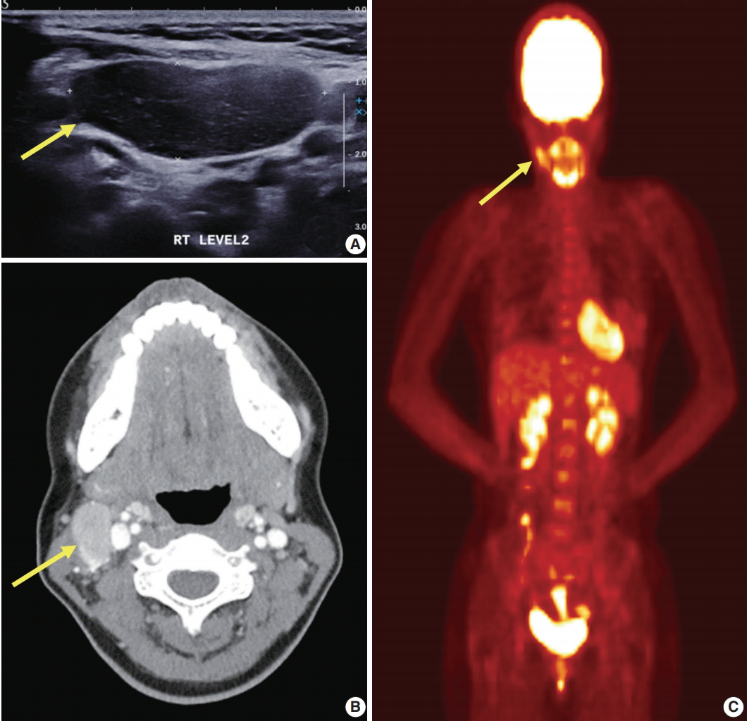
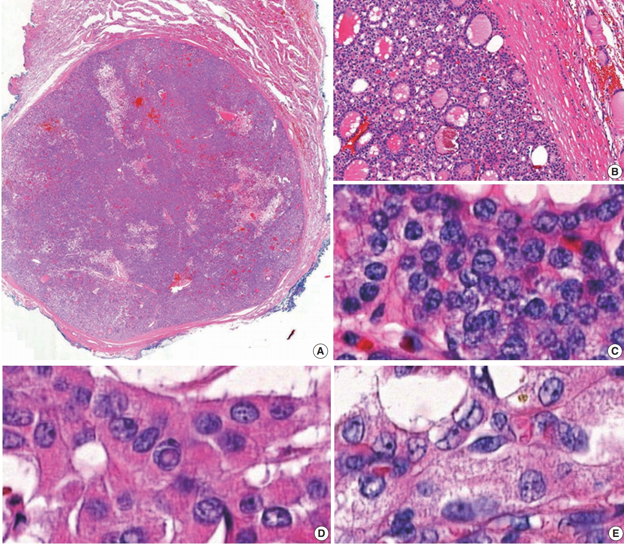
High-grade differentiated thyroid carcinoma (HGDTC) is a recently recognized entity in the 2022 World Health Organization classification, representing a more aggressive subtype of differentiated thyroid carcinoma. Previously, high-grade features such as increased mitotic activity and tumor necrosis were often overlooked, despite being important independent prognostic factors. Although rare, HGDTC carries significant diagnostic, prognostic, and therapeutic implications. Data remain limited in Indonesia. Methods: This retrospective descriptive study reviewed 565 thyroid carcinoma cases diagnosed at Cipto Mangunkusumo Hospital from 2019 to 2024. Eleven cases (1.9%) met HGDTC criteria. Clinicopathological characteristics, histologic subtypes, Ki-67 proliferation index, molecular alterations, treatment modalities, and clinical outcomes were analyzed. Results: Patients had a mean age of 54.6 years, with a female-to-male ratio of 2.7:1. Papillary thyroid carcinoma was the main type (90.9%), with the tall cell subtype predominating. Mean tumor size was 6.4 cm. Lymphatic invasion, vascular invasion, and extrathyroidal extension were present in 54.5%, 18.2%, and 45.5% of cases, respectively. All tumors showed necrosis. Mean mitotic count was 3 per 2 mm². The Ki-67 index ranged from 5% to 45% (median, 14%). BRAFV600E and TERT promoter mutations were detected in 18.2% and 36.4% of cases, respectively, with co-mutations in 18.2%. Six cases (54.5%) had metastases at time of diagnosis. During a mean follow-up of 20.5 months, one patient (9.1%) developed new vertebral metastases and all patients (100%) remained alive. Conclusions: HGDTC presents with more aggressive characteristics and a worse prognosis. Accurate diagnosis, molecular profiling, and long-term monitoring are essential for optimal management.
- Fine needle aspiration cytology diagnoses of follicular thyroid carcinoma: results from a multicenter study in Asia
- Hee Young Na, Miyoko Higuchi, Shinya Satoh, Kaori Kameyama, Chan Kwon Jung, Su-Jin Shin, Shipra Agarwal, Jen-Fan Hang, Yun Zhu, Zhiyan Liu, Andrey Bychkov, Kennichi Kakudo, So Yeon Park
- J Pathol Transl Med. 2024;58(6):331-340. Published online November 7, 2024
- DOI: https://doi.org/10.4132/jptm.2024.10.12
- 7,517 View
- 274 Download
- 1 Web of Science
- 2 Crossref
-
Abstract
PDFSupplementary Material
- Background
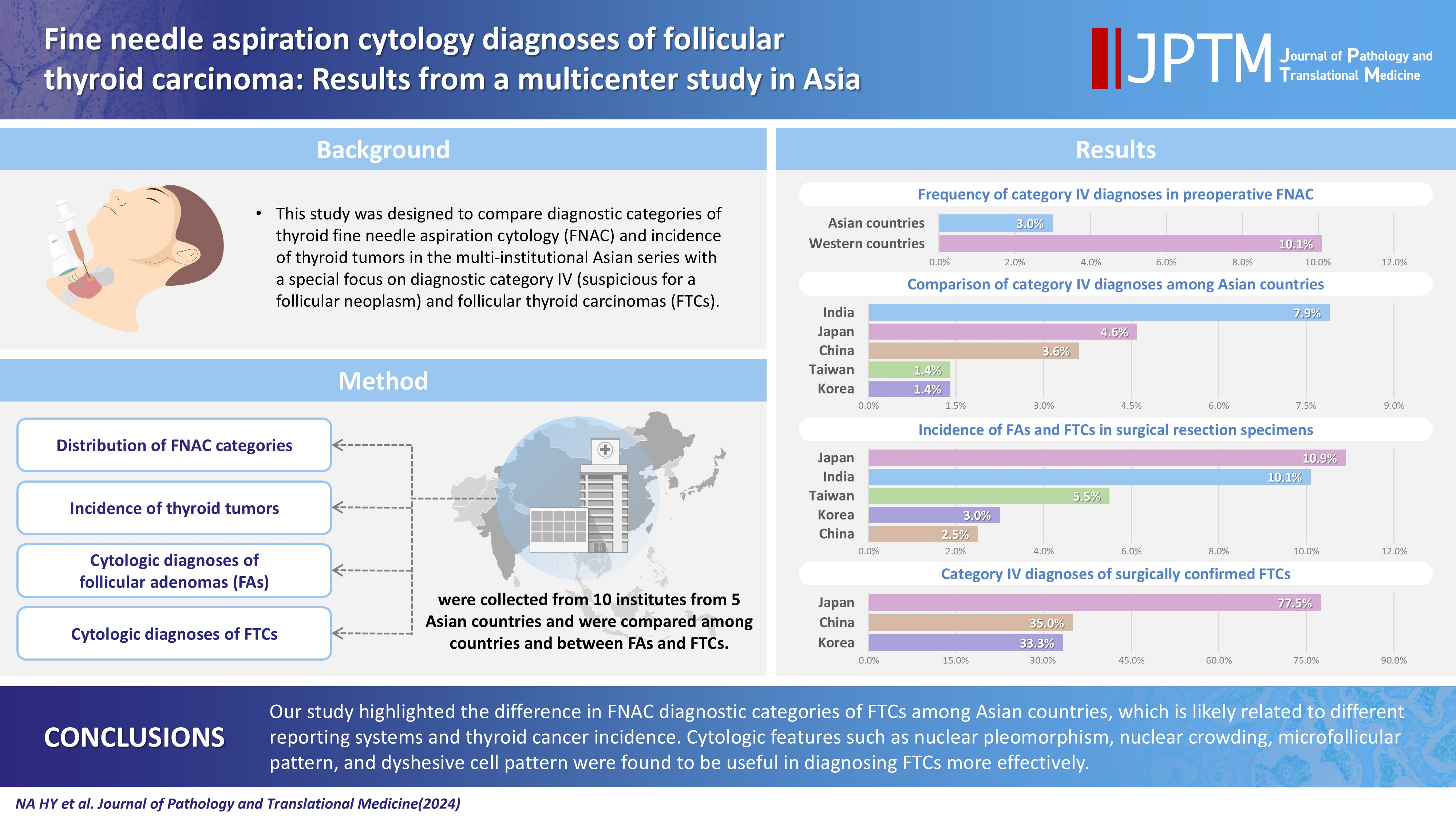
This study was designed to compare diagnostic categories of thyroid fine needle aspiration cytology (FNAC) and incidence of thyroid tumors in the multi-institutional Asian series with a special focus on diagnostic category IV (suspicious for a follicular neoplasm) and follicular thyroid carcinomas (FTCs). Methods: Distribution of FNAC categories, incidence of thyroid tumors in resection specimens and cytologic diagnoses of surgically confirmed follicular adenomas (FAs) and FTCs were collected from 10 institutes from five Asian countries and were compared among countries and between FAs and FTCs. Results: The frequency of category IV diagnoses (3.0%) in preoperative FNAC were significantly lower compared to those in Western countries (10.1%). When comparing diagnostic categories among Asian countries, category IV was more frequent in Japan (4.6%) and India (7.9%) than in Taiwan (1.4%), Korea (1.4%), and China (3.6%). Similarly, incidence of FAs and FTCs in surgical resection specimens was significantly higher in Japan (10.9%) and India (10.1%) than in Taiwan (5.5%), Korea (3.0%), and China (2.5%). FTCs were more commonly diagnosed as category IV in Japan (77.5%) than in Korea (33.3%) and China (35.0%). Nuclear pleomorphism, nuclear crowding, microfollicular pattern, and dyshesive cell pattern were more common in FTCs compared with FAs. Conclusions: Our study highlighted the difference in FNAC diagnostic categories of FTCs among Asian countries, which is likely related to different reporting systems and thyroid cancer incidence. Cytologic features such as nuclear pleomorphism, nuclear crowding, microfollicular pattern, and dyshesive cell pattern were found to be useful in diagnosing FTCs more effectively. -
Citations
Citations to this article as recorded by
- Deep Learning-Based Multimodal Fusion of Ultrasound, Cytology, and Clinical Features to Distinguish Follicular Thyroid Carcinoma from Adenoma: A Multicenter Study
Xiao-Fei Guo, Li Zhou, Xin-Yi Bao, Shui-Qing Liu, Jia-Wei Feng, You-Long Zhu, Yong Jiang, Shu-Ying Zhang
Academic Radiology.2026;[Epub] CrossRef - Misdiagnosed follicular adenoma with 11 year postoperative liver and lung metastases a case report and literature review
Kai-Li Yang, Heng-Tong Han, Shou-Hua Li, Xiao-Xiao Li, Ze Yang, Li-Bin Ma, Yong-Xun Zhao
Discover Oncology.2025;[Epub] CrossRef
- Deep Learning-Based Multimodal Fusion of Ultrasound, Cytology, and Clinical Features to Distinguish Follicular Thyroid Carcinoma from Adenoma: A Multicenter Study
- Uncommon granulomatous manifestation in Epstein-Barr virus–positive follicular dendritic cell sarcoma: a case report
- Henry Goh Di Shen, Yue Zhang, Wei Qiang Leow
- J Pathol Transl Med. 2025;59(2):133-138. Published online October 31, 2024
- DOI: https://doi.org/10.4132/jptm.2024.09.27
- 5,004 View
- 353 Download
- 6 Web of Science
- 6 Crossref
-
Abstract
PDF
- Hepatic Epstein-Barr virus–positive inflammatory follicular dendritic cell sarcoma (EBV+ IFDCS) represents a rare form of liver malignancy. The absence of distinct clinical and radiological characteristics, compounded by its rare occurrence, contributes to a challenging diagnosis. Here, we report a case of a 54-year-old Chinese female with a background of chronic hepatitis B virus treated with entecavir and complicated by advanced fibrosis presenting with a liver mass found on her annual surveillance ultrasound. Hepatectomy was performed under clinical suspicion of hepatocellular carcinoma. Immunomorphologic characteristics of the tumor were consistent with EBV+ IFDCS with distinct non-caseating granulomatous inflammation. Our case illustrates the importance of considering EBV+ IFDCS in the differential diagnosis of hepatic inflammatory lesions. Awareness of this entity and its characteristic features is essential for accurately diagnosing and managing this rare neoplasm.
-
Citations
Citations to this article as recorded by- Mesenchymal Tumors of the Liver: An Update Review
Joon Hyuk Choi, Swan N. Thung
Biomedicines.2025; 13(2): 479. CrossRef - EBV-positive inflammatory follicular dendritic cell sarcoma occurring in different organs: a case report and literature review
Wenhua Bai, Chunfang Hu, Zheng Zhu
Frontiers in Oncology.2025;[Epub] CrossRef - Spleen EBV-positive inflammatory follicular dendritic cell sarcoma: a case report and literature review
Yi Xiao, Lanlan Li, Xiumei Zhan, Juner Xu, Yewu Chen, Qiuchan Zhao, Yinghao Fu, Xian Luo, Huadi Chen, Hao Xu
Frontiers in Oncology.2025;[Epub] CrossRef - Epstein-Barr virus-positive inflammatory follicular dendritic cell sarcoma of the liver: clinical features, imaging findings and potential diagnostic clues
Gui-Ling Huang, Man-Qian Huang, Yu-Ting Zhang, Hui-Ning Huang, Hong-Tao Liu, Xiao-Qing Pei
Abdominal Radiology.2025;[Epub] CrossRef - Epstein‑Barr virus+ inflammatory follicular dendritic cell sarcoma with clonal immunoglobulin heavy chain gene rearrangement: A case report and literature review
Qian Ye, Juan Zhao, Jiao He, Weishan Zhang
Oncology Letters.2025; 31(2): 1. CrossRef - Primary hepatic follicular dendritic cell sarcoma: A case study and literature review
Junjie Zhu, Ying Liang, Li Zhang, Bingqi Li, Danfeng Zheng, Hangyan Wang
Journal of International Medical Research.2025;[Epub] CrossRef
- Mesenchymal Tumors of the Liver: An Update Review
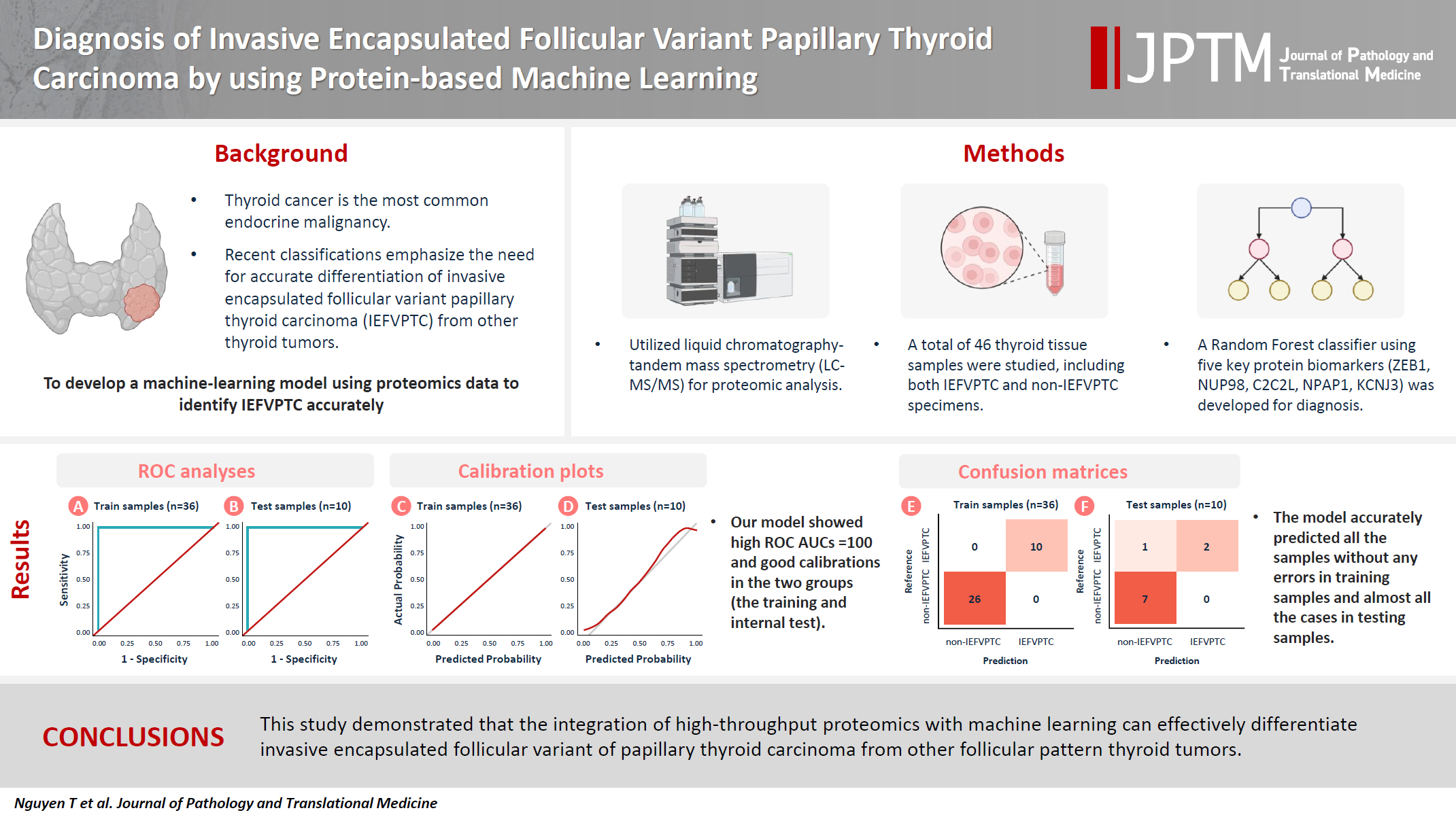
- Diagnosis of invasive encapsulated follicular variant papillary thyroid carcinoma by protein-based machine learning
- Truong Phan-Xuan Nguyen, Minh-Khang Le, Sittiruk Roytrakul, Shanop Shuangshoti, Nakarin Kitkumthorn, Somboon Keelawat
- J Pathol Transl Med. 2025;59(1):39-49. Published online October 24, 2024
- DOI: https://doi.org/10.4132/jptm.2024.09.14
- 5,245 View
- 339 Download
- 2 Web of Science
- 2 Crossref
-
Abstract
PDFSupplementary Material
- Background
Although the criteria for follicular-pattern thyroid tumors are well-established, diagnosing these lesions remains challenging in some cases. In the recent World Health Organization Classification of Endocrine and Neuroendocrine Tumors (5th edition), the invasive encapsulated follicular variant of papillary thyroid carcinoma was reclassified as its own entity. It is crucial to differentiate this variant of papillary thyroid carcinoma from low-risk follicular pattern tumors due to their shared morphological characteristics. Proteomics holds significant promise for detecting and quantifying protein biomarkers. We investigated the potential value of a protein biomarker panel defined by machine learning for identifying the invasive encapsulated follicular variant of papillary thyroid carcinoma, initially using formalin- fixed paraffin-embedded samples.
Methods
We developed a supervised machine-learning model and tested its performance using proteomics data from 46 thyroid tissue samples.
Results
We applied a random forest classifier utilizing five protein biomarkers (ZEB1, NUP98, C2C2L, NPAP1, and KCNJ3). This classifier achieved areas under the curve (AUCs) of 1.00 and accuracy rates of 1.00 in training samples for distinguishing the invasive encapsulated follicular variant of papillary thyroid carcinoma from non-malignant samples. Additionally, we analyzed the performance of single-protein/gene receiver operating characteristic in differentiating the invasive encapsulated follicular variant of papillary thyroid carcinoma from others within The Cancer Genome Atlas projects, which yielded an AUC >0.5.
Conclusions
We demonstrated that integration of high-throughput proteomics with machine learning can effectively differentiate the invasive encapsulated follicular variant of papillary thyroid carcinoma from other follicular pattern thyroid tumors. -
Citations
Citations to this article as recorded by- Advances in immunotherapy for thyroid malignancies: from molecular targets to clinical outcomes
Shuo Lv, Jinbao Wang, Guohao Chen, Yongshun Wang, Naiqing Liu
Frontiers in Medicine.2026;[Epub] CrossRef - Misdiagnosed follicular adenoma with 11 year postoperative liver and lung metastases a case report and literature review
Kai-Li Yang, Heng-Tong Han, Shou-Hua Li, Xiao-Xiao Li, Ze Yang, Li-Bin Ma, Yong-Xun Zhao
Discover Oncology.2025;[Epub] CrossRef
- Advances in immunotherapy for thyroid malignancies: from molecular targets to clinical outcomes
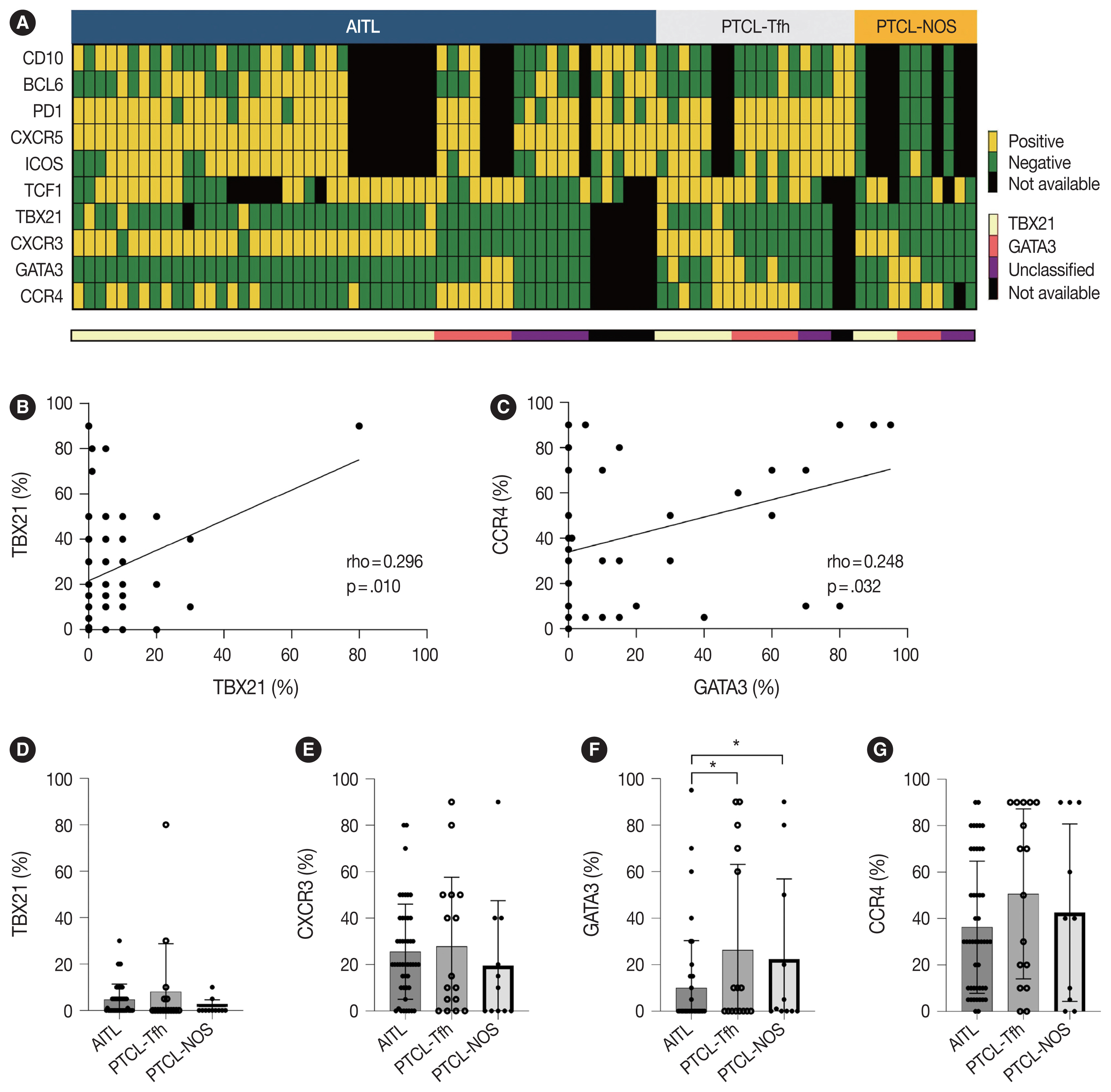
- Clinicopathological implications of immunohistochemical expression of TBX21, CXCR3, GATA3, CCR4, and TCF1 in nodal follicular helper T-cell lymphoma and peripheral T-cell lymphoma, not otherwise specified
- Bogyeong Han, Sojung Lim, Jeemin Yim, Young Keun Song, Jiwon Koh, Sehui Kim, Cheol Lee, Young A Kim, Yoon Kyung Jeon
- J Pathol Transl Med. 2024;58(2):59-71. Published online January 22, 2024
- DOI: https://doi.org/10.4132/jptm.2024.01.04
- 8,961 View
- 379 Download
- 4 Web of Science
- 4 Crossref
-
Abstract
PDFSupplementary Material
- Background
The classification of nodal peripheral T-cell lymphoma (PTCL) has evolved according to histology, cell-of-origin, and genetic alterations. However, the comprehensive expression pattern of follicular helper T-cell (Tfh) markers, T-cell factor-1 (TCF1), and Th1- and Th2-like molecules in nodal PTCL is unclear.
Methods
Eighty-two cases of nodal PTCL were classified into 53 angioimmunoblastic T-cell lymphomas (AITLs)/nodal T-follicular helper cell lymphoma (nTFHL)-AI, 18 PTCLs-Tfh/nTFHL–not otherwise specified (NOS), and 11 PTCLs-NOS according to the revised 4th/5th World Health Organization classifications. Immunohistochemistry for TCF1, TBX21, CXCR3, GATA3, and CCR4 was performed.
Results
TCF1 was highly expressed in up to 68% of patients with nTFHL but also in 44% of patients with PTCL-NOS (p > .05). CXCR3 expression was higher in AITLs than in non-AITLs (p = .035), whereas GATA3 expression was higher in non-AITL than in AITL (p = .007) and in PTCL-Tfh compared to AITL (p = .010). Of the cases, 70% of AITL, 44% of PTCLTfh/ nTFHL-NOS, and 36% of PTCL-NOS were subclassified as the TBX21 subtype; and 15% of AITL, 38% of PTCL-Tfh/nTFHL-NOS, and 36% of PTCL-NOS were subclassified as the GATA3 subtype. The others were an unclassified subtype. CCR4 expression was associated with poor progression-free survival (PFS) in patients with PTCL-Tfh (p < .001) and nTFHL (p = .023). The GATA3 subtype showed poor overall survival in PTCL-NOS compared to TBX21 (p = .046) and tended to be associated with poor PFS in patients with non-AITL (p = .054).
Conclusions
The TBX21 subtype was more prevalent than the GATA3 subtype in AITL. The GATA3 subtype was associated with poor prognosis in patients with non-AITL and PTCL-NOS. -
Citations
Citations to this article as recorded by- T-bet: biological functions, molecular mechanisms, and therapeutic applications: a systematic review
Xiaowen Yang, Min Sun, Xinyi Tang, Xiaoyuan Zhang, Wenzhi Shen
Frontiers in Immunology.2026;[Epub] CrossRef - CXCR Family and Hematologic Malignancies in the Bone Marrow Microenvironment
Yanquan Liu, Huanwen Tang
Biomolecules.2025; 15(5): 716. CrossRef - Diagnostic and therapeutic pathways for lymphoma patients: expert consensus through Nominal Group Technique and Delphi methodology
Attilio Guarini, Valentina Bozzoli, Sabino Ciavarella, Michele Cimminiello, Francesca Donatelli, Angelo Fama, Vincenza Fernanda Fesce, Vincenzo Fraticelli, Francesco Gaudio, Giuseppina Greco, Augusto Martellini, Francesca Merchionne, Rosanna Maria Miccoli
Frontiers in Oncology.2025;[Epub] CrossRef - Prognostic Significance of TBX21 and GATA3 Subtype Classification in Indolent Adult T‐Cell Leukemia‐Lymphoma With Cutaneous Lesions
Kazuhiro Kawai, Youhei Uchida, Takuro Kanekura
The Journal of Dermatology.2025; 52(11): 1674. CrossRef
- T-bet: biological functions, molecular mechanisms, and therapeutic applications: a systematic review
- Identification of invasive subpopulations using spatial transcriptome analysis in thyroid follicular tumors
- Ayana Suzuki, Satoshi Nojima, Shinichiro Tahara, Daisuke Motooka, Masaharu Kohara, Daisuke Okuzaki, Mitsuyoshi Hirokawa, Eiichi Morii
- J Pathol Transl Med. 2024;58(1):22-28. Published online January 10, 2024
- DOI: https://doi.org/10.4132/jptm.2023.11.21
- 6,179 View
- 280 Download
- 4 Web of Science
- 5 Crossref
-
Abstract
PDF
- Background
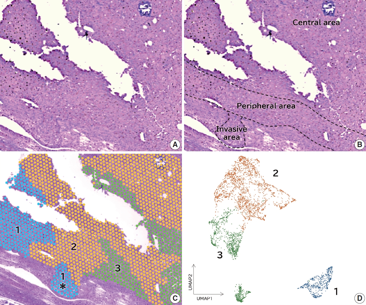
Follicular tumors include follicular thyroid adenomas and carcinomas; however, it is difficult to distinguish between the two when the cytology or biopsy material is obtained from a portion of the tumor. The presence or absence of invasion in the resected material is used to differentiate between adenomas and carcinomas, which often results in the unnecessary removal of the adenomas. If nodules that may be follicular thyroid carcinomas are identified preoperatively, active surveillance of other nodules as adenomas is possible, which reduces the risk of surgical complications and the expenses incurred during medical treatment. Therefore, we aimed to identify biomarkers in the invasive subpopulation of follicular tumor cells.
Methods
We performed a spatial transcriptome analysis of a case of follicular thyroid carcinoma and examined the dynamics of CD74 expression in 36 cases.
Results
We identified a subpopulation in a region close to the invasive area, and this subpopulation expressed high levels of CD74. Immunohistochemically, CD74 was highly expressed in the invasive and peripheral areas of the tumor.
Conclusions
Although high CD74 expression has been reported in papillary and anaplastic thyroid carcinomas, it has not been analyzed in follicular thyroid carcinomas. Furthermore, the heterogeneity of CD74 expression in thyroid tumors has not yet been reported. The CD74-positive subpopulation identified in this study may be useful in predicting invasion of follicular thyroid carcinomas. -
Citations
Citations to this article as recorded by- Carbonic Anhydrase 12 as a Novel Prognostic Biomarker and Therapeutic Target for High‐Risk Follicular Thyroid Carcinoma
Masashi Tanida, Tsuyoshi Takashima, Shinichiro Tahara, Masaharu Kohara, Haruka Kanai, Masami Suzuki, Motoyuki Suzuki, Mitsuyoshi Hirokawa, Ayana Suzuki, Shinya Sato, Daisuke Okuzaki, Satoshi Nojima, Takahiro Matsui, Hidenori Inohara, Eiichi Morii
Cancer Science.2026; 117(1): 257. CrossRef - An emerging role of CD74 in thyroid follicular cells in Hashimoto´s thyroiditis
Pablo Sacristán-Gómez, Ana Serrano-Somavilla, Nuria Sánchez de la Blanca, Andrea Álvarez-Rodríguez, Eduardo Martínez-Parra, Miguel Sampedro-Nuñez, Fernando Sebastián-Valles, Mónica Marazuela, Rebeca Martínez-Hernández
Biomedicine & Pharmacotherapy.2026; 194: 118945. CrossRef - Diagnosis of invasive encapsulated follicular variant papillary thyroid carcinoma by protein-based machine learning
Truong Phan-Xuan Nguyen, Minh-Khang Le, Sittiruk Roytrakul, Shanop Shuangshoti, Nakarin Kitkumthorn, Somboon Keelawat
Journal of Pathology and Translational Medicine.2025; 59(1): 39. CrossRef - Spatial Transcriptomics in Thyroid Cancer: Applications, Limitations, and Future Perspectives
Chaerim Song, Hye-Ji Park, Man S. Kim
Cells.2025; 14(12): 936. CrossRef - A New Tool to Decrease Interobserver Variability in Biomarker Annotation in Solid Tumor Tissue for Spatial Transcriptomic Analysis
Sravya Palavalasa, Emily Baker, Jack Freeman, Aditri Gokul, Weihua Zhou, Dafydd Thomas, Wajd N. Al-Holou, Meredith A. Morgan, Theodore S. Lawrence, Daniel R. Wahl
Current Issues in Molecular Biology.2025; 47(7): 531. CrossRef
- Carbonic Anhydrase 12 as a Novel Prognostic Biomarker and Therapeutic Target for High‐Risk Follicular Thyroid Carcinoma
- Noninvasive follicular thyroid neoplasm with papillary-like nuclear features: its updated diagnostic criteria, preoperative cytologic diagnoses and impact on the risk of malignancy
- Hee Young Na, So Yeon Park
- J Pathol Transl Med. 2022;56(6):319-325. Published online November 9, 2022
- DOI: https://doi.org/10.4132/jptm.2022.09.29
- 10,605 View
- 323 Download
- 9 Web of Science
- 9 Crossref
-
Abstract
PDF
- Due to the extremely indolent behavior, a subset of noninvasive encapsulated follicular variant papillary thyroid carcinomas has been classified as “noninvasive follicular thyroid neoplasm with papillary-like nuclear features (NIFTP)” since 2016 and is no longer considered carcinoma. Since the introduction of this new terminology, changes and refinements have been made in diagnostic criteria. Initially, the incidence of NIFTP was estimated substantial. However, the reported incidence of NIFTP varies greatly among studies and regions, with higher incidence in North American and European countries than in Asian countries. Thus, the changes in the risk of malignancy (ROM) in the Bethesda System for Reporting Thyroid Cytopathology (TBSRTC) differ inevitably among regions. Because more conservative surgery is recommended for NIFTPs, distinguishing NIFTPs from papillary thyroid carcinomas in preoperative fine-needle aspiration cytology became one of the major concerns. This review will provide comprehensive overview of updates on diagnostic criteria, actual incidence and preoperative cytologic diagnoses of NIFTP, and its impact on the ROM in TBSRTC.
-
Citations
Citations to this article as recorded by- Diagnosis of invasive encapsulated follicular variant papillary thyroid carcinoma by protein-based machine learning
Truong Phan-Xuan Nguyen, Minh-Khang Le, Sittiruk Roytrakul, Shanop Shuangshoti, Nakarin Kitkumthorn, Somboon Keelawat
Journal of Pathology and Translational Medicine.2025; 59(1): 39. CrossRef - Papillae, psammoma bodies, and/or many nuclear pseudoinclusions are helpful criteria but should not be required for a definitive cytologic diagnosis of papillary thyroid carcinoma: An institutional experience of 207 cases with surgical follow up
Tarik M. Elsheikh, Matthew Thomas, Jennifer Brainard, Jessica Di Marco, Erica Manosky, Bridgette Springer, Dawn Underwood, Deborah J. Chute
Cancer Cytopathology.2024; 132(6): 348. CrossRef - ThyroSeq overview on indeterminate thyroid nodules: An institutional experience
Sam Sirotnikov, Christopher C. Griffith, Daniel Lubin, Chao Zhang, Nabil F. Saba, Dehong Li, Amanda Kornfield, Amy Chen, Qiuying Shi
Diagnostic Cytopathology.2024; 52(7): 353. CrossRef - Oncocytic Noninvasive Follicular Thyroid Neoplasm with Papillary-Like Nuclear Features: A Case Report
Kaveripakam Ajay Joseph, Sana Ahuja, Sufian Zaheer
Indian Journal of Surgical Oncology.2024; 15(S4): 606. CrossRef - Cytologic hallmarks and differential diagnosis of papillary thyroid carcinoma subtypes
Agnes Stephanie Harahap, Chan Kwon Jung
Journal of Pathology and Translational Medicine.2024; 58(6): 265. CrossRef - Preoperative evaluation of thyroid nodules – Diagnosis and management strategies
Tapoi Dana Antonia, Lambrescu Ioana Maria, Gheorghisan-Galateanu Ancuta-Augustina
Pathology - Research and Practice.2023; 246: 154516. CrossRef - Reevaluating diagnostic categories and associated malignancy risks in thyroid core needle biopsy
Chan Kwon Jung
Journal of Pathology and Translational Medicine.2023; 57(4): 208. CrossRef - Strategies for Treatment of Thyroid Cancer
Deepika Yadav, Pramod Kumar Sharma, Rishabha Malviya, Prem Shankar Mishra
Current Drug Targets.2023; 24(5): 406. CrossRef - Identification of NIFTP-Specific mRNA Markers for Reliable Molecular Diagnosis of Thyroid Tumors
So-Yeon Lee, Jong-Lyul Park, Kwangsoon Kim, Ja Seong Bae, Jae-Yoon Kim, Seon-Young Kim, Chan Kwon Jung
Endocrine Pathology.2023; 34(3): 311. CrossRef
- Diagnosis of invasive encapsulated follicular variant papillary thyroid carcinoma by protein-based machine learning
- Follicular lymphoma: updates for pathologists
- Mahsa Khanlari, Jennifer R. Chapman
- J Pathol Transl Med. 2022;56(1):1-15. Published online December 27, 2021
- DOI: https://doi.org/10.4132/jptm.2021.09.29
- 34,981 View
- 1,077 Download
- 22 Web of Science
- 21 Crossref
-
Abstract
PDF
- Follicular lymphoma (FL) is the most common indolent B-cell lymphoma and originates from germinal center B-cells (centrocytes and centroblasts) of the lymphoid follicle. Tumorigenesis is believed to initiate early in precursor B-cells in the bone marrow (BM) that acquire the t(14;18)(q32;q21). These cells later migrate to lymph nodes to continue their maturation through the germinal center reaction, at which time they acquire additional genetic and epigeneticabnormalities that promote lymphomagenesis. FLs are heterogeneous in terms of their clinicopathologic features. Most FLs are indolent and clinically characterized by peripheral lymphadenopathy with involvement of the spleen, BM, and peripheral blood in a substantial subset of patients, sometimes accompanied by constitutional symptoms and laboratory abnormalities. Diagnosis is established by the histopathologic identification of a B-cell proliferation usually distributed in an at least partially follicular pattern, typically, but not always, in a lymph node biopsy. The B-cell proliferation is biologically of germinal center cell origin, thus shows an expression of germinal center-associated antigens as detected by immunophenotyping. Although many cases of FLs are typical and histopathologic features are straightforward, the biologic and histopathologic variability of FL is wide, and an accurate diagnosis of FL over this disease spectrum requires knowledge of morphologic variants that can mimic other lymphomas, and rarely non-hematologic malignancies, clinically unique variants, and pitfalls in the interpretation of ancillary studies. The overall survival for most patients is prolonged, but relapses are frequent. The treatment landscape in FL now includes the application of immunotherapy and targeted therapy in addition to chemotherapy.
-
Citations
Citations to this article as recorded by- Frequency and Distribution of Lymphomas in Northwestern India: A Retrospective Analysis of 923 Cases Using the Latest World Health Organization Classification 5th Edition
Immanuel Paul Thayakaran, Biren Parikh
Indian Journal of Hematology and Blood Transfusion.2026; 42(3): 809. CrossRef - Follicular Cholecystitis: A Case Report Highlighting the Diagnostic Challenges and Management Implications
Ativitch Asavachaisuvikom, Burana Khiankaew, Narongsak Rungsakulkij
Gastro Hep Advances.2026; 5(2): 100833. CrossRef - Follicular lymphoma with signet ring cell morphology: Clinicopathologic analysis of 31 cases
Xenia Parisi, L. Jeffrey Medeiros
Human Pathology.2026; 171: 106071. CrossRef - PRIMARY SPLENIC DIFFUSE LARGE B-CELL LYMPHOMA WITH CD30 EXPRESSION: A RARE CASE REPORT
Beyza Öztürk, Hüseyin Buğra Kutlu, Meltem Ayyıldız Mercan, Dicle Tamer Türk, Yusuf Emre Aytin, Funda Üstün, Fulya Öz Puyan
TURKISH MEDICAL STUDENT JOURNAL.2026;[Epub] CrossRef - Relapsed/Refractory Follicular Lymphoma: Current Advances and Emerging Perspectives
Giulio Caridà, Enrica Antonia Martino, Antonella Bruzzese, Daniele Caracciolo, Caterina Labanca, Francesco Mendicino, Eugenio Lucia, Virginia Olivito, Teresa Rossi, Antonino Neri, Ernesto Vigna, Pierfrancesco Tassone, Pierosandro Tagliaferri, Fortunato Mo
European Journal of Haematology.2025; 114(5): 775. CrossRef - IGH/IGK gene rearrangement in the diagnosis of B-cell non-Hodgkin lymphoma: experience from three centers
Ke Yang, Zhizhong Wang, Beibei Xin, Yunhang Li, Jiuzhou Zhao, Rui Sun, Weizhen Wang, Dongxu Chen, Chengzhi Zhao, Yongjun Guo, Jie Ma, Bing Wei
Annals of Hematology.2025; 104(7): 3779. CrossRef - Imaging Evaluation of Periarticular Soft Tissue Masses in the Appendicular Skeleton: A Pictorial Review
Francesco Pucciarelli, Maria Carla Faugno, Daniela Valanzuolo, Edoardo Massaro, Lorenzo Maria De Sanctis, Elisa Zaccaria, Marta Zerunian, Domenico De Santis, Michela Polici, Tiziano Polidori, Andrea Laghi, Damiano Caruso
Journal of Imaging.2025; 11(7): 217. CrossRef - Understanding the clinical approach to “pathologically ambiguous follicular lymphoma” through a Real-World cohort
Sarah Matarasso Greenfeld, Svetlana Dmitrienko, Ian Shrier, Jean Luc Deschenes, Sarit Assouline
Leukemia & Lymphoma.2025; 66(12): 2332. CrossRef - Deciphering and targeting oncogenic pathways through integrated approaches and amino acid metabolism in hematologic malignancies
Farhan Ikhtiar, Adil Jamal, Syed M. Safeer Mehdi Bokhari
Discover Oncology.2025;[Epub] CrossRef - Transformation of low-grade follicular lymphoma to a high-grade follicular lymphoma with the histopathological diagnosis from oral biopsy: a case report
Gabriela Silveira de Araujo, Leandro Dorigan de Macedo, Alfredo Ribeiro-Silva, Hilton Marcos Alves Ricz, Lara Maria Alencar Ramos Innocentini
Hematology, Transfusion and Cell Therapy.2024; 46: S380. CrossRef - The follicular lymphoma tumor microenvironment at single-cell and spatial resolution
Andrea J. Radtke, Mark Roschewski
Blood.2024; 143(12): 1069. CrossRef - Chronic pancreatitis for the clinician: complications and special forms of the disease. Interdisciplinary position paper of the Catalan Society of Digestology (SCD) and the Catalan Pancreatic Society (SCPanc)
Xavier MOLERO, Juan R. AYUSO, Joaquim BALSELLS, Jaume BOADAS, Juli BUSQUETS, Anna CASTERÀS, Mar CONCEPCIÓN, Míriam CUATRECASAS, Gloria FERNÀNDEZ ESPARRACH, Esther FORT, Francisco GARCIA BOROBIA, Àngels GINÈS, Lucas ILZARBE, Carme LORAS, Miquel MASACHS, Xa
Minerva Gastroenterology.2024;[Epub] CrossRef - Concurrent identification of follicular lymphoma and papillary thyroid carcinoma
Lama A. Alzelfawi, Norah ALhumaidan, Abrar H. Alageel, Buthaina J. Yahya, Saud D. Alrasheedi, Adel S. Alqahtani
International Journal of Surgery Case Reports.2024;[Epub] CrossRef - Impact of Primary Disease Site of Involvement by Early-Stage Follicular Lymphoma on Patient Outcomes
Olivia Davis, Carmen Lessani, Rana Kasht, Andrew Cohoon, Sami Ibrahimi, Adam Asch, Silas Day, Taha Al-Juhaishi
Clinical Lymphoma Myeloma and Leukemia.2024; 24(12): 837. CrossRef - Recent developments in CD19-targeted therapies for follicular lymphoma
Aditi Saha, Julio C. Chavez
Expert Opinion on Biological Therapy.2024; 24(10): 1049. CrossRef - Unraveling the complexity of follicular lymphoma: insights and innovations
Xijing Li
American Journal of Cancer Research.2024; 14(12): 5573. CrossRef - Clinical features and prognostic factors in 49 patients with follicular lymphoma at a single center: A retrospective analysis
Hao Wu, Hui-Cong Sun, Gui-Fang Ouyang
World Journal of Clinical Cases.2023; 11(14): 3176. CrossRef - A rare case of follicular lymphoma of the bladder
Matthew DeSanto, Robert Strait, Jared Zopp, Kevin Brown, Samuel Deem
Urology Case Reports.2023; 51: 102542. CrossRef - Analysis of immunophenotypic features in hyaline vascular type Castleman disease
Yu Chang, Yu Ma, Chen Chang, Wensheng Li
Diagnostic Pathology.2023;[Epub] CrossRef - Leg Edema Unveiled: The Uncommon Culprit of Follicular Lymphoma
Syed Muhammad IbnE Ali Jaffari, Samaha Nisar, Narjis Malik, Syed Muhammad Aun Ali Jaffari, Omar Nisar
Journal of Shalamar Medical & Dental College - JSHMDC.2023; 4(2): 125. CrossRef - A Review of the Totality of Evidence in the Development of ABP 798, A Rituximab Biosimilar
Patrick Cobb, Dietger Niederwieser, Stanley Cohen, Caroline Hamm, Gerd Burmester, Neungseon Seo, Sonya G Lehto, Vladimir Hanes
Immunotherapy.2022; 14(9): 727. CrossRef
- Frequency and Distribution of Lymphomas in Northwestern India: A Retrospective Analysis of 923 Cases Using the Latest World Health Organization Classification 5th Edition
- Composite follicular lymphoma and classic Hodgkin lymphoma
- Han-Na Kim, Min Ji Jeon, Eun Sang Yu, Dae Sik Kim, Chul-Won Choi, Young Hyeh Ko
- J Pathol Transl Med. 2022;56(1):57-60. Published online November 16, 2021
- DOI: https://doi.org/10.4132/jptm.2021.10.09
- 8,408 View
- 247 Download
- 6 Web of Science
- 6 Crossref
-
Abstract
PDF
- Composite lymphoma is very rare and a combination of Hodgkin lymphoma and non-Hodgkin lymphoma and even histiocytic tumors can occur. Because of the unfamiliarity, not only can this cause diagnostic problems, but can also affect treatment plan. We report a case of composite lymphoma in a 40-year-old male. Initial biopsy showed a composite lymphoma of follicular lymphoma grade 1 and classic Hodgkin lymphoma. After chemotherapy, another lymph node was taken because of disease progression, which revealed follicular lymphoma, grade 3a without Hodgkin lymphoma component.
-
Citations
Citations to this article as recorded by- Composite Lymphoma: A Rare Case of Vomiting
Changqin Liu, Dongyan Han, Xiaomin Sun
United European Gastroenterology Journal.2025; 13(5): 836. CrossRef - BCL2-Rearrangment-Negative CD23+ Follicle Center Lymphoma and Chronic Lymphocytic Leukemia/Small Lymphocytic Lymphoma: A Rare Case of Biclonal Composite Lymphoma
Hira Qadir, Ejas Palathingal Bava, Juan Gomez-Gelvez, Wei Liu, Kedar Inamdar, Elizabeth Wey, John Carey, Yulei Shen, Philip Kuriakose, Sharmila Ghosh
Cureus.2025;[Epub] CrossRef - T cell lymphoma and secondary primary malignancy risk after commercial CAR T cell therapy
Guido Ghilardi, Joseph A. Fraietta, James N. Gerson, Vivianna M. Van Deerlin, Jennifer J. D. Morrissette, Gabriel C. Caponetti, Luca Paruzzo, Jaryse C. Harris, Elise A. Chong, Sandra P. Susanibar Adaniya, Jakub Svoboda, Sunita D. Nasta, Ositadimma H. Ugwu
Nature Medicine.2024; 30(4): 984. CrossRef - Double trouble: insights from a rare case of extranodal composite lymphoma in an elderly man, with comprehensive literature review
Aadya Kerkar
American Journal of Translational Research.2024; 16(6): 2599. CrossRef - Composite Lymphoma with Follicular Lymphoma Transformation to Clonally Related Epstein–Barr Virus (EBV) Positive Diffuse Large B-Cell Lymphoma and EBV-PositiveClassic Hodgkin Lymphoma
Christopher B. Ryder, Hayder Saeed, Mohammad Hussaini, Pier Paolo Piccaluga
Case Reports in Hematology.2023; 2023: 1. CrossRef - Plasticity in Classical Hodgkin Composite Lymphomas: A Systematic Review
Alexis Trecourt, Marie Donzel, Juliette Fontaine, Hervé Ghesquières, Laurent Jallade, Gabriel Antherieu, Camille Laurent, Claire Mauduit, Alexsandra Traverse-Glehen
Cancers.2022; 14(22): 5695. CrossRef
- Composite Lymphoma: A Rare Case of Vomiting
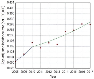
- Upward trend in follicular lymphoma among the Korean population: 10-year experience at a large tertiary institution
- Meejeong Kim, Hee Sang Hwang, Hyungwoo Cho, Dok Hyun Yoon, Cheolwon Suh, Chan Sik Park, Heounjeong Go, Jooryung Huh
- J Pathol Transl Med. 2021;55(5):330-337. Published online September 2, 2021
- DOI: https://doi.org/10.4132/jptm.2021.07.25
- 7,588 View
- 128 Download
- 4 Web of Science
- 5 Crossref
-
Abstract
PDFSupplementary Material
- Background
Follicular lymphoma (FL) is the second most common non-Hodgkin lymphoma (NHL) in Western countries. However, it is relatively rare in Asia. This study examined epidemiologic characteristics of FL in South Korea, with an emphasis on recent trends of increase in cases.
Methods
We retrospectively examined 239 cases of newly diagnosed FL at a large tertiary institution in Korea (Asan Medical Center, Seoul, Republic of Korea) between 2008 and 2017. Age-adjusted incidence rates and clinicopathological variables were analyzed, and joinpoint regression analysis was used to identify the changes.
Results
The age-adjusted incidence of FL significantly increased during the study period (p = .034), and the ratio of (relative incidence) patients with FL to patients with NHL increased from 4.28% to 9.35% in the same period. Over the 10-year study assessment duration, the proportion of patients with stage III/IV FL (p = .035) and expression of BCL2 (p = .022) or BCL6 (p = .039) significantly increased. From 2013–2017, the proportion of patients with highrisk Follicular Lymphoma International Prognostic Index (FLIPI) score increased (21.5% to 28.7%), whereas that of low-risk FLIPI decreased (55.4% to 38.6%), although those results were not statistically significant (p = .066).
Conclusions
We found an increasing incidence of FL, with a disproportionate increase in the incidence of high-stage disease and recent changes in the clinicopathologic features of the Korean patient population. -
Citations
Citations to this article as recorded by- Incidence Trend of Follicular Lymphoma in Taiwan Compared to Japan and Korea, 2001–2019
Liang-Chun Chiu, Chih-Wen Lin, Hung-Ju Li, Jian-Han Chen, Fu-Cheng Chuang, Sheng-Fung Lin, Yu Chang, Yu-Chieh Su
Journal of Clinical Medicine.2023; 12(4): 1417. CrossRef - A Case Report on the Complete Response of a Patient with Recurrent Follicular Lymphoma Treated with Integrative Medicine
Kyung-dug Park, Jisoo Kim, Yoona Oh, Beom-Jin Jeong, Yu-jin Jung, Sunhwi Bang
The Journal of Internal Korean Medicine.2023; 44(3): 585. CrossRef - Recent Updates on Diagnosis and Treatment of Follicular Lymphoma
Ga-Young Song, Deok-Hwan Yang
The Korean Journal of Medicine.2023; 98(5): 231. CrossRef - Classical Hodgkin lymphoma following follicular lymphoma: a case report
Bomi Kim
Journal of Yeungnam Medical Science.2023; 40(Suppl): S113. CrossRef - Incidence, clinicopathological features and genetics of in‐situ follicular neoplasia: a comprehensive screening study in a Japanese cohort
Naoki Oishi, Takahiro Segawa, Kunio Miyake, Kunio Mochizuki, Tetsuo Kondo
Histopathology.2022; 80(5): 820. CrossRef
- Incidence Trend of Follicular Lymphoma in Taiwan Compared to Japan and Korea, 2001–2019
- Morphologic variant of follicular lymphoma reminiscent of hyaline-vascular Castleman disease
- Jiwon Koh, Yoon Kyung Jeon
- J Pathol Transl Med. 2020;54(3):253-257. Published online February 5, 2020
- DOI: https://doi.org/10.4132/jptm.2019.12.17
- 8,808 View
- 242 Download
- 5 Web of Science
- 4 Crossref
-
Abstract
PDF
- Follicular lymphoma (FL) with hyaline-vascular Castleman disease (FL-HVCD)-like features is a rare morphologic variant, with fewer than 20 cases in the literature. Herein, we report a case of FL-HVCD in a 37-year-old female who presented with isolated neck lymph node enlargement. The excised lymph node showed features reminiscent of HVCD, including regressed germinal centers (GCs) surrounded by onion skin-like mantle zones, lollipop lesions composed of hyalinized blood vessels penetrating into regressed GCs, and hyalinized interfollicular stroma. In addition, focal areas of abnormally conglomerated GCs composed of homogeneous, small centrocytes with strong BCL2, CD10, and BCL6 expression were observed, indicating partial involvement of the FL. Several other lymphoid follicles showed features of in situ follicular neoplasia. Based on the observations, a diagnosis of FL-HVCD was made. Although FLHVCD is very rare, the possibility of this variant should be considered in cases resembling CD. Identification of abnormal, neoplastic follicles and ancillary immunostaining are helpful for proper diagnosis.
-
Citations
Citations to this article as recorded by- Unicentric Castleman Disease: Illustration of Its Morphologic Spectrum and Review of the Differential Diagnosis
Siba El Hussein, Andrew G. Evans, Hong Fang, Wei Wang, L. Jeffrey Medeiros
Archives of Pathology & Laboratory Medicine.2024; 148(1): 99. CrossRef - Finding a Needle in the Haystack
Hung-Yu Lin, Yi-Jen Peng, Yi-Ying Wu, Ping-Ying Chang
Journal of Medical Sciences.2023; 43(6): 292. CrossRef - Analysis of immunophenotypic features in hyaline vascular type Castleman disease
Yu Chang, Yu Ma, Chen Chang, Wensheng Li
Diagnostic Pathology.2023;[Epub] CrossRef - In‐situ follicular neoplasia: a clinicopathological spectrum
Gurdip S Tamber, Myriam Chévarie‐Davis, Margaret Warner, Chantal Séguin, Carole Caron, René P Michel
Histopathology.2021; 79(6): 1072. CrossRef
- Unicentric Castleman Disease: Illustration of Its Morphologic Spectrum and Review of the Differential Diagnosis
- A Multi-institutional Study of Prevalence and Clinicopathologic Features of Non-invasive Follicular Thyroid Neoplasm with Papillary-like Nuclear Features (NIFTP) in Korea
- Ja Yeong Seo, Ji Hyun Park, Ju Yeon Pyo, Yoon Jin Cha, Chan Kwon Jung, Dong Eun Song, Jeong Ja Kwak, So Yeon Park, Hee Young Na, Jang-Hee Kim, Jae Yeon Seok, Hee Sung Kim, Soon Won Hong
- J Pathol Transl Med. 2019;53(6):378-385. Published online October 21, 2019
- DOI: https://doi.org/10.4132/jptm.2019.09.18
- 9,440 View
- 346 Download
- 17 Web of Science
- 16 Crossref
-
Abstract
PDF
- Background
In the present multi-institutional study, the prevalence and clinicopathologic characteristics of non-invasive follicular thyroid neoplasm with papillary-like nuclear features (NIFTP) were evaluated among Korean patients who underwent thyroidectomy for papillary thyroid carcinoma (PTC).
Methods
Data from 18,819 patients with PTC from eight university hospitals between January 2012 and February 2018 were retrospectively evaluated. Pathology reports of all PTCs and slides of potential NIFTP cases were reviewed. The strict criterion of no papillae was applied for the diagnosis of NIFTP. Due to assumptions regarding misclassification of NIFTP as non-PTC tumors, the lower boundary of NIFTP prevalence among PTCs was estimated. Mutational analysis for BRAF and three RAS isoforms was performed in 27 randomly selected NIFTP cases.
Results
The prevalence of NIFTP was 1.3% (238/18,819) of all PTCs when the same histologic criteria were applied for NIFTP regardless of the tumor size but decreased to 0.8% (152/18,819) when tumors ≥1 cm in size were included. The mean follow-up was 37.7 months and no patient with NIFTP had evidence of lymph node metastasis, distant metastasis, or disease recurrence during the follow-up period. A difference in prevalence of NIFTP before and after NIFTP introduction was not observed. BRAFV600E mutation was not found in NIFTP. The mutation rate for the three RAS genes was 55.6% (15/27).
Conclusions
The low prevalence and indolent clinical outcome of NIFTP in Korea was confirmed using the largest number of cases to date. The introduction of NIFTP may have a small overall impact in Korean practice. -
Citations
Citations to this article as recorded by- Case report & review: Bilateral NIFTP harboring concomitant HRAS and KRAS mutation: Report of an unusual case and literature review
Marianna Rita Brogna, Francesca Collina, Maria Grazia Chiofalo, Debora De Bartolo, Angela Montone, Maria Rosaria Schiano, Michele Del Sesto, Nubia Pizza, Gerardo Ferrara
Molecular Carcinogenesis.2024; 63(12): 2273. CrossRef - Non-invasive follicular thyroid neoplasm with papillary-like nuclear features (NIFTP): what do we need to know?
Andrés Coca-Pelaz, Juan P. Rodrigo, Abbas Agaimy, Dana M. Hartl, Göran Stenman, Vincent Vander Poorten, Antti A. Mäkitie, Mark Zafereo, Karthik N. Rao, Gregory W. Randolph, Alessandra Rinaldo, Alfio Ferlito
Virchows Archiv.2024; 485(6): 977. CrossRef - Study of non-invasive follicular thyroid neoplasm: A borderline entity
Rupali Bavikar, Ruchi S. Randive, Anubhaw Verma, Madhuri Singh, Vidya Viswanathan, Arpana Dharwadkar
Journal of Cancer Research and Therapeutics.2024; 20(5): 1365. CrossRef - Analysis of a pre-2017 follicular variant papillary thyroid carcinoma cohort reclassified as noninvasive follicular thyroid neoplasm with papillary-like features (NIFTP): an 11-year retrospective single institution experience
Shaham Beg, Sana Irfan Khan, Isabella Cui, Theresa Scognamiglio, Rema Rao
Journal of the American Society of Cytopathology.2023; 12(2): 112. CrossRef - Noninvasive Follicular Thyroid Neoplasm With Papillary-Like Nuclear Features: What a Surgeon Should Know
Jabir Alharbi, Thamer Alraddadi, Haneen Sebeih, Mohammad A Alessa, Haddad H Alkaf, Ahmed Bahaj, Sherif K Abdelmonim
Cureus.2023;[Epub] CrossRef - NTRK Fusion in a Cohort of BRAF p. V600E Wild-Type Papillary Thyroid Carcinomas
Seung Eun Lee, Mi-Sook Lee, Heejin Bang, Mi Young Kim, Yoon-La Choi, Young Lyun Oh
Modern Pathology.2023; 36(8): 100180. CrossRef - A Comprehensive Study on the Diagnosis and Management of Noninvasive Follicular Thyroid Neoplasm with Papillary-Like Nuclear Features
Bayan A. Alzumaili, Lauren N. Krumeich, Reagan Collins, Timothy Kravchenko, Emad I. Ababneh, Adam S. Fisch, William C. Faquin, Vania Nosé, Maria Martinez-Lage, Gregory W. Randolph, Rajshri M. Gartland, Carrie C. Lubitz, Peter M. Sadow
Thyroid®.2023; 33(5): 566. CrossRef - Clinical-Pathological and Molecular Evaluation of 451 NIFTP Patients from a Single Referral Center
Paola Vignali, Agnese Proietti, Elisabetta Macerola, Anello Marcello Poma, Liborio Torregrossa, Clara Ugolini, Alessio Basolo, Antonio Matrone, Teresa Rago, Ferruccio Santini, Rossella Elisei, Gabriele Materazzi, Fulvio Basolo
Cancers.2022; 14(2): 420. CrossRef - Noninvasive follicular thyroid neoplasm with papillary-like nuclear features: its updated diagnostic criteria, preoperative cytologic diagnoses and impact on the risk of malignancy
Hee Young Na, So Yeon Park
Journal of Pathology and Translational Medicine.2022; 56(6): 319. CrossRef - SFE-AFCE-SFMN 2022 Consensus on the management of thyroid nodules : Follow-up: How and how long?
Sophie Leboulleux, Livia Lamartina, Emmanuelle Lecornet Sokol, Fabrice Menegaux, Laurence Leenhardt, Gilles Russ
Annales d'Endocrinologie.2022; 83(6): 407. CrossRef - Different Threshold of Malignancy for RAS-like Thyroid Tumors Causes Significant Differences in Thyroid Nodule Practice
Kennichi Kakudo
Cancers.2022; 14(3): 812. CrossRef - Clinicopathological parameters for predicting non-invasive follicular thyroid neoplasm with papillary features (NIFTP)
Eunju Jang, Kwangsoon Kim, Chan Kwon Jung, Ja Seong Bae, Jeong Soo Kim
Therapeutic Advances in Endocrinology and Metabolism.2021;[Epub] CrossRef - The Incidence of Noninvasive Follicular Thyroid Neoplasm with Papillary-Like Nuclear Features: A Meta-Analysis Assessing Worldwide Impact of the Reclassification
Chanchal Rana, Huy Gia Vuong, Thu Quynh Nguyen, Hoang Cong Nguyen, Chan Kwon Jung, Kennichi Kakudo, Andrey Bychkov
Thyroid.2021;[Epub] CrossRef - The Genomic Landscape of Thyroid Cancer Tumourigenesis and Implications for Immunotherapy
Amandeep Singh, Jeehoon Ham, Joseph William Po, Navin Niles, Tara Roberts, Cheok Soon Lee
Cells.2021; 10(5): 1082. CrossRef - Noninvasive follicular thyroid neoplasm with papillary-like nuclear features (NIFTP) is rare, benign lesion using modified stringent diagnostic criteria: Reclassification and outcome study
David Cubero Rego, Hwajeong Lee, Anne Boguniewicz, Timothy A. Jennings
Annals of Diagnostic Pathology.2020; 44: 151439. CrossRef - Noninvasive Follicular Thyroid Neoplasm with Papillary-Like Nuclear Features: From Echography to Genetic Profile
Francesca Maletta, Enrico Costantino Falco, Alessandro Gambella, Jasna Metovic, Mauro Papotti
The Tohoku Journal of Experimental Medicine.2020; 252(3): 209. CrossRef
- Case report & review: Bilateral NIFTP harboring concomitant HRAS and KRAS mutation: Report of an unusual case and literature review
- Cytologic Diagnosis of Noninvasive Follicular Thyroid Neoplasm with Papillary-like Nuclear Features and Its Impact on the Risk of Malignancy in the Bethesda System for Reporting Thyroid Cytopathology: An Institutional Experience
- Milim Kim, Joung Eun Kim, Hyun Jeong Kim, Yul Ri Chung, Yoonjin Kwak, So Yeon Park
- J Pathol Transl Med. 2018;52(3):171-178. Published online April 3, 2018
- DOI: https://doi.org/10.4132/jptm.2018.04.03
- 12,114 View
- 207 Download
- 24 Web of Science
- 19 Crossref
-
Abstract
PDF
- Background
This study was performed to analyze cytologic diagnosis of noninvasive follicular thyroid neoplasm with papillary-like nuclear features (NIFTP) and its impact on the risk of malignancy (ROM) in the Bethesda System for Reporting Thyroid Cytopathology (TBSRTC).
Methods
Five thousand five hundred and forty-nine cases of thyroid fine-needle aspiration cytology (FNAC) diagnosed between 2012 and 2014 were included in this study. Diagnostic categories based on TBSRTC were compared with final surgical diagnoses, and the ROM in each category was calculated both when NIFTP was included in malignant lesions and when excluded from malignant lesions.
Results
Of the 5,549 thyroid FNAC cases, 1,891 cases underwent surgical resection. In final diagnosis, 1,700 cases were revealed as papillary thyroid carcinoma (PTC), and 25 cases were reclassified as NIFTP. The cytologic diagnoses of NIFTP were non-diagnostic in one, benign in five, atypia of undetermined significance (AUS) in 14, follicular neoplasm in two, and suspicious for malignancy in three cases. Collectively, NIFTP/encapsulated follicular variant of PTC (EFVPTC) were more frequently classified as benign, AUS, or follicular neoplasm and less frequently categorized as malignant compared to conventional PTCs. Exclusion of NIFTP from malignant diagnoses resulted in a slight decrease in malignancy rates in non-diagnostic, benign, AUS, follicular neoplasm, and suspicious for malignancy categories without any statistical significance.
Conclusions
The decrease in the ROM was not significant when NIFTP was excluded from malignant lesions. In thyroid FNACs, NIFTP/EFVPTCs were mostly classified into indeterminate categories. Therefore, it might be feasible to separate NIFTP/EFVPTC from conventional PTC on FNAC to guide clinicians to conservative management for patients with NIFTP/EFVPTC. -
Citations
Citations to this article as recorded by- High Rates of Unnecessary Surgery for Indeterminate Thyroid Nodules in the Absence of Molecular Test and the Cost-Effectiveness of Utilizing Molecular Test in an Asian Population: A Decision Analysis
Man Him Matrix Fung, Ching Tang, Gin Wai Kwok, Tin Ho Chan, Yan Luk, David Tak Wai Lui, Carlos King Ho Wong, Brian Hung Hin Lang
Thyroid®.2025; 35(2): 166. CrossRef - Spatial transcriptomics reveals prognosis‐associated cellular heterogeneity in the papillary thyroid carcinoma microenvironment
Kai Yan, Qing‐Zhi Liu, Rong‐Rong Huang, Yi‐Hua Jiang, Zhen‐Hua Bian, Si‐Jin Li, Liang Li, Fei Shen, Koichi Tsuneyama, Qing‐Ling Zhang, Zhe‐Xiong Lian, Haixia Guan, Bo Xu
Clinical and Translational Medicine.2024;[Epub] CrossRef - Cytological Features of “Non-invasive Follicular Tumour with Papillary Like Nuclear Features” – A Single Institutional Experience in India
K Amita, HB Rakshitha, M Sanjay, Prashantha Kalappa
Journal of Cytology.2023; 40(1): 28. CrossRef - Detailed fine needle aspiration cytopathology findings of noninvasive follicular thyroid neoplasm with papillary‐like nuclear features with nuclear grading correlated to that of biopsy and Bethesda category and systematic review
Sevgiye Kaçar Özkara, Gupse Turan
Diagnostic Cytopathology.2023; 51(12): 758. CrossRef - Non-Invasive Follicular Thyroid Neoplasm with Papillary-Like Nuclear Features Is Not a Cytological Diagnosis, but It Influences Cytological Diagnosis Outcomes: A Systematic Review and Meta-Analysis
Elina Haaga, David Kalfert, Marie Ludvíková, Ivana Kholová
Acta Cytologica.2022; 66(2): 85. CrossRef - Noninvasive follicular thyroid neoplasm with papillary-like nuclear features: its updated diagnostic criteria, preoperative cytologic diagnoses and impact on the risk of malignancy
Hee Young Na, So Yeon Park
Journal of Pathology and Translational Medicine.2022; 56(6): 319. CrossRef - Usage and Diagnostic Yield of Fine-Needle Aspiration Cytology and Core Needle Biopsy in Thyroid Nodules: A Systematic Review and Meta-Analysis of Literature Published by Korean Authors
Soon-Hyun Ahn
Clinical and Experimental Otorhinolaryngology.2021; 14(1): 116. CrossRef - Comprehensive DNA Methylation Profiling Identifies Novel Diagnostic Biomarkers for Thyroid Cancer
Jong-Lyul Park, Sora Jeon, Eun-Hye Seo, Dong Hyuck Bae, Young Mun Jeong, Yourha Kim, Ja Seong Bae, Seon-Kyu Kim, Chan Kwon Jung, Yong Sung Kim
Thyroid®.2020; 30(2): 192. CrossRef - Differences in surgical resection rate and risk of malignancy in thyroid cytopathology practice between Western and Asian countries: A systematic review and meta‐analysis
Huy Gia Vuong, Hanh Thi Tuyet Ngo, Andrey Bychkov, Chan Kwon Jung, Trang Huyen Vu, Kim Bach Lu, Kennichi Kakudo, Tetsuo Kondo
Cancer Cytopathology.2020; 128(4): 238. CrossRef - Noninvasive follicular neoplasm with papillary like nuclear features: A comprehensive analysis with a diagnostic algorithm
Chanchal Rana, Shreyamsa Manjunath, Pooja Ramakant, Kulranjan Singh, Suresh Babu, Anand Mishra
Diagnostic Cytopathology.2020; 48(4): 330. CrossRef - Noninvasive follicular thyroid neoplasm with papillary‐like nuclear features and the risk of malignancy in The Bethesda System for the Reporting of Thyroid Cytopathology
Danielle Elliott Range, Xiaoyin “Sara” Jiang
Diagnostic Cytopathology.2020; 48(6): 531. CrossRef - Did Introducing a New Category of Thyroid Tumors (Non-invasive Follicular Thyroid Neoplasm with Papillary-like Nuclear Features) Decrease the Risk of Malignancy for the Diagnostic Categories in the Bethesda System for Reporting Thyroid Cytopathology?
Janusz Kopczyński, Agnieszka Suligowska, Kornelia Niemyska, Iwona Pałyga, Agnieszka Walczyk, Danuta Gąsior-Perczak, Artur Kowalik, Kinga Hińcza, Ryszard Mężyk, Stanisław Góźdź, Aldona Kowalska
Endocrine Pathology.2020; 31(2): 143. CrossRef - High risk of malignancy in cases with atypia of undetermined significance on fine needle aspiration of thyroid nodules even after exclusion of NIFTP
Sevgiye Kaçar Özkara, Büşra Yaprak Bayrak, Gupse Turan
Diagnostic Cytopathology.2020; 48(11): 986. CrossRef - The importance of risk of neoplasm as an outcome in cytologic‐histologic correlation studies on thyroid fine needle aspiration
Yu‐Hsin Chen, Kristen L. Partyka, Rae Dougherty, Harvey M. Cramer, Howard H. Wu
Diagnostic Cytopathology.2020; 48(12): 1237. CrossRef - Preoperative diagnostic categories of fine needle aspiration cytology for histologically proven thyroid follicular adenoma and carcinoma, and Hurthle cell adenoma and carcinoma: Analysis of cause of under- or misdiagnoses
Hee Young Na, Jae Hoon Moon, June Young Choi, Hyeong Won Yu, Woo-Jin Jeong, Yeo Koon Kim, Ji-Young Choe, So Yeon Park, Paula Soares
PLOS ONE.2020; 15(11): e0241597. CrossRef - How is noninvasive follicular thyroid neoplasm with papillary-like nuclear features (NIFTP) shaping the way we interpret thyroid cytology?
Michiya Nishino
Journal of the American Society of Cytopathology.2019; 8(1): 1. CrossRef - Cytological Diagnoses Associated with Noninvasive Follicular Thyroid Neoplasms with Papillary-Like Nuclear Features According to the Bethesda System for Reporting Thyroid Cytopathology: A Systematic Review and Meta-Analysis
Massimo Bongiovanni, Luca Giovanella, Francesco Romanelli, Pierpaolo Trimboli
Thyroid.2019; 29(2): 222. CrossRef - Preoperative Diagnostic Categories of Noninvasive Follicular Thyroid Neoplasm with Papillary-Like Nuclear Features in Thyroid Core Needle Biopsy and Its Impact on Risk of Malignancy
Hee Young Na, Ji Won Woo, Jae Hoon Moon, June Young Choi, Woo-Jin Jeong, Yeo Koon Kim, Ji-Young Choe, So Yeon Park
Endocrine Pathology.2019; 30(4): 329. CrossRef - Papillary Thyroid Microcarcinoma: Reclassification to Non-Invasive Follicular Thyroid Neoplasm with Papillary-Like Nuclear Features (NIFTP): a Retrospective Clinicopathologic Study
Khurram Shafique, Virginia A. LiVolsi, Kathleen Montone, Zubair W. Baloch
Endocrine Pathology.2018; 29(4): 339. CrossRef
- High Rates of Unnecessary Surgery for Indeterminate Thyroid Nodules in the Absence of Molecular Test and the Cost-Effectiveness of Utilizing Molecular Test in an Asian Population: A Decision Analysis
- Cytological Features That Differentiate Follicular Neoplasm from Mimicking Lesions
- Kanghee Han, Hwa-Jeong Ha, Joon Seog Kong, Jung-Soon Kim, Jae Kyung Myung, Jae Soo Koh, Sunhoo Park, Myung-Soon Shin, Woo-Tack Song, Hye Sil Seol, Seung-Sook Lee
- J Pathol Transl Med. 2018;52(2):110-120. Published online January 29, 2018
- DOI: https://doi.org/10.4132/jptm.2018.01.17
- 17,446 View
- 230 Download
- 6 Web of Science
- 7 Crossref
-
Abstract
PDF
- Background
It is difficult to correctly diagnose follicular neoplasms (FNs) on fine-needle aspiration cytology (FNAC) because it shares many cytological features with other mimicking lesions. The aim of this study was to identify the cytological features that differentiate FNs from mimicking lesions.
Methods
We included the cytological slides from 116 cases of thyroid FN diagnosed on FNAC, and included their subsequent histological diagnoses. We evaluated the cytological architectural pattern and nuclear features of the lesions according to their histological groups.
Results
The final histological diagnoses of the 116 cases varied, and included 51 FNs (44%), 47 papillary thyroid carcinomas (40%) including follicular variant, and seventeen cellular nodular hyperplasias (15%). Regardless of the final histological diagnosis, microfollicular pattern was observed in most cases. On the other hand, trabecular pattern was identified in 34% of FNs, but not in any other lesions. Additionally, elongated nuclei and ground glass chromatin were found in only some papillary thyroid carcinomas.
Conclusions
This study shows that the trabecular pattern is a representative cytological feature of FNs that can be used to distinguish FNs from mimicking lesions. In addition, nuclear shape and chromatin pattern can be used to further confirm the diagnosis of FNs from mimicking lesions through FNAC. -
Citations
Citations to this article as recorded by- АКТУАЛЬНІ ТЕНДЕНЦІЇ ДІАГНОСТИКИ ТА ЛІКУВАННЯ ФОЛІКУЛЯРНИХ НЕОПЛАЗІЙ ЩИТОПОДІБНОЇ ЗАЛОЗИ
А. Я. Пасько, В. Д. Скрипко
Art of Medicine.2025; : 82. CrossRef - Diagnostic implication of thyroid spherules for cytological diagnosis of thyroid nodules
Heeseung Sohn, Kennichi Kakudo, Chan Kwon Jung
Cytopathology.2024; 35(3): 383. CrossRef - Fine needle aspiration cytology diagnoses of follicular thyroid carcinoma: results from a multicenter study in Asia
Hee Young Na, Miyoko Higuchi, Shinya Satoh, Kaori Kameyama, Chan Kwon Jung, Su-Jin Shin, Shipra Agarwal, Jen-Fan Hang, Yun Zhu, Zhiyan Liu, Andrey Bychkov, Kennichi Kakudo, So Yeon Park
Journal of Pathology and Translational Medicine.2024; 58(6): 331. CrossRef - Using Deep Convolutional Neural Networks for Enhanced Ultrasonographic Image Diagnosis of Differentiated Thyroid Cancer
Wai-Kin Chan, Jui-Hung Sun, Miaw-Jene Liou, Yan-Rong Li, Wei-Yu Chou, Feng-Hsuan Liu, Szu-Tah Chen, Syu-Jyun Peng
Biomedicines.2021; 9(12): 1771. CrossRef - The Role of Fine Needle Aspiration Biopsy with Bethesda System in the Evaluation of Thyroid Nodules
Gizem AKKAŞ AKGÜN, Figen ASLAN
Anadolu Kliniği Tıp Bilimleri Dergisi.2021; 26(1): 23. CrossRef - Comprehensive DNA Methylation Profiling Identifies Novel Diagnostic Biomarkers for Thyroid Cancer
Jong-Lyul Park, Sora Jeon, Eun-Hye Seo, Dong Hyuck Bae, Young Mun Jeong, Yourha Kim, Ja Seong Bae, Seon-Kyu Kim, Chan Kwon Jung, Yong Sung Kim
Thyroid®.2020; 30(2): 192. CrossRef - Preoperative diagnostic categories of fine needle aspiration cytology for histologically proven thyroid follicular adenoma and carcinoma, and Hurthle cell adenoma and carcinoma: Analysis of cause of under- or misdiagnoses
Hee Young Na, Jae Hoon Moon, June Young Choi, Hyeong Won Yu, Woo-Jin Jeong, Yeo Koon Kim, Ji-Young Choe, So Yeon Park, Paula Soares
PLOS ONE.2020; 15(11): e0241597. CrossRef
- АКТУАЛЬНІ ТЕНДЕНЦІЇ ДІАГНОСТИКИ ТА ЛІКУВАННЯ ФОЛІКУЛЯРНИХ НЕОПЛАЗІЙ ЩИТОПОДІБНОЇ ЗАЛОЗИ
- The Intraoperative Immunohistochemical Staining of CD56 and CK19 Improves Surgical Decision for Thyroid Follicular Lesions
- Ju Yeon Pyo, Sung-eun Choi, Eunah Shin, JaSeung Koo, SoonWon Hong
- J Pathol Transl Med. 2017;51(5):463-470. Published online August 2, 2017
- DOI: https://doi.org/10.4132/jptm.2017.05.25
- 12,332 View
- 161 Download
- 2 Web of Science
- 2 Crossref
-
Abstract
PDF
- Background
When differential diagnosis is difficult in thyroid follicular lesions with overlapping histological features, the immunohistochemical staining can help confirm the diagnosis. We aimed to evaluate the effectiveness of rapid immunohistochemical stains of CD56 and cytokeratin 19 on frozen sections of thyroid follicular lesion and explore the possible gains and limitations of the practice. Methods: Eighty-six nodules of 79 patients whose intraoperative frozen sections were selected as the control group, and 53 nodules of 48 patients whose intraoperative frozen sections were subject to rapid immunohistochemistry were selected as the study group. Results: Five nodules (6%) in the control group were diagnosed as follicular neoplasm and six nodules (7%) were deferred. In the study group, six nodules (11%) were follicular neoplasm and none were deferred. Three nodules (4%) in the control group showed diagnostic discrepancy between the frozen and permanent diagnoses, but none in the study group. The average turnaround time for the frozen diagnosis of the control group was 24 minutes, whereas it was 54 minutes for the study group. Conclusions: Intraoperative rapid immunohistochemical stains significantly decreased the diagnostic discrepancy in this study. Considering the adverse effects of indefinite frozen diagnosis or discrepancy with permanent diagnoses, the intraoperative rapid immunohistochemical stain can help to accurately diagnose and hence provide guidance to surgical treatment. -
Citations
Citations to this article as recorded by- High-Contrast Facile Imaging with Target-Directing Fluorescent Molecular Rotors, the N3-Modified Thioflavin T Derivatives
Yuka Kataoka, Hiroto Fujita, Arina Afanaseva, Chioko Nagao, Kenji Mizuguchi, Yuuya Kasahara, Satoshi Obika, Masayasu Kuwahara
Biochemistry.2019; 58(6): 493. CrossRef - The diagnostic value of TROP-2, SLP-2 and CD56 expression in papillary thyroid carcinoma
Xueyang Yang, Yifang Hu, He Shi, Chengzhou Zhang, Zhixiao Wang, Xiaoyun Liu, Huanhuan Chen, Lijuan Zhang, Dai Cui
European Archives of Oto-Rhino-Laryngology.2018; 275(8): 2127. CrossRef
- High-Contrast Facile Imaging with Target-Directing Fluorescent Molecular Rotors, the N3-Modified Thioflavin T Derivatives
- Thymoma and Synchronous Primary Mediastinal Seminomas with Florid Follicular Lymphoid Hyperplasia in the Anterior Mediastinum: A Case Report and Review of the Literature
- Hyang-im Lee, In-seok Jang, Kyung Nyeo Jeon, Gyung Hyuck Ko, Jong Sil Lee, Dong Chul Kim, Dae Hyun Song, Jeong-Hee Lee
- J Pathol Transl Med. 2017;51(2):165-170. Published online February 2, 2017
- DOI: https://doi.org/10.4132/jptm.2016.08.24
- 12,393 View
- 144 Download
- 7 Web of Science
- 5 Crossref
-
Abstract
PDF
- Thymoma is the most common neoplasm of the anterior mediastinum and has malignant potential. Germ cell tumors (GCTs) found in the anterior mediastinum are usually benign, and malignant GCTs, such as seminomas, are rare. Histologically, mediastinal seminoma is indistinguishable from testicular seminoma except for site-associated morphological features such as lymphoid follicular hyperplasia. Therefore, excluding metastasis is very important. Recently, we treated a young adult patient with multiple thymic masses that occurred simultaneously. The patient underwent a thymectomy for the removal of the mediastinal masses, one of which was diagnosed as type B2 invasive thymoma, and two of which were diagnosed as primary mediastinal seminomas with massive follicular hyperplasia. The patient received adjuvant chemotherapy after surgical resection. To our knowledge, this is the first description of a thymoma and a mediastinal seminoma occurring simultaneously in the thymus. We present this case along with a literature review.
-
Citations
Citations to this article as recorded by- Primary germ cell tumours of the mediastinum: A review with emphasis on diagnostic challenges
Alexander Fichtner, Alexander Marx, Philipp Ströbel, Felix Bremmer
Histopathology.2024; 84(1): 216. CrossRef - Combined type A thymoma and yolk sac tumour of the mediastinum
Dong Sheng, Yu-Chen Han
Pathology.2024; 56(6): 927. CrossRef - Combined Thymic Epithelial Neoplasms – a Review
Annikka Weissferdt
International Journal of Surgical Pathology.2023; 31(6): 917. CrossRef - Primary mediastinal seminoma presenting with paraneoplastic anti-Hu encephalitis: a case report and literature review
Chelsey M. Williams, Derek B. Allison, Adam B. Coleman, Roshmita Bardhan, Jordan D. Miller, Zin W. Myint
Frontiers in Oncology.2023;[Epub] CrossRef - Primary mediastinal seminoma with florid follicular lymphoid hyperplasia: a case report and review of the literature
Charlotte Holmes, Peh Sun Loo, Sion Barnard
Diagnostic Pathology.2021;[Epub] CrossRef
- Primary germ cell tumours of the mediastinum: A review with emphasis on diagnostic challenges
- Core Needle Biopsy Is a More Conclusive Follow-up Method Than Repeat Fine Needle Aspiration for Thyroid Nodules with Initially Inconclusive Results: A Systematic Review and Meta-Analysis
- Jung-Soo Pyo, Jin Hee Sohn, Guhyun Kang
- J Pathol Transl Med. 2016;50(3):217-224. Published online April 14, 2016
- DOI: https://doi.org/10.4132/jptm.2016.02.15
- 13,878 View
- 119 Download
- 19 Web of Science
- 20 Crossref
-
Abstract
PDF
- Background
This study investigated the appropriate management of thyroid nodules with prior non-diagnostic or atypia of undetermined significance/follicular lesion of undetermined significance (AUS/FLUS) through a systematic review and meta-analysis.
Methods
This study included 4,235 thyroid nodules from 26 eligible studies. We investigated the conclusive rate of follow-up core needle biopsy (CNB) or repeat fine needle aspiration (rFNA) after initial fine needle aspiration (FNA) with non-diagnostic or AUS/FLUS results. A diagnostic test accuracy (DTA) review was performed to determine the diagnostic role of the follow-up CNB and to calculate the area under the curve (AUC) on the summary receiver operating characteristic (SROC) curve.
Results
The conclusive rates of follow-up CNB and rFNA after initial FNA were 0.879 (95% confidence interval [CI], 0.801 to 0.929) and 0.684 (95% CI, 0.627 to 0.736), respectively. In comparison of the odds ratios of CNB and rFNA, CNB had more frequent conclusive results than rFNA (odds ratio, 5.707; 95% CI, 2.530 to 12.875). Upon subgroup analysis, follow-up CNB showed a higher conclusive rate than rFNA in both initial non-diagnostic and AUS/FLUS subgroups. In DTA review of followup CNB, the pooled sensitivity and specificity were 0.94 (95% CI, 0.88 to 0.97) and 0.88 (95% CI, 0.84 to 0.91), respectively. The AUC for the SROC curve was 0.981, nearing 1.
Conclusions
Our results show that CNB has a higher conclusive rate than rFNA when the initial FNA produced inconclusive results. Further prospective studies with more detailed criteria are necessary before follow-up CNB can be applied in daily practice. -
Citations
Citations to this article as recorded by- Diagnostic yield of fine needle aspiration with simultaneous core needle biopsy for thyroid nodules
Mohammad Ali Hasannia, Ramin Pourghorban, Hoda Asefi, Amir Aria, Elham Nazar, Hojat Ebrahiminik, Alireza Mohamadian
Journal of Pathology and Translational Medicine.2025; 59(3): 180. CrossRef - Comparison of Diagnostic Yield Between Fine Needle Aspiration Cytology and Core Needle Biopsy in the Diagnosis of Thyroid Nodule
Yeongrok Lee, Myung Jin Ban, Do Hyeon Kim, Jin-Young Kim, Hyung Kwon Byeon, Jae Hong Park
Diagnostics.2025; 15(20): 2566. CrossRef - Repeatedly non-diagnostic thyroid nodules: the experience of two thyroid clinics
Filippo EGALINI, Mattia ROSSI, Chiara MELE, Yanina LIZET CASTILLO, Francesca MALETTA, Barbara PULIGHEDDU, Ezio GHIGO, Ruth ROSSETTO GIACCHERINO, Loredana PAGANO, Mauro PAPOTTI
Minerva Endocrinology.2025;[Epub] CrossRef - Fine Needle Aspiration Cytology vs. Core Needle Biopsy for Thyroid Nodules: A Prospective, Experimental Study Using Surgical Specimen
Hyuk Kwon, Jandee Lee, Soon Won Hong, Hyeong Ju Kwon, Jin Young Kwak, Jung Hyun Yoon
Journal of the Korean Society of Radiology.2022; 83(3): 645. CrossRef - Comparison of Core Needle Biopsy and Repeat Fine-Needle Aspiration in Avoiding Diagnostic Surgery for Thyroid Nodules Initially Diagnosed as Atypia/Follicular Lesion of Undetermined Significance
Leehi Joo, Dong Gyu Na, Ji-hoon Kim, Hyobin Seo
Korean Journal of Radiology.2022; 23(2): 280. CrossRef - Diagnostic performance of core needle biopsy as a first‐line diagnostic tool for thyroid nodules according to ultrasound patterns: Comparison with fine needle aspiration using propensity score matching analysis
Hye Shin Ahn, Inyoung Youn, Dong Gyu Na, Soo Jin Kim, Mi Yeon Lee
Clinical Endocrinology.2021; 94(3): 494. CrossRef - Usage and Diagnostic Yield of Fine-Needle Aspiration Cytology and Core Needle Biopsy in Thyroid Nodules: A Systematic Review and Meta-Analysis of Literature Published by Korean Authors
Soon-Hyun Ahn
Clinical and Experimental Otorhinolaryngology.2021; 14(1): 116. CrossRef - 2021 Korean Thyroid Imaging Reporting and Data System and Imaging-Based Management of Thyroid Nodules: Korean Society of Thyroid Radiology Consensus Statement and Recommendations
Eun Ju Ha, Sae Rom Chung, Dong Gyu Na, Hye Shin Ahn, Jin Chung, Ji Ye Lee, Jeong Seon Park, Roh-Eul Yoo, Jung Hwan Baek, Sun Mi Baek, Seong Whi Cho, Yoon Jung Choi, Soo Yeon Hahn, So Lyung Jung, Ji-hoon Kim, Seul Kee Kim, Soo Jin Kim, Chang Yoon Lee, Ho K
Korean Journal of Radiology.2021; 22(12): 2094. CrossRef - Malignancy rate of Bethesda category III thyroid nodules according to ultrasound risk stratification system and cytological subtype
Won Sang Yoo, Hwa Young Ahn, Hye Shin Ahn, Yun Jae Chung, Hee Sung Kim, Bo Youn Cho, Mirinae Seo, Jae Hoon Moon, Young Joo Park
Medicine.2020; 99(2): e18780. CrossRef - 2019 Practice guidelines for thyroid core needle biopsy: a report of the Clinical Practice Guidelines Development Committee of the Korean Thyroid Association
Chan Kwon Jung, Jung Hwan Baek, Dong Gyu Na, Young Lyun Oh, Ka Hee Yi, Ho-Cheol Kang
Journal of Pathology and Translational Medicine.2020; 54(1): 64. CrossRef - Laser Ablation Versus Radiofrequency Ablation for Benign Non-Functioning Thyroid Nodules: Six-Month Results of a Randomized, Parallel, Open-Label, Trial (LARA Trial)
Roberto Cesareo, Claudio Maurizio Pacella, Valerio Pasqualini, Giuseppe Campagna, Mario Iozzino, Andrea Gallo, Angelo Lauria Pantano, Roberto Cianni, Claudio Pedone, Paolo Pozzilli, Chiara Taffon, Anna Crescenzi, Silvia Manfrini, Andrea Palermo
Thyroid®.2020; 30(6): 847. CrossRef - Diagnostic Efficacy and Safety of Core Needle Biopsy as a First-Line Diagnostic Method for Thyroid Nodules: A Prospective Cohort Study
Min Ji Hong, Dong Gyu Na, Hunkyung Lee
Thyroid®.2020; 30(8): 1141. CrossRef - Is thyroid core needle biopsy a valid compliment to fine-needle aspiration?
Liron Pantanowitz, Lester D.R. Thompson, Xin Jing, Esther Diana Rossi
Journal of the American Society of Cytopathology.2020; 9(5): 383. CrossRef - A Monocentric Retrospective Study about the Correlation between Histology and Cytology of Thyroid Indeterminate Nodules Classified as TIR 3A and TIR 3B, according to 2014 Italian Consensus for Classification and Reporting of Thyroid Cytology
Francesco Quaglino, Giulia Arnulfo, Sergio Sandrucci, Claudio Rossi, Valentina Marchese, Roberto Saracco, Stefano Guzzetti, Stefano Taraglio, Enrico Mazza
Advances in Medicine.2019; 2019: 1. CrossRef - Nuclear features of papillary thyroid carcinoma: Comparison of Core needle biopsy and thyroidectomy specimens
Jae Yeon Seok, Jungsuk An, Hyun Yee Cho, Younghye Kim, Seung Yeon Ha
Annals of Diagnostic Pathology.2018; 32: 35. CrossRef - Statement and Recommendations on Interventional Ultrasound as a Thyroid Diagnostic and Treatment Procedure
Christoph F. Dietrich, Thomas Müller, Jörg Bojunga, Yi Dong, Giovanni Mauri, Maija Radzina, Manjiri Dighe, Xin-Wu Cui, Frank Grünwald, Andreas Schuler, Andre Ignee, Huedayi Korkusuz
Ultrasound in Medicine & Biology.2018; 44(1): 14. CrossRef - Role of core needle biopsy as a first-line diagnostic tool for thyroid nodules: a retrospective cohort study
Min Ji Hong, Dong Gyu Na, Soo Jin Kim, Dae Sik Kim
Ultrasonography.2018; 37(3): 244. CrossRef - Core Needle Biopsy of the Thyroid: 2016 Consensus Statement and Recommendations from Korean Society of Thyroid Radiology
Dong Gyu Na, Jung Hwan Baek, So Lyung Jung, Ji-hoon Kim, Jin Yong Sung, Kyu Sun Kim, Jeong Hyun Lee, Jung Hee Shin, Yoon Jung Choi, Eun Ju Ha, Hyun Kyung Lim, Soo Jin Kim, Soo Yeon Hahn, Kwang Hwi Lee, Young Jun Choi, Inyoung Youn, Young Joong Kim, Hye Sh
Korean Journal of Radiology.2017; 18(1): 217. CrossRef - Ultrasound-guided fine needle aspiration versus core needle biopsy: comparison of post-biopsy hematoma rates and risk factors
In Hye Chae, Eun-Kyung Kim, Hee Jung Moon, Jung Hyun Yoon, Vivian Y. Park, Jin Young Kwak
Endocrine.2017; 57(1): 108. CrossRef - The Role of Core Needle Biopsy for Thyroid Nodules with Initially Indeterminate Results on Previous Fine-Needle Aspiration: A Systematic Review and Meta-Analysis
C.H. Suh, J.H. Baek, C. Park, Y.J. Choi, J.H. Lee
American Journal of Neuroradiology.2017; 38(7): 1421. CrossRef
- Diagnostic yield of fine needle aspiration with simultaneous core needle biopsy for thyroid nodules
- Smad1 Expression in Follicular Lymphoma
- Jai Hyang Go
- J Pathol Transl Med. 2015;49(3):243-248. Published online May 15, 2015
- DOI: https://doi.org/10.4132/jptm.2015.03.30
- 9,046 View
- 47 Download
-
Abstract
PDF
- Background
Follicular lymphomas present with various immunohistologic patterns. The immunohistochemical markers used in the diagnosis of follicular lymphoma show variable degrees of sensitivity and specificity, and thus, additional germinal center markers are required. Smad1 has been reported to be overexpressed in follicular lymphoma, but little is known regarding the expression patterns of Smad proteins in human lymphoid tissue. Methods: In the present study, we performed immunohistochemistry for traditional germinal center markers and for Smad1 in human reactive lymphoid and follicular lymphoma tissues to investigate Smad1’s usefulness in the diagnosis of follicular lymphoma. Results: In the reactive germinal centers, most cells were positive for Smad1. Among the 27 follicular lymphoma cases, 17 of 21 (80%) were Smad1 positive, 17 of 27 (63%) were positive for CD10, and 23 of 27 (85%) were positive for Bcl6. Notably, three cases expressed CD10 only, and one only expressed Bcl6. All these cases were grade 3 tumors and showed follicular and diffuse growth patterns. Conclusions: These results indicate that Smad1 is a candidate as a germinal center marker. Furthermore, they suggest that the Smad signaling pathway might be involved in follicular lymphoma.
- Follicular Proliferative Lesion Arising in Struma Ovarii
- Min Jee Park, Min A Kim, Mi Kyung Shin, Hye Sook Min
- J Pathol Transl Med. 2015;49(3):262-266. Published online May 15, 2015
- DOI: https://doi.org/10.4132/jptm.2015.03.26
- 10,806 View
- 150 Download
- 5 Web of Science
- 6 Crossref
-
Abstract
PDF
- Malignant struma ovarii is extremely rare and difficult to diagnose histologically, particularly in cases of follicular carcinoma. This case study is intended to describe three cases of follicular proliferative lesion arising in struma ovarii that we experienced. The first case was clearly malignant given the clinical picture of multiple recurrences, but there was little histological evidence of malignancy. Our second case featured architectural and cellular atypia and necrosis and was diagnosed as malignant despite the absence of vascular and stromal invasion. Our third case exhibit-ed solid microfollicular proliferation without any definite evidence of malignancy (even the molecular data was negative); however, we could not completely exclude malignant potential after conducting a literature review. In cases such as our third case, it has been previously suggested that a diagnostic term recognizing the low-grade malignant potential, such as “proliferative stromal ovarii” or “follicular proliferative lesion arising in the stromal ovarii” would be appropriate.
-
Citations
Citations to this article as recorded by- Role of gene sequencing in classifying struma ovarii: BRAF p.G469A mutation and TERT promoter alterations favour malignant struma ovarii
Sophie Neyrand, Alexis Trecourt, Jonathan Lopez, Pierre Alexandre Just, Françoise Descotes, Françoise Borson‐Chazot, Isabelle Ray‐Coquard, Myriam Decaussin‐Petrucci, Mojgan Devouassoux‐Shisheboran
Histopathology.2024; 84(2): 291. CrossRef - Malignant struma ovarii: next-generation sequencing of six cases revealed Nras, Braf, and Jak3 mutations
Roberta Poli, Maria Scatolini, Enrico Grosso, Francesca Maletta, Marco Gallo, Daniele Liscia, Anna Nelva, Flora Cesario, Giuseppe Forte, Jasna Metovic, Marco Volante, Emanuela Arvat, Mauro Papotti
Endocrine.2021; 71(1): 216. CrossRef - Proliferative struma ovarii: A rare case report
Shankhanila Mazumdar, GaganKumar Rangari, Neeraj Dhameja, NishaRani Agrawal
International Journal of Clinicopathological Correlation.2021; 5(2): 85. CrossRef - Malignant struma ovarii presenting with follicular carcinoma: A case report with molecular analysis
Takafumi Tsukada, Hiroshi Yoshida, Mitsuya Ishikawa, Yuka Asami, Kouya Shiraishi, Tomoyasu Kato
Gynecologic Oncology Reports.2019; 30: 100498. CrossRef - A Rare Case: Struma Ovarii in a 14-Year-Old Girl
Elif Iltar, Isin Ureyen, Tayfun Toptas, Melike Savas, Sema Çekiç, Aysel Uysal
Journal of Adolescent and Young Adult Oncology.2018; 7(1): 134. CrossRef - Proliferative Highly Differentiated Follicular Carcinoma Of Ovarian Origin (Hdfco) Presenting Long After Bilateral Oophorectomy
Natalie M. Liu, Neda Moatamed, Racquel S. Bueno, Wendy L. Sacks
AACE Clinical Case Reports.2017; 3(3): e264. CrossRef
- Role of gene sequencing in classifying struma ovarii: BRAF p.G469A mutation and TERT promoter alterations favour malignant struma ovarii
- The Diagnostic Usefulness of HMGA2, Survivin, CEACAM6, and SFN/14-3-3 δ in Follicular Thyroid Carcinoma
- Min Hye Jang, Kyeong Cheon Jung, Hye Sook Min
- J Pathol Transl Med. 2015;49(2):112-117. Published online March 12, 2015
- DOI: https://doi.org/10.4132/jptm.2015.01.31
- 10,672 View
- 75 Download
- 13 Web of Science
- 14 Crossref
-
Abstract
PDF
- Background
Follicular thyroid carcinoma (FTC) is the second most common thyroid malignancy and its differential diagnosis includes follicular adenoma (FA) and adenomatous goiter (AG). Several ancillary markers have been suggested to aid in the diagnosis of FTC, but the successful use of these methods still needs to be validated. Methods: In the present study, we verified the immunoexpression of HMGA2, CEACAM6, survivin, and SFN/14-3-3 δ in lesions including 41 AGs, 72 FAs, and 79 FTCs. We evaluated their diagnostic usefulness, combined with galectin 3, Hector Battifora mesothelial 1 (HBME1), cytokeratin 19, and cyclin D1, in diagnosing FTC. Results: The expressions of HBME1 (65.8%) and HMGA2 (55.7%) were significantly higher in FTCs than in FAs and AGs (p<.001 and p=.005, respectively). HBME1 was the only marker that was more frequently expressed in FTCs than in FAs (p=.021) and it was more frequently expressed in follicular neoplasms than in AGs (p<.001). Among the novel markers, the combination of HMGA2 and HBME1 showed the highest sensitivity (72.2%) and specificity (76.1%) for diagnosing FTC. CEACAM6, survivin, and SFN/14-3-3 δ were barely expressed in most cases. Conclusions: Our present results show that only HMGA2 can be beneficial in differentiating FTC using the novel markers. -
Citations
Citations to this article as recorded by- High mobility group protein 2 (HMGA2) is highly expressed in a broad range of benign and malignant tumors
Viktoria Chirico, Fatih Baybars Ergüven, Katharina Möller, Florian Lutz, Florian Viehweger, Martina Kluth, Claudia Hube-Magg, Christian Bernreuther, Guido Sauter, Andreas H. Marx, Ronald Simon, Frank Jacobsen, Patrick Lebok, Till S. Clauditz, Waldemar Wil
Virchows Archiv.2025; 487(1): 183. CrossRef - Assessing the potential of high-mobility group AT-hook 2 immunohistochemical staining as a prognostic marker of metastatic recurrence in follicular thyroid cancer: a retrospective cohort study
Yuka Ito, Junko Sakumoto, Hideki Hirabayashi, Shinichi Haruna, Wataru Konno, Itsuo Nakajima, Kazuyuki Ishida, Yasuo Haruyama, Toshimi Sairenchi, Eijun Nishihara, Shuji Fukata, Akira Hishinuma, Takahiko Kogai
Endocrine Journal.2025; 72(5): 535. CrossRef - HMGA2 promotes nasopharyngeal carcinoma progression and is associated with tumor resistance and poor prognosis
Xinting Ouyang, Kangxin Li, Jiaqi Wang, Weijian Zhu, Qiang Yi, Jinghua Zhong
Frontiers in Oncology.2024;[Epub] CrossRef - miR‐98‐5p promotes apoptosis and inhibits migration and cell growth in papillary thyroid carcinoma through Bax/Caspase‐3 by HMGA2
Kai Qiu, QingJi Xie, Shan Jiang, Ting Lin
Journal of Clinical Laboratory Analysis.2020;[Epub] CrossRef - High mobility group A protein-2 as a tumor cancer diagnostic and prognostic marker: a systematic review and meta-analysis
Yen Thi-Hai Pham, Ovie Utuama, Claire E. Thomas, Jong A. Park, Carlo La Vecchia, Harvey A. Risch, Chi Thi-Du Tran, Thanh V. Le, Paolo Boffetta, Leon Raskin, Hung N. Luu
European Journal of Cancer Prevention.2020; 29(6): 565. CrossRef - Diagnostic performance of HMGA2 gene expression for differentiation of malignant thyroid nodules: A systematic review and meta‐analysis
Bo Hyun Kim, Seong Jang Kim, Mijin Kim, Sang‐Woo Lee, Shin Young Jeong, Kyoungjune Pak, Keunyoung Kim, In Joo Kim
Clinical Endocrinology.2018; 89(6): 856. CrossRef - Thyroid follicular adenomas and carcinomas: molecular profiling provides evidence for a continuous evolution
Geneviève Dom, Sandra Frank, Sebastien Floor, Pashalina Kehagias, Frederick Libert, Catherine Hoang, Guy Andry, Alex Spinette, Ligia Craciun, Nicolas de Saint Aubin, Christophe Tresallet, Frederique Tissier, Frederique Savagner, Samira Majjaj, Ilse Gutier
Oncotarget.2018; 9(12): 10343. CrossRef - APLP2, RRM2, and PRC1: New Putative Markers for the Differential Diagnosis of Thyroid Follicular Lesions
Esmeralda Castelblanco, Carles Zafon, Javier Maravall, Pilar Gallel, Montserrat Martinez, Ismael Capel, Maria Rosa Bella, Irene Halperin, Jordi Temprana, Carmela Iglesias, Manel Puig-Domingo, Mercedes Robledo, Xavier Matias-Guiu, Didac Mauricio
Thyroid.2017; 27(1): 59. CrossRef - Prognostic implication of histological features associated with EHD2 expression in papillary thyroid carcinoma
Yourha Kim, Min-Hee Kim, Sora Jeon, Jeeyoon Kim, Chankyung Kim, Ja Seong Bae, Chan Kwon Jung, Yves St-Pierre
PLOS ONE.2017; 12(3): e0174737. CrossRef - Survivin DEx3 as a biomarker of thyroid cancers: A study at the mRNA and protein level
Joanna Waligórska-Stachura, Nadia Sawicka-Gutaj, Maciej Zabel, Mirosław Andrusiewicz, Paweł Gut, Agata Czarnywojtek, Marek Ruchała
Oncology Letters.2017; 13(4): 2437. CrossRef - High-Frequency Ultrasound-Guided Injection for the Generation of a Novel Orthotopic Mouse Model of Human Thyroid Carcinoma
Adelaide Greco, Sandra Albanese, Luigi Auletta, Peppino Mirabelli, Antonella Zannetti, Crescenzo D'Alterio, Gennaro Di Maro, Francesca Maria Orlandella, Giuliana Salvatore, Andrea Soricelli, Marco Salvatore
Thyroid.2016; 26(4): 552. CrossRef - The study of galectin-3, Ki-67, ubiquitin, HMGA-2 by polymerase chain reaction in real time (RT-PCR) in the puncture specimens of nodular goiter
Irina S. Berjozkina, Tat'jana V. Saprina, Anastasija P. Zima, Anna V. Isaeva, Venera N. Latipova, Marat R. Muhamedov, Leonid R. Bazilevich, Oleg S. Popov, Dar'ja A. Skuratovskaja, Kristina A. Jurova, Larisa C. Litvinova
Clinical and experimental thyroidology.2016; 12(2): 19. CrossRef - High-mobility group A2 overexpression is an unfavorable prognostic biomarker for nasopharyngeal carcinoma patients
Zhuoxing Liu, Kunpeng Wu, Zhixiong Yang, Aibing Wu
Molecular and Cellular Biochemistry.2015; 409(1-2): 155. CrossRef - Defining the value of CD56, CK19, Galectin 3 and HBME-1 in diagnosis of follicular cell derived lesions of thyroid with systematic review of literature
Duško Dunđerović, Jasmina Marković Lipkovski, Ivan Boričic, Ivan Soldatović, Vesna Božic, Dubravka Cvejić, Svetislav Tatić
Diagnostic Pathology.2015;[Epub] CrossRef
- High mobility group protein 2 (HMGA2) is highly expressed in a broad range of benign and malignant tumors
- Follicular Dendritic Cell Sarcoma of the Inflammatory Pseudotumor-like Variant Presenting as a Colonic Polyp
- Shien-Tung Pan, Chih-Yuan Cheng, Nie-Sue Lee, Peir-In Liang, Shih-Sung Chuang
- Korean J Pathol. 2014;48(2):140-145. Published online April 28, 2014
- DOI: https://doi.org/10.4132/KoreanJPathol.2014.48.2.140
- 12,347 View
- 108 Download
- 34 Crossref
-
Abstract
PDF
Follicular dendritic cell (FDC) sarcoma is rare and is classified either as conventional type or inflammatory pseudotumor (IPT)-like variant. Extranodal presentation is uncommon and nearly all gastrointestinal FDC tumors are of the conventional type. IPT-like variant tumors occur almost exclusively in the liver and spleen and are consistently associated with Epstein-Barr virus (EBV). Here we report the case of a 78-year-old woman with an IPT-like FDC sarcoma presenting as a pedunculated colonic polyp. Histologically, scanty atypical ovoid to spindle cells were mixed with a background of florid lymphoplasmacytic infiltrate, which led to an initial misdiagnosis of pseudolymphoma. These atypical cells expressed CD21, CD23, CD35, and D2-40, and were positive for EBV by
in situ hybridization, confirming the diagnosis. The patient was free of disease five months after polypectomy without adjuvant therapy. Although extremely rare, the differential diagnosis for colonic polyp should include FDC sarcoma to avoid an erroneous diagnosis. A review of the 24 cases of IPT-like FDC sarcoma reported in the literature reveal that this tumor occurs predominantly in females with a predilection for liver and spleen, and has a strong association with EBV.-
Citations
Citations to this article as recorded by- The fifth edition of the WHO classification of mature T cell, NK cell and stroma-derived neoplasms
Ayoma D Attygalle, Kennosuke Karube, Yoon Kyung Jeon, Wah Cheuk, Govind Bhagat, John K C Chan, Kikkeri N Naresh
Journal of Clinical Pathology.2025; 78(4): 217. CrossRef - Genomic and Transcriptomic Landscape of Epstein-Barr Virus-Positive Inflammatory Follicular Dendritic Cell Sarcoma: A Multicenter Study
Yan Li, Ze-Lin Weng, Han-Xiao Fei, Hai-Feng Li, Yi-Na Liu, Le-Le Zhang, Qiong Zhang, Xin Weng, Yuan-Yuan Wang, Wen-Yong Huang, Zhi-Xing Cao, Kai-Yan Yang, Xi-Liang Chen, Jie Gao, Wen-Sheng Yang, Fang Liu, Juan-Juan Yong, Jing-Ping Yun, Hua Zhang, Yu-Hua H
Modern Pathology.2025; 38(10): 100864. CrossRef - What is new in the 5th edition of the World Health Organization classification of mature B and T/NK cell tumors and stromal neoplasms?
Ayoma D. Attygalle, John K. C. Chan, Sarah E. Coupland, Ming-Qing Du, Judith A. Ferry, Daphne de Jong, Dita Gratzinger, Megan S. Lim, Alina Nicolae, German Ott, Andreas Rosenwald, Anna Schuh, Reiner Siebert
Journal of Hematopathology.2024; 17(2): 71. CrossRef - Pathologic characteristics of histiocytic and dendritic cell neoplasms
Sun Och Yoon
Blood Research.2024;[Epub] CrossRef - Epstein-barr virus (EBV)-positive inflammatory pseudotumor-like follicular dendritic cell sarcoma (IPT-like FDCS) presenting as thrombocytopenia: A case report and literature review
Jiawei Jin, Xiaolong Zhu, Yi Wan, Yang Shi
Heliyon.2024; 10(12): e32997. CrossRef - EBV-positive inflammatory follicular dendritic cell sarcoma of the colon with clonal immunoglobulin gene rearrangement: A case report and literature review
Xia Xu, Xiuzhen Li, Qun Deng, Kaihang Yu, Jinfan Li
Heliyon.2024; 10(11): e31947. CrossRef - Challenges in the Diagnosis of Epstein-Barr Virus-positive Inflammatory Follicular Dendritic Cell Sarcoma
Yan Li, Xia Yang, Lili Tao, Weimei Zeng, Min Zuo, Shuo Li, Liyan Wu, Yanshong Lin, Ziying Zhang, Jingping Yun, Yuhua Huang
American Journal of Surgical Pathology.2023; 47(4): 476. CrossRef - Epstein-Barr Virus-Positive Inflammatory Follicular Dendritic Cell Sarcoma Presenting as a Colonic Polyp: Report of a Case with a Literature Review
Jiahui Hu, Dongdong Huang, Chengfu Xu, Yi Chen, Han Ma, Zhe Shen
Medicina.2023; 59(7): 1341. CrossRef - A Clinicopathology Review and Update of Epstein–Barr Virus-Associated Mesenchymal Tumors
Oswald Zhao Jian Lee, Noorjehan Omar, Joshua K. Tay, Victor Kwan Min Lee
Cancers.2023; 15(23): 5563. CrossRef - Granulomatous splenic mass with necrosis revealing an EBV-positive inflammatory follicular dendritic cell sarcoma
Irena Antonia Ungureanu, Renato Micelli Lupinacci, Marie Parrens, Jean-François Emile
Journal of Surgical Case Reports.2022;[Epub] CrossRef - Case report: Hepatic inflammatory pseudotumor-like follicular dendritic cell sarcoma: A rare case and minireview of the literature
Fan Ding, Chao Wang, Chi Xu, Hui Tang
Frontiers in Medicine.2022;[Epub] CrossRef - Follicular dendritic cell sarcoma of gastrointestinal tract with two emerging distinct subtypes: a case report and systemic review
Hongxing Gui, Jigisha Chaudhari, Rifat Mannan
Diagnostic Pathology.2022;[Epub] CrossRef - Surgical treatment of liver inflammatory pseudotumor-like follicular dendritic cell sarcoma: A case report
Li-Yue Fu, Jiu-Liang Jiang, Meng Liu, Jun-Jun Li, Kai-Ping Liu, Hai-Tao Zhu
World Journal of Gastrointestinal Oncology.2022; 14(11): 2288. CrossRef - Inflammatory pseudotumor-like follicular/fibroblastic dendritic cell sarcoma: focus on immunohistochemical profile and association with Epstein-Barr virus
Francesca Pagliuca, Andrea Ronchi, Annamaria Auricchio, Eva Lieto, Renato Franco
Infectious Agents and Cancer.2022;[Epub] CrossRef - Recent Advances in Digestive Tract Tumors: Updates From the 5th Edition of the World Health Organization “Blue Book”
Raul S. Gonzalez, Anwar Raza, Robert Propst, Oyedele Adeyi, Justin Bateman, Sabrina C. Sopha, Janet Shaw, Aaron Auerbach
Archives of Pathology & Laboratory Medicine.2021; 145(5): 607. CrossRef - Hepatic inflammatory pseudotumor-like follicular dendritic cell tumor: a case report
Ana Daniela Pascariu, Andreea Ioana Neagu, Andrei Valentin Neagu, Alexandru Băjenaru, Cezar Iulian Bețianu
Journal of Medical Case Reports.2021;[Epub] CrossRef - Inflammatory pseudotumor-like follicular dendritic cell sarcoma: Literature review of 67 cases
Hao Wu, Peng Liu, Xiao-Ran Xie, Jing-Shu Chi, Huan Li, Can-Xia Xu
World Journal of Meta-Analysis.2021; 9(1): 1. CrossRef - New Clinicopathologic Scenarios of EBV+ Inflammatory Follicular Dendritic Cell Sarcoma
Xiang-Nan Jiang, Yan Zhang, Tian Xue, Jie-Yu Chen, Alex C.L. Chan, Wah Cheuk, John K.C. Chan, Xiao-Qiu Li
American Journal of Surgical Pathology.2021; 45(6): 765. CrossRef - Select Epstein-Barr Virus–Associated Digestive Tract Lesions for the Practicing Pathologist
Zainab I. Alruwaii, Elizabeth A. Montgomery
Archives of Pathology & Laboratory Medicine.2021; 145(5): 562. CrossRef - Overview of Gastrointestinal Lymphoproliferative disorders✰
Aaron Auerbach, Nadine S. Aguilera
Seminars in Diagnostic Pathology.2021; 38(4): 1. CrossRef - Follicular dendritic cell sarcoma
Fabio Facchetti, Matteo Simbeni, Luisa Lorenzi
Pathologica.2021; 113(5): 316. CrossRef - Hepatic inflammatory pseudotumor-like follicular dendritic cell tumor with hepatic lymphoma history
Jiang Li, Hai-su Tao, Dong Chen, Zhi-yong Huang, Er-lei Zhang
Medicine.2021; 100(39): e27392. CrossRef - Clinicopathological characteristics of extranodal follicular dendritic cell sarcoma: A report of two cases
Xing Zhao, Dayong Sun, Gang Zhang
Oncology Letters.2021;[Epub] CrossRef - Inflammatory pseudotumour-like follicular dendritic cell tumour of the colon with plasmacytosis mimicking EBV-positive lymphoproliferative disorder
Ying-Ren Chen, Chi-Lin Lee, Yen-Chien Lee, Kung-Chao Chang
Pathology.2020; 52(4): 484. CrossRef - Beware the inflammatory cell-rich colonic polyp: a rare case of EBV-positive inflammatory pseudotumour-like follicular dendritic cell sarcoma with increased IgG4-positive plasma cells
Lynne Goh, Nan Zun Teo, Lai Mun Wang
Pathology.2020; 52(6): 713. CrossRef - Epstein–Barr virus‐positive inflammatory follicular dendritic cell sarcoma presenting as a solitary colonic mass: two rare cases and a literature review
Xiaokang Ke, Huihua He, Qingping Zhang, Jingping Yuan, Qilin Ao
Histopathology.2020; 77(5): 832. CrossRef - Inflammatory pseudotumor-like follicular dendritic cell sarcoma: A brief report of two cases
Bi-Xi Zhang, Zhi-Hong Chen, Yu Liu, Yuan-Jun Zeng, Yan-Chun Li
World Journal of Gastrointestinal Oncology.2019; 11(12): 1231. CrossRef - Epstein-Barr virus (EBV)–associated lymphoid proliferations, a 2018 update
Sherif A. Rezk, Xiaohui Zhao, Lawrence M. Weiss
Human Pathology.2018; 79: 18. CrossRef - A Rare Case of Epstein-Barr Virus Negative Inflammatory Pseudotumor-like Follicular Dendritic Cell Sarcoma Presenting as a Solitary Colonic Mass in a 53-Year-Old Woman; Case Report and Review of Literature
Rossana Kazemimood, Farid Saei Hamedani, Asma Sharif, Sujata Gaitonde, Elizabeth Wiley, Pier Cristoforo Giulianotti, John Vincent Groth
Applied Immunohistochemistry & Molecular Morphology.2017; 25(5): e30. CrossRef - A Case of Inflammatory Pseudotumor-like Follicular Dendritic Cell Sarcoma of the Lymph Node in the Small Bowel Mesentery Accompanied by Myasthenia Gravis
Daichi KITAGUCHI, Katsuji HISAKURA, Taiki SATO, Masanao KURATA, Tatsuya ODA, Nobuhiro OHKOHCHI
Nihon Rinsho Geka Gakkai Zasshi (Journal of Japan Surgical Association).2017; 78(3): 527. CrossRef - Clinicopathological features of inflammatory pseudotumour‐like follicular dendritic cell tumour of the abdomen
Yanyang Chen, Huijuan Shi, Hui Li, Tiantian Zhen, Anjia Han
Histopathology.2016; 68(6): 858. CrossRef - A Rare Case of Follicular Dendritic Cell Sarcoma with Pseudochylous Effusion and Review of Literature From India
Kamal Kant Sahu, Gaurav Prakash, Sandeep Rao, Amanjit Bal, Pankaj Malhotra, Jasmina Ahluwalia, Rakesh K. Vashistha
Indian Journal of Hematology and Blood Transfusion.2015; 31(2): 307. CrossRef - Epstein-Barr virus–associated inflammatory pseudotumor presenting as a colonic mass
Shunyou Gong, Iwona Auer, Rajan Duggal, Stefania Pittaluga, Mark Raffeld, Elaine S. Jaffe
Human Pathology.2015; 46(12): 1956. CrossRef - Response of follicular dendritic cell sarcoma to gemcitabine and docetaxel: report of two cases and literature review
Robert M Conry
Clinical Sarcoma Research.2014;[Epub] CrossRef
- The fifth edition of the WHO classification of mature T cell, NK cell and stroma-derived neoplasms
- Fine Needle Aspiration Cytology of Thyroid Follicular Neoplasm: Cytohistologic Correlation and Accuracy
- Changyoung Yoo, Hyun Joo Choi, Soyoung Im, Ji Han Jung, Kiouk Min, Chang Suk Kang, Young-Jin Suh
- Korean J Pathol. 2013;47(1):61-66. Published online February 25, 2013
- DOI: https://doi.org/10.4132/KoreanJPathol.2013.47.1.61
- 13,599 View
- 87 Download
- 21 Crossref
-
Abstract
PDF
Background This study evaluated the accuracy of fine needle aspiration cytology (FNAC) in cases of follicular neoplasm (FN) on the basis of histologic diagnosis, and reviewed the cytologic findings of FN according to the FNAC.
Methods Among the 66 cases diagnosed with thyroid FN by FNAC during the 7-year period from 2003 to 2009, 36 cases that had undergone thyroid surgery were available for review. Cytologic diagnosis was compared with the histologic diagnosis of each case.
Results Among the 36 cases with a cytologic diagnosis of thyroid FN, histologic diagnosis was as follows: 20 follicular adenomas (55.6%), 3 Hurthle cell adenomas (8.3%), 2 follicular carcinomas (5.6%), 8 nodular goiters (22.2%), 2 papillary carcinomas (5.6%), and 1 Hashimoto's thyroiditis (2.8%), resulting in a diagnostic accuracy of FNAC for thyroid FN of 69.5%.
Conclusions This study shows that FNAC for thyroid FN is a useful primary screening method because when FN is diagnosed by FNAC, the rate of FN histologic diagnosis is relatively high, however, adequate sampling and experience is a prerequisite for this procedure.
-
Citations
Citations to this article as recorded by- Practical and challenging issue in thyroid cytopathology
Qianqian Zhang, Belen Padial Urtueta, Elisabetta Merenda, Gabriele Rotondaro, Noemi Morelli, Alessia Piermattei, Patrizia Straccia, Federica Cianfrini, Angela Feraco, Alessia Granitto, Antonino Mule, Esther Diana Rossi
Human Pathology.2026; 169: 106019. CrossRef - Evaluation and Management of Thyroid Nodules: A Joint Consensus Statement From the British Thyroid Association (BTA), British Association of Endocrine and Thyroid Surgeons (BAETS) and Collaborating Bodies
Ram Moorthy, Saba P. Balasubramanian, Kate Farnell, Mairead Kelly, Gitta Madani, Mufaddal Moonim, Carla Moran, Julia Priestley, Michael Stechman, Emma Watts, Kristien Boelaert
Clinical Endocrinology.2026;[Epub] CrossRef - Indications for Core Needle Biopsy in Low-Suspicion Thyroid Nodules on US
Myoung Kyoung Kim, Jung Hee Shin, Haejung Kim, Soo Yeon Hahn
Journal of the Korean Society of Radiology.2026;[Epub] CrossRef - Diagnostic approach to FNA biopsy of cystic lesions of the head and neck
Stefen Andrianus, Olivia Leung, Zubair Baloch
Cancer Cytopathology.2025;[Epub] CrossRef - Malignancy risk in AUS thyroid lesions: comparison between FNA and CNB with implications for NIFTP diagnosis
Yeseul Kim, Jae Ho Shin, You-Na Sung, Dawon Park, Harim Oh, Hyo Seon Ryu, Kyeong Jin Kim, Hyun Joo Kim, Sin Gon Kim, Hoon Yub Kim, Kwang Yoon Jung, Seung-Kuk Baek, Sangjeong Ahn
Frontiers in Endocrinology.2025;[Epub] CrossRef - Correlation of Bethesda Categories in Thyroid Lesions With Histopathology: A Single Tertiary Care Center Experience
Sreenidhi Sreeram, Leena Dennis Joseph, Subalakshmi Balasubramanian
Cureus.2025;[Epub] CrossRef - Prevalence and Predictors of Malignancy in Contralateral Thyroid Lobe in Patients Undergoing Completion Thyroidectomy
Pradipta Kumar Parida, Siddhartha Pradhan, Chapity Preetam, Pradeep Pradhan, Dillip Kumar Samal, Saurav Sarkar
Indian Journal of Otolaryngology and Head & Neck Surgery.2022; 74(S2): 2053. CrossRef - Ultrasonographic and cytologic assessments of follicular neoplasms of the thyroid: Predictive features differentiating follicular carcinoma from follicular adenoma
Hye Shin Ahn, Hee Sung Kim, Min Ji Hong, Paula Soares
PLOS ONE.2022; 17(7): e0271437. CrossRef - 2019 Practice guidelines for thyroid core needle biopsy: a report of the Clinical Practice Guidelines Development Committee of the Korean Thyroid Association
Chan Kwon Jung, Jung Hwan Baek, Dong Gyu Na, Young Lyun Oh, Ka Hee Yi, Ho-Cheol Kang
Journal of Pathology and Translational Medicine.2020; 54(1): 64. CrossRef - Preoperative diagnostic categories of fine needle aspiration cytology for histologically proven thyroid follicular adenoma and carcinoma, and Hurthle cell adenoma and carcinoma: Analysis of cause of under- or misdiagnoses
Hee Young Na, Jae Hoon Moon, June Young Choi, Hyeong Won Yu, Woo-Jin Jeong, Yeo Koon Kim, Ji-Young Choe, So Yeon Park, Paula Soares
PLOS ONE.2020; 15(11): e0241597. CrossRef - Core needle biopsy of thyroid nodules: outcomes and safety from a large single-center single-operator study
Jooae Choe, Jung Hwan Baek, Hye Sun Park, Young Jun Choi, Jeong Hyun Lee
Acta Radiologica.2018; 59(8): 924. CrossRef - Cytological Features That Differentiate Follicular Neoplasm from Mimicking Lesions
Kanghee Han, Hwa-Jeong Ha, Joon Seog Kong, Jung-Soon Kim, Jae Kyung Myung, Jae Soo Koh, Sunhoo Park, Myung-Soon Shin, Woo-Tack Song, Hye Sil Seol, Seung-Sook Lee
Journal of Pathology and Translational Medicine.2018; 52(2): 110. CrossRef - Comparison of the Diagnostic Efficacy of Ultrasound‐Guided Core Needle Biopsy With 18‐ Versus 20‐Gauge Needles for Thyroid Nodules
Hye Shin Ahn, Mirinae Seo, Su Min Ha, Hee Sung Kim
Journal of Ultrasound in Medicine.2018; 37(11): 2565. CrossRef - Subclassification of Bethesda Atypical and Follicular Neoplasm Categories According to Nuclear and Architectural Atypia Improves Discrimination of Thyroid Malignancy Risk
Joel Xue Yi Lim, Min En Nga, Dedrick Kok Hong Chan, Wee Boon Tan, Rajeev Parameswaran, Kee Yuan Ngiam
Thyroid.2018; 28(4): 511. CrossRef - The expression profile of integrin receptors and osteopontin in thyroid malignancies varies depending on the tumor progression rate and presence of BRAF V600E mutation
Galina Chernaya, Nina Mikhno, Tatiana Khabalova, Svetlana Svyatchenko, Lyudmila Mostovich, Sergey Shevchenko, Lyudmila Gulyaeva
Surgical Oncology.2018; 27(4): 702. CrossRef - The Usefulness of Immunocytochemistry of CD56 in Determining Malignancy from Indeterminate Thyroid Fine-Needle Aspiration Cytology
Hyunseo Cha, Ju Yeon Pyo, Soon Won Hong
Journal of Pathology and Translational Medicine.2018; 52(6): 404. CrossRef - Core Needle Biopsy of the Thyroid: 2016 Consensus Statement and Recommendations from Korean Society of Thyroid Radiology
Dong Gyu Na, Jung Hwan Baek, So Lyung Jung, Ji-hoon Kim, Jin Yong Sung, Kyu Sun Kim, Jeong Hyun Lee, Jung Hee Shin, Yoon Jung Choi, Eun Ju Ha, Hyun Kyung Lim, Soo Jin Kim, Soo Yeon Hahn, Kwang Hwi Lee, Young Jun Choi, Inyoung Youn, Young Joong Kim, Hye Sh
Korean Journal of Radiology.2017; 18(1): 217. CrossRef - Radiofrequency ablation of small follicular neoplasms: initial clinical outcomes
Su Min Ha, Jin Yong Sung, Jung Hwan Baek, Dong Gyu Na, Ji-hoon Kim, Hyunju Yoo, Ducky Lee, Dong Whan Choi
International Journal of Hyperthermia.2017; : 1. CrossRef - A meta‐analytic review of the Bethesda System for Reporting Thyroid Cytopathology: Has the rate of malignancy in indeterminate lesions been underestimated?
Patrizia Straccia, Esther Diana Rossi, Tommaso Bizzarro, Chiara Brunelli, Federica Cianfrini, Domenico Damiani, Guido Fadda
Cancer Cytopathology.2015; 123(12): 713. CrossRef - Impact of NRAS Mutations on the Diagnosis of Follicular Neoplasm of the Thyroid
Ja-Seong Bae, Seung Kyu Choi, Sora Jeon, Yourha Kim, Sohee Lee, Youn Soo Lee, Chan Kwon Jung
International Journal of Endocrinology.2014; 2014: 1. CrossRef - Diagnosis of Thyroid Follicular Neoplasm: Fine-Needle Aspiration Versus Core-Needle Biopsy
Ra Gyoung Yoon, Jung Hwan Baek, Jeong Hyun Lee, Young Jun Choi, Min Ji Hong, Dong Eun Song, Jae Kyun Kim, Jong Ho Yoon, Won Bae Kim
Thyroid.2014; 24(11): 1612. CrossRef
- Practical and challenging issue in thyroid cytopathology
- Extranodal Follicular Dendritic Cell Sarcoma with Rapid Growth in Parapharynx: A Case Report
- Jung-Soo Pyo, Guhyun Kang, Sung-Im Do, Seoung Wan Chae, Kyungeun Kim, Sang Hyuk Lee, Yoon-La Choi, Joon Hyuk Choi, Jin Hee Sohn, Dong-Hoon Kim
- Korean J Pathol. 2012;46(3):306-310. Published online June 22, 2012
- DOI: https://doi.org/10.4132/KoreanJPathol.2012.46.3.306
- 9,057 View
- 54 Download
- 9 Crossref
-
Abstract
PDF
Follicular dendritic cell sarcoma (FDCS) is a rare malignancy arising from the antigen-presenting cells in the lymph node and extranodal tissue. We describe a 31-year-old male patient who presented with a swelling of the left parapharynx. The radiologic findings showed a 4.7×4.5×1.9 cm-sized, ill-defined mass in the left parapharyngeal space. A fine-needle aspiration cytology was performed and it showed scattered, irregular, cohesive clusters of tumor cells with a spindle-to-ovoid shape with irregular contours in a background of lymphocytes. Based on these findings, a diagnosis of spindle cell neoplasm was made. The surgically resected tumor was composed of elongated, ovoid or polygonal cells showing positive immunohistochemistry for CD21, CD23, and CD35. Postoperatively, the residual tumor was observed to undergo a rapidly growth. There is an overlap in the cytologic and histologic findings between FDCS of the parapharynx and other tumors. Pathologists should therefore be aware of its characteristics not only to provide an accurate diagnosis but also to recommend the appropriate clinical management.
-
Citations
Citations to this article as recorded by- Extranodal Follicular Dendritic Cell Sarcoma of the Head and Neck Region: A Clinicopathological Study of 7 Cases
Nasir Ud Din, Zubair Ahmad, Shabina Rahim, Karen Fritchie, Muhammad Usman Tariq, Arsalan Ahmed
International Journal of Surgical Pathology.2023; 31(6): 1067. CrossRef - Cytomorphology of follicular dendritic cell sarcoma: a report of 7 cases with an emphasis on the diagnostic challenges
Cody Weimholt, Jalal B. Jalaly, Cedric Bailey
Journal of the American Society of Cytopathology.2023; 12(3): 229. CrossRef - Follicular dendritic cells
Seham A. Abd El‐Aleem, Entesar Ali Saber, Neven M. Aziz, Hani El‐Sherif, Asmaa M. Abdelraof, Laiche Djouhri
Journal of Cellular Physiology.2022; 237(4): 2019. CrossRef - Clinicopathological characteristics of extranodal follicular dendritic cell sarcoma: A report of two cases
Xing Zhao, Dayong Sun, Gang Zhang
Oncology Letters.2021;[Epub] CrossRef - Cytological diagnosis of follicular dendritic cell sarcoma: A case report and review of literature
A. Dutta, P. Arun, P. Roy, I. Arun
Cytopathology.2018; 29(5): 461. CrossRef - Follicular dendritic cells and related sarcoma
Fabio Facchetti, Luisa Lorenzi
Seminars in Diagnostic Pathology.2016; 33(5): 262. CrossRef - Extranodal follicular dendritic cell sarcoma: A clinicopathological report of four cases and a literature review
RUI-FEN WANG, WEI HAN, LEI QI, LI-HUI SHAN, ZHENG-CAI WANG, LI-FENG WANG
Oncology Letters.2015; 9(1): 391. CrossRef - Follicular Dendritic Cell Sarcoma of Parapharyngeal Space: A Case Report and Review of the Literature
Turki Al-Hussain, Muhammad Saleem, Suresh Babu Velagapudi, Mohammad Anas Dababo
Head and Neck Pathology.2015; 9(1): 135. CrossRef - Clinical and pathological features of head and neck follicular dendritic cell sarcoma
Ji Li, Min-Li Zhou, Shui-Hong Zhou
Hematology.2015; 20(10): 571. CrossRef
- Extranodal Follicular Dendritic Cell Sarcoma of the Head and Neck Region: A Clinicopathological Study of 7 Cases
- Availability of Immunohistochemistry in the Diagnosis of Follicular Variant of Papillary Thyroid Carcinoma.
- Ji Yun Jeong, Jung Sik Jang, Yoon Kyung Sohn, Jin Hyang Jung, Yi Kyeong Chun, Ji Young Park
- Korean J Pathol. 2010;44(1):48-55.
- DOI: https://doi.org/10.4132/KoreanJPathol.2010.44.1.48
- 5,824 View
- 72 Download
- 1 Crossref
-
Abstract
PDF
- BACKGROUND
Making the diagnosis of the follicular variant of papillary thyroid carcinoma (FVPTC) is often difficult, and there are no accurate immunohistochemical or molecular markers. The purpose of this study is to evaluate performing immunohistochemistry to make the diagnosis of FVPTC.
METHODS
A total of 249 thyroid lesions were studied. We made the tissue microarray, and we assessed the expression of HBME-1, galectin-3, CD56, and p63.
RESULTS
Galectin-3, HBME-1, and p63 were positive in 79.7%, 79.7%, and 15.9% of the FVPTC, respectively. These immunohistochemical features of FVPTC were between those of classic papillary thyroid carcinoma (CPTC) and those of non-PTC. The CD56 expression was positive in 75.4% of the FVPTC, which is much higher than that of the CPTC (28.3%), and even higher than that of the non-PTC lesions (60%). Comparing FVPTC with CPTC, the expression of galectin-3 was significantly higher and the expression of CD56 was significantly lower in the CPTCs. Comparing the FVPTC with follicular carcinoma (FC), the expression of all the markers was significantly higher in the FVPTC. Comparing PTC with FC, the expression of CD56 was lower and the expressions of the other markers were higher in the PTCs.
CONCLUSIONS
Galectin-3, HBME-1, and p63 can help make the diagnosis of FVPTC, and a cocktail of these markers can be even more useful. But CD56 is not thought to be useful to make the diagnosis of FVPTC. -
Citations
Citations to this article as recorded by- A Case of Multifocal Papillary Thyroid Carcinoma Consisting of One Encapsulated Follicular Variant withBRAFK601E Mutation and Three Conventional Types withBRAFV600E Mutation
Wook Youn Kim, Young Sin Ko, Tae Sook Hwang, Hye Seung Han, So Dug Lim, Wan Seop Kim, Seo Young Oh
Korean Journal of Pathology.2013; 47(3): 293. CrossRef
- A Case of Multifocal Papillary Thyroid Carcinoma Consisting of One Encapsulated Follicular Variant withBRAFK601E Mutation and Three Conventional Types withBRAFV600E Mutation
- Cytology of Follicular Dendritic Cell Sarcoma on Intraoperative Touch Imprint Smears: A Case Report.
- Ju Young Song, Xian Ji Jin, Jee Young Han, Lucia Kim, In Suh Park, Joon Mee Kim, Young Chae Chu, Suk Jin Choi
- Korean J Pathol. 2009;43(6):589-593.
- DOI: https://doi.org/10.4132/KoreanJPathol.2009.43.6.589
- 4,471 View
- 34 Download
- 3 Crossref
-
Abstract
PDF
- Follicular dendritic cell (FDC) sarcoma is a neoplastic proliferation of FDCs. Because its cytologic findings can vary widely, both the cytomorphology and histopathology of FDC sarcoma can impose a significant diagnostic dilemma. We present cytologic features of FDC sarcoma assessed by intraoperative touch imprint. Intra-abdominal lymphadenopathies were noted in 54-year-old male with hepatitis B-virus associated liver cirrhosis. In contrast to cytologic features of classical FDC sarcoma, the tumor cells featured a large epithelioid or Reed-Sternberg cell-like shape scattered in a background with abundant inflammatory cells, which led to a mistaken diagnosis of malignant lymphoma. However, in accordance with cytologic features previously described in the literature, the tumor cells were characterized by a fragile cytoplasm with cytoplasmic processes in dendritic or reticulated patterns reminiscent of the ultrastructural features of FDC. Cytoplasmic features rendering nuclei with a tendency to form clusters or syncytial aggregates associated with reactive lymphocytes appear to be the most valuable finding in diagnosis of FDC sarcoma.
-
Citations
Citations to this article as recorded by- Cytomorphology of follicular dendritic cell sarcoma: a report of 7 cases with an emphasis on the diagnostic challenges
Cody Weimholt, Jalal B. Jalaly, Cedric Bailey
Journal of the American Society of Cytopathology.2023; 12(3): 229. CrossRef - A Cytological Review of Follicular Dendritic Cell-Derived Tumors with Emphasis on Follicular Dendritic Cell Sarcoma and Unicentric Castleman Disease
José A. Jiménez-Heffernan, Cristina Díaz del Arco, Magdalena Adrados
Diagnostics.2022; 12(2): 406. CrossRef - Cytological diagnosis of follicular dendritic cell sarcoma: A case report and review of literature
A. Dutta, P. Arun, P. Roy, I. Arun
Cytopathology.2018; 29(5): 461. CrossRef
- Cytomorphology of follicular dendritic cell sarcoma: a report of 7 cases with an emphasis on the diagnostic challenges
- Cytologic Features of Folliculars Adenoma and Follicular Carcinoma of the Thyroid: A Study on the Likelihood of Cytologic Diagnosis by Fine Needle Aspiration Cytology.
- Hee Dae Park, Woon Sun Park, Sun Hee Kim, Seock Hyun Choi, Young Hye Cho, Sung Hee Kang, Kyung Bun Lee, Dong Hoon Kim, Seoung Wan Chae, Jin Hee Shon
- J Pathol Transl Med. 2008;19(2):152-159.
- DOI: https://doi.org/10.3338/kjc.2008.19.2.152
- 13,152 View
- 230 Download
- 2 Crossref
-
Abstract
PDF
- Fine-needle aspiration cytology (FNAC) cannot differentiate follicular adenoma from follicular carcinoma since this distinction can only be based on the presence of capsular or vascular invasion, and this cannot be detected on a cytologic smear. The goal of this study was to define the diagnostic cytologic findings of follicular neoplasm and the possibility of diagnosing follicular neoplasm by performing FNAC. The cases of histologically diagnosed follicular adenoma and follicular carcinoma on the thyroidectomy specimens were retrieved. Among them, the cases with preoperative FNAC that was done within 3 months of the operation were finally selected. Then we reviewed the FNAC and histologic slides of 19 cases: 9 follicular adenomas and 10 follicular carcinomas. Our results suggest that for cases of follicular neoplasm, the aspirates show high or abundant cellularity, frequent follicle formation and occasional cellular atypism of the follicular cells. However, the atypism is more pronounced and more frequently noticed in the cases of follicular carcinoma, which reveals more higher anisocytosis (7/10, 70%), nuclear pleomorphism (9/10, 90%), coarse clumping of chromatin (8/10, 80%) and cellular overlapping (8/10, 80%).
-
Citations
Citations to this article as recorded by- Practical and challenging issue in thyroid cytopathology
Qianqian Zhang, Belen Padial Urtueta, Elisabetta Merenda, Gabriele Rotondaro, Noemi Morelli, Alessia Piermattei, Patrizia Straccia, Federica Cianfrini, Angela Feraco, Alessia Granitto, Antonino Mule, Esther Diana Rossi
Human Pathology.2026; 169: 106019. CrossRef - Fine Needle Aspiration Cytology of Thyroid Follicular Neoplasm: Cytohistologic Correlation and Accuracy
Changyoung Yoo, Hyun Joo Choi, Soyoung Im, Ji Han Jung, Kiouk Min, Chang Suk Kang, Young-Jin Suh
Korean Journal of Pathology.2013; 47(1): 61. CrossRef
- Practical and challenging issue in thyroid cytopathology
- Epstein-Barr virus-associated Inflammatory Pseudotumor-like Follicular Dendritic Cell Tumor in the Spleen of a Patient with Diffuse Large B Cell Lymphoma: A Case Report and Review of the Literature.
- Sun Och Yoon, Hyoungsuk Ko, Baek hui Kim, Ghee Young Kwon, Yoon Kyung Jeon, Chul Woo Kim
- Korean J Pathol. 2007;41(3):198-202.
- 2,558 View
- 35 Download
-
Abstract
PDF
- We report a case of an Epstein-Barr virus (EBV)-associated inflammatory pseudotumor-like follicular dendritic cell tumor (IPT-like FDC tumor). The tumor occurred in the spleen of a 64-year-old woman with a history of a diffuse large B-cell lymphoma (DLBCL) of neck nodes that presented four years ago. The splenectomy specimen revealed a 5 cm-sized, tan-colored and well-circumscribed mass. Histologically, spindle or ovoid cells with large vesicular nuclei were admixed with abundant inflammatory cells. Immunohistochemically, spindle cells were positive for FDC marker CD35, but negative for CD20, CD30 and ALK. EBV was detected almost exclusively in spindle cells by EBER in situ hybridization. IPT-like FDC tumors are rare, and are recognized as a distinctive clinicopathologic variant of FDC tumors. Among only 18 similar cases reported in the English language literature, the present case is the first case of a patient with a history of DLBCL.
- An Unusual Stroma-Rich Variant of Castleman's Disease of the Hyaline-Vascular Type: A Case Report.
- Ji Han Jung, Gyeongsin Park, Hyun Joo Choi, Jinyoung Yoo, Seok Jin Kang, Kyo Young Lee
- Korean J Pathol. 2007;41(4):266-270.
- 2,833 View
- 63 Download
-
Abstract
PDF
- The stroma-rich variant of Castleman's disease of the hyaline-vascular type (CDHV) is a rare entity that shows overgrowth of a variety of stromal cells in the widened interfollicular (IF) area. We report here on a case of a stroma-rich variant of CDHV in an 18-year-old man who presented with an asymptomatic solitary neck mass he'd had for 1 year. Histologically, an enlarged lymph node fulfilled the criteria of CDHV, along with vague nodularity of a widened IF area. The nodular lesion consisted of numerous vessels and a proliferation of spindle cells. Immunohistochemically, the spindle cells were positive for vimentin and smooth muscle actin, they were negative for desmin, CD21, CD34, CD68, ALK-1, and S-100 protein. This stromal lesion is typically hyperplastic and clinically benign, and it must be distinguished from neoplastic stromal proliferation associated with Castleman's disease because of its potential for recurrence and metastasis.
- Preferential Expression of CD44 in Thyroid Papillary Carcinoma.
- jung Yeon Kim, Jinye Yoo, Hyejae Cho
- Korean J Pathol. 2001;35(4):314-318.
- 2,482 View
- 33 Download
-
Abstract
PDF
- BACKGROUND
Papillary carcinoma is one of the most common malignant thyroid tumors and an important prognostic factor is the lymph node status. CD44 is a cell adhesion molecule and is associated with metastasis. The purpose of this study was to discover whether CD44 is valuable in the diagnosis of the papillary carcinoma, and whether Ki-67 and p53 are correlated with CD44 in the papillary carcinoma.
METHODS
We studied CD44, Ki-67 and p53 expressions in 34 cases of formalin-fixed paraffin embedded papillary thyroid carcinomas, and 20 cases of the follicular neoplasm using mouse anti-CD44 (H-CAM), Ki-67 and p53 monoclonal antibodies. RESULTS: Most of the papillary carcinomas expressed diffuse and intense membrane staining of CD44 (32/34 cases, 94.1%). Focal scattered immunoreactivity was observed in the follicular neoplasm (8/20 cases, 40.0%). The staining patterns of CD44 were similar in both follicular adenoma and carcinoma. Both groups with or without lymph node metastasis showed similar expression patterns for CD44. There were no differences in Ki-67 and p53 staining between papillary carcinomas and follicular neoplasms.
CONCLUSIONS
The result shows that papillary carcinomas preferentially display the CD44 antigen, and it is a useful diagnostic tool in the equivocal cases. There is no correlation among CD44, Ki-67 and p53 expressions in the papillary carcinoma.
- Infantile Myofibromatosis(Congenital Generalized Fibromatosis): Associated with multiple congenital malformations and basaloid follicular hamartomas in the skin.
- Eun Sook Nam, Yoo Hun Kim, Han Kyeom Kim, Insun Kim, Je Geun Chi
- Korean J Pathol. 1995;29(6):776-782.
- 2,327 View
- 19 Download
-
Abstract
PDF
- Infantile myofibromatosis with systemic involvement is a very rare disease and is characterized by numerous nodules composed of spindle cells of a myofibroblastic nature. There are often disseminated throughout the subcutis, muscle, skeleton and viscera. We report an autopsy case of infantile myofibromatosis in a stillborn female fetus of 32 weeks of gestation. The nodules, Imm to 2 cm, were found over the whole body and viscera. The involved viscera were the heart, tongue, esophagus, gastrointestinal tract, portal areas of the liver, spleen anc pancreas. There were also associated malformations, viz., frontal meningoencephalocele, flexion defer-mities, syndactyly, cleft palate, agenesis of corpus callosum, pachygyria, diaphragmatic hemia, renal hypoplasia, etc. Multiple basaloid follicular hamartomas of the skin were noted on the face and extremeties. There are no previous reports in the literature of infantile myofibromatosis in conjunction with the above skin lesion and congenital malformations.
- Interfollicular Hodgkin's Lymphoma: A case report.
- Lee So Maeng, Kyung Mee Kim, You Mee Kang, Chang Seok Kang, Sang In Shim
- Korean J Pathol. 1995;29(6):804-806.
- 4,072 View
- 179 Download
-
Abstract
PDF
- An unusual pattern of focal involvement of lymph nodes by Hodgkin's disease is described using the name Interfollicular Hodgkin's Disease. It is characterized by florid reactive follicular hyperplasia which overshadows involvement of the interfollicular zones by Hodgkin's desease. The importance of interfollicular Hodgkin's disease rests on its misdiagnosis as a benign lesion. We report a case of interfollicular Hodgkin's disease in a 34-year-old female patient in the inguinal area. The lymph nodes showed reactive follicular hyperplasia. The focal interfollicular spaces were invaded by Hodgkin's disease. The Reed-Stemberg cells stained positively with CD15 and CD30 antibody in the immunohistochemical stain. An in situ hybridization study looking for EBV was negative in these R-S cells.
- Fine Needle Aspiration Cytologic Features of Follicular Lymphoma.
- Jin Haeng Chung, Hwa Jeong Ha, Sun Hoo Park, Jae Soo Koh, Min Suk Kim, Seung Sook Lee, Kyung Ja Cho
- J Pathol Transl Med. 2002;13(2):60-65.
- 3,732 View
- 128 Download
-
Abstract
PDF
- The accuracy of fine needle aspiration cytology(FNAC) for the diagnosis of follicular lymphoma was investigated by a review of 13 FNAC specimens from 10 patients. All patients included in this study were confirmed by surgical biopsy preceded by FNAC. Three aspirates were unsatisfactory because of scanty cellularity. Among the remaining 10 cases, 5(50%) were diagnosed as lymphoma, 3(30%) as reactive hyperplasia, one(10%) as metastatic small cell carcinoma, and one(10%) as granulomatous inflammation. Cytologic distinction between follicular lymphoma and reactive hyperplasia is very difficult with cytomorphology alone. Compared to reactive hyperplasia, the characteristic cytologic features such as relatively homogeneous cellular constituent, paucity of tingible body macrophages and lymphohistiocytic aggregates, and less mitotic activity in follicular lymphoma are important findings to prevent false negative diagnosis. In addition, lymphoglandular bodies are useful in distinguishing malignant epithelial tumor from lymphoid lesion.
- Macrofollicular Variant of Papillary Thyroid Carcinoma with Extensive Hemorrhage.
- Haeryoung Kim, SoonWon Hong, Kwang Gil Lee, Eun Kyung Kim, Cheong Soo Park, Woung Youn Chung, Woo Ick Yang
- J Pathol Transl Med. 2004;15(1):60-64.
- 2,634 View
- 32 Download
-
Abstract
PDF
- BACKGROUND
The macrofollicular variant of papillary thyroid carcinoma (MVPC) is characterized by macrofollicles occupying more than half of the tumor and demonstrating nuclear features of classic papillary carcinoma. It is difficult to recognize on fine needle aspiration (FNA) cytology due to the paucity of aspirated neoplastic cell clusters, especially when the tumor is associated with extensive areas of hemorrhage.
CASE
A 34-year-old female presented with a well-demarcated nodule in the thyroid gland, diagnosed as a benign nodule on ultrasonography and computed tomography. FNA cytology smear revealed a few small aggregates of follicular cells with morphological features suspicious for papillary carcinoma, set in a background of hemorrhage, inflammatory cells, and hemosiderin-laden macrophages. Intraoperative frozen section revealed macrofollicular nests filled with hemorrhage and composed of follicular cells demonstrating nuclear clearing and grooves.
CONCLUSION
MVPC is a rare but distinctive variant of papillary carcinoma, which is easily mistaken for adenomatous goiter or benign macrofollicular neoplasm on radiologic findings. The cytopathologist should alert oneself on encountering benign radiologic findings and any smear composed of scant numbers of follicular cells with nuclear features suspicious for papillary carcinoma despite the bland-looking background of hemorrhage and hemosiderin-laden macrophages, and recommend intraoperative frozen sections for a definite diagnosis.
- Expressions of Id-1 and Id-2 in Hyperplastic Thyroid Tissue and Thyroid Carcinoma.
- Young A Kim, Young Joo Park, Do Joon Park, Seong Hoe Park, Ji Eun Kim
- Korean J Pathol. 2006;40(1):60-65.
- 7,984 View
- 18 Download
-
Abstract
PDF
- BACKGROUND
Id proteins are a family of helix-loop-helix proteins and are regarded to be negative regulators of cell differentiation. In general, Id-1 and Id-2 expressions are upregulated during tumor development and progression in a variety of neoplasms, and these expressions may be associated with aggressive tumor behavior. However, little is known about the roles of Id-1 and Id-2 in thyroid neoplasms.
METHODS
The expressions of Id-1 and Id-2 were assessed immunohistochemically in 310 normal, hyperplastic, and neoplastic thyroid tissues using tissue microarrays.
RESULTS
Normal thyroid tissues rarely expressed Id-1 or Id-2. Moreover, whilst Id-1 expression was more elevated in malignant thyroid tissue than in hyperplastic thyroid tissue, Id-2 expression was more variable. No significant differences were observed between histologic subtypes of thyroid carcinomas with respect to Id-1 or Id-2 expression. Follicular adenomas showed higher expressions of Id-1 and Id-2 than thyroid carcinomas. No significant association was found between clinicopathological parameters and Id-1 expression, though Id-2 expression was significantly reduced in metastatic, stage IV tumors.
CONCLUSION
The expressions of Id-1 and Id-2 were elevated in hyperplastic and neoplastic thyroid tissues. However, neither appears suitable as a marker of malignancy or an aggressive phenotype, although Id-2 expression in advanced thyroid carcinomas may reflect a favorable prognosis.
- A study of Digital Image Analysis of Chromatin Texture for Discrimination of Thyroid Neoplastic Cells.
- Sang Woo Juhng, Jae Hyuk Lee, Eun Kyung Bum, Chang Won Kim
- J Pathol Transl Med. 1996;7(1):23-30.
- 2,100 View
- 23 Download
-
Abstract
PDF
- Chromatin texture, which partly reflects nuclear organization, is evolving as an important parameter indicating cell activation or transformation. In this study, chromatin pattern was evaluated by image analysis of the electron micrographs of follicular and papillary carcinoma cells of the thyroid gland and tested for discrimination of the two neoplasms. Digital grey images were converted from the electron micrographs; nuclear images, excluding nucleolus and intranuclear cytoplasmic inclusions, were obtained by segmentation; grey levels were standardized; and grey level histograms were generated. The histograms in follicular carcinoma showed Gaussian or near-Gaussian distribution and had a single peak, whereas those in papillary carcinoma had two peaks(bimodal), one at the black zone and the other at the white zone. In papillary carcinoma. the peak in the black zone represented an increased amount of heterochromatin particles and that at the white zone represented decreased electron density of euchromatin or nuclear matrix. These results indicate that the nuclei of follicular and papillary carcinoma cells differ intheir chromatin pattern and the difference may be due to decondensed chromatin and/or matrix substances.
- Demonstration of TCM-9 Monoclonal Antibody in Follicular Neoplasm of Thyroid.
- Yun Jung Kim, Jung Weon Shim, Hye Kyung Ahn, Young Euy Park
- J Pathol Transl Med. 1996;7(2):134-137.
- 1,969 View
- 13 Download
-
Abstract
PDF
- Monoclonal antibody(TCM-9) against human thyroid cancers have been studied by screening with human thyroid cancers, normal and benign thyroid tissue, and normal human serum protein. A monoclonal antibody(TCM-9) that is known to have strong specificity for human thyroid cancer but not for Graves' disease, adenoma or normal thyroid does not bind to native or mature human thyroglobulin(Tg). We used to TCM-9 antibody by immunohistochemical staining on 5 follicular cancer, 2 follicular adenoma, 1 follicular neoplasm with suspicious invasion, 2 papillary cancer to ascer- tain being of help in differentiation between follicular carcinoma and adenoma. Reactivity of TCM-9 was observed in follicular carcinoma and papillary carcinoma but not observed in follicular adenoma. Thus TCM-9 is a novel monoclonal antibody against the thyroid cancer.
- Fine Needle Aspiration Cytology of Parathyroid Neoplasms: A Review of Three Cases.
- Lucia Kim, Jee Young Han, In Suh Park, Suk Jin Choi, Joon Mee Kim, Young Chae Chu
- J Pathol Transl Med. 2007;18(1):74-80.
- 2,584 View
- 41 Download
-
Abstract
PDF
- Parathyroid tumors may be difficult to distinguish from thyroid follicular lesions, especially when a tumor is nonfunctioning. We report here two cases of asymptomatic parathyroid carcinoma preoperatively misdiagnosed as thyroid follicular lesions, and one case of parathyroid adenoma showing hyperparathyroidism, and review the cytologic features favoring the diagnosis of parathyroid neoplasm. The cytologic findings that are characterized by clean background, monomorphic small cells, cohesive three-dimensional papillary clusters, small tight clusters with scattered naked nuclei, and well-defined clear cytoplasm favor a diagnosis for the parathyroid lesions. Cytologic findings such as macrofollicular structure, presence of colloid and macrophages, and presence of perivacuolar cytoplasmic granules on May-Grunwald-Giemsa stain support a diagnosis of a thyroid follicular lesion. The cytomorphology of parathyroid tumors is so variable that the distinction from a thyroid lesion cannot be based on the presence or absence of a single feature only but on the cytologic features as a whole.
- An Ultrastructural Morphometric study of Follicular Center Lymphocytes of the palatine Tonsil: Reinvestigation of the Lukes-Collins' Classification of Malignant lymphoma.
- Min Cheol Lee, Jong Han Shin
- Korean J Pathol. 1994;28(5):493-505.
- 1,960 View
- 12 Download
-
Abstract
PDF
- The model of lymphocyte differentiation described for Lukes-Collins' classification of non-Hodgkin's lymphoma is related particularly to morphological alterations of nuclei of follicular center lymphocytes by antigenic stimulation. The authors carried out ultrastructural and morphometric studies to investigate morphological alterations during lymphocyte transformation on the nuclear profiles of follicular center, parafollicular and mantle zone lymphocytes in ten tonsillectomy cases of chronic hypertrophic tonsillitis. The nuclear parameters measured included nuclear area, contour index, frequency invagination and cleft, depth of invagination and nuclear diameters. Follicular centers contained a mixed population of lymphocytes consisting of untransformed (type 1), partially transformed (type 2) and fully transformed (type 3) lymphocytes. During lymphocyte transformation in both follicular, and parafollicular and mantle zones, the nuclei had a gradual and progressive increase in size. The nuclear contour index of type 2 nuclei of both follicular and parafollicular and mantle zones tended to be higher than those of type 1 and 3, indicating a greater degree of irregularity and variability of nuclear profiles. Invaginated and cleaved lymphocytes were not confined to me transformed lymphocytes. A considerable portion of lymphocytes had invaginations and clefts in parafollicular and mantle zone as well as follicular center. No difference on the depth of invagination was noted in type 1, type 2 and type 3 lymphocytes. The results indicate that some promise of the Lukes-Collins conepts of follicular center cells and the process of lymphocyte transformation in follicular centers may be necessary to revise
- Establishment and Characterization of an Epstein-Barr Virus-negative B-cell Line from a Patient with Dissemination of Peripheral Blood and Bone Marrow by Malignant Lymphoid Cell.
- Ho Jong Jeon, Mi Ja Lee, Yu Kyung Jeong, Yoo Hwan Park, Choon Hae Chung, Yoon Kyung Oh, Chul Heel Choi, Sang Woo Cheong
- Korean J Pathol. 1996;30(9):792-809.
- 2,111 View
- 12 Download
-
Abstract
PDF
- A human malignant lymphoid cell line(JeKo-1) was established from a Korean patient with retroperitoneal tumor presenting peripheral blood and bone marrow involvement by malignant lymphoid cells. This cell line was established from peripheral blood, and the cell line had the identical immunophenotypic features as malignant cells from the peripheral blood. The established cell line had features of a mature B-cell phenotype with no evidence for commitment to other lineages. The JeKo-1 grows in suspension with a doubling time of 33 hours. By light and electron microscopic examination, the established cells had a follicular center showing, a small, cleaved, lymphoid appearance, and had a large amount of cytoplasm containing few vacuoles and an irregular cytoplasmic membrane. Immunophenotypic analyses with monoclonal antibodies using flow cytometry showed a monoclonal IgM kappa and CD5- B-cell phenotype. The cells were non-reactive for T-cells and myeloid/monocyte antigens, and no evidence of Epstein-Barr virus nuclear antigen by polymerase chain reaction. DNA analysis showed a hypodiploid stemline with a DNA index of 0.83. The established cells were strongly reactive for bcl-2 and c-myc onco-protein, but lacked expression of multidrug resistance gene protein, p-glycoprotein by Western blot analysis. Karyotypic analysis of JeKo-1 showed 40-41 chromosomes. This cell line should be a valuable tool to study the dissemination of malignant lymphoma into the peripheral blood and bone marrow.
- Angiofollicular Lymph Node Hyperplasia(Castleman's disease): 3 cases report.
- Jeong Hee Park, Gil Ro Han, Hee Jin Chang, Jin Hee Sohn, Jung Il Suh
- Korean J Pathol. 1992;26(3):298-305.
- 2,163 View
- 14 Download
-
Abstract
PDF
- Angiofollicular lymph node hyperplasia(AFLNH) was first described in 1956 by Castleman et al. It was initially reported as a solitary mediastinal mass but multicentric and extranodal disease is now well known. Histologically two distinct variants, e.g. the hyaline vascular type and the plasma cell type, of AFLNH are recognized. And the plasma cell type is typically associated with clinical syndrome consisting of fever, anemia, elevated erythrocyte sedimentation rate and polyclonal hypergammaglobulinemia. Recently, we experineced three cases of AFLNH. Histologically, two cases were hyaline vascular type, that were presented as a right supraclavicular mass of 49-year-old female, and as an anterior mediastinal mass of 53-year-old female. The remaining one case was plasma cell type that was presented as a left axillary mass of 63-year-old male. The former two cases showed typical features of hyaline vascular type but in case 1, exuberant proliferation of hyalinized vessels of capillary size was characteristic feature. The latter case of plasma cell type characteristically showed clinical syndrome consisting of fever, hypoalbuminemia, polyclonal hypergammaglobulinemia. All cases were presented as a single mass and they were well after surgical excision.
- Morphometric Study on Fine Needle Aspirates from Follicular Adenoma and Follicular Carcinoma of the Thyroid.
- Young Chae Chu, Hee Jung Cha, Soo Kee Min, Joon Mee Kim, Tae Sook Hwang
- J Pathol Transl Med. 1998;9(1):63-68.
- 2,141 View
- 13 Download
-
Abstract
PDF
- Fine needle aspiration cytology of "cold" nodules of the thyroid has proved to be of great value in their preoperative diagnosis. Most types of thyroid tumors are readily recognizable from characteristic cellular patterns in the smears of needle aspirates. But follicular neoplasms present some problems because the cytomorphology of the adenomas frequently is same as in carcinoma. For differentiation of benign from malignant follicular neoplasms of the thyroid we tested the usefulness of two objective parameters - nuclear area and perimeter - by morphometry. This study was made on fine needle aspirates from 30 cases with cytologic diagnosis of follicular neoplasm of thyroid. The histologic classification was follicular adenoma in 22 cases and follicular carcinoma in 8 cases. As a reference group we used seven caes with nodular hyperplasia. The smears of aspirates were stained by Papanicolaou method. On each slide 200 randomly selected cells with intact nuclei were measured. The mean value of nuclear area are 25.32+/-5.50 micrometer2, 34.08+/-7.50 micrometer2 and 39.97+/-6.63 micrometer2 in nodular hyperplasia, follicular adenoma, and follicular carcinoma, respectively. The mean value of perimeter are 19.48+/-2.26 micrometer, 22.95+/-2.65 micrometer and 24.78+/-2.23 micrometer in nodular hyperplasia, follicular adenoma and follicular carcinoma, respectively. The mean nuclear areas and perimeters of cells from follicular adenoma were significantly larger than those from nodular hyperplasia (p<0.05). The mean nuclear areas and perimeters of cells from follicular carcinoma were larger than those from follicular adenoma but the differences are not significant statistically(p<0.05). Therefore, morphometric assessment alone is inadequate to predict malignancy in thyroid aspirates.
- A Study on the Cytologic Features of Fine Needle Aspiration Cytology in the Thyroid Follicular Neoplasm and Nodular Goiter.
- Jin Ye Yoo, Hye Jae Cho, Il Hyang Ko
- J Pathol Transl Med. 1998;9(1):69-78.
- 2,183 View
- 29 Download
-
Abstract
PDF
- There is a lot of difficulty in the diagnosis of follicular lesions of the thyroid by fine needle aspiration cytology(FNAC). The main purpose of this report is offering more guidance regarding the cytologic appearance to distinguish follicular neoplasm from nodular goiter and laying stress on the presence of mixed group. The histologic and cytologic findings of 23 follicular neoplasms and 13 nodular (adenomatous) goiters were reviewed. Histologic specimens were classified into the microfollicular(MIF), mixed(MIX), and nodular goiter(NG) groups. The comparison of histologic patterns with histologic diagnosis revealed that all the lesions with predominantly microfollicular, trabecular, or solid pattern were follicular carcinoma and all the lesions with predominantly macrofollicular pattern were nodular goiter. The distinguishing cytologic features for the MIF group were irregular cell arrangement in cell groups(100%, p=0.00001), absence of atrophic follicular cells(100%, p=0.0007), abundant microfollicles(100%, p=0.002), pleomorphic nuclei(100%, p=0.002), not predominant syncytial smear pattern(100%, p=0.002), heterochromatin(100%, p=0.032), absence of macrofollicles(100%, p=0.038), scant colloid(100%, p=0.04), clear background(83%, p=0.00006), and uniform sized follicles(83%, p=0.014). And regular cell arrangement(honeycomb appearance) in cell groups(85%, p=0.0000), atrophic change of follicular cells(69%, p=0.0002), syncytial smear pattern(54%, p=0.006), monomorphic nuclei(85%, p=0.008), and hemorrhagic background(100%, p=0.027) were characteristic features of the NG group. Seventeen out of 36 cases(47%) were the MIX group composed of combined cytologic features of the MIF and NG groups. Therefore the frequent presence of the MIX group is considered to be main cause of the difficultyin the diagnosis of follicular lesions by FNAC. The mixed morphologic feature may support the hypothesis of a biologic "continuum" between nodular goiter and follicular neoplasm of thyroid gland.
- SPARC Expression in Thyroid Follicular Adenomas and Carcinomas.
- Chung Yeul Kim, Seong Jin Cho, Min Kyung Kim, Yang Seok Chae
- Korean J Pathol. 2000;34(12):1016-1021.
- 2,040 View
- 16 Download
-
Abstract
PDF
- SPARC, secreted protein acidic and rich in cysteine, is a extracellular matrix-associated protein implicated in the modulation of cell adhesion, migration, cell cycle regulation, and angiogenesis. SPARC is expressed in fibrocytes and endothelial cells involved in tissue repair and invasive malignant tumors in the gastrointestinal tract, breast, lung, kidney, adrenal cortex, ovary, and brain. This study was aimed to characterize the different expression of SPARC in the thyroid follicular adenomas and follicular carcinomas. Immunohistochemical staining was performed in paraffin-embedded tissues of 25 follicular adenomas and 15 follicular carcinomas of the thyroid gland. Immunohistochemically, SPARC was not expressed in the 19 follicular adenoma and 2 follicular carcinoma but highly expressed in the 6 follicular adenoma and 13 follicular carcinoma. These findings suggest that SPARC is a potential diagnostic marker of follicular carcinoma and is helpful to distinguish follicular carcinoma from follicular adenoma without vascular or capsular invasion.
 First
First Prev
Prev


