 E-submission
Search
- Page Path
- HOME > Search
- Mucocele of the rectal stump: mucinous cystic neoplasm with low-grade dysplasia simulating low-grade appendiceal mucinous neoplasm
- Hasan Basri Aydin, Maria Faraz, A. David Chismark, Haiyan Qiu, Hwajeong Lee
- J Pathol Transl Med. 2025;59(2):139-146. Published online February 26, 2025
- DOI: https://doi.org/10.4132/jptm.2024.12.27
- 4,356 View
- 176 Download
-
 Abstract
 PDF
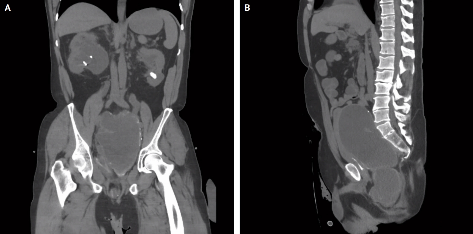
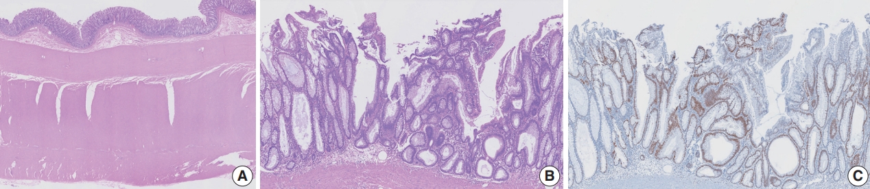
PDF - Mucoceles, commonly observed in the appendix, are mucin-filled, dilated structures arising from a range of etiologies. Cases associated with dysplastic or neoplastic epithelium can rupture and disseminate within the abdominopelvic cavity. Similar lesions in other parts of the colon are exceedingly rare, with only 16 colonic mucoceles having been reported. The first case of a colonic mucinous neoplasm with dysplasia resembling a low-grade appendiceal mucinous neoplasm involving rectal stump was described in 2016. Here, we present the second such case arising in the rectal stump, identified in a 44-year-old male with extensive surgical history. Microscopic examination revealed low-grade dysplastic epithelium lining the cyst and mucin dissecting into the stroma, without evidence of rupture or extramural mucin. The patient was followed for 16 months without recurrence or peritoneal disease. The exact etiology and outcome of these rare lesions remain unknown, requiring close follow-up.
- Tubular adenoma arising in tubular colonic duplication: a case report
- Heonwoo Lee, Hyeong Rok An, Chan Wook Kim, Young Soo Park
- J Pathol Transl Med. 2024;58(4):198-200. Published online July 3, 2024
- DOI: https://doi.org/10.4132/jptm.2024.06.04
- 5,549 View
- 227 Download
- 1 Web of Science
- 1 Crossref
-
Abstract
PDF
- Colonic duplication constitutes a rare congenital anomaly, characterized by the presence of hollow cystic or tubular structures exhibiting an epithelial-lined intestinal wall. Diagnostic challenges persist due to its low incidence and manifestation of nonspecific symptoms such as abdominal pain or constipation, resulting in a reluctance to pursue surgical resection. As associated malignancies in colonic duplication are rare, the inherent malignant potential of these anomalies remains undetermined. Additionally, despite reported instances of associated malignancies in colonic duplication, there is an absence of reports in the literature detailing tubular adenoma within these cases. The histologic features of the presented case are particularly noteworthy, situated at the precancerous stage, intimating potential progression towards adenocarcinoma within colonic duplication.
-
Citations
Citations to this article as recorded by
- Low-grade mucinous neoplasm originating from intestinal duplication: a case report and review of the literature
Huihui Yin, Jie Yu, Yunzhao Chen
World Journal of Surgical Oncology.2025;[Epub] CrossRef
- Low-grade mucinous neoplasm originating from intestinal duplication: a case report and review of the literature
- Aneurysmal bone cyst: a review
- Elham Nasri, John David Reith
- J Pathol Transl Med. 2023;57(2):81-87. Published online March 14, 2023
- DOI: https://doi.org/10.4132/jptm.2023.02.23
- 48,607 View
- 898 Download
- 37 Web of Science
- 42 Crossref
-
Abstract
PDF
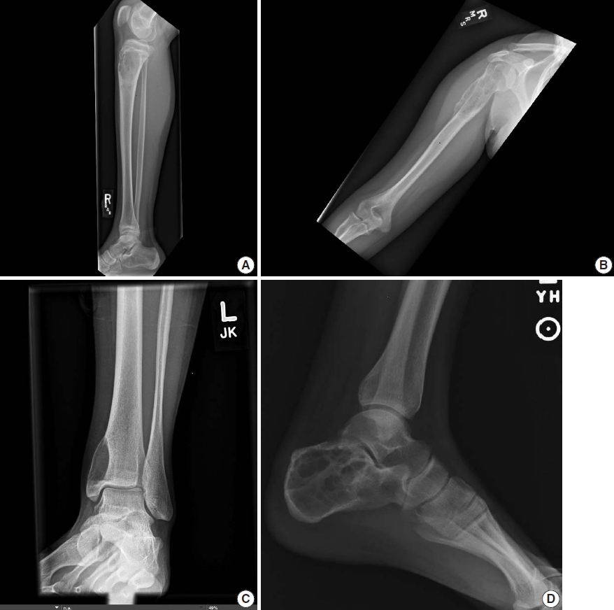
- Aneurysmal bone cyst (ABC) is a benign locally destructive bone neoplasm composed of multi-loculated blood-filled cystic spaces. The most common sites of involvement are the meta-diaphysis of the long bones and posterior elements of the vertebrae. Secondary, ABC-like changes can complicate a variety of other benign and malignant primary bone neoplasms, including giant cell tumor, fibrous dysplasia, and osteosarcoma. About two-third of primary ABCs have a rearrangement of the USP6 gene, which is not present in the ABC-like changes that occur secondary to other primary bone tumors (i.e., secondary ABC). Primary ABC of bone carries a variable but generally high rate of local recurrence. This paper provides an overview of the pathophysiology, clinical presentation, radiographic and pathologic findings, treatment, and prognosis of ABC.
-
Citations
Citations to this article as recorded by- Polidocanol Sclerotherapy Plus Adjuvant Autogenous Bone Marrow Injection for Management of Aneurysmal Bone Cyst: A Prospective Study
Ehab Abdelfattah Elshal, Maysra Abdelhalim Mohamed Byoumy, Abdallah Mousa Elwany Hassan, Abdelaziz Monsef Ali, Mohammed Al-Saeed Abdo Abu Hatab, Ahmed Sayed Ismaeil Khashaba
Indian Journal of Orthopaedics.2026; 60(7): 1612. CrossRef - Pathological proximal humerus fracture as the initial presentation of an aneurysmal bone cyst in a young adult treated with curettage, bone grafting, and PHILOS plate fixation: case report
Carlos Eduardo Purata Ortega, Farid Gallegos Wong, Luis Miguel Rodríguez Gonzále
South Florida Journal of Development.2026; 7(1): e6173. CrossRef - The best solution is the simplest: advances in surgical and minimally invasive management of aneurysmal and simple bone cysts
Abdulrahman Alaseem, Abdullah Addar, Mishari Alanezi, Fahad Alshayhan, Waleed Albishi, Ibrahim Alshaygy
Journal of Orthopaedic Surgery and Research.2026;[Epub] CrossRef - Primary malignant aneurysmal bone cyst of the metatarsal with PAFAH1B1::USP6 fusion: expanding the clinicopathologic spectrum of malignant USP6 translocated neoplasms
James Bennett, Fiona Bonar, Wendy Brown, Pranav Dorwal, Narelle Barton, Martin Lowe, Alison L. Cheah
Skeletal Radiology.2026; 55(7): 1685. CrossRef - Chondroblastoma with prominent secondary aneurysmal bone cyst-like changes: The role of H3.3 K36 M
David Suster, John M. Gross, Gregory W. Charville
Human Pathology.2026; 174: 106143. CrossRef - Maligne primäre Knochentumoren – Teil II
Thomas Grieser
Die Radiologie.2026; 66(6): 432. CrossRef - Aneurysmal Bone Cyst of the Coracoid Process: A Case Report
André Castanheira, Pedro Amaro, Raquel Costa, Hugo Santos, Nuno Oliveira, Luís Pires, Shashank Kaushik
Case Reports in Orthopedics.2026;[Epub] CrossRef - Transphyseal Proximal Humeral Aneurysmal Bone Cyst with Pathologic Fracture in a Child: A Case Report
Taichun Li, Jingmiao Wang, Qin Zhang, Ziming Zhang
Diagnostics.2026; 16(11): 1657. CrossRef - Imaging of Fibrous Dysplasia: A Comprehensive In-Depth Analysis of Monostotic, Polyostotic, Syndromic Forms, and Bone Sarcoma Development
Paolo Spinnato, Nicola Marrone, Domenico Romeo, Matilde Gonçalves, Roberts Naglis, Leonardo Di Battista, Elena Pedrini, Maria Parisi, Raffaella Rinaldi, Silvia Gazzotti, Alberto Righi, Marco Colangeli
Journal of Imaging.2026; 12(6): 241. CrossRef - Brown tumor of the mandible secondary to asymptomatic hyperparathyroidism: a case report
Sajjad Salam, Aatif Sayed, Mohammed Abdulla AlMuharraqi, Gowri Sivaramakrishnan
Discover Medicine.2026;[Epub] CrossRef - Primary aneurysmal bone cyst of the L1 vertebra: multimodality imaging and imaging–pathologic correlation
Krishnarjun Muralinath, Karthik Krishna Ramakrishnan, Priya Dharshini Rajaram, K. Praveen Sharma, Paarthipan Natarajan
Romanian Medical Journal.2026; 73(2): 245. CrossRef - Long‐Term Outcomes of Temporal Bone Aneurysmal Bone Cysts: Ambispective Study With Systematic Review and Pooled Analysis
Nidhin Das K, Anant Mehrotra, Amit Keshri, Mohit Sinha, Nazrin Hameed, Kalyan Chidambaram, Mohd Aqib, Awadesh Kumar Jaiswal, Ravisankar Manogaran
Otolaryngology–Head and Neck Surgery.2025; 172(5): 1493. CrossRef - Assessment and management of periacetabular aneurysmal bone cysts—a series of four cases
Reagan S.H Beyer, Quinn Steiner, David W Hennessy, Humberto G Rosas, David C Goodspeed, Andrea M Spiker
Journal of Hip Preservation Surgery.2025; 12(1): 11. CrossRef - Angiomatoid fibrous histiocytoma with EWSR1-CREB1 gene fusion occurs in lungs and ribs with systemic multiple metastases: a case report and review of the literature
Dongmei Feng, Ying Li, Zhengjin Li, Yun Pan, Yixuan Gao, Jinyan Cha, Chunmei Zhang
Frontiers in Oncology.2025;[Epub] CrossRef - Complete remodelling post-intralesional resection of an aggressive proximal humerus aneurysmal bone cyst mimicking telangiectatic osteosarcoma
Harpreet Singh, Sze Jet Aw, Arjandas Mahadev, Mohammad Ashik Bin Zainuddin, Kenneth Pak Leung Wong
BMJ Case Reports.2025; 18(2): e263437. CrossRef - First insights into the safety and effectiveness of additional courses with cladribine tablets under real-world conditions
Christoph Kleinschnitz, Jelena Skuljec, Markus C. Kowarik, Michael Ernst, Lara Woitschach, Lukas Cepek, Daniela Rau, Benedicta Kühnler, Sylke Schlemilch-Paschen, Matthias Schwab, Refik Pul
Multiple Sclerosis and Related Disorders.2025; 97: 106398. CrossRef - Case Report: Giant cell lesions in the Maxillofacial region: diagnostic points and treatment strategies
Xiaohan Gao, Shuangyi Wang, Xiaohong Zhan, Yanshan Liu, Liqiang Chen, Jian Sun, Haoyue Xu
Frontiers in Oncology.2025;[Epub] CrossRef - Endoscopic Curettage of Aneurysmal Bone Cyst of the Distal Fibula
Tun Hing Lui, Ka Kin Cheung, Wun Kee Szeto
Arthroscopy Techniques.2025;[Epub] CrossRef - Reviewing superficial bone lesions: What the radiologist needs to know
Dâmaris Versiani Caldeira Gonçalves, Isabela Azevedo Nicodemos da Cruz, Marcelo Astolfi Caetano Nico, Alípio Gomes Ormond Filho, Júlio Brandão Guimarães
Clinical Imaging.2025; 123: 110493. CrossRef - Juvenile ossifying fibroma and aneurysmal bone cyst in the mandible: A case report and mini review of literature
Fatma Wageeh Attya, Walaa Hussein Abu El-Ela, Basma Abdelrahman Ahmed, Iman Mohamed Helmy
Pediatric Dental Journal.2025; 35(3): 100354. CrossRef - A clinical case of an aneurysmal bone cyst of the humerus
N.S. Lysenko, V.V. Bayev, І.О. Voronzhev, S.M. Palchyk, А.М. Hrytsenko
Український радіологічний та онкологічний журнал.2025; 33(2): 270. CrossRef - Aneurysmal bone cyst of the rib. Robotic resection of a rare lesion
Luis Arana-Bolaños, Xcaret Luna-Vargas, Amelia Fernández-Avendaño, Mónica Martínez-Ferman, Pablo Gomes-da Silva de Rosenzweig, Francina Bolaños-Morales
Journal of Surgical Case Reports.2025;[Epub] CrossRef - Musculoskeletal tumors and tumor-like lesions with “dark” signal intensity on T2-weighted MR images: A pictorial review
Jingkun Zhang, Fengyuan Luo, Juan Chen, Huijuan Yang, Qi Zhang
Medicine.2025; 104(41): e45179. CrossRef - Escleroterapia con alcohol al 90% previo a exéresis de quiste óseo aneurismático maxilar: Reporte de caso
Glenda Semanate Cajas, Claudia Elizabeth Cabrera Arévalo
Arandu UTIC.2025; 12(3): 4306. CrossRef - Case Report: Adult proximal humeral aneurysmal bone cyst: radical resection and reconstruction with osteoconductive allograft & reverse arthroplasty—Ecuador's first reported case and functional outcomes
Gabriel Gamecho Arteaga, Henry Hernández, Chrystian X. Mestanza, Jaime Zurita, Marlon Arias-Intriago, Juan S. Izquierdo-Condoy
Frontiers in Surgery.2025;[Epub] CrossRef - Aggressive Aneurysmal Bone Cyst of the Mandible: A Rare Case of Rapid Expansion and Surgical Management
Fatemeh Mashhadiabbas, Sanaz Gholami Toghchi, Sara Alehossein, Hoorisa Norouzi, Mohammadreza Kashefi Baher
Clinical Case Reports.2025;[Epub] CrossRef - Management of aggressive recurrent thoracic spine aneurysmal bone cyst in a 7-year-old male: A case report and review of the literature
Pedram Jahangiri, Faramarz Roohollahi, Zohreh Habibi, Mohammad Hosein Mirbolouk, Mohsen Rostami
Surgical Neurology International.2024; 15: 30. CrossRef - Intraosseous hemangioma with aneurysmal bone cyst-like changes of the hyoid bone: Case report and literature review
Jeonghyun Oh, Song Iy Han, Sung-Chul Lim
Medicine.2024; 103(6): e37137. CrossRef - Fibrous dysplasia with aneurysmal bone cyst-like change occurring in pediatric orbit: case report and literature review
Xinyao Wang, Wenbin Guan, Haibo Zhang, Lei Bao, Xiaoqiang Wang
Oral and Maxillofacial Surgery.2024; 28(2): 999. CrossRef - Pathological Fractures in Aneurysmal Bone Cysts: A Systematic Review
Doriana Di Costa, Elena Gabrielli, Mariagrazia Cerrone, Emidio Di Gialleonardo, Giulio Maccauro, Raffaele Vitiello
Journal of Clinical Medicine.2024; 13(9): 2485. CrossRef - Quiste óseo aneurismático torácico, descompresión mediante costotransversectomía, corpectomía y caja telescópica expandible. Reporte de un caso y revisión de literatura
Karoll Ortíz-Guillén, José M García-De la Rosa, Everardo García, Adriana Vargas-Oviedo
Cirugía de Columna.2024; 2(3): 188. CrossRef - The Role of Denosumab Treatment in Recurrent Giant Cell Bone Tumor of the Orbit
Arjav Gupta, Bruce Colwell, David B. Clarke, Emad A. Massoud, Sidney Croul, Ahsen Hussain
Ophthalmic Plastic & Reconstructive Surgery.2024; 40(5): e161. CrossRef - Denosumab Re-Challenge and Long-Term Efficacy for Aneurysmal Bone Cyst of the Spine: Enhanced Treatment Algorithm
Gisberto Evangelisti, Franziska C. S. Altorfer, Luigi Falzetti, Emanuela Palmerini, Cristiana Griffoni, Riccardo Ghermandi, Stefano Boriani, Annalisa Monetta, Marilena Cesari, Toni Ibrahim, Alessandro Gasbarrini
Journal of Clinical Medicine.2024; 13(15): 4522. CrossRef - Rare Aneurysmal Bone Cyst Presentation in the Orbit: A Systematic Review of the Literature with an Illustrative Case Report
Sean O'Leary, Fakhar Hayat, Saketh Amasa, Muhammad Ammar Haider, Saad Akram Asbeutah, Usama AlDallal, Umaru Barrie, Mohamed Ismail
World Neurosurgery.2024; 191: 1. CrossRef - Primary osseous leiomyosarcoma of humerus misinterpreted as aneurysmal bone cyst: A case report and literature review
Yong Jin Cho, Young Kwon Koh, Sung-Chul Lim
Medicine.2024; 103(38): e39762. CrossRef - Recurrent Aneurysmal Bone Cyst Treated with Percutaneous Doxycycline Sclerotherapy
Cory Gall, Daniel C. Allison
JBJS Case Connector.2024;[Epub] CrossRef - Development and Printing of a Customized 3D Model of a Solitary Humeral Cyst as a Stage in Surgical Treatment of Bone Defects Using Orgignal Bone Replased Material
Bakhtiyar Makhatov, Berik Tuleubayev, Amina Koshanova
Journal of Clinical Medicine of Kazakhstan.2024; 21(6): 91. CrossRef - Diagnosis and management of bone cysts
Deepak C. D., Anitha Boregowdanapalya
International Journal of Research in Medical Sciences.2024; 13(1): 509. CrossRef - A rare case of cavitated Schmorl’s node in the cervical spine: imaging features of bone scan and magnetic resonance
Yung-Cheng Chang, Yu-Jing Kao, Ling Chun Sun, Wen-Hsuan Hsiao, Shin-Tsu Chang
MOJ Orthopedics & Rheumatology.2024; 16(5): 278. CrossRef - Metastatic patellar bone tumor due to gastric cancer resembling a primary or secondary aneurysmal bone cyst: A case report
T. Furuta, T. Sakuda, K. Yoshioka, K. Arihiro, N. Adachi
International Journal of Surgery Case Reports.2023; 108: 108379. CrossRef - Clear cell chondrosarcoma: a review of clinicopathologic characteristics, differential diagnoses, and patient management
Borislav A. Alexiev, Erica R. Vormittag-Nocito, Terrance D. Peabody, Jonathan Samet, William B. Laskin
Human Pathology.2023; 139: 126. CrossRef - Malignant transformation of an aneurysmal bone cyst of the femoral neck: A case report
Xiaoyang Song, Yongjie Qiao, Haoqiang Zhang, Lirong Sha, Jinpeng Lou, Xinyuan Yu, Hao Liu, Langfeng Zhu, Shenghu Zhou
Experimental and Therapeutic Medicine.2023;[Epub] CrossRef
- Polidocanol Sclerotherapy Plus Adjuvant Autogenous Bone Marrow Injection for Management of Aneurysmal Bone Cyst: A Prospective Study
- The proteomic landscape shows oncologic relevance in cystitis glandularis
- Jun Yong Kim, Dohyun Han, Hyeyoon Kim, Minsun Jung, Han Suk Ryu
- J Pathol Transl Med. 2023;57(1):67-74. Published online December 22, 2022
- DOI: https://doi.org/10.4132/jptm.2022.10.24
- 6,830 View
- 182 Download
- 2 Web of Science
- 2 Crossref
-
Abstract
PDF
- Background
The relationship between cystitis glandularis (CG) and bladder malignancy remains unclear.
Methods
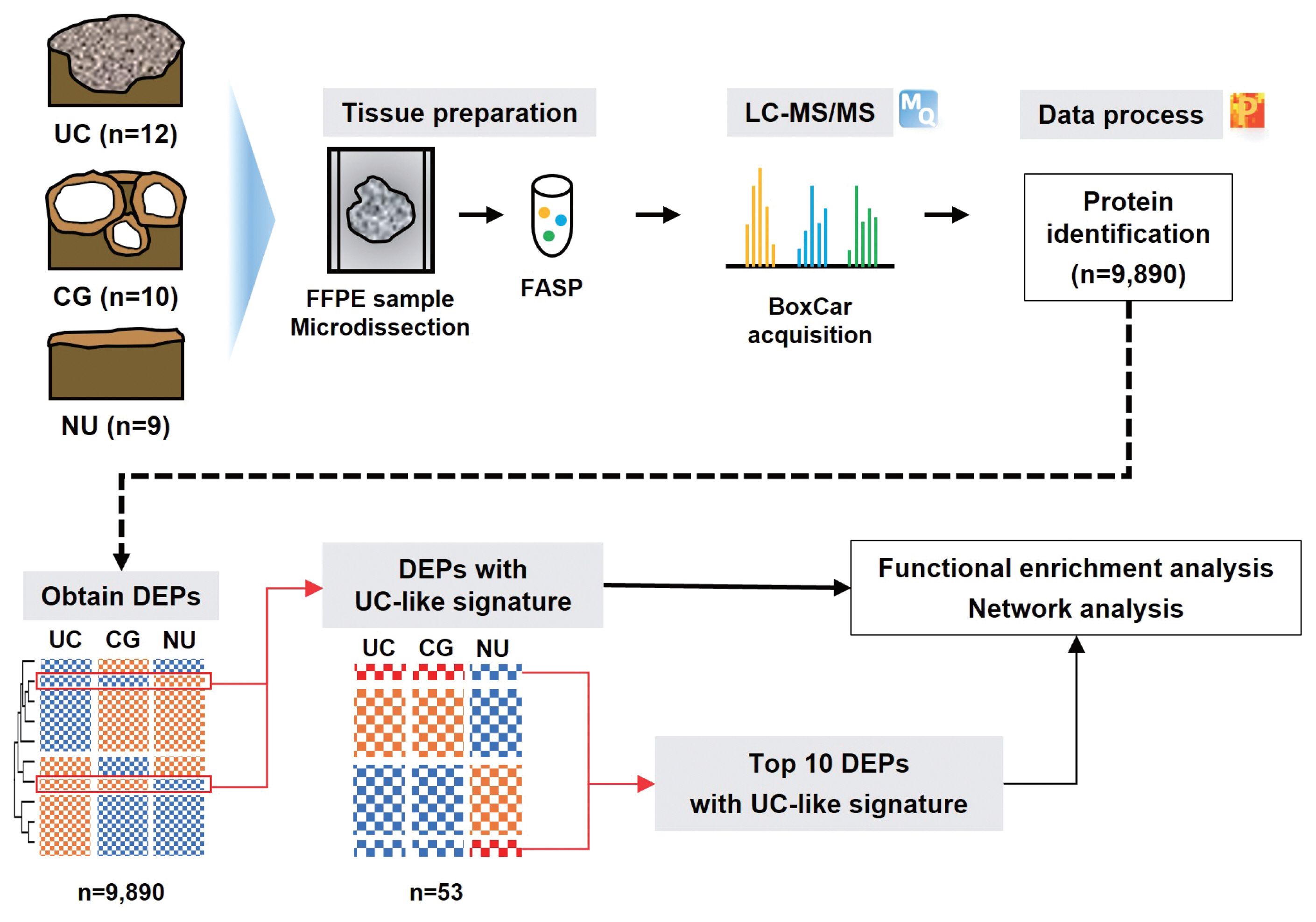
We identified the oncologic significance of CG at the molecular level using liquid chromatography-tandem mass spectrometry-based proteomic analysis of 10 CG, 12 urothelial carcinoma (UC), and nine normal urothelium (NU) specimens. Differentially expressed proteins (DEPs) were identified based on an analysis of variance false discovery rate < 0.05, and their functional enrichment was analyzed using a network model, Gene Set Enrichment Analysis, and Gene Ontology annotation.
Results
We identified 9,890 proteins across all samples and 1,139 DEPs among the three entities. A substantial number of DEPs overlapped in CG/NU, distinct from UC. Interestingly, we found that a subset of DEP clusters (n = 53, 5%) was differentially expressed in NU but similarly between CG and UC. This “UC-like signature” was enriched for reactive oxygen species (ROS) and energy metabolism, growth and DNA repair, transport, motility, epithelial-mesenchymal transition, and cell survival. Using the top 10 shortlisted DEPs, including SOD2, PRKCD, CYCS, and HCLS1, we identified functional elements related to ROS metabolism, development, and transport using network analysis. The abundance of these four molecules in UC/CG than in NU was consistent with the oncologic functions in CG.
Conclusions
Using a proteomic approach, we identified a predominantly non-neoplastic landscape of CG, which was closer to NU than to UC. We also confirmed a small subset of common DEPs in UC and CG, suggesting that altered ROS metabolism might imply potential cancerous risks in CG. -
Citations
Citations to this article as recorded by- Quantitative proteomics and immunohistochemistry uncover NT5DC2 as a diagnostic biomarker for papillary urothelial carcinoma
Jun Yong Kim, Jae Seok Lee, Dohyun Han, Ilias P. Nikas, Hyeyoon Kim, Minsun Jung, Han Suk Ryu
Heliyon.2024; 10(15): e35475. CrossRef - KRT18 as a Novel Biomarker of Urothelial Papilloma while Evaluating Low-Grade Papillary Urothelial Neoplasms: Bi-Center Analysis
Minsun Jung, Bohyun Kim, Jae Seok Lee, Jun Yong Kim, Dohyun Han, Kwangsoo Kim, Sunah Yang, Eun Na Kim, Hyeyooon Kim, Ilias P. Nikas, Sohyeon Yang, Kyung Chul Moon, Hyebin Lee, Han Suk Ryu
Pathobiology.2024; : 1. CrossRef
- Quantitative proteomics and immunohistochemistry uncover NT5DC2 as a diagnostic biomarker for papillary urothelial carcinoma
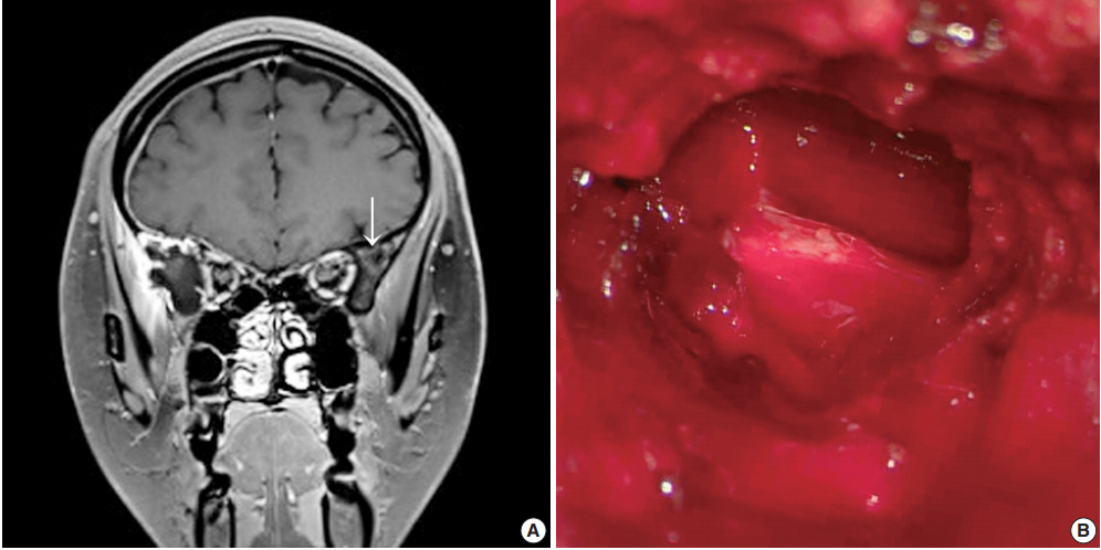
- Intraoperative frozen cytology of intraosseous cystic meningioma in the sphenoid bone
- Na Rae Kim, Gie-Taek Yie
- J Pathol Transl Med. 2020;54(6):508-512. Published online July 1, 2020
- DOI: https://doi.org/10.4132/jptm.2020.05.21
- 6,747 View
- 101 Download
- 2 Web of Science
- 3 Crossref
-
Abstract
PDF
- Meningiomas in bone are rarely subjected to fine-needle aspiration diagnosis, and those arising in the skull bone with a cystic presentation are rare. A 24-year-old woman presented with subdural hemorrhage, and subsequent radiology depicted an osteolytic mass-like lesion in the sphenoid bone. Intraoperatively, a solid and cystic hemorrhagic lesion mimicking an aneurysmal bone cyst was observed in the sphenoid bone with dural tearing. Frozen cytology showed singly scattered or epithelioid clusters of round to elongated cells intermixed with many neutrophils. Tumor cells had bland-looking round nuclei with rare prominent nucleoli and nuclear inclusions and eosinophilic granular to globoid cytoplasm in capillary-rich fragments. Histology revealed intraosseous meningothelial and microcystic meningioma (World Health Organization grade 1) in right lesser wing of the sphenoid bone. Considering its unusual location and cytologic findings, differential diagnoses included chordoma, chondroma, chondrosarcoma, and aneurysmal bone cyst. The present case posed a diagnostic challenge due to possible confusion with these entities.
-
Citations
Citations to this article as recorded by- Purely cystic intraosseous meningioma of the skull: A radiologic conundrum and histologic challenge
Diego Rojas, Arman Kavoussi, Ashley Rose Ricciardelli, Alex Flores, Sricharan Gopakumar, Luis Carrete, Hsiang-Chih Lu, Alex W. Brenner, Akash J. Patel
Surgical Neurology International.2025; 16: 221. CrossRef - Middle ear adenoma: Cytohistologic features and differential diagnosis
Abdullah Almajnooni, Matthew Vega, Lin Cheng, Paolo Gattuso, Mary K. Allen‐Proctor
Diagnostic Cytopathology.2023;[Epub] CrossRef - Exploring the role of epidermal growth factor receptor variant III in meningeal tumors
Rashmi Rana, Vaishnavi Rathi, Kirti Chauhan, Kriti Jain, Satnam Singh Chhabra, Rajesh Acharya, Samir Kumar Kalra, Anshul Gupta, Sunila Jain, Nirmal Kumar Ganguly, Dharmendra Kumar Yadav, Timir Tripathi
PLOS ONE.2021; 16(9): e0255133. CrossRef
- Purely cystic intraosseous meningioma of the skull: A radiologic conundrum and histologic challenge
- Coexisting Mucinous Cystic Neoplasm of the Pancreas and Type 1 Autoimmune Pancreatitis
- Mee-Jeong Kim, Tae Jun Song, Hyoung Jung Kim, Song-Cheol Kim, Myung-Hwan Kim, Seung-Mo Hong
- J Pathol Transl Med. 2019;53(2):125-128. Published online November 14, 2018
- DOI: https://doi.org/10.4132/jptm.2018.10.25
- 11,304 View
- 122 Download
- 3 Web of Science
- 6 Crossref
-
Abstract
PDF
- Type 1 autoimmune pancreatitis (AIP1) is an IgG4-related systemic disease that mimics tumors. We report a rare case of AIP1 accompanied by mucinous cystic neoplasm (MCN). A pancreatic lesion was incidentally detected in a woman in her 60s. After 6 years of follow-up, the lesion abruptly increased in size. Computed tomography showed a 3.5 cm unilocular cyst in the tail of the pancreas and distal pancreatectomy was performed. On microscopic examination, the cyst was lined by mucinous and non-mucinous epithelial cells with mild cytologic atypia. The surrounding stroma comprised ovarian-type spindle cells with progesterone receptor positivity. The pericystic pancreas exhibited multifocal lymphoid follicles, lymphoplasmacytic infiltrations, obliterative phlebitis, and storiform fibrosis. IgG4-positive plasma cell infiltration (215 cells high-power field) and the IgG4/IgG ratio (57%) were increased. Cases of MCN coexisting with AIP1 are extremely rare; only two such cases have been reported in the English-language literature. This third case featured low-grade MCN with AIP1.
-
Citations
Citations to this article as recorded by- Utilizing Immunoglobulin G4 Immunohistochemistry for Risk Stratification in Patients with Papillary Thyroid Carcinoma Associated with Hashimoto Thyroiditis
Faridul Haq, Gyeongsin Park, Sora Jeon, Mitsuyoshi Hirokawa, Chan Kwon Jung
Endocrinology and Metabolism.2024; 39(3): 468. CrossRef - Histological features of autoimmune pancreatitis and IgG4-related sclerosing cholangitis with a correlation with imaging findings
Kenji NOTOHARA
Choonpa Igaku.2023; 50(1): 55. CrossRef - Imaging Features and Risk Factors of Pancreatic Cystic Lesions Complicating
Autoimmune Pancreatitis: A Retrospective Study
Bin-Bin Zhang, Xin-Meng Hou, Yu-Qi Chen, Jian-Wei Huo, Er-Hu Jin
Current Medical Imaging Reviews.2023;[Epub] CrossRef - Histological features of autoimmune pancreatitis and IgG4-related sclerosing cholangitis with a correlation with imaging findings
Kenji Notohara
Journal of Medical Ultrasonics.2021; 48(4): 581. CrossRef - 自己免疫性膵炎診療ガイドライン2020
Suizo.2020; 35(6): 465. CrossRef - Mucinous cystic neoplasm of the pancreas with type-1 autoimmune pancreatitis-like lesion
Kevin Gowing, David F. Schaeffer, Hui-Min Yang
Human Pathology: Case Reports.2019; 18: 200339. CrossRef
- Utilizing Immunoglobulin G4 Immunohistochemistry for Risk Stratification in Patients with Papillary Thyroid Carcinoma Associated with Hashimoto Thyroiditis
- Bile Granuloma Mimicking Peritoneal Seeding: A Case Report
- Hasong Jeong, Hye Won Lee, Hye Ra Jung, Ilseon Hwang, Sun Young Kwon, Yu Na Kang, Sang Pyo Kim, Misun Choe
- J Pathol Transl Med. 2018;52(5):339-343. Published online July 16, 2018
- DOI: https://doi.org/10.4132/jptm.2018.06.02
- 9,733 View
- 120 Download
- 7 Web of Science
- 9 Crossref
-
Abstract
PDF
- Laparoscopic cholecystectomy is a widely used treatment method for most cholelithiasis and is a relatively safe procedure. Foreign body granulomatous reaction to bile or gallstone spillage during laparoscopic cholecystectomy has rarely been reported. We report a case of bile granuloma after laparoscopic cholecystectomy, which mimicked peritoneal seeding. A 59-year-old Korean man presented with right upper quadrant pain. He underwent laparoscopic cholecystectomy for acute cholecystitis with cholelithiasis. Pathologic examination revealed an incidental adenocarcinoma invading the lamina propria with acute cholecystitis and cholelithiasis. After 3 months, follow-up abdominal computed tomography revealed a subhepatic nodule, which showed hypermetabolism on positron emission tomography–computed tomography. Suspecting localized peritoneal seeding, wedge resection of the liver, wedge resection of the transverse colon, and omentectomy were performed. Pathologic examination of the resected specimens revealed multiple bile granulomas. Awareness of bile granuloma mimicking malignancy is noteworthy for patient management to reduce unnecessary procedure during postoperative surveillance.
-
Citations
Citations to this article as recorded by- False Alarm: When Dropped Gallstones Mimic Malignant Recurrence. A Case Report and Literature Review
Nedaa Obeidi, Michael Egan, Michael E. Kelly, Feras Abu Saadeh, Karuna Sharma
Case Reports in Obstetrics and Gynecology.2026;[Epub] CrossRef - A mimic of peritoneal metastatic disease, multifocal intraabdominal foreign body granulomas secondary to feculent peritonitis
Damien Gibson, Christo Joseph, Diarmid P. Foulis, Christophe R. Berney
ANZ Journal of Surgery.2024; 94(4): 763. CrossRef - Practices and Attitudes of Surgeons With Regard to Spilled Gallstones During Laparoscopic Cholecystectomy: A Cross-Sectional Study From Saudi Arabia
Mohammed Alfehaid, Moath Aljohani, Sajad A Salati , Shoug Alaodah, Wejdan Alresheedi, Raghad Almarshud
Cureus.2024;[Epub] CrossRef - Spilled gallstone mimicking intra-abdominal seeding of gallbladder adenocarcinoma: A case report
Cheng-Ken Huang, Ruey-Hwa Lu, Chien-Cheng Chen, Po-Chun Chen, Wen-Chang Hsu, Meng-Jui Tsai, Chin-Tsung Ting
World Journal of Gastrointestinal Surgery.2024; 16(2): 622. CrossRef - Peritoneal bile granuloma formation at the site of caesarean surgical scar
Lila Marshall, Sharlin Varghese, Mary Ciranni-Callon
Journal of Case Reports and Images in Obstetrics and Gynecology.2024; 10(2): 6. CrossRef - Biliary Granulomatous Peritoneal Reaction as Consequence of Cholecystectomy: Case Report and Literature Review
Giuseppe Tarantino, Denise Menghini, Maria Eva Argenziano, Miriam Palmieri, Alessandra Mandolesi, Enrico Dalla Bona, Antonio Benedetti, Mario Guerrieri, Maria Giovanna Danieli
SN Comprehensive Clinical Medicine.2023;[Epub] CrossRef - Foreign body reaction mimicking local recurrence from polyactide adhesion barrier film after laparoscopic colorectal cancer surgery
Tien-Chan Hsieh, Chao-Wen Hsu
Medicine.2022; 101(5): e28692. CrossRef - Spilled gallstones after laparoscopic cholecystectomy: a systematic review
Sajad Ahmad Salati, Mohammed Alfehaid, Saleh Alsuwaydani, Lamees AlSulaim
Polish Journal of Surgery.2022; 94(4): 1. CrossRef - Foreign body granulomas mimic peritoneal dissemination caused by incarcerated femoral hernia perforation: A case report
Shinpei Ogino, Tatsuya Matsumoto, Yosuke Kamada, Noriaki Koizumi, Hiroshi Fujiki, Kenji Nakamura, Takeshi Yamano, Chouhei Sakakura
World Journal of Clinical Oncology.2021; 12(11): 1083. CrossRef
- False Alarm: When Dropped Gallstones Mimic Malignant Recurrence. A Case Report and Literature Review
- Metaplastic Carcinoma with Chondroid Differentiation Arising in Microglandular Adenosis
- Ga-Eon Kim, Nah Ihm Kim, Ji Shin Lee, Min Ho Park
- J Pathol Transl Med. 2017;51(4):418-421. Published online April 4, 2017
- DOI: https://doi.org/10.4132/jptm.2016.10.06
- 10,016 View
- 115 Download
- 5 Web of Science
- 6 Crossref
-
Abstract
PDF
- Microglandular adenosis (MGA) of the breast is a rare, benign proliferative lesion but with a significant rate of associated carcinoma. Herein, we report an unusual case of metaplastic carcinoma with chondroid differentiation associated with typical MGA. Histologically, MGA showed a direct transition to metaplastic carcinoma without an intervening atypical MGA or ductal carcinoma in situ component. The immunohistochemical profile of the metaplastic carcinoma was mostly similar to that of MGA. In both areas, all the epithelial cells were positive for S-100 protein, but negative for estrogen receptor, progesterone receptor, HER2/neu, and epidermal growth factor receptor. An increase in the Ki-67 and p53 labelling index was observed from MGA to invasive carcinoma. To the best of our knowledge, this is the first case of metaplastic carcinoma with chondroid differentiation arising in MGA in Korea. This case supports the hypothesis that a subset of MGA may be a non-obligate morphologic precursor of breast carcinoma, especially the triple-negative subtype.
-
Citations
Citations to this article as recorded by- Two similar but distinct types of breast acinar cell carcinoma: evidence from histological, immunohistochemical and molecular features
Mingfang Sun, Lin Fu, Hongjiu Ren, Jian Wang, Xuyong Lin, Qingfu Zhang
Histopathology.2025; 87(6): 904. CrossRef - Elucidating the nature of acinic cell carcinoma of the breast with high-grade morphology: evidence from case report
Yunjie Ge, Xianping Wei, Jing-Nan Liu, Ping-Li Sun, Hongwen Gao
Diagnostic Pathology.2024;[Epub] CrossRef - New insights into acinic cell carcinoma of the breast: clinicopathology, origin of histology, molecular features, prognosis, and treatment
Yunjie Ge, Xianping Wei, Jing-Nan Liu, Ping-Li Sun, Hongwen Gao
Frontiers in Oncology.2024;[Epub] CrossRef - Metaplastic Matrix-Producing Carcinoma and Apocrine Lobular Carcinoma In Situ Associated with Microglandular Adenosis: A Unique Case Report
Nektarios Koufopoulos, Dionysios Dimas, Foteini Antoniadou, Kyparissia Sitara, Dimitrios Balalis, Ioannis Boutas, Alina Roxana Gouloumis, Adamantia Kontogeorgi, Lubna Khaldi
Diagnostics.2022; 12(6): 1458. CrossRef - Salivary gland-type mammary carcinoma arising in microglandular adenosis: A case report and clinicopathological review of the literature
Victoria Rico, Yukiko Shibahara, Marjorie Monteiro, Elzbieta Slodkowska, Samantha Tam, Pearl Zaki, Carlo De Angelis, Edward Chow, Katarzyna Joanna Jerzak
Cancer Treatment and Research Communications.2020; 24: 100178. CrossRef - Microglandular adenosis is an advanced precursor breast lesion with evidence of molecular progression to matrix-producing metaplastic carcinoma
Christopher J. Schwartz, Igor Dolgalev, Esther Yoon, Iman Osman, Adriana Heguy, Eleazar C. Vega-Saenz de Miera, Diana Nimeh, George Jour, Farbod Darvishian
Human Pathology.2019; 85: 65. CrossRef
- Two similar but distinct types of breast acinar cell carcinoma: evidence from histological, immunohistochemical and molecular features
- Mucinous Cystadenoma of the Testis: A Case Report with Immunohistochemical Findings
- Gilhyang Kim, Dohee Kwon, Hee Young Na, Sehui Kim, Kyung Chul Moon
- J Pathol Transl Med. 2017;51(2):180-184. Published online February 13, 2017
- DOI: https://doi.org/10.4132/jptm.2016.08.30
- 11,800 View
- 128 Download
- 5 Web of Science
- 8 Crossref
-
Abstract
PDF
- Mucinous cystadenoma of the testis is a very rare tumor. Herein, we report a case of mucinous cystadenoma arising in the testis of a 61-year-old man, along with a literature review. Computed tomography showed a 2.5-cm-sized poorly enhancing cystic mass. Grossly, the tumor was a unilocular cystic mass filled with mucinous material and confined to the testicular parenchyma. Histologically, the cyst had a fibrotic wall lined by mucinous columnar epithelium without atypia. Immunohistochemical staining was positive for cytokeratin 20 and CDX2, as well as focally positive for cytokeratin 7. The pathologic diagnosis was mucinous cystadenoma.
-
Citations
Citations to this article as recorded by- Review of Paratesticular Appendageal Tumors, Morphology, Immunohistochemistry, and Recent Molecular Advances
Mathew Vega, Muhammad T. Idrees
Surgical Pathology Clinics.2025; 18(1): 119. CrossRef - Cistoadenoma Mucinoso Paratesticular: Caso Interesante en el Instituto Guatemalteco de Seguridad Social
Edgar Estuardo González López, Carlos Gonzalo Estrada Pazos
Revista Guatemalteca de Urología.2023; 10(2): 16. CrossRef - Primary borderline mucinous tumor of the testis with postoperative metastasis: A rare case report
Yingyu Shi, Ling Song, Yan Luo
Radiology Case Reports.2023; 18(9): 3203. CrossRef - Case report: Misdiagnosis of primary mucinous cystadenoma of the testicle by ultrasound
Linlin Zhang, Jianyuan Xuan, Manxi Li, Mei Zhang, Yu Song, Ziang Pan, Bo Fan, Lin Lu, Hongyan Zhou, Yang Li
Frontiers in Oncology.2023;[Epub] CrossRef - Primary Borderline Mucinous Testicular Tumor: A Case Report and Literature Review
Changjuan Hao, Chunsong Kang, Xiaoyan Kang, Zhuanzhuan Yu, Tingting Li, Jiping Xue
Frontiers in Oncology.2021;[Epub] CrossRef - Ovarian-type Tumors (Mullerian Tumors) of the Testis: Clinicopathologic Findings with Recent Advances
Michelle S Lin, Alberto G Ayala, Jae Y Ro
annals of urologic oncology.2019; : 1. CrossRef - Borderline Mucinous Testicular Tumour: Diagnostic and Management difficulties
Krishan Pratap, Marlon Perera, Frances Malczewski, Rachel Esler
BMJ Case Reports.2018; 2018: bcr-2017-223787. CrossRef - Mucinous tumor arising in a giant sacrococcygeal teratoma
Fengtian Zhang, Xiaolong Yu, Jin Zeng, Min Dai
Medicine.2017; 96(47): e8759. CrossRef
- Review of Paratesticular Appendageal Tumors, Morphology, Immunohistochemistry, and Recent Molecular Advances
- Comprehensive Cytomorphologic Analysis of Pulmonary Adenoid Cystic Carcinoma: Comparison to Small Cell Carcinoma and Non-pulmonary Adenoid Cystic Carcinoma
- Seokhwi Kim, Jinah Chu, Hojoong Kim, Joungho Han
- J Pathol Transl Med. 2015;49(6):511-519. Published online October 19, 2015
- DOI: https://doi.org/10.4132/jptm.2015.09.07
- 12,573 View
- 77 Download
- 6 Web of Science
- 6 Crossref
-
Abstract
PDF
- Background
Cytologic diagnosis of pulmonary adenoid cystic carcinoma (AdCC) is frequently challenging and differential diagnosis with small cell carcinoma is often difficult. Methods: Eleven cytologically diagnosed cases of pulmonary AdCC were collected and reviewed according to fifteen cytomorphologic characteristics: small cell size, cellular uniformity, coarse chromatin, hyperchromasia, distinct nucleolus, frequent nuclear molding, granular cytoplasm, organoid cluster, sheet formation, irregular border of cluster, hyaline globule, hyaline basement membrane material, individual cell necrosis or apoptotic body, and necrotic background. Twenty cases of small cell carcinoma and fifteen cases of non-pulmonary AdCC were also reviewed for the comparison. Results: Statistically significant differences were identified between pulmonary AdCC and small cell carcinoma in fourteen of the fifteen cytomorphologic criteria (differences in sheet formation were not statistically significant). Cellular uniformity, distinct nucleolus, granular cytoplasm, distinct cell border, organoid cluster, hyaline globule, and hyaline basement membrane material were characteristic features of AdCC. Frequent nuclear molding, individual cell necrosis, and necrotic background were almost exclusively identified in small cell carcinoma. Although coarse chromatin and irregular cluster border were observed in both, they favored the diagnosis of small cell carcinoma. Hyaline globules were more frequently seen in non-pulmonary AdCC cases. Conclusions: Using the fifteen cytomorphologic criteria described by this study, pulmonary AdCC could be successfully distinguished from small cell carcinoma. Such a comprehensive approach to an individual case is recommended for the cytologic diagnosis of pulmonary AdCC. -
Citations
Citations to this article as recorded by- Primary pulmonary adenoid cystic carcinoma: A study of clinicopathological features and molecular alterations in twenty-one cases
Zhiyuan Yao, Tong Qiu, Changlei Li, Weimao Kong, Guangqi Li, Peng Song, Guohua Wang, Wenjie Jiao
Lung Cancer.2025; 201: 108414. CrossRef - Recent developments in the pathology of primary pulmonary salivary gland‐type tumours
Julia R Naso, Anja C Roden
Histopathology.2024; 84(1): 102. CrossRef - Bronchial cytology of pulmonary adenoid cystic carcinoma – A multi-institute series with emphasis on immunocytochemistry
Joanna K.M. Ng, Ka Pang Chan, Gary M. Tse, Joshua J.X. Li
Annals of Diagnostic Pathology.2023; 64: 152132. CrossRef - Pulmonary adenoid cystic carcinoma: molecular characteristics and literature review
Zhixin Chen, Jiapeng Jiang, Ying Fan, Hongyang Lu
Diagnostic Pathology.2023;[Epub] CrossRef - Recent updates in salivary gland tumors of the lung
Anja C. Roden
Seminars in Diagnostic Pathology.2021; 38(5): 98. CrossRef - Cytology of Primary Salivary Gland-Type Tumors of the Lower Respiratory Tract: Report of 15 Cases and Review of the Literature
Chiara Saglietti, Marco Volante, Stefano La Rosa, Igor Letovanec, Marc Pusztaszeri, Gaia Gatti, Massimo Bongiovanni
Frontiers in Medicine.2017;[Epub] CrossRef
- Primary pulmonary adenoid cystic carcinoma: A study of clinicopathological features and molecular alterations in twenty-one cases
- Comparison of Cytologic Characteristics between Adenoid Cystic Carcinoma and Adenoid Basal Carcinoma in the Uterine Cervix
- Juhyeon Jeong, Seung Yeon Ha, Hyun Yee Cho, Dong Hae Chung, Jungsuk An
- J Pathol Transl Med. 2015;49(5):396-402. Published online August 17, 2015
- DOI: https://doi.org/10.4132/jptm.2015.07.08
- 12,262 View
- 97 Download
- 1 Web of Science
- 2 Crossref
-
Abstract
PDF
- Background
Adenoid cystic carcinoma (ACC) and adenoid basal carcinoma (ABC) are rare in the uterine cervix. ACC is more aggressive than ABC, thus accurate differential diagnosis is important. In this study, we identified cytologic features useful in distinguishing these two tumors for diagnosis. Methods: Three cases of ACC and five cases of ABC were selected for this study. Cervicovaginal smear slides were reviewed retrospectively, and the area, circumference, major axis, and minor axis of nuclei were measured using an image analyzer. Results: ACC displayed three-dimensional clusters with a small acini pattern. ABC displayed peripheral palisading without an acini pattern. The nuclei of ACC were more irregular and angulated than those of ABC, and the former showed a coarsely granular chromatin pattern. The nucleic area, circumference, major axis, and minor axis were 18.556±8.665 µm2, 23.320±11.412 µm, 5.664±1.537 µm, and 4.127±1.107 µm in ACC and 11.017±4.440 µm2, 15.920±5.664 µm, 4.612±1.025 µm, and 3.088±0.762 µm in the cases of ABC. All measured values showed statistically significant difference (p < .001). Conclusions: Although the nuclei of both of these tumor types were oval shaped, inferred from the ratio of minor axis to major axis (0.728 in ACC and 0.669 in ABC), the area of nuclei was approximately 1.7 times larger in ACC than in ABC. Distinguishing nucleic features, including area, morphology, and chromatin pattern, may be helpful in making a correct diagnosis. -
Citations
Citations to this article as recorded by- Adenoid basal carcinoma of the uterine cervix
Anas Mohamed, Tesfalem Korga, Ahlam Ali, Javier Laurini
International Journal of Gynecological Cancer.2025; : 101873. CrossRef - Adenoid Basal Carcinoma of the Uterine Cervix: A Case Report
Tatsuya Kanuma, Keiko Kigure, Tosio Nishimura, Yuji Ibuki, Shigeru Tsuchida, Harumi Kamiyama, Misa Iijima, Kazuto Nakamura
The KITAKANTO Medical Journal.2016; 66(1): 11. CrossRef
- Adenoid basal carcinoma of the uterine cervix
- Digital Papillary Carcinoma
- Sharon Lim, Inju Cho, Mi Ja Lee
- Korean J Pathol. 2014;48(6):438-441. Published online December 31, 2014
- DOI: https://doi.org/10.4132/KoreanJPathol.2014.48.6.438
- 11,136 View
- 50 Download
- 5 Crossref
-
PDF
-
Citations
Citations to this article as recorded by- Digital Papillary Carcinoma: A Literature Review of Epidemiology, Management Strategies, and Patient Outcomes
William Liu, Rahul Nanda, David Zloty
Dermatologic Surgery.2026; 52(7): 627. CrossRef - Digital Papillary Adenocarcinoma: Uncommon Malignancy of Sweat Glands - Two Rare Cases
Neeti Goyal, Pawan Dhaman, Jasvinder Kaur Bhatia, Pragya Sharma, Prabha Shankar Mishra, Vikram Singh, Anvesh Rathore
Journal of Marine Medical Society.2025; 27(1): 103. CrossRef - Digital papillary adenocarcinoma: A case report of a rare malignant tumour with recommendations on management and follow-up
Varanindu Mudduwa, Mohammad Goodarzi, Richard Chalmers, Haitham Khashaba
International Journal of Surgery Case Reports.2025;[Epub] CrossRef - Digital papillary adenocarcinoma: A case report
Betty A. Kasimo, Vivian Akello, James J. Yahaya
Clinical Case Reports.2021;[Epub] CrossRef - A rare case of a digital papillary carcinoma of the hand with secondary conservative management
Rabeet Khan, Renu Irri, Effie Katsarma
Journal of Surgical Case Reports.2020;[Epub] CrossRef
- Digital Papillary Carcinoma: A Literature Review of Epidemiology, Management Strategies, and Patient Outcomes
- Cytokeratin-Positive Gastrointestinal Stromal Tumor of Biphasic Morphology: A Case Report
- Sung Sun Kim, Yoo Duk Choi, Jae Hyuk Lee, Chan Choi
- Korean J Pathol. 2014;48(5):375-378. Published online October 27, 2014
- DOI: https://doi.org/10.4132/KoreanJPathol.2014.48.5.375
- 9,716 View
- 39 Download
- 2 Crossref
-
PDF
-
Citations
Citations to this article as recorded by- CYTOKERATINS: NOT AN EPITHELIAL ENTITY ANYMORE?
Geetpriya Kaur, Devicharan Shetty, Seema Sikka, Aparna Pathak
INTERNATIONAL JOURNAL OF SCIENTIFIC RESEARCH.2022; : 15. CrossRef - Gastrointestinal stromal tumors of the stomach in a 10-year-old child
Saeed Nasher, Fayed Al-Yousofy, Faisal Ahmed
Journal of Pediatric Surgery Case Reports.2021; 74: 102044. CrossRef
- CYTOKERATINS: NOT AN EPITHELIAL ENTITY ANYMORE?
- Cytomorphological Findings and Histological Correlation of Low-Grade Cribriform Cystadenocarcinoma of Salivary Gland in Fine-Needle Aspiration: A Case Study
- Young Sin Ko, Ja Seung Koo
- Korean J Pathol. 2013;47(6):592-595. Published online December 24, 2013
- DOI: https://doi.org/10.4132/KoreanJPathol.2013.47.6.592
- 10,015 View
- 71 Download
- 13 Crossref
-
Abstract
PDF
Low-grade cribriform cystadenocarcinoma (LGCCC) of the salivary gland is a rare tumor. We report the cytologic features and histologic correlation of a patient with LGCCC. A 57-year-old man had a hardly palpable, nontender mass in the right cheek area followed over nine months. Radiologic analysis revealed a 1.2 cm multiseptated, cystic, solid nodule in an anterior superficial lobe of the right parotid gland. Fine-needle aspiration cytology revealed many irregular overlapping sheets or clusters of ductal epithelial cells forming solid, pseudopapillary, and cribriform architectures. Nuclei of the tumor cells revealed inconspicuous atypia with minimal size variation. On the basis of these findings, we confirmed a diagnosis of ductal epithelial proliferative lesion, favoring neoplasm, with uncertain malignant potential. Tumor excision was performed, revealing a tiny multicystic nodule (0.7 cm). Histopathologically, this tumor showed the characteristic morphology of LGCCC. This is the first report of cytomorphological findings of LGCCC in Korea.
-
Citations
Citations to this article as recorded by- Duct tales of a parotid gland swelling
Swati Raj, Monika Singh, Mamta Gupta, Naveen Thapliyal
Cytojournal.2023; 20: 22. CrossRef - Salivary Gland Intraductal Carcinoma: How Do 183 Reported Cases Fit Into a Developing Classification
Lester D.R. Thompson, Justin A. Bishop
Advances in Anatomic Pathology.2023; 30(2): 112. CrossRef - Intraductal carcinoma of the parotid gland
Yukiya HIRATA, Kayoko HIGUCHI, Toshitaka NAGAO, Yoko ZUKERAN, Takao KINJO, Naoki WADA
The Journal of the Japanese Society of Clinical Cytology.2022; 61(6): 431. CrossRef - Intraductal carcinomas of the salivary glands: systematic review and classification of 93 published cases
Andrea Palicelli
APMIS.2020; 128(3): 191. CrossRef - What do we know about the cytological features of pure intraductal carcinomas of the salivary glands?
Andrea Palicelli
Cytopathology.2020; 31(3): 185. CrossRef - Diagnosing Recently Defined and Uncommon Salivary Gland Lesions in Limited Cellularity Specimens: Cytomorphology and Ancillary Studies
Esther Diana Rossi, Zubair Baloch, William Faquin, Liron Pantanowitz
AJSP: Reviews and Reports.2020; 25(5): 210. CrossRef - Low-grade intraductal carcinoma of salivary glands: A systematic review of this rare entity
Francesco Giovacchini, Caterina Bensi, Stefano Belli, Maria Elena Laurenti, Martina Mandarano, Daniele Paradiso, Michele Giansanti, Antonio Tullio
Journal of Oral Biology and Craniofacial Research.2019; 9(1): 96. CrossRef - The rare entity of cystadenocarcinoma (CAC) in parotid gland: A single-center experience
Bing Guo, Yu-an Cao, Xingjun Qin, Chunyue Ma
Journal of Cranio-Maxillofacial Surgery.2019; 47(5): 826. CrossRef - Cytopathology approach to rare salivary gland lesions with oncocytic features
Siba El Hussein, Samer N. Khader
Diagnostic Cytopathology.2019; 47(10): 1090. CrossRef - Unicystic high‐grade intraductal carcinoma of the parotid gland: cytological and histological description with clinic–pathologic review of the literature
Andrea Palicelli, Paola Barbieri, Narciso Mariani, Paola Re, Stefania Galla, Raffaele Sorrentino, Francesca Locatelli, Nunzio Salfi, Guido Valente
APMIS.2018; 126(9): 771. CrossRef - Low-grade cribriform cystadenocarcinoma arising from a minor salivary gland: a case report
Masashi Kimura, Shinji Mii, Shinichi Sugimoto, Kosuke Saida, Shojiroh Morinaga, Masahiro Umemura
Journal of Oral Science.2016; 58(1): 145. CrossRef - A Case of Cystadenocarcinoma Arising from Parotid Gland
Jong Chul Hong, Tae Kyoung Koh, Min Gyoung Pak, Heon Soo Park
Korean Journal of Otorhinolaryngology-Head and Neck Surgery.2016; 59(4): 300. CrossRef - Mammary analogue secretory carcinoma of parotid gland
Atsuko NASU, Sakae HATA, Masaru FUJITA, Toyoko YAMAUCHI, Satoko NAKAMURA, Takehiro TANAKA, Kouichi ICHIMURA, Hiroyuki YANAI
The Journal of the Japanese Society of Clinical Cytology.2016; 55(2): 112. CrossRef
- Duct tales of a parotid gland swelling
- Cystic Benign Phyllodes Tumor in the Inguinal Region
- Jai Hyang Go
- Korean J Pathol. 2013;47(6):583-586. Published online December 24, 2013
- DOI: https://doi.org/10.4132/KoreanJPathol.2013.47.6.583
- 10,358 View
- 38 Download
- 4 Crossref
-
Abstract
PDF
The present lesion was the first reported case of a benign intracystic phyllodes tumor in the inguinal region. We report the case of a 51-year-old female patient who presented with an inguinal mass. A clinical diagnosis of malignant lymphoma was considered in this case. The resected tumor was well-circumscribed and showed numerous papillary nodular protrusions into a central cystic cavity (3.5×2.5 cm). The microscopic findings showed hyperplastic epithelium-lined cysts with leaf-like intraluminal epithelium-lined bland stromal projections. The epithelial cell linings were strongly positive for estrogen and progesterone receptors.
-
Citations
Citations to this article as recorded by- BENIGN PHYLLODES TUMOR ARISING IN AXILLARY BREAST TISSUE; A RARE CASE WITH AVAILABLE REVIEW OF LITERATURE
MONICA DASH, PRAGNYA PARAMITA MISHRA, PREMANAND PANDA
International Journal of Current Pharmaceutical Research.2025; : 124. CrossRef - Benign phyllodes tumor arising from accessory breast tissue of the axilla: An inquisitive rarity
Sonam Sharma
Saudi Surgical Journal.2024; 12(1): 54. CrossRef - Computed tomography and magnetic resonance imaging in diagnosis of metastatic pleural lesion with pleural effusion in patients with breast carcinoma
P. M. Kotlyarov, I. D. Lagkueva, N. I. Sergeev
Russian Pulmonology.2019; 29(1): 112. CrossRef - Mama ectópica en la región inguinal
V.Y. Presas, L.M. Mastronardi, S. Saucedo, E. Rojas Bilbao
Clínica e Investigación en Ginecología y Obstetricia.2017; 44(2): 89. CrossRef
- BENIGN PHYLLODES TUMOR ARISING IN AXILLARY BREAST TISSUE; A RARE CASE WITH AVAILABLE REVIEW OF LITERATURE
- Development of Six Tumors in a Sebaceus Nevus of Jadassohn: Report of a Case
- Serap Gozel, Melahat Donmez, Noyan Can Akdur, Hulya Yikilkan
- Korean J Pathol. 2013;47(6):569-574. Published online December 24, 2013
- DOI: https://doi.org/10.4132/KoreanJPathol.2013.47.6.569
- 12,022 View
- 96 Download
- 22 Crossref
-
Abstract
PDF
Nevus sebaceus of Jadassohn is a congenital cutaneous hamartoma comprised of multiple skin structures. It has the potential to develop into variety of neoplasms of various epidermal adnexal origins. While multiple tumors may occasionally arise, it is unusual for more than four tumors to arise simultaneously within a single sebaceus nevus. Here in, we report a case of a 70-year-old woman with six neoplastic proliferations including a syringocystadenoma papilliferum, pigmented trichoblastoma, tubular apocrine adenoma, sebaceoma, tumors of follicular infundibulum and superficial epithelioma with sebaceus differentiation arising in a long standing nevus sebaceus on the scalp. Our case is extraordinary because a single nevus sebaceus contained six neoplastic proliferations with differentiation toward the folliculosebaceous-apocrine unit.
-
Citations
Citations to this article as recorded by- Melanotrichoblastoma Arising on Nevus Sebaceous: A Rare Occurence
Apaopa J. Thekho, Deepika Uikey, Shanta Passi, V. Ramesh
Indian Journal of Dermatology.2025; 70(2): 105. CrossRef - Co-occurrence of Tubular Apocrine Adenoma and Syringocystadenoma Papilliferum over the Hypogastrium: A Rare Case Report
R Raghunatha Reddy, Mukunda Ranga Swaroop, Yogesh Devaraj, Greeshma Jagadish, Namratha Govindaraju
Clinical Dermatology Review.2025; 9(1): 69. CrossRef - Tumor of follicular infundibulum – reappraisal in a series of 28 patients with critical review of the literature
Michael Wilk, Bettina G. Zelger, Bernhard Zelger
JDDG: Journal der Deutschen Dermatologischen Gesellschaft.2024; 22(2): 223. CrossRef - Tumor des follikulären Infundibulums – Neubewertung in einer Serie von 28 Patienten mit kritischer Analyse der Literatur
Michael Wilk, Bettina G. Zelger, Bernhard Zelger
JDDG: Journal der Deutschen Dermatologischen Gesellschaft.2024; 22(2): 223. CrossRef - Adnexal neoplasms of the eye
Roman Drozdowski, Jane M. Grant-Kels, Madina Falcone, Campbell L. Stewart
Clinics in Dermatology.2024; 42(4): 321. CrossRef - Melanotrichoblastoma: sixth case report in the literature
Juliana Polizel Ocanha-Xavier, José Cândido Caldeira Xavier-Júnior
Anais Brasileiros de Dermatologia.2023; 98(6): 871. CrossRef - Multiple secondary neoplasms in nevus sebaceus excision
Travis S. Dowdle, David A. Mehegran, Dylan Maldonado, Cort D. McCaughey
Baylor University Medical Center Proceedings.2022; 35(2): 241. CrossRef - Congenital tumors arising from nevus sebaceous in 2 neonates
Lynette Wei Yi Wee, Bori Born, Sharon Mun Yee Wong, Hui-Ling Chia, Sithach Mey, Suresh Chandran, Mark Jean Aan Koh
JAAD Case Reports.2022; 21: 70. CrossRef - Development of seven secondary neoplasms in a nevus sebaceous: a case report and literature review
Yi-Wen Kuo, Jung-Chia Lin, Wei-Hsuan Tsai
Archives of Craniofacial Surgery.2022; 23(2): 83. CrossRef - Multiple rare neoplasms arising from the nevus sebaceous of the scalp: A case report
Deepthi Shetty, Anilkumar Desai, Niranjan Kumar, Dinesh U.S., Aditya Agnihotri, Saurav Bhaduri
Gulhane Medical Journal.2022; 64(2): 197. CrossRef - Syringocystadenoma Papilliferum and Basal Cell Carcinoma Arising in Nevus Sebaceous
Jingjing Jiang, Yujuan Chen, Qi He, Jiao Yang, Zhengzhong Zhang, Hao Yang, Huan Zhang, Chuan Yang
Clinical, Cosmetic and Investigational Dermatology.2022; Volume 15: 2021. CrossRef - Eyelid trichoblastoma – A case series
Gunja Chowdhury, Meghana Tanwar, Usha Kim, Shanthi R. Krishnan
Journal of Clinical Ophthalmology and Research.2021; 9(3): 123. CrossRef - Trilogy Revisited
Anand Bardia, Debajyoti Chatterjee, Keshavamurthy Vinay
Indian Dermatology Online Journal.2021; 12(4): 577. CrossRef - Trichilemmoma coexisting with sebaceous nevus
AngooriG Rao, VangaliS Reddy, M Tejal, M Divya
Indian Dermatology Online Journal.2020; 11(2): 253. CrossRef - Syndromic sebaceous nevus: current findings
Oumama El Ezzi, Anthony S. de Buys Roessingh, Michèle Bigorre, Guillaume Captier
International Journal of Dermatology.2018; 57(5): 599. CrossRef - Syringocystadenoma papilliferum and trichoblastoma arising in the nevus sebaceous
Feifei Wang, Yatong Wu, Zhancai Zheng, Yanping Bai
Indian Journal of Pathology and Microbiology.2018; 61(1): 106. CrossRef - Dermoscopic Analysis of Nevus Sebaceus of Jadassohn: A Study of 13 Cases
Awatef Kelati, Hanane Baybay, Salim Gallouj, Fatima Zahra Mernissi
Skin Appendage Disorders.2017; 3(2): 83. CrossRef - Secondary neoplasms arising from nevus sebaceus: A retrospective study of 450 cases in Taiwan
Ming‐Chun Hsu, Jau‐Yu Liau, Jin‐Liern Hong, Yin Cheng, Yi‐Hua Liao, Jau‐Shiuh Chen, Yi‐Shuan Sheen, Jin‐Bon Hong
The Journal of Dermatology.2016; 43(2): 175. CrossRef - A Histological Snapshot of Hypothetical Multistep Progression From Nevus Sebaceus to Invasive Syringocystadenocarcinoma Papilliferum
Vishwas Parekh, Cesar E. Guerrero, Charles F. Knapp, Craig A. Elmets, Kristopher M. McKay
The American Journal of Dermatopathology.2016; 38(1): 56. CrossRef - Trichoblastoma, syringocystadenoma papilliferum, desmoplastic trichilemmoma and tumor of the follicular infundibulum with signet‐ring cells, all arising in nevus sebaceus
Emilie Dore, Megan H. Noe, Brian L. Swick
Journal of Cutaneous Pathology.2015; 42(9): 645. CrossRef - Ceruminous adenoma (ceruminoma) arising in a nevus sebaceus of Jadassohn within the external auditory canal of a 3 year-old boy – A case report
Elżbieta Niemczyk, Kazimierz Niemczyk, Jadwiga Małdyk, Lidia Zawadzka-Głos
International Journal of Pediatric Otorhinolaryngology.2015; 79(11): 1932. CrossRef - Fehlbildungen und Nävi des behaarten Kopfes
V. Behle, H. Hamm
Der Hautarzt.2014; 65(12): 1022. CrossRef
- Melanotrichoblastoma Arising on Nevus Sebaceous: A Rare Occurence
- A Different Perspective on Macroscopic Sampling of Cholecystectomy Specimens
- Asuman Argon, Ayşe Yağcı, Funda Taşlı, Tulu Kebat, Senem Deniz, Nazif Erkan, Gül Kitapçıoğlu, Enver Vardar
- Korean J Pathol. 2013;47(6):519-525. Published online December 24, 2013
- DOI: https://doi.org/10.4132/KoreanJPathol.2013.47.6.519
- 10,505 View
- 74 Download
- 8 Crossref
-
Abstract
PDF
Background Because there may be interdepartmental differences in macroscopic sampling of cholecystectomy specimens, we aimed to investigate differences between the longitudinal sampling technique and our classical sampling technique in cholecystectomy specimens in which there was no obvious malignancy.
Methods Six hundred eight cholecystectomy specimens that were collected between 2011 and 2012 were included in this study. The first group included 273 specimens for which one sample was taken from each of the fundus, body, and neck regions (our classical technique). The second group included 335 specimens for which samples taken from the neck region and lengthwise from the fundus toward the neck were placed together in one cassette (longitudinal sampling). The Pearson chi-square, Fisher exact, and ANOVA tests were used and differences were considered significant at p<.05.
Results In the statistical analysis, although gallbladders in the first group were bigger, the average length of the samples taken in the second group was greater. Inflammatory cells, pyloric metaplasia, intestinal metaplasia, low grade dysplasia, and invasive carcinoma were seen more often in the second group.
Conclusions In our study, the use of a longitudinal sampling technique enabled us to examine a longer mucosa and to detect more mucosal lesions than did our classical technique. Thus, longitudinal sampling can be an effective technique in detecting preinvasive lesions.
-
Citations
Citations to this article as recorded by- Differentiating Neoplastic From Non-neoplastic Gallbladder Lesions Using MUC1 and MUC5AC: An Immunohistochemical Analysis
Umika Gupta, Vijai Singh, Sanjeev Yadav
Cureus.2025;[Epub] CrossRef - Cholecystectomy in children: indications, clinical, laboratory and histopathological findings and cost analysis
Aysel Ünlüsoy Aksu, Nebiyye Genel, Gülseren Şahin, Ferda Özbay Hoşnut, Ayşegül Tok, Ayşe Karaman
The Turkish Journal of Pediatrics.2024; 66(4): 473. CrossRef - Ultrasonographic features of gallbladder wall thickening in dogs with hypoalbuminemia
Masahiro Murakami, Hock Gan Heng, Sarah Steinbach, Mario Sola
Veterinary Quarterly.2023; 43(1): 1. CrossRef - Can the sampling method affect the detection of incidental gallbladder carcinoma? Comparative analysis of two sampling methods
Ezgi Hacihasanoglu, Esra Pasaoglu, Merve Cin, Enver Yarikkaya, Nevra Dursun, Sevim Baykal Koca
Annals of Diagnostic Pathology.2023; 67: 152187. CrossRef - Current management of incidental gallbladder cancer: A review
Claudio F. Feo, Giorgio C. Ginesu, Alessandro Fancellu, Teresa Perra, Chiara Ninniri, Giulia Deiana, Antonio M. Scanu, Alberto Porcu
International Journal of Surgery.2022; 98: 106234. CrossRef - Accuracy of Right Upper Quadrant Ultrasound in Estimating Gallbladder Wall Thickness
Lindsay Cefalu, Robert McMurray, Grant Sizemore, Gerald Bieniek, Michael Lustik, Christopher Yheulon
Surgical Laparoscopy, Endoscopy & Percutaneous Techniques.2019; 29(1): 26. CrossRef - Optimal block sampling of routine, non‐tumorous gallbladders
Newton A C S Wong
Histopathology.2017; 71(1): 162. CrossRef - The Relationship Between Intracholecystic Papillary-Tubular Neoplasms and Invasive Carcinoma of the Gallbladder
Asuman Argon, Funda Yılmaz Barbet, Deniz Nart
International Journal of Surgical Pathology.2016; 24(6): 504. CrossRef
- Differentiating Neoplastic From Non-neoplastic Gallbladder Lesions Using MUC1 and MUC5AC: An Immunohistochemical Analysis
- Fine-Needle Aspiration Cytology of Low-Grade Cribriform Cystadenocarcinoma with Many Psammoma Bodies of the Salivary Gland
- Ji Yun Jeong, Dongbin Ahn, Ji Young Park
- Korean J Pathol. 2013;47(5):481-485. Published online October 25, 2013
- DOI: https://doi.org/10.4132/KoreanJPathol.2013.47.5.481
- 9,446 View
- 50 Download
- 12 Crossref
-
Abstract
PDF
Low-grade cribriform cystadenocarcinoma (LGCCC) is a rare salivary gland tumor that was recently defined as a variant of cystadenocarcinoma by the 2005 World Health Orgazniation (WHO) classification system. We report cytologic findings of an unusual case of LGCCC with many psammoma bodies. A 90-year-old man presented a palpable mass on his left parotid gland. Fine-needle aspiration (FNA) cytology showed tumor cells that were arranged in clusters and dispersed individually. The tumor cells showed mild atypia and had clear or dense cytoplasm with some vacuoles. Numerous psammoma bodies were noted. After surgical resection, the histologic examination revealed a mixed solid and cystic mass showing intraductal growth with focal stromal invasion. The S-100 protein expressed in the tumor cells, but smooth muscle actin and p63 were positive only in myoepithelial cells. Although LGCCCs resemble other salivary gland tumors, differentiating LGCCC during preoperative FNA is important to avoid unnecessary overtreatment.
-
Citations
Citations to this article as recorded by- Salivary Gland Intraductal Carcinoma: How Do 183 Reported Cases Fit Into a Developing Classification
Lester D.R. Thompson, Justin A. Bishop
Advances in Anatomic Pathology.2023; 30(2): 112. CrossRef - Duct tales of a parotid gland swelling
Swati Raj, Monika Singh, Mamta Gupta, Naveen Thapliyal
Cytojournal.2023; 20: 22. CrossRef - Intraductal carcinoma of the parotid gland
Yukiya HIRATA, Kayoko HIGUCHI, Toshitaka NAGAO, Yoko ZUKERAN, Takao KINJO, Naoki WADA
The Journal of the Japanese Society of Clinical Cytology.2022; 61(6): 431. CrossRef - Intraductal carcinoma of the retromolar trigone found with elevated serum CEA and CA19-9 levels: a case report
Mao KAWAKAMI, Nobuhiro UEDA, Yuka TAKAHASHI, Sho ARIKAWA, Nobuhiro YAMAKAWA, Tadaaki KIRITA
Japanese Journal of Oral and Maxillofacial Surgery.2021; 67(5): 292. CrossRef - Endoscopic trans‐pterygoid resection of a low‐grade cribriform cystadenocarcinoma of the infratemporal fossa
Vikram G. Ramjee, Landon J. Massoth, John P. Richards, Kibwei A. McKinney
World Journal of Otorhinolaryngology - Head and Neck Surgery.2020; 6(2): 115. CrossRef - Psammoma Bodies in a Large Myoepithelioma
Marcela Pessoa de Melo, Diego Filipe Bezerra Silva, Rodrigo Alves Ribeiro, Tony Santos Peixoto, Daliana Queiroga de Castro Gomes, Pollianna Muniz Alves, Cassiano Francisco Weege Nonaka, Bárbara Vanessa de Brito Monteiro
Journal of Craniofacial Surgery.2020; 31(4): e326. CrossRef - Low-grade intraductal carcinoma of salivary glands: A systematic review of this rare entity
Francesco Giovacchini, Caterina Bensi, Stefano Belli, Maria Elena Laurenti, Martina Mandarano, Daniele Paradiso, Michele Giansanti, Antonio Tullio
Journal of Oral Biology and Craniofacial Research.2019; 9(1): 96. CrossRef - What is your diagnosis? Submandibular mass in a dog
Julie Allen, Ashley M. Talley, Carol B. Grindem, Jennifer A. Neel
Veterinary Clinical Pathology.2018; 47(4): 676. CrossRef - Primary acinic cell carcinoma of the lung with psammoma bodies: A case report and review of literature
Xiu-Peng Zhang, Gui-Yang Jiang, Qing-Fu Zhang, Hong-Tao Xu, Qing-Chang Li, En-Hua Wang
Pathology - Research and Practice.2017; 213(4): 405. CrossRef - Cytology of low‐grade cribriform cystadenocarcinoma in salivary glands: Cytological and immunohistochemical distinctions from other salivary gland neoplasms
Yoshiki Ohta, Yuko Hirota, Yohko Kohno, Koji Kishimoto, Tomoko Norose, Nobuyuki Ohike, Masafumi Takimoto, Akira Shiokawa, Hidekazu Ota
Diagnostic Cytopathology.2016; 44(3): 241. CrossRef - Low-grade cribriform cystadenocarcinoma arising from a minor salivary gland: a case report
Masashi Kimura, Shinji Mii, Shinichi Sugimoto, Kosuke Saida, Shojiroh Morinaga, Masahiro Umemura
Journal of Oral Science.2016; 58(1): 145. CrossRef - A Case of Cystadenocarcinoma Arising from Parotid Gland
Jong Chul Hong, Tae Kyoung Koh, Min Gyoung Pak, Heon Soo Park
Korean Journal of Otorhinolaryngology-Head and Neck Surgery.2016; 59(4): 300. CrossRef
- Salivary Gland Intraductal Carcinoma: How Do 183 Reported Cases Fit Into a Developing Classification
- Micronodular Thymoma with Lymphoid Stroma in a Multilocular Thymic Cyst: A Case Study
- Na Rae Kim, Jae Ik Lee, Seung Yeon Ha
- Korean J Pathol. 2013;47(4):392-394. Published online August 26, 2013
- DOI: https://doi.org/10.4132/KoreanJPathol.2013.47.4.392
- 10,714 View
- 84 Download
- 15 Crossref
-
Abstract
PDF
Herein, we report a case of micronodular thymoma with lymphoid stroma in a previously healthy 73-year-old male. Thymectomy was performed. The solid and macrocystic masses were encapsulated with focal invasion. The solid portion consisted of nodules of bland-looking spindle or round epithelial cells in lymphoid stroma containing prominent germinal centers. The epithelial cells had moderate amount of cytoplasm and occasional mucin production. The cystic portion was lined with cuboidal epithelium. According to World Health Organization (WHO) classification, the mass was diagnosed as a micronodular thymoma with lymphoid stroma accompanied by a pre-existing multilocular thymic cyst. Micronodular thymoma with lymphoid stroma, a possible variant of type A thymoma, is an extremely rare tumor. This so-called "unusual" variant may imply the schematic weakness of the current WHO classification that cannot cover all morphologic types. Further study is recommended for clarification of this variant and its incorporation into the current classification.
-
Citations
Citations to this article as recorded by- Micronodular Thymic Carcinoma With Lymphoid Hyperplasia: A Case Report and Next Generation Sequencing Analysis With its Benign Counterpart Multinodular Thymoma With Lymphoid Stroma
Min Gyoung Pak, Seung Yeon Ha, Mee Sook Roh
International Journal of Surgical Pathology.2026; 34(1): 207. CrossRef - Cystic micronodular thymoma with lymphoid stroma disguising as an unilocular thymic cyst
I-Ju Chen, I-Ha Lao, Yi-Che Chang Chien, I-Wei Chang
Asian Journal of Surgery.2025; 48(8): 4915. CrossRef -
GTF2I mutation in micronodular thymoma with lymphoid stroma
Andrea Bille, Katherine Fryer, Andrew Wallace, Daisuke Nonaka
Journal of Clinical Pathology.2024; 77(2): 125. CrossRef - Thymic epithelial tumours: histopathological classification and differential diagnosis
Jan von der Thüsen
Histopathology.2024; 84(1): 196. CrossRef - Minimally invasive thoracoscopic resection of a micronodular thymoma with lymphoid stroma via a subxiphoid single-incision approach: A case report
Qiang Wu, Kun Qiao, Xiaoming Zhang, Zizi Zhou
Medicine.2024; 103(36): e39637. CrossRef - Micronodular Thymomas With Prominent Cystic Changes: A Clinicopathological and Immunohistochemical Study of 25 Cases
Diana M. Oramas, Cesar A. Moran
International Journal of Surgical Pathology.2021; 29(4): 352. CrossRef - Two cases of resection of micronodular thymoma with lymphoid stroma
Seiji Omura, Kyohei Masai, Kaoru Kaseda, Keisuke Asakura, Tomoyuki Hishida, Hisao Asamura
The Journal of the Japanese Association for Chest Surgery.2021; 35(6): 705. CrossRef - Two surgical cases of micronodular thymoma with lymphoid stroma
Yusuke Kita, Yoshimasa Tokunaga, Taku Okamoto
The Journal of the Japanese Association for Chest Surgery.2020; 34(2): 166. CrossRef - Thoracoscopic Thymectomy for Large Thymic Cyst: Myasthenia Gravis With Thymoma Concealed by Thymic Cyst
Motoki Yano, Hiroki Numanami, Takashi Akiyama, Rumiko Taguchi, Chihiro Furuta, Akari Iwakoshi, Masayuki Haniuda
Surgical Laparoscopy, Endoscopy & Percutaneous Techniques.2019; 29(3): e34. CrossRef - A resected case of micronodular thymoma with lympoid stroma
Hiromitsu Domen, Yasuhiro Hida, Yasunari Takakuwa, Yuki Iijima, Kazuomi Ichinokawa, Hidehisa Yamada
The Journal of the Japanese Association for Chest Surgery.2019; 33(5): 504. CrossRef - Thymoma and thymic carcinoma associated with multilocular thymic cyst: a clinicopathologic analysis of 18 cases
Xuxia Shen, Yan Jin, Lei Shen, Yihua Sun, Haiquan Chen, Yuan Li
Diagnostic Pathology.2018;[Epub] CrossRef - Micronodular thymoma with lymphoid stroma: Two cases, one in a multilocular thymic cyst, and literature review
Linlin Qu, Yan Xiong, Qian Yao, Bo Zhang, Ting Li
Thoracic Cancer.2017; 8(6): 734. CrossRef - Cystic Micronodular Thymoma. Report of a Case
Mlika M
Journal of Clinical, Medical and Experimental Images.2017; 1(1): 001. CrossRef - A Rare Case of Mixed Type A Thymoma and Micronodular Thymoma with Lymphoid Stroma
Yoon Jin Cha, Joungho Han, Jimin Kim, Kyung Soo Lee, Young Mog Shim
Journal of Pathology and Translational Medicine.2015; 49(1): 75. CrossRef - Micronodular thymic neoplasms: case series and literature review with emphasis on the spectrum of differentiation
Wadad S Mneimneh, Yesim Gökmen-Polar, Kenneth A Kesler, Patrick J Loehrer Sr, Sunil Badve
Modern Pathology.2015; 28(11): 1415. CrossRef
- Micronodular Thymic Carcinoma With Lymphoid Hyperplasia: A Case Report and Next Generation Sequencing Analysis With its Benign Counterpart Multinodular Thymoma With Lymphoid Stroma
- Sebaceous Carcinoma Arising in Mature Cystic Teratoma of Ovary
- Hyo Jeong An, Yong Han Jung, Hye Kyoung Yoon, Soo Jin Jung
- Korean J Pathol. 2013;47(4):383-387. Published online August 26, 2013
- DOI: https://doi.org/10.4132/KoreanJPathol.2013.47.4.383
- 10,153 View
- 70 Download
- 12 Crossref
-
Abstract
PDF
Roughly 1% of mature cystic teratomas undergo malignant transformation. In particular, cutaneous-type adnexal neoplasms may occur in mature cystic teratomas. Sebaceous carcinomas, which arise from mature cystic teratomas, have rarely been observed, with only seven cases previously reported. Here, we present a case of a 69-year-old female who had pelvic pain for two weeks and who subsequently underwent bilateral salpingo-oophorectomy and hysterectomy. Her left ovary showed a unilocular cyst, measuring 22.0 cm in diameter, filled with sebaceous material and a few hairs. A luminally-protruding solid mass measuring 4.0 cm in diameter was also noted. Microscopic findings revealed lobular or diffusely arranged basophilic, atypical sebaceous cells connected to a typical mature cystic teratoma. Tumor cells demonstrated positive immunoreactivity for high molecular weight cytokeratin, cytokeratin 7, cytokeratin 19, epithelial membrane antigen, and carcinoembryonic antigen. Here, we present a case of sebaceous carcinoma arising from a mature cystic teratoma along with a review of previously published reports.
-
Citations
Citations to this article as recorded by- Teratoma cístico ovariano maduro com transformação maligna para carcinoma sebáceo: um relato de caso raro
Camilla Moreira Lopes, Maria Luiza Julinhaque Beraldo, Beatriz Moreira Salles Juliatto, Bruna Boeira Sobieray, Luir José Ruaro, Bibiana Quatrin Tiellet da Silva, Thais Dvulatk Marques Pançan, Janiceli Blanca Carlotto Hablich Silvestre
Femina.2026; 54(3): 251. CrossRef - How can we best manage ovarian sebaceous carcinomas arising from mature cystic teratomas?
Hong Min Shaye Peng, Sung Hock Chew, Yang Huang Grace Ng, Felicia Hui Xian Chin
BMJ Case Reports.2025; 18(2): e264651. CrossRef - Genetic Profiling of Sebaceous Carcinoma Arising from an Ovarian Mature Teratoma: A Case Report
Sumika Zaitsu, Yoko Aoyagi, Haruto Nishida, Kohei Nakamura, Mitsutake Yano, Eiji Kobayashi
International Journal of Molecular Sciences.2024; 25(12): 6351. CrossRef - Extraocular sebaceous carcinoma arising in a mature cystic teratoma of ovary: A case report and review of literature
Sara Pakbaz, Tanya Chawla, Marcus Q Bernardini, Liat Hogen, Marjan Rouzbahman
Human Pathology Reports.2022; 27: 300592. CrossRef - Sebaceous adenoma occurring within an intracranial dermoid cyst
Takashi Minamisaka, Johji Imura, Keitaro Shiraishi, Kohji Takagi, Takahiko Tomia, Sinichi Tanaka, Akira Noguchi, Takuya Akai, Kyo Noguchi, Satoshi Kuroda
Neuropathology.2022; 42(4): 289. CrossRef - Malignant transformation of mature cystic teratoma of the ovary
Doaa Atwi, Maria Kamal, Michael Quinton, Lewis A. Hassell
Journal of Obstetrics and Gynaecology Research.2022; 48(12): 3068. CrossRef - Sebaceous Carcinoma Arising in Ovarian Teratoma: First Report Associated With Germline Mismatch Repair Gene Mutation
Jacinta Murray, Patrick McIlwaine, Patrick J. Morrison, W. Glenn McCluggage
International Journal of Gynecological Pathology.2022; 41(6): 608. CrossRef - Impact of surgery and adjuvant treatment on the outcome of extraocular sebaceous carcinoma: a systematic review and individual patient's data analysis of 206 cases
Prashanth Giridhar, Lakhan Kashyap, Supriya Mallick, Ashish Dutt Upadhyay, Goura K. Rath
International Journal of Dermatology.2020; 59(4): 494. CrossRef - Mismatch repair deficiency is implicated in carcinoma arising from ovarian teratoma
Alvin Ho-Kwan Cheung, Chit Chow, Mei-Yung Yu, Wendy Wai-Tak Law, Peggy Pui-Ying Law, Paul Cheung-Lung Choi, Wei Kang, Ka-Fai To
Pathology.2019; 51(1): 67. CrossRef - Malignant transformation of an ovary mature cystic teratoma: case report and review of the literature
Elkin Fabián Dorado-Roncancio, Oscar Joel Carrillo-Garibaldi
Obstetrics & Gynecology International Journal.2019;[Epub] CrossRef - A case of ovarian clear cell carcinoma arising from ovarian mature cystic teratoma
Kazuya Maeda, Yoshito Terai, Shinichi Terada, Hiroshi Maruoka, Yuhei Kogata, Keisuke Ashihara, Yoshimichi Tanaka, Tomohito Tanaka, Hiroshi Sasaki, Satoshi Tsunetoh, Takashi Yamada, Masahide Ohmichi
Journal of Ovarian Research.2018;[Epub] CrossRef - Sebaceous carcinoma arising within an ovarian mature cystic teratoma: A case report with discussion of clinical management and genetic evaluation
Alyssa Wield, Melissa Hodeib, Mohammad Khan, Lindsay Gubernick, Andrew J. Li, Shivani Kandukuri
Gynecologic Oncology Reports.2018; 26: 37. CrossRef
- Teratoma cístico ovariano maduro com transformação maligna para carcinoma sebáceo: um relato de caso raro
- Heterotopic Intestinal Cyst of the Submandibular Gland: A Case Study
- Mi Jung Kwon, Dong Hoon Kim, Hye-Rim Park, Soo Kee Min, Jinwon Seo, Eun Soo Kim, Si Whan Kim, Bumjung Park
- Korean J Pathol. 2013;47(3):279-283. Published online June 25, 2013
- DOI: https://doi.org/10.4132/KoreanJPathol.2013.47.3.279
- 10,167 View
- 42 Download
- 8 Crossref
-
Abstract
PDF
Heterotopic gastrointestinal cysts are rarely found in the oral cavity. Most of these cysts are lined with gastric mucosa and involve the tongue. There have been no reported heterotopic intestinal cysts of the submandibular gland that are completely lined with colonic mucosa. An 8-year-old girl presented with an enlarging swelling in the left submandibular area, and a 4-cm unilocular cyst was fully excised. The cyst was completely lined with colonic mucosa that was surrounded by smooth muscle layer, and the lining cells were positive for CDX-2, an intestinal marker, indicating a high degree of differentiation. The pathogenesis remains unclear, but it may be related to the misplacement of embryonic rests within the oral cavity during early fetal development. Although heterotopic intestinal cysts rarely occur in the submandibular gland, they should be considered in the differential diagnosis of facial swellings in the pediatric population.
-
Citations
Citations to this article as recorded by- Large oral heterotopic gastrointestinal cyst in a child: A case report and update
Débora Frota Colares, Julliany Taverny Sousa, André Luis Alves Borges, Bárbara de Assis Araújo, José Sandro Pereira da Silva, Lélia Batista de Souza
Journal of Oral and Maxillofacial Surgery, Medicine, and Pathology.2025; 37(5): 1137. CrossRef - Heterotopic gastrointestinal cyst of the oral cavity: A rare clinical report and literature review
Andrea Maldonado, Rubén Muñoz, Jesús Cabrera, José Alcides Almeida de Arruda, Bruno Augusto Benevenuto de Andrade, Mariana Villarroel-Dorrego, Isabella Bittencourt do Valle
Journal of Stomatology Oral and Maxillofacial Surgery.2025; 126(5): 102406. CrossRef - Atypical Extraoral Presentation of a Heterotopic Gastrointestinal Cyst on the Face: A Case Report
Anita Dhupar, Anupama Mukherjee, Anita E Spadigam, Praveen S Kumar
Cureus.2024;[Epub] CrossRef - Heterotopic gastrointestinal cyst in the floor of mouth: a case report
Naoaki SAITO, Satoshi MARUYAMA, Yusuke KATO, Ryoko TAKEUCHI, Jun-ichi TANUMA, Tadaharu KOBAYASHI
Japanese Journal of Oral and Maxillofacial Surgery.2023; 69(1): 27. CrossRef - A case report of oral heterotopic gastrointestinal cysts (HGIC) and review of the literature
Gursimran Kaur Bains, Richard Pilkington, Joanna Stafford, Sunil Bhatia
Oral Surgery.2022; 15(1): 71. CrossRef - A Rare Case of Ectopic Colonic Mucosa Presenting With Airway Compromise in a Neonate
Justin Hall, Fatima Z Aly, Julia Comer, Michael P Gebhard, Thomas Schrepfer
Cureus.2022;[Epub] CrossRef - Ultrasonic Features of Uncommon Congenital Heterotopic Colon and Pancreas in the Neck: An Extremely Rare Case Report
Yingli Wei, Zhihao Pan, Xiaoling Kang, Cuiqing Huang, Dan Chen
Frontiers in Pediatrics.2021;[Epub] CrossRef - Quiste gastrointestinal heterotópico en la cavidad oral
Beatriz Arango de Samper, Eliana Elisa Muñoz López, Estefanía Morales González
Latin American Journal of Oral and Maxillofacial Surgery.2021; 1(1): 40. CrossRef
- Large oral heterotopic gastrointestinal cyst in a child: A case report and update
- Multicystic Biliary Hamartoma of the Liver
- Ji Soo Song, Sang Jae Noh, Baik Hwan Cho, Woo Sung Moon
- Korean J Pathol. 2013;47(3):275-278. Published online June 25, 2013
- DOI: https://doi.org/10.4132/KoreanJPathol.2013.47.3.275
- 10,526 View
- 92 Download
- 17 Crossref
-
Abstract
PDF
Multicystic biliary hamartoma (MCBH) is a very rare hamartomatous cystic nodule of the liver, which has recently been described as a new entity of a hepatic nodular lesion. We report a unique case of MCBH with a review of the literatures. A hepatic multicystic mass of segment 3 was detected in a 52-year-old male by abdominal computed tomography, and resection of this lesion was performed. Macroscopic examination revealed a 2.7×2.0 cm nodular mass with a multicystic honeycomb cut surface. Histologically, this lesion consisted of multiple dilated cystic ducts lined by biliary type epithelial cells, periductal glands and connective tissue, which included small amounts of hepatic parenchyma and blood vessels. Recognition of this unusual lesion is essential to avoid confusion with other cystic tumors of the liver, and to learn more about its natural history and response to treatment.
-
Citations
Citations to this article as recorded by- Multicystic biliary hamartoma with long-term gradual enlargement treated by laparoscopic partial hepatectomy
Satoshi Nishiwada, Tetsuya Tanaka, Yuki Kirihataya, Takeshi Takei, Tomomi Sadamitsu, Masato Takano, Masayoshi Sawai, Atsushi Yoshimura
Clinical Journal of Gastroenterology.2025; 18(3): 527. CrossRef - Technical Considerations in EEG Source Imaging
Benjamin H. Brinkmann
Journal of Clinical Neurophysiology.2024; 41(1): 2. CrossRef - A Case of Multicystic Biliary Hamartoma with a Marked Peribiliary Gland Component Successfully Treated by Purely Laparoscopic Anatomical Liver Resection
Keita Kai, Takao Ide, Tomokazu Tanaka, Kumpei Yukimoto, Hiroyuki Irie, Hirokazu Noshiro, Shinichi Aishima
Journal of Gastrointestinal Cancer.2023; 54(3): 996. CrossRef - Characteristics of multicystic biliary hamartoma: A case report
Jia Lian, Lixia Sun, Yankai Yang, Jun Li, Ye Zhang, Guiqiu Liu, Weijuan Hu
Frontiers in Surgery.2023;[Epub] CrossRef - Recurrent sepsis in a patient with biliary hamartomas
Maria Beatriz Santos, Magda Ponta Garça, Bárbara Vieira, Paulo Ávila, Alexandra Freitas
European Journal of Case Reports in Internal Medicine.2023;[Epub] CrossRef - Hamartoma multiquístico de vías biliares
Victoria Carmona, Iago Justo, Yolanda Rodríguez-Gil, Alberto Marcacuzco, Carmelo Loinaz, Carlos Jiménez
Cirugía Española.2022; 100(12): 800. CrossRef - Multicystic Biliary Hamartoma With Xanthogranulomatous Inflammation on 18F-FDG PET/CT
Nahomi Shono, Yoichi Otomi, Hideki Otsuka, Takayoshi Shinya, Masafumi Harada
Clinical Nuclear Medicine.2022; 47(10): 882. CrossRef - Intrahepatic multicystic biliary hamartoma: A case report
Chen-Yu Wang, Fu-Yang Shi, Wei-Feng Huang, Yan Tang, Ting Li, Guo-Lin He
World Journal of Clinical Cases.2022; 10(26): 9361. CrossRef - A Case of Multicystic Biliary Hamartoma Treated with Left Medial Sectionectomy
Naomi KUROKI, Tomoaki TANAKA, Takanobu SUGASE, Syoji TANIGUCHI, Takashi GOTO, Rintaro KOGA, Takumi KIWAKI, Hiroyuki TANAKA
Nihon Rinsho Geka Gakkai Zasshi (Journal of Japan Surgical Association).2022; 83(2): 395. CrossRef - Multicystic biliary hamartoma
Victoria Carmona, Iago Justo, Yolanda Rodríguez-Gil, Alberto Marcacuzco, Carmelo Loinaz, Carlos Jiménez
Cirugía Española (English Edition).2022; 100(12): 800. CrossRef - Case Report: Incidentally Discovered a Rare Cystic Lesion of Liver: Multicystic Biliary Hamartoma
Wentao Mu, Peng Su, Shanglei Ning
Pathology and Oncology Research.2021;[Epub] CrossRef - Bile Duct Hamartoma Mimicking Liver Metastasis in Suspected Porcelain Gallbladder: a Case Report
Gautham Krishnamurthy, Harjeet Singh, Sravya Deepika Ganti, Ganga Ram Verma
Journal of Gastrointestinal Cancer.2019; 50(4): 1022. CrossRef - A variant of multicystic biliary hamartoma presenting as an intrahepatic cystic neoplasm
Tetsuro Tominaga, Takafumi Abo, Naoe Kinoshita, Tomonori Murakami, Yasunori Sato, Yasuni Nakanuma, Kenich Harada, Junichi Masuda, Takeshi Nagayasu, Atsushi Nanashima
Clinical Journal of Gastroenterology.2015; 8(3): 162. CrossRef - Hamartoma biliar multiquístico intrahepático: presentación de un caso clínico
María Jezabel Fernández-Carrión, Ricardo Robles Campos, Asunción López Conesa, Roberto Brusadín, Pascual Parrilla Paricio
Cirugía Española.2015; 93(9): e103. CrossRef - Intrahepatic Multicystic Biliary Hamartoma: Presentation of a Case Report
María Jezabel Fernández-Carrión, Ricardo Robles Campos, Asunción López Conesa, Roberto Brusadín, Pascual Parrilla Paricio
Cirugía Española (English Edition).2015; 93(9): e103. CrossRef - Multicystic biliary hamartoma: A report of a rare entity and a review of the literature
Rachel E. Beard, Eric U. Yee, Koenraad J. Mortele, Khalid Khwaja
International Journal of Surgery Case Reports.2014; 5(12): 919. CrossRef - Multicystic biliary hamartoma mimicking intrahepatic cholangiocarcinoma: report of a case
Tomoaki Yoh, Ryuji Okamura, Hiroyuki Nakayama, Xue Lin, Yuya Nakamura, Tatsushi Kato
Clinical Journal of Gastroenterology.2014; 7(5): 418. CrossRef
- Multicystic biliary hamartoma with long-term gradual enlargement treated by laparoscopic partial hepatectomy
- Primary Mucinous Cystadenocarcinoma of the Breast: Cytologic Finding and Expression of MUC5 Are Different from Mucinous Carcinoma
- Sung Eun Kim, Ji Hye Park, SoonWon Hong, Ja Seung Koo, Joon Jeong, Woo-Hee Jung
- Korean J Pathol. 2012;46(6):611-616. Published online December 26, 2012
- DOI: https://doi.org/10.4132/KoreanJPathol.2012.46.6.611
- 10,788 View
- 67 Download
- 18 Crossref
-
Abstract
PDF
Mucinous cystadenocarcinoma (MCA) in the breast is a rare neoplasm. There have been 13 cases of primary breast MCA reported. The MCA presents as a large, partially cystic mass in postmenopausal woman with a good prognosis. The microscopic findings resemble those of ovarian, pancreatic, or appendiceal MCA. The aspiration findings showed mucin-containing cell clusters in the background of mucin and necrotic material. The cell clusters had intracytoplasmic mucin displacing atypical nuclei to the periphery. Histologically, the tumor revealed an abundant mucin pool with small floating clusters of mucin-containing tumor cells. There were also small cysts lined by a single layer of tall columnar mucinous cells, resembling those of the uterine endocervix. The cancer cells were positive for mucin (MUC) 5 and negative for MUC2 and MUC6. This mucin profile is different from ordinary mucinous carcinoma and may be a unique characteristic of breast MCA.
-
Citations
Citations to this article as recorded by- Molecular characterization of mammary mucinous cystadenocarcinoma reveals recurrent TP53 and RB1 alterations with frequent PI3K–AKT pathway abnormalities
Wei-Yu Chen, Wan-Ching Chen, Puay Hoon Tan, Yi-Ming Chen, Han-Ni Chuang, Yu-Ting Kang, Tzu-Hung Hsiao, I-Tien Chiang, I-Chieh Chen, Chih-Jung Chen
Virchows Archiv.2026;[Epub] CrossRef - BMP2 alterations in mucinous cystadenocarcinoma of the breast: insights from whole-exome sequencing
Zhenyu Li, Yi Gong, Guangxin Li, Qingming Jiang, Juanhui Dong, Rui Chen
PeerJ.2025; 13: e19948. CrossRef - Primary mucinous cystadenocarcinoma of the breast commonly harbours TP53 mutations and PI3K/AKT pathway alterations
Cheng Xu, Jing Wu, Ying Ding, Zhihong Zhang, Cong Wang
Virchows Archiv.2025; 487(6): 1235. CrossRef - Imaging Characteristics of Primary Mucinous Cystadenocarcinoma of the Breast: A Case Report and Literature Review
Yizhong Bian, Lei Xu, Yibo Zhou, Jizhen Li
Current Medical Imaging Formerly Current Medical Imaging Reviews.2025;[Epub] CrossRef - Mucinous cystadenocarcinoma of the breast harbours TRPS1 expressions and PIK3CA alterations
Wei‐Yu Chen, Yu‐Hsuan Hu, Yu‐Hsin Tsai, Jen‐Fan Hang, Puay Hoon Tan, Chih‐Jung Chen
Histopathology.2024; 84(3): 550. CrossRef - Pure mucinous adenocarcinoma of the breast with the rare lymphoplasmacytic infiltration: A case report with review of literature
Yash Hasmukhbhai Prajapati, Vishal Bhabhor, Kahan Samirkumar Mehta, Mithoon Barot, Husen Boriwala, Mohamed Omar
Clinical Case Reports.2024;[Epub] CrossRef - HER2‐positive mucinous cystadenocarcinoma of the breast coexisting with invasive lobular carcinoma: A case report and review of the literature
Ismail Guzelis, Betul Bolat Kucukzeybek, Mehmet Ali Uyaroglu, Melek Bekler Gokova, Gulten Sezgin, Yuksel Kucukzeybek
Diagnostic Cytopathology.2024;[Epub] CrossRef - Primary mucinous cystadenocarcinoma of the breast: A case report and literature review
Xi Cao, Yongchao Luo, Songjie Shen, Xinyu Ren
Oncology Letters.2024;[Epub] CrossRef - Mammary mucinous cystadenocarcinoma with long-term follow-up: molecular information and literature review
Ting Lei, Yong Qiang Shi, Tong Bing Chen
Diagnostic Pathology.2023;[Epub] CrossRef - Primary Mucinous Cystadenocarcinoma of the Breast Intermixed with Pleomorphic Invasive Lobular Carcinoma: The First Report of This Rare Association
Federica Vegni, Nicoletta D’Alessandris, Angela Santoro, Giuseppe Angelico, Giulia Scaglione, Angela Carlino, Damiano Arciuolo, Michele Valente, Stefania Sfregola, Maria Natale, Alejandro Martin Sanchez, Valeria Masciullo, Gian Franco Zannoni, Antonino Mu
Journal of Personalized Medicine.2023; 13(6): 948. CrossRef - Special Histologic Type and Rare Breast Tumors – Diagnostic Review and Clinico-Pathological Implications
Benjamin Yongcheng Tan, Elaine Hsuen Lim, Puay Hoon Tan
Surgical Pathology Clinics.2022; 15(1): 29. CrossRef - Mucinous cystadenocarcinoma of the breast: a new entity with broad differentials—a case report
Kanwalpreet Kaur, Ashini Shah, Jahnvi Gandhi, Priti Trivedi
Journal of the Egyptian National Cancer Institute.2022;[Epub] CrossRef - Mucinous carcinoma of the breast: distinctive histopathologic and genetic characteristics
Minjung Jung
Kosin Medical Journal.2022; 37(3): 176. CrossRef - Primary Mucinous Cystadenocarcinoma of the Breast: A Rare Case Report With Review of Literature
Ekta Jain, Abhishek Kumar, Raajul Jain, Shivani Sharma
International Journal of Surgical Pathology.2021; 29(7): 740. CrossRef - Mucinous Cystadenocarcinoma of the Breast: Report of 2 Cases Including One With Long-Term Local Recurrence
Anupma Nayak, Ira J. Bleiweiss, Kimberly Dumoff, Tawfiqul A. Bhuiya
International Journal of Surgical Pathology.2018; 26(8): 749. CrossRef - Mucinous breast carcinoma with tall columnar cells
N Tsoukalas, M Kiakou, M Tolia, ID Kostakis, M Galanopoulos, G Nakos, D Tryfonopoulos, G Kyrgias, G Koumakis
The Annals of The Royal College of Surgeons of England.2018; 100(5): e132. CrossRef - Radiologic Findings of Primary Mucinous Cystadenocarcinoma of the Breast: A Report of Two Cases and a Literature Review
Minjung Seong, Eun Young Ko, Boo-Kyung Han, Soo Youn Cho, Eun Yoon Cho, Se Kyung Lee, Jeong Eon Lee
Journal of Breast Cancer.2016; 19(3): 330. CrossRef - Primary Mucinous Cystadenocarcinoma of the Breast with Endocervical-Like Mucinous Epithelium
Dong-Liang Lin, Ji-Lin Hu, Shi-Hong Shao, Dong-Mei Sun, Ji-Gang Wang
Breast Care.2013; 8(6): 445. CrossRef
- Molecular characterization of mammary mucinous cystadenocarcinoma reveals recurrent TP53 and RB1 alterations with frequent PI3K–AKT pathway abnormalities
- Ghost Cell Odontogenic Carcinoma Arising from Calcifying Cystic Odontogenic Tumor: A Case Report
- Zhi-Yu Zhu, Zhi-Gang Chu, Yu Chen, Wei-Ping Zhang, Di Lv, Ning Geng, Ming-Zhong Yang
- Korean J Pathol. 2012;46(5):478-482. Published online October 25, 2012
- DOI: https://doi.org/10.4132/KoreanJPathol.2012.46.5.478
- 11,711 View
- 86 Download
- 20 Crossref
-
Abstract
PDF
Ghost cell odontogenic carcinoma (GCOC) is an exceptionally rare and malignant odontogenic tumor with aggressive growth characteristics. We describe a case of GCOC which was considerably derived from a previously resected calcifying cystic odontogenic tumor (CCOT). Cellular atypia, mitotic activity, Ki-67 labeling index and matrix metalloprotease-9 positive expression rate were all increased in the currently resected specimen compared to the initial one. This is a rare case of malignant transformation of CCOT to GCOC with respect to its histopathological and immunohistochemical findings.
-
Citations
Citations to this article as recorded by- Ghost Cell Odontogenic Carcinoma of the Anterior Maxilla with ARID1A Mutation: A Case Report and Literature Review
Nasser Mohammed Almadan, Doaa Alghamdi, Meshal AlOrf, Hamed Alali, Mohammed Mohajrye
Head and Neck Pathology.2025;[Epub] CrossRef - Ghost Cell Odontogenic Carcinoma: Case Series and Literature Review
Dalja Parks, Nancy J. Zhou, Danny A. Vazquez, Matthew Fisher, Sakar Budhathoki, Jergin Chen, Shawn Iganej, Onita Bhattasali, Lester D. R. Thompson
Head and Neck Pathology.2025;[Epub] CrossRef - Late recurrence of calcifying odontogenic cyst: Report of a rare case and review of the literature
Paris Tamiolakis, Maria Georgaki, Panagiotis Christopoulos, Nikolaos G. Nikitakis
Oral Surgery.2024; 17(3): 258. CrossRef - Treatment challenges of persistent ghost cell odontogenic carcinoma: a case report and literature review
Ali Al-Sammak, Othman Rezki, Michael Pennington, Frances Manosca, Maria Cuevas-Nunez, Mohammed Qaisi, Even Greenbaum, James Murphy
Oral Surgery, Oral Medicine, Oral Pathology and Oral Radiology.2023; 136(4): e123. CrossRef - Ghost cell odontogenic carcinoma: A rare case report and review of literature
Yong Xia, Zongchang Song, Xinlei Zhang, Xinhong Guan, Guifang Tan, Yi Le, Shuang Liu, Hui Xue, Jing Li, Yajun Zhang, Jing Chen, Huajuan Jiang, Xia Jiang, Yanxia Cheng, Chuchu Zhou, Xu Sha, Jin-Xin Lou
Medicine.2023; 102(38): e35225. CrossRef - A novel parotid carcinoma with a prominent ghost cell population: a masquerading tumor or “salivary ghost cell carcinoma”?
Hiroshi Harada, Mitsuo P. Sato, Naoki Otsuki, Mao Kawamura, Akira Kurose, Takao Satou
Medical Molecular Morphology.2022; 55(1): 76. CrossRef - Dentinogenic ghost cell tumor with focal atypical features suggesting ghost cell odontogenic carcinoma: Report of a challenging diagnosis
Danielle Castex Conde, Gustavo de Souza Vieira, Pâmella de Pinho Montovani, João Pedro Roque Beserra, Mauro César Gaspar Ribeiro, Rafaela Elvira Rozza-de-Menezes, Karin Soares Cunha
Oral Oncology.2022; 124: 105524. CrossRef - Comparative Analysis Between Dentinogenic Ghost Cell Tumor and Ghost Cell Odontogenic Carcinoma: A Systematic Review
Gustavo de Souza Vieira, Pâmella de Pinho Montovani, Rafaela Elvira Rozza-de-Menezes, Karin Soares Gonçalves Cunha, Danielle Castex Conde
Head and Neck Pathology.2021; 15(4): 1265. CrossRef - Ghost cell odontogenic carcinoma of anterior mandible
Gopikrishnan Vijayakumar, Mala Kamboj, Anjali Narwal, Anju Devi
Journal of Oral and Maxillofacial Pathology.2021; 25(Suppl 1): S99. CrossRef - Ghost cell odontogenic carcinoma of the jaws: Report of two cases and a literature review
Meng-Qi Jia, Jun Jia, Li Wang, Hai-Xiao Zou
World Journal of Clinical Cases.2019; 7(3): 357. CrossRef - Ghost cell odontogenic carcinoma with suspected cholesterol granuloma of the maxillary sinus in a patient treated with combined modality therapy
You Qin, Yanwei Lu, Liduan Zheng, Hong Liu
Medicine.2018; 97(7): e9816. CrossRef - A lesion categorized between ghost cell odontogenic carcinoma and dentinogenic ghost cell tumor with CTNNB1 mutation
Yae Ohata, Kou Kayamori, Akane Yukimori, Kanako Sumikura, Toshimitsu Ohsako, Hiroyuki Harada, Kei Sakamoto, Tohru Ikeda
Pathology International.2018; 68(5): 307. CrossRef - A case of large ghost cell odontogenic carcinoma arising in the mandible
Daisuke ARAKI, Shunichi YOSHIDA, Keisuke KOYAMA, Shuutarou ISHII, Toshihiro HASEGAWA, Sadao OOYAMA
Japanese Journal of Oral and Maxillofacial Surgery.2018; 64(12): 708. CrossRef - Three-dimensional volumetric analysis of ghost cell odontogenic carcinoma using 3-D reconstruction software: a case report
João Pedro Perez Gomes, Andre Luiz Ferreira Costa, Carlos Takahiro Chone, Albina Messias de Almeida Milani Altemani, João Maurício Carrasco Altemani, Carmen Silvia Passos Lima
Oral Surgery, Oral Medicine, Oral Pathology and Oral Radiology.2017; 123(5): e170. CrossRef - Ghost cell odontogenic carcinoma transformed from dentinogenic ghost cell tumor of the maxilla after recurrences
Sase Miwako, Itoh Hiroto, Nakano Takahumi, Hayasaka Junichi, Noguchi Tadahide, Jinbu Yoshinori, Mori Yoshiyuki
Journal of Oral and Maxillofacial Surgery, Medicine, and Pathology.2017; 29(5): 438. CrossRef - A Case of Calcifying Cystic Odontogenic Tumor with Complex Odontoma
Sang Shin Lee, Yeon Sook Kim, Suk Keun Lee
The Korean Journal of Oral and Maxillofacial Pathology.2016; 40(5): 857. CrossRef - Ki-67 and p53 expression in ghost cell odontogenic carcinoma: a case report and literature review
G. Del Corso, M. L. Tardio, D. B. Gissi, C. Marchetti, L. Montebugnoli, A. Tarsitano
Oral and Maxillofacial Surgery.2015; 19(1): 85. CrossRef - Integrative genomic analysis of ghost cell odontogenic carcinoma
Pinaki Bose, Erin D. Pleasance, Martin Jones, Yaoqing Shen, Carolyn Ch’ng, Caralyn Reisle, Jacqueline E. Schein, Andrew J. Mungall, Richard Moore, Yussanne Ma, Brandon S. Sheffield, Thomas Thomson, Steven Rasmussen, Tony Ng, Stephen Yip, Christopher W. Le
Oral Oncology.2015; 51(9): e71. CrossRef - Pediatric Metastatic Odontogenic Ghost Cell Carcinoma: A Multimodal Treatment Approach
Safia K. Ahmed, Masayo Watanabe, Daphne E. deMello, Thomas B. Daniels
Rare Tumors.2015; 7(2): 73. CrossRef - Predictive Factors of Potential Malignant Transformation in Recurrent Calcifying Cystic Odontogenic Tumor: Review of the Literature
Sepideh Mokhtari, Zhaleh Mohsenifar, Maedeh Ghorbanpour
Case Reports in Pathology.2013; 2013: 1. CrossRef
- Ghost Cell Odontogenic Carcinoma of the Anterior Maxilla with ARID1A Mutation: A Case Report and Literature Review
- Pathologic Review of Cystic and Cavitary Lung Diseases
- Na Rae Kim, Joungho Han
- Korean J Pathol. 2012;46(5):407-414. Published online October 25, 2012
- DOI: https://doi.org/10.4132/KoreanJPathol.2012.46.5.407
- 25,603 View
- 368 Download
- 18 Crossref
-
Abstract
PDF
Pulmonary cystic and cavitary lesions caused by diverse etiologies are commonly encountered in chest imaging. The terms "cyst" and "cavity" are used to describe air-filled regions in the center of a nodule or consolidation of the lung. To date, only radiologic aspects of these lesions have been addressed. The morphologies of pulmonary cystic and cavitary lesions exhibit a broad spectrum, ranging from benign to malignant pulmonary diseases of acquired or congenital origin, including variable infectious diseases. In this review, we summarized the differential diagnosis of pathological entities to provide pathologists and radiologists with an overview of the diseases most commonly associated with pulmonary cystic and cavitary lesions in adults and children. The results showed slightly different patterns in the distribution of the diseases in the two groups. The most common causes of cavitary lesions include malignancy and infection in adults, and congenital malformation in children. Therefore, identification of pathologic entities correlating with the radiologic findings, clinical course, and location of the lesion is important in the evaluation of cystic and cavitary lung lesions in order to avoid unnecessary surgical procedures or delayed treatment.
-
Citations
Citations to this article as recorded by- Cavitary pulmonary metastasis from renal pelvic urothelial carcinoma with pathological evidence of bronchiolar obstruction supporting a check-valve mechanism: a case report
Kei Nakano, Tomoki Nakagawa, Haruka Kishi, Shota Fujino, Takashi Ishihara, Masaya Ohara, Kazuhiro Matsuo, Tomoki Higeta, Kie Maita, Hirohito Kobayashi, Masatoshi Yamada, Ryota Masuda
Respiratory Medicine Case Reports.2026; 62: 102431. CrossRef - Managing Cavitary Coccidioidomycosis Expert Opinions for Improving Patient Outcomes
Fariba M. Donovan, George R. Thompson, Janis E. Blair, Royce H. Johnson, Josh Malo, Waseem Albasha, Stephanie G. Worrell, Staci E. Beamer, Kavitha Yaddanapudi, John N. Galgiani, Neil M. Ampel
CHEST.2025; 167(5): 1311. CrossRef - Causes and outcome of pulmonary cysts in pediatric age group among Egyptian patients, case-series study
Erini Fawzy, Mai El-Mahdy, Nevine Elhelaly, Iman Abdulaziz, Muhammad Hegazy, Amr Mustafa
Egyptian Pediatric Association Gazette.2025;[Epub] CrossRef - Weakly-Supervised Segmentation-Based Quantitative Characterization of Pulmonary Cavity Lesions in CT Scans
Wenyu Xing, Yanping Yang, Yanan Zhou, Tao Jiang, Yifang Li, Yuanlin Song, Dongni Hou, Dean TA
IEEE Journal of Translational Engineering in Health and Medicine.2024; 12: 457. CrossRef - Chemotherapy-induced cavitating Wilms' tumor pulmonary metastasis: Active disease or scarring? A case report and literature review
Angelo Zarfati, Cristina Martucci, Alessandro Crocoli, Annalisa Serra, Giorgio Persano, Alessandro Inserra
Frontiers in Pediatrics.2023;[Epub] CrossRef - High-Resolution Computed Tomography of Cystic Lung Disease
Joanna E. Kusmirek, Cristopher A. Meyer
Seminars in Respiratory and Critical Care Medicine.2022; 43(06): 792. CrossRef - Miliary tuberculosis in a paediatric patient with psoriasis
Jacob Kilgore, Jonathon Pelletier, Bradford Becken, Stephen Kenny, Samrat Das, Lisa Parnell
BMJ Case Reports.2021; 14(3): e237580. CrossRef - Atypical pulmonary metastases in children: the spectrum of radiologic findings
Michal Scolnik, Luda Glozman, Ronen Bar-Yoseph, Michal Gur, Yazeed Toukan, Lea Bentur, Anat Ilivitzki
Pediatric Radiology.2021; 51(10): 1907. CrossRef - Radiographic and CT appearance of cavitary pulmonary lesions in a lamb
J Kan, J Bauquier, D Tyrrell, K O'Byrne, AW Stent, B Brosnan
Australian Veterinary Journal.2021; 99(12): 529. CrossRef - Community-acquired Achromobacter xylosoxidans infection presenting as a cavitary lung disease in an immunocompetent patient
Chan Hee Hwang, Woo Jin Kim, Hye Young Jwa, Sung Heon Song
Yeungnam University Journal of Medicine.2020; 37(1): 54. CrossRef - Clinical Research of Pulmonary Langerhans Cell Histiocytosis in Children
Dong Wang, Lei Cui, Zhi-Gang Li, Li Zhang, Hong-Yun Lian, Hong-Hao Ma, Yun-Ze Zhao, Xiao-Xi Zhao, Tian-You Wang, Rui Zhang
Chinese Medical Journal.2018; 131(15): 1793. CrossRef - Benign features of infection‐related tumor‐like lesions of the lung: A retrospective imaging review study
Chun‐Chao Huang, Sho‐Ting Hung, Wei‐Chin Chang, Chin‐Yin Sheu
Journal of Medical Imaging and Radiation Oncology.2017; 61(4): 481. CrossRef - Cavitary lung disease in renal transplant recipients: A single center experience
Gizem Kumru, Serkan Akturk, Siyar Erdogmus, Aysegul Gursoy Coruh, Acar Tuzuner, Sule Sengul, Kenan Keven
Transplantation Reports.2017; 2(4): 19. CrossRef - Solitary lung cavities: CT findings in malignant and non-malignant disease
C.S. Nin, V.V.S. de Souza, G.R.T. Alves, R.H. do Amaral, K.L. Irion, E. Marchiori, B. Hochhegger
Clinical Radiology.2016; 71(11): 1132. CrossRef - Radial endobronchial ultrasound with a guide sheath for diagnosis of peripheral cavitary lung lesions: a retrospective study
Manabu Hayama, Norio Okamoto, Hidekazu Suzuki, Motohiro Tamiya, Takayuki Shiroyama, Ayako Tanaka, Takuji Nishida, Takashi Nishihara, Nobuko Uehara, Naoko Morishita, Kunimitsu Kawahara, Tomonori Hirashima
BMC Pulmonary Medicine.2016;[Epub] CrossRef - An infant with a hyperlucent chest mass: An unexpected diagnosis
Zarmina Ehsan, Jaimie D. Nathan, Carolyn M. Kercsmar
Pediatric Pulmonology.2015; 50(12): E52. CrossRef - The Pseudocavitation Sign of Lung Adenocarcinoma
Tina D. Tailor, Rodney A. Schmidt, Keith D. Eaton, Douglas E. Wood, Sudhakar N. J. Pipavath
Journal of Thoracic Imaging.2015; 30(5): 308. CrossRef - A Case of Pulmonary Artery Sarcoma Presented as Cavitary Pulmonary Lesions
Daniel Min, Ji-Hyun Lee, Hye-Cheol Jeong, Jung-Hyun Kim, Suk-Pyo Shin, Hong-Min Kim, Kyu Hyun Han, Hye Yun Jeong, Eun-Kyung Kim
Tuberculosis and Respiratory Diseases.2014; 76(3): 136. CrossRef
- Cavitary pulmonary metastasis from renal pelvic urothelial carcinoma with pathological evidence of bronchiolar obstruction supporting a check-valve mechanism: a case report
- Multifocal Renal Cell Carcinoma of Different Histological Subtypes in Autosomal Dominant Polycystic Kidney Disease
- Ki Yong Na, Hyun-Soo Kim, Yong-Koo Park, Sung-Goo Chang, Youn Wha Kim
- Korean J Pathol. 2012;46(4):382-386. Published online August 23, 2012
- DOI: https://doi.org/10.4132/KoreanJPathol.2012.46.4.382
- 11,916 View
- 77 Download
- 14 Crossref
-
Abstract
PDF
Renal cell carcinoma (RCC) in autosomal dominant polycystic kidney (ADPKD) is rare. To date, 54 cases of RCC in ADPKD have been reported. Among these, only 2 cases have different histologic types of RCC. Here we describe a 45-year-old man who received radical nephrectomy for multifocal RCC with synchronous papillary and clear cell histology in ADPKD and chronic renal failure under regular hemodialysis. The case reported herein is another example of the rare pathological finding of RCC arising in a patient with ADPKD.
-
Citations
Citations to this article as recorded by- Pellino1-mTOR/S6K1 signaling axis is a key pathogenesis for the development of polycystic kidney disease
Suhyeon Kim, Min-Hee Kim, Bo-Kyoung Ko, Kyung-Mo Kim, Naiyu Wang, Su-Mi Jo, Heounjeong Go, Eun-Ji Park, Chang-Woo Lee
Cell Death & Disease.2026;[Epub] CrossRef - Autosomal Dominant Polycystic Kidney Disease-Related Multifocal Renal Cell Carcinoma: A Narrative Iconographic Review
Consolato M. Sergi, Luis Guerra, Josef Hager
International Journal of Molecular Sciences.2025; 26(9): 3965. CrossRef - Autosomal Dominant Polycystic Kidney Disease Patients Requiring Nephrectomy: Characteristics and Surgical Considerations
Joel Ern Zher Chan, Kate S. Olakkengil, Shantanu Bhattacharjya, Santosh Antony Olakkengil
ANZ Journal of Surgery.2025; 95(7-8): 1605. CrossRef - Renal Cell Carcinoma in the Background of Autosomal Dominant Polycystic Kidney Disease: Report of Two Cases and Review of Literature
Poorva Vias, Shikha Goyal, Renu Madan, Nandita Kakkar, Ridhi Sood, Kannan Periasamy, Rajender Kumar
Indian Journal of Medical and Paediatric Oncology.2024; 45(02): 188. CrossRef - Detection of two synchronous histologically different renal cell carcinoma subtypes in the same kidney: a case report and review of the literature
Mohamed Sakr, Merhan Badran, Sarah Ahmed Hassan, Mohamed Elsaqa, Mohamed Anwar Elwany, Nevine M. F. El Deeb, Mohamed Sharafeldeen
Journal of Medical Case Reports.2024;[Epub] CrossRef - The Importance of Genetic Testing in the Differential Diagnosis of Atypical TSC2-PKD1 Contiguous Gene Syndrome—Case Series
Petronella Orosz, Zita Kollák, Ákos Pethő, András Fogarasi, György Reusz, Kinga Hadzsiev, Tamás Szabó
Children.2023; 10(3): 420. CrossRef - Autosomal dominant polycystic kidney disease coming up with an unusual presentation of renal cell carcinoma on its first encounter
Asma Shoukat Masumdar, Anitha Padmanabhan, Nitin Gadgil, Gargi Padalkar
Indian Journal of Pathology and Oncology.2023; 10(4): 417. CrossRef - Sarcomatoid renal cell carcinoma with autosomal dominant polycystic kidney disease: a case report and literature review
Yuji Hakozaki, Kiyotaka Uchiyama, Akane Yanai, Daisuke Yamada, Yuka Kamijo, Yoshitaka Ishibashi
CEN Case Reports.2021; 10(2): 199. CrossRef - CT and MRI findings of cystic renal cell carcinoma: comparison with cystic collecting duct carcinoma
Qingqiang Zhu, Jun Ling, Jing Ye, Wenrong Zhu, Jingtao Wu, Wenxin Chen
Cancer Imaging.2021;[Epub] CrossRef - Incidental occurrence of papillary renal cell carcinoma in the native kidney with autosomal dominant polycystic kidney disease after renal transplantation: A case report
Mahmoud Abbas, Melanie Pätzel, Angelika Thurn, Olaf Brinkmann, Olaf Bettendorf
Molecular and Clinical Oncology.2021;[Epub] CrossRef - Xp11.2 translocation renal cell carcinoma in the autosomal dominant polycystic kidney disease patient with preserved renal function
Hyuk Huh, Hyung Ah Jo, YongJin Yi, Seung Hyup Kim, Kyung Chul Moon, Curie Ahn, Hayne Cho Park
The Korean Journal of Internal Medicine.2017; 32(6): 1108. CrossRef - The Association between Autosomal Dominant Polycystic Kidney Disease and Renal Cell Carcinoma
Chase C. Hansen, Michael Derrick, Irfan Warriach, James Thomas Cammack, James Thomas Cammack, Werner de Riese
Open Journal of Urology.2015; 05(06): 84. CrossRef - The MSCT and MRI findings of collecting duct carcinoma
Q. Zhu, J. Wu, Z. Wang, W. Zhu, W. Chen, S. Wang
Clinical Radiology.2013; 68(10): 1002. CrossRef - Thyroid-like follicular carcinoma of the kidney in a patient with nephrolithiasis and polycystic kidney disease: a case report
Metka Volavšek, Margareta Strojan-Fležar, Gregor Mikuz
Diagnostic Pathology.2013;[Epub] CrossRef
- Pellino1-mTOR/S6K1 signaling axis is a key pathogenesis for the development of polycystic kidney disease
- Primary Thymic Mucinous Adenocarcinoma: A Case Report
- Jamshid Abdul-Ghafar, Suk-Joong Yong, Woocheol Kwon, Il Hwan Park, Soon-Hee Jung
- Korean J Pathol. 2012;46(4):377-381. Published online August 23, 2012
- DOI: https://doi.org/10.4132/KoreanJPathol.2012.46.4.377
- 11,308 View
- 83 Download
- 21 Crossref
-
Abstract
PDF
Primary thymic mucinous adenocarcinoma is an extremely rare aggressive subtype of thymic carcinoma. With a review of literatures, only nine cases have been reported up to present. A 36-year-old woman was admitted for further evaluation and treatment of a mediastinal mass. The patient had no medical history of cancer. The clinicoradiological examination disclosed no tumor elsewhere. After the surgical excision of mediastinal mass, it was grossly a round semi-solid mass with mucin-filled cystic areas. Microscopically solid areas showed cords, small nests and dilated glands infiltrating the fibrotic parenchyma, while the cystic areas were lined by mucinous epithelium with tumor cells floating in extracellular-mucin pools. Some cystic walls underwent malignant transformation of the benign thymic epithelium. Immunohistochemically, the tumor cells were positive for cytokeratin (CK) 7, CK20, CD5, and CDX-2, and negative for thyroid transcription factor-1. In conclusion, the mucinous thymic adenocarcinoma should be recognized as a separate histopathological entity and considered in the differential diagnosis of mediastinal carcinomas.
-
Citations
Citations to this article as recorded by- Unresectable Primary Enteric‐Type Thymic Adenocarcinoma Treated With FOLFOX Chemotherapy: A Case Report
Carl He, Georgia Bentick, Patrick Hosking, Andrew Mant
Cancer Reports.2025;[Epub] CrossRef - Enteric thymic adenocarcinoma: Understanding a unique pathology in the mediastinum
Raja Chhabra, Kartik Mittal, Anmol Tufchi, Sajjan Rajpurohit, Deepak Kumar Mittal, Aditya Vidushi, Swati Saxena, Md Ali Osama
Indian Journal of Pathology and Microbiology.2025; 68(4): 820. CrossRef - Primary mucinous adenocarcinoma of the thymus
Kohei Soejima, Hidehito Matsuoka
The Journal of the Japanese Association for Chest Surgery.2024; 38(6): 545. CrossRef - Stage IV thymic mucinous adenocarcinoma under long-term disease control after primary tumor resection: A case report
Chihaya Maeda, Tomoyuki Hishida, Kyohei Masai, Keisuke Asakura, Katsura Emoto, Hisao Asamura
The Journal of the Japanese Association for Chest Surgery.2022; 36(2): 156. CrossRef - Lenvatinib-refractory thymic mucinous carcinoma with PIK3CA mutation
Akihiro Tsukaguchi, Shoichi Ihara, Hironao Yasuoka, Seigo Minami
International Cancer Conference Journal.2022; 12(1): 36. CrossRef - Thymic mucinous adenocarcinoma: A case report
Hideki Tsubouchi, Naoki Ozeki, Yuka Suzuki, Koji Kawaguchi, Takayuki Fukui, Toyofumi F. Chen-Yoshikawa
The Journal of the Japanese Association for Chest Surgery.2021; 35(5): 547. CrossRef - Metastatic thymic-enteric adenocarcinoma responding to chemoradiation plus anti-angiogenic therapy: A case report
Man Li, Xiao-Yu Pu, Li-Hua Dong, Peng-Yu Chang
World Journal of Clinical Cases.2021; 9(7): 1676. CrossRef - Primary Mucinous Adenocarcinoma of the Thymus: a Rare Type of Thymic Carcinoma—Case Report
Koichi Tomoshige, Tomoshi Tsuchiya, Keitaro Matsumoto, Takuro Miyazaki, Ryoichiro Doi, Ryusuke Machino, Satoshi Mizoguchi, Takamune Matumoto, Yutaka Maeda, Takeshi Nagayasu
SN Comprehensive Clinical Medicine.2021; 3(5): 1233. CrossRef - Primary mucinous adenocarcinoma of the thymus: A case report
Tomoka Hamahiro, Ryo Maeda, Takanori Ayabe, Yuichiro Sato, Masaki Tomita
Respiratory Medicine Case Reports.2021; 34: 101497. CrossRef - Primary Thymic Mucinous Adenocarcinoma: A Case Report Focusing on Radiological Findings and Review of the Literature
Young Hoon Koo, Jae Wook Lee, Jai Soung Park, Kyung Eun Shin, Heon Lee, Susie Chin
Iranian Journal of Radiology.2020;[Epub] CrossRef - Mucinous adenocarcinoma of the thymus: report of a case
Fumihiko Kinoshita, Fumihiro Shoji, Kazuki Takada, Gouji Toyokawa, Tatsuro Okamoto, Tokujiro Yano, Yoshinao Oda, Yoshihiko Maehara
General Thoracic and Cardiovascular Surgery.2018; 66(2): 111. CrossRef - Thymic enteric type adenocarcinoma: A case report with cytological features
Marie Tamai, Mitsuaki Ishida, Yusuke Ebisu, Hisashi Okamoto, Chika Miyasaka, Chisato Ohe, Yoshiko Uemura, Tomohito Saito, Tomohiro Murakawa, Koji Tsuta
Diagnostic Cytopathology.2018; 46(1): 92. CrossRef - Primary thymic adenocarcinomas: a clinicopathological and immunohistochemical study of 16 cases with emphasis on the morphological spectrum of differentiation
Neda Kalhor, Cesar A. Moran
Human Pathology.2018; 74: 73. CrossRef - Histologic characteristics of thymic adenocarcinomas: Clinicopathologic study of a nine-case series and a review of the literature
Ah-Young Kwon, Joungho Han, Jinah Chu, Yong Soo Choi, Byeong-Ho Jeong, Myung-Ju Ahn, Yong Chan Ahn
Pathology - Research and Practice.2017; 213(2): 106. CrossRef - Cytologic Characteristics of Thymic Adenocarcinoma with Enteric Differentiation: A Study of Four Fine-Needle Aspiration Specimens
Ah-Young Kwon, Joungho Han, Hae-yon Cho, Seokhwi Kim, Heejin Bang, Jiyeon Hyeon
Journal of Pathology and Translational Medicine.2017; 51(5): 509. CrossRef - Mucinous cystic tumor with CK20 and CDX2 expression of the thymus: Is this a benign counterpart of adenocarcinoma of the thymus, enteric type?
Jun Akiba, Hiroshi Harada, Shintaro Yokoyama, Toshihiro Hashiguchi, Akihiko Kawahara, Masahiro Mitsuoka, Shinzo Takamori, Hirohisa Yano
Pathology International.2016; 66(1): 29. CrossRef - Colon cancer chemotherapy for a patient with CDX2-expressing metastatic thymic adenocarcinoma: a case report and literature review
Akihiko Sawaki, Mikiya Ishihara, Yuji Kozuka, Hiroyasu Oda, Satoshi Tamaru, Yumiko Sugawara, Yoshiki Yamashita, Toshiro Mizuno, Taizo Shiraishi, Naoyuki Katayama
International Cancer Conference Journal.2016; 5(2): 113. CrossRef - Adenocarcinoma of the Thymus, Enteric Type
Bernhard Moser, Ana Iris Schiefer, Stefan Janik, Alexander Marx, Helmut Prosch, Wolfgang Pohl, Barbara Neudert, Anke Scharrer, Walter Klepetko, Leonhard Müllauer
American Journal of Surgical Pathology.2015; 39(4): 541. CrossRef - A Rare Case of Primary Thymic Adenocarcinoma Mimicking Small Cell Lung Cancer
Eun Na Cho, Hye Sung Park, Tae Hoon Kim, Min Kwang Byun, Hyung Jung Kim, Chul Min Ahn, Yoon Soo Chang
Tuberculosis and Respiratory Diseases.2015; 78(2): 112. CrossRef - A Rare Case of Primary Tubular Adenocarcinoma of the Thymus, Enteric Immunophenotype: A Case Study and Review of the Literature
Hae Yoen Jung, Hyundeuk Cho, Jin-Haeng Chung, Sang Byoung Bae, Ji-Hye Lee, Hyun Ju Lee, Si-Hyong Jang, Mee-Hye Oh
Journal of Pathology and Translational Medicine.2015; 49(4): 331. CrossRef - Primary mucinous adenocarcinoma of the thymus: a case report
Tadashi Sakane, Kotaro Mizuno, Risa Oda, Takuya Matsui, Makoto Ito, Takeshi Yamada
The Journal of the Japanese Association for Chest Surgery.2014; 28(7): 904. CrossRef
- Unresectable Primary Enteric‐Type Thymic Adenocarcinoma Treated With FOLFOX Chemotherapy: A Case Report
- Urachal Mucinous Tumor of Uncertain Malignant Potential: A Case Report
- Jung-Woo Choi, Ju-Han Lee, Young-Sik Kim
- Korean J Pathol. 2012;46(1):83-86. Published online February 23, 2012
- DOI: https://doi.org/10.4132/KoreanJPathol.2012.46.1.83
- 12,177 View
- 65 Download
- 8 Crossref
-
Abstract
PDF
Urachal mucinous tumor of uncertain malignant potential is very rare and is characterized by a multilocular cyst showing the proliferation of atypical mucin-secreting cells without stromal invasion. As in ovarian and appendiceal borderline tumors, it represents a transitional stage of mucinous carcinogenesis in the urachus. In addition, this tumor may recur locally and develop into pseudomyxoma peritonei. Due to its scarcity and diagnostic challenges, we report a mucinous tumor of uncertain malignant potential arising in the urachus.
-
Citations
Citations to this article as recorded by- Exploring the Enigma of a Urachal Mucinous Cystic Tumor of Low Malignant Potential (MCTLMP): A Case Report and Literature Review
Klaas De Corte, Ali Ramadhan
Cureus.2025;[Epub] CrossRef - Mucinous Cystadenoma of the Urachus and Review of Current Classification of Urachal Mucinous Cystic Neoplasms
Diping Wang, Norbert Sule
Archives of Pathology & Laboratory Medicine.2019; 143(2): 258. CrossRef - Urachal Mucinous Cystic Tumor of Low Malignant Potential with Concurrent Sigmoid Colon Adenocarcinoma
Kelly Brennan, Paul Johnson, Heather Curtis, Thomas Arnason
Case Reports in Gastrointestinal Medicine.2019; 2019: 1. CrossRef - Pseudomyxoma Peritonei Arising from Mucinous Cystadenoma of the Urachus with Postoperative Disease-Free Survival over 15 Years
Tomoki Kobayashi, Shinichi Mizuno, Hideki Matsuba, Min Kanamori, Toshio Tamauchi, Makoto Urano
The Japanese Journal of Gastroenterological Surgery.2019; 52(6): 307. CrossRef - Urachal borderline mucinous cystadenoma
Jingjun Wu, Ailian Liu, Anliang Chen, Pengxin Zhang
Medicine.2017; 96(47): e8740. CrossRef - Incidental Finding of a Rare Urachal Pathology: Urachal Mucinous Cystic Tumour of Low Malignant Potential
Luke L. Wang, Heath Liddell, Sharman Tan Tanny, Briony Norris, Sree Appu, David Pan
Case Reports in Urology.2016; 2016: 1. CrossRef - A Case of Borderline Mucinous Cystadenoma Thought to be of Urachal Origin
Kiichiro YAGUCHI, Yoshihito GOMYO, Hiroyasu SAITO, Tatsuo IKENO, Hiromi SAKAGUCHI, Hideo MIYAMOTO
Nihon Rinsho Geka Gakkai Zasshi (Journal of Japan Surgical Association).2014; 75(5): 1418. CrossRef - An unexpected mass of the urachus: a case report
Monica C. Pasternak, Jonathan D. Black, Natalia Buza, Masoud Azodi, Aileen Gariepy
American Journal of Obstetrics and Gynecology.2014; 211(4): e1. CrossRef
- Exploring the Enigma of a Urachal Mucinous Cystic Tumor of Low Malignant Potential (MCTLMP): A Case Report and Literature Review
- Diagnostic Features of Fine Needle Aspiration Cytology of Pleomorphic Adenoma, Adenoid Cystic Carcinoma, and Mucoepidermoid Carcinoma of Salivary Gland.
- Eun Sook Nam, Won Bo Jo, Jung Ho Han, Insun Kim
- J Pathol Transl Med. 1990;1(1):60-67.
- 7,146 View
- 303 Download
-
Abstract
PDF
- The evaluate the diagnostic findings of salivary gland tumors, we reexamined aspiration cytology smears of 7 cases of pleomorphic adenoma, 3 cases of adenoid cystic carcinoma, and 3 cases of mucoepidermoid carcinoma, performed during April 1986 to March 1990, which were comfurmed by surgical excision and histologic diagnosis. The results obtained are summarized as follows : 1. All cases of pleomorphic adenoma showed branching cellular clusters of epithelial and myoepithelial cells. Acellular elements including myxomatous and chondroid components were observed. There were no cellular pleomorphism and nucleoli. Keratinizing squamous epithelial cells and keratin pearls were noted. 2. The smears of adenoid cystic carcinoma showed cell balls or cell cords containing a central hyaline core. Nuclear atypism and the nucleoli were frequently observed. There were no keratinizing squamous epithelial cells. 3. The smears of mucoepidermoid carcinoma showed mainly sheets or clusters of intermediate cells and some mucin-producing cells. Some nuclear pleomorphism was observed. Mucinous material and many inflammatory cells were present in the background.
- Odontogenic Keratocyst Associated with an Ectopic Tooth in the Maxillary Sinus: A Report of Two Cases and a Review of the Literature.
- Hyuk Il Kwon, Won Bong Lim, Ji Sun Kim, Young Jong Ko, In Ae Kim, Suk Ja Yoon, Yoo Duk Choi, Hong Ran Choi, Ok Joon Kim
- Korean J Pathol. 2011;45:S5-S10.
- DOI: https://doi.org/10.4132/KoreanJPathol.2011.45.S1.S5
- 5,327 View
- 37 Download
- 7 Crossref
-
Abstract
PDF
- Odontogenic keratocysts are benign intraosseous tumors of odontogenic origin that occur most commonly in the jaw. In particular, they have a predilection for the angle and ascending ramus of the mandible. In contrast, odontogenic keratocysts arising in the maxillary sinus are relatively rare. Two such cases are reported herein. In addition, the English literature that concerns odontogenic keratocysts of the maxillary sinus is reviewed.
-
Citations
Citations to this article as recorded by- Evaluation of the prognostic role of SOX2 as a tumor stem cell marker in odontogenic cysts and tumors: clinical, radiographic and immunohistochemical correlation
Haneen Mahmoud Zeen El-Abdeen, Mohamed Abdel-Monem Tawfik, Sherif Yousef Elnagdy, Noha Ahmed Mansour
BMC Oral Health.2025;[Epub] CrossRef - Diagnostic Dilemma of Odontogenic keratocyst Mimicking a Dentigerous Cyst: A Case Report
Georgia Benitha, Syed Wali Peeran
International Journal of Head and Neck Pathology.2023; 6(2): 6. CrossRef - Keratocystic odontogenic tumor associated with impacted maxillary third molar extending to the antrum: A challenging diagnosis
Roozbeh Pahlevan, Farnaz Keyhanlou, Sahar Fazel, Fatemeh Shahsavari
Human Pathology: Case Reports.2019; 15: 59. CrossRef - Current Concepts and Occurrence of Epithelial Odontogenic Tumors: II. Calcifying Epithelial Odontogenic Tumor Versus Ghost Cell Odontogenic Tumors Derived from Calcifying Odontogenic Cyst
Suk Keun Lee, Yeon Sook Kim
Korean Journal of Pathology.2014; 48(3): 175. CrossRef - Keratocystic Odontogenic Tumor with an Ectopic Tooth in Maxilla
Basavaraj T. Bhagawati, Manish Gupta, Gaurav Narang, Sharanamma Bhagawati
Case Reports in Dentistry.2013; 2013: 1. CrossRef - A Large Keratocystic Odontogenic Tumor
Saurabh Jolly, Jeevan Lata
World Journal of Dentistry.2013; 4(2): 138. CrossRef - Keratocystic odontogenic tumour (KCOT) misdiagnosed as a dentigerous cyst
Seema Chaudhary, Ashish Sinha, Pranamee Barua, Rachappa Mallikarjuna
BMJ Case Reports.2013; 2013: bcr2012008741. CrossRef
- Evaluation of the prognostic role of SOX2 as a tumor stem cell marker in odontogenic cysts and tumors: clinical, radiographic and immunohistochemical correlation
- Simultaneous Pancreatic Serous Microcystic Adenoma and Intraductal Papillary Mucinous Tumor of the Pancreas: A Case Report.
- Hyoung Jong Kwak, Young Kon Kim, Baik Hwan Cho, Woo Sung Moon
- Korean J Pathol. 2011;45:S29-S31.
- DOI: https://doi.org/10.4132/KoreanJPathol.2011.45.S1.S29
- 3,926 View
- 24 Download
-
Abstract
PDF
- Serous cystadenomas of the pancreas account for approximately a third of pancreatic cystic neoplasms. Their coexistence with a second tumor is extremely rare. We now report a case of a serous microcystic adenoma combined with an intraductal papillary mucinous tumor of the pancreas in a 69-year-old man. Abdominal computed tomography scans demonstrated an incidental cystic mass in the body with cystic dilatation of the duct in the head of the pancreas. Central pancreatectomy with pancreatico-jejunostomy, and cyst excision of the pancreatic head were performed. Histologic examination demonstrated a serous microcystic cystadenoma in the body coexisting with an intraductal papillary mucinous adenoma in the head of the pancreas. This case study highlights the importance of careful intra-operative and pathologic examination for synchronous pancreatic tumors.
- Cystic Nephroma: A Case Report and Comparing Literature Review with Mixed Epithelial and Stromal Tumor of Kidney.
- Hyun Jung Kim, Choong Hee Noh, Giyoung Kwon, Eunah Shin, Jung Yeon Kim, Kyeongmee Park
- Korean J Pathol. 2011;45:S25-S28.
- DOI: https://doi.org/10.4132/KoreanJPathol.2011.45.S1.S25
- 3,952 View
- 44 Download
-
Abstract
PDF
- Cystic nephroma (CN) is a benign cystic neoplasm composed of mixed epithelial and stromal elements. Less than 200 cases have been reported. We had a patient, a 41-year-old woman, who had a huge typical CN. The patient was admitted for a right renal mass that was found incidentally. On laparaoscopic right nephrectomy, there was an encapsulated 7 cm multilocular cystic mass at the upper pole. Microscopically, the cystic wall was lined by a single layer of low cuboidal or hobnail epithelium without a solid area. The thin septa were composed of bland, ovarian type spindle cells. The main differential diagnoses were mixed epithelial and stromal tumor (MEST), low grade multilocular renal cell carcinoma, and tubulocystic carcinoma. The results of immunohistochemical staining were cytokeratin 7/19(+/+) and CD10(-) in lining epithelium, estrogen receptor/progesterone receptor(+/+) in stromal cells. After surgery, she was free of recurrence for 10 months. We report this rare case and compare it with other cystic renal tumors, especially MEST.
- Ectopic Epididymis in Testicular Appendices: Report of Two Cases.
- Hyun Soo Kim, Gou Young Kim, Hyung Lae Lee, Youn Wha Kim, Sung Jig Lim
- Korean J Pathol. 2011;45:S11-S14.
- DOI: https://doi.org/10.4132/KoreanJPathol.2011.45.S1.S11
- 4,181 View
- 25 Download
-
Abstract
PDF
- We report two cases of ectopic epididymal ducts and efferent ductules in the testicular appendices (TAs) of adult men with normally descended testes. In both cases, a sessile TA was incidentally found at the upper pole of the right testis during the scrotal hydrocelectomy. Microscopically, a few closely arranged tubules were detected within the TA. In the first case, the tubules were lined with a pseudostratified columnar epithelium with numerous, long microvilli, and were surrounded by a smooth muscle coat. In contrast, in the second case, the tubules had a wavy luminal surface, because ciliated columnar cells alternated with groups of cuboidal cells. In both cases, strong CD10 immunoreactivity was observed in the luminal border of the lining epithelium. Surgical pathologists should be aware of the presence of both ectopic epididymal ducts and efferent ductules that can occur in TAs, in order to avoid misinterpretation as transected, functional reproductive structures.
- Bronchial Brushing Cytologic Finding of Primary Pulmonary Adenoid Cystic Carcinoma Misinterpretated as Small Cell Carcinoma: A Case Report with Literature Review.
- Hyun Jung Kim, Sangbong Choi, Jieun Kwon, Jeong Yeon Kim, Kyeongmee Park
- Korean J Pathol. 2011;45(4):441-444.
- DOI: https://doi.org/10.4132/KoreanJPathol.2011.45.4.441
- 4,898 View
- 25 Download
- 2 Crossref
-
Abstract
PDF
- An adenoid cystic carcinoma is a very rare primary pulmonary neoplasm. Bronchial washing and brushing cytological findings of pulmonary adenoid cystic carcinoma have rarely been described. Here, we report the bronchial brushing cytological findings of an adenoid cystic carcinoma, finally diagnosed in a 71-year-old female patient. The low-power view showed moderate cellularity and cohesive clusters of small to medium-sized cells. The high-power view revealed distinct nuclear moldings, a coarse chromatin pattern, and inconspicuous nucleoli, which was favorable to a diagnosis of small cell carcinoma. However, apoptotic bodies, nuclear debris, and mitoses were not seen frequently. The bronchial biopsy showed solid, trabecular, and cribriform patterns in small cells. Periodic acid Schiff staining disclosed globular basement membrane-like materials, and the immunohistochemical staining revealed the presence of myoepithelial cell components, strongly suggestive of a salivary gland type tumor, compatible with an adenoid cystic carcinoma. In this report, we describe the exfoliative cytological features of a pulmonary adenoid cystic carcinoma with emphasis on some diagnostic pitfalls.
-
Citations
Citations to this article as recorded by- Bronchial cytology of pulmonary adenoid cystic carcinoma – A multi-institute series with emphasis on immunocytochemistry
Joanna K.M. Ng, Ka Pang Chan, Gary M. Tse, Joshua J.X. Li
Annals of Diagnostic Pathology.2023; 64: 152132. CrossRef - Comprehensive Cytomorphologic Analysis of Pulmonary Adenoid Cystic Carcinoma: Comparison to Small Cell Carcinoma and Non-pulmonary Adenoid Cystic Carcinoma
Seokhwi Kim, Jinah Chu, Hojoong Kim, Joungho Han
Journal of Pathology and Translational Medicine.2015; 49(6): 511. CrossRef
- Bronchial cytology of pulmonary adenoid cystic carcinoma – A multi-institute series with emphasis on immunocytochemistry
- Sporadic Cutaneous Keratocyst without Nevoid Basal Cell Carcinoma Syndrome: Report of 1 Case.
- Hye Won Lee, Ji Young Park, Sun Hee Kang, Misun Choe
- Korean J Pathol. 2011;45(3):322-325.
- DOI: https://doi.org/10.4132/KoreanJPathol.2011.45.3.322
- 5,955 View
- 65 Download
- 3 Crossref
-
Abstract
PDF
- A cutaneous keratocyst is very rare and is ordinarily associated with nevoid basal cell carcinoma syndrome (NBCCS), also known as Gorlin syndrome. NBCCS is a rare autosomal-dominant disorder that results from the mutation in the patched homologue 1 (PTCH1) gene located on chromosome 9q22.3, with high penetrance and variable expressivity. NBCCS demonstrates multisystem manifestations such as multiple basal cell carcinomas in early age, jaw cysts and pits of the hands and feet. Cutaneous keratocysts are characteristically lined by festooned keratinized squamous epithelium with parakeratosis. The cystic wall contains neither granular cell layer nor skin appendages. To the best of our knowledge, only two cases of cutaneous keratocysts not associated with NBCCS have been reported to date. We report one another case of a histologically confirmed cutaneous keratocyst in a 50-year-old female without a family history and clinical features of NBCCS.
-
Citations
Citations to this article as recorded by- Determining the Relationship Between Cutaneous Keratocysts and Basal Cell Nevus Syndrome
Madelyn M. Class, Claire Rose Kissinger, Sidra Ibad, Aspen Trautz, Lisa Zhai, Farhaan Hafeez
Journal of Cutaneous Pathology.2025; 52(6): 403. CrossRef - Sporadic Cutaneous Keratocyst of the Scalp: A Report of an Extremely Rare Lesion
Rana S AL-Zaidi, Eyad Tantawi, Rahaf AL-Radadi, Asrar Banjar
Cureus.2021;[Epub] CrossRef - A Large Extragnathic Keratocystic Odontogenic Tumour
Soumya Makarla, Radhika M. Bavle, Sudhakara Muniswamappa, Srinath Narasimhamurthy
Case Reports in Pathology.2015; 2015: 1. CrossRef
- Determining the Relationship Between Cutaneous Keratocysts and Basal Cell Nevus Syndrome
- Cytologic Distinctive Features of Brenner Tumor.
- Jung Sik Jang, An Na Seo, Seon Jae Lee, Ji Young Park
- Korean J Pathol. 2011;45(2):223-226.
- DOI: https://doi.org/10.4132/KoreanJPathol.2011.45.2.223
- 5,261 View
- 34 Download
- 1 Crossref
-
Abstract
PDF
- Herein, we present two cases of Brenner tumor, a rarely occurring neoplasm in the ovaries, obtained via intraoperative fine needle aspiration. The borderline Brenner tumor exhibited marked squamous metaplasia, characterized by individually distributed atypical squamous cells. A benign Brenner tumor associated with mucinous cystadenoma evidenced typical mucinous metaplastic features and transitional foci. These distinctive features may prove helpful in differential diagnosis of varied ovarian tumors, and particularly for intraoperative consultation.
-
Citations
Citations to this article as recorded by- Pre‐operative cytodiagnosis of an adult granulosa cell tumour: report of a case with its differential diagnosis
S. R. Jinkala, S. E. Jacob, S. Neelaiah, B. A. Badhe
Cytopathology.2014; 25(1): 63. CrossRef
- Pre‐operative cytodiagnosis of an adult granulosa cell tumour: report of a case with its differential diagnosis
- A Case of Ovarian Microinvasive Mucinous Carcinoma and Co-existent Angiosarcoma.
- Jin Hyung Heo, Yoon Hee Lee, Gwang Il Kim, Tae Heon Kim, Haeyoun Kang, Hee Jung An, Bo Sung Yoon, Seok Ju Seong, Hyun Park, Ji Young Kim
- Korean J Pathol. 2011;45(1):96-100.
- DOI: https://doi.org/10.4132/KoreanJPathol.2011.45.1.96
- 4,575 View
- 27 Download
- 5 Crossref
-
Abstract
PDF
- Primary ovarian angiosarcoma is very rare with only 27 cases reported so far in the medical literature. We report here on a rare case of ovarian microinvasive mucinous carcinoma that was coexistent with angiosarcoma in a 54-year-old woman. The tumor was a 26x19x10 cm-sized multilocular cystic mass with a 4x3 cm-sized solid hematoma-like nodule in the center. Microscopically, it was composed mostly of mucinous tumor of various grades from borderline to microinvasive carcinoma. The hematoma-like area turned out to be an angiosarcoma, composed of pleomorphic cells that formed slit-like spaces, spindle cells that formed short fascicles and anastomosing vascular channels with atypical endothelial cells. All these cells were positive for CD31, CD34 and factor VIII-related antigen. The patient developed peritoneal and pleural metastases, which were angiosarcoma and mucinous carcinoma, respectively. We believe this case is only the fourth example of an ovarian collision tumor of angiosarcoma and surface epithelial tumor.
-
Citations
Citations to this article as recorded by- Ovarian angiosarcoma: A systematic review of literature and survival analysis
Shafi Rehman, Arya Harikrishna, Amisha Silwal, B.R. Sumie, Safdar Mohamed, Nisha Kolhe, Meghana Maddi, Linh Huynh, Jesus Gutierrez, Yoshita Rao Annepu, Ameer Mustafa Farrukh
Annals of Diagnostic Pathology.2024; 73: 152331. CrossRef - Tumor to Tumor Metastasis: A Case Report of Metastatic Angiosarcoma to an Ovarian Brenner Tumor and Review of the Literature
Bilge Dundar, Audai Alrwashdeh, Laila Dahmoush
International Journal of Gynecological Pathology.2023; 42(2): 176. CrossRef - Collision Tumors in Ovary: Case Series and Literature Review
Borges A, Loddo A, Martins A, Peiretti M, Fanni D, Djokovic D
Journal of Surgical Oncology.2019; : 1. CrossRef - Angiosarcoma Arising in Ovarian Mucinous Tumor: A Challenge in Intraoperative Frozen Section Diagnosis
Surapan Khunamornpong, Jongkolnee Settakorn, Kornkanok Sukpan, Tip Pongsuvareeyakul, Sumalee Siriaunkgul
Case Reports in Pathology.2016; 2016: 1. CrossRef - Impact of body burden of pesticide residues on the reproductive tract of buffalo
KARANPREET KAUR, SARVPREET SINGH GHUMAN, OPINDER SINGH, JASBIR SINGH BEDI, JATINDER PAUL SINGH GILL
The Indian Journal of Animal Sciences.2016;[Epub] CrossRef
- Ovarian angiosarcoma: A systematic review of literature and survival analysis
- Coexistence of Intrapulmonary Bronchogenic Cyst and Congenital Cystic Adenomatoid Malformation: A Case Report.
- Mee Hye Oh, Eun Ah Jung, Ji Hye Lee, Hyun Deuk Cho, Ki Hyun Seo, Seock Yeol Lee, Young Tong Kim
- Korean J Pathol. 2011;45(1):92-95.
- DOI: https://doi.org/10.4132/KoreanJPathol.2011.45.1.92
- 4,207 View
- 27 Download
- 1 Crossref
-
Abstract
PDF
- Congenital cystic lesions of the lung are uncommon and a conjunction of two or more lesions is very rare. We report here on a case of coexisting intrapulmonary bronchogenic cyst and congenital cystic adenomatoid malformation in a 13-year-old female with a cystic mass in the right upper lobe of the lung. Computed tomography showed a cystic lesion measuring 2.5 cm with an air fluid level and surrounding multicystic lesions in the right upper lobe. On gross examination, the cut surface showed a cystic mass containing inspissated mucinous material, and the cystic mass was surrounded by multiple small cysts. Microscopically, the larger cystic cavity was lined with pseudostratified ciliated columnar epithelium. The submucosal tissue contained mucinous glands and plates of cartilage. The surrounding smaller cysts or irregular spaces were lined with bronchiolar-type respiratory epithelium. We propose that this hybrid lung lesion may represent the missing link in a common embryologic pathway determined by the timing of mesenchymal and epithelial interactions.
-
Citations
Citations to this article as recorded by- Case 2: Coexisting Cystic Lesions of Lung in a Term Neonate: A Management Dilemma
Bichitrananda Raut, Aakriti Soni, Susanta Kumar Badatya, Satish Saluja, Manoj Modi, Arun Soni
NeoReviews.2018; 19(9): e542. CrossRef
- Case 2: Coexisting Cystic Lesions of Lung in a Term Neonate: A Management Dilemma
- Immunohistochemical Identification of Pneumocystis jirovecii in Liquid-based Cytology of Bronchoalveolar Lavage: Nine Cases Report.
- Jeong Hyeon Lee, Ji Young Lee, Mi Ran Shin, Hyeong Kee Ahn, Chul Whan Kim, Insun Kim
- Korean J Pathol. 2011;45(1):115-118.
- DOI: https://doi.org/10.4132/KoreanJPathol.2011.45.1.115
- 5,388 View
- 53 Download
- 3 Crossref
-
Abstract
PDF
- Pneumocystis pneumonia (PCP) is caused by the yeast-like fungus Pneumocystis jirovecii, which is specific to humans. PCP could be a source of opportunistic infection in adults that are immunosuppressed and children with prematurity or malnutrition. The diagnosis should be confirmed by identification of the causative organism, by analysis of the sputum, a bronchoalveolar lavage or a tissue biopsy. In both histologic and cytologic specimens, the cysts are contained within frothy exudates, which form aggregated clumps. The cysts often collapse forming crescent-shaped bodies that resemble ping-pong balls. We recently diagnosed nine cases of PCP using an immunohistochemical stain for Pneumocystis. The patients consisted of five human immunodeficiency virus positive individuals, two renal transplant recipients, and two patients with a malignant disease. All nine patients were infected with P. jirovecii, which was positive for monoclonal antibody 3F6. In conclusion, the immunohistochemical stain used in this report is a new technique for the detection of P. jirovecii infection.
-
Citations
Citations to this article as recorded by- Metabolic Changes in Serum Metabolome of Beagle Dogs Fed Black Ginseng
Dahye Yoon, Ye Jin Kim, Wan Kyu Lee, Bo Ram Choi, Seon Min Oh, Young Seob Lee, Jae Kwang Kim, Dae Young Lee
Metabolites.2020; 10(12): 517. CrossRef - Effects of Red or Black Ginseng Extract in a Rat Model of Inflammatory Temporomandibular Joint Pain
Hyeon-Jeong Lee, Yun-Kyung Kim, Ja-Hyeong Choi, Jung-Hwa Lee, Hye-Jin Kim, Mi-Gyung Seong, Min-Kyung Lee
Journal of Dental Hygiene Science.2017; 17(1): 65. CrossRef - Value of Bronchoalveolar Lavage Fluid Cytology in the Diagnosis ofPneumocystis jiroveciiPneumonia: A Review of 30 Cases
Ji-Youn Sung, Joungho Han, Young Lyun Oh, Gee Young Suh, Kyeongman Jeon, Taeeun Kim
Tuberculosis and Respiratory Diseases.2011; 71(5): 322. CrossRef
- Metabolic Changes in Serum Metabolome of Beagle Dogs Fed Black Ginseng
- First Report of a Gangliocytic Paraganglioma Arising in a Tailgut Cyst.
- Yosep Chong, Mee Yon Cho
- Korean J Pathol. 2010;44(4):435-440.
- DOI: https://doi.org/10.4132/KoreanJPathol.2010.44.4.435
- 4,837 View
- 25 Download
- 2 Crossref
-
Abstract
PDF
- Here we present the first report of a gangliocytic paraganglioma arising in a tailgut cyst; it occurred in a 56-year-old man. Tailgut cysts are uncommon congenital hamartomatous lesions that arise in the retrorectal presacral space in infants or adults. Benign or malignant tumors associated with tailgut cysts are rarely described; the most common tumors are adenocarcinomas and carcinoid tumors. A gangliocytic paraganglioma is a rare benign tumor that occurs nearly exclusively in the second portion of the duodenum. Rare cases have been reported at other locations, but a tailgut cyst has never been described. In our case, a resected 3.9 x 3.3 x 3 cm mass was composed predominantly of a solid yellow white neuroendocrine tumor within the area of a tailgut cyst. The neuroendocrine component of this tumor was different from previously described carcinoid tumors with respect to the histologic findings of neural differentiation as well as the intermixed typical gangliocytic features highlighted by immunohistochemical stains for S-100 protein and neurofilament. Although an intermixed area of the tailgut cyst and gangliocytic paraganglioma were found in some areas, the pathogenesis of this tumor remains to be elucidated.
-
Citations
Citations to this article as recorded by- Diagnosis of Tailgut Cyst in Gynecologic Patients: Systematic Review of the Literature
Polina Schwarzman, Salvatore Andrea Mastrolia, Yael Sciaky-Tamir, Joel Baron, Boaz Sheizaf, Giuseppe Trojano, Reli Hershkovitz
Journal of Endometriosis and Pelvic Pain Disorders.2017; 9(3): 168. CrossRef - Fine Needle Aspiration Cytology Diagnosis of Tailgut Cyst: A Rare Entity
Farhan Asif Siddiqui, Rajan Chopra, Yusef Al-Marzooq
Acta Cytologica.2014; 58(2): 217. CrossRef
- Diagnosis of Tailgut Cyst in Gynecologic Patients: Systematic Review of the Literature
- Pathologic Characteristics of Ovarian Hemorrhagic Polycyst in Estrogen Receptor-alpha (ERalpha) Knockout Mice and Roles of ERalpha in Hemorrhagic Polycyst.
- Hyun Jin Son, Joo Heon Kim, Hye Kyung Lee, Mee Ja Park, Dong Wook Kang, Che Myong Ko
- Korean J Pathol. 2010;44(4):376-383.
- DOI: https://doi.org/10.4132/KoreanJPathol.2010.44.4.376
- 4,235 View
- 53 Download
-
Abstract
PDF
- BACKGROUND
Polycystic ovary syndrome (PCOS) is the most common endocrinopathy causing anovulation in women of childbearing age. It has been well established that estrogen receptor-alpha knockout (ERalphaKO) mice display several pathologic ovarian phenotypes of PCOS. The aims of this study were to determine ovarian pathology in new ERalphaKO mice using a CreloxP approach and intra-ovarian ERalpha function as regulating key aspects of PCOS.
METHODS
ERalphaKO mice, which were deficient in exon 3 of the ERalpha gene, were used. Immunohistochemical studies were done on ovaries of control and ERalphaKO mice using antibodies specific to ERalpha, ERbeta, inhibin-alpha, and alpha-smooth muscle actin (SMA), as well as histochemical staining using Sudan black-B.
RESULTS
All ovaries of ERalphaKO mice were larger than control mouse ovaries and displayed a disrupted theca-interstitial tissue organization, multiple atretic follicles and multiple hemorrhagic cysts. None of the ERalphaKO mouse ovaries showed a corpus luteum. In addition, heavy deposition of Sudan black-B positive foamy cells was seen. The theca externa of preantral immature follicles and hemorrhagic cysts showed strong expression of alpha-SMA.
CONCLUSIONS
ERalphaKO mice show hemorrhagic polycystic ovaries and hyperplasia of the theca externa. This study demonstrates that the ERalpha is the functional key to the pathogenesis of PCOS.
- Complex Bronchopulmonary Foregut Malformation: Extralobar Pulmonary Sequestration Communicating with an Esophageal Duplication Cyst: A Case Report.
- Soyoung Im, Sun Mi Lee, Ji Han Jung, Jinyoung Yoo, Kyu Do Cho, Seok Jin Kang, Kyo Young Lee
- Korean J Pathol. 2010;44(2):207-210.
- DOI: https://doi.org/10.4132/KoreanJPathol.2010.44.2.207
- 5,137 View
- 30 Download
- 2 Crossref
-
Abstract
PDF
- We report here on a case of a rare, complex bronchopulmonary foregut malformation (BPFM) that was composed of an extralobar pulmonary sequestration communicating with an esophageal duplication cyst. A 33-year-old female presented with an incidentally detected chest mass. The computed tomography revealed a 7.5 x 4.0 cm sized heterogeneous, solid and cystic lesion in the right superior mediastinum. Surgical resection demonstrated the solid portion to be isolated lung tissue invested in its own pleura. A unilocular cyst was communicating with the bronchus of the sequestrated lung, and microscopically the cyst was lined by squamous epithelium overlying the thick layers of smooth muscle. This case is important for understanding the spectrum of BPFMs and for differentiating a mediastinal mass, especially one at the unusual location.
-
Citations
Citations to this article as recorded by- Prenatal ultrasound, magnetic resonance imaging and therapeutic options for fetal thoracic anomalies: a pictorial essay
Pablo Caro-Domínguez, Teresa Victoria, Pierluigi Ciet, Estrella de la Torre, Ángel Chimenea Toscano, Lutgardo García Diaz, José Antonio Sainz-Bueno
Pediatric Radiology.2023; 53(10): 2106. CrossRef - Concurrent bronchopulmonary foregut malformations: a rare case of right-sided extralobar pulmonary sequestration and bronchogenic cyst
Carolyn Hanna, Priya G. Sharma, Moiz M. Mustafa, Jennifer Reppucci, Archana Shenoy, Dhanashree Rajderkar
Egyptian Journal of Radiology and Nuclear Medicine.2021;[Epub] CrossRef
- Prenatal ultrasound, magnetic resonance imaging and therapeutic options for fetal thoracic anomalies: a pictorial essay
- A Case of Paraduodenal Pancreatitis and Immunohistochemical Analysis.
- Mi Jung Kwon, Eun Sook Nam, Seong Jin Cho, Hyung Sik Shin, Joo Seop Kim, Doo Jin Kim
- Korean J Pathol. 2010;44(2):199-203.
- DOI: https://doi.org/10.4132/KoreanJPathol.2010.44.2.199
- 3,761 View
- 30 Download
-
Abstract
PDF
- Paraduodenal pancreatitis (PP) is a rare, distinct form of chronic pancreatitis, and it is related to alcohol abuse in middle-aged men. A 36-year-old man with a history of chronic recurrent pancreatitis for 4 years and alcohol abuse for 15 years presented with abdominal pain. Computed tomography revealed a multilocular cystic mass 3.2 x 3 x 3 cm in size and it was located within the muscular layer of the duodenal wall. The cysts were lined by a single layer of eosinophilic cuboidal epithelial cells that stained positively for mucin (MUC)1, MUC6, cytokeratin (CK)7 and CK19 and they stained negatively for MUC2, MUC5AC and CK5/6. Mild, chronic inflammatory reaction around the cystic wall, Brunner's gland hyperplasia and several clusters of heterotopic pancreatic tissue were noted. We report here on a case of PP and we demonstrated that the pancreatitis was of pancreatic ductal cell origin according to the MUC and CK expression patterns we observed on the immunohistochemical analysis.
- Pneumocystis jirovecii Pneumonia Accompanied with Fat Embolism: A Case Report.
- Sung Hak Lee, Ok Ran Shin, Eun Jung Lee, Kyo Young Lee
- Korean J Pathol. 2009;43(4):355-357.
- DOI: https://doi.org/10.4132/KoreanJPathol.2009.43.4.355
- 3,824 View
- 24 Download
-
Abstract
PDF
- Pneumocystis jirovecii is an atypical fungus that causes severe pneumonia in immune compromised patients. While Pneumocystis jirovecii pneumonia (PCP) is more commonly diagnosed in individuals who have HIV infection, it can occur in individuals with other forms of immunosuppression. Fat embolism most commonly develops after orthopedic injuries, but it has also been reported after other forms of trauma such as severe burns, closed-chest cardiac massage, and liposuction. Overlap in the clinical presentation of these diseases has not yet been reported. We report here on a case of PCP with fat embolism in 52-year-old female patient who had no obvious risk factors for HIV infection. Even if risk factors for HIV or other forms of immunosuppression are not present, PCP can also be seen in patients who present with fat embolism, and the clinical presentation of both conditions can overlap.
- Columnar Cell Lesions in Fibrocystic Change of the Breast: The Incidence and Relationship with Microcalcifications.
- Soo Im Choi, Hye Kyoung Yoon
- Korean J Pathol. 2009;43(4):301-305.
- DOI: https://doi.org/10.4132/KoreanJPathol.2009.43.4.301
- 6,171 View
- 82 Download
-
Abstract
PDF
- BACKGROUND
Columnar cell lesions (CCLs) are characterized by the presence of columnar epithelial cells lining the terminal duct lobular units of the breast and frequently found in biopsies for microcalcifications. Their incidence and relationship with other lesions and the locations of microcalcifications have not been established. METHODS: We reviewed 1,038 cases of fibrocystic change (FCC) for the degrees of CCLs and ductal proliferative change (PC) and the locations of microcalcifications. RESULTS: Among 1,038 FCC cases, CCLs were found in 18.9%, columnar cell change (CCC) in 12.5%, columnar cell hyperplasia (CCH) in 5.3% and flat epithelial atypia (FEA) in 1.1%. CCLs were found in 14.2%, 28.8%, and 40.0% of non-PC (NPC), proliferative disease (PD) without atypia and PD with atypia, respectively. Microcalcifications were found in 33.5%, 56.2%, 61.8%, and 81.8% of caese without CCLs, with CCC, CCH and FEA, respectively. Their locations were in NPC in 66.3% of the cases, PD in 14.8% of the cases or both areas in 18.8% of FCC. CONCLUSIONS: The incidence of CCLs increased according to the degree of PD without positive correlation between the degree of CCLs and PD. The frequency of microcalcifications increased according to the degree of CCLs but was statistically insignificant. There is a possibility that a needle biopsy targeting a microcalcification area might leave additional PD around the targeted area because microcalcifications were found more frequently in NPC than PD area.
- Squamous Cell Carcinoma Arising in a Thymic Cyst : A Brief Case Report.
- Chang Ohk Sung, Joungho Han, Ji Yeon Kim, Young Mog Shim, Tae Sung Kim
- Korean J Pathol. 2009;43(3):260-262.
- DOI: https://doi.org/10.4132/KoreanJPathol.2009.43.3.260
- 4,693 View
- 22 Download
- 1 Crossref
-
Abstract
PDF
- We present here the case of a 73-year-old man with squamous cell carcinoma that arose in a thymic cyst, and this was incidentally found by chest radiography. Computed tomography revealed a 3.6 cm-sized predominantly cystic lesion with a mural nodule at the antero-superior mediastinum. The resected specimen was a well demarcated cystic mass with a solid mural nodule. Microscopically, the nodule was determined to be invasive squamous cell carcinoma that had originated from the benign squamous epithelium lining the thymic cyst.
-
Citations
Citations to this article as recorded by- Multilocular Thymic Cyst with Prominent Lymphoid Follicular Hyperplasia: A Case Report
Na-Ra Yoon, Ji Yun Jeong, Joungho Han, Jhingook Kim, Chin A Yi
Journal of Lung Cancer.2012; 11(1): 45. CrossRef
- Multilocular Thymic Cyst with Prominent Lymphoid Follicular Hyperplasia: A Case Report
- The Cytologic Features of Adenoid Cystic Carcinoma of the Uterine Cervix: A Case Report .
- Seung Yeon Ha, Hyuni Cho, Young Ha Oh, Geun Shin Lyu
- J Pathol Transl Med. 1998;9(2):207-212.
- 2,463 View
- 22 Download
-
Abstract
PDF
- Adenoid cystic carcinoma of the uterine cervix is a rare tumor accounting for less than 1% of all cervical adenocarcinoma. This tumor is characterized by aggressive biological behavior with frequent local recurrence or metastatic spread, postmenopausal onset, and occasional association with conventional squamous cell carcinoma. The cytologic diagnosis of adenoid cystic carcinoma in the uterine cervix is often difficult because of negative smear due to intact overlying mucosa, cytologic findings mimicking endometrial cells, and masquerade as squamous cell carcinoma. Recently we have experienced a case of adenoid cystic carcinoma arising in the uterine cervix, which was identified on the routine Papanicolaou smear and was histologically confirmed by the consequent biopsy. The smear showed abundant cellularity composed of relatively uniform cells. The tumor cells were arranged in small clusters, acini, naked cells, and loose sheets with abortive cribriform pattern. There were scattered globoid basement membrane-like materials and tumor diathesis. The nuclei were pleomorphic and showed hyperchromatic and coarsely granular choromatin with inconspicuous nucleoli. The punch biopsy of the uterine cervix showed typical histologic findings of adenoid cystic carcinoma characterized by tumor nests composed of hyperchromatic uniform basaloid cells, cribriform pattern, and cylindrical hyaline bodies.
- Cystandenoma and Primary Cystadenocarcinoma of the Liver.
- Kyoung Ho Kim, Chan Il Park
- Korean J Pathol. 1989;23(2):263-268.
- 2,499 View
- 10 Download
-
Abstract
PDF
- Primary cystic neoplasia of the liver is rare. We report a cystadenoma with mesenchymal stroma (CMS) and a cystadenocarcinoma, and make a review of literature with particular reference to their histogenesis. The CMS has many similarities to the ovarian mucinous cystadenoma; occurring almost exlusively in female, being lined by mucus-secreting epithelial cells, and containing dense ovarian-like stroma. These features suggest that CMS may arise from the ectopic ovarian tissue within the liver. Cystadenocarcinoma may have its origin in CMS or cystadenoma without mesenchymal stroma of CMS. Cholangiocarcinoma arising from the congenital hepatic cysts can be differentiated only when it contains benign epithelia.
- Acinar Cell Cystadenoma of the Pancreas: Report of a Case with Metaplastic Ossification.
- Baek Hee Kim, Seog Yun Park, Bomi Kim, Gyeong Hoon Kang
- Korean J Pathol. 2007;41(3):203-206.
- 2,424 View
- 27 Download
-
Abstract
PDF
- Acinar cell cystadenoma (ACA) is a very rare cystic lesion of the pancreas. The lining epithelium of ACA is morphologically identical to acinar cells of the pancreas. It is uncertain whether ACA is a benign neoplasm or cystic transformation of acinar glands but it is worthy to consider ACA in the differential diagnosis of other cystic neoplasms of the pancreas. We report here a 25-year old-woman who was operated on for a cystic mass of the pancreas. Grossly, a multilocular cystic mass containing clear serous fluid was observed. There was no communication between the cysts and the pancreatic ducts. Microscopically, cysts of various size were lined by columnar, cuboidal or flattened epithelial cells with a few foci of pseudostratification. The cells had granular apical cytoplasm and basally located nuclei with minimal atypia, the same as normal acinar cells. Metaplastic ossification was noted in the stroma. Immunohistochemically, the lining epithelium was positive for cytokeratin 7, antitrypsin and antichymotrypsin.
- Giant Multilocular Cystadenoma of the Prostate: A Case Report.
- Chang Ohk Sung, Jinwon Seo, Sang Yong Song
- Korean J Pathol. 2004;38(2):106-108.
- 2,281 View
- 31 Download
-
Abstract
PDF
- Large multilocular cysts and cystic neoplasms of the prostate are very rare. A healthy 52-year-old man presented with acute urinary retension. Physical examination revealed a large abdominal mass. Pelvic MRI showed a 15x10x9 cm, lobulated, and multiseptated cystic mass in the pelvic cavity. Exploration laparotomy revealed a large cytic mass that compressd and displaced the urinary bladder antero-laterally, and adhered to the prostate and the sigmoid colon. Macroscopically, the mass was grayish white colored, multilocular cysts containing reddish-brown serous fluid. Microscopically, the tumor was composed of glands and cysts lined by the prostatic-type epithelium in the fibrous stroma. The prostatic nature of the lesion was confirmed by the lining epithelium expressing prostate-specific antigen.
 First
First Prev
Prev


