 E-submission
Search
- Page Path
- HOME > Search
- Cutaneous soft tissue tumors in the 5th edition of the World Health Organization classification of skin tumors: key updates and new entities
- Joon Hyuk Choi
- J Pathol Transl Med. 2026;60(2):144-183. Published online March 13, 2026
- DOI: https://doi.org/10.4132/jptm.2026.01.09
- 2,189 View
- 165 Download
-
 Abstract
 PDF
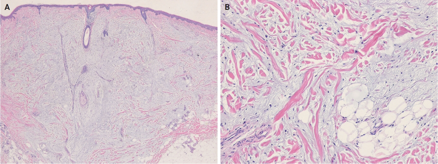
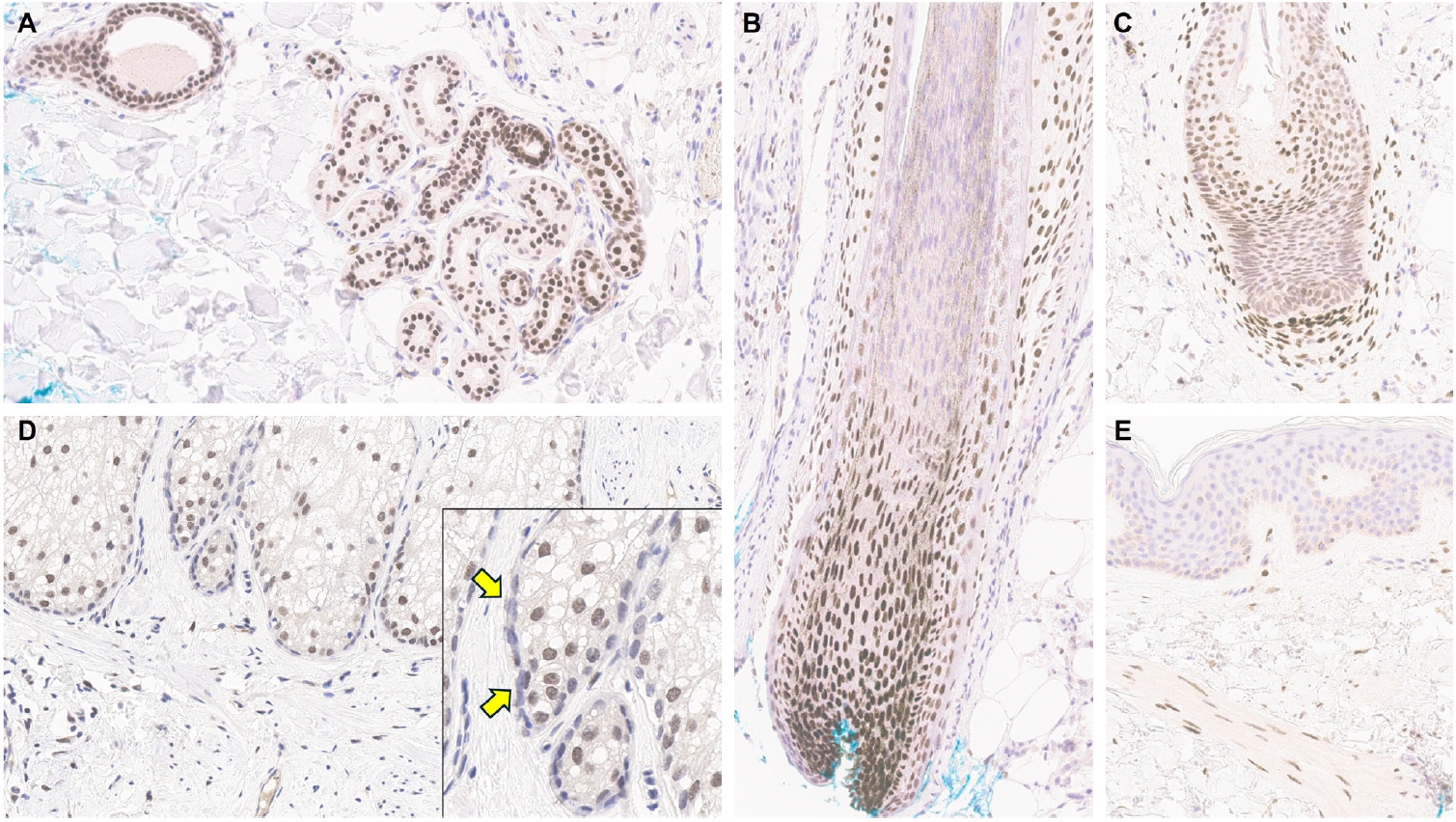
PDF - The 5th edition of the World Health Organization (WHO) classification of skin tumors introduces a dedicated chapter on cutaneous soft tissue tumors, providing a comprehensive, standardized reference with updated diagnostic criteria that directly inform routine dermatopathology practice and molecular diagnostics. This edition incorporates several key changes, including newly recognized entities such as EWSR1::SMAD3-rearranged fibroblastic tumor, neurotrophic tyrosine receptor kinase (NTRK)–rearranged spindle cell neoplasm, superficial CD34-positive fibroblastic tumor, and CRTC1::TRIM11 cutaneous tumor. Diagnostic terminology has also been refined; for example, the term ‘atypical intradermal smooth muscle neoplasm’ replaces ‘cutaneous leiomyosarcoma’ for lesions confined to the dermis, whereas the designation leiomyosarcoma is reserved for tumors with overt subcutaneous infiltration. In addition, epithelioid fibrous histiocytoma has been reassigned to the family of tumors of uncertain differentiation. This review summarizes the key updates and newly recognized entities in the chapter on cutaneous soft tissue tumors in the 5th edition of the WHO classification of skin tumors, emphasizing their clinicopathological and molecular implications.
- The evolving role of TRPS1 in dermatopathology: insights from the past 4 years
- Mokhtar H. Abdelhammed, Woo Cheal Cho
- J Pathol Transl Med. 2026;60(2):129-143. Published online January 29, 2026
- DOI: https://doi.org/10.4132/jptm.2025.11.25
- 3,016 View
- 216 Download
-
Abstract
PDF
- Over the past 4 years, trichorhinophalangeal syndrome type 1 (TRPS1) has rapidly gained attention among practicing pathologists, with numerous studies emerging that both support and question its diagnostic utility. Initially regarded as a highly specific marker for tumors of mammary origin, TRPS1 is now recognized to have broader expression patterns, including in a variety of cutaneous neoplasms. This is likely due to embryologic parallels between breast tissue and skin adnexal structures, an overlap that was underappreciated in early investigations. Although TRPS1 lacks absolute specificity—even among cutaneous neoplasms—it can still offer meaningful diagnostic value when interpreted alongside conventional immunohistochemical markers and within the appropriate morphologic context. Noteworthy diagnostic applications include mammary Paget disease, primary extramammary Paget disease, rare adnexal neoplasms such as endocrine mucin-producing sweat gland carcinoma and primary cutaneous NUT adnexal carcinoma, and cutaneous metastases from breast carcinoma. In this review, we present the most comprehensive and up-to-date evaluation of the utility and limitations of TRPS1 immunohistochemistry in dermatopathology. Our aim is to deepen understanding of this emerging marker and provide practical guidance on its optimal integration with established immunohistochemical panels to enhance diagnostic accuracy in routine practice.
- Primary Cutaneous Mucinous Carcinoma with Extramammary Paget’s Disease: Eccrine or Apocrine?
- Sun-Ju Oh, Young-Ok Kim
- J Pathol Transl Med. 2018;52(4):238-242. Published online January 25, 2018
- DOI: https://doi.org/10.4132/jptm.2017.11.21
- 10,644 View
- 148 Download
- 3 Web of Science
- 4 Crossref
-
Abstract
PDF
- Primary cutaneous mucinous carcinoma (PCMC) is an uncommon tumor of the sweat gland origin. The occurrence of PCMC is mostly in middle-aged and older patients, with a slight male predominance. Most cases of PCMC arise on the head, with a preference for eyelids. The histogenesis of PCMC, whether eccrine or apocrine, remains controversial. We report a rare case of PCMC with secondary extramammary Paget’s disease in the groin of a 75-year-old man, which favored an apocrine origin. Furthermore, based on a review of the literature, we provide several histologic clues that can be used to differentiate PCMC from metastatic mucinous carcinoma.
-
Citations
Citations to this article as recorded by
- Primary cutaneous mucinous carcinoma of the scalp masquerading as a benign dermatological mass - A case report
Fadi Alnehlaoui, Nafad Mohamed Lotfy Elhadidi, Shafik Fwakhrji, Shekhar V. Shikare, Majid Hassan Alhammadi, Salman Yousuf Guraya
International Journal of Surgery Case Reports.2024;[Epub] CrossRef - Primary cutaneous mucinous carcinoma in a periorbital lesion: two case reports and literature review
Jun Woo Kim, Sung Eun Kim
Archives of Craniofacial Surgery.2024; 25(2): 90. CrossRef - Primary Cutaneous Mucinous Carcinoma: A Review of the Literature
Timothy Freeman, Aaron J. Russell, M. Laurin Council
Dermatologic Surgery.2023; 49(12): 1091. CrossRef - A Case of Eccrine Mucinous Carcinoma Involving Scalp
Ramsha Saleem, Sachin Vaidya
Cureus.2021;[Epub] CrossRef
- Primary cutaneous mucinous carcinoma of the scalp masquerading as a benign dermatological mass - A case report
- Characteristics of Cutaneous Lymphomas in Korea According to the New WHO-EORTC Classification: Report of a Nationwide Study
- Jae Ho Han, Young-Hyeh Ko, Yun Kyung Kang, Wan-Seop Kim, Yoon Jung Kim, Insun Kim, Hyun-Jung Kim, Soo Kee Min, Chan-Kum Park, Chan-Sik Park, Bong-Kyung Shin, Woo Ick Yang, Young-Ha Oh, Jong Sil Lee, Juhie Lee, Tae Hui Lee, Hyekyung Lee, Ho Jung Lee, Yoon Kyung Jeon, Hee Jeong Cha, Yoo-Duk Choi, Chul Woo Kim
- Korean J Pathol. 2014;48(2):126-132. Published online April 28, 2014
- DOI: https://doi.org/10.4132/KoreanJPathol.2014.48.2.126
- 10,910 View
- 119 Download
- 14 Crossref
-
Abstract
PDF
Background Previously, cutaneous lymphomas were classified according to either the European Organization for the Research and Treatment of Cancer (EORTC) or the World Health Organization (WHO) classification paradigms. The aim of this study was to determine the relative frequency of Korean cutaneous lymphoma according to the new WHO-EORTC classification system.
Methods A total of 517 patients were recruited during a recent 5 year-period (2006-2010) from 21 institutes and classified according to the WHO-EORTC criteria.
Results The patients included 298 males and 219 females, and the mean age at diagnosis was 49 years. The lesions preferentially affected the trunk area (40.2%). The most frequent subtypes in order of decreasing prevalence were mycosis fungoides (22.2%), peripheral T-cell lymphoma (17.2%), CD30+ T-cell lymphoproliferative disorder (13.7%), and extranodal natural killer/T (NK/T) cell lymphoma, nasal type (12.0%). Diffuse large B-cell lymphoma accounted for 11.2% of cases, half of which were secondary cutaneous involvement; other types of B-cell lymphoma accounted for less than 1% of cases.
Conclusions In comparison with data from Western countries, this study revealed relatively lower rates of mycosis fungoides and B-cell lymphoma in Korean patients, as well as higher rates of subcutaneous panniculitis-like T-cell lymphoma and NK/T cell lymphoma.
-
Citations
Citations to this article as recorded by- Varied presentations of primary cutaneous lymphoma: A case series from a tertiary care center in South India
Baby Shana, Betsy Ambooken, Sunitha Balakrishnan, Asokan Neelakandan, Kidangazhiyathmana Ajithkumar
Indian Journal of Cancer.2024; 61(1): 172. CrossRef - A retrospective study of prognostic factors and treatment outcome in advanced-stage Mycosis Fungoides and Sezary Syndrome
Zhuo-fan Xu, Hongyun Chen, Yuehua Liu, Wei Zhang, Hongzhong Jin, Jie Liu
Hematology.2024;[Epub] CrossRef - Prevalence, clinical features, and survival outcome trends of 627 patients with primary cutaneous lymphoma over 29 years: a retrospective review from single tertiary center in Korea
Ik Jun Moon, Chong Hyun Won, Sung Eun Chang, Chan-Sik Park, Dok-Hyun Yoon, Si Yeol Song, Mi Woo Lee, Woo Jin Lee
Scientific Reports.2024;[Epub] CrossRef - The First Case of Acute Myeloid Leukemia With t(10;11)(p13;q21);PICALM-MLLT10 Rearrangement Presenting With Extensive Skin Involvement
Min-Seung Park, Hyun-Young Kim, Jae Joon Lee, Duck Cho, Chul Won Jung, Hee-Jin Kim, Sun-Hee Kim
Annals of Laboratory Medicine.2023; 43(3): 310. CrossRef - Recent advances on cutaneous lymphoma epidemiology
G. Dobos, M. Miladi, L. Michel, C. Ram-Wolff, M. Battistella, M. Bagot, A. de Masson
La Presse Médicale.2022; 51(1): 104108. CrossRef - Specific cutaneous infiltrates in patients with haematological neoplasms: a retrospective study with 49 patients
Rebeca Calado, Maria Relvas, Francisca Morgado, José Carlos Cardoso, Oscar Tellechea
Australasian Journal of Dermatology.2021;[Epub] CrossRef - Epidemiology of Cutaneous T-Cell Lymphomas: A Systematic Review and Meta-Analysis of 16,953 Patients
Gabor Dobos, Anne Pohrt, Caroline Ram-Wolff, Céleste Lebbé, Jean-David Bouaziz, Maxime Battistella, Martine Bagot, Adèle de Masson
Cancers.2020; 12(10): 2921. CrossRef - Primary cutaneous lymphoma in Argentina: a report of a nationwide study of 416 patients
Alejandra Abeldaño, Paula Enz, Matias Maskin, Andrea B. Cervini, Natallia Torres, Ana C. Acosta, Marina Narbaitz, Silvia Vanzulli, Mirta Orentrajch, Marta A. Villareal, Maria L. Garcia Pazos, Mariana Arias, Evelyn A. Zambrano Franco, Maria I. Fontana, Rob
International Journal of Dermatology.2019; 58(4): 449. CrossRef - Post-thymic CD4 positive cytotoxic T cell infiltrates of the skin: A clinical and histomorphologic spectrum of the unique CD4 positive T cell of immunosenescence
Cynthia M. Magro, Luke C. Olson, Shabnam Momtahen
Annals of Diagnostic Pathology.2019; 38: 99. CrossRef - Cutaneous lymphomas in Taiwan: A review of 118 cases from a medical center in southern Taiwan
Chaw-Ning Lee, Chao-Kai Hsu, Kung-Chao Chang, Cheng-Lin Wu, Tsai-Yun Chen, Julia Yu-Yun Lee
Dermatologica Sinica.2018; 36(1): 16. CrossRef - Imaging analysis of superficial soft tissue lymphomas
In Sook Lee, You Seon Song, Seung Hyun Lee, Young Jin Choi, Sung Moon Lee
Clinical Imaging.2018; 49: 111. CrossRef - Epidemiologic, clinical and demographic features of primary cutaneous lymphomas in Castilla‐La Mancha, Spain: are we different?
C. Ramos‐Rodríguez, M. García‐Rojo, G. Romero‐Aguilera, M. García‐Arpa, L. González‐López, M.P. Sánchez‐Caminero, J. González‐García, M. Delgado‐Portela, M.P. Cortina‐De La Calle, M.F. Relea‐Calatayud, F. Martín‐Dávila, R. López‐Pérez, M. Ramos‐Rodríguez
Journal of the European Academy of Dermatology and Venereology.2018;[Epub] CrossRef - Nasal-type NK/T-cell lymphomas are more frequently T rather than NK lineage based on T-cell receptor gene, RNA, and protein studies: lineage does not predict clinical behavior
Mineui Hong, Taehee Lee, So Young Kang, Suk-Jin Kim, Wonseog Kim, Young-Hyeh Ko
Modern Pathology.2016; 29(5): 430. CrossRef - Cutaneous lymphoma: Kids are not just little people
Katalin Ferenczi, Hanspaul S. Makkar
Clinics in Dermatology.2016; 34(6): 749. CrossRef
- Varied presentations of primary cutaneous lymphoma: A case series from a tertiary care center in South India
- Human Cutaneous Protothecosis: Report of a Case and Literature Review
- Jae Yeon Seok, Yoonho Lee, Hyukmin Lee, Sang Yeop Yi, Hwa Eun Oh, Ji-Sun Song
- Korean J Pathol. 2013;47(6):575-578. Published online December 24, 2013
- DOI: https://doi.org/10.4132/KoreanJPathol.2013.47.6.575
- 14,474 View
- 122 Download
- 19 Crossref
-
Abstract
PDF
The
Prototheca species is achlorophyllic algae and rarely causes human infection. Human protothecosis presents clinically as a cutaneous infection, olecranon bursitis, and disseminated systemic disease. We report a case of human cutaneous protothecosis involving the left wrist. A 68-year-old man presented with an ill-defined erythematous lesion with crust at the dorsal aspect of his left wrist. A punch biopsy was performed to reveal the histologic features of granulomatous inflammation with necrosis at the upper dermis, containingPrototheca organisms, of which, the characteristic features were highlighted by special staining. Through a molecular study, thePrototheca zopfii species was identified.-
Citations
Citations to this article as recorded by- Successful Treatment of Cutaneous Protothecosis Due to Prototheca wickerhamii with Terbinafine
Yue Chen, Aili Gao, Yanan Ke, Xin Zhou, Li Lin, Sha Lu, Yumei Liu
Clinical, Cosmetic and Investigational Dermatology.2024; Volume 17: 913. CrossRef - Disseminated Protothecosis Due to Prototheca zopfii and Literature Review
Feifan Chen, Mario Saab-Chalhoub, Joy Tao, Amanda T. Harrington, Fritzie S. Albarillo, Andrew S. Crone, Nina M. Clark, Jodi J. Speiser
The American Journal of Dermatopathology.2023; 45(4): 237. CrossRef - The oldest patient infected with cutaneous protothecosis
Gabrielle Perrotti, Christian DiLiberto, Danielle M Pineda
Journal of Wound Care.2023; 32(4): 235. CrossRef - Cutaneous protothecosis: contrasting case presentations
Thomas Orschulok, Joseph Whitfield, Timothy Badrick, Pirathaban Sivabalan, David Sowden, Fiona Lehane
Pathology.2023; 55(7): 1032. CrossRef - Indurated Plaque With Ulceration on the Dorsum of the Left Hand
Juanjuan Li, Xiaomo Wu, Borui Chen
JAMA Dermatology.2022; 158(12): 1442. CrossRef - Cerebral protothecosis mimicking high-grade glioma
James Samarasekara, Soumya Mukherjee, Azzam Ismail, Robert Corns
BMJ Case Reports.2021; 14(1): e235362. CrossRef - Protothecosis algaemia in a patient presenting with septic arthritis: A rare case of Prototheca zopfii isolated from Malaysia
Prem Ananth Palaniappan, Cassandra Anne Abot, Ratna Mohd Tap, Fairuz Amran
IDCases.2021; 24: e01121. CrossRef -
Prototheca bovis, a unicellular achlorophyllous trebouxiophyte green alga in the healthy human intestine
Vasana Jinatham, Diego M. Cantoni, Ian R. Brown, Thanakrit Vichaslip, Picha Suwannahitatorn, Siam Popluechai, Anastasios D. Tsaousis, Eleni Gentekaki
Journal of Medical Microbiology .2021;[Epub] CrossRef - Pseudomicosis por oportunistas: desconocimiento y retos diagnósticos
María Adelaida Garcés-Abad, Nora Cardona Castro
CES Medicina.2021; 35(3): 257. CrossRef - Nanotechnology as a Novel Approach in Combating Microbes Providing an Alternative to Antibiotics
Bismillah Mubeen, Aunza Nayab Ansar, Rabia Rasool, Inam Ullah, Syed Sarim Imam, Sultan Alshehri, Mohammed M. Ghoneim, Sami I. Alzarea, Muhammad Shahid Nadeem, Imran Kazmi
Antibiotics.2021; 10(12): 1473. CrossRef - Prototheca zopfii genotype II induces mitochondrial apoptosis in models of bovine mastitis
Muhammad Shahid, Eduardo R. Cobo, Liben Chen, Paloma A. Cavalcante, Herman W. Barkema, Jian Gao, Siyu Xu, Yang Liu, Cameron G. Knight, John P. Kastelic, Bo Han
Scientific Reports.2020;[Epub] CrossRef - Murine and Human Cathelicidins Contribute Differently to Hallmarks of Mastitis Induced by Pathogenic Prototheca bovis Algae
Muhammad Shahid, Paloma Araujo Cavalcante, Cameron G. Knight, Herman W. Barkema, Bo Han, Jian Gao, Eduardo R. Cobo
Frontiers in Cellular and Infection Microbiology.2020;[Epub] CrossRef - Draining dorsal hand pustules, nodules, and ulcers in a patient with immunosuppression
Michael R. Stephens, Oyinade Aderibigbe, Katherine T. Steele, David E. Elder, Laurel Glaser, Jerry Jacob, Misha Rosenbach
JAAD Case Reports.2019; 5(10): 846. CrossRef - Characterization of the complete mitochondrial genome and phylogenetic analysis of Prototheca stagnorum (chlorellales: Chlorellaceae)
Qian Wu, Yanhong Lan, Hui Xu, Yi Cao
Mitochondrial DNA Part B.2019; 4(2): 4000. CrossRef - Cutaneous protothecosis reminiscent of unilateral solar elastotic bands of forearm in an immunocompromised patient
Yi-Hsin Ho, Yun-Wen Chiu, Chen-Yi Wu, Han-Nan Liu
Dermatologica Sinica.2018; 36(2): 93. CrossRef - A Case of Cutaneous Protothecosis in an Immunocompetent Patient
Ju Yun Woo, Eun Ah Suhng, Ji Yeon Byun, Hae Young Choi, Sun Hee Sung, You Won Choi
Annals of Dermatology.2016; 28(2): 273. CrossRef - Protothecal bursitis after simultaneous kidney/liver transplantation: a case report and review
I. Ramírez, J.F. Nieto‐Ríos, C. Ocampo‐Kohn, A. Aristizábal‐Alzate, G. Zuluaga‐Valencia, O. Muñoz Maya, J.C. Pérez
Transplant Infectious Disease.2016; 18(2): 266. CrossRef - Cutaneous Protothecosis in a Patient with Chronic Lymphocytic Leukemia: A Case Report and Literature Review
Quynh-Giao Nguyen, Ted Rosen
Journal of Fungi.2015; 1(1): 4. CrossRef - Natamycin - antimycotic of polyene macrolides class with unusual properties
I. O. Malova, D. D. Petrunin
Vestnik dermatologii i venerologii.2015; 91(3): 161. CrossRef
- Successful Treatment of Cutaneous Protothecosis Due to Prototheca wickerhamii with Terbinafine
- Distribution of Dendritic Cells and Regulatory T-Cells in Cutaneous Lymphomas.
- Changyoung Yoo, Young Seon Hong, Baik Kee Cho, Sang Ho Kim, Sang In Shim, Chang Suk Kang
- Korean J Pathol. 2010;44(6):581-588.
- DOI: https://doi.org/10.4132/KoreanJPathol.2010.44.6.581
- 3,719 View
- 21 Download
-
Abstract
PDF
- BACKGROUND
Dendritic cells (DCs) play an important role in immune reactions. This study was designed to identify the distribution patterns of DCs and regulatory T-cells (Tregs) in cutaneous lymphomas.
METHODS
Immunohistochemistry was used to determine langerin expression on Langerhans cells, CD11b on inflammatory DCs, CD209 and CD11c on dermal DCs, CD303 on plasmacytic DCs, and Foxp3 on Tregs in 81 cases of cutaneous lymphomas.
RESULTS
Various DCs and Tregs were identified in most cutaneous lymphomas. Plasmacytic DCs, inflammatory DCs and Tregs were identified mainly in tumor areas, whereas dermal DCs were distributed both in the tumor and stromal areas. Among DCs, dermal DCs were most prominently identified in the cutaneous lymphomas not only in the tumor area but also in the stroma. The intense stromal infiltration of dermal DCs was consistent finding in T-cell lymphomas. Diffuse large B-cell lymphoma (DLBCL), not otherwise specified also showed intense stromal infiltration of dermal DCs, but stromal infiltration in DLBCL, leg type was relatively scant.
CONCLUSIONS
The results suggest that all types of DCs and Tregs are involved in cutaneous lymphoma tumor immunity. Among them dermal DCs may play a dominant role.
- Histologic and Immunohistochemical Study of Cutaneous Vascular Disorders.
- Jai Hyang Go, Hoon Jin, Dong Hwan Shin, Kwang Gil Lee
- Korean J Pathol. 1995;29(3):327-333.
- 1,917 View
- 14 Download
-
Abstract
PDF
- There is a wide array of designation for cutaneous vascular disorders based on clinical characteristics, histology, embryology, cellular biology, and hemodynamics. The cutaneous vascular disorders can be divided into hemangioma and vascular malformation according to the biologic classification based on cell kinetics i.e. endothelial hyperplasia. There are clinical and histologic differences between them. In this study, clinical, histologic and im-munohistochemical evaluations were attempted on 40 cases of cutaneous vascular disorders diagnosed the period between 1985 and 1993. The results are as follows: 1) Twenty-three out of forty cases were immunoreacive for proliferating cell nuclear antigen(PCNA). The lesions composed of capillary-sized blood vessels with endothelial hyperplasia were diffusely reactive, whereas those composed largely of dilated blood vessels with or without focal endothelial hyperplasia were only focally reactive. 2) Each groups of the classic classification contained both reactive and nonreactive cases except nevus flammeus and juvenfle hemangioma. 3) In contrast to the cases nonreactive for PCNA, those reactive for PCNA contained areas of proliferating small vessels, which showed reactivity for PCNA. In conclusion, the cutaneous vascular disorders diagnosed by the classic classification are heterogeneous in the pattern of the endothelial hyperplasia and the PCNA staining. Therefore it should be classified by the clinical and the histologic characteristics.
- Cutaneous Ciliated Cyst: Report of a case.
- Eundeok Chang, Sangin Shim
- Korean J Pathol. 1995;29(3):391-392.
- 2,074 View
- 22 Download
-
Abstract
PDF
- Farmer and Helwig coined the term cutaneous ciliated cyst in 1978 to describe specific skin cysts lined with ciliated epithelium a resembling the mucosa of the fallopian tube. Twenty case, of cutaneous ciliated cysts have been published to date. All have been found to affect the lower limb and all patients have been women. The etiology of the cyst is as yet unknown. A 37-year-old woman had a cyst excised from the left buttock. Microscopically, the lining epithelium of the cyst wall was composed of columnar cilited cells similar to those seen in the fallopian tubes. Immunoperoxidase stainings for cytokeratin, epithelial membrane antigen and carcinoembryonic antigen were positive.
- Primary Cutaneous Meningioma arising from the Scalp: A case report.
- Sug Hyung Lee, Seok Jin Gang, Sun Moo Kim
- Korean J Pathol. 1993;27(2):181-183.
- 3,027 View
- 93 Download
-
Abstract
PDF
- Primary cutaneous meningiomas are extremely rare tumors found in the cutis or subcutis, and generally have a benign course. The tend to be located on the scalp, face, neck. or paravertebral area. The primary cutaneous meningioma bears similarities to developmental defects and probably originates from arachnoid cell rests in the skin, although diverse groups of cutaneous meningiomas seem to arise from several different sources. A case of primary cutaneous meningioma occuring in the scalp of left parietal area of a 27-year-old female is presented. Clinically the lesion appeared as indolent, slow growing cutaneous mass and has no connection with underlying brain tissue, as determined by examination of the roentgenographs. The definite diagnosis was made after pathological examination. Microscopically the tumor is composed of sheets and nests of meningothelial cells. Immunohistochemical and electron microscopic studies reveal the typical findings of meningioma.
- Cutaneous Bronchogenic Cyst Over the Sternum: A Case Report.
- Ho Sung Park, Hyun Jin Son, Myoung Jae Kang
- Korean J Pathol. 2004;38(5):333-336.
- 2,293 View
- 33 Download
-
Abstract
PDF
- Bronchogenic cyst is usually an extrapulmonary cyst formed as the result of an accessory lung bud from the foregut that becomes isolated from the rest of the tracheobronchial tree producing a usually solitary cyst. Most bronchogenic cysts are in the mediastinum with rare occurrence on the subcutaneous tissue over the sternum. We report a case of cutaneous bronchogenic cyst that occurred in the skin over the sternum in a 13-month-old boy. On ultrasonography, a well circumscribed non-echogenic cystic mass was observed measuring 1.5x1.3 cm. Histologically, the cyst was lined by cilicated, pseudostratified, columnar epithelium with interspersed goblet cells, and there were bundles of smooth muscle fibers, mucous glands, and lymphoid aggregates in the cyst wall.
- A Case of Solitary Cutaneous Myofibroma of the Thigh in An Adult.
- Jung Hwan Park, Chang Woo Lee, Young Chae Chu, Moon Hyang Park
- Korean J Pathol. 2001;35(4):354-356.
- 2,275 View
- 33 Download
-
Abstract
PDF
- Adult solitary cutaneous myofibroma is a recently described benign neoplasm of the skin or subcutis, representing the adult counterpart of infantile myofibroblastoma. The histologic and immunohistochemical features of a 21-year-old woman with a solitary brownish, mildly tender nodule on her right thigh are reported here. The nodule had been present for a duration of 3 years. It showed a nodular dermal mass with an irregular margin. The lesion consisted of interlacing bundles of spindle cells which were positive for smooth muscle actin, muscle specific actin and vimentin. Immunohistochemical stainings for desmin, S-100 protein, CD 34 and CD 68 were negative. Cutaneous myofibroma in an adult is a distinct entity of benign neoplasm.
- Solid and Papillary Epithelial Neoplasm of the Pancreas Diagnosed by Percutaneous Fine Needle Aspiration Biopsy.
- Chan Kum Park, Jung Dal Lee
- Korean J Pathol. 1990;24(3):304-309.
- 2,064 View
- 16 Download
-
Abstract
PDF
- Solid and papillary epithelial neoplasm of the pancreas is a rare, low grade malignant tumor of adolescence. The tumor we described was diagnosed by following preoperative percutaneous fine needle aspiration biopsy and confirmed by tissue examination. Characteristic cytologic findings were monotonous cells arranged in papillary fronds, acinus, and cords in necrotic and hemorrhagic backgrounds. Occasional foamy histiocytes and hyaline globules were present. The cytoplasm of the tumor cells were moderate in amount, pale eosinophilic or vacuolated with distinct borders. The nuclei were centrally located and round. There were one or two small nucleoli and finely granular chromatin throughout the nucleus.
- The Usefulness of Fine Needle Aspiration Cytology of Bone Lesions.
- Bohng Hee Kim, Gyung Yub Gong
- J Pathol Transl Med. 2002;13(2):51-59.
- 2,057 View
- 29 Download
-
Abstract
PDF
- To determine the usefulness of fine needle aspiration cytology(FNAC) of bone lesions and the complementary role of FNAC and percutaneous needle biopsy, 75 cases of FNAC taken from bone lesions were analyzed. Correlations with histopathology were possible in 47 cases, including 14 cases of simultaneous core biopsy and 33 cases of subsequent open biopsy due to inadequate aspirates. Among 75 cases, 4 cases were benign tumors and tumor-like lesion, 11 cases were malignant primary bone tumors, 17 cases were metastatic tumors, and 43 cases were nonneoplastic bone lesions. The aspirates were adequate in 35 cases(46.7%), in all of which the discrimination between benignancy and malignancy was possible. The main reason for inadequate aspirates was due to hypocellularity. In the cases of aspiration and core biopsy simultaneously done, the diagnostic accuracy of aspiration, core biopsy, and both were 57%(8/14), 78.6%(11/14), and 92.9%(13/14), respectively. We conclude that a final diagnosis based on cytology is possible with the adequate aspirates and the clinical and radiological findings. Also we confirm the complementary role between FNAC and core biopsy in bone lesions.
- Subcutaneous Fat Necrosis Associated with Pancreatic Adenocarcinoma: A case report.
- Hee Jung Kim, Kwang Gil Lee
- Korean J Pathol. 1996;30(2):155-160.
- 2,205 View
- 18 Download
-
Abstract
PDF
- Subcutaneous fat necrosis is manifested by erythematous tender nodules on the legs, buttock or trunk and is associated with pancreatic disease including acute and chronic pancreatitis, pancreatic carcinoma, pseudocyst, pancreatic stone and other diseases. Its histologic findings are pathognomonic and reveal foci of subcutaneous fat necrosis with "ghost-like" anucleated cells with thick "shadowy wall" and surrounding inflammatory infiltrate consisting of polymorphonuclear cells, eosinophils, lymphocytes, histiocytes, foam cells and foreign body giant cells. We experienced a case of subcutaneous fat necrosis associated with pancreatic adenocarcinoma manifested by subcutaneous nodules in the buttock and lower extremities and by arthralgia of the left knee in a 67-year-old woman. Therefore, we are reporting to emphasize the importance of the skin findings of the internal disease.
- Histopathologic Features and Immunophenotype of 19 Primary Cutaneous Lymphomas.
- Hee Sung Kim, Young Hyeh Ko, Howe J Ree
- Korean J Pathol. 1999;33(12):1111-1119.
- 2,276 View
- 19 Download
-
Abstract
PDF
- The diagnosis of primary cutaneous lymphoma is based on a combination of clinical, histological, immunophenotypic and genetic criteria. Nineteen cases of primary cutaneous lymphomas were studied for clinicopathologic, immunophenotypic, and genetic features. Seventeen (89%) cases were T cell origin and two cases (11%) were B cell origin. CD30-positive cutaneous lymphoproliferative disorder was the most frequent subtype, occupying 42% (8 cases) of the cases. CD8 was positive in 5 cases consisting of 3 cutaneous T cell lymphomas and 2 anaplastic large cell lymphomas. CD4 was positive in 2 cases of mycosis fungoides and 3 cases of lymphomatoid papulosis. Six (67%) of 9 cases of cutaneous T cell lymphoma were positive for TIA-1. Ten (83%) out of 12 cases showed clonal rearrangements of TCR gamma genes, however, one T/NK cell lymphoma and one anaplastic large cell lymphoma did not. EBV association was detected only in T/NK cell lymphomas among 10 cases examined. In conclusion, our study showed higher proportion of CD30-positive lymphoproliferative disorders and less frequent mycosis fungoides in Korea compared to the incidences in Western countries. Our immunostaining results suggested that mycosis fungoides and lymphomatoid papulosis are CD4-positive T cell origin, however, the remaining primary cutaneous T cell lymphoma is predominantly CD8-positive cytotoxic T cell origin.
- A Pathologic Study of Lymphoproliferative Disorders of the Skin.
- Yee Jeong Kim, Kwang Gil Lee, Soo Il Chun, Yun Woong Ko
- Korean J Pathol. 1991;25(6):551-562.
- 2,774 View
- 30 Download
-
Abstract
PDF
- Forty eight skin biopsies obtained from 24 patients were reviewed, and clinical, histological and immunohistochemical findings were analyzed. Results obtained are as follows: 1) Skin manifestation was plaque, erythroderma, scale and hyperpigmentation in mycosis fungoides, and subcutaneous nodule, mass and ulcerated patch in cutaneous lymphoma. The skin of lymphomatoid papulosis revealed hemorrhagic ulcerated and erythematous papules which healed spontaneously. 2) Histologically, mycosis fungoides showed epidermotropism in most cases. Pautrier's micro-abscesses were present in one-fourth of the cases. Malignant lymphoma was different in histology from mycosis fungoides. As compared with mycosis fungoides, it showed less frequent epidermotropism, more compact and diffuse infiltration of atypical lymphocytes, more often association with ulcer and necrosis, and more frequent mitotic figures. Lymphomatoid papulosis showed striking hemorrhage and edema of the papillary dermis. 3) Based on the results of immunohistochemical study, mycosis fungoides and lymphomatoid papulosis were considered as a T cell proliferative disorder of the skin. According to these findings, lymphoproliferative disorders of the skin occurred predominantly in the elderly and males. Clinical and histopathologic findings overlapped and were similar each other. It was difficult to make a definite diagnosis in early lesions, and a sequential follow up biopsy was required. It is concluded that strict criteria such as marked atypia and clustering of atypical cells are necessary for a histologic diagnosis of malignant lymphoproliferative disorder of the skin.
- EBV in Situ Hybridization Study for Cutaneous T-Cell Lymphomas.
- Chan Kum Park, Chang Woo Lee, Jung Dal Lee
- Korean J Pathol. 1996;30(8):699-705.
- 2,158 View
- 19 Download
-
Abstract
PDF
- We studied 24 cases of cutaneous T-cell lymphomas and six cases of benign lymphoproliferative diseases of the skin (2 Jessner's lymphocytic infiltration, 2 pseudolymphoma, 2 lymphomatoid papulosis) for the presence of Epstein-Barr Virus(EBV) RNA, using the in situ hybridization(ISH) method. Among the 24 cases of cutaneous T-cell lymphomas (CTCL), 18 cases including 12 cases of mycosis fungoides(MF) were primary CTCL, and the other 6 cases were secondary CTCL. The ISH study demonstrated a positive reaction for EBER probe in 6 out of the 24 cases(25%) of CTCL, and a negative reaction for BHLF nuclear RNA probe in all the cases studied. Double-labelling immunohistochemistry/ISH studies revealed that the EBV positive cells were CD45RO positive and CD20 negative. EBV genome was not demonstrated in any benign lymphoproliferative diseases of the skin. Among the EBER positive cases, none of the 12 cases of MF demonstrated EBER signals, and 6 out of the 12(50%) cases of CTCL were positive for EBER probe. In conclusion, latent infection of EBV may play a role in the development of non-mycosis fungoides T-cell lymphomas involving the skin.
- Warty Dyskeratoma with a Cutaneous Horn: Report of a case.
- Ah Won Lee, Hyun Joo Choi, Youn Soo Lee, Seok Jin Kang, Byung Kee Kim, Sun Moo Kim
- Korean J Pathol. 1998;32(8):616-618.
- 2,139 View
- 10 Download
-
Abstract
- The clinical and pathological features of warty dyskeratoma are presented. Warty dyskeratoma is a rarely encountered, solitary, benign cutaneous tumor occurring most often on the head and neck and to our knowledge, no cases associated with cutaneous horn have been published in western literature. We experienced a case of warty dyskeratoma with a cutaneous horn occurring in a 70-year-old male, who had a 1.3 cm-sized and slowly growing nodule on his neck. A brief review of the literature, was made especially in relation to the pathological findings and histogenesis of warty dyskeratoma.
- Epithelioid Leiomyosarcoma of Skin: A case report.
- Han Seong Kim, Nam Bok Cho, Hyun Soon Lee, Kye Yong Song
- Korean J Pathol. 1998;32(9):700-703.
- 2,563 View
- 11 Download
-
Abstract
- This report concerns a rare malignant smooth muscle neoplasm of the skin and the subcutaneous tissue, which was examined immunohistochemically and ultrastructurally. It occured in a 48-year-old female patient who had suffered from painful nodules on the left shoulder. The nodules were removed and diagnosed as a benign fibrohistiocytic tumor. One year after local excision, multiple nodules were detected by physical examination at the previous operation site. Grossly, thirteen well-defined small nodules had spread in the dermis and the subcutaneous tissue, of which nine were located from lower dermis to the subcutaneous tissue and four were in the subcutaneous tissue. Histologically, each nodule consisted mainly of epithelioid tumor cells having eosinophilic or clear plump cytoplasm and round to oval nuclei with small nucleoli, and some peripheral spindle tumor cells. The tumor cells were negative for PAS reaction with and without diastase digestion or alcian blue. Immunohistochemically, tumor cells were positive for smooth muscle actin and vimentin. Ultrastructurally, the tumor cells showed actin filaments and dense bodies in cytoplasm. This case was diagnosed as an epithelioid leiomyosarcoma of the skin and the subcutaneous tissue with local metastasis one year after local excision.
- Molecular Diagnosis of Cutaneous T Cell Lymphoproliferative Diseases.
- Ji Young Park, Myung Hoon Lee, Eun Kyung Kwak, Dong Ja Kim, Tae In Park, Han Ik Bae
- Korean J Pathol. 2000;34(11):941-949.
- 2,530 View
- 17 Download
-
Abstract
PDF
- It is often problematic to diagnose T-cell lymphoproliferative disorders of the skin because of the difficulty in establishing clonality in paraffin-embedded tissue. We used polymerase chain reaction single strand conformational polymorphism (PCR-SSCP) and heteroduplex analysis in paraffin embedded tissue to detect clonal rearrangement of T-cell receptor gamma (TCRgamma) gene in 17 T-cell lymphoproliferative disorders and 6 atypical lymphoproliferative diseases. We used polymerase chain reaction to detect TCR beta gene rearrangement in 8 of 17 cases which did not show TCRgamma gene rearrangement. Jurkat cell lines were used as monoclonal controls. DNA was extracted from 5 biopsies of T-cell lymphomas, 10 biopsies of mycosis fungoides, 2 biopsies of lymphomatoid papulosis, and 6 biopsies of atypical lymphoproliferative lesions. We detected monoclonality in 5 of 5 T-cell lymphoma cases, 2 of 2 lymphomatoid papulosis cases, 6 of 10 mycosis fungoides cases, and 2 of 6 atypical lymphoproliferative disease cases. We conclude that nonradioactive PCR-SSCP for TCR gene rearrangement analysis is a useful adjunct to routine histological and immunophenotypic methods in the diagnosis of cutaneous T cell lymphoproliferative disorders in paraffin embedded tissue.
 First
First Prev
Prev


