 E-submission
Search
- Page Path
- HOME > Search
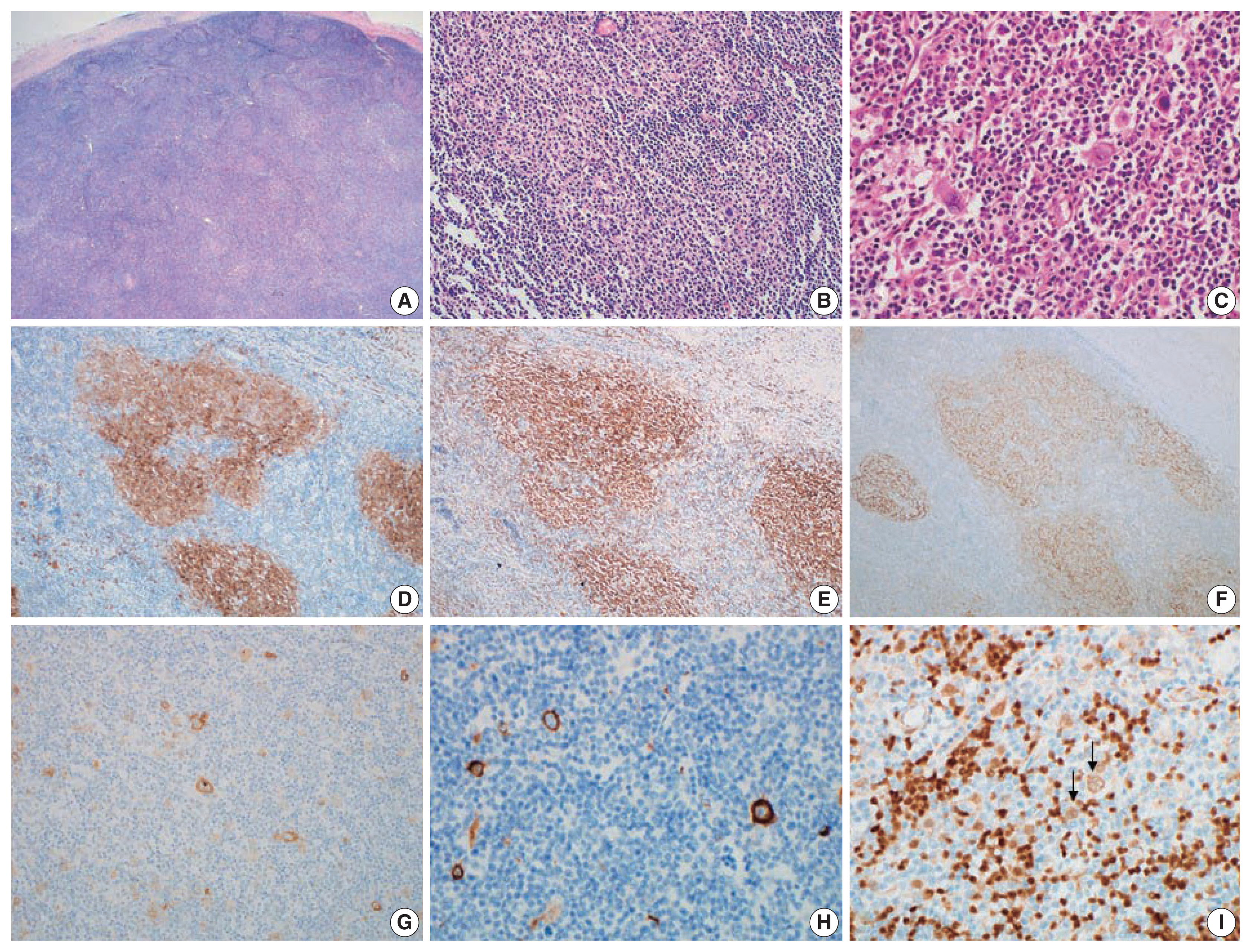
- Composite follicular lymphoma and classic Hodgkin lymphoma
- Han-Na Kim, Min Ji Jeon, Eun Sang Yu, Dae Sik Kim, Chul-Won Choi, Young Hyeh Ko
- J Pathol Transl Med. 2022;56(1):57-60. Published online November 16, 2021
- DOI: https://doi.org/10.4132/jptm.2021.10.09
- 8,488 View
- 247 Download
- 6 Web of Science
- 6 Crossref
-
 Abstract
 PDF
PDF - Composite lymphoma is very rare and a combination of Hodgkin lymphoma and non-Hodgkin lymphoma and even histiocytic tumors can occur. Because of the unfamiliarity, not only can this cause diagnostic problems, but can also affect treatment plan. We report a case of composite lymphoma in a 40-year-old male. Initial biopsy showed a composite lymphoma of follicular lymphoma grade 1 and classic Hodgkin lymphoma. After chemotherapy, another lymph node was taken because of disease progression, which revealed follicular lymphoma, grade 3a without Hodgkin lymphoma component.
-
Citations
Citations to this article as recorded by
- Composite Lymphoma: A Rare Case of Vomiting
Changqin Liu, Dongyan Han, Xiaomin Sun
United European Gastroenterology Journal.2025; 13(5): 836. CrossRef - BCL2-Rearrangment-Negative CD23+ Follicle Center Lymphoma and Chronic Lymphocytic Leukemia/Small Lymphocytic Lymphoma: A Rare Case of Biclonal Composite Lymphoma
Hira Qadir, Ejas Palathingal Bava, Juan Gomez-Gelvez, Wei Liu, Kedar Inamdar, Elizabeth Wey, John Carey, Yulei Shen, Philip Kuriakose, Sharmila Ghosh
Cureus.2025;[Epub] CrossRef - T cell lymphoma and secondary primary malignancy risk after commercial CAR T cell therapy
Guido Ghilardi, Joseph A. Fraietta, James N. Gerson, Vivianna M. Van Deerlin, Jennifer J. D. Morrissette, Gabriel C. Caponetti, Luca Paruzzo, Jaryse C. Harris, Elise A. Chong, Sandra P. Susanibar Adaniya, Jakub Svoboda, Sunita D. Nasta, Ositadimma H. Ugwu
Nature Medicine.2024; 30(4): 984. CrossRef - Double trouble: insights from a rare case of extranodal composite lymphoma in an elderly man, with comprehensive literature review
Aadya Kerkar
American Journal of Translational Research.2024; 16(6): 2599. CrossRef - Composite Lymphoma with Follicular Lymphoma Transformation to Clonally Related Epstein–Barr Virus (EBV) Positive Diffuse Large B-Cell Lymphoma and EBV-PositiveClassic Hodgkin Lymphoma
Christopher B. Ryder, Hayder Saeed, Mohammad Hussaini, Pier Paolo Piccaluga
Case Reports in Hematology.2023; 2023: 1. CrossRef - Plasticity in Classical Hodgkin Composite Lymphomas: A Systematic Review
Alexis Trecourt, Marie Donzel, Juliette Fontaine, Hervé Ghesquières, Laurent Jallade, Gabriel Antherieu, Camille Laurent, Claire Mauduit, Alexsandra Traverse-Glehen
Cancers.2022; 14(22): 5695. CrossRef
- Composite Lymphoma: A Rare Case of Vomiting
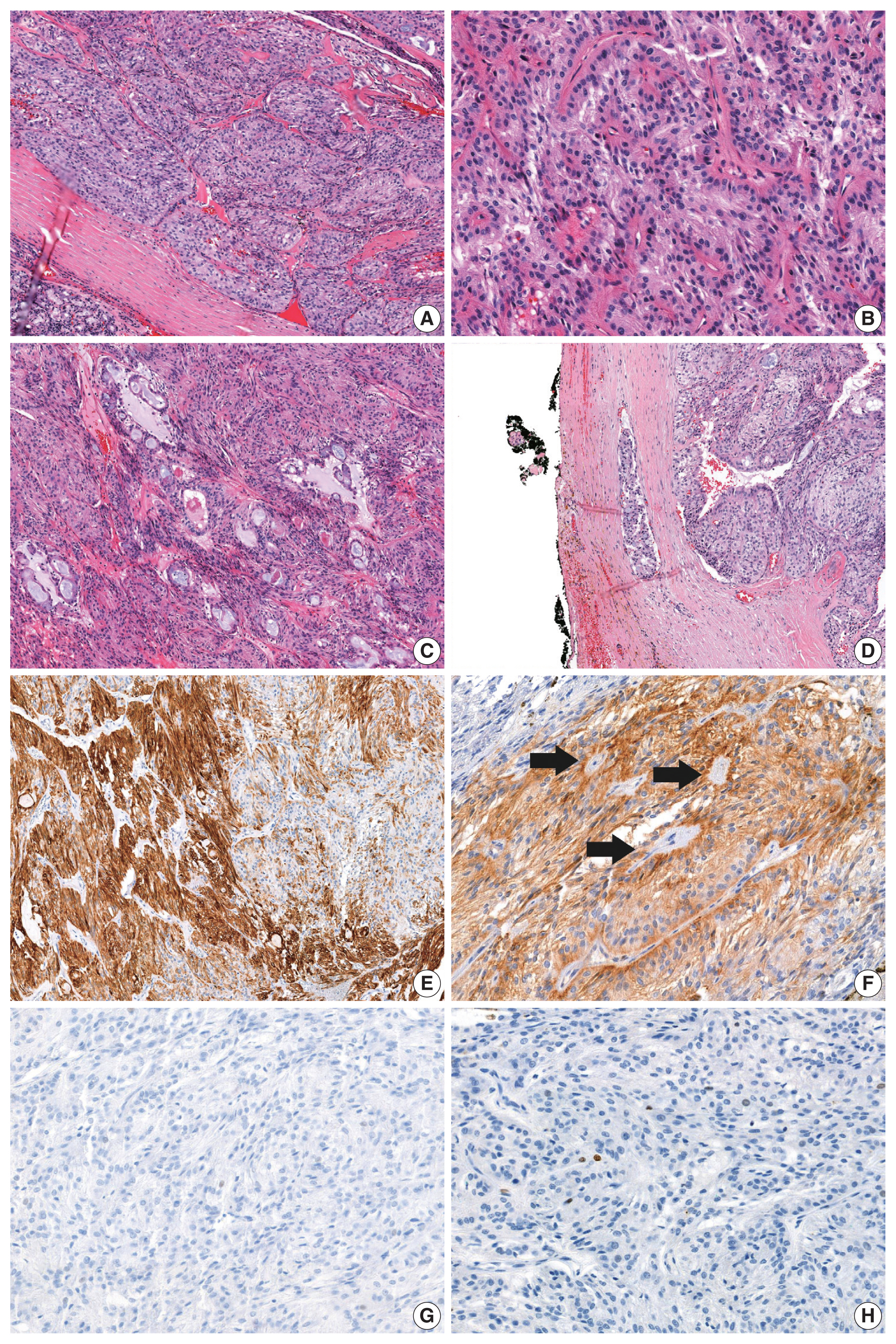
- Adenocarcinoma of the minor salivary gland with concurrent MAML2 and EWSR1 alterations
- Sangjoon Choi, Junhun Cho, Seung Eun Lee, Chung-Hwan Baek, Yi-Kyung Kim, Hyung-Jin Kim, Young Hyeh Ko
- J Pathol Transl Med. 2021;55(2):132-138. Published online January 22, 2021
- DOI: https://doi.org/10.4132/jptm.2020.12.11
- 8,141 View
- 130 Download
- 13 Web of Science
- 9 Crossref
-
Abstract
PDF
- Salivary gland tumors are histologically diverse, and each entity has distinctive histopathological and molecular features. We report two cases of salivary gland tumors with unique histological and molecular findings, which have not been documented previously. The tumors were located in the base of the tongue in both patients. Most tumor cells were arranged in cords and nests, giving a trabecularlike appearance. Focally, glandular structures with intraluminal mucin and perivascular pseudorosette-like configurations were identified. Tumor cells had eosinophilic to clear cytoplasm, and showed mild nuclear atypia. They were positive for pancytokeratin and negative for S-100, p63, c-KIT, androgen receptor, and neuroendocrine markers. Multiple foci of capsular or lymphovascular invasion were identified, but the Ki-67 labeling index was low (< 5%). Fluorescence in situ hybridization revealed concurrent alterations of MAML2 and EWSR1 gene. Further investigations with a larger number of cases with similar histological and molecular features will accurately classify this tumor.
-
Citations
Citations to this article as recorded by- Diagnostic and Prognostic Utility of MAML2 Gene Rearrangement in Mucoepidermoid Carcinoma of Salivary Glands and Its Correlation with Histologic Grading Systems
Akshara Ravichandran, Daphne Fonseca, Sahithi Shilpa Arya, G. Sandhya Devi, Suseela Kodandapani, Chandrasekhara Rao, T. Subramanyeshwar Rao
Indian Journal of Otolaryngology and Head & Neck Surgery.2026; 78(3): 1506. CrossRef - A novel fusion of EWSR1::PRKD1 in cribriform adenocarcinoma of salivary glands: A rare case report
Zecra Yahia
Human Pathology Reports.2025; 42: 300801. CrossRef - Salivary Gland Neoplasms With a Unique Trabecular Histology and MAML2 Translocation
Bokyung Ahn, Seung-Ho Choi, Doeun Kim, Deokhoon Kim, Kyung-Ja Cho
American Journal of Surgical Pathology.2023; 47(10): 1085. CrossRef - Mesonephric-like Adenocarcinoma of the Ovary: Clinicopathological and Molecular Characteristics
Hyun Hee Koh, Eunhyang Park, Hyun-Soo Kim
Diagnostics.2022; 12(2): 326. CrossRef - The evolving role of molecular pathology in the diagnosis of salivary gland tumours with potential pitfalls
Kanwalpreet Kaur, Shailee Mehta, Sangita Vanik, Priti Trivedi, Nirmalya Banerjee, Harsh Dhar, Sourav Datta, Subhadeep Karanjai
European Archives of Oto-Rhino-Laryngology.2022; 279(8): 3769. CrossRef - Alveolar Soft Part Sarcoma of the Uterus: Clinicopathological and Molecular Characteristics
Yurimi Lee, Kiyong Na, Ha Young Woo, Hyun-Soo Kim
Diagnostics.2022; 12(5): 1102. CrossRef - Endometrioid Carcinomas of the Ovaries and Endometrium Involving Endocervical Polyps: Comprehensive Clinicopathological Analyses
Jihee Sohn, Yurimi Lee, Hyun-Soo Kim
Diagnostics.2022; 12(10): 2339. CrossRef - Mesonephric-like Differentiation of Endometrial Endometrioid Carcinoma: Clinicopathological and Molecular Characteristics Distinct from Those of Uterine Mesonephric-like Adenocarcinoma
Sujin Park, Go Eun Bae, Jiyoung Kim, Hyun-Soo Kim
Diagnostics.2021; 11(8): 1450. CrossRef - Mesonephric-like Adenocarcinoma of the Uterine Corpus: Comprehensive Immunohistochemical Analyses Using Markers for Mesonephric, Endometrioid and Serous Tumors
Hyunjin Kim, Kiyong Na, Go Eun Bae, Hyun-Soo Kim
Diagnostics.2021; 11(11): 2042. CrossRef
- Diagnostic and Prognostic Utility of MAML2 Gene Rearrangement in Mucoepidermoid Carcinoma of Salivary Glands and Its Correlation with Histologic Grading Systems
- Epstein-Barr Virus–Associated Lymphoproliferative Disorders: Review and Update on 2016 WHO Classification
- Hyun-Jung Kim, Young Hyeh Ko, Ji Eun Kim, Seung-Sook Lee, Hyekyung Lee, Gyeongsin Park, Jin Ho Paik, Hee Jeong Cha, Yoo-Duk Choi, Jae Ho Han, Jooryung Huh
- J Pathol Transl Med. 2017;51(4):352-358. Published online June 5, 2017
- DOI: https://doi.org/10.4132/jptm.2017.03.15
- 31,149 View
- 1,127 Download
- 69 Web of Science
- 67 Crossref
-
Abstract
PDF
- Epstein-Barr virus (human herpesvirus-4) is very common virus that can be detected in more than 95% of the human population. Most people are asymptomatic and live their entire lives in a chronically infected state (IgG positive). However, in some populations, the Epstein-Barr virus (EBV) has been involved in the occurrence of a wide range of B-cell lymphoproliferative disorders (LPDs), including Burkitt lymphoma, classic Hodgkin’s lymphoma, and immune–deficiency associated LPDs (post-transplant and human immunodeficiency virus–associated LPDs). T-cell LPDs have been reported to be associated with EBV with a subset of peripheral T-cell lymphomas, angioimmunoblastic T-cell lymphomas, extranodal nasal natural killer/T-cell lymphomas, and other rare histotypes. This article reviews the current evidence covering EBV-associated LPDs based on the 2016 classification of the World Health Organization. These LPD entities often pose diagnostic challenges, both clinically and pathologically, so it is important to understand their unique pathophysiology for correct diagnoses and optimal management.
-
Citations
Citations to this article as recorded by- Nodal T-follicular helper cell lymphoma with hodgkin/reed-sternberg-like cells: Clinicopathologic and molecular characterization of 11 cases
Linlin Huang, Jing Li, Huawen Wang, Mei Tang, Min Jing, Xinyi Shi, Yongkun Xiao, Lu Bai, Ling Wang, Dong Liu, Tao Wu, Chao Ding, Jinglong Lv, Huami Ye, Jing Li, Jiamei Fan, Pengchun Wu, Wenbo Zhou, Xiaohui Wu, Hongwei Wang
Pathology - Research and Practice.2026; 278: 156327. CrossRef - Pathogenesis, treatment and prevention of diseases caused by Epstein–Barr virus
A. G. Rumyantsev
Pediatric Hematology/Oncology and Immunopathology.2025; 22(2): 166. CrossRef - Epstein-Barr virus–driven cardiolipin synthesis sustains metabolic remodeling during B cell transformation
Haixi You, Larissa Havey, Zhixuan Li, Yin Wang, John M. Asara, Rui Guo
Science Advances.2025;[Epub] CrossRef - Epstein-Barr virus-transformed B-cells from a hypoxia model of the germinal center requires external unsaturated fatty acids
Larissa Havey, Haixi You, Huimin Xian, John M. Asara, Rui Guo, Bill Sugden
PLOS Pathogens.2025; 21(11): e1013694. CrossRef - Case Report: Periorbital Edema as an Overlooked Presentation of Epstein-Barr Virus
Simran Ohri, Iden Amiri, Gitanjali M. Fleischman, David Fleischman
Case Reports in Ophthalmology.2025; 16(1): 834. CrossRef - Relationship between Epstein-Barr virus and inflammatory bowel disease
Su-Ying Li, Jia Jia, Lu-Zhou Xu, Kai Zheng
World Journal of Gastroenterology.2025;[Epub] CrossRef - Epstein–Barr virus‐positive monoclonal lymphoplasmacytic proliferation associated with neurosyphilis in an immunocompetent patient: A case report
Takashi Hibiya, Kiyotaka Nagahama, Yoshie Matsumoto, Kuniaki Saito, Nobuyoshi Sasaki, Keiichi Kobayashi, Akiyasu Otsu, Teppei Shimasaki, Kengo Takeuchi, Yoshiaki Shiokawa, Motoo Nagane, Junji Shibahara
Neuropathology.2024; 44(2): 104. CrossRef - Epstein-Barr virus-positive iris diffuse large B-cell lymphoma detected by metagenomic next-generation sequencing
Xiao-na Wang, Jing Hong, Yong-gen Xu, Pei Zhang, Ying-yu Li, Hong-liang Dou, Hai-ping Li
BMC Ophthalmology.2024;[Epub] CrossRef - Coinfection of EBV with other pathogens: a narrative review
Fatemeh Ebrahimi, Reyhaneh Rasizadeh, Shabnam Sharaflou, Parisa Shiri Aghbash, Ali Shamekh, Abolfazl Jafari-Sales, Hossein Bannazadeh Baghi
Frontiers in Virology.2024;[Epub] CrossRef - Pharmacological Modulation of the Crosstalk between Aberrant Janus Kinase Signaling and Epigenetic Modifiers of the Histone Deacetylase Family to Treat Cancer
Al-Hassan M. Mustafa, Oliver H. Krämer
Pharmacological Reviews.2023; 75(1): 35. CrossRef - Autophagy-associated immune dysregulation and hyperplasia in a patient with compound heterozygous mutations in ATG9A
Guowu Hu, Pia J Hauk, Nannan Zhang, Waleed Elsegeiny, Carlos M. Guardia, Amy Kullas, Kevin Crosby, Robin R. Deterding, Michaela Schedel, Paul Reynolds, Jordan K Abbott, Vijaya Knight, Stefania Pittaluga, Mark Raffeld, Sergio D. Rosenzweig, Juan S. Bonifac
Autophagy.2023; 19(2): 678. CrossRef - When to suspect inborn errors of immunity in Epstein–Barr virus–related lymphoproliferative disorders
Keith A. Sacco, Luigi D. Notarangelo, Ottavia M. Delmonte
Clinical Microbiology and Infection.2023; 29(4): 457. CrossRef - Primary head and neck cancer cell cultures are susceptible to proliferation of Epstein-Barr virus infected lymphocytes
Senyao Shao, Lars Uwe Scholtz, Sarah Gendreizig, Laura Martínez-Ruiz, Javier Florido, Germaine Escames, Matthias Schürmann, Carsten Hain, Leonie Hose, Almut Mentz, Pascal Schmidt, Menghang Wang, Peter Goon, Michael Wehmeier, Frank Brasch, Jörn Kalinowski,
BMC Cancer.2023;[Epub] CrossRef - Clinical and genetic characterization of Epstein-Barr virus–associated T/NK-cell lymphoproliferative diseases
Hui Luo, Dan Liu, Wenbing Liu, Jin Jin, Xiaoman Bi, Peiling Zhang, Jia Gu, Miao Zheng, Min Xiao, Xin Liu, Jianfeng Zhou, Qian-Fei Wang
Journal of Allergy and Clinical Immunology.2023; 151(4): 1096. CrossRef - Outcomes of programmed death protein-1 inhibitors treatment of chronic active Epstein Barr virus infection: A single center retrospective analysis
Yaxian Ma, Peiling Zhang, Yuhan Bao, Hui Luo, Jiachen Wang, Liang Huang, Miao Zheng
Frontiers in Immunology.2023;[Epub] CrossRef - Epstein–Barr virus-associated B-cell lymphoproliferative disorder meeting the definition of CAEBV B cell disease: a case report
Yaxian Ma, Yuhan Bao, Miao Zheng
BMC Infectious Diseases.2023;[Epub] CrossRef - Unpacking the CNS Manifestations of Epstein-Barr Virus: An Imaging Perspective
N. Soni, M. Ora, R. Singh, P. Mehta, A. Agarwal, G. Bathla
American Journal of Neuroradiology.2023; 44(9): 1002. CrossRef - Oncoviruses: Induction of cancer development and metastasis by increasing anoikis resistance
Zahra Sobhi Amjad, Ali Shojaeian, Javid Sadri Nahand, Mobina Bayat, Mohammad Taghizadieh, Mosayeb Rostamian, Farhad Babaei, Mohsen Moghoofei
Heliyon.2023; 9(12): e22598. CrossRef - Frequency and association of Epstein-Barr Virus genotype in rheumatoid arthritis patients of Khyber Pakhtunkhwa, Pakistan
Ayesha Munir, Suleman Khan, Sanaullah Khan, Sobia Attaullah, Mehwish Munir, Aisha Saleem, Ijaz Ali, Hideo Kato
PLOS ONE.2023; 18(12): e0295124. CrossRef - Successful treatment by using a modified SMILE regimen and autologous hematopoietic stem cell transplantation in a pediatric primary EBV-positive nodular NK/T cell lymphoma patient
Jian Li, Juxin Ye, Yongren Wang, Jun Wang, Yongjun Fang
Annals of Hematology.2022; 101(2): 433. CrossRef - Genetic errors of immunity distinguish pediatric nonmalignant lymphoproliferative disorders
Lisa R. Forbes, Olive S. Eckstein, Nitya Gulati, Erin C. Peckham-Gregory, Nmazuo W. Ozuah, Joseph Lubega, Nader K. El-Mallawany, Jennifer E. Agrusa, M. Cecilia Poli, Tiphanie P. Vogel, Natalia S. Chaimowitz, Nicholas L. Rider, Emily M. Mace, Jordan S. Ora
Journal of Allergy and Clinical Immunology.2022; 149(2): 758. CrossRef - EBV-positive B-cell ulcerative proliferation in the oral cavity associated with EBV-negative follicular lymphoma in a patient with common variable immunodeficiency: A case report and review of the literature
Waleed A. Alamoudi, Antoine Azar, Stefan K. Barta, Faizan Alawi, Takako I. Tanaka, Eric T. Stoopler, Thomas P. Sollecito
Oral Surgery, Oral Medicine, Oral Pathology and Oral Radiology.2022; 133(1): e10. CrossRef - Necrotizing Follicular Lymphoma of the Inguinal Region with Sternbergoid Cells: Clinical–Pathological Features of a Challenging Entity
Federico Scarmozzino, Marco Pizzi, Marta Sbaraglia, Luisa Santoro, Luca Frison, Silvia Nalio, Laura Bonaldi, Livio Trentin, Angelo Paolo Dei Tos
Applied Sciences.2022; 12(3): 1290. CrossRef - High percentages of peripheral blood T-cell activation in childhood Hodgkin's lymphoma are associated with inferior outcome
Fengqing Cai, Hui Gao, Zhongsheng Yu, Kun Zhu, Weizhong Gu, Xiaoping Guo, Xiaojun Xu, Hongqiang Shen, Qiang Shu
Frontiers in Medicine.2022;[Epub] CrossRef - Case Report of a Novel NFkB Mutation in a Lymphoproliferative Disorder Patient
Khashayar Danandeh, Parnian Jabbari, Elham Rayzan, Samaneh Zoghi, Sepideh Shahkarami, Raul Jimenez Heredia, Ana Krolo, Bibi Shahin Shamsian, Kaan Boztug, Nima Rezaei
Endocrine, Metabolic & Immune Disorders - Drug Targets.2022; 22(10): 1040. CrossRef - EBV-associated diseases: Current therapeutics and emerging technologies
Srishti Chakravorty, Behdad Afzali, Majid Kazemian
Frontiers in Immunology.2022;[Epub] CrossRef - Clinical features and treatment strategies for post-transplant and iatrogenic immunodeficiency-associated lymphoproliferative disorders
Akihiro Ohmoto, Shigeo Fuji
Blood Reviews.2021; 49: 100807. CrossRef - Comparative Study on Epstein-Barr Virus-Positive Mucocutaneous Ulcer and Methotrexate-Associated Lymphoproliferative Disorders Developed in the Oral Mucosa: A Case Series of 10 Patients and Literature Review
Kyoichi Obata, Tatsuo Okui, Sawako Ono, Koki Umemori, Shoji Ryumon, Kisho Ono, Mayumi Yao, Norie Yoshioka, Soichiro Ibaragi, Akira Sasaki
Diagnostics.2021; 11(8): 1375. CrossRef - Primary age‐related EBV‐associated effusion‐based lymphoma successfully treated with rituximab and thoracentesis
Justin J. Kuhlman, Muhamad Alhaj Moustafa, Alexander J. Tun, David M. Menke, Han W. Tun, Liuyan Jiang
Clinical Case Reports.2021;[Epub] CrossRef - Viral Manipulation of the Host Epigenome as a Driver of Virus-Induced Oncogenesis
Shimaa Hassan AbdelAziz Soliman, Arturo Orlacchio, Fabio Verginelli
Microorganisms.2021; 9(6): 1179. CrossRef - Spontaneous Regression of Chronic Epstein –Barr Virus Infection-Related Lymphoproliferative Disease
Bharti Kumari, Akshata Rao, Manicka Saravanan Subramanian, Aparajit Ballav Dey
Journal of the Indian Academy of Geriatrics.2021; 17(1): 40. CrossRef - The Pivotal Role of Viruses in the Pathogeny of Chronic Lymphocytic Leukemia: Monoclonal (Type 1) IgG K Cryoglobulinemia and Chronic Lymphocytic Leukemia Diagnosis in the Course of a Human Metapneumovirus Infection
Jérémy Barben, Alain Putot, Anca-Maria Mihai, Jérémie Vovelle, Patrick Manckoundia
Viruses.2021; 13(1): 115. CrossRef - B cells in multiple sclerosis — from targeted depletion to immune reconstitution therapies
Maria T. Cencioni, Miriam Mattoscio, Roberta Magliozzi, Amit Bar-Or, Paolo A. Muraro
Nature Reviews Neurology.2021; 17(7): 399. CrossRef - Development of Mast Cell and Eosinophil Hyperplasia and HLH/MAS-Like Disease in NSG-SGM3 Mice Receiving Human CD34+ Hematopoietic Stem Cells or Patient-Derived Leukemia Xenografts
Laura J. Janke, Denise M. Imai, Heather Tillman, Rosalinda Doty, Mark J. Hoenerhoff, Jiajie J. Xu, Zachary T. Freeman, Portia Allen, Natalie Wall Fowlkes, Ilaria Iacobucci, Kirsten Dickerson, Charles G. Mullighan, Peter Vogel, Jerold E. Rehg
Veterinary Pathology.2021; 58(1): 181. CrossRef - Viral coinfections in COVID‐19
Parisa S. Aghbash, Narges Eslami, Milad Shirvaliloo, Hossein B. Baghi
Journal of Medical Virology.2021; 93(9): 5310. CrossRef - Genetic predisposition to lymphomas: Overview of rare syndromes and inherited familial variants
Bartosz Szmyd, Wojciech Mlynarski, Agata Pastorczak
Mutation Research/Reviews in Mutation Research.2021; 788: 108386. CrossRef - Acute Epstein‐Barr virus associated haemophagocytosis in an Asian female: What is the diagnosis?
Soumya Ojha, Guiyi Ho, Cheryl X. Q. Lim, Siok B. Ng, Sanjay de Mel
American Journal of Hematology.2021; 96(11): 1541. CrossRef - Epstein Barr Virus: Development of Vaccines and Immune Cell Therapy for EBV-Associated Diseases
Xinle Cui, Clifford M. Snapper
Frontiers in Immunology.2021;[Epub] CrossRef - Recent Advances in Diagnosis and Therapy of Angioimmunoblastic T Cell Lymphoma
Mostafa F. Mohammed Saleh, Ahmed Kotb, Ghada E. M. Abdallah, Ibrahim N. Muhsen, Riad El Fakih, Mahmoud Aljurf
Current Oncology.2021; 28(6): 5480. CrossRef - Post-transplant lymphoproliferative disorder in adult renal transplant recipients: case series and review of literature
Dorota Kamińska, Magdalena Krajewska, Oktawia Mazanowska, Paweł Poznański, Maria Boratyńska, Marian Klinger
Central European Journal of Immunology.2021; 45(4): 498. CrossRef - Intestinal ulcers as an initial finding in EBV-associated lymphoproliferative disorder
Sizhu Wang, Yinghuan Dai, Jie Zhang, Dalian Ou, Chunhui Ouyang, Fanggen Lu
Medicine.2020; 99(3): e18764. CrossRef - Microbes as Master Immunomodulators: Immunopathology, Cancer and Personalized Immunotherapies
Joana R. Lérias, Georgia Paraschoudi, Eric de Sousa, João Martins, Carolina Condeço, Nuno Figueiredo, Carlos Carvalho, Ernest Dodoo, Mireia Castillo-Martin, Antonio Beltrán, Dário Ligeiro, Martin Rao, Alimuddin Zumla, Markus Maeurer
Frontiers in Cell and Developmental Biology.2020;[Epub] CrossRef - Epstein Barr Virus-associated Pediatric Neoplasms
Mozhgan Hashemieh, Fariba Shirvani
Archives of Pediatric Infectious Diseases.2020;[Epub] CrossRef - Novel IRF8 and PD-L1 molecular aberrations in systemic EBV-positive T-cell lymphoma of childhood
Atif Saleem, Rohan Joshi, Li Lei, Lhara Lezama, Shyam S. Raghavan, Nastaran Neishaboori, Mohana Roy, Joe Schroers-Martin, Gregory W. Charville, Christian Kunder, Brent Tan, Beth A. Martin, Yasodha Natkunam
Human Pathology: Case Reports.2020; 19: 200356. CrossRef - Fatal SARS-CoV-2 coinfection in course of EBV-associated lymphoproliferative disease
Luca Roncati, Beatrice Lusenti, Vincenzo Nasillo, Antonio Manenti
Annals of Hematology.2020; 99(8): 1945. CrossRef - Epstein-Barr Virus and the Eye
Emmett T. Cunningham, Manfred Zierhut
Ocular Immunology and Inflammation.2020; 28(4): 533. CrossRef - An atypical systemic form of chronic active EBV infection
Neha Gupta, Adam Bagg
Leukemia & Lymphoma.2020; 61(12): 3030. CrossRef - A Shared TCR Bias toward an Immunogenic EBV Epitope Dominates in HLA-B*07:02–Expressing Individuals
Louise C Rowntree, Thi H O Nguyen, Carine Farenc, Hanim Halim, Luca Hensen, Jamie Rossjohn, Tom C Kotsimbos, Anthony W Purcell, Katherine Kedzierska, Stephanie Gras, Nicole A Mifsud
The Journal of Immunology.2020; 205(6): 1524. CrossRef - Chronic active Epstein–Barr virus infection manifesting as coronary artery aneurysm and uveitis
Haijuan Xiao, Bing Hu, Rongmu Luo, Huili Hu, Junmei Zhang, Weiying Kuang, Rui Zhang, Li Li, Gang Liu
Virology Journal.2020;[Epub] CrossRef - Epstein-Barr Virus (EBV)-induced B-cell Lymphoproliferative Disorder Mimicking the Recurrence of EBV-associated Hemophagocytic Lymphohistiocytosis
Yuki Yatsushiro, Takuro Nishikawa, Aki Saito, Yozo Nakazawa, Ken-Ichi Imadome, Shunsuke Nakagawa, Yuichi Kodama, Yasuhiro Okamoto, Hirokazu Kanegane, Yoshifumi Kawano
Journal of Pediatric Hematology/Oncology.2019; 41(1): e44. CrossRef - Epstein-Barr Virus (EBV)-Related Lymphoproliferative Disorders in Ataxia Telangiectasia: Does ATM Regulate EBV Life Cycle?
Moussab Tatfi, Olivier Hermine, Felipe Suarez
Frontiers in Immunology.2019;[Epub] CrossRef - The factors associated with the early diagnosis of nasal NK/T-cell lymphoma with prominent ocular symptoms and general nasal NKTL
Zhen zhen Hu, Ying Wang
American Journal of Otolaryngology.2019; 40(3): 353. CrossRef - Unusual lymphoid malignancy and treatment response in two children with Down syndrome
Ashley Geerlinks, Jennifer Keis, Bo Ngan, Amer Shammas, Reza Vali, Johann Hitzler
Pediatric Blood & Cancer.2019;[Epub] CrossRef - Extreme Peripheral Blood Plasmacytosis Mimicking Plasma Cell Leukemia as a Presenting Feature of Angioimmunoblastic T-Cell Lymphoma (AITL)
Kelsey Sokol, Saritha Kartan, William T. Johnson, Onder Alpdogan, Neda Nikbakht, Bradley M. Haverkos, Jerald Gong, Pierluigi Porcu
Frontiers in Oncology.2019;[Epub] CrossRef - High-Throughput Sequence Analysis of Peripheral T-Cell Lymphomas Indicates Subtype-Specific Viral Gene Expression Patterns and Immune Cell Microenvironments
Hani Nakhoul, Zhen Lin, Xia Wang, Claire Roberts, Yan Dong, Erik Flemington, Blossom Damania
mSphere.2019;[Epub] CrossRef - Quercetin Interrupts the Positive Feedback Loop Between STAT3 and IL-6, Promotes Autophagy, and Reduces ROS, Preventing EBV-Driven B Cell Immortalization
Marisa Granato, Maria Saveria Gilardini Montani, Claudia Zompetta, Roberta Santarelli, Roberta Gonnella, Maria Anele Romeo, Gabriella D’Orazi, Alberto Faggioni, Mara Cirone
Biomolecules.2019; 9(9): 482. CrossRef - Diffuse Large B-Cell Lymphoma Arising within Ileal Neobladder: An Expanding Spectrum of Diffuse Large B-Cell Lymphoma Associated with Chronic Inflammation
Hyekyung Lee, Hyunbin Shin, Nae Yu Kim, Hyun Sik Park, Jinsung Park
Cancer Research and Treatment.2019; 51(4): 1666. CrossRef - EBV-associated lymphoproliferative disorder involving the gastrointestinal tract which mimic IBD in immunocompetent patients: case reports and literature review
Yanhua Zhou, Yanlin Zhang, Haiying Zhao, Xuan Cui, Yongqiu Wei, Yongdong Wu, Shutian Zhang, Ye Zong
International Journal of Colorectal Disease.2019; 34(11): 1989. CrossRef - Mechanistic Insights into Chemoresistance Mediated by Oncogenic Viruses in Lymphomas
Jungang Chen, Samantha Kendrick, Zhiqiang Qin
Viruses.2019; 11(12): 1161. CrossRef - Rapidly Fatal Encephalitis Associated with Atypical Lymphoid Proliferations of the Basal Ganglia Subsequent to Aneurysmal Subarachnoid Hemorrhage
Ayesha Kar, Evin L. Guilliams, Joshua A. Cuoco, Eric A. Marvin
Clinics and Practice.2019; 9(4): 1187. CrossRef - Clinicopathologic features of adult EBV-associated B-cell lymphoproliferative disease
Sonja Wörner, Hans-Konrad Mueller-Hermelink, Hans-Ullrich Voelker
Pathology - Research and Practice.2018; 214(2): 207. CrossRef - Primary Intestinal Epstein–Barr Virus-associated Natural Killer/T-cell Lymphoproliferative Disorder: A Disease Mimicking Inflammatory Bowel Disease
Zhujun Wang, Wenyan Zhang, Chengxin Luo, Min Zhu, Yu Zhen, Jingxi Mu, Yan Zhang, Renwei Hu, Yufang Wang, Zhonghui Wen, Qin Ouyang, Shuyuan Xiao, Hu Zhang
Journal of Crohn's and Colitis.2018; 12(8): 896. CrossRef - Downregulation of CD5 and dysregulated CD8+ T‐cell activation
Taizo Wada
Pediatrics International.2018; 60(9): 776. CrossRef - Chronic active Epstein-Barr virus infection of T-cell type, systemic form in an African migrant: case report and review of the literature on diagnostics standards and therapeutic options
Maxi Wass, Marcus Bauer, Roald Pfannes, Kerstin Lorenz, Andreas Odparlik, Lutz P Müller, Claudia Wickenhauser
BMC Cancer.2018;[Epub] CrossRef - Aggressive B-cell lymphomas in patients with myelofibrosis receiving JAK1/2 inhibitor therapy
Edit Porpaczy, Sabrina Tripolt, Andrea Hoelbl-Kovacic, Bettina Gisslinger, Zsuzsanna Bago-Horvath, Emilio Casanova-Hevia, Emmanuelle Clappier, Thomas Decker, Sabine Fajmann, Daniela A. Fux, Georg Greiner, Sinan Gueltekin, Gerwin Heller, Harald Herkner, Gr
Blood.2018; 132(7): 694. CrossRef - Gammaherpesviral infections in patients with immunological disorders
Anna Żuk-Wasek, Maciej Przybylski, Natalia Żeber, Grażyna Młynarczyk, Tomasz Dzieciątkowski
Postępy Mikrobiologii - Advancements of Microbiology.2018; 57(2): 145. CrossRef - COMPARATIVE ANALYSIS OF SEROLOGICAL MARKERS OF HERPES VIRUSES AND QUANTITATIVE IMMUNOGLOBULINOPATHIES IN PRIMARY PATIENTS WITH ANGIOIMMUNOBLASTIC T-CELL LYMPHOMA
N. G. Chernova, D. S. Tihomirov, N. P. Soboleva, S. A. Mariina, Y. V. Sidorova, M. N. Sinitsyna, V. N. Dvirnyk, S. M. Kulikov, T. A. Tupoleva, E. E. Zvonkov
Problems of Virology.2018; 63(4): 171. CrossRef
- Nodal T-follicular helper cell lymphoma with hodgkin/reed-sternberg-like cells: Clinicopathologic and molecular characterization of 11 cases
- Long Non-coding RNA HOTAIR Expression in Diffuse Large B-Cell Lymphoma: In Relation to Polycomb Repressive Complex Pathway Proteins and H3K27 Trimethylation
- Eun Ji Oh, Soo Hee Kim, Woo Ick Yang, Young Hyeh Ko, Sun Och Yoon
- J Pathol Transl Med. 2016;50(5):369-376. Published online August 22, 2016
- DOI: https://doi.org/10.4132/jptm.2016.06.06
- 11,705 View
- 175 Download
- 27 Web of Science
- 24 Crossref
-
Abstract
PDF
- Background
A long non-coding RNA hox transcript antisense intergenic RNA (HOTAIR) is involved in epigenetic regulation through chromatin remodeling by recruiting polycomb repressive complex 2 (PRC2) proteins (EZH2, SUZ12, and EED) that induce histone H3 trimethylation at lysine 27 (H3K27me3). Deregulation of c-MYC and interaction between c-MYC and EZH2 are well known in lymphomagenesis; however, little is known about the expression status of HOTAIR in diffuse large B-cell lymphomas (DLBCLs).
Methods
The expression status of PRC2 (EZH2, SUZ12, and EED), H3K27me3, c-MYC, and BCL2 was analyzed using immunohistochemistry (n = 231), and HOTAIR was investigated by a quantification real-time polymerase chain reaction method (n = 164) in DLBCLs.
Results
The present study confirmed the positive correlation among PRC2 proteins, H3K27me3, and c-MYC in DLBCLs. Expression level of HOTAIR was also positively correlated to EZH2 (p < .05, respectively). Between c-MYC and HOTAIR, and between c- MYC/BCL2 co-expression and HOTAIR, however, negative correlation was observed in DLBCLs (p < .05, respectively). High level of H3K27me3 was determined as an independent prognostic marker in poor overall survival (hazard ratio, 2.0; p = .023) of DLBCL patients. High expression of HOTAIR, however, was associated with favorable overall survival (p = .004) in the univariate analysis, but the impact was not significant in the multivariate analysis. The favorable outcome of DLBCL with HOTAIR high expression levels may be related to the negative correlation with c- MYC expression or c-MYC/BCL2 co-expression.
Conclusions
HOTAIR expression could be one of possible mechanisms for inducing H3K27me3 via EZH2-related PRC2 activation, and induced H3K27me3 may be strongly related to aggressive DLBCLs which show poor patient outcome. -
Citations
Citations to this article as recorded by- EZH2 Dysregulation and Its Oncogenic Role in Human Cancers
Shiv Verma, Nikita Goyal, Suhani Goyal, Parminder Kaur, Sanjay Gupta
Cancers.2025; 17(19): 3111. CrossRef - HOTAIR in cancer: diagnostic, prognostic, and therapeutic perspectives
Majid Nazari, Emad Babakhanzadeh, Arghavan Mollazadeh, Mohadese Ahmadzade, Elham Mohammadi Soleimani, Elnaz Hajimaqsoudi
Cancer Cell International.2024;[Epub] CrossRef - Long noncoding RNAs (lncRNAs) in human lymphomas
Ali Gholami, Khosro Farhadi, Fatemeh Sayyadipour, Masoud Soleimani, Fakhredin Saba
Genes & Diseases.2022; 9(4): 900. CrossRef - Long noncoding RNAs (lncRNAs) in HIV-mediated carcinogenesis: Role in cell homeostasis, cell survival processes and drug resistance
Lilian Makgoo, Salerwe Mosebi, Zukile Mbita
Non-coding RNA Research.2022; 7(3): 184. CrossRef - Biomedical impact of the expression of HOX locus-associated LncRNAs HOTAIR and HOTTIP in diffuse large B cell lymphoma
Mona Salah Eldin Habieb, Suzy Fawzy Goher, Abd-Elmonem Abd-Elkader El-Torgman, Ibrahim El Tantawy El Sayed, Najlaa Zanati Ali Abd-Elfattah
Human Gene.2022; 34: 201112. CrossRef - Mechanism of LncHOTAIR Regulating Proliferation, Apoptosis, and Autophagy of Lymphoma Cells through hsa-miR-6511b-5p/ATG7 Axis
Fu Gui, Xinyi Yu, Yemeng Wu, Chao Wu, Yulan Zhang, Peng-Yue Zhang
Evidence-Based Complementary and Alternative Medicine.2022; 2022: 1. CrossRef -
Circulating RNA biomarkers in diffuse large B-cell lymphoma: a systematic review
Philippe Decruyenaere, Fritz Offner, Jo Vandesompele
Experimental Hematology & Oncology.2021;[Epub] CrossRef - Circulating long non-coding RNAs HOTAIR, Linc-p21, GAS5 and XIST expression profiles in diffuse large B-cell lymphoma: association with R-CHOP responsiveness
Mahmoud A. Senousy, Aya M. El-Abd, Raafat R. Abdel-Malek, Sherine M. Rizk
Scientific Reports.2021;[Epub] CrossRef - An immunotherapeutic approach to decipher the role of long non-coding RNAs in cancer progression, resistance and epigenetic regulation of immune cells
Krishnapriya M. Varier, Hemavathi Dhandapani, Wuling Liu, Jialei Song, Chunlin Wang, Anling Hu, Yaacov Ben-David, Xiangchun Shen, Yanmei Li, Babu Gajendran
Journal of Experimental & Clinical Cancer Research.2021;[Epub] CrossRef - Cancer‑associated fibroblast‑derived CCL5 contributes to cisplatin resistance in A549 NSCLC cells partially through upregulation of lncRNA HOTAIR expression
Xiangjun Sun, Zhijie Chen
Oncology Letters.2021;[Epub] CrossRef - Competitive Endogenous RNA Network Involving miRNA and lncRNA in Non-Hodgkin Lymphoma: Current Advances and Clinical Perspectives
Mara Fernandes, Herlander Marques, Ana Luísa Teixeira, Rui Medeiros
Biomedicines.2021; 9(12): 1934. CrossRef - EZH2 expression is dependent on MYC and TP53 regulation in diffuse large B‐cell lymphoma
Eduardo Henrique Neves Filho, Carlos Gustavo Hirth, Igor Allen Frederico, Rommel Mario Burbano, Thiago Carneiro, Silvia Helena Rabenhorst
APMIS.2020; 128(4): 308. CrossRef Long Noncoding RNAs in Diffuse Large B-Cell Lymphoma: Current Advances and Perspectives
Xianbo Huang, Wenbin Qian, Xiujin Ye
OncoTargets and Therapy.2020; Volume 13: 4295. CrossRef- Lnc SMAD5-AS1 as ceRNA inhibit proliferation of diffuse large B cell lymphoma via Wnt/β-catenin pathway by sponging miR-135b-5p to elevate expression of APC
Chen-Chen Zhao, Yang Jiao, Yi-Yin Zhang, Jie Ning, Yi-Ruo Zhang, Jing Xu, Wei Wei, Gu Kang-Sheng
Cell Death & Disease.2019;[Epub] CrossRef - H3K18Ac as a Marker of Cancer Progression and Potential Target of Anti-Cancer Therapy
Marta Hałasa, Anna Wawruszak, Alicja Przybyszewska, Anna Jaruga, Małgorzata Guz, Joanna Kałafut, Andrzej Stepulak, Marek Cybulski
Cells.2019; 8(5): 485. CrossRef - HOTAIR as a Prognostic Predictor for Diverse Human Cancers: A Meta- and Bioinformatics Analysis
Halil Ibrahim Toy, Didem Okmen, Panagiota I. Kontou, Alexandros G. Georgakilas, Athanasia Pavlopoulou
Cancers.2019; 11(6): 778. CrossRef - Long Noncoding RNA HOTAIR Promotes Endometrial Carcinoma Cell Proliferation by Binding to PTEN via the Activating Phosphatidylinositol 3-Kinase/Akt Signaling Pathway
Xiao-Hui Zhang, Pin Hu, Yang-Qin Xie, Yong-Jun Kang, Min Li
Molecular and Cellular Biology.2019;[Epub] CrossRef - EZH2 abnormalities in lymphoid malignancies: underlying mechanisms and therapeutic implications
Boheng Li, Wee-Joo Chng
Journal of Hematology & Oncology.2019;[Epub] CrossRef - The prognostic impact of long noncoding RNA HOTAIR in leukemia and lymphoma: a meta-analysis
Yun Lin, Zhihong Fang, Zhijuan Lin, Zhifeng Li, Jintao Zhao, Yiming Luo, Bing Xu
Hematology.2018; 23(9): 600. CrossRef - Retracted: Downregulation of Long Noncoding RNA HOTAIR and EZH2 Induces Apoptosis and Inhibits Proliferation, Invasion, and Migration of Human Breast Cancer Cells
Lu Han, Hai-Chao Zhang, Li Li, Cai-Xia Li, Xu Di, Xin Qu
Cancer Biotherapy and Radiopharmaceuticals.2018; 33(6): 241. CrossRef - Long Non-Coding RNAs Guide the Fine-Tuning of Gene Regulation in B-Cell Development and Malignancy
Mette Dahl, Lasse Sommer Kristensen, Kirsten Grønbæk
International Journal of Molecular Sciences.2018; 19(9): 2475. CrossRef - HOTAIR, a long noncoding RNA, is a marker of abnormal cell cycle regulation in lung cancer
Minghui Liu, Hongyi Zhang, Ying Li, Rui Wang, Yongwen Li, Hongbing Zhang, Dian Ren, Hongyu Liu, Chunsheng Kang, Jun Chen
Cancer Science.2018; 109(9): 2717. CrossRef - The evolving concept of cancer stem-like cells in thyroid cancer and other solid tumors
Heather Hardin, Ranran Zhang, Holly Helein, Darya Buehler, Zhenying Guo, Ricardo V Lloyd
Laboratory Investigation.2017; 97(10): 1142. CrossRef - Emerging roles for long noncoding RNAs in B-cell development and malignancy
M. Winkle, J.L. Kluiver, A. Diepstra, A. van den Berg
Critical Reviews in Oncology/Hematology.2017; 120: 77. CrossRef
- EZH2 Dysregulation and Its Oncogenic Role in Human Cancers
- CD30-Positive T-Cell Lymphoproliferative Disease of the Oral Mucosa in Children: A Manifestation of Epstein-Barr Virus-Associated T-Lymphoproliferative Disorder
- Mineui Hong, Young Hyeh Ko
- J Pathol Transl Med. 2015;49(6):525-530. Published online September 30, 2015
- DOI: https://doi.org/10.4132/jptm.2015.07.13
- 12,656 View
- 114 Download
- 1 Web of Science
- 1 Crossref
-
Abstract
PDF
- Eosinophilic ulcer of the oral mucosa (EUOM) is a very rare, benign, self-limiting ulcerative lesion of the oral cavity of unknown pathogenesis, and belongs to the same spectrum of CD30+ T-cell lymphoproliferative disease (LPD) of the oral mucosa. The etiology and pathogenesis of the disease are unknown. We report two cases in children who were initially diagnosed with EUOM and CD30+ T-cell LPD, respectively. However, retrospective analysis revealed that a majority of infiltrated atypical T cells were positive for Epstein-Barr virus (EBV). The present cases suggest that the pathogenesis and etiology of EUOM or CD30+ T-cell LPD occurring in children are different from those in adults. EUOM or CD30+ T-cell LPD in children is a manifestation of EBV-positive T-cell LPD, and should therefore be distinguished from the disease in adults.
-
Citations
Citations to this article as recorded by- Pediatric oral Epstein-Barr virus associated self-remitting CD30+ lymphoproliferative disorder: A distinct entity
Ziv Schwartz, Robert B. Bowe, Morton Coleman, Cynthia M. Magro
Annals of Diagnostic Pathology.2018; 37: 57. CrossRef
- Pediatric oral Epstein-Barr virus associated self-remitting CD30+ lymphoproliferative disorder: A distinct entity
- Indolent CD56-Positive Clonal T-Cell Lymphoproliferative Disease of the Stomach Mimicking Lymphomatoid Gastropathy
- Mineui Hong, Won Seog Kim, Young Hyeh Ko
- Korean J Pathol. 2014;48(6):430-433. Published online December 31, 2014
- DOI: https://doi.org/10.4132/KoreanJPathol.2014.48.6.430
- 11,171 View
- 70 Download
- 3 Crossref
-
PDF
-
Citations
Citations to this article as recorded by- Indolent T- and Natural Killer-Cell Lymphomas and Lymphoproliferative Diseases—Entities in Evolution
Chi Sing Ng
Lymphatics.2025; 3(4): 41. CrossRef - Case Report: Primary Indolent Epstein-Barr Virus-Positive T-Cell Lymphoproliferative Disease Involving the Central Nervous System
Kun Wang, Jinjian Li, Xuehui Zhou, Junhui Lv, Yirong Wang, Xinwei Li
Frontiers in Surgery.2022;[Epub] CrossRef - Indolent NK cell proliferative lesion mimicking NK/T cell lymphoma in the gallbladder
Su Hyun Hwang, Joon Seong Park, Seong Hyun Jeong, Hyunee Yim, Jae Ho Han
Human Pathology: Case Reports.2016; 5: 39. CrossRef
- Indolent T- and Natural Killer-Cell Lymphomas and Lymphoproliferative Diseases—Entities in Evolution
- EBV-Positive T/NK-Cell Lymphoproliferative Disease of Childhood
- Mineui Hong, Young Hyeh Ko, Keon Hee Yoo, Hong Hoe Koo, Seok Jin Kim, Won Seog Kim, Heejung Park
- Korean J Pathol. 2013;47(2):137-147. Published online April 24, 2013
- DOI: https://doi.org/10.4132/KoreanJPathol.2013.47.2.137
- 17,079 View
- 124 Download
- 32 Crossref
-
Abstract
PDF
Background Epstein-Barr virus (EBV)-associated hemophagocytic lymphohistiocytosis (HLH), EBV-positive systemic T-cell lymphoproliferative disease (STLPD) of childhood, and chronic active EBV (CAEBV) infection may develop after primary EBV infection. This study reviewed the clinicopathological spectrum of EBV-associated T- and natural killer (NK)-cell LPD, including STLPD and CAEBV infection, with an analysis of T-cell clonality.
Methods Clinicopathological features of seven patients with EBV-associated HLH or STLPD and 12 patients with CAEBV infection were reviewed. Immunohistochemical staining and a T-cell receptor (TCR) gene rearrangement study were performed.
Results STLPD and EBV-positive HLH showed significantly overlapping clinicopathological findings. One patient with STLPD and one patient with EBV-positive HLH demonstrated moderate to severe atypia of the infiltrating lymphocytes, whereas the remaining patients lacked significant atypia. Twelve patients had CAEBV infection, four of whom suffered mosquito-bite hypersensitivity, five showed NK lymphocytosis, and one suffered hydroa vacciniforme. Infiltrating lymphocytes were predominantly small and devoid of atypia. Hemophagocytic histiocytosis was found in seven of 11 patients. Monoclonality was detected in three (50%) of the six patients with successful TCR gene analysis.
Conclusions EBV-positive HLH and STLPD share similar clinicopathological findings and may constitute a continuous spectrum of acute EBV-associated T- or NK-cell proliferative disorders. The distinction of EBV-positive T-cell LPD from EBV-positive HLH may be difficult during routine diagnoses because of the technical limitations of clonality assessment.
-
Citations
Citations to this article as recorded by- Chronic active Epstein-Barr virus disease: molecular pathogenesis, evolving concepts, and emerging therapies
Hiroshi Kimura, Jeffrey I. Cohen
Blood.2026; 147(14): 1562. CrossRef - Histopathological characteristics of Epstein-Barr virus (EBV)–associated encephalitis and colitis in chronic active EBV infection
Betty A Kasimo, James J Yahaya, Sun Och Yoon, Se Hoon Kim, Minsun Jung
Journal of Pathology and Translational Medicine.2025; 59(3): 188. CrossRef - Die fünfte Auflage der WHO‐Klassifikation – Was ist neu für kutane Lymphome?
Susanne Melchers, Jana D. Albrecht, Werner Kempf, Jan P. Nicolay
JDDG: Journal der Deutschen Dermatologischen Gesellschaft.2024; 22(9): 1254. CrossRef - Fifth Edition of the World Health Organization Classification of Tumors of the Hematopoietic and Lymphoid Tissues: Mature T-Cell, NK-Cell, and Stroma-Derived Neoplasms of Lymphoid Tissues
Roberto N. Miranda, Catalina Amador, John K.C. Chan, Joan Guitart, Karen L. Rech, L. Jeffrey Medeiros, Kikkeri N. Naresh
Modern Pathology.2024; 37(8): 100512. CrossRef - Clinical epidemiology of Epstein-Barr virus-associated Lymphoproliferative Disorders (EBV-LPDs) in hospitalized children: A six-year multi-institutional study in China
Dilara Dilmurat, Xinyu Wang, Liwei Gao, Jiao Tian, Junhong Ai, Linlin Zhang, Mengjia Liu, Guoshuang Feng, Yueping Zeng, Ran Wang, Zhengde Xie
Italian Journal of Pediatrics.2024;[Epub] CrossRef - The fifth edition of the WHO‐Classification – what is new for cutaneous lymphomas?
Susanne Melchers, Jana D. Albrecht, Werner Kempf, Jan P. Nicolay
JDDG: Journal der Deutschen Dermatologischen Gesellschaft.2024; 22(9): 1254. CrossRef - An update on Epstein-Barr virus–and human T-lymphotropic virus type-1–induced cutaneous manifestations. CME Part II
Alejandro A. Gru, Jose A. Plaza, Jose A. Sanches, Denis Miyashiro, Omar P. Sangueza, Francisco Bravo Puccio, Sonia Toussaint, J. Martin Sangueza
Journal of the American Academy of Dermatology.2023; 88(5): 983. CrossRef - The 5th edition of the World Health Organization Classification of Haematolymphoid Tumours: Lymphoid Neoplasms
Rita Alaggio, Catalina Amador, Ioannis Anagnostopoulos, Ayoma D. Attygalle, Iguaracyra Barreto de Oliveira Araujo, Emilio Berti, Govind Bhagat, Anita Maria Borges, Daniel Boyer, Mariarita Calaminici, Amy Chadburn, John K. C. Chan, Wah Cheuk, Wee-Joo Chng,
Leukemia.2022; 36(7): 1720. CrossRef - Chronic active Epstein–Barr virus enteritis: A literature review
Yang Shen, Yu Fang Wang
Journal of Digestive Diseases.2022; 23(5-6): 248. CrossRef - EBV-Associated Lymphoproliferative Disorders
Young Hyeh Ko
Clinical Pediatric Hematology-Oncology.2021; 28(1): 14. CrossRef - Clinicopathologic findings of chronic active Epstein–Barr virus infection in adults: A single-center retrospective study in China
Jing Lin, Haicong Wu, Lei Gu, Xia Wu, Miaofang Su, Haiyan Lin, Bang Liu, Jiaolong Zheng, Xuan Mei, Dongliang Li
Clinical and Experimental Medicine.2021; 21(3): 369. CrossRef - Outcome of L-DEP regimen for treatment of pediatric chronic active Epstein–Barr virus infection
Honghao Ma, Liping Zhang, Ang Wei, Jun Yang, Dong Wang, Qing Zhang, Yunze Zhao, Sitong Chen, Hongyun Lian, Li Zhang, Chunju Zhou, Maoquan Qin, Zhigang Li, Tianyou Wang, Rui Zhang
Orphanet Journal of Rare Diseases.2021;[Epub] CrossRef - Epstein-Barr virus NK and T cell lymphoproliferative disease: report of a 2018 international meeting
Jeffrey I. Cohen, Keiji Iwatsuki, Young-Hyeh Ko, Hiroshi Kimura, Irini Manoli, Koichi Ohshima, Stefania Pittaluga, Leticia Quintanilla-Martinez, Elaine S. Jaffe
Leukemia & Lymphoma.2020; 61(4): 808. CrossRef - EBV-positive T/NK-associated lymphoproliferative disorders of childhood: A complete autopsy report
JonathanY Keow, WilliamM Stecho, AaronR Haig, NikhilA Sangle
Indian Journal of Pathology and Microbiology.2020; 63(1): 78. CrossRef - Chronic active Epstein‐Barr virus infection: A heterogeneous entity requiring a high index of suspicion for diagnosis
Sarah L. Ondrejka, Eric D. Hsi
International Journal of Laboratory Hematology.2020; 42(S1): 99. CrossRef - Epstein-Barr Virus-Associated T and NK-Cell Lymphoproliferative Diseases
Wook Youn Kim, Ivonne A. Montes-Mojarro, Falko Fend, Leticia Quintanilla-Martinez
Frontiers in Pediatrics.2019;[Epub] CrossRef - A clinicopathologic study of the spectrum of systemic forms of EBV‐associated T‐cell lymphoproliferative disorders of childhood: A single tertiary care pediatric institution experience in North America
Amy M. Coffey, Annisa Lewis, Andrea N. Marcogliese, M. Tarek Elghetany, Jyotinder N. Punia, Chung‐Che Chang, Carl E. Allen, Kenneth L. McClain, Amos S. Gaikwad, Nader Kim El‐Mallawany, Choladda V. Curry
Pediatric Blood & Cancer.2019;[Epub] CrossRef - Unusual lymphoid malignancy and treatment response in two children with Down syndrome
Ashley Geerlinks, Jennifer Keis, Bo Ngan, Amer Shammas, Reza Vali, Johann Hitzler
Pediatric Blood & Cancer.2019;[Epub] CrossRef - EBV-Positive Lymphoproliferations of B- T- and NK-Cell Derivation in Non-Immunocompromised Hosts
Stefan Dojcinov, Falko Fend, Leticia Quintanilla-Martinez
Pathogens.2018; 7(1): 28. CrossRef - Cutaneous Hematolymphoid and Histiocytic Proliferations in Children
Alejandro A Gru, Louis P Dehner
Pediatric and Developmental Pathology.2018; 21(2): 208. CrossRef - Clinicopathological categorization of Epstein–Barr virus-positive T/NK-cell lymphoproliferative disease: an analysis of 42 cases with an emphasis on prognostic implications
Jin Ho Paik, Ji-Young Choe, Hyojin Kim, Jeong-Ok Lee, Hyoung Jin Kang, Hee Young Shin, Dong Soon Lee, Dae Seog Heo, Chul-Woo Kim, Kwang-Hyun Cho, Tae Min Kim, Yoon Kyung Jeon
Leukemia & Lymphoma.2017; 58(1): 53. CrossRef - Cutaneous EBV-related lymphoproliferative disorders
Alejandro A. Gru, Elaine S. Jaffe
Seminars in Diagnostic Pathology.2017; 34(1): 60. CrossRef - T- and NK-Cell Lymphomas and Systemic Lymphoproliferative Disorders and the Immunodeficiency Setting
Dita Gratzinger, Daphne de Jong, Elaine S. Jaffe, Amy Chadburn, John K. C. Chan, John R. Goodlad, Jonathan Said, Yasodha Natkunam
American Journal of Clinical Pathology.2017; 147(2): 188. CrossRef - Systemic Epstein-Barr Virus-positive T-Cell Lymphoproliferative Disease of Childhood With Good Response to Steroid Therapy
Do-Hoon Kim, Myungshin Kim, Yonggoo Kim, Kyungja Han, Eunhee Han, Jae Wook Lee, Nack-Gyun Chung, Bin Cho
Journal of Pediatric Hematology/Oncology.2017; 39(8): e497. CrossRef - Recent advances in the risk factors, diagnosis and management of Epstein-Barr virus post-transplant lymphoproliferative disease
Paibel Aguayo-Hiraldo, Reuben Arasaratnam, Rayne H. Rouce
Boletín Médico del Hospital Infantil de México.2016; 73(1): 31. CrossRef - Severe Epstein–Barr virus infection in primary immunodeficiency and the normal host
Austen J. J. Worth, Charlotte J. Houldcroft, Claire Booth
British Journal of Haematology.2016; 175(4): 559. CrossRef - Recent advances in the risk factors, diagnosis and management of Epstein-Barr virus post-transplant lymphoproliferative disease
Paibel Aguayo-Hiraldo, Reuben Arasaratnam, Rayne H. Rouce
Boletín Médico Del Hospital Infantil de México (English Edition).2016; 73(1): 31. CrossRef - Epstein-Barr Virus–Associated Lymphomas
Ewelina Grywalska, Jacek Rolinski
Seminars in Oncology.2015; 42(2): 291. CrossRef - Epstein–Barr virus-associated T/natural killer-cell lymphoproliferative disorder in children and young adults has similar molecular signature to extranodal nasal natural killer/T-cell lymphoma but shows distinctive stem cell-like phenotype
Siok-Bian Ng, Koichi Ohshima, Viknesvaran Selvarajan, Gaofeng Huang, Shoa-Nian Choo, Hiroaki Miyoshi, Norio Shimizu, Renji Reghunathan, Hsin-Chieh Chua, Allen Eng-Juh Yeoh, Thuan-Chong Quah, Liang-Piu Koh, Poh-Lin Tan, Wee-Joo Chng
Leukemia & Lymphoma.2015; 56(8): 2408. CrossRef - An uncommon presentation of EBV-driven HLH. Primary or secondary? An ongoing dilemma
Tânia Serrão, Alexandra Dias, Pedro Nunes, António Figueiredo
BMJ Case Reports.2015; 2015: bcr2015209615. CrossRef - Hemophagocytic syndromes — An update
Gritta E. Janka, Kai Lehmberg
Blood Reviews.2014; 28(4): 135. CrossRef - Epstein–Barr virus‐associated T/natural killer‐cell lymphoproliferative disorders
Sanghui Park, Young H. Ko
The Journal of Dermatology.2014; 41(1): 29. CrossRef
- Chronic active Epstein-Barr virus disease: molecular pathogenesis, evolving concepts, and emerging therapies
- Hyaline Vascular Castleman Disease Involving Renal Parenchyma and a Lymph Node: A Case Report
- Ji Hyun Kwon, Soo Kee Min, Mi Kyung Shin, Yong Seong Lee, Young-Goo Lee, Young Hyeh Ko
- Korean J Pathol. 2012;46(1):79-82. Published online February 23, 2012
- DOI: https://doi.org/10.4132/KoreanJPathol.2012.46.1.79
- 10,174 View
- 56 Download
- 6 Crossref
-
Abstract
PDF
Castleman disease is a rare lymphoproliferative lesion that is predominantly found in the mediastinum. Retroperitoneal and pararenal localizations are very rare. We describe a 36-year-old man with a hyaline vascular type of Castleman disease involving renal parenchyma and a paraaortic lymph node. Most reported renal Castleman disease was plasma cell type with systemic symptoms. Herein, we report the first Korean case of the hyaline vascular type of Castleman disease involving the renal parenchyma and the paraaortic lymph node simultaneously.
-
Citations
Citations to this article as recorded by- Hiding in the kidney: a series of 13 lymphoid proliferations clinically mimicking renal carcinoma
Jihoon William Lee, Marie E. Perrone, Daniel E. Sabath, Daniel W. Lin, George R. Schade, Funda Vakar-Lopez, Maria Tretiakova
Virchows Archiv.2025;[Epub] CrossRef - Primary hyaline vascular Castleman disease in the kidney: a report and brief literature review
Ibrahim Elsharawi, Sorin Selegean
Journal of Hematopathology.2025;[Epub] CrossRef - Castleman Disease of the Kidney in Computed Tomography Urography
Kai Wang, Fengjuan Xing, Heng Ma, Wenjuan Li
Current Medical Imaging Formerly Current Medical Imaging Reviews.2022; 18(1): 74. CrossRef - Primary hyaline vascular Castleman disease of the kidney: case report and literature review
Yunzhu Li, Haixia Zhao, Bingyin Su, Chan Yang, Shurong Li, Wanlei Fu
Diagnostic Pathology.2019;[Epub] CrossRef - Castleman’s Disease of the Kidney Mimicking Renal Cell Carcinoma on FDG PET/CT
Yang Wang, Aisheng Dong, Bo Yang, Jianping Lu
Clinical Nuclear Medicine.2018; 43(5): e160. CrossRef - Unicentric hyaline vascular type of castleman disease of the renal hilum with diagnostic dilemma: A case report and review of literature
AmitKumar Adhya, ManasRanjan Pradhan
Oncology Journal of India.2018; 2(4): 96. CrossRef
- Hiding in the kidney: a series of 13 lymphoid proliferations clinically mimicking renal carcinoma
- Cytologic findings of Hodgkin's disease with special emphasis on Reed-Sternberg cells and their variants.
- Young Hyeh Ko, Chan Pil Park, Jung Dal Lee
- J Pathol Transl Med. 1991;2(1):1-7.
- 3,141 View
- 44 Download
-
Abstract
PDF
- Cytologic findings from five cases with variable types of Hodgkin' disease were reviewed with special emphasis on the Reed-Sternberg (R-S) cells and their variants. Typical R-S and Hodgkin's cells were mono- or binucleated, and nuclei had rounded smooth contour. Acidophilic prominent nucleoli with perinucleolar halo were conspicuous. In comparison to typical Reed-Sternberg cells, L & H (lymphocytic and histiocytic) cells in the lymphocyte predominant type tended to show pop-corn like irregular nuclear contour and to lack the prominent nucleoli. Lacunar cells in the nodular sclerosis type had multilobated nuclei with prominent acidophilic nucleoli. There was no prominent perinucleolar halo in L & H and lacunar cells. In conjuction with the number of Reed-Sternberg cells and back ground findings observed on the smears, the characteristic features of R-S cells and their variants allowed to make typing of Hodgkin's disease.
- Esophageal Gland Duct Adenoma.
- Yoonjung Kim, Yang Soon Park, Jei So Bang, Ji Yeon Kim, Young Hyeh Ko, Cheol Keun Park, Kyoung Mee Kim
- Korean J Pathol. 2011;45:S45-S47.
- DOI: https://doi.org/10.4132/KoreanJPathol.2011.45.S1.S45
- 3,890 View
- 50 Download
-
Abstract
PDF
- Benign ductal or glandular neoplasms of the esophagus unrelated to Barrett esophagus are extremely rare. Only 9 cases have been reported in the English language literature. We now report a case of esophageal gland duct adenoma incidentally found in a 73-year-old man. A 0.8 cm-sized, polypoid submucosal lesion in the distal esophagus was removed. Histologically, the lesion was well circumscribed and consisted of several ducts or cysts with focal papillary configurations. Interstitial lymphocytic infiltration with germinal centers was also observed. The lining cells of ducts or cysts were composed of two layers: an inner intensely eosinophilic luminal duct cell layer and an outer myoepithelial cell layer that was accentuated by alpha-smooth muscle actin. There was no significant nuclear atypia or mitosis. Mucin production was occasionally observed in a few goblet cells. To the best of our knowledge, this is the first case of benign ductal or glandular neoplasm of the esophagus among Koreans.
- Fine-Needle Aspiration Cytology of the Nodal Marginal Zone Lymphoma.
- Seung Kyu Choi, Ji Eun Kwon, Young Hyeh Ko
- Korean J Pathol. 2011;45(4):406-411.
- DOI: https://doi.org/10.4132/KoreanJPathol.2011.45.4.406
- 3,750 View
- 23 Download
-
Abstract
PDF
- BACKGROUND
Nodal marginal zone lymphoma (NMZL) is a rare B-cell neoplasm consisting of heterogeneous cellular components and residual B-cell follicles. Because of such histological features, it is difficult to diagnose NMZL by fine needle aspiration (FNA) cytology. We reviewed FNA cytology of NMZL to identify a cytological clue to avoid misdiagnosing NMZL.
METHODS
Histological, cytological, and clinical findings of seven cases of NMZL were reviewed.
RESULTS
Most cases showed nodular aggregates of lymphohistiocytes derived from the germinal center irrespective of histological pattern. The cellular components were heterogeneous and composed of mature small lymphocytes, intermediate and large lymphocytes, immunoblasts, tingible body macrophages, and follicular dendritic cells. Intermediate-sized neoplastic cells with a pale nucleus were observed but difficult to identify because of admixed non-neoplastic cells, which outnumbered neoplastic cells. Except for one case with a high proportion of intermediate-sized cells, the other six cases were initially diagnosed as reactive hyperplasia. A flow cytometric analysis was performed in two cases and failed to demonstrate a monoclonal B-cell population.
CONCLUSIONS
The FNA showing a reactive hyperplasia-like smear pattern should be carefully observed by experienced cytopathologists to identify intermediate-sized neoplastic cells. Clinical information including the size of the lymph nodes is important to avoid a misdiagnosis.
- WHO Classification of Malignant Lymphomas in Korea: Report of the Third Nationwide Study.
- Jin Man Kim, Young Hyeh Ko, Seung Sook Lee, Jooryung Huh, Chang Suk Kang, Chul Woo Kim, Yun Kyung Kang, Jai Hyang Go, Min Kyung Kim, Wan Seop Kim, Yoon Jung Kim, Hyun Jung Kim, Hee Kyung Kim, Jong Hee Nam, Hyung Bae Moon, Chan Kum Park, Tae In Park, Young Ha Oh, Dong Wha Lee, Jong Sil Lee, Juhie Lee, Hyekyung Lee, Sung Chul Lim, Kyu Yun Jang, Hee Kyung Chang, Yoon Kyung Jeon, Hye Ra Jung, Min Sun Cho, Hee Jeong Cha, Suk Jin Choi, Jae Ho Han, Sook Hee Hong, Insun Kim
- Korean J Pathol. 2011;45(3):254-260.
- DOI: https://doi.org/10.4132/KoreanJPathol.2011.45.3.254
- 9,478 View
- 154 Download
- 69 Crossref
-
Abstract
PDF
- BACKGROUND
The aim of study was to determine the relative frequency of malignant lymphoma according to World Health Organization (WHO) classification in Korea.
METHODS
A total of 3,998 cases diagnosed at 31 institutes between 2005 and 2006 were enrolled. Information including age, gender, pathologic diagnosis, site of involvement and immunophenotypes were obtained.
RESULTS
The relative frequency of non-Hodgkin lymphoma (NHL) and Hodgkin lymphoma (HL) was 95.4% and 4.6%, respectively. B-cell lymphomas accounted for 77.6% of all NHL, while T/natural killer (T/NK)-cell lymphomas accounted for 22.4%. The most frequent subtypes of NHL were diffuse large B-cell lymphoma (42.7%), extranodal marginal zone B-cell lymphoma (MZBCL) of mucosa-associated lymphoid tissue (19.0%), NK/T-cell lymphoma (6.3%) and peripheral T-cell lymphoma (PTCL), unspecified (6.3%), in decreasing order. The relative frequency of HL was nodular sclerosis (47.4%), mixed cellularity (30.6%), and nodular lymphocyte predominant (12.1%) subtypes. Compared with a previous study in 1998, increase in gastric MZBCL and nodular sclerosis HL, and slight decrease of follicular lymphoma, PTCL, and NK/T-cell lymphoma were observed.
CONCLUSIONS
Korea had lower rates of HL and follicular lymphoma, and higher rates of extranodal NHL, extranodal MZBCL, and NK/T-cell lymphoma of nasal type compared with Western countries. Changes in the relative frequency of lymphoma subtypes are likely ascribed to refined diagnostic criteria and a change in national health care policy. -
Citations
Citations to this article as recorded by- High-density Lipoprotein Cholesterol and the Development of Extranodal Marginal Zone B-cell Lymphoma of Mucosa-associated Lymphoid Tissue
Joon Hyun Cho, Su Youn Nam, Junwoo Jo
Journal of Cancer Prevention.2025; 30(2): 89. CrossRef - Pattern of Bone Marrow Involvement in B-cell Non-Hodgkin’s Lymphoma - Experience from a Tertiary Care Center in North India
Shareefa Akhter, Nusrat Bashir, Mohmad Hussain Mir, Fahim Manzoor, Maniza Ayub, Malik Tariq Rasool, Sheikh Bilal
Journal of Radiation and Cancer Research.2024; 15(2): 64. CrossRef - Novel clinical risk stratification and treatment strategies in relapsed/refractory peripheral T-cell lymphoma
Esther Wei Yin Chang, Ya Hwee Tan, Jason Yongsheng Chan
Journal of Hematology & Oncology.2024;[Epub] CrossRef - Primary extranodal lymphomas: Five-year experience from a tertiary care center of North India
Priyanka Mishra, Manoj Prashar, Nidhin Rehman, Anamika Sinha, Deep K. Raman
Indian Journal of Cancer.2024; 61(1): 16. CrossRef - Characteristics of Gastric Mucosa-associated Lymphoid Tissue Lymphoma in Korea
Tae-Woo Kim, Joon Sung Kim
The Korean Journal of Gastroenterology.2024; 84(6): 243. CrossRef - Identification and overcoming rituximab resistance in diffuse large B-cell lymphoma using next-generation sequencing
Min Ji Jeon, Eun Sang Yu, Chul Won Choi, Dae Sik Kim
The Korean Journal of Internal Medicine.2023; 38(6): 893. CrossRef - Estimating the global burden of Epstein–Barr virus-related cancers
Yide Wong, Michael T. Meehan, Scott R. Burrows, Denise L. Doolan, John J. Miles
Journal of Cancer Research and Clinical Oncology.2022; 148(1): 31. CrossRef - Epidemiological Characteristics of Peripheral T-Cell Lymphoma: A Population-Based Study
Shuo Liu, Weiping Liu, Huichao Li, Lei Yang, Yuqin Song, Xi Zhang, Yangyang Cheng, Qingyu Li, Haoxin Li, Ning Wang, Jun Zhu, Jiafu Ji
Frontiers in Oncology.2022;[Epub] CrossRef - Structure and Function of Ligand CX3CL1 and its Receptor CX3CR1
in Cancer
Xinjie Lu
Current Medicinal Chemistry.2022; 29(41): 6228. CrossRef - Clinical course of duodenal mucosa‐associated lymphoid tissue lymphoma: Comparison with gastric mucosa‐associated lymphoid tissue lymphoma
Hee Kyong Na, Sung Hyun Won, Ji Yong Ahn, Ga Hee Kim, Kee Wook Jung, Jeong Hoon Lee, Do Hoon Kim, Kee Don Choi, Ho June Song, Gin Hyug Lee, Hwoon‐Yong Jung, Hwa Jung Kim
Journal of Gastroenterology and Hepatology.2021; 36(2): 406. CrossRef - Real-world data on the survival outcome of patients with newly diagnosed Waldenström macroglobulinemia
Jang Ho Cho, Joon-Ho Shim, Sang Eun Yoon, Hee-Jin Kim, Sun-Hee Kim, Young Hyeh Ko, Seung-Tae Lee, Kihyun Kim, Won Seog Kim, Seok Jin Kim
The Korean Journal of Internal Medicine.2021; 36(3): 668. CrossRef - Primary Gastrointestinal T/NK Cell Lymphoma
Eun Kyung Kim, Mi Jang, Woo Ick Yang, Sun Och Yoon
Cancers.2021; 13(11): 2679. CrossRef - Mucosa-Associated Lymphoid Tissue Lymphoma of the Cheek Mimicking Benign Entities: a Case Report
Hyun Hwang, Jae Ho Shin, Yon Kwon Ihn, Sungjun Han, Hong Sik Park
Investigative Magnetic Resonance Imaging.2021; 25(2): 129. CrossRef - Classification of non-Hodgkin lymphoma in the Middle Euphrates Region of Iraq according to the World Health Organization classification
Ahmed Mjali, AlyaaHadi Oudah, HaiderHasan Jaleel Al-Shammari, NareenTawfeeq Abbas
Iraqi Journal of Hematology.2021; 10(2): 170. CrossRef - Epstein-Barr virus NK and T cell lymphoproliferative disease: report of a 2018 international meeting
Jeffrey I. Cohen, Keiji Iwatsuki, Young-Hyeh Ko, Hiroshi Kimura, Irini Manoli, Koichi Ohshima, Stefania Pittaluga, Leticia Quintanilla-Martinez, Elaine S. Jaffe
Leukemia & Lymphoma.2020; 61(4): 808. CrossRef - Cutaneous lymphoma in Japan, 2012–2017: A nationwide study
Kazuyasu Fujii, Toshihisa Hamada, Takatoshi Shimauchi, Jun Asai, Yasuhiro Fujisawa, Hironobu Ihn, Norito Katoh
Journal of Dermatological Science.2020; 97(3): 187. CrossRef - Practical Approach to the Histologic Diagnosis of Gastrointestinal Lymphomas Through the First-line Marker Battery of CD20, CD3, CD30, and Epstein-Barr Virus–encoded RNAs
Eun Kyung Kim, Woo Ick Yang, Hyang Joo Ryu, Hee Lee Ji, Sun Och Yoon
Advances in Anatomic Pathology.2020; 27(2): 75. CrossRef - Treatment of mantle cell lymphoma in Asia: a consensus paper from the Asian Lymphoma Study Group
Dok Hyun Yoon, Junning Cao, Tsai-Yun Chen, Koji Izutsu, Seok Jin Kim, Yok Lam Kwong, Tong Yu Lin, Lim Soon Thye, Bing Xu, Deok Hwan Yang, Won Seog Kim
Journal of Hematology & Oncology.2020;[Epub] CrossRef - Morphologic variant of follicular lymphoma reminiscent of hyaline-vascular Castleman disease
Jiwon Koh, Yoon Kyung Jeon
Journal of Pathology and Translational Medicine.2020; 54(3): 253. CrossRef - Discovery of Novel Recurrent Mutations and Clinically Meaningful Subgroups in Nodal Marginal Zone Lymphoma
Jiwon Koh, Insoon Jang, Seongmin Choi, Sehui Kim, Ingeon Jang, Hyun Kyung Ahn, Cheol Lee, Jin Ho Paik, Chul Woo Kim, Megan S. Lim, Kwangsoo Kim, Yoon Kyung Jeon
Cancers.2020; 12(6): 1669. CrossRef - Clinicopathological and Immunophenotype Spectrum of Malignant Lymphoma in Eastern India Population – A Tertiary Care Hospital Study
Debahuti Mohapatra, Rajashree Tripathy, Prateek Das, Pallak Batalia
Journal of Evidence Based Medicine and Healthcare.2020; 7(38): 2120. CrossRef - Cost-utility analysis of pralatrexate for relapsed or refractory peripheral T-cell lymphoma based on a case-matched historical control study along with single arm clinical trial
Seonyoung Park, Ah-Young Kim, Hyeonseok Cho, Deborah Baik, Hankil Lee, Sunghwa Cho, Hye-Young Kang
BMC Cancer.2020;[Epub] CrossRef - Extranodal non-Hodgkin’s lymphoma: A case series at a tertiary care hospital
Pomilla Singh, Ravi Swami, Shashank Singh, N S Mani, M N Karandikar
IP Archives of Cytology and Histopathology Research.2020; 5(4): 302. CrossRef - Pediatric non‐Hodgkin lymphoma: Characteristics, stratification, and treatment at a single institute in Thailand
Worawut Choeyprasert, Usanarat Anurathapan, Samart Pakakasama, Nongnuch Sirachainan, Duantida Songdej, Surapong Lertthammakiat, Suradej Hongeng
Pediatrics International.2019; 61(1): 49. CrossRef - Epstein-Barr Virus-Associated T and NK-Cell Lymphoproliferative Diseases
Wook Youn Kim, Ivonne A. Montes-Mojarro, Falko Fend, Leticia Quintanilla-Martinez
Frontiers in Pediatrics.2019;[Epub] CrossRef - Chimeric Antigen Receptor T-Cell Therapy for Diffuse Large B-Cell Lymphoma
Heejung Chae, Dok Hyun Yoon
The Korean Journal of Medicine.2019; 94(2): 152. CrossRef - Classification of malignant lymphoma subtypes in Korean patients: a report of the 4th nationwide study
Hye-Ra Jung, Jooryung Huh, Young-Hyeh Ko, Yoon Kyung Jeon, Sun Och Yoon, Se Hoon Kim, Woo Ick Yang, Geongsin Park, Jo Heon Kim, Jin Ho Paik, Jae Ho Han, Hee Jung Cha, Kyu Yun Jang, Bong-Kyung Shin, Young-A Kim, Ji Eun Kim, Yoo Duk Choi, Min Gyoung Park, H
Journal of Hematopathology.2019; 12(4): 173. CrossRef - Phase II study of R–CVP followed by rituximab maintenance therapy for patients with advanced marginal zone lymphoma: consortium for improving survival of lymphoma (CISL) study
Sung Yong Oh, Won Seog Kim, Jin Seok Kim, Seok Jin Kim, Dok Hyun Yoon, Deok‐Hwan Yang, Won Sik Lee, Hyo Jung Kim, Ho‐Young Yhim, Seong Hyun Jeong, Jong Ho Won, Suee Lee, Jee Hyun Kong, Sung‐Nam Lim, Jun Ho Ji, Kyung A. Kwon, Gyeong‐Won Lee, Jae Hoon Lee,
Cancer Communications.2019;[Epub] CrossRef - Changing trends in lymphoid neoplasm distribution in South Korea: analysis of 8615 cases from a single institute, 1997–2016
Jongmin Sim, Takuya Takayama, Junhun Cho, Seok Jin Kim, Won Seog Kim, Howe J. Ree, Young Hyeh Ko
Medicine.2019; 98(45): e17641. CrossRef - Non‐Hodgkin lymphoma in South East Asia: An analysis of the histopathology, clinical features, and survival from Thailand
Tanin Intragumtornchai, Udomsak Bunworasate, Kitsada Wudhikarn, Arnuparp Lekhakula, Jakrawadi Julamanee, Kanchana Chansung, Chittima Sirijerachai, Lalita Norasetthada, Weerasak Nawarawong, Archrob Khuhapinant, Noppadol Siritanaratanakul, Tontanai Numbenja
Hematological Oncology.2018; 36(1): 28. CrossRef - Prevalence and Implications of Bone Marrow Involvement in Patients with Gastric Mucosa-Associated Lymphoid Tissue Lymphoma
Sang Il Choi, Myeong-Cherl Kook, Sanghyun Hwang, Young-Il Kim, Jong Yeul Lee, Chan Gyoo Kim, Il Ju Choi, Hyewon Lee, Hyeon Seok Eom, Soo-Jeong Cho
Gut and Liver.2018; 12(3): 278. CrossRef - A Case of Synchronous Lung Squamous Cell Carcinoma and Diffuse Large B-cell Lymphoma
Seung Jae Lee, Si Young Lim, Tae Kyung Yoo, Seul Ki Kim, You Gyung Kim, Hyun Joo Lee, Jae Uk Song
The Korean Journal of Medicine.2018; 93(3): 300. CrossRef - A risk stratification model for nodal peripheral T-cell lymphomas based on the NCCN-IPI and posttreatment Deauville score
Ho-Young Yhim, Yong Park, Yeon-Hee Han, Sungeun Kim, Sae-Ryung Kang, Joon-Ho Moon, Ju Hye Jeong, Ho-Jin Shin, Keunyoung Kim, Yoon Seok Choi, Kunho Kim, Min Kyoung Kim, Eunjung Kong, Dae Sik Kim, Jae Seon Eo, Ji Hyun Lee, Do-Young Kang, Won Sik Lee, Seok M
European Journal of Nuclear Medicine and Molecular Imaging.2018; 45(13): 2274. CrossRef - Low-grade follicular lymphoma involvement of the bone marrow with a mixed paratrabecular, diffuse, and massive pattern expressing typical mantle cell lymphoma immunophenotype CD23−/FMC7+: a case report
Jaewook Kim, Ji-Hun Lim, Joseph Jeong, Seon-Ho Lee, Jae-Cheol Jo, Sang Hyuk Park
Blood Research.2018; 53(3): 261. CrossRef - Aspectos epidemiológicos, clínicos e anatomopatológicos do linfoma folicular em cães
Renata D. Mazaro, Isis P.J. Rizkallah, Flávia S. Luz, Douglas M. Lorensetti, Bruno Cogliati, Rafael A. Fighera
Pesquisa Veterinária Brasileira.2018; 38(9): 1772. CrossRef - Serious fungal infections in Korea
K. Huh, Y. E. Ha, D. W. Denning, K. R. Peck
European Journal of Clinical Microbiology & Infectious Diseases.2017; 36(6): 957. CrossRef - Epidural Lymphoma Mimicking Hematoma: A Case Report
Dong-Yeong Lee, Soon-Taek Jeong, Kun-Tae Kim, Jung-Wook Yang, Dong-Hee Kim
Journal of Korean Society of Spine Surgery.2017; 24(1): 49. CrossRef - Geographical Correlations between Indoor Radon Concentration and Risks of Lung Cancer, Non-Hodgkin’s Lymphoma, and Leukemia during 1999–2008 in Korea
Mina Ha, Seung-sik Hwang, Sungchan Kang, No-Wook Park, Byung-Uck Chang, Yongjae Kim
International Journal of Environmental Research and Public Health.2017; 14(4): 344. CrossRef - Platelet to lymphocyte ratio (PLR) retains independent prognostic significance in advanced stage marginal zone lymphoma patients treated with rituximab, cyclophosphamide, vincristine, and prednisone combination chemotherapy (R-CVP): Consortium for Improvi
Jeongkuk Seo, Won Seog Kim, Jin Seok Kim, Seok Jin Kim, Jae Hoon Lee, Jun Shik Hong, Gyeong-Won Lee, Sung Yong Oh, Ji-Hyun Lee, Dok Hyun Yoon, Won-Sik Lee, Hyo Jung Kim, Jae-Yong Kwak, Hye Jin Kang, Jae-Cheol Jo, Yong Park, Ho Sup Lee, Hyo-Jin Kim, Cheolw
Blood Research.2017; 52(3): 200. CrossRef - Treatment outcome and risk analysis for cataract after radiotherapy of localized ocular adnexal mucosa-associated lymphoid tissue (MALT) lymphoma
Hee Hyun Park, Sea-Won Lee, Soo Yoon Sung, Byung Ock Choi
Radiation Oncology Journal.2017; 35(3): 249. CrossRef - The prognostic significance of CD11b+CX3CR1+ monocytes in patients with newly diagnosed diffuse large B-cell lymphoma
Ho-Young Yhim, Jeong-A Kim, Sun-Hye Ko, Youngrok Park, Eunjung Yim, Hee Sun Kim, Jae-Yong Kwak
Oncotarget.2017; 8(54): 92289. CrossRef - Epidural Lymphoma Mimicking Hematoma: A Case Report
Dong-Yeong Lee, Soon-Taek Jeong, Kun-Tae Kim, Jung-Wook Yang, Dong-Hee Kim
Journal of Korean Society of Spine Surgery.2017; 24(1): 49. CrossRef - A phase II study of oxaliplatin and prednisone for patients with relapsed or refractory marginal zone lymphoma: Consortium for Improving Survival of Lymphoma trial
Sung Yong Oh, Won Seog Kim, Jin Seok Kim, Yee Soo Chae, Gyeong-Won Lee, Hyeon Seok Eom, Hun Mo Ryoo, Suee Lee, Seok Jin Kim, Dok Hyun Yoon, Jong Ho Won, Junshik Hong, Jinny Park, Sang-Min Lee, Jung Yong Hong, Eunkyung Park, Hyo Jung Kim, Deok-Hwan Yang, H
Leukemia & Lymphoma.2016; 57(6): 1406. CrossRef - Diffuse Large B-Cell Lymphoma of the Peri-Implant Mucosa Mimicking Peri-Implantitis
Seong-Ho Jin, Gyeongsin Park, Youngkyung Ko, Jun-Beom Park
Journal of Oral Implantology.2016; 42(2): 220. CrossRef - The Roles of Radiotherapy and Chemotherapy in the Era of Multimodal Treatment for Early-Stage Nasal-Type Extranodal Natural Killer/T-Cell Lymphoma
Tae Hyung Kim, Jin Seok Kim, Yang-Gun Suh, Jaeho Cho, Woo-Ick Yang, Chang-Ok Suh
Yonsei Medical Journal.2016; 57(4): 846. CrossRef - Treatment outcomes of IMEP as a front-line chemotherapy for patients with peripheral T-cell lymphomas
Ji Young Lee, Sang Min Lee, Moon Young Choi, Ki Hyang Kim, Young Don Joo, Sung Nam Im, Won Sik Lee
Blood Research.2016; 51(3): 187. CrossRef - Immune reconstitution inflammatory syndrome versus non-immune reconstitution inflammatory syndrome lymphoma in HIV patients on antiretroviral therapy
Pyoeng Gyun Choe, Jinyong Park, Wan Beom Park, Tae Min Kim, Kyoung-Ho Song, Ji Hwan Bang, Eu Suk Kim, Sang Won Park, Hong Bin Kim, Nam Joong Kim, Myoung-don Oh, Kang Won Choe
International Journal of STD & AIDS.2016; 27(11): 1013. CrossRef - Clinical outcome and prognosis of patients with primary sinonasal tract diffuse large B-cell lymphoma treated with rituximab-cyclophosphamide, doxorubicin, vincristine and prednisone chemotherapy: a study by the Consortium for Improving Survival of Lympho
Gyeong-Won Lee, Se-Il Go, Seok-Hyun Kim, Junshik Hong, Yu Ri Kim, Sukjoong Oh, Sung-Yong Kim, Young Rok Do, Hyewon Lee, Soon Il Lee, Sung Hwa Bae, Sung Yong Oh, Moo Kon Song, Won-Sik Lee, Bohee Lee, Jin Seok Kim, Min Kyoung Kim, Hye Jin Kang, Jae-Sook Ahn
Leukemia & Lymphoma.2015; 56(4): 1020. CrossRef - Therapeutic comparison of Surgery combined with chemotherapy and chemotherapy alone for Primary Gastrointestinal Lymphoma: A single center study
Je Hun Kim, Ho Sup Lee, Jun Seop Lee, Jin Young Lee, Su Young Kim, Cheol Su Kim, Joung Wook Yang, Ga In You
Kosin Medical Journal.2015; 30(1): 29. CrossRef - A Case of Simultaneous Primary Gastric and Duodenal Mucosa-Associated Lymphoid Tissue Lymphoma after Therapeutic Endoscopy
Sun Hee Park, Jae Young Jang, Min A Park, Hyuck Kim, Young Woon Chang
Korean Journal of Medicine.2015; 89(1): 64. CrossRef - Clinicopathologic Study of Chromosomal Aberrations in Ocular Adnexal Lymphomas of Korean Patients
Hokyung Choung, Young A Kim, Namju Kim, Min Joung Lee, Sang In Khwarg
Korean Journal of Ophthalmology.2015; 29(5): 285. CrossRef - A Rare Case of Malignant Lymphoma Occurred at Spinal Epidural Space: A Case Report
Hyun-Jun Cho, Jang-Bo Lee, Junseok W. Hur, Sung-Won Jin, Tai-Hyoung Cho, Jung-Yul Park
Korean Journal of Spine.2015; 12(3): 177. CrossRef - Radiotherapy as an effective treatment modality for follicular lymphoma: a single institution experience
Seo Hee Choi, Jaeho Cho, Jin Seok Kim, June-Won Cheong, Chang-Ok Suh
Radiation Oncology Journal.2015; 33(4): 310. CrossRef - Current therapy of choice for cutaneous lymphomas: Complementary to the Japanese Dermatological Association/Japanese Skin Cancer Society guidelines
Keiji Iwatsuki, Toshihisa Hamada
The Journal of Dermatology.2014; 41(1): 43. CrossRef - The Role of Radiotherapy in the Treatment of Gastric Mucosa-Associated Lymphoid Tissue Lymphoma
Taek-Keun Nam, Jae-Sook Ahn, Yoo-Duk Choi, Jae-Uk Jeong, Yong-Hyeob Kim, Mee Sun Yoon, Ju-Young Song, Sung-Ja Ahn, Woong-Ki Chung
Cancer Research and Treatment.2014; 46(1): 33. CrossRef - Current Concepts in Primary Effusion Lymphoma and Other Effusion-Based Lymphomas
Yoonjung Kim, Chan Jeong Park, Jin Roh, Jooryung Huh
Korean Journal of Pathology.2014; 48(2): 81. CrossRef - Benign Indolent CD56-Positive NK-Cell Lymphoproliferative Lesion Involving Gastrointestinal Tract in an Adolescent
Jaemoon Koh, Heounjeong Go, Won Ae Lee, Yoon Kyung Jeon
Korean Journal of Pathology.2014; 48(1): 73. CrossRef - Peripheral T cell lymphoma in Asia
Sanghui Park, Young Hyeh Ko
International Journal of Hematology.2014; 99(3): 227. CrossRef - Characteristics of Cutaneous Lymphomas in Korea According to the New WHO-EORTC Classification: Report of a Nationwide Study
Jae Ho Han, Young-Hyeh Ko, Yun Kyung Kang, Wan-Seop Kim, Yoon Jung Kim, Insun Kim, Hyun-Jung Kim, Soo Kee Min, Chan-Kum Park, Chan-Sik Park, Bong-Kyung Shin, Woo Ick Yang, Young-Ha Oh, Jong Sil Lee, Juhie Lee, Tae Hui Lee, Hyekyung Lee, Ho Jung Lee, Yoon
Korean Journal of Pathology.2014; 48(2): 126. CrossRef - Epstein–Barr virus‐associated T/natural killer‐cell lymphoproliferative disorders
Sanghui Park, Young H. Ko
The Journal of Dermatology.2014; 41(1): 29. CrossRef - Overexpression of sphingosine-1-phosphate receptor 1 and phospho-signal transducer and activator of transcription 3 is associated with poor prognosis in rituximab-treated diffuse large B-cell lymphomas
Jin Ho Paik, Soo Jeong Nam, Tae Min Kim, Dae Seog Heo, Chul-Woo Kim, Yoon Kyung Jeon
BMC Cancer.2014;[Epub] CrossRef - Clinicopathologic implication of A20/TNFAIP3 deletion in diffuse large B-cell lymphoma: an analysis according to immunohistochemical subgroups and rituximab treatment
Jin Ho Paik, Heounjeong Go, Soo Jeong Nam, Tae Min Kim, Dae Seog Heo, Chul-Woo Kim, Yoon Kyung Jeon
Leukemia & Lymphoma.2013; 54(9): 1934. CrossRef - Clinical features and survival outcomes of patients with diffuse large B-cell lymphoma: analysis of web-based data from the Korean Lymphoma Working Party Registry
Hyeon Gyu Yi, Jin Seok Kim, Cheolwon Suh, Won Seog Kim, Jae-Yong Kwak, Jong-Seok Lee, Yang Soo Kim, Young Don Joo, Yoo Hong Min, Hong Ghi Lee, Sung-Soo Yoon, Jong-Ho Won, Seonyang Park, Hugh Chul Kim, Chul Soo Kim
Blood Research.2013; 48(2): 115. CrossRef - Epidemiologic overview of malignant lymphoma
Jooryung Huh
The Korean Journal of Hematology.2012; 47(2): 92. CrossRef - Gastrointestinal Lymphoma
Yoon Jung Lee, Jun Haeng Lee
The Korean Journal of Helicobacter and Upper Gastrointestinal Research.2012; 12(3): 158. CrossRef - Transformation of CD5-Negative Follicular Lymphoma into CD5-Positive Diffuse Large B-Cell Lymphoma: A Case Report
Hyeong Kug Kim, In Sung Cho, Hye Kyung Lee, Yong Hun Choi, Seong Min Cho, Hyun Jin Moon, Jin A Lee
Korean Journal of Medicine.2012; 83(2): 263. CrossRef - The Role of Radiotherapy for the Treatment of Gastric MALT Lymphoma
Jae-Sook Ahn, Taek-Keun Nam
Korean Journal of Medicine.2012; 83(6): 712. CrossRef - Pathophysiology of Gastric MALT Lymphoma
Gyeongsin Park, Chang Suk Kang
Korean Journal of Medicine.2012; 83(6): 689. CrossRef - CD44s and CD44v6 Are Predominantly Expressed in the Non-germinal Center B-Cell-like Type of Diffuse Large B-Cell Lymphomas
Kyueng-Whan Min, Young-Ha Oh, Chan-Kum Park, So-Dug Lim, Wan-Seop Kim
The Korean Journal of Pathology.2011; 45(6): 589. CrossRef
- High-density Lipoprotein Cholesterol and the Development of Extranodal Marginal Zone B-cell Lymphoma of Mucosa-associated Lymphoid Tissue
- Korean Pediatric/Adolescent Lymphoma: Incidence and Pathologic Characteristics.
- Seung Sook Lee, Jin Man Kim, Young Hyeh Ko, Jooryung Huh, Chang Suk Kang, Chul Woo Kim, Yun Kyung Kang, Jai Hyang Go, Min Kyung Kim, Wan Seop Kim, Yoon Jung Kim, Hyun Jung Kim, Hee Kyung Kim, Jong Hee Nam, Hyung Bae Moon, Chan Kum Park, Tae In Park, Young Ha Oh, Dong Wha Lee, Jong Sil Lee, Juhie Lee, Hyekyung Lee, Sung Chul Lim, Kyu Yun Jang, Hee Kyung Chang, Yoon Kyung Jeon, Hye Ra Jung, Min Sun Cho, Hee Jeong Cha, Suk Jin Choi, Jae Ho Han, Sook Hee Hong, Insun Kim
- Korean J Pathol. 2010;44(2):117-124.
- DOI: https://doi.org/10.4132/KoreanJPathol.2010.44.2.117
- 5,939 View
- 76 Download
- 1 Crossref
-
Abstract
PDF
- BACKGROUND
The Hematopathology Study Group of the Korean Society of Pathologists conducted a nation-wide retrospective analysis of Korean pediatric lymphoma, to provide pathologic data on pediatric/adolescent lymphoma subtypes and features.
METHODS
All lymphoma cases of all age groups were collected during a recent 2 year-period (2005-2006) from 32 institutes in Korea. Among 3,686 lymphoma patients, 142 who were age 18 or less were classified according to the World Health Organization (WHO) classification.
RESULTS
Among 142 pediatric/adolescent lymphoma patients, Hodgkin lymphoma accounted for 21 (14.8%) and non-Hodgkin lymphoma (NHL) for 121 (85.2%). Hodgkin lymphoma appears to be more common in the pediatric/adolescent age group than in the all-ages group (14.8% vs 4.4%). T- and natural killer cell-NHL was more common in the pediatric/adolescent age group than in the all ages group (46.3% vs 22%). The majority of Korean pediatric/adolescent NHL cases was composed of Burkitt lymphoma, T- or B-lymphoblastic lymphoma, anaplastic large-cell lymphoma, and diffuse large B-cell lymphoma. For lymphoma patients under the age of 6 years, most had B-lymphoblastic or Burkitt lymphoma, which commonly presented at extranodal sites.
CONCLUSIONS
The distribution of lymphoma subtypes in the pediatric/adolescent age group is quite different from the distribution of adults, but it was quite similar to distribution in Western countries. -
Citations
Citations to this article as recorded by- A Case of Nasal Type Extranodal NK/T Cell Lymphoma Incidentally Detected in a Child
Ha Jin Oh, Sang Hyun Park, Hae In Jang, Dong Hoon Lee, Yoo Duk Choi, Hee Jo Baek, Hoon Kook
Clinical Pediatric Hematology-Oncology.2015; 22(1): 76. CrossRef
- A Case of Nasal Type Extranodal NK/T Cell Lymphoma Incidentally Detected in a Child
- The Prognostic Subgroups as Defined by the Patterns of Epstein-Barr Virus Infection in Patients with Hodgkin Lymphoma.
- Ji Hyeon Roh, Seok Jin Kim, Won Seog Kim, Young Hyeh Ko
- Korean J Pathol. 2010;44(1):22-27.
- DOI: https://doi.org/10.4132/KoreanJPathol.2010.44.1.22
- 3,607 View
- 29 Download
-
Abstract
PDF
- BACKGROUND
The purpose of this study was to investigate the prognostic significance of the Epstein-Barr virus (EBV) infected non-neoplastic lymphocytes in patients with Hodgkin lymphoma (HL).
METHODS
Seventy-seven cases of HL were evaluated by immunohistochemical analysis and EBV-encoded RNA in situ hybridization. The cases were divided into three groups according to the EBV status. EBV was negative in 48 cases (group 1), EBV was located in the Hodgkin/Reed-Sternberg (HRS) cells, but not in the non-neoplastic lymphocytes in 20 cases (26%) (group 2) and EBV was located in both the HRS cells and the non-neoplastic lymphocytes in 9 cases (12%) (group 3).
RESULTS
The groups differed with respect to the age distribution, the clinical presentation and the prognosis. The median ages were 30 (group 1), 47.5 (group 2) and 23 years (group 3) (p = 0.011). B symptoms (p = 0.007) and the histologic subtype of mixed cellularity classical HL (p = 0.001) were more common in the EBV-positive patients than in their EBV-negative counterparts. Two patients from group 3 had associated chronic EBV infection syndrome. The five-year survival rate was 97.56% in group 1, 75.76% in group 2 and 100% in group 3 (p = 0.0178).
CONCLUSIONS
HL with EBV located in both the HRS cells and the non-neoplastic lymphocytes is a distinct prognostic subgroup that has better survival than the HL with EBV located in only the HRS cells.
- Alveolar Soft Part Sarcoma of the Lung Diagnosed by Fine Needle Aspiration Cytology: A Case Report .
- Dae Su Kim, Young Lyun Oh, Young Hyeh Ko
- J Pathol Transl Med. 1998;9(2):187-192.
- 1,857 View
- 12 Download
-
Abstract
PDF
- Alveolar soft part sarcoma(ASPS) is a rare malignant neoplasm with a distinct clinicopathologic entity of which fine needle aspiration(FNA) cytologic findings have been described in only a few reports. Although patients usually present with an isolated soft-tissue mass in the extremity, metastasis can occur in about 13 % of total cases and the most frequent metastatic site is the lung. We have recently experienced a FNA cytologic case of ASPS in the lung. A 23-year-old female patient was admitted to this hospital due to 2-month-history of cough. She had been good in health before the visit. Chest computed tomography revealed multiple, variable sized, bilateral pulmonary nodules. Physical examination and other staging work up revealed no other lesions except for pulmonary nodules. A percutaneous transthoracic FNA was performed from the pulmonary nodules. The smear was cellular and most cells were arranged singly. In addition, a few clusters lined by thin-walled vasculature with a pseudoalveolar pattern were present. Some of the tumor cells were large and polygonal to oval with abundant granular or vacuolated cytoplasm. Most cells were naked nuclei showing finely granular chromatin pattern with prominent central nucleoli.
- Fetus-in-fetu: Report of a case.
- Young Hyeh Ko, Moon Hyang Park, Doo Pyo Hong, Poong Man Jung
- J Pathol Transl Med. 1985;19(2):223-225.
- 1,839 View
- 12 Download
-
Abstract
PDF
- A case of Fetus-in-fetu is reported. The patient was a 8 month-old-boy with a tumor in the left upper retroperitoneum. The characteristic feature of the specimen consisted of vertebral column including pelvis, both upper and lower extremities, neural canal and brain tissue, and well formed large intestine.
- Retroperitoneal Mucinous Tumor: Report of two Cases.
- Eun Kyung Kim, Seong Ran Hong, Hy Sook Kim, Young Hyeh Ko, Jung Dal Lee
- Korean J Pathol. 1992;26(6):632-634.
- 2,100 View
- 15 Download
-
Abstract
PDF
- Primary retroperitoneal mucinous tumor is very rare. In the worldwide literature, only 14 cases have been reported. Herein, we report two cases of retroperitoneal mucinous tumor found at laparotomy. The patients were females, aged 24 and 21 years, respectively. They had bilateral normal ovaries. Histologically, one was mucinous cystadenoma and the other showed borderline histology. The connective tissue wall resembling ovarian stroma was noted in one case. The pathogenesis was discussed.
- Diagnostic Usefulness and Limitation of Fine Needle Aspiration Cytology of Lymph Node: Analysis of 176 Cases Confirmed by Biopsy .
- Hee Sung Kim, Dae Soo Kim, Young Lyun Oh, Young Hyeh Ko, Howe J Ree
- J Pathol Transl Med. 1999;10(1):35-42.
- 3,544 View
- 53 Download
-
Abstract
PDF
- The accuracy of fine needle aspiration cytology(FNAC) of the lymph node was investigated through a review of 176 FNAC cases and the corresponding biopsies. We chose 157 FNAC cases after the exclusion of 19 inadequate ones. Sensitivity of malignancy was 94.0%, specificity 100%, false negativity 6.0%, and false positivity 0.0%. The overall diagnostic accuracy was 96.8%. Sensitivity of metastatic carcinoma was 98.0% and that of malignant lymphoma was 87.9%. False negative cases included one metastatic carcinoma and four malignant lymphomas. The aspirates of metastatic carcinoma with false negativity exhibited a diffuse smear of keratin debris without viable cells, which led to the difficulty in differentiation from benign epithelial cyst. The cases of malignant lymphoma with false negative diagnosis were two Hodgkin diseases, one Lennert's lymphoma, and one peripheral T cell lymphoma in the histologic sections. On the analysis of 39 cases of tuberculosis, 17 cases(43.6%) were diagnosed as tuberculosis, 4(10.3%) as granulomatous lymphadenitis, 3(7.7%) as necrotizing lymphadenitis, and 15(38.5%) as reactive hyperplasia or pyogenic inflammation. Sensitivity of tuberculosis was 53.9%. In conclusion, lymph node FNAC is an excellent non-invasive diagnostic tool for the diagnosis of metastatic carcinoma. The diagnostic accuracy of malignant lymphoma could be improved with flow cytometry or polymerase chain reaction for antigen receptor genes. For the FNAC diagnosis of tuberculosis, AFB stain, culture, and PCR would be helpful as adjuvant techniques.
- Non-Hodgkin's Lymphoma of the Uterine Cervix: 3 cases report.
- Chan Pil Park, Young Hyeh Ko, Jung Dal Lee, Moon Il Park, Kyung Tai Kim, Sam Hyun Cho
- Korean J Pathol. 1995;29(3):368-373.
- 2,399 View
- 20 Download
-
Abstract
PDF
- Three patients with primary non-Hodgkin's lymphoma of the uterine cervix are reported and the literature is reviewed. All the three patients in the current study presented with vaginal bleeding. They were found to have diffuse large cleaved cell lymphoma, one of which was multilobated variant with marked sclerosis. Histologically, differential diagnsis from undifferentiated small cell carcinoma, endometrial stromal sarcoma & other sarcomas was difficult and requires special stains including immunobistochemical study. Vaginal pap smears were diagnosed as 'class V; malignant lymphoma' in only one patient. Immunologically, two cases were beta-cell lineage and one case was T-cell lineage. All the patients were treated with chemotherapy only and showed good responses.
- A Case of Malignant Lymphoma Misdiagnosed as Focal Lymphoid Hyperplasia in the Esophagus.
- Sook Keum Chung, Young Hyeh Ko, Chan Keum Park, Jung Dal Lee
- Korean J Pathol. 1995;29(3):393-398.
- 2,669 View
- 30 Download
-
Abstract
PDF
- Esophageal involvement by malignant lymphoma is extremely rare. A case of follicular lymphoma of the esophagus, misdiagnosed as focal lymphoid hyperplasia (pseudolymphoma) in a 72-year-old man is presented. The esophagogram revealed diffuse narrowing of the lumen in the middle and distal portion without ulceration. The resected esophagus showed mural thickening without any remarkable mucosal change. Microscopically, the esophagus showed scattered follicular lymphoid aggregates in the submucosa, extending into periadventitial fat tissue. Most follicles were devoid of germinal center and consisted of loosely aggregated small cleaved cells without atypia. The surrounding stroma of the submucosa showed dense fibrosis entrapping the infiltrating small lymphocytes in a "indian-file" appearance. There were some reactive follicles with germinal center. In the lamina propria, many plasma cells and a few eosinophils were infiltrated. The gene rearrangement study showed rearranged band for Jk probe which confirmed monoclonal B-cell nature of infiltrated small lymphoid cells. The small cleaved lymphocytes arranged in follicles were positive for L26 and bcl-2 protein. This case demonstrated the necessity of immunophenotypic and gene rearrangement study in the diagnosis of pseudolymphoma in the digestive tract.
- A Clinicopathological Study of Posttransplant Liver Biopsy.
- Na Rae Kim, Dae Su Kim, Young Lyun Oh, Mi Kyung Kim, Young Hyeh Ko
- Korean J Pathol. 1999;33(3):169-178.
- 2,044 View
- 15 Download
-
Abstract
PDF
- Liver biopsies are used routinely in the assessment of graft dysfunction following liver transplantation and generally considered to be the most reliable method for the diagnosis of posttransplant complications with overlapping clinical and laboratory findings. To investigate posttransplant complications causing graft dysfunction and usefulness of liver biopsy, we analysed clinicopathologic features of 65 posttransplant liver biopsies, 2 autopsies and an explanted liver, taken from 20 patients. The frequencies of posttransplant complications were acute cellular rejection in 9 patients (45%), postoperative infection in 11 patients (55%), of which cytomegalovirus (CMV) infection and systemic invasive aspergillosis with candidiasis occured in 10 patients (50%) and 1 patient (5%), respectively. Remainders were hepatic arterial thrombosis in two (10%), primary graft dysfunction due to fatty donor liver in one (5%), and posttransplant lymphoproliferative disorder (PTLD) in two (10%). There were no chronic rejection or recurrent disease. Postoperative mortality was 25%. Histologic grade by Banff schema was well correlated with clinical parameters associated with unfavorable short term prognosis. CMV infection was associated with acute cellular rejection in 6 out of 10 patients (60%). Immunohistochemical staining for CMV was more sensitive method than CMV in situ hybridization or histologic detection of viral inclusion on tissue section. It was unique that one case of PTLD developed under the circumstances of the lowest dosage of immunosuppression and took grave outcome. Based on these results, we concluded that clinicopathologic correlation with integration of all the clinical and laboratory findings is necessary in the interpretation of accurate and early diagnosis of posttransplant liver biopsies. The interrelationship between chronic rejection and CMV infection as well as pathogenetic factors of PTLD remains to be clarified through further ongoing observation.
- Ki-1 Positive T-Cell Lymphoma of Bone in a Child.
- Hye Seon Ahn, Gil Ro Han, Jin Hee Sohn, Jung Il Suh, Young Hyeh Ko
- Korean J Pathol. 1989;23(4):470-475.
- 2,029 View
- 14 Download
-
Abstract
PDF
- Ki-1 monoclonal antibody is a well known marker for Reed-Sternberg cells in Hodgkin's disease, but also occasionally reacts with activated lymphoid cells of either benign or malignant nature. Recently, Ki-1 antibody positive Non-Hodgkin's lymphoma, usually of large cell and/or polymorphous type, has been reported in the lymph nodes, skin, soft tissue, and stomach, but not in the bone. We report a case of multifocal primary bone lymphoma in a seven-year old body involving the left shoulder and right frontal bone, which proved to be a large cell, polymorphous lymphoma, helper T-cell type expressing Ki-1 antigen.
- Fine Needle Aspiration Cytology for Secretory Carcinoma of the Breast in a Female Adult: A Case Report.
- Na Rae Kim, Young Hyeh Ko, Young Lyun Oh
- J Pathol Transl Med. 2000;11(1):25-30.
- 2,219 View
- 32 Download
-
Abstract
PDF
- Secretory carcinoma of the breast is a rare tumor of the ductal origin with a more favorable prognosis than the conventional ductal carcinoma. To the best of our knowledge, there are a few reports on fine needle aspiration cytology (FNAC) of secretory carcinoma in the English literature and one in the Korean literature. Recently, we experienced a case of secretory carcinoma of the breast performed by FNAC. The cytologic smears revealed several clusters and sheets of cohesive neoplastic cells in eosinophilic secretory background. Individually scattered cells were rarely found. Intracytoplasmic vacuolization and occasional signet ring cells with lacy cytoplasm were detected. To make the diagnosis and differentiation of this rare tumor, an identification of the secretory background and microcystic spaces filled with bluish mucin and occasional nuclear atypism of tumor cells is crucial.
- Fibrous Pseudotumor of the Testicular Tunics: Two case reports.
- Seung Sam Paik, Nam Hoon Kim, Young Hyeh Ko, Moon Hyang Park
- Korean J Pathol. 1995;29(4):533-535.
- 1,887 View
- 18 Download
-
Abstract
PDF
- Fibrous pseudotumor of the testicular tunics is a rare benign fibrous proliferative disorder, but it is the second most common mass-forining lesion of the testicular tunics. While these lesions are not strict neoplasms, they do form nodules and are often mistaken for neoplasms. Herein, we report 2 cases of fibrous pseudotumor with characteristic histologic findings. Both cases had been incidentally found as slow growing scrotal masses and underwent excision. There were several nodules along the testicular tunics which had bulging whitish-gray cut surface with focal myxoid change and a whorling appearance. These two cases showed the histologic spectrum seen in fibrous pseudotumor from a haphazard arrangement of fibroblastic type cells with intervening collagen and focal lymphocytic infiltrates in case 1, to a densely collagenized lesion in case 2.
- Cytologic features of Langerhan's cell histiocytosis.
- Geun Shin Lyu, Young Hyeh Ko, Moon Hyang Park, Jung Dal Lee
- J Pathol Transl Med. 1991;2(2):153-159.
- 1,767 View
- 11 Download
- Morphohistometric Investigation and bcl-2 Expression in the Placenta of Chromosomally Abnormal Pregnancy.
- Joung ho Han, Kyu Rae Kim, Yeon Lim Suh, Mi Kyung Kim, Young Hyeh Ko, Dae Shick Kim, Howe Jung Ree
- Korean J Pathol. 1999;33(5):353-360.
- 2,223 View
- 21 Download
-
Abstract
PDF
- To evaluate the significance of placental histology, a collaborative histological and cytogenetic study was performed on the products of 88 spontaneous abortions, and subsequently bcl-2 immunostaining was performed on 62 cases. The morphometric parameters included were DCIRCLE, FORMSHAPE, CPRATIO, and the expression of bcl-2 immunostainig was graded in four categories (I to IV). The results were as follows: 1) 40% (n=35) were chromosomally abnormal: trisomies predominated (57%, n=20) and was followed by triploidy (14%, n=5), double trisomy (6%, n=2), monosomy X (6%, n=2), inversion (9) (6%, n=2). 2) mean of DCIRCLE in chromosomally abnormal pregnancy was 40 micrometer larger than that in chromosomally normal pregnancy (p=0.012, one side t-test), while no difference was found in FORMSHAPE and CPRATIO between chromosomally abnormal and normal pregnancy. 3) bcl-2 expression was found in syncytiotrophoblast and cytotrophoblast. bcl-2 expression was weaker in chromosomally abnormal pregnancy with intensity I and II of 59% than chromosomally normal pregnancy with intensity I and II of 24%. 4) In comparison bcl-2 expression with DCIRCLE, in chromosomally normal abortion one (10%) in I & II and one (3%) in III & IV showed large DCIRCLE (above 360 micrometer), while 11 (85%) in I & II and 3 (33%) in III & IV in chromosomally abnormal pregnancy. It would mean that bcl-2 protein is necessary in preservation of pregnancy and placental morphology. Abnormal villous diameter and weak bcl-2 expression may be suggestive of chromosomal anomaly. Besides other histologic parameters, application of bcl-2 immunostaining and morphometric analysis probably give more sensitive and specific results in identifying chromosomally abnormal abortion.
- A Positive Hybrid (HMW-CK and E-Cadherin) Carcinoma in situ Arising in a Phyllodes Tumor of the Breast: A Case Report.
- Yun Kyung Kang, Young Hyeh Ko
- Korean J Pathol. 2008;42(2):113-117.
- 2,172 View
- 22 Download
-
Abstract
PDF
- Malignant transformation in phyllodes tumor (PT) is uncommon and almost always confined to the stromal component. Epithelial changes like hyperplasia, metaplasia, and varying degrees of atypia are not uncommon in PT, whereas carcinomatous change is extremely rare. We report a 37-year-old woman with carcinoma in situ (CIS) arising in a benign PT. Grossly, it was a well circumscribed, 4.5 cm-sized mass. The CIS component was confined to the PT and showed overlapping ductal and lobular features with coexpression of E-cadherin and high molecular weight cytokeratin (HMW-CK). The present case emphasizes that careful investigation of multiple microscopic sections is mandatory to find a small carcinomatous lesion within PT. Expression of E-cadherin and HWM-CK in this hybrid CIS suggests that intraepithelial neoplasia of the breast arising in PT may be derived from a common progenitor of the terminal duct-lobular unit.
- Diagnostic Utility of Polymerase Chain Reaction-Based Clonality Analysis for Immunoglobulin Heavy Chain Gene and T-cell Receptor Gamma Chain Gene Rearrangement in Lymphoid Neoplasms.
- Eun Yoon Cho, Young Hyeh Ko, Dae Shick Kim, Jae Joon Han, Howe J Ree
- Korean J Pathol. 2001;35(6):461-469.
- 2,160 View
- 10 Download
-
Abstract
- BACKGROUND
The clonality of lymphoid infiltrates determined by polymerase chain reaction (PCR) for immunoglobulin heavy chain (IgH) or T cell receptor (TCR) genes is not only useful in confirming the diagnosis of malignant lymphoma but also in establishing the lineage of a clonal lymphoid proliferation. We analyzed the efficiency of PCR analyses for IgH and TCRgenes that have been routinely applied for the diagnosis of malignant lymphoma in our laboratory.
METHODS
Paraffin sections of 200 cases were analyzed by seminested PCR. Primers were FRIIIA-LJH/VLJH consensus primer for IgH gene and V-J consensus primer for TCR gene. The cases showing negative results by PCR for TCR gene were further analyzed by multiplex V family primers with heteroduplex analysis.
RESULTS
PCR approach for IgH gene allowed detection of clonality in 100% of cases with false positive rate of 0.3% and false negative rate of 0%. The combination of PCR for TCR consensus primers with multiplex V family primers allowed detection of clonality in 91% of cases with false positive rate of 0.6% and false negative rate of 10.3%.
CONCLUSIONS
Combined analysis of IgH and TCR gene rearragnements by the PCR technique followed by heteroduplex analysis can be a useful diagnostic adjunct to determine the clonality of various lymphoproliferative diseases with high sensitivity. But clinical, morphological and immunophenotypical correlation should be considered to reach the final diagnosis due to a few false positive cases.
- Primary Nodal Marginal Zone B-cell Lymphoma: Clincopathologic Analysis of Splenic and Mucosa-Associated Lymphoid Tissue Type.
- Jae Joon Han, Young Hyeh Ko, Eun Yoon Cho, Mi Kyung Kim, Nam Hun Kim, Howe J Ree
- Korean J Pathol. 2001;35(6):470-476.
- 2,252 View
- 10 Download
-
Abstract
- BACKGROUND
Primary nodal marginal zone B-cell lymphoma (MZBL) is recently divided into mucosa-associated lymphoid tissue (MALT) type and splenic type. Herein, we analyzed clinicopathologic differences of those two types of nodal MZBL.
METHODS
Histologic and clinical findings of eleven cases of primary nodal MZBL lymphoma were reviewed. Immunohistochemical stains for IgD, Ki-67, CD3, and CD20 were performed.
RESULTS
The cases were classified as splenic type in four, MALT type in five, and unclassified in two. The age at presentation was 36.7 years old (range: 16-73) in splenic type and 48 years old (range: 31-68) in MALT type. Two patients with splenic type and one with MALT type had a long history of lymphadenopathy up to 9 years. Whereas tumors of splenic type showed nodular infiltration of tumor cells with follicular colonization and hyperplastic germinal center, tumors of MALT type showed mainly sinusoidal or parafollicular infiltration and atrophic germinal centers. All the patients with splenic type were alive at last follow-up and one patient with MALT type died of disease at 5 months after diagnosis.
CONCLUSIONS
Although the number of cases we analyzed was small, splenic type seems to be distinct from MALT type and lower grade neoplasm.
- Analyses of Genetic Alterations in Breast Cancers by Comparative Genomic Hybridization.
- Jin Man Kim, Young Mi Jeon, Young Hyeh Ko, Kyu Sang Song, Howe J Ree, Joo Seob Keum, Jae Hyuk Lee, Sun Hoe Koo
- Korean J Pathol. 1999;33(8):603-613.
- 2,358 View
- 17 Download
-
Abstract
PDF
- Transformation and progression of breast cancer are thought to be caused by an accumulation of complex genetic alterations, but little is known about specific changes. In this study, the author has undertaken a genome-wide screening to detect genetic changes in 20 cases of breast cancer among Koreans, including 16 infiltrating ductal carcinomas, 2 medullary carcinomas, 1 invasive lobular carcinoma, and 1 borderline phyllodes tumor. Comparative genomic hybridization (CGH) was used to screen for DNA sequence gains and losses across all human chromosomes. Simultaneous immunohistochemical staining for c-erbB-2 (Her-2/neu), c-myc, cyclin D1, and p53 protein was done to make comparisons with nuclear grade and that with CGH results. Biotin-labeled tumor DNA and digoxigenin-labeled normal DNA were hybridized to normal metaphase cells. The fluorescence signals were captured by fluorescence microscope after detection by avidin-FITC and anti-digoxigenin rhodamine. Then, the ratio of fluorescence was calculated by an image analyzer. The immunohistochemical staining was done in paraffin-embedded tissue with an LSAB kit and avidin-biotin complex (ABC) method. The CGH results showed gains on chromosomes 8q (40%), 1q (30%), 17q (15%), 20q (15%), 18q (15%), 5p (15%), and 13q (15%). Deletions were on chromosomes 17p (45%) and 22q (20%). High-level amplifications (green/red ratio >1.5) were noted on chromosomes 1p31, 1q, 3q25-qter, 5p, 7q31-qter, 8q, 9p22-qter, 10p, 11p, 11q22-qter, 12p, 12q24, 14q21-qter, 15q23-qter, 17q, 18p, 18q12-qter, 20p, and 20q. By comparison with infiltrating ductal carcinoma, the two medullary carcinomas showed high-level amplification on chromosomes 1p31, 1q, 8q, 10p, 11p and 12p. c-erbB-2, c-myc, cyclin D1, and p53 protein expression was immunohistochemically detected in 9 of 20 (45%), 8 of 20 (40%), 10 of 20 (50%), and 13 of 20 (65%), respectively. The results indicate that the amplification on chromosome 8q, 1q and the deletions on chromosomes 17p and 22q are the most frequent genetic alterations in breast cancers among Koreans. The results reveal a different pattern of genetic alteration from previous studies. The CGH results were not correlated with the immunohistochemical profiles. The amplification pattern of medullary carcinomas was quite different from the pattern of infiltrating ductal carcinomas. The CGH was thought to be very useful in the screening of genetic alterations of solid tumors.
- Expression of p27kip1, Cyclin D1 and p53 Protein in Ductal Carcinoma In Situ of the Breast.
- Young Lyun Oh, Sang Yong Song, Jong Sun Choi, Young Hyeh Ko, Hwoe J Ree, Geung Hwan Ahn
- Korean J Pathol. 1999;33(9):709-716.
- 2,125 View
- 19 Download
-
Abstract
PDF
- p27(kip1) protein, a cyclin-dependent kinase inhibitor, has been reported to be a powerful negative prognostic marker in patients with breast carcinoma. However, to this day, studies on p27(kip1) protein expression in ductal carcinoma in situ (DCIS) have been extremely limited. We studied the immunohistochemical expression of p27(kip1) protein in 49 cases of the DCIS and compared the findings to the clinicopathologic parameters, cyclin D1, p53 and estrogen receptor (ER). Positive nuclear staining of p27(kip1) protein was identified in 23 (46.9%) cases. The p27(kip1) protein expression correlated positively with the cyclin D1 immunopositivity (p<0.005) and ER expression (p<0.005). No significant associations were seen in the p27(kip1) protein expression and clinicopathologic parameters. The overexpression of cyclin D1 (59.2% of the cases) correlated positively with ER expression (p<0.001). The p53 protein expression was identified in 30.6% and seemed to be correlated inversely with ER expression (p=0.06). The DCISs with high grade nuclei were more likely to be p53-positive (p<0.05). Our data suggest that the expression of p27(kip1) protein as well as cyclin D1 and p53 protein may be influenced by the ER status in DCIS. The significantly positive correlation of p27(kip1) protein and cyclin D1 expression (p<0.005) supports the theory that the balance of the two opposing signals is important in determining the cell proliferation in breast cancers. Therefore, a comprehensive understanding of loop reaction of p27(kip1)-cyclin D1-ER may be necessary for the treatment of DCIS.
- Correlation between p53 Immunohistochemical Expression, DNA Ploidy and Ki-67 Expression in Gastric Carcinoma.
- Young Lyun Oh, Joung Ho Han, Young Hyeh Ko, Cheol Keun Park, Hwoe J Ree
- Korean J Pathol. 1997;31(12):1264-1271.
- 2,430 View
- 18 Download
-
Abstract
PDF
- We examined the p53 protein overexpression and evaluated its correlation with pathobiological variables, including: (1) patient age, sex, tumor size, histological type and grade, invasion depth, vascular invasion, perineural invasion and lymph node status; (2) the Ki-67 labeling index in 100 gastric carcinomas; and (3) the DNA ploidy pattern, S phase fraction (SPF), and the proliferation index (PI) in 84 cases using flow cytometry. The positive rate of p53 staining was 48% and the p53 immunoreactivity was independent of variable clinicopathologic factors. No correlation was made between the Ki-67 labeling index with p53 immunostaining and DNA ploidy parameters. Aneuploidy rate was slightly higher in the p53 positive group (55.6%) than the p53 negative group (44.4%)(p=0.097). The mean values of SPF and PI were significantly higher in the p53 protein positive group. Aneuploidy was more often observed in the intestinal type (p=0.038), advanced gastric carcinoma (p=0.015) and lymph node positive group(p=0.039). The above results suggest that although the p53 protein overexpression has no significant correlation with pathological factors and the Ki-67 labeling index, it may play an important role in tumor cell proliferation. Since p53 protein overexpression was slightly higher in the aneuploidy group showing significant correlation with poor prognostic parameters, it is thought that re-evaluation of the p53 mutation by molecular biological study is needed.
- Quality Assuarance on Fine Needle Aspiration Cytology of Malignant Salivary Gland Neoplasms.
- Young Hyeh Ko, Young Lyun Oh
- J Pathol Transl Med. 2004;15(1):40-44.
- 2,122 View
- 11 Download
-
Abstract
PDF
- To evaluate the quality of fine needle aspiration cytology diagnosis on malignant salivary gland neoplasms, cytologic findings were correlated with histologic diagnosis of 56 surgically removed malignant salivary gland tumors. Seven cases (12.5%) were insufficient, 23 cases (41.1%) were diagnosed as malignant, 17 (30.4%) cases were accurately diagnosed by histologic subtype, and 9 cases (16%) were diagnosed as benign. Five out of 9 false negative cases were misdiagnosed as pleomorphic adenomas. Except the cases with insufficient specimen, overall sensitivity was 81.6%, and the sensitivity varied according to the histologic subtype; 91% in salivary duct carcinoma, 100% in carcinoma ex pleomorphic adenoma, 50% in mucoepidermoid carcinoma, 63% in adenoid cystic carcinoma, and 50% in acinic cell carcinoma. The diagnostic accuracy differed among cytopathologists irrespective of periods after acquisition of board of pathologists. These results confirm that salivary gland neoplasm can be easily misdiagnosed in fine needle aspiration cytology and a great caution should be given in diagnosing the benign appearing salivary aspirates to avoid under-diagnosis of malignant neoplasm with low grade cytologic atypia.
- Histopathologic Features and Immunophenotype of 19 Primary Cutaneous Lymphomas.
- Hee Sung Kim, Young Hyeh Ko, Howe J Ree
- Korean J Pathol. 1999;33(12):1111-1119.
- 2,276 View
- 19 Download
-
Abstract
PDF
- The diagnosis of primary cutaneous lymphoma is based on a combination of clinical, histological, immunophenotypic and genetic criteria. Nineteen cases of primary cutaneous lymphomas were studied for clinicopathologic, immunophenotypic, and genetic features. Seventeen (89%) cases were T cell origin and two cases (11%) were B cell origin. CD30-positive cutaneous lymphoproliferative disorder was the most frequent subtype, occupying 42% (8 cases) of the cases. CD8 was positive in 5 cases consisting of 3 cutaneous T cell lymphomas and 2 anaplastic large cell lymphomas. CD4 was positive in 2 cases of mycosis fungoides and 3 cases of lymphomatoid papulosis. Six (67%) of 9 cases of cutaneous T cell lymphoma were positive for TIA-1. Ten (83%) out of 12 cases showed clonal rearrangements of TCR gamma genes, however, one T/NK cell lymphoma and one anaplastic large cell lymphoma did not. EBV association was detected only in T/NK cell lymphomas among 10 cases examined. In conclusion, our study showed higher proportion of CD30-positive lymphoproliferative disorders and less frequent mycosis fungoides in Korea compared to the incidences in Western countries. Our immunostaining results suggested that mycosis fungoides and lymphomatoid papulosis are CD4-positive T cell origin, however, the remaining primary cutaneous T cell lymphoma is predominantly CD8-positive cytotoxic T cell origin.
- Immunohistochemical Study for Ki-1 and EMA Antigens in Large Cell Lymphoma including Anaplastic Large Cell Lymphoma.
- Soon Ae Oak, Young Hyeh Ko, Jung Dal Lee
- Korean J Pathol. 1994;28(2):135-143.
- 2,164 View
- 19 Download
-
Abstract
PDF
- To evaluate the frequency of EM A and Ki-I antigen expression in the large cell lymphoma and to define the histologic characteristics of Ki-1 positive anaplastic large cell lymphoma, 40 cases of malignant lymphoma, diffuse large cell type were immunostained by Ki-I and EMA monoclonal antibodies. Eight cases of large cell lymphomas expressed EMA, among which 4 cases were positive for Ki-I antibody as well. The positive rate for EMA was much higher in T cell lymphomas than in B cell lymphomas. Among 4 cases of Ki-I positive lymphomas, 2 cases showing membrane staining of Ki-1 with prototypic histologic feature of anaplastic large cell lymphoma were classified as Ki-1 positive anaplastic large cell lymphoma(ALCL). Ki-I positive ALCL were T-cell in one and non-T, non-B cell type in the other, respectively. The remaining 2 cases of Ki-1 positive lymphomas showing cytoplasmic staining were classified as both B-cell centroblastic/centrocytic lymphoma and T-cell pleomorphic large cell lymphoma.
- Immunohistochemical Study of Upper Respiratory Tract Malignant Lymphomas with Features of Polymorphic Reticulosis.
- Jeong Ja Park, Tae In Park, Young Hyeh Ko, Jung Dal Lee
- Korean J Pathol. 1994;28(2):144-148.
- 2,000 View
- 11 Download
-
Abstract
PDF
- Malignant lymphomas of the upper respiratory tract are relatively common in the Orient including Korea. They have a marked clinicopathologic and immunophenotypic diversity. We retrospectively reviewed 20 cases of malignant lymphomas with features of polymorphic reticulosis in the upper respiratory tract. Histopathologic findings consisted of polymorphism of lymphoid cells in 17 cases(85%), angiocentricity in 14 cases(70%), coagulation necrosis in 12 cases(60%), and epitheliotropism in 8 cases(40%). By Working Formulation, 14 cases(70%) were classified as diffuse large cell type, 4 cases(20%) as diffuse mixed small cleaved and large cell type, one case as immunoblastic type, and one case as anaplastic large cell type. On immunostaining, one showed B-cell phenotype and 19 cases showed T-cell phenotype. The B-cell type was histologically diffuse large cell type. From these results, we concluded that polymorphic reticulosis is a type of peripheral T-cell lymphoma with angiocentricity.
- Cytologic Features of Malignant Lymphoma of the Uterine Cervix: A case report.
- Nam Hoon Kim, Chan Kum Park, Young Hyeh Ko, Moon Hyang Park, Jung Dal Lee
- J Pathol Transl Med. 1995;6(1):76-79.
- 1,862 View
- 17 Download
-
Abstract
PDF
- The uterine cervix is an uncommon site of primary non-Hodgkin's lymphoma (NHL). Although the cytologic findings of NHLs are well known, most cervicovaginal smear of uterine NHLs give lower diagnostic yield than common epithelial malignancy because abnormal cells do not appear in the sample in the absence of surface ulceration. Herein, we describe cytologic findings of a case of uterine cervical NHL which was initially diagnosed by cervicovaginal smear. The tumor cells were relatively uniform, isolated, large-sized with scanty cytoplasm and round or indented nuclei. The nuclei had stippled chromatin and small nucleoli. Histologically and immunohistochemically the tumor was proven to be large cell lymphoma of T-cell lineage.
- Disseminated Systemic Candidiasis and Aspergillosis in a Liver Transplant Patient: An Autopsy Report.
- Na Rae Kim, Dae Su Kim, Young Hyeh Ko, Sung Joo Kim
- Korean J Pathol. 2002;36(5):348-352.
- 2,258 View
- 19 Download
-
Abstract
PDF
- Postoperative complications of liver transplantation include rejection, infection, hepatic vascular thrombus, and primary graft failure, etc. Among them, fungal infection shows nonspecific clinical symptoms and overlapping laboratory findings with variable etiologies causing post-transplant hepatic dysfunction. Therefore, early diagnosis of fungal infection is not easy. Here, we report an autopsy case of disseminated candidiasis and aspergillosis in a liver transplant patient. The case was at first misinterpreted as acute cellular rejection on biopsy because the histology of predominantly cellular infiltration, ductulitis and endothelialitis were similar to those of acute cellular rejection. On autopsy, the liver, lung and kidney showed multifocal hemorrhagic infarcts due to intra-arterial fungal emboli, which were composed mostly of candida species and a minor fraction of aspergillus. Fungal thrombi invading portal vein, intrahepatic arterioles with subsequent coagulation necrosis, venulitis and ductulitis were ascribed to the misdiagnosis on biopsies. It is unusual that systemic candidiasis, unlike aspergillosis, involves large arteries.
- Primitive Neuroectodermal Tumor of the Kidney: A case report .
- Sang Yong Song, Eun Youn Cho, Jung Won Lee, Jai Hyang Go, Mi Kyung Kim, Dae Shick Kim, Young Hyeh Ko
- Korean J Pathol. 1998;32(3):231-236.
- 2,200 View
- 14 Download
-
Abstract
PDF
- Peripheral primitive neuroectodermal tumor (pPNET), a rare, highly aggressive neoplasm of indetermined histogenesis, occurs typically in the soft tissues of the chest wall and the paraspinal region. Comprehensive diagnostic studies including histological, ultrastructural, immunohistochemical and molecular analyses have been stressed to diagnose this entity. We report a case of primary renal PNET which was incidentally found in a 59-year-old man who presented with generalized weakness for 4 months. He was diagnosed as a non-insulin dependent diabetes mellitus 15 years ago and has been made well by oral therapy. An ill-defined mass, measuring 3.5 3 cm, located in the left kidney and perirenal fat, was incidentally found by ultrasonogram during a renal diabetic examination. The mass was resected because of the unresponsiveness against one-year chemotherapy and radiation therapy. Grossly, a homogeneously solid, gray-white mass, measuring 2.8 1.8 cm, was noted in the mid portion of renal cortex. The mass showed severe adhesion to the perirenal fatty tissue. Microscopically, tumor cells were rather uniform, small round with scanty cytoplasm and often showed rosette formation. Ultrastructurally, they showed membrane-bound dense core granules, measuring 125~150 nm, intercellular junctions and microvillous cytoplasmic projections. The tumor cells were uniformly immunoreactive for neuron-specific enolase and were focally immunoreactive for CD99 (013), chromogranin, synaptophysin and cytokeratin. They were not reactive for S-100 protein, vimentin, Leu-7, leukocyte common antigen, desmin and smooth muscle actin. To our knowledge, this is the smallest renal PNET in literature.
- Glycogen-Rich Clear Cell Carcinoma of Breast: A case report.
- Nam Hoon Kim, Wan Seop Kim, Young Hyeh Ko, Jung Dal Lee
- Korean J Pathol. 1994;28(3):316-318.
- 3,087 View
- 10 Download
-
Abstract
- Glycogen-rich clear cell carcinoma of the breast is very rare(l -3% of breast cancer). It is defined as a tumor composed of more than 50% of optically clear, neoplastic cells, characterized by centrally located nuclei and abundant cytoplasm, being positive for periodic acid Schiff(PAS) and negative for periodic acid Schiff after diastase(D-PAS) treatment. In the absence of intraductal or in situ lobular carcinoma component, metastatic clear cell carcinomas of other organs should be considered as diagnostic possibilities. We report a case of glycogen-rich clear cell carcinoma arisen in a 62-year-old woman. The tumor revealed both solid and papillary pattern with intraductal component. The neoplastic cells had clear cytoplasm, which were PAS positive, D-PAS negative, mucicarmine negative and oil red 0 negative. More than 90% of tumor cells exhibited moderate staining for anti-estrogen receptor monoclonal antibody. Flow cytometric DNA analysis revealed diploid DNA content.
- Intraductal Papillary-Mucinous Neoplasm of the Pancreas: A case report.
- Ji Eun Kim, Young Hyeh Ko, Howe Jung Ree, Yong Il Kim, Poong Ryul Lee
- Korean J Pathol. 1996;30(8):726-732.
- 2,519 View
- 28 Download
-
Abstract
PDF
- Intraductal papillary mucinous neoplasm (IPMN) is a recently recognized clinicopathologic entity characterized by dilatation of pancreatic duct filled with copious mucin and papillary ductal epithelial proliferation ranging from simple hyperplasia to invasive carcinoma. The exact clinicopathologic identification of this tumor is important because of favorable prognosis contrast to that of conventional ductal adenocarcinoma. Herein we report a case of surgically resected typical IPMN. A 59-year-old man had a long history of diabetes mellitus with epigastric pain of 4 months duration. Ultrasonography and computed tomographic examination revealed cystic dilatations of main pancreatic duct in the head. The patient underwent total pancreatectomy. The gross appearance showed diffuse dilatation of main pancreatic duct associated with cystic dilatation of subbranches in the uncinate process. Histologic examination revealed diffuse papillary proliferations lined by mucinous epithelium with mild atypism within ectatic ducts. No invasive carcinoma was noted. Histochemically, the papillary epithelium contained mostly neutral and acid sialomucin.
- Invasive Extramammary Paget Disease: A Report of 2 Cases with Immunohistochemical and Ultrastructural Findings.
- Kyu Rae Kim, Chong Woo You, Jeong Ho Han, Young Hyeh Ko
- Korean J Pathol. 1996;30(9):858-864.
- 2,389 View
- 42 Download
-
Abstract
PDF
- We present 2 cases of invasive extramammary Paget disease occuring in the vulva area of a 60 year old female, and in the scrotal and penile area of a 63 year old male patient. The histologically typical Paget cells were not only seen in the surface epithelium but were also involved in the outer root sheath of the hair follicles. Stromal infiltration of tumor cells into the upper dermis were present in both cases, however, no underlying primary sweat gland carcinoma was present. Metastatic foci of inguinal lymph nodes showed apocrine-type epithelium with abundant eosinophilic granular cytoplasm, which were positive for anti-CEA and GCDFP-15, as well as eccrine-type epithelium containing mucinous secretory materials in the lumen and the cytoplasm. Ultrastructural findings showed interdigitating plasma membranes with prominent desmosomes between the Paget cells, intracytoplasmic tonofibrils, intracellular tubules, lipid vacuoles, and enlarged mitochondria. Histological, immunohistochemical, and ultrastructural findings suggested that Paget cells showed both eccrine and apocrine differentiation.
- Fine Needle Aspiration Cytology of Metastatic Wilms' Tumor in the Lung: Report of Two Cases.
- Wan Seop Kim, Nam Hoon Kim, Young Hyeh Koh, Moon Hyang Park, Jung Dal Lee
- J Pathol Transl Med. 1996;7(2):218-224.
- 2,134 View
- 25 Download
-
Abstract
PDF
- We describe two cases of metastatic Wilms' tumor in the lung with emphasis on the cytologic features of specimens obtained by needle aspiration. One of them was extrarenal Wilms' tumor. The findings were correlated with the histopathologic fea- tures of the primary lesion. Cellular components in the fine needle aspiration cytology (FNAC) slides included blastemal, epithelial, stromal and inflammatory cells with immature tubular differentiation and rosette formation. Recognition of these cellular components in FNAC smears help in establishing FNAC diagnosis of Wilms' tumor. The blastemal cells were represented by small to medium sized cells with scanty cytoplasm having ill-defined borders and round to slightly oval nuclei with evenly dispersed chromatin and small marginated nucleoli. They were seen in our two cases. The differential diagnosis includes neuroblastoma, malignant lymphoma, malignant rhabdoid tumor, clear cell sarcoma, Ewing's sarcoma and embryonal rhabdo-myosarcoma. In conclusion, making a definite cytologic diagnosis of metastatic Wilms' tumor may be possible by light and electron microscopy and immunohi-stochemical staining. The above findings may contribute to the diagnosis of FNAC of metastatic Wilms' tumor.
- Genotype of Epstein-Barr Virus and Comparative Genomic Hybridization Analysis of NK/T Cell Lymphoma.
- Keying Eun Choi, Young Hyeh Ko
- Korean J Pathol. 2000;34(8):541-549.
- 2,048 View
- 12 Download
-
Abstract
PDF
- NK/T cell lymphoma is a distinct clinicopathologic entity which is more prevalent in Asia than in America and Europe and is highly associated with Epstein-Barr virus (EBV) infection. Although the clinicopathologic features of the tumor have been clearly defined, genetic changes and roles of virus associated with the development and progression of tumor have not been well studied. In this study, we carried out polymerase chain reaction (PCR) for EBNA-3B, EBNA-3C, and LMP-1 30 bp deletion to investigate EBV subtype and variants in tumor tissue and performed comparative genomic hybridization (CGH) to screen chromosomal imbalances using frozen tissues from 7 patients with nasal-type NK/T cell lymphoma and 1 patient with blastic NK cell lymphoma. Of 6 cases infected with EBV, there were EBV type 1 in six, LMP-1 30 bp deletion variant in four, and LMP-1 40 bp deletion variant in one. Frequent chromosomal imbalances included deletions at 1p31-pter (4), 12q23-q24 (3), and 17p (4), and gains at 2q (5), 10q (3), and 13q34-qter (4). Blastic NK cell lymphoma displayed deletions of 9q, 7q, and 6q, similar to that of nasal-type NK/T-cell lymphoma. With these results, we assumed that candidate genes in these imbalanced chromosomal loci would provide the clue for further molecular studies to identify putative tumor suppressor genes or proto-oncogenes associated with pathogenesis of this neoplasm.
- Epstein-Barr Virus in Korean Malignant Lymphomas.
- Young Hyeh Ko, Jung Dal Lee
- Korean J Pathol. 1996;30(11):1011-1017.
- 1,987 View
- 19 Download
-
Abstract
PDF
- To determine the prevalence of Epstein-Barr virus infection in lymphoid neoplasms of various histology and location, the paraffin tissues of 74 non-Hodgkin's lymphomas and 13 Hodgkin's diseases were studied by EBER and BHLF RNA in situ hybridization as well as immunostaining using LMP-1, EBNA-2, and ZEBRA. As a control, non-neoplastic lymphoid tissues from the nasal cavity(10), lymph node(38) and Waldeyer's ring(12) were investigated. In non-neoplastic control, EBV genome was detected in none of 10 nasal mucosa, 6 of 38 lymph node, and 1 of 12 Waldeyer's ring. EBV-positive non-neoplastic lymphocytes expressed CD45RO in 2 cases and CD20 in 4 cases. Non-Hodgkin's lymphoma was positive for EBV in 37.8% of the cases in which T-cell lymphoma showed higher rate(56%) than B-cell lymphoma(15%), especially in nasal lymphoma(80%) and angiocentric lymphoma(63.6%). Hodgkin's disease was EBV positive in 38.4% of the cases. EBV genome in tumor tissue existed in latent form as well as in lytic form. LMP-1 was positive in 80% of Hodgkin's disease and 39% of non-Hodgkin's lymphoma in which EBV genome was detected. EBNA-2 was expressed in 3 cases of non-Hodgkin's lymphoma. On the basis of protein expression, most lymphomas belonged to type II latency. These results support that EBV is associated with pathogenesis of malignant lymphoma although its mechanism still awaits to be clarified.
- Primary Central Nervous System Lymphomas; A Clinicopathologic Study of 18 Cases.
- Yu Kyung Jeong, Young Hyeh Ko, Dong Kyu Na, Yeon Lim Suh, Sang Yong Song, Dae Shik Kim, Mi Kyung Kim, Howe Jung Ree
- Korean J Pathol. 1998;32(9):670-679.
- 2,307 View
- 10 Download
-
Abstract
- The incidence of a primary central nervous system (CNS) lymphoma in western countries is about 1% of all the intracranial tumors and has increased 2.2% over the last decade. A similar pattern of increased frequency is observed in Korea. Although most CNS lymphomas in western countries are high grade tumors carrying poor prognosis, the clinicopathologic features of the Korean CNS lymphoma have not been well studied. We report clinicopathological features of 18 cases of histologically proven primary brain lymphoma. The mean age of the patients was 50 years and there was no sex difference. The clinical and radiological characteristics included multiple site of occurrence, infrequent extracranial spread, and frequent seeding via cerebrospinal fluid. No patients were immune-compromised host. Of 18 cases, 15 cases were of B-lineage and 2 cases were of T-lineage. According to REAL classification, there were 12 cases of diffuse large B cell lymphoma, two cases of B cell lymphomas of small lymphoid cell, and two cases of peripheral T cell lymphoma, unspecified. The remaining subtypes were not subclassified because of inadequate material. Pleomorphic cytologic features and necrosis of varying extent were frequent in the cases of diffuse large B-cell lymphoma. These results suggest that overall clinicopathologic features of primary malignant lymphomas of the central nervous system in Korea are similar to those of western countries.
- Leydig Cell Tumor of the Ovary: Report of a case.
- Jin Haeng Chung, Kyu Rae Kim, Young Hyeh Ko
- Korean J Pathol. 1996;30(11):1050-1052.
- 2,409 View
- 33 Download
-
Abstract
PDF
- Leydig cell tumor of the ovary is a very rare tumor which usually occurs in the postmenopausal age, and which is frequently associated with virilization. It has been classified as a lipid cell tumor due to the presence of intracytoplasmic lipids. However, since up to 25% of these tumors contain little or no lipid, the new term "steroid cell tumor" is proposed for this type of neoplasm. We report a case of Leydig cell tumor of the ovary in a 16 year old girl. The patient exhibited recently recognized hirsuitism and secondary amenorrhea. Physical examination revealed moderate obesity with moderate amounts of coarse hair over the whole body and a deepening of the voice. Testosterone and dehydroepiandrosterone sulfate levels were markedly elevated in the peripheral blood. Pelvic ultrasonography revealed a right ovarian tumor. The resected ovary showed a well demarcated, golden yellow to orange colored solid mass, measuring 7 cm on the cut surface. Microscopically, the tumor was composed of solid and diffuse sheets of tumor cells having abundant eosinophilic cytoplasm, and large round nuclei with frequent crystalloids of Reinke in the cytoplasm. To our knowledge this is the first case of Leydig cell tumor of the ovary in a young girl in the Korean literature.
- Significance of the Expression of Cyclin-Dependent Kinase Inhibitor, p27Kip1, in Human Breast Cancer.
- Sang Yong Song, Duck Hwan Kim, Yeon Lim Suh, Young Hyeh Ko, Dae Shick Kim, Seok Jin Nam, Jung Hyun Yang
- Korean J Pathol. 1998;32(12):1081-1088.
- 2,117 View
- 10 Download
-
Abstract
- p27Kip1 protein, a negative cell cycle regulator in G1 progression, has been reported to be related with human cancers including colon, breast and non-small cell lung carcinomas. To elucidate a possible prognostic indicator, we studied 49 cases of human breast carcinoma for expression of p27Kip1 protein using an immunohistochemical method, and compared these results with known prognostic parameters of the breast cancer. p27Kip1 protein was intensely stained in nuclei of carcinoma cells in 26 cases (53.1%). The expression rate of p27Kip1 protein was significantly higher in higher nuclear grade (p<0.05), lower histologic grade (p<0.01), lower N classification (p<0.001) and lower clinical stage (p<0.05) than in lower nuclear grade, higher histologic grade, higher N classification and higher clinical stage, respectively. p27Kip1 protein expression was significantly correlated with progesterone receptor status (p<0.05) or cyclin D expression (p<0.05). No statistical correlations were found between expression of p27Kip1 protein and other parameters including tumor size, estrogen receptor status, p53 overexpression and c-erbB-2 expression. The results suggest that reduced expression of p27Kip1 protein plays a role in biologically aggressive behavior of breast carcinoma and might contribute in predicting breast cancer patient's survival.
- Fine Needle Aspiration Cytology of Anaplastic Large Cell Lymphoma: A case mimicking malignant fibrous histiocytoma.
- Jung Won Lee, Young Lyun Oh, Young Hyeh Ko
- J Pathol Transl Med. 1998;9(1):99-104.
- 2,223 View
- 20 Download
-
Abstract
PDF
- Anaplastic large cell lymphoma(ALCL) is an uncommon type of non-Hodgkin's lymphoma(NHL) populated with anaplastic, often bizarre cells that express CD30 (Ki-1) antigen. The unusual histologic and cytologic features may cause confusion with other neoplasms, such as poorly differentiated carcinoma, melanoma, Hodgkin's disease, or true histiocytic lymphoma. Although the cytologic features of ALCL have been well described, there are few reports about cytologic findings of the sarcomatoid variant of ALCL. We experienced a case of fine needle aspiration(FNA) cytologic findings of ALCL which mimicks malignant fibrous histiocytoma. FNA cytology of chest wall mass in a 62-year-old female with a history of peripheral T-cell lymphoma(Lennert lymphoma) revealed a heterogeneous population of single cells and poorly cohesive cells with large, pleomorphic nuclei and spindle cells gathering around vascular structures within an inflammatory background. Additional features of the neoplastic cells were eccentric, multilobated nuclei with occasional "wreath-like" configuration; abundant cytoplasm with vacuolization; and prominent nucleoli. The cytologic features suggested sarcoma, especially malignant fibrous histiocytoma. The diagnosis was made retrospectively with an aid of immunocytochemical staining.
- Expressions of Epidermal Growth Factor Receptor, c-erbB-2 and p53 Protein as Useful Markers of Malignant Potential in a Transitional Cell Carcinoma of the Urinary Bladder.
- Gu Kong, Ki Yong Shin, Sun Jin Kim, Young Hyeh Ko, Hae Young Park, Young Nam Woo, Jung Dal Lee
- Korean J Pathol. 1997;31(1):51-58.
- 2,079 View
- 11 Download
-
Abstract
PDF
- Transitional cell carcinoma(TCC) of the urinary bladder shows marked heterogeneity in biological behaviors. Evidence has accumulated that biological markers may provide significant information to predict the potential aggressiveness of TCC. We have assessed the expression of the epidermal growth factor receptor (EGF-R), c-erbB-2 and p53 proteins in 56 cases of TCC to investigate the prognostic significance of differential expression of these oncoproteins using an immunohistochemical method. We analysed the expression patterns of these oncoproteins according to tumor stage and grade. And we assessed the probability of progression-free survival in stage T1 tumors according to their expressions. Positive rates of EGF-R (>+3 staining intensity), c-erbB-2 (intense membrane staining) and p53 proteins (>20% positive cells) were 73.2%, 37.5% and 42.9%, respectively. Invasive tumors had significantly higher positive rates of all three factors than did superficial tumors (p<0.005 for EGF-R and c-erbB-2, p<0.05 for p53). High grade tumors had significantly higher positive rates of c-erbB-2 and p53 proteins (p<0.005). In superficial tumors, T1 tumors had higher positive rate of p53 protein compared with Ta tumors (p<0.05). Twelve cases of superficial tumors (34.3%) were positive for EGF-R and negative for c-erbB-2 and p53 proteins. Nine cases of superficial tumors(25.7%) were negative for all three factors. In invasive tumors, however, 42.5% of the cases were positive for all three factors. The overexpression of p53 protein was the only useful marker to predict the rapid progression in stage T1 tumors (p<0.05, log-rank test). These results suggest that the differential overexpression of EGF-R, c-erbB-2 and p53 proteins could be useful to depict tumor aggressiveness of TCC of the urinary bladder. And, the overexpression of a p53 protein may be a useful marker to predict the possibility of rapid progression in stage T1 tumors.
 First
First Prev
Prev


