 E-submission
Search
- Page Path
- HOME > Search
- Can micro-CT distinguish between solid lung tumors? A comparative evaluation including solid adenocarcinoma, non-keratinizing squamous cell carcinoma, and carcinoid tumor
- Selim Sevim, Serpil Dizbay Sak, Kaan Orhan, Arda Buyuksungur, Duru Karasoy, Hilal Ozakinci, Ayten Kayi Cangir
- J Pathol Transl Med. 2026;60(2):231-245. Published online March 10, 2026
- DOI: https://doi.org/10.4132/jptm.2025.12.16
- 1,836 View
- 122 Download
-
 Abstract
 PDF
PDF Supplementary Material
 - Background
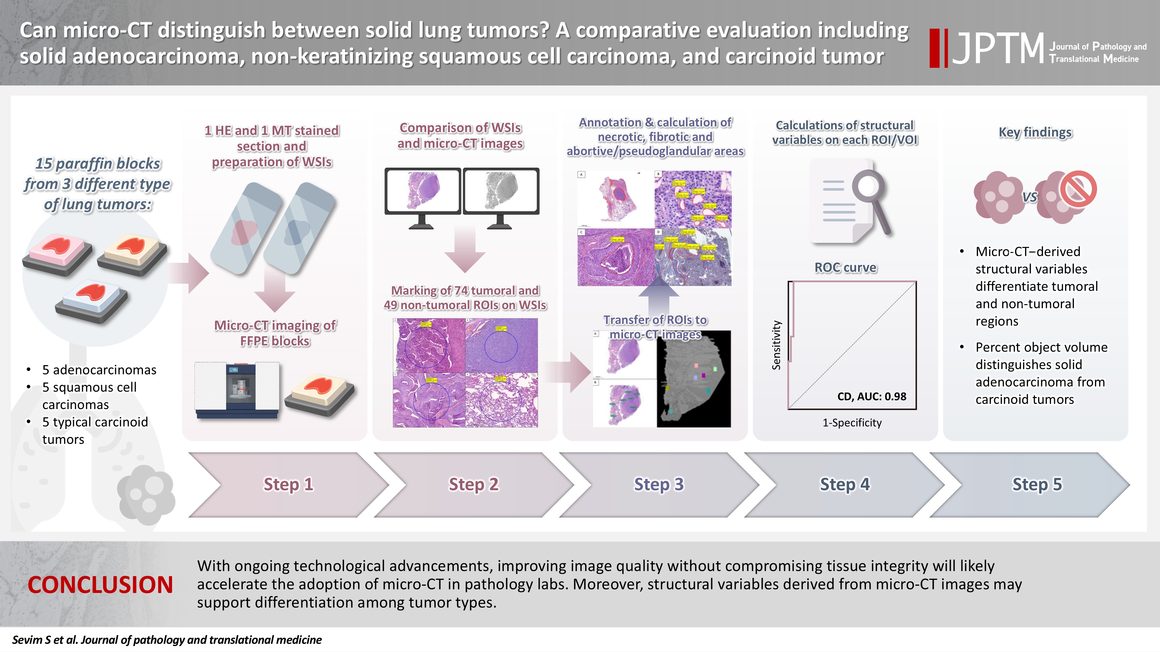
Some pulmonary carcinomas display a solid pattern, and immunohistochemistry is commonly used for tumor differentiation. Micro–computed tomography (micro-CT), with its ability to produce detailed three-dimensional images using small voxel sizes, may offer additional insights. This study investigates whether three solid tumor types, solid adenocarcinoma (sAC), non-keratinizing squamous cell carcinoma, and carcinoid tumor (CaT), can be differentiated using micro-CT. Methods: Fifteen paraffin blocks, five for each type, were scanned with micro-CT (Skyscan 1275, Bruker). These images were compared to whole slide images (WSIs) of the same tumors. Consequently, tumoral (n = 74) and non-tumoral (n = 49) regions of interest (tumor ROIs [tROIs] and non-tumor ROIs [ntROIs]) were selected on the micro-CT images and evaluated in terms of certain structural variables (percent object volume, structure model index, structure thickness, structure linear density, connectivity, connectivity density, open porosity, closed porosity) to investigate whether tumors can be differentiated from normal parenchyma and from each other. Results: Although detailed images comparable to WSIs could not be obtained, it was considered an important advantage to be able to examine the entire depth of the paraffin blocks. tROIs and ntROIs could be distinguished based on all variables (p < .001). Additionally, sAC showed a notable difference from CaT in “percent object volume” (p = .011). Conclusions: With ongoing technological advancements, improving image quality without compromising tissue integrity will likely accelerate the adoption of micro-CT in pathology labs. Moreover, structural variables derived from micro-CT images may support differentiation among tumor types.
- TRPS1 expression in non-melanocytic cutaneous neoplasms: an immunohistochemical analysis of 200 cases
- Yi A. Liu, Phyu P. Aung, Yunyi Wang, Jing Ning, Priyadharsini Nagarajan, Jonathan L. Curry, Carlos A. Torres-Cabala, Doina Ivan, Victor G. Prieto, Qingqing Ding, Woo Cheal Cho
- J Pathol Transl Med. 2024;58(2):72-80. Published online February 26, 2024
- DOI: https://doi.org/10.4132/jptm.2024.01.23
- 9,359 View
- 416 Download
- 15 Web of Science
- 15 Crossref
-
Abstract
PDFSupplementary Material
- Background
Although trichorhinophalangeal syndrome type 1 (TRPS1) was initially thought to be highly sensitive and specific for carcinomas and mesenchymal tumors of mammary origin, more recent data suggest its expression is not limited to breast neoplasms but also can be seen in other cutaneous neoplasms, such as extramammary Paget disease and squamous cell carcinoma (SCC) in situ.
Methods
Two-hundred cases of non-melanocytic cutaneous neoplasm, including basal cell carcinomas (BCCs) (n = 41), SCCs (n = 35), Merkel cell carcinomas (MCCs) (n = 25), and adnexal neoplasms (n = 99), were tested for TRPS1 expression using a monoclonal anti- TRPS1 rabbit anti-human antibody.
Results
TRPS1 expression was present in almost all cases of SCC (94%), with a median H-score of 200, while it was either absent or only focally present in most BCCs (90%), with a median H-score of 5. The difference between BCCs and SCCs in H-score was significant (p < .001). All MCCs (100%) lacked TRPS1 expression. TRPS1 expression was frequently seen in most adnexal neoplasms, benign and malignant, in variable intensity and proportion but was consistently absent in apocrine carcinomas. All endocrine mucin-producing sweat gland carcinomas (EMPSGCs) (100%, 6/6) showed diffuse and strong TRPS1 immunoreactivity, with a median H-score of 300, which was significantly different (p < .001) than that of BCCs.
Conclusions
Our study shows that TRPS1 may be an effective discriminatory marker for BCCs and SCCs. It also has a role in distinguishing BCCs from EMPSGCs. -
Citations
Citations to this article as recorded by
- Metastatic Vulvar Paget's Disease Presenting in a Supraclavicular Lymph Node: A Diagnostic Challenge on Fine Needle Aspiration Cytology
Thiri Htoo Aung, Neha Seth, Anam Khan, Kasturi Das
Diagnostic Cytopathology.2026;[Epub] CrossRef - The evolving role of TRPS1 in dermatopathology: insights from the past 4 years
Mokhtar H. Abdelhammed, Woo Cheal Cho
Journal of Pathology and Translational Medicine.2026; 60(2): 129. CrossRef - Correspondence: Primary Cutaneous NUT Adnexal Carcinoma: A Case Report With Novel Clinical and Pathological Observations
Woo Cheal Cho
Journal of Cutaneous Pathology.2026; 53(7): 622. CrossRef - Trichorhinophalangeal syndrome type 1 (TRPS1) in breast pathology: diagnostic utility and pitfalls
Atif Ali Hashmi, Edi Brogi, Hannah Y. Wen
Diagnostic Pathology.2025;[Epub] CrossRef - Refining NTRK Fusion Detection in Papillary Thyroid Carcinoma Through Pan-TRK Immunohistochemistry and Histopathologic Features
Hyun Lee, Sue Youn Kim, Ji Min Park, Seung-Hyun Jung, Ozgur Mete, Chan Kwon Jung
Endocrine Pathology.2025;[Epub] CrossRef - Endocrine mucin-producing sweat gland carcinoma: Case report and literature review
Nan Guo, Zhenlin Fan, Yitong Chen, Qian Li, Limin Guo
European Journal of Ophthalmology.2025;[Epub] CrossRef - Updates on utility of immunohistochemistry in diagnosis of metastatic breast cancer
Hongxia Sun, Aysegul A. Sahin, Qingqing Ding
Human Pathology.2025; 162: 105821. CrossRef - Primary Cutaneous NUT Adnexal Carcinoma With BRD4::NUTM1 Fusion: A 19-Year Follow-Up
Elsayed Ibrahim, Richard K. Yang, Maria A. Gubbiotti, Victor G. Prieto, Woo Cheal Cho
The American Journal of Dermatopathology.2025; 47(9): 731. CrossRef - Primary mucinous carcinoma of the skin with co-expression of TRPS1 and GATA3: a case report
Liling Song, Ning Zhu, Lei Jiang, Dong Gao, Guohua Yu
Frontiers in Oncology.2025;[Epub] CrossRef - Diagnostic Algorithm for Secondary Extramammary Paget Disease from Institutional Cases and Literature Review
Salin Kiratikanon, Ayaka Fukui, Masahiro Hirata, Jakob M. T. Moran, Masakazu Fujimoto, Mai P. Hoang
Cancers.2025; 17(24): 4014. CrossRef - TRPS1 Expression Is Frequently Seen in a Subset of Cutaneous Mesenchymal Neoplasms and Tumors of Uncertain Differentiation: A Potential Diagnostic Pitfall
Moon Joo Kim, Yi A. Liu, Yunyi Wang, Jing Ning, Woo Cheal Cho
Dermatopathology.2024; 11(3): 200. CrossRef - TRPS1 expression in MPNST is correlated with PRC2 inactivation and loss of H3K27me3
Rossana Lazcano, Davis R. Ingram, Gauri Panse, Alexander J. Lazar, Wei-Lien Wang, Jeffrey M. Cloutier
Human Pathology.2024; 151: 105632. CrossRef - Syringocystadenoma Papilliferum-Like Features in Poroma: An Unusual Morphologic Pattern of Poroma or True Synchronous Occurrence of 2 Distinct Neoplasms?
Mouaz Alsawas, Fiorinda F. Muhaj, Phyu P. Aung, Priyadharsini Nagarajan, Woo Cheal Cho
The American Journal of Dermatopathology.2024; 46(12): 871. CrossRef - A Comprehensive Review of TRPS1 as a Diagnostic Immunohistochemical Marker for Primary Breast Carcinoma: Latest Insights and Diagnostic Pitfalls
Antonia-Carmen Georgescu, Tiberiu-Augustin Georgescu, Simona-Alina Duca-Barbu, Lucian Gheorghe Pop, Daniela Oana Toader, Nicolae Suciu, Dragos Cretoiu
Cancers.2024; 16(21): 3568. CrossRef - Expression of TRPS1 in Metastatic Tumors of the Skin: An Immunohistochemical Study of 72 Cases
Kassiani Boulogeorgou, Christos Topalidis, Triantafyllia Koletsa, Georgia Karayannopoulou, Jean Kanitakis
Dermatopathology.2024; 11(4): 293. CrossRef
- Metastatic Vulvar Paget's Disease Presenting in a Supraclavicular Lymph Node: A Diagnostic Challenge on Fine Needle Aspiration Cytology
- Intrathyroidal metastasis of tonsillar squamous cell carcinoma masquerading as a primary thyroid tumor
- Jai-Hyang Go
- J Pathol Transl Med. 2023;57(4):242-245. Published online July 11, 2023
- DOI: https://doi.org/10.4132/jptm.2023.06.16
- 5,643 View
- 118 Download
- 2 Web of Science
- 2 Crossref
-
Abstract
PDF
- Intrathyroidal metastasis of tonsillar squamous cell carcinoma is rare. To date, only six cases have been reported in the literature. This case was unusual and presented with thyromegaly before the diagnosis of the primary tumor. A 55-year-old male patient was suspected to have a primary thyroid tumor with nodal metastasis. The thyroid gland was diffusely enlarged, with no discernible mass. Histologically, the thyroid parenchyma revealed extensive endolymphatic tumor emboli, which were positive for p40 and p16 in a background of chronic lymphocytic thyroiditis. Positron emission tomography–computed tomography revealed hypermetabolic activity in the right tonsillar region. Tonsillar biopsy revealed human papillomavirus–positive squamous cell carcinoma. The present case is the first reported case of intrathyroidal metastasis of tonsillar squamous cell carcinoma with an initial clinical presentation of thyroid enlargement before the primary tumor of tonsillar cancer was diagnosed.
-
Citations
Citations to this article as recorded by- Metastasis to Thyroid from Recurrent Head and Neck Squamous Cell Carcinoma: A Case Series and Review of Literature
Avneet Kaur, Rohit Nayyar, Harit Kumar Chaturvedi, Akshat Malik
Indian Journal of Surgical Oncology.2025; 16(1): 122. CrossRef - Metastatic oropharyngeal squamous cell carcinoma to the thyroid: A case report and review of literature
Hannah Walker, Jed Speers, Milena Fabry, Sameep Kadakia
American Journal of Otolaryngology.2024; 45(4): 104306. CrossRef
- Metastasis to Thyroid from Recurrent Head and Neck Squamous Cell Carcinoma: A Case Series and Review of Literature
- Evaluation of the characteristics of multiple human papillomavirus (HPV) infections identified using the BD Onclarity HPV assay and comparison with those of single HPV infection
- Jinhee Kim, Moonsik Kim, Ji Young Park
- J Pathol Transl Med. 2022;56(5):289-293. Published online September 13, 2022
- DOI: https://doi.org/10.4132/jptm.2022.08.02
- 9,823 View
- 141 Download
- 12 Web of Science
- 9 Crossref
-
Abstract
PDFSupplementary Material
- Background
Human papillomavirus (HPV) infection is a major cause of cervical cancer and associated precursor lesions. Multiple HPV genotype infections have been reported. However, their clinicopathological characteristics still remain elusive.
Methods
For this study, 814 consecutive patients who had undergone colposcopy and HPV genotyping test using BD Onclarity HPV assay were retrospectively selected. Clinicopathological parameters of multiple HPV infections were compared with those of single HPV infection.
Results
Multiple HPV infections were found in 110 out of 814 cases (13.5%). Multiple HPV infections were associated with a significantly higher incidence of high-grade intraepithelial lesions (HSILs) compared with single HPV infection. Other high-risk HPV genotypes, in addition to HPV 16, were found more frequently in the multiple HPV infections group; these included HPV 51, 52, 33/58, 56/59/66, and 35/39/68. No specific coinfection pattern was not identified. Additionally, the number of HPV genotypes in multiple HPV infections was not associated with the progression to HSIL or squamous cell carcinoma.
Conclusions
Multiple HPV infections have distinct clinicopathological characteristics (compared with single HPV infection). As their biological behavior is uncertain, close and frequent follow-up is warranted. -
Citations
Citations to this article as recorded by- Informative HPV testing after conization and its impact on time-varying estimates: a GAMM-based cohort study
Jie Zhou, Jian hong Liao, Lin Jie Su, Yan Chen, Hong bo Hu
Frontiers in Public Health.2026;[Epub] CrossRef - The Prevalence of Multi-Type Infections Among Human Papillomavirus Types in Korean Women
Jang Mook Kim, Hee Seung Song, Jieun Hwang, Jae Kyung Kim
Pathogens.2025; 14(4): 369. CrossRef - Multiple high-risk human papillomavirus infections exacerbate cervical lesion risk: epidemiological evidence from suining, Sichuan
Yaling Jing, Jianhui Chen, Fang Lin, Xiaonan Huang, Yulin Liu, Mingcai Zhao, Chuan Ye, Lianfang Zhao, Xiaofang Liu, Jiayan Yang
Virology Journal.2025;[Epub] CrossRef - The cervical cancer related distribution, coinfection and risk of 15 HPV types in Baoan, Shenzhen, in 2017–2023
Rukai Li, Weiwei Meng, Yunhai Zuo, Yanli Xu, Shaonan Wu
Virology Journal.2024;[Epub] CrossRef - Molecular findings and virological assessment of bladder papillomavirus infection in cattle
Francesca De Falco, Anna Cutarelli, Francesca Luisa Fedele, Cornel Catoi, Sante Roperto
Veterinary Quarterly.2024; 44(1): 1. CrossRef - Patterns of single and multiple HPV infections in female: A systematic review and meta-analysis
Dan Zhou, Jing Xue, Yaqiong Sun, Liling Zhu, Ming Zhao, Meimei Cui, Min Zhang, Jingjing Jia, Limei Luo
Heliyon.2024; 10(17): e35736. CrossRef - Age distribution of patients with multiple High-Risk Human Papilloma Virus (HR-HPV) genotypes and HPV vaccine recommendations by age
Gülçin Çetin Uysal, Nil Tekin
Family Practice and Palliative Care.2024; 9(3): 80. CrossRef - Relative distribution of HPV genotypes in histological cervical samples and associated grade lesion in a women population over the last 16 years in Burgundy, France
Christelle Auvray, Serge Douvier, Odile Caritey, Jean-Baptiste Bour, Catherine Manoha
Frontiers in Medicine.2023;[Epub] CrossRef - Epidemiologic characteristics of high-risk HPV and the correlation between multiple infections and cervical lesions
Qinli Luo, Xianghua Zeng, Hanyi Luo, Ling Pan, Ying Huang, Haiyan Zhang, Na Han
BMC Infectious Diseases.2023;[Epub] CrossRef
- Informative HPV testing after conization and its impact on time-varying estimates: a GAMM-based cohort study
- Frequency of PIK3CA mutations in different subsites of head and neck squamous cell carcinoma in southern Thailand
- Arunee Dechaphunkul, Phatcharaporn Thongwatchara, Paramee Thongsuksai, Tanadech Dechaphunkul, Sarayut Lucien Geater
- J Pathol Transl Med. 2022;56(3):126-133. Published online February 28, 2022
- DOI: https://doi.org/10.4132/jptm.2022.01.04
- 9,272 View
- 195 Download
- 4 Web of Science
- 4 Crossref
-
Abstract
PDF
- Background
Phosphatidylinositol-4,5-bisphosphate 3-kinase catalytic subunit alpha (PIK3CA) mutations have been reported in many cancers, including head and neck squamous cell carcinoma (HNSCC). The frequency of these mutations varies among tumor locations and might be relevant to treatment outcomes among HNSCC. In this study, we examined the frequency of PIK3CA mutations in the different subsites of HNSCC.
Methods
Ninety-six fresh biopsy specimens were investigated for mutations in PIK3CA exons 4, 9, and 20 using allele-specific real-time polymerase chain reaction. Patient characteristics and survival were analyzed and compared between specimens with or without PIK3CA mutations.
Results
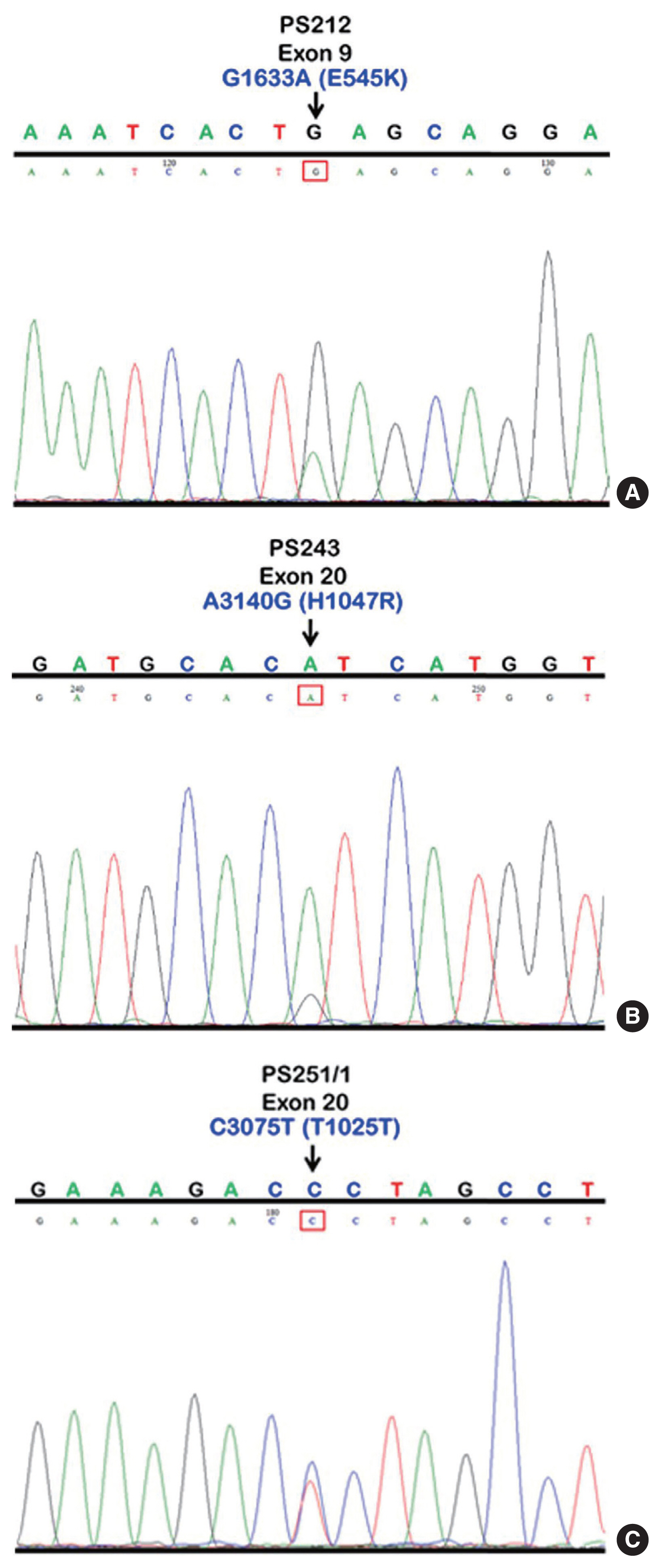
The study included primary tumors originating from the oral cavity (n=63), hypopharynx (n=23), and oropharynx (n=10). We identified mutations in 10.4% of patients (10 of 96 specimens). The overall mutational frequency was 17.4% (4/23) and 9.5% (6/63) in the hypopharynx and oral cavity, respectively. No patients with oropharyngeal carcinoma had mutations. Among the 10 mutant specimens, five were missense mutations (exon 9 [E545K] in two samples and exon 20 [H1047R] in three samples) and five were silent mutations in exon 20 (T1025T). Mutations were not found in exon 4. Among 84 patients with available clinical data, we found no significant differences in clinical characteristics and survival based on the presence or absence of PIK3CA mutations.
Conclusions
The results indicate that PIK3CA mutations are involved in HNSCC carcinogenesis, and the hypopharynx should be considered a primary site of interest for future studies, particularly in Southeast Asian populations. -
Citations
Citations to this article as recorded by- Comprehensive Genomic Profiling of Sinonasal Carcinomas: Identification of Common Mutations and Potential Targets for Therapy
Gabriel Bitar, Beau Hsia, Saif Alshaka, Bastien A. Valencia, Jeeho Kim, Mariko Sato, John Crawford, Michael L. Levy, Sean Polster, Vijay A. Patel
Journal of Neurological Surgery Part B: Skull Base.2026; 87(03): 337. CrossRef - Anti-breast cancer effects of Pterocarpus soyauxii Taub aqueous extract and its compounds by integrating ADMET, network pharmacology, molecular docking, dynamic simulation, CLC-Pred and pdCSM-Cancer/PPI approaches, and in vitro validation
Owona Pascal Emmanuel, Mengue Ngadena Yolande Sandrine, Bilanda Danielle Claude, Ayissi Mbomo Rigobert-Espoir, Oluwafemi Adeleke Ojo, Ella Armand Fils, Bidingha A Goufani Ronald, Bindzi Georges Michel, Dzeufiet Djomeni Paul Désiré, Tariq Aziz, Abdulhakeem
Journal of Ethnopharmacology.2025; 353: 120407. CrossRef - A retrospective study of laryngeal squamous cell carcinoma and the significance of the PIK3CA mutation for survival
Akinobu Kubota, Nobuyuki Bandoh, Takashi Goto, Michihisa Kono, Ryosuke Sato, Shiori Suzuki, Shota Sakaue, Ryuhei Takeda, Shuto Hayashi, Misaki Hayashi, Daisuke Araki, Shogo Baba, Yasutaka Kato, Miki Takahara, Hiroshi Nishihara, Hajime Kamada
Molecular and Clinical Oncology.2025; 23(4): 1. CrossRef - An empirical review on the resistance mechanisms of epidermal growth factor receptor inhibitors and predictive molecular biomarkers in colorectal cancer
Sankha Bhattacharya
Critical Reviews in Oncology/Hematology.2023; 183: 103916. CrossRef
- Comprehensive Genomic Profiling of Sinonasal Carcinomas: Identification of Common Mutations and Potential Targets for Therapy
- Primary squamous cell carcinoma of the salivary gland: immunohistochemical analysis and comparison with metastatic squamous cell carcinoma
- Uiree Jo, Joon Seon Song, Seung-Ho Choi, Soon Yuhl Nam, Sang Yoon Kim, Kyung-Ja Cho
- J Pathol Transl Med. 2020;54(6):489-496. Published online August 31, 2020
- DOI: https://doi.org/10.4132/jptm.2020.07.19
- 12,182 View
- 220 Download
- 19 Web of Science
- 20 Crossref
-
Abstract
PDFSupplementary Material
- Background
Primary squamous cell carcinoma (SCC) of the salivary gland is a rare disease, and distinguishing primary SCC from metastatic SCC is difficult. This study investigated the histological and immunohistochemical differences between primary and metastatic salivary gland SCC to improve the accuracy of diagnosis and to explore the pathogenesis of this disease.
Methods
Data of 16 patients who underwent surgery for SCC of salivary glands between 2000 and 2018 at Asan Medical Center were retrieved. Eight patients had a history of SCC at other sites, and eight patients had only salivary gland SCC. Immunostaining for p16, p53, androgen receptor (AR), gross cystic disease fluid protein 15 (GCDFP-15), and c-erbB2, as well as mucicarmine staining, were compared between the two groups.
Results
Most tumors were located in the center of the salivary glands with extraparenchymal extension. The histology of primary SCC of the salivary gland was consistent with moderately differentiated SCC with extensive desmoplastic reaction and peritumoral inflammation. Involvement of the salivary gland ducts and transition into the ductal epithelium were observed in two cases. Metastatic SCC resembled the primary tumor histologically and was associated with central necrosis. Both groups exhibited negative mucin staining. Two, one, and one primary SCC case exhibited AR, GCDFP-15, and c-erbB2 positivity, respectively.
Conclusions
A subset of primary SCCs originated in salivary ducts or was related to salivary duct carcinoma. Distinguishing primary from metastatic SCC of the salivary gland is difficult using histologic features and immunoprofiles. A comprehensive review of the medical history is essential. -
Citations
Citations to this article as recorded by- Solitary Parotid Metastasis as an Atypical Site of Recurrence in Lung Squamous Cell Carcinoma: A Diagnostic Difficulty
Mamadou Alpha Prateaux, Mohamed Amine Haouane, Issam Rharrassi, Mohamed Amine Azami
Cureus.2026;[Epub] CrossRef - Certainties, Doubts, and Myths in the Diagnosis and Treatment of Salivary Gland Tumors of the Head and Neck
Giulio Cantù
Cancers.2026; 18(13): 2078. CrossRef - Clinical diagnosis, treatment, and survival analysis of 61 cases of salivary duct carcinoma: a retrospective study
Shubin Dong, Mengru Li, Zhiwei Zhang, Bowei Feng, Wei Ding, Jiang Chang, Feng Liu
PeerJ.2025; 13: e19626. CrossRef - Characterization of undifferentiated carcinoma of the salivary gland: clinicopathological and immunohistochemical analyses in comparison with lymphoepithelial carcinoma
Sangjoon Choi, Gyuheon Choi, Hee Jin Lee, Joon Seon Song, Yoon Se Lee, Seung-Ho Choi, Kyung-Ja Cho
Journal of Pathology and Translational Medicine.2025; 59(6): 361. CrossRef - Primary salivary gland squamous cell carcinoma with sialolithiasis in the submandibular gland: A case report and literature review
Sawako Ono, Katsutoshi Hirose, Yuji Hirata, Marie Yamada, Satoko Nakamura, Hidetaka Yamamoto
Journal of Oral and Maxillofacial Surgery, Medicine, and Pathology.2024; 36(5): 768. CrossRef - A case of primary squamous cell carcinoma of the parotid gland and review of the literature
Jingli Zhao, Xinrong Nan, Chuhuan Zhou, Nan Jiang, Liangliang Tian
Journal of Case Reports and Images in Oncology.2024; 10(1): 7. CrossRef - Metastatic cutaneous squamous cell carcinoma accounts for nearly all squamous cell carcinomas of the parotid gland
Patrick J. Bradley, Göran Stenman, Lester D. R. Thompson, Alena Skálová, Roderick H. W. Simpson, Pieter J. Slootweg, Alessandro Franchi, Nina Zidar, Alfons Nadal, Henrik Hellquist, Michelle D. Williams, Ilmo Leivo, Abbas Agaimy, Alfio Ferlito
Virchows Archiv.2024; 485(1): 3. CrossRef - Common skin cancers and their association with other non-cutaneous primary malignancies: a review of the literature
Lindsay Holic
Medical Oncology.2024;[Epub] CrossRef - Salivary duct carcinoma with squamous differentiation: histomorphological and immunophenotypical analysis of six cases
Melad N Dababneh, Christopher C Griffith, Kelly R Magliocca, Ivan J Stojanov
Histopathology.2024; 85(4): 590. CrossRef - Comprehensive Next Generation Sequencing Reveals that Purported Primary Squamous Cell Carcinomas of the Parotid Gland are Genetically Heterogeneous
Justin A. Bishop, Masato Nakaguro, Ilan Weinreb, Doreen Palsgrove, Lisa M. Rooper, Travis W. Vandergriff, Brian Carlile, Jeffrey A. Sorelle, Jeffrey Gagan, Toshitaka Nagao
Head and Neck Pathology.2024;[Epub] CrossRef - Salivary gland fine needle aspiration: a focus on diagnostic challenges and tips for achieving an accurate diagnosis
Carla Saoud, Hansen Lam, Sandra I. Sanchez, Zahra Maleki
Diagnostic Histopathology.2023; 29(8): 357. CrossRef - Salivary gland pathologies: evolution in classification and association with unique genetic alterations
Michał Żurek, Łukasz Fus, Kazimierz Niemczyk, Anna Rzepakowska
European Archives of Oto-Rhino-Laryngology.2023; 280(11): 4739. CrossRef - A retrospective study of nonneoplastic and neoplastic disorders of the salivary glands
Sorin Vamesu, Oana Andreea Ursica, Ana Maria Gurita, Raluca Ioana Voda, Mariana Deacu, Mariana Aschie, Madalina Bosoteanu, Georgeta Camelia Cozaru, Anca Florentina Mitroi, Cristian Ionut Orasanu
Medicine.2023; 102(42): e35751. CrossRef - Pembrolizumab as a first line therapy in a patient with extensive mucoepidermoid salivary gland carcinoma. A complete clinical, radiological and pathological response. A very specific case
Raed Farhat, Noam Asna, Yaniv Avraham, Ashraf Khater, Majd Asakla, Alaa Safia, Sergio Szvalb, Nidal Elkhatib, Shlomo Merchavy
Discover Oncology.2022;[Epub] CrossRef - Morphologic CT and MRI features of primary parotid squamous cell carcinoma and its predictive factors for differential diagnosis with mucoepidermoid carcinoma
Xiaohua Ban, Huijun Hu, Yue Li, Lingjie Yang, Yu Wang, Rong Zhang, Chuanmiao Xie, Cuiping Zhou, Xiaohui Duan
Insights into Imaging.2022;[Epub] CrossRef - A Rare Case of Primary Squamous Cell Carcinoma of the Submandibular Salivary Gland: Brief Overview of Diagnostic Ambiguity and Treatment Challenges
Pawan Hingnikar, Anendd Jadhav, Nitin D Bhola
Cureus.2022;[Epub] CrossRef - Necrotizing Sialometaplasia of the Hard Palate: Diagnosis and
Treatment
Sangeun Lee, Yun Sung Lim, Kyuho Lee, Bo Hae Kim
Journal of Clinical Otolaryngology Head and Neck Surgery.2022; 33(4): 236. CrossRef - Parotid Salivary Duct Carcinoma With a Prominent Squamous Component: Immunohistochemical Profile, Diagnostic Pitfalls, and Therapeutic Implications
Naomi Hardy, Joshua Thompson, Ranee Mehra, Cinthia B. Drachenberg, Kyle Hatten, John C. Papadimitriou
International Journal of Surgical Pathology.2021; 29(7): 726. CrossRef - Intrasalivary Thymic Carcinoma: A Case Report and Literature Review
Michał Kunc, Alexandra Kamieniecki, Grzegorz Walczak, Tomasz Nowicki, Bartosz Wasąg, Bogusław Mikaszewski, Dominik Stodulski, Wojciech Biernat
Head and Neck Pathology.2021; 16(3): 857. CrossRef - Cancer Stem Cell Markers in Squamous Cell Carcinomas of the Salivary Glands
Mattis Bertlich, Julia Kitz, Marie Kruizenga, Jennifer Lee Spiegel, Martin Canis, Friedrich Ihler, Frank Haubner, Bernhard G. Weiss, Mark Jakob
Oncology.2021; 99(6): 402. CrossRef
- Solitary Parotid Metastasis as an Atypical Site of Recurrence in Lung Squamous Cell Carcinoma: A Diagnostic Difficulty
- Prevalence of high-risk human papillomavirus and its genotype distribution in head and neck squamous cell carcinomas
- Yuil Kim, Young-Hoon Joo, Min-Sik Kim, Youn Soo Lee
- J Pathol Transl Med. 2020;54(5):411-418. Published online July 21, 2020
- DOI: https://doi.org/10.4132/jptm.2020.06.22
- 14,064 View
- 191 Download
- 23 Web of Science
- 27 Crossref
-
Abstract
PDF
- Background
High-risk (HR) human papillomavirus (HPV) is found in a subset of head and neck (HN) squamous cell carcinomas (SCCs). For oropharyngeal SCCs, HR HPV positivity is known to be associated with good prognosis, and a separate staging system for HPV-associated carcinomas using p16 immunohistochemistry (IHC) as a surrogate test has been adopted in the 8th American Joint Committee on Cancer staging system. We examined the HR HPV status and the genotype distribution in five HN subsites.
Methods
Formalin-fixed paraffin-embedded tissue sections were used for p16 IHC and DNA extraction. HPV DNA detection and genotyping were done employing either a DNA chip-based or real-time polymerase chain reaction–based method.
Results
During 2011–2019, a total of 466 SCCs were tested for HPV DNA with 34.1% positivity for HR HPV. Among HN subsites, the oropharynx showed the highest HR HPV prevalence (149/205, 75.1%), followed by the sinonasal tract (3/14, 21.4%), larynx (5/43, 11.6%), hypopharynx (1/38, 2.6%), and oral cavity (1/166, 0.6%). The most common HPV genotype was HPV16 (84.3%) followed by HPV35 (6.9%) and HPV33 (4.4%). Compared with HR HPV status, the sensitivity and specificity of p16 IHC were 98.6% and 94.3% for the oropharynx, and 99.2% and 93.8% for the tonsil, respectively.
Conclusions
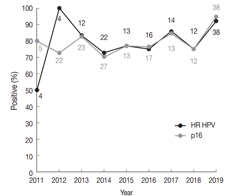
Using a Korean dataset, we confirmed that HR HPV is most frequently detected in oropharyngeal SCCs. p16 positivity showed a good concordance with HR HPV DNA for oropharyngeal and especially tonsillar carcinomas. The use of p16 IHC may further be extended to predict HR HPV positivity in sinonasal tract SCCs. -
Citations
Citations to this article as recorded by- Prevalence of Human Papillomavirus in Head and Neck Cancers in South Korea: A Targeted Literature Review
Aneesha Fathima Syed Mohamed, Ruixuan Wang, Seyoung Oh, Ying Hui Wu, Gyongseon Yang, Isaya Sukarom, Sei Young Lee, Wei Wang
Korean Journal of Otorhinolaryngology-Head and Neck Surgery.2026; 69(3): 115. CrossRef - Diagnostic Accuracy of p16 Immunohistochemistry as a Marker of High-Risk HPV in Invasive Laryngeal Squamous Cell Carcinoma: A Systematic Review
Ana-Maria Stanoiu, Delia Hutanu, Maria Sorop-Florea, Alexandru Alexandru, Norberth-Istvan Varga, Iulia Cristina Bagiu, Mihaela-Diana Popa, Bogdan Hirtie, Nicolae-Constantin Balica, Cristian-Ion Mot, Ioana-Delia Horhat
Medicina.2026; 62(7): 1372. CrossRef - Impact of histopathological parameters in prognosis of oral squamous cell carcinoma
R. P. Ekanayaka, W. M. Tilakaratne
Oral Diseases.2025; 31(5): 1420. CrossRef - Prevalence of human papilloma virus in head and neck mucous squamous cell carcinoma and genotypes by location: an observational study
Emilie Uhlrich, Jerzy Klijanienko, Joey Martin, Emmanuelle Jeannot, Anne Vincent-Salomon, Paul Freneaux, Christophe Le Tourneau, Olivier Choussy, Antoine Dubray-Vautrin
European Journal of Cancer Prevention.2025; 34(5): 426. CrossRef - Risk factors for cervical lymph node metastasis in oropharyngeal cancer and its impact on prognosis
Li Zhang, Zhilin Li, Jing Wang, Chen Wang, Shuxin Wen
Brazilian Journal of Otorhinolaryngology.2025; 91(2): 101520. CrossRef - Co-infection of human papillomavirus genotypes and Epstein-Barr virus in tumors of the oral cavity and oropharynx: a retrospective study in Northeastern Mexico
Gerardo del Carmen Palacios-Saucedo, Jose Manuel Vazquez-Guillen, Alondra Yamileth Alanis-Valdez, Leticia Lizeth Valdez-Treviño, Luis Roberto Galindo-Mendez, Angel Zavala-Pompa, Lydia Guadalupe Rivera-Morales, Ana Carolina Martinez-Torres, Roberto Lopez-V
IJID Regions.2025; 14: 100555. CrossRef - Rates of p16 and p53 expression in head and neck cutaneous squamous cell carcinoma vary according to human papillomavirus status
Rachid Ait Addi
World Journal of Clinical Cases.2025;[Epub] CrossRef - The epidemiological trends and survival of HPV-related oropharyngeal cancer other than tonsils and base of tongue − a systematic review and meta-analysis
Anas Mohammad Al Fadel, Kathrine Kronberg Jakobsen, Lasse Holmgaard Jensen, Amanda-Louise Fenger Carlander, Christian Grønhøj, Christian von Buchwald
Oral Oncology.2025; 165: 107311. CrossRef - Oropharyngeal Helicobacter pylori colonization increases risk and worsens prognosis of head and neck squamous cell carcinoma
Xianyao Jiang, Yongjin Huang, Changwu Li, Hongyan Jiang
Scientific Reports.2025;[Epub] CrossRef - Characteristics of human papillomavirus infection among oropharyngeal cancer patients: A systematic review and meta-analysis
Meimei Cui, Jinling Cheng, Huijuan Cheng, Ming Zhao, Dan Zhou, Min Zhang, Jingjing Jia, Limei Luo
Archives of Oral Biology.2024; 157: 105830. CrossRef - Longitudinal Screening for Oral High-Risk Non-HPV16 and Non-HPV18 Strains of Human Papillomavirus Reveals Increasing Prevalence among Adult and Pediatric Biorepository Samples: A Pilot Study
Jordan Jacobs, Eugene Chon, Karl Kingsley
Vaccines.2024; 12(8): 895. CrossRef - Position Statement about Gender-Neutral HPV Vaccination in Korea
Kyung-Jin Min, Yung-Taek Ouh, Sangrak Bae, Yong-Bae Ji, Jae-Kwan Lee, Jae-Weon Kim, Kwang-Jae Cho, Dong-Hun Im
Vaccines.2024; 12(10): 1110. CrossRef - High-risk HPV Does not Appear to be an Important Risk Factor for Sinonasal Carcinomas in Turkish Population: A Tertiary Center Experience
Evsen Apaydin Arikan, Levent Aydemir, Murat Ulusan, Dilek Yilmazbayhan, Yasemin Ozluk
International Journal of Surgical Pathology.2023; 31(2): 124. CrossRef - Practical Application of Circulating Tumor-Related DNA of Human Papillomavirus in Liquid Biopsy to Evaluate the Molecular Response in Patients with Oropharyngeal Cancer
Agnieszka M. Mazurek, Tomasz W. Rutkowski
Cancers.2023; 15(4): 1047. CrossRef - The Prevalence of HPV in Oral Cavity Squamous Cell Carcinoma
Seyed Keybud Katirachi, Mathias Peter Grønlund, Kathrine Kronberg Jakobsen, Christian Grønhøj, Christian von Buchwald
Viruses.2023; 15(2): 451. CrossRef - The Protective Role of Cranberries and Blueberries in Oral Cancer
César Esquivel-Chirino, Mario Augusto Bolaños-Carrillo, Daniela Carmona-Ruiz, Ambar Lopéz-Macay, Fernando Hernández-Sánchez, Delina Montés-Sánchez, Montserrat Escuadra-Landeros, Luis Alberto Gaitán-Cepeda, Silvia Maldonado-Frías, Beatriz Raquel Yáñez-Ocam
Plants.2023; 12(12): 2330. CrossRef - Unusual cases of sinonasal malignancies: a letter to the editor on HPV-positive sinonasal squamous cell carcinomas
Benedicte Bitsch Lauritzen, Sannia Sjöstedt, Jakob Myllerup Jensen, Katalin Kiss, Christian von Buchwald
Acta Oncologica.2023; 62(6): 608. CrossRef - Prevalence of human Papillomavirus associated oropharyngeal and oral squamous cell carcinoma in Asian countries: A systematic review and large-scale meta-analysis
Yy Jean Tan, Ken Wong Siong Hou, Galvin Sim Siang Lin, Jasmine Lim Suk Wun, Wan Nor Amira Wan Ahmad Abdul Nasir, Lynn Wei Linn Ko
Acta Marisiensis - Seria Medica.2023; 69(2): 77. CrossRef - Top 100 most cited articles on human papillomavirus-induced head and neck squamous cell carcinoma: A bibliographic review
Rahul Mohandas, Subhashree Mohapatra, Mary Oshin, ShubhangiSambhaji Hajare
Journal of International Oral Health.2023; 15(3): 219. CrossRef - Intracellular Toll-Like Receptors Modulate Adaptive Immune Responses in Head and Neck Cancer
Sangeetha K. Nayanar, Deepak Roshan V.G., Shruthi Surendran, Göran Kjeller, Bengt Hasséus, Daniel Giglio
Viral Immunology.2023; 36(10): 659. CrossRef - Positive Rate of Human Papillomavirus and Its Trend in Head and Neck Cancer in South Korea
Hyun Woong Jun, Yong Bae Ji, Chang Myeon Song, Jae Kyung Myung, Hae Jin Park, Kyung Tae
Frontiers in Surgery.2022;[Epub] CrossRef - Transcriptionally active HPV in OPMD and OSCC: A systematic review following the CAP/ASCO guidelines
Laura Borges Kirschnick, Lauren Frenzel Schuch, Maria Eduarda Pérez‐de‐Oliveira, Ana Gabriela Costa Normando, Bruno Augusto Linhares Almeida Mariz, Eliete Neves Silva Guerra, Felipe Martins Silveira, Ana Carolina Uchoa Vasconcelos, Luciana Estevam Simonat
Oral Diseases.2022; 28(8): 2309. CrossRef - Effect of National Oral Health Screening Program on the Risk of Head and Neck Cancer: A Korean National Population-Based
Chan Woo Wee, Hyo-Jung Lee, Jae-Ryun Lee, Hyejin Lee, Min-Jeong Kwoen, Woo-Jin Jeong, Keun-Yong Eom
Cancer Research and Treatment.2022; 54(3): 709. CrossRef - Expression of p16, p53, and TLR9 in HPV-Associated Head and Neck Squamous Cell Carcinoma: Clinicopathological Correlations and Potential Prognostic Significance
Shu Wang, Xibing Zhuang, Caixia Gao, Tiankui Qiao
OncoTargets and Therapy.2021; Volume 14: 867. CrossRef - The Role of Human Papilloma Virus in Dictating Outcomes in Head and Neck Squamous Cell Carcinoma
Shane Brennan, Anne-Marie Baird, Esther O’Regan, Orla Sheils
Frontiers in Molecular Biosciences.2021;[Epub] CrossRef - A Contemporary Systematic Review on Repartition of HPV-Positivity in Oropharyngeal Cancer Worldwide
Amanda F. Carlander, Kathrine K. Jakobsen, Simone K. Bendtsen, Martin Garset-Zamani, Charlotte D. Lynggaard, Jakob Schmidt Jensen, Christian Grønhøj, Christian von Buchwald
Viruses.2021; 13(7): 1326. CrossRef - The Prevalence of High- and Low-Risk Types of HPV in Patients with Squamous Cell Carcinoma of the Head and Neck, Patients with Chronic Tonsillitis, and Healthy Individuals Living in Poland
Joanna Katarzyna Strzelczyk, Krzysztof Biernacki, Jadwiga Gaździcka, Elżbieta Chełmecka, Katarzyna Miśkiewicz-Orczyk, Natalia Zięba, Janusz Strzelczyk, Maciej Misiołek
Diagnostics.2021; 11(12): 2180. CrossRef
- Prevalence of Human Papillomavirus in Head and Neck Cancers in South Korea: A Targeted Literature Review
- Peripheral type squamous cell carcinoma of the lung: clinicopathologic characteristics in comparison to the central type
- Yeoun Eun Sung, Uiju Cho, Kyo Young Lee
- J Pathol Transl Med. 2020;54(4):290-299. Published online June 17, 2020
- DOI: https://doi.org/10.4132/jptm.2020.05.04
- 12,765 View
- 213 Download
- 15 Web of Science
- 17 Crossref
-
Abstract
PDF
- Background
Squamous cell carcinomas (SqCCs) of the lung are known to arise more often in a central area but reports of peripheral SqCCs have increased, with a pathogenesis that is obscured. In this study, the clinicopathologic characteristics of peripheral lung SqCCs were studied and compared with those of the central type.
Methods
This study included 63 peripheral lung SqCCs and 48 randomly selected central cases; hematoxylin and eosin-stained slides of surgically resected specimens were reviewed in conjunction with radiologic images and clinical history. Cytokeratin-7 immunohistochemical staining of key slides and epidermal growth factor receptor (EGFR)/KRAS mutations tested by DNA sequencing were also included.
Results
Stages of peripheral SqCCs were significantly lower than central SqCCs (p=.016). Cystic change of the mass (p=.007), presence of interstitial fibrosis (p=0.007), and anthracosis (p=.049) in the background lung were significantly associated with the peripheral type. Cytokeratin-7 positivity was also higher in peripheral SqCCs with cutoffs of both 10% and 50% (p=.011). Pathogenic mutations in EGFR and KRAS were observed in only one case out of the 72 evaluated. The Cox proportional hazard model indicated a significantly better disease-free survival (p=.009) and the tendency of better overall survival (p=.106) in the peripheral type.
Conclusions
In peripheral type, lower stage is a favorable factor for survival but more frequent interstitial fibrosis and older age are unfavorable factors. Multivariate Cox analysis revealed that peripheral type is associated with better disease-free survival. The pathogenesis of peripheral lung SqCCs needs further investigation, together with consideration of the background lung conditions. -
Citations
Citations to this article as recorded by- Lepidic and alveolar subepithelial squamous cell carcinoma: expansion of the concept of peripheral squamous cell carcinoma with proposal for revised terminology based on morphologic, immunophenotypic, and clinical analysis of 22 cases
Federica Filipello, Francesca Ambrosi, Hans Blaauwgeers, Johanna Grefte, Wim Vos, Luisella Righi, Erik Thunnissen, Teodora Radonic
Virchows Archiv.2026; 489(1): 27. CrossRef - Adenosquamous carcinoma of the lung: Comparative CT and pathological features versus adenocarcinoma and squamous cell carcinoma
Qianyao Yuan, Dai Zhang, Rui Xu, Wenjun Yao, Hong Zhao, Kota V Ramana
PLOS One.2026; 21(6): e0352454. CrossRef - Assessing the performance of chest x‐ray screening in detecting early‐stage lung cancer in the general population
Choy‐Lye Chei, Sho Nakamura, Kaname Watanabe, Takashi Mizutani, Hiroto Narimatsu
International Journal of Cancer.2025; 156(11): 2127. CrossRef - Whole lung radiomic features are associated with overall survival in patients with locally advanced non-small cell lung cancer treated with definitive radiotherapy
Meng Yan, Zhen Zhang, Jia Tian, Jiaqi Yu, Andre Dekker, Dirk de Ruysscher, Leonard Wee, Lujun Zhao
Radiation Oncology.2025;[Epub] CrossRef - Imaging appearances, CT evolution patterns, and surgical prognosis of stage I lung squamous cell carcinoma
Wei-hua Zhao, Tian-you Luo, Fa-jin Lv, Qi Li
Cancer Imaging.2025;[Epub] CrossRef - Pulmonary squamous cell carcinoma and lymphoepithelial carcinoma – morphology, molecular characteristics and differential diagnosis
Sabina Berezowska, Marie Maillard, Mark Keyter, Bettina Bisig
Histopathology.2024; 84(1): 32. CrossRef - Assessment of seasonal variability of PM, BC and UFP levels at a highway toll stations and their associated health risks
Nazneen, Aditya Kumar Patra, Soma Sekhara Rao Kolluru, Abhishek Penchala, Sachidanand Kumar, Namrata Mishra, Naragam Bhanu Sree, Samrat Santra, Ravish Dubey
Environmental Research.2024; 245: 118028. CrossRef - Association between Airport Ultrafine Particles and Lung Cancer Risk: The Multiethnic Cohort Study
Arthur Bookstein, Justine Po, Chiuchen Tseng, Timothy V. Larson, Juan Yang, Sung-shim L. Park, Jun Wu, Salma Shariff-Marco, Pushkar P. Inamdar, Ugonna Ihenacho, Veronica W. Setiawan, Mindy C. DeRouen, Loïc Le Marchand, Daniel O. Stram, Jonathan Samet, Bea
Cancer Epidemiology, Biomarkers & Prevention.2024; 33(5): 703. CrossRef - Clinical and Bronchoscopy Assessment in Diagnosing the Histopathology Type of Primary Central Lung Tumors
Mia Elhidsi, Jamal Zaini, Lisnawati Rachmadi, Asmarinah Asmarinah, Aria Kekalih, Noni Soeroso, Menaldi Rasmin
The Open Respiratory Medicine Journal.2024;[Epub] CrossRef - Possible thoracic metastasis from squamous cell carcinoma of the external auditory canal: A case report
Hiroshi Takehara, Ken Kodama, Toru Momozane, Masashi Takeda, Kaichi Shigetsu, Hiroki Kishima
Clinical Case Reports.2024;[Epub] CrossRef - Radiological precursor lesions of lung squamous cell carcinoma: Early progression patterns and divergent volume doubling time between hilar and peripheral zones
Haruto Sugawara, Yasushi Yatabe, Hirokazu Watanabe, Hiroyuki Akai, Osamu Abe, Shun-ichi Watanabe, Masahiko Kusumoto
Lung Cancer.2023; 176: 31. CrossRef - Loss of GSTO2 contributes to cell growth and mitochondria function via the p38 signaling in lung squamous cell carcinoma
Ryusuke Sumiya, Masayoshi Terayama, Teruki Hagiwara, Kazuaki Nakata, Keigo Sekihara, Satoshi Nagasaka, Hideki Miyazaki, Toru Igari, Kazuhiko Yamada, Yuki I. Kawamura
Cancer Science.2022; 113(1): 195. CrossRef - Primary tumor location in lung cancer: the evaluation and administration
Xueqi Xie, Xiaolin Li, Wenjie Tang, Peng Xie, Xuefen Tan
Chinese Medical Journal.2022; 135(2): 127. CrossRef - Pulmonary squamous cell carcinoma with a lepidic-pagetoid growth pattern
Claudio Guerrieri, Mark Lindner, Joanna Sesti, Abhishek Chakraborti, Rachel Hudacko
Pathologica.2022; 114(4): 304. CrossRef - Deposition modeling of ambient particulate matter in the human respiratory tract
Salman Khan, Bhola Ram Gurjar, Veerendra Sahu
Atmospheric Pollution Research.2022; 13(10): 101565. CrossRef - Selection of the surgical approach for patients with cStage IA lung squamous cell carcinoma: A population-based propensity score matching analysis
Shengteng Shao, Guisong Song, Yuanyong Wang, Tengfei Yi, Shuo Li, Fuhui Chen, Yang Li, Xiaotong Liu, Bin Han, Yuhong Liu
Frontiers in Oncology.2022;[Epub] CrossRef - Virus Nanoparticles & Different Nanoparticles Affect Lung Cancer- A New Approach
Ranajit Nath, Ratna Roy, Soubhik bhattacharyya, Sourav Datta
International Journal of Scientific Research in Science and Technology.2021; : 867. CrossRef
- Lepidic and alveolar subepithelial squamous cell carcinoma: expansion of the concept of peripheral squamous cell carcinoma with proposal for revised terminology based on morphologic, immunophenotypic, and clinical analysis of 22 cases
- Human Papillomavirus Serologic Profiles of Selected Filipinos with Head and Neck Squamous Cell Carcinoma
- Pia Marie Albano, Christianne Salvador, Jose Orosa, Sheryl Racelis, Modesty Leaño, Angelika Michel, John Donnie Ramos, Dana Holzinger, Michael Pawlita
- J Pathol Transl Med. 2019;53(5):273-279. Published online May 30, 2019
- DOI: https://doi.org/10.4132/jptm.2019.05.12
- 9,646 View
- 196 Download
- 1 Web of Science
- 1 Crossref
-
Abstract
PDF
- Background
The low prevalence of human papillomavirus (HPV) DNA and mRNA in biopsy samples of Filipinos with head and neck squamous cell carcinoma (HNSCC) has been reported previously. Here, the HPV serologic profiles of HNSCC cases were analyzed and associated with life-style and sexual practices.
Methods
Serum samples were collected between May 2012 and September 2013 from HNSCC patients (n = 22) in the northwest region of the Philippines, and age- and sex-matched clinically healthy controls. Antibodies to capsid and early oncoproteins of HPV16, 18, 31, 33, 45, 52, 58, 6, and 11 were analyzed using multiplex serology.
Results
Most of the cases were males with tumors of the oral cavity or larynx. Two of the cases tested positive for at least one of the early oncoproteins (E6, E7, E1, and/or E2) of HPV16, and 11 did not display reactivity to any HPV early or late oncoproteins. Of the controls, four tested positive for at least one of the HPV16 early oncoproteins, and 10 were non-reactive to all HPV types. Titers to HPV16 E6 or E7 of the seropositive cases and controls were considerably lower than those typically observed in economically developed countries.
Conclusions
The low HPV titers seen here are consistent with the results of molecular analyses for this population. Hence, the seropositivity of some of the HNSCC cases is likely an indication of prior exposure to the virus and not the presence of HPV-driven tumors. -
Citations
Citations to this article as recorded by- Social determinants of sex disparities in cancer in Southeast Asia
Ma. Veronica Pia N. Arevalo, Ethan Angelo S. Maslog, Katherine Donatela Manlongat, Eric David B. Ornos, Imjai Chitapanarux, Michelle Ann B. Eala, Edward Christopher Dee
iScience.2023; 26(7): 107110. CrossRef
- Social determinants of sex disparities in cancer in Southeast Asia
- Squamous Cell Carcinoma of the Extrahepatic Common Hepatic Duct
- Myunghee Kang, Na Rae Kim, Dong Hae Chung, Hyun Yee Cho, Yeon Ho Park
- J Pathol Transl Med. 2019;53(2):112-118. Published online October 1, 2018
- DOI: https://doi.org/10.4132/jptm.2018.09.03
- 10,063 View
- 179 Download
- 10 Web of Science
- 12 Crossref
-
Abstract
PDF
- We report a rare case of hilar squamous cell carcinoma. A 62-year-old Korean woman complaining of nausea was referred to our hospital. Her biliary computed tomography revealed a 28 mm-sized protruding solid mass in the proximal common bile duct. The patient underwent left hemihepatectomy with S1 segmentectomy and segmental excision of the common bile duct. Microscopically, the tumor was a moderately differentiated squamous cell carcinoma of the extrahepatic bile duct, without any component of adenocarcinoma or metaplastic portion in the biliary epithelium. Immunohistochemically, the tumor was positive for cytokeratin (CK) 5/6, CK19, p40, and p63. Squamous cell carcinoma of the extrahepatic bile duct is rare. To date, only 24 cases of biliary squamous cell carcinomas have been reported. Here, we provide a clinicopathologic review of previously reported extrahepatic bile duct squamous cell carcinomas.
-
Citations
Citations to this article as recorded by- Case Report: Primary squamous cell carcinoma of the pancreatic segment of the extrahepatic bile duct: a rare tumor easily misdiagnosed as pancreatic malignancy
Ting Xu, Xue Meng, Xuan Gou, Xinyuan Wang, Shuai Luo, Yilin Chen
Frontiers in Oncology.2026;[Epub] CrossRef - Deciphering cholangiocarcinoma heterogeneity and specific progenitor cell niche of extrahepatic cholangiocarcinoma at single-cell resolution
Chunliang Liu, Xiang Wang, Erdong Liu, Yali Zong, Wenlong Yu, Youhai Jiang, Jianan Chen, Mingye Gu, Zhengyuan Meng, Jingfeng Li, Yang Liu, Yongjie Zhang, Jing Tang, Hongyang Wang, Jing Fu
Journal of Hematology & Oncology.2025;[Epub] CrossRef - Extrahepatic cholangiocarcinoma: Current concepts in histopathology, immunohistochemistry, and molecular diagnostics
Jared Beyersdorf, M. Lisa Zhang
Seminars in Diagnostic Pathology.2025; 42(6): 150949. CrossRef - Cholangiocarcinoma With Liver Metastasis in Squamous Cell Carcinoma Type: A Case Report
Jane Chiang
Journal of Diagnostic Medical Sonography.2024; 40(6): 609. CrossRef - A Rare Case of Squamous Cell Carcinoma of the Bile Duct
Julianna Tantum, Rachael Schneider, Stefanie Gallagher, Kyley Leroy, Jared Lander, Patricia Wong
ACG Case Reports Journal.2023; 10(8): e01119. CrossRef - Metastatic Anal Squamous Cell Carcinoma Presenting as an Indeterminate Biliary Stricture Diagnosed By Cholangioscopy
Ritu Nahar, Ian Holmes, Jeffrey Baliff, Austin Chiang, Thomas Kowalski
ACG Case Reports Journal.2022; 9(6): e00785. CrossRef - Temporal Changes in Cholangiocarcinoma Incidence and Mortality in the United States from 2001 to 2017
Milind Javle, Sunyoung Lee, Nilofer S Azad, Mitesh J Borad, Robin Kate Kelley, Smitha Sivaraman, Anna Teschemaker, Ishveen Chopra, Nora Janjan, Shreekant Parasuraman, Tanios S Bekaii-Saab
The Oncologist.2022; 27(10): 874. CrossRef - PRIMARY SQUAMOUS CELL CARCINOMA OF THE COMMON BILE DUCT WITH LIVER METASTASES
Dhouha BACHA, Mohamed HAJRI, Wael FERJAOUI, Ghofrane TALBI, Lasaad GHARBI, Mohamed Taher KHALFALLAH, Sana ben SLAMA, Ahlem LAHMAR
ABCD. Arquivos Brasileiros de Cirurgia Digestiva (São Paulo).2021;[Epub] CrossRef - S1510 A Rare Case of Squamous Cell Carcinoma of the Bile Duct
Stefanie Gallagher, Kyley Leroy, Julianna Tantum, Babak Etemad
American Journal of Gastroenterology.2021; 116(1): S688. CrossRef - Heparin
Reactions Weekly.2019; 1752(1): 184. CrossRef - Carcinoma primario de células escamosas del conducto hepático común: a propósito de un caso
Ana Delgado Maroto, Andrés Barrientos Delgado, Marta Lázaro Sáez, Samia Hallouch Toutouh, Enrique Práxedes González
Gastroenterología y Hepatología.2019; 42(7): 436. CrossRef - Primary squamous cell carcinoma of the extrahepatic bile duct: A case report
Ana Delgado Maroto, Andrés Barrientos Delgado, Marta Lázaro Sáez, Samia Hallouch Toutouh, Enrique Práxedes González
Gastroenterología y Hepatología (English Edition).2019; 42(7): 436. CrossRef
- Case Report: Primary squamous cell carcinoma of the pancreatic segment of the extrahepatic bile duct: a rare tumor easily misdiagnosed as pancreatic malignancy
- An Immunohistochemical and Polarizing Microscopic Study of the Tumor Microenvironment in Varying Grades of Oral Squamous Cell Carcinoma
- Aeman Khalid, Safia Siddiqui, Bharadwaj Bordoloi, Nafis Faizi, Fahad Samadi, Noora Saeed
- J Pathol Transl Med. 2018;52(5):314-322. Published online July 27, 2018
- DOI: https://doi.org/10.4132/jptm.2018.07.17
- 10,480 View
- 166 Download
- 2 Web of Science
- 3 Crossref
-
Abstract
PDF
- Background
Invasion of epithelial cells into the connective tissue brings about massive morphological and architectural changes in the underlying stroma. Myofibroblasts reorganize the stroma to facilitate the movement of tumor cells leading to metastasis. The aim of this study was to determine the number and pattern of distribution of myofibroblasts and the qualitative and quantitative change that they cause in the collagen present in the stroma in various grades of oral squamous cell carcinoma (OSCC).
Methods
The study was divided into two groups with group I (test group, 65 cases) consisting of 29 cases of well-differentiated squamous cell carcinoma, 25 moderately differentiated SCC, and 11 poorly differentiated SCC, and group II (control group) consisting of 11 cases of normal mucosa. Sections from each sample were stained with anti–α-smooth muscle actin (α-SMA) antibodies, hematoxylin and eosin, and Picrosirius red. Several additional sections from each grade of OSCC were stained with Masson’s trichrome to observe the changes in collagen. For the statistical analysis, Fisher’s exact test, Tukey’s post hoc honest significant difference test, ANOVA, and the chi-square test were used, and p < .05 was considered statistically significant.
Results
As the tumor stage progressed, an increase in the intensity α-SMA expression was seen, and the network pattern dominated in more dedifferentiated carcinomas. The collagen fibers became thin, loosely packed, and haphazardly aligned with progressing cancer. Additionally, the mean area fraction decreased, and the fibers attained a greenish yellow hue and a weak birefringence when observed using polarizing light microscopy.
Conclusions
Myofibroblasts bring about numerous changes in collagen. As cancer progresses, there isincrease in pathological collagen,which enhances the movement of cells within the stroma. -
Citations
Citations to this article as recorded by- Assessment of collagen birefringence in different grades of oral squamous cell carcinoma using Picrosirius red – polarizing microscopy and comparison with Picrosirius red – Fast green stain
Deba Kumar Das, Jiji George, Ankita Singh, Puneet Ahuja
Journal of Oral and Maxillofacial Pathology.2026; 30(1): 160. CrossRef - Inflammation and Stromal Alterations–Histopathological Assessment of Invasive Changes in Oral Squamous Cell Carcinoma
Surbhi, Anju Devi, Anjali Narwal, Mala Kamboj
Journal of Maxillofacial and Oral Surgery.2026;[Epub] CrossRef - Multifractal Alterations in Oral Sub-Epithelial Connective Tissue During Progression of Pre-Cancer and Cancer
Debaleena Nawn, Sawon Pratiher, Subhankar Chattoraj, Debjani Chakraborty, Mousumi Pal, Ranjan Rashmi Paul, Srimonti Dutta, Jyotirmoy Chatterjee
IEEE Journal of Biomedical and Health Informatics.2021; 25(1): 152. CrossRef
- Assessment of collagen birefringence in different grades of oral squamous cell carcinoma using Picrosirius red – polarizing microscopy and comparison with Picrosirius red – Fast green stain
- Basaloid Squamous Cell Carcinoma of the Head and Neck: Subclassification into Basal, Ductal, and Mixed Subtypes Based on Comparison of Clinico-pathologic Features and Expression of p53, Cyclin D1, Epidermal Growth Factor Receptor, p16, and Human Papillomavirus
- Kyung-Ja Cho, Se Un Jeong, Sung Bae Kim, Sang-wook Lee, Seung-Ho Choi, Soon Yuhl Nam, Sang Yoon Kim
- J Pathol Transl Med. 2017;51(4):374-380. Published online June 8, 2017
- DOI: https://doi.org/10.4132/jptm.2017.03.03
- 24,584 View
- 506 Download
- 11 Web of Science
- 12 Crossref
-
Abstract
PDF
- Background
Basaloid squamous cell carcinoma (BSCC) is a rare variant of squamous cell carcinoma with distinct pathologic characteristics. The histogenesis of BSCC is not fully understood, and the cancer has been suggested to originate from a totipotent primitive cell in the basal cell layer of the surface epithelium or in the proximal duct of secretory glands.
Methods
Twenty-six cases of head and neck BSCC from Asan Medical Center, Seoul, Korea, reported during a 14-year-period were subclassified into basal, ductal, and mixed subtypes according to the expression of basal (cytokeratin [CK] 5/6, p63) or ductal markers (CK7, CK8/18). The cases were also subject to immunohistochemical study for CK19, p53, cyclin D1, epidermal growth factor receptor (EGFR), and p16 and to in situ hybridization for human papillomavirus (HPV), and the results were clinico-pathologically compared.
Results
Mixed subtype (12 cases) was the most common, and these cases showed hypopharyngeal predilection, older age, and higher expression of CK19, p53, and EGFR than other subtypes. The basal subtype (nine cases) showed frequent comedo-necrosis and high expression of cyclin D1. The ductal subtype (five cases) showed the lowest expression of p53, cyclin D1, and EGFR. A small number of p16- and/or HPV-positive cases were not restricted to one subtype. BSCC was the cause of death in 19 patients, and the average follow-up period for all patients was 79.5 months. Overall survival among the three subtypes was not significantly different.
Conclusions
The results of this study suggest a heterogeneous pathogenesis of head and neck BSCC. Each subtype showed variable histology and immunoprofiles, although the clinical implication of heterogeneity was not determined in this study. -
Citations
Citations to this article as recorded by- Histopathological variants of head and neck squamous cell carcinomas: A multicenter study in Latin America
Heitor Albergoni Silveira, Karina Helen Martins, Ana Lia Anbinder, Thais Aguiar Santos, Elton Fernandes Barros, Pollianna Muniz Alves, Cassiano Francisco Weege Nonaka, Ana Terezinha Marques Mesquita, Matheus Henrique Lopes Dominguete, Rafael Rodrigues Dia
Annals of Diagnostic Pathology.2026; 80: 152565. CrossRef - HPV-associated oropharyngeal cancer: epidemiology, molecular biology and clinical management
Matt Lechner, Jacklyn Liu, Liam Masterson, Tim R. Fenton
Nature Reviews Clinical Oncology.2022; 19(5): 306. CrossRef - Neoadjuvant treatment combined with planned endoscopic surgery in locally advanced sphenoid sinus basaloid squamous cell carcinoma
Yinghong Zhang, Suqing Tian, Yali Du, Qiang Zuo, Li Zhu, Furong Ma
Medicine: Case Reports and Study Protocols.2022; 3(6): e0044. CrossRef - Cetuximab and paclitaxel combination therapy for recurrent basaloid squamous cell carcinoma in the ethmoid sinus
Satoshi Koyama, Kazunori Fujiwara, Tsuyoshi Morisaki, Taihei Fujii, Yosuke Nakamura, Takahiro Fukuhara, Hiromi Takeuchi
Auris Nasus Larynx.2021; 48(6): 1189. CrossRef - Constitutive Hedgehog/GLI2 signaling drives extracutaneous basaloid squamous cell carcinoma development and bone remodeling
Marina Grachtchouk, Jianhong Liu, Mark E Hutchin, Paul W Harms, Dafydd Thomas, Lebing Wei, Aiqin Wang, Donelle Cummings, Lori Lowe, Jonathan Garlick, James Sciubba, Arul M Chinnaiyan, Monique E Verhaegen, Andrzej A Dlugosz
Carcinogenesis.2021; 42(8): 1100. CrossRef - Conjunctival ‘mucoepidermoid carcinoma’ revisited: a revision of terminology, based on morphologic, immunohistochemical and molecular findings of 14 cases, and the 2018 WHO Classification of Tumours of the Eye
Hardeep S. Mudhar, Tatyana Milman, Paul J.L. Zhang, Carol L. Shields, Ralph C. Eagle, Sara E. Lally, Jerry A. Shields, Sachin M. Salvi, Paul A. Rundle, Jennifer Tan, Ian G. Rennie
Modern Pathology.2020; 33(7): 1242. CrossRef - Basaloid squamous cell carcinoma with adenoid cystic‐like features of the head and neck region: A report of two cases
Kimihide Kusafuka, Haruna Yagi, Satoshi Baba, Hiroshi Inagaki, Chinatsu Tsuchiya, Kazuki Hirata, Aya Muramatsu, Makoto Suzuki, Kazumori Arai, Tadashi Terada
Pathology International.2020; 70(10): 767. CrossRef - Association study of cell cycle proteins and human papillomavirus in laryngeal cancer in Chinese population
Lifang Cui, Congling Qu, Honggang Liu
Clinical Otolaryngology.2019; 44(3): 323. CrossRef - Liver metastatic basaloid squamous cell carcinoma with negative expression of pancytokeratin: a case report and literature review
Linxiu Liu, Xuemin Xue, Liyan Xue
Diagnostic Pathology.2019;[Epub] CrossRef - Basaloid Squamous Cell Carcinoma at the Floor of the Mouth and Mandible: A Case Report
Jun-Sang Lee, Uk-Kyu Kim, Dae-Seok Hwang, Jun-Ho Lee, Hong-Seok Choi, Na-Rae Choi, Mi Heon Ryu, Gyoo Cheon Kim
The Korean Journal of Oral and Maxillofacial Pathology.2019; 43(5): 197. CrossRef - p53 and p16 expression in oral cavity squamous cell and basaloid squamous cell carcinoma
Allisson Filipe Lopes Martins, Carlos Henrique Pereira, Marília Oliveira Morais, Paulo Otávio Carmo Souza, Lucas Borges Fleury Fernandes, Aline Carvalho Batista, Elismauro Francisco Mendonça
Oral Cancer.2018; 2(1-2): 7. CrossRef - Expression and role of EGFR, cyclin�D1 and KRAS in laryngocarcinoma tissues
Xinsheng Lin, Guofeng Wen, Shuangle Wang, Hangui Lu, Chuangwei Li, Xin Wang
Experimental and Therapeutic Medicine.2018;[Epub] CrossRef
- Histopathological variants of head and neck squamous cell carcinomas: A multicenter study in Latin America
- Significance of Parafibromin Expression in Laryngeal Squamous Cell Carcinomas
- Inju Cho, Mija Lee, Sharon Lim, Ran Hong
- J Pathol Transl Med. 2016;50(4):264-269. Published online June 23, 2016
- DOI: https://doi.org/10.4132/jptm.2016.04.24
- 11,272 View
- 61 Download
- 7 Web of Science
- 5 Crossref
-
Abstract
PDF
- Background
Parafibromin is a product of the tumor suppressor gene that has been studied as a potential indicator of tumor aggressiveness in the parathyroid, breast, colorectum, and stomach. However, the clinical significance and potential function of parafibromin expression in head and neck squamous cell carcinomas remain largely unknown. The aim of this study was to evaluate the expression of parafibromin in laryngeal squamous cell carcinoma (LSCC) and to verify its potential as a biomarker of tumor behavior.
Methods
Parafibromin expression was evaluated in 30 cases of LSCC using immunohistochemistry. The correlations between parafibromin expression and clinicopathologic parameters were investigated.
Results
Parafibromin expression was positive in 15 cases (50%) and negative in 15 cases (50%). Tumor size and T stage showed a statistically significant inverse relationship with parafibromin expression (p=.028 and p<.001, respectively). Parafibromin expression was not associated with age, sex, lymph node metastasis, tumor differentiation, or tumor location. There was no statistically significant relationship between parafibromin expression and progression-free survival in the patients (p>.05).
Conclusions
Our results indicate that the downregulation or loss of parafibromin expression can be employed as a novel marker of tumor progression or aggressiveness in LSCC. -
Citations
Citations to this article as recorded by- Arsenic trioxide regulates DYNAP through hsa-mir-573 and inhibits the proliferation of laryngeal cancer
Yanru Ren, Xiao Yang, Yang Hui, Weiyao Chen, Yi Cheng, Ning Zhang, Tao Liu, Xinxin Yang, Xiaoyu Li
Scientific Reports.2025;[Epub] CrossRef -
CDC73 c.1155-3A>G is a pathogenic variant that causes aberrant splicing, disrupted parafibromin expression, and hyperparathyroidism-jaw tumor syndrome
Leor Needleman, Nicolette Chun, Sathvika Sitaraman, Marilyn Tan, Deborah E Sellmeyer, Electron Kebebew, Justin P Annes
JBMR Plus.2024;[Epub] CrossRef - Advances in the application of label‐free quantitative proteomics techniques in malignancy research
Xiao Meng, Dong Liu, Yan Guan
Biomedical Chromatography.2023;[Epub] CrossRef - The roles of the tumor suppressor parafibromin in cancer
Hua-chuan Zheng, Hang Xue, Cong-yu Zhang
Frontiers in Cell and Developmental Biology.2022;[Epub] CrossRef - The clinicopathological and prognostic significances ofCDC73expression in cancers: a bioinformatics analysis
Hua-Chuan Zheng, Bao-Cheng Gong, Shuang Zhao
Oncotarget.2017; 8(56): 95270. CrossRef
- Arsenic trioxide regulates DYNAP through hsa-mir-573 and inhibits the proliferation of laryngeal cancer
- Prognostic Significance of Aquaporin 5 Expression in Non-small Cell Lung Cancer
- Young Min Jo, Tae In Park, Hwa Young Lee, Ji Yun Jeong, Won Kee Lee
- J Pathol Transl Med. 2016;50(2):122-128. Published online February 8, 2016
- DOI: https://doi.org/10.4132/jptm.2015.10.31
- 14,626 View
- 86 Download
- 16 Web of Science
- 13 Crossref
-
Abstract
PDF
- Background
Aquaporins are water channel proteins that play a major role in the movement of water in various human tissues. Recently, it has been found that aquaporins have influence in the carcinogenesis of human malignancies. We analyzed the prognostic impact of aquaporin 5 (AQP5) in non-small lung cancer (NSCLC). Methods: Seventy-six cases of NSCLC were studied, including 44 cases of adenocarcinoma (ADC) and 32 cases of squamous cell carcinoma (SQCC). Tissue microarray was constructed and immunohistochemical staining for AQP5 was performed. Results: AQP5 was positive in 59.2% of the total enrolled NSCLCs (63.7% in ADC and 53.1% in SQCC). The difference in expression of AQP5 according to the histologic grade of the tumor was significant (p<.047), but not in a serial order. When ADC and SQCC were separately evaluated, no significant difference was observed according to the histologic grade of the tumor (p=.076 in ADC and p=.631 in SQCC). No difference was observed between AQP5 expression and other demographic data and tumor characteristics. Disease-free survival (DFS) was higher in AQP5 negative cases than positive cases in ADC (p=.047), but no significance was found in SQCC (p=.068). We were unable to find a significance between AQP5 overexpression and overall survival in either ADC (p=.210) or SQCC (p=.533). Conclusions: AQP5 expression is associated with DFS in ADC of the lung and tumor grade of NSCLC. The present study suggests that AQP5 can be a prognostic factor of NSCLC. -
Citations
Citations to this article as recorded by- Transmembrane proteome analysis of frozen mouse lung tissues by LC-MS using metal organic framework-based protein extraction
Li Zhu, Miao Guo, Yingjia Liu, Lu Zhang, Suntao Li, Jiaqi Zhao, Jianzheng Zhu, Meihui Sang, Liqiang Qian, Yan Zhang, Hua Xiao
Journal of Chromatography A.2026; 1771: 466733. CrossRef - AQP5 promotes epithelial-mesenchymal transition and tumor growth through activating the Wnt/β-catenin pathway in triple-negative breast cancer
Zhengcai Zhu, Tao Li, Honggang Wang, Lianghe Jiao
Mutation Research - Fundamental and Molecular Mechanisms of Mutagenesis.2024; 829: 111868. CrossRef - Aquaporin-mediated dysregulation of cell migration in disease states
Ian M. Smith, Shohini Banerjee, Allison K. Moses, Kimberly M. Stroka
Cellular and Molecular Life Sciences.2023;[Epub] CrossRef - The Role of Aquaporin 5 (AQP5) in Lung Adenocarcinoma: A Review Article
Lukasz Jaskiewicz, Anna Romaszko-Wojtowicz, Anna Doboszynska, Agnieszka Skowronska
Cells.2023; 12(3): 468. CrossRef - Aquaporins 1, 3 and 5 in Different Tumors, their Expression, Prognosis Value and Role as New Therapeutic Targets
Mahdieh-Sadat Moosavi, Yalda Elham
Pathology & Oncology Research.2020; 26(2): 615. CrossRef - Aquaporins in lung health and disease: Emerging roles, regulation, and clinical implications
Ekta Yadav, Niket Yadav, Ariel Hus, Jagjit S. Yadav
Respiratory Medicine.2020; 174: 106193. CrossRef - Combined Systematic Review and Transcriptomic Analyses of Mammalian Aquaporin Classes 1 to 10 as Biomarkers and Prognostic Indicators in Diverse Cancers
Pak Hin Chow, Joanne Bowen, Andrea J Yool
Cancers.2020; 12(7): 1911. CrossRef - Prognostic Role of S100A8 and S100A9 Protein Expressions in Non-small Cell Carcinoma of the Lung
Hyun Min Koh, Hyo Jung An, Gyung Hyuck Ko, Jeong Hee Lee, Jong Sil Lee, Dong Chul Kim, Jung Wook Yang, Min Hye Kim, Sung Hwan Kim, Kyung Nyeo Jeon, Gyeong-Won Lee, Se Min Jang, Dae Hyun Song
Journal of Pathology and Translational Medicine.2019; 53(1): 13. CrossRef - Effect of FGF/FGFR pathway blocking on lung adenocarcinoma and its cancer‐associated fibroblasts
Ahmed E Hegab, Mari Ozaki, Naofumi Kameyama, Jingtao Gao, Shizuko Kagawa, Hiroyuki Yasuda, Kenzo Soejima, Yongjun Yin, Robert D Guzy, Yoshikazu Nakamura, David M Ornitz, Tomoko Betsuyaku
The Journal of Pathology.2019; 249(2): 193. CrossRef - The prognostic significance of CD63 expressionin patients with non-small cell lung cancer
Hyun Min Koh, Hyo Jung An, Jae Jun Jung, Dae Hyun Song
Polish Journal of Pathology.2019; 70(3): 183. CrossRef - Anti-cancer effect of Aquaporin 5 silencing in colorectal cancer cells in association with inhibition of Wnt/β-catenin pathway
Wei Wang, Qing Li, Tao Yang, Dongsheng Li, Feng Ding, Hongzhi Sun, Guang Bai
Cytotechnology.2018; 70(2): 615. CrossRef - Knockdown of aquaporin-5 sensitizes colorectal cancer cells to 5-fluorouracil via inhibition of the Wnt–β-catenin signaling pathway
Qing Li, Tao Yang, Dongsheng Li, Feng Ding, Guang Bai, Wei Wang, Hongzhi Sun
Biochemistry and Cell Biology.2018; 96(5): 572. CrossRef - Implications of KRAS mutations in acquired resistance to treatment in NSCLC
Marzia Del Re, Eleonora Rofi, Giuliana Restante, Stefania Crucitta, Elena Arrigoni, Stefano Fogli, Massimo Di Maio, Iacopo Petrini, Romano Danesi
Oncotarget.2018; 9(5): 6630. CrossRef
- Transmembrane proteome analysis of frozen mouse lung tissues by LC-MS using metal organic framework-based protein extraction
- The Clinicopathological Significance of Epithelial Mesenchymal Transition Associated Protein Expression in Head and Neck Squamous Cell Carcinoma
- Kyu Ho Kim, Lucia Kim, Suk Jin Choi, Jee Young Han, Joon Mee Kim, Young Chae Chu, Young-Mo Kim, In Suh Park, Joo Han Lim
- Korean J Pathol. 2014;48(4):263-269. Published online August 26, 2014
- DOI: https://doi.org/10.4132/KoreanJPathol.2014.48.4.263
- 11,150 View
- 58 Download
- 14 Crossref
-
Abstract
PDF
Background Epithelial mesenchymal transition (EMT) has an important role in invasion and metastasis of tumor cells. The purpose of this study was to evaluate the roles of EMT-associated proteins on progression and metastasis as a prognostic/predictive factor in curatively-resected (R0) head and neck squamous cell carcinoma (HNSCC).
Methods A total of 118 patients who received curative surgery for HNSCC at Inha University Hospital between January 1996 and December 2011 were included. We used protein immunohistochemistry to evaluate the expression of E-cadherin, vimentin, and EZH2 on tissue microarrays. Also, we reviewed all medical records and analyzed the relationship between the expression of EMT-associated proteins and prognosis.
Results The E-cadherin-negative group showed more moderate/poor differentiation of cancer cell type than the higher E-cadherin-expressing group (p=.016) and high EZH2 expression was significantly correlated with nodal metastasis (p=.012). Our results demonstrate a significant association between high expression of EZH2 and vimentin and presence of distant progression (p=.026). However, expression of E-cadherin, vimentin, and EZH2 was not significantly associated with overall survival.
Conclusions These findings suggest that an EMT-associated protein expression profile is correlated with aggressiveness of disease and prognosis, and could be a useful marker for determination of additional treatment in curatively-resected HNSCC patients.
-
Citations
Citations to this article as recorded by- Correlation of EZH2 expression and response to chemoradiation in patients with locally advanced inoperable oral cavity and oropharyngeal squamous cell cancers
S. K. Soel Ahmed, Saroj Kumar Das Majumdar, Amit Kumar Adhya, Sandip Kumar Barik, Deepak Kumar Das, Mathan Kumar Ramasubbu, Priyanka Mukherjee, Anupam Muraleedharan, Nehla Haroon, Ankur Mahajan, Shaha Sheik Abdulla, Arnab Sarkar, Phanindra Kumar Swain, Pr
Journal of Cancer Research and Therapeutics.2025; 21(7): 1391. CrossRef - Polycomb repressive complex 2 and its core component EZH2: potential targeted therapeutic strategies for head and neck squamous cell carcinoma
Yuxi Cheng, Zhengzheng Song, Xiaodan Fang, Zhangui Tang
Clinical Epigenetics.2024;[Epub] CrossRef - Enhanced Expression of N-Cadherin, but Not of E-Cadherin, in Cutaneous Squamous Cell Carcinoma in Comparison to Basal Cell Carcinoma
Joanna Pogorzelska-Dyrbuś, Danuta Nowicka-Suszko, Aleksandra Piotrowska, Zdzisław Woźniak, Piotr Dzięgiel, Jacek C. Szepietowski
Cancers.2024; 16(24): 4247. CrossRef - Prognostic Value of TWIST1 and EZH2 Expression in Colon Cancer
Samar M. Abdel Raouf, Taiseer R. Ibrahim, Lobna A. Abdelaziz, Mohamed I. Farid, Salem Y Mohamed
Journal of Gastrointestinal Cancer.2021; 52(1): 90. CrossRef - HOXB5 acts as an oncogenic driver in head and neck squamous cell carcinoma via EGFR/Akt/Wnt/β-catenin signaling axis
Kyungmin Lee, Jae Won Chang, Chan Oh, Lihua Liu, Seung-Nam Jung, Ho-Ryun Won, Young Il Kim, Ki-Sang Rha, Bon Seok Koo
European Journal of Surgical Oncology.2020; 46(6): 1066. CrossRef - EZH2 overexpression in head and neck cancer is related to lymph node metastasis
Julie C. Nienstedt, Cornelia Schroeder, Till Clauditz, Ronald Simon, Guido Sauter, Adrian Muenscher, Marco Blessmann, Henning Hanken, Christina Pflug
Journal of Oral Pathology & Medicine.2018; 47(3): 240. CrossRef - MiR-876-5p modulates head and neck squamous cell carcinoma metastasis and invasion by targeting vimentin
Yibo Dong, Yang Zheng, Chundi Wang, Xu Ding, Yifei Du, Laikui Liu, Wei Zhang, Wei Zhang, Yi Zhong, Yunong Wu, Xiaomeng Song
Cancer Cell International.2018;[Epub] CrossRef - Special AT-rich binding protein 1 and Dachshund homolog 1 expression in bladder carcinomas and their role in epithelial–mesenchymal transition
Mohammed I. Shaban, Nanis S. Holah, Mona A. Kandil, Hayam A. Aiad, Amira M. Abd El Maged
Egyptian Journal of Pathology.2018; 38(2): 199. CrossRef - HMGA2 is associated with the aggressiveness of tongue squamous cell carcinoma
H Zhang, Z Tang, C Deng, Y He, F Wu, O Liu, C Hu
Oral Diseases.2017; 23(2): 255. CrossRef - miR-375 Regulates Invasion-Related Proteins Vimentin and L-Plastin
Lizandra Jimenez, Jihyeon Lim, Berta Burd, Thomas M. Harris, Thomas J. Ow, Nicole Kawachi, Thomas J. Belbin, Ruth Angeletti, Michael B. Prystowsky, Geoffrey Childs, Jeffrey E. Segall
The American Journal of Pathology.2017; 187(7): 1523. CrossRef - Functional and therapeutic significance of EZH2 in urological cancers
Xiaobing Liu, Qingjian Wu, Longkun Li
Oncotarget.2017; 8(23): 38044. CrossRef - EZH2, an on–off valve in signal network of tumor cells
Shanshan Sun, Feng Yu, Lun Zhang, Xuan Zhou
Cellular Signalling.2016; 28(5): 481. CrossRef - High EZH2 Protein Expression Is Associated with Poor Overall Survival in Patients with Luminal A Breast Cancer
Si-Hyong Jang, Jong Eun Lee, Mee-Hye Oh, Ji-Hye Lee, Hyun Deuk Cho, Kyung-Ju Kim, Sung Yong Kim, Sun Wook Han, Han Jo Kim, Sang Byung Bae, Hyun Ju Lee
Journal of Breast Cancer.2016; 19(1): 53. CrossRef - EZH2 promotes invasion and metastasis of laryngeal squamous cells carcinoma via epithelial-mesenchymal transition through H3K27me3
HuaNan Luo, Yuan Jiang, SiJing Ma, HuanHuan Chang, ChunXi Yi, Hui Cao, Ying Gao, HaiLi Guo, Jin Hou, Jing Yan, Ying Sheng, XiaoYong Ren
Biochemical and Biophysical Research Communications.2016; 479(2): 253. CrossRef
- Correlation of EZH2 expression and response to chemoradiation in patients with locally advanced inoperable oral cavity and oropharyngeal squamous cell cancers
- Primary Squamous Cell Carcinoma of the Upper Genital Tract: Utility of p16INK4a Expression and HPV DNA Status in its Differential Diagnosis from Extended Cervical Squamous Cell Carcinoma
- Su Hyun Yoo, Eun-Mi Son, Chang Okh Sung, Kyu-Rae Kim
- Korean J Pathol. 2013;47(6):549-556. Published online December 24, 2013
- DOI: https://doi.org/10.4132/KoreanJPathol.2013.47.6.549
- 10,159 View
- 66 Download
- 6 Crossref
-
Abstract
PDF
Background Primary squamous cell carcinoma (SCC) of the upper genital tract, including the endometrium, fallopian tubes, and ovaries, is extremely rare. It must be distinguished from the mucosal extension of primary cervical SCC because determination of the primary tumor site is important for tumor staging. However, patients with SCC of the fallopian tubes or ovarian surface have often undergone prior hysterectomy with inadequate examination of the cervix, making it difficult to determine the primary site.
Methods We compared histologic findings, p16INK4a expression, and human papillomavirus (HPV) DNA status in four patients with primary SCC of the upper genital tract and five patients with primary cervical SCC extending to the mucosa of the upper genital tract.
Results All five SCCs of cervical origin showed strong expression of p16INK4a, whereas all four SCCs of the upper genital tract were negative, although one showed weak focal staining. Three of the five cervical SCCs were positive for HPV16 DNA, whereas all four primary SCCs of the upper genital tract were negative for HPV DNA.
Conclusions Although a thorough histological examination is important, immunonegativity for p16INK4a and negative for HPV DNA may be useful adjuncts in determining primary SCCs of the upper genital tract.
-
Citations
Citations to this article as recorded by- A Case of Squamous Cell Carcinoma Clinically Thought to be Arising From Bursa of Knee Joint
Shoichi Sakamoto, Yuki Yamamoto, Michihiro Takiwaki, Yumi Nakantani, Seiji Kanno, Yoshimasa Mera, Masazumi Tanigami, Yusuke Inada, Yutaka Inaba, Kayo Kunimoto, Hiroshi Yamada, Yoshifumi Iwahashi, Shin‐ichi Murata, Masatoshi Jinnin
Australasian Journal of Dermatology.2025; 66(3): 175. CrossRef - PAX8 Positivity, Abnormal p53 Expression, and p16 Negativity in a Primary Endometrial Squamous Cell Carcinoma: A Case Report and Review of the Literature
Daniela Fanni, Michele Peiretti, Valerio Mais, Elena Massa, Clara Gerosa, Francesca Ledda, Maria Luisa Fais, Gavino Faa, Stefano Angioni
International Journal of Gynecological Pathology.2022; 41(4): 431. CrossRef - Molecular Analysis of HPV-independent Primary Endometrial Squamous Cell Carcinoma Reveals TP53 and CDKN2A Comutations
Mark R. Hopkins, Doreen N. Palsgrove, Brigitte M. Ronnett, Russell Vang, Jeffrey Lin, Tricia A. Murdock
American Journal of Surgical Pathology.2022; 46(12): 1611. CrossRef - Primary squamous cell carcinoma of the endometrium—Case report with cytological characteristics in direct and indirect endometrial samples
Sanda Rajhvajn, Ana Barišić, Lada Škopljanac‐Mačina, Danijela Jurič, Vesna Mahovlić
Cytopathology.2021; 32(6): 823. CrossRef - Überraschung in der Abradatdiagnostik
U. Kellner, A. Kellner, U. Cirkel
Der Pathologe.2015; 36(3): 317. CrossRef - Retropharyngeal Lymph Node Metastasis in 54 Patients with Oropharyngeal Squamous Cell Carcinoma Who Underwent Surgery-Based Treatment
Eun-Jae Chung, Go-Woon Kim, Bum-Ki Cho, Sung-Jin Cho, Dae-Young Yoon, Young-Soo Rho
Annals of Surgical Oncology.2015; 22(9): 3049. CrossRef
- A Case of Squamous Cell Carcinoma Clinically Thought to be Arising From Bursa of Knee Joint
- Histopathologic Predictors of Lymph Node Metastasis and Prognosis in Tonsillar Squamous Cell Carcinoma
- Dong Jin Lee, Mi Jung Kwon, Eun Sook Nam, Ji Hyun Kwon, Jin Hwan Kim, Young-Soo Rho, Hyung Sik Shin, Seong Jin Cho
- Korean J Pathol. 2013;47(3):203-210. Published online June 25, 2013
- DOI: https://doi.org/10.4132/KoreanJPathol.2013.47.3.203
- 12,944 View
- 69 Download
- 10 Crossref
-
Abstract
PDF
Background Risk factors for lymph node metastasis in tonsillar squamous cell carcinoma (TSCC) need to be established to determine the degree of surgery required to achieve high curative rates. However, little is known currently about the histopathological features predicting prognosis, specifically in TSCC.
Methods This study included 53 patients who underwent surgical resection with neck dissection. Clinicopathological factors investigated included age, gender, alcohol use, tobacco consumption, tumor stage, adjacent structure involvement, cell differentiation, squamous dysplasia,
in situ carcinoma associated with primary invasive cancer, carcinomain situ skip lesions, necrosis, invasive front, depth of invasion, and lymphatic, muscle, or perineural invasion.Results Contralateral cervical metastasis was associated with higher T stages and soft palate invasion. Lymphatic and muscle invasion were associated with ipsilateral cervical metastasis. Advanced T stage, invasion to the base of tongue, and skip lesions were associated with decreased disease-free survival. Advanced T stage and skip lesions were associated with worse overall survival.
Conclusions Advanced T stage and soft palate invasion may predict a high risk of contralateral nodal metastasis. T stage and skip lesion are worse prognostic factors in TSCC and should be commented in pathology reports.
-
Citations
Citations to this article as recorded by- Muscle invasion as a reliable prognostic marker of oral squamous cell carcinoma: A clinicopathological study
Raktim Mukherjee, Soumya Dey, Ebrahim Mohammad, Iqbal Khan, Keya Basu, Uttara Chatterjee
Indian Journal of Pathology and Microbiology.2026; 69(1): 59. CrossRef - A Systematic Review of Occult Contralateral Neck Metastasis in Tonsillar Squamous Cell Carcinoma
Nihal Punjabi, Arjun Sharma, Jamie Park, Kari Kennedy, Jared C. Inman
The Laryngoscope.2025; 135(1): 27. CrossRef - OpenAi’s ChatGPT-4, BARD and YOU.com (AI) and the Cancer Patient, for Now, Caveat Emptor, but Stay Tuned
Glenn Tisman, Raju Seetharam
Digital Medicine and Healthcare Technology.2023;[Epub] CrossRef - Comprehensive Transcriptome Analysis Reveals the Distinct Gene Expression Patterns of Tumor Microenvironment in HPV-Associated and HPV-Non Associated Tonsillar Squamous Cell Carcinoma
Reham M. Alahmadi, Najat Marraiki, Mohammed Alswayyed, Hatim A. Khoja, Abdullah E. Al-Anazi, Rawan M. Alahmadi, Meshael M. Alkusayer, Bandar Alosaimi, Maaweya Awadalla
Cancers.2023; 15(23): 5548. CrossRef - Predictors of contralateral‐bilateral nodal disease in oropharyngeal cancer: A National Cancer Data Base Study
Masanari G. Kato, Mark A. Ellis, Shaun A. Nguyen, Terry A. Day
Head & Neck.2018; 40(2): 338. CrossRef - Clinical implication of programmed cell death-1 ligand-1 expression in tonsillar squamous cell carcinoma in association with intratumoral heterogeneity, human papillomavirus, and epithelial-to-mesenchymal transition
Mi Jung Kwon, Young-Soo Rho, Eun Sook Nam, Seong Jin Cho, Hye-Rim Park, Soo Kee Min, Jinwon Seo, Ji-Young Choe, Eun Soo Kim, Bumjung Park, Mineui Hong, Kyueng-Whan Min
Human Pathology.2018; 80: 28. CrossRef - Comparison of the eighth version of the American Joint Committee on Cancer manual to the seventh version for colorectal cancer: A retrospective review of our data
Guo-Jun Tong, Gui-Yang Zhang, Jian Liu, Zhao-Zheng Zheng, Yan Chen, Ping-Ping Niu, Xu-Ting Xu
World Journal of Clinical Oncology.2018; 9(7): 148. CrossRef - HIPK2 Overexpression and Its Prognostic Role in Human Papillomavirus-Positive Tonsillar Squamous Cell Carcinoma
Mi Jung Kwon, So Young Kang, Eun Sook Nam, Seong Jin Cho, Young-Soo Rho
BioMed Research International.2017; 2017: 1. CrossRef - Frequent hepatocyte growth factor overexpression and low frequency of c-Met gene amplification in human papillomavirus–negative tonsillar squamous cell carcinoma and their prognostic significances
Mi Jung Kwon, Dong Hoon Kim, Hye-Rim Park, Hyung Sik Shin, Ji Hyun Kwon, Dong Jin Lee, Jin Hwan Kim, Seong Jin Cho, Eun Sook Nam
Human Pathology.2014; 45(7): 1327. CrossRef - CT and MR imaging findings of palatal tumors
Hiroki Kato, Masayuki Kanematsu, Hiroki Makita, Keizo Kato, Daijiro Hatakeyama, Toshiyuki Shibata, Keisuke Mizuta, Mitsuhiro Aoki
European Journal of Radiology.2014; 83(3): e137. CrossRef
- Muscle invasion as a reliable prognostic marker of oral squamous cell carcinoma: A clinicopathological study
- Human Papillomavirus Prevalence and Cell Cycle Related Protein Expression in Tonsillar Squamous Cell Carcinomas of Korean Patients with Clinicopathologic Analysis
- Miji Lee, Sung Bae Kim, Sang-wook Lee, Jong-Lyel Roh, Seung-Ho Choi, Soon Yuhl Nam, Sang Yoon Kim, Kyung-Ja Cho
- Korean J Pathol. 2013;47(2):148-157. Published online April 24, 2013
- DOI: https://doi.org/10.4132/KoreanJPathol.2013.47.2.148
- 10,126 View
- 52 Download
- 6 Crossref
-
Abstract
PDF
Background Human papillomavirus (HPV)-related tonsillar squamous cell carcinoma (TSCC) has recently been characterized as a distinct subset with a favorable prognosis. The prevalence and clinicopathologic significance of HPV-related TSCC in Koreans are not well known.
Methods HPV
in situ hybridization (ISH) accompanied by p53, p16, pRb, and cyclin D1 immunohistochemical staining were performed on 89 resection cases of TSCC from 2000 through 2010.Results HPV was detected by ISH in 59 of 89 cases (66.3%). HPV-positive TSCCs were more common in younger ages (p=0.005), and tumor sizes were smaller in the HPV-positive compared to the HPV-negative group (p=0.040). Positive HPV staining was significantly correlated with p16 expression (p<0.001), pRb inactivation (p=0.003), and cyclin D1 down-regulation (p<0.001) but not with p53 expression (p=0.334). Seventeen cases that showed p16-immunopositivity with HPV-negativity by ISH were retested by HPV typing; HPV DNA was not detected in all cases. There was no significant difference between HPV-positive and HPV-negative patients either in the disease-specific survival (DSS, p=0.857) or overall survival (p=0.910). Furthermore, pRb-inactivated cases showed better DSS (p=0.023), and p53-positive cases showed worse DSS (p=0.001).
Conclusions Although high HPV prevalence was noted, it was not correlated with histopathologic findings or survival benefit. In addition to p53 expression, pRb inactivation along with p16 overexpression and down-regulation of cyclin D1 are thought to be important pathogenetic steps for developing TSCCs.
-
Citations
Citations to this article as recorded by- Prevalence of Human Papillomavirus in Head and Neck Cancers in South Korea: A Targeted Literature Review
Aneesha Fathima Syed Mohamed, Ruixuan Wang, Seyoung Oh, Ying Hui Wu, Gyongseon Yang, Isaya Sukarom, Sei Young Lee, Wei Wang
Korean Journal of Otorhinolaryngology-Head and Neck Surgery.2026; 69(3): 115. CrossRef - Assessment of the Mutation Profile of Tonsillar Squamous Cell Carcinomas Using Targeted Next-Generation Sequencing
Ha Young Park, Joong Seob Lee, Jee Hye Wee, Jeong Wook Kang, Eun Soo Kim, Taeryool Koo, Hee Sung Hwang, Hyo Jung Kim, Ho Suk Kang, Hyun Lim, Nan Young Kim, Eun Sook Nam, Seong Jin Cho, Mi Jung Kwon
Biomedicines.2023; 11(3): 851. CrossRef - Clinicopathologic characterization of cervical metastasis from an unknown primary tumor: a multicenter study in Korea
Miseon Lee, Uiree Jo, Joon Seon Song, Youn Soo Lee, Chang Gok Woo, Dong-Hoon Kim, Jung Yeon Kim, Sun Och Yoon, Kyung-Ja Cho
Journal of Pathology and Translational Medicine.2023; 57(3): 166. CrossRef - Negative Prognostic Implication of TERT Promoter Mutations in Human Papillomavirus–Negative Tonsillar Squamous Cell Carcinoma Under the New 8th AJCC Staging System
Hyunchul Kim, Mi Jung Kwon, Bumjung Park, Hyo Geun Choi, Eun Sook Nam, Seong Jin Cho, Kyueng-Whan Min, Eun Soo Kim, Hee Sung Hwang, Mineui Hong, Taeryool Koo, Hyo Jung Kim
Indian Journal of Surgical Oncology.2021; 12(S1): 134. CrossRef - Prevalence of high-risk human papillomavirus and its genotype distribution in head and neck squamous cell carcinomas
Yuil Kim, Young-Hoon Joo, Min-Sik Kim, Youn Soo Lee
Journal of Pathology and Translational Medicine.2020; 54(5): 411. CrossRef - Frequent hepatocyte growth factor overexpression and low frequency of c-Met gene amplification in human papillomavirus–negative tonsillar squamous cell carcinoma and their prognostic significances
Mi Jung Kwon, Dong Hoon Kim, Hye-Rim Park, Hyung Sik Shin, Ji Hyun Kwon, Dong Jin Lee, Jin Hwan Kim, Seong Jin Cho, Eun Sook Nam
Human Pathology.2014; 45(7): 1327. CrossRef
- Prevalence of Human Papillomavirus in Head and Neck Cancers in South Korea: A Targeted Literature Review
- Expression of c-Met Is Different along the Location and Associated with Lymph Node Metastasis of Head and Neck Carcinoma
- Ji-Young Choe, Ji Yun Yun, Soo-Jeong Nam, Ji Eun Kim
- Korean J Pathol. 2012;46(6):515-522. Published online December 26, 2012
- DOI: https://doi.org/10.4132/KoreanJPathol.2012.46.6.515
- 10,140 View
- 55 Download
- 19 Crossref
-
Abstract
PDF
Background Activation of the c-Met pathway is involved in cancer progression and the prognosis. We aimed to identify any association of c-Met protein expression with a number of clinicopathologic variables including infection of human papillomavirus and Epstein-Barr virus (EBV) in head and neck carcinomas (HNCa).
Methods Eighty-two cases were enrolled in this study. Expression of c-Met and p16 was investigated immunohistochemically. EBV was detected by
in situ hybridization and amplification of thec-Met gene by fluorescencein situ hybridization.Results The c-Met protein was expressed in 41.5% (34/82), and gene amplification was found in 1.4% (1/71). High expression of c-Met was associated with the primary location of the tumor; the hypopharynx showed the highest expression, followed by the oral cavity, larynx, and nasal cavity. Squamous cell carcinoma expressed c-Met more frequently than undifferentiated carcinoma. Also, p16 immunoreactivity or EBV infection was associated with the tumor location and well-differentiated histologic type, but were not linked to c-Met expression. The patients with positive c-Met expression showed frequent lymph node metastasis.
Conclusions Activation of the c-Met pathway might be involved in a subset of HNCa. Cases showing positive c-Met expression should be carefully monitored because of the high probability of lymph node metastasis.
-
Citations
Citations to this article as recorded by- Case Report: A rare case of MET-amplified gastric cancer with systemic metastasis: remarkable efficacy of crizotinib and the role of precision medicine
Yan Shen, Yaxin Xu, Jing Sun
Frontiers in Oncology.2025;[Epub] CrossRef - c-MET pathway in human malignancies and its targeting by natural compounds for cancer therapy
Chakrabhavi Dhananjaya Mohan, Muthu K Shanmugam, Siddegowda Gopalapura Shivanne Gowda, Arunachalam Chinnathambi, Kanchugarakoppal S. Rangappa, Gautam Sethi
Phytomedicine.2024; 128: 155379. CrossRef - Nadir paranazal sinüs kanserlerinde yeni tanımlanan reseptör tirozin kinaz mutasyonları ve potansiyel fonksiyonel etkileri
Bakiye GÖKER BAGCA, Sercan GÖDE, Göksel TURHAL, Neslihan Pınar ÖZATEŞ, Ali VERAL, Cumhur GÜNDÜZ, Çığır Biray AVCI
Ege Tıp Dergisi.2023; 62(1): 139. CrossRef - The Emerging Role of c-Met in Carcinogenesis and Clinical Implications as a Possible Therapeutic Target
Antonio Faiella, Ferdinando Riccardi, Giacomo Cartenì, Martina Chiurazzi, Livia Onofrio, Rengyun Liu
Journal of Oncology.2022; 2022: 1. CrossRef - NK4 Regulates Laryngeal Squamous Cell Carcinoma Cell Properties and Inhibits Tumorigenicity by Modulating the DKK1/Wnt/β-Catenin Axis
Shoukai Zhang, Hulai Wei, Xiaoqin Ha, Yueyu Zhang, Yufen Guo
Frontiers in Oncology.2021;[Epub] CrossRef - Epstein-Barr Virus-Associated Carcinoma of the Larynx: A Systematic Review with Meta-Analysis
Marcos Antonio Pereira de Lima, Álife Diêgo Lima Silva, Antônio Carlos Silva do Nascimento Filho, Thiago Lima Cordeiro, João Pedro de Souza Bezerra, Maria Aline Barroso Rocha, Sally de França Lacerda Pinheiro, Roberto Flávio Fontenelle Pinheiro Junior, Ma
Pathogens.2021; 10(11): 1429. CrossRef - The roles of PTEN, cMET, and p16 in resistance to cetuximab in head and neck squamous cell carcinoma
Alexandre A. B. A. da Costa, Felipe D’Almeida Costa, Daniel Vilarim Araújo, Marcos Pedro Guedes Camandaroba, Victor Hugo Fonseca de Jesus, Audrey Oliveira, Ana Caroline Fonseca Alves, Carlos Stecca, Larissa Machado, Andrea Cruz Feraz de Oliveira, Thiago B
Medical Oncology.2019;[Epub] CrossRef - Role of c‐Met expression on prognosis of head and neck cancer: A literature review and meta‐analysis
Lei Li, Zhijun Sun, Xin Huang, Xiao Li, Lihua Sun, Lei Zhang, Xiaodan Zhang, Longwei Ye, Jie Yuan, Limin Mao, Guolin Li
Head & Neck.2019; 41(6): 1999. CrossRef - MET Genomic Alterations in Head and Neck Squamous Cell Carcinoma (HNSCC): Rapid Response to Crizotinib in a Patient with HNSCC with a Novel MET R1004G Mutation
Lisa Pei Chu, Debra Franck, Christine A. Parachoniak, Jeffrey P. Gregg, Michael G. Moore, D. Gregory Farwell, Shyam Rao, Andreas M. Heilmann, Rachel L. Erlich, Jeffrey S. Ross, Vincent A. Miller, Siraj Ali, Jonathan W. Riess
The Oncologist.2019; 24(10): 1305. CrossRef - Understanding c-MET signalling in squamous cell carcinoma of the head & neck
P. Szturz, E. Raymond, C. Abitbol, S. Albert, A. de Gramont, S. Faivre
Critical Reviews in Oncology/Hematology.2017; 111: 39. CrossRef - Prognostic value of c-MET in head and neck cancer: A systematic review and meta-analysis of aggregate data
Petr Szturz, Marie Budíková, Jan B. Vermorken, Ivana Horová, Břetislav Gál, Eric Raymond, Armand de Gramont, Sandrine Faivre
Oral Oncology.2017; 74: 68. CrossRef - Activated HGF-c-Met Axis in Head and Neck Cancer
Levi Arnold, Jonathan Enders, Sufi Thomas
Cancers.2017; 9(12): 169. CrossRef - Clinicopathological impacts of high c-Met expression in head and neck squamous cell carcinoma: a meta-analysis and review
Jung Han Kim, Bum Jun Kim, Hyeong Su Kim
Oncotarget.2017; 8(68): 113120. CrossRef - High expression of c‑Met and EGFR is associated with poor survival of patients with glottic laryngeal squamous cell carcinoma
Mei Jiang, Hui Zhang, He Xiao, Zhimin Zhang, Dan Que, Jia Luo, Jian Li, Bijing Mao, Yuanyuan Chen, Meilin Lan, Ge Wang, Hualiang Xiao
Oncology Letters.2017;[Epub] CrossRef - Profiling of cMET and HER Family Receptor Expression in Pancreatic Ductal Adenocarcinomas and Corresponding Lymph Node Metastasis to Assess Relevant Pathways for Targeted Therapies
Alexander Muckenhuber, Galina Babitzki, Marlene Thomas, Gabriele Hölzlwimmer, Magdalena Zajac, Moritz Jesinghaus, Frank Bergmann, Jens Werner, Albrecht Stenzinger, Wilko Weichert
Pancreas.2016; 45(8): 1167. CrossRef - Absent and abundant MET immunoreactivity is associated with poor prognosis of patients with oral and oropharyngeal squamous cell carcinoma
Maria J. De Herdt, Stefan M. Willems, Berdine van der Steen, Rob Noorlag, Esther I. Verhoef, Geert J.L.H. van Leenders, Robert J.J. van Es, Senada Koljenović, Robert J. Baatenburg de Jong, Leendert H.J. Looijenga
Oncotarget.2016; 7(11): 13167. CrossRef - Biological, diagnostic and therapeutic relevance of the MET receptor signaling in head and neck cancer
Lluís Nisa, Daniel Matthias Aebersold, Roland Giger, Yitzhak Zimmer, Michaela Medová
Pharmacology & Therapeutics.2014; 143(3): 337. CrossRef - Frequent hepatocyte growth factor overexpression and low frequency of c-Met gene amplification in human papillomavirus–negative tonsillar squamous cell carcinoma and their prognostic significances
Mi Jung Kwon, Dong Hoon Kim, Hye-Rim Park, Hyung Sik Shin, Ji Hyun Kwon, Dong Jin Lee, Jin Hwan Kim, Seong Jin Cho, Eun Sook Nam
Human Pathology.2014; 45(7): 1327. CrossRef - Distinct c-Met activation mechanisms induce cell rounding or invasion through pathways involving integrins, RhoA and HIP1
Anja Mai, Ghaffar Muharram, Rachel Barrow-McGee, Habib Baghirov, Juha Rantala, Stéphanie Kermorgant, Johanna Ivaska
Journal of Cell Science.2014; 127(9): 1938. CrossRef
- Case Report: A rare case of MET-amplified gastric cancer with systemic metastasis: remarkable efficacy of crizotinib and the role of precision medicine
- Primary Endometrial Squamous Cell Carcinoma: A Case Report and Review of Relevant Literature on Korean Women
- Sung Jong Lee, Hyun Joo Choi
- Korean J Pathol. 2012;46(4):395-398. Published online August 23, 2012
- DOI: https://doi.org/10.4132/KoreanJPathol.2012.46.4.395
- 10,966 View
- 93 Download
- 16 Crossref
-
Abstract
PDF
Primary endometrial squamous cell carcinoma (PESCC) is an extremely rare tumor with unclear pathogenesis. A 54-year-old postmenopausal woman presented with a 6-month history of vaginal bleeding. The patient was provisionally diagnosed with uterine submucosal leiomyoma. This was followed by total hysterectomy with a bilateral salpingo-oophorectomy under the laparoscopic guidance. Histopathologically, the tumor was PESCC which was accompanied by a lack of the tumor in the uterine cervix. The tumor showed positive immunoreactivity for p16INK4a. But there was no evidence of human papillomavirus (HPV) on
in situ hybridization and HPV DNA chip analysis. We also present a review of the relevant literature on Korean women.-
Citations
Citations to this article as recorded by- Primary squamous cell carcinoma of the uterine wall associated with adenomyosis: A rare case report and literature review
Yang Li, Lanlan Xuan, Qiucheng Wang
International Journal of Gynecology & Obstetrics.2026; 172(1): 87. CrossRef - Primary endometrial squamous cell carcinoma with molecular Characterization: A case report
Yifei Zhan, Xiaojing Guo, Jinling Zhang
Gynecologic Oncology Reports.2026; 65: 102098. CrossRef - Primary endometrial squamous cell carcinoma (PESCC): Review of the literature and case report
Kuang-Han Liu, Chia-Chin Tsai, Krystal Baysan Lin, Pei-Shen Huang
Taiwanese Journal of Obstetrics and Gynecology.2025; 64(1): 159. CrossRef - Primary squamous cell carcinoma of the endometrium: A case report
Shuhui Cui, Zhe Dong
Medicine.2025; 104(46): e45920. CrossRef - p16 Block Type Overexpression, p53 Wild Type Reactivity, and Cervical Involvement do not Always Exclude the Diagnosis of Primary Endometrial Squamous Cell Carcinoma (PESCC)
Daniela Fanni, Clara Gerosa, Michele Peiretti, Valerio Mais, Elena Massa, Stefano Angioni, Gavino Faa
International Journal of Gynecological Pathology.2024; 43(2): 200. CrossRef - Pathogenetic characteristics of endometrioid adenocarcinoma of uterus at present stage
T. I. Moiseenko, S. V. Shatalova, E. M. Nepomnyashchaya, V. A. Bandovkina, M. L. Adamyan
Medical alphabet.2024; (36): 35. CrossRef - Case report: Clinicopathological characteristic of two cases of primary endometrial squamous cell carcinoma and review of the literature
Hui-Bin Zhang, Li-Hua Lin, Qiu-Ping Lin, Yuan-Qing Lin, Dan Luo, Shu-Xia Xu
Frontiers in Oncology.2024;[Epub] CrossRef - Treatment of primary squamous cell carcinoma of the endometrium and review of previous literature: A case report
Liyun Song, Qi Wu, Suning Bai, Ren Xu, Xiaona Wang, Yanyan Yang
Medicine.2023; 102(17): e33667. CrossRef -
Prevalence of
human papilloma virus
and
Chlamydia trachomatis
in endometrial and cervical carcinoma: a comparative study in North Indian women
Heena Gautam, Sumita Mehta, Nidhi Nayar, Neha Kumar, Syed Akhtar Husain, Mausumi Bharadwaj
Systems Biology in Reproductive Medicine.2023; 69(6): 399. CrossRef - PAX8 Positivity, Abnormal p53 Expression, and p16 Negativity in a Primary Endometrial Squamous Cell Carcinoma: A Case Report and Review of the Literature
Daniela Fanni, Michele Peiretti, Valerio Mais, Elena Massa, Clara Gerosa, Francesca Ledda, Maria Luisa Fais, Gavino Faa, Stefano Angioni
International Journal of Gynecological Pathology.2022; 41(4): 431. CrossRef - Molecular Analysis of HPV-independent Primary Endometrial Squamous Cell Carcinoma Reveals TP53 and CDKN2A Comutations
Mark R. Hopkins, Doreen N. Palsgrove, Brigitte M. Ronnett, Russell Vang, Jeffrey Lin, Tricia A. Murdock
American Journal of Surgical Pathology.2022; 46(12): 1611. CrossRef - Primary squamous cell carcinoma of the endometrium—Case report with cytological characteristics in direct and indirect endometrial samples
Sanda Rajhvajn, Ana Barišić, Lada Škopljanac‐Mačina, Danijela Jurič, Vesna Mahovlić
Cytopathology.2021; 32(6): 823. CrossRef - Primary squamous cell carcinoma of the endometrium associated with human papilloma virus in a young woman: a case report
Tchin Darré, Abdoul-Samadou Aboubakari, Lantam Sonhaye, Baguilane Douaguibe, Akila Bassowa, Gado Napo-Koura
Journal of Medical Case Reports.2019;[Epub] CrossRef - Primary squamous cell carcinoma of the endometrium in a woman of perimenopausal age
Chao Zhang, Hongyan Zhang, Linqing Yang, Yunfei Wang, Xiaoyu Li, Jinfeng Guo, Jing Xu
Medicine.2018; 97(48): e13418. CrossRef - S100P is Overexpressed in Squamous Cell and Adenosquamous Carcinoma Subtypes of Endometrial Cancer and Promotes Cancer Cell Proliferation and Invasion
Hongfei Jiang, Hang Hu, Fan Lin, Yoon Pin Lim, Yunfen Hua, Xiaomei Tong, Songying Zhang
Cancer Investigation.2016; 34(10): 477. CrossRef - Überraschung in der Abradatdiagnostik
U. Kellner, A. Kellner, U. Cirkel
Der Pathologe.2015; 36(3): 317. CrossRef
- Primary squamous cell carcinoma of the uterine wall associated with adenomyosis: A rare case report and literature review
- Comparison of Diagnostic Cytomorphology of Atypical Squamous Cells in Liquid-Based Preparations and Conventional Smears
- Jung Dal Lee, Young-Ha Oh, Seong Ok Lee, Jong Yull Kim
- Korean J Pathol. 2012;46(4):365-369. Published online August 23, 2012
- DOI: https://doi.org/10.4132/KoreanJPathol.2012.46.4.365
- 13,982 View
- 85 Download
- 3 Crossref
-
Abstract
PDF
Background The aims of this study were to compare the cytomorphologic features diagnostic of atypical squamous cells (ASC) in liquid-based preparations (LBPs) and conventional Pap (CP) smears and to cytomorphologically assess the performance of the Cell Scan 1500™ in cervical cytology practice.
Methods Cervicovaginal smears were obtained from 938 women. Two smears were obtained simultaneously from each individual, one for an LBP and the other for a CP smear; the smears were independently examined. ASC was diagnosed in 24 patients, and their samples were cytomorphologically and semiquantitatively analyzed.
Results A total of 24 of the 938 women (2.6%) were diagnosed with ASC by one or both methods. Results from LBPs and CP smears were in agreement in 13 of 24 cases of ASC diagnosis (absolute direct agreement, 54.2%; k<0.20; p-value from chi-square test=0.085). Diagnostic features of ASC in the LBPs included squamous cell atypia and atypical squamous metaplasia.
Conclusions The cellular features diagnostic of ASC present in one preparation can manifest themselves differently in the other. Changes in individual cells, particularly nuclear changes, are the most reliable features for diagnosing ASC. The Cell Scan 1500™ processor is more effective at detecting ASC than are CP smears.
-
Citations
Citations to this article as recorded by- Risikobewertung von Zytologiebefunden im Zervixkarzinom-Screening
Katrin Marquardt, Peter Ziemke, Konrad Neumann, Wolfgang Kühn
Der Gynäkologe.2019; 52(12): 937. CrossRef - Nationwide cervical cancer screening in Korea: data from the National Health Insurance Service Cancer Screening Program and National Cancer Screening Program, 2009–2014
Seung-Hyuk Shim, Hyeongsu Kim, In-Sook Sohn, Han-Sung Hwang, Han-Sung Kwon, Sun Joo Lee, Ji Young Lee, Soo-Nyung Kim, Kunsei Lee, Sounghoon Chang
Journal of Gynecologic Oncology.2017;[Epub] CrossRef - Morphologic Analysis of Cytomegalovirus Infected Cells in Bronchial Washing Cytology: Comparison of Liquid-Based Preparation and Conventional Smear
Jae Yeon Seok, Jungsuk An, Seung Yeon Ha, Dong Hae Chung, Sangho Lee, Hyunchul Kim
Journal of Pathology and Translational Medicine.2016; 50(2): 147. CrossRef
- Risikobewertung von Zytologiebefunden im Zervixkarzinom-Screening
- Outcome of "Atypical Squamous Cells" in Cervical Cytology: Follow-up Assessment by Loop Electrical Excision Procedure
- Joon Seon Song, Ilseon Hwang, Gyungyub Gong
- Korean J Pathol. 2012;46(4):359-364. Published online August 23, 2012
- DOI: https://doi.org/10.4132/KoreanJPathol.2012.46.4.359
- 11,424 View
- 68 Download
- 1 Crossref
-
Abstract
PDF
Background We have retrospectively assessed the incidence and outcome of women diagnosed during a hospital-based cytology screening program with "atypical squamous cells (ASC)" and followed-up with loop electrical excision procedure (LEEP).
Methods We analyzed 173,947 cases of cervical smears' follow-up cytology and histology findings. Previous or archival cytology with LEEP results were retrieved for 390 women with ASC of undetermined significance (ASC-US) and 112 with ASC, cannot exclude high-grade squamous intraepithelial lesion (ASC-H).
Results On the follow-up cytology, of the 390 women initially diagnosed with ASC-US, 130 (33.3%) had no follow-up records of smears before LEEP; smears of 18 (4.6%) were negative for cytologic abnormalities, 193 (49.5%) were ASC-US, 24 (6.2%) were ASC-H, 111 (28.5%) were low grade squamous intraepithelial lesion (SIL), and 44 (11.4%) were high grade SIL. LEEP findings in these 390 women showed that 183 (46.9%) were negative, 73 (18.7%) were graded as cervical intraepithelial neoplasia (CIN) 1, 25 (6.4%) as CIN 2, 102 (26.2%) as CIN 3, and 7 (1.8%) had carcinoma. LEEP was performed in 112 women initially diagnosed with ASC-H; 36 (32.1%) were negative, 4 (3.6%) were graded as CIN 1, 7 (6.3%) as CIN 2, 60 (53.6%) as CIN 3, and 5 (4.5%) with carcinoma.
Conclusions Patients with ASC-H smears were at increased risk of SIL or carcnoma compared with patients with ASC-US. Careful follow-up is required in ASC patients.
-
Citations
Citations to this article as recorded by- Incisal margin condition after LEEP for cervical intraepithelial neoplasia patients and prognosis
Hong Chen, Xiufeang Liu, Lina Xu
Experimental and Therapeutic Medicine.2016; 12(2): 1019. CrossRef
- Incisal margin condition after LEEP for cervical intraepithelial neoplasia patients and prognosis
- Expressions of E-cadherin, Cortactin and MMP-9 in Pseudoepitheliomatous Hyperplasia and Squamous Cell Carcinoma of the Head and Neck: Their Relationships with Clinicopathologic Factors and Prognostic Implication
- Tack Kune You, Kyoung Min Kim, Sang Jae Noh, Jun Sang Bae, Kyu Yun Jang, Myoung Ja Chung, Woo Sung Moon, Myoung Jae Kang, Dong Geun Lee, Ho Sung Park
- Korean J Pathol. 2012;46(4):331-340. Published online August 23, 2012
- DOI: https://doi.org/10.4132/KoreanJPathol.2012.46.4.331
- 11,245 View
- 86 Download
- 15 Crossref
-
Abstract
PDF
Background E-cadherin, cortactin, and matrix metalloproteinase (MMP)-9 have roles in tumor development or progression, but their expression has not been fully investigated in pseudoepitheliomatous hyperplasia (PEH) and squamous cell carcinoma (SCC) of the head and neck.
Methods We evaluated the immunohistochemical expression of E-cadherin, cortactin, and MMP-9 in 29 cases of PEH and 97 cases of SCC. Additionally, we evaluated their relationship with clinicopathologic factors and prognostic implications in SCC.
Results Thirty-five cases of SCC showed reduced expression of E-cadherin, whereas none of the PEH did. A total of 20 cases and 11 cases of SCC were immunoreactive for cortactin and MMP-9, respectively, whereas none of the PEH did. In SCC, reduced expression of E-cadherin was correlated with cortactin expression and invasion depth. Cortactin expression was correlated with differentiation, T classification, and recurrence and/or metastasis. MMP-9 expression was correlated with invasion depth. Cortactin expression was correlated with poor overall survival and relapse-free survival and it was an independent prognostic factor.
Conclusions The reduced expression of E-cadherin and the expression of cortactin may be helpful for the differential diagnosis of PEH and SCC. Furthermore, cortactin expression in association with reduced E-cadherin expression is correlated with poor prognosis in SCC.
-
Citations
Citations to this article as recorded by- HIV-1 Tat-induced disruption of epithelial junctions and epithelial-mesenchymal transition of oral and genital epithelial cells lead to increased invasiveness of neoplastic cells and the spread of herpes simplex virus and cytomegalovirus
Sharof Tugizov
Frontiers in Immunology.2025;[Epub] CrossRef - Ultrastructural and immunohistochemical evaluation of hyperplastic soft tissues surrounding dental implants in fibular jaws
Kezia Rachellea Mustakim, Mi Young Eo, Mi Hyun Seo, Hyeong-Cheol Yang, Min-Keun Kim, Hoon Myoung, Soung Min Kim
Scientific Reports.2024;[Epub] CrossRef - Virus-associated disruption of mucosal epithelial tight junctions and its role in viral transmission and spread
Sharof Tugizov
Tissue Barriers.2021;[Epub] CrossRef - Leishmaniasis: still a diagnostic challenge?
Ricardo Tadeu Villa
Journal of Dermatology & Cosmetology.2021; 5(2): 23. CrossRef - COMPARISON OF EXPRESSION OF E-CADHERIN IN ORAL PSEUDOEPITHELIOMATOUS HYPERPLASIA AND ORAL SQUAMOUS CELL CARCINOMA
Ayesha Mukhtar Awan, Iram Naz, Muhammad Khurram Mahmood, Hafeez Uddin
Gomal Journal of Medical Sciences.2020; 17(3): 70. CrossRef - EXPRESSION OF MATRIX METALLOPROTEINASE-9 IN ORAL SQUAMOUS CELL CARCINOMA AND ORAL PSEUDOEPITHELIOMATOUS HYPERPLASIA
Ayesha Mukhtar Awan, Iram Naz, Muhammad Khurram Mahmood, Hafeez Uddin
Gomal Journal of Medical Sciences.2020; 18(01): 24. CrossRef - An update of knowledge on cortactin as a metastatic driver and potential therapeutic target in oral squamous cell carcinoma
Pablo Ramos‐García, Miguel Ángel González‐Moles, Lucía González‐Ruiz, Ángela Ayén, Isabel Ruiz‐Ávila, Francisco José Navarro‐Triviño, José Antonio Gil‐Montoya
Oral Diseases.2019; 25(4): 949. CrossRef - Prognostic and clinicopathological significance of CTTN/cortactin alterations in head and neck squamous cell carcinoma: Systematic review and meta‐analysis
Pablo Ramos‐García, Miguel Ángel González‐Moles, Ángela Ayén, Lucía González‐Ruiz, Isabel Ruiz‐Ávila, José Antonio Gil‐Montoya
Head & Neck.2019; 41(6): 1963. CrossRef - The effect of centromere protein U silencing by lentiviral mediated RNA interference on the proliferation and apoptosis of breast cancer
Shuang‑Yan Lin, Yan‑Bo Lv, Gen‑Xiang Mao, Xu‑Jiao Chen, Fang Peng
Oncology Letters.2018;[Epub] CrossRef - Glycosylation: a hallmark of cancer?
Bhairavi N. Vajaria, Prabhudas S. Patel
Glycoconjugate Journal.2017; 34(2): 147. CrossRef - Differential expression of the sirtuin family in renal cell carcinoma: Aspects of carcinogenesis and prognostic significance
Seong Uk Jeh, Jung Je Park, Jong Sil Lee, Dong Chul Kim, Jungmo Do, Sin Woo Lee, See Min Choi, Jae Seog Hyun, Deok Ha Seo, Chunwoo Lee, Sung Chul Kam, Ky Hyun Chung, Jeong Seok Hwa
Urologic Oncology: Seminars and Original Investigations.2017; 35(12): 675.e9. CrossRef - Cortactin promotes colorectal cancer cell proliferation by activating the EGFR-MAPK pathway
Xiaojian Zhang, Kun Liu, Tao Zhang, Zhenlei Wang, Xuan Qin, Xiaoqian Jing, Haoxuan Wu, Xiaopin Ji, Yonggang He, Ren Zhao
Oncotarget.2017; 8(1): 1541. CrossRef - Cortactin in cancer cell migration and invasion
Miao Yin, Wenqing Ma, Liguo An
Oncotarget.2017; 8(50): 88232. CrossRef - Association of SIRT1 and HMGA1 expression in non-small cell lung cancer
SHUANG-YAN LIN, FANG PENG
Oncology Letters.2016; 11(1): 782. CrossRef - Expression of SIRT1 and cortactin is associated with progression of non-small cell lung cancer
Sang Jae Noh, Hyun Ah Baek, Ho Sung Park, Kyu Yun Jang, Woo Sung Moon, Myoung Jae Kang, Dong Geun Lee, Min Ho Kim, Ju Hyung Lee, Myoung Ja Chung
Pathology - Research and Practice.2013; 209(6): 365. CrossRef
- HIV-1 Tat-induced disruption of epithelial junctions and epithelial-mesenchymal transition of oral and genital epithelial cells lead to increased invasiveness of neoplastic cells and the spread of herpes simplex virus and cytomegalovirus
- Expression of Human Papillomavirus-Related Proteins and Its Clinical Implication in Tonsillar Squamous Cell Carcinoma
- Joon Seon Song, Min-Sik Kim, Joon Wook Park, Youn Soo Lee, Chang Suk Kang
- Korean J Pathol. 2012;46(2):177-186. Published online April 25, 2012
- DOI: https://doi.org/10.4132/KoreanJPathol.2012.46.2.177
- 12,212 View
- 42 Download
- 9 Crossref
-
Abstract
PDF
Background Human papillomavirus (HPV) is known to cause of oropharyngeal squamous cell carcinoma (SqCC). HPV positive SqCCs overexpress p16 and are associated with better survival. Several markers of cell cycles and apoptosis have been reported as a prognostic value. We examined the prognostic value of HPV status, p16, cyclin D1, and Bcl-2 in patients with tonsillar SqCC.
Methods Tissue microarrays were constructed in 56 cases of tonsillar SqCC for which we performed an immunohistochemistry and an
in situ hybridization (ISH) of the HPV.Results Of the 56 cases, 31 (55.3%) were positive for p16 and 20 (35.7%) were positive for HPV ISH. The expressions of p16, cyclin D1, and Bcl-2 were not correlated with the clinicopathologic variables including smoking status, differentiation and pT- and pN-stages. The HPV ISH positive group showed a better overall survival than the HPV negative group (p=0.04), and the p16 positive group showed a better disease free survival (DFS) than the negative group (p=0.016). Cox regression analysis showed that only p16 positivity was an independent prognostic factor for DFS (p=0.03; hazard ratio, 10.1).
Conclusions Our results indicate that both p16 expression and HPV status are useful indicators for risk stratification in patients with tonsillar SqCC.
-
Citations
Citations to this article as recorded by- Positive Rate of Human Papillomavirus and Its Trend in Head and Neck Cancer in South Korea
Hyun Woong Jun, Yong Bae Ji, Chang Myeon Song, Jae Kyung Myung, Hae Jin Park, Kyung Tae
Frontiers in Surgery.2022;[Epub] CrossRef - Negative Prognostic Implication of TERT Promoter Mutations in Human Papillomavirus–Negative Tonsillar Squamous Cell Carcinoma Under the New 8th AJCC Staging System
Hyunchul Kim, Mi Jung Kwon, Bumjung Park, Hyo Geun Choi, Eun Sook Nam, Seong Jin Cho, Kyueng-Whan Min, Eun Soo Kim, Hee Sung Hwang, Mineui Hong, Taeryool Koo, Hyo Jung Kim
Indian Journal of Surgical Oncology.2021; 12(S1): 134. CrossRef - In situ hybridization for high risk HPV E6/E7 mRNA in oropharyngeal squamous cell carcinoma
Krish Suresh, Parth V. Shah, Sydney Coates, Borislav A. Alexiev, Sandeep Samant
American Journal of Otolaryngology.2021; 42(1): 102782. CrossRef - Prevalence of high-risk human papillomavirus and its genotype distribution in head and neck squamous cell carcinomas
Yuil Kim, Young-Hoon Joo, Min-Sik Kim, Youn Soo Lee
Journal of Pathology and Translational Medicine.2020; 54(5): 411. CrossRef - Human Papillomavirus Testing in Head and Neck Carcinomas: Guideline From the College of American Pathologists
James S. Lewis, Beth Beadle, Justin A. Bishop, Rebecca D. Chernock, Carol Colasacco, Christina Lacchetti, Joel Todd Moncur, James W. Rocco, Mary R. Schwartz, Raja R. Seethala, Nicole E. Thomas, William H. Westra, William C. Faquin
Archives of Pathology & Laboratory Medicine.2018; 142(5): 559. CrossRef - Detection of HPV infection in head and neck cancers: Promise and pitfalls in the last ten years: A meta-analysis
Carolin G�tz, Clara Bischof, Klaus-Dietrich Wolff, Andreas Kolk
Molecular and Clinical Oncology.2018;[Epub] CrossRef - Frequent hepatocyte growth factor overexpression and low frequency of c-Met gene amplification in human papillomavirus–negative tonsillar squamous cell carcinoma and their prognostic significances
Mi Jung Kwon, Dong Hoon Kim, Hye-Rim Park, Hyung Sik Shin, Ji Hyun Kwon, Dong Jin Lee, Jin Hwan Kim, Seong Jin Cho, Eun Sook Nam
Human Pathology.2014; 45(7): 1327. CrossRef - Human papillomavirus‐stratified analysis of the prognostic role of miR‐21 in oral cavity and oropharyngeal squamous cell carcinoma
Yoon Ho Ko, Hye Sung Won, Der Sheng Sun, Ho Jung An, Eun Kyoung Jeon, Min Sik Kim, Han Hong Lee, Jin Hyoung Kang, Chan Kwon Jung
Pathology International.2014; 64(10): 499. CrossRef - Human Papillomavirus Prevalence and Cell Cycle Related Protein Expression in Tonsillar Squamous Cell Carcinomas of Korean Patients with Clinicopathologic Analysis
Miji Lee, Sung Bae Kim, Sang-wook Lee, Jong-Lyel Roh, Seung-Ho Choi, Soon Yuhl Nam, Sang Yoon Kim, Kyung-Ja Cho
Korean Journal of Pathology.2013; 47(2): 148. CrossRef
- Positive Rate of Human Papillomavirus and Its Trend in Head and Neck Cancer in South Korea
- Difference of Genome-Wide Copy Number Alterations between High-Grade Squamous Intraepithelial Lesions and Squamous Cell Carcinomas of the Uterine Cervix
- Bum Hee Lee, Sangyoung Roh, Yu Im Kim, Ahwon Lee, Su Young Kim
- Korean J Pathol. 2012;46(2):123-130. Published online April 25, 2012
- DOI: https://doi.org/10.4132/KoreanJPathol.2012.46.2.123
- 9,430 View
- 46 Download
- 2 Crossref
-
Abstract
PDF
Background About 10% of high-grade squamous intraepithelial lesions (HSILs) progress to invasive carcinomas within 2-10 years. By delineating the events that occur in the early stage of the invasion, the pathogenesis of cervical cancer could be better understood. This will also propose the possible methods for inhibiting the tumor invasion and improving the survival of patients.
Methods We compared the genomic profiles between the HSIL and the invasive squamous cell carcinoma (SCC) using an array comparative genomic hybridization. Using recurrently altered genes, we performed a principal component analysis to see variation of samples in both groups. To find possibly affected pathways by altered genes, we analyzed genomic profiles with the Kyoto Encyclopedia of Genes and Genomes (KEGG) pathway database and GOEAST software.
Results We found 11q12.3 and 2p24.1 regions have recurrent copy number gains in both groups. 16p12-13 and 20q11-13 regions showed an increased copy number only in cases of HSIL. 1q25.3 and 3q23-29 regions showed copy number gains only in cases of SCC. Altered genes in the SCC group were related to the mitogen-activated protein kinase signaling pathway and the RNA transport. Altered genes in the HSIL group were related to the ubiquitin mediated proteolysis and cell adhesion molecules.
Conclusions Our results showed not only that gains in 11q12.3 and 2p24.1 were early events occurring in the premalignant lesions and then maintained in cases of SCC but also that gains in 1q25.3 and 3q23-29 were late events occurring after invasion in those of SCC.
-
Citations
Citations to this article as recorded by- Cytokeratin and protein expression patterns in squamous cell carcinoma of the oral cavity provide evidence for two distinct pathogenetic pathways
GESCHE FROHWITTER, HORST BUERGER, PAUL J. VAN DIEST, EBERHARD KORSCHING, JOHANNES KLEINHEINZ, THOMAS FILLIES
Oncology Letters.2016; 12(1): 107. CrossRef - 'Drawing' a Molecular Portrait of CIN and Cervical Cancer: a Review of Genome-Wide Molecular Profiling Data
Olga V Kurmyshkina, Pavel I Kovchur, Tatyana O Volkova
Asian Pacific Journal of Cancer Prevention.2015; 16(11): 4477. CrossRef
- Cytokeratin and protein expression patterns in squamous cell carcinoma of the oral cavity provide evidence for two distinct pathogenetic pathways
- Copy Number Alterations of BCAS1 in Squamous Cell Carcinomas.
- Yu Im Kim, Ahwon Lee, Jennifer Kim, Bum Hee Lee, Sung Hak Lee, Suk Woo Nam, Sug Hyung Lee, Won Sang Park, Nam Jin Yoo, Jung Young Lee, Sang Ho Kim, Su Young Kim
- Korean J Pathol. 2011;45(3):271-275.
- DOI: https://doi.org/10.4132/KoreanJPathol.2011.45.3.271
- 5,016 View
- 34 Download
- 1 Crossref
-
Abstract
PDF
- BACKGROUND
Breast carcinoma amplified sequence 1 (BCAS1), located in 20q13, is amplified and overexpressed in breast cancers. Even though BCAS1 is expected to be an oncogene candidate, its contribution to tumorigenesis and copy number status in other malignancies is not reported. To elucidate the role of BCAS1 in squamous cell carcinomas, we investigated the copy number status and expression level of BCAS1 in several squamous cell carcinoma cell lines, normal keratinocytes and primary tumors.
METHODS
We quantitated BCAS1 gene by real-time polymerase chain reaction (PCR). Expression level of BCAS1 was measured by real-time reverse transcription-PCR and immunoblot.
RESULTS
Seven (88%) of 8 squamous cell carcinoma cell lines showed copy number gain of BCAS1 with various degrees. BCAS1 gene in primary tumors (73%) also showed copy number gain. However, expression level did not show a linear correlation with copy number changes.
CONCLUSIONS
We identified copy number gain of BCAS1 in squamous cell carcinomas. Due to lack of linear correlation between copy numbers of BCAS1 and its expression level, we could not confirm that the overexpression of BCAS1 is a common finding in squamous cell carcinoma cell lines. However, this study shows that the copy number gain of BCAS1 is a common finding in squamous cell carcinomas. -
Citations
Citations to this article as recorded by- Electrochemical Approaches for Preparation of Tailor-Made Amino Acids
Nana Wang, Jingcheng Xu, Haibo Mei, Hiroki Moriwaki, Kunisuke Izawa, Vadim A. Soloshonok, Jianlin Han
Chinese Journal of Organic Chemistry.2021; 41(8): 3034. CrossRef
- Electrochemical Approaches for Preparation of Tailor-Made Amino Acids
- Sarcomatoid Carcinoma of the Gallbladder with Pure Squamous Cell Carcinoma: A Brief Case Report.
- Seungkoo Lee, Song Yi Kim, Seong Kweon Hong
- Korean J Pathol. 2011;45(2):209-211.
- DOI: https://doi.org/10.4132/KoreanJPathol.2011.45.2.209
- 4,770 View
- 29 Download
- 1 Crossref
-
Abstract
PDF
- We report here on a rare case of sarcomatoid carcinoma that contained an epithelial component of squamous cell carcinoma. A 77-year-old woman was found to have a gallbladder mass. The gallbladder showed a diffuse infiltrative wall mass with a polypoid lesion, and the mass measured 8x7x3 cm. There were no gallstones. Histologically, the tumor was composed of two components of squamous cell carcinoma and spindle cell malignancy. The tumor extended to the perimuscular connective tissue and one regional lymph node. The postoperative course was uneventful and the patient was well without tumor recurrence at one and a half months after surgery.
-
Citations
Citations to this article as recorded by- Carcinosarcoma of gallbladder: A world review
Thomas Zheng Jie Teng, Branden Qi Yu Chua, Vishal G Shelat
World Journal of Clinical Oncology.2021; 12(12): 1244. CrossRef
- Carcinosarcoma of gallbladder: A world review
- Clinical Implication of Oct4 Expression in Squamous Cell Carcinoma of Lung.
- Tae Jung Kim, Youn Soo Lee, Kyo Young Lee, Chang Suk Kang
- Korean J Pathol. 2010;44(6):631-635.
- DOI: https://doi.org/10.4132/KoreanJPathol.2010.44.6.631
- 4,580 View
- 20 Download
- 1 Crossref
-
Abstract
PDF
- BACKGROUND
Octamer-4 (Oct4), a transcriptional factor involved in regulating embryonic stem cells, may play a role in tumorigenesis. Since little is known about the role of Oct4 as a prognostic factor for squamous cell carcinoma (SCC) of lung, we investigated its expression in SCC tissue and its clinicopathologic significance.
METHODS
Formalin-fixed, paraffin embedded tissues from 79 patients, including 44 complete resections and 35 biopsies, obtained from 1995 to 2008 were immunostained for Oct4, scored, and scores correlated with clinicopathologic parameters and survival.
RESULTS
Oct4 expression in tumors was significantly associated with peripheral location (vs central location) (p = 0.004) and pleural invasion (p = 0.018). In 44 complete resections, survival analysis revealed that Oct4 expression and increased stage (II and III vs I) were significantly associated with worse survival in univariate analysis (p = 0.005 and p = 0.009, respectively) and in multivariate analysis (p = 0.024 and p = 0.033, respectively).
CONCLUSIONS
The expression of Oct4 and high stage in SCC of lung are significant predictors of a poor prognosis and diminished overall survival. -
Citations
Citations to this article as recorded by- The Prognostic and Clinicopathologic Characteristics of OCT4 and Lung Cancer: A Meta-Analysis
Hui Li, Liwen Wang, Shupeng Shi, Yadong Xu, Xuejiao Dai, Hongru Li, Jing Wang, Qiong Zhang, Yonggang Wang, Shuming Sun, Yanping Li
Current Molecular Medicine.2019; 19(1): 54. CrossRef
- The Prognostic and Clinicopathologic Characteristics of OCT4 and Lung Cancer: A Meta-Analysis
- Prognostic Significance of Glycolytic Metabolic Change Related to HIF-1alpha in Oral Squamous Cell Carcinomas.
- Sook Hee Hong, Sang Young Roh, Yoon Ho Ko, Hye Sung Won, Myung Ah Lee, In Sook Woo, Jae Ho Byun, Jin Hyoung Kang, Young Seon Hong, Chan Kwon Jung, Yeon Sil Kim, Young Hoon Ju, Min Sik Kim
- Korean J Pathol. 2010;44(4):360-369.
- DOI: https://doi.org/10.4132/KoreanJPathol.2010.44.4.360
- 5,191 View
- 42 Download
- 1 Crossref
-
Abstract
PDF
- BACKGROUND
Growing tumors adapt to a hypoxic environment and increase anaerobic glycolysis. This metabolic switch is related to aggressive behavior. We investigated the relationship between glycolytic metabolism biomarkers associated with hypoxia-inducible factor (HIF)-1alpha and prognosis.
METHODS
We performed immunohistochemical staining of HIF-1alpha, pyruvate dehydrogenase kinase (PDK) 1 and lactate dehydrogenase (LDH) 5 in 74 patients with oral squamous cell carcinoma (SCC) who had received curative radical resection.
RESULTS
High reactivity of HIF-1alpha, PDK 1 and LDH 5 was observed in 29 (39.2%), 32 (43.2%) and 54 (73.0%) patients, respectively. Expression levels of the three biomarkers were significantly correlated. All three markers were highly expressed in 16 (21.6%) patients. Elevated expression of the three markers was associated with increased invasiveness (p = 0.043) and recurrence (p = 0.017) of tumors. In survival analysis, upregulation of the three markers was additionally associated with shorter disease free survival (DFS, p = 0.001) and overall survival (OS, p = 0.002). High expression of all three markers was a strong independent prognostic factor for DFS (p = 0.030) and OS (p = 0.026).
CONCLUSIONS
Oral SCC with altered glycolytic metabolism exhibits a more invasive and aggressive phenotype. Our results indicate that glycolytic metabolism biomarkers related to HIF-1alpha may be independent prognostic factors in patients with oral SCC. -
Citations
Citations to this article as recorded by- Glucose transporter 1 (GLUT1) of anaerobic glycolysis as predictive and prognostic values in neoadjuvant chemoradiotherapy and laparoscopic surgery for locally advanced rectal cancer
Byoung Yong Shim, Ji-Han Jung, Kang-Moon Lee, Hyung-Jin Kim, Sook Hee Hong, Sung Hwan Kim, Der Sheng Sun, Hyeon-Min Cho
International Journal of Colorectal Disease.2013; 28(3): 375. CrossRef
- Glucose transporter 1 (GLUT1) of anaerobic glycolysis as predictive and prognostic values in neoadjuvant chemoradiotherapy and laparoscopic surgery for locally advanced rectal cancer
- Expression of Minichromosome Maintenance Protein 7 and Smad 4 in Squamous Cell Carcinoma of the Esophagus.
- Ji Hyun Ahn, Hee Kyung Chang
- Korean J Pathol. 2010;44(4):346-353.
- DOI: https://doi.org/10.4132/KoreanJPathol.2010.44.4.346
- 5,153 View
- 32 Download
- 5 Crossref
-
Abstract
PDF
- BACKGROUND
Minichromosome maintenance protein 7 (MCM 7) performs a direct role in the initiation of DNA replication, which suggests that it may prove useful as a marker of cell proliferation. Smad 4 is a tumor suppressor gene that mediates the transforming growth factor beta pathway. The principal objective of this study was to characterize the expression of MCM 7 and Smad 4 and to analyze their relationship to clinicopathological parameters in patients with esophageal squamous cell carcinoma.
METHODS
Expression levels of MCM 7 and Smad 4 were evaluated via immunohistochemistry on formalin-fixed and paraffin-embedded tissues from 67 cases of esophageal squamous cell carcinoma.
RESULTS
High levels of MCM 7 expression were detected in 53 cases (74.6%), and were associated with higher T stages (p = 0.030). Kaplan-Meier survival curves demonstrated that patients with higher levels of MCM 7 expression had poorer prognoses, although this association was not significant (p = 0.086). Loss of Smad 4 expression was noted in 18 cases (23.4%), and was not associated with clinicopathological characteristics, including MCM 7 expression, or prognosis.
CONCLUSIONS
MCM 7 expression is associated with the invasiveness of esophageal squamous cell carcinoma. Altered expression of Smad 4 does not appear to have pathobiological significance in esophageal carcinoma. -
Citations
Citations to this article as recorded by- scDiffCoAM: A complete framework to identify potential biomarkers for esophageal squamous cell carcinoma using scRNA-Seq data analysis
Manaswita Saikia, Dhruba K Bhattacharyya, Jugal K Kalita
Journal of Biosciences.2024;[Epub] CrossRef - Immunohistochemical analysis of proliferating cell nuclear antigen and minichromosome maintenance complex component 7 in benign and malignant salivary gland tumors
Nafiseh Shamloo, Nasim Taghavi, Samane Ahmadi, Soudeh Shalpoush
Dental Research Journal.2022; 19(1): 17. CrossRef - Expression of Minichromosome Maintenance Proteins in Actinic Keratosis and Squamous Cell Carcinoma
Jelena Stojkovic-Filipovic, Dimitrije Brasanac, Martina Bosic, Novica Boricic, Branislav Lekic
Applied Immunohistochemistry & Molecular Morphology.2018; 26(3): 165. CrossRef - Ki-67 protein predicts survival in oral squamous carcinoma cells: an immunohistochemical study
Verena Karla Monteiro LOPES, Adriana Souza de JESUS, Lucas Lacerda de SOUZA, Ligia Akiko Ninokata MIYAHARA, Douglas Magno GUIMARÃES, Helder Antônio Rebelo PONTES, Flavia Sirotheau Correa PONTES, Pedro Luiz de CARVALHO
Brazilian Oral Research.2017;[Epub] CrossRef - Immunohistochemical Expression of MCM2 in Nonmelanoma Epithelial Skin Cancers
Asmaa Gaber Abdou, Mohammed Gaber Abd Elwahed, Marwa Mohammed Serag El-dien, Dina Sharaf Eldien
The American Journal of Dermatopathology.2014; 36(12): 959. CrossRef
- scDiffCoAM: A complete framework to identify potential biomarkers for esophageal squamous cell carcinoma using scRNA-Seq data analysis
- Basaloid Squamous Cell Carcinoma of the Upper Aerodigestive Tract.
- Kyung Ja Cho
- Korean J Pathol. 2010;44(1):1-8.
- DOI: https://doi.org/10.4132/KoreanJPathol.2010.44.1.1
- 5,612 View
- 50 Download
- 2 Crossref
-
Abstract
PDF
- Basaloid squamous cell carcinoma (BSCC) is an uncommon variant of squamous cell carcinoma (SCC), usually occurring in the larynx, hypopharynx, oropharynx and esophagus. BSCCs have been reported from various geographic areas, but esophageal BSCCs are more prevalent in Asia. The morphology of BSCC is quite characteristic, but BSCC occasionally needs to be differentiated from neuroendocrine carcinoma or adenoid cystic carcinoma. Human papillomavirus16-associated oropharyngeal SCC with poorly differentiated or basaloid features has recently been recognized as a new clinical entity with a different etiology and prognosis. Nonoropharyngeal BSCC appears to share etiologic factors, genetic alterations and an immunoprofile with conventional SCC of the upper aerodigestive tract. However, the divergent differentiation of BSCC into various non-basaloid, epithelial or mesenchymal elements suggests the participation of more mulipotential cells than in SCC. The biologic behavior of BSCC has been reported to be worse than or equal to that of SCC, yet the data including the increasing numbers of human papillomavirus-associated cases now require reanalysis. It is presently uncertain whether BSCC is a histogenetically or clinically unique disease entity.
-
Citations
Citations to this article as recorded by- Liver metastatic basaloid squamous cell carcinoma with negative expression of pancytokeratin: a case report and literature review
Linxiu Liu, Xuemin Xue, Liyan Xue
Diagnostic Pathology.2019;[Epub] CrossRef - A Case of Basaloid Squamous Cell Carcinoma of Rectosigmoid Colon
Tae Hwan Ha, Tae Joo Jeon, Ji Young Park, Yong Ho Jang, Deok Hee Kim, Mi Jin Ryu, Dong Hyun Sinn, Tae Hoon Oh
The Korean Journal of Gastroenterology.2013; 62(6): 375. CrossRef
- Liver metastatic basaloid squamous cell carcinoma with negative expression of pancytokeratin: a case report and literature review
- Alteration of G1/S Cell Cycle Regulatory Proteins in Carcinogenesis of Cutaneous Squamous Cell Carcinomas.
- Soyoung Im, Changyoung Yoo, Ji Han Jung, Hyun Joo Choi, Jinyoung Yoo, Seok Jin Kang, Kyo Young Lee
- Korean J Pathol. 2009;43(6):542-549.
- DOI: https://doi.org/10.4132/KoreanJPathol.2009.43.6.542
- 5,415 View
- 24 Download
- 1 Crossref
-
Abstract
PDF
- BACKGROUND
Aberration of the cell cycle regulatory proteins has been reported to contribute to the development and progression of human malignancy. We studied the expression of G1/S cell cycle regulatory proteins to investigate the carcinogenesis in cutaneous squamous cell lesions. METHODS: We evaluated the expressions of p16, pRb, cyclin D1 and Ki-67 protein by immunonohistochemistry in cases of normal skin (n=15), seborrheic keratosis (SK; n=26), actinic keratosis (AK; n=30), Bowen's disease (BD; n=37), keratoacanthoma (KA; n=23), and squamous cell carcinoma (SCC; n=22). RESULTS: The Ki-67 expression gradually increased from SK, through AK, to BD. The expression of p16 was more increased in BD than that in AK. The decreased expressions of p16 and Rb, and the increased expression of cyclin D1 were observed to a greater degree in SCC than those in BD. The expressions of cyclin D1 and Ki-67 were higher in SCC than those in KA. CONCLUSIONS: The altered expressions of p16, Rb, and cyclin D1 were considered to be related to the carcinogenesis in the cutaneous squamous cell lesions. Therefore, immunohistochemical studies of the cell cycle regulatory proteins and a combined analysis may be helpful as an adjunct to the histomorphology in the diagnosis of cutaneous squamous cell lesions. -
Citations
Citations to this article as recorded by- Expression of pRb, p53, p16 and Cyclin D1 and Their Clinical Implications in Urothelial Carcinoma
Kyungji Lee, Eun Sun Jung, Young-Jin Choi, Kyo Young Lee, Ahwon Lee
Journal of Korean Medical Science.2010; 25(10): 1449. CrossRef
- Expression of pRb, p53, p16 and Cyclin D1 and Their Clinical Implications in Urothelial Carcinoma
- Squamous Cell Carcinoma of an Ileal Neobladder: A Case Report.
- Ran Hong, Dong Youl Choi, Dae Eun Shin, Hyung Yoon Moon, Keun Hong Kee
- Korean J Pathol. 2009;43(5):467-470.
- DOI: https://doi.org/10.4132/KoreanJPathol.2009.43.5.467
- 4,273 View
- 18 Download
-
Abstract
PDF
- Bladder reconstruction using bowel segments, especially the ileum, has become a realistic option for urinary diversion. There is only one prior case of squamous cell carcinoma of the ileal neobladder that has been reported in the clinical literature. Here we report a patient with a spectrum of squamous cell lesions, including squamous cell carcinoma, sarcomatoid carcinoma, squamous papilloma and squamous metaplasia that developed in the ileal neobladder. A 46-year-old woman underwent a hysterectomy, cystectomy and ileocystoplasty for tuberculosis 25 years previously complained of urinary frequency and gross hematuria for one week. A pelvic CT revealed a 6.3 cm mass in the neobladder. The histopathological examination showed an 11x8 cm polypoid fragile mass with a microscopically well-differentiated squamous cell carcinoma, squamous papilloma and non-tumor squamous metaplasia.
- Expression of p63 and its Isoform, deltaNp63, in Non-Small Cell Lung Carcinoma.
- Ick Doo Kim, Dong Hoon Shin, Kyung Un Choi, Do Youn Park, Gi Yeong Huh, Mee Young Sol, Min Ki Lee, Young Dae Kim, Chang Hun Lee
- Korean J Pathol. 2009;43(4):321-328.
- DOI: https://doi.org/10.4132/KoreanJPathol.2009.43.4.321
- 4,471 View
- 58 Download
-
Abstract
PDF
- BACKGROUND
Several studies have been conducted on the role of the p63 gene family in non-small cell lung carcinoma (NSCLC). Nevertheless, the role of these genes in the development and progression of NSCLC remains controversial. This study was designed to examine the expression and clinicopathologic significance of the p63 family in NSCLC.
METHODS
Immunohistochemical staining was performed on 92 cases of NSCLC (47 squamous cell carcinomas [SqCCs] and 45 adenocarcinomas [ACs]) using tissue microarray blocks. The results were analyzed and correlated with clinicopathologic data. RESULTS: The expression of delta Np63 (Delta Np63) was elevated in SqCC (39/47), but not in AC (2/45; p<0.01). Both p63 and Delta Np63 had high expression in 39 SqCCs; p63 and Delta Np63 also had a similar geomorphologic distribution in most positive tumors. The expression of Delta Np63 was correlated with histologic type, gender, pT stage, p53 expression, and p63 expression. pT and pN stages were independent factors in survival (p<0.05, respectively).
CONCLUSIONS
The major p63 isoform in NSCLC, Delta Np63, had a strong correlation with p53 and p63, and was exclusively expressed in SqCC. However, our findings suggest that Delta Np63 was not an independent prognostic factor for NSCLC.
- Squamous Cell Carcinoma Arising in a Thymic Cyst : A Brief Case Report.
- Chang Ohk Sung, Joungho Han, Ji Yeon Kim, Young Mog Shim, Tae Sung Kim
- Korean J Pathol. 2009;43(3):260-262.
- DOI: https://doi.org/10.4132/KoreanJPathol.2009.43.3.260
- 4,696 View
- 22 Download
- 1 Crossref
-
Abstract
PDF
- We present here the case of a 73-year-old man with squamous cell carcinoma that arose in a thymic cyst, and this was incidentally found by chest radiography. Computed tomography revealed a 3.6 cm-sized predominantly cystic lesion with a mural nodule at the antero-superior mediastinum. The resected specimen was a well demarcated cystic mass with a solid mural nodule. Microscopically, the nodule was determined to be invasive squamous cell carcinoma that had originated from the benign squamous epithelium lining the thymic cyst.
-
Citations
Citations to this article as recorded by- Multilocular Thymic Cyst with Prominent Lymphoid Follicular Hyperplasia: A Case Report
Na-Ra Yoon, Ji Yun Jeong, Joungho Han, Jhingook Kim, Chin A Yi
Journal of Lung Cancer.2012; 11(1): 45. CrossRef
- Multilocular Thymic Cyst with Prominent Lymphoid Follicular Hyperplasia: A Case Report
- Correlation Analysis Between Cervicovaginal Cytologic and Histopathologic Diagnoses in Cervical Squamous Cell Neoplasm.
- Kyoung Bun Lee, Woon Sun Park, Jin Hee Sohn, Min Kyung Kim, Dong Hoon Kim, Hee Sung Kim, Seoung Wan Chae, Sung Hee Kang, Young Hye Cho, Hee Dae Pak, Sun Hee Kim
- Korean J Pathol. 2009;43(2):157-163.
- DOI: https://doi.org/10.4132/KoreanJPathol.2009.43.2.157
- 4,756 View
- 26 Download
- 3 Crossref
-
Abstract
PDF
- BACKGROUND
The aim of this study was to confirm the usefulness of cervicovaginal smears in the screening of squamous cell neoplasms of the uterine cervix by comparative analysis between the cytologic diagnosis of cervicovaginal smears and the histologic diagnosis of tissue specimens.
METHODS
We selected 743 patients who had both cervicovaginal smears and histologic evaluations of the uterine cervix by colposcopic biopsy, conization, or hysterectomy at the Kangbuk Samsung Medical Center between January 2005 and December 2007.
RESULTS
The accuracy rate of cervicovaginal smears and histologic diagnoses was 93.0% (691/743) and showed a high correspondence (kappa value, 0.770, p-value, 0.000). The false-negative and false-positive rates were 0.5% (6/484) and 17.8% (46/259), respectively. The sampling and interpretation errors were identified in four and two cases of six false-negative cases and 29 and 17 cases of 46 false-positive cases, respectively. In screening high grade squamous cell neoplasms, there were no false-negative cases and only one false-positive case which resulted from sampling error. The false-negative rate of cervicovaginal smears and the false-positive rate in high-grade squamous cell neoplsams were very low.
CONCLUSIONS
The cervicovaginal smear is a powerful tool for screening of cervical squamous cell neoplasms. -
Citations
Citations to this article as recorded by- Overall accuracy of cervical cytology and clinicopathological significance of LSIL cells in ASC‐H cytology
S. H. Kim, J. M. Lee, H. G. Yun, U. S. Park, S. U. Hwang, J.‐S. Pyo, J. H. Sohn
Cytopathology.2017; 28(1): 16. CrossRef - Clinical Validation of Anyplex II HPV HR Detection Test for Cervical Cancer Screening in Korea
Sunkyung Jung, Byungdoo Lee, Kap No Lee, Yonggoo Kim, Eun-Jee Oh
Archives of Pathology & Laboratory Medicine.2016; 140(3): 276. CrossRef - Incidental Finding of Abnormal Cervical Pathology in Hysterectomy Specimens after Normal Preoperative Papanicolaou Smears in Thammasat University Hospital
Pong-Anan Chundarat, Komsun Suwannarurk, Kornkarn Bhamarapravatana, Junya Pattaraarchachai, Yuthadej Thaweekul, Karicha Mairaing, Yenrudee Poomtavorn
Asian Pacific Journal of Cancer Prevention.2014; 15(14): 5811. CrossRef
- Overall accuracy of cervical cytology and clinicopathological significance of LSIL cells in ASC‐H cytology
- The Overexpression of Histone Deacetylase 1 and Its Relationship with p16INK4a Gene Hypermethylation in Pulmonary Squamous Cell Carcinoma and Adenocarcinoma.
- Jong Hyeok Park, Young Seoub Hong, Phil Jo Choi, Na Young Kim, Kyung Eun Lee, Mee Sook Roh
- Korean J Pathol. 2009;43(2):107-112.
- DOI: https://doi.org/10.4132/KoreanJPathol.2009.43.2.107
- 4,957 View
- 25 Download
- 5 Crossref
-
Abstract
PDF
- BACKGROUND
DNA methylation and histone modification are dynamically linked in the epigenetic control of gene silencing and they play an important role in tumorigenesis.
METHODS
To evaluate the role of histone deacetylase 1 (HDAC1) in the development of lung cancer and the relationship between a HDAC1 overexpression and p16INK4a hypermethylation, we performed immunohistochemical staining for HDAC1 in 76 lung cancer specimens (39 squamous cell carcinomas and 37 adenocarcinomas) that had been previously evaluated for their p16INK4a methylation status by real-time quantitative polymerase chain reaction.
RESULTS
A HDAC1 overexpression (>50% of HDAC1 immunoreactive cells) was detected in 65 (85.5%) out of the 76 cases and it was more frequently seen in the squamous cell carcinomas (97.4%) than in the adenocarcinomas (73.0%) (p=0.002). The incidence of HDAC1 overexpression tended to be higher in the heavy smokers with more than 20 pack-years (p=0.067). Although there was no statistical significance, the frequency of p16INK4a hypermethylation in the cases with a HDAC1 overexpression (27.7%) tended to be higher than that in the cases without a HDAC1 overexpression (9.0%) (p=0.175).
CONCLUSIONS
A HDAC1 overexpression might be involved in lung carcinogenesis, and especially in a subgroup of smoking and squamous cell carcinoma patients, and a HDAC1 overexpression may be associated with p16INK4a hypermethylation. -
Citations
Citations to this article as recorded by- Repurposing Nirmatrelvir for Hepatocellular Carcinoma: Network Pharmacology and Molecular Dynamics Simulations Identify HDAC3 as a Key Molecular Target
Muhammad Suleman, Hira Arbab, Hadi M. Yassine, Abrar Mohammad Sayaf, Usama Ilahi, Mohammed Alissa, Abdullah Alghamdi, Suad A. Alghamdi, Sergio Crovella, Abdullah A. Shaito
Pharmaceuticals.2025; 18(8): 1144. CrossRef - Deciphering the Mysterious Relationship between the Cross-Pathogenetic Mechanisms of Neurodegenerative and Oncological Diseases
Yulia Aleksandrova, Margarita Neganova
International Journal of Molecular Sciences.2023; 24(19): 14766. CrossRef - Microbiome dysbiosis and epigenetic modulations in lung cancer: From pathogenesis to therapy
Faizan Haider Khan, Basharat Ahmad Bhat, Bashir Ahmad Sheikh, Lubna Tariq, Roshan Padmanabhan, Jay Prakash Verma, Amritesh Chandra Shukla, Afshin Dowlati, Ata Abbas
Seminars in Cancer Biology.2022; 86: 732. CrossRef - Histone deacetylase HDAC1 expression correlates with the progression and prognosis of lung cancer
Lin-Lin Cao, Xiaoxu Song, Lin Pei, Lianhua Liu, Hui Wang, Mei Jia
Medicine.2017; 96(31): e7663. CrossRef - The synthesis and evaluation of N1-(4-(2-[18F]-fluoroethyl)phenyl)-N8-hydroxyoctanediamide ([18F]-FESAHA), A PET radiotracer designed for the delineation of histone deacetylase expression in cancer
Brian M. Zeglis, NagaVaraKishore Pillarsetty, Vadim Divilov, Ronald A. Blasberg, Jason S. Lewis
Nuclear Medicine and Biology.2011; 38(5): 683. CrossRef
- Repurposing Nirmatrelvir for Hepatocellular Carcinoma: Network Pharmacology and Molecular Dynamics Simulations Identify HDAC3 as a Key Molecular Target
- Metaplastic Squamous Carcinoma of the Breast: Clinicopathologic Analysis of 17 Cases.
- Sun Ah Lee, Kyung Eun Lee, Byung In Moon, Woon Sup Han, Sun Hee Sung
- Korean J Pathol. 2009;43(1):20-25.
- DOI: https://doi.org/10.4132/KoreanJPathol.2009.43.1.20
- 5,554 View
- 50 Download
- 2 Crossref
-
Abstract
PDF
- BACKGROUND
Squamous cell carcinoma of the breast is very rare and it is considered to arise from metaplastic change of ductal carcinoma. Metaplastic squamous cell carcinoma (MSC) of the breast includes pure squamous cell carcinoma, metaplastic adenosquamous carcinoma and low grade adenosquamous carcinoma. Most of the cases of MSC of the breast were reported to have lymph node metastasis and this has a worse prognosis than that of conventional invasive ductal carcinoma.
METHODS
We collected 17 cases of MSC of the breast from 1,173 cases of breast cancer and analyzed the clinicopathological characteristics.
RESULTS
The age range was 31 to 69 years (mean age: 47.2). The mean tumor size was 3.6 cm. Twelve cases (70.6%) had a negative nodal status. The majority of the cases were of a high nuclear grade (grade III: 76.5%), and a high histologic grade (grade III: 88.2%). All the cases had no amplification of HER2, and they were negative for hormonal receptors, except for 2 cases with weak positivity. All the cases showed positivity for EGFR (3+: 14 cases, 1+: 3 cases). Clinical relapse was found in 3 patients on follow up and two of them expired due to lung and bone metastasis.
CONCLUSIONS
MSC is associated with high nuclear and histologic grades, a high EGFR expression and they are triple negative for ER, PR, and HER2. The EGFR immunopositivity of MSC suggests a basal-like subtype. -
Citations
Citations to this article as recorded by- Eccrine ductal and acrosyringeal metaplasia in breast carcinomas: report of eight cases
Tibor Tot
Virchows Archiv.2019; 474(3): 383. CrossRef - Significance of Foxp3 Positive Regulatory T Cell and Tumor Infiltrating T Lymphocyte in Triple Negative Breast Cancer
Hanna Kang, Harin Cheong, Min Sun Cho, Heasoo Koo, Woon Sup Han, Kyung Eun Lee, Byung In Moon, Sun Hee Sung
The Korean Journal of Pathology.2011; 45(1): 53. CrossRef
- Eccrine ductal and acrosyringeal metaplasia in breast carcinomas: report of eight cases
- Squamous Cell Carcinoma Developed in Nevus Sebaceus: A Case Report.
- Hyun Joo Choi, Jinyoung Yoo, Lee So Maeng, Seok Jin Kang, Chang Suk Kang
- Korean J Pathol. 2004;38(1):60-63.
- 2,233 View
- 33 Download
-
Abstract
PDF
- We report a case of squamous cell carcinoma developed within the nevus sebaceus (NS) and review the literature. An 82-year-old woman presented with a 3-month history of pain within the lifelong skin lesion on her right cheek. Examination revealed a 1.0 x 1.0 cm poorly marginated, slightly raised yellow-brown nodule. Microscopically, the skin nodule revealed the typical findings of NS. Squamous cell carcinoma developed contiguously within the keratin-filled infundibulocyst of NS disconnected from the epidermis. We suggest that squamous cell carcinoma is derived from the embryonal stratum germinativum de-differentiated from the primary epithelial germ cells in the infundibulocyst of NS. Most secondary carcinomas associated with NS grow slowly over a period of years. In contrast, squamous cell carcinoma developed in NS often grow rapidly with aggressive behavior. Thus, the accurate pathologic diagnosis is necessary.
- Primary Squamous Cell Carcinoma of the Endometrium Covering Submucosal Leiomyoma.
- Myoung Ja Chung, Dong Geun Lee
- Korean J Pathol. 1999;33(1):65-67.
- 2,570 View
- 34 Download
-
Abstract
PDF
- Primary squamous cell carcinoma of the endometrium is exceedingly rare. To be accepted as a primary carcinoma of the endometrium, the tumor must satisfy the criteria estalished by Fluhmann: There must be; 1) no coexisting endometrial adenocarcinoma, 2) no connection between the endometrial tumor and the squamous epithelium of the cervix, and 3) no squamous cell carcinoma of the cervix. We recently experienced a case of primary squamous cell carcinoma of the endometrium covering the submucosal leiomyoma in a 68-year-old female patient. On gross examination a submucosal leiomyoma covered by an irregular, dirty endometrium was found. On histologic examination the endometrium covering the leiomyoma revealed invasive, well differentiated squamous cell carcinoma. The uterine cervix showed no evidence of malignancy. In situ PCR using a probe for HPV 16/18 was negative in the carcinoma tissue.
- Comparison of Qualified Diagnosis of "Atypical Squamous Cells of Undetermined Significance" with Subsequent Biopsy .
- Jae Soo Koh, Jin Haeng Chung, Seung Sook Lee, Kyung Ja Cho
- J Pathol Transl Med. 1999;10(1):1-5.
- 2,695 View
- 13 Download
-
Abstract
PDF
- To evaluate whether different qualifications of a cytologic diagnosis of "atypical squamous cells of undetermined significance(ASCUS)" predict a greater or lesser likelihood of tissue diagnosis of uterine cervix, we compared different cytologic qualifications of ASCUS with the tissue diagnosis. One hundred twenty-two con secutive Papanicolaou smears showing ASCUS in women who had undergone cervical biopsy within nearest 30 days were collected. The 122 smears were qualified as "favor reactive"(25%), "favor low grade squamous intraepithelial lesion (LSIL)"(24%), "favor squamous intraepithelial lesion(SIL)"(16%), "favor high grade squa mous intraepithelial lesion(HSIL)"(16%), and "not otherwise specified"(19%). Squamous intraepithelial or invasive lesion was pathologically confirmed by cervical biopsy in 13% of the "favor reactive", 27% in "favor LSIL", 70% in "favor SIL", 75% in "favor HSIL", and 35% in "not otherwise specified" smears. There were significant asso ciations between the favor reactive smear and the benign biopsy finding and between the favor SIL smear and the biopsy showing a squamous intraepithelial or more severe lesion. Nevertheless, most of favor LSIL smears exhibit reactive process in tissue biopsy. Conclusively, qualified ASCUS stratifies women into different risk groups for SIL. The cytopathologist should make the cytologic diagnosis of "ASCUS, favor LSIL" circumspectly.
- Expression of Matrix Metalloproteinase-2 and -9 in Oral Squamous Cell Carcinomas in Relation to the Histologic Invasiveness and Cellular Differentiation.
- Seong Doo Hong, San Pyo Hong, Yong Sik Kim, Jae Il Lee, Chang Yun Lim
- Korean J Pathol. 1999;33(4):243-250.
- 2,343 View
- 17 Download
-
Abstract
PDF
- A poor prognosis of oral squamous cell carcinoma (SCC) is partly due to the invasiveness and metastasis of the tumor. A key element in tumor invasion and metastasis in the degradation of extracellular matrix is matrix metalloproteinases (MMPs). This study was performed to determine the expression of MMP-2 and MMP-9 of oral SCCs with regard to the histologic invasiveness and differentiation in 5 normal oral mucosa and 36 oral SCCs. The histologic invasiveness of oral SCCs were classified into 4 grades. The differentiation of oral SCCs was divided into 3 grades. The streptavidin-biotin immunohistochemical staining, using MMP-2 and MMP-9 monoclonal antibodies, was performed to determine the expression of MMP-2 and MMP-9. The expression of MMP-2 was positive in 6 of 17 oral SCCs with weak invasiveness and was positive in 7 of 19 oral SCCs with strong invasiveness. The MMP-2 expression did not increase significantly with respect to the invasiveness of oral SCCs (P>0.05). The expression of MMP-9 was strongly positive in 6 out of 17 SCCs with weak invasiveness and was strongly positive in 14 of 19 SCCs with strong invasiveness. The MMP-9 expression increased significantly with respect to the invasiveness of oral SCCs; the stronger the expression, the stronger the invasiveness (P<0.05). The expression of MMP-9 was in 57.9% of well differentiated SCCs, 57.1% of moderately differentiated ones, and 33.3% of poorly differentiated SCCs. The expression of MMP-2 and MMP-9 did not increase significantly with respect to the histologic differentiation. We conclude that with respect to the invasiveness, the MMP-9 expression increases significantly in oral SCCs but the MMP-2 expression does not; and that with respect to the histologic differentiation, their expressions do not increase significantly. These results suggeste that MMP-9 can be used as a tool to evaluate the invasiveness of oral SCCs.
- Nasal Inverted Papilloma Associated With Squamous Cell Carcinoma: A Report of Two Cases.
- Mi Jin Gu, Dong Sug Kim, Young Kyung Bae, Yong Dae Kim
- Korean J Pathol. 2001;35(3):248-281.
- 2,359 View
- 17 Download
-
Abstract
PDF
- Nasal inverted papilloma (IP) is a benign neoplasm that may be associated with squamous cell carcinoma (SCC). Several studies have suggested that human papilloma virus 16/18 (HPV 16/18) and p53 are closely related to the pathogenesis of IP with transformation to squamous cell carcinoma (IP-SCC). This study was conducted to investigate the role of HPV 16/18 and p53 in the pathogenesis of IP-SCC using immunohistochemistry. We studied two cases of IP-SCC and 10 cases of IP. None of the IP cases presented positivity for HPV 16/18 or p53 protein. Two cases of IP-SCC showed negative reactions for HPV 16/18. The SCC portion of the IP-SCC showed strong positivity for p53, while the IP portion of the IP-SCC was negative for p53. MIB-1 labeling index (LI) was estimated in the IP cases and the IP-SCC as well. In terms of MIB-1 LI, there was no statistical significance between IP and IP-SCC, and between the IP portion and the SCC portion in the cases of IP-SCC. In conclusion, we believe that alteration of the p53 protein is related to IP with malignant transformation, but further studies are required to investigate the correlation of HPV 16/18 and p53 in the pathogenesis of IP with malignant transformation, and the significance of the MIB-1 LI and p53 as biomarkers in IP.
- Detection of Human Papillomavirus DNA by In Situ Hybridization using Biotinylated DNA Probes in Cervical Intraepithelial Neoplasias and Squamous Cell Carcinomas.
- Sang Sook Lee, Ki Kwon Kim, Chai Hong Chung, Seung Won Jin, U Ik Sohn
- Korean J Pathol. 1990;24(1):16-26.
- 2,533 View
- 14 Download
-
Abstract
PDF
- The authors examined 9 condylomas, 26 cervical intraepithelial neoplasms(CIN) and 22 invasive squamous cell carcinomas for the presence of human papillomavirus(HPV) DNA sequences by DNA-DNA in situ hybridization. In situ hybridization revealed target HPV DNA sequences mostly in the nuclei of the superficial cells from epithelium which contained either maturation or koilocytotic atypias. With the use of biotinylated HPV DNA probes 6/11, 16/18 and 31/33/35, 42 of the 57(73.7%) were positive with HPV-6/11, 23 with HPV-16/18, 32 with HPV-31/33/35 and 18 with two or more mixed probes. HPV-31/33/35 was wht most prevalent in CIN and invasive squamous cell carcinomas, follwed by HPV-16/18. The incidence of HPV DNA increased from 66.7% to 86.4% with increasing severity of the lesions from condylomas to invasive squamous cell carcinomas. Flat condyloma was most freuently accompanied by CIN.
- The Cytologic Analysis of Microinvasive Squamous Cell Carcinoma of the Uterine Cervix on Cervical Smear .
- Hyun Joo Choi, In Ae Park
- J Pathol Transl Med. 2001;12(1):31-37.
- 3,110 View
- 31 Download
-
Abstract
PDF
- While cytologic characteristics of squamous dysplasia, carcinoma in situ, and invasive squamous cell carcinoma of the uterine cervix are well documented, relatively few studies have dealt with the cellular features of microinvasive carcinoma. In order to describe the cellular characteristics of microinvasive squamous cell carcinoma, we retrospectively reviewed 45 cervovaginal smears(15 carcinoma in situ, 15 microinvasive cancer, 15 invasive cancer) which were confirmed by histologic examination of specimens obtained by hysterectomy at the Seoul National University Hospital during 5 years from 1995 to 1999. The cytologic features about tumor diathesis, inflammatory background, cell arrangement, anisonucleosis, nuclear membrane irregularity, nuclear chromatin pattern, and nucleoli were observed. The cytologic characteristics of microinvasive squamous cell carcinoma of the uterine cervix are syncytial pattern, mild tumor diathesis, the irregularity of nuclear membrane, irregularly distributed nuclear chromatin, and occurrence of micronucleoli. But, correlation between the depth of invasion and the cytologic feature had limited value.
- Immunohistochemical Observation of Placental Form of Glutathione S-Transferase in Squamous Cell Carcinoma.
- Mi Kyung Kim, Jin Seok Seo, Kye Yong Song, Ja June Jang, Sang Chul Park
- Korean J Pathol. 1990;24(3):190-196.
- 2,137 View
- 11 Download
-
Abstract
PDF
- Glutathione S-Transferase (GST) is a conjugation enzyme in the metabolism of exogenous and endogenous lipophilic compounds for their excretion and detoxification. Acidic isozyme of GST, GST-Pi, has been recognized as a preneoplastic marker in the experimental hyperplastic nodules of liver in rats, and GST-Pi is abundant in the squamous cells of the skin, also. This histochemical study was carried out to evaluate the distribution and the relationship between the differentiation status of squamous cells in dysplastic or neoplastic epithelium in various organs. The human placental form of glutathione S-transferase (GST-Pi) were stained immunohistochemically with specific anti GST-Pi rabbit antibody in 23 cases of human squamous cell carcinomas. The patients consisted of 14 cases from the uterine cervix, 3 cases from the esopahgus, 3 cases from the lung and 3 cases from the larynx. The results obtained were as follows; 1. Basal cells in normal mucosa were stained negative for GST-Pi while superficial keratinocytes were stained moderately positive. Basal dysplastic cells were stained negatively or weakly positive. Carcinoma cells especially large cells either keratinizing or nonkeratinizing were stained moderately to strongly. Carcinoma cells surrounding keratin pearl were strongly reacted with GST-Pi than other carcinoma cells. 2. Differentiated cells of squamous cell carcinoma showed moderate to strong positive reaction to GST-Pi staining irrespective of its site of origin. 3. Therefore, Immunohistochemical staining pattern of GST-Pi in various squamous carcinoma cells showed similar immunohistochemical reaction to the GST-pi, which is closely correlated to the degree of differentiation, keratinigation and also suggested that squamous carcinoma cells had abundant GST-Pi related detoxifying system.
- Early Detection of Metastasis by Immunohistochemistry in Uterine Cervical Carcinoma.
- Kwan Kyu Park, Sun Young Kwon, Eun Sook Chang, Moon Young Jegal, Sang Mi Han, Soon Do Cha, In Sook Han
- Korean J Pathol. 2001;35(5):391-340.
- 2,459 View
- 16 Download
-
Abstract
PDF
- BACKGROUND
Adhesion molecules are important in the maintenance of normal epithelial structure, and altered expression of these molecules may be important in epithelial tumors, particularly in the processes of invasion and metastasis.
METHODS
We have examined the expression of E-cadherin, cathepsin-D, CD44, CD44v6, nm23 and transforming growth factor-1 (TGF-1) proteins in the cervical squamous cell carcinoma to evaluate the prognostic significance of these molecules.
RESULTS
Immunostain for E-cadherin was highly expressed in the majority of cases of cervical carcinomatous lesions (85.7-100%), but cathepsin-D was very low (7.1-32%). Immunostain for CD44 showed a lower expression in invasive carcinoma with and without metastasis (50.4 and 52.2%) than in carcinoma in situ (68.0%). CD44v6 protein showed some controversy of expression between invasive carcinoma with metastasis (35.7%) without metastasis (56.5%). Staining for nm23 was observed in the high expression of invasive lesions (85.7%). TGF-1 and C-erbB-2 protein were highly expressed, especially in the microinvasive carcinoma (81.8%, 42.8%, respectively).
CONCLUSIONS
These results suggest that CD44 and CD44v6 were not highly expressed in the invasive squamous carcinoma of the uterine cervix. However, it is notable that TGF-1 and c-erbB-2 in the microinvasive carcinoma and nm23 in invasive carcinoma were highly expressed compared to these of the other lesions of the uterine cervix.
- A Study on the Histopathologic Features of Bowenoid Papulosis and the Numerical Change in Langerhans Cells.
- Kwang Gil Lee, Soo Im Choi
- Korean J Pathol. 1990;24(3):275-286.
- 3,896 View
- 70 Download
-
Abstract
PDF
- Fourteen cases of bowenoid papulosis were studied both clinically and histologically and were compared with 22 cases of Bowen's disease to find histologic differences between the two diseases. Bowenoid papulosis was prevalent in young, sexually active adults, consisted of multiple erythematous or hyperpigmented papules, and located mostly on the penis and vulva. The main histologic characteristics of bowenoid papulosis were an orderly background of keratinocyte maturation of the epidermis with monotonous pattern, the frequent presence of 'skip area', and the lesser degree of cytologic atypia than seen in lesions of Bowen's disease. In addition, mitotic figures more than 5 per x400 filed, formation of keratin pearl, and spindling of keratinocytes, which have not been described in other articles yet, are considered very important findings for the diagnosis of bowenoid papulosis. It is our opinion that bowenoid papulosis can be reliably distinguished from Bowen's disease by light microscopic features. Paraffin-embedded samples of bowenoid papulosis, Bowen's disease, and normal skin were investigated with antibodies against S 100-protein to relate the number of intraepidermal Langerhans cells and the disease. Langerhans cells were found to be reduced when compared with normal skin. No significant differences between Bowen's disease and bowenoid papulosis were found in density of Langerhans cells.
- Detection of Numerical Chromosomal Aberration in Squamous Cell Carcinoma of the Lung by In Situ Hybridization Using #17 Centromeric Probes.
- Sang Sook Lee, Seong Beom Han, Soong Kook Park
- Korean J Pathol. 1993;27(5):443-458.
- 2,283 View
- 13 Download
-
Abstract
PDF
- This study was carried out to understand the relationship between specific chromosome changes and their phenotypic consequences at the tissue level of human lung cancers. Then paraffin-embedded human lung squamous cell carcinoma samples were investigated for in evidence of genetic alterations, using chromosome 7 and 17-specific repetitive alpha-satellite DNA probes. In situ hybridization procedure with chromosome-specific DNA probes was optimized for use on formalin-fixed paraffin-embedded lung tissue sections. The chromosome index ranged from 1.10 to 1.88(median, 1.49) for chromosome 7 and 1.20 to 1.98(median, 1.69) for chromosome 17. Normal lymphocytes and stromal cells showed one or two chromosome signals per cell in most cases. All tumors showed three or more chromosome signals per cell with range of 16.0% to 80.6% of cancer cells(median, 50.9%) for chromosome 7 and 32.7% to 84.7%(median, 69.9%) for chromosome 17. The chromosome index did not correlate with the DNA content in most cases. Chromosomes 7 and 17 were either overrepresented or underrepresented when they were compared with corresponding DNA index determined by FCM. An increase in copy number, particularly of chromosome 7 was associated with a less favorable phenotype, including high nuclear grade. In addition, chromosome alterations were differentially expressed in the different areas of the same tissue section, correlating with histologic heterogeneity. These results suggest that chromosome polysomy can be reliably detected in tissue sections using in situ hybridization. There is a strong correlation between genotypic abnormalities and tumor phenotype in human lung cancer. This capability will prove to be an important tool for determining the underlying genetic basis for tumor development, tissue phenotype heterogeneity and progression by allowing genetic determination to be made on paraffin-embedded tissue sections where tumor histologic architecture is preserved.
- Expression of ras Oncogene Product, MHC class II Antigen and Human Papillomavirus 16/18 DNA in Carcinomas of the Uterine Cervix.
- K J Cho, Ja June Jang
- Korean J Pathol. 1993;27(5):485-490.
- 2,159 View
- 10 Download
-
Abstract
PDF
- Immunohistochemical study for ras oncogene product(p21) and MHC class II(HLA-DR) antigen, and in situ hybridization for human papillomavirus(HPV) type 16/18 were performed on 50 squamous cell carcinomas of the uterine cervix. Activated ras and aberrant DR expression were noted in 26 cases(52%) and 11 cases(22%), respectively, without a difference between keratinizing and non-keratinizing types. No direct correlation between ras and DR expression was histologically found. p21 was diffusely distributed with a finely granular pattern in the cytoplasm. Aberrant DR expression was also diffuse, with linear staining along the cell membrane. In situ hybridization revealed HPV type 16/18 DNAs in superficial koilocytotic cells of 4 cases, in which ras or DR expression was not correlated with the presence of HPV DNA.
 First
First Prev
Prev


