 E-submission
Search
- Page Path
- HOME > Search
- Solitary fibrous tumor: an updated review
- Joon Hyuk Choi
- J Pathol Transl Med. 2026;60(1):20-46. Published online December 29, 2025
- DOI: https://doi.org/10.4132/jptm.2025.10.08
- 2,061 View
- 191 Download
-
 Abstract
 PDF
PDF - Solitary fibrous tumor (SFT) is a fibroblastic neoplasm characterized by a branching, thin-walled dilated staghorn-shaped (hemangiopericytoma-like) vasculature and a NAB2::STAT6 gene fusion. SFTs can occur in almost any anatomical location, including superficial and deep soft tissues, visceral organs, and bone. They most commonly occur in extrapleural locations, equally affect both sexes, and are typically present in adults. Although metastasis is rare, SFTs frequently show local recurrence. The diagnosis of SFTs is difficult because of their broad histological and morphological overlap with other neoplasms. An accurate diagnosis is important for guiding disease management and prognosis. Despite advances in molecular diagnostics and therapeutic strategies, the biological complexity and unpredictable clinical behavior of SFTs present significant challenges. This review provides an updated overview of SFT, with a focus on its molecular genetics, histopathological features, and diagnostic considerations.
- Recurrent malignant solitary fibrous tumor of the scalp: a case report and literature review
- Ahmed Rabie, Abdulkarim Hasan, Yasein Mohammed, Ayman Abdelmaksoud, Ali A. Rabaan
- J Pathol Transl Med. 2022;56(2):103-108. Published online January 21, 2022
- DOI: https://doi.org/10.4132/jptm.2021.10.29
- 9,969 View
- 137 Download
- 5 Web of Science
- 7 Crossref
-
Abstract
PDF
- Solitary fibrous tumor (SFT) is a rare type of mesenchymal neoplasm that first was discovered in the pleura but can also affect the peritoneum, lungs, mediastinum, and skin. Cutaneous malignant SFT is an extremely rare tumor that resembles dermatofibrosacoma protuberance (DFSP) histologically and immunohistochemically. Herein, we describe a case of malignant SFT that presented as a recurrent mass on the scalp. The first lesion was totally excised one year before recurrence and was diagnosed as a DFSP based on the histopathology and cluster of differentiation 34 immunostaining positivity. Re-examination of the previously examined specimen was considered. Activator of transcription 6 positivity was also detected in the tissue, confirming the diagnosis of a recurrent malignant SFT rather than DFSP. There was no evidence of recurrence, locoregional, or distant metastases at six months after lesion removal with a safety margin.
-
Citations
Citations to this article as recorded by
- Malignant solitary fibrous tumor of the temporal bone with NAB2 ex6::STAT6 ex16 fusion: a case report with literature review
Nasser Almadan, Doaa Ali AlGhamdi, Mohammed Tashkandi, Saad Alghamdi, Abdulaziz Alzeer
World Journal of Surgical Oncology.2025;[Epub] CrossRef - Prolonged generalized osteomalacia associated with a sinonasal cavity phosphaturic mesenchymal tumor: A case report
Mehdi Montazer, Naser Tayyebi Meibodi, Elmira Teymouri, Zohreh Mousavi, Sedigheh Reisian, Motahare Ebrahimnejad
Clinical Case Reports.2024;[Epub] CrossRef - Giant Cell Tumor of Soft Tissue on the Forearm Skin: Case Report and Literature Review
Abdulkarim Hasan, Khalid Nafie, Mohamed Adwi, Ayman Abdelmaksoud, Mohammed S. Abdelwahed, Abdulhadi Samman, Mohammad A. Alghamdi, Hasan S. Al-Ghamdi, Hind Ali Hendi, S. K. A. Horsu
Open Access Macedonian Journal of Medical Sciences.2023; 11(C): 71. CrossRef - Primary sclerosing liposarcoma of the ovary: Case report and a review of the literature
Thyagaraja Dhanurjaya, Turnbull Hilary, Jasenka Mazibrada
International Journal of Surgery Case Reports.2023; 109: 108513. CrossRef - Favorable outcome of a histiocytic sarcoma patient treated with immune checkpoint inhibitor: a case report
Long Thanh Nguyen, Giang Hoang Pham, Phuong Thi Vu, Hyeon Gyu Yi
Annals of Medicine & Surgery.2023; 85(12): 6274. CrossRef - Adrenal Solitary Fibrous Tumor: A Case Report
Elena Casademunt-Gras, Isabel Salinas, Pau Moreno Santabarbara, Gustavo Tapia Melendo, Jordi L Reverter
Cureus.2023;[Epub] CrossRef - A Rare Case of Malignant Solitary Fibrous Tumor on the Scalp
Kwang-Ryeol Kim, Ki Hong Kim
Keimyung Medical Journal.2023; 42(2): 107. CrossRef
- Malignant solitary fibrous tumor of the temporal bone with NAB2 ex6::STAT6 ex16 fusion: a case report with literature review
- Frozen Cytology of Meningeal Malignant Solitary Fibrous Tumor/Hemangiopericytoma
- Myunghee Kang, Na Rae Kim, Dong Hae Chung, Gie-Taek Yie
- J Pathol Transl Med. 2019;53(3):192-197. Published online April 11, 2019
- DOI: https://doi.org/10.4132/jptm.2019.03.20
- 8,218 View
- 160 Download
- 7 Web of Science
- 8 Crossref
-
Abstract
PDF
- A 51-year-old woman presented with severe dizziness. The brain magnetic resonance image revealed a 5.5 cm multiloculated mass with a thick rim in the left temporal lobe. Cytological examination of frozen diagnosis of the mass showed hypercellular sheets of round and rhabdoid cells in a hemorrhagic background, and two mitotic figures were observed. Histologically, the excised dura-based mass consisted of predominantly round cells with small foci of rhabdoid tumor cells in a pseudoalveolar pattern in a hemorrhagic background, and the cells showed nuclear positivity for signal transducer and activator of transcription 6 as well as frequent mitosis. The mass was diagnosed as a grade 3 solitary fibrous tumor (SFT)/hemangiopericytoma (HPC). The cytological diagnosis of SFT/HPC is challenging because of the heterogeneous cytological findings, such as histological heterogeneity, and because there are no standardized cytological criteria for malignant SFT/HPC. Cytological findings, such as singly scattered small cells, hypercellularity, rare ropy collagen, and round and rhabdoid cells with pseudoalveolar pattern, may assist in the diagnosis of malignant SFT/HPC.
-
Citations
Citations to this article as recorded by- A Rare Case of Cervical Solitary Fibrous Tumor in a Pediatric Patient: Case Report and Literature Review
Eleonora Becattini, Lorenzo Sgarbanti, Giuseppina Bevacqua, Valentina Grespi, Carlo Conti
NeuroSci.2025; 6(2): 49. CrossRef - Meningeal Solitary Fibrous Tumor: A Cytological Report With Emphasis on the Usefulness of Immunocytochemical Analysis for STAT6
Hiroyuki Okanishi, Mitsuaki Ishida, Naoto Kohno, Isako Kataoka, Mari Tomiuka, Mayumi Uragami, Shizuka Ono, Chihiro Deguchi, Reika Takeda, Yoshitaka Kurisu, Yoshinobu Hirose
Diagnostic Cytopathology.2025;[Epub] CrossRef - Cytologic features of mesenchymal, melanocytic and haematolymphoid tumours of the central nervous system and metastases
Carmen Bárcena, José A. Jiménez‐Heffernan
Cytopathology.2024; 35(5): 590. CrossRef - A Hemangiopericytoma in the External Auditory Canal: A Rare Clinical Presentation and Management
Vaibhavi Patil, Prasad Deshmukh, Sagar S Gaurkar , Ayushi Ghosh Moulic, Jasleen Kaur
Cureus.2024;[Epub] CrossRef - Scoring system for intraoperative diagnosis of intracranial schwannoma by squash cytology
Hirotaka Fujita, Takuma Tajiri, Tomohisa Machida, Nozomi Nomura, Suguru Toguchi, Hitoshi Itoh, Shinichiro Hiraiwa, Tomoko Sugiyama, Chie Inomoto, Masaaki Imai, Shinri Oda, Masami Shimoda, Naoya Nakamura
Cytopathology.2022; 33(2): 196. CrossRef - Occurrence of a solitary fibrous tumor adjacent to the resection bed of a high-grade meningioma: A case report
Coby Cunningham, Rocco Dabecco, Justin Davanzo
Interdisciplinary Neurosurgery.2021; 25: 101277. CrossRef - A case of solitary fibrous tumor arising in the meninge
Saori NAKANISHI, Naoto KURODA, Toshiko TAKAI, Mari KOJIMA, Misato OONOGI
The Journal of the Japanese Society of Clinical Cytology.2021; 60(4): 224. CrossRef - Intraoperative frozen cytology of intraosseous cystic meningioma in the sphenoid bone
Na Rae Kim, Gie-Taek Yie
Journal of Pathology and Translational Medicine.2020; 54(6): 508. CrossRef
- A Rare Case of Cervical Solitary Fibrous Tumor in a Pediatric Patient: Case Report and Literature Review
- Malignant Solitary Fibrous Tumor with Heterologous Rhabdomyosarcomatous Differentiation: A Case Report
- Jeong-Hwa Kwon, Joon Seon Song, Hye Won Jung, Jong-Seok Lee, Kyung-Ja Cho
- J Pathol Transl Med. 2017;51(2):171-175. Published online February 3, 2017
- DOI: https://doi.org/10.4132/jptm.2016.08.29
- 9,886 View
- 117 Download
- 4 Web of Science
- 4 Crossref
-
Abstract
PDF
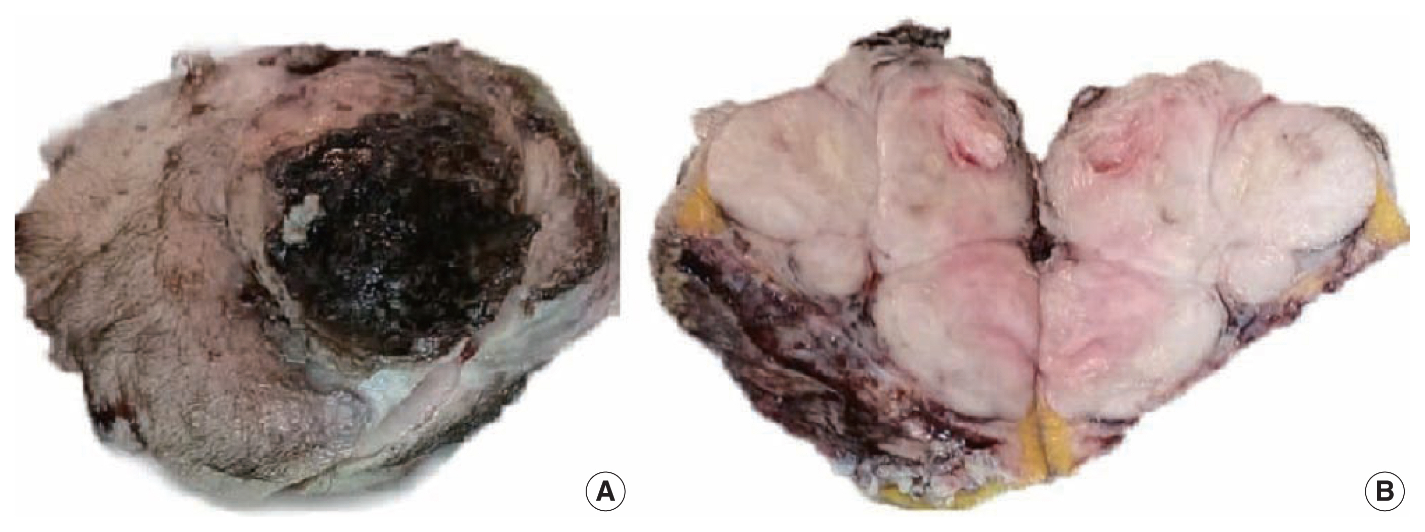
- Malignant solitary fibrous tumor (MSFT) is a well-described entity, from which heterologous differentiation is extremely rare. We encountered a case of MSFT with rhabdomyosarcomatous differentiation in a 56-year-old man. This patient presented with a large mass in his posterior thigh. He had been treated with chemoradiation for sarcoma involving the cervical spine, right femoral head, and both lungs 6 months earlier. A wide excision was performed. The mass measured 10.6 cm and showed a fish-flesh cut surface with necrotic foci. Microscopically, the tumor showed heterogeneous cellularity with a hemangiopericytic vascular pattern. A hypercellular area showed spindle cells or epithelioid cells with high mitotic activity (63/10 high-power fields) and immunoreactivity for CD34 and CD99. A hypocellular area and a cystic area showed pleomorphic rhabdoid cells with immunoreactivity for desmin and myogenin. This is a report of a rare case of MSFT with rhabdomyosarcomatous differentiation and presents new histologic features of MSFT.
-
Citations
Citations to this article as recorded by- A Rare Case of Malignant Solitary Fibrous Tumor on the Scalp
Kwang-Ryeol Kim, Ki Hong Kim
Keimyung Medical Journal.2023; 42(2): 107. CrossRef - Malignant solitary fibrous tumor of maxilla presenting as proptosis: A case report
Pravin Kumar, Arpita Jindal, Bhushan Bhalgat, Phanindra Kumar Swain, Raj Govind Sharma
Journal of Cancer Research and Therapeutics.2023; 19(Suppl 2): S991. CrossRef - Recurrent malignant solitary fibrous tumor of the scalp: a case report and literature review
Ahmed Rabie, Abdulkarim Hasan, Yasein Mohammed, Ayman Abdelmaksoud, Ali A. Rabaan
Journal of Pathology and Translational Medicine.2022; 56(2): 103. CrossRef - Frozen Cytology of Meningeal Malignant Solitary Fibrous Tumor/Hemangiopericytoma
Myunghee Kang, Na Rae Kim, Dong Hae Chung, Gie-Taek Yie
Journal of Pathology and Translational Medicine.2019; 53(3): 192. CrossRef
- A Rare Case of Malignant Solitary Fibrous Tumor on the Scalp
- Sclerosing Perivascular Epithelioid Cell Tumor of the Lung: A Case Report with Cytologic Findings
- Ha Yeon Kim, Jin Hyuk Choi, Hye Seung Lee, Yoo Jin Choi, Aeree Kim, Han Kyeom Kim
- J Pathol Transl Med. 2016;50(3):238-242. Published online April 11, 2016
- DOI: https://doi.org/10.4132/jptm.2016.02.19
- 10,548 View
- 110 Download
- 9 Web of Science
- 9 Crossref
-
Abstract
PDF
- Benign perivascular epithelioid cell tumor (PEComa) of the lung is a rare benign neoplasm, a sclerosing variant of which is even rarer. We present a case of 51-year-old man who was diagnosed with benign sclerosing PEComa by percutaneous fine needle aspiration cytology and biopsy. The aspirate revealed a few cell clusters composed of bland-looking polygonal or spindle cells with fine granular or clear cytoplasm. Occasional fine vessel-like structures with surrounding hyalinized materials were seen. The patient later underwent wedge resection of the lung. The histopathological study of the resected specimen revealed sheets of polygonal cells with clear vacuolated cytoplasm, variably sized thin blood vessels, and densely hyalinized stroma. In immunohistochemical studies, reactivity of tumor cells for human melanoma black 45 and Melan-A further supported the diagnosis of benign sclerosing PEComa. To the best of our knowledge, this is the first case of benign sclerosing PEComa described in lung.
-
Citations
Citations to this article as recorded by- A rare case of pulmonary perivascular epithelioid cell neoplasm
Maolin Xu, Xiaofang Guo
Asian Journal of Surgery.2026; 49(3): 1492. CrossRef - Renal sclerosing AML/PEComa in a male - A case report and literature review
Zoe Williams, Paul Kim, James Kovacic, Andrew Shepherd, Krishan Rasiah, Ankur Dhar, Kathleen Young
Urology Case Reports.2026; 65: 103326. CrossRef - Robotic Treatment of Adrenal Sclerosing PEComa: A Case Report with 13 Years of Follow-Up and a Literature Review
Alessio Paladini, Raffaele La Mura, Michele Del Zingaro, Luca Lepri, Andrea Vitale, Jessica Pagnotta, Matteo Mearini, Guido Massa, Ettore Mearini, Giovanni Cochetti
Applied Sciences.2025; 15(16): 9161. CrossRef - Cytopathology of rare gastric mesenchymal neoplasms: A series of 25 cases and review of literature
Carla Saoud, Peter B. Illei, Momin T. Siddiqui, Syed Z. Ali
Cytopathology.2023; 34(1): 15. CrossRef - Retroperitoneal Sclerosing Angiomyolipoma with Long-Term Follow up: A Case Report with Unique Clinicopathologic and Genomic Profile
Liwei Jia, Vandana Panwar, Michelle Parmley, Elena Lucas, Ivan Pedrosa, Payal Kapur
International Journal of Surgical Pathology.2022; 30(1): 86. CrossRef - Perivascular epithelioid cell tumor of the lung: A case report and literature review
Shaofu Yu, Shasha Zhai, Qian Gong, Xiaoping Hu, Wenjuan Yang, Liyu Liu, Yi Kong, Lin Wu, Xingxiang Pu
Thoracic Cancer.2022; 13(17): 2542. CrossRef - Cytopathology of extra-renal perivascular epithelioid cell tumor (PEComa): a series of 7 cases and review of the literature
Sintawat Wangsiricharoen, Tatianna C. Larman, Paul E. Wakely, Momin T. Siddiqui, Syed Z. Ali
Journal of the American Society of Cytopathology.2021; 10(2): 175. CrossRef - Clear cell sugar tumour: a rare tumour of the lung
Sarah Page, Matthew S. Yong, Alka Sinha, Pankaj Saxena
ANZ Journal of Surgery.2020;[Epub] CrossRef - Perivascular Epithelioid Cell Tumors (PEComas) of the Orbit
Panagiotis Paliogiannis, Giuseppe Palmieri, Francesco Tanda, Antonio Cossu
Journal of Pathology and Translational Medicine.2017; 51(1): 7. CrossRef
- A rare case of pulmonary perivascular epithelioid cell neoplasm
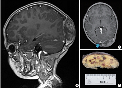
- Meningeal Solitary Fibrous Tumors with Delayed Extracranial Metastasis
- Nayoung Han, Hannah Kim, Soo Kee Min, Sun-Ha Paek, Chul-Kee Park, Seung-Hong Choi, U-Ri Chae, Sung-Hye Park
- J Pathol Transl Med. 2016;50(2):113-121. Published online December 14, 2015
- DOI: https://doi.org/10.4132/jptm.2015.10.30
- 13,379 View
- 120 Download
- 26 Web of Science
- 23 Crossref
-
Abstract
PDF
- Background
The term solitary fibrous tumor (SFT) is preferred over meningeal hemangiopericytoma (HPC), because NAB2-STAT6 gene fusion has been observed in both intracranial and extracranial HPCs. HPCs are now considered cellular variants of SFTs. Methods: This study analyzes 19 patients with STAT6-confirmed SFTs, who were followed for over 11 years in a single institution. Ten patients (10/19, 56.2%) had extracranial metastases (metastatic group), while the remainder (9/19) did not (non-metastatic group). These two groups were compared clinicopathologically. Results: In the metastatic group, the primary metastatic sites were the lungs (n = 6), bone (n = 4), and liver (n = 3). There was a mean lag time of 14.2 years between the diagnosis of the initial meningeal tumor to that of systemic metastasis. The median age at initial tumor onset was 37.1 years in the metastatic group and 52.5 in the non-metastatic group. The 10-year survival rates of the metastatic- and non-metastatic groups were 100% and 33%, respectively. The significant prognostic factors for poor outcomes on univariate analysis included advanced age (≥45 years) and large initial tumor size (≥5 cm). In contrast, the patients with higher tumor grade, high mitotic rate (≥5/10 high-power fields), high Ki-67 index (≥5%), and the presence of necrosis or CD34 positivity showed tendency of poor prognosis but these parameters were not statistically significant poor prognostic markers. Conclusions: Among patients with SFTs, younger patients (<45 years) experienced longer survival times and paradoxically had more frequent extracranial metastases after long latent periods than did older patients. Therefore, young patients with SFTs require careful surveillance and follow-up for early detection of systemic metastases. -
Citations
Citations to this article as recorded by- Single-fraction stereotactic radiosurgery for residual, recurrent, or metastatic intracranial solitary fibrous tumors: An IRRF study toward management guidance
Salem M Tos, Ahmed Shaaban, Dawood Hamdan, Georgios Mantziaris, Bardia Hajikarimloo, Mariam Ishaque, Yuki Shinya, Vanshika Lohia, Zhishuo Wei, Orbay Askeroglu, Christian Amezquita-Contreras, Andrea Becerril-Gaitan, Onam Verma, Keiss Douri, Nathalia Lora,
Neuro-Oncology.2026;[Epub] CrossRef - High-grade, metastatic disease, and adjuvant radiotherapy are independent prognostic factors for progression-free survival in patients with solitary fibrous tumors
Jan Paul Alker, Ramin Rahmanzade, Thomas Held, Christel Herold-Mende, Andreas Unterberg, Felix Sahm, Sandro Manuel Krieg, Gerhard Jungwirth
Neuro-Oncology Advances.2025;[Epub] CrossRef - Meningeal malignant solitary fibrous tumor with multiple recurrence, extracranial extension, cervical lymph node metastases: case report and review of the literature
Rong He, Peng Zhong, Juntao Hu, Guangkuo Guo, He Xiao, Lin Lei, Yun Liu, Mingying Geng, Jungang Ma
Discover Oncology.2025;[Epub] CrossRef - A Case of Intracranial Solitary Fibrous Tumor Followed by Distant Metastasis without Local Recurrence
Masafumi YOSHIDA, Koki MORIYOSHI, Kento DOI, Yukihiro YAMAO, Natsue KISHIDA, Hiroya UEMURA, Shunichi FUKUDA
NMC Case Report Journal.2025; 12: 181. CrossRef - The association between WHO grading and the long-term outcomes and radiotherapy efficacy of intracranial solitary fibrous tumors
Leihao Ren, Lingyang Hua, AO Feng, Jiaojiao Deng, Hiroaki Wakimoto, Tareq Juratli, Qing Xie, Ye Gong
Acta Neuropathologica Communications.2025;[Epub] CrossRef - Meningeal Solitary Fibrous Tumor: A Single-Center Retrospective Cohort Study
Siyer Roohani, Yasemin Alberti, Maximilian Mirwald, Felix Ehret, Carmen Stromberger, Soleiman Fabris Roohani, Katja Bender, Anne Flörcken, Sven Märdian, Daniel Zips, David Kaul, Manish Charan
Sarcoma.2024; 2024: 1. CrossRef - De-differentiation associated with drop metastasis of a recurrent intracranial solitary fibrous tumor: a case report and literature review
Chenhui Zhao, Xiran Fan, Wanwan Gao, Fan Zhang, Haijun Lv, Xiaochun Jiang, Guangfu Di
International Journal of Neuroscience.2022; 132(8): 843. CrossRef - Long-term extracranial metastatic relapse of an intraventricular solitary fibrous tumor: a case report
Tarek Assi, Elie Samaha, Hussein Nassereddine
Anti-Cancer Drugs.2022; 33(1): e764. CrossRef - Multidisciplinary Treatment of Liver Metastases from Intracranial SFTs/HPCs: A Report of Three Consecutive Cases
Felix J. Krendl, Franka Messner, Gregor Laimer, Angela Djanani, Andreas Seeber, Georg Oberhuber, Dietmar Öfner, Dominik Wolf, Stefan Schneeberger, Reto Bale, Christian Margreiter
Current Oncology.2022; 29(11): 8720. CrossRef - A review of solitary fibrous tumor/hemangiopericytoma tumor and a comparison of risk factors for recurrence, metastases, and death among patients with spinal and intracranial tumors.
Enrico Giordan, Elisabetta Marton, Alexandra M. Wennberg, Angela Guerriero, Giuseppe Canova
Neurosurgical Review.2021; 44(3): 1299. CrossRef - Intracranial Solitary Fibrous Tumor of the Skull Base: 2 Cases and Systematic Review of the Literature
Sricharan Gopakumar, Visish M. Srinivasan, Caroline C. Hadley, Adrish Anand, Marc Daou, Patrick J. Karas, Jacob Mandel, Shankar P. Gopinath, Akash J. Patel
World Neurosurgery.2021; 149: e345. CrossRef - Hemangiopericytoma/Solitary Fibrous Tumor in the central nervous system. Experience with surgery and radiotherapy as a complementary treatment: A 10-year analysis of a heterogeneous series in a single tertiary center
Pedro Miguel González-Vargas, José Luis Thenier-Villa, Pablo Sanromán Álvarez, Alexandre Serantes Combo, Lourdes Calero Félix, Raúl Alejandro Galárraga Campoverde, Eva Azevedo González, Álvaro Martín-Gallego, Rosa Martínez-Rolan, Adolfo de la Lama Zaragoz
Neurocirugía.2020; 31(1): 14. CrossRef - Hemangiopericytoma/Solitary Fibrous Tumor in the central nervous system. Experience with surgery and radiotherapy as a complementary treatment: A 10-year analysis of a heterogeneous series in a single tertiary center
Pedro Miguel González-Vargas, José Luis Thenier-Villa, Pablo Sanromán Álvarez, Alexandre Serantes Combo, Lourdes Calero Félix, Raúl Alejandro Galárraga Campoverde, Eva Azevedo González, Álvaro Martín-Gallego, Rosa Martínez-Rolan, Adolfo de la Lama Zaragoz
Neurocirugía (English Edition).2020; 31(1): 14. CrossRef - Solitary fibrous tumor/hemangiopericytoma: treatment results based on the 2016 WHO classification
Kyoung Su Sung, Ju Hyung Moon, Eui Hyun Kim, Seok-Gu Kang, Se Hoon Kim, Chang-Ok Suh, Sun Ho Kim, Kyu-Sung Lee, Won Seok Chang, Jong Hee Chang
Journal of Neurosurgery.2019; 130(2): 418. CrossRef - Grading of meningeal solitary fibrous tumors/hemangiopericytomas: analysis of the prognostic value of the Marseille Grading System in a cohort of 132 patients
Nicolas Macagno, Rob Vogels, Romain Appay, Carole Colin, Karima Mokhtari, Benno Küsters, Pieter Wesseling, Dominique Figarella‐Branger, Uta Flucke, Corinne Bouvier
Brain Pathology.2019; 29(1): 18. CrossRef - Solitary fibrous tumor of the pineal region with delayed ectopic intracranial metastasis: A case report and review of the literature
Yongjie Wang, Jingying Zhang, Qichang Liu, Fuyi Liu, Xiangdong Zhu, Jianmin Zhang
Medicine.2019; 98(21): e15737. CrossRef - Case report: neonatal giant forehead hemangiopericytoma with a 5-year follow-up
AiJun Peng, LiBing Zhang, Hai Zhao, LiangXue Zhou
Medicine.2019; 98(47): e17888. CrossRef - Liquid Biopsy in Rare Cancers: Lessons from Hemangiopericytoma
Chiara Nicolazzo, Luciano Colangelo, Alessandro Corsi, Guido Carpino, Angela Gradilone, Chiara Sonato, Cristina Raimondi, Eugenio Gaudio, Paola Gazzaniga, Walter Gianni
Analytical Cellular Pathology.2018; 2018: 1. CrossRef - Surveillance for metastatic hemangiopericytoma-solitary fibrous tumors-systematic literature review on incidence, predictors and diagnosis of extra-cranial disease
Tarini Ratneswaren, Florence Rosie Avila Hogg, Mathew Joseph Gallagher, Keyoumars Ashkan
Journal of Neuro-Oncology.2018; 138(3): 447. CrossRef - Intracranial Solitary Fibrous Tumor
Eveline Claus, Patrick Seynaeve, Jeroen Ceuppens, Alain Vanneste, Koenraad Verstraete
Journal of the Belgian Society of Radiology.2017;[Epub] CrossRef - Comparison and evaluation of risk factors for meningeal, pleural, and extrapleural solitary fibrous tumors: A clinicopathological study of 92 cases confirmed by STAT6 immunohistochemical staining
Ji Min Kim, Yoon-La Choi, Yu Jin Kim, Hyung Kyu Park
Pathology - Research and Practice.2017; 213(6): 619. CrossRef - Molecular Testing of Brain Tumor
Sung-Hye Park, Jaekyung Won, Seong-Ik Kim, Yujin Lee, Chul-Kee Park, Seung-Ki Kim, Seung-Hong Choi
Journal of Pathology and Translational Medicine.2017; 51(3): 205. CrossRef - Solitary fibrous tumour presenting with a single bone metastasis: report of six cases and literature review
Vittoria Colia, Salvatore Provenzano, Carlo Morosi, Paola Collini, Salvatore Lorenzo Renne, Paolo G. Dagrada, Claudia Sangalli, Angelo Paolo Dei Tos, Andrea Marrari, Paolo G. Casali, Silvia Stacchiotti
Clinical Sarcoma Research.2016;[Epub] CrossRef
- Single-fraction stereotactic radiosurgery for residual, recurrent, or metastatic intracranial solitary fibrous tumors: An IRRF study toward management guidance
- Paediatric Primary Pachymeningeal Xanthogranuloma with Scattered Foci Displaying Reticulohistiocytoma-like Features
- Miguel Fdo. Salazar, María del Rocío Estrada Hernández, Erick Gómez Apo, Laura G. Chávez Macías, Carlos Alfonso Rodríguez Álvarez
- J Pathol Transl Med. 2015;49(5):403-408. Published online June 17, 2015
- DOI: https://doi.org/10.4132/jptm.2015.05.28
- 10,848 View
- 51 Download
- 1 Web of Science
- 1 Crossref
-
Abstract
PDF
- We report a unique case of a 4-year-old girl with an intriguing fibrohistiocytic tumour. Magnetic resonance imaging scans showed a dural mass of variegated intensity compressing the left occipital pole and apparently extending toward the superior sagittal sinus. Grossly, the cut surface of the surgical specimen was yellow, pale, and soft with reddish kernel-like crusts. Histologically, the yellow areas resembled cholesterol granulomas with widespread coagulative necrosis, cholesterol clefts, powdery calcification, foreign body-type giant cells, and foamy macrophages, while the scattered red spots contained numerous multinucleated giant cells of foreign-body and Touton types, the former with amphophilic to slightly eosinophilic cytoplasm. Immunoperoxidase reactions confirmed the expression of histiocytic markers and vimentin. As far as we know, no tumour displaying these peculiar morphological features has yet been described.
-
Citations
Citations to this article as recorded by- Reticulohistiocytoses: a revision of the full spectrum
A. Bonometti, E. Berti
Journal of the European Academy of Dermatology and Venereology.2020; 34(8): 1684. CrossRef
- Reticulohistiocytoses: a revision of the full spectrum
- A Solitary Fibrous Tumor with Giant Cells in the Lacrimal Gland: A Case Study
- Da Hye Son, Su Hyun Yoo, Ho-Seok Sa, Kyung-Ja Cho
- Korean J Pathol. 2013;47(2):158-162. Published online April 24, 2013
- DOI: https://doi.org/10.4132/KoreanJPathol.2013.47.2.158
- 9,333 View
- 61 Download
- 9 Crossref
-
Abstract
PDF
Orbital solitary fibrous tumor (SFT) has recently been proposed as the encompassing terminology for hemangiopericytoma, giant cell angiofibroma (GCAF), and fibrous histiocytoma of the orbit. The lacrimal gland is a very rare location for both SFT and GCAF. A 39-year-old man presented with a painless left upper eyelid mass. An orbital computed tomography scan identified a 1.1 cm-sized well-defined nodule located in the left lacrimal gland. He underwent a mass excision. Histopathologic examination showed a proliferation of relatively uniform spindle cells with a patternless or focally storiform pattern. Dilated vessels were prominent, but angiectoid spaces lined with giant cells were absent. Floret-type giant cells were mostly scattered in the periphery. The tumor was immunoreactive for CD34 and CD99, but negative for smooth muscle actin and S-100 protein. This is the first Korean case of SFT of the lacrimal gland with overlapping features of GCAF, suggesting a close relationship between the two entities.
-
Citations
Citations to this article as recorded by- Radiological features of lacrimal gland masses
Sonia Huang, Jessica Y. Tong, Valerie Juniat, Abdullah Almater, Ilse Mombaerts, Dinesh Selva
Survey of Ophthalmology.2026; 71(2): 700. CrossRef - Imaging features of lacrimal gland disease
Carmelo Caltabiano, Khizar Rana, Alexander Buckby, Sandy Patel, Dinesh Selva
Frontiers in Ophthalmology.2026;[Epub] CrossRef - A review of solitary fibrous tumours of the orbit and ocular adnexa
Cornelius René, Paolo Scollo, Dominic O’Donovan
Eye.2023; 37(5): 858. CrossRef - A giant orbital solitary fibrous tumor treated by surgical excision: a case report and literature review
Qi Zhou, Yuting Liu, Fang Wang, Yang Cao, Hongbin Lv, Xibo Zhang
Diagnostic Pathology.2023;[Epub] CrossRef - Giant cell-rich solitary fibrous tumour of the lacrimal gland with prominent angiomatoid cystic changes and an underlying NAB2ex3-STAT6ex18 fusion
Khaled A Alsaadi, Manar Alwohaib, Karen Pinto, Rola H Ali
BMJ Case Reports.2022; 15(2): e247141. CrossRef - Cystic appearance - a new feature of solid fibrous tumours in the lacrimal gland: a case report with literature review
Ancuta-Augustina Gheorghisan-Galateanu, Dana Cristina Terzea, Iulia Burcea, Roxana Dusceac, Cristina Capatina, Catalina Poiana
Diagnostic Pathology.2019;[Epub] CrossRef - Solitary Fibrous Tumor in the Lacrimal Gland Fossa: A Case Report
Jacqueline Mupas-Uy, Yoshiyuki Kitaguchi, Yasuhiro Takahashi, Emiko Takahashi, Hirohiko Kakizaki
Case Reports in Ophthalmology.2016; 7(2): 398. CrossRef - Ocular adnexal (orbital) solitary fibrous tumor: nuclear STAT6 expression and literature review
Aleksandra Petrovic, Aurélie Obéric, Alexandre Moulin, Mehrad Hamedani
Graefe's Archive for Clinical and Experimental Ophthalmology.2015; 253(9): 1609. CrossRef - Angiofibroma de células gigantes en mucosa yugal: una entidad rara en una localización infrecuente
Alejandro Rubio Fernández, María López Macías, Weimar Toro Zambrano, Mario Díaz Delgado, Alicia Hernández Amate
Revista Española de Patología.2014; 47(4): 223. CrossRef
- Radiological features of lacrimal gland masses
- Solitary Fibrous Tumor of the Conjunctiva with Heretofore Undescribed Pathologic Findings.
- Na Rae Kim, Jae Y Ro, Kyung Hwan Shin, Hae Jung Paik, Jung Suk An, Seung Yeon Ha
- Korean J Pathol. 2011;45(3):315-318.
- DOI: https://doi.org/10.4132/KoreanJPathol.2011.45.3.315
- 4,711 View
- 27 Download
- 1 Crossref
-
Abstract
PDF
- A 37-year-old female presented with a conjunctival mass discovered 3 years prior. An excisional biopsy revealed a patternless proliferation of round and spindle-shaped cells with an eosinophilic fibrillary cytoplasm and vesicular nuclei with occasional inclusions. Psammoma bodies were arranged around the dilated irregularly-shaped vessels. Differential diagnoses included conjunctival solitary fibrous tumor (SFT), nevus, glomangioma, ectopic meningioma, and mesectodermal leiomyoma. The tumor cells were immunoreactive for CD34, CD99, bcl-2 and vimentin, and were negative for smooth muscle actin, desmin, glial fibrillary acidic protein, S-100 protein, epithelial membrane antigen, and human melanoma black-45. Ultrastructurally, the tumor cells had rough endoplasmic reticulum, free ribosomes, and scattered mitochondria without basal lamina or cellular junctions, which are features of fibroblasts. A diagnosis of SFT was rendered based on the light microscopic, immunohistochemical, and electron microscopic findings. We report here on the second case of a SFT arising in the conjunctiva, which clinically and histologically mimics conjunctival nevus, glomangioma, ectopic meningioma, and a hybrid neurogenic-myogenic tumor such as mesectodermal leiomyoma.
-
Citations
Citations to this article as recorded by- Meningeal Solitary Fibrous Tumors with Delayed Extracranial Metastasis
Nayoung Han, Hannah Kim, Soo Kee Min, Sun-Ha Paek, Chul-Kee Park, Seung-Hong Choi, U-Ri Chae, Sung-Hye Park
Journal of Pathology and Translational Medicine.2016; 50(2): 113. CrossRef
- Meningeal Solitary Fibrous Tumors with Delayed Extracranial Metastasis
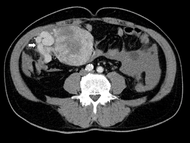
- Solitary Fibrous Tumor of the Liver: A Case Report.
- Hee Chul Yu, Baik Hwan Cho, Young Kon Kim, Sang Jae Noh, Woo Sung Moon
- Korean J Pathol. 2010;44(5):536-539.
- DOI: https://doi.org/10.4132/KoreanJPathol.2010.44.5.536
- 4,377 View
- 25 Download
- 1 Crossref
-
Abstract
PDF
- Solitary fibrous tumor is an uncommon neoplasm of mesenchymal origin that primarily affects the pleura. This tumor has been rarely found in liver parenchyma. We present an additional case of a solitary fibrous tumor in the liver of a 46-year-old woman. A contrast-enhanced magnetic resonance image revealed a well-defined round hepatic mass with strong homogeneous enhancement on arterial phase imaging. The tumor was composed of cytologically bland spindle cells with alternating hypercellular and hypocellular sclerotic areas. Immunohistochemistry indicated that the tumor cells were positive for vimentin, CD34, CD99 and smooth muscle actin, but negative for cytokeratin, human melanoma black 45, CD117, bcl-2, and S-100 protein.
-
Citations
Citations to this article as recorded by- Meningeal Solitary Fibrous Tumors with Delayed Extracranial Metastasis
Nayoung Han, Hannah Kim, Soo Kee Min, Sun-Ha Paek, Chul-Kee Park, Seung-Hong Choi, U-Ri Chae, Sung-Hye Park
Journal of Pathology and Translational Medicine.2016; 50(2): 113. CrossRef
- Meningeal Solitary Fibrous Tumors with Delayed Extracranial Metastasis
- Solitary Fibrous Tumor of the Kidney: A Report of Two Cases with Review of Literature.
- Sun A Kim, Jung Eun Hwang, Jae Y Ro, Kyung Ja Cho, Cheryn Song, Mi Jung Kim
- Korean J Pathol. 2010;44(4):420-425.
- DOI: https://doi.org/10.4132/KoreanJPathol.2010.44.4.420
- 4,642 View
- 26 Download
- 2 Crossref
-
Abstract
PDF
- Solitary fibrous tumor (SFT) is a benign mesenchymal neoplasm usually occurring in the pleura. Kidney is one of the rarest sites for SFT. We report here on two cases of renal SFT found in 30-year-old and 33-year-old men with review of the literatures. Both cases manifested as well-enhanced solid masses in kidney and radical nephrectomies were done. The tumors consisted of bland-looking spindle cells arranged in short, ill-defined fascicles and storiform pattern with characteristic hemangiopericytoma-like blood vessels. The tumor cells were strongly positive for CD34 and CD99, focally positive for bcl-2, and negative for cytokeratin and human melanoma black-45 on immunohistochemical stainings. Possibility of SFT should be considered in the differential diagnosis of a renal mass which consists of benign-looking spindle cells and hemangiopericytomatous blood vessels. Immunohistochemical staining for CD34 is essential to confirm the renal solitary fibrous tumor.
-
Citations
Citations to this article as recorded by- Solitary fibrous tumor located in the sella turcica: A report of two cases and review of the literature
XIAO YANG, QINGJUN JIANG, BINGBING YU
Oncology Letters.2015; 10(1): 354. CrossRef - Pediatric Renal Solitary Fibrous Tumor
William W. Wu, Julia T. Chu, Stephen G. Romansky, Lisa Shane
International Journal of Surgical Pathology.2015; 23(1): 34. CrossRef
- Solitary fibrous tumor located in the sella turcica: A report of two cases and review of the literature
- Extrapleural Solitary Fibrous Tumor A clinical & pathological study of 8 cases.
- Mi Kyung Lee, Dong Hwan Shin, Min Sun Cho, Yuon Mee Kim, Jin Kim
- Korean J Pathol. 1999;33(2):108-114.
- 2,221 View
- 10 Download
-
Abstract
- We reviewed eight solitary fibrous tumors occurring at sites other than pleura (three orbit, two retroperitoneum, one each hard palate, thyroid, and tongue) which shared the histologic and immunohistochemical features of solitary fibrous tumors of pleura. Six patients were women, and two were men, aged from 26 to 74 years. The tumors ranging from 1.5 to 19 cm in diameter presented as well-circumscribed, unencapsulated, soft to rubbery tissue masses. Histologically they were characterized by a proliferation of spindle or ovoid cells intervened by a dense bundles of collagen. A variety of growth patterns was identified but the so-called patternless pattern was the predominant one. One tumor exhibited highly cellular sarcomatous areas with extensive necrosis, which was diagnosed as malignant solitary fibrous tumor. Immunohistochemical studies showed that all of the tumors were strongly positive for both CD34 and vimentin, but negative for cytokeratin, S-100 protein, EMA, and desmin. One case examined ultrastructurally showed features of fibroblast. All but one showed no evidence of recurrence or metastasis over follow-up period of 14 to 32 months. We conclude that extrapleural solitary fibrous tumors represent a distinct mesenchymal tumor with variable histologic features and should be differentiated from other spindle cell mesenchymal tumors.
- Solitary Fibrous Tumor A clinicopathologic review of five cases.
- Bum Kyung Kim, Dong Wook Kang, Kyeong Hee Kim, Seong Ki Min, Jin Man Kim, Kyu Sang Song, Dae Yung Kang, Si Whan Choi
- Korean J Pathol. 1999;33(2):115-120.
- 2,185 View
- 10 Download
-
Abstract
- We experienced five cases of solitary fibrous tumor; two in the pleura, two in the orbital soft tissue, and one in the lung parenchyma. Three patients were male, and the age of the patients ranged from 38 to 71 years (mean age: 53.6). Grossly, the masses were well circumscribed and had varying sizes from 2.5 to 30.0 cm. The cut surfaces were grayish-yellow firm with focal variegated hemorrhage, necrosis, cystic change, and myxoid area. Microscopically, these were characterized by a haphazard proliferation of spindle cells or polygonal cells separated by variable amounts of hyalinized collagen and showed a prominent vascular channels reminiscent of hemangiopericytoma in foci. Immunoperoxidase stains showed a strong reactivity for CD34, and were weakly positive for vimentin. Electron microscopical examination revealed features of fibroblast; spindle to round tumor cells were arranged in groups and surrounded by collagen. Nucleoli were seldom prominent. The cytoplasm contained many microfilaments and a moderate number of cisternae of rough endoplasmic reticulum.
- Solitary Fibrous Tumor of the Urinary Bladder: A Case Report.
- Jong Sil Lee, Jeong Seok Hwa, Gyung Hyuck Ko, Jeong Hee Lee, Hwal Woong Kim
- Korean J Pathol. 2004;38(2):129-131.
- 2,033 View
- 20 Download
-
Abstract
PDF
- Solitary fibrous tumor (SFT) most commonly affects the pleura and these tumors have been recently reported to be found in unusual locations. We describe here a solitary fibrous tumor of the urinary bladder that was removed from a 79-year-old man having a history of gross hematuria and dysuria. Transabdominal ultrasonography showed a huge soft tissue mass in the urinary bladder. The cut surface of the tumor showed a grayish-white, hemorrhagic and gelatinous appearance. Necrosis was not found. Microscopically, the tumor showed a proliferation of spindle or ovoid cells that were intervened by a collagenous stroma. A variety of growth patterns was identified but the so-called patternless pattern was the predominant one. The spindle cells had almost no mitotic figures, and there was very little or no nuclear atypia. Immunohistochemical stains showed a strong reactivity for CD34 and a focal reactivity for bcl-2. The ultrastructure of the tumor cells showed mesenchymal-myofibroblastic traits.
- Malignant Solitary Fibrous Tumor of the Pleura in Mediastinum.
- Yun Kyung Kang, Hyun Joo Yoo, Ho Kee Yum, Hong Sup Lee
- Korean J Pathol. 1997;31(4):351-356.
- 2,087 View
- 14 Download
-
Abstract
PDF
- Solitary fibrous tumors (SFTs) most often involve the pleura and also may encompass the peritoneum and nonserosal sites. They occur as solitary encapsulated tumors and pursue a relatively benign clinical course. The usual criteria for malignancy are high cellularity, mitotic activity (more than 4 per 10 high-power fields), cellular pleomorphism, hemorrhage and necrosis as well as infiltrative growth. We report a case of malignant SFT of pleura who presented with an anterior mediastinal mass. Grossly, it was a 10x8x6.5 cm sized, encapsulated and well-demarcated, solid neoplasm with areas of extensive necrosis. Microscopically, parallel or haphazard arrangement of spindle cells with variable degrees of collagenous background were noted. Storiform fascicle formation, hemangiopericytoma-like pattern, and epithelioid cell clusters were often intermingled. Nodular areas with high cellularity and mitotic activity (> or =10/10 HPFs) were scattered throughout the neoplasm, however no definite cellular pleomorphism was encountered. Tumor cells were immunoreactive for vimentin and CD-34, which distinguished them from the mesothelial cells. Electron microscopically, they revealed fibroblastic and myofibroblastic differentiation.
- Solitary Fibrous Tumor of the Scrotum: A case report.
- Jung Weon Shim, Jae Y Ro
- Korean J Pathol. 1999;33(4):295-298.
- 2,423 View
- 44 Download
-
Abstract
PDF
- Solitary fibrous tumor (SFT) is a rare spindle cell neoplasm that usually arises in the pleura and has been recently reported from unusual locations. We present a case of SFT that occurred in the scrotum. The patient was a 38-year-old man who presented with a painless, left, scrotal mass of five years' duration. Scrotal sonography and pelvic CT scan showed a soft-tissue mass of 11 cm in diameter. The resected tumor measured 11 8 7 cm and was well demarcated from the surrounding soft tissue. The cut surface revealed homogenously grayish-white and gelatinous appearance. No hemorrhage or necrosis was found. Microscopically, the tumor showed hypercellular spindle cell areas intermixed with hypocellular areas lying in a myxoid or collagenous stroma. The spindle cells had no mitosis or low mitotic figures, and little or no nuclear atypia. They exhibited a variety of growth patterns, including "patternless" pattern, and a prominent vasculature with hemangiopericytic pattern. Vimentin, CD34, and bcl-2 protein immunoreactivity were observed. Characteristic histologic and immunohistochemical features of this lesion were consistent with SFT. To arrive at a correct diagnosis of this lesion, especially when it occurs in unusual sites, immunohistochemical study including CD34 & bcl-2 protein is required in addition to characteristic histologic features.
- Intrapulmonary Cystic Lymphangioma.
- Mi Yeong Jeon, Je Geun Chi
- Korean J Pathol. 1997;31(5):492-494.
- 2,079 View
- 20 Download
-
Abstract
PDF
- Solitary intrapulmonary cystic lymphangioma in newborn or infant is an extremely rare disease. We describe a case of solitary intrapulmonary cystic lymphangioma in a 4-month-old boy with dyspnea and tachypnea. It was in the left lower lobe and type 1 congenital cystic adenomatoid malformation was suspected radiologically. The resected cyst was unilocular with a thin wall, and was 9.5cm in size. Histologically, the cyst was lined by flattened endothelial cells supported by a minimal fibrous stroma.
- A Case of Solitary Cutaneous Myofibroma of the Thigh in An Adult.
- Jung Hwan Park, Chang Woo Lee, Young Chae Chu, Moon Hyang Park
- Korean J Pathol. 2001;35(4):354-356.
- 2,216 View
- 33 Download
-
Abstract
PDF
- Adult solitary cutaneous myofibroma is a recently described benign neoplasm of the skin or subcutis, representing the adult counterpart of infantile myofibroblastoma. The histologic and immunohistochemical features of a 21-year-old woman with a solitary brownish, mildly tender nodule on her right thigh are reported here. The nodule had been present for a duration of 3 years. It showed a nodular dermal mass with an irregular margin. The lesion consisted of interlacing bundles of spindle cells which were positive for smooth muscle actin, muscle specific actin and vimentin. Immunohistochemical stainings for desmin, S-100 protein, CD 34 and CD 68 were negative. Cutaneous myofibroma in an adult is a distinct entity of benign neoplasm.
- Fine Needle Aspiration Cytology of Solitary Fibrous Tumor of the Pleura: Report of a case misdiagnosed as denocarcinoma of lung.
- Yoon La Choi, Young Lyun Oh, Mee Sook Lee, Jung Ho Han, Geung Hwan Ahn
- J Pathol Transl Med. 2001;12(2):111-115.
- 2,377 View
- 22 Download
-
Abstract
PDF
- Solitary fibrous tumor of the pleura is rare but should be included in the differential diagnosis of a peripheral pulmonary nodule. Cytologic features of solitary fibrous tumor of the pleura is not familar to the pathologist and may be misdiagnosed as malignancy. We report fine needle aspiration cytologic(FNAC) findings of a case of solitary fibrous tumor misdiagnosed as adenocarcinoma in a 48-year-old woman. The FNAC displayed a mixture of bland-looking spindle cells and clusters of epithelioid cells, which have hyperchromatic nuclei with prominent nucleoli. The helpful finding to distinguish it from other circumscribed benign and malignant lesions is the presence of fibromyxoid matrix admixed with blood vessels and thin collagen fibers. Familiarity with these features is essential to avoid misdiagnosis and overtreatment.
- Solitary Fibrofolliculoma: A case report.
- Hun Kyung Lee, Lee So Maeng, Seok Jin Kang, Soo Il Chung, Sun Moo Kim
- Korean J Pathol. 1996;30(5):460-462.
- 2,015 View
- 26 Download
-
Abstract
PDF
- The clinical and pathological features of solitary fibrofolliculoma are presented. Solitary fibrofolliculoma is very rarely encountered and to our knowledge, only 7 cases have been reported in the Western literature and no cases have been published in Korea. We experienced a case of solitary fibrofolliculoma occurring in a 56-year-old female, who had a 1.0 cm-sized and slowly growing nodule on her chin. A brief review of the literature, was made especially in relation to the pathological findings and histogenesis of solitary fibrofolliculoma.
- Warty Dyskeratoma with a Cutaneous Horn: Report of a case.
- Ah Won Lee, Hyun Joo Choi, Youn Soo Lee, Seok Jin Kang, Byung Kee Kim, Sun Moo Kim
- Korean J Pathol. 1998;32(8):616-618.
- 2,080 View
- 10 Download
-
Abstract
- The clinical and pathological features of warty dyskeratoma are presented. Warty dyskeratoma is a rarely encountered, solitary, benign cutaneous tumor occurring most often on the head and neck and to our knowledge, no cases associated with cutaneous horn have been published in western literature. We experienced a case of warty dyskeratoma with a cutaneous horn occurring in a 70-year-old male, who had a 1.3 cm-sized and slowly growing nodule on his neck. A brief review of the literature, was made especially in relation to the pathological findings and histogenesis of warty dyskeratoma.
- Cytologic Features of Plasmacytoma of the Ovary and Breast Occurred in a Patient with Solitary Plasmacytoma of Vertebra .
- Mi OK Park, Hoon Kyu Oh, Yong Jin Kim, Jae Bok Park
- J Pathol Transl Med. 1997;8(2):164-169.
- 2,017 View
- 21 Download
-
Abstract
PDF
- A case of plasmacytoma of the ovary and breast, which developed in a patient with a solitary plasmacytoma in the lumbar vertebra for nine months, was diagnosed cytologically and histologically. Enlargement of the right ovary and multiple palpable masses in the right and left breast were already present at six months after the diagnosis of vertebral solitary plasmacytoma. At eight months, plasma cell leukemia developed, and nine months the enlarged both ovaries, replaced by yellowish-gray solid tumors showed infiltration of immature plasma cells. The cytologic features of the ovarian tumors were same with those of the breast tumor. The tumor cells were of predominantly immature plasma cells with one or more nuclei. Some mature plasma cell had an eccentric nucleus with single nucleolus and peripherally clumped chromatin. Binucleated or multinucleated giant cells were often present. Histologically, sheets of poorly differentiated plasmacytoid tumor cells were separated by strands of hyaline fibrous tissue. On immunohistochemical stains, the tumor cells showed strong reactivity for lambda-light chain but no reaction for kappa-light chain, cytokeratin, or leukocyte common antigen.
 First
First Prev
Prev


