 E-submission
Search
- Page Path
- HOME > Search
- Current state of cytopathology residency training: a Korean national survey of pathologists
- Uiju Cho, Tae Jung Kim, Wan Seop Kim, Kyo Young Lee, Hye Kyoung Yoon, Hyun Joo Choi
- J Pathol Transl Med. 2023;57(2):95-101. Published online March 14, 2023
- DOI: https://doi.org/10.4132/jptm.2023.01.06
- 4,965 View
- 85 Download
- 1 Web of Science
- 1 Crossref
-
 Abstract
 PDF
PDF Supplementary Material
 - Background
Although the Korean Society for Cytopathology has developed educational goals as guidelines for cytopathology education in Korea, there is still no systematic approach to cytopathology education status for pathology residents. Furthermore, satisfaction with cytopathology education and with the outcome of the current training/educational program has not been investigated in Korea. This study aimed to obtain comprehensive data on the current state of cytopathology education for residents and evaluate education outcomes.
Methods
An online survey was conducted in December 2020 for the board-certified pathologists and training residents registered as members of the Korean Society for Cytopathology. The questionnaire comprised questions that investigated the current status of cytopathology at each training institution, the degree of satisfaction with the work and education related to cytopathology, outcomes of cytopathology training, and educational accomplishments.
Results
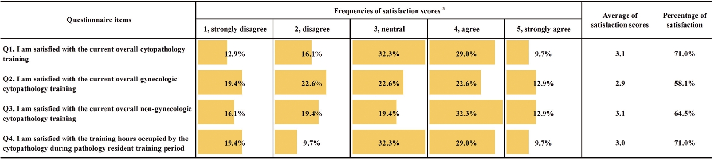
Of the participants surveyed, 12.3% (132/1,075) completed the questionnaire, and 36.8% (32/87) of cytopathology residents participated. The mean overall satisfaction with cytopathology education was 3.1 points (on a 1- to 5-point scale, 5: very satisfied). The most frequent suggestion among the free description format responses was to expand educational opportunities, such as online education opportunities, outside of the individual institutions.
Conclusions
Our results showed that cytopathology training in Korea needs further improvement. We expect that this study will inform systematic training of competent medical personnel armed with broad cytopathology knowledge and strong problem-solving abilities. -
Citations
Citations to this article as recorded by
- Artificial Intelligence–Assisted Daily Quality Control System for the Histologic Diagnosis of Gastrointestinal Endoscopic Biopsies: A 1-Year Experience
Seung-Yeon Yoo, Yuri Hwang, Seokju Yun, Ok Hee Lee, Jiwook Jang, Youngjin Park, Tae Young Cho, Young Sin Ko
Archives of Pathology & Laboratory Medicine.2025; 149(7): 659. CrossRef
- Artificial Intelligence–Assisted Daily Quality Control System for the Histologic Diagnosis of Gastrointestinal Endoscopic Biopsies: A 1-Year Experience
- Peripheral type squamous cell carcinoma of the lung: clinicopathologic characteristics in comparison to the central type
- Yeoun Eun Sung, Uiju Cho, Kyo Young Lee
- J Pathol Transl Med. 2020;54(4):290-299. Published online June 17, 2020
- DOI: https://doi.org/10.4132/jptm.2020.05.04
- 12,744 View
- 213 Download
- 15 Web of Science
- 17 Crossref
-
Abstract
PDF
- Background
Squamous cell carcinomas (SqCCs) of the lung are known to arise more often in a central area but reports of peripheral SqCCs have increased, with a pathogenesis that is obscured. In this study, the clinicopathologic characteristics of peripheral lung SqCCs were studied and compared with those of the central type.
Methods
This study included 63 peripheral lung SqCCs and 48 randomly selected central cases; hematoxylin and eosin-stained slides of surgically resected specimens were reviewed in conjunction with radiologic images and clinical history. Cytokeratin-7 immunohistochemical staining of key slides and epidermal growth factor receptor (EGFR)/KRAS mutations tested by DNA sequencing were also included.
Results
Stages of peripheral SqCCs were significantly lower than central SqCCs (p=.016). Cystic change of the mass (p=.007), presence of interstitial fibrosis (p=0.007), and anthracosis (p=.049) in the background lung were significantly associated with the peripheral type. Cytokeratin-7 positivity was also higher in peripheral SqCCs with cutoffs of both 10% and 50% (p=.011). Pathogenic mutations in EGFR and KRAS were observed in only one case out of the 72 evaluated. The Cox proportional hazard model indicated a significantly better disease-free survival (p=.009) and the tendency of better overall survival (p=.106) in the peripheral type.
Conclusions
In peripheral type, lower stage is a favorable factor for survival but more frequent interstitial fibrosis and older age are unfavorable factors. Multivariate Cox analysis revealed that peripheral type is associated with better disease-free survival. The pathogenesis of peripheral lung SqCCs needs further investigation, together with consideration of the background lung conditions. -
Citations
Citations to this article as recorded by- Lepidic and alveolar subepithelial squamous cell carcinoma: expansion of the concept of peripheral squamous cell carcinoma with proposal for revised terminology based on morphologic, immunophenotypic, and clinical analysis of 22 cases
Federica Filipello, Francesca Ambrosi, Hans Blaauwgeers, Johanna Grefte, Wim Vos, Luisella Righi, Erik Thunnissen, Teodora Radonic
Virchows Archiv.2026; 489(1): 27. CrossRef - Adenosquamous carcinoma of the lung: Comparative CT and pathological features versus adenocarcinoma and squamous cell carcinoma
Qianyao Yuan, Dai Zhang, Rui Xu, Wenjun Yao, Hong Zhao, Kota V Ramana
PLOS One.2026; 21(6): e0352454. CrossRef - Assessing the performance of chest x‐ray screening in detecting early‐stage lung cancer in the general population
Choy‐Lye Chei, Sho Nakamura, Kaname Watanabe, Takashi Mizutani, Hiroto Narimatsu
International Journal of Cancer.2025; 156(11): 2127. CrossRef - Whole lung radiomic features are associated with overall survival in patients with locally advanced non-small cell lung cancer treated with definitive radiotherapy
Meng Yan, Zhen Zhang, Jia Tian, Jiaqi Yu, Andre Dekker, Dirk de Ruysscher, Leonard Wee, Lujun Zhao
Radiation Oncology.2025;[Epub] CrossRef - Imaging appearances, CT evolution patterns, and surgical prognosis of stage I lung squamous cell carcinoma
Wei-hua Zhao, Tian-you Luo, Fa-jin Lv, Qi Li
Cancer Imaging.2025;[Epub] CrossRef - Pulmonary squamous cell carcinoma and lymphoepithelial carcinoma – morphology, molecular characteristics and differential diagnosis
Sabina Berezowska, Marie Maillard, Mark Keyter, Bettina Bisig
Histopathology.2024; 84(1): 32. CrossRef - Assessment of seasonal variability of PM, BC and UFP levels at a highway toll stations and their associated health risks
Nazneen, Aditya Kumar Patra, Soma Sekhara Rao Kolluru, Abhishek Penchala, Sachidanand Kumar, Namrata Mishra, Naragam Bhanu Sree, Samrat Santra, Ravish Dubey
Environmental Research.2024; 245: 118028. CrossRef - Association between Airport Ultrafine Particles and Lung Cancer Risk: The Multiethnic Cohort Study
Arthur Bookstein, Justine Po, Chiuchen Tseng, Timothy V. Larson, Juan Yang, Sung-shim L. Park, Jun Wu, Salma Shariff-Marco, Pushkar P. Inamdar, Ugonna Ihenacho, Veronica W. Setiawan, Mindy C. DeRouen, Loïc Le Marchand, Daniel O. Stram, Jonathan Samet, Bea
Cancer Epidemiology, Biomarkers & Prevention.2024; 33(5): 703. CrossRef - Clinical and Bronchoscopy Assessment in Diagnosing the Histopathology Type of Primary Central Lung Tumors
Mia Elhidsi, Jamal Zaini, Lisnawati Rachmadi, Asmarinah Asmarinah, Aria Kekalih, Noni Soeroso, Menaldi Rasmin
The Open Respiratory Medicine Journal.2024;[Epub] CrossRef - Possible thoracic metastasis from squamous cell carcinoma of the external auditory canal: A case report
Hiroshi Takehara, Ken Kodama, Toru Momozane, Masashi Takeda, Kaichi Shigetsu, Hiroki Kishima
Clinical Case Reports.2024;[Epub] CrossRef - Radiological precursor lesions of lung squamous cell carcinoma: Early progression patterns and divergent volume doubling time between hilar and peripheral zones
Haruto Sugawara, Yasushi Yatabe, Hirokazu Watanabe, Hiroyuki Akai, Osamu Abe, Shun-ichi Watanabe, Masahiko Kusumoto
Lung Cancer.2023; 176: 31. CrossRef - Loss of GSTO2 contributes to cell growth and mitochondria function via the p38 signaling in lung squamous cell carcinoma
Ryusuke Sumiya, Masayoshi Terayama, Teruki Hagiwara, Kazuaki Nakata, Keigo Sekihara, Satoshi Nagasaka, Hideki Miyazaki, Toru Igari, Kazuhiko Yamada, Yuki I. Kawamura
Cancer Science.2022; 113(1): 195. CrossRef - Primary tumor location in lung cancer: the evaluation and administration
Xueqi Xie, Xiaolin Li, Wenjie Tang, Peng Xie, Xuefen Tan
Chinese Medical Journal.2022; 135(2): 127. CrossRef - Pulmonary squamous cell carcinoma with a lepidic-pagetoid growth pattern
Claudio Guerrieri, Mark Lindner, Joanna Sesti, Abhishek Chakraborti, Rachel Hudacko
Pathologica.2022; 114(4): 304. CrossRef - Deposition modeling of ambient particulate matter in the human respiratory tract
Salman Khan, Bhola Ram Gurjar, Veerendra Sahu
Atmospheric Pollution Research.2022; 13(10): 101565. CrossRef - Selection of the surgical approach for patients with cStage IA lung squamous cell carcinoma: A population-based propensity score matching analysis
Shengteng Shao, Guisong Song, Yuanyong Wang, Tengfei Yi, Shuo Li, Fuhui Chen, Yang Li, Xiaotong Liu, Bin Han, Yuhong Liu
Frontiers in Oncology.2022;[Epub] CrossRef - Virus Nanoparticles & Different Nanoparticles Affect Lung Cancer- A New Approach
Ranajit Nath, Ratna Roy, Soubhik bhattacharyya, Sourav Datta
International Journal of Scientific Research in Science and Technology.2021; : 867. CrossRef
- Lepidic and alveolar subepithelial squamous cell carcinoma: expansion of the concept of peripheral squamous cell carcinoma with proposal for revised terminology based on morphologic, immunophenotypic, and clinical analysis of 22 cases
- WITHDRAWN:A Clinicopathologic Study of 220 Cases of Pulmonary Sclerosing Pneumocytoma in Korea: A Nationwide Survey
- Myunghee Kang, Seung Yeon Ha, Joung Ho Han, Mee Sook Roh, Se Jin Jang, Hee Jin Lee, Heae Surng Park, Geon Kook Lee, Kyo Young Lee, Jin-Haeng Chung, Yoo Duk Choi, Chang Hun Lee, Lucia Kim, Myoung Ja Chung, Soon Hee Jung, Gou Young Kim, Wan-Seop Kim
- Received April 4, 2018 Accepted July 9, 2018 Published online July 16, 2018
- DOI: https://doi.org/10.4132/jptm.2018.07.10 [Accepted]
- 6,018 View
- 63 Download
- Erdheim-Chester Disease Involving Lymph Nodes and Liver Clinically Mimicking Lymphoma: A Case Report
- Yeoun Eun Sung, Yoon Seo Lee, Jieun Lee, Kyo Young Lee
- J Pathol Transl Med. 2018;52(3):183-190. Published online December 27, 2017
- DOI: https://doi.org/10.4132/jptm.2017.10.16
- 11,260 View
- 234 Download
- 11 Web of Science
- 12 Crossref
-
Abstract
PDF
- Erdheim-Chester disease (ECD) is a rare non-Langerhans cell histiocytosis and multisystem disease. First described in 1930, there are no more than 750 cases reported. The etiology remains unknown, but a majority of cases of ECD and Langerhans cell histiocytosis were found to have clonal mutations involving genes of the mitogen-activated protein kinase pathway. We recently encountered a 53-year-old male patient with extensive ECD involving the systemic lymph nodes, pleura, liver, and long bones clinically mimicking malignant lymphoma. Biopsies were performed at multiple sites, including a pleural mass, an external iliac lymph node, bone marrow, and the liver. Based on histopathological and immunohistochemical findings of positivity for CD68 and negativity for CD1a and S-100, the patient was diagnosed with ECD. Interferon-α was administered as the first-line treatment, but the patient rapidly progressed to hepatic failure after 2 months of treatment. We report this rare case of ECD clinically mimicking malignant lymphoma and diagnosed by careful pathological review.
-
Citations
Citations to this article as recorded by- Pleuropulmonary Involvement in Erdheim–Chester Disease
Steven Tessier, Aldo A. Acosta-Medina, Brandon T. Larsen, Jason R. Young, Ronald S. Go, Jay H. Ryu
Mayo Clinic Proceedings.2026; 101(6): 971. CrossRef - Case Report: Erdheim–Chester disease with liver involvement successfully treated with trametinib
Long Chang, Kai-ni Shen, Ya-Ping Luo, Xin-xin Cao
Frontiers in Oncology.2026;[Epub] CrossRef - Progress and challenges towards high-density magnetic random access memory: evolution of high-performance perpendicularly magnetized magnetic tunnel junctions with elaborate cell structure design
Toshihiko Nagase, Hisanori Aikawa, Masatoshi Yoshikawa, Masahiko Nakayama
Journal of Physics D: Applied Physics.2026; 59(21): 213001. CrossRef - Pathologic characteristics of histiocytic and dendritic cell neoplasms
Sun Och Yoon
Blood Research.2024;[Epub] CrossRef - Erdheim Chester Disease Mimicking Lymphoma: A Case Report
Philipp Moritz Wunschel, Wolfgang Voss, Marc Keberle
RöFo - Fortschritte auf dem Gebiet der Röntgenstrahlen und der bildgebenden Verfahren.2022; 194(03): 310. CrossRef - Erdheim–Chester Disease with Isolated CNS Involvement: A Systematic Review of the Literature
Anam Haque, Carlos A. Pérez, Thejasvi A. Reddy, Rajesh K. Gupta
Neurology International.2022; 14(3): 716. CrossRef - Exploring Erdheim-Chester Disease: A Histopathological Insight into a Rare Disorder
Banyameen Iqbal, Indranil Dey, Iqra Mushtaq
Journal of Medical Sciences and Interdisciplinary Research.2022; 2(2): 26. CrossRef - Erdheim‐Chester disease with long‐standing diabetes insipidus and generalized edema
Faezeh Sadat Naji, Minoo Sadat Hajmiri, Zahra Mazari, Faeze Salahshour, Vahid Soleimani, Manouchehr Nakhjavani, Mahboobeh Hemmatabadi
Clinical Case Reports.2021;[Epub] CrossRef - Morbus Erdheim-Chester
J. Knitza, E. Kampylafka, J. Wacker, G. Schett, B. Manger
Zeitschrift für Rheumatologie.2019; 78(1): 66. CrossRef - New causes of hypophysitis
Kevin C.J. Yuen, Vera Popovic, Peter J. Trainer
Best Practice & Research Clinical Endocrinology & Metabolism.2019; 33(2): 101276. CrossRef - Primary Necrobiotic Xanthogranulomatous Sialadenitis with Submandibular Gland Localization without Skin Involvement
Myunghee Kang, Na Rae Kim, Dong Hae Chung, Jae Yeon Seok, Dong Young Kim
Journal of Pathology and Translational Medicine.2019; 53(4): 261. CrossRef - Interferon-α
Reactions Weekly.2018; 1709(1): 128. CrossRef
- Pleuropulmonary Involvement in Erdheim–Chester Disease
- A Rare Case of Angioleiomyoma Arising in the Subglottic Area to Upper Trachea of a Patient with Underlying Asthma
- Yeoun Eun Sung, Chin Kook Rhee, Kyo Young Lee
- J Pathol Transl Med. 2017;51(1):92-95. Published online August 22, 2016
- DOI: https://doi.org/10.4132/jptm.2016.06.21
- 10,609 View
- 114 Download
- 4 Web of Science
- 3 Crossref
-
Abstract
PDF
- Angioleiomyoma is a rare disease that is histologically characterized by smooth muscle cells arranged around vascular spaces. Although angioleiomyomas occur rarely in the head and neck region, they can cause various symptoms according the site involved. Here, we present a 44-yearold male patient with a 15-year history of asthma, who presented with recent onset of chest discomfort, globus sensation and throat pain. Medication was not effective in relieving his symptoms, and further evaluation revealed a polypoid ovoid mass, almost obstructing the airway at the border of the larynx and upper trachea on chest computed tomography. The mass was completely resected via a rigid bronchoscopy procedure. Histopathologic examination revealed that the excised mass was angioleiomyoma, which was immunohistochemically positive for smooth muscle actin and negative for desmin.
-
Citations
Citations to this article as recorded by- Angioleiomyoma of the Epiglottis Mimicking Epiglottic Hemangioma: Clinical Experience and Literature Review
Yang-Yang Bao, Xiao-Jie Shi, Li-Bo Dai, Yu Guo, Hong-Tian Yao, Shui-Hong Zhou
Ear, Nose & Throat Journal.2025; 104(3): NP125. CrossRef - Angioleiomyoma of the Larynx: A Case Report and Literature Review
Federica Perardi, Giuseppe Abbate, Leonardo R. Iannuzzelli, Rossella Contini, Manuela De Munari, Francesco G. Sciuto, Monica Leutner, Antonio Scotti
Ear, Nose & Throat Journal.2020; 99(10): 658. CrossRef - Flexible bronchoscopy and cryoextraction for critical airway obstruction caused by an endobronchial angioleiomyoma
Sumit Chatterji, Efrat Ofek, Tiberiu Shulimzon
Respirology Case Reports.2019;[Epub] CrossRef
- Angioleiomyoma of the Epiglottis Mimicking Epiglottic Hemangioma: Clinical Experience and Literature Review
- Analysis of Mutations in Epidermal Growth Factor Receptor Gene in Korean Patients with Non-small Cell Lung Cancer: Summary of a Nationwide Survey
- Sang Hwa Lee, Wan Seop Kim, Yoo Duk Choi, Jeong Wook Seo, Joung Ho Han, Mi Jin Kim, Lucia Kim, Geon Kook Lee, Chang Hun Lee, Mee Hye Oh, Gou Young Kim, Sun Hee Sung, Kyo Young Lee, Sun Hee Chang, Mee Sook Rho, Han Kyeom Kim, Soon Hee Jung, Se Jin Jang, The Cardiopulmonary Pathology Study Group of Korean Society of Pathologists
- J Pathol Transl Med. 2015;49(6):481-488. Published online October 13, 2015
- DOI: https://doi.org/10.4132/jptm.2015.09.14
- 14,060 View
- 110 Download
- 25 Web of Science
- 27 Crossref
-
Abstract
PDF
- Background
Analysis of mutations in the epidermal growth factor receptor gene (EGFR) is important for predicting response to EGFR tyrosine kinase inhibitors. The overall rate of EGFR mutations in Korean patients is variable. To obtain comprehensive data on the status of EGFR mutations in Korean patients with lung cancer, the Cardiopulmonary Pathology Study Group of the Korean Society of Pathologists initiated a nationwide survey. Methods: We obtained 1,753 reports on EGFR mutations in patients with lung cancer from 15 hospitals between January and December 2009. We compared EGFR mutations with patient age, sex, history of smoking, histologic diagnosis, specimen type, procurement site, tumor cell dissection, and laboratory status. Results: The overall EGFR mutation rate was 34.3% in patients with non-small cell lung cancer (NSCLC) and 43.3% in patients with adenocarcinoma. EGFR mutation rate was significantly higher in women, never smokers, patients with adenocarcinoma, and patients who had undergone excisional biopsy. EGFR mutation rates did not differ with respect to patient age or procurement site among patients with NSCLC. Conclusions: EGFR mutation rates and statuses were similar to those in published data from other East Asian countries. -
Citations
Citations to this article as recorded by- Projected cancer burden attributable to population aging: Insight from a rapidly aging society
Minh‐Thao Tu, Hoejun Kwon, Yoon‐Jung Choi, Hyunsoon Cho
International Journal of Cancer.2026; 158(4): 951. CrossRef - Initial biomarker testing strategies and clinical outcomes in advanced non-small cell lung cancer
Masaki Ishida, Tadaaki Yamada, Yasuhiro Goto, Taiichiro Otsuki, Hirokazu Taniguchi, Taishi Harada, Akihiro Yoshimura, Shinsuke Shiotsu, Asuka Okada, Kazuki Jinno, Hibiki Kanda, Noeru Inoguchi, Mototaka Fukui, Takahiro Yamada, Tae Hata, Hayato Kawachi, Yuk
iScience.2026; 29(6): 115966. CrossRef - Diagnostic performance of machine learning-based radiomics models for predicting epidermal growth factor receptor mutation status in lung adenocarcinoma in Chinese patients: A systematic review and meta-analysis
Jia Yang, Junyu Jiang, Jin Peng, Jie Li, Peng Mi, Guangwen Chen
Journal of International Medical Research.2026;[Epub] CrossRef - Assessing mutation-clinical correlations and treatment outcomes in Vietnamese non-small cell lung cancer patients
Hoang-Bac Nguyen, Bang-Suong Nguyen-Thi, Huu-Huy Nguyen, Minh-Khoi Le, Quoc-Trung Lam, Tuan-Anh Nguyen
Practical Laboratory Medicine.2025; 45: e00477. CrossRef - Gradual Increase in Lung Cancer Risk Due to Particulate Matter Exposure in Patients With Pulmonary Function Impairments: A Nationwide Korean Database Analysis
Jongin Lee, Joon Young Choi, Jeong Uk Lim
Journal of Korean Medical Science.2025;[Epub] CrossRef - Real-world treatment outcomes in South Korean patients with epidermal growth factor receptor-mutant non-small cell lung cancer
Young Saing Kim, Eun Young Lee, Hyun Woo Lee, Jin-Hyuk Choi, Tae-Hwan Kim, Yong Won Choi, Mi Sun Ahn
The Korean Journal of Internal Medicine.2025; 40(6): 1029. CrossRef - The role of oncogenes and tumor suppressor genes in determining survival rates of lung cancer patients in the population of North Sumatra, Indonesia
Noni Novisari Soeroso, Fannie Rizki Ananda, Johan Samuel Sitanggang, Noverita Sprinse Vinolina
F1000Research.2023; 11: 853. CrossRef - Comprehensive analysis of NGS and ARMS-PCR for detecting EGFR mutations based on 4467 cases of NSCLC patients
Changlong He, Chengcheng Wei, Jun Wen, Shi Chen, Ling Chen, Yue Wu, Yifan Shen, Huili Bai, Yangli Zhang, Xueping Chen, Xiaosong Li
Journal of Cancer Research and Clinical Oncology.2022; 148(2): 321. CrossRef - Unique characteristics of G719X and S768I compound double mutations of epidermal growth factor receptor (EGFR) gene in lung cancer of coal-producing areas of East Yunnan in Southwestern China
Jun-Ling Wang, Yu-Dong Fu, Yan-Hong Gao, Xiu-Ping Li, Qian Xiong, Rui Li, Bo Hou, Ruo-Shan Huang, Jun-Feng Wang, Jian-Kun Zhang, Jia-Ling Lv, Chao Zhang, Hong-Wei Li
Genes and Environment.2022;[Epub] CrossRef - Continuous Vaginal Bleeding Induced By EGFR-TKI in Premenopausal Female Patients With EGFR Mutant NSCLC
Min Yu, Xiaoyu Li, Xueqian Wu, Weiya Wang, Yanying Li, Yan Zhang, Shuang Zhang, Yongsheng Wang
Frontiers in Oncology.2022;[Epub] CrossRef - The role of oncogenes and tumor suppressor genes in determining survival rates of lung cancer patients in the population of North Sumatra, Indonesia
Noni Novisari Soeroso, Fannie Rizki Ananda, Johan Samuel Sitanggang, Noverita Sprinse Vinolina
F1000Research.2022; 11: 853. CrossRef - Adverse Event Profiles of Epidermal Growth Factor Receptor Tyrosine Kinase Inhibitors in Adenocarcinoma Lung Patients in North Sumatera Population
Moh. Ramadhani Soeroso, Noni Novisari Soeroso, Setia Putra Tarigan, Elisna Syahruddin
Open Access Macedonian Journal of Medical Sciences.2022; 10(T7): 134. CrossRef - Landscape of EGFR mutations in lung adenocarcinoma: a single institute experience with comparison of PANAMutyper testing and targeted next-generation sequencing
Jeonghyo Lee, Yeon Bi Han, Hyun Jung Kwon, Song Kook Lee, Hyojin Kim, Jin-Haeng Chung
Journal of Pathology and Translational Medicine.2022; 56(5): 249. CrossRef - Traditional Chinese Medicine Syndromes are Associated with Driver Gene Mutations and Clinical Characteristics in Patients with Lung Adenocarcinoma
Jili Yang, Haiyan Lu, Niancai Jing, Bo Wang, Huanyu Guo, Shoukun Sun, Yue Zhang, Chan-Yen Kuo
Evidence-Based Complementary and Alternative Medicine.2022; 2022: 1. CrossRef - Exosome-based detection of EGFR T790M in plasma and pleural fluid of prospectively enrolled non-small cell lung cancer patients after first-line tyrosine kinase inhibitor therapy
Yoonjung Kim, Saeam Shin, Kyung-A Lee
Cancer Cell International.2021;[Epub] CrossRef - Molecular biomarker testing for non–small cell lung cancer: consensus statement of the Korean Cardiopulmonary Pathology Study Group
Sunhee Chang, Hyo Sup Shim, Tae Jung Kim, Yoon-La Choi, Wan Seop Kim, Dong Hoon Shin, Lucia Kim, Heae Surng Park, Geon Kook Lee, Chang Hun Lee
Journal of Pathology and Translational Medicine.2021; 55(3): 181. CrossRef - Osimertinib in Patients with T790M-Positive Advanced Non-small Cell Lung Cancer: Korean Subgroup Analysis from Phase II Studies
Myung-Ju Ahn, Ji-Youn Han, Dong-Wan Kim, Byoung Chul Cho, Jin-Hyoung Kang, Sang-We Kim, James Chih-Hsin Yang, Tetsuya Mitsudomi, Jong Seok Lee
Cancer Research and Treatment.2020; 52(1): 284. CrossRef - Long non-coding RNA ATB promotes human non-small cell lung cancer proliferation and metastasis by suppressing miR-141-3p
Guojie Lu, Yaosen Zhang, Klaus Roemer
PLOS ONE.2020; 15(2): e0229118. CrossRef - Prognostic Role of S100A8 and S100A9 Protein Expressions in Non-small Cell Carcinoma of the Lung
Hyun Min Koh, Hyo Jung An, Gyung Hyuck Ko, Jeong Hee Lee, Jong Sil Lee, Dong Chul Kim, Jung Wook Yang, Min Hye Kim, Sung Hwan Kim, Kyung Nyeo Jeon, Gyeong-Won Lee, Se Min Jang, Dae Hyun Song
Journal of Pathology and Translational Medicine.2019; 53(1): 13. CrossRef - Epidermal growth factor receptor T790M mutations in non-small cell lung cancer (NSCLC) of Yunnan in southwestern China
Yongchun Zhou, Yuhui Ma, Hutao Shi, Yaxi Du, Yunchao Huang
Scientific Reports.2018;[Epub] CrossRef - Does the efficacy of epidermal growth factor receptor (EGFR) tyrosine kinase inhibitor differ according to the type of EGFR mutation in non-small cell lung cancer?
Yong Won Choi, Jin-Hyuk Choi
The Korean Journal of Internal Medicine.2017; 32(3): 422. CrossRef - Molecular Testing of Lung Cancers
Hyo Sup Shim, Yoon-La Choi, Lucia Kim, Sunhee Chang, Wan-Seop Kim, Mee Sook Roh, Tae-Jung Kim, Seung Yeon Ha, Jin-Haeng Chung, Se Jin Jang, Geon Kook Lee
Journal of Pathology and Translational Medicine.2017; 51(3): 242. CrossRef - MET Exon 14 Skipping Mutations in Lung Adenocarcinoma: Clinicopathologic Implications and Prognostic Values
Geun Dong Lee, Seung Eun Lee, Doo-Yi Oh, Dan-bi Yu, Hae Min Jeong, Jooseok Kim, Sungyoul Hong, Hun Soon Jung, Ensel Oh, Ji-Young Song, Mi-Sook Lee, Mingi Kim, Kyungsoo Jung, Jhingook Kim, Young Kee Shin, Yoon-La Choi, Hyeong Ryul Kim
Journal of Thoracic Oncology.2017; 12(8): 1233. CrossRef - Epidermal growth factor receptor (EGFR) mutations in non-small cell lung cancer (NSCLC) of Yunnan in southwestern China
Yongchun Zhou, Yanlong Yang, Chenggang Yang, Yunlan Chen, Changshao Yang, Yaxi Du, Guangqiang Zhao, Yinjin Guo, Lianhua Ye, Yunchao Huang
Oncotarget.2017; 8(9): 15023. CrossRef - Detection of EGFR and KRAS Mutation by Pyrosequencing Analysis in Cytologic Samples of Non-Small Cell Lung Cancer
Seung Eun Lee, So-Young Lee, Hyung-Kyu Park, Seo-Young Oh, Hee-Joung Kim, Kye-Young Lee, Wan-Seop Kim
Journal of Korean Medical Science.2016; 31(8): 1224. CrossRef - MassARRAY, pyrosequencing, and PNA clamping for EGFR mutation detection in lung cancer tissue and cytological samples: a multicenter study
Kyueng-Whan Min, Wan-Seop Kim, Se Jin Jang, Yoo Duk Choi, Sunhee Chang, Soon Hee Jung, Lucia Kim, Mee-Sook Roh, Choong Sik Lee, Jung Weon Shim, Mi Jin Kim, Geon Kook Lee
Journal of Cancer Research and Clinical Oncology.2016; 142(10): 2209. CrossRef - Clinicopathologic characteristics of EGFR, KRAS, and ALK alterations in 6,595 lung cancers
Boram Lee, Taebum Lee, Se-Hoon Lee, Yoon-La Choi, Joungho Han
Oncotarget.2016; 7(17): 23874. CrossRef
- Projected cancer burden attributable to population aging: Insight from a rapidly aging society
- No Detection of Simian Virus 40 in Malignant Mesothelioma in Korea
- Minseob Eom, Jamshid Abdul-Ghafar, Sun-Mi Park, Joung Ho Han, Soon Won Hong, Kun Young Kwon, Eun Suk Ko, Lucia Kim, Wan Seop Kim, Seung Yeon Ha, Kyo Young Lee, Chang Hun Lee, Hye Kyoung Yoon, Yoo Duk Choi, Myoung Ja Chung, Soon-Hee Jung
- Korean J Pathol. 2013;47(2):124-129. Published online April 24, 2013
- DOI: https://doi.org/10.4132/KoreanJPathol.2013.47.2.124
- 11,429 View
- 56 Download
- 6 Crossref
-
Abstract
PDF
Background Simian virus 40 (SV40), a polyomavirus, was discovered as a contaminant of a human polio vaccine in the 1960s. It is known that malignant mesothelioma (MM) is associated with SV40, and that the virus works as a cofactor to the carcinogenetic effects of asbestos. However, the reports about the correlation between SV40 and MM have not been consistent. The purpose of this study is to identify SV40 in MM tissue in Korea through detection of SV40 protein and DNA.
Methods We analyzed 62 cases of available paraffin-blocks enrolled through the Korean Malignant Mesothelioma Surveillance System and performed immunohistochemistry for SV40 protein and real-time polymerase chain reaction (PCR) for SV40 DNA.
Results Of 62 total cases, 40 had disease involving the pleura (64.5%), and 29 (46.8%) were found to be of the epithelioid subtype. Immunostaining demonstrated that all examined tissues were negative for SV40 protein. Sufficient DNA was extracted for real-time PCR analysis from 36 cases. Quantitative PCR of these samples showed no increase in SV40 transcript compared to the negative controls.
Conclusions SV40 is not associated with the development of MM in Korea.
-
Citations
Citations to this article as recorded by- Association Study of Pleural Mesothelioma and Oncogenic Simian Virus 40 in the Crocidolite-Contaminated Area of Dayao County, Yunnan Province, Southwest China
Ru-ai Liu, Bo-yong Wang, Xin Chen, Yuan-qian Pu, Jia-ji Zi, Wen Mei, Ye-pin Zhang, Lu Qiu, Wei Xiong
Genetic Testing and Molecular Biomarkers.2024; 28(5): 189. CrossRef - Binding of SV40’s Viral Capsid Protein VP1 to Its Glycosphingolipid Receptor GM1 Induces Negative Membrane Curvature: A Molecular Dynamics Study
Raisa Kociurzynski, Sophie D. Beck, Jean-Baptiste Bouhon, Winfried Römer, Volker Knecht
Langmuir.2019; 35(9): 3534. CrossRef - Estimated future incidence of malignant mesothelioma in South Korea: Projection from 2014 to 2033
Kyeong Min Kwak, Domyung Paek, Seung-sik Hwang, Young-Su Ju, Mark Allen Pershouse
PLOS ONE.2017; 12(8): e0183404. CrossRef - The function, mechanisms, and role of the genes PTEN and TP53 and the effects of asbestos in the development of malignant mesothelioma: a review focused on the genes' molecular mechanisms
Leonardo Vinícius Monteiro de Assis, Mauro César Isoldi
Tumor Biology.2014; 35(2): 889. CrossRef - The role of key genes and pathways involved in the tumorigenesis of Malignant Mesothelioma
Leonardo Vinícius Monteiro de Assis, Jamille Locatelli, Mauro César Isoldi
Biochimica et Biophysica Acta (BBA) - Reviews on Cancer.2014; 1845(2): 232. CrossRef - Pleural Mesothelioma: An Institutional Experience of 66 Cases
Soomin Ahn, In Ho Choi, Joungho Han, Jhingook Kim, Myung-Ju Ahn
Korean Journal of Pathology.2014; 48(2): 91. CrossRef
- Association Study of Pleural Mesothelioma and Oncogenic Simian Virus 40 in the Crocidolite-Contaminated Area of Dayao County, Yunnan Province, Southwest China
- Prognostic Significance of Amplification of the c-MYC Gene in Surgically Treated Stage IB-IIB Cervical Cancer.
- Tae Jung Kim, Ahwon Lee, Sung Jong Lee, Won Chul Lee, Yeong Jin Choi, Kyo Young Lee, Chang Suk Kang
- Korean J Pathol. 2011;45(6):596-603.
- DOI: https://doi.org/10.4132/KoreanJPathol.2011.45.6.596
- 5,507 View
- 42 Download
- 1 Crossref
-
Abstract
PDF
- BACKGROUND
Mutations of c-MYC have been described in cervical cancer. However, association between c-MYC gene status and its prognostic significance have not been clarified.
METHODS
Tissue microarray sections from 144 patients with stage IB-IIB cervical cancer treated by radical hysterectomy were analyzed by fluorescence in situ hybridization using a region-specific probe for c-MYC and a centromere-specific probe for chromosome 8.
RESULTS
Seventy five percent (108/144) of c-MYC gain and 6.9% (10/144) of c-MYC gene amplification were observed. c-MYC gene alteration was more frequently observed in squamous cell carcinoma than adenocarcinoma or adenosquamous carcinoma and were associated with low Ki67 labeling index (p=0.013). c-MYC amplification was not associated with clinicopathologic parameters except absence of bcl2 expression (p=0.048). Survival analysis revealed that patients with c-MYC amplification were significantly associated with higher risk of disease recurrence (p=0.007) and cancer related death (p=0.020). However, c-MYC gain was not associated with unfavorable outcome. Multivariate analysis proved c-MYC amplification as independent prognostic factors of shorter disease free survival and cancer-related death (p=0.028 and p=0.025, respectively).
CONCLUSIONS
c-MYC amplification, not gain, is an independent prognostic marker for shorter disease free and cancer specific survival in cervical cancer treated by radical hysterectomy. -
Citations
Citations to this article as recorded by- A Rare Case of Cutaneous Plasmacytosis in a Korean Male
Corey Georgesen, Meenal Kheterpal, Melissa Pulitzer
Case Reports in Pathology.2017; 2017: 1. CrossRef
- A Rare Case of Cutaneous Plasmacytosis in a Korean Male
- Detection Limit of Monoclonal B-Cells Using Multiplex PCR and Laser-Induced Fluorescence Capillary Electrophoresis.
- Sung Hak Lee, Yeonsook Moon, Byunghoo Song, Hyung Nam Lee, Ahwon Lee, Eun Sun Jung, Yeong Jin Choi, Kyo Young Lee, Chang Suk Kang, Gyeongsin Park
- Korean J Pathol. 2011;45(6):582-588.
- DOI: https://doi.org/10.4132/KoreanJPathol.2011.45.6.582
- 4,543 View
- 28 Download
- 1 Crossref
-
Abstract
PDF
- BACKGROUND
The identification of monoclonality has been widely used for making diagnoses of lymphoproliferative lesions. Awareness of the sensitivity and detection limit of the technique used would be important for the data to be convincing.
METHODS
We investigated the minimum requirement of cells and sensitivity of gel electrophoresis (GE) and laser-induced fluorescence capillary electrophoresis (LFCE) for identifying IgH gene rearrangement using BIOMED-2 protocols. DNA extracted from Raji cells were diluted serially with peripheral blood mononuclear cells (PBMNCs) DNA. DNA from mixtures of diffuse large B-cell lymphoma (DLBCL) and reactive lymph nodes were also serially diluted.
RESULTS
For Raji cells, the detection limit was 62 and 16 cell-equivalents for GE and LFCE, respectively. In the condition with PBMNCs mixture, 2.5% and 1.25% of clonal cells was the minimum requirement for GE and LFCE, respectively. In 23% of DLBCL cells in tissue section, the detection limit was 120 and 12 cell-equivalents for GE and LFCE, respectively. In 3.2% of DLBCL cells, that was 1,200 and 120 cell-equivalents for GE and LFCE, respectively.
CONCLUSIONS
These results show that LFCE method is more sensitive than GE and the sensitivity of clonality detection can be influenced by the amount of admixed normal lymphoid cells. -
Citations
Citations to this article as recorded by- Molecular pathology diagnosis of diffuse large B cell lymphoma using BIOMED-2 clonal gene rearrangements
Saeid Ghorbian
Annals of Diagnostic Pathology.2017; 29: 28. CrossRef
- Molecular pathology diagnosis of diffuse large B cell lymphoma using BIOMED-2 clonal gene rearrangements
- Apocrine Carcinoma of the Axilla with Predominant Signet Ring Cell Features A Case Report.
- Jeana Kim, Tae Eun Kim, Ah Won Lee, Yeong Jin Choi, Kyo Young Lee, Eun Sun Jung
- Korean J Pathol. 2011;45(3):326-328.
- DOI: https://doi.org/10.4132/KoreanJPathol.2011.45.3.326
- 3,944 View
- 40 Download
-
Abstract
PDF
- Apocrine carcinoma arising from the apocrine sweat glands is a rare cutaneous malignant tumor which occurs predominantly in the axilla of elderly individuals. The typical histologic features of apocrine carcinoma is within a well developed glandular lumina with abundant eosinophilic cytoplasm and evidence of decapitation secretion. In rare instances, predominant signet ring cell features in apocrine carcinoma has been reported. We experienced a case that occured in the right axilla of a 59-year-old. Histopathologic examination showed a solid tumor that extended from the upper dermis into the subcutis, with a delicate infiltrate of epithelial cells. The cells had granular amphophilic cytoplasm, predominantly showed distinct signet ring cell morphology, and were strongly positive for epithelial mucin. Both lysozyme and gross cystic disease fluid protein-15 were identified in the tumor cells. We diagnosed this to be a case of primary signet ring cell apocrine carcinoma of the axilla after several immunohistochemical and clinical evaluations.
- The Usefulness of p16INK4a Immunocytochemical Staining in ASC-H Patients.
- Kwang Il Yim, Yeo Ju Kang, Tae Eun Kim, Gyeongsin Park, Eun Sun Jung, Yeong Jin Choi, Kyo Young Lee, Chang Seok Kang, Ahwon Lee
- Korean J Pathol. 2011;45(3):290-295.
- DOI: https://doi.org/10.4132/KoreanJPathol.2011.45.3.290
- 5,244 View
- 26 Download
- 1 Crossref
-
Abstract
PDF
- BACKGROUND
The grey zone of cervical cytology, and in particular atypical squamous cells, cannot exclude HSIL (ASC-H) causes diagnostic difficulties and increases medical expenses. We analyzed p16INK4a expression in ASC-H liquid-based cytology specimens (LBCS) to develop more effective methods for the management of ASC-H patients.
METHODS
We carried out p16INK4a immunostaining with 57 LBCS of ASC-H diagnostic categories, all of which were histologically cofirmed and 43 cases of which were compared with the results of a human papillomavirus (HPV) chip test.
RESULTS
p16INK4a immunostaining with ASC-H LBCS was positive in 20% (3/15) of cervicitis, 25.0% (3/12) of tissue-low-grade squamous intraepithelial lesion, 75.0% (18/24) of tissue-high grade squamous intraepithelial lesion (HSIL), and 100% (6/6) of invasive cancer cases. The positivity of p16INK4a in LBCS was correlated with higher grade of histologic diagnosis (r=0.578, p=0.000). The sensitivity, specificity, positive predictive value (PPV), and negative predictive value (NPV) of p16INK4a immunostaining for the prediction of tissue-HSIL+ were 80.0%, 77.8%, 80.0%, and 77.8%, respectively. The sensitivity, specificity, PPV, and NPV of p16INK4a immunostaining plus HPV chip test for predicting tissue-HSIL+ were 71.2%, 86.4%, 84.2%, and 79.2%.
CONCLUSIONS
p16INK4a immunostaining as well as HPV chip testing with remaining LBCS with ASC-H are useful objective markers for the prediction of tissue-HSIL+. -
Citations
Citations to this article as recorded by- Usefulness of p16INK4a Immunocytochemical staining for the Differentiation between Atrophy and ASCUS in Diagnosis of Uterine Cervical Cancer
Hye Ryoung Shin, Taekil Eom, Wan-Su Choi
Biomedical Science Letters.2023; 29(3): 144. CrossRef
- Usefulness of p16INK4a Immunocytochemical staining for the Differentiation between Atrophy and ASCUS in Diagnosis of Uterine Cervical Cancer
- Alteration of Apoptosis-Related Proteins (Apaf-1, Caspase-9, Bcl-2, p53, and Survivin) According to Malignant Progression in Cutaneous Melanocytic Lesions.
- Yeo Ju Kang, Ji Han Jung, Kwnag Il Yim, Kyo Young Lee, Youn Soo Lee, Seok Jin Kang, Chang Suk Kang, Si Yong Kim
- Korean J Pathol. 2011;45(3):247-253.
- DOI: https://doi.org/10.4132/KoreanJPathol.2011.45.3.247
- 4,379 View
- 37 Download
-
Abstract
PDF
- BACKGROUND
Apoptosis protease activating factor-1 (Apaf-1), caspase-9, Bcl-2, p53, and survivin are important factors in the pathway of apoptosis, but their clinicopathologic significance remains unclear in human cutaneous melanoma. We investigated the expression of these proteins and their clinical value in human cutaneous melanocytic lesions.
METHODS
We performed an immunohistochemical analysis to examine the expression and distribution of Apaf-1, caspase-9, Bcl-2, p53, and survivin in 36 cases of malignant melanoma (22 cases of primary melanoma and 14 cases of metastatic melanoma) and 41 cases of melanocytic nevus.
RESULTS
The expression of p53 was significantly higher in malignant melanoma than in melanocytic nevus (p<0.01), however the expressions of Apaf-1 and caspase-9 were significantly lower in malignant melanoma compared with melanocytic nevus (p<0.01 and p=0.027, respectively). Also, there was a significant difference for Bcl-2 staining between primary melanomas and metastatic lesions (p=0.004). Nuclear staining for survivin were absent in nevus, but were positive in 14 of 36 melanomas (p<0.01).
CONCLUSIONS
The altered expression of Apaf-1, caspase-9, p53, and survivin are considered to be related to malignant progression in human cutaneous melanocytic lesions. Loss of Bcl-2 can be considered as a prognostic marker of malignant melanomas.
- Lipofibromatosis: A Case Report.
- Tae Eun Kim, Tae Jung Kim, Youn Soo Lee, Chang Suk Kang, Sang In Shim, Kyo Young Lee
- Korean J Pathol. 2011;45(1):106-110.
- DOI: https://doi.org/10.4132/KoreanJPathol.2011.45.1.106
- 3,938 View
- 59 Download
-
Abstract
PDF
- Lipofibromatosis is a recently described rare benign fibrofatty tumor of childhood. It typically forms as an ill defined, slowly growing, painless mass. We present here the case of lipofibromatosis that occurred in a 21-year-old male who had complained of a bulging enlarged mass involving the right thigh and prepatella area for the previous 1 year. Magnetic resonance imaging showed an ill-defined reticular infiltration in the subcutaneous layer with subtle linear enhancement and high T2 signal intensity. The mass was surgically excised and it displayed an 11.0x5.5x1.5 cm-sized adipose appearance without encapsulation. Microscopically, the tumor was composed of alternating streaks of mature adipose tissue and a fibroblastic component that mainly involved the septa of adipose tissue. On immunohistochemical study, the fibroblastic component was positive for S-100, CD99, CD34, actin and bcl-2. He has shown an eventful recovery for 6 months after surgery.
- CK20 Negative and CK7 Positive Merkel Cell Carcinoma of the Conjunctiva: Brief Case Report.
- Jung Ha Shin, Jae Young Park, Hyun Seung Kim, Ok Ran Shin, Kyo Young Lee
- Korean J Pathol. 2010;44(6):675-678.
- DOI: https://doi.org/10.4132/KoreanJPathol.2010.44.6.675
- 7,138 View
- 58 Download
-
Abstract
PDF
- Merkel cell carcinoma (MCC) is an uncommon but potentially aggressive neuroendocrine carcinoma of the skin. It typically develops on sun-exposed areas of the head and neck, particularly the eyelid, periorbital region, and extremities. We report a case of unusually located MCC, presenting as a conjunctival mass, which has only been reported once in the English literature. An 83-year-old male presented with a 0.8 x 0.7 cm sized mass protruding from the lower fornix of the right conjunctiva. The mass was excised. The tumor was located in the submucosa without connection to the overlying mucosa. Light microscopic findings showed a carcinoma with undifferentiated small round cells and the presence of cytokeratin (CK AE1/3, CK7) and neuroendocrine markers. We established a diagnosis of MCC. As reported in the literature, the majority of MCCs are positive for CK20 but negative for CK7. But, this case showed an uncommon cytokeratin immunohistochemical profile of positive for CK7 and negative for CK20, suggesting a new immunophenotypic MCC variant.
- Clinical Implication of Oct4 Expression in Squamous Cell Carcinoma of Lung.
- Tae Jung Kim, Youn Soo Lee, Kyo Young Lee, Chang Suk Kang
- Korean J Pathol. 2010;44(6):631-635.
- DOI: https://doi.org/10.4132/KoreanJPathol.2010.44.6.631
- 4,574 View
- 20 Download
- 1 Crossref
-
Abstract
PDF
- BACKGROUND
Octamer-4 (Oct4), a transcriptional factor involved in regulating embryonic stem cells, may play a role in tumorigenesis. Since little is known about the role of Oct4 as a prognostic factor for squamous cell carcinoma (SCC) of lung, we investigated its expression in SCC tissue and its clinicopathologic significance.
METHODS
Formalin-fixed, paraffin embedded tissues from 79 patients, including 44 complete resections and 35 biopsies, obtained from 1995 to 2008 were immunostained for Oct4, scored, and scores correlated with clinicopathologic parameters and survival.
RESULTS
Oct4 expression in tumors was significantly associated with peripheral location (vs central location) (p = 0.004) and pleural invasion (p = 0.018). In 44 complete resections, survival analysis revealed that Oct4 expression and increased stage (II and III vs I) were significantly associated with worse survival in univariate analysis (p = 0.005 and p = 0.009, respectively) and in multivariate analysis (p = 0.024 and p = 0.033, respectively).
CONCLUSIONS
The expression of Oct4 and high stage in SCC of lung are significant predictors of a poor prognosis and diminished overall survival. -
Citations
Citations to this article as recorded by- The Prognostic and Clinicopathologic Characteristics of OCT4 and Lung Cancer: A Meta-Analysis
Hui Li, Liwen Wang, Shupeng Shi, Yadong Xu, Xuejiao Dai, Hongru Li, Jing Wang, Qiong Zhang, Yonggang Wang, Shuming Sun, Yanping Li
Current Molecular Medicine.2019; 19(1): 54. CrossRef
- The Prognostic and Clinicopathologic Characteristics of OCT4 and Lung Cancer: A Meta-Analysis
- Application of Bethesda System for Reporting Thyroid Aspiration Cytology.
- Kyungji Lee, Chan Kwon Jung, Kyo Young Lee, Ja Seong Bae, Dong Jun Lim, So Lyung Jung
- Korean J Pathol. 2010;44(5):521-527.
- DOI: https://doi.org/10.4132/KoreanJPathol.2010.44.5.521
- 6,677 View
- 75 Download
- 20 Crossref
-
Abstract
PDF
- BACKGROUND
The Bethesda classification system for reporting on thyroid fine-needle aspiration (FNA) cytology was recently proposed by the National Cancer Institute, USA. We aimed to report our experience with applying this system for thyroid FNA, with a focus on comparing it with the four categorical system.
METHODS
We retrospectively reviewed the 4,966 thyroid FNAs that were performed at the Seoul St. Mary's Hospital between October 2008 and September 2009. All the FNAs were classified according to the Bethesda system and the four tier system.
RESULTS
The cytologic diagnoses of the Bethesda system included 10.0% unsatisfactory, 67.7% benign, 3.1% atypia of undetermined significance, 0.6% follicular neoplasm, 0.5% follicular neoplasm, Hurthle cell type, 5.1% suspicious for malignancy and 13.0% malignancy. Using four tier system, 10.1%, 67.6%, 9.3%, and 13% were diagnosed as unsatisfactory, negative for malignancy, atypical cells and malignancy, respectively. Of the 4,966 nodules, 905 were histologically confirmed. The specificity of the Bethesda system and the four tier system for diagnosing malignancy was 99.6% and 82.6%, respectively.
CONCLUSIONS
The Bethesda system can classify indeterminate thyroid nodules into more detailed categories and provide clinicians with useful information for management. -
Citations
Citations to this article as recorded by- The Bethesda System for Reporting Thyroid Cytopathology: Validating at Tribhuvan University Teaching Hospital
Kunjan Acharya, Shreya Shrivastav, Prashant Triipathi, Bigyan Raj Gyawali, Bijaya Kharel, Dharma Kanta Baskota, Pallavi Sinha
International Archives of Otorhinolaryngology.2022; 26(01): e097. CrossRef - Ultrasound-guided fine needle aspiration cytology and ultrasound examination of thyroid nodules in the UAE: A comparison
Suhail Al-Salam, Charu Sharma, Maysam T. Abu Sa’a, Bachar Afandi, Khaled M. Aldahmani, Alia Al Dhaheri, Hayat Yahya, Duha Al Naqbi, Esraa Al Zuraiqi, Baraa Kamal Mohamed, Shamsa Ahmed Almansoori, Meera Al Zaabi, Aysha Al Derei, Amal Al Shamsi, Juma Al Kaa
PLOS ONE.2021; 16(4): e0247807. CrossRef - Subclassification of the Bethesda Category III (AUS/FLUS): A study of thyroid FNA cytology based on ThinPrep slides from the National Cancer Center in China
Huan Zhao, HuiQin Guo, LinLin Zhao, Jian Cao, Yue Sun, Cong Wang, ZhiHui Zhang
Cancer Cytopathology.2021; 129(8): 642. CrossRef - McGill Thyroid Nodule Score in Differentiating Thyroid Nodules in Total Thyroidectomy Cases of Indeterminate Nodules
Hadi A Al-Hakami, Reem Al-Mohammadi, Rami Al-Mutairi, Haya Al-Subaie, Mohammed A Al Garni
Indian Journal of Surgical Oncology.2020; 11(2): 268. CrossRef - Fine-needle aspiration cytology of nodular thyroid lesions: A 1-year experience of the thyroid cytopathology in a large regional and a University Hospital, with histological correlation
Kaumudi Konkay, Radhika Kottu, Mutheeswaraiah Yootla, Narendra Hulikal
Thyroid Research and Practice.2019; 16(2): 60. CrossRef - Reproducibility of atypia of undetermined significance/follicular lesion of undetermined significance category using the bethesda system for reporting thyroid cytology when reviewing slides from different institutions: A study of interobserver variability
Vijayalakshmi Padmanabhan, Carrie B Marshall, Guliz Akdas Barkan, Mohiedean Ghofrani, Alice Laser, Idris Tolgay Ocal, Charles David Sturgis, Rhona Souers, Daniel F.I Kurtycz
Diagnostic Cytopathology.2017; 45(5): 399. CrossRef - The Use of the Bethesda System for Reporting Thyroid Cytopathology in Korea: A Nationwide Multicenter Survey by the Korean Society of Endocrine Pathologists
Mimi Kim, Hyo Jin Park, Hye Sook Min, Hyeong Ju Kwon, Chan Kwon Jung, Seoung Wan Chae, Hyun Ju Yoo, Yoo Duk Choi, Mi Ja Lee, Jeong Ja Kwak, Dong Eun Song, Dong Hoon Kim, Hye Kyung Lee, Ji Yeon Kim, Sook Hee Hong, Jang Sihn Sohn, Hyun Seung Lee, So Yeon Pa
Journal of Pathology and Translational Medicine.2017; 51(4): 410. CrossRef - Thyroid FNA cytology in Asian practice—Active surveillance for indeterminate thyroid nodules reduces overtreatment of thyroid carcinomas
K. Kakudo, M. Higuchi, M. Hirokawa, S. Satoh, C. K. Jung, A. Bychkov
Cytopathology.2017; 28(6): 455. CrossRef - Bethesda System for Reporting Thyroid Cytopathology: A three-year study at a tertiary care referral center in Saudi Arabia
Mohamed Abdulaziz Al Dawish, Asirvatham Alwin Robert, Aljuboury Muna, Alkharashi Eyad, Abdullah Al Ghamdi, Khalid Al Hajeri, Mohammed A Thabet, Rim Braham
World Journal of Clinical Oncology.2017; 8(2): 151. CrossRef - Thyroid Fine-Needle Aspiration Cytology Practice in Korea
Yoon Jin Cha, Ju Yeon Pyo, SoonWon Hong, Jae Yeon Seok, Kyung-Ju Kim, Jee-Young Han, Jeong Mo Bae, Hyeong Ju Kwon, Yeejeong Kim, Kyueng-Whan Min, Soonae Oak, Sunhee Chang
Journal of Pathology and Translational Medicine.2017; 51(6): 521. CrossRef - Malignancy Risk for Fine-Needle Aspiration of Thyroid Nodules according to the Bethesda System for Reporting Thyroid Cytopathology, at King Abdul-Aziz, National Guard Hospital
Omimah Abdullah
Journal of Otolaryngology-ENT Research.2017;[Epub] CrossRef - The Bethesda System for Reporting Thyroid Fine-Needle Aspiration Cytology: A Kuwaiti Experience - A Cytohistopathological Study of 374 Cases
Kusum Kapila, Laila Qadan, Rola H. Ali, Mohammed Jaragh, Sara S. George, Bahiya E. Haji
Acta Cytologica.2015; 59(2): 133. CrossRef - Review of the Bethesda System for Reporting Thyroid Cytopathology: A Local Study in Bohol Island, Philippines
Annette L. Salillas, Faye Candice S. Sun, Emelisa G. Almocera
Acta Cytologica.2015; 59(1): 77. CrossRef - The role of core needle biopsy in the preoperative diagnosis of follicular neoplasm of the thyroid
Hye Sook Min, Ji‐Hoon Kim, Inseon Ryoo, So Lyung Jung, Chan Kwon Jung
APMIS.2014; 122(10): 993. CrossRef - Incidence and Malignancy Rates of Diagnoses in the Bethesda System for Reporting Thyroid Aspiration Cytology: An Institutional Experience
Ji Hye Park, Sun Och Yoon, Eun Ju Son, Hye Min Kim, Ji Hae Nahm, SoonWon Hong
Korean Journal of Pathology.2014; 48(2): 133. CrossRef - Thyroid “Atypia of undetermined significance” with nuclear atypia has high rates of malignancy and BRAF mutation
Hyo Jin Park, Jae Hoon Moon, Cha Kyong Yom, Kyu Hyung Kim, June Young Choi, Sang Il Choi, Soon‐Hyun Ahn, Woo‐Jin Jeong, Won Woo Lee, So Yeon Park
Cancer Cytopathology.2014; 122(7): 512. CrossRef - Molecular Genotyping of Follicular Variant of Papillary Thyroid Carcinoma Correlates with Diagnostic Category of Fine-Needle Aspiration Cytology: Values of RAS Mutation Testing
Sang Ryung Lee, Chan Kwon Jung, Tae Eun Kim, Ja Seong Bae, So Lyung Jung, Yeong Jin Choi, Chang Suk Kang
Thyroid.2013; 23(11): 1416. CrossRef - Fine Needle Aspiration Cytology of Thyroid Follicular Neoplasm: Cytohistologic Correlation and Accuracy
Changyoung Yoo, Hyun Joo Choi, Soyoung Im, Ji Han Jung, Kiouk Min, Chang Suk Kang, Young-Jin Suh
Korean Journal of Pathology.2013; 47(1): 61. CrossRef - Diagnostic Dilemma of a Follicular Lesions/Neoplasm in Thyroid Fine Needle Aspiration Cytology
Chan Kwon Jung
Journal of Korean Thyroid Association.2012; 5(2): 104. CrossRef - Review of atypical cytology of thyroid nodule according to the Bethesda system and its beneficial effect in the surgical treatment of papillary carcinoma
Yoo Seung Chung, Changyoung Yoo, Ji Han Jung, Hyun Joo Choi, Young-Jin Suh
Journal of the Korean Surgical Society.2011; 81(2): 75. CrossRef
- The Bethesda System for Reporting Thyroid Cytopathology: Validating at Tribhuvan University Teaching Hospital
- Evaluation of the HPV ISH Assay in Cervical Cancer.
- Jung Uee Lee, Jung Ha Shin, Jong Ok Kim, Yeong Jin Choi, Kyo Young Lee, Jong Sup Park, Won Chul Lee, Ahwon Lee
- Korean J Pathol. 2010;44(5):513-520.
- DOI: https://doi.org/10.4132/KoreanJPathol.2010.44.5.513
- 6,078 View
- 122 Download
- 3 Crossref
-
Abstract
PDF
- BACKGROUND
Human papillomavirus (HPV) infection can be detected by in situ hybridization (ISH), in which a punctate signal pattern indicates integrated HPV DNA and a diffuse pattern denotes the presence of episomal viral DNA. This study was conducted to evaluate the usefulness of an HPV ISH assay for invasive cervical cancer.
METHODS
The HPV ISH assay for high-risk HPV and immunohistochemical staining for p16(INK4a), p53, bcl-2, and Ki-67 were performed in a tissue microarray of 279 cervical cancers.
RESULTS
High-risk HPV ISH was positive in 194 (69.5%) of the samples. Punctate, diffuse, and mixed signal patterns were observed in 157 (56.3%), one (0.4%), and 36 cases (12.9%), respectively. Positive results in high-risk HPV ISH were associated with p16 and bcl-2 expression (p = 0.01 and p < 0.01, respectively). According to a Cox regression analysis, HPV infection and its surrogate immunohistochemical markers such as p16, bcl-2, and Ki-67 were not independent prognostic factors, but stage and grade were independent prognostic factors.
CONCLUSIONS
Our results confirm that an HPV ISH assay is reasonably sensitive for HPV infection and that it might be useful to identify integrated HPV DNA in formalin-fixed and paraffin-embedded specimens. Further study encompassing HPV type, E2/E6 ratio, and therapeutic modality is necessary to understand the clinical meaning of HPV status in cervical cancer. -
Citations
Citations to this article as recorded by- Prevalence of human papillomavirus in eyelid carcinoma among Koreans: a clinicopathological study
Min Kyu Yang, Namju Kim, Hokyung Choung, Ji Eun Kim, Sang In Khwarg
BMC Ophthalmology.2023;[Epub] CrossRef - Cervical cancer screening by molecular Pap‐transformation of gynecologic cytology
Shaikhali M Barodawala, Kirti Chadha, Vikas Kavishwar, Anuradha Murthy, Shamma Shetye
Diagnostic Cytopathology.2019; 47(5): 374. CrossRef - Prognostic Significance of Amplification of thec-MYCGene in Surgically Treated Stage IB-IIB Cervical Cancer
Tae-Jung Kim, Ahwon Lee, Sung-Jong Lee, Won-Chul Lee, Yeong-Jin Choi, Kyo-Young Lee, Chang Suk Kang
The Korean Journal of Pathology.2011; 45(6): 596. CrossRef
- Prevalence of human papillomavirus in eyelid carcinoma among Koreans: a clinicopathological study
- Malignant Fibrous Histiocytoma Arising in a Mature Cystic Teratoma of the Ovary: A Case Report.
- Soyoung Im, Sun Mi Lee, Ji Han Jung, Hyun Joo Choi, Jinyoung Yoo, Seok Jin Kang, Kyo Young Lee
- Korean J Pathol. 2010;44(3):322-325.
- DOI: https://doi.org/10.4132/KoreanJPathol.2010.44.3.322
- 3,539 View
- 17 Download
-
Abstract
PDF
- A 50-year-old female patient presented with anorexia and weight loss. Pelvic computed tomography revealed a 12.5 x 7.3 cm heterogeneous mass in the left ovary. About 30% of the tumor was occupied by a mature cystic teratoma. The remaining solid portion was composed of fibrous and histiocytic elements, arranged in storiform patterns admixed with bizarre giant cells. The mitotic index was 8 per 10 high power fields, including atypical mitoses. The only immunopositivity was for vimentin. The tumor was diagnosed as a malignant fibrous histiocytoma arising in a mature cystic teratoma. To the best of our knowledge, this is only the third such case in the English language literature.
- Comparison of Various Detection Methods of Mycobacterium Species in Formalin-Fixed Paraffin-Embedded Tissue with Chronic Granulomatous Inflammation.
- Hyun Seung Lee, Hyoungnam Lee, Soyoung Im, Yun Su Lee, Kyo Young Lee, Yeong Jin Choi
- Korean J Pathol. 2010;44(3):259-266.
- DOI: https://doi.org/10.4132/KoreanJPathol.2010.44.3.259
- 5,241 View
- 52 Download
- 2 Crossref
-
Abstract
PDF
- BACKGROUND
To determine the most effective method for detecting mycobacteria in formalin- fixed paraffin-embedded (FFPE) tissue, we compared the results of Ziehl-Neelsen stain (ZNS) and mycobacterial culture with those of polymerase chain reaction (PCR) and real-time quantitative PCR (RQ-PCR).
METHODS
We analyzed 54 cases diagnosed as chronic granulomatous inflammation. In all cases, ZNS and nested PCR using three different primers, IS6110, Mpb64 and IS6110/Rpobeta were done. RQ-PCR with the IS6110/Rpobeta primer was done in 51 cases.
RESULTS
Mycobacteria were identified by ZNS in 15/54 (27.8%) cases. RQ-PCR had the highest sensitivity (80.0%) compared to PCR with IS6110 (73.3%), Mpb64 (60.0%) and IS6110/Rpobeta (73.3%). Specificity was higher in all PCR experiments (79.5-82.1%) than in RQ-PCR (69.4%) experiments. The false negative rate was lowest for RQ-PCR (20.0%) than for PCR with IS6110 (26.7%), Mpb64 (40.0%) and IS6110/Rpobeta (26.7%). The false positive rate was highest for RQ-PCR (30.6%) compared to PCR with IS6110 (20.5%), Mpb64 (17.9%) and IS6110/Rpobeta (20.5%).
CONCLUSIONS
RQ-PCR had the highest sensitivity, and the lowest false negative rate, but it also had a higher false positive rate than PCR for detection of mycobacteria in FFPE tissues. -
Citations
Citations to this article as recorded by- Clinical Usefulness of PCR for Differential Diagnosis of Tuberculosis and Nontuberculous Mycobacterial Infection in Paraffin-Embedded Lung Tissues
Yo Na Kim, Kyoung Min Kim, Ha Na Choi, Ju Hyung Lee, Ho Sung Park, Kyu Yun Jang, Woo Sung Moon, Myoung Jae Kang, Dong Geun Lee, Myoung Ja Chung
The Journal of Molecular Diagnostics.2015; 17(5): 597. CrossRef - Usefulness of PCR to Mycobacterium Tuberculous and Nontuberculous Mycobacteria from Paraffin-embedded Tissues
Yeon-Il Choi, Hye-Young Kim
Korean Journal of Clinical Laboratory Science.2014; 46(2): 47. CrossRef
- Clinical Usefulness of PCR for Differential Diagnosis of Tuberculosis and Nontuberculous Mycobacterial Infection in Paraffin-Embedded Lung Tissues
- Fine Needle Aspiration Biopsy of a Myxoid Leiomyosarcoma with Epithelioid Features and It Metastasized to the Abdominal Wall: A Case Report.
- Lee So Maeng, Hiun Suk Chae, Anhi Lee, Yongan Chung, Kyo Young Lee
- Korean J Pathol. 2010;44(2):220-224.
- DOI: https://doi.org/10.4132/KoreanJPathol.2010.44.2.220
- 3,712 View
- 20 Download
-
Abstract
PDF
- We present the cytologic findings observed in a fine needle aspiration biopsy specimen of a rare myxoid variant of leiomyosarcoma with epithelioid features and the tumor had metastasized to the abdominal wall. The aspirate showed hypercellularity in a hemorrhagic background. Some large 3-dimensional aggregates of spindle cells were observed. Each cell had a solitary ovoid-to-elongated nucleus with finely granulated chromatin, one or two small distinct nucleoli and an irregular nuclear membrane. There were irregular fascicles of spindle cells with cigar-shaped, blunt-ended nuclei admixed with inflammatory cells. Epithelioid cells with a rather narrow, dense cytoplasmic rim and a well-defined cell border were embedded in a myxoid matrix in a cord-like and cluster arrangement. The matrix appeared as a pale green substance with sharply defined edges. There were very few mitoses. These cytologic features were the same as those of a uterine myxoid leiomyosarcoma that was surgically excised 7 years ago, and immunohistochemical staining revealed the smooth muscle origin of the tumor.
- Complex Bronchopulmonary Foregut Malformation: Extralobar Pulmonary Sequestration Communicating with an Esophageal Duplication Cyst: A Case Report.
- Soyoung Im, Sun Mi Lee, Ji Han Jung, Jinyoung Yoo, Kyu Do Cho, Seok Jin Kang, Kyo Young Lee
- Korean J Pathol. 2010;44(2):207-210.
- DOI: https://doi.org/10.4132/KoreanJPathol.2010.44.2.207
- 5,132 View
- 30 Download
- 2 Crossref
-
Abstract
PDF
- We report here on a case of a rare, complex bronchopulmonary foregut malformation (BPFM) that was composed of an extralobar pulmonary sequestration communicating with an esophageal duplication cyst. A 33-year-old female presented with an incidentally detected chest mass. The computed tomography revealed a 7.5 x 4.0 cm sized heterogeneous, solid and cystic lesion in the right superior mediastinum. Surgical resection demonstrated the solid portion to be isolated lung tissue invested in its own pleura. A unilocular cyst was communicating with the bronchus of the sequestrated lung, and microscopically the cyst was lined by squamous epithelium overlying the thick layers of smooth muscle. This case is important for understanding the spectrum of BPFMs and for differentiating a mediastinal mass, especially one at the unusual location.
-
Citations
Citations to this article as recorded by- Prenatal ultrasound, magnetic resonance imaging and therapeutic options for fetal thoracic anomalies: a pictorial essay
Pablo Caro-Domínguez, Teresa Victoria, Pierluigi Ciet, Estrella de la Torre, Ángel Chimenea Toscano, Lutgardo García Diaz, José Antonio Sainz-Bueno
Pediatric Radiology.2023; 53(10): 2106. CrossRef - Concurrent bronchopulmonary foregut malformations: a rare case of right-sided extralobar pulmonary sequestration and bronchogenic cyst
Carolyn Hanna, Priya G. Sharma, Moiz M. Mustafa, Jennifer Reppucci, Archana Shenoy, Dhanashree Rajderkar
Egyptian Journal of Radiology and Nuclear Medicine.2021;[Epub] CrossRef
- Prenatal ultrasound, magnetic resonance imaging and therapeutic options for fetal thoracic anomalies: a pictorial essay
- Clinicopathologic Significances of EGFR Expression at Invasive Front of Colorectal Cancer.
- Yeo Ju Kang, Chan Kwon Jung, Yeong Jin Choi, Kyo Young Lee, Hyung Jin Kim, Won Kyung Kang, Seong Taek Oh
- Korean J Pathol. 2010;44(1):16-21.
- DOI: https://doi.org/10.4132/KoreanJPathol.2010.44.1.16
- 4,603 View
- 42 Download
-
Abstract
PDF
- BACKGROUND
Epidermal growth factor receptor (EGFR) is frequently expressed in the invasive front of colorectal cancer (CRC), but its clinicopathologic significance remains unclear. We investigated the clinical value of the EGFR expression at the invasive front of CRC.
METHODS
We performed an immunohistochemical analysis in order to examine the expression and distribution of EGFR in 214 cases of CRC. The EGFR status was considered positive when > or =1% of the tumor cells had membranous staining.
RESULTS
Overall, an EGFR expression was observed in 144 (67%) cases and it had no significant relationship with the clinicopathologic parameters. However, an EGFR expression at the invasive front was correlated with lymphatic invasion, lymph node metastasis and a high level of serum carcinoembryonic antigen (p = 0.028, p = 0.043, and p = 0.045, respectively). For the budding-positive CRCs liver metastases were found in the cases with an EGFR expression at the budding, but no liver metastasis occurred in the EGFR negative cases at the budding (p = 0.030).
CONCLUSIONS
An EGFR expression at the invasive front has clinicopathologic significances in patients with CRC. An EGFR expression at tumor cell budding is a pathologic marker that suggests the high potential for liver metastasis in CRC.
- Alteration of G1/S Cell Cycle Regulatory Proteins in Carcinogenesis of Cutaneous Squamous Cell Carcinomas.
- Soyoung Im, Changyoung Yoo, Ji Han Jung, Hyun Joo Choi, Jinyoung Yoo, Seok Jin Kang, Kyo Young Lee
- Korean J Pathol. 2009;43(6):542-549.
- DOI: https://doi.org/10.4132/KoreanJPathol.2009.43.6.542
- 5,405 View
- 24 Download
- 1 Crossref
-
Abstract
PDF
- BACKGROUND
Aberration of the cell cycle regulatory proteins has been reported to contribute to the development and progression of human malignancy. We studied the expression of G1/S cell cycle regulatory proteins to investigate the carcinogenesis in cutaneous squamous cell lesions. METHODS: We evaluated the expressions of p16, pRb, cyclin D1 and Ki-67 protein by immunonohistochemistry in cases of normal skin (n=15), seborrheic keratosis (SK; n=26), actinic keratosis (AK; n=30), Bowen's disease (BD; n=37), keratoacanthoma (KA; n=23), and squamous cell carcinoma (SCC; n=22). RESULTS: The Ki-67 expression gradually increased from SK, through AK, to BD. The expression of p16 was more increased in BD than that in AK. The decreased expressions of p16 and Rb, and the increased expression of cyclin D1 were observed to a greater degree in SCC than those in BD. The expressions of cyclin D1 and Ki-67 were higher in SCC than those in KA. CONCLUSIONS: The altered expressions of p16, Rb, and cyclin D1 were considered to be related to the carcinogenesis in the cutaneous squamous cell lesions. Therefore, immunohistochemical studies of the cell cycle regulatory proteins and a combined analysis may be helpful as an adjunct to the histomorphology in the diagnosis of cutaneous squamous cell lesions. -
Citations
Citations to this article as recorded by- Expression of pRb, p53, p16 and Cyclin D1 and Their Clinical Implications in Urothelial Carcinoma
Kyungji Lee, Eun Sun Jung, Young-Jin Choi, Kyo Young Lee, Ahwon Lee
Journal of Korean Medical Science.2010; 25(10): 1449. CrossRef
- Expression of pRb, p53, p16 and Cyclin D1 and Their Clinical Implications in Urothelial Carcinoma
- Predictive Significance of KRAS and Tau for Chemoresponse in Advanced Non-Small-Cell Lung Cancer.
- Jinyoung Yoo, Byoung Yong Shim, Chang Young Yoo, Seok Jin Kang, Kyo Young Lee
- Korean J Pathol. 2009;43(5):435-440.
- DOI: https://doi.org/10.4132/KoreanJPathol.2009.43.5.435
- 4,897 View
- 40 Download
- 1 Crossref
-
Abstract
PDF
- BACKGROUND
Taxane-platinum combinations are often used as first-line treatments for patients with advanced non-small cell lung cancer (NSCLC). Response to chemotherapy for these patients is still poor. The aim of our study was to investigate, for this disease, whether KRAS and Tau proteins affect responses to taxane-platinum combinations.
METHODS
Expression of KRAS and Tau was examined immunohistochemically in 71 tumor samples obtained from patients with stage IIIB or IV NSCLC prior to combination therapy. Expression was correlated with tumor responses.
RESULTS
The response rate was 55% (39 of 71). KRAS and Tau were expressed in seven (10%) and 31 (44%) patients, respectively. All seven KRAS-positive patients were non-responders (p=0.014). Among Tau-positive patients, 35% (11 of 31) responded to therapy, whereas a partial response was observed in 70% (28 of 40) of Tau-negatives (p=0.045). Two were positive for both, and they were non-responders. In patients negative for both, the response rate was 71% (25 of 35) (p=0.012).
CONCLUSIONS
Expression of KRAS and Tau are significantly correlated with poor responses to this combination therapy in advanced NSCLC patients, and may be a useful marker for chemoresistance. -
Citations
Citations to this article as recorded by- Emerging Evidences for an Implication of the Neurodegeneration-Associated Protein TAU in Cancer
Stéphanie Papin, Paolo Paganetti
Brain Sciences.2020; 10(11): 862. CrossRef
- Emerging Evidences for an Implication of the Neurodegeneration-Associated Protein TAU in Cancer
- Pneumocystis jirovecii Pneumonia Accompanied with Fat Embolism: A Case Report.
- Sung Hak Lee, Ok Ran Shin, Eun Jung Lee, Kyo Young Lee
- Korean J Pathol. 2009;43(4):355-357.
- DOI: https://doi.org/10.4132/KoreanJPathol.2009.43.4.355
- 3,819 View
- 24 Download
-
Abstract
PDF
- Pneumocystis jirovecii is an atypical fungus that causes severe pneumonia in immune compromised patients. While Pneumocystis jirovecii pneumonia (PCP) is more commonly diagnosed in individuals who have HIV infection, it can occur in individuals with other forms of immunosuppression. Fat embolism most commonly develops after orthopedic injuries, but it has also been reported after other forms of trauma such as severe burns, closed-chest cardiac massage, and liposuction. Overlap in the clinical presentation of these diseases has not yet been reported. We report here on a case of PCP with fat embolism in 52-year-old female patient who had no obvious risk factors for HIV infection. Even if risk factors for HIV or other forms of immunosuppression are not present, PCP can also be seen in patients who present with fat embolism, and the clinical presentation of both conditions can overlap.
- C1q Nephropathy: A Distinct Pathologic Entity.
- Jung Ha Shin, Tae Eun Kim, Kyo Young Lee, Sang In Shim, Yeong Jin Choi
- Korean J Pathol. 2009;43(4):335-341.
- DOI: https://doi.org/10.4132/KoreanJPathol.2009.43.4.335
- 3,981 View
- 45 Download
-
Abstract
PDF
- BACKGROUND
C1q nephropathy (C1qN) is a controversial diagnostic entity defined by Jennette and Hipp in 1985. The prevalence is very low and a few large scale studies have been reported. Application of the criteria for clinical diagnostics of C1qN may cause confusion with other glomerulonephropathies, such as minimal change disease (MCD) or focal segmental glomerulosclerosis (FSGS). In order to clarify the confusion with glomerulonephropathies, we did this study to identify the clinicopathological characteristics and the exact disease entity of C1qN.
METHODS
A total of 5,258 kidney biopsies at Kangnam St Mary's Hospital were reviewed. Twenty three cases (0.44%) met the criteria of C1qN. Twenty eight cases showing dominant C1q deposits without electron dense depostis (EDD) grouped as C1q+EDD-, and previously diagnosed typical cases of MCD and FSGS were selected for this study. Four groups were compared to each other with regard to the clinical and pathological aspects of the disease. RESULTS: C1qN patients had an average age of 30.4 years. Eighteen were males and 5 were females. Eighty seven percent had proteinuria and 18% had hematuria. By electron microscopy analysis, 100% had mesangial EDD and 47.8% showed foot process effacement. C1qN had some significant differences compared with C1q+EDD-, MCD and FSGS. CONCLUSIONS: C1qN is clinically and morphologically different from MCD and FSGS. However, additional long term studies are needed to fully define C1qN from other glomerulonephritis with C1q deposits.
- Schwannoma Arising in a Lymph Node : A Brief Case Report.
- Ji Han Jung, Jinyoung Yoo, Seok Jin Kang, Kyo Young Lee
- Korean J Pathol. 2009;43(3):271-273.
- DOI: https://doi.org/10.4132/KoreanJPathol.2009.43.3.271
- 5,298 View
- 50 Download
- 7 Crossref
-
Abstract
PDF
- Intranodal schwannomas are extremely rare and only three cases have currently been reported in the English language literature. We report here on a case of a schwannoma that arose in a retroperitoneal lymph node. A 59-year-old male patient had experienced abdominal discomfort for two months. An abdominal CT scan demonstrated a heterogeneous density mass in the retroperitoneum. Histological examination of the mass identified it as a lymph node due to the presence of a peripheral rim of compressed lymphoid tissue that contained a well-demarcated benign spindle cell tumor in its center. The spindle cells were positive for S-100 protein, and they were negative for smooth muscle actin, desmin, and CD 34. Although an intranodal schwannoma is histologically benign, it is important to distinguish this lesion from an intranodal metastasis of a spindle cell tumor and other common benign spindle cell tumors that can arise in a lymph node.
-
Citations
Citations to this article as recorded by- Cervical Intranodal Schwannoma and Its Malignant Transformation: A Case Report With Literature Review
Shahab Hussain, Zia Ullah Khan, Nazneen Liaqat, Syed Wajihullah Shah, Kainat Khan, Jibran Ikram
Clinical Case Reports.2026;[Epub] CrossRef - Submandibular nodal schwannoma: where did it come from?
Siti Farhana Abdul Razak, Hardip Singh Gendeh, Anuar Idris
BMJ Case Reports.2023; 16(8): e253868. CrossRef - Intranodal Neurofibroma: A Case Report and Literature Review
Steven H. Adams, Tara L. Huston, Daniel Lozeau
The American Journal of Dermatopathology.2022; 44(4): 306. CrossRef - Cervical Lymph Node Schwannoma—An Unexpected Diagnosis
Catarina Falcão Silvestre, Joana Almeida Tavares, Dolores López-Presa, Vanessa Rebelo dos Santos, José Rocha, Maria João Bugalho
Clinical Pathology.2019;[Epub] CrossRef - Mesenteric intranodal schwannoma: uncommon case of neurogenic benign tumor
Adrian Medina-Gallardo, Yuhamy Curbelo-Peña, Jose Molinero-Polo, Maria Saladich-Cubero, Xavier De Castro-Gutierrez, Helena Vallverdú-Cartie
Journal of Surgical Case Reports.2017;[Epub] CrossRef - Intranodal Hybrid Benign Nerve Sheath Tumor
Brian D. Hayes, Maureen J. O'Sullivan
Pediatric and Developmental Pathology.2011; 14(4): 313. CrossRef - Intranodal Schwannoma Mimicking a Gastrointestinal Stromal Tumor of the Stomach: A Case Report
Kyung Bum Nam, Sook Namkung, Heung Cheol Kim, Hae Sung Kim, Byoung Yoon Ryu, Young Hee Choi
Journal of the Korean Society of Radiology.2011; 65(4): 395. CrossRef
- Cervical Intranodal Schwannoma and Its Malignant Transformation: A Case Report With Literature Review
- IgA Nephropathy: Correlation of WHO Classification and Morphologic Semi-quantitative Scoring System.
- Kyung Jin Seo, Tae Jung Kim, Kyo Young Lee, Sang In Shim, Yeong Jin Choi
- Korean J Pathol. 2009;43(3):244-249.
- DOI: https://doi.org/10.4132/KoreanJPathol.2009.43.3.244
- 5,467 View
- 51 Download
- 2 Crossref
-
Abstract
PDF
- BACKGROUND
IgA nephropathy (IgAN) is the most common glomerulonephritis worldwide, and the clinical course of IgAN shows marked variability. Many efforts have made to histologically predict the clinical outcome. There are two methods to classify IgAN. One is mainly based on the glomerular changes, such as the WHO and the Lee and Haas classification systems. The other is a morphologic semi-quantitative scoring system, which counts the changes of the glomerular, tubulointerstitial and vascular structures, respectively. The purpose of this study is to determine whether the WHO classification properly reflects the various morphologic findings of IgAN.
METHODS
We analyzed 354 cases of IgAN by both the WHO classification system and the semiquantitative scoring system and evaluated the correlations of these two methods.
RESULTS
The severity of the glomerular lesions (glomerulosclerosis, capsular adhesion and mesangial matrix expansion) and the tubulointerstitial lesions (interstitial fibrosis, tubular atrophy and interstitial lymphocytic infiltration) are strongly correlated with the increase of the WHO classes of IgAN (Spearman's rho [R] > or =0.5, p<0.05). There is a weak correlation between crescent formation and the increase of the WHO classes (R=0.3, p<0.05).
CONCLUSIONS
This study shows that the WHO classification well reflects the severity of various morphologic findings and this suggests a complementary role for the semi-quantitative scoring system in classifying IgAN. -
Citations
Citations to this article as recorded by- Significance of KM55 immunohistochemical staining in the diagnosis and prognosis of IgA nephropathy
Hoe In Jeong, Beom Jin Lim, Minsun Jung
Journal of Pathology and Translational Medicine.2026; 60(1): 69. CrossRef - The Oxford classification as a predictor of prognosis in patients with IgA nephropathy
S. H. Kang, S. R. Choi, H. S. Park, J. Y. Lee, I. O. Sun, H. S. Hwang, B. H. Chung, C. W. Park, C. W. Yang, Y. S. Kim, Y. J. Choi, B. S. Choi
Nephrology Dialysis Transplantation.2012; 27(1): 252. CrossRef
- Significance of KM55 immunohistochemical staining in the diagnosis and prognosis of IgA nephropathy
- Loss of Heterozygosity on Chromosome 15q15 Near Thrombospondin-1 Gene in Breast Carcinomas.
- Jeana Kim, Kyoung Mee Kim, Heejeong Lee, Kyungji Lee, Mun Gan Rhyu, Anhi Lee, Seok Jin Kang, Kyo Young Lee
- Korean J Pathol. 2009;43(3):221-230.
- DOI: https://doi.org/10.4132/KoreanJPathol.2009.43.3.221
- 3,597 View
- 24 Download
-
Abstract
PDF
- BACKGROUND
Chromosome 15q15 near the thrombospondin-1 (THBS-1) gene may be associated with tumor progression and metastasis. To clarify the potential role of the15q15 region in progression of breast carcinoma, we investigated the loss of heterozygosity (LOH) and the microsatellite instability (MSI) status of chromosome 15q15. Methods : LOH and MSI were detected in 84 breast carcinoma specimens using PCR-based microsatellite analysis with three microsatellite markers.
METHODS
LOH and MSI were detected in 84 breast carcinoma specimens using PCR-based microsatellite analysis with three microsatellite markers.
RESULTS
Of 77 breast carcinomas containing the heterozygous alleles, 25 (32%) showed LOH in at least one microsatellite marker. Partial LOH and total LOH were detected in 14 (18.27%) and 11 (14.3%) cases. The total LOH were inversely correlated with node metastasis. A single LOH at D15S514 was inversely correlated with nuclear grade and a single LOH at the D15S129 allele was associated with increased expression of the THBS-1 gene. MSI-positive breast carcinomas detected in 14 (17%) cases showed no correlation with any clinicopathologic feature.
CONCLUSIONS
These results indicate that loss of the chromosome 15q15 region delays the progression of breast carcinoma because the magnitude of LOH is large and involves the THBS-1 gene and additional genetic elements. The genes located on chromosome 15q15 probably play a tissue-type-dependent role in malignant growth of the tumor.
- The Cytologic Features of Chronic Myelogenous Leukemia and Its Lymphoid Blast Phase in Body Fluid: A Case Report.
- Soyoung Im, Changyoung Yoo, Youn Soo Lee, Chang Suk Kang, Sang In Sim, Kyo Young Lee
- Korean J Pathol. 2009;43(2):189-194.
- DOI: https://doi.org/10.4132/KoreanJPathol.2009.43.2.189
- 4,342 View
- 54 Download
-
Abstract
PDF
- Although chronic myelogenous leukemia (CML) may be involved in any part of the body, infiltration of the body fluid has rarely reported in the literature. Here we report on a 35 year-old male patient who was diagnosed chronic myelogenous leukemia ten years previously and he received allogenic hematopoietic stem cell transplantation. He then presented with left knee pain eight years after the initial diagnosis. MRI revealed a soft tissue mass at the distal femur. Cytology of the joint fluid revealed myeloblasts, promyelocytes, eosinophilic myelocytes, band neutrophils, megakaryocytes and orthochromatic erythroblasts, which was all consistent with leukemic infiltration of the knee joint fluid. The immunohistochemistry was positive for CD34, CD117 and myeloperoxidase (MPO). Despite that the patient underwent radiation therapy, MRI revealed growth of the mass, and ten months later, the lymphoid blast phase of CML was confirmed after biopsy. The patient received an above knee amputation. Five months later, multiple masses were revealed on PET-CT at the left iliopsoas muscle, abdominal wall and bones. Bilateral pleural effusion occurred shortly after this. Cytologic evaluation of the pleural fluid also revealed blast-like cells, and histologic evaluation of the abdominal mass confirmed the lymphoid blast phase of CML with positivity for CD3, UCHL-1, CD34 and CD117, but negativity for MPO.
- The Cytology for Leukemic Cells in Cerebrospinal Fluid; Comparison of Conventional Cytology with Liquid-Based Cytology.
- Changyoung Yoo, Youn Soo Lee, Chang Suk Kang, Sang In Shim, Kyo Young Lee
- Korean J Pathol. 2009;43(2):164-170.
- DOI: https://doi.org/10.4132/KoreanJPathol.2009.43.2.164
- 5,317 View
- 153 Download
-
Abstract
PDF
- BACKGROUND
The cytological examination of cerebrospinal fluid (CSF) using conventional cytology with a cytocentrifuge (cytospin) is an important method for evaluating the involvement of leukemia in the CNS. Liquid-based cytology (LBC) is now a widely used cytological method not only for gynecological and non-gynecological specimens, but its application to CSF for the identification of leukemic cell has not yet been reported. In this study, we tried to compare conventional cytology with using a cytospin with LBC and Papanicolaou (Pap) staining. We also examined the modified LBC with Wright staining to assess whether this modified method can be useful for diagnosing Leukemia.
METHODS
We studied 30 cases of CSF that were obtained from 16 patients, including 17 cases of acute myeloid leukemia, 12 cases of acute lymphoblastic leukemia and 1 case of diffuse large B cell lymphoma. We applied conventional cytology with a cytocentrifuge (cytospin), LBC with Pap staining and modified LBC with Wright staining.
RESULTS
The morphological features of the LBC with Pap staining showed difficulty for interpretation when compared with conventional cytology with a cytospin, and mainly because of cellular shrinkage. The modified LBC with Wright staining showed good morphological features.
CONCLUSIONS
We suggest that modified LBC with Wright staining may be useful for examining CSF.
- Comparison of Clinical Efficacy between an HPV DNA Chip and a Hybrid-Capture II Assay in a Patient with Abnormal Colposcopic Findings.
- Tae Jung Kim, Chan Kwon Jung, Ahwon Lee, Eun Sun Jung, Young Jin Choi, Kyo Young Lee, Jong Sup Park
- J Pathol Transl Med. 2008;19(2):119-125.
- DOI: https://doi.org/10.3338/kjc.2008.19.2.119
- 3,720 View
- 13 Download
- 1 Crossref
-
Abstract
PDF
- This study was performed to compare the efficacy between a DNA chip method and a Hybrid-Capture II assay (HC-II) for detecting human papillomavirus in patients with intraepithelial lesions of the uterine cervix. From May, 2005, to June, 2006, 192 patients with abnormal colposcopic findings received cervical cytology, HC-II and HPV DNA chip tests, and colposcopic biopsy or conization. We compared the results of HC-II and HPV DNA chip in conjunction with liquid based cervical cytology (LBCC) and confirmed the results of biopsy or conization. The sensitivity of the HPV DNA chip test was higher than HC-II or LBCC. The HPV DNA chip in conjunction with LBCC showed higher sensitivity than any single method and higher sensitivity than HC-II with LBCC. We confirmed that the HPV DNA chip test was more sensitive for detecting HPV in cervical lesions than HC-II, and that it would provide more useful clinical information about HPV type and its multiple infections.
-
Citations
Citations to this article as recorded by- Comparison of Analytical and Clinical Performance of HPV 9G DNA Chip, PANArray HPV Genotyping Chip, and Hybrid-Capture II Assay in Cervicovaginal Swabs
Ho Young Jung, Hye Seung Han, Hyo Bin Kim, Seo Young Oh, Sun-Joo Lee, Wook Youn Kim
Journal of Pathology and Translational Medicine.2016; 50(2): 138. CrossRef
- Comparison of Analytical and Clinical Performance of HPV 9G DNA Chip, PANArray HPV Genotyping Chip, and Hybrid-Capture II Assay in Cervicovaginal Swabs
- Cytologic Features of Adenoma Malignum of the Uterine Cervix: A Case Report.
- Hyun Joo Choi, Young Shin Kim, Kyo Young Lee, Chang Suk Kang, Sang In Shim
- J Pathol Transl Med. 1998;9(2):201-206.
- 2,560 View
- 24 Download
-
Abstract
PDF
- Adenoma malignum is an extremely well-differentiated variant of cervical adeno carcinoma in which the cells composing the tumor lack the typical cytological features of malignancy. The prognosis of this rare tumor is poor in spite of high degree of differentiation. The cytologic characteristics are extremely bland, so frequently make a confusion of adenoma malignum with endocervical glandular hyperplasia. We report a case of adenoma malignum in a 36-year-old woman who complained of mucoid vaginal discharge and vaginal bleeding. The cervicovaginal smear showed endocervical cells exhibiting a spectrum of atypical changes. The cells were arranged in multilayered strips and monolayered sheets. Individual cells ranged from cuboidal to columnar; typically the columnar cells had abundant lacy or vacuolated cytoplasm. The smear showed the majority of only atypical cells and small numbers of frankly malignant cells.
- Pigmented Trichogerminoma: A Case Report.
- Jung Ha Shin, Ji Han Jung, Jinyoung Yoo, Seok Jin Kang, Kyo Young Lee
- Korean J Pathol. 2007;41(3):187-189.
- 2,389 View
- 22 Download
-
Abstract
PDF
- Trichogerminoma is a rare cutaneous adnexal tumor that dysplays differentiation toward the hair germ epithelium. This tumor was first described by Sau et al. in 1992 and it is characterized by densely packed round nests or cell balls resembling hair bulbs. Herein, we report on a deeply pigmented and sharply circumscribed nodular tumor that was excised on the abdomen of a 48-year-old woman. Histologically the tumor involved both dermis and subcutis without any epidermal connection. The main nodule was subdivided into the smaller lobules by fibrous stroma. The lobules were composed of masses of basaloid cells that displayed peripheral palisading. Within the lobules, the basaloid cells formed numerous cell balls. The tumor presented the characteristic features of trichogerminoma. Interestingly, our case was the pigmented type, which was remarkable by virtue of the heavy melanin deposits found within and around the tumor nests. To the best of our knowledge, this is the first case of pigmented trichogerminoma that has been reported worldwide.
- The Expression of p53, c-erbB-2 and nm23 Proteins in Breast Cancer.
- Kyo Young Lee, Yong Goo Kim, Young Shin Kim, Kyung Ja Han, Chang Suk Kang, Jean A Kim, Won Il Kim, Sang In Shim
- Korean J Pathol. 1999;33(2):88-95.
- 2,051 View
- 10 Download
-
Abstract
- Recently, p53, c-erbB-2 and nm23 proteins have been studied in breast cancer. The expression of p53 protein indicates the mutation of p53 gene known as a tumor supressor gene, and c-erbB-2 gene amplification has been considered an indicator of poor prognosis and nm23 a metastsis suppressor gene. In order to elucidate the roles and relations of these proteins in the develpoment, progression and metastasis in breast cancer, we studied 89 cases of invasive breast cancer and 32 cases of lymph node metastasis for the expression of p53, c-erbB-2 and nm23 proteins using an immunohistochemical method. The results were as follows: 1) The expression rates of p53, c-erbB-2, and nm23 proteins in breast cancer were 40.4%, 34.8% and 55.1%, respectively. Co-expression of p53 protein and c-erbB-2 protein was found in 20.2% of cases, showing the highest incidence in poorly differentiated type (40%). 2) p53 protein expression was increased in poorly differentiated type but was not statistically significant. On the other hand, the expression of nm23 protein was decreased in poorly differentiated type, which was statistically significant (p<0.05). 3) The correlation of p53 protein expression with c-erbB-2 protein expression was statistically significant (p<0.05) but that with nm23 protein was not. 4) In the cases with lymph node metastasis, discordant expression of p53, c-erbB-2 and nm23 proteins between primary tumor and the lymph node metastatic tumor was found in 9.4%, 3.1% and 18.8% of cases, respectively. The above results suggest that overexpression of p53 and c-erbB-2 proteins and downregulation of nm23 protein are associated with the tumor progression in the breast cancer.
- Concurrence of Spatially Separated Medullary Carcinoma and Papillary Carcinoma of the Thyroid Gland: A Report of Three Cases.
- Changyoung Yoo, Chan Kwon Jung, Hyeok Sang Kwon, Sung Hun Kim, Min Sik Kim, Seung Nam Kim, Kyo Young Lee
- Korean J Pathol. 2007;41(3):207-212.
- 2,303 View
- 19 Download
-
Abstract
PDF
- Although medullary thyroid carcinoma (MTC) may coexist with papillary thyroid carcinoma (PTC) as a collision tumor within the same nodule or as two or more spatially separated tumors, these two carcinomas rarely coexist. We encountered three cases of sporadic MTCs spatially separated from PTCs, which occurred concurrently, either within the same thyroid lobe or in different thyroid lobes. In each of the cases the patients underwent total thyroidectomy and neck dissection. PTC metastases of the lymph node were observed in two of the cases and MTC metastasis of the lymph node was observed in one case. Among the multiple thyroid nodules affected by both MTCs and PTCs, only the dominant nodules had spread to the lymph nodes. Because MTC has a different clinical significance from PTC, in patients with multiple thyroid nodules, appropriate diagnostic approaches, such as fine needle aspiration of all possible nodules and measurement of serum calcitonin level, should be performed.
- Expression of Claudin-1 and -4 in Benign Lesions and Invasive Ductal Carcinomas of the Breast.
- Hyun Joo Choi, Ji Han Jung, Jinyoung Yoo, Seok Jin Kang, Kyo Young Lee
- Korean J Pathol. 2007;41(4):232-237.
- 2,439 View
- 20 Download
-
Abstract
PDF
- BACKGROUND
The claudins are a family of transmembrane proteins associated with tight junctions and they are critical for maintaining cell-to-cell adhesion in sheets of epithelial cells. However, their role in the progression of cancer remains largely unexplored. The aims of this study were to evaluate the expression patterns of claudin-1 and -4 in benign lesions and invasive ductal carcinomas (IDC) of the breast, and relationships between the expression of these markers and the clinicopathological characteristics in IDC patients.
METHODS
We examined the claudin-1 and -4 protein expressions by performing immunohistochemical stainings in 54 benign lesions and 120 IDCs via the tissue microarray method. We evaluated the correlation between the expression of these markers and the clinicopathological characteristics of IDC.
RESULTS
The expressions of claudin-1 (p=0.099) and -4 (p=0.000) were up-regulated in IDCs as compared with benign lesions. The claudin-1 expression correlated with the loss of estrogen receptor (p=0.036) and progesterone receptor (p=0.011). The claudin-4 expression correlated with lymph node metastasis (p=0.043), the nuclear grade (p=0.030), the histologic grade (p=0.007), and the loss of estrogen receptor (p=0.001) and progesterone receptor (p= 0.029).
CONCLUSIONS
These results suggest that claudin-1 and -4 may play a significant role in the carcinogenesis of IDC of the breast and these may represent novel markers for this disease.
- Expression of Survivin According to Malignant Progression of Breast Lesions.
- Hyun Joo Choi, Ji Han Jung, Chan Kwon Jung, Jinyoung Yoo, Eun Jung Lee, Chang Suk Kang, Seok Jin Kang, Kyo Young Lee
- Korean J Pathol. 2007;41(4):238-243.
- 2,381 View
- 17 Download
-
Abstract
PDF
- BACKGROUND
The aim of this study was to examine the survivin expression pattern in benign lesions, atypical ductal hyperplasias (ADH), ductal carcinomas in situ (DCIS) and in invasive carcinomas of the breast and to evaluate the effect of expression of this marker on the malignant progression of breast cancers. In addition, the relationship between the expression of the marker and the clinicopathological characteristics for invasive carcinomas were investigated.
METHODS
Immunohistochemical staining using a tissue microarray method for survivin was performed for 103 benign lesions, 30 ADHs, 26 DCISs and 116 invasive carcinomas.
RESULTS
The expression of cytoplasmic survivin was higher for invasive carcinomas than for ADHs and DCISs (p<0.05). For breast invasive carcinomas, expression of cytoplasmic survivin significantly correlated with tumor size, lymph node metastasis and stage (p<0.05).
CONCLUSIONS
These results suggest that overexpression of cytoplasmic survivin may be involved in the development of the late stage of breast malignancy, especially invasiveness. In breast invasive carcinomas, expression of survivin may be a useful indicator for the evaluation of patient prognosis.
- Sarcomatoid Transitional Cell Carcinoma of the Renal Pelvis A report of two cases.
- Kyo Young Lee, Mi seon Kwon, Yeong Jin Choi, Chang Suk Kang, Seok Jin Kang, Baying Kee Kim, Sang In Shim
- Korean J Pathol. 1999;33(2):128-132.
- 2,270 View
- 10 Download
-
Abstract
- Sarcomatoid carcinomas are malignant epithelial neoplasms in which the tumor cells assume a partial or complete spindle cell pattern of growth, leading to the erroneous classification of some true carcinomas as sarcomas. These spindle cells are malignant and manifest various amount of both vimentin and cytokeratin. Positive reaction of some of the spindle cells for cytokeratin antibodies is confirmatory. Clinical features do not differ significantly from those of patients with high-grade transitional cell carcinoma. So far, 13 cases of sarcomatoid transitional cell carcinoma of the renal pelvis have been reported in English and Korean literature. In this report, we describe clinicopathologic features of recently observed two cases of sarcomatoid transitional cell carcinoma of the renal pelvis and summarize the pathologic findings of previously reported cases with review of the literature.
- B-cell Prolymphocytic Leukemia Involving Entire Female Genital Tract: A case report.
- Hee Jung Lee, Young Shin Kim, Yong Gu Kim, Kyung Ja Han, Kyo Young Lee, Chang Suk Kang, Sang In Shim, Jong Wook Lee, Woong Shick Ahn, Soo Pyung Aim, Seung Il Kim
- Korean J Pathol. 1999;33(2):145-148.
- 2,346 View
- 10 Download
-
Abstract
- Prolymphocytic leukemia is a chronic lymphoproliferative disorder, characterized by prominent splenomegaly, prolymphocytes accounting for more than 55% of circulating lymphocytes, no significant peripheral lymphadenopathy and short term survival with terminal fatal multi-organ failure. We report a case of B-cell prolymphocytic leukemia in a 57-year-old woman who presented with easy bruising and arthritis for 1 year and low abdominal pain for 2 months. Physical examination revealed gingival hypertrophy and mild splenomegaly. On peripheral blood smears the leukocytes were markedly increased in number due to leukemic cells that count about 62% of leukocytes. The bone marrow aspiration smear and biopsy revealed diffuse infiltration of medium to large prolymphocytes having moderate amount of basophilic cytoplasm, round to oval nuclei with coarse chromatin, and prominent nucleoli. Abdominal pain aggravated despite chemotherapy, and pelvic computed tomography (CT) revealed a huge lobular pelvic mass which had increased in size on the follow-up CT. Total hysterectomy with bilateral adnexectomy was performed. Microscopic findings included massive infiltration of prolymphocytic cells in the uterus, upper vaginal wall, bilateral ovaries, and bilateral mesosalpinges. On immunohistochemistry, the leukemic cells showed B cell gamma light chain phenotype.
- An Unusual Stroma-Rich Variant of Castleman's Disease of the Hyaline-Vascular Type: A Case Report.
- Ji Han Jung, Gyeongsin Park, Hyun Joo Choi, Jinyoung Yoo, Seok Jin Kang, Kyo Young Lee
- Korean J Pathol. 2007;41(4):266-270.
- 2,959 View
- 63 Download
-
Abstract
PDF
- The stroma-rich variant of Castleman's disease of the hyaline-vascular type (CDHV) is a rare entity that shows overgrowth of a variety of stromal cells in the widened interfollicular (IF) area. We report here on a case of a stroma-rich variant of CDHV in an 18-year-old man who presented with an asymptomatic solitary neck mass he'd had for 1 year. Histologically, an enlarged lymph node fulfilled the criteria of CDHV, along with vague nodularity of a widened IF area. The nodular lesion consisted of numerous vessels and a proliferation of spindle cells. Immunohistochemically, the spindle cells were positive for vimentin and smooth muscle actin, they were negative for desmin, CD21, CD34, CD68, ALK-1, and S-100 protein. This stromal lesion is typically hyperplastic and clinically benign, and it must be distinguished from neoplastic stromal proliferation associated with Castleman's disease because of its potential for recurrence and metastasis.
- Loss of Heterozygosity of Chromosome 3p in Squamous Cell Carcinomas and Adenocarcinomas of the Lung.
- Gyeong Shin Park, Young Shin Kim, Kyo Young Lee, Chang Suk Kang, Sang In Shim, Byung Kee Kim
- Korean J Pathol. 1999;33(3):151-157.
- 2,151 View
- 12 Download
-
Abstract
PDF
- We evaluated the frequency of genetic alteration of chromosome 3p in lung cancer, and analyzed the patterns of genetic alterations between two distinct histologic types, squamous cell carcinomas (SCC) and adenocarcinomas (AC). PCR-LOH analysis for 40 Korean non-small cell lung cancer including 20 SCC and 20 AC was performed using microsatellite markers, D3S1300, D3S1029 and D3S1038. These markers represented the loci of FHIT gene (3p14), mismatch repair gene hMLH1 (3p21) and VHL gene (3p25), respectively. For SCC, the frequency of LOH at D3S1300, D3S1029 and D3S1038 was 78.6%, 61.5% and 64.3%, and for AC, was 62.5%, 62.5% and 46.7%, and for total 40 cases of SCC and AC, was 70.0%, 62.1% and 55.2%, respectively. Among 27 cases showing heterozygosity at three examined loci, 7 cases (25.9%) revealed LOH at only one locus and 16 cases (59.3%) revealed LOH at two or three loci. The differences of incidence of LOH and the patterns of genetic alterations at chromosome 3p between two distinct histologic types of lung cancer were not significant. The genetic deletion of relatively broad area, including more than two loci, was more frequent than that of small area, including only one locus.
- Trichoblastic Carcinoma arising in Trichoblastoma: A Case Report.
- Kyung Jin Seo, Jinyoung Yoo, Seok Jin Kang, Ji Han Jung, Hye Kyung Lee, Kyo Young Lee
- Korean J Pathol. 2007;41(4):274-277.
- 4,124 View
- 86 Download
-
Abstract
PDF
- Trichoblastic carcinoma is a rare malignant neoplasm of follicular germ cell origin; to the best of our knowledge, only six trichoblastic carcinomas arising in trichoblastomas have appeared on the literature. We recently experienced a trichoblastic carcinoma arising in a long standing trichoblastoma. A 68-year old woman came to the hospital with a chief complaint of an ulcerated nodule on her face. The nodule, 1.1x0.9 cm with an asymmetrical configuration, had been present and unchanged for more than 20 years, however, it grew suddenly with painful burning sensation. An excisional biopsy was performed and disclosed a deeply invasive, highgrade carcinoma, which consisted of cohesive epithelial cells arranged in irregular plump or germinative cell nests of various sizes. However, at the periphery of the tumor was a benign trichoblastoma surrounded by perifollicular sheath. We believe that the present case is a trichoblastic carcinoma developed via malignant transformation of pre-existing trichoblastoma.
- Fine Needle Aspiration Cytology of a Pilomatrixoma Misdiagnosed as Carcinoma: A Case Report.
- Ji Han Jung, Young Shin Kim, Kyo Young Lee, Chang Suk Kang, Sang In Shim
- J Pathol Transl Med. 1999;10(1):91-96.
- 2,155 View
- 12 Download
-
Abstract
PDF
- Sometimes a pilomatrixoma on upper neck can be misdiagnosed as primary salivary gland tumor, skin adnexal tumor or metastatic carcinoma. On fine needle aspiration cytology(FNAC), characteristic features are ghost cells, basaloid cells, and calcium deposits, among which presence of ghost cells seems to be the key to recognize it. Here we present a cytologically misdiagnosed case of pilomatrixoma. A 32-year-old man presented a subcutaneous mass on the right posterior neck. It has grown slowly for 1 year, and was nontender, well circumscribed, hard, and movable mass. An initial FNAC yielded only monomorphic population of highly mitotic basaloid cells, without anucleated ghost cells, chronic inflammatory cells or foreign-body giant cells, suggestive of a poorly differentiated carcinoma. However, that was inconsistent with patient's clinical findings. For further correct diagnosis, FNAC was repeated, which yielded the basaloid cells and foreign-body giant cells. The diagnosis of pilomatrixoma could be made and the mass was excised. This case demonstrates that the pilomatrixoma should be considered in the differential diagnosis of subcutaneous aspirates containing not ghost cells but a dominant population of basaloid cells.
- Pulmonary Aspergillus niger Fungus Ball Associated with Calcium Oxalate Crystal: A Case Report.
- Eunhee Lee, Heejung Kim, Eundeok Chang, Kyo Young Lee, Byung Kee Kim
- Korean J Pathol. 2001;35(2):165-167.
- 2,609 View
- 28 Download
-
Abstract
PDF
- Pulmonary aspergillomas are considered to represent saprophytic growth of hyphae within the preexisting lung cavity. Causative Aspergillus species are mostly composed of A. fumigatus and A. flavus and, occasionally, A. niger. It is difficult to identify these species using histopathologic evaluation without a culture of sputum and bronchoalveolar lavage. We recently experienced a case of pulmonary Aspergillus niger infection associated with characteristic calcium oxalate crystal formation. A large cavity contained hyphae with a typical conidial head, conidiophore, and conidia. Characteristic birefrigent calcium oxalate crystals were demonstrated in the cavity wall.
- Epidermal Growth Factor Receptor Overexpression and the Tumor Response to Preoperative Radiochemotherapy for Patients with Advanced Rectal Cancer.
- Jinyoung Yoo, Ju Won Chyung, Ji Han Jung, Hyun Joo Choi, Seok Jin Kang, Kyo Young Lee
- Korean J Pathol. 2007;41(5):316-323.
- 2,313 View
- 25 Download
-
Abstract
PDF
- BACKGROUND
An association between the epidermal growth factor receptor (EGFR) signaling pathway and the response of cancer cells to ionizing radiation has been previously described. Preoperative radiochemotherapy (PRCT) has been administered for treating locally advanced rectal cancer to improve the outcomes, and to preserve the sphincter from lowlying tumor. However, the responses of tumors to PRCT are variable and there are currently no reliable markers that predict the therapeutic benefits. We studied the association between EGFR overexpression and the tumor response to PRCT in rectal cancer.
METHODS
The EGFR protein expression, as determined by immunohistochemistry, was analyzed in the pretreatment biopsy specimens from 120 patients with advanced rectal cancer. The tumor response was graded in the surgically resected specimens by using a three-scale grading system: no response (NR), partial remission (PR) and complete remission (CR).
RESULTS
NR was identified in 70 cases (58.3%). Fifty patients (41.7%) responded to PRCT; 27 (22.5%) achieved a PR and 23 (19.2%) achieved a CR. EGFR overexpression was detected in 78 (65%) cases. Seventy-eight percent (39/50) of the tumors with a CR/PR revealed EGFR reactivity, whereas 55.7% (39/70) of the tumors with NR showed an EGFR expression (p=0.048).
CONCLUSIONS
The EGFR protein expression might be a valuable marker for identifying those patients who are most likely to benefit from PRCT.
- Fine Needle Aspiration Cytology of Columnar Cell Variant of Papillary Carcinoma of the Thyroid: A Case Report .
- Ji Han Jung, Kyo Young Lee, Chang Suk Kang, Sang In Shim, Byung Kee Kim
- J Pathol Transl Med. 1999;10(2):179-184.
- 2,562 View
- 29 Download
-
Abstract
PDF
- The columnar cell variant of papillary carcinoma is a rare tumor of the thyroid, associated with aggressive behavior, early metastasis, and a rapidly fatal course. We present the fine needle aspiration cytologic(FNAC) findings of columnar cell variant of papillary carcinoma with cytohistologic correlation. In the smears, the tumor fragments showed mainly papillary pattern and a few scattered individual cells were present around the papillary fragments. The tumor cells were columnar or cuboidal and exhibited pseudostratification of the nuclei. The nuclei were oval to elongated with finely stippled chromatin and inconspicous nucleoli. Neither nuclear grooves nor intracytoplasmic inclusion was found. The FNAC diagnosis was consistent with papillary carcinoma. Total thyroidectomy was done and the histologic finding of the mass showed a predominantly papillary and focal solid proliferation of columar cells with marked nuclear pseudostratification. The unique histopathologic features and highly aggressive nature of columnar cell variant of papillary carcinoma require that this variant should be differeniated from common papillary carcinoma of the thyroid.
- Spinal Enterogenous Cyst: A report of a case.
- Ki Hwa Yang, Kyo Young Lee, Young Sup Park, Seok Jin Kang, Byoung Kee Kim, Sun Moo Kim
- Korean J Pathol. 1989;23(4):490-493.
- 2,083 View
- 16 Download
-
Abstract
PDF
- Although varying types of cystic lesion, true to false, in the spinal cord have been described, spinal enterogenous cyst is a rare condition. It is considered as a result of congenital remnant of primitive foregut. The authors experienced a case of a spinal enterogenous cyst in the intradural space along the fouth, fifth and sixth cervical spine, occurred in a twenty-seven years old male patient. The cyst was composed of hyalinized fibrous wall with single layered lining epithelium, such as simple non-ciliated cuboidal epithelium or ciliated tall columnar epithelium with focal mucinous columnar epithelium.
- Primary Pulmonary Hodgkin's Lymphoma: A case report.
- Mi Seon Kwon, Kyo Young Lee, Chang Suk Kang, Byung Kee Kim, Sang In Shim, Myeong Im Ahn, Chi Hong Kim
- Korean J Pathol. 1999;33(4):285-287.
- 2,254 View
- 24 Download
-
Abstract
PDF
- Primary pulmonary Hodgkin's lymphoma is a rare but distinct entity to be distinguished from nodal Hodgkin's lymphoma and from lymphomas involving lung secondarily. This lymphoma affects women more frequently than men, and typically involves superior portions of the lung. This case is reported to illustrate the clinical, radiographic and anatomic characteristics of the primary pulmonary Hodgkin's lymphoma. A 34-year-old woman presented for the evaluation of hemoptysis. A chest CT revealed a large poorly defined mass in the medial aspect of the right upper lobe, extending to the right mediastinum and trachea. The microscopic examination of the biopsied lesion revealed fibroblastic stroma infiltrated by a mixture of lymphocytes, histiocytes, and eosinophils. The clinical impression was inflammatory pseudotumor, presumably due to slightly favorable response to corticosteroid therapy. Two months later the patient's symptoms worsened despite the steroid therapy and a lobectomy was done. The specimen showed a soft to firm, pale yellow, ill defined mass, 10.0 8.0 cm, involving the visceral pleura. A few satellite nodules around the main mass were noted. The histologic findings were consistent with Hodgkin's lymphoma, nodular sclerosis type.
- Borderline Clear Cell Adenofibromatous Tumors of the Ovary: Two Case Reports.
- Heejeong Lee, Tae Jung Kim, Jeana Kim, Eun Joo Seo, Kyo Young Lee
- Korean J Pathol. 2007;41(6):420-423.
- 2,311 View
- 16 Download
-
Abstract
PDF
- Borderline clear cell adenofibromatous tumors are rare with only 26 cases reported in the English literature. Five of these cases exhibited microinvasion and 4 demonstrated intraepithelial carcinoma. We report 2 cases, one typical case and the other with microinvasion. The histological findings revealed widely spaced and focally crowded, variably-sized atypical glands or tubules lined by clear, eosinophilic or hobnail cells set in a dense fibrous stroma. One of the two cases had small solid nests or single cells in the stroma around the proliferative glands less than 1 mm in length that was considered to be a microinvasion.
 First
First Prev
Prev


