 E-submission
Search
- Page Path
- HOME > Search
Original Articles
- Bronchial lesions in a high serum IgA mouse model: pulmonary venular IgA deposition and spatially distinct lymphoid cell aggregation
- Areum Kim, Minhyeok Lee, Yohan Park, Wan Jin Hwang, Hyeseung Lee, Joo Heon Kim, Jin Man Kim, Yong Min Kim, Jin Sun Park, Junguee Lee
- Received March 10, 2026 Accepted June 1, 2026 Published online July 16, 2026
- DOI: https://doi.org/10.4132/jptm.2026.06.01 [Epub ahead of print]
- 159 View
- 10 Download
-
 Abstract
 PDF
PDF - Background
Immunoglobulin A (IgA) nephropathy is a systemic immune complex–mediated disease primarily affecting the kidneys, yet pulmonary involvement remains poorly characterized. This study investigated pulmonary structural alterations, IgA deposition, immune cell distribution, and the impact of chronic environmental immune stimulation. Methods: High-IgA (HIGA) mice and BALB/c controls were examined under baseline conditions and following chronic particulate matter (PM) exposure. Histopathology, immunofluorescence, immunohistochemistry, and lectin-based assays were used to assess pulmonary IgA deposition, lymphoid cell aggregation, and immune activation. Results: Compared with BALB/c controls, HIGA mice exhibited pulmonary venular remodeling characterized by thickening of the venular tunica media and IgA deposition within the smooth muscle layer. Under baseline conditions, lymphoid cell aggregation in HIGA mice was predominantly localized to peribronchial regions, whereas IgA deposition and C3a deposition were confined to pulmonary venules with minimal spatial overlap. Following PM exposure, HIGA mice developed additional perivenular lymphoid cell aggregation that spatially corresponded with IgA deposition, whereas BALB/c mice showed predominantly peribronchial aggregation. PM exposure was associated with increased pulmonary Toll-like receptor 9 (TLR9) expression in both strains. In HIGA mice, TLR9-positive immune cells and interleukin-6 (IL6) expression were enriched in perivenular lymphoid cell aggregates. Conclusions: Pulmonary IgA deposition in HIGA mice is associated with vascular remodeling and compartment-specific immune cell distribution, particularly under environmental stimulation. These findings support an association between IgA deposition and localized immune activation in the lung. However, the causal roles of TLR9 and IL6 in this process remain to be determined.
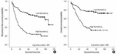
- Increased Expression of Thymosin β4 Is Independently Correlated with Hypoxia Inducible Factor-1α (HIF-1α) and Worse Clinical Outcome in Human Colorectal Cancer
- Seung Yun Lee, Mee Ja Park, Hye Kyung Lee, Hyun Jin Son, Chang Nam Kim, Joo Heon Kim, Dong Wook Kang
- J Pathol Transl Med. 2017;51(1):9-16. Published online October 16, 2016
- DOI: https://doi.org/10.4132/jptm.2016.08.23
- 13,996 View
- 166 Download
- 5 Web of Science
- 6 Crossref
-
Abstract
PDF
- Background
Thymosin β4 is a multi-functional hormone-like polypeptide, being involved in cell migration, angiogenesis, and tumor metastasis. This study was undertaken to clarify the clinicopathologic implications of thymosin β4 expression in human colorectal cancers (CRCs).
Methods
We investigated tissue sections from 143 patients with CRC by immunohistochemistry. In addition, we evaluated the expression patterns and the clinico-pathological significance of thymosin β4 expression in association with hypoxia inducible factor-1α (HIF-1α) expression in the CRC series.
Results
High expression of thymosin β4 was significantly correlated with lymphovascular invasion, invasion depth, regional lymph node metastasis, distant metastasis, and TNM stage. Patients with high expression of thymosin β4 showed poor recurrence-free survival (p = .001) and poor overall survival (p = .005) on multivariate analysis. We also found that thymosin β4 and HIF-1α were overexpressed and that thymosin β4 expression increased in parallel with HIF-1α expression in CRC.
Conclusions
A high expression level of thymosin β4 indicates poor clinical outcomes and may be a useful prognostic factor in CRC. Thymosin β4 is functionally related with HIF-1α and may be a potentially valuable biomarker and possible therapeutic target for CRC. -
Citations
Citations to this article as recorded by
- Predicting the risk of lymph node metastasis in colon cancer: development and validation of an online dynamic nomogram based on multiple preoperative data
Longlian Deng, Lemuge Che, Haibin Sun, Riletu En, Bowen Ha, Tao Liu, Tengqi Wang, Qiang Xu
BMC Gastroenterology.2025;[Epub] CrossRef - Thymosin β4 Is an Endogenous Iron Chelator and Molecular Switcher of Ferroptosis
Joanna I. Lachowicz, Giusi Pichiri, Marco Piludu, Sara Fais, Germano Orrù, Terenzio Congiu, Monica Piras, Gavino Faa, Daniela Fanni, Gabriele Dalla Torre, Xabier Lopez, Kousik Chandra, Kacper Szczepski, Lukasz Jaremko, Mitra Ghosh, Abdul-Hamid Emwas, Mass
International Journal of Molecular Sciences.2022; 23(1): 551. CrossRef - Metal coordination of thymosin β4: Chemistry and possible implications
Joanna Izabela Lachowicz, Mariusz Jaremko, Lukasz Jaremko, Giuseppina Pichiri, Pierpaolo Coni, Marco Piludu
Coordination Chemistry Reviews.2019; 396: 117. CrossRef - Adipose-Derived Mesenchymal Stem Cells Enhance Ovarian Cancer Growth and Metastasis by Increasing Thymosin Beta 4X-Linked Expression
Yijing Chu, Min You, Jingjing Zhang, Guoqiang Gao, Rendong Han, Wenqiang Luo, Tingting Liu, Jianxin Zuo, Fuling Wang
Stem Cells International.2019; 2019: 1. CrossRef - An Investigation on the Therapeutic Effect of Thymosinβ4 and Its Expression Levels in Streptozotocin-Induced Diabetic Mice
Kyung Sook Cho, Dong-Jin Kim, Bomee Shim, Jung Yeon Kim, Jun Mo Kang, Seon Hwa Park, Sang-Ho Lee, Hyung-In Yang, Kyoung Soo Kim
BioMed Research International.2018; 2018: 1. CrossRef - Hypoxia-inducible factor-1α expression in colorectal carcinoma
Ahmed M. Abd ElAziz, Hanan S. Abd ElHamid, Rasha R. Mostafa, Yousra R.A. Shalaby
Egyptian Journal of Pathology.2018; 38(1): 18. CrossRef
- Predicting the risk of lymph node metastasis in colon cancer: development and validation of an online dynamic nomogram based on multiple preoperative data
- Difference of the Nuclear Green Light Intensity between Papillary Carcinoma Cells Showing Clear Nuclei and Non-neoplastic Follicular Epithelia in Papillary Thyroid Carcinoma
- Hyekyung Lee, Tae Hwa Baek, Meeja Park, Seung Yun Lee, Hyun Jin Son, Dong Wook Kang, Joo Heon Kim, Soo Young Kim
- J Pathol Transl Med. 2016;50(5):355-360. Published online August 22, 2016
- DOI: https://doi.org/10.4132/jptm.2016.05.19
- 9,162 View
- 90 Download
-
Abstract
PDF
- Background
There is subjective disagreement regarding nuclear clearing in papillary thyroid carcinoma. In this study, using digital instruments, we were able to quantify many ambiguous pathologic features and use numeric data to express our findings.
Methods
We examined 30 papillary thyroid carcinomas. For each case, we selected representative cancer cells showing clear nuclei and surrounding non-neoplastic follicular epithelial cells and evaluated objective values of green light intensity (GLI) for quantitative analysis of nuclear clearing in papillary thyroid carcinoma.
Results
From 16,274 GLI values from 600 cancer cell nuclei and 13,752 GLI values from 596 non-neoplastic follicular epithelial nuclei, we found a high correlation of 94.9% between GLI and clear nuclei. GLI between the cancer group showing clear nuclei and non-neoplastic follicular epithelia was statistically significant. The overall average level of GLI in the cancer group was over two times higher than the non-neoplastic group despite a wide range of GLI. On a polygonal line graph, there was a fluctuating unique difference between both the cancer and non-neoplastic groups in each patient, which was comparable to the microscopic findings.
Conclusions
Nuclear GLI could be a useful factor for discriminating between carcinoma cells showing clear nuclei and non-neoplastic follicular epithelia in papillary thyroid carcinoma.
Case Study
- A Case of Giant Colonic Muco-submucosal Elongated Polyps Associated with Intussusception
- Joo Heon Kim, Seung Yun Lee, Je Ho Jang, Hyun Young Han, Dong Wook Kang
- J Pathol Transl Med. 2016;50(6):474-478. Published online May 23, 2016
- DOI: https://doi.org/10.4132/jptm.2016.04.27
- 12,848 View
- 135 Download
- 6 Web of Science
- 8 Crossref
-
Abstract
PDF
- Colonic muco-submucosal elongated polyp (CMSEP), a newly categorized non-neoplastic colorectal polyp, is a pedunculated and elongated polyp composed of normal mucosal and submucosal layers without any proper muscle layer. We herein report a giant variant of CMSEP associated with intussusception in the rectosigmoid colon, with a review of the literature. A 48-year-old woman underwent a laparoscopic low anterior resection due to multiple large submucosal polypoid masses associated with intussusception. Grossly, the colonic masses were multiple pedunculated polyps with a long stalk and branches ranging in size from a few millimeters to 14.0 cm in length. Microscopically, there was no evidence of hyperplasia, atypia, or active inflammation in the mucosa. The submucosal layers were composed of edematous and fibrotic stroma with fat tissue, dilated vessels, and lymphoid follicles.
-
Citations
Citations to this article as recorded by- Unusually rapid growth of a duodenal muco-submucosal elongated polyp: A case report
Yi Yang, Ding-Fu Zhong
World Journal of Gastrointestinal Surgery.2025;[Epub] CrossRef - Multiple enteric muco-submucosal elongated polyps causing intussusception
Atsuki Taniguchi, Izuru Endo, Takeyoshi Nishiyama, Nobuyuki Watanabe, Osamu Yoshida, Hiroaki Asano, Masatoshi Kubo, Tetsunobu Udaka
Clinical Journal of Gastroenterology.2024; 17(1): 41. CrossRef - Intussusception due to a Muco-submucosal Elongated Polyp in the Small Intestine—A Case Report—
Hiroki ISHIGE, Ken IMAIZUMI, Takumu FUKASAWA, Keiichiro ITO, Hiroyuki KASAJIMA, Satoru MUNAKATA, Norihiko SHIMOYAMA, Kazuaki NAKANISHI
Nihon Rinsho Geka Gakkai Zasshi (Journal of Japan Surgical Association).2024; 85(6): 744. CrossRef - Jejunal Intussusception Caused by Enteric Muco-submucosal Elongated Polyp: A Case Report
Young Min Jo
Soonchunhyang Medical Science.2024; 30(2): 60. CrossRef - Jejunal intussusception and perforation due to enteric muco-submucosal elongated polyp: a case report and literature review
Ryosuke Kikuchi, Shigenobu Emoto, Hiroaki Nozawa, Kazuhito Sasaki, Koji Murono, Shinya Abe, Hirofumi Sonoda, Aya Shinozaki-Ushiku, Soichiro Ishihara
Surgical Case Reports.2023;[Epub] CrossRef - A stalk with no polyp—A muco‐submucosal elongated polyp in the duodenum
Neil O’Morain, Ciaran McCloskey, Sinead Flanagan, Glen Doherty
United European Gastroenterology Journal.2023; 11(4): 392. CrossRef - Duodenal Worm-Like Polyp
Pan Pan, Guoshan Zhang, Xiao Cui, Liang Liu
Digestive Diseases and Sciences.2023; 68(12): 4275. CrossRef - Colonic Mucosubmucosal Elongated Polyp in the Sigmoid Colon on Surveillance Colonoscopy
Xiaowen Fan, Melissa Hershman, Gabriel Levi, Ilan Weisberg
ACG Case Reports Journal.2019; 6(6): e00110. CrossRef
- Unusually rapid growth of a duodenal muco-submucosal elongated polyp: A case report
Original Article
- The Stromal Overexpression of Decay Accelerating Factor (DAF/CD55) Correlates with Poor Clinical Outcome in Colorectal Cancer Patients.
- Tae Hwa Baek, Joo Heon Kim, Mee Ja Park, Hye Kyung Lee, Hyun Jin Son, Hyun Ki Soon, Chang Nam Kim, Che Myong Ko, Dong Wook Kang
- Korean J Pathol. 2011;45(5):445-454.
- DOI: https://doi.org/10.4132/KoreanJPathol.2011.45.5.445
- 4,945 View
- 40 Download
- 1 Crossref
-
Abstract
PDF
- BACKGROUND
Decay accelerating factor (DAF/CD55), regulates the complement system by accelerating decay of the C3 convertase, has been described in several malignancies, however, the clinicopathologic significance of CD55 and its receptor CD97 has not been fully investigated. We examined the expression patterns of both CD55 and CD97 and their association with clinicopathologic parameters in colorectal cancers (CRCs).
METHODS
Expression patterns of CD55 and CD97 in the stroma and tumor cells at tumor center and invasive front were examined in 130 CRCs, and their significance was statistically evaluated.
RESULTS
CD55-high stroma was correlated with tumor border (p=0.006) and invasion depth (p=0.013). CD55-high tumor cells at tumor center and invasive front were correlated with histologic grade, and CD55-high tumor cells at invasive front with tumor, node and metastasis (TNM) stage (p<0.05). CD97-high stroma was correlated with lymph node metastasis (p=0.016) and TNM stage (p=0.030). CD97-high tumor cells at tumor center and invasive front were correlated with tumor size and CD97-high tumor cells at tumor center with tumor border (p<0.05). Patients with CD55-high stroma showed poor overall and recurrence-free survival (p<0.05) in univariate analysis, and were independently associated with short recurrence-free survival (p=0.025) in multivariate analysis.
CONCLUSIONS
Stromal CD55 overexpression would be an indicator of adverse clinical outcome and a useful prognostic factor. -
Citations
Citations to this article as recorded by- Non-canonical extracellular complement pathways and the complosome paradigm in cancer: a scoping review
Camila de Freitas Oliveira-Tore, Amarilis Giaretta de Moraes, Helena Musetti B. S. Plácido, Nathalia M. D. L. Signorini, Pamela Dias Fontana, Tatiane da Piedade Batista Godoy, Angelica Beate Winter Boldt, Iara de Messias
Frontiers in Immunology.2025;[Epub] CrossRef
- Non-canonical extracellular complement pathways and the complosome paradigm in cancer: a scoping review
Case Report
- Langerhans Cell Sarcoma Arising in a Lymph Node: A Case Report and Review of the Literature.
- Dong Wook Kang, Hyun Jin Son, Tae Hwa Baek, Hye Kyung Lee, Joo Ryung Huh, Joo Heon Kim, Mee Ja Park
- Korean J Pathol. 2011;45(1):101-105.
- DOI: https://doi.org/10.4132/KoreanJPathol.2011.45.1.101
- 4,038 View
- 38 Download
- 1 Crossref
-
Abstract
PDF
- We report a case of Langerhans cell sarcoma presented as a solitary mass in the left supraclavicular area in a 31-year-old woman. Computed tomography revealed a relatively well-defined and lightly enhancing mass in the left supraclavicular area, measuring 5.5x4.5x3.2 cm. Excision was subsequently performed. Microscopically, the specimen consisted of an enlarged and partially effaced lymph node. Nests of different size composed of atypical tumor cells were located in the paracortex and the medulla of the lymph node. The tumor cells exhibited abundant eosinophilic or clear cytoplasm and displayed marked nuclear atypia and increased mitotic figures. Infiltration of many eosinophils was identified in the periphery and between the tumor cells. The tumor cells were reactive for CD1a and S100 protein. Ultrastructually, they were found to have Birbeck granules in the cytoplasm.
-
Citations
Citations to this article as recorded by- Langerhans cell sarcoma: a case report and review of the literature
David T. Liu, Joerg Friesenbichler, Lukas A. Holzer, Bernadette Liegl-Atzwanger, Christine Beham-Schmid, Andreas Leithner
Polish Journal of Pathology.2016; 67(2): 172. CrossRef
- Langerhans cell sarcoma: a case report and review of the literature
Original Article
- Pathologic Characteristics of Ovarian Hemorrhagic Polycyst in Estrogen Receptor-alpha (ERalpha) Knockout Mice and Roles of ERalpha in Hemorrhagic Polycyst.
- Hyun Jin Son, Joo Heon Kim, Hye Kyung Lee, Mee Ja Park, Dong Wook Kang, Che Myong Ko
- Korean J Pathol. 2010;44(4):376-383.
- DOI: https://doi.org/10.4132/KoreanJPathol.2010.44.4.376
- 4,201 View
- 53 Download
-
Abstract
PDF
- BACKGROUND
Polycystic ovary syndrome (PCOS) is the most common endocrinopathy causing anovulation in women of childbearing age. It has been well established that estrogen receptor-alpha knockout (ERalphaKO) mice display several pathologic ovarian phenotypes of PCOS. The aims of this study were to determine ovarian pathology in new ERalphaKO mice using a CreloxP approach and intra-ovarian ERalpha function as regulating key aspects of PCOS.
METHODS
ERalphaKO mice, which were deficient in exon 3 of the ERalpha gene, were used. Immunohistochemical studies were done on ovaries of control and ERalphaKO mice using antibodies specific to ERalpha, ERbeta, inhibin-alpha, and alpha-smooth muscle actin (SMA), as well as histochemical staining using Sudan black-B.
RESULTS
All ovaries of ERalphaKO mice were larger than control mouse ovaries and displayed a disrupted theca-interstitial tissue organization, multiple atretic follicles and multiple hemorrhagic cysts. None of the ERalphaKO mouse ovaries showed a corpus luteum. In addition, heavy deposition of Sudan black-B positive foamy cells was seen. The theca externa of preantral immature follicles and hemorrhagic cysts showed strong expression of alpha-SMA.
CONCLUSIONS
ERalphaKO mice show hemorrhagic polycystic ovaries and hyperplasia of the theca externa. This study demonstrates that the ERalpha is the functional key to the pathogenesis of PCOS.
Case Reports
- Primary Synovial Sarcoma of the Kidney: A Case Report and Literature Review.
- Mee Ja Park, Tae Hwa Baek, Joo Heon Kim, Dong Wook Kang, Hye Kyung Lee, Hyun Jin Son
- Korean J Pathol. 2009;43(3):274-278.
- DOI: https://doi.org/10.4132/KoreanJPathol.2009.43.3.274
- 5,008 View
- 28 Download
- 2 Crossref
-
Abstract
PDF
- Synovial sarcoma is a rare renal neoplasm that is not easy to diagnose unless SYT-SSX fusion transcripts are identified. We report here on a case of primary renal synovial sarcoma in a 35-year-old woman. A mass was discovered by accident in the lower part of the right kidney when ultrasonography was performed, and it was removed via radical nephrectomy. Grossly, the tumor was a homogeneously tan-brown soft mass that measured 4.5x3.2x3.0 cm, and it was encircled by a well-defined cystic space. The lesion exhibited hypercellularity of the oval or short spindle cells that were arranged in various solid sheets or intersecting fascicles. Immunohistochemically, the tumor showed diffuse positivity for vimentin, bcl-2 and CD99, and it showed focal positivity for epithelial membrane antigen. The SYT-SSX fusion transcripts were detected by reverse transcription-polymerase chain reaction (RT-PCR). Synovial sarcoma should be considered in the differential diagnosis when a spindle cell neoplasm is encountered in the kidney.
-
Citations
Citations to this article as recorded by- Primary Renal Synovial Sarcoma and Clinical and Pathological Findings: a Systematic Review
Leandro Blas, Javier Roberti
Current Urology Reports.2021;[Epub] CrossRef - Primary Renal Synovial Sarcoma - A rare histology
Premkumar Krishnappa, Mohan keshavamurthy, Shakir Tabrez, Sreeharsha Harinatha, Mohan Balaiah Aswathaiya
Urology Case Reports.2020; 33: 101402. CrossRef
- Primary Renal Synovial Sarcoma and Clinical and Pathological Findings: a Systematic Review
- Meningeal Melanocytoma Associated with Ota's Nevus: Report of a case.
- Woo Sung Moon, Joo Heon Kim, Dong Geun Lee, Ho Yeul Choi, Sang Ho Kim
- Korean J Pathol. 1992;26(6):605-609.
- 2,489 View
- 19 Download
-
Abstract
PDF
- Primary meningeal melanocytoma of the central nervous system is extremely rare. We report a case of meningeal melanocytoma associated with Ota's nevus as a recurrent form in a 53-year old male. The meningeal melanocytoma was removed from right parietooccipital lobe 4 years ago and recurred in right parietal, occipital and left frontal lobes. Ultrastructurally, the tumor cells were characterized by the presence of numerous melanosomes and premelanosomes in their cytoplasm. Moreover, the tumor was lacking in histologic and ultrastructural features of pigmented meningioma, melanotic schwannoma and prolonged clinical course was different from primary meningeal melanoma or metastatic malignant melanoma.
Original Articles
- The Expression of Transforming Growth Factor-1 and Its Signaling Receptors in Human Colorectal Carcinoma.
- Gyeong Seon Kim, Joo Heon Kim, Woo Sung Moon, Myoung Ja Chung, Dong Geun Lee, Myoung Jae Kang
- Korean J Pathol. 2001;35(2):115-122.
- 2,231 View
- 14 Download
-
Abstract
PDF
- BACKGROUND
Resistance to the potent growth inhibitory effects of TGF- (transforming growth factor-) is a characteristic of many malignancies. TGF- insensitivity has been attributed to alterations in the number and function of the TGF- receptors as well as disturbances of downstream signal transduction. The aim of this study was to examine the expression of TGF-1 and its receptors in human colorectal cancer tissue and determine its relationship with cancer growth and with prognostic factors.
METHODS
Immunohistochemical staining of TGF-1, TGF-RI, and TGF-RII was performed on 20 human colorectal adenomas, 30 carcinomas and 10 normal mucosas as a control.
RESULTS
The staining indices of TGF-1, TGF-RI, and TGF-RII increased in adenomas and carcinomas compared with normal mucosas and adenomas, respectively. In adenomas the staining index of TGF-1 significantly increased with the severity of atypism. The staining index of TGF-RII increased in the carcinomas in the right colon and rectum, compared with those in the left colon.
CONCLUSION
The enhanced expression of TGF-1, TGF-RI and II in the colorectal carcinoma suggests an important role of colorectal carcinogenesis and tumor progression.
- Expression of Cyclin-Dependent Kinase-Associated Protein Phosphatase in Colorectal Carcinomas.
- Chang Nam Kim, Soo Young Kim, Jae Wha Kim, Dong Wook Kang, Hyun Jin Son, Hye Kyung Lee, Mee Ja Park, Joo Heon Kim
- Korean J Pathol. 2007;41(6):367-372.
- 2,413 View
- 24 Download
-
Abstract
PDF
- BACKGROUND
Cyclin-dependent kinase-associated phosphatase (KAP) is a human dual-specificity protein phosphatase that dephosphorylates Cdk2 on threonine160 in a cyclin-dependent manner and that is known as an up-regulated molecule in some malignant tumors. We investigated the expression and clinicopathologic significance of KAP protein in relation to tumorigenesis of colorectal carcinoma.
METHODS
The expression patterns of KAP protein in tumor tissue were examined by reverse transcription-PCR and immunohistochemical staining.
RESULTS
An enhanced transcriptional level of KAP mRNA was observed in 11 out of 12 colorectal carcinoma specimens. Immunohistochemical examination showed that KAP protein was more highly expressed in the tumors than that in the adjacent non-neoplastic mucosal tissues for 52 of 102 colorectal cancer tissues. The statistical analysis showed that an increased level of KAP protein in the colorectal cancer tissues was inversely correlated with the histologic grade, tumor size and Duke's stage.
CONCLUSION
The present study suggests that alteration of KAP might play a role, at least in part, in the tumorigenicity of colorectal carcinoma through the mechanism of cell cycle regulation.
- Fine Needle Aspiration Cytology of Inflammatory Myofibroblastic Tumor in Mesentery: A Case Report.
- Hyun Jin Son, Joo Heon Kim, Woo Sung Moon, Myoung Jae Kang, Ho Yeul Choi
- J Pathol Transl Med. 2000;11(1):35-40.
- 2,352 View
- 19 Download
-
Abstract
PDF
- Since inflammatory myofibroblastic tumor was initially recognized in the lung, this tumor has been described in other extrapulmonary sites. In spite of relatively uniform histologic findings in various organs, a rarity in extrapulmonary sites and highly vascular characteristics frequently lead to a misdiagnosis in preoperative radiology and fine needle aspiration cytology. We present a case of inflammatory myofibro blastic tumor occurring in the mesentery of a 4-month-old girl. Fine needle aspira tion cytology smear disclosed characteristic spindle cells intermixed with prominent mature plasma cells and lymphocytes. According to the immunohistochemical staining, we recognized that the intervening spindle cells are myofibroblasts which have reactivity for the both actin and vimentin.
- Polymerase Chain Reaction Detection of Mycobacterium tuberculosis and Fine Needle Aspiration Cytology for the Diagnosis of Tuberculous Lymphadenitis .
- Joo Heon Kim, Nam Hoon Kim, Dong Wook Kang, Mee Ja Park, Sang Kyoung Moon, Tae Cho Yu, Eun Ju Jang
- J Pathol Transl Med. 2001;12(1):25-30.
- 2,328 View
- 20 Download
-
Abstract
PDF
- Tuberculous lymphadenitis is not uncommon in Korea. Therefore, an inexpensive, safe and rapid method is needed to diagnose the tuberculous lymphadenitis. Fine needle aspiration cytology is a good method for this purpose, but has several limitations in the diagnosis of tuberculous lymphadenitis, especially when the presence of acid-fast bacilli is not proved. To evaluate the usefulness of the polymerase chain reaction with enzyme immunoassay technique in the detection of Mycobacterium tuberculosis (M. tuberculosis) in the cervical lymph node aspirates, the authors performed fine needle aspiration cytology and M. tuberculosis PCR with enzyme immunoassay for mycobacterial DNA sequences from 15 cases of the fine needle aspirates. Cytomorphologically, the cases were categorized into three types: predominantly necrotic materials; typical epithelioid cell granulomas with or without giant cells and caseous necrosis; and non-tuberculous lesions, such as reactive lymphadenitis, abscess, metastatic carcinoma and malignant lymphoma. M. tuberculosis DNA was found in 8 of 15 cases by PCR with enzyme immunoassay. Negative findings on PCR were achieved in 7 cases, which revealed non-tuberculous lymphadenopathy. In conclusion, we suggest that M. tuberculosis PCR with enzyme immunoassay using the fine needle aspirates is a very useful tool for the diagnosis of tuberculous lymphadenitis.
Case Report
- Embryonal Rhabdomyosarcoma of Urinary Bladder Diagnosed by Urine Cytology: A Case Report.
- Joo Heon Kim, Ho Lee, Myoung Jae Kang, Dong Geun Lee, Sang Ho Kim
- J Pathol Transl Med. 1994;5(1):71-73.
- 2,168 View
- 29 Download
-
Abstract
PDF
- Rhabdomyosarcomas are found mainly in young patients, but rare in adults. A correlated cytological and histologic study of one case of embryonal rhabdomyosarcoma is presented. The cytologic appearance of the urine smear corresponded well with the histologic findings. Cytologically, two main cell types were distinguished; a predominant primitive, small round cell with scant cytoplasm and a large cell with abundant cytoplasm. The cytologic feature proving rhabdomyoblastic differenti-ation, such as cross-striation, was absent.
Original Article
- Expression of Epidermal Growth Factor Related Peptides, EGF-R, and c-erbB-2 and Their Relationship with the Prognostic Factors in Gastric Carcinoma.
- Joo Heon Kim, Jin Wook Lee, Woo Sung Moon, Myoung Jae Kang, Dong Geun Lee
- Korean J Pathol. 1999;33(11):1039-1046.
- 2,186 View
- 11 Download
-
Abstract
PDF
- Recent investigations have revealed that autocrine growth factors and their receptors are closely related and play an important role in controlling cancer cell growth. We performed an immunohistochemical study on the expression of epidermal growth factor (EGF), transforming growth factor-alpha (TGF-alpha), epidermal growth factor receptor (EGF-R), c-erbB-2, and PCNA labelling index in 60 cases of human gastric carcinomas. TGF-alpha was detected in 38 cases (63.3%), EGF in 26 cases (43.3%), EGF-R in 44 cases (73.3%), and c-erbB-2 in 18 cases (30%). These growth factors, EGF-R and c-erbB-2, were found more often in advanced gastric cancers. The PCNA labeling index was significantly higher in tumors with the expression of EGF-R or c-erbB-2. Tumors with simultaneous expression of EGF, TGF-alpha, EGF-R and c-erbB-2 was associated with a high PCNA labeling index. A correlation was observed between the synchronous expression of growth factors and its receptors and histological differentiation. The results suggest that the expression of EGF, TGF-alpha, EGF-R and c-erbB-2 are closely related and plays an important role in the growth and progression of human gastric carcinoma.
Case Report
- Menetrier's Disease Report of two cases.
- Joo Heon Kim, Dong Geun Lee, Sang Woo Juhng
- Korean J Pathol. 1998;32(2):142-146.
- 2,271 View
- 14 Download
-
Abstract
PDF
- Menetrier's disease is characterized by enlarged gastric folds with foveolar hyperplasia and cystic dilatation of gastric glands. The additional biochemical features of hypoproteinemia, hypochlorhydria, and increased gastric mucus are often encountered. The pathogenesis and etiologic factors have not been clearly defined. In this report, we present two cases of Menetrier's disease in the stomach, one occurring in a 38-year-old male, associated with massive hematemesis, and the other in a 39-year-old male. Grossly, both cases showed marked giant gastric rugal folds resembling cerebral convolutions, sparing the antral portion. Microscopically, the giant gastric rugal folds consisted of the striking foveolar hyperplasia accompanied by an occasional presence of the smooth muscle fibers from the muscularis mucosa. The immunohistochemical stain revealed an intense positive reaction for transforming growth factor-alpha (TGF-alpha) and epidermal growth factor receptor (EGF-R) in the majority of mucous cells throughout the gastric mucosa and parietal cells, but did not reveal for epidermal growth factor (EGF). We suggested that TGF-alpha and EGF-R might be involved in the pathogenesis of Menetrier's disese.
Original Article
- Detection of HBV DNA in Needle Biopsied Paraffin Embedded Liver Tissues of Chronic Hepatitis B Patients by PCR: Comparison with Serological and Immunohistochemical Studies.
- Hye Soo Lee, Kahng Yeul Oh, Joo Heon Kim, Yoon Jeong Kim, Sam Im Choi, Dong Geun Lee, Sang Ho Kim
- Korean J Pathol. 1996;30(6):495-504.
- 2,303 View
- 11 Download
-
Abstract
PDF
- In this study, the prevalence of Hepatitis B virus(HBV) DNA in the needle biopsied paraffin embedded liver tissues of chronic hepatitis B patients by rapid nested PCR was examined. DNA was extracted by NaOH with boiling, and amplified by rapid air thermocycler with glass capillary tubes and nested PCR with two primer sets specific for the surface and the core genes of HBV. The PCR results were compared to that of serum HBeAg, serum HBV DNA by dot blot hybridization with a radioactive DNA probe, and tissue immunohistochemical (HBsAg/ HBcAg) studies. Among 44 patients with chronic hepatitis with serum HBsAg positivity, HBV DNA could be detected by PCR in 43 liver tissues (98%). This results were comparable to the positive rates of 94%(31/33) for serum HBV DNA, 80%(35/44) for serum HBeAg, and 59%(26/44) and 75%(33/44) for tissue HBsAg and HBcAg, respectively. The accordance rate between tissue PCR and serum DNA probe testing was 91%. The results indicate that HBV DNA detection by rapid nested PCR of paraffin embedded liver tissues by needle biopsy is a more sensitive method to detect the HBV DNA carrier than the serum HBeAg or tissue HBsAg/HBcAg status, and is well correlated with the result of serum HBV DNA probe testing. Therefore this method is a practical indicator for the diagnosis and replication status in retrospective analysis.
Case Reports
- Gastrointestinal Stromal Tumors associated with Neurofibromatosis Type I: A Report of Two Cases.
- Joo Heon Kim, Ock Seong In, Seong Kyu Lee, Haing Woon Baik, Seong Ho Kim, Dong Wook Kang, Kyung Hee Kim, Mee Ja Park, Yong Il Kim
- Korean J Pathol. 2006;40(2):137-141.
- 2,434 View
- 23 Download
-
Abstract
PDF
- Gastrointestinal stromal tumor (GIST) is the most common non-epithelial neoplasm arising in the gastrointestinal tract, but this tumor is rarely seen in association with type 1 neurofibromatosis (NF-1). We report here on two cases of multiple GISTs of the small intestine that occurred in NF-1 patients. We also analyzed the mutations of c-kit exons 9, 11, 13 and 17 and the plateletderived growth factor receptor-alpha (PDGFRA) exons 12 and 18 in two GIST patients. Histologically, the NF-1-associated GISTs were similar to those of non-the NF-1 GISTs, but they characteristically revealed hyperplastic interstitial cells of Cajal around the GISTs. Immunohistochemically, these tumors showed strong co-expressions of CD117 and CD34. The molecular genetic analysis of the GISTs showed that all of the c-kit and PDGFRA exons that were analyzed in the GISTs of the two patients were the wild-type, suggesting a limited role for the c-kit and PDGFRA mutations in the tumorigenesis of NF-1-associated GISTs.
- Multicystic Renal Dysplasia with Ipsilateral Ectopic Ureteral Orifice and Seminal Vesicle Cyst: A case report.
- Hyun Jin Son, Joo Heon Kim, Myoung Jae Kang
- Korean J Pathol. 2000;34(4):310-313.
- 2,444 View
- 12 Download
-
Abstract
PDF
- Renal dysplasia results from aberrant metanephric histogenesis caused fundamentally by a defect in inducer tissue or responding tissue. Dysplastic kidneys vary tremendously in gross and microscopic appearance but are characterized by abnormal organization and a mixed population of primitive structures, such as fetal or immature cartilage, dysplastic ducts, immature tubules, and undifferentiated mesenchyme. We report a case of unilateral multicystic renal dysplasia associated with an ipsilateral ectopic ureteral orifice entering a seminal vesicle cyst in a 33-year-old man. He was admitted due to primary infertility which had developed three years ago. The his semen analysis revealed oligospermia. No evidence of a family history of renal dysplasia was reported. Microscopic examination showed that the entire kidney was composed of cysts lined by flattened cells, dysplastic ducts and immature tubules surrounded by collars of spindle cells, primitive mesenchyme, and a few aberrantly formed glomeruli.
- Hyalinizing Trabecular Carcinoma of the Thyroid Gland: A report of two cases.
- Kyu Yun Jang, Joo Heon Kim, Myoung Ja Chung, Woo Sung Moon, Myoung Jae Kang
- Korean J Pathol. 2000;34(4):318-322.
- 2,226 View
- 16 Download
-
Abstract
PDF
- We report two cases of hyalinizing trabecular carcinoma (HTC) of the thyroid gland. These two patients were euthyroid women aged 36 and 65 years of age. The tumors were encapsulated and measured 0.8 and 4.0 cm in diameter, respectively. Histologically, the tumors were composed of a compact proliferation of cells in a lobular and trabecular pattern with an intervening hyalinized, fibrotic vascular stroma. Occasionally the cells were arrayed in microfollicles. Multiple and serial sections showed cords of tumor cells invading into the capsule in both cases and vascular invasion in one case. These findings suggested that HTCs are a malignant counterpart of hyalinizing trabecular adenoma, similar to conventional follicular tumor. Positive immunostaining of tumor cells for thyroglobulin and negative staining for high molecular weight cytokeratin, cytokeratin 19, neuron specific enolase, chromogranin, and synaptophysin allowed distinction from medullary carcinoma. Even though HTCs are an heterogeneous group of tumors, the present two cases are probably variants of follicular carcinoma rather than papillary carcinoma.
- Goblet Cell Carcinoid of the Appendix: A case report.
- Joo Heon Kim, Ho Lee, So Young Oh, Myoung Jae Kang, Ho Yeul Choi, Dong Geun Lee
- Korean J Pathol. 1996;30(9):839-842.
- 2,428 View
- 22 Download
-
Abstract
PDF
- Carcinoid tumors of the appendix are common incidental findings, but appendiceal tumors with histologic features of both carcinoids and adenocarcinomas are rare, and their biologic behavior and histogenesis are still unclear. We report a case of goblet cell carcinoid of the appendix in a 54-year-old male, who exhibited pain in the right lower abdomen. Microscopically, the tumor contained smooth-bordered, widely separated nests composed of tumor cells with abundant mucin. The principal tumor cell type had a close resemblance to the normal goblet cell. Histochemically, the tumor cells revealed positive reaction for PAS and alcian blue stain. Immunohistochemically, the tumor showed strong reactivity for carcinoembryonic antigen, chromogranin and, neuron specific enolase but none for cytokeratin and epithelial membrane antigen.
Original Article
- c-kitMutation and Immunohistochemical Expression in Gastrointestinal Stromal Tumors.
- Dong Wook Kang, Joo Heon Kim, Dong Hun Kim, Kung Hee Kim, Mee Ja Park, Dae Young Kang
- Korean J Pathol. 2003;37(4):246-254.
- 2,382 View
- 32 Download
-
Abstract
PDF
- BACKGROUND
Gastrointestinal stromal tumor (GIST) is the most common non-epithelial neoplasm arising in the gastrointestinal tract. The aim of this study is to investigate the correlation among the clinicopathologic features, presence of c-kit mutation, and immunohistochemical expression of c-kit in 61 cases of GISTs.
METHODS
We divided the GISTs into three groups as benign, boderline and malignant, according to histologic grade. Exon 11 of the c-kit was amplified by PCR and sequenced. We performed immunohistochemical study for CD117, CD34, vimentin, SMA, desmin, and S-100 protein.
RESULTS
Twenty-one cases were diagnosed as benign GISTs, 14 cases as borderline GISTs, and 26 cases as malignant GISTs. The shape, atypia, cellularity, and necrosis showed good correlations with the histologic grades of the GISTs.Mutations of exon 11 of the c-kit were detected in 3 benign GISTs, 4 borderline GISTs, and 13(%) malignant GISTs. Sequence analysis confirmed the deletion mutation (n=16) and the singlebase pair mutation (n=4). The immunohistochemical stainings showed myogenic differentiation(n=20), neurogenic differentiation (n=15), and neither myogenic or neurogenic differentiation(n=34).
CONCLUSIONS
The GIST is the primitive mesenchymal tumor capable of divergent differentiation, and the mutation of the c-kit is a good parameter for the malignant GIST.
Case Reports
- Adenoid Basal Cell Tumor of the Prostate: A case report.
- Joo Heon Kim, Woo Sung Moon, Myoung Jae Kang, Dong Geun Lee, Jae Y Ro
- Korean J Pathol. 2000;34(7):534-536.
- 2,209 View
- 16 Download
-
Abstract
PDF
- Adenoid basal cell tumor of the prostate is a rare tumorous lesion that can be misdiagnosed as adenocarcinoma of the prostate. The malignant potential of adenoid basal cell tumor remains uncertain due to small number of reported cases. This 66-year-old man presented with symptoms of urinary tract obstruction. Under the impression of benign prostatic hyperplasia, a transurethral resection of the prostate (TURP) was performed. The patient was alive with no evidence of recurrence or metastasis 15 months after TURP. Microscopically, most of the lesions were composed of nodular collections of small nests of basaloid cells with peripheral palisading, and clusters of tumor cells forming cribriform pattern. Multiple areas of basal cell hyperplasia and atypical basal cell hyperpalsia were also observed. The coexistence of basal cell hyperplasia, atypical basal cell hyperpalsia, and adenoid basal cell tumor with cribriform pattern in this case supports a morphologic continuum from the benign hyperplastic lesion to malignant neoplasia.
- High-grade Transformation of Primary Nodal Marginal Zone B-Cell Lymphoma: A Case Report.
- Joo Heon Kim, Dong Wook Kang, Mee Ja Park, Jin Man Kim
- Korean J Pathol. 2003;37(4):282-286.
- 2,673 View
- 50 Download
-
Abstract
PDF
- Primary nodal marginal zone B-cell lymphoma (MZBCL) is recognized as a rare and distinct entity. The rate of histologic transformation into diffuse large B-cell lymphoma (DLBCL) seems lower than the rate of transformation in follicular lymphoma. We herein report a rare case ofnodal MZBCL showing transformation into DLBCL. The patient was a 73-year-old female withcervical lymphadenopathy. On the initial biopsy, the lymph node architecture was diffuselyeffaced with an extensive interfollicular and parafollicular infiltrate of monocytoid B-cells. Therewere scattered large blastic B-cells without formations of compact sheets. The diagnosis ofnodal MZBCL was made. The patient did not receive chemotherapy and was treated with aconservatively supportive regimen. Forty two months later, the patient developed a new cervicallymphadenopathy and a biopsy was performed. Histologically, the lymph node revealeddiffuse sheets of transformed large B-cells showing prominent nucleoli. The diagnosis ofDLBCL transformed from nodal MZBCL was made. The patient was treated with 3 cycles ofcombined CHOP chemotherapy and she showed clinical improvement. These observationssuggest that an untreated primary nodal MZBCL may undergo high-grade transformation.
Original Articles
- Squamous Cell Carcinoma Arising in Mature Cystic Teratoma of the Ovary: a Report of Six Cases and Immunohistochemistry of the p53 Protein and p21WAF1/CIP1.
- Kyung Hee Kim, Kwang Sun Suh, Joo Heon Kim, Dong Wook Kang, Dong Hoon Kim, Seong Ho Kim, Jong Ho Back, Mee Ja Park
- Korean J Pathol. 2003;37(5):316-319.
- 2,712 View
- 12 Download
-
Abstract
- BACKGROUND
Mature cystic teratoma is a common type of ovarian tumor. Although squamous cell carcinoma (SCC) is the most common carcinoma in malignant transformations of ovarian mature cystic teratomas, SCC arising in a mature teratoma is rare.
METHODS
This paper reports four cases of invasive SCC, a case of an adenosquamous cell carcinoma and a case of a pure in situ SCC arising in a mature cystic teratoma including a clinicopathological evaluation and an immunohistochemical study of the p53 protein and p21WAF1/CIP1.
RESULTS
The mean age of the patients was 60 years. The sizes of the mature cystic teratomas in all cases were greater than 7.5 cm in the largest diameter. Five cases showed the nuclear accumulation of the p53 protein with no p21WAF1/CIP1 immunoreactivity. The other case showed the nuclear accumulation of p21WAF1/CIP1 without p53 expression. There was a significant inverse relationship between the p53 protein level and p21WAF1/CIP1 expression.
CONCLUSION
A clinicopathological evaluation showed that a SCC arising from a mature cystic teratoma must be included in a differential diagnosis when the patient is over 42 years of age and the size of a mature cystic teratoma is greater than 75 mm in the largest diameter. It is suggested that p53 overexpression is implicated in the malignant transformation, and the p21WAF1/CIP1 expression level is dependent on alterations in the level of the p53 protein in these tumors.
- Immunohistochemical Study on the Proliferative Activity of Human Thyroid Tumors.
- Myoung Jae Kang, Young Jin Jeong, Woo Sung Moon, Myoung Ja Jeong, Joo Heon Kim, Dong Geun Lee, Ho Yeul Choi, Sang Ho Kim
- Korean J Pathol. 1995;29(1):77-84.
- 2,419 View
- 15 Download
-
Abstract
PDF
- For the estimation of the proliferative activity, related to the biologic behaviour, malignant potential, and prognosis, of human thyroid tumors, PCNA(proliferating cell nuclear antigen) immunohistochemical staining was performed on paraffin-embedded sections of 9 normal thyroid tissues, 9 adenomatous goiters, 9 follicular adenomas, 4 Hurthle cell tumors, 12 papillary carcinomas, 4 follicular carcinomas, and 3 anaplastic carcinomas. The results were as follows: 1) The PCNA labeling indices in adenomatous goiter, follicular adenoma, and Hurthle cell tumor were 1.1, 1.5, and 2.4, respectively. They were significantly higher than the labeling index in normal thyroid. 2) The PCNA labeling indices in papillary carcinoma and follicular carcinoma were 3.5 and 4.4, respectively. They were significantly higher than the labeling indices in adenomatous goiter and follicular adenoma, but there was no significant difference between papillary and follicular carcinoma. 3) The PCNA labeling index in anaplastic carcinoma, 14.1, was significantly higher than those in benign and other malignant tumors. According to the results, the PCNA labeling index was well correlated with the malignant potential of a tumor. So the PCNA immunohistochemical staining is thought to be a useful method for the evaluation of the malignant potential and prognosis of a tumor.
Case Report
- Pleomorphic Xanthoastrocytoma in a 58-year-old Woman: A case report.
- Joo Heon Kim, Myoung Jae Kang, Dong Geun Lee, Ho Yeul Choi, Sang Ho Kim, Min Cheol Lee
- Korean J Pathol. 1995;29(1):122-125.
- 2,013 View
- 10 Download
-
Abstract
- A case of left parietal pleomorphic xanthoastrocytoma that occurred in a 58-year-old woman is reported clinicopathologically. Histopathologic diagnosis of pleomorphic xanthoastrocytoma was made because of the unique pleomorphic histologic features, positive glial fibrillary acidic protein in immunohistochemical staining. The flow cytometric analysis reveals DNA aneuploidy and relatively high S-phase fraction. The pleomorphic xanthoastrocytoma is considered as a special subgroup of glioma on the basis of superficial cortical location, GFAP expression, marked cellular atypia, xanthomatous cells, and relatively favorable prognosis.
Original Article
- Expression Patterns of S100A6 Gene in Human Thyroid Diseases.
- Joo Heon Kim, Jae Wha Kim, Seon Young Yoon, Jong Hyuck Joo, In Seong Choi, Mee Ja Park
- Korean J Pathol. 2000;34(11):934-940.
- 2,243 View
- 18 Download
-
Abstract
PDF
- S100A6 (calcyclin) is a member of the S100 family and has been originally isolated from the cDNA library of Syrian baby hamster kidney cells. The S100A6 gene expression is reported to remain high throughout the cell cycle following induction by serum or growth factors, suggesting that the gene may be required for cell cycle progression. Nevertheless, the role that S100A6 may play in tumor progression remains unknown. In this study, we have explored the expression patterns of S100A6 gene in human thyroid tissues by northern blot analysis. Using the S100A6 monoclonal antibody, we carried out the immunohistochemical staining to determine the distribution/localization of S100A6 protein within tumor or non-tumorous cells of the thyroid. To modulate the regulation of endogenously expressed S100A6 protein in the intracellular level, overexpressed or anti-sense treated transfectant was constructed by using the eukaryotic expression vector. As a result, immunohistochemistry for S100A6 showed a strong positivity in the malignant tumors of thyroid and a high expression level of S100A6 protein affected cell proliferation in the overexpressed transfectant. These findings suggest that S100A6 may be involved in the tumor pathogenesis and provides another parameter for the differentiation of malignant and benign lesions. A well defined monoclonal antibody against S100A6 protein is now available for the immunohistochemical studies of the various thyroid tissues.
 First
First Prev
Prev


