 E-submission
Search
- Page Path
- HOME > Search
- Renal intravascular large B cell lymphoma: the first case report in Korea and a review of the literature
- Moonsik Kim, Haerim Chung, Woo Ick Yang, Hyeon Joo Jeong
- J Pathol Transl Med. 2020;54(5):426-431. Published online August 13, 2020
- DOI: https://doi.org/10.4132/jptm.2020.06.18
- 7,268 View
- 122 Download
- 7 Web of Science
- 7 Crossref
-
 Abstract
 PDF
PDF Supplementary Material
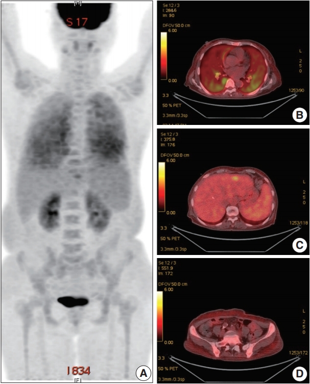
 - Herein, we describe the first case of renal intravascular large B cell lymphoma in Korea occurring in a 66-year-old female. She presented with mild fever and dyspnea. On physical and laboratory evaluations, hemophagocytic lymphohistiocytosis was suspected, but the bone marrow biopsy results were unremarkable. During the work-up, massive proteinuria developed, which led to a renal biopsy. The renal architecture was relatively well-preserved, but the glomeruli were hypercellular with the infiltration of atypical, large lymphoid cells with increased nucleus-cytoplasm ratio and clumped chromatin. Similar cells were also present in the peritubular capillaries. The tumor cells exhibited membranous staining for CD20 and CD79a. After the diagnosis of intravascular large B cell lymphoma, the patient received rituximab-based chemotherapy under close follow-up.
-
Citations
Citations to this article as recorded by
- Pauci-Immune Endocapillary Proliferative Glomerulonephritis With Glomerular M2 Macrophage Infiltration
Lei Ma, Meizi Kang, Ziyang Qiao, Shaojun Liu, Guolan Xing, Ruimin Hu, Yafen Yu, Rong Tan, Ruoyu Jia, Zhengyun Zhu, Fan Yang, Lijuan Li, Dan Zhou, Shaoshan Liang, Feng Xu, Yujie Wang, Xiaodong Zhu, Xinchen Yao, Jing Tian, Yongzhong Zhong, Caihong Zeng
Kidney International Reports.2026; 11(4): 103791. CrossRef - Intravascular Lymphoma Associated with the Female Genital Tract—Diagnostic Considerations, Therapeutic Approaches, and Outcomes
Aleksandar Ristic, Marija Rovcanin, Ana Tomic, Aleksandar Rakic, Nebojsa Zecevic, Svetlana Jankovic
Diseases.2026; 14(3): 109. CrossRef - Intravascular large B-cell lymphoma of the central nervous system with renal involvement: a case report and literature review
Jun Li, Zhaojiao Li, Yifeng Shi, Jiajie Chen, Heng Zhao, Xueye Mao, Shan Li, Huiying Wang, Qiang Meng, Lingchun Liu
Frontiers in Oncology.2025;[Epub] CrossRef - EBV-Positive Intravascular Large B-Cell Lymphoma of the Small Intestine: A Case Report and Literature Review
Chenglong Pan, Xiaoling Ma, Yanfei Yao, Chunyan Wang
International Journal of Surgical Pathology.2024; 32(3): 586. CrossRef - Intravascular large B‐cell lymphoma in renal cell carcinoma incidentally detected by robot‐assisted partial nephrectomy
Michio Noda, Yutaka Enomoto, Yukari Shirasugi, Sumiyo Ando, Yukimasa Matsuzawa, Haruki Kume
IJU Case Reports.2022; 5(3): 191. CrossRef - Case Report: Intravascular Large B-Cell Lymphoma: A Clinicopathologic Study of Four Cases With Review of Additional 331 Cases in the Literature
Yingying Han, Qingjiao Li, Dan Wang, Lushan Peng, Tao Huang, Chunlin Ou, Keda Yang, Junpu Wang
Frontiers in Oncology.2022;[Epub] CrossRef - Renal Involvement of CD20-Negative Intravascular Large B Cell Lymphoma with Neurological Manifestations
Faten Aqeel, Serena M. Bagnasco, Duvuru Geetha, Yoshihide Fujigaki
Case Reports in Nephrology.2022; 2022: 1. CrossRef
- Pauci-Immune Endocapillary Proliferative Glomerulonephritis With Glomerular M2 Macrophage Infiltration
- Aberrant Blood Vessel Formation Connecting the Glomerular Capillary Tuft and the Interstitium Is a Characteristic Feature of Focal Segmental Glomerulosclerosis-like IgA Nephropathy
- Beom Jin Lim, Min Ju Kim, Soon Won Hong, Hyeon Joo Jeong
- J Pathol Transl Med. 2016;50(3):211-216. Published online April 11, 2016
- DOI: https://doi.org/10.4132/jptm.2016.02.01
- 11,068 View
- 75 Download
- 2 Web of Science
- 2 Crossref
-
Abstract
PDF
- Background
Segmental glomerulosclerosis without significant mesangial or endocapillary proliferation is rarely seen in IgA nephropathy (IgAN), which simulates idiopathic focal segmental glomerulosclerosis (FSGS). We recently recognized aberrant blood vessels running through the adhesion sites of sclerosed tufts and Bowman’s capsule in IgAN cases with mild glomerular histologic change.
Methods
To characterize aberrant blood vessels in relation to segmental sclerosis, we retrospectively reviewed the clinical and histologic features of 51 cases of FSGS-like IgAN and compared them with 51 age and gender-matched idiopathic FSGS cases.
Results
In FSGS-like IgAN, aberrant blood vessel formation was observed in 15.7% of cases, 1.0% of the total glomeruli, and 7.3% of the segmentally sclerosed glomeruli, significantly more frequently than in the idiopathic FSGS cases (p = .009). Aberrant blood vessels occasionally accompanied mild cellular proliferation surrounding penetrating neovessels. Clinically, all FSGS-like IgAN cases had hematuria; however, nephrotic range proteinuria was significantly less frequent than idiopathic FSGS.
Conclusions
Aberrant blood vessels in IgAN are related to glomerular capillary injury and may indicate abnormal repair processes in IgAN. -
Citations
Citations to this article as recorded by- Twin Glomeruli: a Newly Discovered Marker of Neonephrogenesis in the Ischemia–Reperfusion Injured Adult Mouse Kidney
Hanguk Hwang, Dongju Woo, You Ri Park, Min Jung Kong, Heedong Lee, Kwon Moo Park, Yong Seok Nam, Je-Yong Choi, Sungwook Nam, Eon Jung Nam, Sun-Hee Park, Hongtae Kim, Sang Yeon Lee, Soo Ho Lee, Jeong Ok Lim, Mae Ja Park
Tissue Engineering and Regenerative Medicine.2026; 23(2): 253. CrossRef - IgA nephropathy
Maria F. Soares, Ian S.D. Roberts
Current Opinion in Nephrology and Hypertension.2017; 26(3): 165. CrossRef
- Twin Glomeruli: a Newly Discovered Marker of Neonephrogenesis in the Ischemia–Reperfusion Injured Adult Mouse Kidney
- Overview of IgG4-Related Tubulointerstitial Nephritis and Its Mimickers
- Hyeon Joo Jeong, Su-Jin Shin, Beom Jin Lim
- J Pathol Transl Med. 2016;50(1):26-36. Published online December 14, 2015
- DOI: https://doi.org/10.4132/jptm.2015.11.09
- 16,112 View
- 237 Download
- 14 Web of Science
- 14 Crossref
-
Abstract
PDF
- Tubulointerstitial nephritis (TIN) is the most common form of renal involvement in IgG4-related disease. It is characterized by a dominant infiltrate of IgG4-positive plasma cells in the interstitium and storiform fibrosis. Demonstration of IgG4-positive plasma cells is essential for diagnosis, but the number of IgG4-positive cells and the ratio of IgG4-positive/IgG-positive plasma cells may vary from case to case and depending on the methods of tissue sampling even in the same case. IgG4-positive plasma cells can be seen in TIN associated with systemic lupus erythematosus, Sjögren syndrome, or anti-neutrophil cytoplasmic antibody–associated vasculitis, which further add diagnostic confusion and difficulties. To have a more clear view of IgG4-TIN and to delineate differential points from other TIN with IgG4-positive plasma cell infiltrates, clinical and histological features of IgG4-TIN and its mimickers were reviewed. In the rear part, cases suggesting overlap of IgG4-TIN and its mimickers and glomerulonephritis associated with IgG4-TIN were briefly described.
-
Citations
Citations to this article as recorded by- Recurrent IgG4 Disease-Related Tubulointerstitial Nephritis Treated With Rituximab As Maintenance Therapy
Alejandro Valdesuso, Jill Nehrbas, Joseph Dan Khoa Nguyen , Rakesh Malhotra, Tushar Chopra
Cureus.2026;[Epub] CrossRef - Isolated pulmonary and renal IgG4-related disease: a rare dual-organ presentation diagnosed via video-assisted thoracoscopic surgery
Natsumi Yazaki, Yuka Katsurada, Takehiro Okumura, Soichiro Ueda
BMJ Case Reports.2026; 19(6): e273528. CrossRef - Glycosylation in kidney diseases
Yingying Ling, Fei Cai, Tao Su, Yi Zhong, Ling Li, Bo Meng, Guisen Li, Meng Gong, Hao Yang, Xinfang Xie, Zhenyu Sun, Yang Zhao, Fang Liu, Yong Zhang
Precision Clinical Medicine.2025;[Epub] CrossRef - IgG4-related kidney disease: Clinicopathologic features, differential diagnosis, and mimics
Sarwat I. Gilani, Alessia Buglioni, Lynn D. Cornell
Seminars in Diagnostic Pathology.2024; 41(2): 88. CrossRef - Utilizing Immunoglobulin G4 Immunohistochemistry for Risk Stratification in Patients with Papillary Thyroid Carcinoma Associated with Hashimoto Thyroiditis
Faridul Haq, Gyeongsin Park, Sora Jeon, Mitsuyoshi Hirokawa, Chan Kwon Jung
Endocrinology and Metabolism.2024; 39(3): 468. CrossRef - IgG4-assoziierte Nierenerkrankungen
Christina Thompson, Frank O. Henes, Oliver M. Steinmetz, Simon Melderis
Die Nephrologie.2023; 18(4): 249. CrossRef - Concurrent anti-neutrophil cytoplasmic antibody-associated glomerulonephritis and IgG4-associated tubulointerstitial nephritis with C3 glomerulonephritis
Jianan Feng, Jinyu Yu, Xueyao Wang, Yue Wang, Yang Liu, Zhonggao Xu, Weixia Sun
Medicine.2020; 99(5): e18857. CrossRef - A case of eosinophilic granulomatosis with polyangiitis as a mimicker of IgG4-related disease
Ryuichiro Kanda, Satoshi Kubo, Kazuhisa Nakano, Akio Kawabe, Aya Nawata, Kentaro Hanami, Shingo Nakayamada, Yoshiya Tanaka
Modern Rheumatology Case Reports.2020; 4(2): 278. CrossRef - Renal tubular acidosis as the initial presentation of Sjögren’s syndrome
Karen Ho, Pouneh Dokouhaki, Mark McIsaac, Bhanu Prasad
BMJ Case Reports.2019; 12(8): e230402. CrossRef - Hypocomplementemic interstitial nephritis with long-term follow-up
Alyssa Penning, Claire Kassakian, Donald C Houghton, Nicole K Andeen
Journal of Clinical Nephrology.2019; 3(1): 042. CrossRef - Immunoglobulin G4-related kidney diseases: An updated review
Maurizio Salvadori, Aris Tsalouchos
World Journal of Nephrology.2018; 7(1): 29. CrossRef - Systemic lupus erythematosus in a patient with an organic lesion of the central nervous system: practicaldifferential diagnosis
E. V. Lebedeva, M. V. Novoseltsev, A. N. Lvov, I. V. Khamaganova
Klinicheskaya dermatologiya i venerologiya.2018; 17(6): 21. CrossRef - Concurrent IgG4-related tubulointerstitial nephritis and IgG4 myeloperoxidase-anti-neutrophil cytoplasmic antibody positive crescentic glomerulonephritis
Tao Su, Li Yang, Zhao Cui, Su-xia Wang, Ming-hui Zhao
Medicine.2017; 96(20): e6707. CrossRef - IgG4-Related Kidney Disease: Report of a Case Presenting as a Renal Mass
Daniele Bianchi, Luca Topazio, Gabriele Gaziev, Valerio Iacovelli, Pierluigi Bove, Alessandro Mauriello, Enrico Finazzi Agrò
Case Reports in Surgery.2017; 2017: 1. CrossRef
- Recurrent IgG4 Disease-Related Tubulointerstitial Nephritis Treated With Rituximab As Maintenance Therapy
- Renal Histologic Parameters Influencing Postoperative Renal Function in Renal Cell Carcinoma Patients
- Myoung Ju Koh, Beom Jin Lim, Kyu Hun Choi, Yon Hee Kim, Hyeon Joo Jeong
- Korean J Pathol. 2013;47(6):557-562. Published online December 24, 2013
- DOI: https://doi.org/10.4132/KoreanJPathol.2013.47.6.557
- 8,599 View
- 45 Download
- 5 Crossref
-
Abstract
PDF
Background Pre-existing non-neoplastic renal diseases or lesions may influence patient renal function after tumor removal. However, its description is often neglected or omitted in pathologic reports. To determine the incidence and clinical significance of non-neoplastic lesions, we retrospectively examined renal tissues obtained during 85 radical nephrectomies for renal cell carcinoma.
Methods One paraffin-embedded tissue block from each case containing a sufficient amount of non-tumorous renal parenchyma was cut and processed with hematoxylin and eosin and periodic acid-Schiff methods. Non-neoplastic lesions of each histological compartment were semi-quantitatively and quantitatively evaluated.
Results Among the various histologic lesions found, tubular atrophy, arterial intimal thickening, and glomerulosclerosis were the most common (94.1%, 91.8%, and 88.2%, respectively). Glomerulosclerosis correlated with estimated glomerular filtration rate at the time of surgery, as well as at 1- and 5-years post-surgery (p=.0071), but tubulointerstitial fibrosis or arterial fibrous intimal thickening did not.
Post-hoc analysis revealed that glomerulosclerosis of more than 20% predicted post-operative renal function. However, its significance disappeared when gender and age were considered.Conclusions In conclusion, non-neoplastic lesions, especially with regard to glomerulosclerosis percentage, should be described in pathology reports to provide additional information on renal function decline.
-
Citations
Citations to this article as recorded by- Diffusion kurtosis versus diffusion-weighted magnetic resonance imaging in differentiating clear cell renal cell carcinoma and renal angiomyolipoma with minimal fat: a comparative study
Yarong Lin, Wenrong Zhu, Qingqiang Zhu
Diagnostic and Interventional Radiology.2025;[Epub] CrossRef - Role of intravoxel incoherent motion diffusion-weighted MRI in differentiation of renal cell carcinoma subtypes
Amira R. Mahmoud, Nehad Fouda, Eman Mohamed Helmy, Ali Elsorougy
Egyptian Journal of Radiology and Nuclear Medicine.2024;[Epub] CrossRef - Chronic kidney damage pathology score for systematic assessment of the non-neoplastic kidney tissue and prediction of post-operative renal function outcomes
Yong Jia, Seyed M.M. Poor, Brenden Dufault, Vivian Lu, Jasmir G. Nayak, Deepak K. Pruthi, Ian W. Gibson
Human Pathology.2022; 124: 76. CrossRef - Value of intravoxel incoherent motion for differential diagnosis of renal tumors
Qingqiang Zhu, Wenrong Zhu, Jing Ye, Jingtao Wu, Wenxin Chen, Zhihua Hao
Acta Radiologica.2019; 60(3): 382. CrossRef - Conventional and Papillary Renal Cell Carcinomas and Focal Segmental Glomerulosclerosis in a Nephrectomy
Firas Al-Delfi, Guillermo A. Herrera
Pathology Case Reviews.2015; 20(6): 263. CrossRef
- Diffusion kurtosis versus diffusion-weighted magnetic resonance imaging in differentiating clear cell renal cell carcinoma and renal angiomyolipoma with minimal fat: a comparative study
- Cytologic Features of Giant Cell Ependymoma: A Case Report and Review of the Literature
- Myoung Ju Koh, Sun Och Yoon, Hyae Min Jeon, Hyeon Joo Jeong, Soon Won Hong, Se Hoon Kim
- Korean J Pathol. 2012;46(5):507-513. Published online October 25, 2012
- DOI: https://doi.org/10.4132/KoreanJPathol.2012.46.5.507
- 11,208 View
- 71 Download
- 5 Crossref
-
Abstract
PDF
Here, we present a case of anaplastic giant cell ependymoma (GCE) occurring in a 15-year-old woman. Squash smear slides for intraoperative frozen section diagnosis revealed oval to round cell clusters with a papillary structure in a fibrillary background. This was occasionally accompanied by the presence of bizarre pleomorphic giant cells with hyperchromatic nuclei and prominent intranuclear inclusions. These intranuclear inclusions were a key clue to diagnosis of ependymoma. Histologic analysis revealed features of a high-grade tumor with perivascular pseudorosettes and bizarre pleomorphic giant cells, which established the diagnosis of GCE. We performed a review of literatures about the cytologic features of GCE, including our case, thus proposing that intraoperative frozen diagnosis of GCE would be established by squash smear preparations featuring the mitosis and necrosis, as well as the high cellularity, and the presence of giant cells showing hyperchromatic nuclei with eosinophilic cytoplasm and intranuclear inclusions/pseudoinclusions.
-
Citations
Citations to this article as recorded by- A case of myxopapillary ependymoma with predominant giant cell morphology: A rare entity with comprehensive genomic profiling and review of literature
Bryan Morales‐Vargas, Hassan Saad, Daniel Refai, Matthew Schniederjan, Zied Abdullaev, Kenneth Aldape, Malak Abedalthagafi
Neuropathology.2025; 45(1): 13. CrossRef - Report of a case of giant cell ependymoma with unusual clinical and pathological presentation
Mónica B. Mezmezian, Victor Del Caño, Liliana G. Olvi
Neuropathology.2019; 39(4): 313. CrossRef - Giant Cell Ependymoma of Cervicomedullary Junction: A Case Report of a Long-Term Survivor and Literature Review
Martina Cappelletti, Andrea G. Ruggeri, Giorgia Iacopino, Roberto Delfini
World Neurosurgery.2018; 116: 121. CrossRef - Immunohistochemical features of giant cell ependymoma of the filum terminale with unusual clinical and radiological presentation
Fernando Candanedo-Gonzalez, Cindy Sharon Ortiz-Arce, Samuel Rosales-Perez, Ana Lilia Remirez-Castellanos, Candelaria Cordova-Uscanga, Armando Gamboa-Dominguez
Diagnostic Pathology.2017;[Epub] CrossRef - Giant Cell Ependymoma of Lateral Ventricle: Case Report, Literature Review, and Analysis of Prognostic Factors and Genetic Profile
Hirokazu Takami, Christopher S. Graffeo, Avital Perry, Aditya Raghunathan, Robert B. Jenkins, Caterina Giannini, Terry C. Burns
World Neurosurgery.2017; 108: 997.e9. CrossRef
- A case of myxopapillary ependymoma with predominant giant cell morphology: A rare entity with comprehensive genomic profiling and review of literature
- Urinary Decoy Cell Grading and Its Clinical Implications
- Myoung Ju Koh, Beom Jin Lim, Songmi Noh, Yon Hee Kim, Hyeon Joo Jeong
- Korean J Pathol. 2012;46(3):233-236. Published online June 22, 2012
- DOI: https://doi.org/10.4132/KoreanJPathol.2012.46.3.233
- 12,868 View
- 95 Download
- 7 Crossref
-
Abstract
PDF
Background Examination of urine for decoy cells (DCs) is a useful screening test for polyomavirus (PV) activation. We explored the significance of the amount of DCs in persistent shedding, PV nephropathy and acute rejection.
Methods A case-controlled study was performed in 88 renal allograft patients who had DCs detected at least once in four or more urine samples.
Results Fifty one patients were classified into the high-grade shedding group (HG) and 37 patients into the low-grade shedding group (LG) according to DC shedding (≥10 or <10 DCs/10 high power field [HPF]). DC shedding of more than three consecutive months was significantly more prevalent in the HG as compared with their LG counterparts (p<0.0001). Urinary DCs were present for more than one year in 29.4% of the HG and 8.1% of the LG. Real-time polymerase chain reaction for PV was higher in both urine (51.4% vs. 11.1%) and plasma (9.1% vs. 0%) of the HG than the LG. The prevalence of PV nephropathy was higher in the HG than the LG (p=0.019). However, there was no significant difference in the prevalence of acute rejection.
Conclusions Shedding of ≥10 DCs/10 HPF is associated with sustained shedding, polymerase chain reaction positivity and PV nephropathy, but not a predictor of acute rejection.
-
Citations
Citations to this article as recorded by- Urinary VP1 Flow Cytometry as a Complementary Approach for BK Polyomavirus Monitoring: A Proof-Of-Concept Study
Haris Omic, David Vecsei, Michael Eder, Karim Abd El-Ghany, Wolfgang Winnicki, Alice Schmidt, Sebastian Kapps, Daniela Gerges, Robert Strassl, Ludwig Wagner, Farsad Eskandary
Transplant International.2026;[Epub] CrossRef - Polyomavirus nephropathy: diagnosis, histologic features, and differentiation from acute rejection
Cynthia C. Nast
Clinical Transplantation and Research.2024; 38(2): 71. CrossRef - Challenges and opportunities in research on BK virus infection after renal transplantation
Yukun Tang, Zipei Wang, Dunfeng Du
International Immunopharmacology.2024; 141: 112793. CrossRef - BK Virus-Associated Nephropathy after Renal Transplantation
Yasuhito Funahashi
Pathogens.2021; 10(2): 150. CrossRef - Diagnostic utility of urine cytology in detection of decoy cells in renal transplant patients: Report of five cases and review of literature
Santosh Tummidi, Kanchan Kothari, Mona Agnihotri, Leena Naik, Amey Rojekar
Diagnostic Cytopathology.2020; 48(3): 222. CrossRef - Association of Pretransplant BK Polyomavirus Antibody Status with BK Polyomavirus Infection After Kidney Transplantation: A Prospective Cohort Pilot Study of 47 Transplant Recipients
Yu Hisadome, Hiroshi Noguchi, Yuki Nakafusa, Kukiko Sakihama, Takanori Mei, Keizo Kaku, Yasuhiro Okabe, Kosuke Masutani, Yuki Ohara, Kazuyuki Ikeda, Yoshinao Oda, Masafumi Nakamura
Transplantation Proceedings.2020; 52(6): 1762. CrossRef - Association Between the Polyomaviruses Titers and Decoy Cell Positivity Rates After Renal Transplantation
Y. Funahashi, M. Kato, T. Fujita, S. Ishida, A. Mori, M. Gotoh
Transplantation Proceedings.2016; 48(3): 921. CrossRef
- Urinary VP1 Flow Cytometry as a Complementary Approach for BK Polyomavirus Monitoring: A Proof-Of-Concept Study
- Practical Standardization in Renal Biopsy Reporting.
- So Young Jin, Hyeon Joo Jeong, Sun Hee Sung, Beom Jin Lim, Jee Young Han, Soon Won Hong, Hyun Ee Yim, Yeong Jin Choi, Yong Mee Cho, Myoung Jae Kang, Kyung Chul Moon, Hee Jeong Cha, Seung Yeon Ha, Mi Seon Kang, Mee Young So, Kwang Sun Suh, Jong Eun Joo, Yong Jin Kim, Nam Hee Won, Moon Hyang Park
- Korean J Pathol. 2010;44(6):613-622.
- DOI: https://doi.org/10.4132/KoreanJPathol.2010.44.6.613
- 6,237 View
- 197 Download
- 3 Crossref
-
Abstract
PDF
- BACKGROUND
To standardize renal biopsy reporting and diagnosis, The Renal Pathology Study Group of the Korean Society of Pathologists (RPSKSP) has developed a renal pathology reporting format for the native and allograft kidney.
METHODS
A consensus checklist of a provisional renal biopsy format was sent to all members of the RPSKSP. Feed back opinions regarding the practical application of the checklist to the diagnostic work were received.
RESULTS
Kidney biopsies require three essential examinations: by light microscopy, immunofluorescence (IF), and electron microscopy (EM). A final report of a renal biopsy should include information on specimen adequacy and a description of the morphologic change using a systematic semiquantitative method for each of the compartments, with optional separate IF and EM reports.
CONCLUSIONS
A standard renal biopsy report format is important in establishing clinicopathologic correlations, making reliable prognostic considerations, comparing the findings in sequential biopsies and evaluating the effects of therapy. -
Citations
Citations to this article as recorded by- Interobserver agreement analysis among renal pathologists in classification of lupus nephritis using a digital pathology image dataset: after a third evaluation
Ju Yeon Pyo, Nara Jeon, Su-Jin Shin, Minsun Jung, Beom Jin Lim, Minseob Eom, Sung-Eun Choi
Kidney Research and Clinical Practice.2026; 45(4): 504. CrossRef - Additional antihypertensive effect of magnesium supplementation with an angiotensin II receptor blocker in hypomagnesemic rats
Kyubok Jin, Tae Hee Kim, Yeong Hoon Kim, Yang Wook Kim
The Korean Journal of Internal Medicine.2013; 28(2): 197. CrossRef - Clinicopathologic Features of IgA-Dominant Postinfectious Glomerulonephritis
Tai Yeon Koo, Gheun-Ho Kim, Hyang Park
Korean Journal of Pathology.2012; 46(2): 105. CrossRef
- Interobserver agreement analysis among renal pathologists in classification of lupus nephritis using a digital pathology image dataset: after a third evaluation
- An Analysis of Focal Segmental Glomerulosclerosis according to Morphologic Subtypes.
- Min Ju Kim, Dokyung Kim, Beom Jin Lim, Hyeon Joo Jeong
- Korean J Pathol. 2010;44(6):589-596.
- DOI: https://doi.org/10.4132/KoreanJPathol.2010.44.6.589
- 5,016 View
- 31 Download
- 2 Crossref
-
Abstract
PDF
- BACKGROUND
The histological subtypes of focal segmental glomerulosclerosis (FSGS) have different significance and influence clinical presentations and outcomes in patients with FSGS. However, no such data has been reported in Korea.
METHODS
We reviewed renal biopsy specimens of 69 adult patients who were diagnosed with idiopathic FSGS between 2000 and 2008, subclassified them according to the Columbia classification and correlated the results with clinical findings.
RESULTS
The frequencies of the FSGS subtypes were not otherwise specified (NOS) (n = 28), tip (n = 21), perihilar (n = 11), collapsing (n = 5) and cellular types (n = 4) in descending order. Nephrotic syndrome was more common in patients with the tip and collapsing types than the perihilar type. The prevalence of chronic kidney disease stage 4/5 at the time of renal biopsy was significantly higher in patients with the cellular type than the NOS or the tip type. The remission rate after treatment tended to be higher in patients with the NOS type (22.0%) and the tip type (15.2%) than the perihilar (6.8%) and collapsing types (3.4%).
CONCLUSIONS
Classifying FSGS subtypes may be helpful to predict of clinical features and renal outcomes. -
Citations
Citations to this article as recorded by- Podocytopathy and Morphologic Changes in Focal Segmental Glomerulosclerosis
Hyeon Joo Jeong
Journal of the Korean Society of Pediatric Nephrology.2013; 17(1): 13. CrossRef - Pathology and Classification of Focal Segmental Glomerulosclerosis
Yong-Jin Kim
Journal of the Korean Society of Pediatric Nephrology.2012; 16(1): 21. CrossRef
- Podocytopathy and Morphologic Changes in Focal Segmental Glomerulosclerosis
- Nitric Oxide Synthase Expression in Early Stage of Aging Rat Kidney.
- Kye Won Kwon, Hyeon Joo Jeong
- Korean J Pathol. 2004;38(2):86-92.
- 2,307 View
- 20 Download
-
Abstract
PDF
- BACKGROUND
Nitric oxide synthase (NOS) has been suggested to have a role in renal injury of aging rats.
METHODS
Renal function and histology were compared between 12 month-and 7-9 week-old rats. Proliferating activity and cell death were evaluated by PCNA index and apoptosis. Three isoforms of NOS (eNOS, iNOS, and nNOS) were stained by immunohistochemistry.
RESULTS
Serum creatinine level was increased in old rats (1.0 mg/dL vs 0.5 mg/dL, p=0.000). 24 h proteinuria and urinary NO were comparable between the two groups. The percentage of global and segmental glomerulosclerosis increased in old rats. PCNA index decreased in the glomeruli (0.1 vs 0.6/glomerulus, p=0.005) and the tubulointerstitium (10.2 vs 19.2/mm2, p=0.019) of old rats compared to that of young rats. However, no difference was observed in the number of TUNEL positive cells. eNOS was not stained in young and old rat kidney, whereas iNOS was stained in the interstitial inflammatory cells of old rats (0.3 vs 0.0 of young rats/mm2, p=0.188). Macula densa nNOS staining significantly decreased in old rats compared to young rats (5.6 vs 9.5/mm2, p=0.009).
CONCLUSIONS
Proliferating activity is more affected than cell death with aging. Decreased nNOS expression without alteration of eNOS and iNOS expressions may implicate nNOS as a marker of renal injury in the early stage of aging.
- Acute Renal Failure Associated with Gross Hematuria in a Patient with Focal Glomerulonephritis.
- Hee Jung Kim, Hyeon Joo Jeong, Dae Suk Han
- Korean J Pathol. 1997;31(3):263-268.
- 2,619 View
- 18 Download
-
Abstract
PDF
- A 58-year-old female with an episode of gross hematuria two months before and fever and chill for the past three days presented oliguric acute renal failure. She has taken NSAID intermittently for 18 years due to rheumatoid arthritis, and herb medicine for one week two months ago when gross hematuria developed. Physical examination revealed mild tenderness on costovertebral angles. Her blood pressure was 170/100 mmHg, the urinalysis showed >300 mg protein with many RBCs and 10-20 WBCs and the serum creatinine was 5.8 mg/dl. A renal biopsy performed on the 4th hospital day showed that it was overwhelmed by severe tubular lesions which reveal intratubular obstruction by massive erythrocyte casts and tubular necrosis. The glomeruli showed focal minimal crescents with many red blood cells entrapped in the crescents and in the capillaries. Immune deposits were not present. A renal failure resolved spontaneously and the patient was discharged three weeks later with creatinine of 2.4 mg/dl. In this patient, acute renal failure was considered to be due to a tubular lesion related to the glomerular bleeding from focal glomerulonephritis revealing minimal crescents.
- A Pathologic and Immunopathologic Study of Behcet's Syndrome.
- Hyeon Joo Jeong, In Joon Choi
- J Pathol Transl Med. 1985;19(3):374-376.
- 2,023 View
- 11 Download
-
Abstract
PDF
- The pathologic and immunopathologic findings of 15 skin biopsies of Behcet's syndrome were studied to investigate the pathogenetic mechanism of this syndrome. The age range was 28 to 50 years; male to female ratio was 1 : 1.2. Ulceration with acute necrotizing and chronic nonspecific inflammation was present in most cases with leukocytoclastic vasculitis and perivascular lymphohistiocytic, plasmocytic infiltrations. Eight of 15 cases showed C3 deposit in the walls of venules and capillaries, one of them was associated with IgM and fibrinogen deposits. With these findings humoral factor seemed to play a main role in the pathogenesis of vasculitis observed in Behcet's syndrome, but the possibility of other mechanisms operative in complex pattern was also considered.
- Image Analysis of Glomerular Changes in Patients with Post-transplant IgA Nephropathy.
- Kye Won Kwon, Hyeon Joo Jeong
- Korean J Pathol. 2001;35(3):206-211.
- 2,259 View
- 18 Download
-
Abstract
PDF
- BACKGROUND
IgA nephropathy after renal transplantation (post-transplant IgAN) may recapitulate the IgAN of native kidneys, however, little has been reported about the histologic characteristics. The aim of this study is to apply glomerular morphometry using an image analyser to examine the histologic characteristics of post-transplant IgAN.
METHODS
The outer margin of the glomerulus (Bowman's area, BA) and glomerular tuft area (GA) were traced manually. The measured area were automatically calculated by KS300 image analysis system (Kontron, Munchen, Germany). The mesangial area (MA) was calculated with a summing each manually traced mesangial area. The total number of glomerular (GC) and mesangial cells (MC) were counted. Eight cases of renal section obtained by nephrectomy due to renal cell carcinoma (normal control: N-CTRL) and nineteen cases of renal section obtained from post-transplantation patients without IgAN (transplantation control: Tx-CTRL) served as controls.
RESULTS
A total of 35 biopsies were finally selected for measurement. BA and GA of post-transplant IgAN were 1.6 and 1.4 times larger than the N-CTRL, respectively, and were not significantly different from Tx-CTRL. MA was 1.4 times significantly larger than that of the Tx-CTRL. As compared to that of the N-CTRL, it was 1.2 times larger, but this difference was not statistically significant. The GC and MC of post-transplant IgAN and the Tx-CTRL were significantly lower than the N-CTRL. There were no significant correlations between glomerular hypertrophy and duration after renal transplantation, mesangial changes, segmental sclerosis, or degree of renal cortical interstitial fibrosis in post-transplant IgAN.
CONCLUSIONS
Prominent glomerular hypertrophy and mesangial expansion suggest a hyperfiltration injury in post-transplant IgAN and a possible way to glomerulosclerosis.
- Melanocyte Colonization and Pigmentation of Breast Carcinoma: Report of a case.
- Kyu Rae Kim, Hyeon Joo Jeong, Yoo Bock Lee
- J Pathol Transl Med. 1985;19(4):446-448.
- 2,061 View
- 18 Download
-
Abstract
PDF
- Colonization of mammary cancers by melanocytes with heavy pigmentation of cancer cells by melanin can occur very rarely. It is not certain which mechanism operates to achieve this transfer. However, the melanocytes might inject melanin through their dendritic processes or alternatively, the cancer cells phagocytose the terminal parts of the dendritic processes with subsequent dispersal of the contained melanin contained melanin granules. We report a case of infiltrating ductal carcinoma of breast with melanocyte colonization and pigmentation in a 59-year old female.
- Immunohistochemistry of Fibrohistiocytic Tumor and Malignant Soft Tissue Tumor Simulating Malignant Fibrous Histiocytoma.
- Young Bae Kim, Hyeon Joo Jeong, In Joon Choi
- Korean J Pathol. 1986;20(1):1-11.
- 6,750 View
- 62 Download
-
Abstract
PDF
- Soft tissue tumor is defined as a tumor occurring in voluntary muscles, fat, fibrous tissue, along with the vessels serving these tissue and peripheral nervous system.
It is difficult to make a diagnosis by conventional microscopic observation because of their pleuripotentiality and similar growth characteristics. Although their morphological findings of tumors are similar to one another, their clinical courses, treatment and prognosis are different. So early, correct diagnosis and proper treatment are neccessary. The present study is aimed to evaluate a value of immunoperoxidase staining to make definite diagnosis of soft tissue tumors and its application to surgical pathology. The material consisted of 106 cases of fibrohistiocytic tumors and malignant soft tissue tumors which are morphologically similar to malignant fibrohistiocytic tumors for 5 years period lasting from 1980 to 1984 at the Department of Pathology, Yonsei University College of Medicine. After the classificationof fibrohistiocytic tumors by the Enzinger (1983), clinical finndings were reviewed and peroxidase antiperoxidase(PAP) method with alpha1-antichymotrypsin was done in 15 cases of all fibrohistiocytic tumors. Other soft tissue tumors which were difficult to differentiate from MFH by light microscopic observation were liposarcoma, rhabdomyosarcoma, fibrosarcoma and malignant schwannoma. These 21 cases of tumors including MFH were stained with PAP method for alpha1-antichymotrypsin, S-100 protein and myoglobin.
Results
obtained were as follows: 1) The cases on study consisted of 19 cases of malignant fibrous histiocytoma, 2 dermatofibrosarcoma protuberans, 45 fibrohistiocytic tumors and 11 other benign fibrohistiocytic tumors. 2) The male to female ratio was 1 : 1.8 in benign and intermediate group of fibrohistiocytic tumor, but 2.2 : 1 in malignant histiocytic tumor. 3) Most cases of benign fibrohistiocytic tumors were occurred in 4th and 5th decade of life. Intermediate and malignant fibrohistiocytic tumors were mostly found in late adult life and their mean age was 43.6 year. 4) The most common sites were trunk and both extrimities in benign fibrohistiocytic tumors(88.9%), but head, neck and lower extremities in MFH (78.9%). Two cases of dermatofibrosarcoma protuberans were occurred in turnk and upper extremity. 5) The PAP stain for alpha1-antichymotrypsin was done in 15 cases of 77 fibrohistiocytic tumors which included MFH, dermatofibrosarcoma protuberans, xanthoma, xanthofibroma, dermatofibroma showed variable degree of positivity to alpha1-antichymotrypsin. The positivity of alpha1-antichymotrypsin revealed no significant difference according to differentiation of the tumors, such as benign, intermediate and malignant. 6) The PAP stain for alpha1-antichymotrypsin revealed diffuse positivity in all cases of MFH and also in a case of malignant schwannoma, fibrosarcoma, liposarcoma and rhabdomyosarcoma, but myoglobin and S-100 protein were negative. In three cases of leiomyosarcoma, two of rhabdomyosarcoma and three of malignant schwannoma, alpha1-antichymotrypsin, S-100 protein and myoglobin were negative, although a few positive tumor cells were present, which may the considered as metatypci differentiation. Another possibility of this discordance was loss of antigenicity by improper procedure of paraffin embedding and poor differentiation of tumor cells. In summary, PAP method for specific tumor marker is important for proper diagnosis of soft tissue tumors, and application to surgical pathology.
- Cell Mediated Immunity in Tubulointerstitial Nephritis of Rats.
- Hyeon Joo Jeong
- Korean J Pathol. 1995;29(5):634-643.
- 4,496 View
- 10 Download
-
Abstract
- To investigate the tubular major histocompatibility complex(MHC) expression and inflammatory phenotypes in tubulointerstitial nephritis, Lewis rats were inununized with azobenzen-earsonate-tyrosine in complete Freund adjuvant and challenged either foot pad or kidney, either by subcapsular injection or by ex vivo perfusion. The rats were sacrificed 2, 3, 5, 10 and 15 days after antigenic challenge. Foot pad swelling was significant at the antigenic challenge site (151.8 vs 6.8 x 10(-2) mm) at 24 hours. Tubulointerstitial nephritis was induced by both methods and the inflammatory infiltrate which first appeared on day 2, became prominent at day 5, then gradually subsided in ex vivo perfused rats, while inflannnation started on day 3 in subcapsular injected rats. The major site of inflammation was in the cortex and outer stripe of the outer medulla, with predominance of mononuclear cells throughout the course. The inflammatory cells showed mainly OX8 and ED1 positivity with OX19, W3/25 and CD5 positivity in minority. RT1B expression was diffuse in the cytoplasm of proximal tubules at day 2 and 5. These results suggest the involvement of cell mediated immunity in this experimental model, and the possibility that tubular epidielial cells process antigen and then become targets in immune injury.
- Emphysematous Pyelonephritis in Diabetic Nephropathy A report of two cases.
- Jae Ho Han, Lucia Kim, Sung Eun Kim, Soon Won Hong, Hyeon Joo Jeong
- Korean J Pathol. 1999;33(5):367-370.
- 2,767 View
- 15 Download
-
Abstract
PDF
- Diabetic nephropathy is characterized by one or a combination of the following lesions: (1) glomerular involvement with three distinctive patterns: diffuse glomerulosclerosis, nodular glomerulosclerosis, and exudative lesions; (2) arteriolo sclerosis; (3) urinary tract bacterial infection with pyelonephritis and sometimes emphysematous pyelonephritis. Emphysematous pyelonephritis is an uncommon life-threatening and acute suppurative infection of the kidney, and usually occurs in diabetic female patients. It is characterized by the production of intraparenchymal gas. Glucose fermentation has been considered the main cause of the gas formation. We presented two illustrative nephrectomy cases of emphysematous pyelonephritis in addition to the typical pathologic features of diabetic nephropathy.
- An Unusual Type of Acute Renal Failure due to Extensive Crystal Deposition in the Renal Tubular Epithelium and Interstitium: A Case Report.
- Ja Seung Koo, Eunah Shin, Shin Woo Kang, Hyeon Joo Jeong
- Korean J Pathol. 2004;38(5):337-340.
- 2,250 View
- 19 Download
-
Abstract
PDF
- Acute tubular necrosis is a major cause of acute renal failure. Acute renal failure that is caused by crystal deposition can result from drug toxicity, lymphoplasmacytic neoplasms, ingestion of industrial organic solvents, or intratubular obstruction due to degenerated red blood cells and red blood cell casts. We herein present an uncommon case of acute renal failure in a 57-year-old woman showing an unusually massive accumulation of variable-sized, round, ellipsoid or rhomboid, pale-pink, refractile bodies in the proximal and distal tubular epithelial cells, interstitial macrophages and Bowman's spaces. These bodies were electron dense with a maximum diameter of 3 micrometer. The information we gathered from the patient history, the laboratory data and the various histochemical and immunohistochemical analyses failed to reveal the exact nature of these crystal-like structures.
- Fine Needle Aspiration Cytology of the Renal Angiomyolipoma.
- Yong Hee Lee, Dong Won Min, Hyeon Joo Jeong, Kwang Gil Lee
- J Pathol Transl Med. 1994;5(1):65-70.
- 2,366 View
- 35 Download
-
Abstract
PDF
- We describe a case of fine needle aspiration cytologyof renal angiomyolipoma which was not associated with the clinical complex of tuberous sclerosis and was incidentally found. It was a solitary lesion and the clinical impression before needle aspiration was renal cell carcinoma. The aspirated specimen showed mature fat cells, clusters of renal tubular epithelial cells and sheets of pleomorphic smooth muscle cells with fibrillary cytoplasm. The nuclei of smooth muscle cells varied in size and shape. Since the treatment of renal angiomyolipoma differs from that of renal cell carcinoma, the preoperative cytological diagnosis is of great value.
- Expression of Biologic Markers and DNA Ploidy Analysis in Atypical Ductal Hyperplasia and Ductal Carcinoma in Situ of the Breast.
- Hee Jung Kim, Woo Hee Jung, Hyeon Joo Jeong, Hy De Lee
- Korean J Pathol. 1999;33(11):1076-1089.
- 2,289 View
- 18 Download
-
Abstract
PDF
- Status of margins and the size of the lesion are independent prognostic factors of ductal carcinoma in situ (DCIS). Histologic grading of DCIS and expression of biologic marker also appear to act as prognostic factors. However, DNA ploidy analysis using flow cytometry in the DCIS and atypical ductal hyperplasia (ADH) has been rarely reported, and the biologic behavior of ADH is unknown. We performed immunohistochemical staining and DNA ploidy analysis using flow cytometry on 45 cases of pure DCIS without microinvasion and 34 cases of ADH to compare the expression of biologic markers and DNA ploidy patterns according to the histologic grade of DCIS, to evaluate the usefulness of the Van Nuys classification, and to investigate the biologic behavior of ADH and low grade DCIS. A total of 41.9% of DCIS and 32.1% of ADH were detected mammographically in asymptomatic patients. The most common subtype of the high grade DCIS was comedo type (56.3%), while the low and intermediate grade DCIS were cribriform type. Expression of ER, c-erbB-2 and Ki-67 proliferative index (PI) was significantly associated with nuclear grade and histologic grade of DCIS. Expression of c-erbB-2 was also significantly correlated with presence of necrosis. In low grade DCIS, Ki-67 PI was significantly higher than ADH. A total of 63.6% of DCIS and 70% of ADH were diploidy and 15.9% of DCIS was aneuploidy. There was no aneuploidy in ADH. No significant association was noted between DNA ploidy and histologic grade or nuclear grade. However, in high grade DCIS, the frequency of aneuploidy was high. In conclusion, histologic grading of DCIS employing nuclear grade and necrosis is a useful tool accounting for biologic behavior. High grade DCIS and comedo DCIS impart aggressive biologic behavior and suggest a higher possibility of local recurrence or progression to invasive carcinoma. In the differential diagnosis of ADH and low grade DCIS, the use of Ki-67 PI and DNA ploidy analysis by flow cytometry will be helpful for accurate diagnosis and prediction of biologic behavior.
- Hyperkeratosis of Renal Pelvis and Ureter: A case report.
- Mi Kyung Lee, Hyeon Joo Jeong, In Joon Choi
- Korean J Pathol. 1987;21(4):298-302.
- 2,186 View
- 12 Download
-
Abstract
PDF
- Hyperkeratosis of the renal pelvis and ureter is a rare condition and is explained by the metaplastic change from transitional to cornified squamous epithelium. This lesion is frequently associated with, and perhaps caused by chronic urinary infection, but potentially premalignant, accompaning with carcinoma in about 10 to 20 per cent of the cases at the time of diagnosis. A case of hyperkeratosis of renal pelvis & ureter which was clinically suspected of carcinoma in a 49-year old woman with a long history of chronic pyelonephritis is presented with review of literature.
- Low-grade Uterine Endometrial Stromal Sarcoma Resembling Ovarian Sex-Cord Tumor : A case report.
- Mee Yon Cho, Kyu Rae Kim, Woo Hee Jung, Hyeon Joo Jeong, Kyi Beum Lee
- Korean J Pathol. 1991;25(5):476-480.
- 2,151 View
- 10 Download
-
Abstract
- A case of low-grade endometrial stromal sarcoma resembling ovarian sex-cord tumor in the uterus of a 43-year-old woman is described. This tumor belongs to the group II category of uterine tumors resembling ovarian sex-cord tumor described by Clement and Scully, and the epithelial-like elements show prominent smooth muscle differentiation, proved by immunoreactivity for desmin and actin. The patient did not receive any adjuvant therapy; she is alive and well without recurrence 8 months postoperatively.
- Interstitial Mononuclear Cell Infiltration and its Phenotypes in IgA Nephropathy.
- Hyeon Joo Jeong, Hyunee Yim, Sun Hee Sung, In Joon Choi
- Korean J Pathol. 1994;28(5):506-510.
- 2,406 View
- 14 Download
-
Abstract
PDF
- To know the correlation between glomerular and tubulointerstitial lesion and to define the characteristics of interstitial inflammatory cell in IgA nephropathy and classified according to WHO classification and graded tubulointerstitial lesion as mild, moderate and severe. Paraffin-embedded 5u sections were stained with UCHL-l, L26 and CD68 antibodies. More than 20 fields were examined in each case under the high power microscopy and the number of positive cells were counted. There was positive correlation between the severity of glomerular and that of tubulointerstitial lesion. The mostcommoninflammatory cells in the interstitiuin were UCHL-l positive cells followed by CD68 and L26 positive cells. As the WHO grade or tubulointerstitial lesion increased, the numbers of positive cells were increased in all three groups. The proportion of UCHL-1 Positive cells were increased in cases with high WHO grade whereas that of L26 positive cells incases with severe tubulointerstitial lesion Proteinuria was correlated with the degree of inflammatory cell infiltration, especially with that of L26 positive cells.
- Frozen Section: Indications, limitations, and accuracy.
- Hyeon Joo Jeong, Kwang Kil Lee, In Joon Choi
- J Pathol Transl Med. 1985;19(1):45-50.
- 9,886 View
- 552 Download
-
Abstract
PDF
- The rapid frozen section method is a means of intraoperative pathological diagnosis, first introduced by Welch in 1891 and developed as a diagnostic tool by Cullen, Wilson, MacCarty et al. This method serves useful purposes, such as determining the malignancy or benignancy of a suspected lesion, determining the adequacy of a biopsy of a suspected lesion, confirming the presence or absence of metatasis, and identifying small structures. But it bears many disadvantages, the most of which is the danger of incorrect diagnosis. We studied the indications, the limitations and the accuracy of the frozen section method and the materials studied was total cases of frozen section during recent 5 years. The ovarall accuracy of the frozen section diagnosis of 1,603 cases was 96.2% with 0.3% of false positive, 3.5% of false negative and 2.8% of incorrect histological diagnoses or grading errors the tissues submitted for frozen section were lymph node, breast, gastrointestinal tract and soft tissue in decreasing order of frequency. The false positive cases were four in number, while the false negative cases were 53, one third of which were the misdiagnoses of the presence of ganglion cells in Hirschsprung's disease.
- Histopathological Analysis of 9 Cases of Plexiform Schwannoma.
- Nam Hoon Cho, Jung Hoon Yoon, Hyeon Joo Jeong
- Korean J Pathol. 1988;22(4):435-439.
- 2,162 View
- 17 Download
-
Abstract
PDF
- We present 9 cases of plexiform schwannoma examined at Severance hospital from January, 1980 to September, 1987. The predilection sites of plexiform schwannoma included head and neck including oral cavity. The mean age at the time of diagnosis was 32 years. The difference of sex ratio was not apparent. Histopathological findings revealed multiple round to oval nodules encapsulated by thin fibroconnective tissue with nuclear palisading and Verocay body in each nodule. There was mild to moderate cellular atypia in some area, but mitotic figure was not found. The Bielschowsky's silver stain did not demonstrate any axon within or outside of the nodule. The thin capsule was positive with reticulin stain.
- A Pathological and Immunohistochemical Study of 9 Cases of Inflammatory Fibroid Polyp.
- Nam Hoon Cho, Hyeon Joo Jeong, Ho Guen Kim
- Korean J Pathol. 1989;23(1):20-28.
- 2,434 View
- 16 Download
-
Abstract
PDF
- We aimed to study the clinicopathologic features of inflammatory fibroid polyp by histological and immunohistochemical methods. The materials used in this study consisted of 9 cases of inflammatory fibroid polyp: 4 in the stomach, 4 in the small intestine and 1 in the cecum. The results were as follows: Females were affected more frequently than males and the average age was 45 years(range:27-61). In cases of gastric lesion, the size tended to be smaller, the mass was mainly located along the greater curvature side of antrum, and confined to the submucosa. However inflammatory fibroid polyp of the small intestine was over 2.5 cm in size, located along the antemesenteric border, and involved the proper muscle layer. In addition, intussusception was accompanied by polyp in 2 cases of small intestinal lesions. Histologically inflammatory fibroid polyps of the stomach were characterized by prominent lymphocytic infiltration and occasional onion-skinning of stromal cells, whereas plasmocytic infiltration was prominent in those of the small intestine. Main component cells comprising this lesion were confirmed to be fibroblasts by immunohistochemistry which revealed strong reactivity to vimentin in the cytoplasm of slindle cells.
- PAX2 Expression in Renal Dysplasia.
- Yoon Hee Lee, Woo Hee Jung, Soon Won Hong, Hyeon Joo Jeong
- Korean J Pathol. 2007;41(2):69-78.
- 2,471 View
- 24 Download
-
Abstract
PDF
- BACKGROUND
Renal dysplasia is the abnormal development of the kidney. The condition is usually detected in childhood along with other urinary tract anomalies, but can remain unnoticed until adulthood. It was recently reported that a PAX2 gene mutation plays a major role in the development of renal dysplasia. The aim of this study was to examine the expression of PAX2 in dysplastic kidneys of children and adults.
METHODS
A total of 30 cases diagnosed with renal dysplasia after a nephrectomy were examined. PAX2 expression was evaluated using immunohistochemistry. Apoptosis was detected using an Apop Tag detection kit.
RESULTS
In the dysplastic kidneys, there was strong PAX2 expression in the epithelia of the primitive ducts in both children and adults, but the degree was significantly lower in adults (p=0.007). However, the mesenchyme surrounding the primitive ducts of children showed stronger staining for the smooth muscle actin antibody and trichrome than the adults. The apoptosis index was significantly higher in the primitive duct epithelia than in the surrounding normal collecting duct epithelia (p=0.000).
CONCLUSIONS
PAX2 is overexpressed in the primitive ducts of renal dysplasia, which is sustained until adulthood and is associated with increased apoptosis. However, a decrease in PAX2 expression in the dysplastic epithelia and mesenchymal cuff of adults suggest a gradual regression of the dysplastic elements with time.
- Glomerular Basement Membrane Thickness in Minimal Change Disease.
- Yoon Mee Kim, Soon Hee Jung, Hyeon Joo Jeong
- Korean J Pathol. 2000;34(12):994-1000.
- 3,142 View
- 31 Download
-
Abstract
PDF
- The thickness of the glomerular basement membrane may vary not only in glomerular disease, but also in normal persons according to age and sex. But there has been no data on the normal thickness of the basement membrane in Korea. This study was designed to determine the glomerular basement membrane thickness as a reference value according to age and sex, in 50 cases of minimal change disease obtained from patients aged 2~67 years. Measurement of glomerular basement membrane was made on electron micrograph using an image analyzer. The thickness of each case was estimated by the arithmetic and harmonic mean methods. The mean thickness of the glomerular basement membrane was 291.9 47.9 nm by harmonic mean method and 284.2 43.7 nm by arithmetic mean method. And the harmonic mean thickness of the glomerular basement membrane according to age was 249.1 32.5 nm (1~5 years), 256.6 45.3 nm (6~10 years), 279.2 57.9 nm (11~15 years), 303.2 43.8 nm (16~20 years), 335.3 37.5 nm (21~30 years), and 291.1 22.5 nm (over 30 years), respectively. There was a trend that the thickness of glomerular basement membranes increased with the age till 30 years of age. There was no significant sex-related difference. In conclusion, the mean glomerular basement membrane thickness is comparable to the data from western people and shows a trend of increasing thickness according to the age.
 First
First Prev
Prev


