 E-submission
Search
- Page Path
- HOME > Search
- Aquaporin 1 promotes proliferation and migration of tumor by up-regulating claudin-1 expression in colon cancer
- Wei Wei Xie, Lin Xu, Qian Li, Dao Quan Zhang, Yu Bao Zhou
- J Pathol Transl Med. 2026;60(3):307-318. Published online March 20, 2026
- DOI: https://doi.org/10.4132/jptm.2026.01.01
- 2,233 View
- 117 Download
-
 Abstract
 PDF
PDF - Background
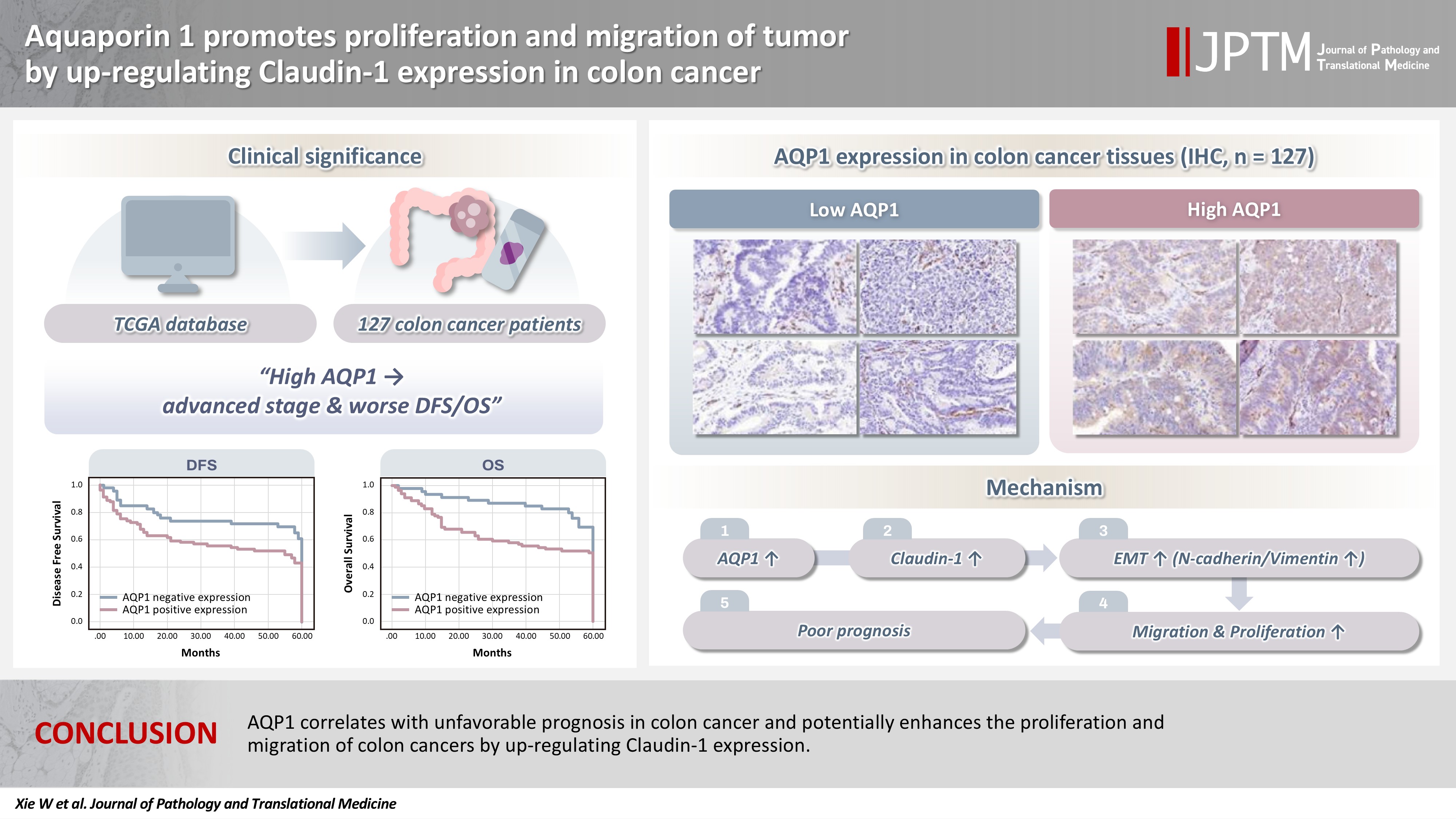
With the rising incidence of colon cancer, several studies have indicated that aquaporin 1 (AQP1) expression is associated with the development of colon cancer. This study aims to elucidate the potential molecular mechanisms between them. Methods: We screened data from The Cancer Genome Atlas (TCGA) database and retrospectively examined AQP1 protein expression in 127 colon cancer patients to analyze the relationship between AQP1 expression and pathological stages, prognosis. We created stable colon cancer cell lines with differential AQP1 expression, the effect of AQP1 expression on the proliferation and migration of colon cancer cells was assessed by in vitro and in vivo studies, and explored potential molecular mechanisms through Western blotting. Results: High AQP1 expression was associated with poorer survival (overall survival [OS], p = .028) in colon cancer patients from the TCGA database. Similarly, retrospective clinical data indicated that high AQP1 expression was associated with reduced disease-free survival and OS (p = .036 and p = .017, respectively). The low-expressing AQP1 colon cancer cells exhibited a decrease in proliferation and migration ability of colon cancer cells compared to the overexpressing AQP1 group (p < .05) in vitro and in vivo. Immunohistochemistry and western blotting experiments validated heightened expression of N-cadherin, vimentin, and claudin- 1 in the tumor tissues of the overexpressing AQP1 group. Conversely, reduced AQP1 expression resulted in decreased expression of claudin- 1. Conclusions: AQP1 correlates with unfavorable prognosis in colon cancer and potentially enhances the proliferation and migration of colon cancer by up-regulating claudin-1 expression.
- Characterization of undifferentiated carcinoma of the salivary gland: clinicopathological and immunohistochemical analyses in comparison with lymphoepithelial carcinoma
- Sangjoon Choi, Gyuheon Choi, Hee Jin Lee, Joon Seon Song, Yoon Se Lee, Seung-Ho Choi, Kyung-Ja Cho
- J Pathol Transl Med. 2025;59(6):361-370. Published online September 8, 2025
- DOI: https://doi.org/10.4132/jptm.2025.07.07

- 5,105 View
- 314 Download
-
Abstract
PDF
- Background
This study aimed to reclassify a subset of poorly differentiated salivary gland carcinoma that do not conform to any entities of the current World Health Organization (WHO) classification into the category of undifferentiated carcinoma (UDC) because they lack specific histologic differentiation or immunophenotype. Methods: Cases of salivary gland carcinomas from Asan Medical Center (2002–2020) that did not fit any existing WHO classification criteria and were diagnosed as poorly differentiated carcinoma, high-grade carcinoma, or UDC, were retrospectively reviewed. Immunohistochemical (IHC) staining for p40, neuroendocrine markers, androgen receptor (AR), and gross cystic disease fluid protein 15 (GCDFP-15) and Epstein-Barr virus (EBV) in situ hybridization (ISH) were performed. Clinical data were collected from the electronic medical records. Results: Six salivary gland carcinomas did not align with any specific entities and lacked distinct differentiation. Two of six cases displayed lymphoepithelial carcinoma (LEC)-like morphology but were negative or showed negligible immunoreactivity for p40 and EBV ISH, distinguishing them from LEC of the salivary gland. Two cases showed strong AR positivity, suggesting a potential overlap with salivary duct carcinoma (SDC) but lacked classic SDC morphologies and GCDFP-15 expression. No cases expressed neuroendocrine markers. Conclusions: This study proposes reclassifying these poorly differentiated or high-grade salivary gland carcinomas as UDC based on their indeterminate differentiation and IHC profiles. This may lead to a clearer diagnostic category and enhance our understanding of these high-grade tumors.
- Cervical intraepithelial neoplasia and cervical cytology in pregnancy
- Ji-Young Kim, Jeong Yun Shim
- J Pathol Transl Med. 2024;58(6):283-290. Published online November 7, 2024
- DOI: https://doi.org/10.4132/jptm.2024.10.17
- 15,053 View
- 531 Download
- 3 Web of Science
- 6 Crossref
-
Abstract
PDF
- Cervical cancer screening during pregnancy presents unique challenges for cytologic interpretation. This review focuses on pregnancy-associated cytomorphological changes and their impact on diagnosis of cervical intraepithelial neoplasia (CIN) and cervical cancer. Pregnancy-induced alterations include navicular cells, hyperplastic endocervical cells, immature metaplastic cells, and occasional decidual cells or trophoblasts. These changes can mimic abnormalities such as koilocytosis, adenocarcinoma in situ, and high-grade squamous intraepithelial lesions, potentially leading to misdiagnosis. Careful attention to nuclear features and awareness of pregnancy-related changes are crucial for correct interpretation. The natural history of CIN during pregnancy shows higher regression rates, particularly for CIN 2, with minimal risk of progression. Management of abnormal cytology follows modified risk-based guidelines to avoid invasive procedures, with treatment typically deferred until postpartum. The findings reported in this review emphasize the importance of considering pregnancy status in cytological interpretation, highlight potential problems, and provide guidance on differentiating benign pregnancy-related changes from true abnormalities. Understanding these nuances is essential for accurate diagnosis and proper management of cervical abnormalities in pregnant women.
-
Citations
Citations to this article as recorded by
- HPV in Pregnancy: Implications for Screening, Vaccination, and Maternal–Fetal Health
Suman Kumar, Swati, Swati Salila, Akanksha Raj, Pratima Gupta, Neha Sharad, Nidhi Chaudhary
Journal of Pregnancy.2026;[Epub] CrossRef - Approaches to Intraepithelial Cervical Neoplasia Management in Pregnancy: A Narrative Review
Delia-Maria Bogheanu, Awatif Jaafar Sadeq Al Bayati, Mircea-Octavian Poenaru, Octavian Gabriel Olaru, Gabriel-Petre Gorecki, Andreea Gratiana Boiangiu, Bashar Haj Hamoud, Romina-Marina Sima, Liana Ples
Life.2026; 16(5): 809. CrossRef - From treatment to trauma: Womens lived experiences of adverse pregnancy outcomes following cervical intraepithelial neoplasia treatment in Zambia
Mwiinga-Kalusopa Victoria, E. Maree Johanna, N. Kwaleyela Concepta, Uwamahoro Marie-Claire, Mwila Musenge Emmanuel, Anila Nkhata Loveness, Katowa-Mukwato Patricia
International Journal of Nursing and Midwifery.2026; 18(2): 14. CrossRef - Cervical Cytological Findings and Vaginal Microbiota Alterations During Pregnancy: A Retrospective Analysis
Federica Cianfrini, Antonio d’Amati, Clelia Molinario, Belen Padial Urteta, Chiara Boccaccini, Antonio Benedetto Maria Donateo, Antonietta Vella, Rosaria Santangelo, Rosa Pasqualina De Vincenzo, Angela Santoro, Gian Franco Zannoni
Sage Open Pathology.2026;[Epub] CrossRef - The significance of biological samples from pregnant women in cervical intraepithelial neoplasia
Xue Mi, Maharjan Rashmi, Zangyu Pan, Di Wu, Jinwei Miao
Frontiers in Medicine.2025;[Epub] CrossRef - Oncologic and pregnancy outcomes of cervical high-grade intraepithelial lesions and delivery mode
Olga P. Matylevich, Ilya A. Tarasau, Sviatlana Y. Shelkovich, Aliaksandr F. Martsinkevich
Academia Oncology.2025;[Epub] CrossRef
- HPV in Pregnancy: Implications for Screening, Vaccination, and Maternal–Fetal Health
- EWSR1 rearranged primary renal myoepithelial carcinoma: a diagnostic conundrum
- Nilay Nishith, Zachariah Chowdhury
- J Pathol Transl Med. 2023;57(5):284-288. Published online September 15, 2023
- DOI: https://doi.org/10.4132/jptm.2023.08.08
- 5,332 View
- 215 Download
- 2 Web of Science
- 2 Crossref
-
Abstract
PDF
- Primary renal myoepithelial carcinoma is an exceedingly rare neoplasm with an aggressive phenotype and Ewing sarcoma breakpoint region 1 (EWSR1) rearrangement in a small fraction of cases. In addition to its rarity, the diagnosis can be challenging for the pathologist due to morphologic heterogeneity, particularly on the biopsy specimen. At times, immunohistochemistry may be indecisive; therefore, molecular studies should be undertaken for clinching the diagnosis. We aim to illustrate a case of primary myoepithelial carcinoma of the kidney with EWSR1-rearrangement in a 67-year-old male patient who presented with right supraclavicular mass, which was clinically diagnosed as carcinoma of an unknown primary. An elaborate immunohistochemical work-up aided by fluorescent in-situ hybridization allowed us to reach a conclusive diagnosis. This unusual case report advocates that one should be aware of the histological mimickers and begin with broad differential diagnoses alongside sporadic ones and then narrow them down with appropriate ancillary studies.
-
Citations
Citations to this article as recorded by- EWSR1-rearranged renal neoplasia: Clinicopathologic and molecular characterization of 39 cases from a single institution
Robert G. Colef, Ganesh P. Pujari, Daniel R. Sill, Beth A. Pitel, Pingchuan Zhang, Burak Tekin, Peter C. Lucas, Rumeal D. Whaley, Loren Herrera Hernandez, Rafael E. Jimenez, Lori A. Erickson, John C. Cheville, Benjamin R. Kipp, Kevin C. Halling, Patricia
Human Pathology.2026; 174: 106135. CrossRef - Primary Ewing Sarcoma of the Kidney
João Lobo, Huiying He, Raheel Ahmed, Bassel Zein-Sabatto, Thomas Winokur, Shi Wei, Shuko Harada, Jesse K. McKenney, Jonathan L. Myles, Jane K. Nguyen, Christopher G. Przybycin, Sean R. Williamson, Cristina Magi-Galluzzi, Reza Alaghehbandan
American Journal of Surgical Pathology.2025; 49(10): 1078. CrossRef
- EWSR1-rearranged renal neoplasia: Clinicopathologic and molecular characterization of 39 cases from a single institution
- Trouble-makers in cytologic interpretation of the uterine cervix
- Eunah Shin, Jaeeun Yu, Soon Won Hong
- J Pathol Transl Med. 2023;57(3):139-146. Published online May 15, 2023
- DOI: https://doi.org/10.4132/jptm.2023.04.25
- 13,816 View
- 498 Download
- 5 Web of Science
- 6 Crossref
-
Abstract
PDF
- The development and standardization of cytologic screening of the uterine cervix has dramatically decreased the prevalence of squamous cell carcinoma of the uterine cervix. Advances in the understanding of biology of human papillomavirus have contributed to upgrading the histologic diagnosis of the uterine cervix; however, cytologic screening that should triage those that need further management still poses several difficulties in interpretation. Cytologic features of high grade intraepithelial squamous lesion (HSIL) mimics including atrophy, immature metaplasia, and transitional metaplasia, and glandular lesion masquerades including tubal metaplasia and HSIL with glandular involvement are described with accentuation mainly on the differential points. When the cytologic features lie in a gray zone between the differentials, the most important key to the more accurate interpretation is sticking to the very basics of cytology; screening the background and cellular architecture, and then scrutinizing the nuclear and cytoplasmic details.
-
Citations
Citations to this article as recorded by- Cytology–Biopsy Concordance in High-Risk Human Papillomavirus–Positive Women with Abnormal Cytology Findings: Menopause-Stratified Analysis
Isik Sozen, Gozde Sahin, Yuksel Ulu, Dilara Yitiz, Basak Ozge Kayan, Ilkbal Temel Yuksel
Medicina.2026; 62(4): 631. CrossRef - Pathologists Recommend Repeat Pap Testing: In Clinical Practice What Do Gylecologist Do?
Gizem Ay Haldız
Muğla Sıtkı Koçman Üniversitesi Tıp Dergisi.2026; 13(1): 1. CrossRef - The Relationship of Vaginal Symptoms and Cervical Inflammation Severity with Cytological Abnormalities and HPV Positivity: A Prospective Observational Study
Alihan Tigli, Rulin Deniz, Toros Taskin, Guzide Ece Akinci, Sultan Deniz Altindag, Nazli Sener, Yasemin Ercan Degirmenci, Sefer Ustebay, Muhammet Bora Uzuner, Erdem Gurkan, Oguzhan Karakoc, Yakup Baykus
Biomedicines.2026; 14(6): 1384. CrossRef - Risk of cervical stenosis after cervical excision in postmenopausal patients
Eva Hauge, Line Winther Gustafson, Mette Tranberg, Pinar Bor
European Journal of Obstetrics & Gynecology and Reproductive Biology.2025; 308: 208. CrossRef - Pitfalls in Gynecological Cytology: Review of the Common and Less Frequent Entities in Pap Test
Danijela Vrdoljak-Mozetič, Snježana Štemberger-Papić, Damjana Verša Ostojić, Roberta Rubeša, Marko Klarić, Senija Eminović
Acta Cytologica.2024; 68(3): 281. CrossRef - Cytological features of human papillomavirus‐infected immature squamous metaplastic cells from cervical intraepithelial neoplasia grade 2
Mitsuaki Okodo, Kaori Okayama, Koji Teruya, Ruku Shinohara, Shuichi Mizuno, Rei Settsu, Yasuyoshi Ishii, Masahiko Fujii, Hirokazu Kimura, Mizue Oda
Journal of Medical Virology.2023;[Epub] CrossRef
- Cytology–Biopsy Concordance in High-Risk Human Papillomavirus–Positive Women with Abnormal Cytology Findings: Menopause-Stratified Analysis
- Diagnostic distribution and pitfalls of glandular abnormalities in cervical cytology: a 25-year single-center study
- Jung-A Sung, Ilias P. Nikas, Haeryoung Kim, Han Suk Ryu, Cheol Lee
- J Pathol Transl Med. 2022;56(6):354-360. Published online November 9, 2022
- DOI: https://doi.org/10.4132/jptm.2022.09.05
- 11,295 View
- 167 Download
- 7 Web of Science
- 5 Crossref
-
Abstract
PDF
- Background
Detection of glandular abnormalities in Papanicolaou (Pap) tests is challenging. This study aimed to review our institute’s experience interpreting such abnormalities, assess cytohistologic concordance, and identify cytomorphologic features associated with malignancy in follow-up histology.
Methods
Patients with cytologically-detected glandular lesions identified in our pathology records from 1995 to 2020 were included in this study.
Results
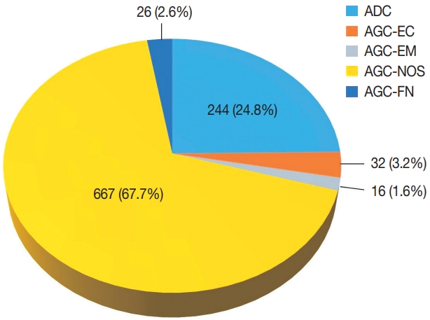
Of the 683,197 Pap tests performed, 985 (0.144%) exhibited glandular abnormalities, 657 of which had tissue follow-up available. One hundred eighty-eight cases were cytologically interpreted as adenocarcinoma and histologically diagnosed as malignant tumors of various origins. There were 213 cases reported as atypical glandular cells (AGC) and nine cases as adenocarcinoma in cytology, yet they were found to be benign in follow-up histology. In addition, 48 cases diagnosed with AGC and six with adenocarcinoma cytology were found to have cervical squamous lesions in follow-up histology, including four squamous cell carcinomas. Among the cytomorphological features examined, nuclear membrane irregularity, three-dimensional clusters, single-cell pattern, and presence of mitoses were associated with malignant histology in follow-up.
Conclusions
This study showed our institute’s experience detecting glandular abnormalities in cervical cytology over a 25-year period, revealing the difficulty of this task. Nonetheless, the present study indicates that several cytological findings such as membrane irregularity, three-dimensional clusters, single-cell pattern, and evidence of proliferation could help distinguishing malignancy from a benign lesion. -
Citations
Citations to this article as recorded by- “Atypical Glandular Cells” on Cervical Cytology: Correlation Between Glandular Cell Component Volume and Histological Follow‐Up
Havva Gokce Terzioglu, Alessa Aragao, Julieta E. Barroeta
Diagnostic Cytopathology.2026; 54(2): 71. CrossRef - Morphological differentiation of hyperchromatic crowded groups (HCG) in cervical cytology. Possible clinical significance
Julio César Villarreal Ramírez, Jesús E. Guaithero Rivas, Lorena Ramírez, Jesús Peña Guillén, Morelva Toro de Méndez
Revista Española de Patología.2026; 59(3): 100883. CrossRef - Expertise in Gynecological Pathology Impacts Diagnosis of Atypical Glandular Cell Category in Cervical Cytology
Havva Gökce Terzioglu, Alessa Aragao, Julieta E. Barroeta
Journal of Lower Genital Tract Disease.2025; 29(4): 297. CrossRef - Comparison of Cytological and/or Histopathological Results of Patients with Single and Multiple HPV Positivity
Fatih Mehmet Kaya, Şafak Ersöz, Cihan Comba, Ömer Demir
Acta Cytologica.2025; : 1. CrossRef - Analysis of atypical glandular cells in ThinPrep Pap smear and follow-up histopathology
Tengfei Wang, Yinan Hua, Lina Liu, Bing Leng
Baylor University Medical Center Proceedings.2024; 37(3): 403. CrossRef
- “Atypical Glandular Cells” on Cervical Cytology: Correlation Between Glandular Cell Component Volume and Histological Follow‐Up
- Evaluation of the characteristics of multiple human papillomavirus (HPV) infections identified using the BD Onclarity HPV assay and comparison with those of single HPV infection
- Jinhee Kim, Moonsik Kim, Ji Young Park
- J Pathol Transl Med. 2022;56(5):289-293. Published online September 13, 2022
- DOI: https://doi.org/10.4132/jptm.2022.08.02
- 9,801 View
- 141 Download
- 12 Web of Science
- 9 Crossref
-
Abstract
PDF
 Supplementary Material
 - Background
Human papillomavirus (HPV) infection is a major cause of cervical cancer and associated precursor lesions. Multiple HPV genotype infections have been reported. However, their clinicopathological characteristics still remain elusive.
Methods
For this study, 814 consecutive patients who had undergone colposcopy and HPV genotyping test using BD Onclarity HPV assay were retrospectively selected. Clinicopathological parameters of multiple HPV infections were compared with those of single HPV infection.
Results
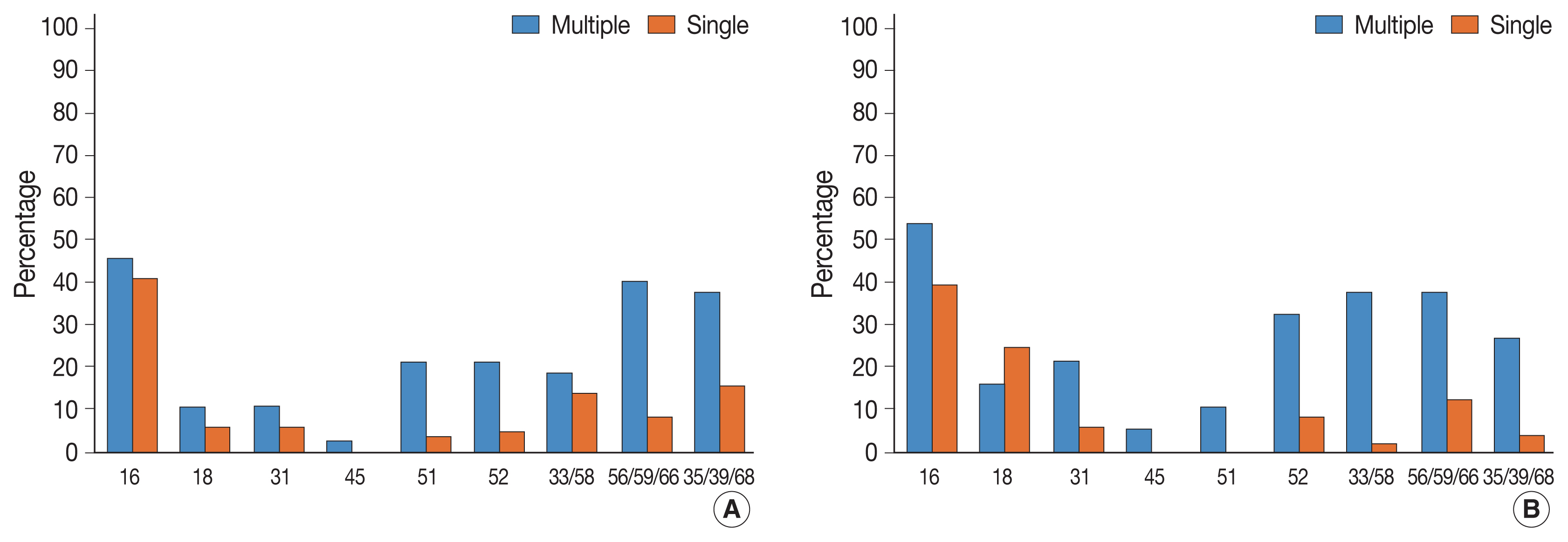
Multiple HPV infections were found in 110 out of 814 cases (13.5%). Multiple HPV infections were associated with a significantly higher incidence of high-grade intraepithelial lesions (HSILs) compared with single HPV infection. Other high-risk HPV genotypes, in addition to HPV 16, were found more frequently in the multiple HPV infections group; these included HPV 51, 52, 33/58, 56/59/66, and 35/39/68. No specific coinfection pattern was not identified. Additionally, the number of HPV genotypes in multiple HPV infections was not associated with the progression to HSIL or squamous cell carcinoma.
Conclusions
Multiple HPV infections have distinct clinicopathological characteristics (compared with single HPV infection). As their biological behavior is uncertain, close and frequent follow-up is warranted. -
Citations
Citations to this article as recorded by- Informative HPV testing after conization and its impact on time-varying estimates: a GAMM-based cohort study
Jie Zhou, Jian hong Liao, Lin Jie Su, Yan Chen, Hong bo Hu
Frontiers in Public Health.2026;[Epub] CrossRef - The Prevalence of Multi-Type Infections Among Human Papillomavirus Types in Korean Women
Jang Mook Kim, Hee Seung Song, Jieun Hwang, Jae Kyung Kim
Pathogens.2025; 14(4): 369. CrossRef - Multiple high-risk human papillomavirus infections exacerbate cervical lesion risk: epidemiological evidence from suining, Sichuan
Yaling Jing, Jianhui Chen, Fang Lin, Xiaonan Huang, Yulin Liu, Mingcai Zhao, Chuan Ye, Lianfang Zhao, Xiaofang Liu, Jiayan Yang
Virology Journal.2025;[Epub] CrossRef - The cervical cancer related distribution, coinfection and risk of 15 HPV types in Baoan, Shenzhen, in 2017–2023
Rukai Li, Weiwei Meng, Yunhai Zuo, Yanli Xu, Shaonan Wu
Virology Journal.2024;[Epub] CrossRef - Molecular findings and virological assessment of bladder papillomavirus infection in cattle
Francesca De Falco, Anna Cutarelli, Francesca Luisa Fedele, Cornel Catoi, Sante Roperto
Veterinary Quarterly.2024; 44(1): 1. CrossRef - Patterns of single and multiple HPV infections in female: A systematic review and meta-analysis
Dan Zhou, Jing Xue, Yaqiong Sun, Liling Zhu, Ming Zhao, Meimei Cui, Min Zhang, Jingjing Jia, Limei Luo
Heliyon.2024; 10(17): e35736. CrossRef - Age distribution of patients with multiple High-Risk Human Papilloma Virus (HR-HPV) genotypes and HPV vaccine recommendations by age
Gülçin Çetin Uysal, Nil Tekin
Family Practice and Palliative Care.2024; 9(3): 80. CrossRef - Relative distribution of HPV genotypes in histological cervical samples and associated grade lesion in a women population over the last 16 years in Burgundy, France
Christelle Auvray, Serge Douvier, Odile Caritey, Jean-Baptiste Bour, Catherine Manoha
Frontiers in Medicine.2023;[Epub] CrossRef - Epidemiologic characteristics of high-risk HPV and the correlation between multiple infections and cervical lesions
Qinli Luo, Xianghua Zeng, Hanyi Luo, Ling Pan, Ying Huang, Haiyan Zhang, Na Han
BMC Infectious Diseases.2023;[Epub] CrossRef
- Informative HPV testing after conization and its impact on time-varying estimates: a GAMM-based cohort study
- Prognostic Significance of CD109 Expression in Patients with Ovarian Epithelial Cancer
- So Young Kim, Kyung Un Choi, Chungsu Hwang, Hyung Jung Lee, Jung Hee Lee, Dong Hoon Shin, Jee Yeon Kim, Mee Young Sol, Jae Ho Kim, Ki Hyung Kim, Dong Soo Suh, Byung Su Kwon
- J Pathol Transl Med. 2019;53(4):244-252. Published online May 2, 2019
- DOI: https://doi.org/10.4132/jptm.2019.04.16
- 9,690 View
- 130 Download
- 9 Web of Science
- 7 Crossref
-
Abstract
PDF
- Background
Ovarian epithelial cancer (OEC) is the second-most common gynecologic malignancy. CD109 expression is elevated in human tumor cell lines and carcinomas. A previous study showed that CD109 expression is elevated in human tumor cell lines and CD109 plays a role in cancer progression. Therefore, this study aimed to determine whether CD109 is expressed in OEC and can be useful in predicting the prognosis.
Methods
Immunohistochemical staining for CD109 and reverse transcription-quantitative polymerase chain reaction was performed. Then we compared CD109 expression and chemoresistance, overall survival, and recurrence-free survival of OEC patients. Chemoresistance was evaluated by dividing into good-response group and poor-response group by the time to recurrence after chemotherapy.
Results
CD109 expression was associated with overall survival (p = .020), but not recurrence-free survival (p = .290). CD109 expression was not an independent risk factor for overall survival due to its reliability (hazard ratio, 1.58; p = .160; 95% confidence interval, 0.82 to 3.05), although we found that CD109 positivity was related to chemoresistance. The poor-response group showed higher rates of CD109 expression than the good-response group (93.8% vs 66.7%, p = .047). Also, the CD109 mRNA expression level was 2.88 times higher in the poor-response group as compared to the good-response group (p = .001).
Conclusions
Examining the CD109 expression in patients with OEC may be helpful in predicting survival and chemotherapeutic effect. -
Citations
Citations to this article as recorded by- CD109 Expression in Tumor and Stromal Cells Serves as a Prognostic Biomarker for Tumor Progression and Outcome in Gallbladder Adenocarcinoma
Taro Kogami, Masaaki Ichinoe, Yasutaka Sakurai, Takuya Kato, Masahiro Matsushita, Akihiro Tamaki, Yurika Kesen, Shoko Hayashi, Itaru Sanoyama, Yoshiko Numata, Atsuko Umezawa, Masatoshi Ichihara, Chika Kusano, Yoshiki Murakumo
Pathology International.2026;[Epub] CrossRef - Advances in the Study of CD109 in Tumors
平慧 周
Medical Diagnosis.2024; 14(02): 167. CrossRef - Identification of CD109 in the extracellular vesicles derived from ovarian cancer stem-like cells
Ye Eun Kim, Jun Se Kim, Min Joo Shin, Seo Yul Lee, Dae Kyoung Kim, Nam-Kyung Lee, Yang Woo Kwon, Kyung-Un Choi, Dong-Soo Suh, Byoung Soo Kim, Sanghwa Jeong, Jae Ho Kim
BMB Reports.2024; 57(12): 527. CrossRef - CD109 Promotes Drug Resistance in A2780 Ovarian Cancer Cells by Regulating the STAT3-NOTCH1 Signaling Axis
Jun Se Kim, Min Joo Shin, Seo Yul Lee, Dae Kyoung Kim, Kyung-Un Choi, Dong-Soo Suh, Dayea Kim, Jae Ho Kim
International Journal of Molecular Sciences.2023; 24(12): 10306. CrossRef - CD109 facilitates progression and 5-fluorouracil resistance of nasopharyngeal carcinoma
Zhenwei Zhu, Fang Zhou, Cheng Mao
Materials Express.2022; 12(9): 1189. CrossRef - Usefulness of CD109 expression as a prognostic biomarker in patients with cancer
Hyun Min Koh, Hyun Ju Lee, Dong Chul Kim
Medicine.2021; 100(11): e25006. CrossRef - Serum CD109 levels reflect the node metastasis status in head and neck squamous cell carcinoma
Sumitaka Hagiwara, Eiichi Sasaki, Yasuhisa Hasegawa, Hidenori Suzuki, Daisuke Nishikawa, Shintaro Beppu, Hoshino Terada, Michi Sawabe, Masahide Takahashi, Nobuhiro Hanai
Cancer Medicine.2021; 10(4): 1335. CrossRef
- CD109 Expression in Tumor and Stromal Cells Serves as a Prognostic Biomarker for Tumor Progression and Outcome in Gallbladder Adenocarcinoma
- Pathogenesis of Focal Segmental Glomerulosclerosis
- Beom Jin Lim, Jae Won Yang, Woo Sung Do, Agnes B. Fogo
- J Pathol Transl Med. 2016;50(6):405-410. Published online October 16, 2016
- DOI: https://doi.org/10.4132/jptm.2016.09.21
- 23,911 View
- 922 Download
- 54 Web of Science
- 50 Crossref
-
Abstract
PDF
- Focal segmental glomerulosclerosis (FSGS) is characterized by focal and segmental obliteration of glomerular capillary tufts with increased matrix. FSGS is classified as collapsing, tip, cellular, perihilar and not otherwise specified variants according to the location and character of the sclerotic lesion. Primary or idiopathic FSGS is considered to be related to podocyte injury, and the pathogenesis of podocyte injury has been actively investigated. Several circulating factors affecting podocyte permeability barrier have been proposed, but not proven to cause FSGS. FSGS may also be caused by genetic alterations. These genes are mainly those regulating slit diaphragm structure, actin cytoskeleton of podocytes, and foot process structure. The mode of inheritance and age of onset are different according to the gene involved. Recently, the role of parietal epithelial cells (PECs) has been highlighted. Podocytes and PECs have common mesenchymal progenitors, therefore, PECs could be a source of podocyte repopulation after podocyte injury. Activated PECs migrate along adhesion to the glomerular tuft and may also contribute to the progression of sclerosis. Markers of activated PECs, including CD44, could be used to distinguish FSGS from minimal change disease. The pathogenesis of FSGS is very complex; however, understanding basic mechanisms of podocyte injury is important not only for basic research, but also for daily diagnostic pathology practice.
-
Citations
Citations to this article as recorded by- miRNAs involved in the TGFB signaling as possible markers of steroid-resistant nephrotic syndrome in children
Ahmedz Widiasta, Yunia Sribudiani, Husna Nugrahapraja, Dedi Rachmadi
Gene Reports.2025; 39: 102173. CrossRef - Purinergic Receptor Activation Protects Glomerular Microvasculature from Increased Mechanical Stress in Angiotensin II-Induced Hypertension: A Modeling Study
Owen Richfield, Ricardo Cortez, Supaporn Kulthinee, Martha Franco, L. Gabriel Navar
International Journal of Molecular Sciences.2025; 26(5): 1928. CrossRef - Podocyte A20/TNFAIP3 Controls Glomerulonephritis Severity via the Regulation of Inflammatory Responses and Effects on the Cytoskeleton
Paulina Köhler, Andrea Ribeiro, Mohsen Honarpisheh, Ekaterina von Rauchhaupt, Georg Lorenz, Chenyu Li, Lucas Martin, Stefanie Steiger, Maja Lindenmeyer, Christoph Schmaderer, Hans-Joachim Anders, Dana Thomasova, Maciej Lech
Cells.2025; 14(5): 381. CrossRef - Clinical implications of apolipoprotein L1 testing in patients with focal segmental glomerulosclerosis: a review of diagnostic and prognostic implications
Aiman Waheed, Muhammad Hamza Gul, Risha Naeem, Sardar Noman Qayyum, Khizra Batool, Abeeha Shaukat, Nashmiya Khan, Safa Irfan Shah, Aisha Rehman Siddiqui, Asad Ullah Farooq, Eeshah Nasir, Samim Noori
Annals of Medicine & Surgery.2025; 87(3): 1543. CrossRef - Focal Segmental Glomerulosclerosis: Comprehensive Review and Exploration of the Dual Potential of Cyclodextrins in Therapeutic Optimization
Filipa Mascarenhas-Melo, Bruna Martins, Inês Monteiro, Alka Lohani, Karolline Krambeck
International Journal of Molecular Sciences.2025; 26(18): 8760. CrossRef - Focal segmental glomerulosclerosis associated with undescribed mutation in the LMX1B gene
María Adoración Martín Gómez, Mercedes Caba Molina, Miriam León Fradejas, Juana Alonso Titos, Rafael del Pozo Alvarez
European Journal of Medical Genetics.2024; 72: 104980. CrossRef - MicroRNA-155-5p Aggravates Adriamycin-Induced Focal Segmental Glomerulosclerosis through Targeting Nrf2
Guoyong Liu, Liyu He, Xiaomeng Yang, Lingling Tang, Wei Shi, Jian She, Jiali Wei
Nephron.2023; 147(2): 108. CrossRef - The role of HLA antigens in recurrent primary focal segmental glomerulosclerosis
Ibrahim Batal, Pascale Khairallah, Astrid Weins, Nicole K. Andeen, Michael B. Stokes
Frontiers in Immunology.2023;[Epub] CrossRef - IgM and C3 Deposition in Primary Focal Segmental Glomerulosclerosis (FSGS): A Clinical and Histopathological Spectrum
Faizan Amer, Madiha Syed , Aurangzeb Afzal, Mudassar Hussain , Usman Hassan, Shaarif Bashir, Maryam Hameed, Sheeba Ishtiaq
Cureus.2023;[Epub] CrossRef - Identification of key biomarkers of the glomerulus in focal segmental glomerulosclerosis and their relationship with immune cell infiltration based on WGCNA and the LASSO algorithm
Yun Xia Zhang, Juan Lv, Jun Yuan Bai, XiaoWei Pu, En Lai Dai
Renal Failure.2023;[Epub] CrossRef - Rituximab in the treatment of primary FSGS: time for its use in routine clinical practice?
Adam D Morris, Lauren Floyd, Alexander Woywodt, Ajay Dhaygude
Clinical Kidney Journal.2023; 16(8): 1199. CrossRef - High Rate of Mutations of Adhesion Molecules and Extracellular Matrix Glycoproteins in Patients with Adult-Onset Focal and Segmental Glomerulosclerosis
Sara Marcos González, Emilio Rodrigo Calabia, Ignacio Varela, Michal Červienka, Javier Freire Salinas, José Javier Gómez Román
Biomedicines.2023; 11(6): 1764. CrossRef - Glomerular parietal epithelial expression of CD44 in minimal change nephrotic syndrome and primary focal segmental glomerulosclerosis: A clinico-pathological study
ENithin Paul, Suchitha Satish, KiranKrishnamurthy Kelur, ManjunathSanjeev Shetty
Indian Journal of Pathology and Microbiology.2023; 66(3): 526. CrossRef - Calcineurin inhibitors or cyclophosphamide in the treatment of membranous nephropathy superimposed with FSGS lesions: a retrospective study from China
Hong-Guang He, Yi-Yun Huang, Qin-Qing Liang, Qiu-Rong Ye, An-Dong Li, Kun Ye, Qiu-Xia Wu, Yan-Wu You
Renal Failure.2023;[Epub] CrossRef - CD44-negative parietal–epithelial cell staining in minimal change disease: association with clinical features, response to corticosteroids and kidney outcome
Neus Roca, Elias Jatem, Anabel Abo, Maria Santacana, Alejandro Cruz, Álvaro Madrid, Gloria Fraga, Marisa Martin, Jorge Gonzalez, Cristina Martinez, Anna Balius, Alfons Segarra
Clinical Kidney Journal.2022; 15(3): 545. CrossRef - Monogenic focal segmental glomerulosclerosis: A conceptual framework for identification and management of a heterogeneous disease
Meenakshi Sambharia, Prerna Rastogi, Christie P. Thomas
American Journal of Medical Genetics Part C: Seminars in Medical Genetics.2022; 190(3): 377. CrossRef - Small Nucleolar RNAs in Pseudoexfoliation Glaucoma
Karolina Gasińska, Marcin Czop, Ewa Kosior-Jarecka, Dominika Wróbel-Dudzińska, Janusz Kocki, Tomasz Żarnowski
Cells.2022; 11(17): 2738. CrossRef - Collapsing focal segmental glomerulosclerosis in a patient with oral cavity cancer
Sae Byeol Choi, Kyoung Min Kim, Moon Hyang Park, Kyung Pyo Kang
Medicine.2021; 100(18): e25857. CrossRef - MRTF: Basic Biology and Role in Kidney Disease
Maria Zena Miranda, Zsuzsanna Lichner, Katalin Szászi, András Kapus
International Journal of Molecular Sciences.2021; 22(11): 6040. CrossRef - The Unique Difference Between Serum Level of Soluble Urokinase Plasminogen Activator Receptor (suPAR) in Steroid-Resistant Nephrotic Syndrome Children Treated with an Alkylating Agent and Calcineurin Inhibitors
Ahmedz Widiasta, Kurnia Wahyudi, Husna Nugrahapraja, Yunia Sribudiani, Dedi Rachmadi
Journal of Comprehensive Pediatrics.2021;[Epub] CrossRef - Recessive, gain-of-function toxicity in an APOL1 BAC transgenic mouse model mirrors human APOL1 kidney disease
Gizelle M. McCarthy, Angelo Blasio, Olivia G. Donovan, Lena B. Schaller, Althea Bock-Hughes, Jose M. Magraner, Jung Hee Suh, Calum F. Tattersfield, Isaac E. Stillman, Shrijal S. Shah, Zsuzsanna K. Zsengeller, Balajikarthick Subramanian, David J. Friedman,
Disease Models & Mechanisms.2021;[Epub] CrossRef - CLEC14A protects against podocyte injury in mice with adriamycin nephropathy
Zeyu Su, Yujia Li, Hang Lv, Xiaoyang Cui, Min Liu, Ziying Wang, Yan Zhang, Junhui Zhen, Wei Tang, Xiaojie Wang, Fan Yi
The FASEB Journal.2021;[Epub] CrossRef - Interplay between extracellular matrix components and cellular and molecular mechanisms in kidney fibrosis
Sandra Rayego-Mateos, Sofia Campillo, Raúl R. Rodrigues-Diez, Antonio Tejera-Muñoz, Laura Marquez-Exposito, Roel Goldschmeding, Diego Rodríguez-Puyol, Laura Calleros, Marta Ruiz-Ortega
Clinical Science.2021; 135(16): 1999. CrossRef - The recruitment mechanisms and potential therapeutic targets of podocytes from parietal epithelial cells
Lihua Ni, Cheng Yuan, Xiaoyan Wu
Journal of Translational Medicine.2021;[Epub] CrossRef - miR-150 inhibitor ameliorates adriamycin-induced focal segmental glomerulosclerosis
Huimeng Qi, Jingqi Fu, Junjun Luan, Congcong Jiao, Xiangfei Cui, Xiangyan Cao, Yixiao Zhang, Yanqiu Wang, Jeffrey B. Kopp, Jingbo Pi, Hua Zhou
Biochemical and Biophysical Research Communications.2020; 522(3): 618. CrossRef - From protein uptake to Dent disease: An overview of the CLCN5 gene
Lisa Gianesello, Dorella Del Prete, Monica Ceol, Giovanna Priante, Lorenzo Arcangelo Calò, Franca Anglani
Gene.2020; 747: 144662. CrossRef - Non-ischemic cardiomyopathy with focal segmental glomerulosclerosis
Parminder Kaur, Balraj Singh, Prem Patel, Rahul Vasudev, Upamanyu Rampal, Fayez Shamoon
Journal of Community Hospital Internal Medicine Perspectives.2020; 10(2): 154. CrossRef - Glomerular Endothelial Cells as Instigators of Glomerular Sclerotic Diseases
Marloes Sol, Jan A. A. M. Kamps, Jacob van den Born, Marius C. van den Heuvel, Johan van der Vlag, Guido Krenning, Jan-Luuk Hillebrands
Frontiers in Pharmacology.2020;[Epub] CrossRef - A novel heterozygous variant of the COL4A4 gene in a Chinese family with hematuria and proteinuria leads to focal segmental glomerulosclerosis and chronic kidney disease
Liang‐Liang Fan, Lv Liu, Fang‐Mei Luo, Ran Du, Chen‐Yu Wang, Yi Dong, Ji‐Shi Liu
Molecular Genetics & Genomic Medicine.2020;[Epub] CrossRef - Cellular and molecular mechanisms of kidney fibrosis
Sonja Djudjaj, Peter Boor
Molecular Aspects of Medicine.2019; 65: 16. CrossRef - Albumin induces CD44 expression in glomerular parietal epithelial cells by activating extracellular signal‐regulated kinase 1/2 pathway
Xueying Zhao, Xiaoming Chen, Ashmeer Chima, Yuanyuan Zhang, Jasmine George, Alyssa Cobbs, Nerimiah Emmett
Journal of Cellular Physiology.2019; 234(5): 7224. CrossRef - Analysis of the genomic architecture of a complex trait locus in hypertensive rat models links Tmem63c to kidney damage
Angela Schulz, Nicola Victoria Müller, Nina Anne van de Lest, Andreas Eisenreich, Martina Schmidbauer, Andrei Barysenka, Bettina Purfürst, Anje Sporbert, Theodor Lorenzen, Alexander M Meyer, Laura Herlan, Anika Witten, Frank Rühle, Weibin Zhou, Emile de H
eLife.2019;[Epub] CrossRef - Podocyte-Specific Sialylation-Deficient Mice Serve as a Model for Human FSGS
Kristina M. Niculovic, Linda Blume, Henri Wedekind, Elina Kats, Iris Albers, Stephanie Groos, Markus Abeln, Jessica Schmitz, Esther Beuke, Jan H. Bräsen, Anette Melk, Mario Schiffer, Birgit Weinhold, Anja K. Münster-Kühnel
Journal of the American Society of Nephrology.2019; 30(6): 1021. CrossRef - Inhibition of the ERK1/2-mTORC1 axis ameliorates proteinuria and the fibrogenic action of transforming growth factor-β in Adriamycin-induced glomerulosclerosis
Ranjan Das, Soo-Jin Kim, Nhung Thi Nguyen, Hyeong Ju Kwon, Seung-Kuy Cha, Kyu-Sang Park
Kidney International.2019; 96(4): 927. CrossRef - Mechanisms of Scarring in Focal Segmental Glomerulosclerosis
Jianyong Zhong, Jacob B. Whitman, Hai-Chun Yang, Agnes B. Fogo
Journal of Histochemistry & Cytochemistry.2019; 67(9): 623. CrossRef - A bigenic mouse model of FSGS reveals perturbed pathways in podocytes, mesangial cells and endothelial cells
Andrew S. Potter, Keri Drake, Eric W. Brunskill, S. Steven Potter, Peter Hohenstein
PLOS ONE.2019; 14(8): e0216261. CrossRef - Chronic kidney disease: a review of proteomic and metabolomic approaches to membranous glomerulonephritis, focal segmental glomerulosclerosis, and IgA nephropathy biomarkers
Amir Taherkhani, Reyhaneh Farrokhi Yekta, Maede Mohseni, Massoud Saidijam, Afsaneh Arefi Oskouie
Proteome Science.2019;[Epub] CrossRef - Sphingolipid signaling in renal fibrosis
Andrea Huwiler, Josef Pfeilschifter
Matrix Biology.2018; 68-69: 230. CrossRef - A novel assay provides sensitive measurement of physiologically relevant changes in albumin permeability in isolated human and rodent glomeruli
Sara Desideri, Karen L. Onions, Yan Qiu, Raina D. Ramnath, Matthew J. Butler, Christopher R. Neal, Matthew L.R. King, Andrew E. Salmon, Moin A. Saleem, Gavin I. Welsh, C. Charles Michel, Simon C. Satchell, Andrew H.J. Salmon, Rebecca R. Foster
Kidney International.2018; 93(5): 1086. CrossRef - Recent advances of animal model of focal segmental glomerulosclerosis
Jae Won Yang, Anne Katrin Dettmar, Andreas Kronbichler, Heon Yung Gee, Moin Saleem, Seong Heon Kim, Jae Il Shin
Clinical and Experimental Nephrology.2018; 22(4): 752. CrossRef - Urinary podocyte-associated molecules and albuminuria in hypertension
Javier Perez-Hernandez, Maria D. Olivares, Elena Solaz, Fernando Martinez, Sergio Martínez-Hervas, Gernot Pichler, Felipe J. Chaves, Josep Redon, Raquel Cortes
Journal of Hypertension.2018; 36(8): 1712. CrossRef - Segmental Sclerosis and Extracapillary Hypercellularity Predict Diabetic ESRD
Amy K. Mottl, Adil Gasim, Fernanda Payan Schober, Yichun Hu, Askia K. Dunnon, Susan L. Hogan, J. Charles Jennette
Journal of the American Society of Nephrology.2018; 29(2): 694. CrossRef - Chlormethine Hydrochloride is Not Inferior to Tacrolimus in Treating Steroid-Resistant Nephrotic Syndrome
Yuan Yang, Li Zhao, Li Xiao, Yumei Liang, Chang Wang, Xiao Fu, Xuejing Zhu, Shuguang Yuan, Jianling Zhu, Xiaoping Zhu, Yinghong Liu, Jun Li, Jian Luo, Fuyou Liu, Lin Sun
Kidney and Blood Pressure Research.2018; 43(1): 68. CrossRef - Multiple Myeloma in a Patient with Focal Segmental Glomerulosclerosis: A Case Report
Ashraf O. Oweis, Sameeha A. Al Shelleh, Najla Aldaoud, Osama Mohammed Alshari, Mousa A. Al-Abbadi
American Journal of Case Reports.2018; 19: 946. CrossRef - Can podocytes be regenerated in adults?
Stuart J. Shankland, Benjamin S. Freedman, Jeffrey W. Pippin
Current Opinion in Nephrology and Hypertension.2017; 26(3): 154. CrossRef - Plasma exchange in kidney transplantation: Still a valuable option for nephrotic syndrome recurrence
Licia Peruzzi, Roberto Albiani, Karol Giancaspero
Transfusion and Apheresis Science.2017; 56(4): 525. CrossRef - Is CD44 in glomerular parietal epithelial cells a pathological marker of renal function deterioration in primary focal segmental glomerulosclerosis?
Brunna Pinto Froes, Stanley de Almeida Araújo, Eduardo Alves Bambirra, Eduardo Araújo Oliveira, Ana Cristina Simões e Silva, Sérgio Veloso Brant Pinheiro
Pediatric Nephrology.2017; 32(11): 2165. CrossRef - Injury-induced actin cytoskeleton reorganization in podocytes revealed by super-resolution microscopy
Hani Y. Suleiman, Robyn Roth, Sanjay Jain, John E. Heuser, Andrey S. Shaw, Jeffrey H. Miner
JCI Insight.2017;[Epub] CrossRef - Resveratrol Attenuates Adriamycin-Induced Focal Segmental Glomerulosclerosis through C3aR/C5aR- Sphingosine Kinase 1 Pathway
Guoyong Liu, Qiang Wang, Yan Shi, Xiaofei Peng, Hong Liu, Youming Peng, Liyu He
Pharmacology.2017; 100(5-6): 253. CrossRef - The Multifaceted Role of the Lysosomal Protease Cathepsins in Kidney Disease
Pasquale Cocchiaro, Valeria De Pasquale, Rossella Della Morte, Simona Tafuri, Luigi Avallone, Anne Pizard, Anna Moles, Luigi Michele Pavone
Frontiers in Cell and Developmental Biology.2017;[Epub] CrossRef
- miRNAs involved in the TGFB signaling as possible markers of steroid-resistant nephrotic syndrome in children
- Clinicopathologic Correlations of E-cadherin and Prrx-1 Expression Loss in Hepatocellular Carcinoma
- Kijong Yi, Hyunsung Kim, Yumin Chung, Hyein Ahn, Jongmin Sim, Young Chan Wi, Ju Yeon Pyo, Young-Soo Song, Seung Sam Paik, Young-Ha Oh
- J Pathol Transl Med. 2016;50(5):327-336. Published online August 31, 2016
- DOI: https://doi.org/10.4132/jptm.2016.06.22
- 12,555 View
- 166 Download
- 5 Web of Science
- 4 Crossref
-
Abstract
PDF
- Background
Developing predictive markers for hepatocellular carcinoma (HCC) is important, because many patients experience recurrence and metastasis. Epithelial to mesenchymal transition (EMT) is a developmental process that plays an important role during embryogenesis and also during cancer metastasis. Paired-related homeobox protein 1 (Prrx-1) is an EMT inducer that has recently been introduced, and its prognostic significance in HCC is largely unknown.
Methods
Tissue microarray was constructed using surgically resected primary HCCs from 244 cases. Immunohistochemical staining of E-cadherin and Prrx-1 was performed. The correlation between E-cadherin loss and Prrx-1 expression, as well as other clinicopathologic factors, was evaluated.
Results
E-cadherin expression was decreased in 96 cases (39.4%). Loss of E-cadherin correlated with a higher recurrence rate (p < .001) but was not correlated with patient’s survival. Thirty-two cases (13.3%) showed at least focal nuclear Prrx-1 immunoreactivity while all non-neoplastic livers (n = 22) were negative. Prrx-1 expression was not associated with E-cadherin loss, survival or recurrence rates, pathologic factors, or the Ki-67 labeling index. Twenty tumors that were positive for E-cadherin and Prrx-1 had significantly higher nuclear grades than the rest of the cohort (p = .037). In Cox proportional hazard models, E-cadherin loss and large vessel invasion were independent prognostic factors for shorter disease-free survival. Cirrhosis and high Ki-67 index (> 40%) were independent prognostic factors for shorter overall survival.
Conclusions
Prrx-1 was expressed in small portions of HCCs but not in normal livers. Additional studies with a large number of Prrx-1-positive cases are required to confirm the results of this study. -
Citations
Citations to this article as recorded by- Matrigel and collagen I impact hepatocellular carcinoma cell behavior: a confluency-dependent study
Zeynep Akbulut, Can Daylan, Gamze Demirel
Cukurova Medical Journal.2025; 50(3): 899. CrossRef - The Prognostic Importance of Ki-67 in Gastrointestinal Carcinomas: A Meta-analysis and Multi-omics Approach
Mahdieh Razmi, Fatemeh Tajik, Farideh Hashemi, Ayna Yazdanpanah, Fatemeh Hashemi-Niasari, Adeleh Divsalar
Journal of Gastrointestinal Cancer.2024; 55(2): 599. CrossRef - Homotypic cell-in-cell structures as an adverse prognostic predictor of hepatocellular carcinoma
Ruizhi Wang, Yichao Zhu, Hao Zhong, Xinyue Gao, Qiang Sun, Meifang He
Frontiers in Oncology.2022;[Epub] CrossRef - Dysregulated paired related homeobox 1 impacts on hepatocellular carcinoma phenotypes
Weronika Piorońska, Zeribe Chike Nwosu, Mei Han, Michael Büttner, Matthias Philip Ebert, Steven Dooley, Christoph Meyer
BMC Cancer.2021;[Epub] CrossRef
- Matrigel and collagen I impact hepatocellular carcinoma cell behavior: a confluency-dependent study
- Clinical Significance of an HPV DNA Chip Test with Emphasis on HPV-16 and/or HPV-18 Detection in Korean Gynecological Patients
- Min-Kyung Yeo, Ahwon Lee, Soo Young Hur, Jong Sup Park
- J Pathol Transl Med. 2016;50(4):294-299. Published online June 26, 2016
- DOI: https://doi.org/10.4132/jptm.2016.05.09
- 11,655 View
- 80 Download
- 3 Web of Science
- 2 Crossref
-
Abstract
PDF
- Background
Human papillomavirus (HPV) is a major risk factor for cervical cancer.
Methods
We evaluated the clinical significance of the HPV DNA chip genotyping assay (MyHPV chip, Mygene Co.) compared with the Hybrid Capture 2 (HC2) chemiluminescent nucleic acid hybridization kit (Digene Corp.) in 867 patients.
Results
The concordance rate between the MyHPV chip and HC2 was 79.4% (kappa coefficient, κ = 0.55). The sensitivity and specificity of both HPV tests were very similar (approximately 85% and 50%, respectively). The addition of HPV result (either MyHPV chip or HC2) to cytology improved the sensitivity (95%, each) but reduced the specificity (approximately 30%, each) compared with the HPV test or cytology alone. Based on the MyHPV chip results, the odds ratio (OR) for ≥ high-grade squamous intraepithelial lesions (HSILs) was 9.9 in the HPV-16/18 (+) group and 3.7 in the non-16/18 high-risk (HR)-HPV (+) group. Based on the HC2 results, the OR for ≥ HSILs was 5.9 in the HR-HPV (+) group. When considering only patients with cytological diagnoses of “negative for intraepithelial lesion or malignancy” and “atypical squamous cell or atypical glandular cell,” based on the MyHPV chip results, the ORs for ≥ HSILs were 6.8 and 11.7, respectively, in the HPV-16/18 (+) group.
Conclusions
The sensitivity and specificity of the MyHPV chip test are similar to the HC2. Detecting HPV-16/18 with an HPV DNA chip test, which is commonly used in many Asian countries, is useful in assessing the risk of high-grade cervical lesions. -
Citations
Citations to this article as recorded by- Human papilloma virus identification in ocular surface squamous neoplasia by p16 immunohistochemistry and DNA chip test
Tina Shrestha, Won Choi, Ga Eon Kim, Jee Myung Yang, Kyung Chul Yoon
Medicine.2019; 98(2): e13944. CrossRef - Comparison of the PANArray HPV Genotyping Chip Test with the Cobas 4800 HPV and Hybrid Capture 2 Tests for Detection of HPV in ASCUS Women
Eun Young Ki, Yoon Kyung Lee, Ahwon Lee, Jong Sup Park
Yonsei Medical Journal.2018; 59(5): 662. CrossRef
- Human papilloma virus identification in ocular surface squamous neoplasia by p16 immunohistochemistry and DNA chip test
- Significance of Parafibromin Expression in Laryngeal Squamous Cell Carcinomas
- Inju Cho, Mija Lee, Sharon Lim, Ran Hong
- J Pathol Transl Med. 2016;50(4):264-269. Published online June 23, 2016
- DOI: https://doi.org/10.4132/jptm.2016.04.24
- 11,256 View
- 61 Download
- 7 Web of Science
- 5 Crossref
-
Abstract
PDF
- Background
Parafibromin is a product of the tumor suppressor gene that has been studied as a potential indicator of tumor aggressiveness in the parathyroid, breast, colorectum, and stomach. However, the clinical significance and potential function of parafibromin expression in head and neck squamous cell carcinomas remain largely unknown. The aim of this study was to evaluate the expression of parafibromin in laryngeal squamous cell carcinoma (LSCC) and to verify its potential as a biomarker of tumor behavior.
Methods
Parafibromin expression was evaluated in 30 cases of LSCC using immunohistochemistry. The correlations between parafibromin expression and clinicopathologic parameters were investigated.
Results
Parafibromin expression was positive in 15 cases (50%) and negative in 15 cases (50%). Tumor size and T stage showed a statistically significant inverse relationship with parafibromin expression (p=.028 and p<.001, respectively). Parafibromin expression was not associated with age, sex, lymph node metastasis, tumor differentiation, or tumor location. There was no statistically significant relationship between parafibromin expression and progression-free survival in the patients (p>.05).
Conclusions
Our results indicate that the downregulation or loss of parafibromin expression can be employed as a novel marker of tumor progression or aggressiveness in LSCC. -
Citations
Citations to this article as recorded by- Arsenic trioxide regulates DYNAP through hsa-mir-573 and inhibits the proliferation of laryngeal cancer
Yanru Ren, Xiao Yang, Yang Hui, Weiyao Chen, Yi Cheng, Ning Zhang, Tao Liu, Xinxin Yang, Xiaoyu Li
Scientific Reports.2025;[Epub] CrossRef -
CDC73 c.1155-3A>G is a pathogenic variant that causes aberrant splicing, disrupted parafibromin expression, and hyperparathyroidism-jaw tumor syndrome
Leor Needleman, Nicolette Chun, Sathvika Sitaraman, Marilyn Tan, Deborah E Sellmeyer, Electron Kebebew, Justin P Annes
JBMR Plus.2024;[Epub] CrossRef - Advances in the application of label‐free quantitative proteomics techniques in malignancy research
Xiao Meng, Dong Liu, Yan Guan
Biomedical Chromatography.2023;[Epub] CrossRef - The roles of the tumor suppressor parafibromin in cancer
Hua-chuan Zheng, Hang Xue, Cong-yu Zhang
Frontiers in Cell and Developmental Biology.2022;[Epub] CrossRef - The clinicopathological and prognostic significances ofCDC73expression in cancers: a bioinformatics analysis
Hua-Chuan Zheng, Bao-Cheng Gong, Shuang Zhao
Oncotarget.2017; 8(56): 95270. CrossRef
- Arsenic trioxide regulates DYNAP through hsa-mir-573 and inhibits the proliferation of laryngeal cancer
- Aquaporin 1 Is an Independent Marker of Poor Prognosis in Lung Adenocarcinoma
- Sumi Yun, Ping-Li Sun, Yan Jin, Hyojin Kim, Eunhyang Park, Soo Young Park, Kyuho Lee, Kyoungyul Lee, Jin-Haeng Chung
- J Pathol Transl Med. 2016;50(4):251-257. Published online June 7, 2016
- DOI: https://doi.org/10.4132/jptm.2016.03.30
- 12,968 View
- 123 Download
- 28 Web of Science
- 29 Crossref
-
Abstract
PDF
- Background
Aquaporin 1 (AQP1) overexpression has been shown to be associated with uncontrolled cell replication, invasion, migration, and tumor metastasis. We aimed to evaluate AQP1 expression in lung adenocarcinomas and to examine its association with clinicopathological features and prognostic significance. We also investigated the association between AQP1 overexpression and epithelial-mesenchymal transition (EMT) markers.
Methods
We examined AQP1 expression in 505 cases of surgically resected lung adenocarcinomas acquired at the Seoul National University Bundang Hospital from 2003 to 2012. Expression of AQP1 and EMT-related markers, including Ecadherin and vimentin, were analyzed by immunohistochemistry and tissue microarray.
Results
AQP1 overexpression was associated with several aggressive pathological parameters, including venous invasion, lymphatic invasion, and tumor recurrence. AQP1 overexpression tended to be associated with higher histological grade, advanced pathological stage, and anaplastic lymphoma kinase (ALK) translocation; however, these differences were not statistically significant. In addition, AQP1 overexpression positively correlated with loss of E-cadherin expression and acquired expression of vimentin. Lung adenocarcinoma patients with AQP1 overexpression showed shorter progression- free survival (PFS, 46.1 months vs. 56.2 months) compared to patients without AQP1 overexpression. Multivariate analysis confirmed that AQP1 overexpression was significantly associated with shorter PFS (hazard ratio, 1.429; 95% confidence interval, 1.033 to 1.977; p=.031).
Conclusions
AQP1 overexpression was thereby concluded to be an independent factor of poor prognosis associated with shorter PFS in lung adenocarcinoma. These results suggested that AQP1 overexpression might be considered as a prognostic biomarker of lung adenocarcinoma. -
Citations
Citations to this article as recorded by- Natural Modulators of Aquaporins in Cancer Therapy: Functional Mechanisms and Clinical Potential
Paulina Małkowska, Maciej Tarnowski
Molecules.2026; 31(7): 1072. CrossRef - Aquaporin‐1, aquaporin‐3 and aquaporin‐5 differentially modulate cell biophysical and biomechanical properties, impacting cell stiffness and cell–cell adhesion
Catarina Pimpão, Filomena A. Carvalho, Inês Vieira da Silva, Andreia Barateiro, Nuno C. Santos, Graça Soveral
The FEBS Journal.2026; 293(3): 806. CrossRef - AQP1 Suppresses Clear Cell Renal Cell Carcinoma via Epigenetic Silencing and TNF-Mediated Apoptosis
Shuo Pang, Yingwei Bi, Yuxin Liu, Shiming Wang, Bolin Yi, Liang Zhu, Jianbo Wang
International Journal of Molecular Sciences.2026; 27(12): 5215. CrossRef - Methylation heterogeneity of the AQP1 promoter as a candidate prognostic biomarker in cholangiocarcinoma
Seiya Yokoyama, Hirotsugu Noguchi, Taiji Hamada, Kei Matsuo, Toshiaki Akahane, Ikumi Kitazono, Takashi Tasaki, Miki Murakami, Takao Ohtsuka, Michiyo Higashi, Tatsuhiko Furukawa, Akihide Tanimoto
Scientific Reports.2026;[Epub] CrossRef - The Expanding Role of Aquaporin-1, Aquaporin-3 and Aquaporin-5 as Transceptors: Involvement in Cancer Development and Potential Druggability
Catarina Pimpão, Inês V. da Silva, Graça Soveral
International Journal of Molecular Sciences.2025; 26(3): 1330. CrossRef - The comprehensive potential of AQP1 as a tumor biomarker: evidence from kidney neoplasm cohorts, cell experiments and pan-cancer analysis
Yifan Liu, Donghao Lyu, Yuntao Yao, Jinming Cui, Jiangui Liu, Zikuan Bai, Zihui Zhao, Yuanan Li, Bingnan Lu, Keqin Dong, Xiuwu Pan
Human Genomics.2025;[Epub] CrossRef - The Association of Aquaporins with MAPK Signaling Pathway Unveils Potential Prognostic Biomarkers for Pancreatic Cancer: A Transcriptomics Approach
Inês V. da Silva, Paula A. Lopes, Elisabete Fonseca, Emanuel Vigia, Jorge Paulino, Graça Soveral
Biomolecules.2025; 15(4): 488. CrossRef - Obesity Impacts Post‐Myocardial Infarction Neovascularization by Downregulating AQP1 Expression via the TRPC5‐NFATc3 Signaling Pathway
Mengru Gao, Jing Han, Yifei Zhu, Xin Wen, Lei Feng, Tingting Zhou
Comprehensive Physiology.2025;[Epub] CrossRef - Prognostic Assessment of Aquaporins in Pancreatic Adenocarcinoma: An In Silico Analysis
Vignesh Krishnasamy, Lalhmingliana, Nachimuthu Senthil Kumar
Current Biotechnology.2025; 14(2): 130. CrossRef - Clinical application of cold atmospheric-pressure plasma: mechanisms and irradiation conditions
Eun Ji Jeong, Hyun Min Park, Dong Jae Lee, Jun Lee, Jun Yeong Cho, Kyung Deok Seo, Seokjun Je, Min Hyung Jung, Woo Yeon Hwang, Kyung Sook Kim
Journal of Physics D: Applied Physics.2024; 57(37): 373001. CrossRef - Aquaporins in Cancer Biology
Chul So Moon, David Moon, Sung Koo Kang
Frontiers in Oncology.2022;[Epub] CrossRef - A Comprehensive Prognostic Analysis of Tumor-Related Blood Group Antigens in Pan-Cancers Suggests That SEMA7A as a Novel Biomarker in Kidney Renal Clear Cell Carcinoma
Yange Wang, Chenyang Li, Xinlei Qi, Yafei Yao, Lu Zhang, Guosen Zhang, Longxiang Xie, Qiang Wang, Wan Zhu, Xiangqian Guo
International Journal of Molecular Sciences.2022; 23(15): 8799. CrossRef - Differential modulation of lung aquaporins among other pathophysiological markers in acute (Cl2 gas) and chronic (carbon nanoparticles, cigarette smoke) respiratory toxicity mouse models
Sukanta S. Bhattacharya, Brijesh Yadav, Ekta Yadav, Ariel Hus, Niket Yadav, Perminder Kaur, Lauren Rosen, Roman Jandarov, Jagjit S. Yadav
Frontiers in Physiology.2022;[Epub] CrossRef - Aquaporin water channels as regulators of cell-cell adhesion proteins
Sarannya Edamana, Frédéric H. Login, Soichiro Yamada, Tae-Hwan Kwon, Lene N. Nejsum
American Journal of Physiology-Cell Physiology.2021; 320(5): C771. CrossRef - Targeting Aquaporins in Novel Therapies for Male and Female Breast and Reproductive Cancers
Sidra Khan, Carmela Ricciardelli, Andrea J. Yool
Cells.2021; 10(2): 215. CrossRef - Targeting ion channels for the treatment of lung cancer
Liqin Zhang, Shuya Bing, Mo Dong, Xiaoqiu Lu, Yuancheng Xiong
Biochimica et Biophysica Acta (BBA) - Reviews on Cancer.2021; 1876(2): 188629. CrossRef - Comprehensive Analysis of Aquaporin Superfamily in Lung Adenocarcinoma
Guofu Lin, Luyang Chen, Lanlan Lin, Hai Lin, Zhifeng Guo, Yingxuan Xu, Chanchan Hu, Jinglan Fu, Qinhui Lin, Wenhan Chen, Yiming Zeng, Yuan Xu
Frontiers in Molecular Biosciences.2021;[Epub] CrossRef - Diagnostic accuracy of urinary aquaporin-1 as a biomarker for renal cell carcinoma
Abhilash Cheriyan, Arun Jose Nellickal, Nirmal Thampi John, Lakshmanan Jeyaseelan, Santosh Kumar, Antony Devasia, Nitin Kekre
Indian Journal of Urology.2021; 37(1): 59. CrossRef - Aquaporin 1, 3, and 5 Patterns in Salivary Gland Mucoepidermoid Carcinoma: Expression in Surgical Specimens and an In Vitro Pilot Study
Mérin Barbara Stamboni, Ágatha Nagli de Mello Gomes, Milena Monteiro de Souza, Katia Klug Oliveira, Claudia Fabiana Joca Arruda, Fernanda de Paula, Barbara Beltrame Bettim, Márcia Martins Marques, Luiz Paulo Kowalski, Clóvis Antônio Lopes Pinto, Victor El
International Journal of Molecular Sciences.2020; 21(4): 1287. CrossRef - Combined Systematic Review and Transcriptomic Analyses of Mammalian Aquaporin Classes 1 to 10 as Biomarkers and Prognostic Indicators in Diverse Cancers
Pak Hin Chow, Joanne Bowen, Andrea J Yool
Cancers.2020; 12(7): 1911. CrossRef - Aquaporins in lung health and disease: Emerging roles, regulation, and clinical implications
Ekta Yadav, Niket Yadav, Ariel Hus, Jagjit S. Yadav
Respiratory Medicine.2020; 174: 106193. CrossRef - The prognostic significance of CD63 expressionin patients with non-small cell lung cancer
Hyun Min Koh, Hyo Jung An, Jae Jun Jung, Dae Hyun Song
Polish Journal of Pathology.2019; 70(3): 183. CrossRef - Dissecting gene‐environment interactions: A penalized robust approach accounting for hierarchical structures
Cen Wu, Yu Jiang, Jie Ren, Yuehua Cui, Shuangge Ma
Statistics in Medicine.2018; 37(3): 437. CrossRef - Immunohistochemical Expression of Aquaporin-1 in Fluoro-Edenite-Induced Malignant Mesothelioma: A Preliminary Report
Giuseppe Angelico, Rosario Caltabiano, Carla Loreto, Antonio Ieni, Giovanni Tuccari, Caterina Ledda, Venerando Rapisarda
International Journal of Molecular Sciences.2018; 19(3): 685. CrossRef - Mechanisms of Aquaporin-Facilitated Cancer Invasion and Metastasis
Michael L. De Ieso, Andrea J. Yool
Frontiers in Chemistry.2018;[Epub] CrossRef - Aquaporin 1 suppresses apoptosis and affects prognosis in esophageal squamous cell carcinoma
Yuzo Yamazato, Atsushi Shiozaki, Daisuke Ichikawa, Toshiyuki Kosuga, Katsutoshi Shoda, Tomohiro Arita, Hirotaka Konishi, Shuhei Komatsu, Takeshi Kubota, Hitoshi Fujiwara, Kazuma Okamoto, Mitsuo Kishimoto, Eiichi Konishi, Yoshinori Marunaka, Eigo Otsuji
Oncotarget.2018; 9(52): 29957. CrossRef - Aquaporin 1 expression is associated with response to adjuvant chemotherapy in stage�II and III colorectal cancer
Hideko Imaizumi, Keiichiro Ishibashi, Seiichi Takenoshita, Hideyuki Ishida
Oncology Letters.2018;[Epub] CrossRef - Aquaporin 3 facilitates tumor growth in pancreatic cancer by modulating mTOR signaling
Xunwei Huang, Li Huang, Minhua Shao
Biochemical and Biophysical Research Communications.2017; 486(4): 1097. CrossRef - Prognostic implication of aquaporin 1 overexpression in resected lung adenocarcinoma†
Guido Bellezza, Jacopo Vannucci, Fortunato Bianconi, Giulio Metro, Rachele Del Sordo, Marco Andolfi, Ivana Ferri, Paola Siccu, Vienna Ludovini, Francesco Puma, Angelo Sidoni, Lucio Cagini
Interactive CardioVascular and Thoracic Surgery.2017; 25(6): 856. CrossRef
- Natural Modulators of Aquaporins in Cancer Therapy: Functional Mechanisms and Clinical Potential
- Dysembryoplastic Neuroepithelial Tumors
- Yeon-Lim Suh
- J Pathol Transl Med. 2015;49(6):438-449. Published online October 23, 2015
- DOI: https://doi.org/10.4132/jptm.2015.10.05
- 20,492 View
- 305 Download
- 29 Web of Science
- 35 Crossref
-
Abstract
PDF
- Dysembryoplastic neuroepithelial tumor (DNT) is a benign glioneuronal neoplasm that most commonly occurs in children and young adults and may present with medically intractable, chronic seizures. Radiologically, this tumor is characterized by a cortical topography and lack of mass effect or perilesional edema. Partial complex seizures are the most common presentation. Three histologic subtypes of DNTs have been described. Histologically, the recognition of a unique, specific glioneuronal element in brain tumor samples from patients with medically intractable, chronic epilepsy serves as a diagnostic feature for complex or simple DNT types. However, nonspecific DNT has diagnostic difficulty because its histology is indistinguishable from conventional gliomas and because a specific glioneuronal element and/or multinodularity are absent. This review will focus on the clinical, radiographic, histopathological, and immunohistochemical features as well as the molecular genetics of all three variants of DNTs. The histological and cytological differential diagnoses for this lesion, especially the nonspecific variant, will be discussed.
-
Citations
Citations to this article as recorded by- A Case of Dysembryoplastic Neuroepithelial Tumor in an HIV-Positive Adult: Diagnostic Lessons for Clinicians
Mariana Lobo, Susana Viana, Andreia Sá Lima, Isabel Monteiro, Carolina M Cerqueira, Frederico Duarte, Luís M Ribeiro, Sara Camões
Cureus.2026;[Epub] CrossRef - Magnetic resonance imaging findings of dysembryoplastic neuroepithelial tumors and low-grade astrocytomas
Kai-Wei Yu, Shih-Chieh Lin, Hsin-Hung Chen, Chia-Hung Wu, Wei-An Tai, Chung-Han Yang, Te-Ming Lin, Feng-Chi Chang
Journal of the Chinese Medical Association.2026; 89(3): 228. CrossRef - Imaging diagnosis of cystic intraparenchymal brain neoplasms
Sonoko Oshima, Yasutaka Fushimi, Sachi Okuchi, Satoshi Nakajima, Akihiko Sakata, Takayuki Yamamoto, Yuji Nakamoto, Noriko Salamon
Japanese Journal of Radiology.2026;[Epub] CrossRef - Histopathological and molecular heterogeneity of dysembryoplastic neuroepithelial tumors
Yuxiu Wang, Sarra Belakhoua, Yiying Yang, Jonathan Serrano, Craig Horbinski, Daniel R Boué, John C DeWitt, Benjamin Liechty, Declan McGuone, Qinwen Mao, Olga Krasnozhen-Ratush, Stephen Yip, Christopher Dunham, Melissa Umphlett, Seema Shroff, Matija Snuder
Journal of Neuropathology & Experimental Neurology.2026;[Epub] CrossRef - An Imaging Review of Common Pediatric Brain Tumors
Joseph Yang, Brandon Collins, Matthew Beniuk, Alexandra Hodder, Dani Bahnam, Angela Pickles
Roentgen Ray Review.2025;[Epub] CrossRef - Ruptured intratumoral arteriovenous malformation in a patient with dysembryoplastic neuroepithelial tumor: A case report
Takashi Aoka, Masaaki Nishimoto, Hideki Ogiwara
Surgical Neurology International.2025; 16: 375. CrossRef - Pediatric Neuroglial Tumors: A Review of Ependymoma and Dysembryoplastic Neuroepithelial Tumor
Melissa Arfuso, Sandeepkumar Kuril, Harshal Shah, Derek Hanson
Pediatric Neurology.2024; 156: 139. CrossRef - From bedside to bench: New insights in epilepsy‐associated tumors based on recent classification updates and animal models on brain tumor networks
Silvia Cases‐Cunillera, Lea L. Friker, Philipp Müller, Albert J. Becker, Gerrit H. Gielen
Molecular Oncology.2024; 18(12): 2951. CrossRef - Imaging of pediatric glioneuronal and neuronal tumors
Vivek Pai, Suzanne Laughlin, Birgit Ertl-Wagner
Child's Nervous System.2024; 40(10): 3007. CrossRef - Dysembryoplastic Neuroepithelial Tumor: A Case Report of A Benign Intracranial Lesion Masquerading as Seizure Disorder
Garima S Agarwal, Anil K Agrawal, Daksh Singhal, Jayashree Bhawani
Cureus.2024;[Epub] CrossRef - Super T2-FLAIR mismatch sign: a prognostic imaging biomarker for non-enhancing astrocytoma, IDH-mutant
Iori Ozono, Shumpei Onishi, Ushio Yonezawa, Akira Taguchi, Novita Ikbar Khairunnisa, Vishwa Jeet Amatya, Fumiyuki Yamasaki, Yukio Takeshima, Nobutaka Horie
Journal of Neuro-Oncology.2024; 169(3): 571. CrossRef - Genotype-relevant neuroimaging features in low-grade epilepsy-associated tumors
Keiya Iijima, Hiroyuki Fujii, Fumio Suzuki, Kumiko Murayama, Yu-ichi Goto, Yuko Saito, Terunori Sano, Hiroyoshi Suzuki, Hajime Miyata, Yukio Kimura, Takuma Nakashima, Hiromichi Suzuki, Masaki Iwasaki, Noriko Sato
Frontiers in Neurology.2024;[Epub] CrossRef - Extra-temporal pediatric low-grade gliomas and epilepsy
José Hinojosa, Victoria Becerra, Santiago Candela-Cantó, Mariana Alamar, Diego Culebras, Carlos Valencia, Carlos Valera, Jordi Rumiá, Jordi Muchart, Javier Aparicio
Child's Nervous System.2024; 40(10): 3309. CrossRef - Atypical Presentation of Dysembryoplastic Neuroepithelial Tumor
Varis S. Khalilov, Aleksey N. Kislyakov, Natalia A. Medvedeva, Natalia S. Serova
Annals of Clinical and Experimental Neurology.2024; 18(3): 109. CrossRef - Unusual low-grade neuroepithelial tumour in a child
Leia Salongo, Ali Nael, Pournima Navalkele, John Ross Crawford
BMJ Case Reports.2024; 17(10): e262692. CrossRef - Glioneuronal and Neuronal Tumors: Who? When? Where? An Update Based on the 2021 World
Health Organization Classification
A.S. Ayres, G.A. Bandeira, S.F. Ferraciolli, J.T. Takahashi, R.A. Moreno, L.F. de Souza Godoy, Y.R. Casal, L.G.C.A. de Lima, F.P. Frasseto, L.T. Lucato
Neurographics.2023; 13(1): 1. CrossRef - Biological functions of the Olig gene family in brain cancer and therapeutic targeting
Jenny I. Szu, Igor F. Tsigelny, Alexander Wojcinski, Santosh Kesari
Frontiers in Neuroscience.2023;[Epub] CrossRef - Aspekte der Bildgebung des Hippokampus
Isabela S. Alves, Artur M. N. Coutinho, Ana Vieira, Bruno P. Rocha, Ula L. Passos, Vinicius T. Gonçalves, Paulo D. S. Silva, Malia X. Zhan, Paula C. Pinho, Daniel S. Delgado, Marcos F. L. Docema, Hae W. Lee, Bruno A. Policeni, Claudia C. Leite, Maria G. M
Neuroradiologie Scan.2023; 13(03): 197. CrossRef - T2-FLAIR Mismatch Sign in Pediatric Low-Grade Glioma
M.W. Wagner, L. Nobre, K. Namdar, F. Khalvati, U. Tabori, C. Hawkins, B.B. Ertl-Wagner
American Journal of Neuroradiology.2023; 44(7): 841. CrossRef - Clinicopathological features of dysembryoplastic neuroepithelial tumor: a case series
Shabina Rahim, Nasir Ud Din, Jamshid Abdul-Ghafar, Qurratulain Chundriger, Poonum Khan, Zubair Ahmad
Journal of Medical Case Reports.2023;[Epub] CrossRef - Imaging Aspects of the Hippocampus
Isabela S. Alves, Artur M. N. Coutinho, Ana P. F. Vieira, Bruno P. Rocha, Ula L. Passos, Vinicius T. Gonçalves, Paulo D. S. Silva, Malia X. Zhan, Paula C. Pinho, Daniel S. Delgado, Marcos F. L. Docema, Hae W. Lee, Bruno A. Policeni, Claudia C. Leite, Mari
RadioGraphics.2022; 42(3): 822. CrossRef - Dysembryoplastic neuroepithelial tumors: A single-institutional series with special reference to glutamine synthetase expression
Chiara Caporalini, Mirko Scagnet, Selene Moscardi, Gioia Di Stefano, Gianna Baroni, Flavio Giordano, Federico Mussa, Carmen Barba, Iacopo Sardi, Lorenzo Genitori, Anna Maria Buccoliero
Annals of Diagnostic Pathology.2021; 54: 151774. CrossRef - Unusual case of occipital lobe dysembryoplastic neuroepithelial tumour with GNAi1-BRAF fusion
Jennifer H Yang, Denise M Malicki, Michael L Levy, John Ross Crawford
BMJ Case Reports.2021; 14(1): e241440. CrossRef - Malformations of Cortical Development, Cognitive Involvementand Epilepsy: A Single Institution Experience in 19 Young Patients
Valeria Venti, Maria Chiara Consentino, Pierluigi Smilari, Filippo Greco, Claudia Francesca Oliva, Agata Fiumara, Raffaele Falsaperla, Martino Ruggieri, Piero Pavone
Children.2021; 8(8): 637. CrossRef - A Case of Dysembryoplastic Neuroepithelial Tumor in an Adolescent Male
Marcel Yibirin, Diana De Oliveira, Isabella Suarez, Gabriela Lombardo, Carlos Perez
Cureus.2021;[Epub] CrossRef - Clinical and histopathological profile of dysembryoplastic neuroepithelial tumor
Pooja Gupta, Fouzia Siraj, Akanksha Malik, K. B. Shankar
Journal of Cancer Research and Therapeutics.2021; 17(4): 912. CrossRef - Neuroradiological and pathomorphological features of epilepsy associated brain tumors
V. S. Khalilov, A. A. Kholin, A. N. Kisyakov, N. A. Medvedeva, B. R. Bakaeva
Diagnostic radiology and radiotherapy.2021; 12(2): 7. CrossRef - Evaluación prequirúrgica mediante resonancia magnética funcional en pacientes con tumores neuroepiteliales disembrioplásicos: una serie de casos
Natalia García-Casares, Francisco Alfaro-Rubio, José Ramón Ramos-Rodríguez, Álvaro Ocaña-Ledesma, Bernarda Márquez-Márquez, Victoria E. Fernández-Sánchez, Guillermo Ibáñez-Botella, Miguel Ángel Arráez-Sánchez, Pedro J. Serrano-Castro
Neurocirugía.2020; 31(4): 158. CrossRef - Features of the neuroradiological picture of ganglioglioma on the example of 20 clinical cases
V.S. Khalilov, A.A. Kholin, Kh.Sh. Gazdieva, A.N. Kislyakov, N.N. Zavadenko
Zhurnal nevrologii i psikhiatrii im. S.S. Korsakova.2020; 120(11): 90. CrossRef - Malignant Glial Neuronal Tumors After West Nile Virus Neuroinvasive Disease: A Coincidence or a Clue?
Akanksha Sharma, Marie F. Grill, Scott Spritzer, A. Arturo Leis, Mark Anderson, Parminder Vig, Alyx B. Porter
The Neurohospitalist.2019; 9(3): 160. CrossRef - Particularities in differential diagnostics of epileptogenic brain malformations on the low-field MRI-device
V. S. Khalilov, A. A. Kholin, B. R. Bakaeva, M. Yu. Bobylova, Kh. Sh. Gazdieva
Russian Journal of Child Neurology.2019; 13(4): 23. CrossRef - Dysembryoplastic Neuroepithelial Tumors: What You Need to Know
Sabino Luzzi, Angela Elia, Mattia Del Maestro, Samer K. Elbabaa, Sergio Carnevale, Francesco Guerrini, Massimo Caulo, Patrizia Morbini, Renato Galzio
World Neurosurgery.2019; 127: 255. CrossRef - The miR‐139‐5p regulates proliferation of supratentorial paediatric low‐grade gliomas by targeting the PI3K/AKT/mTORC1 signalling
G. Catanzaro, Z. M. Besharat, E. Miele, M. Chiacchiarini, A. Po, A. Carai, C. E. Marras, M. Antonelli, M. Badiali, A. Raso, S. Mascelli, D. Schrimpf, D. Stichel, M. Tartaglia, D. Capper, A. von Deimling, F. Giangaspero, A. Mastronuzzi, F. Locatelli, E. Fe
Neuropathology and Applied Neurobiology.2018; 44(7): 687. CrossRef - Dysembryoplastic Neuroectodermal Tumor: An Analysis from the Surveillance, Epidemiology, and End Results Program, 2004–2013
Ha Son Nguyen, Ninh Doan, Michael Gelsomino, Saman Shabani
World Neurosurgery.2017; 103: 380. CrossRef - Common Histologically Benign Tumors of the Brain
Roy E. Strowd, Jaishri O. Blakeley
Continuum.2017; 23(6): 1680. CrossRef
- A Case of Dysembryoplastic Neuroepithelial Tumor in an HIV-Positive Adult: Diagnostic Lessons for Clinicians
- The Role of TWIST in Ovarian Epithelial Cancers
- Kyungbin Kim, Eun Young Park, Man Soo Yoon, Dong Soo Suh, Ki Hyung Kim, Jeong Hee Lee, Dong Hoon Shin, Jee Yeon Kim, Mee Young Sol, Kyung Un Choi
- Korean J Pathol. 2014;48(4):283-291. Published online August 26, 2014
- DOI: https://doi.org/10.4132/KoreanJPathol.2014.48.4.283
- 10,518 View
- 42 Download
- 13 Crossref
-
Abstract
PDF
Background Epithelial-mesenchymal transition (EMT) is associated with tumor hypoxia. EMT is regulated, in part, by the action of TWIST, which inhibits of E-cadherin expression and may interfere with the p53 tumor-suppressor pathway.
Methods We examined the expression of TWIST, E-cadherin, hypoxia-inducible factor 1α (HIF1α), and p53 by immunohistochemistry in 123 cases of ovarian epithelial cancers (OEC) to evaluate the role of TWIST in OEC. We assessed the association between protein expression and clinicopathologic parameters.
Results The expression of TWIST, E-cadherin, HIF1α, and p53 proteins was found in 28.5%, 51.2%, 35.0%, and 29.3% of cases, respectively. TWIST expression was associated with higher histologic grade and unfavorable survival. TWIST expression was correlated with HIF1α expression and reduced E-cadherin expression. The altered HIF1α/TWIST/E-cadherin pathway was associated with lower overall survival (OS), while the co-expression of TWIST and p53 was correlated with lower progression-free survival. In the multivariate analyses, TWIST expression was an independent prognostic factor for OS.
Conclusions Our data imply that TWIST expression could be a useful predictor of unfavorable prognosis for OEC. TWIST may affect the p53 tumor-suppressor pathway. Moreover, hypoxia-mediated EMT, which involves the HIF1α/TWIST/E-cadherin pathway may play an important role in the progression of OEC.
-
Citations
Citations to this article as recorded by- Evaluation of Serum HIF-1α as a Hypoxia-Related Biomarker in Patients with Malignant Salivary Gland Neoplasms
Wojciech Domka, Maciej Misiołek, Angelika Myśliwiec, Tomasz Kubrak, Agnieszka Przygórzewska, Dorota Bartusik-Aebisher, David Aebisher
Biomedicines.2026; 14(7): 1611. CrossRef - The Mechanism and Dynamic Regulation of Epithelial to Mesenchymal Transition in Ovarian Cancer
Pande Kadek Aditya Prayudi, I Gde Sastra Winata, I Nyoman Bayu Mahendra, I Nyoman Gede Budiana, Kade Yudi Saspriyana, Ketut Suwiyoga
Clinical and Experimental Obstetrics & Gynecology.2023;[Epub] CrossRef - E-Cadherin Expression in Relation to Clinicopathological Parameters and Survival of Patients with Epithelial Ovarian Cancer
Michal Kielbik, Izabela Szulc-Kielbik, Magdalena Klink
International Journal of Molecular Sciences.2022; 23(22): 14383. CrossRef - Oxygen sensing, mitochondrial biology and experimental therapeutics for pulmonary hypertension and cancer
Danchen Wu, Asish Dasgupta, Austin D. Read, Rachel E.T. Bentley, Mehras Motamed, Kuang-Hueih Chen, Ruaa Al-Qazazi, Jeffrey D. Mewburn, Kimberly J. Dunham-Snary, Elahe Alizadeh, Lian Tian, Stephen L. Archer
Free Radical Biology and Medicine.2021; 170: 150. CrossRef - Hypoxia-Induced Epithelial-Mesenchymal Transition in Cancers: HIF-1α and Beyond
Shing Yau Tam, Vincent W. C. Wu, Helen K. W. Law
Frontiers in Oncology.2020;[Epub] CrossRef - Expression of selected epithelial–mesenchymal transition transcription factors in serous borderline ovarian tumors and type I ovarian cancers
Pawel Sadlecki, Jakub Jóźwicki, Paulina Antosik, Marek Grabiec
Tumor Biology.2018; 40(6): 101042831878480. CrossRef - Expression and prognostic significance of epithelial-mesenchymal transition-related markers and phenotype in serous ovarian cancer
In Hye Song, Kyu-Rae Kim, Sehun Lim, Seok-Hyung Kim, Chang Ohk Sung
Pathology - Research and Practice.2018; 214(10): 1564. CrossRef - Transcription factors controlling E-cadherin down-regulation in ovarian cancer
Holly Russell, Md Zahidul Islam Pranjol
Bioscience Horizons: The International Journal of Student Research.2018;[Epub] CrossRef - Immunohistochemical expression of TWIST in oral squamous cell carcinoma and its correlation with clinicopathologic factors
Maryam Seyedmajidi, Safoura Seifi, Dariush Moslemi, Seyyedeh-Fatemeh Mozaffari, Hemmat Gholinia, Zahra Zolfaghari
Journal of Cancer Research and Therapeutics.2018; 14(5): 964. CrossRef - Activation of TWIST1 by COL11A1 promotes chemoresistance and inhibits apoptosis in ovarian cancer cells by modulating NF‐κB‐mediated IKKβ expression
Yi‐Hui Wu, Yu‐Fang Huang, Tzu‐Hao Chang, Cheng‐Yang Chou
International Journal of Cancer.2017; 141(11): 2305. CrossRef - MicroRNA-219-5p inhibits the proliferation, migration, and invasion of epithelial ovarian cancer cells by targeting the Twist/Wnt/β-catenin signaling pathway
Chunyan Wei, Xi Zhang, Sai He, Bianli Liu, Hongfang Han, Xuejun Sun
Gene.2017; 637: 25. CrossRef - Inhibition of proliferation and invasion of hepatocellular carcinoma cells by lncRNA-ASLNC02525 silencing and the mechanism
Zi Chen, Dongwen Xu, Tao Zhang
International Journal of Oncology.2017; 51(3): 851. CrossRef - Is overexpression of TWIST, a transcriptional factor, a prognostic biomarker of head and neck carcinoma? Evidence from fifteen studies
Xianlu Zhuo, Huanli Luo, Aoshuang Chang, Dairong Li, Houyu Zhao, Qi Zhou
Scientific Reports.2015;[Epub] CrossRef
- Evaluation of Serum HIF-1α as a Hypoxia-Related Biomarker in Patients with Malignant Salivary Gland Neoplasms
- Alteration of the E-Cadherin/β-Catenin Complex Is an Independent Poor Prognostic Factor in Lung Adenocarcinoma
- Hyojin Kim, Seol Bong Yoo, Pingli Sun, Yan Jin, Sanghoon Jheon, Choon Taek Lee, Jin-Haeng Chung
- Korean J Pathol. 2013;47(1):44-51. Published online February 25, 2013
- DOI: https://doi.org/10.4132/KoreanJPathol.2013.47.1.44
- 13,213 View
- 65 Download
- 37 Crossref
-
Abstract
PDF
Background Epithelial-mesenchymal transition (EMT) is an important step in the invasion and progression of cancer and in the development of chemoresistance by cancer cells.
Methods To address the clinical significance of the EMT pathway in lung adenocarcinoma and the association of the pathway with histological subtype, we examined 193 surgically resected lung adenocarcinoma samples for the expression of representative EMT-related proteins (E-cadherin, β-catenin, and vimentin) by immunohistochemistry. Histological subtypes were classified according to the 2011 International Association for the Study of Lung Cancer/American Thoracic Society/European Respiratory Society classification. The results for EMT-related protein expression were analyzed for correlation with clinicopathological features and with survival.
Results The loss of E-cadherin expression and aberrant β-catenin expression were significantly associated with larger tumor size, pleural invasion, lymphatic/vascular invasion, and advanced pathological stage (p<0.05). The alteration of the E-cadherin/β-catenin complex was least frequently observed in the lepidic-predominant group, but these associations were not statistically significant. In the multivariate analysis, altered E-cadherin/β-catenin complex expression was found to be an independent poor prognostic factor (p=0.017; hazard ratio, 1.926; 95% confidence interval, 1.119 to 3.314).
Conclusions The alteration of the expression of the E-cadherin/β-catenin complex was associated with aggressive tumor behavior in lung adenocarcinoma.
-
Citations
Citations to this article as recorded by- Prognostic significance of stem cell-related marker expression and its correlation with histologic subtypes in lung adenocarcinoma
Eunhyang Park, Soo Young Park, Ping-Li Sun, Yan Jin, Ji Eun Kim, Sanghoon Jheon, Kwhanmien Kim, Choon Taek Lee, Hyojin Kim, Jin-Haeng Chung
Oncotarget.2106; 7(27): 42502. CrossRef - E-cadherin–driven adherens junction reinforcement promotes spheroid-mediated invasion and progression in ALK–rearranged lung cancer chemoresistance
Dawon Hong, Hyun Jung Kwon, Jiwon Jeong, Seokhyun Yoon, Jin-Haeng Chung, Sunjoo Jeong
Molecules and Cells.2026; 49(4): 100329. CrossRef - Low E-cadherin expression is associated with poor prognosis in pulmonal adenocarcinoma
Fiete Gehrisch, Kiara A. Schmid, Martina Kluth, Georgia Makrypidi-Fraune, Katharina Möller, Maximilian Lennartz, Veit Bertram, Florian Lutz, Stefan Steurer, Philipp Busch, Birgit Hantzsch-Kuhn, Martin Reck, Till Olchers, David Benjamin Ellebrecht, Christo
Scientific Reports.2026;[Epub] CrossRef - Epigenetic Alterations of DNA Methylation and miRNA Contribution to Lung Adenocarcinoma
Wenhan Cai, Miao Jing, Jiaxin Wen, Hua Guo, Zhiqiang Xue
Frontiers in Genetics.2022;[Epub] CrossRef - Clinicopathological significance and prognostic value of E-cadherin expression in non-small cell lung cancer
Dong Chao, Gawei Hu, Qingxin Li
Medicine.2021; 100(7): e24748. CrossRef - Aberrant Expression of β-Catenin Correlates with Infiltrating Immune Cells and Prognosis in NSCLC
Hongmei Zheng, Yue Ning, Yang Yang, Yuting Zhan, Haihua Wang, Qiuyuan Wen, Jinwu Peng, Songqing Fan
Pathology and Oncology Research.2021;[Epub] CrossRef - Prognostic Significance of Wnt1, Wnt2, E-Cadherin, and β-catenin Expression in Operable Non-small Cell Lung Cancer
Anna Wrona, Aleksandra Sejda, Rafał Dziadziuszko, Jacek Jassem
Journal of Histochemistry & Cytochemistry.2021; 69(11): 711. CrossRef - Analysis of genes associated with prognosis of lung adenocarcinoma based on GEO and TCGA databases
Ye Yu, Xuemei Tian
Medicine.2020; 99(19): e20183. CrossRef - The roles of TG-interacting factor in cadmium exposure-promoted invasion and migration of lung cancer cells
Yadong Wang, Li Shi, Jiangmin Li, Haiyu Wang, Haiyan Yang
Toxicology in Vitro.2019; 61: 104630. CrossRef - Pizotifen inhibits the proliferation and invasion of gastric cancer cells
Ying Jiang, Wei Wang, Xi Wu, Jihua Shi
Experimental and Therapeutic Medicine.2019;[Epub] CrossRef - SNAI2 and TWIST1 in lymph node progression in early stages of NSCLC patients
Camille Emprou, Pauline Le Van Quyen, Jérémie Jégu, Nathalie Prim, Noëlle Weingertner, Eric Guérin, Erwan Pencreach, Michèle Legrain, Anne‐Claire Voegeli, Charlotte Leduc, Bertrand Mennecier, Pierre‐Emmanuel Falcoz, Anne Olland, Nicolas Santelmo, Elisabet
Cancer Medicine.2018; 7(7): 3278. CrossRef - E-Cadherin Loss Accelerates Tumor Progression and Metastasis in a Mouse Model of Lung Adenocarcinoma
Kerstin W. Sinkevicius, Kelly J. Bellaria, Juliana Barrios, Patrizia Pessina, Manav Gupta, Christine Fillmore Brainson, Roderick T. Bronson, Carla F. Kim
American Journal of Respiratory Cell and Molecular Biology.2018; 59(2): 237. CrossRef - Downregulation of peroxiredoxin II suppresses the proliferation and metastasis of gastric cancer cells
Linjun Niu, Ang Liu, Wei Xu, Liang Yang, Wugang Zhu, Yuming Gu
Oncology Letters.2018;[Epub] CrossRef - Implications of NOVA1 suppression within the microenvironment of gastric cancer: association with immune cell dysregulation
Eun Kyung Kim, Sun Och Yoon, Woon Yong Jung, Hyunjoo Lee, Youngran Kang, You-Jin Jang, Soon Won Hong, Seung Ho Choi, Woo Ick Yang
Gastric Cancer.2017; 20(3): 438. CrossRef - High FDG uptake on PET is associated with negative cell-to-cell adhesion molecule E-cadherin expression in lung adenocarcinoma
Kotaro Higashi, Yoshimichi Ueda, Miyako Shimasaki, Yasuhito Ishigaki, Yuka Nakamura, Manabu Oguchi, Tsutomu Takegami, Naoto Watanabe
Annals of Nuclear Medicine.2017; 31(8): 590. CrossRef - MicroRNA expression profiles and clinicopathological implications in lung adenocarcinoma according to EGFR, KRAS, and ALK status
Hyojin Kim, Jeong Mi Yang, Yan Jin, Sanghoon Jheon, Kwhanmien Kim, Choon Taek Lee, Jin-Haeng Chung, Jin Ho Paik
Oncotarget.2017; 8(5): 8484. CrossRef - A meta-analysis of abnormal β-catenin immunohistochemical expression as a prognostic factor in lung cancer: location is more important
Y. Yang, J. Shen, Jiaxi He, Jianxing He, G. Jiang
Clinical and Translational Oncology.2016; 18(7): 685. CrossRef - Aquaporin 1 Is an Independent Marker of Poor Prognosis in Lung Adenocarcinoma
Sumi Yun, Ping-Li Sun, Yan Jin, Hyojin Kim, Eunhyang Park, Soo Young Park, Kyuho Lee, Kyoungyul Lee, Jin-Haeng Chung
Journal of Pathology and Translational Medicine.2016; 50(4): 251. CrossRef - Prognostic Values of Vimentin Expression and Its Clinicopathological Significance in Non-Small Cell Lung Cancer: A Meta-Analysis of Observational Studies with 4118 Cases
Zhihua Ye, Xin Zhang, Yihuan Luo, Shikang Li, Lanshan Huang, Zuyun Li, Ping Li, Gang Chen, Aamir Ahmad
PLOS ONE.2016; 11(9): e0163162. CrossRef - Myoferlin expression in non-small cell lung cancer: Prognostic role and correlation with VEGFR-2 expression
DAE HYUN SONG, GYUNG HYUCK KO, JEONG HEE LEE, JONG SIL LEE, GYEONG-WON LEE, HYEON CHEOL KIM, JUNG WOOK YANG, ROK WON HEO, GU SEOB ROH, SUN-YOUNG HAN, DONG CHUL KIM
Oncology Letters.2016; 11(2): 998. CrossRef - Short hairpin RNA silencing of TGF-βRII and FZD-7 synergistically suppresses proliferation and metastasis of hepatocellular carcinoma cells
CONG CHEN, YUYANG XUE, DEJUN ZHANG, WEI XU, HAO XU, HONG YAO, DONGSHENG PEI, YUMING GU
Oncology Letters.2016; 11(3): 2039. CrossRef - NOVA1 inhibition by miR-146b-5p in the remnant tissue microenvironment defines occult residual disease after gastric cancer removal
Sun Och Yoon, Eun Kyung Kim, Mira Lee, Woon Yong Jung, Hyunjoo Lee, Youngran Kang, You-Jin Jang, Soon Won Hong, Seung Ho Choi, Woo Ick Yang
Oncotarget.2016; 7(3): 2475. CrossRef - Immunohistochemical demonstration of alteration of β-catenin during tumor metastasis by different mechanisms according to histology in lung cancer
XIANHUA XU, JI EUN KIM, PING-LI SUN, SEOL BONG YOO, HYOJIN KIM, YAN JIN, JIN-HAENG CHUNG
Experimental and Therapeutic Medicine.2015; 9(2): 311. CrossRef - FRK Inhibits Migration and Invasion of Human Glioma Cells by Promoting N-cadherin/β-catenin Complex Formation
Qiong Shi, Xu Song, Jun Wang, Jia Gu, Weijian Zhang, Jinxia Hu, Xiuping Zhou, Rutong Yu
Journal of Molecular Neuroscience.2015; 55(1): 32. CrossRef - Dissociation of E-cadherin/β-catenin complex by MG132 and bortezomib enhances CDDP induced cell death in oral cancer SCC-25 cells
Lanhai Lü, Xiqiang Liu, Cheng Wang, Fengchun Hu, Jianning Wang, Hongzhang Huang
Toxicology in Vitro.2015; 29(8): 1965. CrossRef - Epigallocatechin-3-gallate inhibits nicotine-induced migration and invasion by the suppression of angiogenesis and epithelial-mesenchymal transition in non-small cell lung cancer cells
JINGLI SHI, FEI LIU, WENZHANG ZHANG, XIN LIU, BIHUA LIN, XUDONG TANG
Oncology Reports.2015; 33(6): 2972. CrossRef - Clinicopathologic and prognostic significance of c-MYC copy number gain in lung adenocarcinomas
A N Seo, J M Yang, H Kim, S Jheon, K Kim, C T Lee, Y Jin, S Yun, J-H Chung, J H Paik
British Journal of Cancer.2014; 110(11): 2688. CrossRef - The Clinicopathological Significance of Epithelial Mesenchymal Transition Associated Protein Expression in Head and Neck Squamous Cell Carcinoma
Kyu Ho Kim, Lucia Kim, Suk Jin Choi, Jee Young Han, Joon Mee Kim, Young Chae Chu, Young-Mo Kim, In Suh Park, Joo Han Lim
Korean Journal of Pathology.2014; 48(4): 263. CrossRef - Adenoid dysplasia of the oral mucosa
Belinda Bunn, Keith Hunter, Syed Ali Khurram, W.F.P. van Heerden
Oral Surgery, Oral Medicine, Oral Pathology and Oral Radiology.2014; 118(5): 586. CrossRef - FGFR1 amplification is associated with poor prognosis and smoking in non-small-cell lung cancer
An Na Seo, Yan Jin, Hee Jin Lee, Ping-Li Sun, Hyojin Kim, Sanghoon Jheon, Kwhanmien Kim, Choon-Taek Lee, Jin-Haeng Chung
Virchows Archiv.2014; 465(5): 547. CrossRef - FNC, a Novel Nucleoside Analogue, Blocks Invasion of Aggressive Non-Hodgkin Lymphoma Cell Lines Via Inhibition of the Wnt/β-Catenin Signaling Pathway
Yan Zhang, Chen-Ping Wang, Xi-Xi Ding, Ning Wang, Fang Ma, Jin-Hua Jiang, Qing-Duan Wang, Jun-Biao Chang
Asian Pacific Journal of Cancer Prevention.2014; 15(16): 6829. CrossRef - β-Catenin overexpression is associated with gefitinib resistance in non-small cell lung cancer cells
Xia Fang, Pan Gu, Caicun Zhou, Aibin Liang, Shenxiang Ren, Fang Liu, Yu Zeng, Yunjin Wu, Yinmin Zhao, Binbin Huang, Zongmei Zhang, Xianghua Yi
Pulmonary Pharmacology & Therapeutics.2014; 28(1): 41. CrossRef - Tumor‐size‐based morphological features of metastatic lymph node tumors from primary lung adenocarcinoma
Eiji Yamada, Genichiro Ishii, Nao Aramaki, Keiju Aokage, Tomoyuki Hishida, Junji Yoshida, Motohiro Kojima, Kanji Nagai, Atsushi Ochiai
Pathology International.2014; 64(12): 591. CrossRef - Prognostic and Clinicopathological Significance of Downregulated E-Cadherin Expression in Patients with Non-Small Cell Lung Cancer (NSCLC): A Meta-Analysis
Yan-Long Yang, Ming-Wu Chen, Lei Xian, Alfons Navarro
PLoS ONE.2014; 9(6): e99763. CrossRef - Hepatic Stellate Cells Secreted Hepatocyte Growth Factor Contributes to the Chemoresistance of Hepatocellular Carcinoma
Guofeng Yu, Yingying Jing, Xingrui Kou, Fei Ye, Lu Gao, Qingmin Fan, Yang Yang, Qiudong Zhao, Rong Li, Mengchao Wu, Lixin Wei, Xin-Yuan Guan
PLoS ONE.2013; 8(9): e73312. CrossRef - A Comprehensive Comparative Analysis of the Histomorphological Features of ALK-Rearranged Lung Adenocarcinoma Based on Driver Oncogene Mutations: Frequent Expression of Epithelial-Mesenchymal Transition Markers than Other Genotype
Hyojin Kim, Se Jin Jang, Doo Hyun Chung, Seol Bong Yoo, Pingli Sun, Yan Jin, Kyung Han Nam, Jin-Ho Paik, Jin-Haeng Chung, Alfredo Fusco
PLoS ONE.2013; 8(10): e76999. CrossRef - Non-small cell lung cancer cells survived ionizing radiation treatment display cancer stem cell and epithelial-mesenchymal transition phenotypes
Roberto Gomez-Casal, Chitralekha Bhattacharya, Nandita Ganesh, Lisa Bailey, Per Basse, Michael Gibson, Michael Epperly, Vera Levina
Molecular Cancer.2013;[Epub] CrossRef
- Prognostic significance of stem cell-related marker expression and its correlation with histologic subtypes in lung adenocarcinoma
- Prognostic Relevance of the Expression of CA IX, GLUT-1, and VEGF in Ovarian Epithelial Cancers
- Kyungbin Kim, Won Young Park, Jee Yeon Kim, Mee Young Sol, Dong Hun Shin, Do Youn Park, Chang Hun Lee, Jeong Hee Lee, Kyung Un Choi
- Korean J Pathol. 2012;46(6):532-540. Published online December 26, 2012
- DOI: https://doi.org/10.4132/KoreanJPathol.2012.46.6.532
- 11,654 View
- 43 Download
- 16 Crossref
-
Abstract
PDF
Background Tumor hypoxia is associated with malignant progression and treatment resistance. Hypoxia-related factors, such as carbonic anhydrase IX (CA IX), glucose transporter-1 (GLUT-1), and vascular endothelial growth factor (VEGF) permit tumor cell adaptation to hypoxia. We attempted to elucidate the correlation of these markers with variable clinicopathological factors and overall prognosis.
Methods Immunohistochemistry for CA IX, GLUT-1, and VEGF was performed on formalin-fixed, paraffin-embedded tissues from 125 cases of ovarian epithelial cancer (OEC).
Results CA IX expression was significantly associated with an endometrioid and mucinous histology, nuclear grade, tumor necrosis, and mitosis. GLUT-1 expression was associated with tumor necrosis and mitosis. VEGF expression was correlated only with disease recurrence. Expression of each marker was not significant in terms of overall survival in OECs; however, there was a significant correlation between poor overall survival rate and high coexpression of these markers.
Conclusions The present study suggests that it is questionable whether CA IX, GLUT-1, or VEGF can be used alone as independent prognostic factors in OECs. Using at least two markers helps to predict patient outcomes in total OECs. Moreover, the inhibition of two target gene combinations might prove to be a novel anticancer therapy.
-
Citations
Citations to this article as recorded by- Glucose-transporter 1 (GLUT1) as a prognostic biomarker: evidence from 14,966 human tumors across 134 cancer types
Seyma Büyücek, Katharina Möller, Florian Viehweger, Ria Schlichter, Anne Menz, Andreas M Luebke, Viktor Reiswich, Martina Kluth, Claudia Hube-Magg, Andrea Hinsch, Florian Lutz, Sören Weidemann, Frank Jacobsen, Maximilian Lennartz, David Dum, Christian Ber
BMC Cancer.2026;[Epub] CrossRef - Role of Carbonic Anhydrase IX Expression in Triple-negative Breast Cancer
Hemali B Kotadiya, Nupur A Patel, Hemangini H Vora
Gujarat Cancer Society Research Journal.2026; 27(2): 31. CrossRef - Evaluation of the effect of Endogenous and Exogenous Antioxidants on Hypoxia Induced Breast Adenocarcinoma Cell Invasion and Migration:
In vitro
Subarnarekha Maitra, Poulami Sen, Dibya Sinha, Tathagata Roy
Research Journal of Pharmacy and Technology.2025; : 3153. CrossRef - Effect of Ultrasound Therapy at the ST11 on Sympathetic Nervous System Change: A Prospective Randomized Controlled Study
Shinwoo Kang, Dongho Keum
Journal of Korean Medicine Rehabilitation.2023; 33(4): 167. CrossRef - A systematic review verified by bioinformatic analysis based on TCGA reveals week prognosis power of CAIX in renal cancer
Zikuan Zhang, Bo Wu, Yuan Shao, Yongquan Chen, Dongwen Wang, Lucia Magnelli
PLOS ONE.2022; 17(12): e0278556. CrossRef - Effect of Acupuncture at the Field of the Auricular Branch of the Vagus Nerve on Autonomic Nervous System Change
Sunjoo An, Dongho Keum
Journal of Korean Medicine Rehabilitation.2021; 31(2): 81. CrossRef - Responses of Autonomic Nervous System and Gastrointestinal Function to Acupuncture at Abdominal Anterior Cutaneous Nerve : A Pilot Study
Seohyun Park, Hojun Kim, Dongho Keum
Journal of Korean Medicine.2019; 40(1): 99. CrossRef - Omega-3 polyunsaturated fatty acid docosahexaenoic acid and its role in exhaustive-exercise-induced changes in female rat ovulatory cycle
Abeer F. Mostafa, Shereen M. Samir, R.M. Nagib
Canadian Journal of Physiology and Pharmacology.2018; 96(4): 395. CrossRef - Clear cell carcinomas of the ovary and kidney: clarity through genomics
Jennifer X Ji, Yi Kan Wang, Dawn R Cochrane, David G Huntsman
The Journal of Pathology.2018; 244(5): 550. CrossRef - Prognostic Significance of Carbonic Anhydrase IX Expression in Cancer Patients: A Meta-Analysis
Simon J. A. van Kuijk, Ala Yaromina, Ruud Houben, Raymon Niemans, Philippe Lambin, Ludwig J. Dubois
Frontiers in Oncology.2016;[Epub] CrossRef - Review of Research Topics on Abdominal Examination
Jihye Kim, Jeong Hwan Park, Keun Ho Kim
Journal of Korean Medicine.2016; 37(3): 1. CrossRef - Expression of hypoxic markers and their prognostic significance in soft tissue sarcoma
JEUNG IL KIM, KYUNG UN CHOI, IN SOOK LEE, YOUNG JIN CHOI, WON TACK KIM, DONG HOON SHIN, KYUNGBIN KIM, JEONG HEE LEE, JEE YEON KIM, MEE YOUNG SOL
Oncology Letters.2015; 9(4): 1699. CrossRef - Evaluation of a Hypoxia Regulated Gene Panel in Ovarian Cancer
Amanda F. Baker, Scott W. Malm, Ritu Pandey, Cindy Laughren, Haiyan Cui, Denise Roe, Setsuko K. Chambers
Cancer Microenvironment.2015; 8(1): 45. CrossRef - Sulforaphane reduces molecular response to hypoxia in ovarian tumor cells independently of their resistance to chemotherapy
MICHAL PASTOREK, VERONIKA SIMKO, MARTINA TAKACOVA, MONIKA BARATHOVA, MARIA BARTOSOVA, LUBA HUNAKOVA, OLGA SEDLAKOVA, SONA HUDECOVA, OLGA KRIZANOVA, FRANCK DEQUIEDT, SILVIA PASTOREKOVA, JAN SEDLAK
International Journal of Oncology.2015; 47(1): 51. CrossRef - Overexpression of Glucose Transporter-1 (GLUT-1) Predicts Poor Prognosis in Epithelial Ovarian Cancer
Hanbyoul Cho, You Sun Lee, Julie Kim, Joon-Yong Chung, Jae-Hoon Kim
Cancer Investigation.2013; 31(9): 607. CrossRef - Towards Lipidomics of Low-Abundant Species for Exploring Tumor Heterogeneity Guided by High-Resolution Mass Spectrometry Imaging
Jonathan Cimino, David Calligaris, Johann Far, Delphine Debois, Silvia Blacher, Nor Sounni, Agnès Noel, Edwin De Pauw
International Journal of Molecular Sciences.2013; 14(12): 24560. CrossRef
- Glucose-transporter 1 (GLUT1) as a prognostic biomarker: evidence from 14,966 human tumors across 134 cancer types
- Cellular Pseudosarcomatous Fibroepithelial Stromal Polyp of the Vagina during Pregnancy: A Lesion That Is Overdiagnosed as a Malignant Tumor
- Joon Seon Song, Dong Eun Song, Kyu-Rae Kim, Jae Y. Ro
- Korean J Pathol. 2012;46(5):494-498. Published online October 25, 2012
- DOI: https://doi.org/10.4132/KoreanJPathol.2012.46.5.494
- 14,044 View
- 118 Download
- 12 Crossref
-
Abstract
PDF
Fibroepithelial stromal polyp (FSP) is a benign lesion that can occur at various sites, including the lower female genital tract. In rare cases, however, it may exhibit hypercellularity, bizarre cytomorphological features, and atypical mitoses resulting in an overdiagnosis as a malignant tumor despite its benign clinical course. Recently, we experienced one case of a 30-year-old pregnant woman with cellular pseudosarcomatous FSP that was initially diagnosed as a malignant fibrous histiocytoma at a primary clinic. In addition to describing the rare features of this case, we wish to increase awareness about this benign lesion which will be essential for avoiding unnecessary radical surgery or chemoradiation treatment.
-
Citations
Citations to this article as recorded by- A Giant Fibroepithelial Polyp of the Nipple: A Case Report of a Rare Benign Tumor
Saif S Al Sahee, Jaideep Rait, Layloma Hamidi-Latifi
Cureus.2026;[Epub] CrossRef - Giant hypopharyngeal fibroepithelial polyp: A case report and literature review
Muhammad Nour Alabdullah, Nagham Halaweek, Yasser Al Ghabra, Mohammad Hamdi, Mhd Ayham Abo Trab, Faysal Hajjar
Ear, Nose & Throat Journal.2025;[Epub] CrossRef - A Rare Case of A Pseudosarcomatous Stromal Tumor: A Unique Benign Ampullary Tumor
Jacob C. Jones, Penelope I. Quiles, John G. Quiles, Jerome G. Edelson
ACG Case Reports Journal.2025; 12(7): e01767. CrossRef - Fibroepithelial Polyp of the Vagina With Torsion: A Difficult Diagnosis Based on Clinical and Morphological Findings of the Vaginal Lesion
Efthymia Thanasa, Anna Thanasa, Gerasimos Kontogeorgis, Ektoras-Evangelos Gerokostas, Ioannis-Rafail Antoniou, Athanasios Chasiotis, Emmanouil M Xydias, Apostolos C Ziogas, Evangelos Kamaretsos, Ioannis Thanasas
Cureus.2024;[Epub] CrossRef - Recurrent fibroepithelial vaginal polyp in a 2-year-old girl: a case report and review of the literature
Mohammad Hakam Shehadeh, Ahmad M. Abualrub, Waleed M. Malhes, Amar Msarweh, Wael Amro
Annals of Medicine & Surgery.2024; 86(4): 2335. CrossRef - A vaginal fibroepithelial stromal polyp: a case report with magnetic resonance images
Naoko Ogura, Mieko Inagaki, Ritsuko Yasuda, Shigeki Yoshida, Tetsuo Maeda
BJR|case reports.2022;[Epub] CrossRef - Fast-growing fibroepithelial stromal vaginal polyp
Ana Marta Pinto, Maria Boia Martins, Isabel Ferreira, Clara Moreira
BMJ Case Reports.2022; 15(6): e250076. CrossRef - Mesenchymal lesions of the vulva
David B. Chapel, Nicole A. Cipriani, Jennifer A. Bennett
Seminars in Diagnostic Pathology.2021; 38(1): 85. CrossRef - Giant Fibroepithelial Stromal Polyp of the Vulva: Diffusion-Weighted and Conventional Magnetic Resonance Imaging Features and Pathologic Correlation
Joonghyun Yoo, Bo-Kyung Je, Suk Keu Yeom, Ye Sul Park, Kyung-Jin Min, Joo Han Lee
Journal of Pediatric and Adolescent Gynecology.2019; 32(1): 93. CrossRef - Cellular Pseudosarcomatous Fibroepithelial Stromal Polyp of the Cervix: A Lesion Mimicking as Sarcoma
Ruquiya Afrose
Advances in Cytology & Pathology.2018;[Epub] CrossRef - A Large Vaginal Bulge Might Not Be a Genital Prolapse
Natasha Ferreira Teixeira Meletti, Susana Cristina Aidé Viviani Fialho, Fabiana Resende Rodrigues, Carlos Augusto Faria
Open Journal of Obstetrics and Gynecology.2018; 08(04): 362. CrossRef - Pseudosarcomatous Vaginal Polyp
Alexis Heller, Adanna Ukazu, Qing Wang
International Journal of Surgical Pathology.2017; 25(1): 54. CrossRef
- A Giant Fibroepithelial Polyp of the Nipple: A Case Report of a Rare Benign Tumor
- Loss of E-cadherin and Acquisition of Vimentin in Epithelial-Mesenchymal Transition are Noble Indicators of Uterine Cervix Cancer Progression
- Na-Hye Myong
- Korean J Pathol. 2012;46(4):341-348. Published online August 23, 2012
- DOI: https://doi.org/10.4132/KoreanJPathol.2012.46.4.341
- 14,044 View
- 86 Download
- 64 Crossref
-
Abstract
PDF
Background Epithelial-mesenchymal transition (EMT) has been known to play a key role in the stromal invasion of carcinoma
in situ (CIS) lesion. Loss of E-cadherin and acquisition of vimentin are two critical steps in EMT, that are induced by Snail-1 upregulation associated with overexpression of epidermal growth factor receptor (EGFR). However, roles of EMT-related proteins in human cervical tissues have not been fully elucidated. In this study, we investigated the immunoexpressions of EMT-related proteins in CIS, microinvasive squamous cell carcinoma (SCC), and invasive SCC to demonstrate their key roles in tumor progression.Methods Eighty one CIS, 17 microinvasive, and 21 invasive SCC cases were immunostained with primary antibodies for Snail-1, EGFR, E-cadherin, and vimentin on paraffin-embedded tissue microarray blocks.
Results EGFR and Snail-1 proteins were highly expressed but the levels were not significantly different between the three groups. However, loss of E-cadherin and acquisition of vimentin were proven to occur significantly higher in microinvasive and invasive SCC cases than in CIS.
Conclusions E-cadherin and vimentin were found to be two useful indicators of EMT in evaluating stromal invasion of CIS. However, it was not demonstrated for Snail-1 and EGFR proteins to play any key role in the progression of cervix cancer.
-
Citations
Citations to this article as recorded by- Anti-Colorectal Cancer Mechanisms of Natural Saponins: Advances and Therapeutic Implications
Yi-fan Liu, Zong-yue Gao, Cheng-zhi Wang, Zuo-wu Xi
Chinese Journal of Integrative Medicine.2026; 32(4): 364. CrossRef - Crosstalk Between the Oncoproteins of High‐Risk Human Papillomaviruses Types 16 and 18 in Colorectal Cancer Cell Models
Queenie Fernandes, Varghese Philipose Inchakalody, Sarra Mestiri, Takwa Bedhiafi, Shereena Hydrose, Sara S. Bashraheel, Maysaloun Merhi, Said Dermime, Ala‐Eddin Al Moustafa
Cancer Reports.2025;[Epub] CrossRef - Endocrine Mucin-Producing Sweat Gland Carcinoma (EMPSGC) in a Dog: Immunohistochemical Characterization
Warisraporn Tangchang, Gi-young Jung, Jun-yeop Song, Poornima Kumbukgahadeniya, Dae-hyun Kim, Hyo-jung Kwon, Hwa-young Son
Animals.2024; 14(24): 3637. CrossRef - The effects of low-dose sorafenib on epithelial-mesenchymal transition and multidrug resistance markers in HepG2 cell line
Yaprak Dönmez Çakıl, Zeynep Akbulut, Gamze Demirel, Ranan Gülhan, Zeynep Ozunal
The European Research Journal.2023; 9(2): 367. CrossRef - Oral Submucous Fibrosis: Etiological Mechanism, Malignant Transformation, Therapeutic Approaches and Targets
Xiaofeng Qin, Yujie Ning, Liming Zhou, Youming Zhu
International Journal of Molecular Sciences.2023; 24(5): 4992. CrossRef - Comparative Assessment of E-cadherin’s Expression between the Metastatic and Non-metastatic Oral Squamous Cell Carcinoma: An Immunohistochemical Study
Sufia Khan, Veda Hegde, Deepti Shrivastava, Mohammed Azamulla, Mohammad Khursheed Alam, Kumar Chandan Srivastava
Pesquisa Brasileira em Odontopediatria e Clínica Integrada.2023;[Epub] CrossRef - Role of Vimentin and E-cadherin Expression in Premalignant and Malignant Lesions of Oral Cavity
Sandesh Kumar Gupta, Sunita Agarwal, Shashank Nath Singh, Ritu Sehra, Prem Singh Jat, Pawan Singhal
Indian Journal of Otolaryngology and Head & Neck Surgery.2022; 74(3): 350. CrossRef - Increased Sphingosine Kinase 1 Expression Is Associated with Poor Prognosis in Human Solid Tumors: A Meta-Analysis
Chuanmeng Zhang, Chenglin Zhou, Jie Xu, Shanshan Xue, Alexander G Mathioudakis
Disease Markers.2022; 2022: 1. CrossRef - Current Progress of EMT: A New Direction of Targeted Therapy for Colorectal Cancer with Invasion and Metastasis
Zhuomin Tan, Wenyan Sun, Ya Li, Xingmeng Jiao, Mingliang Zhu, Junfei Zhang, Chen Qing, Yinnong Jia
Biomolecules.2022; 12(12): 1723. CrossRef - Study of Expression of Epithelial Cadherin in Benign and Malignant Epithelial Lesions of Uterine Cervix
Pooja Gupta, Pooja Agarwal, Lalit Kumar, Shikha Prakash, Poonam Yadav
Journal of Datta Meghe Institute of Medical Sciences University.2022; 17(3): 662. CrossRef - Evaluation of e-cadherin and vimentin expression for different grades of oral epithelial dysplasia and oral squamous cell carcinoma – An immunohistochemical study
Nagiredla Puneeta, Tummidi Santosh, Isha Mishra, Pravin Gaikwad, Anshuta Sahu
Journal of Oral and Maxillofacial Pathology.2022; 26(2): 285. CrossRef - Low-dose cisplatin exposure and SNAIL, Vimentin, E-cadherin expression in HEPG2 cell line
Yaprak Dönmez Çakıl, Zeynep Akbulut, Ranan Gülhan Aktaş, Zeynep Gunes Ozunal
International Journal of Medical and Surgical Sciences.2022; : 1. CrossRef - Morphological and molecular characteristics of spheroid formation in HT-29 and Caco-2 colorectal cancer cell lines
Elmira Gheytanchi, Marzieh Naseri, Feridoun Karimi-Busheri, Fatemeh Atyabi, Ensie Sadat Mirsharif, Mahmood Bozorgmehr, Roya Ghods, Zahra Madjd
Cancer Cell International.2021;[Epub] CrossRef - MACC1 Is Associated With Epithelial–Mesenchymal Transition and Can Predict Poor Prognosis in Nasopharyngeal Carcinoma
Hao Cheng, Linxiang Zhou, Yalan Long, Juanjuan Xiang, Longhua Chen
Frontiers in Oncology.2021;[Epub] CrossRef - Cellular and biomolecular detection based on suspended microchannel resonators
Juhee Ko, Jaewoo Jeong, Sukbom Son, Jungchul Lee
Biomedical Engineering Letters.2021; 11(4): 367. CrossRef - Anticancer Properties of Eugenol: A Review
Ali T. Zari, Talal A. Zari, Khalid Rehman Hakeem
Molecules.2021; 26(23): 7407. CrossRef - Up-regulated microRNA-33b inhibits epithelial–mesenchymal transition in gallbladder cancer through down-regulating CROCC
Guohui Xu, Xiaoyong Wei, Qiang Tu, Cuncai Zhou
Bioscience Reports.2020;[Epub] CrossRef - Independent Validation of Tumor Budding Activity and Cell Nest Size as Determinants of Patient Outcome in Squamous Cell Carcinoma of the Uterine Cervix
Somaye Y. Zare, Omonigho Aisagbonhi, Farnaz Hasteh, Oluwole Fadare
American Journal of Surgical Pathology.2020; 44(9): 1151. CrossRef - Study of long non-coding RNA highly upregulated in liver cancer (HULC) in breast cancer
Reyhaneh Ravanbakhsh Gavgani, Esmaeil Babaei, Mohammad Ali Hosseinpourfeizi, Ashraf Fakhrjou, Vahid Montazeri
Indian Journal of Medical Research.2020; 152(3): 244. CrossRef - Resveratrol suppresses the growth and metastatic potential of cervical cancer by inhibiting STAT3Tyr705 phosphorylation
Xiaodong Sun, Qianqian Xu, Lian Zeng, Lixia Xie, Qiang Zhao, Hongxia Xu, Xuanbin Wang, Nan Jiang, Pan Fu, Ming Sang
Cancer Medicine.2020; 9(22): 8685. CrossRef - microRNA-877 contributes to decreased non-small cell lung cancer cell growth via the PI3K/AKT pathway by targeting tartrate resistant acid phosphatase 5 activity
Xue Bai, Changjun He, Bicheng Fu, Xianglong Kong, Jianlong Bu, Kaibin Zhu, Wei Zheng, Fucheng Zhou, Boxiong Ni
Cell Cycle.2020; 19(23): 3260. CrossRef - TRPM4 channel is involved in regulating epithelial to mesenchymal transition, migration, and invasion of prostate cancer cell lines
Alfredo I. Sagredo, Eduardo A. Sagredo, Victor Pola, César Echeverría, Rodrigo Andaur, Luis Michea, Andrés Stutzin, Felipe Simon, Katherine Marcelain, Ricardo Armisén
Journal of Cellular Physiology.2019; 234(3): 2037. CrossRef - Overexpression of microRNA‐190 inhibits migration, invasion, epithelial‐mesenchymal transition, and angiogenesis through suppression of protein kinase B‐extracellular signal‐regulated kinase signaling pathway via binding to stanniocalicin 2 in breast canc
Guiming Sun, Meirong Liu, Hui Han
Journal of Cellular Physiology.2019; 234(10): 17824. CrossRef - Effect of tumor microenvironment on pathogenesis of the head and neck squamous cell carcinoma: a systematic review
Barbora Peltanova, Martina Raudenska, Michal Masarik
Molecular Cancer.2019;[Epub] CrossRef - YY1: A novel therapeutic target for diabetic nephropathy orchestrated renal fibrosis
Tingting Yang, Fanglin Shu, Hao Yang, Cai Heng, Yi Zhou, Yibing Chen, Xuan Qian, Lei Du, Xia Zhu, Qian Lu, Xiaoxing Yin
Metabolism.2019; 96: 33. CrossRef - Malignant ascites-derived exosomes promote peritoneal tumor cell dissemination and reveal a distinct miRNA signature in advanced gastric cancer
Yanting Hu, Changsong Qi, Xiang Liu, Cheng Zhang, Jing Gao, Yi Wu, Jing Yang, Qian Zhao, Jian Li, Xiaojuan Wang, Lin Shen
Cancer Letters.2019; 457: 142. CrossRef - Silencing of lysyl oxidase‑like 2 inhibits the migration, invasion and epithelial‑to‑mesenchymal transition of renal cell carcinoma cells through the Src/FAK signaling pathway
Xi Hong, Jian‑Jun Yu
International Journal of Oncology.2019;[Epub] CrossRef - YY1 Complex Promotes Quaking Expression via Super-Enhancer Binding during EMT of Hepatocellular Carcinoma
Jingxia Han, Jing Meng, Shuang Chen, Xiaorui Wang, Shan Yin, Qiang Zhang, Huijuan Liu, Rong Qin, Zhongwei Li, Weilong Zhong, Chao Zhang, Heng Zhang, Yuanhao Tang, Tingting Lin, Wanfeng Gao, Xiaoyun Zhang, Lan Yang, Yanrong Liu, Hong-gang Zhou, Tao Sun, Ch
Cancer Research.2019; 79(7): 1451. CrossRef - E-cadherin: Its dysregulation in carcinogenesis and clinical implications
Sonia How Ming Wong, Chee Mun Fang, Lay-Hong Chuah, Chee Onn Leong, Siew Ching Ngai
Critical Reviews in Oncology/Hematology.2018; 121: 11. CrossRef - NDRG2 suppresses proliferation, migration, invasion and epithelial-mesenchymal transition of esophageal cancer cells through regulating the AKT/XIAP signaling pathway
Cheng-Liang Yang, Xiao-Li Zheng, Ke Ye, Hong Ge, Ya-Nan Sun, Yu-Fei Lu, Qing-Xia Fan
The International Journal of Biochemistry & Cell Biology.2018; 99: 43. CrossRef - NUSAP1 gene silencing inhibits cell proliferation, migration and invasion through inhibiting DNMT1 gene expression in human colorectal cancer
Guoda Han, Zhijiang Wei, Haibin Cui, Wei Zhang, Xiaonan Wei, Zhiliang Lu, Xiyong Bai
Experimental Cell Research.2018; 367(2): 216. CrossRef - Human papillomavirus type 16 E5-mediated upregulation of Met in human keratinocytes
Matthew L. Scott, David T. Coleman, Kinsey C. Kelly, Jennifer L. Carroll, Brittany Woodby, William K. Songock, James A. Cardelli, Jason M. Bodily
Virology.2018; 519: 1. CrossRef - Lamin A/C might be involved in the EMT signalling pathway
Lingkun Zuo, Huanying Zhao, Ronghui Yang, Liyong Wang, Hui Ma, Xiaoxue Xu, Ping Zhou, Lu Kong
Gene.2018; 663: 51. CrossRef - Potential role of periodontal pathogens in compromising epithelial barrier function by inducing epithelial‐mesenchymal transition
A. A. Abdulkareem, R. M. Shelton, G. Landini, P. R. Cooper, M. R. Milward
Journal of Periodontal Research.2018; 53(4): 565. CrossRef - Loss of CAMSAP3 promotes EMT via the modification of microtubule–Akt machinery
Varisa Pongrakhananon, Onsurang Wattanathamsan, Masatoshi Takeichi, Paninee Chetprayoon, Pithi Chanvorachote
Journal of Cell Science.2018;[Epub] CrossRef - A Role for βA3/A1-Crystallin in Type 2 EMT of RPE Cells Occurring in Dry Age-Related Macular Degeneration
Sayan Ghosh, Peng Shang, Hiroto Terasaki, Nadezda Stepicheva, Stacey Hose, Meysam Yazdankhah, Joseph Weiss, Taiji Sakamoto, Imran A. Bhutto, Shuli Xia, J. Samuel Zigler, Ram Kannan, Jiang Qian, James T. Handa, Debasish Sinha
Investigative Opthalmology & Visual Science.2018; 59(4): AMD104. CrossRef - Special AT-rich binding protein 1 and Dachshund homolog 1 expression in bladder carcinomas and their role in epithelial–mesenchymal transition
Mohammed I. Shaban, Nanis S. Holah, Mona A. Kandil, Hayam A. Aiad, Amira M. Abd El Maged
Egyptian Journal of Pathology.2018; 38(2): 199. CrossRef - Nlrp3 Activation Induces Il-18 Synthesis and Affects the Epithelial Barrier Function in Reactive Cholangiocytes
Luca Maroni, Laura Agostinelli, Stefania Saccomanno, Claudio Pinto, Debora M. Giordano, Chiara Rychlicki, Samuele De Minicis, Luciano Trozzi, Jesus M. Banales, Espen Melum, Tom H. Karlsen, Antonio Benedetti, Gianluca Svegliati Baroni, Marco Marzioni
The American Journal of Pathology.2017; 187(2): 366. CrossRef - Corepressor metastasis-associated protein 3 modulates epithelial-to-mesenchymal transition and metastasis
Liang Du, Zhifeng Ning, Fuxing Liu, Hao Zhang
Chinese Journal of Cancer.2017;[Epub] CrossRef - JNK-associated scattered growth of YD-10B oral squamous carcinoma cells while maintaining the epithelial phenotype
Gayoung Lee, Hyun-Man Kim
Biochemical and Biophysical Research Communications.2017; 487(4): 862. CrossRef - Invasive Front Grading and Epithelial-Mesenchymal Transition in Canine Oral and Cutaneous Squamous Cell Carcinomas
E. Nagamine, K. Hirayama, K. Matsuda, M. Okamoto, T. Ohmachi, K. Uchida, T. Kadosawa, H. Taniyama
Veterinary Pathology.2017; 54(5): 783. CrossRef - Protein kinase B
Bhumika Wadhwa, Ubaid Makhdoomi, Ram Vishwakarma, Fayaz Malik
Anti-Cancer Drugs.2017; 28(6): 569. CrossRef - Correlation analysis between the parameters of contrast-enhanced ultrasonography in evaluating cervical cancer metastasis and expression of E-cadherin
Xiaolan Lv, Min Hou, Xiaojing Duan
Oncology Letters.2017; 14(4): 4641. CrossRef - Distribution of telocytes in the corpus and cervix of human uterus: an immunohistochemical study
Martin Klein, Ladislav Urban, Ivan Deckov, Lubos Danisovic, Stefan Polak, Ludovit Danihel, Ivan Varga
Biologia.2017; 72(10): 1217. CrossRef - Epithelial–mesenchymal transition, proliferation, and angiogenesis in locally advanced cervical cancer treated with chemoradiotherapy
Leonardo Rojas‐Puentes, Andrés F. Cardona, Hernán Carranza, Carlos Vargas, Luis F. Jaramillo, Delma Zea, Lucely Cetina, Beatriz Wills, Erika Ruiz‐Garcia, Oscar Arrieta
Cancer Medicine.2016; 5(8): 1989. CrossRef - Tumor Necrosis Factor-Like Weak Inducer of Apoptosis Accelerates the Progression of Renal Fibrosis in Lupus Nephritis by Activating SMAD and p38 MAPK in TGF-β1 Signaling Pathway
Zhiqin Liu, Leixi Xue, Zhichun Liu, Jun Huang, Jian Wen, Ji Hu, Lin Bo, Ru Yang
Mediators of Inflammation.2016; 2016: 1. CrossRef - Identification of SEC62 as a potential marker for 3q amplification and cellular migration in dysplastic cervical lesions
Maximilian Linxweiler, Florian Bochen, Bernhard Schick, Silke Wemmert, Basel Al Kadah, Markus Greiner, Andrea Hasenfus, Rainer-Maria Bohle, Ingolf Juhasz-Böss, Erich-Franz Solomayer, Zoltan Ferenc Takacs
BMC Cancer.2016;[Epub] CrossRef - The indicative function of Twist2 and E-cadherin in HPV oncogene-induced epithelial-mesenchymal transition of cervical cancer cells
YUAN LIU, WENYAN QIAN, JIAWEN ZHANG, YU DONG, CAN SHI, ZHIQIANG LIU, SUFANG WU
Oncology Reports.2015; 33(2): 639. CrossRef - Prognostic significance of YY1 protein expression and mRNA levels by bioinformatics analysis in human cancers: A therapeutic target
Benjamin Bonavida, Samantha Kaufhold
Pharmacology & Therapeutics.2015; 150: 149. CrossRef - Expression of podoplanin and vimentin is correlated with prognosis in esophageal squamous cell carcinoma
MAKIKO TANAKA, HIROSHI KIJIMA, HIDEO SHIMADA, HIROYASU MAKUUCHI, SOJI OZAWA, SADAKI INOKUCHI
Molecular Medicine Reports.2015; 12(3): 4029. CrossRef - Podoplanin expression is correlated with the prognosis of lung squamous cell carcinoma
Yoichiro IKOMA, Hiroshi KIJIMA, Ryota MASUDA, Makiko TANAKA, Sadaki INOKUCHI, Masayuki IWAZAKI
Biomedical Research.2015; 36(6): 393. CrossRef - Astrocyte Elevated Gene-1 Promotes Progression of Cervical Squamous Cell Carcinoma by Inducing Epithelial-Mesenchymal Transition via Wnt Signaling
Enlin Song, Weiwei Yu, Xiujuan Xiong, Xiaodong Kuang, Yousheng Ai, Xiaoliang Xiong
International Journal of Gynecological Cancer.2015; 25(3): 345. CrossRef - Abnormal expression of EMT-related proteins, S100A4, vimentin and E-cadherin, is correlated with clinicopathological features and prognosis in HCC
Xiaolu Zhai, Huijun Zhu, Wei Wang, Shu Zhang, Yixin Zhang, Guoxin Mao
Medical Oncology.2014;[Epub] CrossRef - The Clinicopathological Significance of Epithelial Mesenchymal Transition Associated Protein Expression in Head and Neck Squamous Cell Carcinoma
Kyu Ho Kim, Lucia Kim, Suk Jin Choi, Jee Young Han, Joon Mee Kim, Young Chae Chu, Young-Mo Kim, In Suh Park, Joo Han Lim
Korean Journal of Pathology.2014; 48(4): 263. CrossRef - Thrombomodulin mediates the migration of cervical cancer cells through the regulation of epithelial–mesenchymal transition biomarkers
Cheng-Jeng Tai, Chao-Wen Cheng, Hou-Yu Su, Wei-Yu Chen, Chun-Te Wu, Feng-Yen Lin, Chien-Kai Wang, Chen-Jei Tai, Po-Li Wei
Tumor Biology.2014; 35(1): 47. CrossRef - Role of epithelial to mesenchymal transition proteins in gynecological cancers: pathological and therapeutic perspectives
Xiao-mei Zhou, Hai Zhang, Xia Han
Tumor Biology.2014; 35(10): 9523. CrossRef - Normal Fibroblasts Induce E-Cadherin Loss and Increase Lymph Node Metastasis in Gastric Cancer
Wen Xu, Xinlei Hu, Zhongting Chen, Xiaoping Zheng, Chenjing Zhang, Gang Wang, Yu Chen, Xinglu Zhou, Xiaoxiao Tang, Laisheng Luo, Xiang Xu, Wensheng Pan, Elad Katz
PLoS ONE.2014; 9(5): e97306. CrossRef - The Culture of Cancer Cell Lines as Tumorspheres Does Not Systematically Result in Cancer Stem Cell Enrichment
Christophe Y. Calvet, Franck M. André, Lluis M. Mir, Anita B. Hjelmeland
PLoS ONE.2014; 9(2): e89644. CrossRef - Role of YY1 in the pathogenesis of prostate cancer and correlation with bioinformatic data sets of gene expression
Vaishali Kashyap, Benjamin Bonavida
Genes & Cancer.2014; 5(3-4): 71. CrossRef - Expression and Significance of Twist and E-cadherin in Ovarian Cancer Tissues
Wen-Shuang Wang, Shou-Li Yu, Xing-Sheng Yang, Shu-De Chang, Jian-Qing Hou
Asian Pacific Journal of Cancer Prevention.2013; 14(2): 669. CrossRef - The glioma-associated oncogene homolog 1 promotes epithelial–mesenchymal transition in human esophageal squamous cell cancer by inhibiting E-cadherin via Snail
S Min, X Xiaoyan, P Fanghui, W Yamei, Y Xiaoli, W Feng
Cancer Gene Therapy.2013; 20(7): 379. CrossRef - TACC3 Is Essential for EGF-Mediated EMT in Cervical Cancer
Geun-Hyoung Ha, Jung-Lye Kim, Eun-Kyoung Yim Breuer, Antimo Migliaccio
PLoS ONE.2013; 8(8): e70353. CrossRef - Overexpression of E2F1 Promotes Tumor Malignancy And Correlates with TNM Stages in Clear Cell Renal Cell Carcinoma
Xin Ma, Yu Gao, Yang Fan, Dong Ni, Yu Zhang, Weihao Chen, Peng Zhang, Erlin Song, Qingbo Huang, Qing Ai, Hongzhao Li, Baojun Wang, Tao Zheng, Taoping Shi, Xu Zhang, Hiromu Suzuki
PLoS ONE.2013; 8(9): e73436. CrossRef - Transforming growth factor-β1 induces bronchial epithelial cells to mesenchymal transition by activating the Snail pathway and promotes airway remodeling in asthma
ZHAO-CHUAN YANG, MING-JI YI, NI RAN, CHONG WANG, PENG FU, XUE-YING FENG, LEI XU, ZHENG-HAI QU
Molecular Medicine Reports.2013; 8(6): 1663. CrossRef
- Anti-Colorectal Cancer Mechanisms of Natural Saponins: Advances and Therapeutic Implications
- Difference of Genome-Wide Copy Number Alterations between High-Grade Squamous Intraepithelial Lesions and Squamous Cell Carcinomas of the Uterine Cervix
- Bum Hee Lee, Sangyoung Roh, Yu Im Kim, Ahwon Lee, Su Young Kim
- Korean J Pathol. 2012;46(2):123-130. Published online April 25, 2012
- DOI: https://doi.org/10.4132/KoreanJPathol.2012.46.2.123
- 9,421 View
- 46 Download
- 2 Crossref
-
Abstract
PDF
Background About 10% of high-grade squamous intraepithelial lesions (HSILs) progress to invasive carcinomas within 2-10 years. By delineating the events that occur in the early stage of the invasion, the pathogenesis of cervical cancer could be better understood. This will also propose the possible methods for inhibiting the tumor invasion and improving the survival of patients.
Methods We compared the genomic profiles between the HSIL and the invasive squamous cell carcinoma (SCC) using an array comparative genomic hybridization. Using recurrently altered genes, we performed a principal component analysis to see variation of samples in both groups. To find possibly affected pathways by altered genes, we analyzed genomic profiles with the Kyoto Encyclopedia of Genes and Genomes (KEGG) pathway database and GOEAST software.
Results We found 11q12.3 and 2p24.1 regions have recurrent copy number gains in both groups. 16p12-13 and 20q11-13 regions showed an increased copy number only in cases of HSIL. 1q25.3 and 3q23-29 regions showed copy number gains only in cases of SCC. Altered genes in the SCC group were related to the mitogen-activated protein kinase signaling pathway and the RNA transport. Altered genes in the HSIL group were related to the ubiquitin mediated proteolysis and cell adhesion molecules.
Conclusions Our results showed not only that gains in 11q12.3 and 2p24.1 were early events occurring in the premalignant lesions and then maintained in cases of SCC but also that gains in 1q25.3 and 3q23-29 were late events occurring after invasion in those of SCC.
-
Citations
Citations to this article as recorded by- Cytokeratin and protein expression patterns in squamous cell carcinoma of the oral cavity provide evidence for two distinct pathogenetic pathways
GESCHE FROHWITTER, HORST BUERGER, PAUL J. VAN DIEST, EBERHARD KORSCHING, JOHANNES KLEINHEINZ, THOMAS FILLIES
Oncology Letters.2016; 12(1): 107. CrossRef - 'Drawing' a Molecular Portrait of CIN and Cervical Cancer: a Review of Genome-Wide Molecular Profiling Data
Olga V Kurmyshkina, Pavel I Kovchur, Tatyana O Volkova
Asian Pacific Journal of Cancer Prevention.2015; 16(11): 4477. CrossRef
- Cytokeratin and protein expression patterns in squamous cell carcinoma of the oral cavity provide evidence for two distinct pathogenetic pathways
- A Cutaneous Myoepithelial Carcinoma Arising in a Papillary Eccrine Adenoma.
- Ji Han Jung, Soyoung Im, Seok Jin Kang, Gyong Moon Kim, Ki Taik Han, Jin Young Yoo, Chang Suk Kang
- Korean J Pathol. 2011;45(6):644-649.
- DOI: https://doi.org/10.4132/KoreanJPathol.2011.45.6.644
- 4,492 View
- 22 Download
- 4 Crossref
-
Abstract
PDF
- Cutaneous myoepithelial neoplasms and papillary eccrine adenomas (PEA) are rare conditions. Malignant tumors within a PEA are even rarer, with only one case reported to date. Herein, we present an extremely rare case of a cutaneous myoepithelial carcinoma arising in a PEA in a 70-year-old man presenting a solid mass in the left buttock. Histopathologically, most of the resected tumor revealed features consistent with the diagnosis of PEA. Some small nests and nodules were intermixed with the PEA and were present adjacent to the PEA. The tumor cells of nests and nodules showed ovoid to spindle shaped nuclei and slightly eosinophilic cytoplasm. Immunohistochemically, they were positive for both epithelial and myogenic markers, consistent with myoepithelioma. An inguinal lymph node with a metastatic lesion showed the same findings of myoepithelioma despite inconspicuous atypia. Our case showed malignant transformation of the myoepithelial cells at the outermost layers of the PEA.
-
Citations
Citations to this article as recorded by- TRPS1::PLAG1 Fusion in a Primary Cutaneous Myoepithelial Carcinoma: A Case Report and Literature Review
Timber Gillis, Jenika Howell, Fatemeh Jafarian
Journal of Cutaneous Pathology.2026; 53(2): 169. CrossRef - Papillary eccrine adenoma associated with syringocystadenoma papilliferum
Neha Meena, PrafullaKumar Sharma, Sawan Kumar, Minakshi Bhardwaj
Indian Journal of Dermatology, Venereology and Leprology.2018; 84(3): 322. CrossRef - Pleural fluid metastases of myoepithelial carcinoma: A case report and review of the literature
Alicia Calderon Bhambra, Yanhong Zhang, Eric C. Huang, John Bishop, Mahan Matin, Alaa Afify
CytoJournal.2016; 13: 13. CrossRef - Myoepithelial carcinoma of buccal mucosa: A rare tumor
Jeevan Lata, Fahad Ahmad, Vimal Chand
Contemporary Clinical Dentistry.2014; 5(2): 247. CrossRef
- TRPS1::PLAG1 Fusion in a Primary Cutaneous Myoepithelial Carcinoma: A Case Report and Literature Review
- Idiopathic Duct Centric Pancreatitis in Korea: A Clinicopathological Study of 14 Cases.
- Hyo Jeong Kang, Tae Jun Song, Eunsil Yu, Jihun Kim
- Korean J Pathol. 2011;45(5):491-497.
- DOI: https://doi.org/10.4132/KoreanJPathol.2011.45.5.491
- 4,529 View
- 22 Download
-
Abstract
PDF
- BACKGROUND
Idiopathic duct centric pancreatitis (IDCP) is a subtype of autoimmune pancreatitis (AIP) that is histologically characterized by granulocytic epithelial lesion and scarce IgG4-positive cells. This subtype of AIP has not been documented in Asian countries.
METHODS
We reviewed 38 histologically confirmed AIP cases and classified them into lymphoplasmacytic sclerosing pancreatitis (LPSP) and IDCP. Then, clinicopathological characteristics were compared between LPSP and IDCP.
RESULTS
Fourteen cases (36.8%) were IDCP. IDCP affected younger patients more than LPSP. IDCP was associated with ulcerative colitis in 35.7% of cases, whereas LPSP was associated with IgG4-related sclerosing diseases such as cholangitis, retroperitoneal fibrosis or sialadenitis in 41.7% of cases. IDCP was microscopically characterized by neutrophilic ductoacinitis with occasional granulocytic epithelial lesions, whereas LPSP was characterized by storiform inflammatory cell-rich fibrosis and obliterative phlebitis. IgG4-positive cells were not detected in any IDCP case but more than 20 IgG4-positive cells per high-power-field were invariably detected in LPSP cases. All patients with IDCP responded dramatically to steroids without recurrence, whereas 33.3% of patients with LPSP developed recurrences.
CONCLUSIONS
IDCP is clinicopathologically distinct from LPSP and can be diagnosed when neutrophilic ductoacinitis or granulocytic epithelial lesions are observed in a pancreatic biopsy under the appropriate clinical setting.
- The Usefulness of p16INK4a Immunocytochemical Staining in ASC-H Patients.
- Kwang Il Yim, Yeo Ju Kang, Tae Eun Kim, Gyeongsin Park, Eun Sun Jung, Yeong Jin Choi, Kyo Young Lee, Chang Seok Kang, Ahwon Lee
- Korean J Pathol. 2011;45(3):290-295.
- DOI: https://doi.org/10.4132/KoreanJPathol.2011.45.3.290
- 5,245 View
- 26 Download
- 1 Crossref
-
Abstract
PDF
- BACKGROUND
The grey zone of cervical cytology, and in particular atypical squamous cells, cannot exclude HSIL (ASC-H) causes diagnostic difficulties and increases medical expenses. We analyzed p16INK4a expression in ASC-H liquid-based cytology specimens (LBCS) to develop more effective methods for the management of ASC-H patients.
METHODS
We carried out p16INK4a immunostaining with 57 LBCS of ASC-H diagnostic categories, all of which were histologically cofirmed and 43 cases of which were compared with the results of a human papillomavirus (HPV) chip test.
RESULTS
p16INK4a immunostaining with ASC-H LBCS was positive in 20% (3/15) of cervicitis, 25.0% (3/12) of tissue-low-grade squamous intraepithelial lesion, 75.0% (18/24) of tissue-high grade squamous intraepithelial lesion (HSIL), and 100% (6/6) of invasive cancer cases. The positivity of p16INK4a in LBCS was correlated with higher grade of histologic diagnosis (r=0.578, p=0.000). The sensitivity, specificity, positive predictive value (PPV), and negative predictive value (NPV) of p16INK4a immunostaining for the prediction of tissue-HSIL+ were 80.0%, 77.8%, 80.0%, and 77.8%, respectively. The sensitivity, specificity, PPV, and NPV of p16INK4a immunostaining plus HPV chip test for predicting tissue-HSIL+ were 71.2%, 86.4%, 84.2%, and 79.2%.
CONCLUSIONS
p16INK4a immunostaining as well as HPV chip testing with remaining LBCS with ASC-H are useful objective markers for the prediction of tissue-HSIL+. -
Citations
Citations to this article as recorded by- Usefulness of p16INK4a Immunocytochemical staining for the Differentiation between Atrophy and ASCUS in Diagnosis of Uterine Cervical Cancer
Hye Ryoung Shin, Taekil Eom, Wan-Su Choi
Biomedical Science Letters.2023; 29(3): 144. CrossRef
- Usefulness of p16INK4a Immunocytochemical staining for the Differentiation between Atrophy and ASCUS in Diagnosis of Uterine Cervical Cancer
- Evaluation of Low-Grade Squamous Intraepithelial Lesions, Cannot Exclude High-Grade Squamous Intraepithelial Lesions on Cervical Smear.
- Sung Ran Hong, Bock Man Kim, Hye Sun Kim, Yi Kyeong Chun, Hy Sook Kim
- Korean J Pathol. 2010;44(5):528-535.
- DOI: https://doi.org/10.4132/KoreanJPathol.2010.44.5.528
- 6,032 View
- 32 Download
- 1 Crossref
-
Abstract
PDF
- BACKGROUND
We examined cervicovaginal smears that contained definite low-grade squamous intraepithelial lesion (LSIL) cells and rare atypical cells suggestive of high-grade SIL (HSIL) (ASC-H) or contained borderline dysplastic cells between LSIL and HSIL. Such lesions were classified as LSIL-H. This study aimed to investigate the cytologic and histologic characteristics of LSIL-H category and we evaluated the associated clinical risk.
METHODS
The histologic outcomes of LSIL-H were compared with those of LSIL and ASC-H. Both the cytologic and histologic findings of LSIL-H that were confirmed as cervical intraepithelial neoplasia 2 (CIN2) or greater (CIN2+) were investigated.
RESULTS
LSIL-H accounted for 0.09% of the Pap tests. On the follow-up histology, the most frequent outcome was CIN2, and the risk of CIN2+ was higher than that for ASC-H. In the cases of LSIL-H that was histologically confirmed as CIN2+, most of the atypical cells suggestive of HSIL were cytologically similar to those of CIN2, and the corresponding cervical tissues were characterized by small CIN2+ lesions in a large background of flat condyloma/CIN1. The LSIL-H cases not confirmed on initial colposcopically-directed biopsy required further follow-up.
CONCLUSIONS
LSIL-H may be a valid diagnostic category with distinctive features that are different from LSIL or ASC-H. LSIL-H needs further follow-up for the proper management. -
Citations
Citations to this article as recorded by- The Clinical Significance of “Squamous Intraepithelial Lesion of Indeterminate Grade” as a Distinct Cytologic Category
Dorothy Wong, Crystal Teschendorf, Grace Y. Lin, Farnaz Hasteh
American Journal of Clinical Pathology.2012; 137(5): 753. CrossRef
- The Clinical Significance of “Squamous Intraepithelial Lesion of Indeterminate Grade” as a Distinct Cytologic Category
- Relationship between the Endogenous Hypoxic Markers Hypoxia Inducible Factor-1alpha, Carbonic Anhydrase IX, and Epithelial Mesenchymal Transition Regulator TWIST Expression in Non-small Cell Lung Cancer.
- Jung Hee Lee, Won Young Park, Seong Muk Jeong, Min Ki Lee, Young Dae Kim, Dong Hoon Shin, Chang Hun Lee
- Korean J Pathol. 2010;44(5):469-476.
- DOI: https://doi.org/10.4132/KoreanJPathol.2010.44.5.469
- 4,972 View
- 37 Download
- 3 Crossref
-
Abstract
PDF
- BACKGROUND
The epithelial mesenchymal transition (EMT) is intimately associated with tumor hypoxia. The present study was conducted to investigate the immunohistochemical relationship between hypoxic and EMT-related molecules in non-small cell lung carcinoma (NSCLC).
METHODS
Immunohistochemical staining for hypoxia inducible factor (HIF)-1alpha, carbonic anhydrase (CA) IX, TWIST, and E-cadherin proteins was performed in 146 cases of NSCLC (80 cases of adenocarcinoma and 66 cases of squamous cell carcinoma) using tissue microarray blocks.
RESULTS
HIF-1alpha, TWIST, CA IX, and E-cadherin were expressed in 58 (40%), 90 (62%), 82 (56%), and 36 (25%) of 146 NSCLC cases, respectively. TWIST expression was positively correlated with HIF-1alpha expression (p = 0.03) and inversely correlated with E-cadherin expression (p < 0.01). TWIST and CA IX expression were not significantly interrelated, but each showed a relationship with histological tumor grade. However, the expression of these molecules had no significant effect on clinical staging or patient survival.
CONCLUSIONS
Although TWIST expression was correlated positively with HIF-1alpha expression and inversely correlated with E-cadherin, HIF-1alpha expression was not associated with E-cadherin expression. However, considering the relationship between HIF-1alpha and TWIST expression, further studies should be performed to demonstrate the role of hypoxia-induced EMT in NSCLC. -
Citations
Citations to this article as recorded by- Transcriptome analysis in gill reveals the adaptive mechanism of domesticated common carp to the high temperature in shallow rice paddies
Xiangbing Cheng, Fangcheng Li, Junjie Lu, Yuanlin Wen, Zhili Li, Jiayi Liao, Jiangwei Cao, Xumeng He, Jiamin Sun, Qigen Liu
Aquaculture.2024; 578: 740107. CrossRef - Clinicopathological and prognostic significance of Twist overexpression in NSCLC
Meng Li, Xing Zhang, Xiaoqing Xu, Jiubin Wu, Kaiwen Hu, Xiuwei Guo, Peitong Zhang
Oncotarget.2018; 9(18): 14642. CrossRef - The Role of TWIST in Ovarian Epithelial Cancers
Kyungbin Kim, Eun Young Park, Man Soo Yoon, Dong Soo Suh, Ki Hyung Kim, Jeong Hee Lee, Dong Hoon Shin, Jee Yeon Kim, Mee Young Sol, Kyung Un Choi
Korean Journal of Pathology.2014; 48(4): 283. CrossRef
- Transcriptome analysis in gill reveals the adaptive mechanism of domesticated common carp to the high temperature in shallow rice paddies
- Fine-Needle Aspiration Cytology of Epithelial-Myoepithelial Carcinoma of Parotid Gland: A Report of Two Cases.
- Hyekyung Lee, Eunjoo Jang, Jooheon Kim, Dongwook Kang, Hyunjin Son, Meeja Park
- J Pathol Transl Med. 2008;19(2):173-177.
- DOI: https://doi.org/10.3338/kjc.2008.19.2.173
- 3,126 View
- 33 Download
-
Abstract
PDF
- Epithelial-myoepithelial carcinoma (EMC) is a rare low grade malignant tumor of the salivary glands and it shows a characteristic biphasic population of epithelium and myoepithelium. It shows various cytologic and histologic features, so making an exact diagnosis is difficult. We report here on two cases of epithelial-myoepithelial carcinoma arising from the parotid gland and we compare the cytologic findings of the aspirated samples with the histologic findings of the tumors. We think the finding of mixed pattern of large, clear myoepithelia and small epithelia is the most valuable finding in the diagnosis of EMC.
- Cytologic Features of Epithelial-Myoepithelial Carcinoma Occurring in External Auditory Canal: A Case Report.
- Ho chang Lee, Hyung Geun Song, Young Seok Choi, Ok Jun Lee
- J Pathol Transl Med. 2008;19(1):52-56.
- DOI: https://doi.org/10.3338/kjc.2008.19.1.52
- 3,173 View
- 16 Download
-
Abstract
PDF
- Epithelial-myoepithelial carcinoma (EMC) is a low grade malignant neoplasm that commonly occurs in the parotid gland. Recently, we investigated a case of EMC that occurred in the external auditory canal (EAC) in a 35-year-old male, and this tumor was initially diagnosed as pleomorphic adenoma. The difficulty associated with diagnosing EMC by aspiration cytology arises from both the rare incidence of this tumor and the overlapping spectrum of cytological features found in various salivary gland tumors, such as pleomorphic adenoma, basal cell adenoma and adenoid cystic carcinoma.
- Human Papillomavirus Infection and Its Relationship to Cervical Intraepithelial Neoplasia: An immunohistochemical, histopathological and Cytological Study.
- Mi Jin Kim, Won Hee Choi, Tae Sook Lee
- Korean J Pathol. 1989;23(2):240-253.
- 2,252 View
- 14 Download
-
Abstract
PDF
- It has suggested that a significant proportion of intraepithelial lesion of the cervix may be related to the influence of human papillomaviurs (HPV). Its etiological relation with cervical intraepithelial neoplasia (CIN) and cervical squamous cell carcinoma has recently been proposed. The 131 cases of CIN and 6 condyloma acuminata were stained by immunoperoxidase technique for HPV anigen. The results are as follows: The 18 cases (13.1%) exhibited positive staining, localized in nuclei of koilocytotic cells confined to superficial and intermediate layer of epithelium. HPV antigen was found in 1 case (16.7) of 6 condyloma acuminata, 4 cases (12.5%) of 32 mild dysplasia, 3 cases (13.6%) of 22 moderate dysplasia, 2 cases (14.3%) of 14 severe dysplasia and 8 cases (12.7%) of 63 carcinoma in situ. In the positive cases of mild and moderate dysplasia, HPV antigen was localized directly within the lesion, while those cases of severe dysplaia and carcinoma in situ contained positive cells in areas of mild or moderate dysplasia adjacent to the lesion. Of three distinct morphologic patterns, flat type was most common and papillary type was least. Histologically condylomatous lesions were present in 67 cases (51.1%) out of 131 cases of CIN. The histological and cytological feature encountered most frequently was koilocytotic arypia. Other main histological features are bi- or multinucleation, exocytosis of inflammatory cells, acanthosis, mitotic figures, dyskeratosis and epithelial pearl. On the cervical smear, the evidence of condyloma was proved in 50.0%. Cytologically in the condyloma with high degree of CIN, the nuclear atypia was so prominent that the differentiation from dysplasia or carcinoma in situ was very difficult, although the chromatin appeared somewhat smudged. The mean age of 18 cases was 42.4 years which is older than previous study. The results of this study provide common association with HPV and CIN and add great weight to the suggestion that the infection with HPV plays an important part in genesis of cervical cancer.
- Odontogenic Gingival Epithelial Hamartoma; with Reference to the Expression of Ameloblastin Gene by in situ Hybridization and Immunohistochemistry.
- Na Rae Kim, Yeon Lim Suh, Je G Chi, Young Joon Lee, Suk Keun Lee, Jae Il Lee, Chang Yun Lim, Ji Young Park
- Korean J Pathol. 2004;38(2):116-120.
- 2,549 View
- 33 Download
-
Abstract
PDF
- Odontogenic gingival epithelial hamartoma (OGEH) is an extremely rare lesion characterized by an abnormal proliferation of odontogenic epithelium. This lesion is thought to arise from the rest of the dental lamina lying dormant in the gingival tissue after odontogenesis. Distinguishing OGEH from the granular cell variant of ameloblastoma and central odontogenic fibroma is important. To date, only eleven cases have been reported, and its pathogenesis remains unclear. We report here on a case of OGEH, where the epithelial strands in the lesion were conspicuously positive for the antisera of cytokeratin 19 and ameloblastin. Tumor cells intensely expressed ameloblastin mRNA by in situ hybridization. To the best of our knowledge, this is the first case of OGEH to which ameloblastin immunohistochemical stain and in situ hybridization were applied. Although our study is limited to a single case, the coexpression of cytokeratin 19 and ameloblastin might indicate the origin and specific cytodifferentiation of OGEH is quite different and unique, when contrasted to other odontogenic tumors.
- Experimental Study of the Progressive Glomerulosclerosis Induced by Long-term Administration of Puromycin Aminonucleoside in Rats.
- Mi Kyung Kim, Hyun Soon Lee
- Korean J Pathol. 1993;27(1):1-10.
- 2,172 View
- 17 Download
-
Abstract
PDF
- Pathogenetic mechanisms of progressive glomerulosclerosis are not clear. We studied the long-term(10 weeks) effects of puromycin aminonucleoside(PAN) in Sprague-Dawley rats with or without uninephrectomy(UN). Compared to rats with PAN injections only, rats with uninephrectomy and PAN injections showed significantly higher serum levels of urea nitrogen(153 +/- 155 mg/dl vs. 16 +/- 4 mg/dl, p<0.01), ceatinine(2.96 +/- 1.21 mg/dl vs. 0.92 +/- 0.36 mg/dl, p<0.01), cholesterol(466 +/- 125 mg/dl vs. 94 +/- 27 mg/dl, p<0.01), and triglyceride(337 +/- 237 mg/dl vs. 111 +/- 36 mg/dl, p<0.05) as well as increased amounts of proteinuria(428 +/- 90 mg/day vs. 136 +/- 130 mg/day, p<0.01). Lesions of focal segmental glomerulosclerosis(FSGS) were more frequently observed in rats with UN and PAN injections than rats with PAN infections only(39.5 +/- 17.2% vs. 4.3 +/- 4.7%, p<0.01). Ultrastructural examination of the glomeruli from rats with UN and PAN injections revealed severe epithelial cell changes including foot process effacement, vaculoar change or pseudocyst formation and focal detachment of epithelial cells from the underlying basement membrane. The results suggest that chronic nephrosis induced by PAN showed functional and morphologic features similar to those of human FSGS. Cytotoxic effect of PAN on the glomerular epithelial cells may be an initiating factor for the development of FSGS. which may be aggravated by some hemodynamic changes induced by uninephrectomy.
- Secretory Meningioma: A case report.
- Na Hye Myung, Je G Chi
- Korean J Pathol. 1993;27(1):64-68.
- 2,587 View
- 32 Download
-
Abstract
PDF
- Secretory meningioma is now a distinctive subtype of mostly meningotheliomatous type of meningioma, which was first defined by Alguacil-Garcia et al. It shows characteristic light-microscopic, ultrastructural, and immunohistochemical features of epithelial and secretory differentiation of meningothelial cells with accumulation of secretory material in the from of hyaline inclusions. A 38-year-old female presented with headache for about 5 months. Magnetic resonance imaging revealed a round multilobated mass, measuring 4x4x3 cm, in the right inferior frontal lobe near the skull base, with surrounding brain edema. Histologically, the tumor basically showed a pattern of meningotheliomatous meningioma but tended to deposit eosinophilic homogeneous material both in the intracellular and extracellular spaces. The shape was globular intracellularly and of variable shape and often conglomerated extracellularly. Histochemical stains revealed the material not of psammomatous but of pseudopsammomatous proteinaceous nature. On electron microscopy, there was no intracellular lumen with secretion but granular electron-dense material of variable size accumulated in the degenerated endoplasmic reticulums, suggestive of proteinaceous secretion.
- An Immunohistochemical Study of CA 125, CA 19-9, and CA 15-3 in Ovarian Epithelial Tumors.
- Hye Rim Park, Hye Kyung Ahn, Young Euy Park, Joo Hyun Nam
- Korean J Pathol. 1993;27(2):134-142.
- 2,505 View
- 21 Download
-
Abstract
PDF
- To evaluate the relationship between the expression of CA 125, CA 19-9, and CA 15-3 and ovarian epithelial tumors, immunohistochemical stainings were performed and analyzed to the types of tumor, and the degree o malignancy and differentiation in the total 42 cases. The results were summarized as follows: 1) The expression of CA 19-9 and CA 15-3 was demonstrated in most cases of benign, borderline and malignant mucinous tumors. All types tumors were negative for CA 125. 2) All types of serous tumors including benign, borderline and malignant revealed strong positive reaction for CA 125 and CA 15-3 in contrast to some weak positivity for CA 19-9. Expression of CA 125 was significantly increased in high grade serous adenocarcinomas compared with low of grade. 3) Endometrioid carcinomas revealed strong positive reaction for CA 15-3 in contrast to some positivity for CA 125 and CA 19-9. One case of Brenner tumor expressed CA 19-9 and CA 15-3 without expression of CA 125. 4) Serum CA 125 levels were elevated in 80% and CA 15-3 levels in 100% of ovarian epithelial malignancy. The correlation between serum levels and staining intensities was moderate.
- Expression of p27kip1 and Cyclin D1 in Serous Epithelial Ovarian Tumors.
- Sun Young Kwon, Eun Sook Chang, Kun Young Kwon, Kwan Kyu Park, Soo Kyung Kim
- Korean J Pathol. 2001;35(3):220-225.
- 2,157 View
- 21 Download
-
Abstract
PDF
- BACKGROUND
p27kip1 is a member of the Cip/Kip family of the cyclin-dependent kinase inhibitors and is a potential tumor suppressor gene. Decreased expression of p27kip1 is associated with high histologic grade and poor prognosis in a variety of human tumors.
METHODS
Sixty-six cases of serous epithelial ovarian tumors were investigated by immunohistochemical staining for p27kip1, cyclin D1, and Ki-67. Clinicopathologic parameters (WHO classification, histologic grade and FIGO stage) were compared with the incidence of p27kip1, cyclin D1 and Ki-67 protein expression in ovarian serous tumors.
RESULTS
Reduced expression of p27kip1 was found more freguently in serous cystadenocarcinoma than in serous cystadenoma and borderline malignancy (p<0.05). The decreased expression of p27kip1 was correlated with a high histologic grade and an advanced FIGO stage. Overexpression of cyclin D1 is associated with borderline malignancy and grade I serous cystadenocarcinoma. An inverse relationship was observed between the p27kip1 protein and the Ki-67 labeling index within serous cystadenocarcinoma, but it was not significant.
CONCLUSIONS
Reduced expression of p27kip1 protein plays an important role in the biologically aggressive behavior of serous epithelial ovarian tumors and might represent a useful prognostic marker for predicting the recurrence in primary ovarian tumors.
- Grading System for Gastric Epithelial Proliferative Diseases Standardized Guidelines proposed by Korean Study Group for Pathology of Digestive Diseases.
- Hoguen Kim, So Young Jin, Ja June Jang, Woo Ho Kim, Sang Yong Song, Kyu Rae Kim, Eun Sil Yu, Hyung Sik Shin, Han Kyeom Kim, Jin Hee Sohn, Eun Kyung Hong, Youn Wha Kim, Jin Sook Jeong, Chang Jin Kim, Shin Eun Choi, In Suh Park, Chan Il Park, Yong Il Kim
- Korean J Pathol. 1997;31(5):389-400.
- 2,420 View
- 45 Download
-
Abstract
PDF
- The assessment of epithelial changes in gastric mucosal biopsies has been one of the major problems caused by inconsistencies in and disagreements about nomenclature and interpretation. To resolve these issues, members of the Study Group for Pathology of Digestive Diseases reviewed microslides of 50 gastric lesions showing varying degrees of mucosal abnormality and reached the following consensus; 1) the proliferating gastric epithelium can be divided into hyperplastic and neoplastic; 2) the term "dysplasia" is reserved for the microscopic epithelial changes that are unequivocally neoplastic; 3) Biopsy specimens can be categorized as regenerative(negative for dysplasia), indefinite(questionable dysplasia), positive(positive for dysplasia) and overt carcinoma; 4) The positive category can be divided into two subgroups, high grade dysplasia and low grade dysplasia. Criteria for each grade are presented and discussed. We offer these guidelines for establishing the correct diagnosis of the gastric mucosal biopsy specimens and for prospective studies.
- Fine Needle Aspiration Cytology of Metastatic Epithelial-Myoepithelial Carcinoma of the Scalp: A Case Report .
- Mi Seon Kwon, Seung Sook Lee, Jae Soo Koh, Jin Haeng Chung
- J Pathol Transl Med. 2000;11(2):93-97.
- 2,332 View
- 17 Download
-
Abstract
PDF
- Epithelial-myoepithelial carcinoma is an uncommon, low grade malignant epithelial neoplasm and metastasis is exceedingly rare. This article highlights the fine needle aspiration cytology(FNAC) of a case of metastatic epithelial-myoepithelial carcinoma of the scalp. A 51-year-old female presented with the left parietotemporal scalp mass two months after the left parotidectomy for epithelial-myoepithelial carcinoma. FNAC from the scalp mass showed a biphasic population of ductal epithelial and myoepithelial origin. These epithelial aggregates were numerous and formed a distinct three dimensional architecture in the background of numerous naked nuclei. The three dimensional architectures were predominantly composed of tightly cohesive eosinophilic ductular epithelial cells which tended to aggregate, overlap, and form tubules. Clear myoepithelial cells in three dimensional tissue fragment were inapparent and a few were attached to the periphery of the fragments. A few myoepithelial cells with clear abundant vaculoated cytoplasm were found in the foamy background. The cytological diagnosis was metastatic epithelial-myoepithelial carcinoma. The histologic findings of the scalp mass were those of typical epithelial-myoepithelial carcinoma. Cytologic distinction of epithelial-myoepithleial carcinoma, pleomorphic adenoma, and adenoid cystic carcinoma may be very difficult but careful attention to clinical features and cellualr details can classify these neoplasms correctly.
- A Case of Intracranial Malignant Teratoma.
- Bong Kwon Chun, Hee Kyung Chang, Man Ha Huh
- Korean J Pathol. 1990;24(1):85-90.
- 2,152 View
- 14 Download
-
Abstract
PDF
- The authors report a case of malignant teratoma in a 3-year-old girl who suffered from occipital headache and vomiting for about 2 months. The tumor occupied left cerebellopontine angle resulting in a moderate degree of hydorcephalus. Histologically, the tumor consisted mainly of neuroepithelial tissues showing varying degrees of differentiation, with areas of epidermis, mature fat tissue, connective tissue, gastrointestinal glands and smooth muscle bundles containing ganglions. Also noted are groups pf polygonal or spindle cells representing immature mesodermal tissue. In contrast to two malignant intracranial teratomas previously reported in Korean literatures, this case is characterized by the presence of predominent neuroepithelial components and by uncommon tumor location, the posterior fossa far from middle line of the body.
- Expression Patterns of Bcl-2 and PCNA in Cervical Intraepithelial Neoplasia.
- Mee Sook Roh, Gi Yeung Huh, Sook Hee Hong
- Korean J Pathol. 1995;29(6):703-713.
- 2,205 View
- 12 Download
-
Abstract
PDF
- Immunohistochemical stains for bcl-2 oncoprotein and PCNA and examination of the mitosis level were perfon-ned in 76 cases of cervical intraepithelial neoplasia (CIN). We studied the expression pattern of bcl-2 protein according to histologic grades and the function of bcl-2 oncogene associated with cellular proliferation by comparing with PCNA expression and the mitosis level. The results were as follows: 1) Of 76 cervical intraepithelial neoplasias, 23 (30.3%) were CIN I, 23 (30.3%) were CIN II, and 30 (39.4%) were CIN III. 2) Of 23 CIN I cases, grade 0 and 1 mitosis level were seen in 20 (87.0%), PCNA in 16 (69.6%), and bcl-2 in 19 (82.6%) cases, respectively, which indicates that CIN I lesions have a low cellular proliferative activity. 3) Of 30 CIN III cases, grade 2 and 3 mitosis level were noted in 28 (93.3%), PCNA in 25 (83.3%) and bcl-2 in 19 (63.3%) cases, respectively, which indicates that CIN III lesions have a high cellular proliferative activity. The results suggest that progressive increase of dysfunctional proliferative activity and abnormal decrease of cell death result in increased number of neoplastic cells according to CIN grade. Also the expression rate of bcl-2, PCNA and mitosis level were significantly different between CIN I and 111, which suggest that they might be good parameters for classifying CIN into low and high grade and for prediction of the biologic behavior of the CIN lesion.
- Morphometric Study on Cervical Intraepithelial Neoplasia.
- Jae Dong Cho, Byung Tae Park, Jung Dal Lee
- Korean J Pathol. 1990;24(3):267-274.
- 2,238 View
- 20 Download
-
Abstract
PDF
- Cervical intraepithelial neoplasia in human consist of dysplasia of various developmental stages and squamous cell carcinoma in situ of various types. These lesions can be diagnosed cytologically on cervico-vaginal smears, although the diagnostic reproducibility is limited. To obtain the objects morphologic distinction between normal squamous epithelial cell in different maturation, different stages of dysplastic cells and varieties of in situ carcinoma cells, Kontron IBAS-1 imaging analyzer was applied for the measurement of nuclear and cytoplasmic areas of each categorised cells. The followings are results obtained: 1) Nuclear and cytoplasmic areas of superfical (36.9 micrometer2, 2319.9 micrometer2) intermedicate (45.7 micrometer2, 2989.7 micrometer2) and parabasal cells (50.8 micrometer2, 432.7 micrometer2) of normal squamous epithelium origin are mostly distinctive between cell types. However, cytoplasmic areas of both superficial and intermediate cells and nuclear areas of both intermediate and parabasal cells are not significantly different. 2) Normal squamous cells and various dysplastic cells show obvious difference in their nuclear and cytoplasmic areas, while difference between cytoplasmic areas of both parabasal (432.7 micrometer2) and severe dysplastic cells (409.7 micrometer2) are not statistically significant. 3) No statistical difference is observed in between nuclear areas of both moderate dysplastic (112.3 micrometer2) and severe dysplastic cell (117.6 micrometer2). 4) Varieties of carcinoma in situ cells and severe dysplastic cells are in difference in their nuclear and cytoplastic areas, whereas nuclear areas from both in situ carcinoma cells (95.3 micrometer2) of large cell type and severe dysplasia (117.6 micrometer2) are not distinctive. The results lead the author to consider that the morphometric analysis for various parameters of cell constituents are of value in making objective distinction between cells from cervical intraepithelial neoplasia in human.
- Solid and Papillary Epithelial Neoplasm of the Pancreas Diagnosed by Percutaneous Fine Needle Aspiration Biopsy.
- Chan Kum Park, Jung Dal Lee
- Korean J Pathol. 1990;24(3):304-309.
- 2,166 View
- 16 Download
-
Abstract
PDF
- Solid and papillary epithelial neoplasm of the pancreas is a rare, low grade malignant tumor of adolescence. The tumor we described was diagnosed by following preoperative percutaneous fine needle aspiration biopsy and confirmed by tissue examination. Characteristic cytologic findings were monotonous cells arranged in papillary fronds, acinus, and cords in necrotic and hemorrhagic backgrounds. Occasional foamy histiocytes and hyaline globules were present. The cytoplasm of the tumor cells were moderate in amount, pale eosinophilic or vacuolated with distinct borders. The nuclei were centrally located and round. There were one or two small nucleoli and finely granular chromatin throughout the nucleus.
- Iris Cyst: A case report.
- Sang Yong Song, Je G Chi, Jin Hak Lee
- Korean J Pathol. 1993;27(4):427-429.
- 2,728 View
- 34 Download
-
Abstract
PDF
- Cysts of the iris may be primary lesions of either epithelial or stromal in origin, or secondary lesions after trauma or surgery. The stromal cyst is usually of primary lesion, although it could be secondary as shown in our case. We report a case of recurrent iris cyst after trauma. The patient was an 8-year-old girl. She had a penetrating injury of the involved eye three years ago. She had a similar iris cyst removed two years before this surgery. Histologically, the cyst was lined by non-keratinizing stratified squamous epithelia and subjacent fibrovascular tissue. Islands of melanin-containing epithelial cells and acellular basophilic degenerating materials were also seen in the lesion. This case represents a stromal epithelial cyst that by history appears to be of post-traumatic etiology.
- Qualification of Atypical Squamous Cells of Undetermined Significance - "ASCUS, R/O HSIL": Cytologic Features and Histologic Correlation.
- Hye Sun Kim, Bock Man Kim, Yee Jung Kim, Hy Sook Kim
- J Pathol Transl Med. 2002;13(1):14-20.
- 2,827 View
- 26 Download
-
Abstract
PDF
- Cytologic and histopathologic features and human papillomavirus (HPV) DNA detection associated with 101 cervicovaginal smears which are classified as 'atypical squamous cells of undetermined significance, rule out high grade squamous intraepithelial lesion(ASCUS, R/O HSIL)' were reviewed and compared to 89 smears of 'ASCUS, not otherwise specified(NOS)' . Cytologic fieatures of ASCUS, R/O HSIL included atypical single small cells(36.6%), hyperchromatic tissue fragments(35.6%), atypical metaplastic cells(18.8%), endometrial cell-like clusters(5.9%), and atypical parakeratotic cells(3.0%). A final diagnosis of HSIL on biopsy was assigned to 47(54.0%) of 87 women with ASCUS, R/O HSIL and to 13(14.6%) of 89 women with ASCUS, NOS ( p=0.000). There was no difference in HPV DNA detection rate between ASCUS, R/O HSIL and ASCUS, NOS smears. These data suggest that subclassification of ASCUS is helpful to manage patients because ASCUS, R/O HSIL is more often associated with an underlying HSIL on biopsy. Therefore, women with ASCUS, rule out HSIL should be actively managed with colposcopic examination.
- Cytologic Features of Papillary Immature Metaplasia of Uterine Cervix.
- Hye Sun Kim, Mee Im Seon, Yee Jung Kim, Hy Sook Kim
- J Pathol Transl Med. 2002;13(1):21-27.
- 3,580 View
- 65 Download
-
Abstract
PDF
- Papillary immature metaplasia (PIM) of the uterine cervix (immature condyloma) is a subset of low grade squamous intraepithelial lesion (LSIL) which is frequently associated with human papilloma virus (HPV) types 6 and 11. The histologic features of PIM include filiform papillae lined by evenly spaced immature metaplastic-type cells with frequent nucleoli, mild anisokaryosis, and a low mitotic index. To characterize the cytologic changes associated with PIM, we analyzed 14 cases of PIM from our file. We reviewed biopsy slides and the cervicovaginal smears taken proximate to the time of biopsy. Histologically, nine cases had either flat condyloma (7 cases) or high grade squamous intraepithelial lesion (HSIL) (2 cases). Cytologic changes included cells in various stages of maturation with karyomegaly (14 cases), cells with irregularities in the nuclear membrane (13 cases), intermediate cells with karyomegaly(13 cases), cells with binucleation (13 cases), and aborted koilocytes (11 cases). Cervicovaginal smears from all cases were interpreted as atypical squamous cells of undetermined significance (ASCUS), NOS or ASCUS, rule out squamous intraepithelial lesion (SIL) or LSIL in two cases with flat condyloma or HSIL in a case with severe dysplasia. PIM is a distinct histologic entity that can present with a spectrum of cytologic findings, but cytologic findings may resemble variable reactive conditions and immature HSIL. Therefore, it is difficult to diagnose PIM by cytology alone. However, the meticulous efforts for making the cytologic diagnoses which can induce active management of patients are recommended because PIM is a variant of LSIL and frequently has a flat condyloma or HSIL.
- A Branchial Cleft Cyst-Like Lymphoepithelial Cyst in the Thyroid Gland: A case report.
- Gil Hyun Kang, Jee Soo Kim, Haing Sub R Chung
- Korean J Pathol. 1999;33(7):533-536.
- 2,931 View
- 60 Download
-
Abstract
PDF
- The lymphoepithelial cyst (LEC) rarely occurs in the thyroid gland. The LEC has been thought to be related to developmental rest, namely solid cell nest, which is derived from ultimobranchial body. We report a case of lymphoepithial cyst in a 34- year-old woman clinically diagnosed with Hashimoto's thyroiditis. The cyst was located in mid to lower portion of the left lobe. It was a single unilocular cyst, which for the most part was lined with squamous epithelium, and at certain foci with ciliated columnar epithelium. The cyst wall showed a dense lymphocytic infiltration, numerous lymphoid follicles with germinal centers and admixed thyroid follicles. This morphology is similar to the branchial cleft cyst, with the exception of the thyroid follicles in the cyst wall. Near the cyst were several solid epidermoid cell nests. Immunohistochemical stain of this cyst-lining epithelium and solid cell nests showed CEA positivity. In view of the similarity in histomorphology and CEA positivity to branchial cleft cyst of the lateral neck, the LEC of the thyroid could also have been of branchial origin. However, the admixed thyroid follicles in the cyst wall suggests that the LEC of the thyroid gland might have derived from another branchial cleft as a ultimobranchial body, because it has the potential for thyroid follicular differentiation.
- Expression of p53, c-myc, Transforming Growth Factor-alpha and -beta in Human Epithelial Ovarian Tumors.
- Jae Hwa Lee, Young Ok Lee, Man Ha Huh
- Korean J Pathol. 1996;30(1):23-31.
- 2,126 View
- 18 Download
-
Abstract
PDF
- The author examined expression of tumor-related antigens, such as p53 tumor supressor protein, c-myc, TGF-alpha, and TGF-beta proteins in 75 cases of surgically resected epithelial ovarian tumors. Peroxidase immunohistochemistry was used to determine the frequency of expression, the relationship among expression of these antigens and histopathological spectrums, and clinical stage, and their potential prognostic significance. The results are summarized as follows. A positive correlation was found between expression of p53(P=0.02), c-myc(P=0.03), and TGF-alpha(P=0.001) and histological degrees of malignancy(benign, borderline, or malignant) in epithelial ovarian tumors. A significant correlation was found between expression of p53 and histological degrees of malignancy in serous ovarian tumors(P=0.003) and mucinous tumors (P=0.049). A significant correlation was also found between expression of c-myc and the histological grade of serous carcinomas(P=0.02). A correlation between expression of these antigenic proteins and clinical stage of epithelial ovarian tumors was not demonstrated. Expression of p53 and c-myc was closely correlated with expression of TGF-alpha irrespective of the histological degrees of malignancy and type of epithelial ovarian tumors(0.4 < or = K < or = 0.7). The results of this study support the ideas that expression of c-myc and TGF-alpha might be a useful prognostic indicator in human ovarian carcinomas, and expression of p53 could be another indicator of prognosis, as the expression of p53 is characteristic in that the expression is mostly seen in invasive ovarian carcinomas.
- Epithelial-Myoepithelial Carcinoma of the Parotid Gland: Report of a Case Misinterpreted as Pleomorphic denoma on Fine Needle Aspiration Cytology.
- Dong Chul Kim, Ahwon Lee, Kyo Young Lee, Cang Suk Kang, Sang In Shim
- J Pathol Transl Med. 2002;13(1):42-46.
- 2,420 View
- 17 Download
-
Abstract
PDF
- Epithelial-myoepithelial carcinoma(EMC) is a rare, low grade malignant tumor of the salivary glands. The EMC has a distinctive histological appearance comprising ductal structures with an inner epithelial cell component and an outer layer of myoepithelial cells which show plump clear cytoplasm. The cytologic features of the EMC have been rarely described. A correct cytological diagnosis to this rare tumor is difficult with high false negative rate. We report a case of EMC in which fine needle aspiration cytologic findings were misinterpreted as a pleomorphic adenoma.
- Fine Needle Aspiration Cytology of Lymphoepithelial Carcinoma of Parotid Gland: A Case Report.
- Sung Jig Lim, Jung Yeon Kim, Kyeong Mee Park
- J Pathol Transl Med. 2002;13(2):74-77.
- 2,578 View
- 56 Download
-
Abstract
PDF
- Lymphoepithelial carcinoma, also known as malignant lymphoepithelial lesion or lymphoepithelioma, is a rare tumor in salivary glands. Lymphoepithelial carcinoma has a characteristic histological findings comprising irregularly-shaped nests of malignant epithelial cells within a lymphocyte-rich stroma, occasionally forming lymphoid follicles. We recently experienced a case of fine needle aspiration cytology(FNAC) of lymphoepithelial carcinoma of parotid gland in a 61-year-old male. The FNAC yielded a hypercellular smear of many irregular clusters of malignant epithelial cells in the background of lymphoid stroma.
- Peutz-Jeghers Syndrome with Extensive Epithelial Misplacements and Adenomatous and Carcinomatous Transformation: A case report.
- Jeong Ja Kwak, So Young Jin, Dong Wha Lee
- Korean J Pathol. 1993;27(6):630-637.
- 2,371 View
- 21 Download
-
Abstract
PDF
- Peutz-Jeghers syndrome is an autosomal dominant disease characterized by gastrointestinal ployposis and mucocutaneous melanin pigmentation involving the lip, oral mucosa, digits, palms and soles. The polyps are almost hamartomatous. The relationship of gastrointestinal carcinoma and the Peutz-Jeghers syndrome has been discussed for many years. The question is unsettled whether gastrointestinal carcinoma arise in hamartomatous polyps itself. Recently, there are a few reports that adenomatous and carcinomatous changes were superimposed upon the background of the hamartoma. Occasionally epithelial misplacement of the epithelium is found in the small intestinal polyps. Since the epithelial misplacement may involve submucosa, muscularis propria and serosa, a difficulty of histopathologic differential diagnosis between the epithelial misplacement and invasive adenocarcinoma cause overdiagnosis of cancer in the gastrointestinal polyps of Peutz-Jeghers syndrome. We present a case of Peutz-Jeghers syndrome of 39-year-old woman with multiple gastrointestinal polyps, two of which showed extensive epithelial misplacement even into the pancreas and another one at the colon showed carcinomatous change at the tip portion. Areas of hamartoma, adenoma and in situ carcinoma were noted in this colonic hamartomatous polyp. This case support that adenoma and carcinomatous changes may evolve directly within a hamartomaous polyp itself.
- Expression of Apoptosis, bcl-2, and PCNA in Uterine Cervical Intraepithelial Neoplasia and Invasive Carcinoma.
- Myoung Ja Chung, Kyu Yun Jang, Myoung Jae Kang, Dong Geen Lee, Byung Chan Oh
- Korean J Pathol. 1997;31(11):1180-1189.
- 2,526 View
- 10 Download
-
Abstract
- This study was undertaken to know the extent of apoptosis, expression of bcl-2 and proliferating cell nuclear antigen (PCNA) in uterine cervical intraepithelial neoplasia (CIN; 15 cases) and invasive carcinoma (27 cases) and to evaluate them as a prognostic marker. Apoptosis was analysed by using the in situ apoptosis detection kit and bcl-2 and PCNA were detected by the immunohistochemical method. The results were as follows: Apoptotic indices (AI) in the invasive carcinoma (mean: 4.3) were 10-times higher than that in the CIN (mean: 0.43). Bcl-2 was expressed 60% of the cases in the dysplastic cells of the CIN II and CIN III, 33.3% of cases in the invasive carcinoma and not expressed in the CIN I except basal cells. The expression of the PCNA was increased by the grades of CIN and was strong in invasive carcinoma. The mean survival time of the patient with invasive carcinoma was significantly decreased in the higher AI index (above 4.3) than in the lower AI index (below 4.3). There was no significant correlation between the extent of apoptosis and the expression of bcl-2. According to the above results, AI are able to be used as an independent prognostic marker in the invasive cervical carcinoma, and bcl-2 and PCNA have an important role in the tumorigenesis of uterine cervical carcinoma.
- Fine Needle Aspiration Cytology of Biphasic Synovial Sarcoma: A Case Report.
- Hee Kyung Kim, So Young Jin, Dong Wha Lee
- J Pathol Transl Med. 2003;14(2):96-101.
- 2,647 View
- 10 Download
-
Abstract
- Synovial sarcoma is a rare soft tissue tumor accounting for 5-10% of soft tissue sarcomas. Most arise in the extremities and trunk, and a small proportion occur in the abdominal wall, head and neck region, and mediastinum. It manifests different phenotypic subtypes that render their cytologic evaluation challenging. Moreover, cytomorphologic description of the epithelial component of synovial sarcoma is rare in Korea. We report a case of biphasic synovial sarcoma on the right lower extremity in a 49-year-old woman diagnosed by fine needle aspiration cytology. The aspirate was moderately cellular and composed of a mixture of tissue fragments and dissociated cells with bland chromatin, inconspicuous nucleoli, and oval to spindle-shaped cytoplasm. Mitosis was rare. A monolayer sheet of epithelial component was seen. The cells in this monolayer sheet had more abundant distinct cytoplasm, round nuclei, and prominent micronucleoli. Histologic examination showed a biphasic pattern consisting of mostly sarcomatous stroma and a few small glandular areas at the periphery of the tumor.
- Papillary and Solid Epithelial Neoplasm of the Pancreas with Multiple Metastases.
- Duck Hwan Kim, Youn Ju Kim, Seung Eun Yang, Sung Suk Paeng, Hee Jin Chang, Jung Il Suh
- Korean J Pathol. 1996;30(3):272-275.
- 2,022 View
- 14 Download
-
Abstract
PDF
- Papillary and solid epithelial neoplasm is a rare pancreatic tumor of low-grade malignancy. We report a case of a 23 year old female having solid and papillary neoplasm of the pancreatic tail with mutiple omental and peritoneal metastases. Microscopically, the main tumor showed typical histologic findings including solid and papillary areas with cystic change. But the metastasizing nodules were largely solid and the tumor cells demonstrated increased nuclear pleomorphism, hyperchromasia and an increased mitotic rate. The tumor cells contained considerable amount of intracellular and extracellular eosinophilic inclusions which were ultrastructually zymogen-like granules. These inclusions were more frequently found in the metastatic nodules. By flow cytometric study, the tumor was hyperdiploid. The DNA index was not significant.
 First
First Prev
Prev


