 E-submission
Search
- Page Path
- HOME > Search
Original Articles
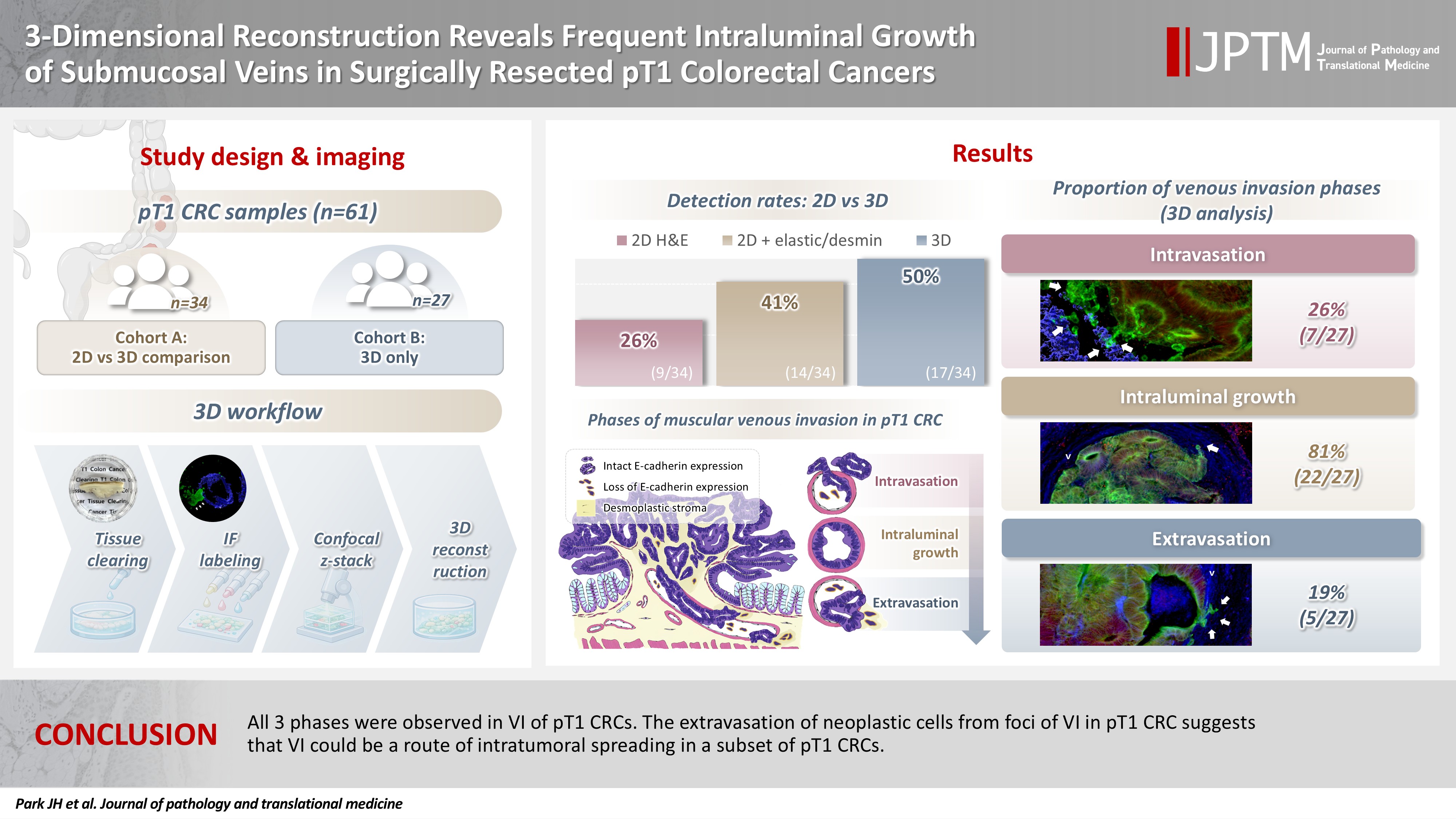
- 3-Dimensional reconstruction reveals frequent intraluminal growth of submucosal veins in surgically resected pT1 colorectal cancers
- Jihyun Park, Mi-Ju Kim, Yeon Wook Kim, Byong-Wook Lee, Junyoung Shin, Jinho Shin, Chan-Gi Pack, Dong-Hoon Yang, Jihun Kim, In Ja Park, Ralph H. Hruban, Seung-Mo Hong
- J Pathol Transl Med. 2026;60(2):246-262. Published online March 10, 2026
- DOI: https://doi.org/10.4132/jptm.2025.12.19
- 375 View
- 33 Download
-
 Abstract
 PDF
PDF - Background
Although venous invasion (VI) is associated with distant metastasis and observed in >50% of pT2–4 colorectal cancers (CRCs), the role of VI in pT1 CRCs is not well-defined. Methods: Thirty-four surgically resected pT1 CRCs were reevaluated for 2-dimensional (2D) VI using hematoxylin and eosin (H&E)–stained slides with additional elastic and desmin immunohistochemical staining (cohort A). Additionally, 27 pT1 CRCs without knowing VI status were selected for 3-dimensional (3D) VI evaluation only (cohort B). All 61 cases (cohorts A and B) were studied in 3D using tissue clearing. Results: VI was detected more commonly in 3D (17/34, 50.0%) than in 2D H&E slide evaluation (9/34, 26.5%, p = .047). When VI was identified in 3D (27/61, 44.3%), the most common phase was that of intraluminal growth (22/27, 81.5%), followed by intravasation (7/27, 25.9%) and extravasation (5/27, 18.5%). E-cadherin expression was characterized in 3D in foci of VI and varied in each phase of invasion. Conclusions: All three phases were observed in VI of pT1 CRCs. The extravasation of neoplastic cells from foci of VI in pT1 CRC suggests that VI could be a route of intratumoral spreading in a subset of pT1 CRCs.
- E-cadherin expression and tumor-stroma ratio as prognostic biomarkers of peritoneal recurrence in advanced gastric cancer: a digital image analysis-based stratification study
- Somang Lee, Binnari Kim
- J Pathol Transl Med. 2025;59(6):408-420. Published online November 6, 2025
- DOI: https://doi.org/10.4132/jptm.2025.08.27

- 3,134 View
- 117 Download
-
Abstract
PDF
- Background
Gastric cancer remains a significant global health burden, with a high peritoneal recurrence rates after curative surgery. E-cadherin and the tumor-stroma ratio (TSR) have been proposed as prognostic indicators, but their combined prognostic utility remains unclear. Methods: This retrospective study included 130 patients with T3/T4a gastric cancer who underwent curative gastrectomy at Ulsan University Hospital between 2014 and 2019. Immunohistochemistry for E-cadherin and Vimentin was performed. Digital image analysis using QuPath’s object classifier quantified E-cadherin expression and TSR. Results: Low E-cadherin expression was associated with diffuse-type histology and advanced T stage. Low TSR was linked to younger age, female sex, and XELOX treatment. In Kaplan-Meier analysis, low TSR showed a non-significant trend toward higher peritoneal recurrence (p = .054), while low E-cadherin expression was significantly associated with increased peritoneal recurrence (p = .002). Combined biomarker analysis also revealed a significant difference in recurrence-free survival (RFS) among the four groups (p = .005); patients with both high TSR and high E-cadherin expression experienced the most favorable RFS. In multivariable analysis, E-cadherin expression remained the only independent predictor of peritoneal recurrence (high vs. low; hazard ratio, 0.348; 95% confidence interval, 0.149 to 0.816; p = .015). Conclusions: E-cadherin and TSR reflect distinct tumor biology such as epithelial integrity and stromal composition, and their combined evaluation improves prognostic stratification. Digital image analysis enhances reproducibility and objectivity, supporting their integration into clinical workflows.
- Expression of E-cadherin and beta-catenin is Altered at Tumor Budding Sites, Whose Number is Associated with the Progression of Colorectal Carcinoma.
- Tae Jung Jang
- Korean J Pathol. 2009;43(6):523-527.
- DOI: https://doi.org/10.4132/KoreanJPathol.2009.43.6.523
- 4,899 View
- 50 Download
- 9 Crossref
-
Abstract
PDF
- BACKGROUND
Tumor budding is present in the stroma at the invasive margin of colorectal carcinomas (CRC). The disintegration of cell adhesion molecules is closely related to this process. This study investigated the role of tumor budding in the progression of CRC, and compared the expression of beta-catenin and E-cadherin between tumor budding and tumor center to determine whether epithelial-to-mesenchymal transitions (EMTs) occur in tumor budding. METHODS: The number of tumor budding (NTB) instances was determined in 58 cases of CRC, and immunoreactivities of E-cadherin and beta-catenin were compared at the tumor center and at the tumor budding site. Immunohistochemical staining for vimentin was also done.
RESULTS
Tumor budding was seen in 52 tumors (90%). There were significant associations between NTB and cliniopathologic parameters such as tumor depth, nodal metastasis and clinical stage. Expression of cytoplasmic and nuclear beta-catenin were significantly higher at tumor budding sites than in the tumor center. In contrast, expression of membranous and cytoplasmic E-cadherin were significantly higher in the tumor center than at the tumor budding sites. Vimentin was expressed at tumor budding foci of only 2 cases (3%).
CONCLUSIONS
This study suggests that EMT occurs at tumor budding, and that NTB may be a good marker for predicting a poor prognosis in CRC. -
Citations
Citations to this article as recorded by
- Spread Through Air Spaces in Colorectal Lung Metastases Signals Local Recurrenece and Reflects Morphologic Aggressiveness of the Primary Tumor
Taketo Nakai, Satoru Morita, Yutaka Kurebayashi, Masayoshi Monno, Ryo Seishima, Kohei Shigeta, Koji Okabayashi, Mari Mino‐Kenudson, Yuko Kitagawa, Keisuke Asakura
Pathology International.2026;[Epub] CrossRef - E-Cadherin Expression Varies Depending on the Location within the Primary Tumor and Is Higher in Colorectal Cancer with Lymphoid Follicles
Adam R. Markowski, Konstancja Ustymowicz, Anna J. Markowska, Wiktoria Romańczyk, Katarzyna Guzińska-Ustymowicz
Cancers.2023; 15(12): 3260. CrossRef - Gland Attenuation, a Novel Morphological Feature of Colorectal Cancer: Evidence for an Epithelial-Mesenchymal Transition
Tae-Hwa Baek, Dong-Wook Kang, Joo-Heon Kim, Hyun-Jin Son
Annals of Coloproctology.2018; 34(4): 187. CrossRef - Differential membranous E-cadherin expression, cell proliferation and O-GlcNAcylation between primary and metastatic nodal lesion in colorectal cancer
Tae Jung Jang
Pathology - Research and Practice.2016; 212(2): 113. CrossRef - Differential β-catenin expression levels are associated with morphological features and prognosis of colorectal cancer
ZHAO-HUA GAO, CHONG LU, MEI-XIAN WANG, YI HAN, LI-JUAN GUO
Oncology Letters.2014; 8(5): 2069. CrossRef - C4.4A is associated with tumor budding and epithelial–mesenchymal transition of colorectal cancer
Ryota Oshiro, Hirofumi Yamamoto, Hidekazu Takahashi, Masahisa Ohtsuka, Xin Wu, Junichi Nishimura, Ichiro Takemasa, Tsunekazu Mizushima, Masataka Ikeda, Mitsugu Sekimoto, Nariaki Matsuura, Yuichiro Doki, Masaki Mori
Cancer Science.2012; 103(6): 1155. CrossRef - Assessment of tumor budding in colorectal carcinoma: Correlation with β-catenin nuclear expression
S. El-Gendi, A. Al-Gendi
Journal of the Egyptian National Cancer Institute.2011; 23(1): 1. CrossRef - Cell Surface Markers in Colorectal Cancer Prognosis
Larissa Belov, Jerry Zhou, Richard I. Christopherson
International Journal of Molecular Sciences.2010; 12(1): 78. CrossRef - Lethal Giant Larvae2 Expression Is Reduced or Localized at Cytoplasm in Colon Adenomas and Adenocarcinomas
Tae Jung Jang
The Korean Journal of Pathology.2010; 44(5): 488. CrossRef
- Spread Through Air Spaces in Colorectal Lung Metastases Signals Local Recurrenece and Reflects Morphologic Aggressiveness of the Primary Tumor
- The Loss of E-cadherin is Associated with the Epigenetic Alteration of CDH1 in Breast Cancer and it is also Associated with an Abnormal beta-catenin Expression in Lobular Carcinoma.
- Gwangil Kim, Ji Young Kim, Hee Jung An, Haeyoun Kang, Tae Heon Kim, Jung Yon Shim, Jin Hyung Heo, Hai Lin Park, Young Kil Choi
- Korean J Pathol. 2009;43(5):400-407.
- DOI: https://doi.org/10.4132/KoreanJPathol.2009.43.5.400
- 4,535 View
- 41 Download
- 1 Crossref
-
Abstract
PDF
- BACKGROUND
APC and E-cadherin are the key molecules in the Wnt/beta-catenin pathway. We attempted to define the epigenetic alteration of APC and CDH1 (the E-cadherin gene) and the expression of Wnt-related molecules in human mammary carcinomas.
METHODS
Sixty-four mammary carcinomas, including 52 invasive ductal carcinomas (IDCs) and 12 invasive lobular carcinomas (ILCs), were evaluated using methylation-specific PCR and immunohistochemistry. We performed immunohistochemistry for E-cadherin, beta-catenin, APC, Wnt1, cyclin D1, ER, PR and C-erb B2.
RESULTS
Hypermethylation of APC and CDH1 was observed in 38 (59%) and 28 (44%) cases, respectively. CDH1 hypermethylation in ILCs was increased compared to that in IDCs (p=0.002) and it was associated with the loss of E-cadherin (p=0.02) and beta-catenin (p=0.042). APC methylation was positively correlated with the ER expression (p=0.021). Abnormal cytoplasmic localization of beta-catenin was found in 10 cases and any expression was not detected in six cases. In ILCs, the E-cadherin or beta-catenin expression was markedly decreased compared to that in IDCs (p<0.001 in both).
CONCLUSIONS
Methylation of APC or CDH1 was relatively frequent in mammary carcinomas. The loss of E-cadherin in mammary carcinoma was associated with CDH1 methylation, and abnormal beta-catenin expression was related to the loss of E-cadherin in ILC. -
Citations
Citations to this article as recorded by- Wnt/β-catenin signaling pathway activation reverses gemcitabine resistance by attenuating Beclin1-mediated autophagy in the MG63 human osteosarcoma cell line
Hao Tao, Feng Chen, Haifei Liu, Yanling Hu, Yingzhen Wang, Haiyan Li
Molecular Medicine Reports.2017; 16(2): 1701. CrossRef
- Wnt/β-catenin signaling pathway activation reverses gemcitabine resistance by attenuating Beclin1-mediated autophagy in the MG63 human osteosarcoma cell line
- Expression of E-cadherin in Chromophobe Renal Cell Carcinoma and Its Prognostic Implication.
- Eun Jung Jung, Heae Sung Park, Sun Young Min, Jeong Mo Bae, Kyung Chul Moon
- Korean J Pathol. 2009;43(3):238-243.
- DOI: https://doi.org/10.4132/KoreanJPathol.2009.43.3.238
- 4,946 View
- 42 Download
- 1 Crossref
-
Abstract
PDF
- BACKGROUND
Chromophobe renal cell carcinoma is a category of renal cell carcinoma composed of histologically characteristic tumor cells. E-cadherin is an intercellular adhesion protein that has been correlated with tumor aggressiveness in many carcinomas, including clear cell renal cell carcinoma. However, the significance of an E-cadherin expression in chromophobe renal cell carcinoma is not known.
METHODS
We evaluated the E-cadherin expression status of 65 chromophobe renal cell carcinomas by performing immunohistochemical staining with the tissue microarray method. The percentage of positively stained tumor cells was evaluated and this was then classified into two categories: a low expression where 0 to 25% of the cells are positive, and a high expression where more than 25% of the cells are positive.
RESULTS
Among 65 cases, 11 cases (17%) showed a low expression, and 54 cases (83.0%) showed a high expression. The tumors with low expression were more likely to have a higher stage but this was not significant (p=0.056). On the survival analysis, a low E-cadherin expression was significantly associated with poor cancer-specific survival (p=0.005) and progression-free survival (p=0.003).
CONCLUSIONS
The E-cadherin expression is a good prognostic marker for survival in patients with chromophobe renal cell carcinoma. -
Citations
Citations to this article as recorded by- A systematic review and meta-analysis combined with bioinformatic analysis on the predictive value of E-cadherin in patients with renal cell carcinoma
Zikuan Zhang, Bo Xue, Yongquan Chen, Yuan Shao, Dongwen Wang
Expert Review of Molecular Diagnostics.2024; 24(9): 859. CrossRef
- A systematic review and meta-analysis combined with bioinformatic analysis on the predictive value of E-cadherin in patients with renal cell carcinoma
- The Relationship between Prognostic Factors and the Expression Pattern of Fascin and E-cadherin in Renal Cell Carcinoma.
- Sung Hee Kang, Seoung Wan Chae, Kyoung Bun Lee, Dong Hoon Kim, Min Kyoung Kim, Jin Hee Sohn
- Korean J Pathol. 2009;43(2):139-144.
- DOI: https://doi.org/10.4132/KoreanJPathol.2009.43.2.139
- 3,550 View
- 19 Download
-
Abstract
PDF
- BACKGROUND
Fascin is associated with motility in various transformed cells. Overexpression of fascin is known to aid in the progression of some cancers and is associated with a poor prognosis. E-cadherin is a major protein of epithelial cells and its expression is involved in the regulation of cell proliferation and differentiation. The aim of this study was to determine the expression pattern for fascin and E-cadherin and how it is related to the prognostic factors for renal cell carcinoma (RCC).
METHODS
The expression of fascin and E-cadherin was evaluated in 208 RCCs including 175 clear cell, 20 papillary, and 9 chromophobe types using tissue array analysis.
RESULTS
The expression of fascin increased as the tumor stage (p=0.00) and Fuhrman grade (p=0.00) increased. A high positive rate of expression for fascin was observed in cases with sarcomatoid changes (p=0.27). E-cadherin expression was seen in the distal tubules and collecting ducts of normal kidneys with a membranous pattern. The positive rate of expression for E-cadherin increased as the Fuhrman grade increased (1, 0%; 2, 23.2%; 3, 34.9%; and 4, 53.8%, p=0.00). An inverse correlation in RCCs was observed in the expression of fascin and E-cadherin (p=0.026, r=-0.158).
CONCLUSIONS
In patients with RCC, the increased expression of fascin and E-cadherin was positively correlated to poor prognostic factors such as a higher Fuhrman nuclear grade and advanced pTNM stage.
- Expression of E-cadherin and p53 Proteins in Gastric Adenocarcinoma.
- Sook Hee Hong, Mee Sook Roh
- Korean J Pathol. 1999;33(2):80-87.
- 2,181 View
- 10 Download
-
Abstract
- The gastric carcinoma shows various molecular and genetic alterations in its development and progression. There are evidences that the changes of the expression of cell adhesion molecules affect the morphogenesis of the tumor as well as the tumor progression and metastasis. The purpose of this study is the evaluation of the expression pattern of a cell adhesion molecule, E-cadherin, and a tumor suppression gene, p53, by immunohistochemical stain and the relationship of their expressions with clinicopathologic findings in gastric adenocarcinoma tissue. The E-cadherin expression was absent or reduced in 93 cases (73.2%) and p53 was positive in 98 cases (77.2%) of 127 gastric adenocarcinomas. The frequency of reduced E-cadherin expression was significantly higher in poorly differentiated adenocarcinomas (p=0.04) and in diffuse type (p=0.01), but that of p53 positivity was not significantly correlated with tumor differentiation. Both proteins showed no correlation with depth of invasion, lymph node and distant metastasis, and tumor stage. There was no correlation between E-cadherin and p53 expression. This study indicates that the altered expressions of E-cadherin and p53 are associated with the development of intestinal and diffuse types of gastric adenocarcinoma and the differentiation of the gastric adenocarcinoma is affected by cell adhesion mediated by E-cadherin, but the modes of tumor progression and metastasis are not affected by E-cadherin and p53.
- Usefulness of E-Cadherin Expression in Malignant Effusion .
- Sung Jig Lim, Gou Young Kim, Youn Wha Kim, Yong Koo Park, Juhie Lee, Moon Ho Yang, Nam Hee Won
- J Pathol Transl Med. 1999;10(2):121-126.
- 2,097 View
- 12 Download
-
Abstract
PDF
- The usefulness of E-cadherin immunostaining as a marker of malignancy in the body fluids was investigated in the present study. Thirty-three histologically proven cases of cell blocks from the pleural, peritoneal, and pericardial fluids were studied by immunocytochemistry for E-cadherin antibody using LSAB method. These cases were cytologically diagnosed as adenocarcinoma (25 cases) and atypical cells (8 cases). Tumor cells showed strong positive membranous staining for E-cadherin antibody in 21 out of 25 cases (84%) of adenocarcinoma. E-cadherin staining was not found in 6 of 8 cases of suspicious maligancy. The sensitivity and specificity were 84% and 75%, respectively. Reactive mesothelial cells and inflammatory cells scattered were all negative. In conclusion, E-cadherin is an useful adjunctive marker to distinguish reactive mesothelial cells from the carcinoma cells in the body fluids.
- The Significance of the Expression of p53, E-cadherin, nm23, CD44, and Tumor Angiogenesis in Colorectal Adenocarcinoma.
- Sung Suk Paeng, Hee Jin Chang, Jung Il Suh
- Korean J Pathol. 1997;31(4):314-325.
- 2,012 View
- 12 Download
-
Abstract
PDF
- Many oncogenes and tumor supressor genes have been identified and studied in colorectal carcinoma. Among them, p53 is a tumor supressor gene and its mutation is frequently noted in human tumors. E-cadherin is a cell adhesion molecule and associated with tumor differentiation. CD44 is a cell surface glycoprotein that plays a role in cell migration and metastasis. nm23 is a gene known to lower metastatic potential of tumors and has been proposed to be a metastasis supressor gene. Tumor angiogenesis is required for the expansion of the primary tumor and metastasis and its degree is related to the potential of malignancy. We studied the expression of p53, E-cadherin, nm23, CD44 and tumor angiogenesis in 36 cases of colorectal adenocarcinomas. They were compared with previously known prognostic factors such as the stage, tumor size, depth of invasion, differentiation, presence of lymphatic or venous invasion, the lymph node and distant metastasis. The results were as follows. 1) The expression of p53 was not significantly associated with any prognostic factors. 2) The expression of E-cadherin was significantly associated with tumor differentiation. In the well differentiated adenocarcinomas, its expression was higher than in the poorly differentiated adenocarcinoma. 3) The expression of nm23 was also significantly associated with tumor differentiation. In carcinoma with lymph node metastasis, the expression of nm23 was reduced, but statistically it was not significant. 4) The expression of CD44 was higher in tumors with lymph node metastasis than in tumors without lymph node metastasis, but it was not statistically significant. 5) The degree of microvessel density was significantly associated with lymphatic invasion. According to the above results, the expression of E-cadherin and nm23 are related to the differentiation of the tumor and tumor angiogenesis is related to the lymphatic invasion of the colorectal adenocarcinoma.
- Significance of Cyclin E, p53, E-cadherin, and beta-Catenin Expressions in Gastric Adenocarcinomas.
- Long Pei Xuan, Mi Ja Lee, Chae Hong Suh
- Korean J Pathol. 2004;38(4):213-220.
- 2,076 View
- 22 Download
-
Abstract
PDF
- BACKGROUND
Gastric cancer is reported to be one of the leading causes of mortality in Korea. Our aim was to evaluate the clinicopathologic usefulness of cyclin E, p53, E-cadherin and beta-catenin expressions in gastric adenocarcinomas.
METHODS
Immunohistochemical staining was performed on the 40 early gastric carcinoma (EGC) cases and 69 advanced gastric carcinoma (AGC) cases to examine the relationship with the clinicopathologic parameters.
RESULTS
Cyclin E and p53 expressions were significantly lower in the mucosal or submucosal invasion group compared with those in the muscle invasion and subserosal or serosal invasion groups. Cyclin E expression was significantly higher in the node-positive group compared with that in the node-negative group. The loss of beta-catenin expression was significantly higher in the node-negative group. p53 expression was significantly higher in the intestinal type group than that in the diffuse type group. Loss of E-cadherin expression was significantly higher in the diffuse type group. Cyclin E expression correlates with p53 expression.
CONCLUSIONS
The depth of invasion seems to correlate with cyclin E and p53 expressions. Lymph node metastasis may correlate with loss of beta-catenin expression.
- E-Cadherin Expression and DNA Ploidy Analysis in Invasive Squamous Cell Carcinoma of the Uterine Cervix Comparison with those of CIN.
- Yoo Jin Kim, Mee Young Sol, Man Ha Huh, Sun Kyung Lee
- Korean J Pathol. 1997;31(6):557-565.
- 2,058 View
- 13 Download
-
Abstract
PDF
- Epithelial cadherin (E-cadherin) is a Ca2+ -dependent cell-cell adhesion molecule that connects cells via homotypic interactions. Its function is critical in the induction and maintenance of cell polarity and differentiation, and its loss is associated with an invasive and poorly differentiated phenotype in a wide range of tumors. Formalin-fixed, paraffin-embedded tissue sections from 36 cases of cervical intraepithelial neoplasia (CIN) and 14 cervical squamous cell carcinomas were investigated for the expression of E-cadherin immunohistochemically. While E-cadherin expression was usually restricted on the cell membrane of basal and parabasal cells in normal cervix, the presence of cytoplasmic E-cadherin was found to be associated with its grade in CIN lesions. Also, marked cytoplasmic staining was commonly revealed in poorly differentiated ones than well-differentiated squamous cell carcinomas. More intense reactivity of cytoplasmic E-cadherin was frequently seen in the foci of invasion than adjacent carcinoma in situ, and in its periphery than the center of tumor islands. In addition, DNA ploidy and S-phase fraction of squamous cell carcinomas were analyzed and compared with those of CIN lesion. We found that invasive squamous cell carcinomas more frequently disclosed DNA aneuploidy than CIN lesions, and there was correlation between cytoplasmic E-cadherin expression and DNA aneuploidy. Also, cytoplasmic E-cadherin-reactive cervical neoplasms had a higher rate of cell proliferation than that of membranous E-cadherin-reactive cases. These data suggest that the increased cytoplasmic E-cadherin expression may represent one of the abnormalities underlying the loss of polarity and invasiveness of cancer cells, and the abnormal E-cadherin expression combined with/without DNA ploidy or S-phase fraction may serve as a prognostic indicator.
- Immunocytochemical Expression of E-cadherin in Cell Blocks of Serous Effusions.
- Byung Heon Kim, O Jun Kwon
- J Pathol Transl Med. 2001;12(2):81-88.
- 2,011 View
- 18 Download
-
Abstract
PDF
- The differentiation between reactive mesothelial and carcinoma cells in serous effusion cytology can be a diagnostic challenge based on morphology alone. The expression of some cell adhesion molecules may be helpful in the differential diagnosis. This study evaluated the usefulness of E-cadherin immunocytochemistry for discrimination of carcinoma cells from reactive mesothelial cells. Alcohol fixed, paraffin embedded cell blocks taken from 42 reactive and 102 malignant serous effusions with histologically confirmed diagnoses were immunostained with monoclonal antibody to E-cadherin by LSAB method. E-cadherin expression was identified in only 2 benign reactive serous effusions(5%) whereas 91 malignant serous effusions(89%) expressed E-cadherin. The differences in immunostaining for E-cadherin between reactive and malignant serous effusions were statistically significant(p<0.001). The sensitivity and specificity of the E-cadherin immunostaining for carcinoma cells were 89% and 95%, respectively. In conclusion, E-cadherin is a useful diagnostic adjunct for differentiation between reactive mesothelial and carcinoma cells in serous effusions.
Case Report
- A Positive Hybrid (HMW-CK and E-Cadherin) Carcinoma in situ Arising in a Phyllodes Tumor of the Breast: A Case Report.
- Yun Kyung Kang, Young Hyeh Ko
- Korean J Pathol. 2008;42(2):113-117.
- 2,123 View
- 22 Download
-
Abstract
PDF
- Malignant transformation in phyllodes tumor (PT) is uncommon and almost always confined to the stromal component. Epithelial changes like hyperplasia, metaplasia, and varying degrees of atypia are not uncommon in PT, whereas carcinomatous change is extremely rare. We report a 37-year-old woman with carcinoma in situ (CIS) arising in a benign PT. Grossly, it was a well circumscribed, 4.5 cm-sized mass. The CIS component was confined to the PT and showed overlapping ductal and lobular features with coexpression of E-cadherin and high molecular weight cytokeratin (HMW-CK). The present case emphasizes that careful investigation of multiple microscopic sections is mandatory to find a small carcinomatous lesion within PT. Expression of E-cadherin and HWM-CK in this hybrid CIS suggests that intraepithelial neoplasia of the breast arising in PT may be derived from a common progenitor of the terminal duct-lobular unit.
Original Articles
- Immunohistochemical Study of E-cadherin Expression in Gastric Adenocarcinomas.
- Jee Yeon Kim, Mee Young Sol, Sun Kyung Lee
- Korean J Pathol. 1997;31(8):745-753.
- 2,220 View
- 25 Download
-
Abstract
PDF
- E-cadherin (ECD) is a Ca++ -dependent adhesion molecule which plays a major role in the maintenance of intercellular adhesion in epithelial tissues. The expression pattern of ECD in 77 surgically resected gastric adenocarcinomas was examined by immunohistochemistry, using a rat monoclonal antibody raised against murine E-cadherin (DECAM-1). ECD was strongly expressed uniformly at cell to cell borders in normal gastric epithelium without exception. But, various staining patterns were observed in the cancer tissues. The frequency of tumors with preserved ECD expression (Pre-type) and reduced ECD expression (Rd-type) was 44% and 56%, respectively. Using Lauren's classification, the high frequency of the Pre-type expression in adenocarcinoma of the intestinal type was significantly higher than that in adenocarcinoma of the diffuse type (p<0.05). But, no significant correlation between the ECD expression and the gross type, invasion depth, growth pattern or metastasis was observed. These results suggest that ECD might play a key role in the morphogenesis of gastric adenocarcinoma.
- E-Cadherin Expression in Breast Carcinoma: Correlation with Tumor Grade and Hormone Receptor.
- Haeng Ji Kang, Chan Pil Park, Chan Kum Park
- Korean J Pathol. 1997;31(11):1172-1179.
- 2,343 View
- 10 Download
-
Abstract
- E-cadherin (E-CD), a Ca2+ -dependent adhesion molecule, plays a major role in the maintenance of intercellular junctions in normal epithelial cells in most organs. Recently, a correlation has been observed between a loss of E-CD and increased invasiveness of neoplastic cells. In this study, E-CD expression in the breast carcinoma was investigated using monoclonal antibody, anti-E-CD by immunohistochemical method. Expression of E-CD were evaluated in 57 breast carcinomas and correlated with their tumor grade, lymph node involvement, and hormonal receptor status. Histological types included in this study were 54 invasive ductal carcinomas (IDCs) of otherwise not specified and 3 invasive lobular carcinomas. Cases of histologic grade I IDC were 6, grade II 30, and grade III 18. Of 54 IDCs 39 (72.2%) showed moderate to strong linear staining at the cell borders regardless of their histologic grade, status of lymph node metastasis, and status of hormone receptor. Staining intensity of E-CD was reduced in 54 cases (83%) of IDC when compared with that of normal or benign breast lesions (P<0.01). All seven cases of intraductal carcinoma, which were included in 54 IDCs showed one or two grade reduced expression of E-CD than that of infiltrative lesions. Three invasive lobular carcinomas showed strong (1 case), moderate (1 case), and negative reactivity (1 case). The data indicated that loss of E-CD expression is a crucial event in the development of breast carcinoma.
- E-cadherin Expression Loss in T1 Invasive Ductal Carcinoma of the Breast as a Predictive Marker for Lymph Node Metastasis.
- Eun Kyung Kim, Aysegul Sahin
- Korean J Pathol. 2005;39(3):187-191.
- 2,863 View
- 44 Download
-
Abstract
PDF
- BACKGROUND
E-cadherin is a transmembrane glycoprotein, which has been shown to mediate calcium-dependent epithelial cell adhesion. A loss of E-cadherin expression has been associated with the tumor invasion and metastatic potential in some human cancers. The objective of this study was to evaluate E-cadherin expression in T1 breast ductal carcinomas in order to determine whether the loss of E-cadherin expression is correlated with lymph node metastasis.
METHODS
One hundred seventy nine patients with breast invasive ductal carcinoma, measuring less than 2 cm, were enrolled in this study. The subjects were divided into two groups on the basis of the status of the ipsilateral axillary lymph node, T1N1 (lymph node positive, n=91) or T1N0 (lymph node negative, n=88). None of the patients in this study had undergone preoperative chemotherapy. Formalin-fixed paraffin-embedded tissue sections of the primary breast cancers were stained by immunohistochemistry, using a mouse monoclonal antibody against E-cadherin. E-cadherin expression was designated as either positive (complete membranous staining) or negative (absent or incomplete membranous staining).
RESULTS
Benign breast parenchyma adjacent to invasive carcinoma was positive for E-cadherin. The loss of E-cadherin expression in the tumor was observed in 42% of patients of the T1N1 group, and in 24% of the T1N0 group. There was a significant correlation between the loss of E-cadherin expression and lymph node metastasis in the examined breast invasive ductal carcinomas (p=0.011).
CONCLUSIONS
Our findings suggest that E-cadherin is an important molecule with regard to both tumor cell adhesion and metastasis, and its absence may constitue an early event in metastatic development. Therefore, E-cadherin may be a useful predictive marker for nodal metastasis in patients suffering from invasive ductal carcinoma.
- Expression of Claudin-1, p53 and E-cadherin in Pseudoepitheliomatous Hyperplasia and Squamous Cell Carcinoma of the Head and Neck.
- Keum Ha Choi, Jae Hong Lim, Ju Hyung Lee, Keun Sang Kwon, Ho Lee, Ho Sung Park, Myoung Ja Chung, Woo Sung Moon, Jae Soon Eun, Dong Geun Lee, Kyu Yun Jang
- Korean J Pathol. 2008;42(5):287-293.
- 2,592 View
- 26 Download
-
Abstract
PDF
- BACKGROUND
Pseudoepitheliomatous hyperplasia (PEH) is a reactive proliferation of surface epithelium and can be confused with invasive squamous cell carcinoma (SCC) in head and neck biopsy specimens. To distinguish PEH from invasive SCC, immunohistochemical staining for claudin-1, E-cadherin and p53 was performed. METHODS: Eighteen cases of PEH and 29 invasive SCC from head and neck lesions were immunostained and examined. RESULTS: The invasive SCC showed increased staining of claudin-1 (p<0.001) and p53 (p<0.001) and decreased staining of E-cadherin (p=0.005) compared to the PEH specimens. The combined score calculated by adding the positive sum of claudin-1 and p53 and subtracting E-cadherin was useful for the differentiation of SCC from PEH (89.7% sensitivity and 88.9% specificity, p<0.001). CONCLUSION: The combined immunostaining for claudin-1, p53 and E-cadherin may help differentiate PEH from invasive SCC. The results of this study suggest that the increased expression of claudin-1 and p53 and the decreased expression of E-cadherin maybe markers for the aggressive growth of invasive SCC.
- Expression of Osteopontin, ZO-1 and E-cadherin in Adenoma and Adenocarcinoma of the Colon.
- Yu Kyung Jeong, Mi Ja Lee, Sung Chul Lim, Keun Hong Kee, Ho Jong Jeon, Chae Hong Suh
- Korean J Pathol. 2005;39(4):242-250.
- 2,236 View
- 17 Download
-
Abstract
PDF
- Background
: The expressions of osteopontin (OPN), zonula occludens-1 (ZO-1) and E-cadherin, known as cell adhesion-associated substances, were examined in adenoma and adenocarcinoma of the colon. The relationship of their expressions with clinicopathologic factors was examined to investigate the roles of these proteins in the development, invasion or metas- tasis of colon adenocarcinoma. Methods : The expressions of OPN, ZO-1, and E-cadherin were examined in 54 cases of adenoma and 67 cases of adenocarcinoma of the colon by immunohistochemical staining. Results : The expression of OPN in colon adenocarcinoma correlated with staging (p=0.012) and distant metastasis (p=0.021). The expression of ZO-1 was closely related with tumor cell differentiation (p<0.001), and the reduced expression of E-cadherin was associated with tumor cell differentiation (p=0.05) and lymph node metastasis (p<0.001). Co-expression of ZO-1 and E-cadherin was significantly associated with tumor cell differentiation, and the expressions of ZO-1 and E-cadherin were reduced or lost in all cases (5 cases) of poorly differentiated adenocarcinoma. Conclusions : Our data suggest that OPN is involved in the process of invasion and metastasis of colon adenocarcinoma, and ZO-1- and E-cadherin-mediated cell adhesion may play an important role in the differentiation of colon adenocarcinoma.
- The Differential Expressions of the Epithelial-Mesenchymal Transition Regulator, Slug and the Cell Adhesion Molecule, E-cadherin in Colorectal Adenocarcinoma.
- Ran Hong, Dong Yul Choi, Sung Chul Lim, Chae Hong Suh, Keun Hong Kee, Mi Ja Lee
- Korean J Pathol. 2008;42(6):351-357.
- 2,536 View
- 24 Download
-
Abstract
PDF
- BACKGROUND
Slug is a member of the Snail family of transcription factors, and it plays a crucial role in the regulation of the epithelial-mesenchymal transition by suppression of several epithelial proteins and adhesion molecules, including E-cadherin. METHODS: The aim of the present study was to examine the significance between the expression of Slug in colorectal adenocarcinoma (CRA) specimens and the clinicopathological parameters of CRA, as determined by immunohistochemical analysis, and to determine the correlation between the Slug and E-cadherin expressions in non-neoplastic colorectal mucosa (n=45), primary CRA (n= 109) and metastatic CRA (n=17). A semiquantitative scoring system was applied based on the intensity and extent of the positive immunohistochemical staining. RESULTS: The expressions of Slug and E-cadherin were associated with the depth of tumor invasion (pT) (p=0.019, p=0.001, respectively), and these expressions showed a significant inverse correlation (p<0.001) each other. CONCLUSIONS: Our results demonstrated a positive role for Slug in the development of CRA, and Slug is a mediator of tumor invasion in CRA. In addition, an up-regulated Slug expression is significantly correlated with the loss of an E-cadherin expression, which suggests that Slug may play some role in the epithelial-mesenchymal transition (EMT) by down-regulating the E-cadherin expression.
- Expression of the nm23 and E-cadherin Proteins in Breast Carcinoma.
- Jean a Kim, Won Il Kim, Sang In Shim, Chang Suck Kang, Kyo Young Lee, Young Shin Kim
- Korean J Pathol. 1998;32(1):29-34.
- 1,828 View
- 20 Download
-
Abstract
PDF
- Expression of the nm23 and E-cadherin proteins has been studied in a number of tumors. Reduced expression of the nm23 and E-cadherin proteins seems to be associated with metastasis or disease progression in some tumors, including breast carcinoma. To assess the role of nm23 and E-cadherin in tumor differentiation and metastasis of breast carcinoma, immunohistochemical staining for the nm23 and E-cadherin proteins was performed in paraffin embedded tumor samples from 86 breast carcinomas. The results were as follows: 1) Expression of the nm23 protein in breast carcinoma was strong positive in 32 cases (37.2%), weak positive in 26 cases (30.2%), and negative in 28 cases (32.6%) of the cases. Expression of the nm23 protein in breast carcinoma decreased according to histological grade and lymph node metastasis, but was not statistically significant. 2) Expression of the E-cadherin protein in breast carcinoma was strong positive in 50 cases (58.1%), weak positive in 24 cases (27.9%), and negative in 12 cases (14%) of the cases. Expression of the E-cadherin protein in breast carcinoma decreased according to histological grade and lymph node metastasis, but was not statistically significant. 3) There was a statistically significant correlation between the expression of the nm23 protein and the E-cadherin protein in breast carcinoma (p<0.05). These results suggest that the expression of the nm23 and E-cadherin proteins is related to tumor differentiation, and may also be an useful prognostic factor in breast carcinoma.
- E-Cadherin Expression in Renal Cell Carcinoma according to the Mainz Classification.
- Ju Han Lee, Hyun Deuk Cho, Dale Lee, Nam Hee Won
- Korean J Pathol. 1999;33(12):1131-1138.
- 2,540 View
- 27 Download
-
Abstract
PDF
- According to the Mainz classification, renal cell carcinoma (RCC) consists of three subtypes: each has characteristic genetic alterations within the chromosomal or mitochondrial DNA. The three subtypes are: clear cell type, chromophil type, and chromophobe type. E-cadherin is a Ca++-dependent adhesion molecule which plays a major role in the maintenance of intercellular adhesion in epithelial tissues. In a normal kidney, E-cadherin is expressed in the distal tubule and the collecting duct, but not in the proximal tubule. We reclassified 110 cases of RCC according to mainz classification. Immunohistochemical staining for E-cadherin was done on twenty eight cases of RCC, including 18 cases of clear cell type, four cases of chromophil type, and six cases of chromophobe type. The results were as follows: 1) of the 110 cases of RCC, 96 cases (87.3%) were of clear cell type, four cases (3.6%) of chromophil type, and ten cases (9.1%) of chromophobe type, 2) there was no significant correlation between the nuclear grade and clinical stage according to each subtype, 3) E-cadherin expression showed a strong positive reaction along the cell membranes in all six cases of chromophobe type. The differential expression of E-cadherin in RCC may suggest that the chromophobe type may have different biologic characteristics from other types of RCC.
- Immunohistochemical Study of p53 and E-cadherin Proteins in Prostate Carcinoma.
- Lee So Maeng, Won Il Kim, Kyo Young Lee, Young Shin Kim, Chang Suk Kang, Sang In Shim
- Korean J Pathol. 1998;32(3):215-221.
- 1,951 View
- 15 Download
-
Abstract
PDF
- Considerable controversy exists concerning the value of histomorphological data in the assessment of the malignant potential of prostate carcinomas. Mutations in the p53 gene resulting in the accumulation of altered p53 proteins with prolonged half-life have been found in a large variety of human malignancies. E-Cadherin is a specific epithelial cell-to- cell adhesion molecule which has previously been found to be expressed in well-differentiated non-invasive carcinoma cell lines, but it is lost in many poorly differentiated invasive cell lines. We performed immunohistochemical staining of p53 and E-cadherin in formalin fixed paraffin embedded tissues of 58 primary prostatic carcinomas. The expression rates of p53 and E-cadherin proteins in prostate carcinoma were positive in 15.5% and 44.8% of the cases, respectively. Histologically high-grade prostate carcinoma shows an increased expression of the p53 protein and a decreased one of the E-cadherin protein (P<0.05). The expression rates of the E-cadherin protein in prostate carcinoma decreased significantly according to the higher clinical stages and PSA levels (P<0.05). There was no accordance between the expression rate of p53 and E-cadherin. There were no significant correlation between each of the clinical stages and the expression rate of p53 protein or the PSA levels and the expression rates of p53 protein (P<0.05). Based on the present study, the expression of p53 and down regulation of E-cadherin are correlated with tumor progression and metastasis, and may be a useful prognostic factor in prostate carcinoma.
- Correlation of Expression of E-Cadherin, alpha-Catenin, beta-Catenin, and Clinicopathologic Parameters in Colorectal Adenocarcinomas.
- Hyoung Joong Kim, Tae Jin Lee, Eon Sub Park, Jae Hyung Yoo
- Korean J Pathol. 2000;34(4):264-272.
- 2,120 View
- 17 Download
-
Abstract
PDF
- The E-cadherin, alpha-catenin, and beta-catenin expressions were immunohistochemically investigated in paraffin-embedded materials of 80 cases of colorectal adenocarcinomas. The staining similar to normal colorectal mucosa with preserved strong membranous staining pattern was considered normal or preserved expression. The X2 test was used to analyse the statistical correlation of cadherin/catenin expression with clinicopathologic parameters and the Breslow test for the correlation with survival length. Normal colorectal mucosa showed strong membranous expression of cadherin/catenin complex. The reduced E-cadherin, alpha-catenin, and beta-catenin expression were found in 53/80 (66.3%), 46/80 (57.5%), and 44/80 (55.5%) cases of colorectal cancers examined, respectively. There were significant correlations between E- cadherin and alpha -catenin (p=0.035), and between alpha-catenin and beta-catenin (p=0.013). The reduced E-cadherin expression was associated with histologic dedifferentiation, tumor depth, lymph node metastasis, clinical stage (p<0.05), poor clinical outcome in stage II (p=0.016) and the reduced alpha-catenin expression with lymph node metastasis and clinical stage (p<0.05). Reduced expression of two or more proteins was correlated with lymph node matastasis, histologic dedifferentiation, clinical stage, and survival (p<0.05). The present study demonstrates a significant down-regulation of E-cadherin and alpha-catenin expression in colorectal cancer is associated with tumor invasiveness, histologic dedifferentiation, lymph node metastasis, and clinical stage. These results suggest that E-cadherin and alpha-catenin may be useful markers of invasiveness, lymph node metastatic potential, and clinical stage and of value as prognostic markers in the earlier stage. Further studies are needed to confirm the prognostic value of these cadherin/catenin complex.
- Immunohistochemical Expression of p53, E-cadherin, and nm23 Proteins in Metastatic Carcinoma of Neck Lymph Node and Corresponding Primary Carcinoma.
- Jong Kook Kim, O Jun Kwon, Byung Heon Kim
- Korean J Pathol. 2000;34(9):615-624.
- 1,931 View
- 17 Download
-
Abstract
PDF
- This study was carried out to evaluate the immunohistochemical expressions of p53, E-cadherin, and nm23 proteins in 114 cases of metastatic carcinoma of the neck lymph node (MTLNCA) and corresponding primary carcinoma (PRCA). The positive expressions of p53, E-cadherin, and nm23 proteins were 62.3%, 58.8% and 64.0%, respectively in PRCA, and 40.4%, 38.6%, and 43.9%, respectively in MTLNCA with significant down-regulation from PRCA to MTLNCA (p<0.05). The down-regulation was correlated with female gender, moderate and poor differentiation, and adenocarcinoma in p53 protein, female gender, respiratory and gastrointestinal carcinoma in E-cadherin protein, and female gender, respiratory carcinoma, moderate differentiation, and squamous cell carcinoma in nm23 protein (p<0.05). There was no significant relationship among expressions of p53, E-cadherin, and nm23 proteins (p<0.05). In conclusion, these results suggest that the expressions of p53, E-cadherin, and nm23 proteins seem to be down-regulated from PRCA to MTLNCA and this down-regulation may play a role in invasion and metastasis.
- Expression of E-cadherin, Matrix Metalloproteinase, and Vascular Endothelial Growth Factor in Squamous Cell Carcinoma and Adenocarcinoma of the Lung.
- Ji Sun Song, Mee Yon Cho, Kwang Hwa Park, Soon Hee Jung, Kwang Gil Lee
- Korean J Pathol. 2000;34(12):972-981.
- 2,115 View
- 14 Download
-
Abstract
PDF
- E-cadherin is a calcium-dependent epithelial adhesion molecule which plays a role in the initial step of invasion of cancer cells. The step that follows the migration of separated tumor cells is a proteolytic lysis of basement membrane and extracellular matrix by protease of epithelial and endothelial cells such as matrix metalloproteinase (MMP). Vascular endothelial growth factor (VEGF) is known to be an endothelial cell-specific powerful mitogen as well as a vascular permeability factor. This study is aimed to evaluate the correlation between expression of these factors and pathologic or clinical variables and the roles and prognostic significance of those factors in squmous cell carcinoma and adenocarcinoma of the lung. Immunohistochemical stains were performed for E-cadherin, MMP-2, and VEGF in surgically resected specimens from 90 patients with squmous cell carcinoma and adenocarcinoma of the lung. Mean age of the patients was 59.7 years. Histologic type was categorized into 56 cases of squamous cell carcinoma and 34 cases of adenocarcinoma. Mean survival period of the 35 patients was 54 months. The immunohistochemical stains for E-cadherin, MMP-2, and VEGF revealed positive reaction in 67 cases (74.4%), 43 cases (47.8%), and 34 cases (37.8%), respectively. The expression of E-cadherin was higher in adenocarcinoma (82.4%) than in squamous cell carcinoma (69.6%). MMP-2 was expressed in the tumor cells, especially those invading into the surrounding stroma. The expression of MMP-2 was significantly correlated with the survival rate (p<0.05). The expression of VEGF in the tumor cells was significantly higher in cases with lymph node metastasis (p<0.05). In conclusion, these findings suggest that the expression of MMP-2 and VEGF predict poor prognosis of patients with squmous cell carcinoma and adenocarcinoma of the lung and that VEGF may play a role in tumor metastasis.
- Expression of Glutathione S-Transferase, E-Cadherin, and Catenins during N,N-Diethylnitrosamine-Induced Hepatocarcinogenesis in Rat Liver.
- Hyoung Joong Kim, Yon Sik Yoo, Tae Jin Lee, Mi Kyung Kim, Eon Sub Park, Jae Hyung Yoo
- Korean J Pathol. 2000;34(12):982-993.
- 2,308 View
- 30 Download
-
Abstract
PDF
- N,N-Diethylnitrosamine (DEN) has been proved to have carcinogenic potential in the initiation or promotion stage and the transformed cells proliferate to form preneoplastic nodules which are positive for placental form of glutathione S-transferase (GST-P). E-Cadherin, a member of the cadherin family, is expressed in epithelial cells. To evaluate the role of adhesion molecules (E-Cadherin, alpha-catenin, and beta-catenin), which have not been well understood in carcinogenesis, we investigated the changes of E-cadherin, alpha-Catenin and beta-Catenins by immunohistochemistry and immunoblotting in DEN-induced hepatocarcinogenesis of rat liver. In addition, the sequential analysis of histopathology and the expression of GST-P were also examined. Immunoreactive areas for GST-P were gradually increased from early period of carcinogenesis and strong GST-P positive foci were noted in various lesions, especially in the clear cell and eosinophilic cell nodules. Immunohistochemically, the E-Cadherin expression was increased in DEN-treated preneoplastic nodules in 4 and 10 weeks and hepatocellular carcinomas displayed relatively reduced expression compared with the hyperplastic nodules. But alpha- and beta-catenin expression was increased in hyperplastic nodules and hepatocellular carcinomas. Immunoblotting studies revealed that the level of alpha-catenin (cytosol and membranous fraction) was overexpressed in hyperplastic nodules as well as hepatocellular carcinomas, which showed markedly increased expression. The membranous fraction of beta-catenin was markedly increased in 10 weeks of DEN treatment and slightly reduced in hepatocellular carcinomas. These findings suggest that during DEN-induced hepatocarcinogenesis, the clear cell and eosinophilic cell nodules expressing GST-P in their cytoplasm are early transformed cell nodules. The altered expression of E-Cadherin and catenins is closely related with tumor propagation. Loss or reduced expression of E-cadherin may play a role in the progression of late hyperplastic nodule to hepatocellular carcinoma in DEN-induced rat hepato carcinogenesis.
 First
First Prev
Prev


