 E-submission
Search
- Page Path
- HOME > Search
- Primary leiomyosarcoma of the bone: a case report
- Ala Abu-Dayeh, Samir Alhyassat
- J Pathol Transl Med. 2024;58(1):35-39. Published online January 10, 2024
- DOI: https://doi.org/10.4132/jptm.2023.11.14
- 7,954 View
- 278 Download
- 3 Web of Science
- 3 Crossref
-
 Abstract
 PDF
PDF - Primary leiomyosarcoma of the bone is rare. Histologically, it resembles leiomyosarcoma of soft tissue. Given the rarity of this entity, its diagnosis should be made only after clinical studies and workup have excluded metastasis from other sites. Herein, we describe an additional case of primary bone leiomyosarcoma. We report a 32-year-old female patient, who presented with right knee pain and was found to have a right distal femur mass by imaging studies. Biopsy showed a neoplasm composed of fascicles of spindle cells, arranged in different patterns, with significant pleomorphism. The tumor cells were positive for smooth muscle actin, focally positive for desmin and H-caldesmon. No other masses in the body were detected by imaging studies. The diagnosis of leiomyosarcoma of the bone was rendered. Given the broad diagnostic differential of primary bone leiomyosarcoma, it is important to be aware of this rare bone tumor phenotype and of its histomorphologic and immunohistochemical features for an accurate diagnosis.
-
Citations
Citations to this article as recorded by
- Primary Leiomyosarcoma of Bone: A Rare Case Series with Review of Literature
Jitin Goyal, Bineeta Parihar, Nitin Agarwal, Sulagna Manna, Anila Sharma, Sunil Kumar Puri
Indian Journal of Medical and Paediatric Oncology.2025;[Epub] CrossRef - Primary Limb Leiomyosarcoma With Multifocal Musculoskeletal Soft Tissue Metastasis: A Case Report and Literature Review
Milad Haji Agha Bozorgi, Hoda Borooghani, Taghi Aghajanlou
Clinical Case Reports.2025;[Epub] CrossRef - Chronic Ethanol Exposure Induces Early Epithelial-to-Mesenchymal Transition (EMT) and Premalignant Changes in Gingival Keratinocytes: An In Vitro Model of Very Early Oral Carcinogenesis
Martin Philipp Dieterle, Thorsten Steinberg, Ayman Husari, Pascal Tomakidi
Cells.2025; 14(23): 1887. CrossRef
- Primary Leiomyosarcoma of Bone: A Rare Case Series with Review of Literature
- Aneurysmal bone cyst: a review
- Elham Nasri, John David Reith
- J Pathol Transl Med. 2023;57(2):81-87. Published online March 14, 2023
- DOI: https://doi.org/10.4132/jptm.2023.02.23
- 48,552 View
- 898 Download
- 37 Web of Science
- 42 Crossref
-
Abstract
PDF
- Aneurysmal bone cyst (ABC) is a benign locally destructive bone neoplasm composed of multi-loculated blood-filled cystic spaces. The most common sites of involvement are the meta-diaphysis of the long bones and posterior elements of the vertebrae. Secondary, ABC-like changes can complicate a variety of other benign and malignant primary bone neoplasms, including giant cell tumor, fibrous dysplasia, and osteosarcoma. About two-third of primary ABCs have a rearrangement of the USP6 gene, which is not present in the ABC-like changes that occur secondary to other primary bone tumors (i.e., secondary ABC). Primary ABC of bone carries a variable but generally high rate of local recurrence. This paper provides an overview of the pathophysiology, clinical presentation, radiographic and pathologic findings, treatment, and prognosis of ABC.
-
Citations
Citations to this article as recorded by- Polidocanol Sclerotherapy Plus Adjuvant Autogenous Bone Marrow Injection for Management of Aneurysmal Bone Cyst: A Prospective Study
Ehab Abdelfattah Elshal, Maysra Abdelhalim Mohamed Byoumy, Abdallah Mousa Elwany Hassan, Abdelaziz Monsef Ali, Mohammed Al-Saeed Abdo Abu Hatab, Ahmed Sayed Ismaeil Khashaba
Indian Journal of Orthopaedics.2026; 60(7): 1612. CrossRef - Pathological proximal humerus fracture as the initial presentation of an aneurysmal bone cyst in a young adult treated with curettage, bone grafting, and PHILOS plate fixation: case report
Carlos Eduardo Purata Ortega, Farid Gallegos Wong, Luis Miguel Rodríguez Gonzále
South Florida Journal of Development.2026; 7(1): e6173. CrossRef - The best solution is the simplest: advances in surgical and minimally invasive management of aneurysmal and simple bone cysts
Abdulrahman Alaseem, Abdullah Addar, Mishari Alanezi, Fahad Alshayhan, Waleed Albishi, Ibrahim Alshaygy
Journal of Orthopaedic Surgery and Research.2026;[Epub] CrossRef - Primary malignant aneurysmal bone cyst of the metatarsal with PAFAH1B1::USP6 fusion: expanding the clinicopathologic spectrum of malignant USP6 translocated neoplasms
James Bennett, Fiona Bonar, Wendy Brown, Pranav Dorwal, Narelle Barton, Martin Lowe, Alison L. Cheah
Skeletal Radiology.2026; 55(7): 1685. CrossRef - Chondroblastoma with prominent secondary aneurysmal bone cyst-like changes: The role of H3.3 K36 M
David Suster, John M. Gross, Gregory W. Charville
Human Pathology.2026; 174: 106143. CrossRef - Maligne primäre Knochentumoren – Teil II
Thomas Grieser
Die Radiologie.2026; 66(6): 432. CrossRef - Aneurysmal Bone Cyst of the Coracoid Process: A Case Report
André Castanheira, Pedro Amaro, Raquel Costa, Hugo Santos, Nuno Oliveira, Luís Pires, Shashank Kaushik
Case Reports in Orthopedics.2026;[Epub] CrossRef - Transphyseal Proximal Humeral Aneurysmal Bone Cyst with Pathologic Fracture in a Child: A Case Report
Taichun Li, Jingmiao Wang, Qin Zhang, Ziming Zhang
Diagnostics.2026; 16(11): 1657. CrossRef - Imaging of Fibrous Dysplasia: A Comprehensive In-Depth Analysis of Monostotic, Polyostotic, Syndromic Forms, and Bone Sarcoma Development
Paolo Spinnato, Nicola Marrone, Domenico Romeo, Matilde Gonçalves, Roberts Naglis, Leonardo Di Battista, Elena Pedrini, Maria Parisi, Raffaella Rinaldi, Silvia Gazzotti, Alberto Righi, Marco Colangeli
Journal of Imaging.2026; 12(6): 241. CrossRef - Brown tumor of the mandible secondary to asymptomatic hyperparathyroidism: a case report
Sajjad Salam, Aatif Sayed, Mohammed Abdulla AlMuharraqi, Gowri Sivaramakrishnan
Discover Medicine.2026;[Epub] CrossRef - Primary aneurysmal bone cyst of the L1 vertebra: multimodality imaging and imaging–pathologic correlation
Krishnarjun Muralinath, Karthik Krishna Ramakrishnan, Priya Dharshini Rajaram, K. Praveen Sharma, Paarthipan Natarajan
Romanian Medical Journal.2026; 73(2): 245. CrossRef - Long‐Term Outcomes of Temporal Bone Aneurysmal Bone Cysts: Ambispective Study With Systematic Review and Pooled Analysis
Nidhin Das K, Anant Mehrotra, Amit Keshri, Mohit Sinha, Nazrin Hameed, Kalyan Chidambaram, Mohd Aqib, Awadesh Kumar Jaiswal, Ravisankar Manogaran
Otolaryngology–Head and Neck Surgery.2025; 172(5): 1493. CrossRef - Assessment and management of periacetabular aneurysmal bone cysts—a series of four cases
Reagan S.H Beyer, Quinn Steiner, David W Hennessy, Humberto G Rosas, David C Goodspeed, Andrea M Spiker
Journal of Hip Preservation Surgery.2025; 12(1): 11. CrossRef - Angiomatoid fibrous histiocytoma with EWSR1-CREB1 gene fusion occurs in lungs and ribs with systemic multiple metastases: a case report and review of the literature
Dongmei Feng, Ying Li, Zhengjin Li, Yun Pan, Yixuan Gao, Jinyan Cha, Chunmei Zhang
Frontiers in Oncology.2025;[Epub] CrossRef - Complete remodelling post-intralesional resection of an aggressive proximal humerus aneurysmal bone cyst mimicking telangiectatic osteosarcoma
Harpreet Singh, Sze Jet Aw, Arjandas Mahadev, Mohammad Ashik Bin Zainuddin, Kenneth Pak Leung Wong
BMJ Case Reports.2025; 18(2): e263437. CrossRef - First insights into the safety and effectiveness of additional courses with cladribine tablets under real-world conditions
Christoph Kleinschnitz, Jelena Skuljec, Markus C. Kowarik, Michael Ernst, Lara Woitschach, Lukas Cepek, Daniela Rau, Benedicta Kühnler, Sylke Schlemilch-Paschen, Matthias Schwab, Refik Pul
Multiple Sclerosis and Related Disorders.2025; 97: 106398. CrossRef - Case Report: Giant cell lesions in the Maxillofacial region: diagnostic points and treatment strategies
Xiaohan Gao, Shuangyi Wang, Xiaohong Zhan, Yanshan Liu, Liqiang Chen, Jian Sun, Haoyue Xu
Frontiers in Oncology.2025;[Epub] CrossRef - Endoscopic Curettage of Aneurysmal Bone Cyst of the Distal Fibula
Tun Hing Lui, Ka Kin Cheung, Wun Kee Szeto
Arthroscopy Techniques.2025;[Epub] CrossRef - Reviewing superficial bone lesions: What the radiologist needs to know
Dâmaris Versiani Caldeira Gonçalves, Isabela Azevedo Nicodemos da Cruz, Marcelo Astolfi Caetano Nico, Alípio Gomes Ormond Filho, Júlio Brandão Guimarães
Clinical Imaging.2025; 123: 110493. CrossRef - Juvenile ossifying fibroma and aneurysmal bone cyst in the mandible: A case report and mini review of literature
Fatma Wageeh Attya, Walaa Hussein Abu El-Ela, Basma Abdelrahman Ahmed, Iman Mohamed Helmy
Pediatric Dental Journal.2025; 35(3): 100354. CrossRef - A clinical case of an aneurysmal bone cyst of the humerus
N.S. Lysenko, V.V. Bayev, І.О. Voronzhev, S.M. Palchyk, А.М. Hrytsenko
Український радіологічний та онкологічний журнал.2025; 33(2): 270. CrossRef - Aneurysmal bone cyst of the rib. Robotic resection of a rare lesion
Luis Arana-Bolaños, Xcaret Luna-Vargas, Amelia Fernández-Avendaño, Mónica Martínez-Ferman, Pablo Gomes-da Silva de Rosenzweig, Francina Bolaños-Morales
Journal of Surgical Case Reports.2025;[Epub] CrossRef - Musculoskeletal tumors and tumor-like lesions with “dark” signal intensity on T2-weighted MR images: A pictorial review
Jingkun Zhang, Fengyuan Luo, Juan Chen, Huijuan Yang, Qi Zhang
Medicine.2025; 104(41): e45179. CrossRef - Escleroterapia con alcohol al 90% previo a exéresis de quiste óseo aneurismático maxilar: Reporte de caso
Glenda Semanate Cajas, Claudia Elizabeth Cabrera Arévalo
Arandu UTIC.2025; 12(3): 4306. CrossRef - Case Report: Adult proximal humeral aneurysmal bone cyst: radical resection and reconstruction with osteoconductive allograft & reverse arthroplasty—Ecuador's first reported case and functional outcomes
Gabriel Gamecho Arteaga, Henry Hernández, Chrystian X. Mestanza, Jaime Zurita, Marlon Arias-Intriago, Juan S. Izquierdo-Condoy
Frontiers in Surgery.2025;[Epub] CrossRef - Aggressive Aneurysmal Bone Cyst of the Mandible: A Rare Case of Rapid Expansion and Surgical Management
Fatemeh Mashhadiabbas, Sanaz Gholami Toghchi, Sara Alehossein, Hoorisa Norouzi, Mohammadreza Kashefi Baher
Clinical Case Reports.2025;[Epub] CrossRef - Management of aggressive recurrent thoracic spine aneurysmal bone cyst in a 7-year-old male: A case report and review of the literature
Pedram Jahangiri, Faramarz Roohollahi, Zohreh Habibi, Mohammad Hosein Mirbolouk, Mohsen Rostami
Surgical Neurology International.2024; 15: 30. CrossRef - Intraosseous hemangioma with aneurysmal bone cyst-like changes of the hyoid bone: Case report and literature review
Jeonghyun Oh, Song Iy Han, Sung-Chul Lim
Medicine.2024; 103(6): e37137. CrossRef - Fibrous dysplasia with aneurysmal bone cyst-like change occurring in pediatric orbit: case report and literature review
Xinyao Wang, Wenbin Guan, Haibo Zhang, Lei Bao, Xiaoqiang Wang
Oral and Maxillofacial Surgery.2024; 28(2): 999. CrossRef - Pathological Fractures in Aneurysmal Bone Cysts: A Systematic Review
Doriana Di Costa, Elena Gabrielli, Mariagrazia Cerrone, Emidio Di Gialleonardo, Giulio Maccauro, Raffaele Vitiello
Journal of Clinical Medicine.2024; 13(9): 2485. CrossRef - Quiste óseo aneurismático torácico, descompresión mediante costotransversectomía, corpectomía y caja telescópica expandible. Reporte de un caso y revisión de literatura
Karoll Ortíz-Guillén, José M García-De la Rosa, Everardo García, Adriana Vargas-Oviedo
Cirugía de Columna.2024; 2(3): 188. CrossRef - The Role of Denosumab Treatment in Recurrent Giant Cell Bone Tumor of the Orbit
Arjav Gupta, Bruce Colwell, David B. Clarke, Emad A. Massoud, Sidney Croul, Ahsen Hussain
Ophthalmic Plastic & Reconstructive Surgery.2024; 40(5): e161. CrossRef - Denosumab Re-Challenge and Long-Term Efficacy for Aneurysmal Bone Cyst of the Spine: Enhanced Treatment Algorithm
Gisberto Evangelisti, Franziska C. S. Altorfer, Luigi Falzetti, Emanuela Palmerini, Cristiana Griffoni, Riccardo Ghermandi, Stefano Boriani, Annalisa Monetta, Marilena Cesari, Toni Ibrahim, Alessandro Gasbarrini
Journal of Clinical Medicine.2024; 13(15): 4522. CrossRef - Rare Aneurysmal Bone Cyst Presentation in the Orbit: A Systematic Review of the Literature with an Illustrative Case Report
Sean O'Leary, Fakhar Hayat, Saketh Amasa, Muhammad Ammar Haider, Saad Akram Asbeutah, Usama AlDallal, Umaru Barrie, Mohamed Ismail
World Neurosurgery.2024; 191: 1. CrossRef - Primary osseous leiomyosarcoma of humerus misinterpreted as aneurysmal bone cyst: A case report and literature review
Yong Jin Cho, Young Kwon Koh, Sung-Chul Lim
Medicine.2024; 103(38): e39762. CrossRef - Recurrent Aneurysmal Bone Cyst Treated with Percutaneous Doxycycline Sclerotherapy
Cory Gall, Daniel C. Allison
JBJS Case Connector.2024;[Epub] CrossRef - Development and Printing of a Customized 3D Model of a Solitary Humeral Cyst as a Stage in Surgical Treatment of Bone Defects Using Orgignal Bone Replased Material
Bakhtiyar Makhatov, Berik Tuleubayev, Amina Koshanova
Journal of Clinical Medicine of Kazakhstan.2024; 21(6): 91. CrossRef - Diagnosis and management of bone cysts
Deepak C. D., Anitha Boregowdanapalya
International Journal of Research in Medical Sciences.2024; 13(1): 509. CrossRef - A rare case of cavitated Schmorl’s node in the cervical spine: imaging features of bone scan and magnetic resonance
Yung-Cheng Chang, Yu-Jing Kao, Ling Chun Sun, Wen-Hsuan Hsiao, Shin-Tsu Chang
MOJ Orthopedics & Rheumatology.2024; 16(5): 278. CrossRef - Metastatic patellar bone tumor due to gastric cancer resembling a primary or secondary aneurysmal bone cyst: A case report
T. Furuta, T. Sakuda, K. Yoshioka, K. Arihiro, N. Adachi
International Journal of Surgery Case Reports.2023; 108: 108379. CrossRef - Clear cell chondrosarcoma: a review of clinicopathologic characteristics, differential diagnoses, and patient management
Borislav A. Alexiev, Erica R. Vormittag-Nocito, Terrance D. Peabody, Jonathan Samet, William B. Laskin
Human Pathology.2023; 139: 126. CrossRef - Malignant transformation of an aneurysmal bone cyst of the femoral neck: A case report
Xiaoyang Song, Yongjie Qiao, Haoqiang Zhang, Lirong Sha, Jinpeng Lou, Xinyuan Yu, Hao Liu, Langfeng Zhu, Shenghu Zhou
Experimental and Therapeutic Medicine.2023;[Epub] CrossRef
- Polidocanol Sclerotherapy Plus Adjuvant Autogenous Bone Marrow Injection for Management of Aneurysmal Bone Cyst: A Prospective Study
- Juxtacortical chondromyxoid fibroma in the small bones: two cases with unusual location and a literature review
- Sun-Ju Oh, So Hak Chung
- J Pathol Transl Med. 2022;56(3):157-160. Published online January 21, 2022
- DOI: https://doi.org/10.4132/jptm.2021.12.15
- 7,274 View
- 199 Download
- 3 Web of Science
- 1 Crossref
-
Abstract
PDF
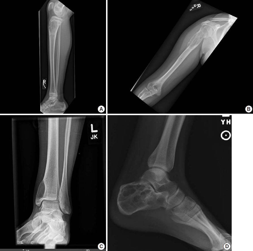
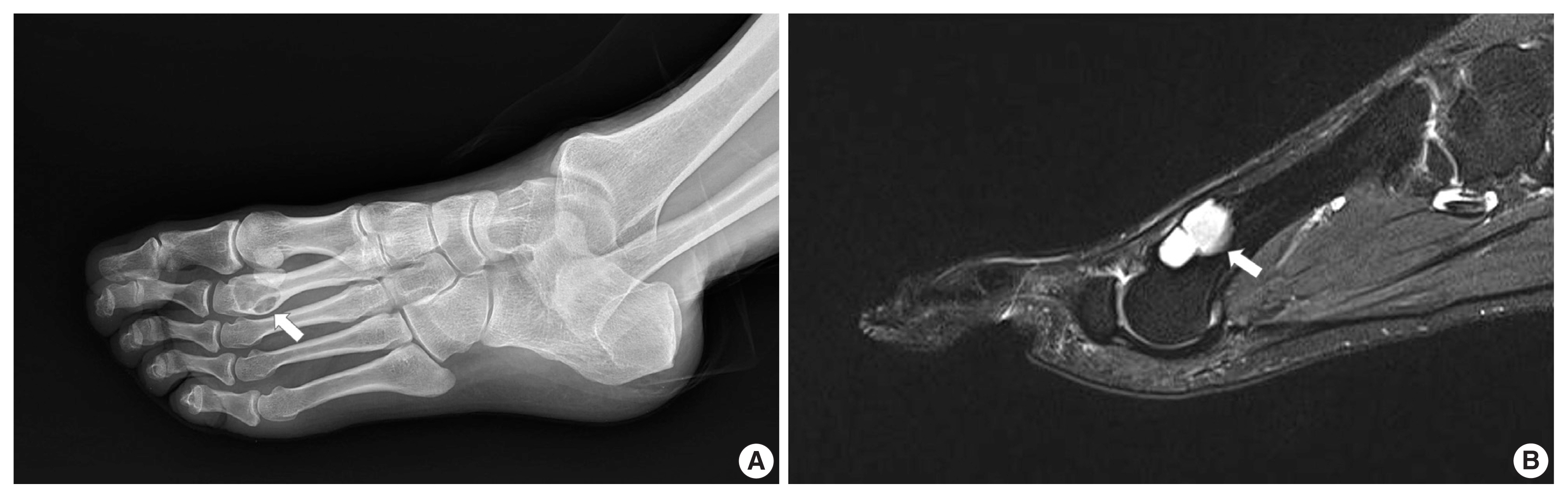
- Chondromyxoid fibroma is a rare bone tumor of cartilaginous origin, representing less than 1% of all bone tumors. It preferentially arises in the eccentric location of the metaphysis of a long tubular bone. Juxtacortical locations are reported infrequently in the long bones and even more rarely in short tubular bones, with only three cases documented. Here we present two new cases of juxtacortical chondromyxoid fibroma in the small bones. One was an intracortical osteolytic lesion of the metatarsal bone of the foot with degenerative atypia that histologically should be differentiated from chondrosarcoma. The other was a phalangeal mass protruding into the interphalangeal joint of the hand, which had been labeled mistakenly as a soft tissue mass preoperatively. These cases illustrated that chondromyxoid fibromas have various the manifestations and should be included in the differential diagnosis of an osteolytic lesion or an exophytic mass in the small bones.
-
Citations
Citations to this article as recorded by- Cartilage Forming Tumors of the Skeleton
Julio A. Diaz-Perez, Andrew E. Rosenberg
Advances in Anatomic Pathology.2025; 32(2): 132. CrossRef
- Cartilage Forming Tumors of the Skeleton
- Intraoperative frozen cytology of intraosseous cystic meningioma in the sphenoid bone
- Na Rae Kim, Gie-Taek Yie
- J Pathol Transl Med. 2020;54(6):508-512. Published online July 1, 2020
- DOI: https://doi.org/10.4132/jptm.2020.05.21
- 6,740 View
- 101 Download
- 2 Web of Science
- 3 Crossref
-
Abstract
PDF
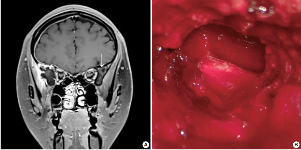
- Meningiomas in bone are rarely subjected to fine-needle aspiration diagnosis, and those arising in the skull bone with a cystic presentation are rare. A 24-year-old woman presented with subdural hemorrhage, and subsequent radiology depicted an osteolytic mass-like lesion in the sphenoid bone. Intraoperatively, a solid and cystic hemorrhagic lesion mimicking an aneurysmal bone cyst was observed in the sphenoid bone with dural tearing. Frozen cytology showed singly scattered or epithelioid clusters of round to elongated cells intermixed with many neutrophils. Tumor cells had bland-looking round nuclei with rare prominent nucleoli and nuclear inclusions and eosinophilic granular to globoid cytoplasm in capillary-rich fragments. Histology revealed intraosseous meningothelial and microcystic meningioma (World Health Organization grade 1) in right lesser wing of the sphenoid bone. Considering its unusual location and cytologic findings, differential diagnoses included chordoma, chondroma, chondrosarcoma, and aneurysmal bone cyst. The present case posed a diagnostic challenge due to possible confusion with these entities.
-
Citations
Citations to this article as recorded by- Purely cystic intraosseous meningioma of the skull: A radiologic conundrum and histologic challenge
Diego Rojas, Arman Kavoussi, Ashley Rose Ricciardelli, Alex Flores, Sricharan Gopakumar, Luis Carrete, Hsiang-Chih Lu, Alex W. Brenner, Akash J. Patel
Surgical Neurology International.2025; 16: 221. CrossRef - Middle ear adenoma: Cytohistologic features and differential diagnosis
Abdullah Almajnooni, Matthew Vega, Lin Cheng, Paolo Gattuso, Mary K. Allen‐Proctor
Diagnostic Cytopathology.2023;[Epub] CrossRef - Exploring the role of epidermal growth factor receptor variant III in meningeal tumors
Rashmi Rana, Vaishnavi Rathi, Kirti Chauhan, Kriti Jain, Satnam Singh Chhabra, Rajesh Acharya, Samir Kumar Kalra, Anshul Gupta, Sunila Jain, Nirmal Kumar Ganguly, Dharmendra Kumar Yadav, Timir Tripathi
PLOS ONE.2021; 16(9): e0255133. CrossRef
- Purely cystic intraosseous meningioma of the skull: A radiologic conundrum and histologic challenge
- Hepatocellular Carcinoma Arising in a Huge Hepatocellular Adenoma with Bone Marrow Metaplasia
- Hyo Jeong Kang, Hui Jeong Jeong, So-Woon Kim, Eunsil Yu, Young-Joo Lee, So Yeon Kim, Jihun Kim
- J Pathol Transl Med. 2018;52(4):226-231. Published online December 27, 2017
- DOI: https://doi.org/10.4132/jptm.2017.11.12
- 9,988 View
- 152 Download
- 5 Web of Science
- 6 Crossref
-
Abstract
PDF
- Hepatocellular adenoma (HCA) is the most common type of benign liver tumor, and its major complication is malignant transformation to hepatocellular carcinoma (HCC). Here, we report a case of HCC arising in HCA with bone marrow metaplasia in a 24-year-old Korean woman who presented with abdominal discomfort. A huge liver mass was found on abdominal ultrasonography. She underwent surgical hepatic resection, and the resected specimen was entirely involved by a 20-cm-sized tumor. Histological review revealed a well differentiated HCC arising from inflammatory HCA with β-catenin nuclear positivity and bone marrow metaplasia that contained hematopoietic cells. This case was unique because malignant transformation, inflammatory type HCA, β-catenin nuclear staining, and bone marrow metaplasia were simultaneously observed. Additionally, it should be noted that a large HCA with β-catenin activation can undergo malignant transformation and should be surgically resected in a timely manner.
-
Citations
Citations to this article as recorded by- Adult Hepatocellular Carcinoma Coexisting with Extramedullary Hematopoiesis
Hirotsugu Noguchi, Michiyo Higashi, Ryo Desaki, Takashi Tasaki, Mari Kirishima, Ikumi Kitazono, Kazuhiro Tabata, Akihide Tanimoto
International Journal of Surgical Pathology.2022; 30(3): 339. CrossRef - Spontaneous Occurrence of Various Types of Hepatocellular Adenoma in the Livers of Metabolic Syndrome-Associated Steatohepatitis Model TSOD Mice
Wenhua Shao, Orgil Jargalsaikhan, Mayuko Ichimura-Shimizu, Qinyi Cai, Hirohisa Ogawa, Yuko Miyakami, Kengo Atsumi, Mitsuru Tomita, Mitsuko Sutoh, Shunji Toyohara, Ryoji Hokao, Yasusei Kudo, Takeshi Oya, Koichi Tsuneyama
International Journal of Molecular Sciences.2022; 23(19): 11923. CrossRef - Bilateral Diffuse Nodular Pulmonary Ossification Mimicking Metastatic Disease in a Patient with Fibrolamellar Hepatocellular Carcinoma
Pattamon Sutthatarn, Cara E. Morin, Jessica Gartrell, Wayne L. Furman, Max R. Langham, Teresa Santiago, Andrew J. Murphy
Children.2021; 8(3): 226. CrossRef - Malignant transformation of liver fatty acid binding protein-deficient hepatocellular adenomas: histopathologic spectrum of a rare phenomenon
Juan Putra, Linda D. Ferrell, Annette S.H. Gouw, Valerie Paradis, Arvind Rishi, Christine Sempoux, Charles Balabaud, Swan N. Thung, Paulette Bioulac-Sage
Modern Pathology.2020; 33(4): 665. CrossRef - Hepatocellular carcinoma arising from hepatic adenoma in a young woman
Haythem Yacoub, Hela Kchir, Dhouha Cherif, Hajer Hassine, Slim Haouet, Asma Ayari, Habiba Mizouni, Saber Mannai, Mohamed Tahar Khalfallah, Nadia Maamouri
Clinical Case Reports.2020; 8(9): 1659. CrossRef - Metanephric adenoma with osseous metaplasia and bone marrow elements
Alessandro Pietro Aldera, Jeff John, Dharshnee Chetty, Dhirendra Govender
Human Pathology: Case Reports.2019; 17: 200316. CrossRef
- Adult Hepatocellular Carcinoma Coexisting with Extramedullary Hematopoiesis
- Intraosseous Hibernoma: A Rare and Unique Intraosseous Lesion
- Boram Song, Hye Jin Ryu, Cheol Lee, Kyung Chul Moon
- J Pathol Transl Med. 2017;51(5):499-504. Published online August 22, 2017
- DOI: https://doi.org/10.4132/jptm.2017.07.28
- 12,163 View
- 139 Download
- 17 Web of Science
- 22 Crossref
-
Abstract
PDF
- Background
Hibernoma is a rare benign tumor of adults that is composed of multivacuolated adipocytes resembling brown fat cells. Hibernoma typically occurs in soft tissue, and intraosseous examples are very rare. Intraosseous hibernomas can radiologically mimic metastatic carcinoma and other tumorous conditions. Methods: To collect the intraosseous hibernomas, we searched the pathologic database and reviewed the hematoxylin and eosin (H&E)–stained slides of bone biopsy samples performed to differentiate radiologically abnormal bone lesions from 2006 to 2016. A total of six intraosseous hibernoma cases were collected, and clinical and radiological information was verified from electronic medical records. H&E slide review and immunohistochemical staining for CD68, pan-cytokeratin, and S-100 protein were performed. Results: Magnetic resonance imaging of intraosseous hibernomas showed low signal intensity with slightly hyperintense foci on T1 and intermediate to high signal intensity on T2 weighted images. Intraosseous hibernomas appeared as heterogeneous sclerotic lesions with trabecular thickening on computed tomography scans and revealed mild hypermetabolism on positron emission tomography scans. Histopathologically, the bone marrow space was replaced by sheets of multivacuolated, foamy adipocytes resembling brown fat cells, without destruction of bone trabeculae. In immunohistochemical analysis, the tumor cells were negative for CD68 and pan-cytokeratin and positive for S-100 protein. Conclusions: Intraosseous hibernoma is very rare. This tumor can be overlooked due to its rarity and resemblance to bone marrow fat. Pathologists need to be aware of this entity to avoid misdiagnosis of this rare lesion. -
Citations
Citations to this article as recorded by- Test yourself answer: 38-year-old female with left hip pain
Ruhaid Khurram, Amar Nitin Kanani, Mohammed Saif Sait, Khamaeel Khaleel Al Lami, Ramanan Rajakulasingam
Skeletal Radiology.2026; 55(6): 1429. CrossRef - Clinical, Radiological, and Pathological Features of Intraosseous Hibernoma: A Systematic Review of Case Reports and Case Series
Jawad Albashri, Ahmed Albashri, Muhannad Alhamrani, Abdulrahman Hassan, Hisham Shamah, Rayan Alhefzi, Najim Z. Alshahrani, Mohammed R. Algethami, Louis-Romée Le Nail, Ramy Samargandi
Current Oncology.2025; 32(10): 535. CrossRef - Imaging of Bone Surface Lesions
Utkarsh Parwal, Allison Khoo, Nicholas G. Rhodes, Patrick G. McEnulty, Eric V. Pang, Jonathan C. Baker, Benjamin E. Northrup, Theodore L. Vander Velde, Mariam A. Malik, Jack W. Jennings, Kelby B. Napier
RadioGraphics.2025;[Epub] CrossRef - Intraosseous hibernoma of the mandible: A case report
Jin-Woo Han
Journal of Korean Dental Association.2025; 63(10): 335. CrossRef - Intraosseous Lipoma of the Maxillary Sinus: First Documented Case in an Asian Patient and Review of the Literature
Eng Seng Yeoh, Tzy Harn Chua, Jacqueline S. G. Hwang, Sathiyamoorthy Selvarajan, Noah B. T. Teo, Kevin Seymour
Case Reports in Dentistry.2025;[Epub] CrossRef - A Rare Case of Large Lateral Chest Wall Hibernoma
Lyubomir Gaydarski, Boycho Landzhov, Ivaylo Kamenov, Julian M Ananiev, Georgi P Georgiev
Cureus.2024;[Epub] CrossRef - Intraosseous hibernoma mimicking sclerotic bone metastasis—a case report
Ali Shaikh, Adil Basha, George Ray, Justin A. Bishop, Avneesh Chhabra
Skeletal Radiology.2024;[Epub] CrossRef - Femoral hibernoma: unique intraosseous tumor
Gökhan Tonkaz, Ertugrul Cakir, Mehmet Tonkaz, Demet Sengul
Wiener klinische Wochenschrift.2024; 136(19-20): 581. CrossRef - Unusual Imaging Findings of Epithelioid Hemangioma: Case Report of Single Intramedullary Sclerotic Bone Lesion
Yun Chul Hwang, Tae Eun Kim, Jae Hyuck Yi
Journal of the Korean Society of Radiology.2024; 85(5): 986. CrossRef - Benign incidental do-not-touch bone lesions
Nuttaya Pattamapaspong, Wilfred CG Peh
The British Journal of Radiology.2023;[Epub] CrossRef - Intraosseous hibernoma: clinicopathologic and imaging analysis of 18 cases
Chiraag N Gangahar, Carina A Dehner, David P Wang, Behrang Amini, Travis Hillen, Christopher O'Conor, Sydney N Jennings, Kathleen Byrnes, Elizabeth A Montgomery, Bogdan A Czerniak, Julia A Bridge, Molly C Schroeder, Jack W Jennings, Wei‐Lien Wang, John S
Histopathology.2023; 83(1): 40. CrossRef - Intraosseous Hibernoma: A Rare Entity in Orthopedics With Peculiar Radiological Features
Ramy Samargandi, Louis-Romée Le Nail, Gonzague de Pinieux, Matthias Tallegas, Elodie Miquelestorena-Standley
Cureus.2023;[Epub] CrossRef - Intraosseous hibernoma of the appendicular skeleton
Salvatore Gitto, Thom Doeleman, Michiel A. J. van de Sande, Kirsten van Langevelde
Skeletal Radiology.2022; 51(6): 1325. CrossRef - Intraosseous hibernoma: Two case reports and a review of the literature
Samantha N. Weiss, Ankit Mohla, Gord Guo Zhu, Christina Gutowski, Tae Won B Kim, Rohan Amin
Radiology Case Reports.2022; 17(7): 2477. CrossRef - Hibernoma of two contiguous vertebrae: uniqueness of a lesion already rare in itself
Donato MASTRANTUONO, Domenico MARTORANO, Guido REGIS, Federica ARABIA, Alessandra LINARI, Federica SANTORO
Journal of Radiological Review.2022;[Epub] CrossRef - Primary extradural tumors of the spinal column
Varun Arvind, Edin Nevzati, Maged Ghaly, Mansoor Nasim, Mazda Farshad, Roman Guggenberger, Daniel Sciubba, Alexander Spiessberger
Journal of Craniovertebral Junction and Spine.2021; 12(4): 336. CrossRef - Spinal Intraosseous Hibernoma: A Case Report and Review of Literature
Mi-Kyung Um, Eugene Lee, Joon Woo Lee, Kyu Sang Lee, Yusuhn Kang, Joong Mo Ahn, Heung Sik Kang
Journal of the Korean Society of Radiology.2020; 81(4): 965. CrossRef - Intraosseous hibernoma: A metastatic mimicker to consider on the differential
Allen Ko, Colin C. Rowell, James B. Vogler, Dmitri E. Samoilov
Radiology Case Reports.2020; 15(12): 2677. CrossRef - A Diagnostic Dilemma of a Subcutaneous Hibernoma: Case Report
Abdullah Saleh AlQattan, Alaa A. Al Abdrabalnabi, Mohammed Abdulrazzaq Al Duhileb, Tarek Ewies, Miral Mashhour, Ahmed Abbas
American Journal of Case Reports.2020;[Epub] CrossRef - Co-expression of MDM2 and CDK4 in transformed human mesenchymal stem cells causes high-grade sarcoma with a dedifferentiated liposarcoma-like morphology
Yu Jin Kim, Mingi Kim, Hyung Kyu Park, Dan Bi Yu, Kyungsoo Jung, Kyoung Song, Yoon-La Choi
Laboratory Investigation.2019; 99(9): 1309. CrossRef - Intraosseous Hibernoma: Five Cases and a Review of the Literature
Francisco A. Myslicki, Andrew E. Rosenberg, Ivan Chaitowitz, Ty K. Subhawong
Journal of Computer Assisted Tomography.2019; 43(5): 793. CrossRef - Hibernoma Mimicking Atypical Lipomatous Tumor
Youssef Al Hmada, Inga-Marie Schaefer, Christopher D.M. Fletcher
American Journal of Surgical Pathology.2018; 42(7): 951. CrossRef
- Test yourself answer: 38-year-old female with left hip pain
- Proposal of an Appropriate Decalcification Method of Bone Marrow Biopsy Specimens in the Era of Expanding Genetic Molecular Study
- Sung-Eun Choi, Soon Won Hong, Sun Och Yoon
- J Pathol Transl Med. 2015;49(3):236-242. Published online May 15, 2015
- DOI: https://doi.org/10.4132/jptm.2015.03.16
- 21,404 View
- 375 Download
- 57 Web of Science
- 61 Crossref
-
Abstract
PDF
- Background
The conventional method for decalcification of bone specimens uses hydrochloric acid (HCl) and is notorious for damaging cellular RNA, DNA, and proteins, thus complicating molecular and immunohistochemical analyses. A method that can effectively decalcify while preserving genetic material is necessary. Methods: Pairs of bilateral bone marrow biopsies sampled from 53 patients were decalcified according to protocols of two comparison groups: EDTA versus HCl and RDO GOLD (RDO) versus HCl. Pairs of right and left bone marrow biopsy samples harvested from 28 cases were allocated into the EDTA versus HCl comparison group, and 25 cases to the RDO versus HCl comparison group. The decalcification protocols were compared with regards to histomorphology, immunohistochemistry, and molecular analysis. For molecular analysis, we randomly selected 5 cases from the EDTA versus HCl and RDO versus HCl groups. Results: The decalcification time for appropriate histomorphologic analysis was the longest in the EDTA method and the shortest in the RDO method. EDTA was superior to RDO or HCl in DNA yield and integrity, assessed via DNA extraction, polymerase chain reaction, and silver in situ hybridization using DNA probes. The EDTA method maintained intact nuclear protein staining on immunohistochemistry, while the HCl method produced poor quality images. Staining after the RDO method had equivocal results. RNA in situ hybridization using kappa and lambda RNA probes measured RNA integrity; the EDTA and RDO method had the best quality, followed by HCl. Conclusions: The EDTA protocol would be the best in preserving genetic material. RDO may be an acceptable alternative when rapid decalcification is necessary. -
Citations
Citations to this article as recorded by- Seeing beyond the surface: bone histomorphometry re-visited—implications for diagnostic pathology
Terrence Diamond, Cherie Chiang, Grahame J. Elder
Pathology.2026; 58(2): 230. CrossRef - RETRACTED ARTICLE: Evaluation of the protective effect of the Echinacea Purpurea against apoptosis and inflammation induced by overdose of iron dextran on the hematopoietic system
Doaa Kahim Abdul Ridha, Ali Faris Hassan
Journal of Complementary and Integrative Medicine.2026; 23(1): 163. CrossRef - Effects of fixation and demineralization on immunohistochemical assessment of canine bone marrow
Gabriella M. L. Diamantino, Janet Beeler-Marfisi, Robert A. Foster, Gabrielle Monteith, William Sears, Alice Defarges, Dorothee Bienzle
Veterinary Pathology.2026;[Epub] CrossRef - Bone marrow biopsy as a new practice in a tertiary hospital center in Tirana Correlation with the literature and future perspective
Ejona Çeliku
Balkan Journal of Interdisciplinary Research.2026; 12(1): 107. CrossRef - Single cell sequencing in acute myeloid leukemia: Linking genotype to functional phenotype for precision risk stratification and treatment decisions
Annabelle J. Anandappa, Wenbin Xiao, Linde A. Miles
Human Pathology.2026; : 106174. CrossRef - Germline and somatic testing for homologous repair deficiency in patients with prostate cancer (part 1 of 2)
Andrew J. Armstrong, Amy Taylor, Michael C. Haffner, Wassim Abida, Alan H. Bryce, Lawrence I. Karsh, Scott T. Tagawa, Przemyslaw Twardowski, Anthony V. Serritella, Joshua M. Lang
Prostate Cancer and Prostatic Diseases.2025; 28(3): 652. CrossRef - Spatial transcriptomic approaches for characterising the bone marrow landscape: pitfalls and potential
Rosalin A. Cooper, Emily Thomas, Anna M. Sozanska, Carlo Pescia, Daniel J. Royston
Leukemia.2025; 39(2): 291. CrossRef - Qualitative comparison of decalcifiers for mouse bone cryosections for subsequent biophotonic analysis
Shibarjun Mandal, Ramya Motganhalli Ravikumar, Astrid Tannert, Annett Urbanek, Rustam R. Guliev, Max Naumann, Sina M. Coldewey, Uta Dahmen, Lina Carvalho, Luís Bastião Silva, Ute Neugebauer
Scientific Reports.2025;[Epub] CrossRef - Spatial Platform for Periodontal Ligament Angulation and Regeneration: In Vivo Pilot Study
Min Guk Kim, Do-Yeon Kim, Hyoung-Gon Ko, Jin-Seok Byun, Joong-Hyun Kim, Chan Ho Park
Journal of Functional Biomaterials.2025; 16(3): 99. CrossRef - Spatial proteomics and transcriptomics characterization of tissue and multiple cancer types including decalcified marrow
Cecilia CS Yeung, Daniel C Jones, David W. Woolston, Brandon Seaton, Elizabeth Lawless Donato, Minggang Lin, Coral Backman, Vivian Oehler, Kristin L Robinson, Kristen Shimp, Rima Kulikauskas, Annalyssa N Long, David Sowerby, Anna E Elz, Kimberly S Smythe,
Cancer Biomarkers.2025;[Epub] CrossRef - Circulating tumor cell markers for early detection and drug resistance assessment through liquid biopsy
Priya Yadav, Saravanan Rajendrasozhan, Ramzi Hadj Lajimi, Raja Ramadevi Patel, Dominique Heymann, N. Rajendra Prasad
Frontiers in Oncology.2025;[Epub] CrossRef - Morphological Bone Score as a Predictive Tool for Molecular Profiling Success
Kirill Kriukov, Dmitry Ivchenkov, Anna Bejanyan, Aleksandr Sarachakov, Aleksandra Kviatkovskaia, Gleb Khegai, Dominique Knipper-Davis, Amber Berlinski, Tayla Soares, Jochen K. Lennerz, Vladimir Kushnarev
The Journal of Molecular Diagnostics.2025; 27(8): 747. CrossRef - Optimizing cytology and small biopsy specimen processing for ancillary studies: recommendations from the American Society of Cytopathology taskforce
Sinchita Roy-Chowdhuri, Christine N. Booth, Jonas J. Heymann, Elizabeth Jenkins, Joshua R. Menke, Sara E. Monaco, Ritu Nayar, Michiya Nishino, Roberto Ruiz-Cordero, Donna K. Russell, Anjali Saqi, Kaitlin E. Sundling, Michael J. Thrall, Vanda F. Torous, Ch
Journal of the American Society of Cytopathology.2025; 14(5): 285. CrossRef - Ethylenediaminetetraacetic Acid (EDTA)-Decalcified, Formalin-Fixed Paraffin-Embedded (FFPE) Tumor Tissue Shows Comparable Quality and Quantity of DNA to Non-Decalcified Tissue in Next-Generation Sequencing (NGS)
Francis Hong Xin Yap, Jen-Hwei Sng, Jeremy Wee Kiat Ng, Hanis Abdul Kadir, Pei Yi Chan, Timothy Kwang Yong Tay
Journal of Molecular Pathology.2025; 6(3): 21. CrossRef - Case Report: Pitfalls in bone marrow evaluation: importance of adequate bone marrow sampling
Alireza Ghezavati, Elham Vali Betts, Ananya Datta Mitra
Frontiers in Oncology.2025;[Epub] CrossRef - Optimization of Formic Acid-Formalin-Based Decalcification Protocol for Rat Calvarial Bone Histology
S. Amitha Banu, Khan Sharun, Merlin Mamachan, Athira Subash, Vadapalli Deekshita, Kirtika Sharma, Karikalan Mathesh, Obli Rajendran Vinodh kumar, Swapan Kumar Maiti, Abhijit M. Pawde, Laith Abualigah, Kuldeep Dhama, Amarpal
Journal of Experimental Biology and Agricultural Sciences.2024; 12(2): 218. CrossRef - Effects of fixation and demineralization on histomorphology and DNA amplification of canine bone marrow
Gabriella M. L. Diamantino, Janet Beeler-Marfisi, Robert A. Foster, William Sears, Alice Defarges, William Vernau, Dorothee Bienzle
Veterinary Pathology.2024; 61(6): 943. CrossRef - In situ metabolomic analysis of osteonecrosis of the femoral head (ONFH) using MALDI MSI
Chen Li, Jikun Liu, Yiqi Sheng, Yinghao Wang, Lan Jia, Yinguang Zhang, Jiantao Li, Shuangshuang Di, Honggang Nie, Yehua Han
Analytical and Bioanalytical Chemistry.2024; 416(23): 5155. CrossRef - A set of pretreatment reagents including improved formula fixation and decalcification facilitating immunohistochemistry and DNA analyses of formalin-fixed paraffin-embedded bone marrow trephine biopsy
Ting Sun, Liming Xu, Hongtian Yao, Jing Zhao, Zhen Chen, Zexin Chen, Bo Wang, Wei Ding
Acta Histochemica.2024; 126(8): 152188. CrossRef - To Freeze or Not to Freeze? Recommendations for Intraoperative Examination and Gross Prosection of Thyroid Glands
Fouad R. Zakka, Nicole A. Cipriani
Surgical Pathology Clinics.2023; 16(1): 15. CrossRef - Effect of Surface Decalcification With Hydrochloric Acid on the Determination of Estrogen Receptor, Progesterone Receptor, Ki67, and Human Epidermal Growth Factor Receptor 2 Expressions in Invasive Breast Carcinoma Based on Immunohistochemistry and Fluore
Wu Ping, Rao Xin, Zhang Li, Chen Yupeng, Song Fangling, Ren Caihong, Hu Shun, Zhang Sheng
Applied Immunohistochemistry & Molecular Morphology.2023; 31(4): 232. CrossRef - Diagnostic value of MDM2 RNA in situ hybridization for low-grade osteosarcoma: Consistency comparison of RNA in situ hybridization, fluorescence in situ hybridization, and immunohistochemistry
Chen Chen, Xin He, Min Chen, Tianhai Du, Weiji Qin, Wenyi Jing, Hongying Zhang
Virchows Archiv.2023; 482(6): 999. CrossRef - Bone marrow fibrosis is associated with non‐response to CD19 CAR T‐cell therapy in B‐acute lymphoblastic leukemia
Joshua Anil, Ahab Alnemri, Andrew Lytle, Brian Lockhart, Ashley E. Anil, Michael Baumgartner, Kirubel Gebre, Jared McFerran, Stephan A. Grupp, Susan R. Rheingold, Vinodh Pillai
American Journal of Hematology.2023; 98(12): 1888. CrossRef - Epithelioid haemangioendothelioma of the mandible – A case report and review of the literature
Ali Rizvi, Tim K. Blackburn, Guy N. J. Betts
Oral Surgery.2022; 15(3): 387. CrossRef - Evaluation of EDTA and nitric acid solutions for decalcification of joints in AG/WT, BALB/c, C57, DBA1/J mice, and in Wistar rats
Eduarda Correa Freitas, Suelen Pizzolatto Dalmolin, Mateus Müller da Silva, Francine Hehn de Oliveira, Emily Ferreira Salles Pilar
Biotechnic & Histochemistry.2022; 97(5): 372. CrossRef - Coupling Lipid Labeling and Click Chemistry Enables Isolation of Extracellular Vesicles for Noninvasive Detection of Oncogenic Gene Alterations
Na Sun, Benjamin V. Tran, Zishan Peng, Jing Wang, Ceng Zhang, Peng Yang, Tiffany X. Zhang, Josephine Widjaja, Ryan Y. Zhang, Wenxi Xia, Alexandra Keir, Jia‐Wei She, Hsiao‐hua Yu, Jing‐Jong Shyue, Hongguang Zhu, Vatche G. Agopian, Renjun Pei, James S. Toml
Advanced Science.2022;[Epub] CrossRef - The Expressions of CD30 and CD123 of Mastocytosis in Taiwan
Ching-Fen Yang, Chih-Yi Hsu
Applied Immunohistochemistry & Molecular Morphology.2022; 30(4): 278. CrossRef - Unusual Patterns of HER2 Expression in Breast Cancer: Insights and Perspectives
Dora Grassini, Eliano Cascardi, Ivana Sarotto, Laura Annaratone, Anna Sapino, Enrico Berrino, Caterina Marchiò
Pathobiology.2022; 89(5): 278. CrossRef - Expert opinion on NSCLC small specimen biomarker testing — Part 1: Tissue collection and management
Frédérique Penault-Llorca, Keith M. Kerr, Pilar Garrido, Erik Thunnissen, Elisabeth Dequeker, Nicola Normanno, Simon J. Patton, Jenni Fairley, Joshua Kapp, Daniëlle de Ridder, Aleš Ryška, Holger Moch
Virchows Archiv.2022; 481(3): 335. CrossRef - Comparison of bone demineralisation procedures for DNA recovery from burned remains
Meghan Mckinnon, Denice Higgins
Forensic Science International: Genetics.2021; 51: 102448. CrossRef - A review of the current understanding of burned bone as a source of DNA for human identification
Meghan Mckinnon, Maciej Henneberg, Denice Higgins
Science & Justice.2021; 61(4): 332. CrossRef - Time is bone — Quantitative comparison of decalcification solvents in human femur samples using dual-X-ray-absorptiometry and computed tomography
Joshua Gawlitza, Jakob Steinhäuser, Arno Bücker, Gabriela Krasteva-Christ, Thomas Tschernig
Annals of Anatomy - Anatomischer Anzeiger.2021; 235: 151696. CrossRef - Molecular biomarker testing for non–small cell lung cancer: consensus statement of the Korean Cardiopulmonary Pathology Study Group
Sunhee Chang, Hyo Sup Shim, Tae Jung Kim, Yoon-La Choi, Wan Seop Kim, Dong Hoon Shin, Lucia Kim, Heae Surng Park, Geon Kook Lee, Chang Hun Lee
Journal of Pathology and Translational Medicine.2021; 55(3): 181. CrossRef - Effect of EDTA decalcification on estrogen receptor and progesterone receptor immunohistochemistry and HER2/neu fluorescence in situ hybridization in breast carcinoma
Erik Washburn, Xiaoyu Tang, Carla Caruso, Michelle Walls, Bing Han
Human Pathology.2021; 117: 108. CrossRef - Performances of single tube nested polymerase chain reaction and GeneXpert ultra on Formalin fixed paraffin embedded tissues in the diagnosis of tuberculous spondylodiscitis
Emna Romdhane, Soumaya Rammeh, Chelli Mouna Bouaziz, Hend Riahi, Meriam Rekaya Ben, Meriam Ksentini, Yosra Chebbi, Wafa Achour, Asma Ferjani, Ben Boubaker Ilhem Boutiba, Leila Slim-Saidi, Mohamed Fethi Ladeb
Clinical Rheumatology.2021; 40(10): 4317. CrossRef - Molecular Characterization of Prostate Cancers in the Precision Medicine Era
Emilio Francesco Giunta, Laura Annaratone, Enrico Bollito, Francesco Porpiglia, Matteo Cereda, Giuseppe Luigi Banna, Alessandra Mosca, Caterina Marchiò, Pasquale Rescigno
Cancers.2021; 13(19): 4771. CrossRef - Increased NF-κB Activity in Osteoprogenitor-Lineage Cells Impairs the Balance of Bone Versus Fat in the Marrow of Skeletally Mature Mice
Tzuhua Lin, Jukka Pajarinen, Yusuke Kohno, Akira Nabeshima, Laura Lu, Karthik Nathan, Zhenyu Yao, Joy Y. Wu, Stuart Goodman
Regenerative Engineering and Translational Medicine.2020; 6(1): 69. CrossRef - Percutaneous CT-guided biopsy of lytic bone lesions in patients clinically suspected of lung cancer: Diagnostic performances for pathological diagnosis and molecular testing
Anne-Claire Toffart, Stéphane Asfari, Anne Mc Leer, Emilie Reymond, Adrien Jankowski, Denis Moro-Sibilot, Olivier Stephanov, Julien Ghelfi, Sylvie Lantuejoul, Gilbert R. Ferretti
Lung Cancer.2020; 140: 93. CrossRef - Effect of decalcification protocols on immunohistochemistry and molecular analyses of bone samples
Elodie Miquelestorena-Standley, Marie-Lise Jourdan, Christine Collin, Corinne Bouvier, Frédérique Larousserie, Sébastien Aubert, Anne Gomez-Brouchet, Jean-Marc Guinebretière, Matthias Tallegas, Bénédicte Brulin, Louis-Romée Le Nail, Anne Tallet, François
Modern Pathology.2020; 33(8): 1505. CrossRef - Identifying Opportunities and Challenges for Patients With Sarcoma as a Result of Comprehensive Genomic Profiling of Sarcoma Specimens
Margaret A. Hay, Eric A. Severson, Vincent A. Miller, David A. Liebner, Jo-Anne Vergilio, Sherri Z. Millis, James L. Chen
JCO Precision Oncology.2020; (4): 176. CrossRef - Comparison of Methods for the Histological Evaluation of Odontocete Spiral Ganglion Cells
Tania Ramírez, Simona Sacchini, Yania Paz, Rubén S. Rosales, Nakita Câmara, Marisa Andrada, Manuel Arbelo, Antonio Fernández
Animals.2020; 10(4): 683. CrossRef - Molecular Pathology of Primary Non-small Cell Lung Cancer
David Ilan Suster, Mari Mino-Kenudson
Archives of Medical Research.2020; 51(8): 784. CrossRef - Comparison of ethylenediaminetetraacetic acid and rapid decalcificier solution for studying human temporal bones by immunofluorescence
Sumana Ghosh, Mark B. Lewis, Bradley J. Walters
Laryngoscope Investigative Otolaryngology.2020; 5(5): 919. CrossRef - A novel cryo-embedding method for in-depth analysis of craniofacial mini pig bone specimens
Pavla Ticha, Igor Pilawski, Xue Yuan, Jie Pan, Ustun S. Tulu, Benjamin R. Coyac, Waldemar Hoffmann, Jill A. Helms
Scientific Reports.2020;[Epub] CrossRef - Accelerating precision medicine in metastatic prostate cancer
Joaquin Mateo, Rana McKay, Wassim Abida, Rahul Aggarwal, Joshi Alumkal, Ajjai Alva, Felix Feng, Xin Gao, Julie Graff, Maha Hussain, Fatima Karzai, Bruce Montgomery, William Oh, Vaibhav Patel, Dana Rathkopf, Matthew Rettig, Nikolaus Schultz, Matthew Smith,
Nature Cancer.2020; 1(11): 1041. CrossRef - Tissue Morphology and Antigenicity in Mouse and Rat Tibia: Comparing 12 Different Decalcification Conditions
Kristofor Bogoevski, Anna Woloszyk, Keith Blackwood, Maria A. Woodruff, Vaida Glatt
Journal of Histochemistry & Cytochemistry.2019; 67(8): 545. CrossRef - Cellular and collagen reference values of gingival and periodontal ligament tissues in rats: a pilot study
Antoine Alves, Nina Attik, Carine Wirth, Yves Bayon, Alexis Piat, Brigitte Grosgogeat, Kerstin Gritsch
Histochemistry and Cell Biology.2019; 152(2): 145. CrossRef - Implementing Precision Medicine Programs and Clinical Trials in the Community-Based Oncology Practice: Barriers and Best Practices
Jennifer L. Ersek, Lora J. Black, Michael A. Thompson, Edward S. Kim
American Society of Clinical Oncology Educational Book.2018; (38): 188. CrossRef - Integration of next-generation sequencing in clinical diagnostic molecular pathology laboratories for analysis of solid tumours; an expert opinion on behalf of IQN Path ASBL
Zandra C Deans, Jose Luis Costa, Ian Cree, Els Dequeker, Anders Edsjö, Shirley Henderson, Michael Hummel, Marjolijn JL Ligtenberg, Marco Loddo, Jose Carlos Machado, Antonio Marchetti, Katherine Marquis, Joanne Mason, Nicola Normanno, Etienne Rouleau, Ed S
Virchows Archiv.2017; 470(1): 5. CrossRef - Protocolo para el estudio de muestras y estandarización del informe patológico de tumores óseos
Isidro Machado, José Juan Pozo, David Marcilla, Julia Cruz, Juan C. Tardío, Aurora Astudillo, Sílvia Bagué
Revista Española de Patología.2017; 50(1): 34. CrossRef - Extremely Well-Differentiated Papillary Thyroid Carcinoma Resembling Adenomatous Hyperplasia Can Metastasize to the Skull: A Case Report
Ju Yeon Pyo, Jisup Kim, Sung-eun Choi, Eunah Shin, Seok-Woo Yang, Cheong Soo Park, Seok-Mo Kim, SoonWon Hong
Yonsei Medical Journal.2017; 58(1): 255. CrossRef - Treatment of steroid-induced osteonecrosis of the femoral head using porous Se@SiO2 nanocomposites to suppress reactive oxygen species
Guoying Deng, Kerun Niu, Feng Zhou, Buxiao Li, Yingjie Kang, Xijian Liu, Junqing Hu, Bo Li, Qiugen Wang, Chengqing Yi, Qian Wang
Scientific Reports.2017;[Epub] CrossRef - Precision Medicine Starts With Preanalytics: Real-Time Assessment of Tissue Fixation Quality by Ultrasound Time-of-Flight Analysis
Melissa L. Lerch, Daniel R. Bauer, David Chafin, Abbey Theiss, Michael Otter, Geoffrey S. Baird
Applied Immunohistochemistry & Molecular Morphology.2017; 25(3): 160. CrossRef - Good Laboratory Standards for Clinical Next-Generation Sequencing Cancer Panel Tests
Jihun Kim, Woong-Yang Park, Nayoung K. D. Kim, Se Jin Jang, Sung-Min Chun, Chang-Ohk Sung, Jene Choi, Young-Hyeh Ko, Yoon-La Choi, Hyo Sup Shim, Jae-Kyung Won
Journal of Pathology and Translational Medicine.2017; 51(3): 191. CrossRef - An international survey about nail histology processing techniques
Christina Wlodek, Pauline Lecerf, Josette Andre, Beth S. Ruben, David de Berker
Journal of Cutaneous Pathology.2017; 44(9): 749. CrossRef - pSTAT5 and ERK exhibit different expression in myeloproliferative neoplasms
Ewa Wiśniewska-Chudy, Łukasz Szylberg, Grzegorz Dworacki, Ewa Mizera-Nyczak, Andrzej Marszałek
Oncology Reports.2017; 37(4): 2295. CrossRef - How we do: optimizing bone marrow biopsy logistics for sign-out within 2 days
I. de Laak–de Vries, A. G. Siebers, L. Burgers, C. Diepenbroek, M. Link, P. Groenen, J. H. J. M. van Krieken, K. M. Hebeda
Journal of Hematopathology.2016; 9(2): 67. CrossRef - Do More With Less: Tips and Techniques for Maximizing Small Biopsy and Cytology Specimens for Molecular and Ancillary Testing: The University of Colorado Experience
Dara L. Aisner, Mathew D. Rumery, Daniel T. Merrick, Kimi L. Kondo, Hala Nijmeh, Derek J. Linderman, Robert C. Doebele, Natalie Thomas, Patrick C. Chesnut, Marileila Varella-Garcia, Wilbur A. Franklin, D. Ross Camidge
Archives of Pathology & Laboratory Medicine.2016; 140(11): 1206. CrossRef - Analysis of the Effects of Bone Marrow Biopsy Decalcification Methods on Histopathological Examination
Ji Young Park, Kyung Hee Han
The Korean Journal of Clinical Laboratory Science.2016; 48(4): 371. CrossRef - Distinguishing between Microbial Habitats Unravels Ecological Complexity in Coral Microbiomes
Amy Apprill, Laura G. Weber, Alyson E. Santoro, Nicole S. Webster
mSystems.2016;[Epub] CrossRef - Optimal Fixation and Decalcification Methods for Bone Marrow Biopsy
Myung-Sub Choi, Hyunsup Lee, Hyuk-Chul Kwon, Moon-Hwan Bae, Young-Hye Ko, Hee-Jin Kim, Beom-Se Lee, Bon-Kyung Koo
Korean Journal of Clinical Laboratory Science.2015; 47(4): 243. CrossRef
- Seeing beyond the surface: bone histomorphometry re-visited—implications for diagnostic pathology
- Role of Osteal Macrophages in Bone Metabolism
- Sun Wook Cho
- J Pathol Transl Med. 2015;49(2):102-104. Published online March 12, 2015
- DOI: https://doi.org/10.4132/jptm.2015.02.02
- 13,862 View
- 137 Download
- 29 Web of Science
- 28 Crossref
-
Abstract
PDF
- Macrophages have been shown to have pleiotropic functions in various pathophysiologies, especially in terms of anti-inflammatory and regenerative activity. Recently, the novel functions of bone marrow resident macrophages (called osteal macrophages) were intensively studied in bone development, remodeling and tissue repair processes. This review discusses the current evidence for a role of osteal macrophages in bone modeling, remodeling, and fracture healing processes.
-
Citations
Citations to this article as recorded by- 3D printed composite scaffold accelerates bone regeneration by modulating immunity and promoting angiogenesis
Yiye Fan, Jiaxin Yao, Wan Liu, Lebin Wang, Jing Yang, Xiaoyan Zheng, Junfeng Hui, Daidi Fan
Journal of Materials Science & Technology.2026; 240: 1. CrossRef - Rescuing Mitochondrial Dysfunction in Macrophages Prevents Osteonecrosis of the Jaw in Anti‐Resorptive Therapy
Hang Zhang, Xin Shen, Haiyang Liu, Xinxi Yuan, Mumin Cao, Xuepeng Lv, Ziji Ling, Songsong Guo, Rongyao Xu, Xiang Li, Hongbing Jiang
Advanced Science.2026;[Epub] CrossRef - Macrophage reprogramming nodes for bone repair identified by single-cell and spatial omics
Hang Chen, Chang Lei, Giselle C. Yeo, Khoon S. Lim, Chun Xu
Bone.2026; 210: 117937. CrossRef - Transcriptomic profiling reveals a dramatic inflammatory shift in osteal macrophages during colitis-induced osteoporosis
Ryota Suzuki, Liyile Chen, Tsutomu Endo, Taiki Tokuhiro, Masaya Nakajo, Yuki Ogawa, Hend Alhasan, Taku Ebata, Daisuke Takahashi, Ken Kadoya, Masahiko Takahata, Norimasa Iwasaki, M. Alaa Terkawi
Inflammation Research.2025;[Epub] CrossRef - Surgical stress induced tumor immune suppressive environment
Fan Yang, Qing Hua, Xiaoyan Zhu, Pingbo Xu
Carcinogenesis.2024; 45(4): 185. CrossRef - A Systematic Review and Meta-Analysis of the Outcomes of Reconstruction with Vascularised vs Non-Vascularised Bone Graft after Surgical Resection of Primary Malignant and Non-Malignant Bone Tumors
R. PATEL, G. MCCONAGHIE, M. M. KHAN, W. GIBSON, R. SINGH, R. BANERJEE
Acta chirurgiae orthopaedicae et traumatologiae Cechoslovaca.2024; 91(3): 143. CrossRef - Macrophage Polarization during MRONJ Development in Mice
A. Soundia, N. Elzakra, D. Hadaya, I. Gkouveris, O. Bezouglaia, S. Dry, T. Aghaloo, S. Tetradis
Journal of Dental Research.2024; 103(9): 899. CrossRef - 3D printing of gear-inspired biomaterials: Immunomodulation and bone regeneration
Xiaopeng Yu, Yufeng Wang, Meng Zhang, Hongshi Ma, Chun Feng, Bingjun Zhang, Xin Wang, Bing Ma, Qingqiang Yao, Chengtie Wu
Acta Biomaterialia.2023; 156: 222. CrossRef - Origin, production and molecular determinants of macrophages for their therapeutic targeting
Sangita Chowdhury, Arun K. Trivedi
Cell Biology International.2023; 47(1): 15. CrossRef - The Macrophage’s Role on Bone Remodeling and Osteogenesis: a Systematic Review
João Maria Orvalho, Juliana Campos Hasse Fernandes, Rogerio Moraes Castilho, Gustavo Vicentis Oliveira Fernandes
Clinical Reviews in Bone and Mineral Metabolism.2023; 21(1-4): 1. CrossRef - Neglected immunoregulation: M2 polarization of macrophages triggered by low‐dose irradiation plays an important role in bone regeneration
Shaoqing Chen, Su Ni, Chun Liu, Mu He, Yiwen Pan, Pengfei Cui, Cheng Wang, Xinye Ni
Journal of Cellular and Molecular Medicine.2023; 27(8): 1095. CrossRef - Insight into the effect of biomaterials on osteogenic differentiation of mesenchymal stem cells: A review from a mitochondrial perspective
Ziyi Feng, Meiqi Jin, Junzhi Liang, Junning Kang, Huazhe Yang, Shu Guo, Xiaoting Sun
Acta Biomaterialia.2023; 164: 1. CrossRef - Nano wear particles and the periprosthetic microenvironment in aseptic loosening induced osteolysis following joint arthroplasty
Yu Xie, Yujie Peng, Guangtao Fu, Jiewen Jin, Shuai Wang, Mengyuan Li, Qiujian Zheng, Feng-Juan Lyu, Zhantao Deng, Yuanchen Ma
Frontiers in Cellular and Infection Microbiology.2023;[Epub] CrossRef - Integrated computational and in vivo models reveal Key Insights into macrophage behavior during bone healing
Etienne Baratchart, Chen Hao Lo, Conor C. Lynch, David Basanta, Dominik Wodarz
PLOS Computational Biology.2022; 18(5): e1009839. CrossRef - Strategies of Macrophages to Maintain Bone Homeostasis and Promote Bone Repair: A Narrative Review
Yingkun Hu, Jinghuan Huang, Chunying Chen, Yi Wang, Zhuowen Hao, Tianhong Chen, Junwu Wang, Jingfeng Li
Journal of Functional Biomaterials.2022; 14(1): 18. CrossRef - Macrophages and Stem Cells—Two to Tango for Tissue Repair?
Emilia Manole, Cristina Niculite, Ioana Maria Lambrescu, Gisela Gaina, Octavian Ioghen, Laura Cristina Ceafalan, Mihail Eugen Hinescu
Biomolecules.2021; 11(5): 697. CrossRef - Bone remodeling stages under physiological conditions and glucocorticoid in excess: Focus on cellular and molecular mechanisms
V. V. Povoroznyuk, N. V. Dedukh, M. A. Bystrytska, V. S. Shapovalov
Regulatory Mechanisms in Biosystems.2021; 12(2): 212. CrossRef - Menaquinone-7 Supplementation Improves Osteogenesis in Pluripotent Stem Cell Derived Mesenchymal Stem Cells
Asim Cengiz Akbulut, Grzegorz B. Wasilewski, Nikolas Rapp, Francesco Forin, Heike Singer, Katrin J. Czogalla-Nitsche, Leon J. Schurgers
Frontiers in Cell and Developmental Biology.2021;[Epub] CrossRef - The Effects of Biomaterial Implant Wear Debris on Osteoblasts
Li Zhang, El-Mustapha Haddouti, Kristian Welle, Christof Burger, Dieter C. Wirtz, Frank A. Schildberg, Koroush Kabir
Frontiers in Cell and Developmental Biology.2020;[Epub] CrossRef Local Cellular Responses to Metallic and Ceramic Nanoparticles from Orthopedic Joint Arthroplasty Implants
Li Zhang, El-Mustapha Haddouti, Kristian Welle, Christof Burger, Koroush Kabir, Frank A Schildberg
International Journal of Nanomedicine.2020; Volume 15: 6705. CrossRef- Mesenchymal stem cell-macrophage crosstalk and bone healing
Jukka Pajarinen, Tzuhua Lin, Emmanuel Gibon, Yusuke Kohno, Masahiro Maruyama, Karthik Nathan, Laura Lu, Zhenyu Yao, Stuart B. Goodman
Biomaterials.2019; 196: 80. CrossRef - Inflammation, mesenchymal stem cells and bone regeneration
Hongrui Liu, Dongfang Li, Yi Zhang, Minqi Li
Histochemistry and Cell Biology.2018; 149(4): 393. CrossRef - Inflammatory and degenerative phases resulting from anterior cruciate rupture in a non‐invasive murine model of post‐traumatic osteoarthritis
Sophie J. Gilbert, Cleo S. Bonnet, Paulina Stadnik, Victor C. Duance, Deborah J. Mason, Emma J. Blain
Journal of Orthopaedic Research.2018; 36(8): 2118. CrossRef - M2 macrophages are closely associated with accelerated clavicle fracture healing in patients with traumatic brain injury: a retrospective cohort study
Ran Zhang, Yi Liang, Shuxiang Wei
Journal of Orthopaedic Surgery and Research.2018;[Epub] CrossRef - Digesting the role of bone marrow macrophages on hematopoiesis
Esther Heideveld, Emile van den Akker
Immunobiology.2017; 222(6): 814. CrossRef - Concise Review: Stem Cells in Osteoimmunology
Fernando A. Fierro, Jan A. Nolta, Iannis E. Adamopoulos
Stem Cells.2017; 35(6): 1461. CrossRef - Aging, inflammation, stem cells, and bone healing
Emmanuel Gibon, Laura Lu, Stuart B. Goodman
Stem Cell Research & Therapy.2016;[Epub] CrossRef - The roles of immune cells in bone healing; what we know, do not know and future perspectives
Jehan J. El-Jawhari, Elena Jones, Peter V. Giannoudis
Injury.2016; 47(11): 2399. CrossRef
- 3D printed composite scaffold accelerates bone regeneration by modulating immunity and promoting angiogenesis
- Periductal Stromal Tumor of Breast: A Case Report and A Review of Literature
- Salma L. Abbasi, Kate McNamara, Mohammed S. Absar, Alison Darlington, Francene Clucas, Sami Titi
- Korean J Pathol. 2014;48(6):442-444. Published online December 31, 2014
- DOI: https://doi.org/10.4132/KoreanJPathol.2014.48.6.442
- 14,523 View
- 89 Download
- 5 Crossref
-
PDF
-
Citations
Citations to this article as recorded by- Genetic Profiling of Mammary Periductal Stromal Tumors With Histologic Correlation Highlights High-Grade and Low-Grade Groups and Similarities to Phyllodes Tumors
Gregor Krings, Gregory R. Bean, Elizabeth M. Hosfield, J Jordi Rowe, Joseph Geradts, Yunn-Yi Chen
Modern Pathology.2026; 39(3): 100961. CrossRef - Survey of recurrent diagnostic challenges in breast phyllodes tumours
Benjamin Yongcheng Tan, Stephen B Fox, Sunil R Lakhani, Puay Hoon Tan
Histopathology.2023; 82(1): 95. CrossRef - Management of a periductal stromal tumor in a young woman: Our breast unit experience
Irene Valente, Adela Ristani, Cristina Mancini, Eugenia Martella, Leonardo Quartieri, Cecilia D'Aloia
The Breast Journal.2020; 26(7): 1375. CrossRef - A Diagnostic Approach to Fibroepithelial Breast Lesions
Benjamin Yongcheng Tan, Puay Hoon Tan
Surgical Pathology Clinics.2018; 11(1): 17. CrossRef - A case of local recurrence of periductal stromal sarcoma of the breast
Kana TERAMOTO, Yasuro DOI, Kayo YAMAMOTO, Kaname MATSUKAWA, Hisaka IWAIHARA, Rumi MOTOSHIMA, Noboru TAKATA, Ichiro YOSHINAKA, Kazunori HARADA
Choonpa Igaku.2018; 45(1): 61. CrossRef
- Genetic Profiling of Mammary Periductal Stromal Tumors With Histologic Correlation Highlights High-Grade and Low-Grade Groups and Similarities to Phyllodes Tumors
- Morphologic Alteration of Metastatic Neuroblastic Tumor in Bone Marrow after Chemotherapy
- Go Eun Bae, Yeon-Lim Suh, Ki Woong Sung, Jung-Sun Kim
- Korean J Pathol. 2013;47(5):433-442. Published online October 25, 2013
- DOI: https://doi.org/10.4132/KoreanJPathol.2013.47.5.433
- 9,978 View
- 50 Download
- 1 Crossref
-
Abstract
PDF
Background The aim of this study is to evaluate the histologic features of metastatic neuroblastic tumors (NTs) in bone marrow (BM) before and after chemotherapy in comparison with those of primary NTs.
Methods A total of 294 biopsies from 48 children diagnosed with NTs with BM metastasis were examined. There were 48 primary neoplasm biopsies, 48 BM biopsies before chemotherapy, 36 primary neoplasm excisional biopsies after chemotherapy, and 162 BM biopsies after chemotherapy.
Results Metastatic NTs in BM before chemotherapy were composed of undifferentiated and/or differentiating neuroblasts, but had neither ganglion cells nor Schwannian stroma. Metastatic foci of BM after chemotherapy were found to have differentiated into ganglion cells or Schwannian stroma, which became more prominent after further cycles of chemotherapy. Persistence of NTs or tumor cell types in BM after treatment did not show statistically significant correlation to patients' outcome. However, three out of five patients who newly developed poorly differentiated neuroblasts in BM after treatment expired due to disease progression.
Conclusions Metastatic NTs in BM initially consist of undifferentiated or differentiating neuroblasts regardless of the primary tumor subtype, and become differentiated after chemotherapy. Newly appearing poorly differentiated neuroblasts after treatment might be an indicator for poor prognosis.
-
Citations
Citations to this article as recorded by- Postchemotherapy gross residual tumor in non‐high‐risk neuroblastoma: Clinical significance and the role of adjuvant therapy
Eun Seop Seo, Hana Lim, Hee Won Cho, Hee Young Ju, Ji Won Lee, Keon Hee Yoo, Sanghoon Lee, Do Hoon Lim, Ki Woong Sung, Hong Hoe Koo
Pediatric Blood & Cancer.2022;[Epub] CrossRef
- Postchemotherapy gross residual tumor in non‐high‐risk neuroblastoma: Clinical significance and the role of adjuvant therapy
- Biologic Response to Carbonated Hydroxyapatite Associated with Orthopedic Device: Experimental Study in a Rabbit Model
- Samira Jebahi, Mongi Saoudi, Riadh Badraoui, Tarek Rebai, Hassane Oudadesse, Zoubaier Ellouz, Hassib Keskese, Abdelfattah El Feki, Hafed El Feki
- Korean J Pathol. 2012;46(1):48-54. Published online February 23, 2012
- DOI: https://doi.org/10.4132/KoreanJPathol.2012.46.1.48
- 9,606 View
- 61 Download
- 10 Crossref
-
Abstract
PDF
Background Carbonated hydroxyapatite (CHA) and related calcium phosphates have been studied for many years as implant materials due to their similarity with the mineral phase of bone. The main limitation of CHA ceramics as well as other bioactive materials is that they have poor mechanical proprieties. It is thought that the mechanical device can cause an increase in metabolic activity and bone healing. In this study we investigated the reactivity and tissue behaviour of implanted CHA biomaterial reinforced by mini external fixator.
Methods The evaluation of biomaterial biocompatibility and osteogenesis was performed on a rabbit model over a period of 6 weeks by radiological, histological and scanning electron microscopy (SEM) coupled with energy dispersive X-ray SEM-energy-dispersive X-ray (EDX) analysis.
Results While rabbits treated with CHA exhibited more bone formation, and fibrous tissue was observed when empty bone defects were observed. EDX analysis detected little calcium and phosphorus on the surface of the bone that was not implanted, while high content of calcium (62.7%) and phosphorus (38%) was found on the interface bone cement.
Conclusions Bone repairing showed that the mini external fixator stimulated the ossification which was pushed when grafted by CHA. This effect may play an important role in the prevention of implant loosening.
-
Citations
Citations to this article as recorded by- Comparative Evaluation of Sticky Bone with Alloplastic Bone Graft in the Treatment of Intrabony Defects: A Randomized Controlled Trial
Pooja Velraj, Senthilnathan Sivaramalingam, Gayathri Haritheertham, Hema Pannerselvam, Kalyani Ramkumar Sadhana, Sesha Reddy
World Journal of Dentistry.2026; 17(5): 417. CrossRef - A review: In vivo studies of bioceramics as bone substitute materials
Ali A. Al‐allaq, Jenan S. Kashan
Nano Select.2023; 4(2): 123. CrossRef - Improvement in bioactivity and corrosion resistance of Ti by hydroxyapatite deposition using ultrasonic mechanical coating and armoring
Ming-Hong Lin, Yi-Cheng Chen, Chun-Chung Liao, Liang-Wei Lin, Chin-Fu Chen, Kuang-Kuo Wang, Shyi-Tien Chen, Yi-Huang Hsueh, Chien-Hui Wu, Shih-Fu Ou
Ceramics International.2022; 48(4): 4999. CrossRef - Novel bioactive collagen-polyurethane-pectin scaffolds for potential application in bone regenerative medicine
Myriam L. Guzmán-Chávez, Jesús A. Claudio-Rizo, Martín Caldera-Villalobos, Denis A. Cabrera-Munguía, Juan J. Becerra-Rodríguez, Nayeli Rodríguez-Fuentes
Applied Surface Science Advances.2022; 11: 100317. CrossRef - Chitosan-Based Gastric Dressing Materials Loaded with Pomegranate Peel as Bioactive Agents: Pharmacokinetics and Effects on Experimentally Induced Gastric Ulcers in Rabbits
Samira Jebahi, Ghada Ben Salah, Soufien Jarray, Mounir Naffati, Mohammad Ayaz Ahmad, Faten Brahmi, Mohd Saeed, Arif J. Siddiqui, Khabir Abdelmajid, Riadh Badraoui
Metabolites.2022; 12(12): 1158. CrossRef - Is THP‐1 viability affected by the crystallinity of nanostructured carbonated hydroxyapatites?
Renata Moraes Lira, Suelen Cristina Sartoretto, Carolina da Silva Gouveia Pedrosa, Mônica Diuana Calasans‐Maia, Paulo Emílio Leite, José Mauro Granjeiro
Journal of Biomedical Materials Research Part A.2021; 109(7): 1266. CrossRef - Does the incorporation of zinc into calcium phosphate improve bone repair? A systematic review
Rebecca Cruz, José Calasans-Maia, Suelen Sartoretto, Vittório Moraschini, Alexandre Malta Rossi, Rafael Seabra Louro, José Mauro Granjeiro, Monica Diuana Calasans-Maia
Ceramics International.2018; 44(2): 1240. CrossRef - Adsorption of nucleotides on biomimetic apatite: The case of adenosine 5′ monophosphate (AMP)
K. Hammami, H. El Feki, O. Marsan, C. Drouet
Applied Surface Science.2015; 353: 165. CrossRef - Finite element analysis and cellular studies on advanced, controlled porous structures with subsurface continuity in bio-implantable titanium alloys
P. Lambert, S. Ankem, Z. Wyatt, K. M. Ferlin, J. Fisher
Journal of Biomedical Materials Research Part A.2014; 102(1): 225. CrossRef - The impact of orthopedic device associated with carbonated hydroxyapatite on the oxidative balance: experimental study of bone healing rabbit model
Samira Jebahi, Riadh Nsiri, Mohammed Boujbiha, Ezedine Bouroga, Tarek Rebai, Hassib Keskes, Abdelfattah El Feki, Hassane Oudadesse, Hafed El Feki
European Journal of Orthopaedic Surgery & Traumatology.2013; 23(7): 759. CrossRef
- Comparative Evaluation of Sticky Bone with Alloplastic Bone Graft in the Treatment of Intrabony Defects: A Randomized Controlled Trial
- Chondromyxoid Fibroma of the Ethmoid Sinus Complicated by a Brain Abscess: A Case Report and Literature Review.
- Kyu Yeoun Won, Juhie Lee, Youn Wha Kim, Eui Jong Kim, Sung Wan Kim, Yong Koo Park
- Korean J Pathol. 2010;44(5):547-550.
- DOI: https://doi.org/10.4132/KoreanJPathol.2010.44.5.547
- 4,611 View
- 26 Download
- 2 Crossref
-
Abstract
PDF
- Chondromyxoid fibroma (CMF) is a relatively rare bone tumor that was first described by Jaffe and Lichtenstein in 1948. CMF of the sinonasal tract is very rare. A 28-year-old male presented with long-standing, intermittent, pulsatile pain in the right temporal area. A computed tomography scan showed a 20 x 19 mm round, bony density in the right ethmoid sinus with fluid collection in the ethmoid and frontal sinuses. Additionally, a cystic lesion with surrounding edema was found in the right frontal lobe. The patient underwent a partial ethmoidectomy and frontostomy. A histological examination showed polygonal and stellate cells in a myxoid and chondroid background with a pattern of lobulation and plaque-like calcification. The bone lesion was revealed as a CMF of the ethmoidal sinus, and the frontal lobe cystic lesion was a brain abscess associated with the CMF. We present the case of a CMF of the ethmoid sinus complicated by a brain abscess.
-
Citations
Citations to this article as recorded by- Juxtacortical chondromyxoid fibroma in the small bones: two cases with unusual location and a literature review
Sun-Ju Oh, So Hak Chung
Journal of Pathology and Translational Medicine.2022; 56(3): 157. CrossRef - Treatment of cryotherapy and orthotopic transplantation following chondromyxoid fibroma of zygomatic bone
Zhi-Chao Zhu, Yi-Fei Yang, Xu Yang, Yan Liu, Yi-nan Cheng, Zhao-Yao Sun, Tian-Shu Xu, Wen-Jun Yang
Medicine.2018; 97(31): e11707. CrossRef
- Juxtacortical chondromyxoid fibroma in the small bones: two cases with unusual location and a literature review
- Intraosseous Neurilemmoma of the Mandible: A Case Report.
- Kyu Yun Jang, Woo Sung Moon, Ho Sung Park
- Korean J Pathol. 2009;43(1):88-91.
- DOI: https://doi.org/10.4132/KoreanJPathol.2009.43.1.88
- 4,496 View
- 35 Download
- 6 Crossref
-
Abstract
PDF
- Neurilemmoma (Schwannoma) is a benign nerve sheath tumor that's composed entirely of well-differentiated Schwann cells. Intraosseous neurilemmomas are rare and they represent less than 1% of all benign primary bone tumors. We report here on an additional case of intraosseous neurilemmoma that was located in the mandible of a 77-year-old woman. CT revealed an expansile, well-defined lesion on the right side of the mandibular body with thinning of the cortex. The lesion was surgically removed and it was found to be a 2x1.7 cm-sized, bright yellowish, hard mass with hemorrhage and cyst formation. Histologically, the mass was a moderately cellular neoplasm and it showed distinct nuclear palisading, numerous Verocay bodies and tumor cells that were positively immunohistostained for S-100 protein. Two months after the operation, the patient has remained in a good condition with no signs or symptoms of tumor recurrence.
-
Citations
Citations to this article as recorded by- Mandibular Schwannoma: A Systematic Review of 33 Case Reports
Ahmad Al Malak, Yasmina El Masri, Jad El Masri, Farah Sarmout, Mohammad Hassoun, Georges Aoun
Oral Diseases.2026; 32(3): 657. CrossRef - Intraosseous benign peripheral nerve sheath tumor of the jaws: report of 4 new cases and a comprehensive literature review
Brendo Vinícius Rodrigues Louredo, Paulo Victor Mendes Penafort, Ana Luiza Oliveira Corrêa Roza, Maria Cecília Querido De Oliveira, Ricardo Pelletti Ocaña, Alexandre Machado Torres, Samuel de Barros Ferreira Júnior, André Caroli Rocha, Rafael Cabral da Co
Oral Surgery, Oral Medicine, Oral Pathology and Oral Radiology.2025; 139(4): e104. CrossRef - Imaging Features of Intraosseous Schwannoma: A Case Series and Review of the Literature
Firoozeh Shomal Zadeh, Arash Azhideh, Jose G. Mantilla, Vijaya Kosaraju, Nitin Venugopal, Cree M. Gaskin, Atefe Pooyan, Ehsan Alipour, Majid Chalian
Diagnostics.2023; 13(9): 1610. CrossRef - Gnathic Schwannomas: A Report of Two Cases and Systematic Review of the Literature
Alberto Jose Peraza Labrador, Luciano Hermios Matos Valdez, Nestor Ricardo Gonzalez Marin, Karem Annelise Rodriguez Ibazetta, Marcelo Villacis, Joan Lopez Chacon, Hebert Ochoa Huaman, Harold Cuzcano Pariahuamán, Hosting Barría Angulo, Victoria Woo
Head and Neck Pathology.2023; 17(4): 984. CrossRef - Intraosseous schwannoma of the humerus: a rarity yet warrants consideration
Jagannath Kamath, Harshit Bhaskar Shetty, Arkesh Madegowda, Anusha S Bhatt
BMJ Case Reports.2021; 14(9): e240007. CrossRef - Intraosseous Schwannoma of the Jaws: An Updated Review of the Literature and Report of 2 New Cases Affecting the Mandible
Dru Perkins, Tudor I. Stiharu, James Q. Swift, Tran Volong Dao, Gisele N. Mainville
Journal of Oral and Maxillofacial Surgery.2018; 76(6): 1226. CrossRef
- Mandibular Schwannoma: A Systematic Review of 33 Case Reports
- Intraosseous Well Differentiated Osteosarcoma: A case report.
- Mee Hye Oh, So Young Park, Yeon Lim Suh, Shin Khang Kang
- Korean J Pathol. 1992;26(6):627-631.
- 2,280 View
- 18 Download
-
Abstract
PDF
- Well differentiated osteosarcomas are variants of osteosarcoma composed mainly of fibrous and osseous tissue with minimal cystologic atypia. This tumor may be misinterpretated as a benign lesion if the radiologic and clinical features are not taken into account. We report a typical case of intraosseous well differentiated osteosarcoma occuring in the left distal femur of a 58-year-old woman. Radiologically, it appered as an ill-defined lesion with a mixture of sclerotic and osteolytic ares. But there was a lack of highly destructive appearance of conventional osteosarcoma. Grossly, the mass occupied a metaphysis of the distal femur with extension into the diaphysis and epiphysis. Multifocal cortical destruction and sclerosis were also associated. Histologically, the mass showed typical features of intraosseous well differentiated osteosarcoma. There were various patterns of osteoid deposits and bone formation mimicking those of fibrous dysplasia, nonossifying fibroma or parosteal osteosarcoma.
- Nasal Chondromesenchymal Hamartoma: A Case Report.
- Jun Kang, Young Ok Hong, Geung Hwan Ahn, Young Min Kim, Hee Jeong Cha, Hye Jeong Choi
- Korean J Pathol. 2007;41(4):258-262.
- 2,471 View
- 29 Download
-
Abstract
PDF
- We report a case of nasal chondromesenchymal hamartoma. A 14-year-old boy presented with a 5 cm sized mass in the left maxillary sinus, facial swelling and a loose tooth. A subtotal left maxillectomy with a bone graft was performed. The excised mass was composed of partly encapsulated, solid and cystic fragments of soft tissues. The mass contained chondroid and myxoid areas consisting of mesenchymal tissues including hyaline cartilage, osteoid and spindle cells in various proportions. The hyaline cartilage component was the most prominent. The spindle cell component had a fibrous matrix with variable myxoid or sclerotic changes. Thick hyalinized eosinophilic osteoid-like trabeculae were focally present. Immunohistochemically, all the mesenchymal cells tested positive for vimentin. The chondrocytes tested positive for the S-100 protein, and the spindle cell component showed focal immunoreactivity for smooth muscle actin and desmin. However, the cells were negative to pan-cytokeratin and p63.
- Cytologic Diagnosis of Metastatic Hepatocellular Carcinoma by Aspiration Cytology of Sacrum.
- Jungweon Shim, Illhyang Ko
- J Pathol Transl Med. 1990;1(2):179-184.
- 2,354 View
- 17 Download
-
Abstract
PDF
- Bone metastasis of hepatocellular carcinoma appears to be peculiar when clinical manifestation of liver disease is not apparent, and initial diagnosis of metastatic hepatocellular carcinoma by fine needle aspiration cytology is rarely obtained. We experienced a case of 45-year-old man with metastatic hepatocellular carcinoma in the sacrum, which was diagnosed by fine needle aspiration cytology. The intrahepatic mass, measuring 1.2 cm in diameter and kept unchanged in size for two years, was never proved to be hepatocellular carcinoma histopathologically. The aspirated neoplastic cells were mostly in sheets, showing abundant acidophilic cytoplasm and large, round. centrally located nuclei with single, prominent acidophilic mucleoti. In the cell block section, diagnosis of metastatic well-differentiated hepatocellular carcinoma was made without difficulty, and definite trabecular fashion with sinusoidal endothelial cell lining was found.
- Clear Cell Sarcoma of the Kidney: A case report.
- Soon Ae Oak, Bang Hur, Man Ha Huh
- Korean J Pathol. 1993;27(1):81-84.
- 2,474 View
- 20 Download
-
Abstract
PDF
- Clear Cell Sarcoma of the Kidney(CCSK) is a rare malignant childhood tumor which is distinguished from Wilms tumor by its pathologic features, clinical presentation and frequent occurrence of metastasis to bone. We report a case of CCSK from a 2 year-old girl in the right kidney, followed by metastasis to thoracic vertebrae and left temporal lobe. Histogenesis of this tumor is controversial, although some studies suggest primitive mesenchymal origin. This case was studied with the aids of immunohistochemistry and electron microscopy in an effort to verify the histogenesis of the tumor. Vimentin was reactive in tumor cell, but cytokeratin, GFAP, S-100 protein and desmin were not stained, which confirmed the previous reports by others. Ultrastructural observation of the tumor cells showed neither features of epithelial cell nor differentiated mesenchymal cells.
- An Effective Role Pulsed Unipolar Magnetic Field for Bony Decalcification.
- Suk Keum Lee, Eun Young Chung, Gi Jin Kim, Dae Beom Song, Jo Ho Kim, Je G Chi
- Korean J Pathol. 1993;27(2):125-133.
- 2,080 View
- 12 Download
-
Abstract
PDF
- To achieve optimal decalcification in tissue and tissue preservation, we have tried magnetic field method and made some promising results. We used pulsed unipolar magnetic field obtained by rectification of 250 V-60 cycle, A.C. As a new method of bony decalcification, using 5% nitric acid, 10% formic acid and 10% formic acid+3% hydrochloric acid solutions, experimental groups were decalcified in the center of the magnetic field. The concentration of calcium ion in the decalcifying solution was measured by calcium-oxalate turbidity test by photometry method, and direct visualization of calcium radiopacity was obtained by soft X-ray view during the decalcification process. The pH change during decalcification was continuously checked and needle penetration method was also used. All the decalcification solution used in this study showed accelerated effect of bony decalcification in the strong magnetic field. Among them 5% nitric acid produced complete decalcification for the medium size bony specimen (less than 10x10x10 mm) within 24 hours, and the histologic feature was almost free of acid-chemical degeneration. The pH of all the decalcification solutions decreased in the strong magnetic field, maximum within 4~6 hours, and kept strong acidity throughout the decalcification procedure. After removal of the magnetic field the pH of all the decalcification solution returned to their original values after 24 hours. It was presumed that the cause of the accelerated decalcification in the magnetic field was due to combined effects of the rapid increase of acidity and the increased molecular resonance to stimulate the ionization of mineral elements.
- Ki-1 Positive T-Cell Lymphoma of Bone in a Child.
- Hye Seon Ahn, Gil Ro Han, Jin Hee Sohn, Jung Il Suh, Young Hyeh Ko
- Korean J Pathol. 1989;23(4):470-475.
- 2,127 View
- 16 Download
-
Abstract
PDF
- Ki-1 monoclonal antibody is a well known marker for Reed-Sternberg cells in Hodgkin's disease, but also occasionally reacts with activated lymphoid cells of either benign or malignant nature. Recently, Ki-1 antibody positive Non-Hodgkin's lymphoma, usually of large cell and/or polymorphous type, has been reported in the lymph nodes, skin, soft tissue, and stomach, but not in the bone. We report a case of multifocal primary bone lymphoma in a seven-year old body involving the left shoulder and right frontal bone, which proved to be a large cell, polymorphous lymphoma, helper T-cell type expressing Ki-1 antigen.
- Clear Cell Chondrosarcoma Arising in Hyoid Bone.
- Hae Jin Jeong, Sug Kyoung Ko, Myeng Sun Park, Hee Kyung Chang, Man Ha Huh
- Korean J Pathol. 1997;31(5):470-475.
- 2,297 View
- 23 Download
-
Abstract
PDF
- Clear cell chondrosarcoma, first described by Unni in 1976, is distinguished from classical chondrosarcoma by a typical histological picture, mostly an epiphyseal site of origin, and relatively a benign clinical course. We present a case of clear cell chondrosarcoma arising from hyoid bone in a 70-year-old male. Histologically, large areas of closely packed cells with characteristic clear cytoplasm were seen in addition to the usual elements of a conventional chondrosarcoma. Our search and review of the literature did not reveal any reported case of clear cell chondrosarcoma arising from hyoid bone.
- Intraosseous Lipoma A report of four cases.
- Hye Jeong Choi, Mi Jin Gu, Joon Hyuk Choi, Duk Seop Shin, Kil Ho Cho
- Korean J Pathol. 1999;33(6):467-470.
- 2,374 View
- 28 Download
-
Abstract
PDF
- Intraosseous lipoma is a very rare primary tumor of the bone. We report four cases of intraosseous lipoma. The patient ranged in age from 34 to 59-year-old (median age: 35 year-old). There were three men and one woman. All of four cases presented with pain. The involved bones were calcaneus in two cases, tibia in one case and ilium in one case, respectively. In all cases plain x-ray film revealed well-defined lytic lesion. Their size ranged 2 to 4.5 cm (mean size: 3.5 cm). Histologic examination showed mature adipose tissue. Three cases showed secondary changes such as atrophic bone, fat necrosis, fibrosis, dystrophic calcification, and reactive ischemic bone formation. The clinicopathologic and roentgenographic correlation are necessary in establishing correct diagnosis of this tumor.
- The Usefulness of Fine Needle Aspiration Cytology of Bone Lesions.
- Bohng Hee Kim, Gyung Yub Gong
- J Pathol Transl Med. 2002;13(2):51-59.
- 2,145 View
- 29 Download
-
Abstract
PDF
- To determine the usefulness of fine needle aspiration cytology(FNAC) of bone lesions and the complementary role of FNAC and percutaneous needle biopsy, 75 cases of FNAC taken from bone lesions were analyzed. Correlations with histopathology were possible in 47 cases, including 14 cases of simultaneous core biopsy and 33 cases of subsequent open biopsy due to inadequate aspirates. Among 75 cases, 4 cases were benign tumors and tumor-like lesion, 11 cases were malignant primary bone tumors, 17 cases were metastatic tumors, and 43 cases were nonneoplastic bone lesions. The aspirates were adequate in 35 cases(46.7%), in all of which the discrimination between benignancy and malignancy was possible. The main reason for inadequate aspirates was due to hypocellularity. In the cases of aspiration and core biopsy simultaneously done, the diagnostic accuracy of aspiration, core biopsy, and both were 57%(8/14), 78.6%(11/14), and 92.9%(13/14), respectively. We conclude that a final diagnosis based on cytology is possible with the adequate aspirates and the clinical and radiological findings. Also we confirm the complementary role between FNAC and core biopsy in bone lesions.
- Factors Influencing Regeneration of Calvarial Defects in Rats.
- Sung Chul Lim, Young Sook Kim
- Korean J Pathol. 1999;33(11):999-1008.
- 1,987 View
- 14 Download
-
Abstract
PDF
- An experimental study was done to evaluate factors influencing guided regeneration of bone in standardized calvarial bony defect. An 8 mm circular transosseous calvarial bony defect was made. Various material such as demineralized freeze-dried bone (DFDB), BioMesh , Millipore filter and its combination was placed in the bony defect. A sequential histopathologic, histochemical, immunohistochemical, and histomorphometric studies were done on the guided bone regeneration in the calvarial bony defect. Bone formation was sigificantly enhanced when the DFDB was retained within the bony defect with a protective bioabsorbable membrane. Inframembranous DFDB-filling was required to prevent collapse of the membrane and preserve spaces for bone regeneration. The bioabsorbable membrane should presumably remain intact for longer than at least 5 weeks to facilitate bone regeneration. The new bone formation was dependent on the barrier-effect (preserving secluded spaces) and inflammation-inducing property of membrane, and guiding bone regeneration of the grafts. Macrophages recruited by grafts were partly involved in decrease of bone regeneration via the sequential events of release of fibronectin, chemotactic effect of the fibronectin to fibroblasts, and collagen lay-down.
- Lectin Binding Activities in Ewing's Sarcoma of the Bone.
- Chul Woo Kim, Hyo Min Kim, Eui Keun Ham
- Korean J Pathol. 1987;21(4):267-273.
- 2,303 View
- 11 Download
-
Abstract
PDF
- The binding activities of 6 plant lectins were studied in a total of 6 cases of Ewing's sarcoma of the bone, using the avidin-biotin-peroxidase complex (ABC) method on paraffin embedded material. The lectins used in this study were Ricinus communis agglutinin I (RCA I), Concanavalin A (Con-A), Peanut agglutinin (PNA), Wheat germ agglutinin (WGA), Ulex europeus I (UEA I), and Soybean agglutinin (SBA). However, no specific lectin, which showed consistent binding activity in the majority of Ewing's sarcoma cells was identified, in 5 of 6 cases, intracytoplasmic RCA I binding cells were occasionally observed, at somewhat regular intervals, among the neoplastic cell nests. Con-A stains were also positive in 4 cases, but the numbers of Cona-A binding cell were fewer than that of positive cell with RCA I, With PNA, WGA, UEA I, and SBA virtually no tumor cells were stained. These findings suggest that all Con-A positive cells and a part of RCA I binding cells are probably stromal macrophage-histiocytes, which are incorperated in neoplastic cells. And remaining RCA I positive cells seem to be either a kind of differentiating neoplastic cells or supporting cells having close correlation with the neoplastic cells.
- Cytologic Findings of Polyomavirus Infection in the Urine: A Case Report.
- Mi Seon Kwon, Young Shin Kim, Kyo Young Lee, Yeong Jin Choi, Chang Suk Kang, Sang In Shim
- J Pathol Transl Med. 1996;7(2):192-196.
- 2,815 View
- 28 Download
-
Abstract
PDF
- The principal significance of the urothelial changes caused by polyomavirus activation is in an erroneous diagnosis of urothelial cancer; however, the clue to their benign nature is the smooth structureless nuclear configuration and the relative paucity of affected cells. Though virologic studies and electron microscopy are usually needed to firmly establish the diagnosis, cytology is the most readily available and rapid means of establishing a presumptive diagnosis of human polyomavirus infection. A urine specimen of a 24-year-old man with hemorrhagic cystitis beginning two months after bone marrow transplantation for acute myeloblastic leukemia(M2) was submitted for cytologic evaluation. Cytologic findings revealed a few inclusion-bearing epithelial cells intermingled with erythrocytes, neutrophils, lymphocytes, and macrophages. Most of the inclusion-bearing -cells had large, round to ovoid nuclei almost completely filed with homogeneous dark, basophilic inclusion. The chromatin was clumped along the periphery and the cytoplasm was mostly degenerated. The other cells exhibited irregular inclusions attached to the nuclear membrane surrounded by an indistinct halo. These findings were consistent with polyomavirus infection.
- Fine Needle Aspiration Cytology of Angiosarcoma of the Rib: A Case Report.
- Hee Jung Kim, Mee Yon Cho, Soon Hee Jung, Kwang Gil Lee
- J Pathol Transl Med. 1996;7(2):207-212.
- 2,019 View
- 17 Download
-
Abstract
PDF
- Angiosarcoma of the bone is rare with an incidence of 0.13%. It may be solitary or multiple. Its cytologic findings by FNA have rarely been reported. We report a case of angiosarcoma of the rib in a 66 year-old man. FNA revealed single or clusters of round to oval shaped cells in a hemorrhagic and myxoid background. The large central nuclei had irregular nuclear membrane, chromatin clumping and prominent nucleoli. The cytoplasm was scanty with an eosinophilic distinct cytoplasmic border. Erythrophagocytosis by malignant cells was also found. Histopathologic examination confirmed the diagnosis of angiosarcoma revealing irregular and com- plex anastomosing vascular channels lined by malignant round tumor cells, protruding into the lumen. Immunohistochemical staining revealed diffuse strong positive reaction to factor VIII-related antigen and CD31.
- Expression of VEGF, MMP-9 and Neovascularization in Relationship to the Clinical Behavior of Giant Cell Tumors of Bone.
- Kyung Hwa Lee, Jo Heon Kim, Min Keun Shim, Chang Woo Han, Sung Sun Kim, Sang Woo Juhng, Sung Taek Jung, Jae Hyuk Lee
- Korean J Pathol. 2006;40(6):420-426.
- 2,530 View
- 25 Download
-
Abstract
PDF
- BACKGROUND
Giant cell tumors (GCT(s)) of bone are benign but can be locally aggressive neoplasms. Their clinical behavior has been difficult to predict on the basis of histology alone. This study investigated the neovascularization and expression of vascular endothelial growth factor (VEGF) as well as matrix metalloproteinase-9 (MMP-9) in GCT(s) of bone; in addition we evaluated their relationship to clinical behavior.
METHODS
We evaluated the microvessel number and density in 33 samples of giant cell tumor using CD34 immunohistochemistry. In addition, we examined the immunohistochemical expression of VEGF and MMP-9.
RESULTS
The microvessel number alone, not the microvessel density, had statistical association with the clinical stage of GCT(s) (p=0.045). The proportion of cases with strong expression of VEGF increased with advancing clinical stage, however, these results were not statistically significant (p=0.257). The percentage of the cases with strong expression of MMP-9 also increased with advancing clinical stage and this was statistically significant (p=0.022).
CONCLUSIONS
These results suggest that intratumor microvessel count and the expression of MMP-9 correlate with GCT stage. Evaluation of their expression may therefore provide prognostic information on the aggressive behavior of GCT(s) of bone.
- Endolymphatic Sac Tumor: A Case Report.
- Dae Woon Eom, Jae Y Ro, Shin Kwang Khang, Chang Jin Kim, Kyung Ja Cho
- Korean J Pathol. 2003;37(5):351-354.
- 2,324 View
- 26 Download
-
Abstract
PDF
- Endolymphatic sac tumor (ELST) is a very rare adenomatous tumor of the temporal bone histologically characterized by a typical papillary pattern. This tumor often shows a locally aggressive growth and recurrence despite its relatively benign histology. We report a case of endolymphatic sac tumor of the right jugular foramen in a 50 year-old male.
Microscopically, the tumor was composed of uniform cuboidal to low columnar epithelial cells arranged in an arborizing papillary pattern. Under immunohistochemistry, the tumor cells were positive for cytokeratin, CD56, epithelial membrane antigen, neuron specific enolase, and vimentin.
Discussion
on the classification and histogenesis of adenomatous tumors of the middle ear and temporal bone has been active in western countries during the recent decades; however, these tumors have been very unusual in Korea. This is the second report of ELST in Korea, and consists of a discussion on related problems.
- Intracranial Encephalocele: an autopsy case of anterior basal type.
- Hyun Wook Kang, Je G Chi, Tae Dong Park, Hum Rae Park
- Korean J Pathol. 1988;22(4):500-504.
- 2,301 View
- 16 Download
-
Abstract
PDF
- Encephalocele is a relatively rare congenital anomaly which is classified into occipital, parietal, anterior syncipital and anterior basal type regarding to the protrusion site through the bony defect of the skull. Anterior basal type of encephalocele is important in view of it's pathogenesis as well as diagnostic difficulty because of invisibility on external appearance. We have experienced a case which could be best fit to anterior basal encephalocele. This type of encephalocele is extremely rare. This report deals with a case of deadborn of 34 weeks of gestation with body weight of 2400gm and head circumference of 32 cm. There was no evidence of protrusion of brain on external examination. On autopsy the normal brain structure was compressed by abnormal mass of brain with normal consistency which was found in the petrous portion of the parietal area and covered partly by the dura. In this case, there were another associated anomalies, such as atrophy of the left optic nerve, hemihypoplasia of the left mandible, patent ductus arteriosus, bilateral hydrocele, and Meckel's diverticulum.
- Parosteal Ossifying Lipoma of Femur: A Case Report.
- Jae Seok Lee, Wan Hyung Cho, Ji Yoong Yu, Min Suk Kim, Jae Soo Koh
- Korean J Pathol. 2007;41(2):123-126.
- 2,320 View
- 30 Download
-
Abstract
PDF
- Parosteal lipoma is a rare and generally asymptomatic benign tumor of mature adipose tissue that is located in direct apposition to the external surface of the bone. These tumors are occasionally associated with reactive changes in the underlying bone. The reactive bone formation is generally restricted to the base of the tumor, that is, near the periosteum. We recently experienced a case of parosteal lipoma in the proximal femur, which displayed exuberant bone and cartilage formation, and this led us to a misdiagnosis of chondroblastic osteosarcoma on the initial biopsy. We report here on this case with a special emphasis on making the differential diagnosis from osteosarcoma.
 First
First Prev
Prev


