 E-submission
Articles
- Page Path
- HOME > J Pathol Transl Med > Volume 46(1); 2012 > Article
-
Original Article
Biologic Response to Carbonated Hydroxyapatite Associated with Orthopedic Device: Experimental Study in a Rabbit Model - Samira Jebahi1,2,3,4,5,, Mongi Saoudi3,, Riadh Badraoui1, Tarek Rebai1, Hassane Oudadesse4, Zoubaier Ellouz5, Hassib Keskese5, Abdelfattah El Feki3, Hafed El Feki2
-
Korean Journal of Pathology 2012;46(1):48-54.
DOI: https://doi.org/10.4132/KoreanJPathol.2012.46.1.48
Published online: February 23, 2012
1Laboratory of Histology, Medicine Faculty of Sfax, Sfax, Tunisia.
2Laboratory of Science of Materials and Environment, Sciences Faculty of Sfax, Sfax, Tunisia.
3Animal Ecophysiology Laboratory, Department of Life Sciences, Sciences Faculty of Sfax, Sfax, Tunisia.
4University of Rennes 1, UMR CNRS 6226, Campus of Beaulieu, 263 av. General Leclerc, 35042 Rennes, France.
5Laboratory of Orthopaedic and Traumatology, Medicine Faculty of Sfax, Sfax, Tunisia.
- Corresponding Author: Samira Jebahi, Ph.D. Animal Ecophysiology Laboratory, Department of Life Sciences, Sciences Faculty of Sfax, Sfax, Tunisia. Tel: +216-976222227, Fax: +216-74-274-437, jbahisamira@yahoo.fr
*Samira Jebahi and Mongi Saoudi contributed equally to this work.
© 2012 The Korean Society of Pathologists/The Korean Society for Cytopathology
This is an Open Access article distributed under the terms of the Creative Commons Attribution Non-Commercial License (http://creativecommons.org/licenses/by-nc/3.0) which permits unrestricted non-commercial use, distribution, and reproduction in any medium, provided the original work is properly cited.
Abstract
-
Background
- Carbonated hydroxyapatite (CHA) and related calcium phosphates have been studied for many years as implant materials due to their similarity with the mineral phase of bone. The main limitation of CHA ceramics as well as other bioactive materials is that they have poor mechanical proprieties. It is thought that the mechanical device can cause an increase in metabolic activity and bone healing. In this study we investigated the reactivity and tissue behaviour of implanted CHA biomaterial reinforced by mini external fixator.
-
Methods
- The evaluation of biomaterial biocompatibility and osteogenesis was performed on a rabbit model over a period of 6 weeks by radiological, histological and scanning electron microscopy (SEM) coupled with energy dispersive X-ray SEM-energy-dispersive X-ray (EDX) analysis.
-
Results
- While rabbits treated with CHA exhibited more bone formation, and fibrous tissue was observed when empty bone defects were observed. EDX analysis detected little calcium and phosphorus on the surface of the bone that was not implanted, while high content of calcium (62.7%) and phosphorus (38%) was found on the interface bone cement.
-
Conclusions
- Bone repairing showed that the mini external fixator stimulated the ossification which was pushed when grafted by CHA. This effect may play an important role in the prevention of implant loosening.
- Animal model
- New Zealand White Male rabbits weighing 1.6-1.9 kg and bred in the Central Animal House were used in this study. The animals were fed on a pellet diet (Sico, Sfax, Tunisia) and water ad libitum. The animals were maintained in a controlled environment under standard conditions of temperature (22±2℃) and humidity (55±5%) with an alternating light-and-dark (12/12 hours) cycle. All rabbits were acclimatized for one week prior to the start of the experimentation. They were divided into 3 groups, each one containing 5 rabbits: control rabbits (T), intact tibias without the surgical creation of bone defects; operated rabbits with implant (IP), bone defects filled with CHA to be tested; operated rabbits without implant (NIP), bone defects without any filler.
- Surgical and postoperative protocol
- CHA powder (particle≤20 µm), was sterilized by γ-irradiation from a 60Co Source gamma irradiation at a dose of 25 Gy (Theratron external beam teletherapy, Equinox, Ottawa, ON, Canada), and then implanted and stabilized by mini external fixator (Fig. 1A). Anesthesia was induced with 10 mg/kg of ketamine (KetaminoL, Intervet International GmbH, Unterschleibheim, Germany) and 0.1 mg/kg of Xylazine (Rompun, Bayer Healthcare, Puteaux, France). Supplemented local anesthesia was applied after 15 to 20 minutes using 4 mg/kg carprofen (Rimadyl, Pfizer, Paris, France). Cutaneous and under cutaneous incisions on the inner face of the tibia followed by an opening of the muscular aponeurose were carried out. A gap (1 cm of diameter) in the mid-diaphyseal level of the tibia was created aseptically. CHA filled the loss of osseous substance only for the first group (IP) (Fig. 1B). On the other hand, no filling was made for the second group (NIP) (Fig. 1C). During a period of 6 weeks, the subjects were checked daily for clinical lameness or other complications. The handling of the animals was approved by the Tunisian Ethical Committee for the care and use of laboratory animals.
- Scanning electron microscopy (SEM) characterization
- Implanted bones were analyzed by SEM, using a JEOL JSM 6301F (Tokyo, Japan). Polymethylmethacrylate embedded specimens obtained from the histological preparations were mounted on the SEM. Quantification for calcium (Ca, mol%), phosphorus (P, mol%) and magnesium (Mg, mol%) was determined by wavelength dispersive X-ray spectroscopic, and named CHA (A), bone (B) and interface between the implant and bone (C), the ratio of calcium to phosphorus (Ca/P) was calculated by dividing the values obtained from calcium (mol%) and phosphorous (mol%) at each point.
- Radiographic analysis
- To assess bone and CHA density, conventional X-ray radiographs (Polymobil III, Siemens AG Wittelsbacherplatz 2 D-80333, Muenchen, Germany) were taken following craniocaudal views. Radiography evaluated the quality of CHA osteointegration and the levels of resorption.
- Microscopic evaluation
- The callus formation of each animal was carefully dissected out. The sections were taken on both sides of the implants (CHA-bone specimens were cut sagittally or transversally). The undecalcified CHA-bone specimens were defleshed and fixed in 10% formaldehyde for 24 hours at 4℃. The next day, the bone samples were embedded in methylmetacrylate without previous decalcification, as previously reported. Seven micrometer-thick coronal sections were cut dry in parallel to the long axis of the tibia bone samples, using a heavy-duty microtome (Polycut S Reichert Jung, Heidelberg, Germany) equipped with tungsten carbide knives. The sections were stained with a modified Goldner trichrome. Other sections were stained with Giemsa fast (10%).9 In addition, the decalcified samples of CHA-bone specimens were examined with hematoxylin and eosin staining. The histological observations in the rabbit tibias after 6 weeks permitted us to study the repairing potency of the osseous substitute. The histological features, such as intensity of inflammatory reaction, cellular activity, necrosis, hemorrhage, presence of connective tissue, apposition of bone tissue and presence of particles of biomaterial were recorded in a descriptive way.
- Statistical analysis
- The statistical analysis of the data was made using the Student's t-test. All values are expressed as means±standard error. Differences are considered significant at the 95% confidence level (p<0.05).
MATERIALS AND METHODS
- General features and behavior
- The animals were allowed to move freely in their cages after surgical operation. All surgical interventions were performed without complications. The postoperative healing was uneventful in all rabbits. The rabbits walked without a limp after 5 days. The operated rabbits remained healthy and showed no signs of discomfort or lameness. Any infection surrounding the fixator or the osseous-substitute was detected. No problems occurred in the bone defects during surgeries and healing, i.e., no failure of any implantation site was observed during the whole study period. All the implants appeared to be fixed securely in the tibias.
- SEM findings
- The results presented by energy-dispersive X-ray analysis (Fig. 2) in the IP group revealed a high distribution of Ca and P by (62.7%) and (38%), respectively (Table 1). The percentage of Ca and P determined in this group was similar to those determined in the T group. Moreover, the Ca/P ratio (1.65) in CHA-bone interface was close to that determined in the T group (1.63). However, Ca/P ratio (1.58) in the surface of the NIP bone tissue (Fig. 3) was lower than in the T group (Table 2). In addition, in the IP group the Mg quantity (0.94%) was similar to the T group (1.20%). This rate of Mg presented only about (0.01%) in NIP rabbits.
- Macroscopic and radiographic evaluation
- Macroscopically, all experimental bone seemed to be well osteointegrated within the bone tissue. No obvious inflammatory reaction or bone tissue formation in the adjacent soft tissue was observed. The implant was well osteointegrated in IP rabbits. Bone defects filled with CHA detected by radiology were slightly radio-opaque. The evaluations of new calli graft materials allowed the checking of the implant position in the IP group (Fig. 4A), and osseous consolidation for the NIP group (Fig. 4B).
- Qualitative histological findings
- Clinically and histologically, the implanted biomaterials were well tolerated. There were no signs of rejection, necrosis or infection. We noted the presence of mesenchymal stem cells surrounded by amorphous and homogeneous substances (Fig. 5A). Gradually, these cells appeared to be transformed into fibroblasts resorption which metamorphosed into active osteoblasts at 6 weeks. Furthermore, we noticed the presence of an array of randomly oriented collagen (Fig. 5B). The newly formed bone showed new blood vessels (cavities of small arteries) (Fig. 5D). Histological studies also showed the deposition of salts on collagen fibres (Fig. 5C). The ossification was set up gradually with periphery towards the center and thus in a centripetal way (Fig. 5E, F). The NIP rabbit (empty gap defects) indicated that only endochondral ossification formed (Fig. 5I). Moreover, a fibrous conjunctive tissue layer with low bone density also appeared (Fig. 5J-M). The direction of progression was always of the centripetal type. We noted that IP rabbits exhibited more intensive bone remodeling when compared to the NIP group. Despite the important bone tissue regeneration in the IP group, it's considered as limited when compared to the T group (Fig. 5N, O). SEM analysis confirmed the biocompatibility (Fig. 5G, H) and oseointegraion of CHA (Fig. 6) in the bone tissue.
RESULTS
- There is currently great interest in the development of materials capable of helping bone repair. In the present study, the osseointegration of CHA with the assistance of mini external fixator was investigated in a rabbit model over an observation period of 6 weeks. The intimate contact between CHA and bone was demonstrated by SEM observation. It revealed that new bone bound to CHA grew along the surface. A creeping substitution of CHA by bone and a line of calcium phosphate was found.10 However, fibrous tissue was observed in the NIP group. In fact, the dissolution of CHA leads to the enrichment of the microenvironment due to the release of Ca and P ions. The layer of Ca-P could be favour cell proliferation, and new bone bonding, as previously demonstrated.11,12 The increase in the concentrations of Ca and P ions promotes the formation of carbonate apatite intimately associated with organic matrices which are similar to the bone apatite. One of the principal effects reported for CHA was the inhibition of bone resorption due to reduced osteoclast formation.13,14 In elemental analysis, the Ca/P ratio (1.65) at the peripheral region of the CHA became gradually close to that of normal bone (1.63). Mg was also detected, which was one of the essential minerals of the hard tissue and associated with the calcification and the crystallization processes of bone and apatites.15,16 Probably due to these chemical characteristics, CHA was efficiently replaced with bone through a normal process of osteoblast-osteoclast coupling, which led to good osteoconduction. In this study, the NIP (empty bone defects) were used as indicative sites of the metabolic profile through spontaneous healing of the bone defect. These empty controls provide only qualitative data without any further relevance of bone substitution. However, histology sections in the IP group illustrated resorption of CHA with a high degree of vascular penetration and osteoblasts formation. The osteointegration of the CHA showed that after 6 weeks, the graft was partially colonized by new bone tissue with no interposition of fibrous tissues. However, NIP bone tissue was considered to be fibrous. The implant acts as a support for deposition of newly formed bone demonstrated bioactive bone-bonding property because only bioactive materials show the apposition of osteogenic cells.17,18 In the IP group, some parts of the central area of the surgical cavity showed newly formed bone surrounded by connective tissue. However, it was not possible to observe complete closure of the surgical cavity by bone tissue19 which was in accordance with the present results.
DISCUSSION
- 1. Huh GY, Lee SK. Extraskeletal osteosarcoma: a case report. Korean J Pathol 1988; 22: 489-494.
- 2. Kotoura Y, Yamamuro T, Kasahara K. Application of biomaterials in the treatment of osteosarcoma. Gan To Kagaku Ryoho 1987; 14(5 Pt 2): 1423-1429. PubMed
- 3. Friedlaender GE, Horowitz MC. Immune responses to osteochondral allografts: nature and significance. Orthopedics 1992; 15: 1171-1175. ArticlePubMed
- 4. Einhorn TA, Lane JM, Burstein AH, Kopman CR, Vigorita VJ. The healing of segmental bone defects induced by demineralized bone matrix: a radiographic and biomechanical study. J Bone Joint Surg Am 1984; 66: 274-279. ArticlePubMed
- 5. Tsukeoka T, Suzuki M, Ohtsuki C, et al. Enhanced fixation of implants by bone ingrowth to titanium fiber mesh: effect of incorporation of hydroxyapatite powder. J Biomed Mater Res B Appl Biomater 2005; 75: 168-176. ArticlePubMed
- 6. Tieanboon P, Jaruwangsanti N, Kiartmanakul S. Efficacy of hydroxyapatite in pedicular screw fixation in canine spinal vertebra. Asian Biomed 2009; 3: 177-181.
- 7. Miño-Fariña N, Muñoz-Guzón F, López-Peña M, et al. Quantitative analysis of the resorption and osteoconduction of a macroporous calcium phosphate bone cement for the repair of a critical size defect in the femoral condyle. Vet J 2009; 179: 264-272. ArticlePubMed
- 8. Liljensten E, Adolfsson E, Strid KG, Thomsen P. Resorbable and nonresorbable hydroxyapatite granules as bone graft substitutes in rabbit cortical defects. Clin Implant Dent Relat Res 2003; 5: 95-101. ArticlePubMed
- 9. Badraoui R, Abdelmoula NB, Sahnoun Z, Fakhfakh Z, Rebai T. Effect of subchronic exposure to tetradifon on bone remodelling and metabolism in female rat. C R Biol 2007; 330: 897-904. ArticlePubMed
- 10. Doi Y, Shibutani T, Moriwaki Y, Kajimoto T, Iwayama Y. Sintered carbonate apatites as bioresorbable bone substitutes. J Biomed Mater Res 1998; 39: 603-610. ArticlePubMed
- 11. Landi E, Celotti G, Logroscino G, Tampieri A. Carbonated hydroxyapatite as bone substitute. J Eur Ceram Soc 2003; 23: 2931-2937. Article
- 12. Pan H, Zhao X, Darvell BW, Lu WW. Apatite-formation ability: predictor of "bioactivity"? Acta Biomater 2010; 6: 4181-4188. ArticlePubMed
- 13. Okumura M, Ohgushi H, Dohi Y, et al. Osteoblastic phenotype expression on the surface of hydroxyapatite ceramics. J Biomed Mater Res 1997; 37: 122-129. ArticlePubMed
- 14. Mastrogiacomo M, Scaglione S, Martinetti R, et al. Role of scaffold internal structure on in vivo bone formation in macroporous calcium phosphate bioceramics. Biomaterials 2006; 27: 3230-3237. ArticlePubMed
- 15. Rey C, Renugopalakrishnan V, Collins B, Glimcher MJ. Fourier transform infrared spectroscopic study of the carbonate ions in bone mineral during aging. Calcif Tissue Int 1991; 49: 251-258. ArticlePubMedPDF
- 16. Ooms EM, Wolke JG, van der Waerden JP, Jansen JA. Trabecular bone response to injectable calcium phosphate (Ca-P) cement. J Biomed Mater Res 2002; 61: 9-18. ArticlePubMed
- 17. Yuan H, Li Y, de Bruijn JD, de Groot K, Zhang X. Tissue responses of calcium phosphate cement: a study in dogs. Biomaterials 2000; 21: 1283-1290. ArticlePubMed
- 18. Theiss F, Apelt D, Brand B, et al. Biocompatibility and resorption of a brushite calcium phosphate cement. Biomaterials 2005; 26: 4383-4394. ArticlePubMed
- 19. Eriksson C, Ohlson K, Richter K, Billerdahl N, Johansson M, Nygren H. Callus formation and remodeling at titanium implants. J Biomed Mater Res A 2007; 83: 1062-1069. ArticlePubMedPDF
REFERENCES
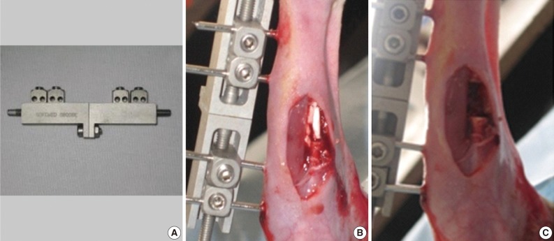
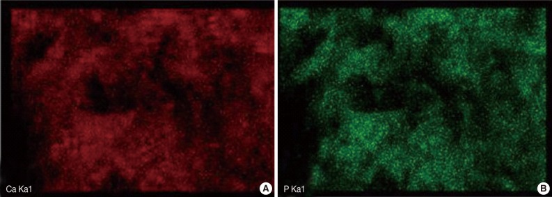
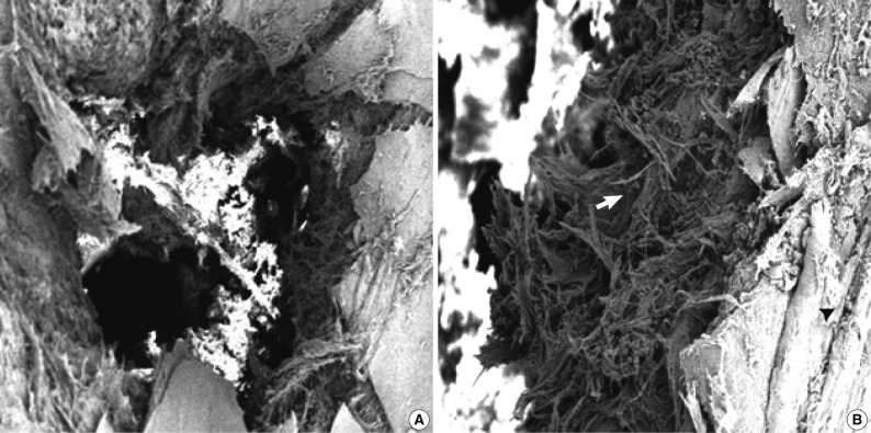
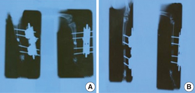

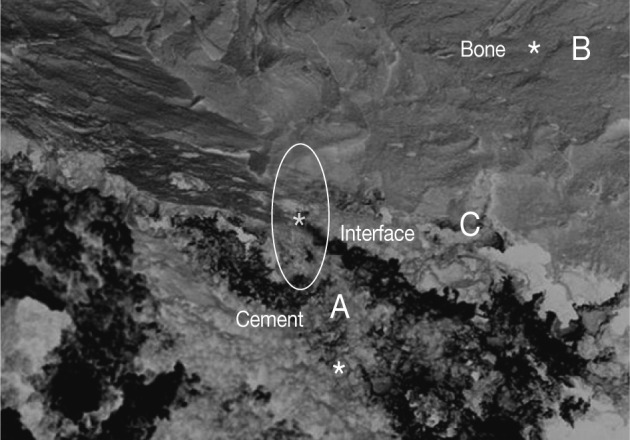
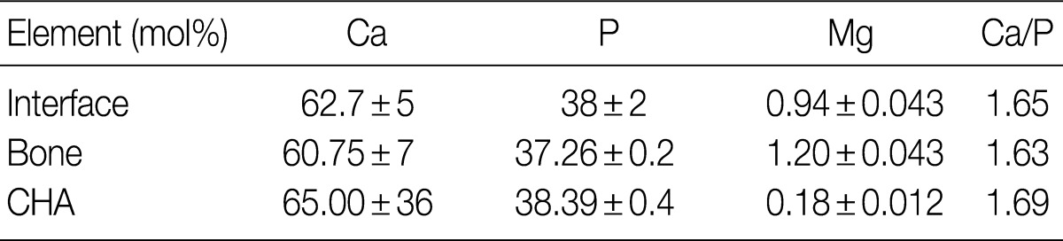
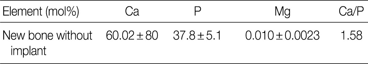
Figure & Data
References
Citations
- Comparative Evaluation of Sticky Bone with Alloplastic Bone Graft in the Treatment of Intrabony Defects: A Randomized Controlled Trial
Pooja Velraj, Senthilnathan Sivaramalingam, Gayathri Haritheertham, Hema Pannerselvam, Kalyani Ramkumar Sadhana, Sesha Reddy
World Journal of Dentistry.2026; 17(5): 417. CrossRef - A review: In vivo studies of bioceramics as bone substitute materials
Ali A. Al‐allaq, Jenan S. Kashan
Nano Select.2023; 4(2): 123. CrossRef - Improvement in bioactivity and corrosion resistance of Ti by hydroxyapatite deposition using ultrasonic mechanical coating and armoring
Ming-Hong Lin, Yi-Cheng Chen, Chun-Chung Liao, Liang-Wei Lin, Chin-Fu Chen, Kuang-Kuo Wang, Shyi-Tien Chen, Yi-Huang Hsueh, Chien-Hui Wu, Shih-Fu Ou
Ceramics International.2022; 48(4): 4999. CrossRef - Novel bioactive collagen-polyurethane-pectin scaffolds for potential application in bone regenerative medicine
Myriam L. Guzmán-Chávez, Jesús A. Claudio-Rizo, Martín Caldera-Villalobos, Denis A. Cabrera-Munguía, Juan J. Becerra-Rodríguez, Nayeli Rodríguez-Fuentes
Applied Surface Science Advances.2022; 11: 100317. CrossRef - Chitosan-Based Gastric Dressing Materials Loaded with Pomegranate Peel as Bioactive Agents: Pharmacokinetics and Effects on Experimentally Induced Gastric Ulcers in Rabbits
Samira Jebahi, Ghada Ben Salah, Soufien Jarray, Mounir Naffati, Mohammad Ayaz Ahmad, Faten Brahmi, Mohd Saeed, Arif J. Siddiqui, Khabir Abdelmajid, Riadh Badraoui
Metabolites.2022; 12(12): 1158. CrossRef - Is THP‐1 viability affected by the crystallinity of nanostructured carbonated hydroxyapatites?
Renata Moraes Lira, Suelen Cristina Sartoretto, Carolina da Silva Gouveia Pedrosa, Mônica Diuana Calasans‐Maia, Paulo Emílio Leite, José Mauro Granjeiro
Journal of Biomedical Materials Research Part A.2021; 109(7): 1266. CrossRef - Does the incorporation of zinc into calcium phosphate improve bone repair? A systematic review
Rebecca Cruz, José Calasans-Maia, Suelen Sartoretto, Vittório Moraschini, Alexandre Malta Rossi, Rafael Seabra Louro, José Mauro Granjeiro, Monica Diuana Calasans-Maia
Ceramics International.2018; 44(2): 1240. CrossRef - Adsorption of nucleotides on biomimetic apatite: The case of adenosine 5′ monophosphate (AMP)
K. Hammami, H. El Feki, O. Marsan, C. Drouet
Applied Surface Science.2015; 353: 165. CrossRef - Finite element analysis and cellular studies on advanced, controlled porous structures with subsurface continuity in bio-implantable titanium alloys
P. Lambert, S. Ankem, Z. Wyatt, K. M. Ferlin, J. Fisher
Journal of Biomedical Materials Research Part A.2014; 102(1): 225. CrossRef - The impact of orthopedic device associated with carbonated hydroxyapatite on the oxidative balance: experimental study of bone healing rabbit model
Samira Jebahi, Riadh Nsiri, Mohammed Boujbiha, Ezedine Bouroga, Tarek Rebai, Hassib Keskes, Abdelfattah El Feki, Hassane Oudadesse, Hafed El Feki
European Journal of Orthopaedic Surgery & Traumatology.2013; 23(7): 759. CrossRef
 PubReader
 Cite this Article


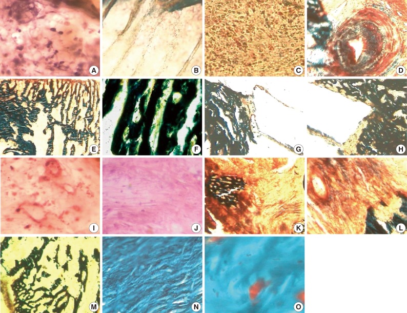


Fig. 1
Fig. 2
Fig. 3
Fig. 4
Fig. 5
Fig. 6


EDX, energy-dispersive X-ray; CHA, carbonated hydroxyapatite.
EDX, energy-dispersive X-ray.

