
 E-submission

Current issue
- Page Path
- HOME > Articles and issues > Current issue
- Volume 60(6); November 2026
-


Original Articles
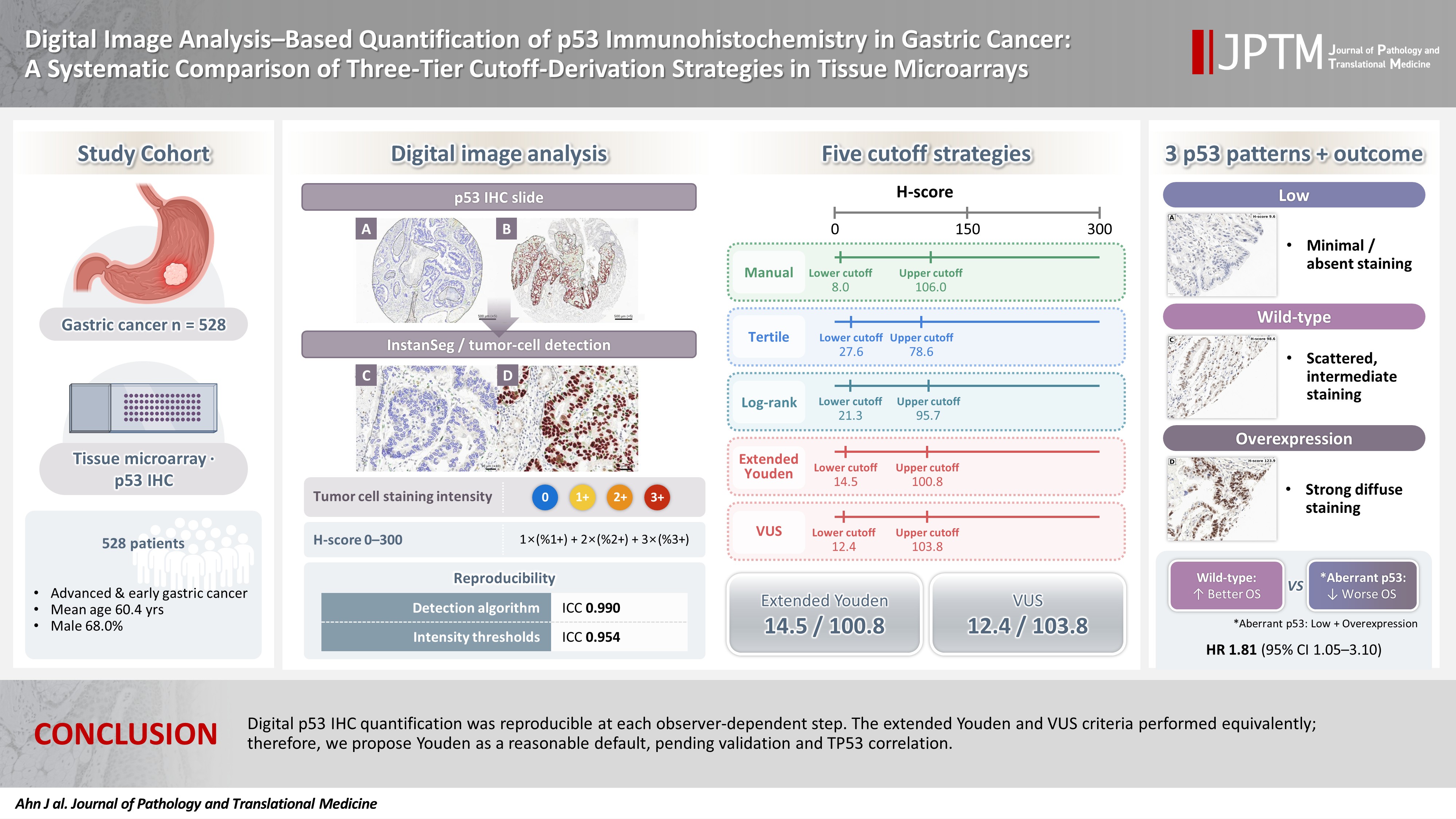
- Digital image analysis–based quantification of p53 immunohistochemistry in gastric cancer: a systematic comparison of three-tier cutoff-derivation strategies in tissue microarrays
- Jaeyoung Ahn, Kyungeun Kim, Hee-Jung Yang, Boram Song, Dong Hoon Kim, Yumin Chung, Seoung Wan Chae
- J Pathol Transl Med. 2026;60(6):593-610. Published online October 19, 2026
- DOI: https://doi.org/10.4132/jptm.2026.08.13
- 1,032 View
- 21 Download
-
 Abstract
 PDF
 Supplementary Material
- Background
p53 immunohistochemistry (IHC) is a common surrogate for TP53 alteration, but in gastric cancer, its concordance is imperfect and depends on a non-standardized cutoff. Digital pathology enables H-score quantification, yet neither its reproducibility nor the choice of cutoff- derivation strategy has been systematically compared. Methods: A semi-automated digital workflow quantified p53 IHC in tissue microarrays from 528 gastric cancer patients. Tumor cells were detected with a deep-learning model (InstanSeg), and nuclear staining was expressed as a continuous H-score (0–300). Reproducibility was assessed at each observer-dependent step. Five thresholding strategies were compared: manual cutoffs, tertiles, log-rank optimization, the extended Youden index, and volume under the receiver operating characteristic surface (VUS). The latter two strategies used the manual three-tier IHC interpretation as the reference standard. Results: Quantification was reproducible: two independent manual interpretations agreed in 91.7% of cores (κ = 0.845). The H-score was robust to the detection algorithm (intraclass correlation coefficient, 0.990) and to repeated threshold determination (0.954). H-scores were right-skewed (median, 45.5), and the strategies yielded distinct cutoffs (8.0–27.6 and 78.6–106.0). Log-rank optimization provided the widest separation but lost significance after multiplicity adjustment (p = .168). The extended Youden and VUS cutoffs were statistically indistinguishable. As a supporting check, all classifications showed nominal overall-survival separation, with low and overexpression faring similarly worse than wild-type. Conclusions: Digital p53 IHC quantification was reproducible at each observer-dependent step. The extended Youden and VUS criteria performed equivalently; therefore, we propose Youden as a reasonable default, pending validation and TP53 correlation.
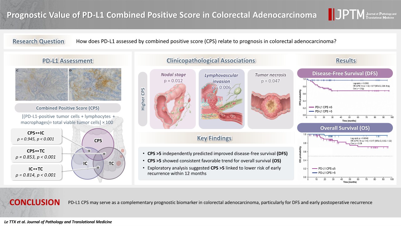
- Prognostic value of PD-L1 combined positive score in colorectal adenocarcinoma
- Thi Thanh Xuan Le, Van Chu Nguyen, Thi Minh Hanh Ngo, Thi Hue Le, Minh Son Le, Huu Song Le, Xuan Hoan Nghiem
- J Pathol Transl Med. 2026;60(6):611-624. Published online November 13, 2026
- DOI: https://doi.org/10.4132/jptm.2026.08.24
- 87 View
- 3 Download
-
Abstract
PDFSupplementary Material
- Background
The prognostic significance of programmed cell death ligand 1 (PD-L1) in colorectal adenocarcinoma remains unclear. We evaluated the clinicopathological and prognostic relevance of the PD-L1 combined positive score (CPS). Methods: This single-center retrospective cohort included 124 patients with histopathologically confirmed colorectal adenocarcinoma; PD-L1 was assessed by CPS. Disease-free survival (DFS) and overall survival (OS) were evaluated using Kaplan-Meier methods and parsimonious Cox regression, and early recurrence within 12 months of curative-intent treatment with a truncated Cox model. Results: Higher CPS was significantly associated with nodal stage (p = .012), lymphovascular invasion (p = .006), and tumor necrosis (p = .047). CPS correlated very strongly with immune-cell PD-L1 (rho = 0.945, p < .001) and strongly with tumor-cell PD-L1 (rho = 0.853, p < .001). CPS >5 remained associated with favorable DFS in a prespecified parsimonious multivariable model (adjusted hazard ratio [HR], 0.17; 95% confidence interval [CI], 0.04 to 0.75; p = .019). No PD-L1 metric was significantly associated with OS; however, CPS showed a numerically favorable but non-significant trend (adjusted HR, 0.17; 95% CI, 0.02 to 1.30; p = .088). In the exploratory early recurrence analysis, CPS >5 was associated with a lower risk of early recurrence in a model adjusted only for nodal stage (adjusted HR, 0.21; 95% CI, 0.05 to 0.94; p = .042). Conclusions: PD-L1 CPS may serve as a complementary, hypothesis-generating prognostic marker in colorectal adenocarcinoma, particularly for disease-free survival. However, the lack of mismatch repair (MMR)/microsatellite instability (MSI) data prevents definitive conclusions regarding an MMR/MSI-independent prognostic effect. Further validation in larger, prospectively collected, multicenter cohorts with comprehensive molecular annotation is required.
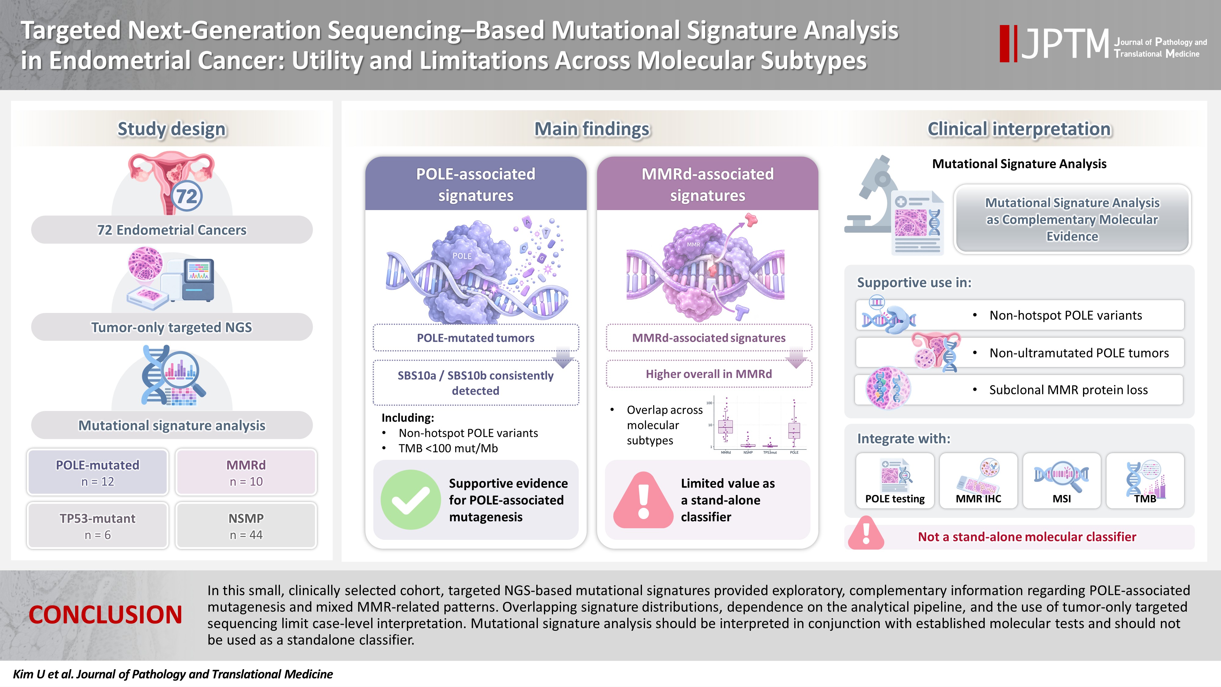
- Targeted next-generation sequencing–based mutational signature analysis in endometrial cancer: utility and limitations across molecular subtypes
- Uijin Kim, Ahwon Lee, Keun Ho Lee, Sung Jong Lee, Sook Hee Hong, Donghyuk Lee, Jun Kang, Miseon Lee
- J Pathol Transl Med. 2026;60(6):625-634. Published online November 10, 2026
- DOI: https://doi.org/10.4132/jptm.2026.09.07
- 92 View
- 1 Download
-
Abstract
PDFSupplementary Material
- Background
Molecular subtype classification is essential for staging endometrial cancer, planning adjuvant treatments, and stratifying prognosis. However, the correlation between mutational signature analysis using tumor-only targeted next-generation sequencing (NGS) data and molecular subtypes, as well as the interpretive value at the individual case level, remains inadequately characterized. Methods: We performed mutational signature analysis on tumor-only targeted NGS data from 72 endometrial cancer cases using the SATS R package. Molecular classification into four subtypes (POLE-mutated, mismatch repair deficient [MMRd], TP53-mutant, and no specific molecular profile [NSMP]) was established through a standard stepwise algorithm that integrated NGS and immunohistochemistry. Results: Out of the 72 endometrial cancers, 12 were classified as POLE-mutated, 10 as MMRd, six as TP53-mutant, and 44 as NSMP. All POLE-mutated tumors exhibited POLE-associated signatures (SBS10a/SBS10b), including cases with non-hotspot POLE exonuclease variants and tumor mutation burdens below the conventional ultramutation threshold. MMRd-associated signatures showed a broader distribution across molecular subtypes; while the summed MMRd-associated signatures were typically higher in MMRd tumors, low-level contributions were also noted in subsets of NSMP and TP53-mutant tumors. Within the POLE-mutated group, MMRd-associated signatures were more pronounced in cases displaying subclonal MMR protein loss as determined by immunohistochemistry. Conclusions: In this small, clinically selected cohort, targeted NGS-based mutational signatures provided exploratory, complementary information regarding POLE-associated mutagenesis and mixed MMR-related patterns. Overlapping signature distributions, dependence on the analytical pipeline, and the use of tumor-only targeted sequencing limit case-level interpretation. Mutational signature analysis should be interpreted in conjunction with established molecular tests and should not be used as a stand-alone classifier.
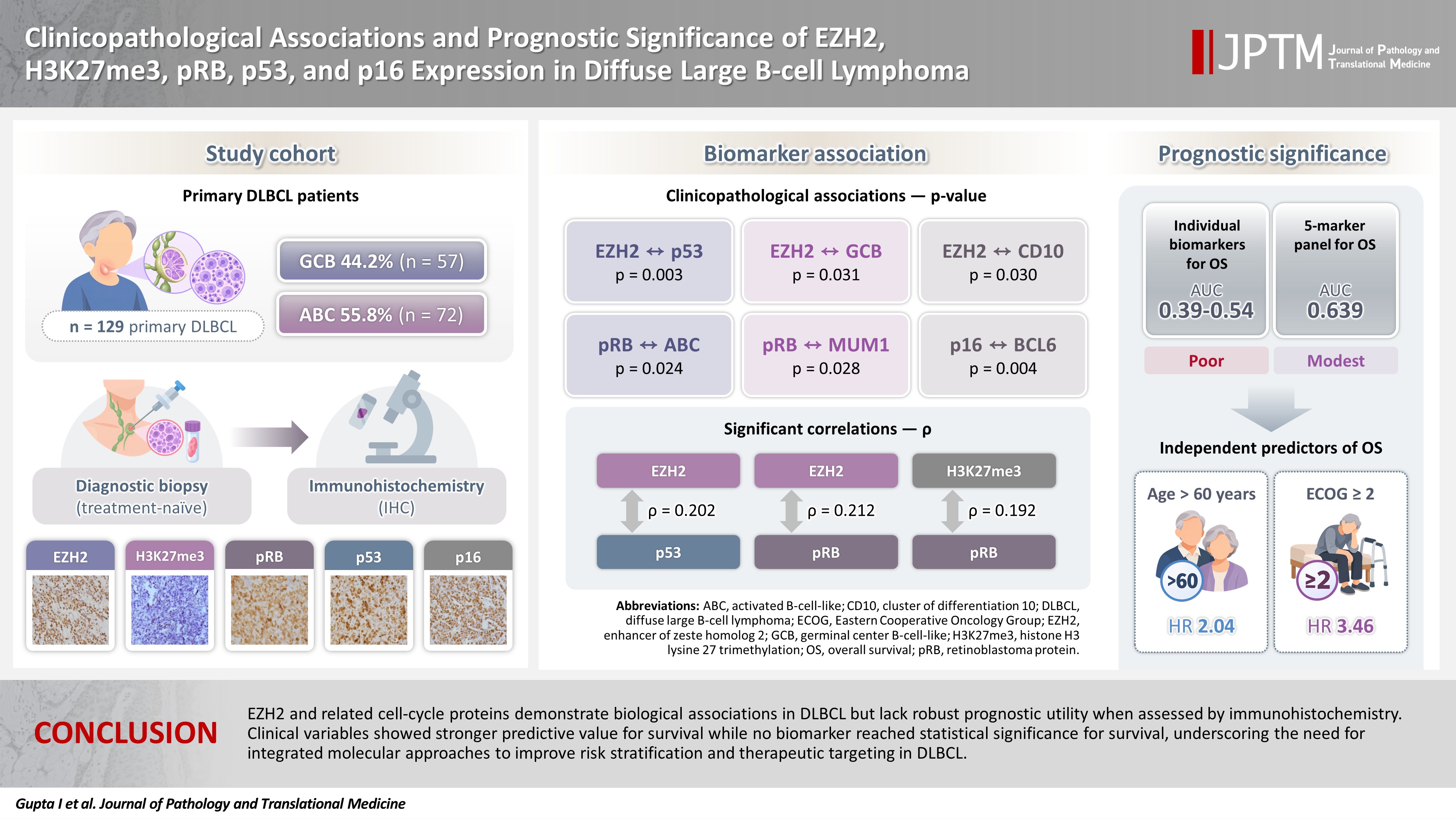
- Clinicopathological associations and prognostic significance of EZH2, H3K27me3, pRB, p53, and p16 expression in diffuse large B-cell lymphoma
- Ishan Gupta, Surabhi Jain, Moien Rasheed Lone, Ajay Gogia, Chandan Krushna Das, Mehar Chand Sharma, Ashish Datt Upadhyay, Saumyaranjan Mallick
- J Pathol Transl Med. 2026;60(6):635-645. Published online November 13, 2026
- DOI: https://doi.org/10.4132/jptm.2026.09.14
- 62 View
- 2 Download
-
Abstract
PDFSupplementary Material
- Background
Diffuse large B-cell lymphoma (DLBCL) is characterized by pronounced heterogeneity in its morphological and biological attributes, with emerging evidence highlighting the pivotal role of epigenomic alterations in its pathogenesis. This study aimed to investigate the relationship between EZH2 expression and cell-cycle regulatory proteins, focusing on their prognostic implications. Methods: This retrospective study included 131 patients with nodal primary DLBCL, who were categorized according to the Hans algorithm and additional immunohistochemistry was performed for selected epigenetic (EZH2 and H3K27me3) and cell-cycle regulatory markers (phosphorylated retinoblastoma [pRB], p16 and p53). Receiver operating characteristic (ROC) curve analysis and Cox regression models were applied to assess prognostic utility. Appropriate statistical analysis was performed. Results: EZH2 was expressed in 59.5% of cases and significantly associated with p53 (p = .003), germinal center B-cell-like (GCB) subtype (p = .031), and CD10 positivity (p = .030). pRB positivity was more frequent in the activated B-cell-like subtype (82.5% vs. 64.3%, p = .024) and was associated with MUM1 (p = .028), while p16 expression was linked to BCL6 (p = .004). ROC analyses demonstrated poor discriminatory ability for overall survival (area under the curve, 0.39 to 0.54), with no clinically actionable cutoffs identified. Stepwise Cox regression identified age >60 years (hazard ratio [HR], 2.04; p = .042) and Eastern Cooperative Oncology Group ≥ 2 (HR, 3.46; p = .001) as independent predictors of overall survival. For progression-free survival, no biomarker reached statistical significance. Conclusions: EZH2 and related cell-cycle proteins demonstrate biological associations in DLBCL but lack robust prognostic utility when assessed by immunohistochemistry. Clinical variables showed stronger predictive value for survival, while no biomarker reached statistical significance for survival, underscoring the need for integrated molecular approaches to improve risk stratification and therapeutic targeting in DLBCL.
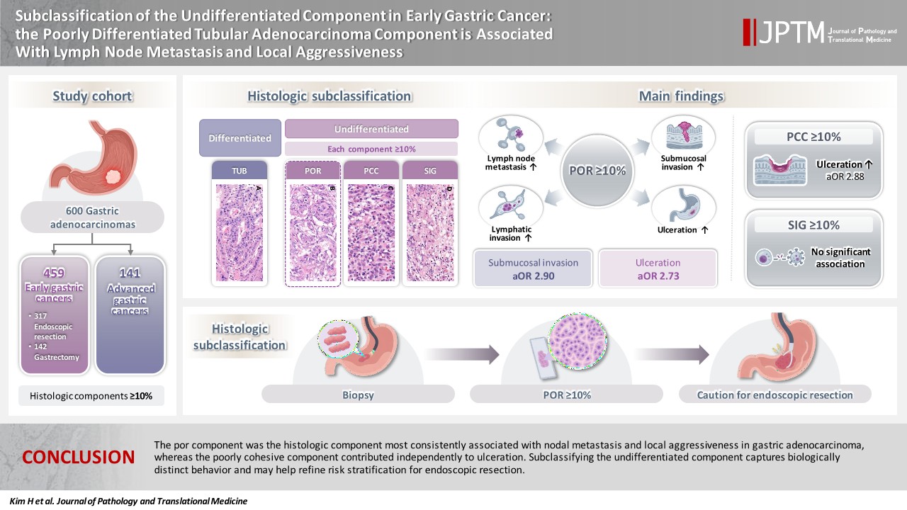
- Subclassification of the undifferentiated component in early gastric cancer: the poorly differentiated tubular adenocarcinoma component is associated with lymph node metastasis and local aggressiveness
- Heesoo Kim, Baek-hui Kim
- J Pathol Transl Med. 2026;60(6):646-656. Published online November 13, 2026
- DOI: https://doi.org/10.4132/jptm.2026.09.15
- 71 View
- 1 Download
-
Abstract
PDFSupplementary Material
- Background
Gastric adenocarcinoma is histologically heterogeneous, and the undifferentiated category encompasses tubular adenocarcinoma, poorly differentiated (por), poorly cohesive carcinoma (pcc), and signet ring cell carcinoma (sig). These components are often treated as a single entity, and which one drives the aggressive behavior of mixed-type tumors remains unclear. We subclassified the undifferentiated component and examined the association of each histologic component with adverse pathologic features. Methods: We retrospectively analyzed 600 gastric adenocarcinomas (459 early, 141 advanced). Each histologic component (tub [tubular adenocarcinoma, well to moderately differentiated], por, pcc, sig) was assessed separately in the mucosal and invasive portions and dichotomized at 10%. Associations with lymph node metastasis (LNM), submucosal invasion, lymphatic invasion, and ulceration were evaluated by univariate testing and multivariable logistic regression. Results: In early gastric cancer, por ≥10% was associated with LNM (p = .006) and lymphatic invasion (p < .001), and was an independent risk factor for submucosal invasion (adjusted odds ratio [OR], 2.90; 95% confidence interval [CI], 1.80 to 4.68; p < .001) and ulceration (adjusted OR, 2.73; 95% CI, 1.40 to 5.34; p = .003). By contrast, pcc ≥10% was independently associated only with ulceration (adjusted OR, 2.88; 95% CI, 1.25 to 6.65; p = .013), and sig showed no association with any adverse feature (all p > .05). Findings in the mucosal component paralleled those in the whole tumor. No histologic component was associated with survival. Conclusions: The por component was the histologic component most consistently associated with nodal metastasis and local aggressiveness in gastric adenocarcinoma, whereas the poorly cohesive component contributed independently to ulceration. Subclassifying the undifferentiated component captures biologically distinct behavior and may help refine risk stratification for endoscopic resection.
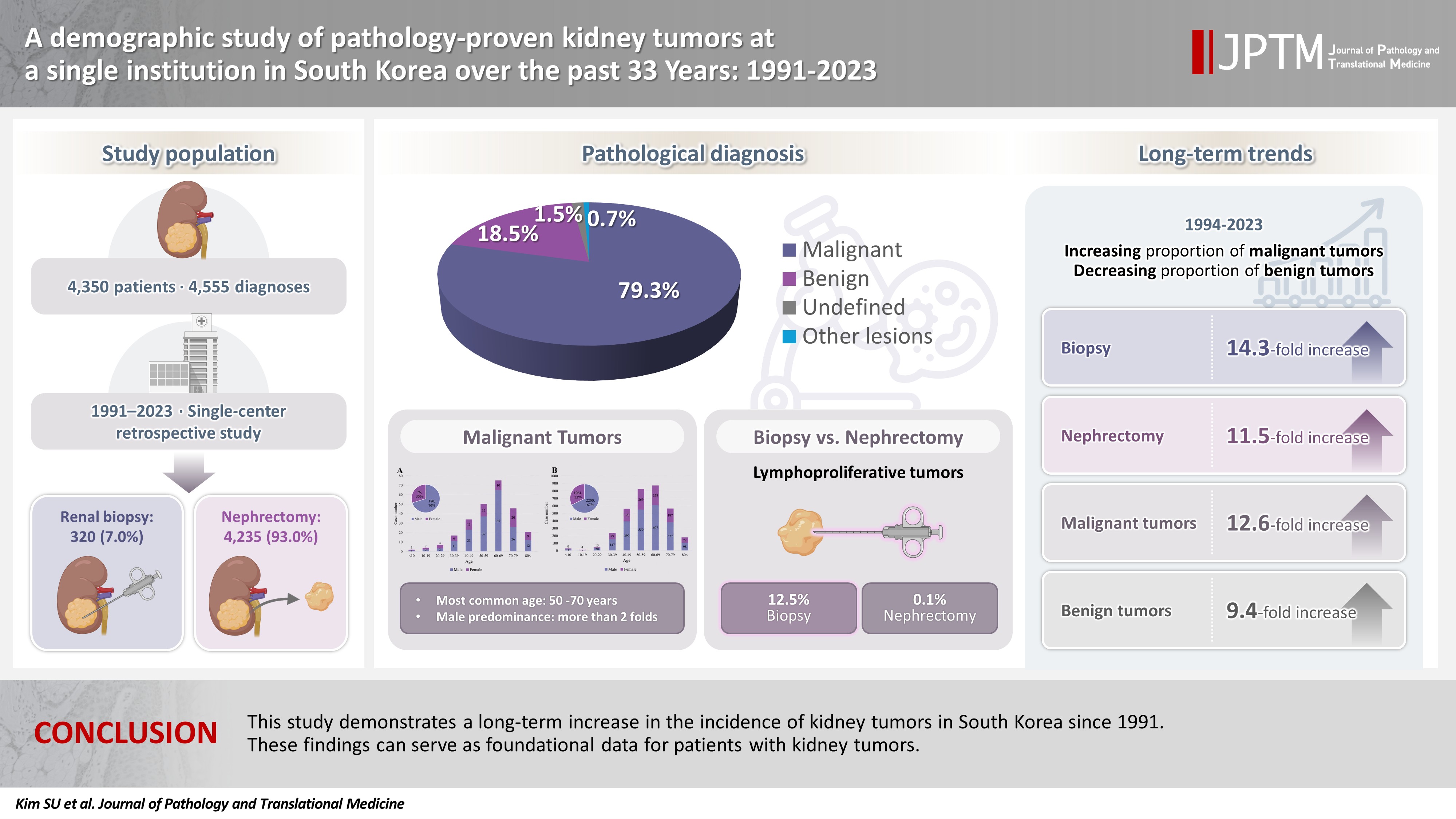
- A demographic study of pathology-proven kidney tumors at a single institution in South Korea over the past 33 years: 1991–2023
- Seon Ung Kim, Hyejin Noh, Jiyeon Kim, Yeong Jin Choi
- J Pathol Transl Med. 2026;60(6):657-669. Published online November 13, 2026
- DOI: https://doi.org/10.4132/jptm.2026.10.19
- 51 View
- 1 Download
-
Abstract
PDF
- Background
While the incidence of kidney tumors is increasing, long-term demographic analyses based on pathological diagnoses remain insufficient. This study aimed to investigate demographic changes regarding pathology-proven kidney tumors over the past 33 years. Methods: We retrospectively analyzed 4,555 cases from 4,350 patients who underwent renal biopsy or nephrectomy for suspected kidney tumors at Seoul St. Mary’s Hospital from 1991 to 2023. Results: The average age was 58 years, and the male-to-female ratio was 1.7:1. Among 4,555 cases, 7.0% were biopsies and 93.0% were nephrectomies. Tumors were classified into four diagnostic categories: malignant (79.3%), benign (18.5%), undefined (1.5%), and other lesions (0.7%). Over 33 years, biopsies increased 14.3-fold and nephrectomies 11.5-fold, while malignant tumors increased 12.6-fold and benign tumors 9.4-fold. Over time, the proportion of malignant tumors increased (p < .001), while benign tumors decreased (p < .001). As age increased, the proportion of malignant tumors increased (p = .013), while benign tumors showed a decreasing trend (p = .070). Malignant tumors showed a male predominance, while benign tumors showed a female predominance. The frequency of malignant tumors was similar at approximately 80% in biopsy and nephrectomy. However, benign tumors were more common in nephrectomy (19.1%) than in biopsy (10.9%) (p < .001), and unclassified tumors (4.1%) were more common in biopsies (p < .001). Renal cell carcinoma was most common (61.5% of total cases, 77.6% of malignant tumors). Lymphoproliferative tumors were diagnosed more frequently by biopsy (12.5%) than nephrectomy (0.1%) (p < .001). Conclusions: This study demonstrates a long-term increase in the incidence of renal tumors in South Korea. These findings can serve as foundational data for patients with kidney tumors.
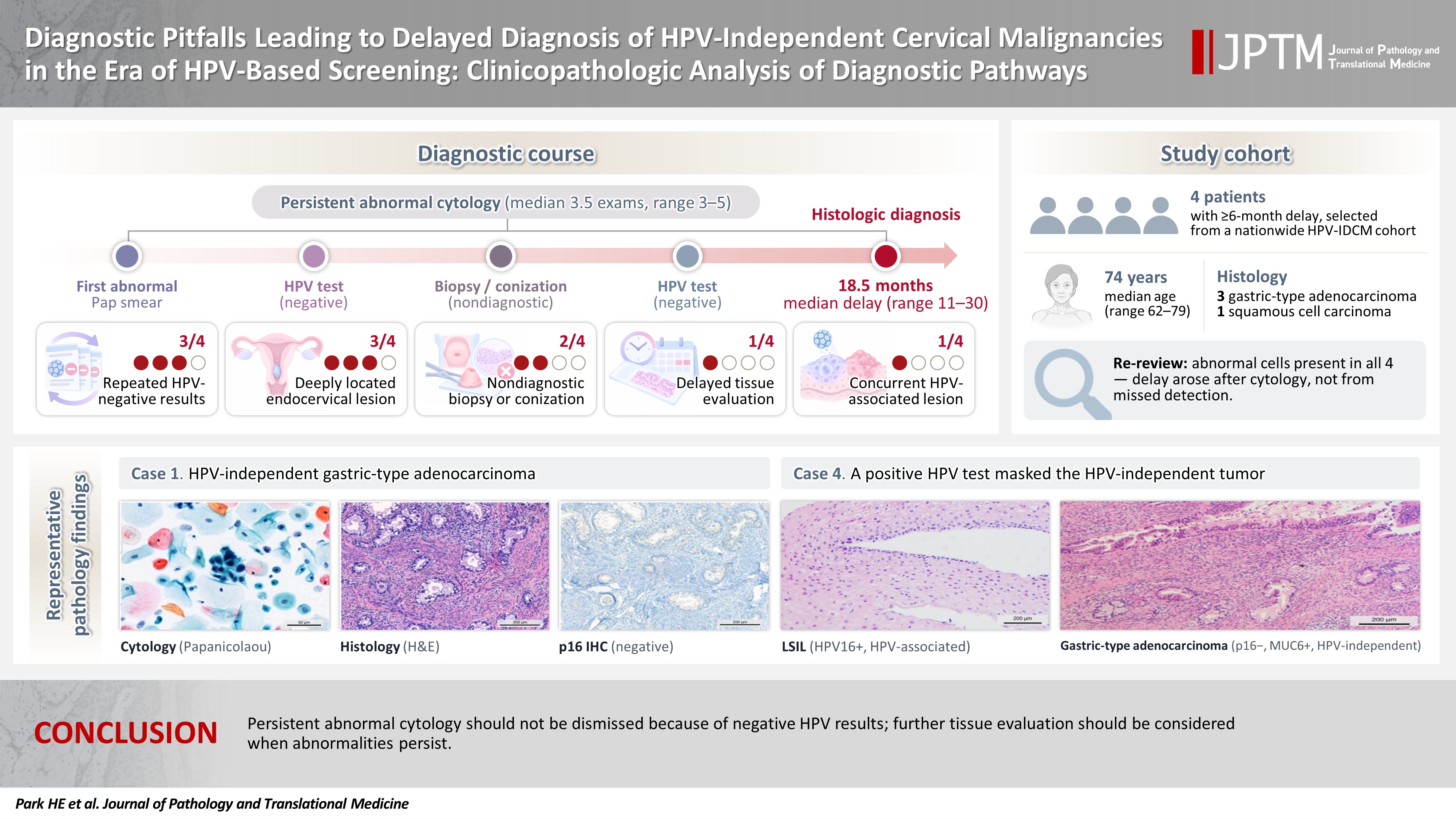
- Diagnostic pitfalls leading to delayed diagnosis of human papillomavirus (HPV)–independent cervical malignancies in the era of HPV-based screening: clinicopathologic analysis of diagnostic pathways
- Hye Eun Park, Hye-Ra Jung, Hyun-Jung Kim
- J Pathol Transl Med. 2026;60(6):670-679. Published online November 13, 2026
- DOI: https://doi.org/10.4132/jptm.2026.08.26
- 64 View
- 0 Download
-
Abstract
PDF
- Background
Human papillomavirus (HPV)–independent cervical malignancies (HPV-IDCM) are a rare but clinically important subset that may escape detection by HPV-based screening. This study investigated diagnostic pitfalls contributing to delayed HPV-IDCM diagnosis and assessed the role of cervical cytology in the diagnostic pathway. Methods: This multicenter retrospective case series was conducted as a policy research project of the Korean Society for Cytopathology. Patients with HPV-IDCM and repeated abnormal cytologic findings before definitive histologic diagnosis were identified. Clinical, cytologic, histologic, and HPV test results were reviewed to identify factors associated with diagnostic delay. Results: Four patients were included, with a median age of 74 years (range, 62 to 79 years). Histologic diagnoses comprised three HPV-independent gastric-type adenocarcinomas and one HPV-independent squamous cell carcinoma. The median interval from the first abnormal cytologic finding to definitive diagnosis was 18.5 months (range, 11 to 30 months). Before diagnosis, all patients underwent a median of 3.5 cytologic examinations (range, 3 to 5). Associated factors included repeatedly negative HPV test results (3/4), nondiagnostic biopsy or conization (2/4), delayed tissue evaluation despite persistent cytologic abnormalities (1/4), deeply located endocervical lesions (3/4), and a concurrent HPV-associated squamous lesion (1/4). Re-review identified atypical or malignant cells in all cases, indicating that the delay did not result from failure of cytologic detection. Conclusions: HPV-IDCM remain vulnerable to delayed diagnosis in the era of HPV-based screening. Persistent abnormal cytologic findings should not be dismissed solely because HPV test results are negative. Careful correlation of cytologic, clinical, and histologic findings remains essential for timely HPV-IDCM detection.
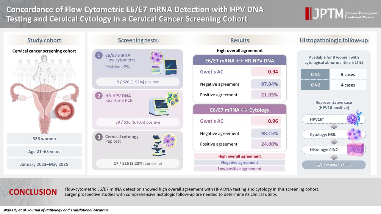
- Concordance of flow cytometric E6/E7 mRNA detection with human papillomavirus DNA testing and cervical cytology in a cervical cancer screening cohort
- Dat Quoc Ngo, Trang Kieu Ha, Mi Ngoc Thi Tran, Anh Mai Thi Nguyen, Truong Nhat Trinh, Thien Thanh Ly, Linh Ngoc Khanh Nguyen, Thu Dang Anh Phan
- J Pathol Transl Med. 2026;60(6):680-686. Published online November 10, 2026
- DOI: https://doi.org/10.4132/jptm.2026.09.09
- 184 View
- 9 Download
-
Abstract
PDF
- Background
Overexpression of viral oncogenes E6 and E7 of high-risk (HR)–human papillomavirus (HPV) plays a key role in the progression to cervical intraepithelial neoplasia (CIN) and cancer. Detection of E6/E7 mRNA may help distinguish between transient and transforming infections, thus improving cervical cancer screening accuracy. This study aimed to evaluate the association of E6/E7 mRNA expression with HR-HPV infection, cervical cytology, and histologically confirmed cervical lesions and to compare its agreement with HPV DNA testing and cytology. Methods: A total of 526 women undergoing cervical cancer screening between January 2023 and May 2025 were enrolled. E6/E7 mRNA expression was assessed using flow cytometry with the HPV OncoTect 3Dx E6/E7 mRNA assay and compared with HR-HPV DNA testing, cervical cytology, and available histopathologic findings from biopsy specimens. Results: Among 526 samples, eight (1.5%) tested positive for E6/E7 mRNA, compared to 30 (5.7%) HR-HPV DNA–positive and 17 (3.2%) abnormal cytology results. Median E6/E7 mRNA expression was higher in HR-HPV– positive cases and in cytologically abnormal cases. Almost perfect agreement was observed between E6/E7 mRNA results, HR-HPV DNA testing, and cytology. Histopathologic follow-up was available for nine women with cytological abnormalities ≥low-grade squamous intraepithelial lesion, including four CIN2 and five CIN1 cases. E6/E7 mRNA expression was markedly elevated in one CIN2 case associated with HPV16 infection (37.51%), while two CIN1 cases demonstrated high mRNA expression despite negative HPV DNA results. Conclusions: Flow-cytometric E6/E7 mRNA detection showed high overall agreement with HPV DNA testing and cytology in this screening cohort. Larger prospective studies with comprehensive histologic follow-up are needed to determine its clinical utility.
Case Studies
- Ultrasound-guided cryoablation of recurrent thyroid cancer: radiologic-pathologic correlation
- Yeseul Kim, Jae Ho Shin, Kyeong Jin Kim, Soo Myoung Shin, Dawon Park
- J Pathol Transl Med. 2026;60(6):687-692. Published online August 3, 2026
- DOI: https://doi.org/10.4132/jptm.2026.06.04

- 1,407 View
- 47 Download
-
Abstract
PDFSupplementary Material
- Cryoablation is a minimally invasive thermal ablation modality that destroys tumor cells through intracellular ice crystal formation and osmotic injury. Although it offers theoretical advantages, its application in recurrent papillary thyroid cancer (PTC) remains limited, and the radiologic-pathologic findings of treatment response have not been well characterized. A 55-year-old male patient with biopsy-proven recurrent PTC in the left supraclavicular fossa and a history of total thyroidectomy, neck dissection, and advanced colorectal cancer underwent a single-session ultrasound-guided cryoablation. The 20-minute procedure was completed without complications. Immediately post-ablation, the lesion volume increased by over 200% with marked hypoechogenicity and obliteration of abnormal feeding vessels on microvascular imaging. Pathology demonstrated disrupted tumor cell membranes, necrotic changes, and myxoid peritumoral stromal alteration, consistent with direct and indirect injuries caused by cryoablation. At the first-month follow-up, repeat biopsy showed no viable tumor, with subacute inflammatory infiltrates, histiocytic aggregates, and progressive fibrosis. On ultrasound, a volume reduction rate of 71.4% was achieved at one month follow-up and 97.1% at three months follow-up. This case report provides the first radiologic-pathologic findings of both immediate and delayed effects of cryoablation in recurrent PTC, supporting its role as a promising minimally invasive palliative therapy.
- Gastric amphicrine carcinoma in the stomach: an unexpected presentation of MUTYH-associated polyposis
- Seungkyun Lee, Byung-Hoon Min, Young-Gon Kim, Soomin Ahn, Kyoung-Mee Kim
- J Pathol Transl Med. 2026;60(6):693-697. Published online September 7, 2026
- DOI: https://doi.org/10.4132/jptm.2026.06.24
- 1,107 View
- 14 Download
-
Abstract
PDF
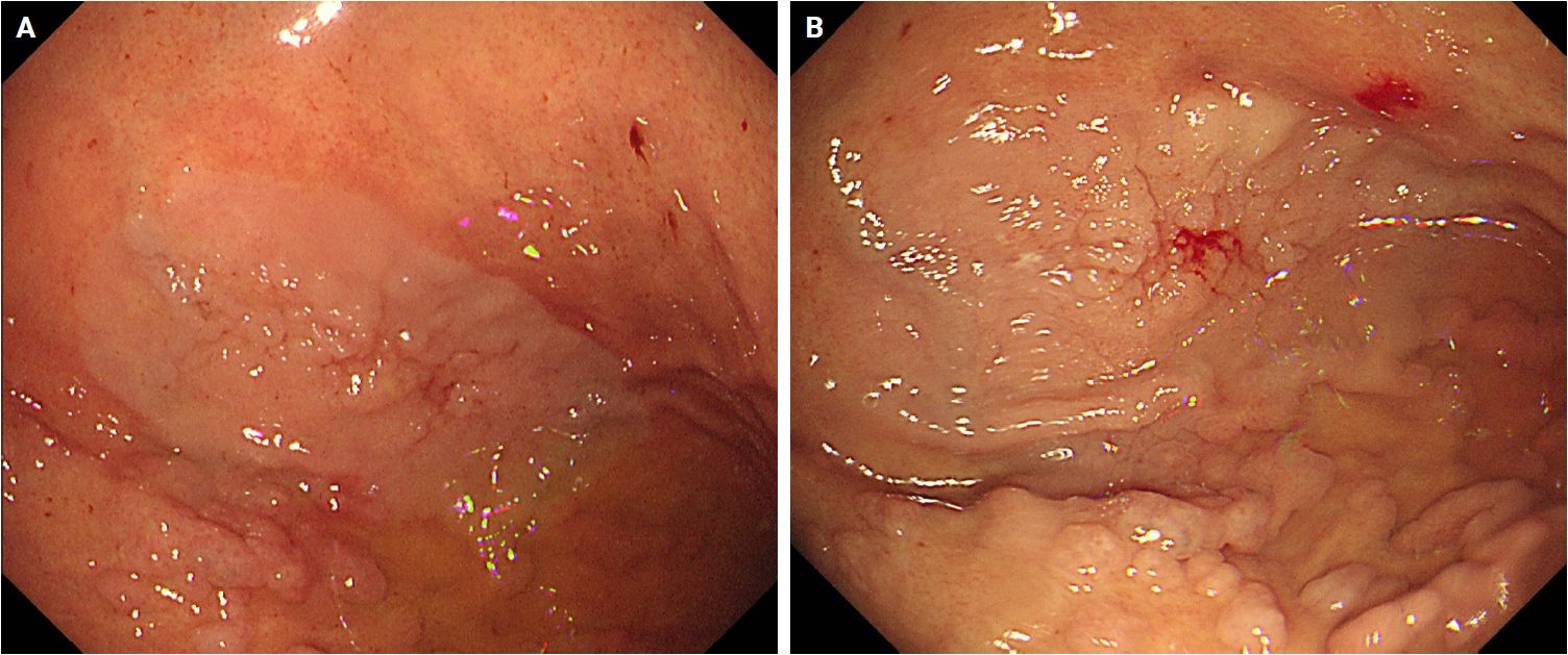
- Amphicrine carcinomas of the stomach, defined by dual exocrine and neuroendocrine differentiation within the same neoplastic cell, are exceedingly rare. MUTYH-associated polyposis (MAP) is an autosomal recessive polyposis syndrome characterized by multiple colorectal adenomas and variable upper gastrointestinal involvement; however, amphicrine carcinomas have not been previously documented in this setting. We report a gastric amphicrine carcinoma arising in the background of extensive fundic gland polyposis in a patient with MAP. Endoscopy revealed a 3.5-cm flat elevated lesion in the gastric fundus amid extensive fundic gland polyposis. Histologically, the tumor consisted of a single population of cells exhibiting combined glandular and neuroendocrine differentiation without zonal or biphasic architecture, and many of these cells demonstrated true amphicrine morphology. Immunohistochemistry confirmed co-expression of cytokeratin and the neuroendocrine markers chromogranin A and synaptophysin in the same cell population. Germline targeted next-generation sequencing identified biallelic MUTYH variants in trans (c.733C>T, p.Arg245Cys [likely pathogenic]; c.842C>T, p.Ala281Val [variant of uncertain significance]), supporting a diagnosis of MAP. To our knowledge, this is the first reported case of a gastric amphicrine carcinoma in a MAP patient, expanding the spectrum of MAP-associated upper gastrointestinal neoplasia and underscoring the importance of vigilant endoscopic surveillance in hereditary polyposis syndromes.
- Colorectal intramucosal carcinoma with lymphatic invasion and lymph node metastasis: a case report
- Joon Mee Kim, Lucia Kim, Jee Young Han, Suk Jin Choi, Jongbeom Shin, Sun Keun Choi
- J Pathol Transl Med. 2026;60(6):698-703. Published online October 19, 2026
- DOI: https://doi.org/10.4132/jptm.2026.08.14
- 782 View
- 14 Download
-
Abstract
PDF
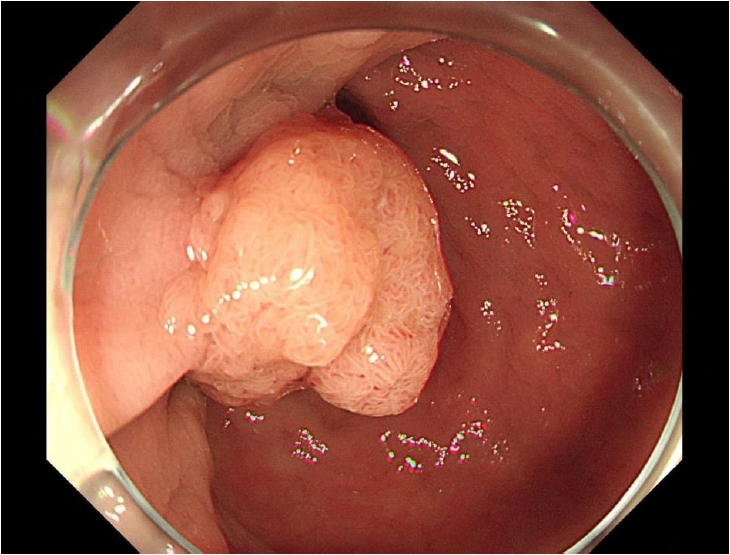
- Colorectal intramucosal carcinoma (IMC) is considered to carry negligible risk of lymph node metastasis, reflecting the presumed absence of lymphatics within the lamina propria, and current guidelines endorse endoscopic resection alone as curative. We report a 64-year-old man in whom endoscopic submucosal dissection of a lower rectal tumor revealed a well-differentiated IMC with extensive lymphatic invasion confirmed by D2-40 immunostaining. Subsequent laparoscopic ultralow anterior resection showed no residual tumor at the primary site but confirmed metastasis in one of 12 regional lymph nodes. The tumor was microsatellite-stable, epidermal growth factor receptor–positive, and harbored a KRAS codon 13 mutation (G13X). The patient received adjuvant chemoradiotherapy and remained disease-free at 2 years. This case demonstrates that regional lymph node metastasis can occur in colorectal IMC even without conventional adverse histologic features such as poor differentiation or tumor budding, challenging current risk-stratification frameworks and supporting a more cautious approach when lymphatic invasion is identified.
- Meningioma harboring IDH1 R132H mutation: a case report
- Soyeon Ahn, Chung Lee, Jong-Pil Park, Se Hoon Kim
- J Pathol Transl Med. 2026;60(6):704-709. Published online November 3, 2026
- DOI: https://doi.org/10.4132/jptm.2026.08.19
- 413 View
- 17 Download
-
Abstract
PDF
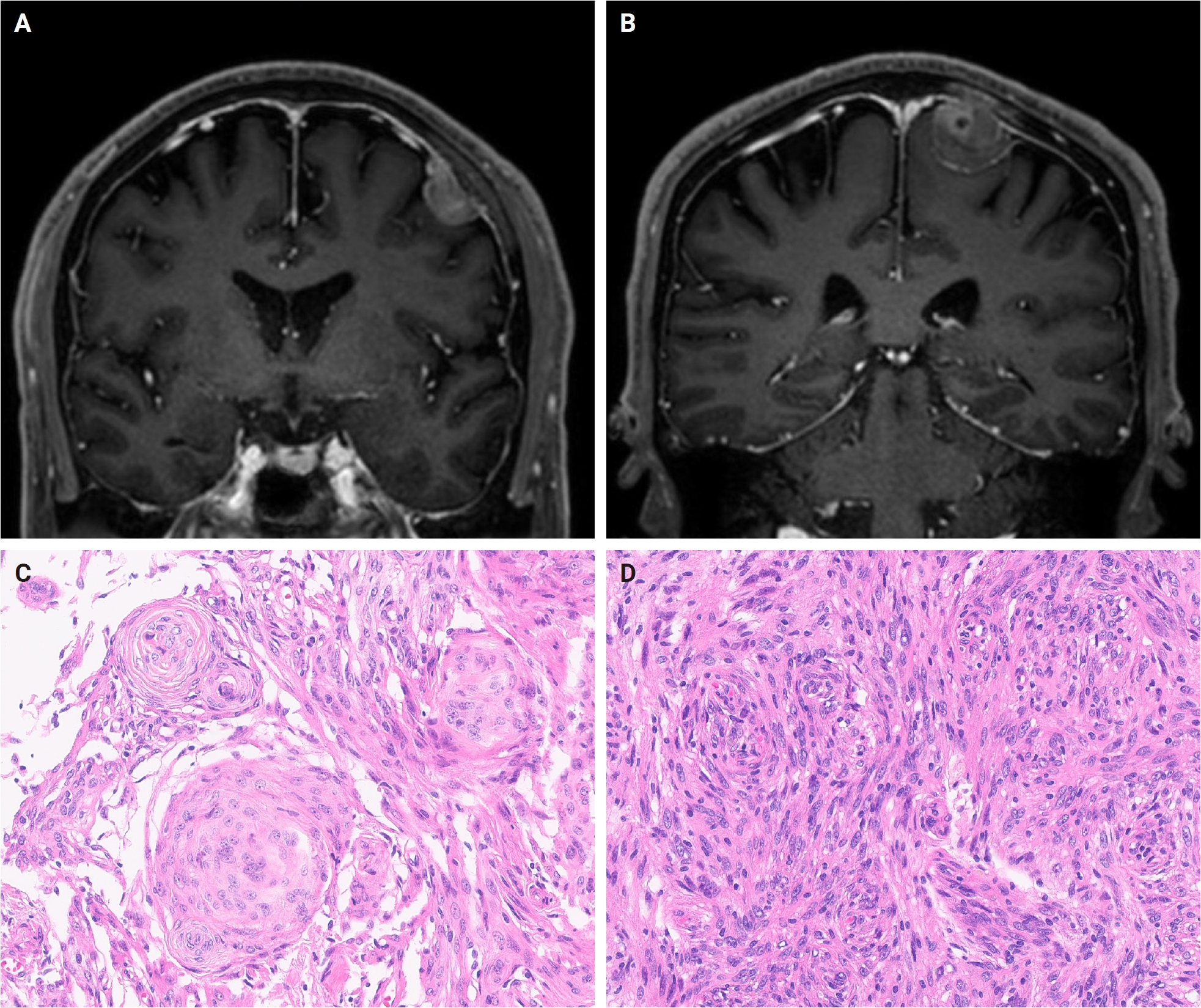
- Meningiomas are the most common primary central nervous system tumors. Monosomy of chromosome 22 is the most frequently reported genetic abnormality in meningioma and the occurrence of IDH1 mutations in meningioma is exceedingly rare. We report an uncommon case of IDH1 R132H-mutant meningioma. A 69-year-old man presented with two intracranial masses, one of which was identified as an IDH1 R132H-mutant meningioma by next-generation sequencing and further confirmed by pyrosequencing and immunohistochemistry. The rarity of this molecular finding and the potentially favorable prognosis observed in previously reported cases highlight the need for its recognition as a distinct molecular subset. Isocitrate dehydrogenase–targeted therapy may represent a future therapeutic option, pending validation through prospective studies and larger case series.
- Extra-axial poorly differentiated chordoma of the knee masquerading as periprosthetic joint infection: a case report
- Hee-Jung Yang, Hye Ra Jung, Joon Hyuk Choi, Chang-Jin Yon, Sang Pyo Kim
- J Pathol Transl Med. 2026;60(6):710-717. Published online November 10, 2026
- DOI: https://doi.org/10.4132/jptm.2026.08.31
- 124 View
- 2 Download
-
Abstract
PDFSupplementary Material
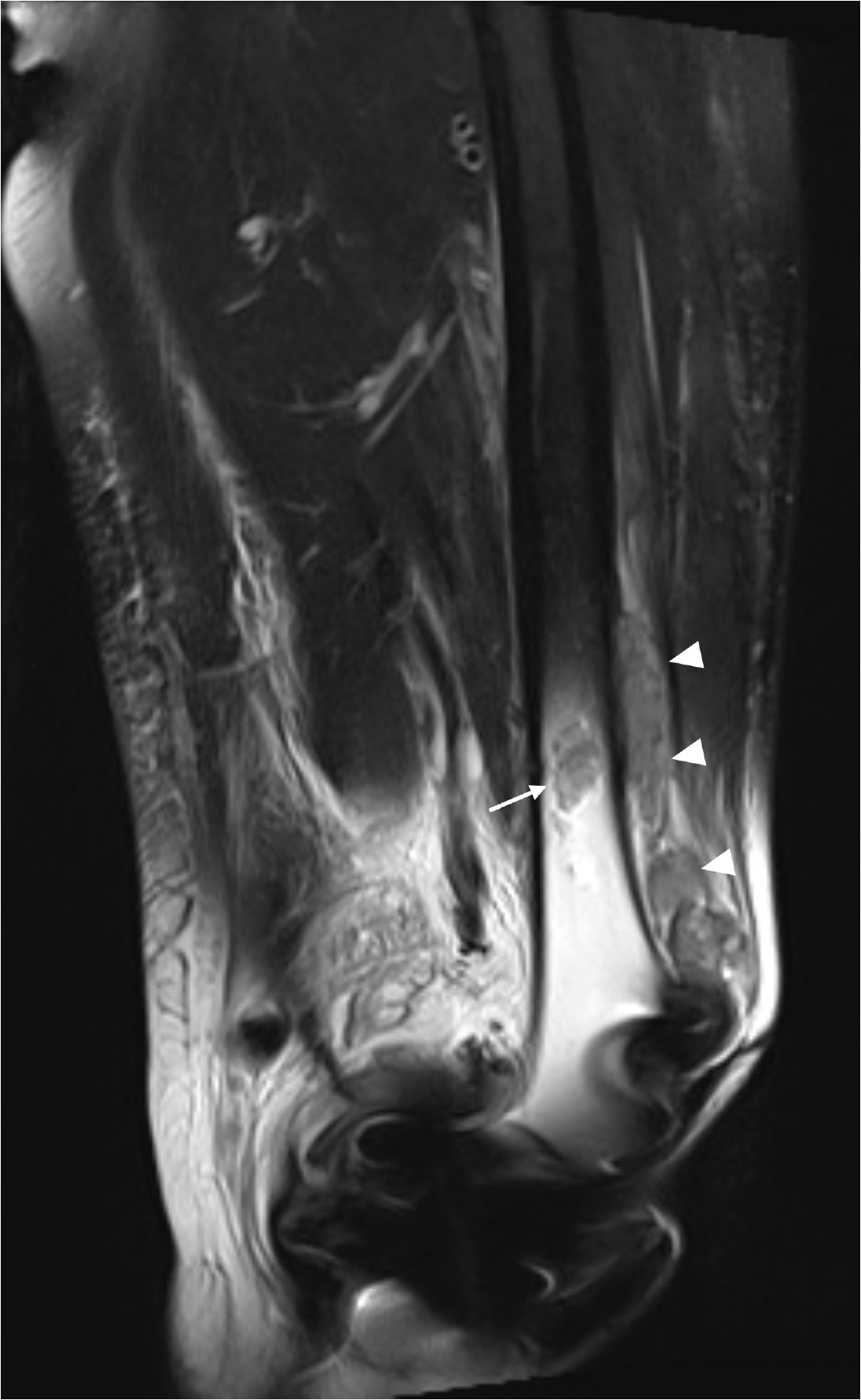
- Poorly differentiated chordoma (PDC) is a malignant neoplasm defined by the co-expression of brachyury and cytokeratin with loss of SMARCB1 (INI-1) expression. Extra-axial PDC is exceedingly rare, with only 10 cases reported to date. Here, we report an additional case of extra-axial PDC arising in the soft tissues surrounding the knee joint of a 69-year-old woman, which was initially misdiagnosed as periprosthetic joint infection following total knee arthroplasty. Histologically, the tumor comprised epithelioid to spindled cells with vacuolated, eosinophilic cytoplasm, focal rhabdoid areas, without myxoid stroma. Immunohistochemically, the tumor cells showed diffuse positivity for brachyury and cytokeratin, with loss of INI-1 expression. Next-generation sequencing identified a homozygous deletion of CDKN2A, but no SMARCB1 genomic alteration was detected despite INI-1 loss. This case shows that extra-axial PDC should be considered in the differential diagnosis of soft tissue tumors, irrespective of anatomical site or morphology.
- Intracranial malignant peripheral nerve sheath tumor with myxoid morphology evolving across recurrences: a case report
- Frantisek Vana, Petra Pokorna, Petr Pospisil, Zdenek Mackerle, Marketa Hermanova, Iva Staniczkova Zambo, Martin Barak, Robin Jugas, Radim Jancalek, Ondrej Slaby, Michal Hendrych
- J Pathol Transl Med. 2026;60(6):718-725. Published online November 11, 2026
- DOI: https://doi.org/10.4132/jptm.2026.09.17
- 143 View
- 2 Download
-
Abstract
PDF
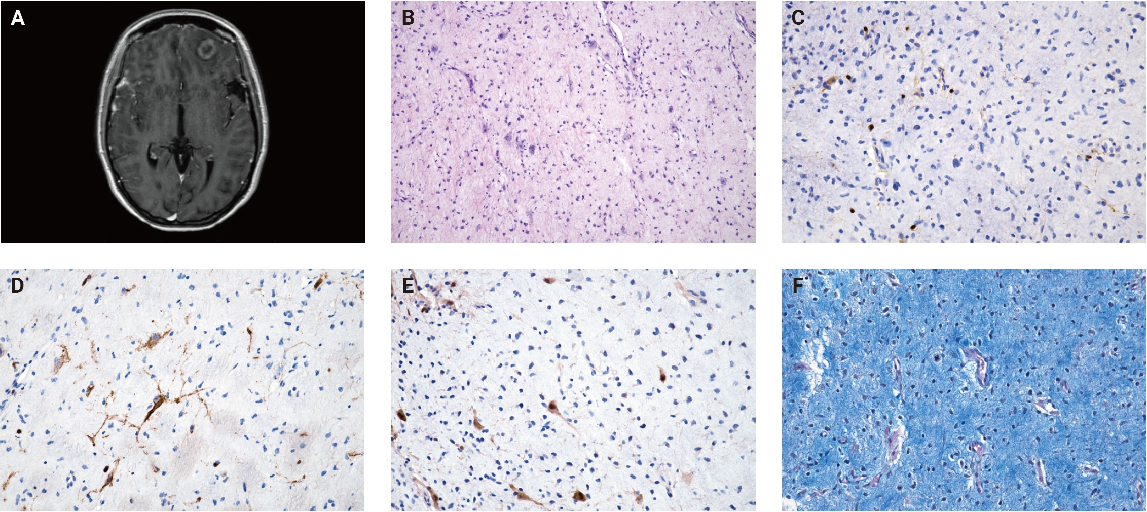
- A malignant peripheral nerve sheath tumor (MPNST) is a rare, aggressive spindle-cell sarcoma that exceptionally occurs intracranially. Herein, we report a case of intracranial spindle-cell neoplasm with abundant myxoid stroma. The immunophenotype of the primary tumor did not align with any known peripheral nerve sheath tumor and the lesion did not correspond to any methylation category defined by the Heidelberg classifier with a sufficiently high classification score, although MPNST was the highest-scoring category. Over the course of repeated recurrences, the tumor acquired increased cellularity, mitotic activity and loss of H3K27me3 expression. Subsequent methylation profiling supported a diagnosis of MPNST. The tumor harbored a gain-of-function variant in NRAS p.Gly13Arg and TP53 p.Arg273Cys from the initial tumor onwards, consistent with previous reports describing activation of Ras/mitogen-activated protein kinase as an early event in MPNST tumorigenesis. These findings illustrate the diagnostic complexity of rare intracranial sarcomas with myxoid morphology and suggest that some may represent early MPNST.
- Silicone lymphadenopathy of the axilla: a potential for misdiagnosis as metastatic carcinoma on fine-needle aspiration cytology
- Nah Ihm Kim, Hyo-Jae Lee, Ji Shin Lee
- J Pathol Transl Med. 2026;60(6):726-731. Published online November 3, 2026
- DOI: https://doi.org/10.4132/jptm.2026.09.08
- 343 View
- 16 Download
-
Abstract
PDF
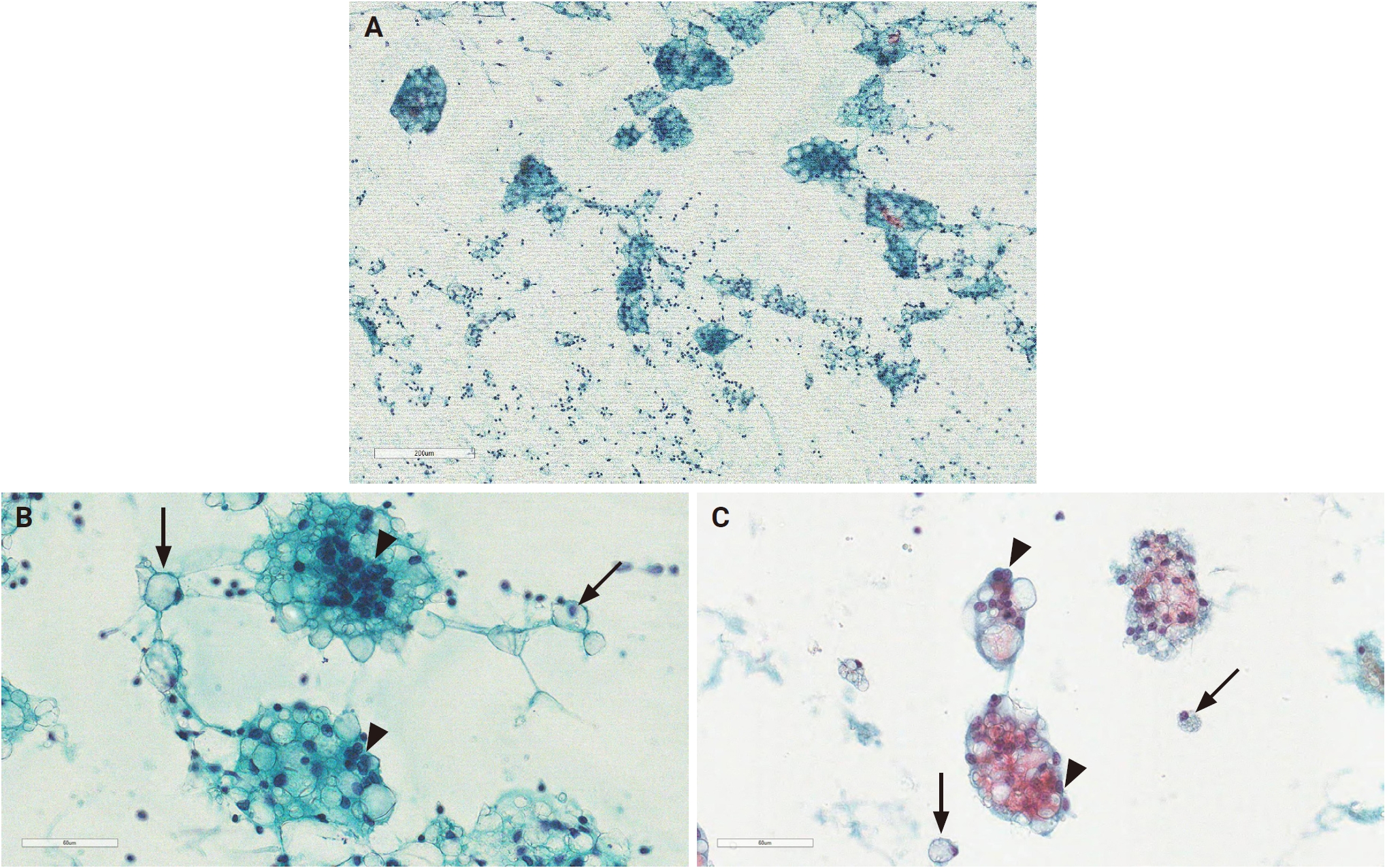
- Silicone lymphadenopathy is an uncommon but well-recognized complication of silicone implant placement resulting from migration of silicone to regional lymph nodes. In patients with prior breast implantation, silicone lymphadenopathy most commonly involves the axillary lymph nodes. Because axillary lymphadenopathy may reflect malignant disease, including metastatic carcinoma, thorough evaluation to exclude neoplasia is essential. Fine-needle aspiration cytology (FNAC) serves as a valuable initial diagnostic modality in the assessment of axillary lymphadenopathy; however, silicone-induced foreign body reactions within lymph nodes may be misinterpreted as metastatic malignancy when silicone lymphadenopathy is not included in the initial differential diagnosis. Herein, a case of axillary silicone lymphadenopathy initially misdiagnosed as metastatic carcinoma on FNAC is presented. This case highlights the importance of recognizing the distinctive cytomorphologic features of silicone lymphadenopathy, which may be sufficiently characteristic to prompt consideration of prior breast implant history during cytopathologic evaluation.
Letter to the Editor
- Comment on “What's new in digital and computational pathology 2026: advances in adoption, standards, AI technologies, and clinical integration”
- Muhammad Abu Bakar Moin
- J Pathol Transl Med. 2026;60(6):732-733. Published online November 10, 2026
- DOI: https://doi.org/10.4132/jptm.2026.09.16
- 73 View
- 0 Download
Newsletter
- What’s new in gastrointestinal pathology 2026: updates from the latest WHO classification
- Raul S. Gonzalez
- J Pathol Transl Med. 2026;60(6):734-737. Published online October 27, 2026
- DOI: https://doi.org/10.4132/jptm.2026.10.20
- 1,775 View
- 258 Download
-
Abstract
PDF
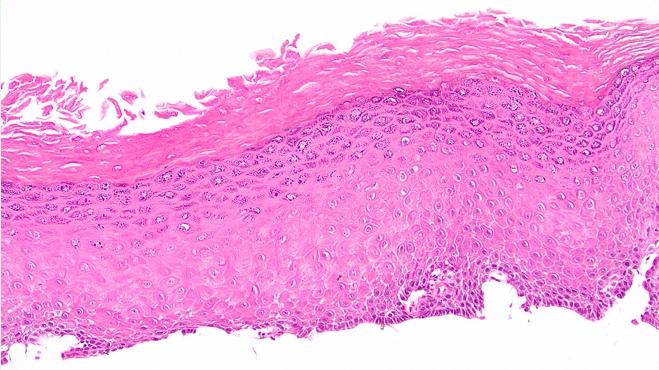
- The 6th edition of the World Health Organization Classification of Tumors: Digestive System Tumors was released online in a beta edition in spring 2026, with a physical print edition released in August. Compared to the 5th edition from 2019, the new edition includes several new entities, completely overhauls a few key concepts, and offers timely updates on most additional neoplasms. Most entries also include new photomicrographs and example whole-slide images. This newsletter will cover selected highlights from the new online edition.
Correspondence
- Corrigendum: Current status and future perspectives of liquid biopsy in non-small cell lung cancer
- Sunhee Chang, Jae Young Hur, Yoon-La Choi, Chang Hun Lee, Wan Seop Kim
- J Pathol Transl Med. 2026;60(6):738-738. Published online November 13, 2026
- DOI: https://doi.org/10.4132/jptm.2020.02.27.r
- Corrects: J Pathol Transl Med 2020;54(3):204
- 34 View
- 1 Download
 First
 Prev




