
 E-submission

Articles
- Page Path
- HOME > J Pathol Transl Med > Volume 60(6); 2026 > Article
-
Original Article
Diagnostic pitfalls leading to delayed diagnosis of human papillomavirus (HPV)–independent cervical malignancies in the era of HPV-based screening: clinicopathologic analysis of diagnostic pathways -
Hye Eun Park1
 , Hye-Ra Jung2, Hyun-Jung Kim3
-
Journal of Pathology and Translational Medicine 2026;60(6):670-679.
DOI: https://doi.org/10.4132/jptm.2026.08.26
Published online: November 13, 2026
1Department of Pathology, Seoul Metropolitan Government-Seoul National University Boramae Medical Center, Seoul National University College of Medicine, Seoul, Korea
2Department of Pathology, Keimyung University School of Medicine, Daegu, Korea
3Department of Pathology, Inje University Sanggye Paik Hospital, Seoul, Korea
- Corresponding Author: Hyun-Jung Kim, MD Department of Pathology, Inje University Sanggye Paik Hospital, 1342 Dongil-ro, Nowon-gu, Seoul 01757, Korea Tel: +82-2-950-1261 Fax: +82-2-951-6964 E-mail: hjkim@paik.ac.kr
© The Korean Society of Pathologists/The Korean Society for Cytopathology
This is an Open Access article distributed under the terms of the Creative Commons Attribution Non-Commercial License (https://creativecommons.org/licenses/by-nc/4.0) which permits unrestricted non-commercial use, distribution, and reproduction in any medium, provided the original work is properly cited.
- 64 Views
- 0 Download


Abstract
-
Background
- Human papillomavirus (HPV)–independent cervical malignancies (HPV-IDCM) are a rare but clinically important subset that may escape detection by HPV-based screening. This study investigated diagnostic pitfalls contributing to delayed HPV-IDCM diagnosis and assessed the role of cervical cytology in the diagnostic pathway.
-
Methods
- This multicenter retrospective case series was conducted as a policy research project of the Korean Society for Cytopathology. Patients with HPV-IDCM and repeated abnormal cytologic findings before definitive histologic diagnosis were identified. Clinical, cytologic, histologic, and HPV test results were reviewed to identify factors associated with diagnostic delay.
-
Results
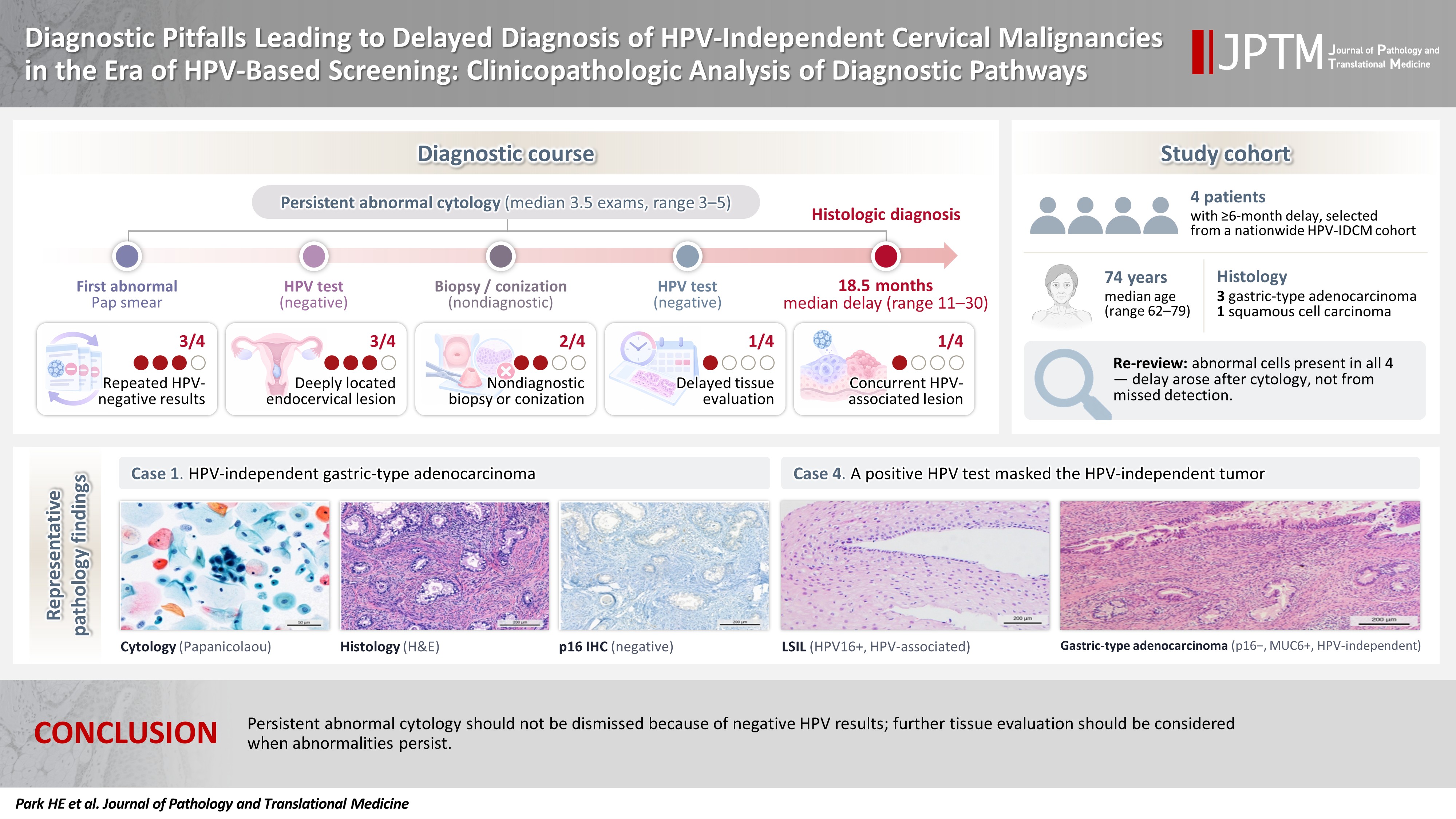
- Four patients were included, with a median age of 74 years (range, 62 to 79 years). Histologic diagnoses comprised three HPV-independent gastric-type adenocarcinomas and one HPV-independent squamous cell carcinoma. The median interval from the first abnormal cytologic finding to definitive diagnosis was 18.5 months (range, 11 to 30 months). Before diagnosis, all patients underwent a median of 3.5 cytologic examinations (range, 3 to 5). Associated factors included repeatedly negative HPV test results (3/4), nondiagnostic biopsy or conization (2/4), delayed tissue evaluation despite persistent cytologic abnormalities (1/4), deeply located endocervical lesions (3/4), and a concurrent HPV-associated squamous lesion (1/4). Re-review identified atypical or malignant cells in all cases, indicating that the delay did not result from failure of cytologic detection.
-
Conclusions
- HPV-IDCM remain vulnerable to delayed diagnosis in the era of HPV-based screening. Persistent abnormal cytologic findings should not be dismissed solely because HPV test results are negative. Careful correlation of cytologic, clinical, and histologic findings remains essential for timely HPV-IDCM detection.
- The 2020 World Health Organization (WHO) classification of female genital tumors categorizes cervical neoplasms as human papillomavirus (HPV)–associated or HPV-independent [1-3]. Although HPV-independent cervical malignancies (HPV-IDCM) comprise a small proportion of cervical malignancies, they are increasingly recognized as a distinct group with unique clinicopathologic features and poor clinical outcomes. Approximately 5%–7% of cervical squamous cell carcinomas are HPV-independent and are associated with advanced stage, increased lymph node metastasis, and worse survival [4,5]. HPV-independent adenocarcinomas include gastric-type, clear cell, and mesonephric adenocarcinomas. Gastric-type adenocarcinoma is the most common subtype, accounting for approximately 10%–15% of cervical adenocarcinomas [6,7], and has significantly poorer survival than HPV-associated usual-type adenocarcinoma [8,9].
- Advances in cervical cancer prevention have accelerated adoption of HPV-based screening. The 2021 WHO guideline strongly recommends primary HPV DNA testing because of its superior sensitivity for detecting cervical precancerous lesions and its potential to reduce cervical cancer incidence and mortality [10]. Similarly, the Korean cervical cancer screening guideline recognizes high-risk HPV testing as an alternative to cytology in women aged ≥25 years [11,12]. These recommendations reflect that most cervical cancers result from persistent high-risk HPV infection, particularly HPV types 16 and 18 [13].
- Despite its success, HPV-based screening remains limited for HPV-IDCM. Because these tumors develop independently of HPV infection, HPV testing alone cannot detect them. Furthermore, some HPV-independent malignancies, particularly gastric-type endocervical adenocarcinoma, are clinically difficult to detect because they often arise deep within the endocervical canal and may be missed by colposcopy or limited biopsy [14,15]. Consequently, diagnosis often occurs at an advanced stage, contributing to poor prognosis. In these cases, abnormal cervical cytology may provide the earliest evidence of underlying malignancy.
- In our previous nationwide study of HPV-IDCM, several patients had abnormal cytologic findings that preceded the final diagnosis by a prolonged interval [16]. Diagnosis was delayed despite abnormal cytology and subsequent evaluation because of persistently negative HPV test results, inconclusive biopsy findings, and subtle endocervical lesions. In this multicenter study, we describe HPV-IDCM with delayed diagnosis and analyze the clinical and cytologic factors underlying these diagnostic pitfalls. We also highlight the continuing value of cervical cytology and raise awareness of the diagnostic challenges posed by HPV-IDCM in the era of HPV-based screening.
INTRODUCTION
- Case selection
- This multicenter study was conducted as part of a policy research project organized by the Korean Society for Cytopathology. Patients with HPV-IDCM were identified from participating institutions [16]. These patients represented a subset of the HPV-IDCM cohort from our previous nationwide study and were selected for delayed diagnosis. Whereas the previous study examined the overall frequency of HPV-IDCM and the diagnostic yield of cytology, the present study focuses on individual diagnostic pathways and cytologic features associated with delayed diagnosis and therefore does not overlap in objectives or analyses.
- Patients were eligible for inclusion if they (1) had histologically confirmed HPV-IDCM, (2) had one or more abnormal cervical cytologic findings before definitive histologic diagnosis, and (3) had an interval of ≥6 months between the first abnormal cytologic finding and definitive histologic diagnosis. This threshold was consistent with the definition applied in our previous nationwide study [16]. In addition, according to the Korean cervical cancer screening guideline, abnormal cytologic findings warrant follow-up within 6 months or earlier diagnostic evaluation depending on the severity of the cytologic abnormality [17]. Therefore, an interval of ≥6 months was considered a conservative threshold for defining delayed diagnosis after an abnormal cytologic finding. Patients with available clinical records and cytology slides underwent detailed clinicopathologic review. Only cases with cervical cytology, histologic evaluation, and clinical follow-up available within the same institution were included to ensure reliable assessment of the diagnostic interval. Cross-institutional records were unavailable for verification.
- HPV-IDCM were defined according to the 2020 WHO classification of female genital tumors based on histologic features, HPV test results, and immunohistochemical findings, including p16 expression when available [1,6,7]. Cases with incomplete clinical information or unavailable cytology slides were excluded.
- Data collection and review
- Clinical data, including age, cervical cytology, HPV test results, final histologic diagnosis, treatment history, and clinical outcome, were obtained from medical records. The interval between the first abnormal cytologic finding and definitive histologic diagnosis was defined as the diagnostic delay period.
- Available cytology slides, histologic specimens, and immunohistochemical results were independently reviewed by three pathologists (H.E.P., H.-R.J., and H.-J.K.). All archived cytology slides (12 slides from four patients) were independently reexamined to determine whether atypical or malignant cells were identifiable and to assign a Bethesda category [18]. Discrepancies were resolved by consensus, and the final interpretation was compared with the original screening result. Histologic diagnoses were reviewed according to the 2020 WHO Classification of Female Genital Tumors [1]. Following consensus review, the principal factors associated with diagnostic delay were identified by correlating the clinical course with cytologic, histologic, and HPV test findings. Given the descriptive nature of this case series and the limited sample size, no comparative or inferential statistical analyses were performed.
MATERIALS AND METHODS
- Clinicopathologic characteristics
- Among 208 patients with HPV-IDCM identified in the nationwide cohort [16], four (1.9%) met the criteria for delayed diagnosis and comprised the study population. The median age at diagnosis was 74 years (range, 62 to 79 years). Histologic diagnoses included three HPV-independent gastric-type adenocarcinomas and one HPV-independent squamous cell carcinoma. The median interval from the initial abnormal cytologic finding to definitive histologic diagnosis was 18.5 months (range, 11 to 30 months). Clinicopathologic characteristics are summarized in Table 1.
- Cytologic findings and diagnostic course
- All patients had abnormal cytologic findings before definitive diagnosis. The median number of cytologic examinations before diagnosis was 3.5 (range, 3 to 5). Cytologic abnormalities included atypical glandular or endocervical cells in three patients, atypical squamous cells, cannot exclude high-grade squamous intraepithelial lesion (ASC-H) in one patient, and high-grade squamous intraepithelial lesion (HSIL) in one patient. Persistent abnormal cytologic findings were documented in all patients.
- On re-review of the 12 available cytology slides from the four patients, the consensus interpretation agreed with the original diagnosis in 11 slides. One slide from case 1 was reclassified from ASC-H to atypical glandular cells. At least one slide from each patient contained atypical or malignant cells, confirming that abnormal cytologic findings were detectable throughout the diagnostic course. These findings indicate that diagnostic delay resulted from events after cytologic evaluation rather than failure of cytologic detection.
- Factors associated with diagnostic delay
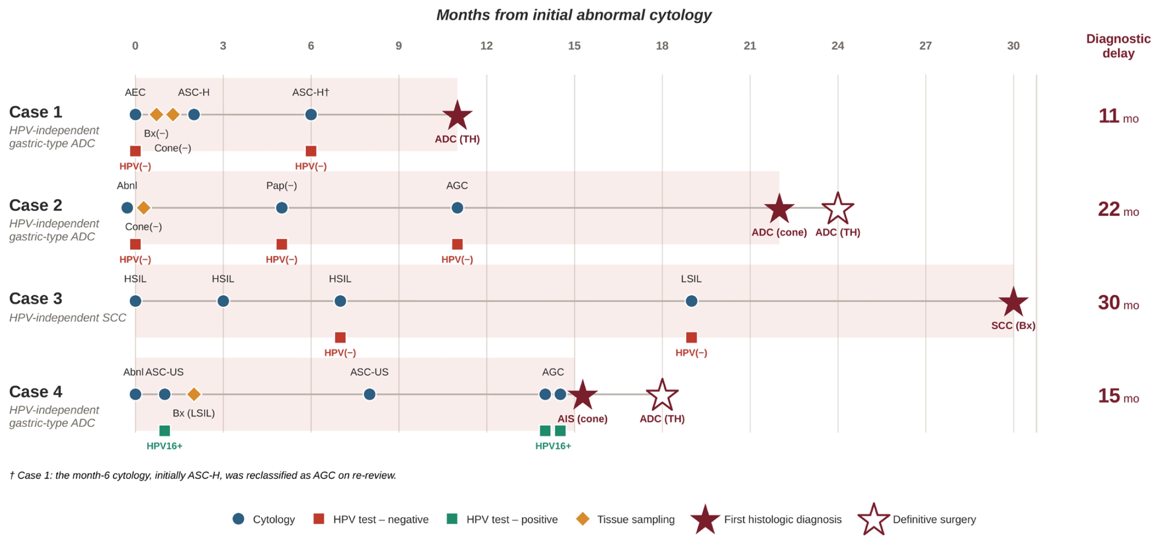
- Recurring factors associated with delayed diagnosis are summarized in Table 2, and the diagnostic courses of all four patients are shown in Fig. 1. Repeated HPV-negative results occurred in three patients. Two patients underwent biopsy or conization that remained nondiagnostic despite persistent cytologic abnormalities. Three patients had deeply located endocervical glandular lesions that were difficult to detect by routine evaluation. In one patient, tissue diagnosis was delayed despite persistent abnormal cytologic findings, including repeated HSIL results. In another, a concurrent HPV-associated squamous lesion may have masked an underlying HPV-independent glandular neoplasm. Collectively, these factors may have contributed to delayed histologic confirmation and appropriate clinical management despite persistent cytologic abnormalities.
- Clinical course of each case
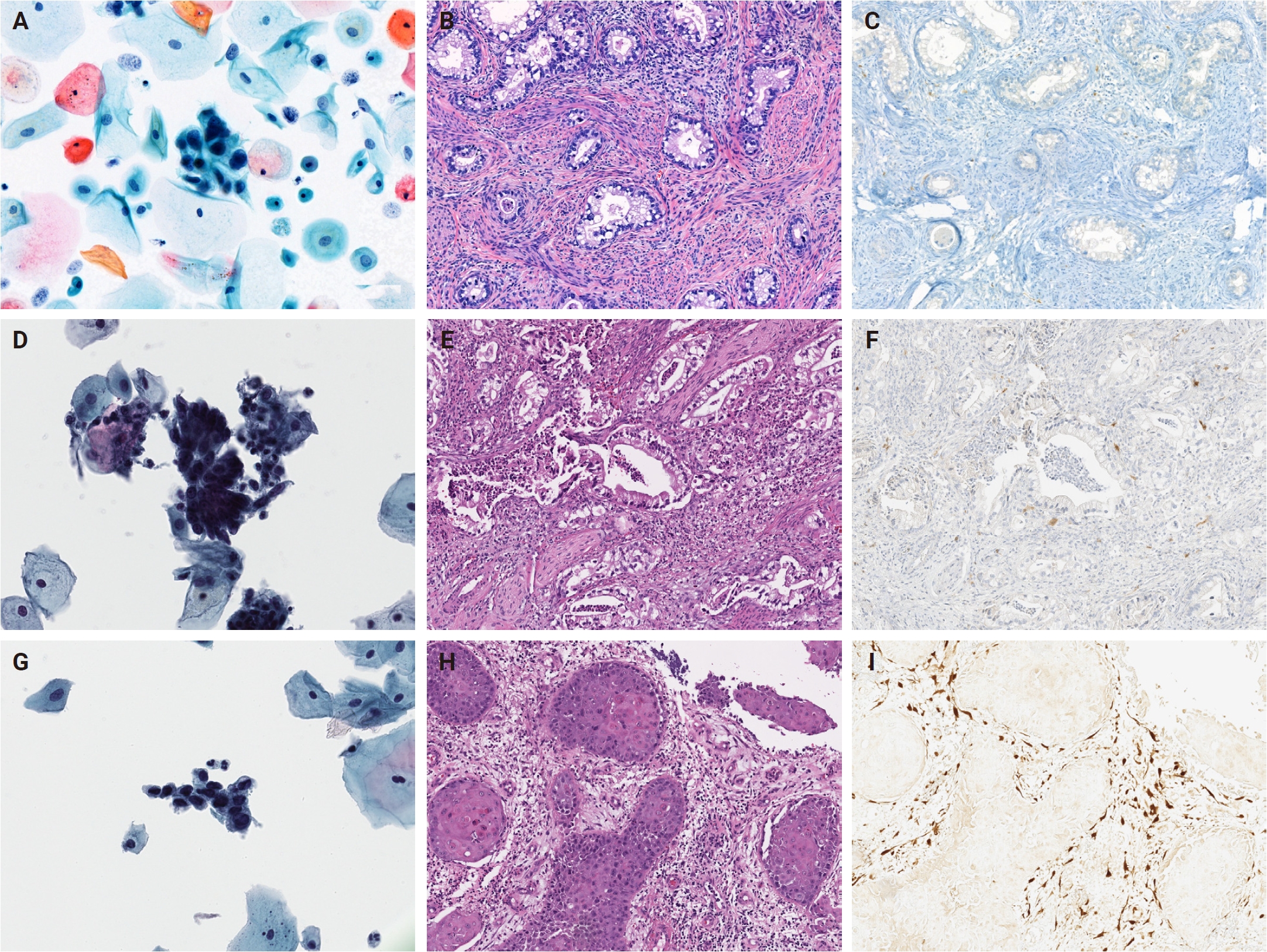
- Representative cytologic, histologic, and p16 immunohistochemical findings from cases 1–3 are shown in Fig. 2.
- A 71-year-old female presented with atypical endocervical cells on cervical cytology. Subsequent punch biopsy and conization were nondiagnostic, and HPV testing remained repeatedly negative. Persistent cytologic abnormalities, including repeated ASC-H results, prompted further evaluation. Total hysterectomy was performed 11 months after the initial abnormal cytologic finding revealed HPV-independent gastric-type adenocarcinoma involving the endocervix.
- A 77-year-old female was referred after abnormal cervical cytology. Initial conization demonstrated no significant lesion and HPV testing was negative. Follow-up cytology demonstrated atypical glandular cells, but additional evaluation was delayed. Repeat conization 22 months after the initial abnormal cytologic finding identified invasive HPV-independent gastric-type adenocarcinoma. The patient subsequently underwent hysterectomy and adjuvant chemoradiotherapy.
- A 79-year-old female had persistent abnormal cytology for more than 2 years, with repeated HSIL findings followed by LSIL. Repeated HPV testing remained negative. Despite multiple abnormal cytologic findings, cervical biopsy performed 30 months after the initial HSIL result established the diagnosis of invasive squamous cell carcinoma. The tumor was p16-negative and classified as HPV-independent squamous cell carcinoma.
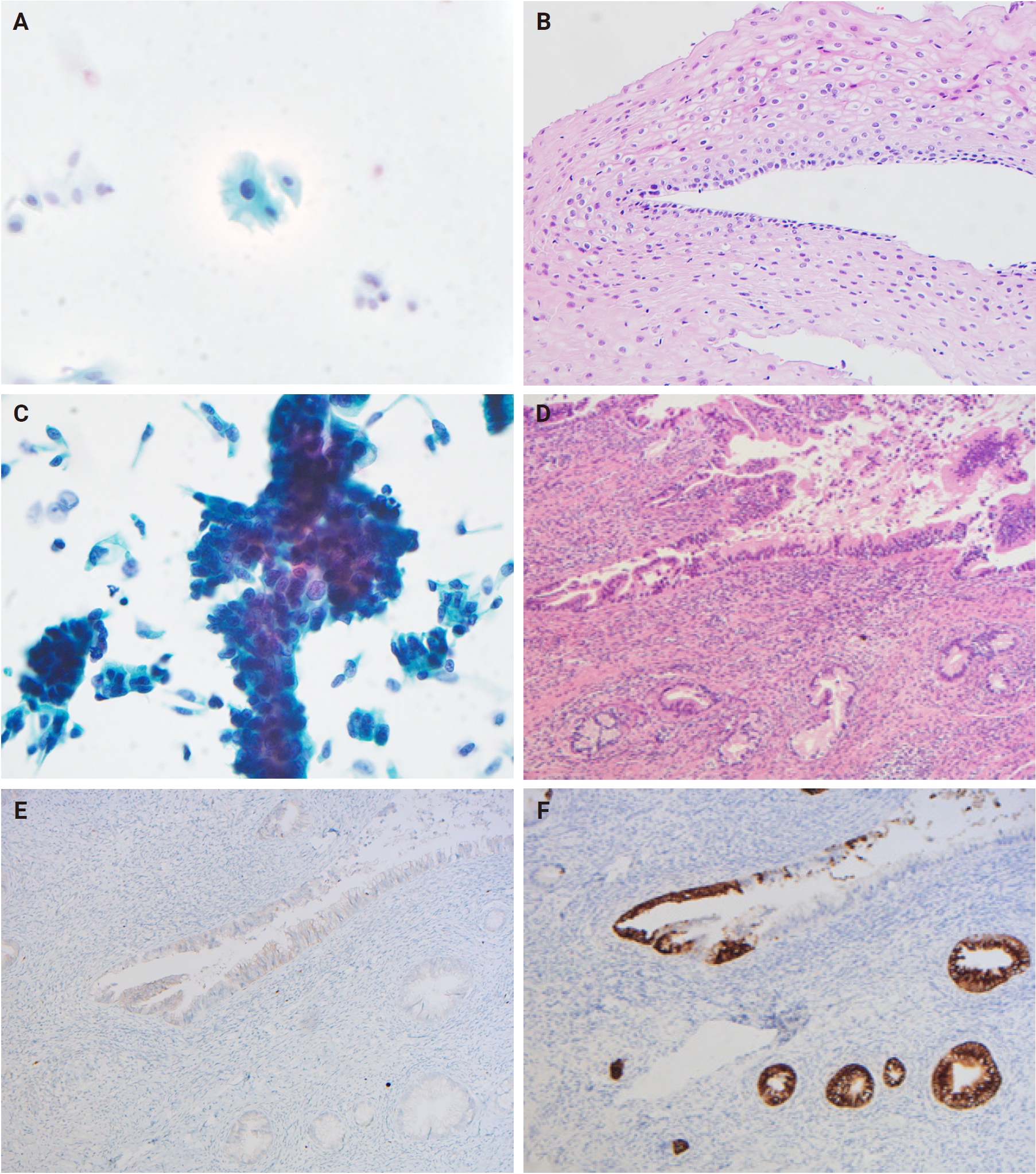
- A 62-year-old female was referred after abnormal cervical cytology detected during routine health screening. Initial biopsy showed only low-grade squamous intraepithelial lesion with koilocytic change. Follow-up cytology repeatedly demonstrated atypical glandular cells, prompting conization 15 months after the initial abnormal cytologic finding. Histologic examination revealed HPV-independent adenocarcinoma in situ with focal mucin 6 (MUC6) expression and negative p16 staining. Subsequent hysterectomy confirmed HPV-independent gastric-type adenocarcinoma. The repeatedly positive high-risk HPV (HPV16) result was attributed to a concurrent HPV-associated squamous lesion, whereas the gastric-type adenocarcinoma was confirmed as HPV-independent based on characteristic gastric-type morphology, negative p16 staining, and MUC6 positivity in the glandular tumor. Coexistence of HPV-associated squamous and HPV-independent glandular lesions likely delayed recognition of the latter. Representative findings from case 4, including the concurrent HPV-associated squamous lesion and HPV-independent gastric-type adenocarcinoma, are shown in Fig. 3.
RESULTS
Case 1
Case 2
Case 3
Case 4
- In this study, four patients with HPV-IDCM experienced delayed diagnosis despite persistent abnormal cytologic findings. The median interval from the initial abnormal cytologic finding to definitive diagnosis was 18.5 months, and all patients underwent repeated cytologic examinations before histologic confirmation. Recurrent diagnostic pitfalls included persistently negative HPV test results, nondiagnostic biopsy or conization, delayed tissue evaluation despite persistent cytologic abnormalities, and concurrent HPV-associated lesions. These findings highlight the diagnostic challenges of HPV-IDCM and reinforce the continuing value of cytology in the era of HPV-based cervical cancer screening.
- HPV-IDCM are a rare but clinically important subset of cervical neoplasms. Although squamous cell carcinoma remains the predominant cervical cancer, the proportion of adenocarcinoma has increased over recent decades [19,20]. Gastric-type adenocarcinoma is the most common HPV-independent adenocarcinoma and the predominant HPV-independent glandular neoplasm encountered in practice [7,8]. Since the introduction of the 2020 WHO classification, HPV-IDCM have been recognized as a distinct group with unique pathogenesis, aggressive behavior, and poorer outcomes than HPV-associated cervical cancers [1,21,22]. Gastric-type adenocarcinoma is associated with advanced-stage disease and inferior survival [8,23], whereas HPV-independent squamous cell carcinoma also demonstrates unfavorable clinicopathologic features [4,5]. These tumors also present substantial diagnostic challenges. Gastric-type adenocarcinomas often arise deep within the endocervical canal and may be missed by colposcopy or limited biopsy. More importantly, because they develop independently of HPV infection, they can escape HPV-based screening. Consequently, HPV-IDCM remain an important diagnostic blind spot despite widespread primary HPV screening [24].
- A key finding was persistent abnormal cytologic findings despite repeatedly negative HPV test results. Three of four patients remained HPV-negative throughout the diagnostic course. Although HPV testing is the most sensitive method for detecting HPV-associated cervical neoplasms and has substantially improved cervical cancer screening [10], HPV-IDCM remain an important exception [25,26]. Repeated negative HPV results may falsely reassure clinicians and patients, especially when cytologic abnormalities are mild or histologic findings are inconclusive. Although the retrospective design precluded determining the direct influence of HPV results on clinical decision-making, persistent HPV negativity may have favored continued surveillance rather than more aggressive evaluation in several cases. These findings emphasize that persistent abnormal cytologic findings warrant careful evaluation, even when HPV test results are repeatedly negative.
- Another important finding was the frequent discordance between cytologic and histologic findings. Three patients had persistent glandular or endocervical cytologic abnormalities despite nondiagnostic or discordant biopsy results. This discrepancy is particularly relevant in HPV-independent glandular neoplasms, especially gastric-type adenocarcinoma. Unlike HPV-associated cervical neoplasms, gastric-type adenocarcinomas often arise deep within the endocervical canal and exhibit an infiltrative growth pattern that is difficult to sample by routine punch biopsy or limited excision [14,15]. Consequently, histologic evaluation may yield false-negative or nonspecific findings despite persistent cytologic abnormalities [16,25]. In our cohort, abnormal cytologic findings preceded definitive histologic diagnosis by several months, illustrating the limitations of tissue sampling. One patient also had a concurrent HPV-associated squamous lesion that may have obscured recognition of the underlying HPV-independent glandular neoplasm. Notably, review identified atypical or malignant cells in cytology specimens from all four patients, indicating that delayed diagnosis resulted from limitations in histologic confirmation and subsequent clinical evaluation rather than failure of cytologic detection. These findings reinforce the value of cytology as an early indicator of HPV-IDCM, even when biopsy is nondiagnostic or HPV testing is negative.
- These findings should not be interpreted as evidence against HPV-based screening. HPV testing remains the cornerstone of cervical cancer prevention and contemporary screening strategies [10]. However, HPV-IDCM represent a small but clinically important group that may not be detected by HPV testing alone [27]. In our cohort, persistent cytologic abnormalities consistently provided the earliest indication of underlying malignancy. These observations support the continuing value of cervical cytology as a complementary test rather than a replacement for HPV testing [16]. In healthcare systems where repeated cytologic follow-up and timely diagnostic evaluation are feasible, persistent cytologic abnormalities may facilitate earlier detection of HPV-IDCM. Because this case series specifically included patients with persistent abnormal cytologic findings, it characterizes the diagnostic pathways of cytology-positive delayed cases and does not address the overall diagnostic performance of cervical cytology for HPV-IDCM.
- The low proportion of eligible cases (1.9%) likely reflects the clinical presentation of HPV-IDCM. These tumors are often diagnosed only after symptom onset, when cytology and histologic evaluation occur during the same diagnostic episode. Consequently, few patients have a measurable interval between the first abnormal cytologic finding and definitive histologic diagnosis within a single institution. This observation is consistent with a nationwide Swedish registry study showing that HPV-negative cervical cancers occurred predominantly in older females with advanced-stage disease and that most HPV-negative results were obtained near the time of diagnosis [28]. Although HPV-negative cervical cancers are not synonymous with HPV-independent tumors because HPV-negative results may also reflect sampling or assay limitations [29], both observations highlight a diagnostic limitation of HPV-based screening alone. Together with evidence that cytology–high-risk HPV cotesting improves detection of cervical adenocarcinoma compared with either modality alone [30], our findings support careful evaluation of persistent cytologic abnormalities, even when HPV test results are negative.
- This study has several clinical implications. Persistent abnormal cytologic findings should not be dismissed solely because HPV test results are negative, particularly when abnormalities recur or remain unexplained by histologic evaluation. Careful integration of cytologic, clinical, and histologic findings may facilitate earlier recognition of HPV-IDCM and reduce diagnostic delay. When persistent abnormal cytology is accompanied by repeatedly negative HPV test results or nondiagnostic biopsy, HPV-IDCM should be considered, and additional evaluation, including endocervical curettage, deeper or repeat sampling, or conization, should be pursued instead of continued routine surveillance.
- A major strength of this study is its analysis of real-world diagnostic pathways in a rare and underrecognized group of cervical neoplasms. However, this study has several limitations, including the retrospective design, small sample size, and multicenter heterogeneity. Additionally, only cases with linked cytologic and histologic records within the same institution were included because cross-institutional linkage was unavailable; more complete screening histories would likely identify additional delayed cases. An HPV-associated comparison group was also unavailable, precluding assessment of the relative magnitude of diagnostic delay. Future studies incorporating cross-institutional data linkage and an HPV-associated comparison group would help clarify the true frequency and relative magnitude of diagnostic delay in HPV-IDCM. Accordingly, the present findings should be regarded as observations from a small case series, and larger cohort studies are needed to confirm their broader clinical applicability. Despite these limitations, the findings consistently demonstrate that persistent abnormal cytologic findings may provide an important clue to HPV-IDCM. In conclusion, abnormal cytologic findings should not be disregarded solely because HPV test results are negative. Continued attention to persistent cytologic abnormalities may be important for timely diagnosis of HPV-IDCM in the era of HPV-based screening.
DISCUSSION
Ethics Statement
All procedures performed in the current study were approved by the Institutional Review Board (IRB) of Inje University Sanggye Paik Hospital (IRB No. SGPAIK2024-04-011; approved April 30, 2024) in accordance with the 1964 Helsinki Declaration of Helsinki and its later amendments. Formal written informed consent was not required with a waiver by the appropriate IRB.
Availability of Data and Material
The datasets generated or analyzed during this study are available from the corresponding author upon reasonable request.
Code Availability
Not applicable.
Author Contributions
Conceptualization: HEP, HRJ, HJK. Data curation: HEP, HRJ, HJK. Formal analysis: HEP, HRJ, HJK. Investigation: HEP, HRJ, HJK. Funding acquisition: HEP, HRJ, HJK. Methodology: HEP, HRJ, HJK. Project administration: HEP, HRJ, HJK. Supervision: HJK. Validation: HEP, HRJ, HJK. Writing—original draft: HEP. Writing—review and editing: HEP, HRJ, HJK. Approval of final manuscript: all authors.
Conflicts of Interest
The authors declare that they have no potential conflicts of interest.
Funding Statement
This study was supported in part by the Korean Society for Cytopathology (Grant No. 23-01), Republic of Korea.
Acknowledgments
The authors gratefully acknowledge the cooperation and scholarly discussions provided by members of the Korean Society for Cytopathology during the course of this study.
| Case No. | Age (yr) | Initial cytology | HPV test | Initial biopsy/conization result | Final diagnosis | p16 | Diagnostic delay (mo) |
|---|---|---|---|---|---|---|---|
| 1 | 71 | Atypical endocervical cells (NOS) | Negative | Biopsy negative; conization negative | HPV-independent gastric-type adenocarcinoma | Negative | 11 |
| 2 | 77 | Abnormal Pap smeara | Negative | Conization negative | HPV-independent gastric-type adenocarcinoma | Negative | 22 |
| 3 | 79 | HSIL | Negative | No tissue diagnosis despite persistent abnormal cytology | HPV-independent squamous cell carcinoma | Negative | 30 |
| 4 | 62 | ASC-US | HPV16 positive | LSIL (koilocytosis) | HPV-independent gastric-type adenocarcinoma | Negative | 15 |
HPV, human papillomavirus; IDCM, independent cervical malignancies; NOS, not otherwise specified; HSIL, high-grade squamous intraepithelial lesion; ASC-US, atypical squamous cells of undetermined significance; LSIL, low-grade squamous intraepithelial lesion.
aExact cytologic interpretation from the outside institution was unavailable.
- 1. WHO Classification of Tumours Editorial Board. WHO classification of tumours. 5th ed. Vol. 4. Female genital tumours. Lyon: International Agency for Research on Cancer, 2020.
- 2. Hohn AK, Brambs CE, Hiller GG, May D, Schmoeckel E, Horn LC. 2020 WHO classification of female genital tumors. Geburtshilfe Frauenheilkd 2021; 81: 1145-53. ArticlePubMedPMC
- 3. McCluggage WG, Singh N, Gilks CB. Key changes to the World Health Organization (WHO) classification of female genital tumours introduced in the 5th edition (2020). Histopathology 2022; 80: 762-78. ArticlePubMedPDF
- 4. Nicolas I, Marimon L, Barnadas E, et al. HPV-negative tumors of the uterine cervix. Mod Pathol 2019; 32: 1189-96. ArticlePubMedPDF
- 5. Rodriguez-Carunchio L, Soveral I, Steenbergen RD, et al. HPV-negative carcinoma of the uterine cervix: a distinct type of cervical cancer with poor prognosis. BJOG 2015; 122: 119-27. ArticlePubMed
- 6. Hodgson A, Park KJ, Djordjevic B, et al. International endocervical adenocarcinoma criteria and classification: validation and interobserver reproducibility. Am J Surg Pathol 2019; 43: 75-83. ArticlePubMedPMC
- 7. Stolnicu S, Barsan I, Hoang L, et al. International Endocervical Adenocarcinoma Criteria and Classification (IECC): a new pathogenetic classification for invasive adenocarcinomas of the endocervix. Am J Surg Pathol 2018; 42: 214-26. ArticlePubMedPMC
- 8. Karamurzin YS, Kiyokawa T, Parkash V, et al. Gastric-type endocervical adenocarcinoma: an aggressive tumor with unusual metastatic patterns and poor prognosis. Am J Surg Pathol 2015; 39: 1449-57. ArticlePubMedPMC
- 9. Kojima A, Mikami Y, Sudo T, et al. Gastric morphology and immunophenotype predict poor outcome in mucinous adenocarcinoma of the uterine cervix. Am J Surg Pathol 2007; 31: 664-72. ArticlePubMed
- 10. World Health Organization. WHO guideline for screening and treatment of cervical pre-cancer lesions for cervical cancer prevention. 2nd ed. Geneva: World Health Organization, 2021.
- 11. Kim JH, Shim SH, Min KJ, et al. Clinical practice guideline for high-risk human papillomavirus testing in cervical cancer screening: a consensus statement from the Korean Society of Gynecologic Oncology. J Gynecol Oncol 2026; 37: e90. ArticlePubMedPMCPDF
- 12. Kong TW, Kim M, Kim YH, et al. High-risk human papillomavirus testing as a primary screening for cervical cancer: position statement by the Korean Society of Obstetrics and Gynecology and the Korean Society of Gynecologic Oncology. J Gynecol Oncol 2020; 31: e31. ArticlePubMedPMCPDF
- 13. Walboomers JM, Jacobs MV, Manos MM, et al. Human papillomavirus is a necessary cause of invasive cervical cancer worldwide. J Pathol 1999; 189: 12-9. ArticlePubMed
- 14. Kyaw EM, Alhindawi B, French A. Cervical adenocarcinoma in the shadows: the impact of missed screening. Cureus 2025; 17: e94089. ArticlePubMedPMC
- 15. Stolnicu S, Park KJ, Kiyokawa T, Oliva E, McCluggage WG, Soslow RA. Tumor typing of endocervical adenocarcinoma: contemporary review and recommendations from the International Society of Gynecological Pathologists. Int J Gynecol Pathol 2021; 40(Suppl 1): S75-91. ArticlePubMedPMC
- 16. Jung HR, Shin J, Yoo CW, et al. Diagnostic value of cytology in detecting human papillomavirus-independent cervical malignancies: a nation-wide study in Korea. J Pathol Transl Med 2025; 59: 444-52. ArticlePubMedPMCPDF
- 17. Lee JK, Hong JH, Kang S, et al. Practice guidelines for the early detection of cervical cancer in Korea: Korean Society of Gynecologic Oncology and the Korean Society for Cytopathology 2012 edition. J Gynecol Oncol 2013; 24: 186-203. ArticlePubMedPMC
- 18. Nayar R, Wilbur DC. The Bethesda System for Reporting Cervical Cytology: definitions, criteria, and explanatory notes. 3rd ed. Cham: Springer, 2015.
- 19. Adegoke O, Kulasingam S, Virnig B. Cervical cancer trends in the United States: a 35-year population-based analysis. J Womens Health (Larchmt) 2012; 21: 1031-7. ArticlePubMedPMC
- 20. Suh DH, Ha HI, Lee YJ, Lim J, Won YJ, Lim MC. Incidence and treatment outcomes of uterine cervical cancer in Korea 1999-2018 from the national cancer registry. J Gynecol Oncol 2023; 34: e39. ArticlePubMedPMCPDF
- 21. Giannella L, Grelloni C, Natalini L, et al. Molecular features of HPV-independent cervical cancers. Pathogens 2025; 14: 668.ArticlePubMedPMC
- 22. Stolnicu S, Hoang L, Chiu D, et al. Clinical outcomes of HPV-associated and unassociated endocervical adenocarcinomas categorized by the International Endocervical Adenocarcinoma Criteria and Classification (IECC). Am J Surg Pathol 2019; 43: 466-74. ArticlePubMedPMC
- 23. Radomska A, Lee D, Neufeld H, et al. A retrospective study on incidence, diagnosis, and clinical outcome of gastric-type endocervical adenocarcinoma in a single institution. Diagn Pathol 2021; 16: 68.ArticlePubMedPMCPDF
- 24. He J, Tu Lu Weng Jiang G, Zeng L. Analysis of the differences between HPV-independent and HPV-related cervical adenocarcinoma. Front Oncol 2025; 15: 1544207.ArticlePubMedPMC
- 25. Nishio S. Current status and molecular biology of human papillomavirus-independent gastric-type adenocarcinoma of the cervix. J Obstet Gynaecol Res 2023; 49: 1106-13. ArticlePubMedPDF
- 26. Tjokroprawiro BA, Sulistya HA, Wijaya N, et al. Clinicopathological characteristics and outcomes of gastric-type adenocarcinoma of the cervix: a systematic review. AJOG Glob Rep 2026; 6: 100623.ArticlePubMedPMC
- 27. Hurjui RM, Hurjui IA, Butureanu TA, Popovici D, Avadanei ER, Balan RA. HPV-independent cervical cancer: a new challenge of modern oncology. Int J Mol Sci 2025; 26: 10051.ArticlePubMed
- 28. Hassan SS, Nordqvist Kleppe S, Asinger N, Wang J, Dillner J, Arroyo Muhr LS. Invasive cervical cancers after an HPV-negative test: insights from screening histories [Preprint]. Posted 2026 Apr 13. medRxiv 2026.04.11.26350679. https://doi.org/10.64898/2026.04.11.26350679. Article
- 29. Dai W, Zeng X, Pantanowitz L, Zhao C. Human papillomavirus negative cervical cancers and precancerous lesions: prevalence, pathological and molecular features, and clinical implications. Gynecol Obstet Clin Med 2025; 5: e000160. Article
- 30. Xie F, Zhang L, Zhao D, et al. Prior cervical cytology and high-risk HPV testing results for 311 patients with invasive cervical adenocarcinoma: a multicenter retrospective study from China's largest independent operator of pathology laboratories. BMC Infect Dis 2019; 19: 962.ArticlePubMedPMCPDF
REFERENCES
Figure & Data
References
Citations

 PubReader
 ePub Link
-
 Cite this Article
- Cite this Article
-
- Close
- Download Citation
- Close
 XML Download
- Figure
-
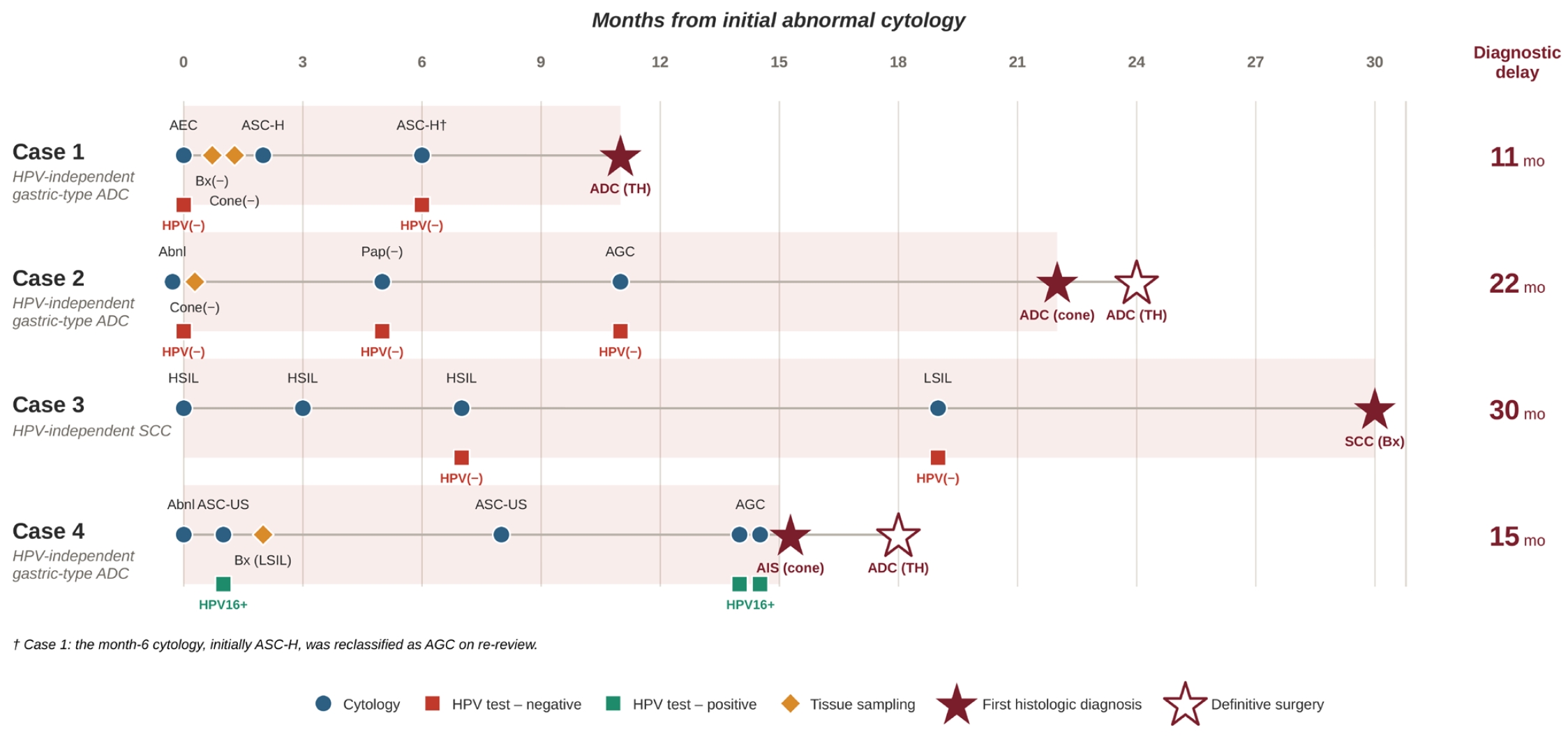
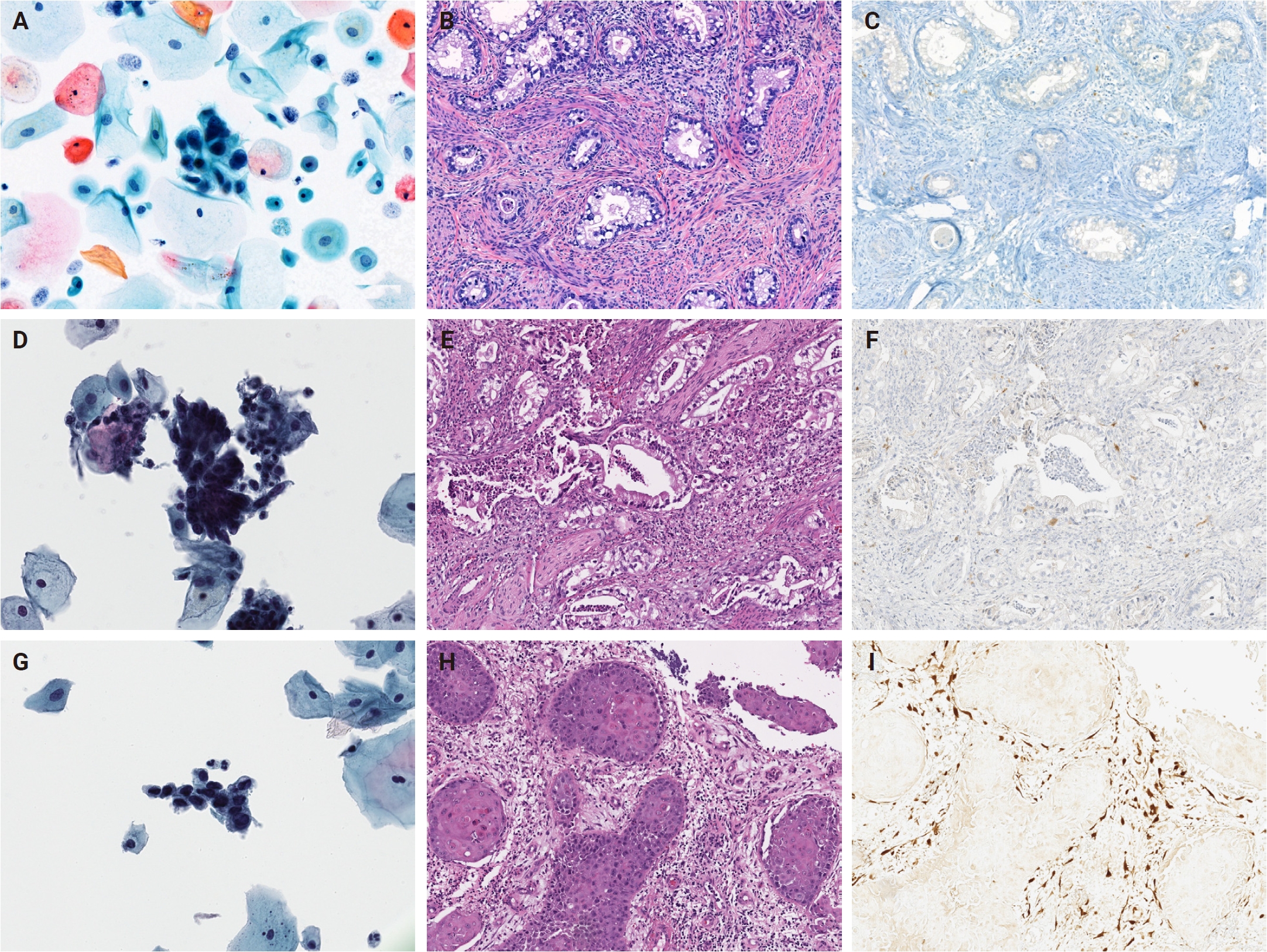

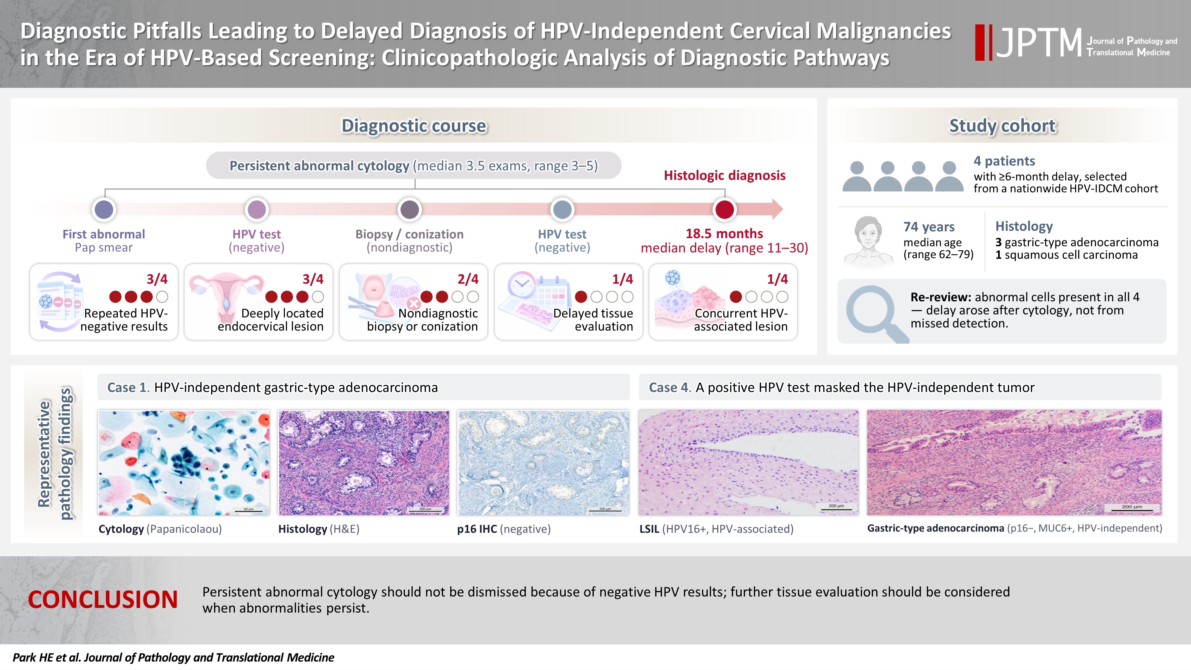




Fig. 1.
Fig. 2.
Fig. 3.
Graphical abstract
| Case No. | Age (yr) | Initial cytology | HPV test | Initial biopsy/conization result | Final diagnosis | p16 | Diagnostic delay (mo) |
|---|---|---|---|---|---|---|---|
| 1 | 71 | Atypical endocervical cells (NOS) | Negative | Biopsy negative; conization negative | HPV-independent gastric-type adenocarcinoma | Negative | 11 |
| 2 | 77 | Abnormal Pap smear |
Negative | Conization negative | HPV-independent gastric-type adenocarcinoma | Negative | 22 |
| 3 | 79 | HSIL | Negative | No tissue diagnosis despite persistent abnormal cytology | HPV-independent squamous cell carcinoma | Negative | 30 |
| 4 | 62 | ASC-US | HPV16 positive | LSIL (koilocytosis) | HPV-independent gastric-type adenocarcinoma | Negative | 15 |
| Diagnostic pitfall | Case 1 | Case 2 | Case 3 | Case 4 |
|---|---|---|---|---|
| Persistent abnormal cytology | ○ | ○ | ○ | ○ |
| Repeated HPV-negative results | ○ | ○ | ○ | - |
| Non-diagnostic biopsy/conization | ○ | ○ | - | - |
| Delayed tissue evaluation despite persistent cytologic abnormalities | - | - | ○ | - |
| Deeply located endocervical glandular lesion | ○ | ○ | - | ○ |
| Concurrent HPV-associated lesion | - | - | - | ○ |
HPV, human papillomavirus; IDCM, independent cervical malignancies; NOS, not otherwise specified; HSIL, high-grade squamous intraepithelial lesion; ASC-US, atypical squamous cells of undetermined significance; LSIL, low-grade squamous intraepithelial lesion. Exact cytologic interpretation from the outside institution was unavailable.
HPV, human papillomavirus.


