E-submission

Articles
- Page Path
- HOME > J Pathol Transl Med > Forthcoming articles > Article
-
Case Study
Intracranial malignant peripheral nerve sheath tumor with myxoid morphology evolving across recurrences: a case report -
Frantisek Vana1,*
 , Petra Pokorna2,3,*, Petr Pospisil4, Zdenek Mackerle5, Marketa Hermanova1, Iva Staniczkova Zambo1, Martin Barak5, Robin Jugas2,3, Radim Jancalek5, Ondrej Slaby2,3, Michal Hendrych1
-
DOI: https://doi.org/10.4132/jptm.2026.09.17
Published online: November 11, 2026
1First Department of Pathology, Faculty of Medicine, Masaryk University and St. Anne’s University Hospital Brno, Brno, Czech Republic
2Department of Biology, Faculty of Medicine and Central European Institute of Technology (CEITEC), Masaryk University, Brno, Czech Republic
3Center for Precision Medicine, University Hospital Brno, Brno, Czech Republic
4Department of Radiation Oncology, Masaryk Memorial Cancer Institute Brno and Faculty of Medicine, Masaryk University, Brno, Czech Republic
5Department of Neurosurgery, Faculty of Medicine, Masaryk University and St. Anne’s University Hospital Brno, Brno, Czech Republic
- Corresponding Author: Michal Hendrych, PhD First Department of Pathology, Faculty of Medicine, Masaryk University and St. Anne’s University Hospital Brno, Pekarska 53, 656 91 Brno, Czech Republic Tel: +420-543-183-221, E-mail: michal.hendrych@fnusa.cz
*Frantisek Vana and Petra Pokorna contributed equally to this work.
© The Korean Society of Pathologists/The Korean Society for Cytopathology
This is an Open Access article distributed under the terms of the Creative Commons Attribution Non-Commercial License (https://creativecommons.org/licenses/by-nc/4.0) which permits unrestricted non-commercial use, distribution, and reproduction in any medium, provided the original work is properly cited.
- 60 Views
- 1 Download


Abstract
- A malignant peripheral nerve sheath tumor (MPNST) is a rare, aggressive spindle-cell sarcoma that exceptionally occurs intracranially. Herein, we report a case of intracranial spindle-cell neoplasm with abundant myxoid stroma. The immunophenotype of the primary tumor did not align with any known peripheral nerve sheath tumor and the lesion did not correspond to any methylation category defined by the Heidelberg classifier with a sufficiently high classification score, although MPNST was the highest-scoring category. Over the course of repeated recurrences, the tumor acquired increased cellularity, mitotic activity and loss of H3K27me3 expression. Subsequent methylation profiling supported a diagnosis of MPNST. The tumor harbored a gain-of-function variant in NRAS p.Gly13Arg and TP53 p.Arg273Cys from the initial tumor onwards, consistent with previous reports describing activation of Ras/mitogen-activated protein kinase as an early event in MPNST tumorigenesis. These findings illustrate the diagnostic complexity of rare intracranial sarcomas with myxoid morphology and suggest that some may represent early MPNST.
- Malignant peripheral nerve sheath tumors (MPNSTs) are rare spindle cell tumors originating from the peripheral nerve sheath. While these tumors can occur throughout the body, they are most frequently present in the extremities, trunk and head and neck [1]. MPNSTs are divided into three distinct groups based on their etiology: approximately 50% are associated with neurofibromatosis type 1 (nfMPNST), 40% arise sporadically (sMPNST), and 10% are radiation-induced (rMPNST) [2]. Prognostically, sMPNSTs exhibit the most favorable outcomes, with a median survival of 101.1 months, compared to 35.5 months for nfMPNSTs and 24.3 months for rMPNSTs [3].
- Herein, we report a rare case of sporadic intracranial MPNST, confirmed through methylation profiling, which initially developed as a dura mater–based, brain-infiltrating tumor exhibiting atypical histopathological features. This unusual presentation illustrates the wide morphological spectrum of intracranial MPNST and highlights the diagnostic complexity.
INTRODUCTION
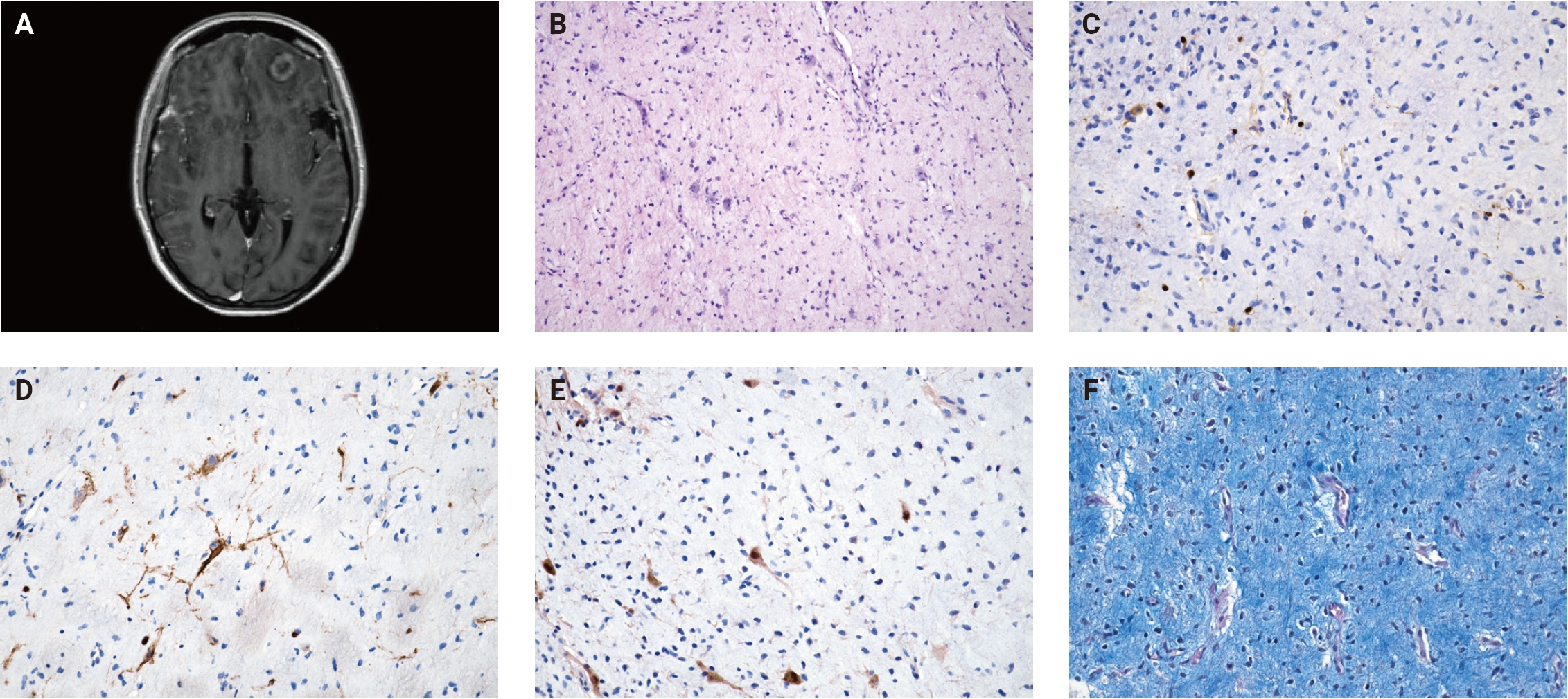
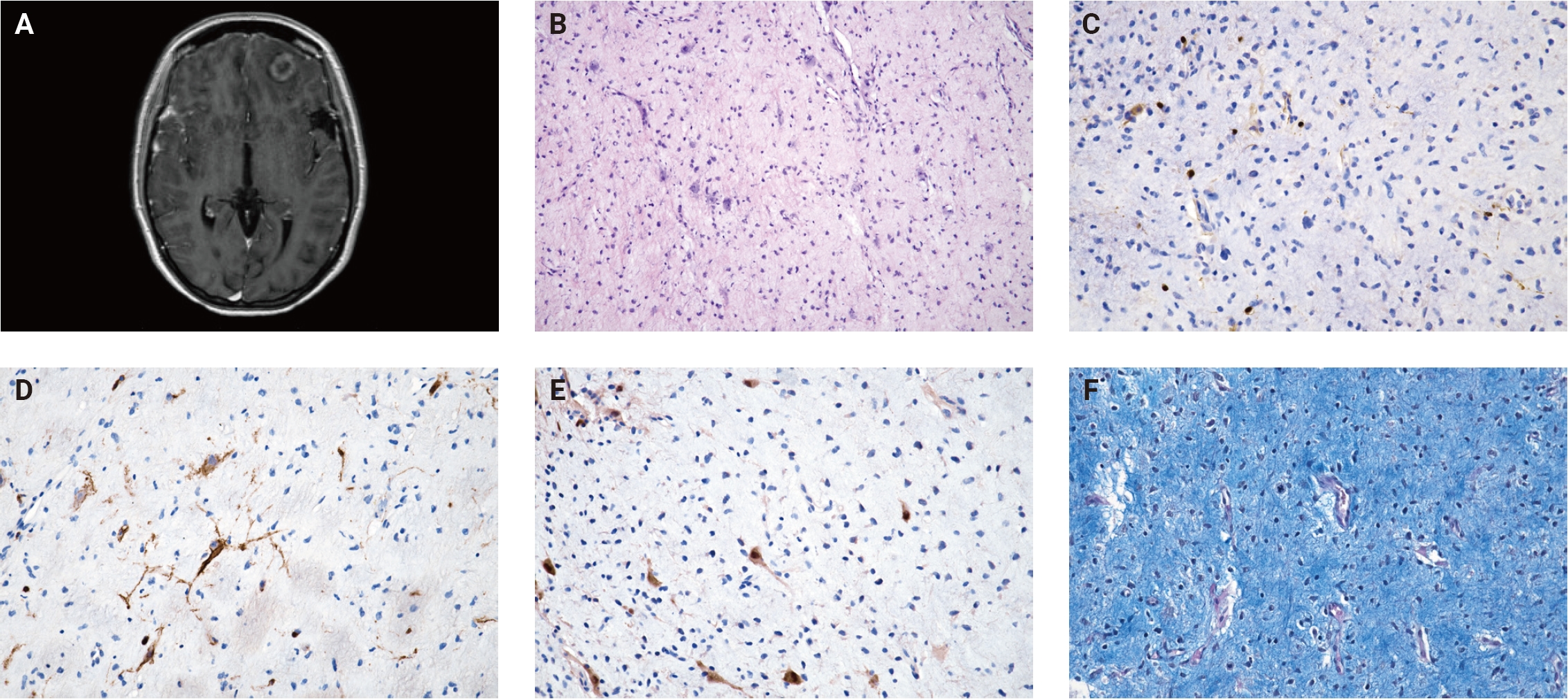
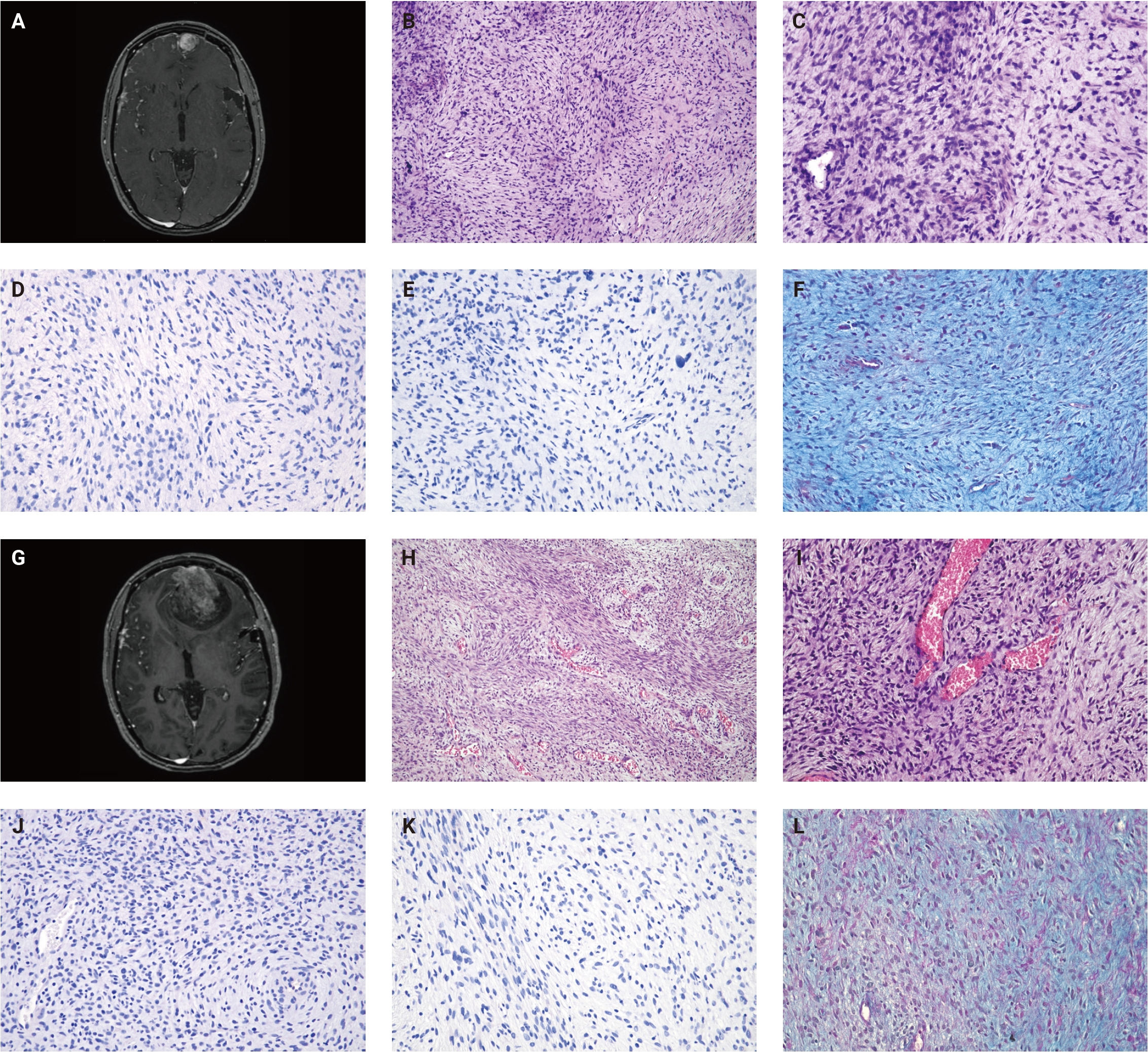
- A 21-year-old female was admitted to the hospital following three consecutive tonic-clonic (grand mal) seizures. Computed tomography of the brain revealed a cystic lesion at the base of the left frontal region, prompting further investigation with magnetic resonance imaging (MRI). The initial MRI confirmed the presence of a non-specific cystic-like lesion without enhancement or collateral brain edema in the left frontobasal region involving the left frontal lobe of the brain. Consequently, the patient was admitted to the Neurology department for additional examination. The patient's medical history was without any significant remarks except for a car accident with a mild head injury 10 years ago. Clinical examination demonstrated a positive Tromner's sign, subtle reflex asymmetry, with left-sided dominance and minor memory dysfunction. Cerebrospinal fluid analysis revealed no signs of inflammation. Follow-up MRI in 3 months revealed a significant appearance change of the lesion connected to the frontonasal dura mater and involving the left frontal lobe (Fig. 1A). A ring-like enhancement and surrounding brain edema favored tumor etiology, and the resection surgery was indicated.
- In July 2020, the patient underwent surgical resection of the frontal lobe lesion infiltrating the dura mater of the skull base, which measured 25 × 22 × 10 mm. Microscopically, the lesion was a hypocellular neoplasm composed of short spindle-shaped and stellate cells within a rich myxoid stroma infiltrating the brain. The cells had indistinct borders, faintly eosinophilic cytoplasm, and atypical, enlarged hyperchromatic nuclei (Fig. 1B). The highest mitotic rate was 1 mitosis/10 high-power fields (HPF). The tumor infiltration was associated with a network of curvilinear, thin-walled blood vessels, some of which had perivascular condensation of tumor cells, with occasional perivascular lymphocytes and plasma cells. At the periphery of the lesion, entrapped astrocytes and neurons were documented. Immunohistochemical analysis revealed no expression of AE1/AE3, CD117, epithelial membrane antigen (EMA), MUC4, CD56, STAT6, desmin, smooth muscle actin or brachyury. SOX10 was expressed in scattered stellate cells, while spindle cells were negative (Fig. 1C). S100 expression was limited to entrapped glial fibrillary acidic protein–positive glial cells (Fig. 1D) and NeuN–positive neurons (Fig. 1E). Integrase interactor 1 expression was retained. Collagen IV expression was restricted to the walls of blood vessels. TLE1 (transducin-like enhancer of split 1) showed weak and non-diffuse expression, and CD34 was weak and focal. Alcian blue special staining demonstrated diffuse positivity in the myxoid tumor stroma (Fig. 1F). Spindle tumor cells diffusely expressed strong nuclear accumulation of p53 corresponding to the mutant phenotype. Fluorescence in situ hybridization confirmed the absence of DDIT3, CIC, FUS, and EWSR1 rearrangements. The tumor board recommended radiotherapy for the patient.
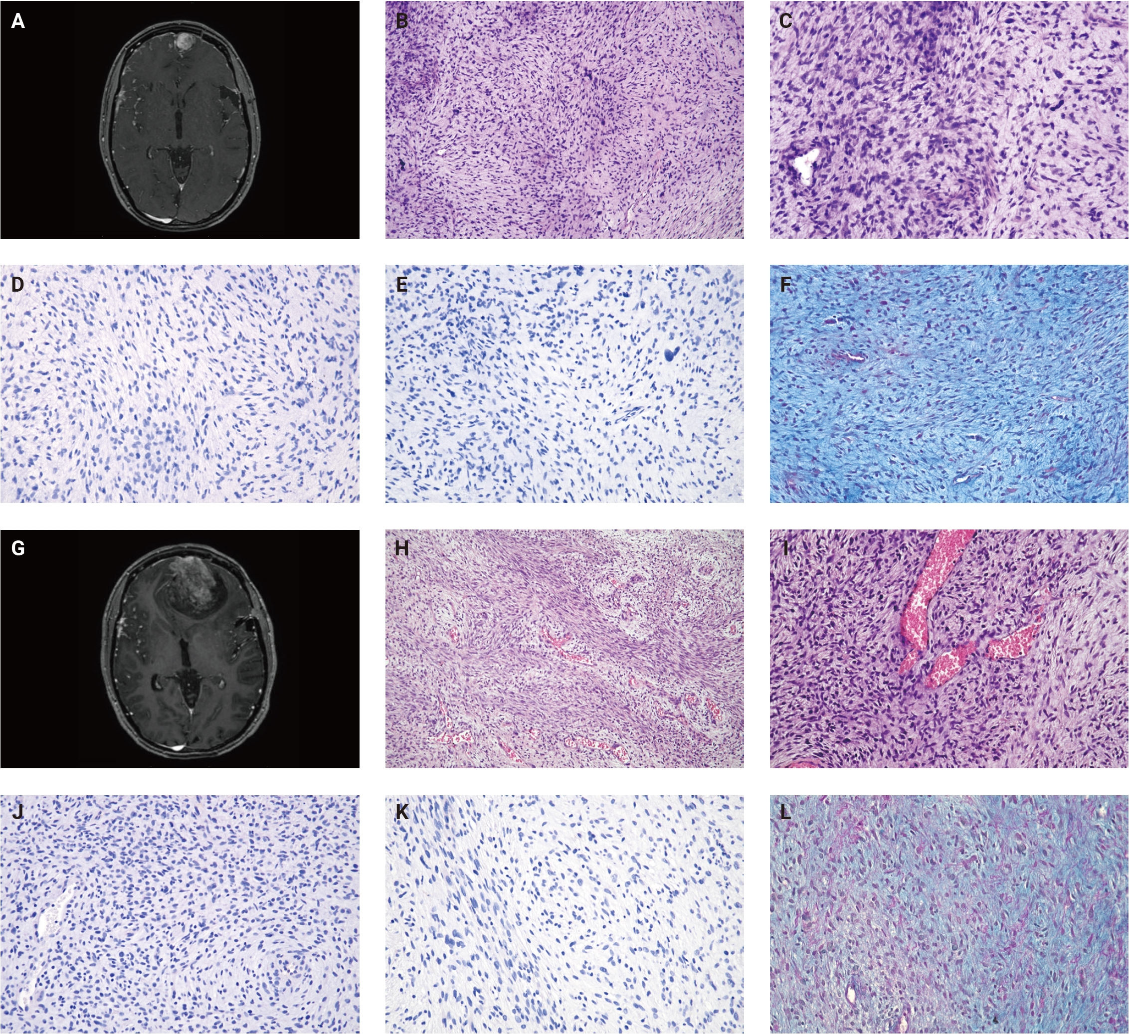
- Two months later, an MRI for radiotherapy planning displayed early recurrence located medially from the previous resection cavity (Fig. 2A). A subsequent resection was performed in October 2020, i.e., 2 months after primary surgery, along with the removal of adjacent parts of the meninges. Histological examination showed a proliferation of atypical spindle cells within a myxoid stroma (Fig. 2B–E). The spindle cells exhibited cytonuclear atypia, anisokaryosis, irregular nuclear contours, and brisk mitotic activity (7 mitotic figures/10 HPF). Dispersed cells with large, atypical nuclei were also observed. No necrosis was observed in the whole tumor.
- The patient finally underwent planned radiotherapy, which was completed in January 2021. A follow-up MRI in November 2021 revealed another recurrence of the tumor (Fig. 2G), necessitating the third resection surgery in the same month. Histopathological analysis showed a dura mater–adherent, moderate-to-hypercellular proliferation of spindle-shaped cells, this time with multiple necrotic foci and less prominent myxoid stroma (Fig. 2H–L). Tumor cells displayed cytonuclear atypia and brisk mitotic activity, with numerous cells showing large pleomorphic nuclei or multinucleation. Comprehensive genomic profiling (CGP) aiming to discover potentially targetable alterations was performed using the TruSight Oncology 500 panel (Illumina, San Diego, CA, USA). The tumor was microsatellite stable with a low tumor mutational burden of 2.3 mutations per megabase. Oncogenic variants were detected in NRAS (NM_002524.4: c.37G>C; p.Gly13Arg), TP53 (NM_000546.5: c.817C>T; p.Arg273Cys), and FBXW7 (NM_001349798.2: c.1393C>T; p.Arg465Cys). However, the obtained results did not ultimately impact subsequent clinical management.
- In April 2022, an MRI revealed repeated recurrence in the left frontobasal region. During surgery, tumor invasion into the skull base and paranasal cavities was observed. Histologically, a hypercellular spindle cell sarcoma with extensive necrosis was identified. The patient was subsequently admitted to palliative care and, eventually, in October 2022, passed away. No autopsy was performed.
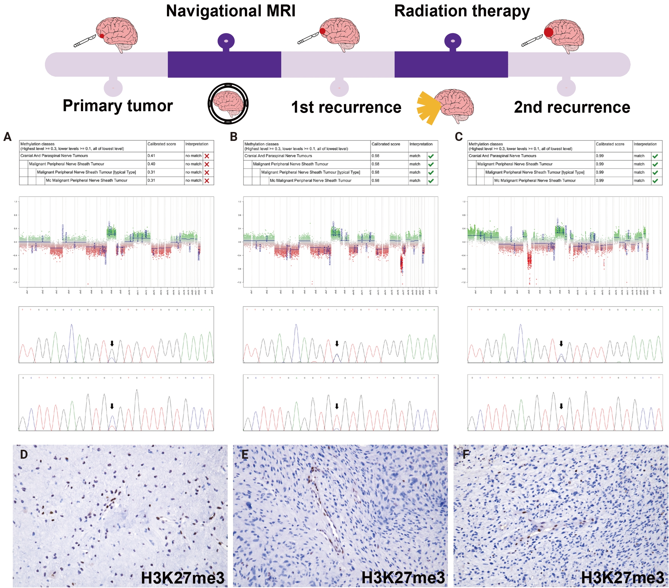
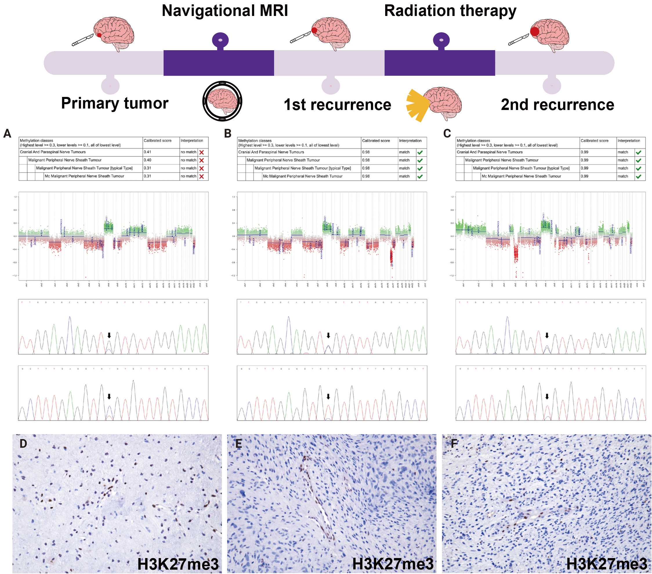
- Retrospectively, DNA methylation profiling of the first three tumor samples was performed using EPICv2 BeadChip arrays (Illumina) to classify the tumors by the Heidelberg brain tumor classifier ver. 12.8 (Fig. 3A–C) [4]. Based on the results of methylation profiling, the second and third samples were reclassified as MPNST. However, the primary tumor failed to meet the classification threshold despite MPNST being the highest-scoring methylation category (calibrated score 0.31). Additionally, the NRAS p.Gly13Arg and TP53 p.Arg273Cys variants initially detected in the third biopsy were also found in the remaining samples, including the primary tumor. Immunohistochemical analysis of the first two recurrent tumors displayed a loss of H3K27me3 (Fig. 3E, F). Although CGP did not reveal any inactivating variants to explain the polycomb repressive complex 2 (PRC2) inactivation responsible for the H3K27me3 loss, low coverage in exon 10 of the SUZ12 gene suggested that this could have been the underlying genomic event, possibly in conjunction with loss of heterozygosity of the second allele. Further immunohistochemistry displayed heterogeneous nuclear positivity of H3K27me3 in the primary tumor (Fig. 3D), and a small cellular focus with elevated mitotic activity reaching 4 mitotic figures/10 HPF was also found.
- The broad differential diagnosis was extensive, encompassing chordoid meningioma, intracranial chordoma, solitary fibrous tumor, schwannoma, myxofibrosarcoma, primary intracranial sarcoma, DICER1-mutant, CIC-rearranged sarcoma, and intracranial mesenchymal tumor FET::CREB fusion–positive. Specific alteration-driven neoplasms were systematically excluded through targeted assessments. Solitary fibrous tumor was excluded by lack of STAT6 expression by immunohistochemistry (IHC) and STAT6 wild-type status by next-generation sequencing (NGS) (TSO500). Primary intracranial sarcoma, DICER1-mutant, was excluded by the absence of pathogenic DICER1 variants on NGS and lack of myogenic differentiation. CIC-rearranged sarcoma was excluded by the absence of CIC gene rearrangement on fluorescence in situ hybridization (FISH) and NGS (TSO500), and intracranial mesenchymal tumor FET::CREB fusion–positive by the absence of EWSR1 and FUS rearrangement on FISH and NGS (TSO500), absence of myogenic differentiation, and lack of EMA expression on IHC. Furthermore, an intracranial chordoma was unsupported due to negative brachyury and EMA expression on IHC, along with extraskeletal localization, while chordoid meningioma was ruled out by the spindle and stellate cell morphology paired with absent EMA expression. A schwannoma diagnosis was atypical given the lack of S100 and SOX10 staining, a mutant p53 phenotype, infiltrative growth, and rapid local recurrences. Finally, while myxofibrosarcoma was considered due to shared morphological overlap and common TP53 alterations, methylation profiling ultimately excluded it and definitively established the final diagnosis of MPNST in recurrent tumors.
CASE REPORT
- Recent advancements in understanding the pathogenesis of MPNST, particularly nfMPNST, have led to the establishment of the full spectrum of peripheral nerve sheath tumors, illustrating the stepwise malignant progression of nfMPNST. These include benign neurofibromas, atypical neurofibromatous neoplasms of uncertain biologic potential (ANNUBP), as well as low-grade and high-grade MPNSTs [5]. In some instances, a single tumor may exhibit varying degrees of malignant transformation [6].
- Furthermore, significant progress has been made in elucidating molecular pathways driving MPNST evolution. Constitutive activation of the Ras/mitogen-activated protein kinase (MAPK) pathway, most commonly through biallelic NF1 inactivation, is an early and frequent event in tumor development, often accompanied by alterations in CDKN2A or, less frequently, TP53. The subsequent tumor evolution is influenced by the presence or absence of PRC2 complex inactivation [7], which occurs in approximately 80% of conventional MPNSTs and is routinely evaluated through immunohistochemical loss of H3K27me3 [8]. This sequential molecular progress towards MPNST development in neurofibromatosis type 1 (NF1) patients starts with benign neurofibromas carrying biallelic inactivation of NF1 [9], progressing into ANNUBP, often further harboring a homozygous deletion of CDKN2A/B or SMARCA2 [6,10] and subsequently into nfMPNST with complex chromosomal alterations with or without PRC2 inactivation as a result of somatic mutations in SUZ12, EED, or EZH2 and TP53 mutations [6,11]. The well-established pathogenesis and progression from precursor lesions in nfMPNST are linked with a low risk of malignant transformation, with only 8%–15% of precursor lesions progressing to nfMPNST over a few years [12]. In contrast, the pathogenesis of sMPNST remains poorly understood, with no identified precursor lesion, suggesting that it arises de novo [13].
- In our case, the absence of NF1 alterations in tumor samples ruled out NF1, and the patient had no prior history of radiotherapy, allowing us to classify the tumor as sMPNST. Initially, the primary tumor did not show a loss of H3K27me3 and did not meet the classification threshold by the Heidelberg brain tumor classifier, although MPNST was the highest-scoring methylation category. However, within 3 months, the recurrent tumor developed morphologic and epigenetic features allowing confident classification as sMPNST, accompanied by a loss of H3K27me3 expression on immunohistochemistry suggestive of PRC2 complex inactivation.
- In line with the proposed timing of genetic alterations underlying MPNST development, where Ras/MAPK activation serves as an initiating event, we confirmed the presence of a gain-of-function NRAS variant in the initial biopsy. NRAS variants in MPNST are quite rare, occurring in only 2% to 2.4% of cases, and have been described exclusively in sMPNST due to their mutual exclusivity with NF1 alterations typically associated with nfMPNST. To our knowledge, the NRAS p.Gly13Arg variant has not been previously reported in dedicated MPNST cohorts. However, codon 13 variants are well-established activating hotspots with oncogenic effects comparable to p.Gln61 variants described in sMPNST by other authors [14]. Moreover, prior work [15] has demonstrated that the hallmark disruption of PRC2 activity contributes to tumorigenesis by amplifying Ras-driven transcriptional programs through chromatin remodeling. Although this synergy has been specifically shown in NF1-deficient models, the analogous downstream activation produced by mutant NRAS suggests that a similar cooperative mechanism may also be relevant in tumors harboring activating NRAS variants. In addition to the NRAS variant, the tumor exhibited a mutant p53 phenotype on immunohistochemistry, with the TP53 p.Arg273Cys variant also detected in the initial biopsy. TP53 variants are present in approximately 20%–40% of MPNSTs [16], and while specific codon-level data are usually not disclosed, the alteration itself is located in a canonical DNA-binding hotspot and is a well-established loss-of-function event. Some reports link TP53 inactivation to poor prognosis, as in a pooled cohort combining both sporadic and NF1-associated cases, TP53-mutant cases showed a significantly worse prognosis [17]. The identification of this alteration in the first biopsy specimen supports the notion that TP53 inactivation, similar to NRAS mutation, might be an early event facilitating the progression to MPNST, as previously suggested for a subset of cases [7].
- The low calibrated score for the methylation class MPNST typical in the primary tumor may be explained by both low tumor cellularity and low tumor purity [18]. In contrast to the recurrent tumors, the primary tumor sample contained a significant proportion of non-neoplastic residual brain tissue infiltrated by tumor cells. In addition, the relative sparsity of tumor cells and the presence of inflammatory cells within the tumor microenvironment may have further reduced sample purity. An additional contributing factor may be the retained H3K27me3 expression in the primary tumor. MPNST cases with retained H3K27me3, although uncommon, are more frequently classified by the Heidelberg CNS Tumor Methylation Classifier as methylation class MPNST atypical, which was also the second-highest scoring class in our analysis (calibrated score 0.08). In contrast, the recurrent tumors demonstrated loss of H3K27me3 expression and were classified as methylation class MPNST typical, with high calibrated scores (0.98 and 0.99, respectively).
- Moreover, there is marked morphological similarity with reported intracranial myxofibrosarcomas [19,20], suggesting diagnostic overlap with our described sMPNST case, notably in instances featuring S100 expression and high-grade histomorphology of the recurrences [20]. This case further highlights the need for the implementation of more robust genetic and epigenetic testing in the diagnosis of rare or atypical tumors, as morphology alone and an uncharacteristic immunophenotype may be misleading.
- In conclusion, we present a rare case of intracranial MPNST initially manifesting as a myxoid spindle-cell neoplasm. Comprehensive evaluation combining morphology, immunohistochemistry, DNA methylation profiling, and genomic analysis ultimately supported the diagnosis of early sMPNST. Importantly, the tumor’s evolution from an ambiguous myxoid lesion to a high-grade MPNST over a short time underscores the diagnostic challenges. While our findings raise the possibility that some intracranial myxoid neoplasms represent early stages of MPNST, broader series with integrative molecular profiling are needed to substantiate this hypothesis and to better characterize the morphological spectrum of sMPNST. In routine diagnostic practice, MPNST should be considered in the differential diagnosis of myxoid tumors, particularly when they are in proximity to a peripheral nerve.
DISCUSSION
Ethics Statement
This study was approved by the Institutional Ethics Committee (St. Anne’s University Hospital Brno, EK-FNUSA-26/2024). The research was conducted ethically, with all study procedures being performed in accordance with the requirements of the World Medical Association’s Declaration of Helsinki. Written informed consent was obtained from the patient for study participation and data publication.
Availability of Data and Material
The datasets generated or analyzed during the study are available from the corresponding author on reasonable request.
Code Availability
Not applicable.
Author Contributions
Conceptualization: FV, PP (Petra Pokorna), MH (Michal Hendrych). Data curation: PP (Petr Pospisil), ZM, MB, RJ (Robin Jugas), MH (Marketa Hermanova), ISZ, PP (Petra Pokorna), OS. Formal analysis: RJ (Robin Jugas). Investigation: PP (Petr Pospisil), ZM, MB, RJ (Radim Jancalek), MH (Marketa Hermanova), ISZ, PP (Petra Pokorna), OS. Supervision: MH (Marketa Hermanova), RJ (Radim Jancalek), OS. Visualization: FV, PP (Petra Pokorna), MB, MH (Michal Hendrych). Writing—original draft: FV, PP (Petra Pokorna), MB, MH (Michal Hendrych). Writing—review & editing: all authors. Approval of final manuscript: all authors.
Conflicts of Interest
The authors declare that they have no potential conflicts of interest.
Funding Statement
This work was supported by Masaryk University in Brno, Czech Republic, under Grant MUNI/A/1843/2025, the Ministry of Health, Czech Republic, grant project no. NU23-03-00100 and by an internal grant from St. Anne’s University Hospital Brno. This work was supported by the project National Institute for Cancer Research (Programme EXCELES, ID Project No. LX22NPO5102) - Funded by the European Union – Next Generation EU.
- 1. Valentin T, Le Cesne A, Ray-Coquard I, et al. Management and prognosis of malignant peripheral nerve sheath tumors: the experience of the French Sarcoma Group (GSF-GETO). Eur J Cancer 2016; 56: 77-84. ArticlePubMed
- 2. Farid M, Demicco EG, Garcia R, et al. Malignant peripheral nerve sheath tumors. Oncologist 2014; 19: 193-201. ArticlePubMedPMCPDF
- 3. Miao R, Wang H, Jacobson A, et al. Radiation-induced and neurofibromatosis-associated malignant peripheral nerve sheath tumors (MPNST) have worse outcomes than sporadic MPNST. Radiother Oncol 2019; 137: 61-70. ArticlePubMed
- 4. Sill M, Schrimpf D, Patel A, et al. Advancing CNS tumor diagnostics with expanded DNA methylation-based classification. Cancer Cell 2026; 44: 340-54. ArticlePubMed
- 5. Miettinen MM, Antonescu CR, Fletcher CD, et al. Histopathologic evaluation of atypical neurofibromatous tumors and their transformation into malignant peripheral nerve sheath tumor in patients with neurofibromatosis 1: a consensus overview. Hum Pathol 2017; 67: 1-10. ArticlePubMedPMC
- 6. Beert E, Brems H, Daniels B, et al. Atypical neurofibromas in neurofibromatosis type 1 are premalignant tumors. Genes Chromosomes Cancer 2011; 50: 1021-32. ArticlePubMed
- 7. Cortes-Ciriano I, Steele CD, Piculell K, et al. Genomic patterns of malignant peripheral nerve sheath tumor (MPNST) evolution correlate with clinical outcome and are detectable in cell-free DNA. Cancer Discov 2023; 13: 654-71. ArticlePubMedPMCPDF
- 8. Conway E, Healy E, Bracken AP. PRC2 mediated H3K27 methylations in cellular identity and cancer. Curr Opin Cell Biol 2015; 37: 42-8. ArticlePubMed
- 9. Jouhilahti EM, Peltonen S, Heape AM, Peltonen J. The pathoetiology of neurofibromatosis 1. Am J Pathol 2011; 178: 1932-9. ArticlePubMedPMC
- 10. Pemov A, Hansen NF, Sindiri S, et al. Low mutation burden and frequent loss of CDKN2A/B and SMARCA2, but not PRC2, define premalignant neurofibromatosis type 1-associated atypical neurofibromas. Neuro Oncol 2019; 21: 981-92. ArticlePubMedPMCPDF
- 11. Pemov A, Li H, Presley W, Wallace MR, Miller DT. Genetics of human malignant peripheral nerve sheath tumors. Neurooncol Adv 2020; 2(Suppl 1): i50-61. ArticlePubMedPMCPDF
- 12. Higham CS, Dombi E, Rogiers A, et al. The characteristics of 76 atypical neurofibromas as precursors to neurofibromatosis 1 associated malignant peripheral nerve sheath tumors. Neuro Oncol 2018; 20: 818-25. ArticlePubMedPMC
- 13. Somatilaka BN, Sadek A, McKay RM, Le LQ. Malignant peripheral nerve sheath tumor: models, biology, and translation. Oncogene 2022; 41: 2405-21. ArticlePubMedPMCPDF
- 14. Kao EY, Wakeman KM, Wu Y, et al. Prevalence and detection of actionable BRAF V600 and NRAS Q61 mutations in malignant peripheral nerve sheath tumor by droplet digital PCR. Hum Pathol 2022; 129: 90-7. ArticlePubMed
- 15. De Raedt T, Beert E, Pasmant E, et al. PRC2 loss amplifies Ras-driven transcription and confers sensitivity to BRD4-based therapies. Nature 2014; 514: 247-51. ArticlePubMedPDF
- 16. Cui Q, Zhang F, Liu J, et al. Genomic profiling and pathological assessment of malignant peripheral nerve sheath tumors. J Cancer Res Clin Oncol 2025; 151: 165.ArticlePubMedPMCPDF
- 17. Holand M, Kolberg M, Danielsen SA, et al. Inferior survival for patients with malignant peripheral nerve sheath tumors defined by aberrant TP53. Mod Pathol 2018; 31: 1694-707. ArticlePubMedPDF
- 18. Aldape K, Capper D, von Deimling A, et al. cIMPACT-NOW update 9: recommendations on utilization of genome-wide DNA methylation profiling for central nervous system tumor diagnostics. Neurooncol Adv 2025; 7: vdae228.ArticlePubMedPMCPDF
- 19. Wen S, Tu M, Liao R, Li H. Recurrent primary intracranial myxofibrosarcoma: a case report and review of the literature. Front Oncol 2026; 16: 1755019.ArticlePubMedPMC
- 20. Majumdar K, Mandal S, Saran RK, Gupta R. Recurrent intracranial myxofibrosarcoma presenting as an extensive fronto-parieto-occipital SOL: an unusual sarcoma of meningeal origin. Clin Neurol Neurosurg 2013; 115: 354-8. ArticlePubMed
REFERENCES
Figure & Data
References
Citations

 PubReader
 ePub Link
-
 Cite this Article
- Cite this Article
-
- Close
- Download Citation
- Close
 XML Download
- Figure
-