 E-submission
Articles
- Page Path
- HOME > J Pathol Transl Med > Volume 47(5); 2013 > Article
-
Original Article
Fine Needle Aspiration Cytology of Parathyroid Lesions - Ilyeong Heo, Sunhoo Park, Chang Won Jung, Jae Soo Koh, Seung-Sook Lee, Hyesil Seol, Hee Seung Choi, Soo Youn Cho
-
Korean Journal of Pathology 2013;47(5):466-471.
DOI: https://doi.org/10.4132/KoreanJPathol.2013.47.5.466
Published online: October 25, 2013
Department of Pathology, Korea Cancer Center Hospital, Seoul, Korea.
- Corresponding Author: Soo Youn Cho, M.D. Department of Pathology, Korea Cancer Center Hospital, 75 Nowon-ro, Nowon-gu, Seoul 139-706, Korea. Tel: +82-2-970-2545, Fax: +82-2-970-2430, pathsycho@naver.com
• Received: June 26, 2013 • Revised: September 9, 2013 • Accepted: September 27, 2013
© 2013 The Korean Society of Pathologists/The Korean Society for Cytopathology
This is an Open Access article distributed under the terms of the Creative Commons Attribution Non-Commercial License (http://creativecommons.org/licenses/by-nc/3.0/) which permits unrestricted non-commercial use, distribution, and reproduction in any medium, provided the original work is properly cited.
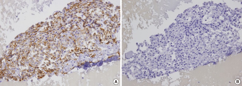

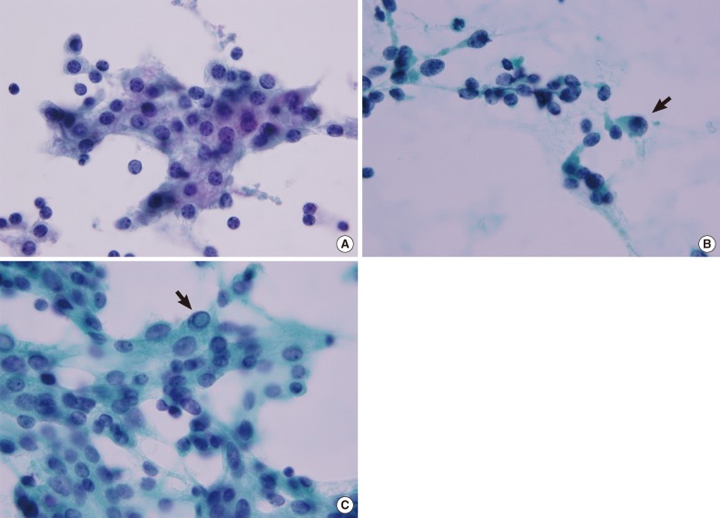
Figure & Data
References
Citations
Citations to this article as recorded by 
- Shear wave elastography can improve the preoperative diagnosis of parathyroid carcinoma or atypical parathyroid tumor: initial experience
Ruigang Lu, Teng Zhao, Dichen Guo, Bojun Wei, Wei Zhao
BMC Medical Imaging.2026;[Epub] CrossRef - Diagnostic approach to FNA biopsy of cystic lesions of the head and neck
Stefen Andrianus, Olivia Leung, Zubair Baloch
Cancer Cytopathology.2025;[Epub] CrossRef - Cytological Findings in Pediatric Thoracic Tumors: A Review of Diagnostic Insights and Pitfalls
Parikshaa Gupta, Pranab Dey
Acta Cytologica.2025; : 1. CrossRef - Sonographic Features of Atypical and Initially Missed Parathyroid Adenomas: Lessons Learned From a Single-Center Cohort
Seyfettin Ilgan, Berna İmge Aydoğan, Özdeş Emer, Cüneyd Anıl, Alptekin Gürsoy, Mustafa Cesur, Banu Bilezikçi
The Journal of Clinical Endocrinology & Metabolism.2024; 109(2): 439. CrossRef - Fine needle aspiration biopsy of parathyroid; is it meaningful? A cytologic study of 81 cases with histological and clinical correlations
Elwira Bakuła‐Zalewska, Joanna Długosińska, Agata Stanek‐Widera, Piotr Góralski, Jacek Gałczyński, Agnieszka Żyłka, Monika Durzyńska, Marek Dedecjus
Cytopathology.2024; 35(3): 362. CrossRef - Primary hyperparathyroidism with intrathyroidal location of parathyroid adenoma
M. I. Fadeeva, L. Z. Vasifova, E. A. Savelyeva, L. S. Urusova, A. K. Eremkina, N. G. Mokrysheva
Meditsinskiy sovet = Medical Council.2024; (16): 230. CrossRef - Diagnosis of parathyroid incidentaloma detected on thyroid ultrasonography: the role of fine-needle aspiration cytology and washout parathyroid hormone measurements
Boeun Lee, Sae Rom Chung, Young Jun Choi, Tae-Yon Sung, Dong Eun Song, Tae Yong Kim, Jeong Hyun Lee, Jung Hwan Baek
Ultrasonography.2023; 42(1): 129. CrossRef - Intrathyroidal parathyroid adenomas: Scoping review on clinical presentation, preoperative localization, and surgical treatment
Shravan V. Gowrishankar, Rohan Bidaye, Tilak Das, Veronika Majcher, Brian Fish, Ruth Casey, Liam Masterson
Head & Neck.2023; 45(3): 706. CrossRef - A stepwise approach to fine needle aspiration cytology of lymph nodes
Yosep Chong, Gyeongsin Park, Hee Jeong Cha, Hyun-Jung Kim, Chang Suk Kang, Jamshid Abdul-Ghafar, Seung-Sook Lee
Journal of Pathology and Translational Medicine.2023; 57(4): 196. CrossRef - The Utility of Fine Needle Aspiration (FNA) Biopsy in the Diagnosis of Mediastinal Lesions
Uma Kundu, Qiong Gan, Deepak Donthi, Nour Sneige
Diagnostics.2023; 13(14): 2400. CrossRef - Case presentation of the smallest non-functional parathyroid carcinoma and review of the literature
S. Ivaniš, M. Jovanović, D. Dunđerović, G. Zorić, B. Odalović, N. Slijepčević, K. Taušanović, B. Rovčanin, M. Buzejić, D. Vučen, B. Stepanović, J. Ilić, M. Parezanović, M. Marinković, M. Stojanović, A. Tošković, I. Mojsić, V. Živaljević
European Archives of Oto-Rhino-Laryngology.2023; 280(12): 5637. CrossRef - Utility of parathyroid hormone immunocytochemistry in fine needle aspiration diagnosis of parathyroid tissue
Ruhani Sardana, Rita Abi‐Raad, Adebowale J. Adeniran, Guoping Cai
Cytopathology.2023; 34(6): 597. CrossRef - Parathyroid carcinoma: impact of preoperative diagnosis on the choice of surgical procedure
Yoshitaka Kawai, Yo Kishimoto, Hisanobu Tamaki, Takashi Fujiwara, Ryo Asato, Koji Ushiro, Shogo Shinohara, Shinpei Kada, Shinji Takebayashi, Tsuyoshi Kojima, Shuya Otsuki, Masakazu Miyazaki, Yohei Kumabe, Koichi Omori
Endocrine Journal.2023; 70(10): 969. CrossRef - The Value of Preoperative and Intraoperative Ultrasound in the Localization of Intrathyroidal Parathyroid Adenomas
Wei Zhao, Ruigang Lu, Li Yin, Bojun Wei, Mulan Jin, Chun Zhang, Ruijun Guo, Xiuzhang Lv
Journal of Investigative Surgery.2022; 35(4): 752. CrossRef - Cyto‐morphological features of parathyroid lesions: Fine‐needle aspiration cytology series from an endocrine tumor referral center
Sanna Steen, Martin Hysek, Jan Zedenius, Henrik Falhammar, Carl Christofer Juhlin
Diagnostic Cytopathology.2022; 50(2): 75. CrossRef - Non‐secreting parathyroid carcinoma in a dog
Giovanni Tremolada, Paula Schaffer, Kathryne Pitt
Veterinary Record Case Reports.2022;[Epub] CrossRef - Fine needle aspiration of an intrathyroidal parathyroid carcinoma mimicking a primary thyroid anaplastic carcinoma: A case report with review of the literature
Fatima Mir, Prih Rohra, Nfn Aakash, Karina Furlan, Ritu Ghai, Vijaya Reddy, Lin Cheng, Paolo Gattuso
Diagnostic Cytopathology.2021;[Epub] CrossRef - Fine‐needle aspiration of parathyroid adenomas: Indications as a diagnostic approach
Ayana Suzuki, Mitsuyoshi Hirokawa, Risa Kanematsu, Aki Tanaka, Naoki Yamao, Miyoko Higuchi, Toshitetsu Hayashi, Seiji Kuma, Akihiro Miya, Akira Miyauchi
Diagnostic Cytopathology.2021; 49(1): 70. CrossRef - Two-year changes of biochemical profiles and bone mineral density after percutaneous ultrasound-guided microwave ablation for primary hyperparathyroidism
Wenjun Wu, Qi Zhou, Shihao Xu, Siqin An, Feixia Shen, Huanbin Li, Xiaohua Gong, Xiaojun Chen
Endocrine.2021; 71(2): 476. CrossRef - Multimodal imaging of thyroid cancer
Katrin Brauckhoff, Martin Biermann
Current Opinion in Endocrinology, Diabetes & Obesity.2020; 27(5): 335. CrossRef - Major Clues and Pitfalls in the Differential Diagnosis of Parathyroid and Thyroid Lesions Using Fine Needle Aspiration Cytology
Hwa Jeong Ha, Eun Ju Kim, Jung-Soon Kim, Myung-Soon Shin, Insup Noh, Sunhoo Park, Jae Soo Koh, Seung-Sook Lee
Medicina.2020; 56(11): 558. CrossRef - Comparison of ultrasound-guided percutaneous microwave ablation and parathyroidectomy for primary hyperparathyroidism
Fangyi Liu, Xiaoling Yu, Zhoulu Liu, Zhi Qiao, Jianping Dou, Zhigang Cheng, Zhiyu Han, Jie Yu, Ping Liang
International Journal of Hyperthermia.2019; 36(1): 834. CrossRef - Giant parathyroid adenoma: a case report and review of the literature
Mohamed S. Al-Hassan, Menatalla Mekhaimar, Walid El Ansari, Adham Darweesh, Abdelrahman Abdelaal
Journal of Medical Case Reports.2019;[Epub] CrossRef - Water Clear Cell Parathyroid Adenoma: A Report of Two Cases
Abdelrahman M. Radaideh, Hisham Alkhalidi, Mohamad Nusier, Mohammad Alqudah
Arab Gulf Journal of Scientific Research.2019; : 33. CrossRef - Cytological challenges in the diagnosis of intrathyroidal parathyroid carcinoma: A case report and review of literature
Meera Balakrishnan, Smiley Annie George, Sayed Hashim Rajab, Issam M Francis, Kusum Kapila
Diagnostic Cytopathology.2018; 46(1): 47. CrossRef - Association of parathyroid carcinoma and thyroid disorders: A clinical review
Alfredo Campennì, Salvatore Giovinazzo, Salvatore Antonio Pignata, Francesca Di Mauro, Domenico Santoro, Lorenzo Curtò, Francesco Trimarchi, Rosaria Maddalena Ruggeri, Sergio Baldari
Endocrine.2017; 56(1): 19. CrossRef - Fine needle cytology pre‐surgical differentiation of parathyroid neoplasms: Is it reliable?
A. Caleo, M. Vitale, L. Valvano, M. Siano, B. Angrisani, M. Forlenza, A. Massari, A. Puzziello, F. Salzano, P. Zeppa
Cytopathology.2017; 28(4): 273. CrossRef - Identification of parathyroid tissue in thyroid fine‐needle aspiration: A combined approach using cytology, immunohistochemical, and molecular methods
Robert P. Domingo, Lorna L. Ogden, Laura C. Been, Giulia C. Kennedy, S. Thomas Traweek
Diagnostic Cytopathology.2017; 45(6): 526. CrossRef - Cytomorphologic features distinguishing Bethesda category IV thyroid lesions from parathyroid
Simon Sung, Anjali Saqi, Elizabeth M. Margolskee, John P. Crapanzano
CytoJournal.2017; 14: 10. CrossRef - Core-needle biopsy for the preoperative diagnosis of follicular neoplasm in thyroid nodule screening: A validation study
Sung Hak Lee, Gyeong Sin Park, So Lyung Jung, Min-Hee Kim, Ja Seong Bae, Dong Jun Lim, Chan Kwon Jung
Pathology - Research and Practice.2016; 212(1): 44. CrossRef - Parathyroid lesions: Difficult diagnosis on cytology
Charu Agarwal, Manju Kaushal
Diagnostic Cytopathology.2016; 44(8): 704. CrossRef - Intrathyroidal parathyroid adenoma: Diagnostic pitfalls on fine‐needle aspiration: Two case reports and literature review
Chang Shi, Hongwei Guan, Wenjing Qi, Jialin Ji, Jialing Wu, Feng Yan, Huali Wang
Diagnostic Cytopathology.2016; 44(11): 921. CrossRef - Diagnostic value of GATA-3 in cytological identification of parathyroid tissues
Nami Takada, Mitsuyoshi Hirokawa, Ayana Suzuki, Miyoko Higuchi, Seiji Kuma, Akira Miyauchi
Endocrine Journal.2016; 63(7): 621. CrossRef - PARATHYROID CYTOLOGY: A DIAGNOSTIC DILEMMA
Naval Kishore Bajaj, Shrinivas Somalwar, Akhtar Mohammad, Ezhil Arasi Nagamuthu
Journal of Evidence Based Medicine and Healthcare.2016; 3(71): 3849. CrossRef - Fine-Needle Aspiration Cytology of Parathyroid Lesions
Teklu Legesse, Paul N. Staats
Pathology Case Reviews.2015; 20(5): 227. CrossRef - A nonfunctioning parathyroid carcinoma misdiagnosed as a follicular thyroid nodule
Filomena Cetani, Gianluca Frustaci, Liborio Torregrossa, Silvia Magno, Fulvio Basolo, Alberto Campomori, Paolo Miccoli, Claudio Marcocci
World Journal of Surgical Oncology.2015;[Epub] CrossRef
 PubReader
 ePub Link
-
 Cite this Article
- Cite this Article
-
- Close
- Download Citation
- Close
- Figure
-


Fine Needle Aspiration Cytology of Parathyroid Lesions


Fig. 1 Parathyroid adenoma. Cells are positive for chromogranin A (A) and negative for thyroid transcription factor-1 (B).
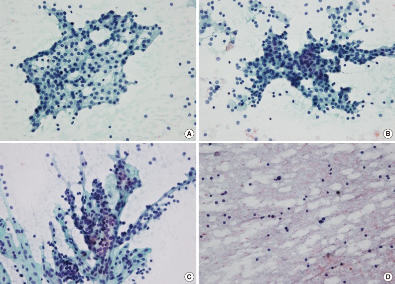
Fig. 2 Representative cytological features of a parathyroid lesion. (A) A loose cluster demonstrates a round configuration. (B) A tight cluster shows overlapping nuclei. (C) A papillary pattern has a fibrovascular core with parathyroid cells arranged in a vascular network. (D) Smears from an aspirated intrathyroidal parathyroid lesion show a lymphoid pattern (A-D, Papanicolaou stain).
Fig. 3 Representative nuclear features of a parathyroid lesion. (A) Nuclei are uniform and round to oval with stippled chromatin. A few cells show a well-defined cell border and an intercellular window. (B) Eccentric nuclei and anisokaryosis with a few atypical nuclei in the periphery (arrow). (C) An intranuclear pseudoinclusion has a distinct boundary resembling a shape drawn with a pencil (arrow) (A-C, Papanicolaou stain).
Fig. 1
Fig. 2
Fig. 3
Fine Needle Aspiration Cytology of Parathyroid Lesions
| Feature | No. (%) |
|---|---|
| Gender | |
| Male | 6 (24) |
| Female | 19 (76) |
| Mean age (yr) | 51 |
| Mass size in mean (cm) | 1.9 |
| Serum PTH | |
| Normal | 9 (36) |
| Elevated | 13 (52) |
| Unknown | 3 (12) |
| Location | |
| Posterior to thyroid gland | 9 (36) |
| Inferior to thyroid gland | 7 (28) |
| Intrathyroidal | 6 (24) |
| Paratracheal | 3 (12) |
| Clinicoradiologic impression | |
| Incidentaloma | 11 (44) |
| Primary hyperparathyroidism | 8 (32) |
| Parathyroid lesion | 4 (16) |
| Metastatic papillary thyroid carcinoma | 2 (8) |
| Cytological diagnosis | |
| Parathyroid lesion | 18 (72) |
| Benign follicular cells | 2 (8) |
| Adenomatous goiter | 2 (8) |
| Atypical cells | 2 (8) |
| Metastatic carcinoma | 1 (4) |
| Histologic diagnosis | |
| Parathyroid adenoma | 18 (72) |
| Parathyroid carcinoma | 2 (8) |
| None |
5 (20) |
| Cytological features | No. (%) |
|---|---|
| Architectural features | |
| Scattered naked nuclei | 25 (100) |
| Loose clusters | 22 (88) |
| Papillary pattern with fibrovascular core | 17 (68) |
| Tight clusters | 14 (56) |
| Follicular pattern | 11 (44) |
| Nuclear features | |
| Anisokaryosis | 24 (96) |
| Stippled chromatin | 21 (84) |
| Eccentric nuclei | 2 (8) |
| Prominent nucleoli | 1 (4) |
| Intranuclear pseudoinclusion | 1 (4) |
| Other features | |
| Well-defined cell border | 9 (36) |
| Oxyphilic cytoplasm | 9 (36) |
| Colloid-like material | 3 (12) |
| Macrophages | 0 (0) |
| Feature | No. (%) |
|---|---|
| Serum parathyroid hormone | |
| Normal | 5 (71.4) |
| Elevated | 1 (14.3) |
| Unknown | 1 (14.3) |
| Location | |
| Posterior to thyroid gland | 2 (28.6) |
| Inferior to thyroid gland | 2 (28.6) |
| Intrathyroidal | 1 (14.3) |
| Paratracheal | 2 (28.6) |
| Clinicoradiologic impression | |
| Incidentaloma | 3 (42.9) |
| Primary hyperparathyroidism | 1 (14.3) |
| Parathyroid lesion | 1 (14.3) |
| Metastatic papillary thyroid carcinoma | 2 (28.6) |
| Treatment | |
| Minimally invasive parathyroidectomy | 4 (57.1) |
| Total thyroidectomy and central neck dissection |
1 (14.3) |
| Lobectomy | 1 (14.3) |
| Neck dissection | 1 (14.3) |
Table 1. Clinicopathologic characteristics of the 25 cases
They are immunohistochemically diagnosed as parathyroid lesion.
Table 2. Summary of cytological features
Table 3. Clinical findings of the seven patients who are misdiagnosed
This patient has simultaneous papillary thyroid carcinoma.

