 E-submission
Articles
- Page Path
- HOME > J Pathol Transl Med > Volume 47(2); 2013 > Article
-
Case Study
Silent Colonic Malakoplakia in a Living-Donor Kidney Transplant Recipient Diagnosed during Annual Medical Examination - Go Eun Bae, Nara Yoon, Ha Young Park, Sang Yun Ha, Junhun Cho, Yunkyung Lee, Kyoung-Mee Kim, Cheol Keun Park
-
Korean Journal of Pathology 2013;47(2):163-166.
DOI: https://doi.org/10.4132/KoreanJPathol.2013.47.2.163
Published online: April 24, 2013
Department of Pathology, Samsung Medical Center, Sungkyunkwan University School of Medicine, Seoul, Korea.
- Corresponding Author: Yunkyung Lee, M.D. Department of Pathology, Samsung Medical Center, Sungkyunkwan University School of Medicine, 81 Irwon-ro, Gangnam-gu, Seoul 135-710, Korea. Tel: +82-2-3410-2801, Fax: +82-2-3410-6396, yk427.lee@samsung.com
• Received: June 14, 2012 • Revised: July 25, 2012 • Accepted: July 26, 2012
© 2013 The Korean Society of Pathologists/The Korean Society for Cytopathology
This is an Open Access article distributed under the terms of the Creative Commons Attribution Non-Commercial License (http://creativecommons.org/licenses/by-nc/3.0/) which permits unrestricted non-commercial use, distribution, and reproduction in any medium, provided the original work is properly cited.


Figure & Data
References
Citations
Citations to this article as recorded by 
- Malakoplakia in kidney transplant recipients: Three case reports
Prathap Kumar Simhadri, Renish Contractor, Deepak Chandramohan, Matthew McGee, Udit Nangia, Mohammad Atari, Syed Bushra, Sanjana Kapoor, Ramya Krishna Velagapudi, Pradeep K Vaitla
World Journal of Nephrology.2025;[Epub] CrossRef - Caecal malakoplakia: a rare mimic of malignancy
Jeffrey Li Voon Chong, Noor Ali
BMJ Case Reports.2024; 17(1): e257130. CrossRef - Malakoplakia Associated with Colonic Adenocarcinoma: Case Report and Literature Review of a Rare Occurrence
Sarah Tolaymat, Faryal Afridi, MD, Emily Groves, MD
West Virginia Medical Journal.2024; 120(4): 18. CrossRef - A Surgical Challenge Generated by Colonic Malakoplakia in Disguise as a Locally Advanced Colonic Malignancy—A Case Report
Cristina Șerban, Alexandra Toma, Dragoș Cristian Voicu, Constantin Popazu, Dorel Firescu, George Țocu, Raul Mihailov, Laura Rebegea
Medicina.2023; 59(1): 156. CrossRef - Colonic malakoplakia in a cardiac transplant recipient: A case report
Sadiya Shafijan
Indian Journal of Pathology and Microbiology.2020; 63(2): 322. CrossRef - Immunosuppressive drugs and the gastrointestinal tract in renal transplant patients
Merel M. Tielemans, Gerben A.J. van Boekel, Teun van Gelder, Eric T. Tjwa, Luuk B. Hilbrands
Transplantation Reviews.2019; 33(2): 55. CrossRef - Malakoplakia of the colon following renal transplantation in a 73 year old woman: report of a case presenting as intestinal perforation
Andrew Mitchell, Alexandre Dugas
Diagnostic Pathology.2019;[Epub] CrossRef - Colonic malakoplakia in a liver transplant recipient: A case report
Rana Ajabnoor, Mohammad Mawardi, Abdulmonem Almutawa
Human Pathology: Case Reports.2019; 18: 200323. CrossRef - Malakoplakia after kidney transplantation: Case report and literature review
John Fredy Nieto‐Ríos, Isabel Ramírez, Mónica Zuluaga‐Quintero, Lina María Serna‐Higuita, Federico Gaviria‐Gil, Alejandro Velez‐Hoyos
Transplant Infectious Disease.2017;[Epub] CrossRef - Megalocytic Interstitial Nephritis Following Acute Pyelonephritis with Escherichia coli Bacteremia: A Case Report
Hee Jin Kwon, Kwai Han Yoo, In Young Kim, Seulkee Lee, Hye Ryoun Jang, Ghee Young Kwon
Journal of Korean Medical Science.2015; 30(1): 110. CrossRef
 PubReader
 ePub Link
-
 Cite this Article
- Cite this Article
-
- Close
- Download Citation
- Close
- Figure
-
Silent Colonic Malakoplakia in a Living-Donor Kidney Transplant Recipient Diagnosed during Annual Medical Examination


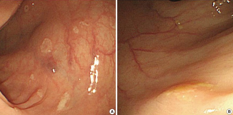
Fig. 1 Endoscopic features of the ascending (A) and transverse (B) colon. A colonoscopy reveals several depressed flat lesions and elevated polyps, which are measuring up to 4 mm in size with whitish exudates.
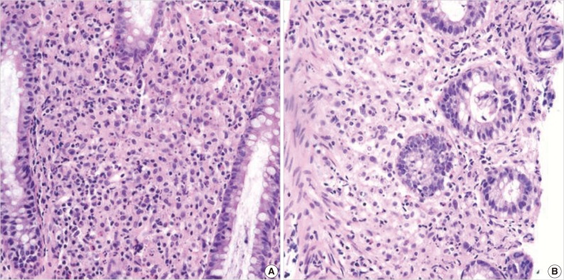
Fig. 2 (A) There is an infiltration of many histiocytes with ample granular eosinophilic cytoplasm with some lymphocytes and plasma cells (ascending colon). (B) Round basophilic structures are identified, measuring approximately between 1 µm and 10 µm in size within histiocytes, so-called Michaelis-Gutmann bodies (transverse colon).
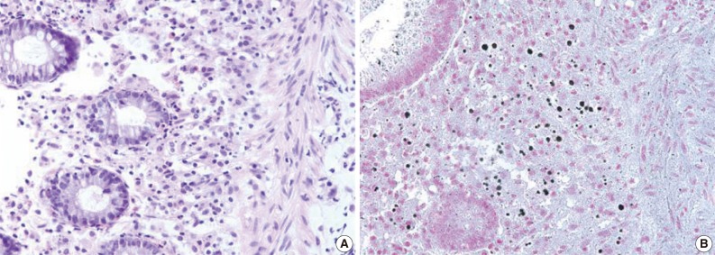
Fig. 3 Michaelis-Gutmann bodies are positively stained on von Kossa stain. (A) Hematoxylin and eosin stain. (B) von Kossa stain.
Fig. 1
Fig. 2
Fig. 3
Silent Colonic Malakoplakia in a Living-Donor Kidney Transplant Recipient Diagnosed during Annual Medical Examination
| Reference | Site of involvement | Complaints | Presentation | Age (yr)/Sex |
|---|---|---|---|---|
| Shah et al. [5] | Cecum ascending and sigmoid colon | Diarrhea | 3-4 mm polypoid nodule | 45/M |
| Berney et al. [2] | Cecum and rectum | Acute intestinal perforation | Mass forming | 52/M |
| Yousif et al. [6] | Rectum | Painful defecation | Mass and fistulous tract forming | 40/M |
| Present case | From cecum to descending colon | No symptom | 3-4 mm multiple polypoid nodules and depressed lesions | 55/F |
Table 1. Clinical features of previously reported colonic malakoplakia in a kidney transplant patient

