Warning: mkdir(): Permission denied in /home/virtual/lib/view_data.php on line 81

Warning: fopen(upload/ip_log/ip_log_2024-03.txt): failed to open stream: No such file or directory in /home/virtual/lib/view_data.php on line 83

Warning: fwrite() expects parameter 1 to be resource, boolean given in /home/virtual/lib/view_data.php on line 84
Cytologic Findings of Thyroid Carcinoma Showing Thymus-like Differentiation: A Case Report
Skip Navigation
Skip to contents

J Pathol Transl Med : Journal of Pathology and Translational Medicine

OPEN ACCESS
SEARCH
Search

Articles

Page Path
HOME > J Pathol Transl Med > Volume 46(3); 2012 > Article
Case Report
Cytologic Findings of Thyroid Carcinoma Showing Thymus-like Differentiation: A Case Report
Sunhee Chang, Mee Joo, Hanseong Kim
Korean Journal of Pathology 2012;46(3):302-305.
DOI: https://doi.org/10.4132/KoreanJPathol.2012.46.3.302
Published online: June 22, 2012

Department of Pathology, Inje University Ilsan Paik Hospital, Inje University College of Medicine, Goyang, Korea.

Corresponding Author: Sunhee Chang, M.D. Department of Pathology, Inje University Ilsan Paik Hospital, Inje University College of Medicine,170 Juhwa-ro, Ilsanseo-gu, Goyang 411-706, Korea. Tel: +82-31-910-7138, Fax: +82-31-910-7139, 'changsh@paik.ac.kr'
• Received: January 16, 2012   • Revised: March 25, 2012   • Accepted: April 25, 2012

© 2012 The Korean Society of Pathologists/The Korean Society for Cytopathology

This is an Open Access article distributed under the terms of the Creative Commons Attribution Non-Commercial License (http://creativecommons.org/licenses/by-nc/3.0) which permits unrestricted non-commercial use, distribution, and reproduction in any medium, provided the original work is properly cited.
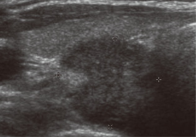
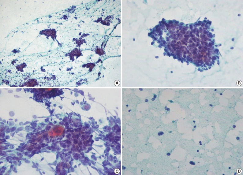
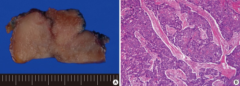
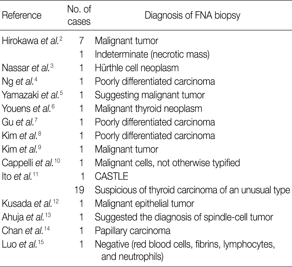
  • 8,648 Views
  • 58 Download
  • 13 Crossref
  • 16 Scopus
  • Carcinoma showing thymus-like differentiation (CASTLE) is a rare carcinoma of the thyroid or adjacent soft tissue of the neck with a histologic resemblance to thymic epithelial tumors. Although the fine-needle aspiration (FNA) plays a central role in the initial evaluation of thyroid nodules, few reports about the cytologic findings of CASTLE have been found according to a review of literatures. We report cytologic findings of a case of CASTLE. A 34-year-old woman presented with a 2-month history of sore throat. The FNA showed that the smear was composed of three dimensional clusters and sheets. The tumor cells were round to ovoid with high nuclear : cytoplasmic ratios. The nuclei were vesicular with small nucleoli. There were some tumor cells showing keratinization. Some lymphocytes were found on the background and within clusters. The presence of poorly-differentiated tumor cells with a focal keratinization and a lymphocytic background on the FNA is suggestive of CASTLE.
Carcinoma showing thymus-like differentiation (CASTLE) is a rare carcinoma of the thyroid gland or adjacent soft tissue of the neck with a histological resemblance to thymic epithelial tumors.1 Its incidence in thyroid malignant tumor is estimated at 0.075%.2 Although the fine-needle aspiration (FNA) biopsy plays a central role in the initial evaluation of solitary thyroid nodules, few reports about the cytologic findings of CASTLE have been found according to a review of literatures.3-6 In Korea, three cases of CASTLE have been reported in the literature but its cytologic findings have not been described.7-9 We report cytological findings of CASTLE in a 34-year-old woman.
A 34-year-old woman presented with a 2-month history of sore throat. The patient had no notable medical or family history. Physical examination revealed a palpable right thyroid mass. Thyroid function and calcium tests were within normal limits. Ultrasound showed a 1.7-cm, well demarcated, heterogeneous, low echoic solid mass in the lower pole of the right thyroid (Fig. 1). A computed tomography scan of the neck revealed a 3.3-cm, exophytic, low-attenuated mass in the lower pole of the right thyroid. The FNA biopsy of the mass was performed, which was accompanied by the standard Papanicolaou stain.
The smear was composed of not only cohesive three dimensional clusters and sheets but also singly scattered cells (Fig. 2A). The tumor cells showed mild anisonucleosis and high nuclear : cytoplasmic (N/C) ratios. Nuclei were round to ovoid with irregular nuclear contours and small, distinct nucleoli (Fig. 2B). The nuclear chromatin was vesicular or coarsely granular. Nuclear grooves were rarely seen. Cytoplasm was scanty and amphophilic. Cytoplasmic border was indistinct. Some tumor cells showed dense orangeophilic cytoplasm, which is suggestive of individual cell keratinization (Fig. 2C). Some lymphocytes were found on the background and within clusters, which was accompanied by the presence of a few stripped nuclei (Fig. 2D). Mitotic figures were rarely seen. Intranuclear cytoplasmic inclusions were not observed. Papillary or follicular structure was not observed. The aspirate was interpreted as a high-grade malignant thyroid neoplasm without further definitive classification. The total thyroidectomy was performed.
On gross examination, the thyroid contained a 4.0-cm solid mass replacing most of the right thyroid. The mass had a lobular, tan-colored cut surface and a firm consistency (Fig. 3A). On microscopic examination, the tumor was well-circumscribed and it was composed of variably sized and irregularly shaped lobules of cohesive polygonal tumor cells which were separated by bands of dense fibrous stroma (Fig. 3B). The tumor cells had high N/C ratios, eosinophilic cytoplasm and ill-defined cell borders. The nuclei were vesicular or coarsely granular with prominent nucleoli. The mitotic figures were sparse. There was mild lymphoplasmacytic infiltration within the tumor nodules and septae. Cytoplasmic keratinization was seen. The tumor cells showed positivity for CD5, carcinoembryonic antigen, high molecular weight keratin (HMWK), cytokeratin 5 (CK5), and p63. The tumor was negative for thyroid transcription factor-1, thyroglobulin, and calcitonin. Vascular invasion and extensive extra-thyroidal extension were present. One regional lymph node was found and involved by tumor.
Over a 27-month follow-up period, the patient had no evidence of tumor recurrence.
The cytological diagnoses were mentioned in previous 14 reports consisting of 40 cases. Malignant tumor or poorly-differentiated carcinoma were the most frequent cytological diagnosis in cases of CASTLE, except for six cases (Table 1).2-15 One case was diagnosed as CASTLE for which no cytological findings have been described.11
In previously reported cases of CASTLE, the most common cytological findings include tight clusters and sheets of round tumor cells with high N/C ratios, vesicular nuclei, prominent nucleoli, amphophilic cytoplasm, and lymphocytic background, which correspond well with the present case.2-4,6 In the present case and those of Hirokawa et al.,2 keratinized cells were present. Youens et al.6 reported indistinct cell borders, intranuclear grooves and sparse mitotic figures, which were also seen in the present case. Hirokawa et al.2 reported intracytoplasmic space surrounded by cell membrane and spindle tumor cells. Youens et al.6 reported not only intranuclear cytoplasmic inclusions and papillary-like structures but also granular chromatin and rosette-like structures. But these findings were not detected in the present case.
The differential diagnoses of CASTLE include papillary carcinoma, Hürthle cell neoplasm, undifferentiated carcinoma, medullary carcinoma and metastatic lymphoepithelioma-like carcinoma.
CASTLE may show intranuclear pseudoinclusions or papillary-like structures, but it does not show fine pale chromatin and monolayer sheets of cells with dense cytoplasm. Intranuclear pseudoinclusions can be seen in various malignant or benign thyroid nodules. Intranuclear pseudoinclusions should be interpreted in light of the other architectural and nuclear features.
Although Hürthle cell neoplasm contains cellular aggregates with abundant cytoplasm and prominent nucleoli, lymphocytic background of CASTLE is lacking. Unlike Hürthle cell neoplasm, CASTLE does not have granular cytoplasm.
It is very difficult to distinguish CASTLE from metastatic lymphoepithelioma-like carcinoma, which also shows sheets of poorly differentiated round cells intermixed with small lymphocytes. Subtle differences, such as frequent mitotic activity and a polymorphous inflammatory background, favor metastatic lymphoepithelioma-like carcinoma.4
CASTLE may resemble undifferentiated thyroid carcinoma with squamous differentiation, but it does not show marked cytologic atypia, frequent mitotic activity and necrotic background characteristic of undifferentiated carcinoma.
Although CASTLE may show rosette-like structures and granular chromatin, along with an abundance of single tumor cells as in medullary carcinoma, it is lacking of typical plasmacytoid cells, pink cytoplasmic granules and amyloid deposition.6
Immunocytochemical study for CD5 is helpful to differentiate CASTLE from other malignant thyroid neoplasm. CD5 is a surface glycoprotein expressed on mature T cells and a subset of B cells.16 Thymic carcinoma cells are also known to be positive for CD5.17 CD5 is almost always expressed in CASTLE. CASTLE is positive for HMWK, CK5, and p63 because it is also a squamous cell carcinoma. However, thyroid squamous cell carcinoma and poorly-differentiated carcinoma were negative for CD5.17 Ito et al.11 reported that the sensitivity and specificity for CD5 immunohistochemistry were 82% and 100%, respectively, in making a diagnosis of CASTLE.
In conclusion, the presence of poorly-differentiated tumor cells with a focal keratinization and a lymphocytic background on the FNA biopsy is suggestive of CASTLE.
This work was supported by grant from Inje University, 2008.

No potential conflict of interest relevant to this article was reported.

  • 1. DeLellis RA, Lloyd RV, Heitz PU, Eng C. World Health Organization Classification of tumours: pathology and genetics of tumours of endocrine organs. 2004; Lyon: IARC Press, 96–97.
  • 2. Hirokawa M, Kuma S, Miyauchi A. Cytological findings of intrathyroidal epithelial thymoma/carcinoma showing thymus-like differentiation: a study of eight cases. Diagn Cytopathol 2010 11 02 [Epub]http://dx.doi.org/10.1002/dc.21511. Article
  • 3. Nassar A, Saqi A, Baloch Z, LiVolsi V. Carcinoma showing thymus-like element of the thyroid. Acta Cytol 2003; 47: 313–315. PMID: 12685208. PubMed
  • 4. Ng WK, Collins RJ, Shek WH, Ng IO. Cytologic diagnosis of "CASTLE" of thyroid gland: report of a case with histologic correlation. Diagn Cytopathol 1996; 15: 224–227. PMID: 8955605. ArticlePubMed
  • 5. Yamazaki M, Fujii S, Daiko H, Hayashi R, Ochiai A. Carcinoma showing thymus-like differentiation (CASTLE) with neuroendocrine differentiation. Pathol Int 2008; 58: 775–779. PMID: 19067852. ArticlePubMed
  • 6. Youens KE, Bean SM, Dodd LG, Jones CK. Thyroid carcinoma showing thymus-like differentiation (CASTLE): case report with cytomorphology and review of the literature. Diagn Cytopathol 2011; 39: 204–209. PMID: 20607745. ArticlePubMed
  • 7. Gu MJ, Shim YR, Choi JH, Choi WH. Carcinoma showing thymus-like differentiation (CASTLE) of the thyroid gland: a case report. Korean J Pathol 1999; 33: 460–462.
  • 8. Kim EH, Jeong JY, Kim EY, et al. A case of carcinoma showing thymus-like differentiation (CASTLE) in the thyroid. J Korean Endocr Soc 2008; 23: 272–276. Article
  • 9. Kim JP, Sung BG, Ahn SK, Lee JH. A case of carcinoma showing thymus-like differentiation. Korean J Otolaryngol-Head Neck Surg 2004; 47: 470–473.
  • 10. Cappelli C, Tironi A, Marchetti GP, et al. Aggressive thyroid carcinoma showing thymic-like differentiation (CASTLE): case report and review of the literature. Endocr J 2008; 55: 685–690. PMID: 18560200. ArticlePubMed
  • 11. Ito Y, Miyauchi A, Nakamura Y, Miya A, Kobayashi K, Kakudo K. Clinicopathologic significance of intrathyroidal epithelial thymoma/carcinoma showing thymus-like differentiation: a collaborative study with Member Institutes of The Japanese Society of Thyroid Surgery. Am J Clin Pathol 2007; 127: 230–236. PMID: 17210519. ArticlePubMed
  • 12. Kusada N, Hara Y, Kobayashi S, et al. A case of aggressive carcinoma showing thymus-like differentiation with distant metastases. Thyroid 2005; 15: 1383–1388. PMID: 16405413. ArticlePubMed
  • 13. Ahuja AT, Chan ES, Allen PW, Lau KY, King W, Metreweli C. Carcinoma showing thymiclike differentiation (CASTLE tumor). AJNR Am J Neuroradiol 1998; 19: 1225–1228. PMID: 9726458. PubMedPMC
  • 14. Chan LP, Chiang FY, Lee KW, Kuo WR. Carcinoma showing thymus-like differentiation (CASTLE) of thyroid: a case report and literature review. Kaohsiung J Med Sci 2008; 24: 591–597. PMID: 19239992. ArticlePubMed
  • 15. Luo CM, Hsueh C, Chen TM. Extrathyroid carcinoma showing thymus-like differentiation (CASTLE) tumor: a new case report and review of literature. Head Neck 2005; 27: 927–933. PMID: 15952197. ArticlePubMed
  • 16. Bikah G, Lynd FM, Aruffo AA, Ledbetter JA, Bondada S. A role for CD5 in cognate interactions between T cells and B cells, and identification of a novel ligand for CD5. Int Immunol 1998; 10: 1185–1196. PMID: 9723705. ArticlePubMed
  • 17. Reimann JD, Dorfman DM, Nosé V. Carcinoma showing thymus-like differentiation of the thyroid (CASTLE): a comparative study. Evidence of thymic differentiation and solid cell nest origin. Am J Surg Pathol 2006; 30: 994–1001. PMID: 16861971. ArticlePubMed
Fig. 1
Thyroid ultrasonography shows a 1.7-cm, well-defined, hypoechoic, solid mass in the lower pole of the right thyroid.
kjpathol-46-302-g001.jpg
Fig. 2
(A) The smear shows cohesive sheets and clusters, and singly scattered cells. (B) Tumor cells have round to ovoid vesicular nuclei, small prominent nucleoli and scanty cytoplasm. A few lymphoid cells are present on the background and within the clusters. (C) Keratinization is seen. (D) Stripped nuclei are present in the background (A-D, Papanicolaou stain).
kjpathol-46-302-g002.jpg
Fig. 3
(A) The cut surface of the tumor is lobulated, solid, and tan-colored. (B) Broad anastomosing islands of tumor cells are separated by desmoplastic stroma. Squamous differentiation is present.
kjpathol-46-302-g003.jpg
Table 1
Diagnosis of CASTLE on the FNA biopsy in patients with CASTLE
kjpathol-46-302-i001.jpg

CASTLE, carcinoma showing thymus-like differentiation; FNA, fine-needle aspiration.

Figure & Data

References

    Citations

    Citations to this article as recorded by  
    • Intrathyroidal Thymic Carcinoma: A Retrospective Case Series Study
      Jinhui Liang, Mei Huang, Helang Huang, Li Li, Hailin Luo, Weidong Mao, Shan Gao, Haoxiang Xu
      Ear, Nose & Throat Journal.2023; 102(9): 584.     CrossRef
    • Carcinoma showing thymus‐like differentiation of the parotid gland: The brief report of cytomorphology and review of the literature
      Tomoko Uchiyama, Chiyoko Terada, Yuma Tachibana, Hirokazu Nishiura, Maiko Takeda, Tomomi Fujii, Takahiro Kimura, Masahiro Tsutsumi, Chiho Ohbayashi
      Diagnostic Cytopathology.2023;[Epub]     CrossRef
    • A case of concurrent occurrence of carcinoma showing thymus-like differentiation and follicular variant of papillary thyroid cancer in the same thyroid
      Takahito Kimura, Keisuke Enomoto, Masamitsu Kono, Masanobu Hiraoka, Saori Takeda, Naoko Kumashiro, Shun Hirayama, Eri Kimura, Shunji Tamagawa, Makiko Ohtani, Shin-Ichi Murata, Muneki Hotomi
      Journal of Surgical Case Reports.2022;[Epub]     CrossRef
    • Ultrasonographic Features of Intrathyroidal Thymic Carcinoma: Review and Analysis of 10 Cases
      Wang, MD Yanhai, Yang, MD Hua, Liu, MD Hanqing, Luo, MD Xiaoli, Liu, BS Luying, Zhou, BS Pingting
      ADVANCED ULTRASOUND IN DIAGNOSIS AND THERAPY.2022; 6(2): 58.     CrossRef
    • Thyroid carcinoma with thymus-like differentiation (CASTLE) tumor: а сase report
      A. A. Ilyin, V. V. Polkin, P. A. Isaev, F. E. Sevrukov, N. Yu. Dvinskych, M. I. Ryzhenkova, S. A. Ivanov, A. D. Kaprin
      Head and Neck Tumors (HNT).2021; 11(2): 64.     CrossRef
    • Metastatic Renal Cell Neoplasm Within a Papillary Thyroid Carcinoma as Incidental Finding in an Asymptomatic Patient: a Case Report
      Maria-Rosa Bella-Cueto, Mireia Pascua-Solé, Albert Cano-Palomares, M. Àngels Cabezuelo-Hernandez, Maria-Rosa Escoda-Giralt, Santiago Barcons-Vilaplana, Paula Serret-Miralles, Carmen Caral-Vanaclocha, Xavier Guirao-Garriga, Joan Prats-Lopez, Meritxell Meda
      SN Comprehensive Clinical Medicine.2020; 2(7): 978.     CrossRef
    • Intrathyroidal thymic carcinoma exhibiting neuroendocrine differentiation: Case report with cytomorphology, immunocytochemistry, and review of the literature focusing on cytology
      Wen‐hao Ren, Kun Dong, Xiao‐zheng Huang, Yan‐li Zhu
      Diagnostic Cytopathology.2019; 47(11): 1197.     CrossRef
    • Management and Prognostic Factors for Thyroid Carcinoma Showing Thymus-Like Elements (CASTLE): A Case Series Study
      Rui Gao, Xi Jia, Ting Ji, Jinteng Feng, Aimin Yang, Guangjian Zhang
      Frontiers in Oncology.2018;[Epub]     CrossRef
    • Multiple squamous cells in thyroid fine needle aspiration: Friends or foes?
      Heather Gage, Elizabeth Hubbard, Laurentia Nodit
      Diagnostic Cytopathology.2016; 44(8): 676.     CrossRef
    • Clinical analysis of 82 cases of carcinoma showing thymus-like differentiation of the thyroid
      WEI GE, YONG-ZHONG YAO, GANG CHEN, YI-TAO DING
      Oncology Letters.2016; 11(2): 1321.     CrossRef
    • Carcinoma Showing Thymus-Like Differentiation (CASTLE): Cytopathological Features and Differential Diagnosis
      Jennifer A. Collins, Bo Ping, Justin A. Bishop, Syed Z. Ali
      Acta Cytologica.2016; 60(5): 421.     CrossRef
    • Carcinoma Showing Thymus-Like Elements of the Thyroid Gland: Report of Three Cases Including One Case with Breast Cancer History
      Guanjun Zhang, Xi Liu, Wei Huang, Xiaofeng Li, Marianne Johnstone, Yuan Deng, Yongqiang Ke, Quentin M. Nunes, Hongyan Wang, Yili Wang, Xuebin Zhang
      Pathology & Oncology Research.2015; 21(1): 45.     CrossRef
    • Potential Role of Adjuvant Radiation Therapy in Cervical Thymic Neoplasm Involving Thyroid Gland or Neck
      Jae Myoung Noh, Sang Yun Ha, Yong Chan Ahn, Dongryul Oh, Seung Won Seol, Young Lyun Oh, Joungho Han
      Cancer Research and Treatment.2014; 47(3): 436.     CrossRef

    • PubReader PubReader
    • Cite this Article
      Cite this Article
      export Copy Download
      Close
      Download Citation
      Download a citation file in RIS format that can be imported by all major citation management software, including EndNote, ProCite, RefWorks, and Reference Manager.

      Format:
      • RIS — For EndNote, ProCite, RefWorks, and most other reference management software
      • BibTeX — For JabRef, BibDesk, and other BibTeX-specific software
      Include:
      • Citation for the content below
      Cytologic Findings of Thyroid Carcinoma Showing Thymus-like Differentiation: A Case Report
      Korean J Pathol. 2012;46(3):302-305.   Published online June 22, 2012
      Close
    • XML DownloadXML Download
    Figure

    J Pathol Transl Med : Journal of Pathology and Translational Medicine