 E-submission
Articles
- Page Path
- HOME > J Pathol Transl Med > Volume 50(2); 2016 > Article
-
Original Article
Interobserver Variability of Ki-67 Measurement in Breast Cancer - Yul Ri Chung1, Min Hye Jang2, So Yeon Park1,2, Gyungyub Gong3, Woo-Hee Jung4, *The Korean Breast Pathology Ki-67 Study Group
-
Journal of Pathology and Translational Medicine 2016;50(2):129-137.
DOI: https://doi.org/10.4132/jptm.2015.12.24
Published online: February 15, 2016
1Department of Pathology, Seoul National University College of Medicine, Seoul, Korea
2Department of Pathology, Seoul National University Bundang Hospital, Seongnam, Korea
3Department of Pathology, Asan Medical Center, University of Ulsan College of Medicine, Seoul, Korea
4Department of Pathology, Gangnam Severance Hospital, Yonsei University College of Medicine, Seoul, Korea
- Corresponding Author: So Yeon Park, MD, PhD Department of Pathology, Seoul National University Bundang Hospital, 82 Gumi-ro 173beon-gil, Bundang-gu, Seongnam 13620, Korea Tel: +82-31-787-7712 Fax: +82-31-787-4012 E-mail: sypmd@snu.ac.kr
*Lists of participants and their affiliations appear at the end of the paper.
© 2016 The Korean Society of Pathologists/The Korean Society for Cytopathology
This is an Open Access article distributed under the terms of the Creative Commons Attribution Non-Commercial License (http://creativecommons.org/licenses/by-nc/3.0/) which permits unrestricted noncommercial use, distribution, and reproduction in any medium, provided the original work is properly cited.


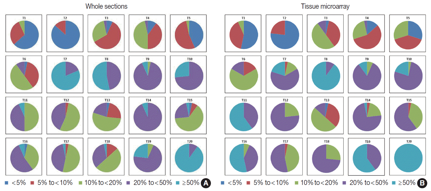
Figure & Data
References
Citations
- Dynamic biomarkers in hormone receptor-positive/HER2-negative breast cancer trials: a new hope for precision oncology
Giuseppe Di Grazia, Rodrigo Sánchez-Bayona, Climent Casals-Pascual, Tomás Pascual, Daniele Generali, Alessandra Gennari, Paolo Vigneri, Nadia Harbeck, Javier Cortés, Aleix Prat, Francesco Schettini
npj Breast Cancer.2026;[Epub] CrossRef - Low-Proliferative Invasive Mucinous Carcinoma of the Breast in an Octogenarian: A Case Report Highlighting Pathology-Guided De-escalation
Rae-Anne Kastle, Christopher M Ahmad, Rachana Tadakamalla, Hima Patel, Amer Abboud
Cureus.2026;[Epub] CrossRef - Low E-cadherin expression is associated with poor prognosis in pulmonal adenocarcinoma
Fiete Gehrisch, Kiara A. Schmid, Martina Kluth, Georgia Makrypidi-Fraune, Katharina Möller, Maximilian Lennartz, Veit Bertram, Florian Lutz, Stefan Steurer, Philipp Busch, Birgit Hantzsch-Kuhn, Martin Reck, Till Olchers, David Benjamin Ellebrecht, Christo
Scientific Reports.2026;[Epub] CrossRef - Evaluating the Association of Ki-67 with Oncotype DX Recurrence Score in Early-Stage ER-Positive/HER2-Negative Breast Cancer
Dimitrios Dragoumis, George Kapetsis, Konstantinos Louis, Dimitrios Maniatis, Eleni Mpalampou, Konstantinos Bouloukos, Xenophon Xenakis, Nikolaos Papaioannou, Styliani Parpoudi, Grigorios Pesmatzoglou, Anna Sachoulidou, Eleftherios Sfakianakis, Sofia Tria
Cancers.2026; 18(11): 1731. CrossRef - Comparative analysis of Ki-67 labeling index morphometry using deep learning, conventional image analysis, and manual counting
Mohammad Rizwan Alam, Kyung Jin Seo, Kwangil Yim, Phoebe Liang, Joe Yeh, Chifu Chang, Yosep Chong
Translational Oncology.2025; 51: 102159. CrossRef - Machine Learning-Based Approaches for Breast Density Estimation from Mammograms: A Comprehensive Review
Khaldoon Alhusari, Salam Dhou
Journal of Imaging.2025; 11(2): 38. CrossRef - Letter re: A critical appraisal of the DATA trial analysis on the prognostic and predictive value of the luminal-like subtype
M. Rizk, K. Mokbel
ESMO Open.2025; 10(5): 105067. CrossRef - Clinico-Histomorphological and Mib-1 Analysis of Recurrent Meningiomas: A Retrospective Study
Sujata Sarangi, Asha Shenoy, Ashvini Kolhe, Kanchan Kothari
Asian Journal of Neurosurgery.2025; 20(04): 785. CrossRef - Emerging Diagnostics and Therapies in Neuroendocrine Neoplasms: A Critical Review
Jorge H. Hernandez-Felix, Monica Isabel Meneses-Medina, Rachel Riechelmann, Jonathan Strosberg, Rocio Garcia-Carbonero, Jaydira del Rivero
Cancers.2025; 17(22): 3632. CrossRef - Ki-67 Testing in Breast Cancer: Assessing Variability With Scoring Methods and Specimen Types and the Potential Subsequent Impact on Therapy Eligibility
Therese Bocklage, Virgilius Cornea, Caylin Hickey, Justin Miller, Jessica Moss, Mara Chambers, S. Emily Bachert
Applied Immunohistochemistry & Molecular Morphology.2024; 32(3): 119. CrossRef - Interobserver agreement and diagnostic challenges of Congo red staining for amyloid detection on fat pad aspiration biopsies
Levent Trabzonlu, T. Leif Helland, Melanie C. Kwan, Nathalie Kumiega, M. Lisa Zhang, Ivan Chebib, Vanda F. Torous
Journal of the American Society of Cytopathology.2024; 13(5): 359. CrossRef - Assessment of the Predictive Role of Ki-67 in Breast Cancer Patients’ Responses to Neoadjuvant Chemotherapy
Ghizlane Rais, Rania Mokfi, Farah Boutaggount, Meryem Maskrout, Soundouss Bennour, Chaymae Senoussi, Fadoua Rais
European Journal of Breast Health.2024; : 199. CrossRef - Improving the accuracy of reporting Ki-67 IHC by using an AI tool
Sahil Ajit Saraf, Aahan Singh, Wai Po Kevin Teng, Sencer Karakaya, M. Logaswari, Kaveh Taghipour, Rajasa Jialdasani, Li Yan Khor, Kiat Hon Lim, Sathiyamoorthy Selvarajan, Vani Ravikumar, Md Ali Osama, Priti Chatterjee, Santosh KV
Heliyon.2024; 10(22): e40193. CrossRef - Predictive Value of Ki-67 Index in Evaluating Sporadic Vestibular Schwannoma Recurrence: Systematic Review and Meta-analysis
Kunal Vakharia, Hirotaka Hasegawa, Christopher Graffeo, Mohammad H. A. Noureldine, Salomon Cohen-Cohen, Avital Perry, Matthew L. Carlson, Colin L. W. Driscoll, Maria Peris-Celda, Jamie J. Van Gompel, Michael J. Link
Journal of Neurological Surgery Part B: Skull Base.2023; 84(02): 119. CrossRef - Venous invasion and lymphatic invasion are correlated with the postoperative prognosis of pancreatic neuroendocrine neoplasm
Sho Kiritani, Junichi Arita, Yuichiro Mihara, Rihito Nagata, Akihiko Ichida, Yoshikuni Kawaguchi, Takeaki Ishizawa, Nobuhisa Akamatsu, Junichi Kaneko, Kiyoshi Hasegawa
Surgery.2023; 173(2): 365. CrossRef - Automated Molecular Subtyping of Breast Carcinoma Using Deep Learning Techniques
S. Niyas, Ramya Bygari, Rachita Naik, Bhavishya Viswanath, Dhananjay Ugwekar, Tojo Mathew, J Kavya, Jyoti R Kini, Jeny Rajan
IEEE Journal of Translational Engineering in Health and Medicine.2023; 11: 161. CrossRef - Grade Progression and Intrapatient Tumor Heterogeneity as Potential Contributors to Resistance in Gastroenteropancreatic Neuroendocrine Tumors
Diana Grace Varghese, Jaydira Del Rivero, Emily Bergsland
Cancers.2023; 15(14): 3712. CrossRef - Diagnostic Role and Prognostic Impact of PSAP Immunohistochemistry: A Tissue Microarray Study on 31,358 Cancer Tissues
Laura Sophie Tribian, Maximilian Lennartz, Doris Höflmayer, Noémi de Wispelaere, Sebastian Dwertmann Rico, Clara von Bargen, Simon Kind, Viktor Reiswich, Florian Viehweger, Florian Lutz, Veit Bertram, Christoph Fraune, Natalia Gorbokon, Sören Weidemann, C
Diagnostics.2023; 13(20): 3242. CrossRef - AI-Powered Segmentation of Invasive Carcinoma Regions in Breast Cancer Immunohistochemical Whole-Slide Images
Yiqing Liu, Tiantian Zhen, Yuqiu Fu, Yizhi Wang, Yonghong He, Anjia Han, Huijuan Shi
Cancers.2023; 16(1): 167. CrossRef - Expression of estrogen and progesterone receptors, HER2 protein and Ki-67 proliferation index in breast carcinoma in both tumor tissue and tissue microarray
UP Hacısalihoğlu, MA Dogan
Biotechnic & Histochemistry.2022; 97(4): 298. CrossRef - Diffusive Ki67 and vimentin are associated with worse recurrence-free survival of upper tract urothelial carcinoma: A retrospective cohort study from bench to bedside
Che Hsueh Yang, Wei Chun Weng, Yen Chuan Ou, Yi Sheng Lin, Li Hua Huang, Chin Heng Lu, Tang Yi Tsao, Chao Yu Hsu, Min Che Tung
Urologic Oncology: Seminars and Original Investigations.2022; 40(3): 109.e21. CrossRef - Should Ki-67 be adopted to select breast cancer patients for treatment with adjuvant abemaciclib?
P. Tarantino, H.J. Burstein, N.U. Lin, I.E. Krop, E.P. Winer, S.J. Schnitt, E.P. Hamilton, S.A. Hurvitz, H.S. Rugo, G. Curigliano, S.M. Tolaney
Annals of Oncology.2022; 33(3): 234. CrossRef - A novel deep classifier framework for automated molecular subtyping of breast carcinoma using immunohistochemistry image analysis
Tojo Mathew, S. Niyas, C.I. Johnpaul, Jyoti R. Kini, Jeny Rajan
Biomedical Signal Processing and Control.2022; 76: 103657. CrossRef - Deep learning for the standardized classification of Ki-67 in vulva carcinoma: A feasibility study
Matthias Choschzick, Mariam Alyahiaoui, Alexander Ciritsis, Cristina Rossi, André Gut, Patryk Hejduk, Andreas Boss
Heliyon.2021; 7(7): e07577. CrossRef - Oncotype DX Predictive Nomogram for Recurrence Score Output: The Novel System ADAPTED01 Based on Quantitative Immunochemistry Analysis
Fabio Marazzi, Roberto Barone, Valeria Masiello, Valentina Magri, Antonino Mulè, Angela Santoro, Federica Cacciatori, Luca Boldrini, Gianluca Franceschini, Francesca Moschella, Giuseppe Naso, Silverio Tomao, Maria Antonietta Gambacorta, Giovanna Mantini,
Clinical Breast Cancer.2020; 20(5): e600. CrossRef - Study of Ki-67 index in the molecular subtypes of breast cancer: Inter-observer variability and automated scoring
Divya Meermira, Meenakshi Swain, Swarnalata Gowrishankar
Indian Journal of Cancer.2020; 57(3): 289. CrossRef - Improving the accuracy of gastrointestinal neuroendocrine tumor grading with deep learning
Darshana Govind, Kuang-Yu Jen, Karen Matsukuma, Guofeng Gao, Kristin A. Olson, Dorina Gui, Gregory. E. Wilding, Samuel P. Border, Pinaki Sarder
Scientific Reports.2020;[Epub] CrossRef - Practical approaches to automated digital image analysis of Ki-67 labeling index in 997 breast carcinomas and causes of discordance with visual assessment
Ah-Young Kwon, Ha Young Park, Jiyeon Hyeon, Seok Jin Nam, Seok Won Kim, Jeong Eon Lee, Jong-Han Yu, Se Kyung Lee, Soo Youn Cho, Eun Yoon Cho, Irina V. Lebedeva
PLOS ONE.2019; 14(2): e0212309. CrossRef - Evaluation of Ki-67 Index in Core Needle Biopsies and Matched Breast Cancer Surgical Specimens
Soomin Ahn, Junghye Lee, Min-Sun Cho, Sanghui Park, Sun Hee Sung
Archives of Pathology & Laboratory Medicine.2018; 142(3): 364. CrossRef - Assessment of Ki-67 for Predicting Effective Prognosis in Breast Cancer Subtypes
Sangjung Park, Sunyoung Park, Jungho Kim, Sungwoo Ahn, Kwang Hwa Park, Hyeyoung Lee
Biomedical Science Letters.2018; 24(1): 9. CrossRef - Quantitative tumor heterogeneity assessment on a nuclear population basis
Anne‐Sofie Wessel Lindberg, Knut Conradsen, Rasmus Larsen, Michael Friis Lippert, Rasmus Røge, Mogens Vyberg
Cytometry Part A.2017; 91(6): 574. CrossRef - A comparison of Ki-67 counting methods in luminal Breast Cancer: The Average Method vs. the Hot Spot Method
Min Hye Jang, Hyun Jung Kim, Yul Ri Chung, Yangkyu Lee, So Yeon Park, William B. Coleman
PLOS ONE.2017; 12(2): e0172031. CrossRef - A Novel Breast Cancer Index for Prediction of Distant Recurrence in HR+ Early-Stage Breast Cancer with One to Three Positive Nodes
Yi Zhang, Brock E. Schroeder, Piiha-Lotta Jerevall, Amy Ly, Hannah Nolan, Catherine A. Schnabel, Dennis C. Sgroi
Clinical Cancer Research.2017; 23(23): 7217. CrossRef
 PubReader
 ePub Link
-
 Cite this Article
- Cite this Article
-
- Close
- Download Citation
- Close
- Figure
-
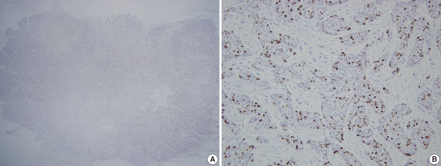
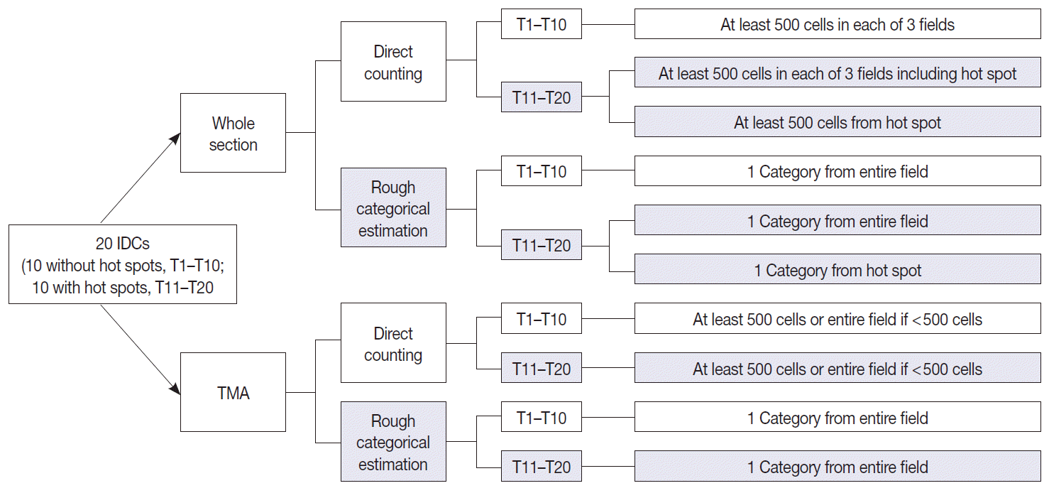
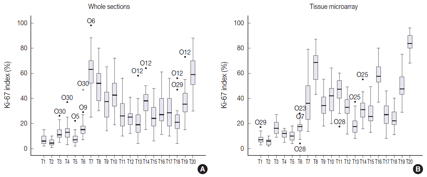


Fig. 1.
Fig. 2.
Fig. 3.
Fig. 4.
| T11 | T12 | T13 | T14 | T15 | T16 | T17 | T18 | T19 | T20 | |
|---|---|---|---|---|---|---|---|---|---|---|
| Whole section, average method | ||||||||||
| Minimum | 10.00 | 12.00 | 4.00 | 15.00 | 6.00 | 11.00 | 10.00 | 4.00 | 15.00 | 30.00 |
| Maximum | 56.00 | 41.00 | 58.00 | 64.00 | 48.00 | 61.00 | 56.00 | 56.00 | 73.00 | 88.00 |
| Mean | 28.20 | 24.43 | 20.50 | 37.20 | 24.97 | 31.30 | 29.27 | 21.93 | 37.13 | 59.90 |
| Median | 26.00 | 25.00 | 19.00 | 38.00 | 24.00 | 27.00 | 28.50 | 21.00 | 35.50 | 59.00 |
| SD | 12.50 | 7.12 | 11.77 | 10.09 | 10.24 | 13.08 | 12.76 | 10.58 | 12.74 | 14.68 |
| SE | 2.28 | 1.30 | 2.15 | 1.84 | 1.87 | 2.39 | 2.33 | 1.93 | 2.33 | 2.68 |
| Whole section, hot spot method | ||||||||||
| Minimum | 24.80 | 23.50 | 13.50 | 20.60 | 17.50 | 27.00 | 15.80 | 16.50 | 33.30 | 62.50 |
| Maximum | 62.20 | 56.00 | 36.50 | 59.50 | 61.00 | 79.30 | 54.50 | 49.00 | 72.50 | 94.00 |
| Mean | 44.59 | 35.74 | 26.73 | 41.71 | 34.40 | 56.43 | 37.03 | 29.38 | 51.58 | 82.50 |
| Median | 46.00 | 34.00 | 26.85 | 41.75 | 35.80 | 55.85 | 39.00 | 28.45 | 49.75 | 83.35 |
| SD | 9.65 | 8.71 | 6.14 | 8.42 | 9.26 | 11.46 | 9.60 | 7.81 | 9.35 | 7.86 |
| SE | 1.76 | 1.59 | 1.12 | 1.54 | 1.69 | 2.09 | 1.75 | 1.43 | 1.71 | 1.44 |
| Tissue microarray | ||||||||||
| Minimum | 17.50 | 11.60 | 8.00 | 15.80 | 13.00 | 37.00 | 8.00 | 11.00 | 29.00 | 68.00 |
| Maximum | 60.00 | 49.00 | 37.00 | 55.00 | 49.00 | 80.00 | 46.00 | 40.00 | 75.00 | 96.00 |
| Mean | 45.62 | 32.99 | 18.19 | 31.46 | 28.42 | 59.07 | 26.83 | 24.09 | 50.33 | 83.97 |
| Median | 47.00 | 33.00 | 17.50 | 31.00 | 25.50 | 57.50 | 27.00 | 22.00 | 47.50 | 83.50 |
| SD | 10.78 | 9.11 | 6.98 | 8.38 | 8.97 | 10.11 | 9.61 | 7.14 | 10.72 | 7.40 |
| SE | 1.97 | 1.66 | 1.27 | 1.53 | 1.64 | 1.85 | 1.76 | 1.30 | 1.96 | 1.35 |
| T11 | T12 | T13 | T14 | T15 | T16 | T17 | T18 | T19 | T20 | |
|---|---|---|---|---|---|---|---|---|---|---|
| Whole section, average method | ||||||||||
| < 5% | 0 | 0 | 1 | 0 | 1 | 1 | 0 | 0 | 0 | 0 |
| 5 to < 10% | 0 | 1 | 7 | 0 | 2 | 0 | 1 | 4 | 0 | 0 |
| 10 to < 20% | 15 | 16 | 16 | 0 | 19 | 11 | 15 | 15 | 2 | 0 |
| 20 to < 50% | 13 | 13 | 6 | 28 | 7 | 16 | 13 | 10 | 21 | 3 |
| ≥ 50% | 2 | 0 | 0 | 2 | 1 | 2 | 1 | 1 | 7 | 27 |
| Whole section, hot spot method | ||||||||||
| < 5% | 0 | 0 | 0 | 0 | 0 | 0 | 0 | 0 | 0 | 0 |
| 5 to < 10% | 0 | 0 | 1 | 0 | 0 | 0 | 0 | 0 | 0 | 0 |
| 10 to < 20% | 0 | 4 | 8 | 0 | 6 | 1 | 0 | 6 | 0 | 0 |
| 20 to < 50% | 22 | 24 | 20 | 15 | 22 | 8 | 20 | 22 | 8 | 0 |
| ≥ 50% | 8 | 2 | 1 | 15 | 2 | 21 | 10 | 2 | 22 | 30 |
| Tissue microarray | ||||||||||
| < 5% | 0 | 0 | 1 | 0 | 0 | 0 | 0 | 0 | 0 | 0 |
| 5 to < 10% | 0 | 0 | 10 | 1 | 1 | 0 | 1 | 0 | 0 | 0 |
| 10 to < 20% | 0 | 7 | 15 | 6 | 11 | 2 | 13 | 8 | 0 | 0 |
| 20 to < 50% | 12 | 23 | 4 | 22 | 18 | 12 | 16 | 22 | 12 | 0 |
| ≥ 50% | 18 | 0 | 0 | 1 | 0 | 16 | 0 | 0 | 18 | 30 |
Values are presented as percentage. SD, standard deviation; SE, standard error of mean.
The numbers presented indicate the number of cases that belong to each category.

