 E-submission
Search
- Page Path
- HOME > Search
- Cutaneous soft tissue tumors in the 5th edition of the World Health Organization classification of skin tumors: key updates and new entities
- Joon Hyuk Choi
- J Pathol Transl Med. 2026;60(2):144-183. Published online March 13, 2026
- DOI: https://doi.org/10.4132/jptm.2026.01.09
- 4,449 View
- 268 Download
-
 Abstract
 PDF
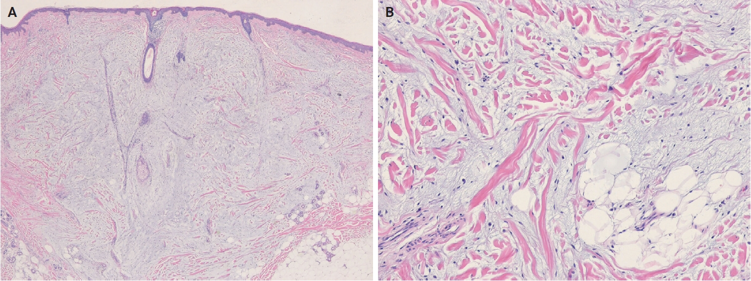
PDF - The 5th edition of the World Health Organization (WHO) classification of skin tumors introduces a dedicated chapter on cutaneous soft tissue tumors, providing a comprehensive, standardized reference with updated diagnostic criteria that directly inform routine dermatopathology practice and molecular diagnostics. This edition incorporates several key changes, including newly recognized entities such as EWSR1::SMAD3-rearranged fibroblastic tumor, neurotrophic tyrosine receptor kinase (NTRK)–rearranged spindle cell neoplasm, superficial CD34-positive fibroblastic tumor, and CRTC1::TRIM11 cutaneous tumor. Diagnostic terminology has also been refined; for example, the term ‘atypical intradermal smooth muscle neoplasm’ replaces ‘cutaneous leiomyosarcoma’ for lesions confined to the dermis, whereas the designation leiomyosarcoma is reserved for tumors with overt subcutaneous infiltration. In addition, epithelioid fibrous histiocytoma has been reassigned to the family of tumors of uncertain differentiation. This review summarizes the key updates and newly recognized entities in the chapter on cutaneous soft tissue tumors in the 5th edition of the WHO classification of skin tumors, emphasizing their clinicopathological and molecular implications.
- A comprehensive review of ossifying fibromyxoid tumor: insights into its clinical, pathological, and molecular landscape
- Kyriakos Chatzopoulos, Antonia Syrnioti, Mohamed Yakoub, Konstantinos Linos
- J Pathol Transl Med. 2026;60(1):6-19. Published online January 14, 2026
- DOI: https://doi.org/10.4132/jptm.2025.10.02

- 5,015 View
- 210 Download
-
Abstract
PDF
- Ossifying fibromyxoid tumor (OFMT) is a rare mesenchymal neoplasm first described in 1989. It typically arises in the superficial soft tissues of the extremities as a slow-growing, painless mass. Histologically, it is commonly characterized by a multilobular architecture composed of uniform epithelioid cells embedded in a fibromyxoid matrix, often surrounded by a rim of metaplastic bone. While classic cases are readily identifiable, the tumor's histopathological heterogeneity can mimic a range of benign and malignant neoplasms, posing significant diagnostic challenges. Molecularly, most OFMTs harbor PHF1 rearrangements, commonly involving fusion partners such as EP400, MEAF6, or TFE3. This review underscores the importance of an integrated diagnostic approach- incorporating histopathological, immunohistochemical, and molecular data- to accurately classify OFMT and distinguish it from its mimics. Expanding awareness of its morphologic and molecular spectrum is essential for precise diagnosis, optimal patient management, and a deeper understanding of this enigmatic neoplasm.
- Diagnostic conundrums of schwannomas: two cases highlighting morphological extremes and diagnostic challenges in biopsy specimens of soft tissue tumors
- Chankyung Kim, Yang-Guk Chung, Chan Kwon Jung
- J Pathol Transl Med. 2023;57(5):278-283. Published online August 24, 2023
- DOI: https://doi.org/10.4132/jptm.2023.07.13
- 9,154 View
- 274 Download
- 3 Web of Science
- 4 Crossref
-
Abstract
PDF
- Schwannomas are benign, slow-growing peripheral nerve sheath tumors commonly occurring in the head, neck, and flexor regions of the extremities. Although most schwannomas are easily diagnosable, their variable morphology can occasionally create difficulty in diagnosis. Reporting pathologists should be aware that schwannomas can exhibit a broad spectrum of morphological patterns. Clinical and radiological examinations can show correlation and should be performed, in conjunction with ancillary tests, when appropriate. Furthermore, deferring a definitive diagnosis until excision may be necessary for small biopsy specimens and frozen sections. This report underscores these challenges through examination of two unique schwannoma cases, one predominantly cellular and the other myxoid, both of which posed significant challenges in histological interpretation.
-
Citations
Citations to this article as recorded by
- Oral and maxillofacial schwannoma (OMSCH): An institutional study of 102 patients
Lingli Huang, Wenya Zhu, Qicheng Ye, Shengwen Liu, Hao Lu, Wenjun Yang, Wanlin Xu
Journal of Stomatology Oral and Maxillofacial Surgery.2026; 127(3): 102678. CrossRef - Plexiform Schwannoma Over the Anterior Chest Wall: A Clinicopathological Review
Debojyoti Sasmal, Saswata Barenya, Hinglaj Saha, Pankaj Kumar Halder
Amrita Journal of Medicine.2025; 21(2): 95. CrossRef - Giant Retroperitoneal Schwannoma: Case Report and Review of the Literature
Magdalena Alexieva, Evgeni V Mekov, Silvia Ivanova, Alexandrina Vlahova, Georgi Yankov
Cureus.2025;[Epub] CrossRef - Breast schwannoma: review of entity and differential diagnosis
Sandra Ixchel Sanchez, Ashley Cimino-Mathews
Journal of Pathology and Translational Medicine.2025; 59(6): 353. CrossRef
- Oral and maxillofacial schwannoma (OMSCH): An institutional study of 102 patients
- Imprint Cytology of Soft Tissue Myoepithelioma: A Case Study
- Seok Ju Park, Ae Ri Kim, Mi Jin Gu, Joon Hyuk Choi, Duk Seop Shin
- Korean J Pathol. 2013;47(3):299-303. Published online June 25, 2013
- DOI: https://doi.org/10.4132/KoreanJPathol.2013.47.3.299
- 11,414 View
- 53 Download
- 7 Crossref
-
Abstract
PDF
Soft tissue myoepithelioma is a rare neoplasm composed of myoepithelial cells. Here, we describe the cytologic features of soft tissue myoepithelioma arising on the right forearm in an 18-year-old man. The excised tumor (3.0×1.8×1.5 cm) was well-demarcated, yellow-gray, soft, and myxoid. The cytologic smears showed round to spindle, epithelioid, and plasmacytoid cells in the myxoid background. The nuclei were uniform, round to ovoid, with finely distributed chromatin and eosinophilic or pale cytoplasm. The tumor cells demonstrated immunoreactivity for cytokeratin (AE1/AE3), epithelial membrane antigen, S100 protein, and glial fibrillary acidic protein. Electron microscopy showed intermediate filaments, desmosomes, and basal lamina.
-
Citations
Citations to this article as recorded by- Myoepithelial tumors of soft tissue and bone in children and young adults: A clinicopathologic study of 40 cases occurring in patients ≤ 21 Years of age
Suzanna J. Logan, Carina A. Dehner, Fatimah I. Alruwaii, Nasir Ud Din, Damon R. Olson, Karen J. Fritchie, Gregory W. Charville, Melissa M. Blessing, Andrew L. Folpe
Human Pathology.2024; 149: 10. CrossRef - Fine-needle aspiration cytopathology of soft tissue myoepithelioma: an analysis of seven cases
Paul E. Wakely, Momin T. Siddiqui
Journal of the American Society of Cytopathology.2022; 11(1): 31. CrossRef - Cytology‐histology correlation of myoepithelial tumors harboring EWSR1‐POU5F1 fusions: A report of two cases
Ian A. Gelarden, Lucy Fu, Kai Lee Yap, Aida I. Richardson, Pauline M. Chou
Diagnostic Cytopathology.2022;[Epub] CrossRef - A case of myoepithelial carcinoma of the left shoulder
Shuhei ISHII, Noriyuki FURUTA, Kyoko KOMATSU, Yoshiya SUGIURA, Noriko MOTOI, Yutaka TAKAZAWA, Yuko SUGIYAMA, Yuichi ISHIKAWA
The Journal of the Japanese Society of Clinical Cytology.2018; 57(2): 129. CrossRef - Fine‐needle aspiration of soft tissue myoepithelioma
Gang Wang, Tracy Tucker, Tony L. Ng, Carlos F. Villamil, Malcolm M. Hayes
Diagnostic Cytopathology.2016; 44(2): 152. CrossRef - A case report of spindle cell myoepithelioma with extensive lipomatous metaplasia and thick collagen bundles in the submandibular gland
Mi Jung Kwon, Hye Jeong Kim, Bumjung Park, Seong Jin Cho, Hyung Sik Shin, Hye‐Rim Park, Soo Kee Min, Jinwon Seo, Kyueng‐Whan Min, Eun Sook Nam
Diagnostic Cytopathology.2016; 44(9): 764. CrossRef - Myoepithelioma of soft tissue, a case report
Hassania Ameurtesse, Leila Chbani, JM Coindre, Hinde Elfatemi, Toufik Harmouch, Afaf Amarti
Research.2014;[Epub] CrossRef
- Myoepithelial tumors of soft tissue and bone in children and young adults: A clinicopathologic study of 40 cases occurring in patients ≤ 21 Years of age
- A Proposal for Creating a Guideline for Cancer Registration of the Fibromatosis, PEComa Group, Malignant Lymphoma
In Situ and Dendritic Cell Tumors (III) - Changyoung Yoo, Chang Suk Kang, Yoon La Choi, Hye Yoon Kang, Jin Man Kim, Young Hye Koh, Joo Hee Lee, Seung Sook Lee, In Sun Kim, Dong Hoon Kim, Yong Ku Park, Jin Hee Sohn
- Korean J Pathol. 2012;46(5):436-442. Published online October 25, 2012
- DOI: https://doi.org/10.4132/KoreanJPathol.2012.46.5.436
- 10,781 View
- 55 Download
-
Abstract
PDF
Background Understanding the biologic behavior of a tumor is a prerequisite for tumor registration code assignment. The aim of this report was to propose appropriate behavior codes of the International Classification of Disease Oncology 3 (ICD-O3) to rare, yet pathologically interesting hematopoietic and soft tissue tumors.
Methods The Study Group for Hematopathology, the Bone and Soft Tissue Pathology Study Group, and the Cancer Registration Committee prepared the questionnaire containing provisional behavior codes of selected diseases.
Results In situ lesions of mantle cell and follicular lymphomas, dendritic cell tumors, and neoplasms with perivascular epithelioid cell differentiation (PEComa), not otherwise specified were classified as malignant (-/3). The fibromatosis group, with the exception of lipofibromatosis, was proposed as benign (-/0). Lipofibromatosis and several diseases that belong to the PEComa group were proposed as uncertain malignant potential (-/1). For the hematologic and soft tissue tumors, 274 and 288 members of the Korean Society of Pathologists, respectively, provided opinions through questionnaire, and most responders showed agreement with the provisional behavior code proposed.Conclusions The determination of behavior codes for the rare diseases described in this study, especially those of the PEComa group or malignant lymphoma, could be viewed as impractical and premature, but this study provides the basis for future research on this topic.
- A Soft Tissue Perineurioma and a Hybrid Tumor of Perineurioma and Schwannoma
- Ji Young Park, Nam Jo Park, Sang Pyo Kim, Kun Young Kwon, Sang Sook Lee
- Korean J Pathol. 2012;46(1):75-78. Published online February 23, 2012
- DOI: https://doi.org/10.4132/KoreanJPathol.2012.46.1.75
- 10,290 View
- 64 Download
- 15 Crossref
-
Abstract
PDF
Perineuriomas are composed of differentiated perineurial cells. Perineuriomas have been recently recognized by the immunoreactivity for epithelial membrane antigen (EMA). Microscopically, perineuriomas show proliferation of spindle cells with wavy nuclei and delicate elongated bipolar cytoplasmic processes. The tumor cells are usually negative for the S-100 protein. Ultrastructurally, perineurial cells reveal slender, nontapered processes containing pinocytic vesicles and discontinuous basal lamina. Interestingly, hybrid tumors of benign peripheral nerve sheath tumor (PNST) have been recently reported by using immunohistochemical and ultrastructural investigations. Herein, we report a case of soft tissue perineurioma arising in the skin of a 56-year-old female; another case of a hybrid tumor of perineurioma and schwannoma in the posterior mediastinum occurred in a 53-year-old male, which is the first case of the hybrid PNST tumor reported in Korea.
-
Citations
Citations to this article as recorded by- Plexiform Fibromyxoma of the Gastric Body: A Rare Benign Mesenchymal Tumor in an Unconventional Location
Anan Bseiso, Dalia Ibrahim
International Journal of Surgical Pathology.2026; 34(2): 513. CrossRef - Neurogenic tumours of the posterior mediastinum and differential diagnosis considerations
Michael A den Bakker, Annikka Weissferdt
Histopathology.2024; 84(1): 238. CrossRef - Hybrid tumors with perineurioma components: a systematic review of the literature and illustrative case
Karina A. Lenartowicz, Dileep D. Monie, Kimberly K. Amrami, Christopher J. Klein, Caterina Giannini, Robert J. Spinner
Acta Neurochirurgica.2022; 165(4): 935. CrossRef - Hybrid Schwannoma/Perineurioma: Morphologic Variations and Genetic Profiles
Takanori Hirose, Anna Kobayashi, Sumihito Nobusawa, Naoe Jimbo
Applied Immunohistochemistry & Molecular Morphology.2021; 29(6): 433. CrossRef - Mesenchymal Tumors of the Mediastinum: An Update on Diagnostic Approach
Joon Hyuk Choi, Jae Y. Ro
Advances in Anatomic Pathology.2021; 28(5): 351. CrossRef - Neurogenic Tumors of the Mediastinum
Erika F. Rodriguez, Robert Jones, Daniel Miller, Fausto J. Rodriguez
Seminars in Diagnostic Pathology.2020; 37(4): 179. CrossRef - A Rare Perineurioma/Granular Cell Tumor Hybrid Peripheral Nerve Sheath Tumor
Koorosh Haghayeghi, Gladys Telang, Sonja Chen, Jack Bevivino, Shamlal Mangray, Yiang Hui, Leslie Robinson-Bostom
The American Journal of Dermatopathology.2020; 42(10): 762. CrossRef - Hybrid peripheral nerve sheath tumors
Emine KILIÇ BAĞIR, Arbil AÇIKALIN, Gülfiliz GÖNLÜŞEN, Suzan ZORLUDEMİR, Mehmet Ali DEVECİ
Cukurova Medical Journal.2019; 44(3): 804. CrossRef - Primary intraosseous hybrid epithelioid schwannoma/perineurioma in the proximal tibia: a case report of benign hybrid neoplasm with local hypercellularity
Yuejiao Lang, Dawei Liu, Pei Xiang, Jilin Wang, Yang Li
Diagnostic Pathology.2019;[Epub] CrossRef - Hybrid peripheral nerve sheath tumors: report of five cases and detailed review of literature
Nasir Ud Din, Zubair Ahmad, Jamshid Abdul-Ghafar, Rashida Ahmed
BMC Cancer.2017;[Epub] CrossRef - Primary intraosseous hybrid nerve sheath tumor of femur: A hitherto undescribed occurrence in bone with secondary aneurysmal bone cyst formation resulting in pathological fracture
Louis Tsun Cheung Chow
Pathology - Research and Practice.2015; 211(5): 409. CrossRef - Mesenchymal tumours of the mediastinum—part II
Michael A. den Bakker, Alexander Marx, Kiyoshi Mukai, Philipp Ströbel
Virchows Archiv.2015; 467(5): 501. CrossRef - Primary pleural hybrid cellular schwannoma/perineurioma: A case report
Danny Soria-Céspedes, Carlos Robles-Vidal, Arturo Gómez-González, Rosalinda Peñaloza-Ramírez, Carlos Ortiz-Hidalgo
Respiratory Investigation.2014; 52(4): 269. CrossRef - Hybrid peripheral nerve sheath tumour with intermingled perineuriomatous and schwannomatous areas reflected in skin ultrasonography image
H. Saeki, K. Ito, Y. Nobeyama, T. Ishiji, M. Fukunaga, H. Nakagawa
Clinical and Experimental Dermatology.2014; 39(6): 747. CrossRef - Périneuriome extraneural des tissus mous localisé au nez
A. Zaouak, R. Benmously, M. Belhadj Salah, W. Koubaa, A. Debbiche, I. Mokhtar
Annales de Dermatologie et de Vénéréologie.2013; 140(8-9): 540. CrossRef
- Plexiform Fibromyxoma of the Gastric Body: A Rare Benign Mesenchymal Tumor in an Unconventional Location
- High-Grade Myxofibrosarcoma Showing Pleomorphic Hyalinizing Angiectatic Tumor-like Appearance: A Case Report.
- Mi Seon Kang, Hye Jung Jo, Sung Hee Son
- Korean J Pathol. 2011;45:S1-S4.
- DOI: https://doi.org/10.4132/KoreanJPathol.2011.45.S1.S1
- 4,437 View
- 22 Download
- 2 Crossref
-
Abstract
PDF
- Myxofibrosarcomas (MFSs), which consist of multiple nodules with a variable cellular population in a myxoid matrix, are primarily located in subcutaneous tissue. Pleomorphic hyalinizing angiectatic tumors (PHATs) are rare soft-tissue tumors characterized by a proliferation of highly pleomorphic spindle or polygonal cells and abundant ectatic blood vessels in cellular or myxoid stroma. We present here an unusual case of a high-grade MFS with a PHAT-like appearance. A 67-year-old man presented with an asymptomatic subcutaneous mass in the right forearm. The tumor had myxoid, hypo-, and hypercellular areas with highly pleomorphic spindle or polygonal tumor cells that showed frequent mitoses and nuclear pseudoinclusions. Foci of punctuate necrosis and inflammatory infiltration were present throughout the tumor, and abundant ectatic, thick-walled vessels containing blood clots were noted. The tumor cells were immunohistochemically positive for vimentin but negative for CD34, S-100 protein, smooth muscle actin, desmin, and bcl-2.
-
Citations
Citations to this article as recorded by- Primary Myxofibrosarcoma of the Left Breast in a Middle-Aged Woman: A Rare Mesenchymal Malignancy – A Case Report
Sadeka Sultana Ahmed, Mostofa Aziz Sumon, A. F. M. Kamal Uddin, Sharif Ahmed, Kamrun Nahar Liza
Journal of Clinical Practice and Medical Research.2026; 2(2): 12. CrossRef - Children's kinetic family drawings and their internalizing problem behaviors
Jin Kyung Kim, Joo Hyun Suh
The Arts in Psychotherapy.2013; 40(2): 206. CrossRef
- Primary Myxofibrosarcoma of the Left Breast in a Middle-Aged Woman: A Rare Mesenchymal Malignancy – A Case Report
- Calcifying Aponeurotic Fibroma of the Elbow: A Case Report.
- Mee Hye Oh, Eun Ah Jung, Ji Hye Lee, Hyun Deuk Cho, Jong Kyu Han, Yong Koo Park
- Korean J Pathol. 2009;43(1):75-78.
- DOI: https://doi.org/10.4132/KoreanJPathol.2009.43.1.75
- 4,742 View
- 35 Download
-
Abstract
PDF
- Calcifying aponeurotic fibroma is a rare soft tissue tumor that mostly occurs in the distal extremities of children and adolescents. We report here on a case of calcifying aponeurotic fibroma of the right elbow in an 8-year-old boy, and the tumor was diagnosed by surgical excision. The patient complained of painless swelling and mild limitation of the range of motion of the elbow joint. Radiologically, the mass was ill-defined and showed stippled calcification with shallow bony erosion. Microscopically, the tumor was composed of spindle cells with nodular deposits of hyalination and calcification, and these deposits were surrounded by palisading polygonal plump cells. Immunohistochemically, the tumor showed a diffuse positive expression for CD99 and negativity for smooth muscle actin, S-100 protein and CD34. The patient has been well with no signs of recurrence during the 42 months after surgery.
- Chondroblastoma-like Extraskeletal Chondroma: A case report.
- Jung Won Lee, Dae Su Kim, Mi Kyung Kim, Yeon Lim Suh
- Korean J Pathol. 1999;33(1):55-58.
- 2,377 View
- 49 Download
-
Abstract
PDF
- Extraskeletal chondromas are relatively uncommon benign cartilaginous tumors of the soft tissue and well known to pose a considerable diagnostic problem because of histological variations including the immature appearance of their tumor cells. Recently, we have experienced a case of extraskeletal chondroma mimicking benign chondroblastoma. The patient was a 47-year-old woman who complained of a painful subcutaneous swelling on the radial aspect of 4th proximal interphalangeal (PIP) joint in the left hand for 6 months. Radiologic examination of the 4th finger revealed a 1cm-sized soft tissue mass. Histologically, the tumor was characterized by a lobulated mass which was composed of dense proliferation of chondroblast-like cells admixed with a few multinucleated giant cells of osteoclastic type. However, there were focal areas of typical chondroma which showed lace-like intense calcification around the differentiated chondrocytes.
- Mesenchymal Chondrosarcoma Arising from Orbital Soft Tissue: A case report.
- Yu Mee Kang, Mi Kyung Jee, Seok Jin Gang, Byung Kee Kim, Sun Moo Kim
- Korean J Pathol. 1989;23(2):273-277.
- 2,152 View
- 15 Download
-
Abstract
PDF
- Orbital mesenchymal chondrosarcoma, first described by Luis et. al in 1971, is a very rare tumor of characteristic histologic features. A 21-year-woman was admitted with a 4-month histoiry of rapidly progressive proptosis and visual disturbance. Right orbital exenteration was performed under the clinical diagnosis of orbital calcifying tumor. Grossly, the tumor presented as a multibloblated, circumscribed mass that measures 5.5 cm in the greatest dimentsion. Cut sections resembled ordinary chonrosarcoma. Microscopically, the tumor was composed of undifferentiated mesenchymal cells, interspersed nodules of well differentated cartilagenous tissue, areas of gradual transition from undifferentiated mesenchymal cells to cartilage, and hemangiopericytoma-like areas. A brief summary of the histopathological aspect of this tumor and a review of literature are presented.
- Imprint Cytologic Feature of Extraskeletal Osteosarcoma: A Case Report.
- Mi Jin Gu, Young Kyung Bae, Mi Jin Kim, Joon Hyuk Choi, Won Hee Choi
- J Pathol Transl Med. 2000;11(1):59-63.
- 2,314 View
- 18 Download
-
Abstract
PDF
- Extraskeletal osteosarcoma is an uncommon tumor originated from soft tissue without evidence of skeletal involvement. It usually affects adults and its common locations are extremity, buttock, and retroperitoneum. Although the histologic feature of this tumor is well known, there have been few reports on the fine needle aspiration cytologic findings. We report the imprint cytologic feature of extraskeletal osteosarcoma. The patient was a 49-year-old man with a mass of the left anterior chest for 2 years. On the imprint preparation, the smears showed malignant round, polygonal or spindle cells with coarsely clumped chromatin and occasionally prominent nucleoli. The malignant cells occur singly, in clusters, or associated with amorphous eosinophilic osteoid. Mitotic figures are also seen.
- Dendritic Myxofibrolipoma.
- Sung Nam Kim, Kye Hyun Kwon, Yeon Lim Suh
- Korean J Pathol. 2001;35(5):447-450.
- 2,431 View
- 24 Download
-
Abstract
PDF
- Dendritic myxofibrolipoma is a recently described disease entity that represents a distinctive benign soft tissue neoplasm showing the combined features of spindle cell lipoma and the solitary fibrous tumor. Immunohistochemical stains reveal a strong positivity for vimentin, CD34 and bcl-2, which highlight the dendritic nature of the tumor cells by demonstrating slender complex cytoplasmic prolongations. There have been 12 cases of dendritic myxofibrolipomas reported in literature. In Korea, none of the cases have been described. We report such a case with a 28-year-old man who had a palpable subcutaneous mass on his right shoulder for 4 months. Grossly, the removed mass measured 11X7X5 cm and appeared to be a well-encapsulated, lipomatous tumor with marked myxoid appearance. Microscopically, this tumor consisted of spindle cells admixed with dense collagen fibers and mature adipocytes in abundant myxoid stroma with high vascularity. Immunohistochemically, the tumor cells were strongly reactive for vimentin and CD34 and weakly reactive for bcl-2, and negative for S-100 protein.
- Fibro-osseous Pseudotumor of the Digits: A case report .
- In Seo Park, Jee Young Han, Hye Seung Han, Young Bae Kim, Young Chae Chu
- Korean J Pathol. 1999;33(7):540-543.
- 2,856 View
- 45 Download
-
Abstract
PDF
- Fibro-osseous pseudotumor of the digits is a heterotopic ossification closely related to myositis ossificans and occurs in the subcutaneous tissue of the digits. This lesion is considered a reactive fibroblastic proliferation with metaplastic bone formation. We report a case of fibro-osseous pseudotumor of left index finger in a 28-year-old woman. She had had an ovoid smooth subcutaneous mass with tenderness on the left index finger for one month. In gross, the specimen consisted of a relatively circumscribed, rubbery soft mass with grayish white cut surface measuring 2.0 1.7 1.5 cm. Upon microscopic examination the lesion showed irregular multinodular growth with considerably variable cellularity. Because of the focal hypercellularity, cellular atypia, and increased mitotic activity this lesion may be confused with extraskeletal osteosarcoma or parosteal osteosarcoma. This rare lesion is curable by complete local excision.
- A Well-Differentiated Extraskeletal Osteosarcoma: A Case Report.
- Jung Wook Yang, Dae Hyun Song, Dong Hee Kim, Gyung Hyuck Ko
- Korean J Pathol. 2008;42(4):247-250.
- 2,234 View
- 16 Download
-
Abstract
PDF
- A well-differentiated extraskeletal osteosarcoma is very rare, and only seven cases have been reported in the English language clinical literature. We report an additional case of this rare tumor. A 71-year-old man had noticed a mass in the left pubic area for ten years. A CT scan demonstrated the presence of a lobulated calcified mass within the soft tissue. A 5 cm-sized well-circumscribed mass was excised. Histologically the tumor was composed of mature collagenous tissue and bony trabeculae rimmed by osteoblasts. After 43 months, the tumor recurred at the same site and was re-excised. The re-excised tumor contained focal areas of higher cellularity and atypism. We believe that this is the first case of well-differentiated extraskeletal osteosarcoma reported in Korea.
- Synovial Sarcoma with Massive Myxoid Feature: A Case Report.
- Joon Hyuk Choi, Young Ran Shim, Young Kyung Bae, Mi Jin Kim, Duk Seop Shin, Kil Ho Cho
- Korean J Pathol. 2005;39(4):273-277.
- 2,624 View
- 49 Download
-
Abstract
PDF
- Focal myxoid change in synovial sarcoma is not uncommon, although the presence of predominantly myxoid stroma is very rare. Recognition of synovial sarcomas with massive myxoid feature is important because these can easily be mistaken for other myxoid soft tissue neoplasms. We report a case of a synovial sarcoma with massive myxoid feature in the left thigh of a 54-year-old woman. Wide excision of an 8.5*7.0*5.0 cm, well-circumscribed and lobulated tumor was performed. The cut surface was gray, soft, and myxoid. Histological examination showed proliferation of spindle cells in the predominantly myxoid stroma. There were small areas with features more typical of synovial sarcoma, including uniform, spindled cells with fascicular growth patterns, collagenous stroma, mast cell infiltration, and hemangiopericytoma-like vascular patterns. Immunohistochemical examination showed focal positivity of the tumor cells for epithelial membrane antigen (EMA). Tumor cells were all negative for cytokeratin (AE1/AE3), cytokeratin 7, S-100 protein, smooth muscle actin, and desmin. Ultrastructurally, tumor cells showed desmosomes and microvilli. Our case underscores that, in order to make a correct diagnosis, immunohistochemical and ultrastructural examination is essential.
- Imprint Cytologic Features of Epithelioid Sarcoma: A Case Report.
- Young Ran Shim, Joon Hyuk Choi
- J Pathol Transl Med. 2004;15(1):65-69.
- 1,946 View
- 13 Download
-
Abstract
PDF
- Epithelioid sarcoma is a malignant soft tissue neoplasm with an uncertain histogenesis. We report the imprint cytologic features of epithelioid sarcoma in the left shoulder of a 29-year-old male patient. Imprint cytologic findings showed dissociated and loose aggregates of anaplastic epithelioid cells on the necrotic, bloody, and inflammatory background. Tumor cells were round to polygonal shaped. Tumor cells had vesicular nuclei with abundant cytoplasm. The nuclei were irregular in shape and often eccentrically located. Some tumor cells were oval to spindle shaped. Binucleated and multinucleated cells were found. Intracytoplasmic vacuoles were present. On immunohistochemical stain, the tumor cells were positive for epithelial membrane antigen, vimentin, and CD34.
- Pleomorphic Hyalinizing Angiectatic Tumor of Soft Parts: A Case Report.
- Young Chae Chu, Sun Keun Choi, In Suh Park, Hye Seung Han, Jee Young Han, Joon Mee Kim
- Korean J Pathol. 2002;36(3):195-198.
- 2,466 View
- 19 Download
-
Abstract
PDF
- A case of rare pleomorphic hyalinizing angiectatic tumor (PHAT) of soft parts is reported. A 35-year-old woman presented with a subcutaneous solid mass in the left inguinal area, which had been present for 3 months, was presented to us. The tumor was histologically characterized by sheets of mitotically inactive oval and pleomorphic cells, mono-and multinucleated giant cells, intranuclear cytoplasmic inclusions, and prominent clusters of thinwalled ectatic vessels with perivascular hyalinization. A focal hemangiopericytoma-like vascular pattern, pseudovascular spaces, stromal collagen with degenerative change and abundant mast cells were observed. The tumor cells were reactive for vimentin and CD34. This tumor shared several features with malignant fibrous histiocytoma, ancient schwannoma, giant cell angiofibroma, giant cell fibroblastoma and solitary fibrous tumor. The patient was well with no evidence of disease for 10 months.
- Cytologic Features of Soft Tissue Lesions.
- Soon Ae Oak, Gyung Yub Gong, Ghee Young Choe, Jur Yung Hch, Eun Sil Yu, In Chul Lee
- J Pathol Transl Med. 1995;6(1):27-35.
- 2,062 View
- 13 Download
-
Abstract
PDF
- We reviewed 93 cases of fine needle aspiration of skin\subcutaneous and soft tissue lesions with histologic confirmation. On the basis of the most prominent cytologic features, morphologic classification of the aspirates was done. Neoplastic lesions of soft tissue were categorized as ; round cell, spindle cell, polygonal cell, well-differentiated and myxoid tumor. This classification is convenient to recognize and categorize most soft tissue tumors.
- Fine Needle Aspiration Cytology of Parasitic Infestation in Soft Tissue.
- Kyeong Mee Park, Ill Hyang Ko
- J Pathol Transl Med. 1995;6(1):36-40.
- 1,857 View
- 13 Download
-
Abstract
PDF
- In the past, parasitic diseases were a major problem in public health in Korea. In recent years, however, nematodiasis that used be prevalent are no longer a serious problem. Instead some cestodiasis, particularly cysticercosis and sparganosis have become comparatively more important in recent years. Parasitic infestation of soft tissue is presented as a subcutaneous nodule or mass with nonspecific clinical manifestations.
- Fine Needle Aspiration Cytology of Peripheral Neuroepithelioma of Soft Tissue: Report of A Case.
- Yang Soon Park, Soon Ae Oak, Gyung Yub Gong, Ghee Young Choe, Joor Yung Huh, Eun Sil Yu, In Chul Lee
- J Pathol Transl Med. 1995;6(1):62-66.
- 2,019 View
- 14 Download
-
Abstract
PDF
- Peripheral neuroepithelioma (PNE) of soft tissue is a malignant neuroectodermal tumor arising from peripheral(nonautonomic) nerve. It may occur in both children and adults, and are highly aggressive neoplasms that rapidly give rise to metastatic disease and death. We exprienced a case of peripheral neuroepithelioma of soft tissue in the upper arm in a 18-year-old female. Cytologic features revealed small round cells with scanty cytoplasm occurring both singly and in clusters. The clusters frequently tended to form Homer-Wright rosettes. The cells had a round to oval nucleus with fine chromatin and inconspicuous nucleoli in a hemorrhagic background.
- Submandibular Soft Tissue Actinomycosis Diagnosed by Fine Needle Aspiration Cytology: A Case Report.
- Ho Jung Lee, Dong Hoon Kim, Won Mi Lee, Eun Kyung Kim, Jong Eun Joo
- J Pathol Transl Med. 2005;16(1):57-60.
- 2,314 View
- 31 Download
-
Abstract
PDF
- A patient with actinomyces infection of the submandibular soft tissue was diagnosed by fine needle aspiration cytology (FNAC). A 38-year-old woman presented with a right submandibular mass which slowly grew in size over one month. Clinically and radiologically, the lesion was considered as tuberculous lymphadenitis or cellulitis. The polymerase chain reaction for tuberculosis was done by aspirated specimen but the result was negative. The smears of aspiration cytology showed characteristic colonies(sulfur granules) of actinomyces in inflammatory background. After antibiotic therapy for eight months, the patient has been well, showing no detectable mass. This patient was simply and rapidly diagnosed by FNAC and can avoid unnecessary surgical biopsy.
- Fine Needle Aspiration Cytology of Alveolar Soft Part Sarcoma: A Case Report.
- Joon Hyuk Choi, Young Ran Shim, Duk Seop Shin, Kil Ho Cho
- J Pathol Transl Med. 2006;17(1):69-74.
- 2,254 View
- 33 Download
-
Abstract
PDF
- Alveolar soft part sarcoma (ASPS) is a rare soft tissue sarcoma, which occurs predominantly in adolescents and young adults. The cytological characteristics of this condition have been described only rarely in the literature. Here, we report a case of alveolar soft part sarcoma. A 28-year-old man presented with a mass in his right buttock, which had persisted for three years. The mass was subjected to a fine needle aspiration cytology (FNAC). The smears were cellular. The observed tumor cells were round or polygonal, and exhibited vesicular nuclei with prominent nucleoli and finely granular cytoplasm. Naked nuclei were frequently detected. Tumor cells were arranged singularly, but occasionally in a pseudoalveolar pattern.
- Composite Hemangioendothelioma: A Case Report.
- Young Chae Chu, Suk Jin Choi, In Suh Park, Lucia Kim, Jee Young Han, Joon Mee Kim
- Korean J Pathol. 2006;40(2):142-147.
- 2,520 View
- 42 Download
-
Abstract
PDF
- Composite hemangioendothelioma (CHE) is a recently described vascular tumor of low-grade malignancy. We report a case of CHE in an 18-year-old woman who presented with a 2-month history of an enlarging palpable mass in the left axilla. Grossly, the excised tumor was relatively circumscribed, nodular, firm, and soft. It measured 6.0 x 4.5 x 4.0 cm. The cut surface revealed a whitish gray solid area and a dark red to tan cystic area containing necrotic material. Histologically, the tumor demonstrated variably intermixed benign and malignant vascular components. The benign components showed features of an arteriovenous malformation, capillary hemangioma, spindle cell hemangioma and cavernous hemangioma. The malignant components were composed of areas resembling retiform hemangioendothelioma, epithelioid hemangioendothelioma, Kaposiform hemangioendothelioma, and angiosarcoma. The angiosarcoma component showed a mixed epithelioid and spindle shaped cell morphology with moderate differentiation. A nearly imperceptible transition between the benign and malignant components was noted.
- Cytological Features of Low Grade Fibromyxoid Sarcoma : Report of a Case with a Review of the Literature.
- Mi Seon Kwon
- J Pathol Transl Med. 2006;17(2):153-158.
- 2,609 View
- 35 Download
-
Abstract
PDF
- Low-grade fibromyxoid sarcoma (LGFMS) is a rare soft tissue tumor. There have been only a few prior fine-needle aspiration (FNA) cytological reports. Recognition of this tumor is important because of its potential for metastasis despite its indolent nature and its deceptively bland cytologic appearance. A 60-year-old male presented with a slowly growing mass in the left calf detected 10 years ago. The patient underwent surgical excision. FNA cytology was performed directly on the mass. The smears showed low cellularity composed of hypercellular tissue fragments, hypocellular loose aggregates, and stripped nuclei. The cytoplasm was seen as either collagenous material or very thin fibrillary collagen strands. Tumor cells had spindle, ovoid, or irregular nuclei, fine chromatin, and small nucleoli. Focally slight degree of nuclear pleomorphism is noted. There were no mitotic figures. Blood vessels were frequently seen. Immunocytochemically, tumor cells were negative for S-100 protein, desmin, smooth muscle actin, and CD34. The diagnosis of LGFMS is rarely possible by cytology alone; however, LGFMS should be included in the differential diagnosis of spindle-cell tumors consisting of hypercellular and hypocellular components with some capillary-sized vessels arising in the deep soft tissue of the lower extremities, particularly the thigh. The immunocytochemical findings are of help in the differential diagnosis.
- Imprint Cytology of Granular Cell Tumor: A Case Report.
- Woo Jung Sung, Joon Hyuk Choi
- J Pathol Transl Med. 2007;18(2):170-174.
- 2,268 View
- 21 Download
-
Abstract
PDF
- Granular cell tumor is a rare tumor of the soft tissue and this is characterized by proliferation of large cells with granular appearing eosinophilic cytoplasm. We report the imprint cytologic features of a case of granular cell tumor in the left calf of a 52-year-old woman. Microscopic examination showed moderate cellularity. The tumor cells were arranged both as single cells and in clusters. The cells were large polygonal-shaped and they had small round nuclei with finely granular chromatin and occasionally conspicuous nucleoli. The cytoplasm was abundant eosinophilic and granular. Naked nuclei and spindle-shaped tumor cells were occasionally noted. No mitosis and necrosis were present. The background showed cytoplasmic granular materials. The tumor cells showed positivity for S-100 protein. Ultrastructurally, abundant lysosomes were present in the cytoplasm of the tumor cells.
- Immunohistochemical Expression of p53 Protein, Estrogen and Progesterone Receptor in Soft Tissue Leiomyosarcoma and Its Significance.
- Byung Heon Kim
- Korean J Pathol. 1998;32(11):1015-1024.
- 2,247 View
- 10 Download
-
Abstract
- This study was carried out to evaluate the expressions of the p53 protein, the estrogen receptor (ER) and the progesterone receptor (PR), as well as the relationship between their expressions and clinicopathologic prognostic factors with storage duration of a paraffin block, and correlation between the p53 protein, the ER and the PR expressions in 29 cases of leiomyosarcoma of soft tissue. The expressions of the p53 protein, the ER and the PR were semiquantiatively analyzed in paraffin sections by the immunohistochemical method out of 29 cases the p53 protein, ER and PR were expressed in 9 (31.0%), 2 (6.9%) and 5 (17.2%), respectively. The expression of the p53 protein was not significantly associated with sex, age, anatomic site, tumor size, tumor depth, histological differentiation or mitotic rate (p>0.05), but statistically correlated to storage duration of a paraffin block (p=0.028). There was no significant relationship between the expression of the ER and all the clinocopathological prognostic factors with storage duration of a paraffin block (p>0.05). The expression of the PR was related to the histological differentiation (p=0.02), but not related to other clinicopathological prognostic parameters and storage duration of a paraffin block (p>0.05). The expression of the p53 protein and the PR had a significant relationship (p=0.022), but the expression of the p53 protein and the ER had no significant correlation. In conclusion, these results suggest that the expression of the p53 protein and the PR may play a role in development and growth of soft tissue leiomyosarcoma. Further studies of large numbers are needed to clarify the exact relationship between tumorigenesis and the p53 and the PR expressions in leiomyosarcoma of soft tissue.
 First
First Prev
Prev


