 E-submission
Search
- Page Path
- HOME > Search
- Breast schwannoma: review of entity and differential diagnosis
- Sandra Ixchel Sanchez, Ashley Cimino-Mathews
- J Pathol Transl Med. 2025;59(6):353-360. Published online November 3, 2025
- DOI: https://doi.org/10.4132/jptm.2025.08.12
- 4,109 View
- 179 Download
-
 Abstract
 PDF
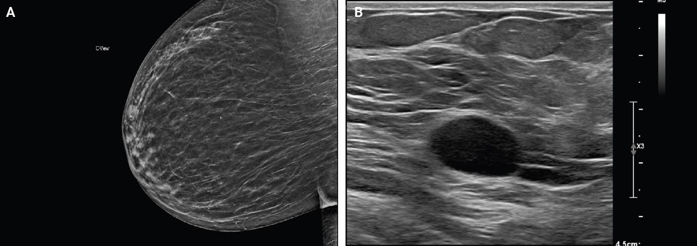
PDF - Schwannomas are benign peripheral nerve sheath tumors composed of Schwann cells, which uncommonly involve the breast. Most breast schwannomas are clinically present as a superficial palpable breast mass but may also be detected on screening mammography. Excision is the preferred treatment if symptomatic, and these are not known to recur. Histomorphology is similar to other anatomic sites: bland spindle cells with wavy nuclei, nuclear palisading (Verocay bodies), variably hypercellular (Antoni A) and hypocellular (Antoni B) areas, myxoid stroma, hyalinized vessels and variable cystic degeneration. Classic immunohistochemistry is diffuse and strong labeling for S100 and Sox10. Notable diagnostic pitfalls specific to the breast include myofibroblastoma, particularly the palisaded variant, and fascicular pseudoangiomatous stromal hyperplasia.
- A Rare Case of Aggressive Melanotic Schwannoma Occurred in Spinal Nerve of a 59-Year-Old Male
- Sung-eun Choi, Yoon Jin Cha, Jisup Kim, Hyunseo Cha, Jayeong Seo, Sung-Uk Kuh, Sung-Jun Kim, Se Hoon Kim
- J Pathol Transl Med. 2017;51(5):505-508. Published online April 4, 2017
- DOI: https://doi.org/10.4132/jptm.2017.01.04
- 15,593 View
- 222 Download
- 21 Web of Science
- 18 Crossref
-
Abstract
PDF
- Melanotic schwannoma (MS) is a rare variant of nerve sheath neoplasm that shows ultrastructural and immunophenotypical features of Schwann cells but also has cytoplasmic melanosomes and is reactive for melanocytic markers as well. Unlike conventional schwannoma, which is totally benign, MS has an unpredictable prognosis and is thought to have low-malignant potential. Herein, we present a rare case of recurrent MS in lumbar spine of a 59-year-old male.
-
Citations
Citations to this article as recorded by
- The “Pigmented Side” of Nerve Sheaths: Malignant Melanotic Nerve Sheath Tumor
Raduan Ahmed Franca, Rosa Maria Di Crescenzo, Lorenzo Ugga, Rosa Della Monica, Elena D'Avella
International Journal of Surgical Pathology.2025; 33(4): 1068. CrossRef - Case Report: Cutaneous melanocytic schwannoma with concomitant melanocytoma in a canine
Olwam H. Monakali, Nicolize O'Dell, Louise van der Weyden
Wellcome Open Research.2024; 8: 364. CrossRef - Intradural Melanotic Schwannoma of the Sacral Spine: An Illustrated Case Report of Diagnostic Conundrum
Jiunn-Kai Chong, Navneet Kumar Dubey, Wen-Cheng Lo
Reports.2024; 7(3): 56. CrossRef - Rare giant retroperitoneal melanotic schwannoma: a case report and literature review
Pan Chen, Junfeng Cheng, Lin Zhang
Frontiers in Oncology.2024;[Epub] CrossRef - A Rare Case of Melanotic Schwannoma Occurred Intraosseous of Sacrum: A Literature Review
Xiaobo Yan, Keyi Wang, Nong Lin, Xin Huang, YanBiao Fu, Zhaoming Ye
Orthopaedic Surgery.2023; 15(2): 655. CrossRef - Sporadic spinal psammomatous malignant melanotic nerve sheath tumor: A case report and literature review
Giulio Bonomo, Alessandro Gans, Elio Mazzapicchi, Emanuele Rubiu, Paolo Alimonti, Marica Eoli, Rosina Paterra, Bianca Pollo, Guglielmo Iess, Francesco Restelli, Jacopo Falco, Francesco Acerbi, Marco Paolo Schiariti, Paolo Ferroli, Morgan Broggi
Frontiers in Oncology.2023;[Epub] CrossRef - Case Report: Cutaneous melanocytic schwannoma with concomitant melanocytoma in a canine
Olwam H. Monakali, Nicolize O'Dell, Louise van der Weyden
Wellcome Open Research.2023; 8: 364. CrossRef - Fine‐needle aspiration cytology of melanotic schwannoma in the submandibular gland
Yu‐Hua Huang, Ying‐Chou Lu, Hsuan‐Ying Huang, Chien‐Chin Chen
Diagnostic Cytopathology.2021; 49(1): 142. CrossRef - Checkpoint inhibitors and radiotherapy in refractory malignant melanocytic schwannoma with Carney complex: first evidence of efficacy
Jyoti Bajpai, Akhil Kapoor, Rakesh Jalali, Mrinal M Gounder
BMJ Case Reports.2021; 14(5): e240296. CrossRef - 18F-FDG PET/CT imaging for aggressive melanotic schwannoma of the L3 spinal root
Xun-Ze Shen, Wei Wang, Zhou-Ye Luo
Medicine.2021; 100(8): e24803. CrossRef - Hemorrhagic spinal melanotic schwannoma presenting as acute chest pain: A case report and literature review
Dallas J. Soyland, Dylan R. Goehner, Kayla M. Hoerschgen, Troy D. Gust, Shawn M. Vuong
Surgical Neurology International.2021; 12: 164. CrossRef - Retroperitoneal Recurrence of Melanotic Schwannoma on 18F-FDG PET/CT
Xiangliu OuYang, Lichun Zheng, Xiaoming Zhang
Clinical Nuclear Medicine.2021; 46(12): 991. CrossRef - Schwannoma originating from the common iliac artery: a case report
Seung-Myoung Son, Chang Gok Woo
Journal of International Medical Research.2020;[Epub] CrossRef - Intraosseous Melanotic Schwannoma in the Sacrum Mimicking Primary Bone Tumor
Yoshitaka Nagashima, Yusuke Nishimura, Kaoru Eguchi, Takayuki Awaya, Satoshi Yoshikawa, Shoichi Haimoto, Toshihiko Wakabayashi, Masahito Hara
NMC Case Report Journal.2020; 7(3): 107. CrossRef - Extramedullary melanotic schwannoma recurrence in the cervical vertebral arch: a case report and review of the literature
Zongbin Hou, Teng Shi, Guangrun Li, Lin Tian, Xinna Li, Xiaoyang Liu
Journal of International Medical Research.2020;[Epub] CrossRef - Extramedullary malignant melanotic schwannoma of the spine: Case report and an up to date systematic review of the literature
Georgios Solomou, Adikarige Haritha Dulanka Silva, Adrianna Wong, Ute Pohl, Nikolaos Tzerakis
Annals of Medicine and Surgery.2020; 59: 217. CrossRef - Melanotic Schwannoma of the Vagina: A Report of a Very Rare Tumor and Review of the Literature
Kofi Effah, Stefan Seidl, Edith Gorges, Patrick Kafui Akakpo
Case Reports in Obstetrics and Gynecology.2019; 2019: 1. CrossRef - Melanotic Schwannomas Are Rarely Seen Pigmented Tumors with Unpredictable Prognosis and Challenging Diagnosis
Elif Keskin, Sumeyye Ekmekci, Ozgur Oztekin, Gulden Diniz
Case Reports in Pathology.2017; 2017: 1. CrossRef
- The “Pigmented Side” of Nerve Sheaths: Malignant Melanotic Nerve Sheath Tumor
- A Case of Type II Enteropathy-Associated T-Cell Lymphoma with Epstein-Barr Virus Positivity
- Min Jeong Song, Chan Sik Park, Hee Sang Hwang, Cheol Won Suh, Dok Hyun Yoon, Jooryung Huh
- Korean J Pathol. 2014;48(6):426-429. Published online December 31, 2014
- DOI: https://doi.org/10.4132/KoreanJPathol.2014.48.6.426
- 10,818 View
- 53 Download
- 6 Crossref
-
PDF
-
Citations
Citations to this article as recorded by- Various Endoscopic Features in Monomorphic Epitheliotropic Intestinal T-Cell Lymphoma
Yasuhiro Aoki, Tomohisa Sujino, Kaoru Takabayashi, Makoto Mutakuchi, Katsura Emoto, Naoki Hosoe, Haruhiko Ogata, Takanori Kanai
Case Reports in Gastroenterology.2021; 15(1): 312. CrossRef - A Case Report of Monomorphic Epithelial Intestinal T-Cell Lymphoma and Literature Review
Jing Deng, Yongguang Zou, Guangshu Li
International Journal of Clinical Medicine.2020; 11(02): 15. CrossRef - A viral map of gastrointestinal cancers
Natália R. Costa, Rui M. Gil da Costa, Rui Medeiros
Life Sciences.2018; 199: 188. CrossRef - Type II Enteropathy-Associated T-cell Lymphoma: A Rare Report from Iran
Neda Nozari
Middle East Journal of Digestive Diseases.2017; 9(1): 55. CrossRef - Unusual Cause of Dysphagia
Shahram Agah, Ramak Ghavam, Ahmad Darvishi Zeidabadi, Arash Sarveazad
Middle East Journal of Digestive Diseases.2017; 9(1): 58. CrossRef - Multiple lesions of gastrointestinal tract invasion by monomorphic epitheliotropic intestinal T-cell lymphoma, accompanied by duodenal and intestinal enteropathy-like lesions and microscopic lymphocytic proctocolitis: a case series
Hideki Ishibashi, Satoshi Nimura, Yoshiyuki Kayashima, Yasushi Takamatsu, Kunihiko Aoyagi, Naohiko Harada, Masanori Kadowaki, Takihiko Kamio, Shotaro Sakisaka, Morishige Takeshita
Diagnostic Pathology.2016;[Epub] CrossRef
- Various Endoscopic Features in Monomorphic Epitheliotropic Intestinal T-Cell Lymphoma
- Bronchial Schwannomas: Clinicopathologic Analysis of 7 Cases
- Yoon Yang Jung, Min Eui Hong, Joungho Han, Tae Sung Kim, Jhingook Kim, Young-Mog Shim, Hojoong Kim
- Korean J Pathol. 2013;47(4):326-331. Published online August 26, 2013
- DOI: https://doi.org/10.4132/KoreanJPathol.2013.47.4.326
- 10,290 View
- 68 Download
- 26 Crossref
-
Abstract
PDF
Background It has long been recognized that bronchial schwannomas are extremely rare. As such, diagnosing tumors in this extraordinary location can sometimes be problematic.
Methods We reviewed seven cases of bronchoscopically or surgically resected endobronchial schwannomas and evaluated their clinical and pathologic features.
Results The present study included five female and two male patients, with ages ranging from 16 to 81 years (mean age, 44.9 years). The clinical presentation varied according to tumor size and location. Patients with more centrally (trachea or main bronchus) located tumors experienced respiratory symptoms (80%) more often than patients with more peripherally (lobar or segmental bronchus) located tumors (0%). Histologically, the tumors were composed of spindle cells that stained with S100 protein. Some of the tumors showed typical Antoni A areas with Verocay body formation. Five of six patients (83.3%) underwent complete tumor removal by rigid bronchoscopy.
Conclusions Pathologists should consider endobronchial schwannoma in the differential diagnosis of a spindle cell tumor involving the bronchus. Additionally, our results showed that rigid bronchoscopy is an effective tool for tumor removal in endobronchial schwannoma patients.
-
Citations
Citations to this article as recorded by- Rare tracheal schwannoma in a child: successful resection via cervical incision
Hariharan Krishnamurthi, Vijayakumar Raju, Naveen Srinivasan, Kaushik Jothinath, Karthik Babu Murugesan, Antony Terance Benjamin, Nagarajan Muthialu
Indian Journal of Thoracic and Cardiovascular Surgery.2026; 42(3): 416. CrossRef - Tracheal schwannoma presenting as pneumonia: A rare case report in an adolescent
Wanyan Xiong, Zhixuan Deng
Respiratory Medicine Case Reports.2026; 60: 102398. CrossRef - Tracheal schwannoma: A pseudo-asthmatic syndrome with successful laser Nd-YAG resection as first-line therapy
Ranim Nakhal, Raneem Ahmad, Bassel Ibrahim, Sultaneh Haddad, Arabi Abbas, Nizar Abbas
International Journal of Surgery Case Reports.2025;[Epub] CrossRef - CT-Occult Primary Benign Tracheobronchial Neoplasms: A Single-Center 40-Case Clinicopathologic Series
Qiliang Liu, Yan Hu, Minhui Mei, Xuan Wang, Jun Li, Chao Quan, Peng Liu
International Journal of General Medicine.2025; Volume 18: 5765. CrossRef - Outcomes of Different Surgical Approaches in Tracheal Schwannoma: A Narrative Review
Hassan Fahmi Alkhars, Mohammad Almayouf, Majed Albarrak, Faisal Alzahrani, Naif Fnais, Mohammed Alessa, Khalid Alqahtani, Saleh Aldhahri
Ear, Nose & Throat Journal.2025;[Epub] CrossRef - Video-assisted thoracic surgery for an endobronchial ancient schwannoma obstructing the left main bronchus
Jiyeon Kang, Yeon Soo Kim, Ji-Ye Kim
Journal of Surgical Case Reports.2024;[Epub] CrossRef - Interventional treatment of right lower lobe bronchial schwannoma under bronchoscopy: A case report and literature review
Xiyue Li, Ximiao Yu, Ruiqi Luo, Xun Wang
Science Progress.2024;[Epub] CrossRef - Two cases of large tracheobronchial schwannomas completely resected by rigid bronchoscopy with multiple instruments
Changhwan Kim, Hae‐Seong Nam, Yousang Ko
Respirology Case Reports.2023;[Epub] CrossRef - Tracheobronchial schwannoma: a case report and literature review
Guo Lina, Hou Pengguo, Xiao Zhihua, Wang Jianxin, Bai Baoqin, Zhang Mingyue, Sun Junping
Journal of International Medical Research.2023;[Epub] CrossRef - Malignant and Benign Tracheobronchial Neoplasms: Comprehensive Review with Radiologic, Bronchoscopic, and Pathologic Correlation
Francis Girvin, Alexander Phan, Sharon Steinberger, Eugene Shostak, Jamie Bessich, Fang Zhou, Alain Borczuk, Geraldine Brusca-Augello, Margaret Goldberg, Joanna Escalon
RadioGraphics.2023;[Epub] CrossRef - Clinicopathological Characteristics and Pathogenesis of Granular Cell Tumours of the Airways
Jesús Machuca-Aguado, Fernando Cózar-Bernal, Enrique Rodríguez-Zarco, Juan José Ríos-Martin, Miguel Ángel Idoate Gastearena
Journal of Bronchology & Interventional Pulmonology.2023; 30(4): 390. CrossRef - Treatment of primary tracheal schwannoma with endoscopic resection: A case report
Yong-Shuai Shen, Xiang-Dong Tian, Yi Pan, Hua Li
World Journal of Clinical Cases.2022; 10(28): 10279. CrossRef - Primary bronchial schwannoma: A case report
Yosuke Aoyama, Atsushi Miyamoto, Takeshi Fujii, Sakashi Fujimori, Meiyo Tamaoka, Daiya Takai
Medicine.2022; 101(40): e31062. CrossRef - Endobronchial schwannoma in adult: A case report
Touil Imen, Boudaya Mohamed Sadok, Aloui Raoudha, Souhir Ksissa, Brahem Yosra, Ben Attig Yosr, Ksontini Meriem, Bouchareb Soumaya, Keskes Boudawara Nadia, Boussoffara Leila, Knani Jalel
Respiratory Medicine Case Reports.2021; 33: 101396. CrossRef - Primary intratracheal schwannoma misdiagnosed as severe asthma in an adolescent: A case report
Hui-Rong Huang, Pei-Qiang Li, Yi-Xin Wan
World Journal of Clinical Cases.2021; 9(17): 4388. CrossRef - PD‐1/PD‐L1 negative schwannoma mimicking obstructive bronchial malignancy: A case report
Daibing Zhou, Xiaoyan Xing, Jie Fan, Youzhi Zhang, Jie Liu, Yi Gong
Thoracic Cancer.2020; 11(8): 2335. CrossRef - Case report: A tracheobronchial schwannoma in a child
Li Zhang, Wen Tang, Qing-Shan Hong, Pei-feng Lv, Kui-Ming Jiang, Rui Du
Respiratory Medicine Case Reports.2020; 30: 101047. CrossRef - Recurrent transmural tracheal schwannoma resected by video-assisted thoracoscopic window resection
Huiguo Chen, Kai Zhang, Mingjun Bai, Haifeng Li, Jian Zhang, Lijia Gu, Weibin Wu
Medicine.2019; 98(51): e18180. CrossRef - Primary intratracheal schwannoma resected during bronchoscopy using argon plasma coagulation
Purva V Sharma, Yash B Jobanputra, Tatiana Perdomo Miquel, J Ryan Schroeder, Adam Wellikoff
BMJ Case Reports.2018; 2018: bcr-2018-225140. CrossRef - Dumbbell posterior mediastinal schwannoma invading trachea: Multidisciplinary management – weight off the chest
Abhijeet Singh, VallandramamR Pattabhiraman, Arjun Srinivasan, Sivaramakrishnan Mahadevan
Lung India.2018; 35(3): 269. CrossRef - Primary tracheal schwannoma a review of a rare entity: current understanding of management and followup
Shadi Hamouri, Nathan M. Novotny
Journal of Cardiothoracic Surgery.2017;[Epub] CrossRef - A Case of Primary Tracheal Schwannoma
Sung Min Choi, Ji Hong You, Sang Bae Lee, Seong Han Kim, Yon Soo Kim
Kosin Medical Journal.2017; 32(2): 258. CrossRef - Bronchial schwannoma: a singular lesion as a cause of obstructive pneumonia
Rui Caetano Oliveira, Tiago Nogueira, Vítor Sousa, Lina Carvalho
BMJ Case Reports.2016; 2016: bcr2016217300. CrossRef - Bronchoscopic and surgical management of rare endobronchial tumors
Kamlesh Pandey, Preyas J. Vaidya, Arvind H. Kate, Vinod B. Chavhan, Pruthviraj Jaybhaye, Kamlakar Patole, Ramakant K. Deshpande, Prashant N. Chhajed
Journal of Cancer Research and Therapeutics.2016; 12(2): 1093. CrossRef - Endobronchial Neurilemmoma Mimicking a Bronchial Polyp
Ryoung Eun Ko, Seung Yong Park, Yeong Hun Choe, So Ri Kim, Heung Bum Lee, Yong Chul Lee, Seoung Ju Park
Soonchunhyang Medical Science.2015; 21(2): 176. CrossRef - Optimal treatment for primary benign intratracheal schwannoma: A case report and review of the literature
XIAHUI GE, FENGFENG HAN, WENBIN GUAN, JINYUAN SUN, XUEJUN GUO
Oncology Letters.2015; 10(4): 2273. CrossRef
- Rare tracheal schwannoma in a child: successful resection via cervical incision
- Use of Calretinin, CD56, and CD34 for Differential Diagnosis of Schwannoma and Neurofibroma.
- Ji Young Park, Hoon Park, Nam Jo Park, June Sik Park, Hyun Jung Sung, Sang Sook Lee
- Korean J Pathol. 2011;45(1):30-35.
- DOI: https://doi.org/10.4132/KoreanJPathol.2011.45.1.30
- 6,790 View
- 188 Download
- 22 Crossref
-
Abstract
PDF
- BACKGROUND
It is important to differentiate between schwannomas and neurofibromas for the cases in which the histopathologic features overlap. Depending on the tumor type, surgeons can decide on a treatment method and whether to preserve or sacrifice the nerve; the possibility of malignant transformation in the case of neurofibromas also needs to be considered.
METHODS
We studied 101 cases of schwannoma and 103 cases of neurofibroma. All the hematoxylin and eosin slides for these cases were reviewed, and tissue microarrays were prepared from the representative areas. Immunohistochemical analysis was performed using antibodies for S-100 protein, calretinin, CD56 and CD34.
RESULTS
All the tumors except 3 neurofibromas were positive for the S-100 protein. Calretinin was found in 26.7% of the schwannomas (27/101), but it was not found in any of the neurofibromas. CD56 was positive in 77.2% of the schwannomas (78/101) and in 9.8% of the neurofibromas (10/102). CD34 was positive in 42.5% of the schwannomas (43/101) and in 80.2% of the neurofibromas (81/101). Statistically, calretinin was significantly specific for schwannomas (p<0.001) and CD56 was also sensitive for these tumors (p<0.001). On the other hand, a CD34 expression seemed highly sensitive (p<0.001) for neurofibromas.
CONCLUSIONS
We concluded that combined immunohistochemical analysis for calretinin, CD56, and CD34 may be very useful for differentiating schwannomas from neurofibromas. -
Citations
Citations to this article as recorded by- Histomorphological Spectrum and Diagnostic Utility of S100, SOX10, CD34, Calretinin, and Ki67 in the Evaluation of Schwannomas: A Retrospective Study of 26 Cases in a Tertiary Health Institute, Zaria-Nigeria
Zainab Ali Adamu, Mikhail O. Buhari, Abdullahi Mohammed
Journal of West African College of Surgeons.2026; 16(2): 119. CrossRef - Microcystic Pseudoglandular Cutaneous Neurofibroma: The First Japanese Case of a Rare Neurofibroma Variant
Shinichi Nakazato, Yasuyuki Fujita, Takashi Anan
The Journal of Dermatology.2025;[Epub] CrossRef - A rare case of giant intercostal nerve schwannoma
Bandari A. Ahmed, Ashley I. Simpson
International Surgery Journal.2025; 12(7): 1142. CrossRef - HISTOMORPHOLOGICAL SPECTRUM AND IMMUNOHISTOCHEMICAL EXPRESSION OF S100 AND CD56 AMONG TUMORS OF PERIPHERAL NERVES-A CROSS-SECTIONAL STUDY
KARTHIK SIGAMANI, SHOBANA B, NAYANA CHANDRAN
Asian Journal of Pharmaceutical and Clinical Research.2025; : 142. CrossRef - The skin's secret script: fingerprints of CD34 and the dolphin dance of Schwann cells
Vijay Joshi, Ketki Bhoite, Vidya Kharkar, Rajiv Joshi, Surupa Das
International Journal of Research in Dermatology.2025; 11(6): 541. CrossRef - Breast schwannoma: review of entity and differential diagnosis
Sandra Ixchel Sanchez, Ashley Cimino-Mathews
Journal of Pathology and Translational Medicine.2025; 59(6): 353. CrossRef - Primary Pulmonary Neurofibroma: Diagnostic Approach to a Common Tumor at an Uncommon Site – A Case Report
Rakesh Kumar Gupta, Bhoomika Kaushik, Shaurya Vijayran, Ranganath Ganga, Mudalsha Ravina
SN Comprehensive Clinical Medicine.2025;[Epub] CrossRef - Ancient cervical vagal schwannoma and an interposition great auricular to vagus nerve graft
Nicholas Figaro, Rickhi Ramoutar, David Richards, Rodolfo Arozarena, Mala Geelal, Solaiman Juman
Edorium Journal of Otolaryngology.2024; 5(2): 1. CrossRef - Lung schwannomas, an unusual entity
Nazia M. Walvir, Rumana H. Makhdoomi, Meesa Zargar, Aiffa Aiman, Shadab Maqsood
Lung India.2023; 40(1): 70. CrossRef - A Spectrum of Histomorphological and Immunohistochemical Expression Profiles of S-100, CD56 and Calretinin in Benign Peripheral Nerve Sheath Tumours
Poornima Jaiswal, Anand CD, Jaison Jacob John
Cureus.2023;[Epub] CrossRef - An unusual diffuse CD34 staining in an olfactory groove cellular schwannoma: Case report
Marios Theologou, Jorge D. Perez Ruiz, Panagiotis Varoutis, Nicolaos Flaris, Nikolaos `Skoulios
Archivos de Neurociencias.2023;[Epub] CrossRef - An unusual diffuse CD34 staining in an olfactory groove cellular schwannoma: Case report
Marios Theologou, Jorge D. Perez Ruiz, Panagiotis Varoutis, Nicolaos Flaris, Nikolaos `Skoulios
Archivos de Neurociencias.2023;[Epub] CrossRef - Intranodal Neurofibroma: A Case Report and Literature Review
Steven H. Adams, Tara L. Huston, Daniel Lozeau
The American Journal of Dermatopathology.2022; 44(4): 306. CrossRef - A rare case of pseudoglandular schwannoma
Fadime Eda GÖKALP SATICI, Hamide SAYAR
Journal of Surgery and Medicine.2022; 6(4): 1. CrossRef - Gastric Schwannoma as an Important and Infrequent Differential Diagnosis of Gastric Mesenchymal Tumours: A Case Report and Review of Literature
Abdalla Saad Abdalla Al-Zawi, Salma Lahmadi, Saman Jalilzadeh Afshari, Ipshita Kak, Salem Alowami
Cureus.2022;[Epub] CrossRef - Spindle cell proliferations of the sigmoid colon, rectum and anus: a review with emphasis on perineurioma
Patrice Grech, John B Schofield
Histopathology.2020; 76(3): 342. CrossRef - Large retroperitoneal schwannoma: a rare cause of chronic back pain
Milan Radojkovic, Dragan Mihailovic, Miroslav Stojanovic, Danijela Radojković
Journal of International Medical Research.2018; 46(8): 3404. CrossRef - Nasal Septal Schwannoma
Hyun Jin Min, Seok Chan Hong, Kyung Soo Kim
Journal of Craniofacial Surgery.2017; 28(1): e97. CrossRef - Neurofibroma of the Colon: A Diagnostic Mimicker of Gastrointestinal Stromal Tumor
Soomin Ahn, Choon Sik Chung, Kyoung-Mee Kim
Case Reports in Gastroenterology.2016; 10(3): 674. CrossRef - Solitary Epibulbar Neurofibroma in Older Adult Patients
Thais Shiota Tanaka, Victor M. Elner, Hakan Demirci
Cornea.2015; 34(4): 475. CrossRef - Syncytial nuclear aggregates in normal placenta show increased nuclear condensation, but apoptosis and cytoskeletal redistribution are uncommon
S.J. Coleman, L. Gerza, C.J.P. Jones, C.P. Sibley, J.D. Aplin, A.E.P. Heazell
Placenta.2013; 34(5): 449. CrossRef - Analysis of syncytial nuclear aggregates in preeclampsia shows increased sectioning artefacts and decreased inter-villous bridges compared to healthy placentas
S.J. Calvert, C.J.P. Jones, C.P. Sibley, J.D. Aplin, A.E.P. Heazell
Placenta.2013; 34(12): 1251. CrossRef
- Histomorphological Spectrum and Diagnostic Utility of S100, SOX10, CD34, Calretinin, and Ki67 in the Evaluation of Schwannomas: A Retrospective Study of 26 Cases in a Tertiary Health Institute, Zaria-Nigeria
- Schwannoma Arising in a Lymph Node : A Brief Case Report.
- Ji Han Jung, Jinyoung Yoo, Seok Jin Kang, Kyo Young Lee
- Korean J Pathol. 2009;43(3):271-273.
- DOI: https://doi.org/10.4132/KoreanJPathol.2009.43.3.271
- 5,069 View
- 49 Download
- 7 Crossref
-
Abstract
PDF
- Intranodal schwannomas are extremely rare and only three cases have currently been reported in the English language literature. We report here on a case of a schwannoma that arose in a retroperitoneal lymph node. A 59-year-old male patient had experienced abdominal discomfort for two months. An abdominal CT scan demonstrated a heterogeneous density mass in the retroperitoneum. Histological examination of the mass identified it as a lymph node due to the presence of a peripheral rim of compressed lymphoid tissue that contained a well-demarcated benign spindle cell tumor in its center. The spindle cells were positive for S-100 protein, and they were negative for smooth muscle actin, desmin, and CD 34. Although an intranodal schwannoma is histologically benign, it is important to distinguish this lesion from an intranodal metastasis of a spindle cell tumor and other common benign spindle cell tumors that can arise in a lymph node.
-
Citations
Citations to this article as recorded by- Cervical Intranodal Schwannoma and Its Malignant Transformation: A Case Report With Literature Review
Shahab Hussain, Zia Ullah Khan, Nazneen Liaqat, Syed Wajihullah Shah, Kainat Khan, Jibran Ikram
Clinical Case Reports.2026;[Epub] CrossRef - Submandibular nodal schwannoma: where did it come from?
Siti Farhana Abdul Razak, Hardip Singh Gendeh, Anuar Idris
BMJ Case Reports.2023; 16(8): e253868. CrossRef - Intranodal Neurofibroma: A Case Report and Literature Review
Steven H. Adams, Tara L. Huston, Daniel Lozeau
The American Journal of Dermatopathology.2022; 44(4): 306. CrossRef - Cervical Lymph Node Schwannoma—An Unexpected Diagnosis
Catarina Falcão Silvestre, Joana Almeida Tavares, Dolores López-Presa, Vanessa Rebelo dos Santos, José Rocha, Maria João Bugalho
Clinical Pathology.2019;[Epub] CrossRef - Mesenteric intranodal schwannoma: uncommon case of neurogenic benign tumor
Adrian Medina-Gallardo, Yuhamy Curbelo-Peña, Jose Molinero-Polo, Maria Saladich-Cubero, Xavier De Castro-Gutierrez, Helena Vallverdú-Cartie
Journal of Surgical Case Reports.2017;[Epub] CrossRef - Intranodal Hybrid Benign Nerve Sheath Tumor
Brian D. Hayes, Maureen J. O'Sullivan
Pediatric and Developmental Pathology.2011; 14(4): 313. CrossRef - Intranodal Schwannoma Mimicking a Gastrointestinal Stromal Tumor of the Stomach: A Case Report
Kyung Bum Nam, Sook Namkung, Heung Cheol Kim, Hae Sung Kim, Byoung Yoon Ryu, Young Hee Choi
Journal of the Korean Society of Radiology.2011; 65(4): 395. CrossRef
- Cervical Intranodal Schwannoma and Its Malignant Transformation: A Case Report With Literature Review
- Cytologic Findings of Fine Needle Aspiration Biopsy of 23 Schwannomas.
- Sunhee Chang, Mee Joo, Hanseong Kim
- J Pathol Transl Med. 2008;19(1):41-46.
- DOI: https://doi.org/10.3338/kjc.2008.19.1.41
- 3,764 View
- 20 Download
- 1 Crossref
-
Abstract
PDF
- In an attempt to better define the cytologic characteristics of schwannomas, we have reviewed aspirates and corresponding histologic sections from 23 schwannomas. Of this number, the original cytologic diagnoses were: schwannoma in 14 cases (61%), benign soft tissue tumor in 2 cases (9%), and insufficient specimen in 7 cases (30%). The cytologic findings common to all cases of schwannoma included fragments of tightly cohesive fascicles with variable cellularity and corresponding Antoni type A area. The Antoni type B area, consisting of scattered spindle cells and some histiocytes and lymphocytes against a myxoid background, was seen in 14 cases. Fibrillary stroma was seen in 12 cases. The tumor cells had spindle- or oval-shaped nuclei, with pointed ends and indistinct cell borders. Nuclear palisading was seen in 10 cases, and distinctive Verocay bodies were seen in 5 cases. In ancient schwannomas, there were no Verocay bodies. Most schwannomas have distinct cytomorphologic features that allow correct diagnosis. The major problem with fine needle aspiration cytology of these tumors is the high frequency of poor cellularity, particularly in lesions with cystic degeneration. Of 7 cases with insufficient specimen, 4 showed marked cystic changes and 1 showed marked hyaline changes on histologic sections. In conclusion, we believe that if cytopathologist reminds the situation such as cystic degeneration or hyaline degeneration, the correct diagnosis of the schwannoma will be easily made.
-
Citations
Citations to this article as recorded by- Multiple Cervical Schwannomas Mimicking Metastatic Lymph Nodes from Papillary Thyroid Cancer
Ji-Sun Kim, Chang-Young Yoo, Rae-Hyung Kim, Jung-Hae Cho
Journal of Korean Thyroid Association.2014; 7(1): 102. CrossRef
- Multiple Cervical Schwannomas Mimicking Metastatic Lymph Nodes from Papillary Thyroid Cancer
- Fine Needle Aspiration Cytologic Features of Schwannomas .
- Sun Young Kim, Hae Joo Nam, Jung Hee Kim, Young Rok Cho, Yu Na Kang, Sang Sook Lee
- J Pathol Transl Med. 1999;10(1):55-60.
- 6,310 View
- 242 Download
-
Abstract
PDF
- Schwannoma is a benign encapsulated nerve sheath tumor and is histologically characterized by a mixture of Antoni type A and B tissue. A preoperative diagnosis of schwannoma by fine needle aspiration cytology or by any other means is important to preserve clinically important nerves. Therefore, it is necessary to make a specific cytological diagnosis of nerve sheath tumor. However, there are a few reports regarding cytological features of schwannoma in Korea. We experienced seven cases of solitary schwannoma and here we report their characteristic cytological features with a review of literatures. The characteristic features of schwannoma on cytology were the presence of both Antoni type A and B tissue. The cytologic findings common to all cases of schwannoma generally corresponded to the histologic findings of Antoni type A tissue, consisting of fragments of tightly cohesive fascicles with variable cellularity. Dense fibrillary substances were found, along with palisading nuclei and Verocay bodies. Individual tumor cells consisted of cohesive cells having spindle or oval nuclei, with pointed ends and indistinct cell borders. Variation in nuclear size and shape was also present. The Antoni type B consisted of scattered wavy or short spindle cells and some histiocytes and lymphocytes in the abundant myxoid background with formation of microcysts. Immunohistochemistry for S-100 protein revealed a uniformly strong positive reaction and was helpful to make more accurate diagnosis of schwannoma.
- Cytologic Findings of Fine Needle Aspiration Biopsy of Ancient Schwannoma.
- Hee Jae Joo, Kwang Gil Lee
- J Pathol Transl Med. 1990;1(2):158-163.
- 3,227 View
- 40 Download
-
Abstract
PDF
- Ancient (degenerated) schwannomas are benign tumors that display pronounced degenerative changes including cyst formation, calcification, hemorrhage, and hyalinization. The tumors are usually infiltrated by large numbers of inflammatory cells such as neutrophils, lymphocytes, siderophages, and histiocytes. They are located in deep locations such as the retroperitoneum and must be differentiated from malignant soft tissue tumors because spindle cells with nuclear atypia may be present in the fine needle aspiration biopsy. The cytologic findings of two cases of ancient schwannoma occurring in posterior mediastinum and retroperitoneum, respectively, are described. Computerized tomographic findings of both cases showed changes of cystic degeneration or necrosis. A case occurred in retroperitoneum revealed features of destruction of vertebral body to suggest a malignant soft tussue tumor radiologically. The cytologic findings of aspiration biopsy of both tumors revealed that the tumor cells were spindle in shape with elongated nuclei. Some of tumor cells were arranged in a palisading fashion. Cell structures that resemble the Verocay bodies were observed. Some of the tumor cells showed pleomorphic bizarre nuclei, but no mitotic activity or chromatin clumping was seen. Inflammatory cells, siderophages and histiocytes were scattered in fibrillar material. These cytologic findings are important in the diagnosis of ancient schwannoma and in the differentiation of this tumor from the malignant spindle cell tumor.
- Cellular Schwannoma Arising in a Facial Nerve.
- Mee Joo, Hye Sung Kim, Yun Kyung Kang, Hye Kyung Lee, Jae Young Park
- Korean J Pathol. 1997;31(7):688-691.
- 2,163 View
- 19 Download
-
Abstract
PDF
- Cellular schwaninoma is a variant of schwannoma, which is characterized by predominance of cellular Antoni A area, presence of mitotic activity, nuclear hyperchromasia, pleomorphism, and absence of Verocay body. These pathologic features often prompted a misdiagnosis of malignancy. However, the clinical outcome has indicated the benignity of the tumor. We have experienced a case of cellular schwannoma arising from right facial nerve with right hemifacial weakness and erosion of mastoid process. Grossly, it was a 3.5 x 3 cm sized and relatively well encapsulated mass with yellowish, friable cut surface. Microscopically, cellular growth with moderate cellular pleomorphism and some mitotic activity (5/40 HPFS, up to 2/HPF) were noted. Immunostaining for S-100 protein showed diffuse strong positive reaction.
- Schwannoma of the Adrenal Gland: A case report.
- Yong Chan Chun, Sun Hee Sung, Chan Il Park
- Korean J Pathol. 1993;27(4):424-426.
- 2,054 View
- 16 Download
-
Abstract
PDF
- Retroperitoneum is often the site of occurrence of schwannoma, but reports on schwanoma of the adrenal gland is exceptional and only 4 cases have been documented in the literature. This report is to add one such case occurred in a 53 year-old male who had anorexia, nausea and indigestion for one month. Whole body bone scan and abdominal CT scan revealed a 10 cm sized solid mass at upper pole of the left kidney. Under the impression of renal cell carcinoma, an operation was performed. The tumor was well encapsulated and appeared not to involve the kidney. The cut surfaces were light yellow and seemed to be composed of several hard lobules with areas of mucoid, cystic and calcific changes. No adrenal gland was identified grossly. But microscopically, the tumor was found to be partly surrounded by a small portion of adrenal cortical tissue. Histologically the tumor was a typical schwannoma with Verocay bodies, although modified in some extents by mucoid degeneration, cystic change, hyaline change and focal calcification. It is worthwhile to remember that the retroperitoneal schwannoma commonly had a huge size, sometimes involving the adjacent structures.
- Solitary Schwannoma of the Breast: A Case Report.
- Jung Uee Lee, Hae Joung Sul, Byoung Jeon Park
- Korean J Pathol. 2005;39(1):66-68.
- 2,151 View
- 24 Download
-
Abstract
PDF
- Schwannomas arise from the neural crest-derived Schwann cells. Their most common locations are the extremities, neck, mediastinum, retroperitoneum, and cerebellopontine angle. They have rarely been reported to occur in the breast. We herein report on a case of schwannoma of the breast in a 30-year-old woman. The ultrasonographic examination showed a well defined ovoid hypoechoic mass in the upper outer quadrant of her left breast. A presumptive diagnosis of fibroadenoma was made, and the mass was then removed. The histopathologic and immunohistochemical features confirmed the diagnosis of schwannoma of the breast.
- Histopathologic and Immunohistochemical observation on Malignant Schwannoma.
- Tae Sook Hwang, Seong Hoe Park, Eui Keun Ham
- Korean J Pathol. 1990;24(4):446-455.
- 2,169 View
- 12 Download
-
Abstract
PDF
- Histopathologic and immunohistochemical analysis using antibodies for S-100 protein and keratin has been conducted on 21 cases of malignant schwannomas. The 21 cases were divided into the following three groups Group A: tumors originating from the nerve trunk or neurofibroma; Group B: tumors related to von Recklinghausen's disease; and Group C: other tumors not belonging to the above groups but histologically diagnosed as malignant schwannoma. The commonest histological pattern consisted of either closely packed or loosely arranged interlacing fascicles of slender spindle cells with wavy fibrillar cytoplasm, followed by myxoid change, perithelial pattern, hyaline change of the blood vessels, and hyalinlzed cords or nodules. Nine out of 12 cases of malignant schwannomas in group A and B, and 7 out of 9 cases of group C were positive for S-100 protein. None of the above cases showed positive staining reaction for keratin. Since 7 of 9 malignant schwannomas in Group C stained with S-100 protein, we can conclude that careful histological analysis supplemented by immunohistichemical study can make a conclusive diagnosis in most of the cases of malignant schwannomas even in cases that do not fulfil the traditional strict criteria.
- Congenital Melanocytic Schwannoma in Ankle Joint Potentially Malignant: A case report.
- Jong Tae Park, Chang Soo Park, Sang Woo Juhng, Kyu Hyuk Cho
- Korean J Pathol. 1987;21(4):308-316.
- 2,103 View
- 10 Download
-
Abstract
PDF
- Congenital malignant melanocytic schwannoma in ankle joint was not reported on literature and it was a very interesting case. Light microscopically, melaninladen cells were mixed in abundant wavy spindle cells, some mitotic cells were also observed. Ultrastructurally, melanosomes in variable stages of development were scattered in the cytoplasm which had basal lamina. Collagen bundles were abundant in the intercellular connective tissue. It was histologically malignant tumor and clinically recurred. But in non-congenital potentially malignant melanocytic schwannoma which had been reported, reccurrence or distant metastasis were not noted. So, further clinical survey may be necessary for evaluation of the malignant behavior of this neoplasm.
- Plexiform Schwannoma.
- Kyo Beom Lee, Yang Seok Chae, Nam Hee Won, Seung Yong Paik
- Korean J Pathol. 1988;22(1):105-109.
- 2,067 View
- 15 Download
-
Abstract
PDF
- Three case of plexiform schwannoma displayed multinodular masses and microscopically a multicentric pattern of growth featuring Antoni A cellular component, Verocay bodies and presence of Antoni B areas. Clinically von Recklinghausen's disease was not observed in all cases. The first patient was a 17 year old male who had a protruding nodule of walnut size which was located at the dermis of the left flank for 13 years. The second case, a 25 year old male, had an irregular whitish brown multinodular mass in the choana for 5 years. The last case, a 56 year old woman, had an ovoid yellowish brown mass with multiple nodules in the retroperitoneum.
- Crush Cytology Features and Differential Diagnosis of Meningiomas and Schwannomas in Central Nervous System.
- Young Ju Kim, Mi Yeong Jeon, Young Il Yang, Chan Hwan Kim, Hae Kyoung Yoon, Shin Kwang Khang
- J Pathol Transl Med. 1996;7(2):169-176.
- 3,011 View
- 62 Download
-
Abstract
PDF
- This study was performed in order to evaluate the usefulness of the crush cytologic features and differential diagnosis between meningiomas and schwannomas in the central nervous system. Deeply seated and unusually located meningiomas and schwannomas with equivocal or erroneous frozen section diagnosis can be correctly diagnosed cytologically in crush preparations. Twenty-four meningiomas and nine schwannomas were studied by frozen section and crush preparation technique. These tumors displayed distinctive cytologic features. In meningiomas, the tumor tissue fragments were easy to crush, and the tumor cells were arranged in small clusters, flat sheets, papilla-like, whorling pattern or singly. Individual tumor cells displayed round or oval nuclei with finely granular chromatin pattern and inconspicuous small nucleoli. Occasionally psammoma bodies, nuclear pseudoinclusion or nuclear grooves were found. In schwannomas, tissue fragments were hard in consistency and difficult to crush. The crushed tissue presented as thick, irregular fragments with sharp borders. The cells showed ill-defined cytoplasm and round, oval, cigar-shaped or curved nuclei. It is important to emphasize that the smear pattern under low-power view and cytologic features are helpful in discriminating between these two tumors.
- Multiple Plexiform Schwannomas Associated with Neurofibromatosis Type 2: A case report.
- Ho Sung Park, Myoung Ja Chung, Myoung Jae Kang, Dong Geun Lee, Byung Cook Ahn
- Korean J Pathol. 2000;34(5):389-392.
- 2,133 View
- 16 Download
-
Abstract
PDF
- Plexiform schwannoma is a rare benign tumor arising from the peripheral nerve sheath and characterized by a multinodular and plexiform growth pattern. This tumor usually arises sporadically. In rare cases, plexiform schwannomas have been associated with neurofibromatosis type 2. Plexiform schwannoma should be differentiated from plexiform neurofibroma, because the latter is pathognomonic tumor of neurofibromatosis type 1 and has a potential of malignant transformation. We report a case of multiple plexiform schwannomas associated with bilateral acoustic neuromas and meningioma.
- Histopathological Analysis of 9 Cases of Plexiform Schwannoma.
- Nam Hoon Cho, Jung Hoon Yoon, Hyeon Joo Jeong
- Korean J Pathol. 1988;22(4):435-439.
- 2,039 View
- 17 Download
-
Abstract
PDF
- We present 9 cases of plexiform schwannoma examined at Severance hospital from January, 1980 to September, 1987. The predilection sites of plexiform schwannoma included head and neck including oral cavity. The mean age at the time of diagnosis was 32 years. The difference of sex ratio was not apparent. Histopathological findings revealed multiple round to oval nodules encapsulated by thin fibroconnective tissue with nuclear palisading and Verocay body in each nodule. There was mild to moderate cellular atypia in some area, but mitotic figure was not found. The Bielschowsky's silver stain did not demonstrate any axon within or outside of the nodule. The thin capsule was positive with reticulin stain.
- A Clinicopathologic Study of 53 Gastrointestinal Mesenchymal Tumors.
- Young Kyung Bae, Dong Sug Kim, Mi Jin Gu, Joon Hyuk Choi, Mi Jin Kim, Young Jin Kim, Won Hee Choi, Sun Kyo Song, Koing Bo Kwun
- Korean J Pathol. 2000;34(11):909-918.
- 2,278 View
- 25 Download
-
Abstract
PDF
- The gastrointestinal mesenchymal tumors (GIMTs) form a heterogenous group with controversy centering on both the cell of origin and the prediction of clinical behavior. They include a small group of tumors with mature smooth muscle or Schwann cell differentiation and a larger group with inconsistent or no evidence of differentiation. Tumors in the latter are now referred to as gastrointestinal stromal tumors (GISTs). A clinicopathologic and immunohistochemical study was performed on 53 cases of GIMTs to identify cellular differentiation and predictors of clinical behavior. Fifty three cases of GIMTs could be histologically and immunophenotypically divided into three categories, 6 leiomyomas (11.3%), 4 schwannomas (7.6%), and 43 GISTs (81.1%). All leiomyomas (SMA desmin ) and schwannomas (S-100 ) were located in stomach and negative for CD34 and CD117. Thirty nine cases of GISTs were either CD34 (n=26) or CD117 (n=23) immunoreactive. Of these 39 GISTs, 26 were negative for myoid (SMA, desmin) and neural marker (S-100), 10 SMA desmin-S-100-, two SMA-desmin-S-100 , and one SMA desmin-S-100 . Two out of 4 GISTs, which were negative for CD34 and CD117, were immunohistochemically considered leiomyosarcoma (SMA desmin ). GISTs of small intestine had a tendency to be malignant than those of stomach. Pathologic grade of GISTs was not correlated with cellular differentiation. In 29 GISTs with clinical follow-up information, tumor size, mitotic counts, Ki-67 labelling index, tumor necrosis, mucosal invasion, and CD34 expression were significantly correlated with metastasis/recurrence.
- Pigmented(melanotic) Schwannoma of the Cervical Spinal Canal: A case report.
- Hyunee Yim, Jai Hyang Go, Chang Soo Ahn, Sun Woon Hong, Woo Hee Jung
- Korean J Pathol. 1995;29(2):256-262.
- 2,086 View
- 17 Download
-
Abstract
PDF
- Pigmented(melanotic) schwannoma is a very rare variant of schwannoma that characteristically has massive cytoplasmic melanin. Since it was described in 1946 by Bjorneboe, about 44 cases have been reported in the English literature. It has a relatively benign clinical course, but the cases arising in the cranial nerve and sympathetic chain show aggressive behavior with malignant potential. We herein report a typical case of pigmented schwannoma with light microscopic findings. The results of immunohistochemical and electronmicroscopic study are also presented. The patient was a 30 year-old Korean male who had a mass in his posterior neck for 10 years that recently.began to induce neurologic manifestations. The tumor, which was mainly in the extramedullary intradural space of the cervical canal extending to the extradural space and soft tissues of the neck, was relatively well defined and composed of black solid tissue. Microscopic, densely pigmented spindle cells forming fascicles, nuclear palisading, whorling and polygonal cells with vacuolated or clear cytoplasm were characteristic. Immunohistochemically, the tumor cells were positive for vimentin, S-100 protein, and HMB-45. Electron microscopic study revealed that the tumor cells have interdigitating cytoplasmic processes containing varying stages of melanosomes with a very focal basal lamina and Luse body like collagen bundles.
 First
First Prev
Prev


