 E-submission
Search
- Page Path
- HOME > Search
- Characteristics of RET gene mutations in Vietnamese medullary thyroid carcinoma patients: a single-center analysis
- Van Hung Pham, Quoc Thang Pham, Minh Nguyen, Hoa Nhat Ngo, Thao Thi Thu Luu, Nha Dao Thi Minh, Trâm Đặng, Anh Tu Thai, Hoang Anh Vu, Dat Quoc Ngo
- J Pathol Transl Med. 2025;59(2):125-132. Published online March 14, 2025
- DOI: https://doi.org/10.4132/jptm.2025.01.18
- 5,309 View
- 189 Download
-
 Abstract
 PDF
PDF Supplementary Material
 - Background
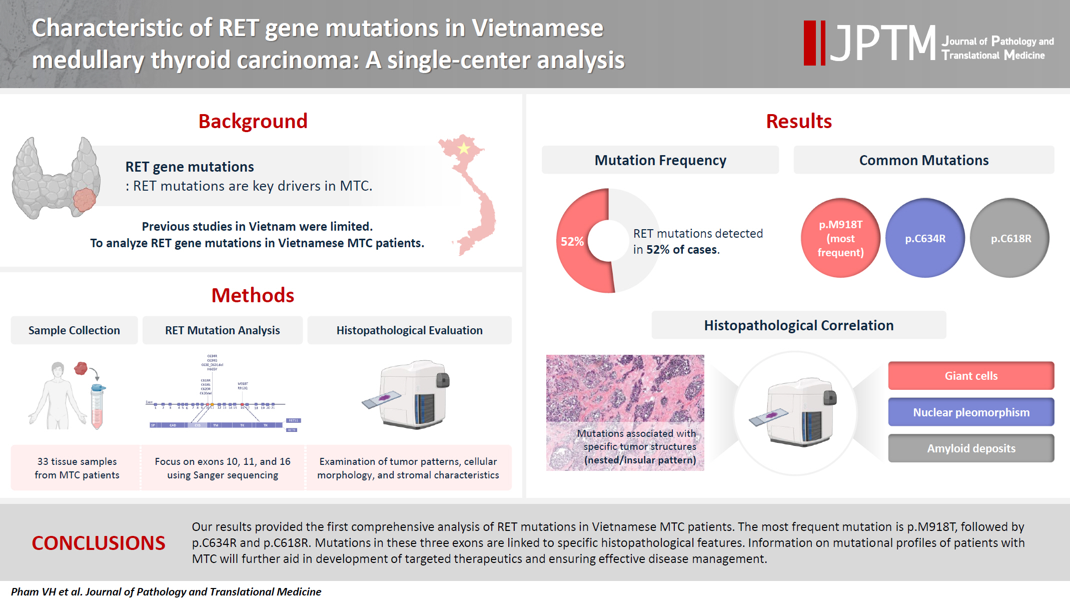
The RET gene point mutation is the main molecular alteration involved in medullary thyroid carcinoma (MTC) tumorigenesis. Previous studies in Vietnam mainly consisted of case reports, with limited data on larger sample sizes. In this study, we investigated RET gene mutations in exons 10, 11, and 16 and analyzed clinicopathological features of a series of Vietnamese MTC patients. Methods: We collected 33 tissue samples from patients with MTC and analyzed RET mutations using the Sanger sequencing method. The relationship between hotspot RET mutations (exons 10, 11, 16) and clinicopathological features were investigated. Results: Among the 33 analyzed cases, 17 tumors (52%) harbored RET mutations in exon 10, 11, or 16. A total of 10 distinct genetic alterations were identified, including eight missense mutations and two short indels. Of these, seven were classified as pathogenic mutations based on previous publications, with p.M918T being the most frequent (4 cases), followed by p.C634R (3 cases) and p.C618R (3 cases). Mutations were significantly associated with specific histological patterns, such as the nested/insular pattern (p=.026), giant cells (p=.007), nuclear pleomorphism (p=.018), stippled chromatin (p=.044), and amyloid deposits (p=.024). No mutations were found in germline analyses, suggesting these were somatic alterations. Conclusions: Our results provided the first comprehensive analysis of RET mutations in Vietnamese MTC patients. The most frequent mutation was p.M918T, followed by p.C634R and p.C618R. Mutations in these three exons were linked to specific histopathological features. Information on mutational profiles of patients with MTC will further aid in the development of targeted therapeutics to ensure effective disease management.
- Histopathologic classification and immunohistochemical features of papillary renal neoplasm with potential therapeutic targets
- Jeong Hwan Park, Su-Jin Shin, Hyun-Jung Kim, Sohee Oh, Yong Mee Cho
- J Pathol Transl Med. 2024;58(6):321-330. Published online September 12, 2024
- DOI: https://doi.org/10.4132/jptm.2024.07.31

- 7,124 View
- 434 Download
- 1 Web of Science
- 2 Crossref
-
Abstract
PDF
- Background
Papillary renal cell carcinoma (pRCC) is the second most common histological subtype of renal cell carcinoma and is considered a morphologically and molecularly heterogeneous tumor. Accurate classification and assessment of the immunohistochemical features of possible therapeutic targets are needed for precise patient care. We aimed to evaluate immunohistochemical features and possible therapeutic targets of papillary renal neoplasms
Methods
We collected 140 papillary renal neoplasms from three different hospitals and conducted immunohistochemical studies on tissue microarray slides. We performed succinate dehydrogenase B, fumarate hydratase, and transcription factor E3 immunohistochemical studies for differential diagnosis and re-classified five cases (3.6%) of papillary renal neoplasms. In addition, we conducted c-MET, p16, c-Myc, Ki-67, p53, and stimulator of interferon genes (STING) immunohistochemical studies to evaluate their pathogenesis and value for therapeutic targets.
Results
We found that c-MET expression was more common in pRCC (classic) (p = .021) among papillary renal neoplasms and Ki-67 proliferation index was higher in pRCC (not otherwise specified, NOS) compared to that of pRCC (classic) and papillary neoplasm with reverse polarity (marginal significance, p = .080). Small subsets of cases with p16 block positivity (4.5%) (pRCC [NOS] only) and c-Myc expression (7.1%) (pRCC [classic] only) were found. Also, there were some cases showing STING expression and those cases were associated with increased Ki-67 proliferation index (marginal significance, p = .063).
Conclusions
Our findings suggested that there are subsets of pRCC with c-MET, p16, c-MYC, and STING expression and those cases could be potential candidates for targeted therapy. -
Citations
Citations to this article as recorded by
- Tissue-Based Biomarkers Important for Prognostication and Management of Genitourinary Tumors, Including Surrogate Markers of Genomic Alterations
Leonie Beauchamp, Shreeya Indulkar, Eric Erak, Mohammad Salimian, Andres Matoso
Surgical Pathology Clinics.2025; 18(1): 175. CrossRef - Papillary renal neoplasm with reverse polarity: a case report and literature review
Diego Gonzalez, Kris Kokoneshi, Sam Kwon, Ryan Thomas Mathews, Ryan Michael Antar, Maher Ali, Abiye Kassa, Michael Whalen
Frontiers in Oncology.2025;[Epub] CrossRef
- Tissue-Based Biomarkers Important for Prognostication and Management of Genitourinary Tumors, Including Surrogate Markers of Genomic Alterations
- PLAG1, SOX10, and Myb Expression in Benign and Malignant Salivary Gland Neoplasms
- Ji Hyun Lee, Hye Ju Kang, Chong Woo Yoo, Weon Seo Park, Jun Sun Ryu, Yuh-Seog Jung, Sung Weon Choi, Joo Yong Park, Nayoung Han
- J Pathol Transl Med. 2019;53(1):23-30. Published online November 14, 2018
- DOI: https://doi.org/10.4132/jptm.2018.10.12
- 13,006 View
- 384 Download
- 28 Web of Science
- 34 Crossref
-
Abstract
PDF
- Background
Recent findings in molecular pathology suggest that genetic translocation and/oroverexpression of oncoproteins is important in salivary gland tumorigenesis and diagnosis. Weinvestigated PLAG1, SOX10, and Myb protein expression in various salivary gland neoplasm tissues.
Methods
A total of 113 cases of surgically resected salivary gland neoplasms at the NationalCancer Center from January 2007 to March 2017 were identified. Immunohistochemical stainingof PLAG1, SOX10, and Myb in tissue samples was performed using tissue microarrays.
Results
Among the 113 cases, 82 (72.6%) were benign and 31 (27.4%) were malignant. PLAG1 showednuclear staining and normal parotid gland was not stained. Among 48 cases of pleomorphicadenoma, 29 (60.4%) were positive for PLAG1. All other benign and malignant salivary glandneoplasms were PLAG1-negative. SOX10 showed nuclear staining. In normal salivary gland tissuesSOX10 was expressed in cells of acinus and intercalated ducts. In benign tumors, SOX10 expressionwas observed in all pleomorphic adenoma (48/48), and basal cell adenoma (3/3), but not inother benign tumors. SOX10 positivity was observed in nine of 31 (29.0%) malignant tumors.Myb showed nuclear staining but was not detected in normal parotid glands. Four of 31 (12.9%)malignant tumors showed Myb positivity: three adenoid cystic carcinomas (AdCC) and onemyoepithelial carcinoma with focal AdCC-like histology.
Conclusions
PLAG1 expression is specificto pleomorphic adenoma. SOX10 expression is helpful to rule out excretory duct origin tumor,but its diagnostic value is relatively low. Myb is useful for diagnosing AdCC when histology isunclear in the surgical specimen. -
Citations
Citations to this article as recorded by- Pleomorphic adenoma: A comprehensive review
Wilson Duplessis, Jason K. Wasserman
Seminars in Diagnostic Pathology.2026; 43(2): 150991. CrossRef - Pleomorphic adenoma gene 1 (PLAG1) protects p53-/- myoepithelial cells from mitochondria-related apoptosis caused by hypoxia
Nodoka Kindaichi, Yoshiki Mukudai, Yuzo Abe, Masataka Watanabe, Maki Nara, Konomi Yamada, Asami Houri, Toshikazu Shimane, Tatsuo Shirota
Journal of Oral and Maxillofacial Surgery, Medicine, and Pathology.2025; 37(4): 654. CrossRef - Retrospective Clinicopathological Study of 33 Cases of Pleomorphic Salivary Adenoma Diagnosed in Benghazi
Siraj S. Najem, Elhoni Ashour, Rehab Elmaddani, Ali M. Elmurtadi
Libyan Journal of Dentistry .2025; 8(2): 29. CrossRef - Pleomorphic adenoma of palatal minor salivary glands
Afrah Aldelaimi, Tahrir Aldelaimi, Suzan Abdulkareem
Revista Española de Cirugía Oral y Maxilofacial.2025;[Epub] CrossRef - Immunohistochemical Characterization of a Large Cohort of Triple Negative Breast Cancer
Rachel Han, Sharon Nofech-Mozes, Dina Boles, Hannah Wu, Nikolina Curcin, Elzbieta Slodkowska
International Journal of Surgical Pathology.2024; 32(2): 239. CrossRef - Proceedings of the 2024 North American Society of Head and Neck Pathology Companion Meeting, Baltimore, MD, March 24, 2024: Navigating Ancillary Studies in Basaloid/Blue Salivary Tumors
Kristine S. Wong
Head and Neck Pathology.2024;[Epub] CrossRef - Insights into the molecular alterations of PLAG1 and HMGA2 associated with malignant phenotype acquisition in pleomorphic adenoma
Reydson Alcides de Lima-Souza, Gustavo de Souza Vieira, Talita de Carvalho Kimura, João Figueira Scarini, Luccas Lavareze, Tayná Figueiredo Maciel, Moisés Willian Aparecido Gonçalves, Erika Said Abu Egal, Albina Altemani, Fernanda Viviane Mariano
Critical Reviews in Oncology/Hematology.2024; 204: 104494. CrossRef - Expanding the Molecular Spectrum of Carcinoma Ex Pleomorphic Adenoma
Reydson Alcides de Lima-Souza, Albina Altemani, Michal Michal, Fernanda Viviane Mariano, Ilmo Leivo, Alena Skálová
American Journal of Surgical Pathology.2024; 48(12): 1491. CrossRef - Utility of SOX10 and estrogen receptor immunohistochemical expression in endometrial carcinoma of Egyptian patients
Mona A. Kora, Alyaa A. Moselhy, Rania A. Abdallah
Egyptian Journal of Pathology.2024; 44(2): 190. CrossRef - Exploring Advanced Diagnostic Techniques for Salivary Gland Disorders: A Narrative Overview
Chuan-Xiang Li-, Liu Zhang, Ya-Ru Yan, Yong-Jie Ding, Ying-Ni Lin, Jian-Ping Zhou, Ning Li, Hong-Peng Li, Shi-Qi Li, Xian-Wen Sun, Qing-Yun Li
Asian journal of Current Research in Clinical Cancer.2024; 4(1): 1. CrossRef - The Challenge of “Monomorphic” Mucoepidermoid Carcinoma—Report of a Rare Case with Pure Spindle-Clear Cell Morphology
Xinyi Qu, Edwin Jun Chen Chew, Sathiyamoorthy Selvarajan, Bingcheng Wu, Abbas Agaimy, Fredrik Petersson
Head and Neck Pathology.2023; 17(3): 864. CrossRef - SOX10
Albert L Sy, Mai P Hoang
Journal of Clinical Pathology.2023; 76(10): 649. CrossRef - Activating Transcription Factor 1 (ATF1) Immunohistochemical Marker Distinguishes HCCC from MEC
Wafaey Badawy, Asmaa S. Abdelfattah, Haneen A. Sallam
Journal of Molecular Pathology.2023; 4(3): 178. CrossRef - Rare case of pleomorphic adenoma presenting as peritonsilar tumor
Anđelina Jovanović, Svetlana Valjarević, Milan Jovanović
Medicinska istrazivanja.2023; 56(3): 95. CrossRef - Pleomorphic Adenoma of a Minor Salivary Gland of the Hard Palate: A Case Report
Ishank Panchal, Anil Wanjari
Cureus.2023;[Epub] CrossRef - Advanced Diagnostic Methods for Salivary Glands Diseases: A Narrative Review Study
Malak Mohammed AlOsaimi, Abdulaziz Mohammed AlSubaheen, Taif Saleh Jameel, Rand Abdulrahman AlSalamah, Dalal Naseh AlAnzi, Norah Ameen AlOushan, Fahad Fadhel AlShammari, Cristalle Soman
Clinical Cancer Investigation Journal.2023; 12(4): 19. CrossRef - Clinical Significance of SOX10 Expression in Human Pathology
Hisham F. Bahmad, Aran Thiravialingam, Karthik Sriganeshan, Jeffrey Gonzalez, Veronica Alvarez, Stephanie Ocejo, Alvaro R. Abreu, Rima Avellan, Alejandro H. Arzola, Sana Hachem, Robert Poppiti
Current Issues in Molecular Biology.2023; 45(12): 10131. CrossRef - NR4A3 Immunostain Is a Highly Sensitive and Specific Marker for Acinic Cell Carcinoma in Cytologic and Surgical Specimens
Kartik Viswanathan, Shaham Beg, Bing He, Taotao Zhang, Richard Cantley, Daniel J Lubin, Qiuying Shi, Zahra Maleki, Saeed Asiry, Rema Rao, Nora Katabi, Masato Nakaguro, William C Faquin, Peter M Sadow, Momin T Siddiqui, Theresa Scognamiglio
American Journal of Clinical Pathology.2022; 157(1): 98. CrossRef - Recent Advances on Immunohistochemistry and Molecular Biology for the Diagnosis of Adnexal Sweat Gland Tumors
Nicolas Macagno, Pierre Sohier, Thibault Kervarrec, Daniel Pissaloux, Marie-Laure Jullie, Bernard Cribier, Maxime Battistella
Cancers.2022; 14(3): 476. CrossRef - Diagnostic accuracy of human transcriptional activator (Myb) expression by ELISA technique versus immunohistochemistry in detecting salivary gland carcinomas
Yousra Refaey, OlfatGamil Shaker, Ayman Abdelwahab, ImanAdel Mohamed Abdelmoneim, Fat’heyaMohamed Zahran
Journal of International Oral Health.2022; 14(1): 61. CrossRef - SLUG is a key regulator of epithelial-mesenchymal transition in pleomorphic adenoma
Hyesung Kim, Seung Bum Lee, Jae Kyung Myung, Jeong Hwan Park, Eunsun Park, Dong Il Kim, Cheol Lee, Younghoon Kim, Chul-Min Park, Min Bum Kim, Gil Chai Lim, Bogun Jang
Laboratory Investigation.2022; 102(6): 631. CrossRef - Assessment of MEF2C as a novel myoepithelial marker using normal salivary gland and pleomorphic adenoma: An immunohistochemical study
Ikuko Takakura, Satoko Kujiraoka, Rika Yasuhara, Junichi Tanaka, Fumio Ide, Kenji Mishima
Journal of Oral and Maxillofacial Surgery, Medicine, and Pathology.2022; 34(4): 523. CrossRef - Update on selective special types of breast neoplasms: Focusing on controversies, differential diagnosis, and molecular genetic advances
Shi Wei
Seminars in Diagnostic Pathology.2022; 39(5): 367. CrossRef - Cutaneous Melanomas: A Single Center Experience on the Usage of Immunohistochemistry Applied for the Diagnosis
Costantino Ricci, Emi Dika, Francesca Ambrosi, Martina Lambertini, Giulia Veronesi, Corti Barbara
International Journal of Molecular Sciences.2022; 23(11): 5911. CrossRef - Distinct clinicopathological and genomic features in solid and basaloid adenoid cystic carcinoma of the breast
Juan Ji, Fang Zhang, Fanglei Duan, Hong Yang, Jun Hou, Yang Liu, Jie Dai, Qiong Liao, Xian Chen, Qingsong Liu
Scientific Reports.2022;[Epub] CrossRef - NR4A3 fluorescence in situ hybridization analysis in cytologic and surgical specimens of acinic cell carcinoma
Qiuying Shi, Bin Zhang, Caroline Bsirini, Liqiong Li, Ellen J. Giampoli, Kelly R. Magliocca, Michelle Reid, Zhongren Zhou
Human Pathology.2022; 127: 86. CrossRef - Evaluation of NR4A3 immunohistochemistry (IHC) and fluorescence in situ hybridization and comparison with DOG1 IHC for FNA diagnosis of acinic cell carcinoma
John M. Skaugen, Raja R. Seethala, Simion I. Chiosea, Michael S. Landau
Cancer Cytopathology.2021; 129(2): 104. CrossRef -
MYB-NFIB Translocation by FISH in Adenoid Cystic Carcinoma of the Head and Neck in Nigerian Patients: A Preliminary Report
Adepitan A. Owosho, Olufunlola M. Adesina, Oluwole Odujoko, Olujide O. Soyele, Akinwumi Komolafe, Robert Bauer, Kallie Holte, Kurt F. Summersgill
Head and Neck Pathology.2021; 15(2): 433. CrossRef - Liquid-based cytology of oral brushings in a case of adenoid cystic carcinoma arising from the palate
Ryo MAKINO, Akihiko KAWAHARA, Hideyuki ABE, Yorihiko TAKASE, Chihiro FUKUMITSU, Kazuya MURATA, Tomoko YOSHIDA, Yukako SHINODA, Yoshiki NAITO, Jun AKIBA
The Journal of the Japanese Society of Clinical Cytology.2021; 60(1): 33. CrossRef - MYB Translocations in Both Myoepithelial and Ductoglandular Epithelial Cells in Adenoid Cystic Carcinoma: A Histopathologic and Genetic Reappraisal in Six Primary Cutaneous Cases
Keisuke Goto, Kazuyoshi Kajimoto, Takashi Sugino, Shin-ichi Nakatsuka, Makoto Yoshida, Mai Noto, Michihiro Kono, Toshihiro Takai
The American Journal of Dermatopathology.2021; 43(4): 278. CrossRef - Co-expression of Myoepithelial and Melanocytic Features in Carcinoma Ex Pleomorphic Adenoma
Costantino Ricci, Federico Chiarucci, Francesca Ambrosi, Tiziana Balbi, Barbara Corti, Ottavio Piccin, Ernesto Pasquini, Maria Pia Foschini
Head and Neck Pathology.2021; 15(4): 1385. CrossRef - Juvenile onset pleomorphic adenoma presenting as giant tumor of parotid gland in a young female
Surender Verma, Shivika Aggarwal, Pradeep Garg, Anjali Verma, Mridul Gera, Swaran S. Yadav
Journal of Dr. NTR University of Health Sciences.2021; 10(4): 286. CrossRef - Cytopathology and diagnostics of Warthin's tumour
Mirna Sučić, Nives Ljubić, Leila Perković, Dunja Ivanović, Leo Pažanin, Tena Sučić Radovanović, Dubravka Župnić‐Krmek, Fabijan Knežević
Cytopathology.2020; 31(3): 193. CrossRef - Clear cell papillary neoplasm of the breast with MAML2 gene rearrangement: Clear cell hidradenoma or low-grade mucoepidermoid carcinoma?
Raima A. Memon, Carlos N Prieto Granada, Shi Wei
Pathology - Research and Practice.2020; 216(10): 153140. CrossRef
- Pleomorphic adenoma: A comprehensive review
- Abrupt Dyskeratotic and Squamoid Cells in Poorly Differentiated Carcinoma: Case Study of Two Thoracic NUT Midline Carcinomas with Cytohistologic Correlation
- Taebum Lee, Sangjoon Choi, Joungho Han, Yoon-La Choi, Kyungjong Lee
- J Pathol Transl Med. 2018;52(5):349-353. Published online July 27, 2018
- DOI: https://doi.org/10.4132/jptm.2018.07.16
- 10,280 View
- 147 Download
- 14 Web of Science
- 14 Crossref
-
Abstract
PDF
- Cytologic diagnosis of nuclear protein in testis (NUT) midline carcinoma (NMC) is important due to its aggressive behavior and miserable prognosis. Early diagnosis of NMC can facilitate proper management, and here we report two rare cases of thoracic NMC with cytohistologic correlation. In aspiration cytology, the tumor presented with mixed cohesive clusters and dispersed single cells, diffuse background necrosis and many neutrophils. Most of the tumor cells had scanty cytoplasm and medium-sized irregular nuclei, which had fine to granular nuclear chromatin. Interestingly, a few dyskeratotic cells or squamoid cell clusters were present in each case. Biopsy specimen histology revealed more frequent squamous differentiation, and additional immunohistochemistry tests showed nuclear expression of NUT. Because this tumor has a notorious progression and has been previously underestimated in terms of its prevalence, awareness of characteristic findings and proper ancillary tests should be considered in all suspicious cases.
-
Citations
Citations to this article as recorded by- Diagnostic significance and cytological features of NUT carcinoma by EBUS‐FNA, a case report and literature review
Yaping Ju, Miriam Velazquez, Andy Sherrod, Tiannan Wang
Cytopathology.2024; 35(4): 497. CrossRef - Exploring cytologic features and potential diagnostic challenges of metastatic NUT carcinoma to the parotid gland: A case report and a comprehensive literature review
Crystal Y. Li, Salih Salihoglu, Francisco J. Civantos, Jaylou M. Velez Torres
Diagnostic Cytopathology.2024;[Epub] CrossRef - Nuclear Protein in Testis (NUT) Carcinoma: A Comprehensive Immunohistochemical Analysis of 57 Cases With Consideration of Interpretation and Pitfall Recognition
Ayesha Farooq, Allison L. Kerper, Jennifer M. Boland, Ying-Chun Lo
Archives of Pathology & Laboratory Medicine.2024; 148(8): 898. CrossRef - BRD3‐NUTM1‐expressing NUT carcinoma of lung on endobronchial ultrasound‐guided transbronchial needle aspiration cytology, a diagnostic pitfall
Sameer Chhetri Aryal, Shereen Zia, Shannon Rodgers, Yulei Shen, Kyle Perry, Lisi Yuan
Diagnostic Cytopathology.2022;[Epub] CrossRef - Nuclear protein of the testis midline carcinoma of the thorax
Ayae Saiki, Keita Sakamoto, Yuan Bee, Takehiro Izumo
Japanese Journal of Clinical Oncology.2022; 52(6): 531. CrossRef - Approach to Mediastinal Fine Needle Aspiration Cytology
Zaibo Li, Huihong Xu, Fang Fan
Advances in Anatomic Pathology.2022; 29(6): 337. CrossRef - Diagnosis, Treatment and Prognosis of Primary Pulmonary NUT Carcinoma: A Literature Review
Jiaqian Yuan, Zhili Xu, Yong Guo
Current Oncology.2022; 29(10): 6807. CrossRef - Case report: Immunovirotherapy as a novel add-on treatment in a patient with thoracic NUT carcinoma
Linus D. Kloker, Branko Calukovic, Katrin Benzler, Alexander Golf, Sebastian Böhm, Sven Günther, Marius Horger, Simone Haas, Susanne Berchtold, Julia Beil, Mary E. Carter, Tina Ganzenmueller, Stephan Singer, Abbas Agaimy, Robert Stöhr, Arndt Hartmann, Tho
Frontiers in Oncology.2022;[Epub] CrossRef - Cytomorphology of primary pulmonary NUT carcinoma in different cytology preparations
Rimlee Dutta, Aruna Nambirajan, Saurabh Mittal, Sinchita Roy‐Chowdhuri, Deepali Jain
Cancer Cytopathology.2021; 129(1): 53. CrossRef - Update on genetically defined lung neoplasms: NUT carcinoma and thoracic SMARCA4-deficient undifferentiated tumors
Kyriakos Chatzopoulos, Jennifer M. Boland
Virchows Archiv.2021; 478(1): 21. CrossRef - Immunotherapy and Targeting the Tumor Microenvironment: Current Place and New Insights in Primary Pulmonary NUT Carcinoma
Xiang Li, Hui Shi, Wei Zhang, Chong Bai, Miaoxia He, Na Ta, Haidong Huang, Yunye Ning, Chen Fang, Hao Qin, Yuchao Dong
Frontiers in Oncology.2021;[Epub] CrossRef - Prevalence of NUT carcinoma in head and neck: Analysis of 362 cases with literature review
Taebum Lee, Junhun Cho, Chung‐Hwan Baek, Young‐Ik Son, Han‐Sin Jeong, Man Ki Chung, Sang Duk Hong, Yong Chan Ahn, Dong Ryul Oh, Jae Myoung Noh, Keunchil Park, Myung‐Ju Ahn, Hyung‐Jin Kim, Yi Kyung Kim, Young Hyeh Ko
Head & Neck.2020; 42(5): 924. CrossRef - Lung nuclear protein in testis carcinoma in an elderly Korean woman: A case report with cytohistological analysis
Hwa Jin Cho, Hyun‐Kyung Lee
Thoracic Cancer.2020; 11(6): 1724. CrossRef - Clinicopathological characteristics of primary lung nuclear protein in testis carcinoma: A single‐institute experience of 10 cases
Yoon Ah Cho, Yoon‐La Choi, Inwoo Hwang, Kyungjong Lee, Jong Ho Cho, Joungho Han
Thoracic Cancer.2020; 11(11): 3205. CrossRef
- Diagnostic significance and cytological features of NUT carcinoma by EBUS‐FNA, a case report and literature review
- Expression of c-MET in Invasive Meningioma
- Sumi Yun, Jae Moon Koh, Kyu Sang Lee, An Na Seo, Kyung Han Nam, Gheeyoung Choe
- J Pathol Transl Med. 2015;49(1):44-51. Published online January 15, 2015
- DOI: https://doi.org/10.4132/jptm.2014.10.13
- 11,632 View
- 75 Download
- 24 Web of Science
- 22 Crossref
-
Abstract
PDF
- Background
Meningiomas show high recurrence rates even after curative tumor removal. The invasiveness of meningiomas may contribute to their high recurrence rates. Recently, c-MET and hepatocyte growth factor (HGF) have been reported to be involved in cancer invasion. Methods: We examined the immunohistochemical expression of c-MET and HGF in 100 cases of patients with meningiomas who have undergone complete tumor removal. Results: c-MET-High and HGFHigh were found in 17% and 13% of meningiomas, respectively. Brain invasion was observed in 17.6% of c-MET-High meningiomas, but in only 2.4% of c-MET-Low meningiomas (p=.033). Bone/ soft tissue invasion was observed in 23.5% of c-MET-High meningiomas and in 9.6% of c-MET-Low meningiomas (p=.119). HGF-High did not show statistical association with brain invasion or bone/ soft tissue invasion. c-MET-High demonstrated shorter recurrence-free survival (RFS, 93.5±8.2 months vs 96.1±1.9 months); however, this difference was not statistically significant (p=.139). There was no association of HGF-High with RFS. Conclusions: This study demonstrates that c- MET-High is associated with brain invasion of meningiomas, and that c-MET expression may be a useful predictive marker for meningioma recurrence. Patients with invasive meningiomas with high expressions of c-MET may be good candidates for targeted therapy using c-MET inhibitors. -
Citations
Citations to this article as recorded by- MRI morphological features combined with apparent diffusion coefficient can predict brain invasion in meningioma
Xiaoyu Huang, Yuntai Cao, Guojin Zhang, FuQiang Tang, Dandan Sun, Jialiang Ren, Wenyi Li, Junlin Zhou, Jing Zhang
Computers in Biology and Medicine.2025; 187: 109763. CrossRef - Integrating diffusion-weighted MRI radiomics features to predict brain invasion of meningiomas
Zongmeng Wang, Lihong Chen, Ye Li, Dingfu Wei, Yizhu Chen, Xiaodan Chen, Sihui Liu, Yichao Zhang, Tianjin Zhong, Peirong Jiang, Haixia Li, Yunjing Xue, Lin Lin
Neurosurgical Review.2025;[Epub] CrossRef - Drug target therapy and emerging clinical relevance of exosomes in meningeal tumors
Swati Sharma, Rashmi Rana, Prem Prakash, Nirmal Kumar Ganguly
Molecular and Cellular Biochemistry.2024; 479(1): 127. CrossRef - The efficacy of preoperative MRI features in the diagnosis of meningioma WHO grade and brain invasion
Jun Jiang, Juan Yu, Xiajing Liu, Kan Deng, Kaichao Zhuang, Fan Lin, Liangping Luo
Frontiers in Oncology.2023;[Epub] CrossRef - The Role of Pharmacotherapy in Treatment of Meningioma: A Systematic Review
Ataollah Shahbandi, Darsh S. Shah, Caroline C. Hadley, Akash J. Patel
Cancers.2023; 15(2): 483. CrossRef - Advances in the systemic therapy for recurrent meningiomas and the challenges ahead
Yi Li, Jan Drappatz
Expert Review of Neurotherapeutics.2023; 23(11): 995. CrossRef - Clinical and pathological impact of an optimal assessment of brain invasion for grade 2 meningioma diagnosis: lessons from a series of 291 cases
Thiébaud Picart, Chloé Dumot, Jacques Guyotat, Vladislav Pavlov, Nathalie Streichenberger, Alexandre Vasiljevic, Tanguy Fenouil, Anne Durand, Emmanuel Jouanneau, François Ducray, Timothée Jacquesson, Moncef Berhouma, David Meyronet
Neurosurgical Review.2022; 45(4): 2797. CrossRef - Nomogram based on MRI can preoperatively predict brain invasion in meningioma
Jing Zhang, Yuntai Cao, Guojin Zhang, Zhiyong Zhao, Jianqing Sun, Wenyi Li, Jialiang Ren, Tao Han, Junlin Zhou, Kuntao Chen
Neurosurgical Review.2022; 45(6): 3729. CrossRef - Overexpression of Hepatocyte growth factor and its soluble receptor (s-cMet) in the serum of patients with different grades of meningioma
Farhad Mashayekhi, Soheila Talesh Sasani, Alia Saberi, Zivar Salehi
Journal of Clinical Neuroscience.2021; 93: 1. CrossRef - Brain-invasive meningiomas: molecular mechanisms and potential therapeutic options
Chaoying Qin, Meng Huang, Yimin Pan, Yuzhe Li, Wenyong Long, Qing Liu
Brain Tumor Pathology.2021; 38(3): 156. CrossRef - YAP1-FAM118B Fusion Defines a Rare Subset of Childhood and Young Adulthood Meningiomas
Kathleen M. Schieffer, Vibhuti Agarwal, Stephanie LaHaye, Katherine E. Miller, Daniel C. Koboldt, Tara Lichtenberg, Kristen Leraas, Patrick Brennan, Benjamin J. Kelly, Erin Crist, Jerome Rusin, Jonathan L. Finlay, Diana S. Osorio, Eric A. Sribnick, Jeffre
American Journal of Surgical Pathology.2021; 45(3): 329. CrossRef - Regression of Intracranial Meningiomas Following Treatment with Cabozantinib
Rupesh Kotecha, Raees Tonse, Haley Appel, Yazmin Odia, Ritesh R. Kotecha, Guilherme Rabinowits, Minesh P. Mehta
Current Oncology.2021; 28(2): 1537. CrossRef - Prognostic significance of brain invasion in meningiomas: systematic review and meta-analysis
Satoshi Nakasu, Yoko Nakasu
Brain Tumor Pathology.2021; 38(2): 81. CrossRef - Curcumin Inhibits HGF-Induced EMT by Regulating c-MET-Dependent PI3K/Akt/mTOR Signaling Pathways in Meningioma
Xiaodong Chen, Fen Tian, Peng Lun, Yugong Feng, Ho Lin
Evidence-Based Complementary and Alternative Medicine.2021; 2021: 1. CrossRef - Letter to the Editor. Preoperative seizures as predictive sign of brain invasion by meningioma
Mikhail F. Chernov
Journal of Neurosurgery.2019; 130(3): 1030. CrossRef - Investigating Trk Protein Expression between Oropharyngeal and Non-oropharyngeal Squamous Cell Carcinoma: Clinical Implications and Possible Roles of Human Papillomavirus Infection
Yoon Ah Cho, Ji Myung Chung, Hyunmi Ryu, Eun Kyung Kim, Byoung Chul Cho, Sun Och Yoon
Cancer Research and Treatment.2019; 51(3): 1052. CrossRef - Prediction of brain invasion in patients with meningiomas using preoperative magnetic resonance imaging
Alborz Adeli, Katharina Hess, Christian Mawrin, Eileen Maria Susanne Streckert, Walter Stummer, Werner Paulus, André Kemmling, Markus Holling, Walter Heindel, Rene Schmidt, Dorothee Cäcilia Spille, Peter B. Sporns, Benjamin Brokinkel
Oncotarget.2018; 9(89): 35974. CrossRef - Visceral and bone metastases of a WHO grade 2 meningioma: A case report and review of the literature
A. Paix, W. Waissi, D. Antoni, R. Adeduntan, G. Noël
Cancer/Radiothérapie.2017; 21(1): 55. CrossRef - Letter: Brain Invasion in Meningiomas—Sex-Associated Differences are not Related to Estrogen- and Progesterone Receptor Expression
Katharina Heß, Dorothee Cäcilia Spille, Andrea Wagner, Walter Stummer, Werner Paulus, Benjamin Brokinkel
Neurosurgery.2017; 81(2): E25. CrossRef - Brain invasion in meningiomas—clinical considerations and impact of neuropathological evaluation: a systematic review
Benjamin Brokinkel, Katharina Hess, Christian Mawrin
Neuro-Oncology.2017; 19(10): 1298. CrossRef - Systemic therapy for recurrent meningioma
E. Le Rhun, S. Taillibert, M. C. Chamberlain
Expert Review of Neurotherapeutics.2016; 16(8): 889. CrossRef - Brain Invasion in Meningiomas: Incidence and Correlations with Clinical Variables and Prognosis
Dorothee Cäcilia Spille, Katharina Heß, Cristina Sauerland, Nader Sanai, Walter Stummer, Werner Paulus, Benjamin Brokinkel
World Neurosurgery.2016; 93: 346. CrossRef
- MRI morphological features combined with apparent diffusion coefficient can predict brain invasion in meningioma
- Anaplastic Transformation of Papillary Thyroid Carcinoma in a Young Man: A Case Study with Immunohistochemical and
BRAF Analysis - Ji Hye Park, Hyeong Ju Kwon, Cheong Soo Park, SoonWon Hong
- Korean J Pathol. 2014;48(3):234-240. Published online June 26, 2014
- DOI: https://doi.org/10.4132/KoreanJPathol.2014.48.3.234
- 12,160 View
- 75 Download
- 7 Crossref
-
Abstract
PDF
This study reports a case of anaplastic transformation from a well-differentiated thyroid carcinoma in a young patient. The first recurrent tissue contained poorly differentiated foci that revealed lower thyroglobulin, thyroid transcription factor 1 (TTF-1), and galectin-3 expression than the well-differentiated area. However there was no increased p53 or Ki-67 expression in the poorly differentiated foci, nor in the well-differentiated area. The tissue subsequently relapsed and revealed only anaplastic features, complete loss of thyroglobulin, TTF-1, and galectin-3 expression and revealed an increase in p53 and Ki-67 expression. The
BRAF V600E andBRAF V600V mutation were found in the initially diagnosed papillary thyroid carcinoma and the poorly differentiated foci of the recurring papillary thyroid carcinoma; however, only theBRAF V600V mutation was found in the anaplastic carcinoma. These results suggest that overexpression of p53 and Ki-67 contributed to the anaplastic transformation. We also found that theBRAF type changed during the tumor relapse.-
Citations
Citations to this article as recorded by- Regressed Papillary Thyroid Carcinoma with Anaplastic Transformation into Lymph Node Metastasis: Case Report with Review of the Literature
Bozidar Kovacevic, Bojana Rancic, Sasa Jovic, Snezana Cerovic, Vesna Skuletic, Jelena Karajovic, Milka Gardasevic, Gordana Supic, Kennichi Kakudo
Diagnostics.2025; 15(5): 523. CrossRef - Rare but Complex: Outcomes and Challenges in Managing Composite Follicular-Anaplastic Thyroid Cancer
Dilay Aykan, Hala al Asadi, Anjani Turaga, Lutske Lodewijk, Brendan M. Finnerty, Thomas J. Fahey, Inne H. M. Borel Rinkes, Menno R. Vriens, Rasa Zarnegar
Annals of Surgical Oncology.2025; 32(7): 4754. CrossRef - Immunomodulation exerted by galectins: a land of opportunity in rare cancers
Laura Díaz-Alvarez, Georgina I. López-Cortés, Erandi Pérez-Figueroa
Frontiers in Immunology.2023;[Epub] CrossRef - Coexisting well-differentiated and anaplastic thyroid carcinoma in the same primary resection specimen: immunophenotypic and genetic comparison of the two components in a consecutive series of 13 cases and a review of the literature
Moira Ragazzi, Federica Torricelli, Benedetta Donati, Alessia Ciarrocchi, Dario de Biase, Giovanni Tallini, Eleonora Zanetti, Alessandra Bisagni, Elisabetta Kuhn, Davide Giordano, Andrea Frasoldati, Simonetta Piana
Virchows Archiv.2021; 478(2): 265. CrossRef - HDAC Inhibition Induces PD-L1 Expression in a Novel Anaplastic Thyroid Cancer Cell Line
Luca Hegedűs, Dominika Rittler, Tamás Garay, Paul Stockhammer, Ildikó Kovács, Balázs Döme, Sarah Theurer, Thomas Hager, Thomas Herold, Stavros Kalbourtzis, Agnes Bankfalvi, Kurt W. Schmid, Dagmar Führer, Clemens Aigner, Balázs Hegedűs
Pathology & Oncology Research.2020; 26(4): 2523. CrossRef -
EWSR1 rearrangement is a frequent event in papillary thyroid carcinoma and in carcinoma of the thyroid with Ewing family tumor elements (CEFTE)
G. Oliveira, A. Polónia, J. M. Cameselle-Teijeiro, D. Leitão, S. Sapia, M. Sobrinho-Simões, C. Eloy
Virchows Archiv.2017; 470(5): 517. CrossRef - Anaplastic carcinoma of the thyroid in a 12-year old girl
Rodrigo Mon, James Newlon
Journal of Pediatric Surgery Case Reports.2015; 3(9): 404. CrossRef
- Regressed Papillary Thyroid Carcinoma with Anaplastic Transformation into Lymph Node Metastasis: Case Report with Review of the Literature
- Expression of c-Met Is Different along the Location and Associated with Lymph Node Metastasis of Head and Neck Carcinoma
- Ji-Young Choe, Ji Yun Yun, Soo-Jeong Nam, Ji Eun Kim
- Korean J Pathol. 2012;46(6):515-522. Published online December 26, 2012
- DOI: https://doi.org/10.4132/KoreanJPathol.2012.46.6.515
- 9,325 View
- 52 Download
- 19 Crossref
-
Abstract
PDF
Background Activation of the c-Met pathway is involved in cancer progression and the prognosis. We aimed to identify any association of c-Met protein expression with a number of clinicopathologic variables including infection of human papillomavirus and Epstein-Barr virus (EBV) in head and neck carcinomas (HNCa).
Methods Eighty-two cases were enrolled in this study. Expression of c-Met and p16 was investigated immunohistochemically. EBV was detected by
in situ hybridization and amplification of thec-Met gene by fluorescencein situ hybridization.Results The c-Met protein was expressed in 41.5% (34/82), and gene amplification was found in 1.4% (1/71). High expression of c-Met was associated with the primary location of the tumor; the hypopharynx showed the highest expression, followed by the oral cavity, larynx, and nasal cavity. Squamous cell carcinoma expressed c-Met more frequently than undifferentiated carcinoma. Also, p16 immunoreactivity or EBV infection was associated with the tumor location and well-differentiated histologic type, but were not linked to c-Met expression. The patients with positive c-Met expression showed frequent lymph node metastasis.
Conclusions Activation of the c-Met pathway might be involved in a subset of HNCa. Cases showing positive c-Met expression should be carefully monitored because of the high probability of lymph node metastasis.
-
Citations
Citations to this article as recorded by- Case Report: A rare case of MET-amplified gastric cancer with systemic metastasis: remarkable efficacy of crizotinib and the role of precision medicine
Yan Shen, Yaxin Xu, Jing Sun
Frontiers in Oncology.2025;[Epub] CrossRef - c-MET pathway in human malignancies and its targeting by natural compounds for cancer therapy
Chakrabhavi Dhananjaya Mohan, Muthu K Shanmugam, Siddegowda Gopalapura Shivanne Gowda, Arunachalam Chinnathambi, Kanchugarakoppal S. Rangappa, Gautam Sethi
Phytomedicine.2024; 128: 155379. CrossRef - Nadir paranazal sinüs kanserlerinde yeni tanımlanan reseptör tirozin kinaz mutasyonları ve potansiyel fonksiyonel etkileri
Bakiye GÖKER BAGCA, Sercan GÖDE, Göksel TURHAL, Neslihan Pınar ÖZATEŞ, Ali VERAL, Cumhur GÜNDÜZ, Çığır Biray AVCI
Ege Tıp Dergisi.2023; 62(1): 139. CrossRef - The Emerging Role of c-Met in Carcinogenesis and Clinical Implications as a Possible Therapeutic Target
Antonio Faiella, Ferdinando Riccardi, Giacomo Cartenì, Martina Chiurazzi, Livia Onofrio, Rengyun Liu
Journal of Oncology.2022; 2022: 1. CrossRef - NK4 Regulates Laryngeal Squamous Cell Carcinoma Cell Properties and Inhibits Tumorigenicity by Modulating the DKK1/Wnt/β-Catenin Axis
Shoukai Zhang, Hulai Wei, Xiaoqin Ha, Yueyu Zhang, Yufen Guo
Frontiers in Oncology.2021;[Epub] CrossRef - Epstein-Barr Virus-Associated Carcinoma of the Larynx: A Systematic Review with Meta-Analysis
Marcos Antonio Pereira de Lima, Álife Diêgo Lima Silva, Antônio Carlos Silva do Nascimento Filho, Thiago Lima Cordeiro, João Pedro de Souza Bezerra, Maria Aline Barroso Rocha, Sally de França Lacerda Pinheiro, Roberto Flávio Fontenelle Pinheiro Junior, Ma
Pathogens.2021; 10(11): 1429. CrossRef - The roles of PTEN, cMET, and p16 in resistance to cetuximab in head and neck squamous cell carcinoma
Alexandre A. B. A. da Costa, Felipe D’Almeida Costa, Daniel Vilarim Araújo, Marcos Pedro Guedes Camandaroba, Victor Hugo Fonseca de Jesus, Audrey Oliveira, Ana Caroline Fonseca Alves, Carlos Stecca, Larissa Machado, Andrea Cruz Feraz de Oliveira, Thiago B
Medical Oncology.2019;[Epub] CrossRef - Role of c‐Met expression on prognosis of head and neck cancer: A literature review and meta‐analysis
Lei Li, Zhijun Sun, Xin Huang, Xiao Li, Lihua Sun, Lei Zhang, Xiaodan Zhang, Longwei Ye, Jie Yuan, Limin Mao, Guolin Li
Head & Neck.2019; 41(6): 1999. CrossRef - MET Genomic Alterations in Head and Neck Squamous Cell Carcinoma (HNSCC): Rapid Response to Crizotinib in a Patient with HNSCC with a Novel MET R1004G Mutation
Lisa Pei Chu, Debra Franck, Christine A. Parachoniak, Jeffrey P. Gregg, Michael G. Moore, D. Gregory Farwell, Shyam Rao, Andreas M. Heilmann, Rachel L. Erlich, Jeffrey S. Ross, Vincent A. Miller, Siraj Ali, Jonathan W. Riess
The Oncologist.2019; 24(10): 1305. CrossRef - Understanding c-MET signalling in squamous cell carcinoma of the head & neck
P. Szturz, E. Raymond, C. Abitbol, S. Albert, A. de Gramont, S. Faivre
Critical Reviews in Oncology/Hematology.2017; 111: 39. CrossRef - Prognostic value of c-MET in head and neck cancer: A systematic review and meta-analysis of aggregate data
Petr Szturz, Marie Budíková, Jan B. Vermorken, Ivana Horová, Břetislav Gál, Eric Raymond, Armand de Gramont, Sandrine Faivre
Oral Oncology.2017; 74: 68. CrossRef - Activated HGF-c-Met Axis in Head and Neck Cancer
Levi Arnold, Jonathan Enders, Sufi Thomas
Cancers.2017; 9(12): 169. CrossRef - Clinicopathological impacts of high c-Met expression in head and neck squamous cell carcinoma: a meta-analysis and review
Jung Han Kim, Bum Jun Kim, Hyeong Su Kim
Oncotarget.2017; 8(68): 113120. CrossRef - High expression of c‑Met and EGFR is associated with poor survival of patients with glottic laryngeal squamous cell carcinoma
Mei Jiang, Hui Zhang, He Xiao, Zhimin Zhang, Dan Que, Jia Luo, Jian Li, Bijing Mao, Yuanyuan Chen, Meilin Lan, Ge Wang, Hualiang Xiao
Oncology Letters.2017;[Epub] CrossRef - Profiling of cMET and HER Family Receptor Expression in Pancreatic Ductal Adenocarcinomas and Corresponding Lymph Node Metastasis to Assess Relevant Pathways for Targeted Therapies
Alexander Muckenhuber, Galina Babitzki, Marlene Thomas, Gabriele Hölzlwimmer, Magdalena Zajac, Moritz Jesinghaus, Frank Bergmann, Jens Werner, Albrecht Stenzinger, Wilko Weichert
Pancreas.2016; 45(8): 1167. CrossRef - Absent and abundant MET immunoreactivity is associated with poor prognosis of patients with oral and oropharyngeal squamous cell carcinoma
Maria J. De Herdt, Stefan M. Willems, Berdine van der Steen, Rob Noorlag, Esther I. Verhoef, Geert J.L.H. van Leenders, Robert J.J. van Es, Senada Koljenović, Robert J. Baatenburg de Jong, Leendert H.J. Looijenga
Oncotarget.2016; 7(11): 13167. CrossRef - Biological, diagnostic and therapeutic relevance of the MET receptor signaling in head and neck cancer
Lluís Nisa, Daniel Matthias Aebersold, Roland Giger, Yitzhak Zimmer, Michaela Medová
Pharmacology & Therapeutics.2014; 143(3): 337. CrossRef - Frequent hepatocyte growth factor overexpression and low frequency of c-Met gene amplification in human papillomavirus–negative tonsillar squamous cell carcinoma and their prognostic significances
Mi Jung Kwon, Dong Hoon Kim, Hye-Rim Park, Hyung Sik Shin, Ji Hyun Kwon, Dong Jin Lee, Jin Hwan Kim, Seong Jin Cho, Eun Sook Nam
Human Pathology.2014; 45(7): 1327. CrossRef - Distinct c-Met activation mechanisms induce cell rounding or invasion through pathways involving integrins, RhoA and HIP1
Anja Mai, Ghaffar Muharram, Rachel Barrow-McGee, Habib Baghirov, Juha Rantala, Stéphanie Kermorgant, Johanna Ivaska
Journal of Cell Science.2014; 127(9): 1938. CrossRef
- Case Report: A rare case of MET-amplified gastric cancer with systemic metastasis: remarkable efficacy of crizotinib and the role of precision medicine
- Oncocytoma and Oncocytic Carcinoma of the Salivary Glands, Single Institute Experience.
- Jeong Hyeon Jo, Seung Ho Choi, Jong Lyel Roh, Soon Yuhl Nam, Sang Yoon Kim, Kyung Ja Cho
- Korean J Pathol. 2010;44(4):370-375.
- DOI: https://doi.org/10.4132/KoreanJPathol.2010.44.4.370
- 4,592 View
- 67 Download
- 2 Crossref
-
Abstract
PDF
- BACKGROUND
Oncocytic neoplasms of the salivary glands are rare and the differential diagnosis between oncocytic carcinomas (OCs) and oncocytomas is difficult. We present 5 cases of oncocytoma and 3 cases of OC of the salivary glands with clinicopathological and immunohistochemical comparisons.
METHODS
Eight cases of oncocytic neoplasms diagnosed at Asan Medical Center between 1998 and 2009 were reviewed for clinical data and histological features. Immunohistochemical staining for epidermal growth factor receptor (EGFR), human epidermal growth factor receptor 2 (Her-2), c-kit, p53, and Ki-67 was done.
RESULTS
Cytological differences between oncocytomas and OCs were not obvious, but unequivocal infiltrative growths were identified in 3 cases, rendering the diagnosis of oncocytic carcinoma. When the remaining cases were classified as oncocytomas, there was no difference in age, size, and clinical symptoms between oncocytomas and OCs. Two of 3 OCs showed strong membranous expression of c-kit, but all oncocytomas were negative. The proportion of p53-positive cells was larger in OCs than oncocytomas. Her-2 or EGFR expression was absent, and Ki-67 labeling indices were less than 1% in all cases.
CONCLUSIONS
An infiltrative growth pattern, strong membranous expression of c-kit, and an increased proportion of p53-positive cells are features that can differentiate OCs from oncocytomas of the salivary glands. -
Citations
Citations to this article as recorded by- Clinicopathological Study of Oncocytomas of Head and Neck Region: A Systematic Review
João Paulo Gonçalves de Paiva, Laura Borges Kirschnick, Daniela Giraldo Roldán, Manoela Domingues Martins, Alan Roger Santos‐Silva, Ciro Dantas Soares, Jacks Jorge
Journal of Oral Pathology & Medicine.2025; 54(8): 635. CrossRef - Primary oncocytic carcinoma of ectopic salivary gland: a unique case
E. Touli, A. Manganaris, C. Nikolaidou, I. Karasmanis
International Journal of Oral and Maxillofacial Surgery.2022; 51(4): 463. CrossRef
- Clinicopathological Study of Oncocytomas of Head and Neck Region: A Systematic Review
- Immunohistochemical Array for Clear Cell Type Mucoepidermoid Carcinoma.
- Yeon Sook Kim, Sang Shin Lee, Ji Yong Song, Eun Cheol Kim, Suk Keun Lee
- Korean J Pathol. 2010;44(3):284-294.
- DOI: https://doi.org/10.4132/KoreanJPathol.2010.44.3.284
- 4,257 View
- 25 Download
- 3 Crossref
-
Abstract
PDF
- BACKGROUND
The protein expression profile of clear cell type mucoepidermoid carcinoma (MEC) is not well known.
METHODS
We examined a case of clear cell type MEC by immunohistochemical (IHC) array using 59 antibodies against oncoproteins, proliferation-related proteins, apoptosis-related proteins, growth factor-related proteins, angiogenesis-related proteins, and matrix proteins.
RESULTS
MEC tumor cells showed 40 to 60% more expression of BCL-2 and cyclin-dependent kinase 4 than normal gingival tissue, and 20-40% more expression of BCL-2-associated agonist of cell death, deleted in malignant brain tumors 1, E-cadherin, eIF5A, hypoxia-inducible factor, vimentin, and Wnt-1. Expression of other proteins, including p53, epidermal growth factor receptor, proliferating cell nuclear antigen, survivin, carcinoembryonic antigen, beta-catenin, poly-ADP ribose-polymerase, etc. were relatively weak in MEC tumor cells.
CONCLUSIONS
The IHC array for our MEC contained strong oncogenic signals involving Wnt-1/adenomatous polyposis coli, tumor necrosis factor a/signal transducer and activator of transcription 3/BCL-2, and pAKT pathways, signals that could result in the prolonged survival of clear tumor cells. -
Citations
Citations to this article as recorded by- Adolescent tongue base tumour with diagnostic histopathological dilemma
Mousam Maiti, Ajay Mallick, Debangshu Ghosh, Indranil Chakrabarti
BMJ Case Reports.2024; 17(8): e259570. CrossRef - A review: Immunological markers for malignant salivary gland tumors
P.C. Anila Namboodiripad
Journal of Oral Biology and Craniofacial Research.2014; 4(2): 127. CrossRef - DISPLACEMENT OF MAXILLARY LATERAL INCISOR CAUSED BY IDIOPATHIC GINGIVAL FIBROMATOSIS
Ji-Sook Jung, Ho-Won Park, Ju-Hyun Lee, Hyun-Woo Seo, Suk-Keun Lee
THE JOURNAL OF THE KOREAN ACADEMY OF PEDTATRIC DENTISTRY.2011; 38(3): 296. CrossRef
- Adolescent tongue base tumour with diagnostic histopathological dilemma
- Prognostic Value of Phosphorylated Akt and Survivin Expression in Gastric Adenocarcinoma.
- Soong Lee, Yun Cheol Kim, Hyeon Min Lee, Ki Sang Lee, Byung Chul Shin, Hyung Seok Kim, Jae Hyuk Lee, Chang Soo Park, Kyung Hwa Lee
- Korean J Pathol. 2010;44(3):252-258.
- DOI: https://doi.org/10.4132/KoreanJPathol.2010.44.3.252
- 4,825 View
- 22 Download
- 3 Crossref
-
Abstract
PDF
- BACKGROUND
pAkt (the phosphorylated form of the proto-oncogene protein c-akt) and survivin (human BIRC5 protein) are candidate apoptosis-related molecules that may be responsible for cancer progression. The aim of this study was to determine the expression of pAkt and survivin in malignant stomach neoplasm, and their value as prognostic indicators of cancer.
METHODS
The expression of pAkt and survivin in 144 cases of gastric cancer was detected by immunohistochemistry and compared with established clinicopathological parameters and prognosis of this disease.
RESULTS
Expression of pAkt showed significant correlations with depth of invasion, lymph node and distant metastasis, as well as the stage (p < 0.05 for all three correlations), but not with the Lauren classification. Survivin expression closely correlated with histological type, Lauren classification, depth of invasion, metastasis, and stage (p < 0.05 for all). The overall survival of patients with pAkt/survivin expression was inferior to that of patients with loss of pAkt/survivin expression. Cox multivariate analysis demonstrated a significant correlation between stage (p = 0.04), survivin expression (p = 0.02), and prognosis.
CONCLUSIONS
Patients with pAkt/survivin expression in gastric cancer are at increased risk of cancer-related mortality via the apoptosis resistance pathway. Expression of pAkt and survivin could be used as a prognostic indicator for gastric cancer. -
Citations
Citations to this article as recorded by- Transcriptome analysis reveals GPNMB as a potential therapeutic target for gastric cancer
Feifei Ren, Qitai Zhao, Bin Liu, Xiangdong Sun, Youcai Tang, Huang Huang, Lu Mei, Yong Yu, Hui Mo, Haibin Dong, Pengyuan Zheng, Yang Mi
Journal of Cellular Physiology.2020; 235(3): 2738. CrossRef - The Relationship Between Estrogen/Progesterone Receptor and Expression of mTOR/pAkt Proteins and the Analysis of Prognosis in Breast Cancer
Sun Wook Han, Moon Soo Lee, Sung Yong Kim, Gil Ho Kang, Zi Sun Kim, Cheol Wan Lim, Ji Hyun Lee, Hyun Ju Lee, Mee-Hye Oh, Min Hyuk Lee
Journal of Breast Disease.2013; 1(1): 8. CrossRef - Clinicopathological correlations of mTOR and pAkt expression in non-small cell lung cancer
Mee-Hye Oh, Hyun Ju Lee, Seol Bong Yoo, Xianhua Xu, Jae Sung Choi, Yong Hoon Kim, Seok Yeol Lee, Choon-Taek Lee, Sanghoon Jheon, Jin-Haeng Chung
Virchows Archiv.2012; 460(6): 601. CrossRef
- Transcriptome analysis reveals GPNMB as a potential therapeutic target for gastric cancer
- KIT/PDGFRA Expression and Mutation in Testicular Seminoma and Ovarian Dysgerminoma.
- Song Yi Choi, Kwang Sun Suh, Yong Beom Kim, Hyun Jeong Lee, Eun Sun Kim, Mee Ja Park
- Korean J Pathol. 2009;43(6):528-534.
- DOI: https://doi.org/10.4132/KoreanJPathol.2009.43.6.528
- 4,807 View
- 22 Download
- 1 Crossref
-
Abstract
PDF
- BACKGROUND
KIT and PDGFRA are tyrosine kinase receptors. Stem cell factor/KIT-mediated signaling plays a role in normal spermatogenesis, and the alteration of KIT is important in the pathogenesis of seminomas/dysgerminomas (SD). METHODS: To determine the role of expression and mutation of the KIT and PDGFRA genes, we analyzed 16 seminoma cases, 4 spermatocytic seminoma (SS) cases and 8 dysgerminoma cases for KIT and PDGFRA expression and mutation of KIT (exons 9, 11, 13, and 17) and PDGFRA (exons 12 and 18) using PCR-SSCP methods. RESULTS: KIT was immunohistochemically positive in all 24 SD cases, and one of four (25%) SS cases. PDGFRA was immunohistochemically evident in 16 of the 24 (66.6%) SD cases, and two of the four (50%) SS cases. KIT expression was significantly reduced in SS compared with seminoma (p=0.0035). Four cases (14.3%) displayed mutation in KIT exon 17 or PDGFRA exon 12. Distant metastasis was present in three cases (10.7%), one of which had a nonsense mutation in KIT. CONCLUSIONS: These results indicate that KIT is expressed in the majority of SD cases, but not in most SS cases. However, there was no significant correlation between the clinicopathologic features and mutation or expression of KIT and PDGFRA. -
Citations
Citations to this article as recorded by- Expression of DOG1, PDGFRA, and p16 in Gastrointestinal Stromal Tumors
Sung Hee Jung, Kwang Sun Suh, Dae Young Kang, Dong Wook Kang, Young-Beum Kim, Eun-Sun Kim
Gut and Liver.2011; 5(2): 171. CrossRef
- Expression of DOG1, PDGFRA, and p16 in Gastrointestinal Stromal Tumors
- Expression of P-glycoprotein and Apoptosis in Diffuse Large B-cell Lymphoma.
- Ji Eun Kim, Young A Kim, Mee Soo Chang, Yunkyeong Jeon, JinHo Paik, Seon Og Yoon
- Korean J Pathol. 2009;43(4):317-320.
- DOI: https://doi.org/10.4132/KoreanJPathol.2009.43.4.317
- 3,937 View
- 74 Download
-
Abstract
PDF
- BACKGROUND
Diffuse large B-cell lymphoma (DLBCL) is the most common type of malignant lymphoma which responds well to conventional chemotherapy. However, quite a few patients have a recurrence with more aggressive forms after completion of therapy. Multidrug resistance proteins (MRP) are related to this process in several ways such as cell cycle alteration and modulation of apoptosis. METHODS: We investigated the expression of P-glycoprotein (Gp), one of the well-known MRP, as well as apoptosis associated proteins in DLBCL. Immunohistochemical staining for Gp, p53, Bcl-2, Ki-67, active caspase 3 and FADD was done in forty DLBCL cases. The association between MRP and apoptosis associated proteins to clinical findings was also tested. RESULTS: Twenty-nine patients out of 40 (73%) with DLBCL were positive for Gp, and 26 cases (65%) had a strong positive for Gp. Gp expression was stronger in high-grade lesions than in low-grade lesions and was associated to Bcl-2 expression. However, we could not find an adverse impact of Gp expression on patients' overall survival or relapse free survival rate. CONCLUSIONS: Our study revealed a high frequency of expression for Gp in DLBCL with a possible relationship between the expressions of Gp to apoptosis associated proteins.
- Immunohistochemical Study on the Ha-ras p21 Expression in the Gastric Carcinoma.
- Kwang Min Lee, Joo Yong Yoo
- Korean J Pathol. 1990;24(1):1-9.
- 2,019 View
- 16 Download
-
Abstract
PDF
- We have investigated an immunohistochemical expression of the Human-Ha-ras oncogene product p21 in tumor cells of the primary mass and metastatic lymph nodes with different histological features of gastric cancer by using avidinbiotin complex immunoperoxidase method in formalin-fixed tissue sections from 73 cases of primary tumor mass and 23 cases of metastatic lymph node. Histologic type of the gastric cancer was classification. The results obtained were as follows: 1) Expression of Ha-ras p21 was consistantly increased in the well differentiated tubular adenocarcinoma as compared with poorly differentiated tubular adenocarcinoma (p<0.01), and was substantially decreased in mucinous carcinoma and signet ring cell carcinoma. 2) Signet ring cell carcinoma showed that positive immunoperoxidase reaction for Ha-ras p21 exhibited in the majority of immature signet ring cell with scant cytoplasm rather than in the mature signet ring cells which have abundant cytoplasm filled with mucin. This findings indicate that mucin production from the tumor cell was not correlated with activation of ras gene in the tumor tissue of gastric carcinoma. 3) In general Ha-ras p21 expression was enhanced in the metastatic tumor cells of the regional lymph node compared with primary tumor, especially it was consistantly increase in the well differentiated tubular adenocarcinoma.
- Primary Extragastrointestinal Stromal Tumor (EGIST) of the Greater Omentum.
- Kyung Un Choi, Jee Yeun Kim, Do Youn Park, Chang Hun Lee, Mee Young Sol, Kang Suek Suh, Jun Woo Lee
- Korean J Pathol. 2001;35(4):347-350.
- 3,979 View
- 34 Download
-
Abstract
PDF
- Gastrointestinal stromal tumors (GISTs) were recently defined as spindle cell, epithelioid, or occasionally, pleomorphic mesenchymal tumors of the gastrointestinal tract that express the CD117 (proto-oncogene c-kit protein, stem cell factor receptor), as detected using immunohistochemistry. And they show a new tendency to include the CD117-positive mesenchymal spindle cell or epithelioid neoplasms primary in the omentum and mesentery, and is so termed extragastrointestinal stromal tumors (EGISTs). Omental EGISTs are very rare and similar to their gastrointestinal counterpart. We present a case of primary EGIST of the greater omentum in a 58-year-old man. The resected tumor mass measured 20X15X5 cm and weighed 1,150 g. The cut surface displayed a central cystic change and partial mural nodules. Microscopically, most parts of the tumor were composed of round or polygonal cells, with many of them containing perinuclear vacuoles. The mitotic count was less than one per 50 high-power-fields. Immunohistochemically, the tumor cells were diffusely positive for CD117 and vimentin, and focally for smooth muscle actin and CD34. Ultrastructurally, partially smooth muscle differentiation was confirmed in this case.
- Immunohistochemical Expression of CD117, CD34, Vimentin and alpha-Smooth Muscle Actin in Gastrointestinal Stromal Tumors.
- Jong Kuk Kim, O Jun Kwon, Byung Heon Kim
- Korean J Pathol. 2001;35(6):506-512.
- 4,779 View
- 10 Download
-
Abstract
- BACKGROUND
The interstitial cell of Cajal (ICC), the cell of origin for gastrointestinal stromal tumor (GIST), expresses CD117 (c-kit) which is a receptor for KIT ligand in cell membranes. It is immunohistochemically positive for CD117, CD34 and vimentin, but not for alpha-smooth muscle actin (SMA).
METHODS
We performed the immunohistochmical study with anti-CD117, anti-CD34, anti-VMT and anti-alpha-SMA in paraffin-embedded tissue of 28 GISTs and 19 smooth muscle tumors arising in the gastrointestinal tract, mesentery, omentum and retroperitoneum (GISMT) to determine the precise nature of GIST cells.
RESULTS
The positive rates of CD117, CD34 and vimentin in extraGISTs were significantly higher than in GISMTs. The positive rate of alpha-SMA in GIST was not significantly different than in GISMTs.
CONCLUSIONS
A subset of GISTs may express alpha-SMA as well as CD117 and the cell of their origin may be a ICC precursor cell which is capable of differentiating bidirectionally into ICC and smooth muscle cell. This explains why GISTs may arise out of gut where ICC is not present and that they may represent the tumors arising from ICC precursor cell present around the gastrointestinal tract.
- Expression of p63,bcl-2,bcl-6 and p16 in Basal Cell Carcinoma and Squamous Cell Carcinoma of the Skin.
- Zhenlong Zheng, Youngchul Kye, Xianglan Zhang, Aeree Kim, Insun Kim
- Korean J Pathol. 2005;39(2):91-98.
- 2,561 View
- 34 Download
-
Abstract
PDF
- BACKGROUND
Basal cell carcinoma (BCC) and squamous cell carcinoma (SCC) are the common malignant neoplasms of the skin. The p63 is a p53 homologue which is considered to be a reliable keratinocyte stem cell marker. Bcl-2 plays a key role in cell longevity by preventing apoptosis, whereas the bcl-6 gene functions as a transcriptional repressor. The p16-CDK4/6 complex arrests the cell cycle at G0 /G1 phase. In the present study, the expression of p63, bcl-2, bcl-6, and p16 in BCC and SCC was evaluated.
METHODS
Forty-seven BCCs and 43 SCCs were selected and microarrayed in paraffin blocks. Immunohistochemical analysis was performed with specific antibodies for bcl-2, bcl-6, p16 and p63.
RESULTS
p63 was found to be expressed in all BCCs and SCCs. Bcl-2 was exclusively expressed in BCCs (100%), but there was negative expression in SCCs, whereas bcl-6 was positively expressed in 18.2% of SCCs, and was negative in BCCs. In SCCs, p16 was expressed at high frequency (47.7%) than in BCCs (14.9%). The expression of p16 was correlated with the histologic grades of SCCs.
CONCLUSION
The different patterns of bcl-2, bcl-6, p63 and p16 protein expression between BCCs and SCCs may represent the different histogenesis and morphologic features of two lesions.
- The Expression of ras and myc Oncogene in Transitional Cell Carcinoma of the Urinary Bladder.
- Chang Soo Park, Byoung Dong Juhng, Sang Woo Juhng, Kyu Hyuk Cho
- Korean J Pathol. 1987;21(4):233-239.
- 1,900 View
- 11 Download
-
Abstract
PDF
- The oncogenes, which have been detected in various human solid tumors, transform culture cells, and the level of m-RNA specific for an oncogene increases in the cellular extract of the tumor cells. These findings suggested that oncogene expression was closely related with carcinogenesis. Recently, oncogen products were considered as tumor markers, but it was not confirmed that the relationship between quantitative change of oncogene product and malignant potential of a neoplasm. To evaluate the relationship between the quantitative change of oncogene product and malignant potential, immunohistochemical staining for the ras and myc oncogene products was performed in the sections of papilloma and transitional cell carcinoma of urinary bladder. 1) Positive reaction of c-ras oncogene product was noted along the cell membrane and in the cytoplasm, and c-myc oncogene product in the nucleus, and along the unclear membrane and cell membrane. 2) Tissue expression of c-ras oncogene was homogeneous and strong in the transitional cell carcinoma rather than in papilloma. 3) The ratio of the positive cells with c-ras oncogene product was 35.1% in the papilloma, 79.4% in the grade I, 81.9% in the grade II, 87.6%, in the grade III of transitional cell carcinoma of the urinary bladder. There was a tendency for the ratio to increase with the degree of histological grading. 4) By the immunoperoxidase staining of c-myc oncogene product, the number of the cells showing positive nuclear staining incrased with the tumor grading.
- c-kitMutation and Immunohistochemical Expression in Gastrointestinal Stromal Tumors.
- Dong Wook Kang, Joo Heon Kim, Dong Hun Kim, Kung Hee Kim, Mee Ja Park, Dae Young Kang
- Korean J Pathol. 2003;37(4):246-254.
- 2,225 View
- 32 Download
-
Abstract
PDF
- BACKGROUND
Gastrointestinal stromal tumor (GIST) is the most common non-epithelial neoplasm arising in the gastrointestinal tract. The aim of this study is to investigate the correlation among the clinicopathologic features, presence of c-kit mutation, and immunohistochemical expression of c-kit in 61 cases of GISTs.
METHODS
We divided the GISTs into three groups as benign, boderline and malignant, according to histologic grade. Exon 11 of the c-kit was amplified by PCR and sequenced. We performed immunohistochemical study for CD117, CD34, vimentin, SMA, desmin, and S-100 protein.
RESULTS
Twenty-one cases were diagnosed as benign GISTs, 14 cases as borderline GISTs, and 26 cases as malignant GISTs. The shape, atypia, cellularity, and necrosis showed good correlations with the histologic grades of the GISTs.Mutations of exon 11 of the c-kit were detected in 3 benign GISTs, 4 borderline GISTs, and 13(%) malignant GISTs. Sequence analysis confirmed the deletion mutation (n=16) and the singlebase pair mutation (n=4). The immunohistochemical stainings showed myogenic differentiation(n=20), neurogenic differentiation (n=15), and neither myogenic or neurogenic differentiation(n=34).
CONCLUSIONS
The GIST is the primitive mesenchymal tumor capable of divergent differentiation, and the mutation of the c-kit is a good parameter for the malignant GIST.
- Clinicopathological Analysis of Systemic Anaplastic Large Cell Lymphoma.
- Soo Young Chung, Han Suk Ryu, Jae Soo Ko, Baek Youl Ryoo, Seung Sook Lee
- Korean J Pathol. 2006;40(6):399-405.
- 2,181 View
- 19 Download
-
Abstract
PDF
- BACKGROUND
Several studies from western countries have reported variable prognoses for patients with systemic anaplastic large cell lymphoma (ALCL) depending strongly on the expression of anaplastic lymphoma kinase (ALK). However, no prognostic significance of ALK expression in Koreans was reported in a single report regarding these patients, although the number of cases was limited in that study.
METHODS
We analyzed the clinicopathological features of ALK+ ALCL and ALK- ALCL in 30 Korean patients diagnosed with primary systemic ALCL.
RESULTS
ALK expression was detected in 60% of all ALCL patients (18/30), and there was no statistical significance to ALK expression in overall survival. Patients with ALK+ ALCL were younger in age and had negative bcl-2 expression; these differences were statistically significant. Tumors positive for ALK protein and granzyme B expression, and negative for bcl-2 expression with a null-cell phenotype tended to have better survival outcomes, althought this trend failed to reach statistical significance (p<0.2), probably due to the limited number of cases in this study.
CONCLUSION
ALK protein expression and the absence of bcl-2 in tumor cells tend to result in better survival despite the failure of this trend to achieve statistical significance. Further studies that examine potential pathologic prognostic factors combined with the expression of ALK and apoptotic factors such as bcl-2 are needed. Additional larger-scale studies are also needed to conclude that ALK expression has no prognostic significance among Koreans.
- The Expression of ras Oncogene in Benign and Malignant Lesions of Breast.
- Jong Hee Nahm, Jong Hyun Lee, Chang Soo Park, Kyu Hyuk Cho
- Korean J Pathol. 1989;23(1):85-93.
- 2,119 View
- 13 Download
-
Abstract
PDF
- To evaluate correlation between the amount of oncogene products in tumor cell extracts and malignant potentiality in breast tumor, immunohistochemical staining for the ras Oncogene products was performed in the sections of benign and malignant lesions of the breast. The results obtained were as follows: 1) The positive reaction to ras Oncogene products was usually observed in the cytoplasm and cell membrane. 2) The ratio of positive reaction was 30.4% in epithelial hyperplasia of fibrocystic disease, 26.5% in fibroadenoma. 49.5% in intraductal carcinoma 71.6% in infiltrating ductal carcinoma, 85.2% in metastatic infiltrating ductal carcinoma, and 89.7% in relatively preserved neighboring lobules of infiltrating ductal carcinoma. In conclusion, the ras oncogene products are found by a significantly higher ratio in the more aggressive lesions, and the infiltrating ductal carcinoma might represent its potential of malignant transformation. 3) The expression of ras oncogene was heterogeneous in primary as well as metastatic mammary carcinomas.
 First
First Prev
Prev


