 E-submission
Search
- Page Path
- HOME > Search
Review
- Exploring histological predictive biomarkers for immune checkpoint inhibitor therapy response in non–small cell lung cancer
- Uiju Cho, Soyoung Im, Hyung Soon Park
- J Pathol Transl Med. 2024;58(2):49-58. Published online February 26, 2024
- DOI: https://doi.org/10.4132/jptm.2024.01.31
- 11,413 View
- 353 Download
- 3 Web of Science
- 3 Crossref
-
 Abstract
 PDF
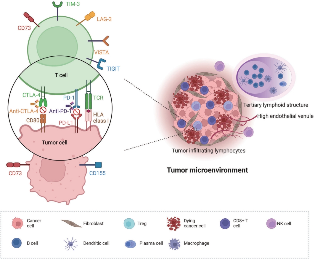
PDF - Treatment challenges persist in advanced lung cancer despite the development of therapies beyond the traditional platinum-based chemotherapy. The early 2000s marked a shift to tyrosine kinase inhibitors targeting epidermal growth factor receptor, ushering in personalized genetic-based treatment. A further significant advance was the development of immune checkpoint inhibitors (ICIs), especially for non–small cell lung cancer. These target programmed death-ligand 1 (PD-L1) and cytotoxic T lymphocyte antigen 4, which enhanced the immune response against tumor cells. However, not all patients respond, and immune-related toxicities arise. This review emphasizes identifying biomarkers for ICI response prediction. While PD-L1 is a widely used, validated biomarker, its predictive accuracy is imperfect. Investigating tumor-infiltrating lymphocytes, tertiary lymphoid structure, and emerging biomarkers such as high endothelial venule, Human leukocyte antigen class I, T-cell immunoreceptors with Ig and ITIM domains, and lymphocyte activation gene-3 counts is promising. Understanding and exploring additional predictive biomarkers for ICI response are crucial for enhancing patient stratification and overall care in lung cancer treatment.
-
Citations
Citations to this article as recorded by
- Machine learning methods for histopathological image analysis: Updates in 2024
Daisuke Komura, Mieko Ochi, Shumpei Ishikawa
Computational and Structural Biotechnology Journal.2025; 27: 383. CrossRef - Beyond single biomarkers: multi-omics strategies to predict immunotherapy outcomes in blood cancers
Mohammad Pirouzbakht, Soroosh Hamzeh, Hamed Soleimani Samarkhazan
Clinical and Experimental Medicine.2025;[Epub] CrossRef - Temporal changes in tongue color during immune checkpoint inhibitor therapy in patients with non-small-cell lung cancer: a prospective observational study using digital tongue diagnosis
Eunbyul Cho, Woosu Choi, Jun Hyeok Lim, Ji Woong Son, Seung Hun Jang, Seung Hyeun Lee, Jong Gwon Choi, In-Jae Oh, Tae-Won Jang, Seong Hoon Yoon, Seung Joon Kim, Chang-Min Choi, Sung Yong Lee, Mi Mi Ko, Mi-Kyung Jeong
Oncology Reviews.2025;[Epub] CrossRef
- Machine learning methods for histopathological image analysis: Updates in 2024
Case Study
- TTF1-positive SMARCA4/BRG1 deficient lung adenocarcinoma
- Anurag Mehta, Himanshi Diwan, Divya Bansal, Manoj Gupta
- J Pathol Transl Med. 2022;56(1):53-56. Published online November 16, 2021
- DOI: https://doi.org/10.4132/jptm.2021.09.16
- 9,095 View
- 197 Download
- 7 Web of Science
- 6 Crossref
-
Abstract
PDF
- SMARCA4/BRG1-deficient lung adenocarcinoma (SD-LUAD) is being recognized as a distinct subtype based on subtle differences in its clinical, morphological, and immunophenotypic attributes compared to other non–small cell lung carcinomas. We present here a case of SD-LUAD with curious thyroid transcription factor 1 (TTF1) expression in a morphologically heterogenous lung adenocarcinoma. The better differentiated area showed preservation of TTF1 expression, and a poorly differentiated tumor had loss of TTF1 expression with universal BRG1 loss.
-
Citations
Citations to this article as recorded by- Therapeutic misalignment averted by clonal evolutionary evidence: molecular confirmation of hepatic metastasis in SMARCA4-deficient non-small cell lung cancer initially misdiagnosed as resectable cholangiocarcinoma
Ruirui Fan, Yanyan Zhan, Junrong Yan, Jie Gao
Frontiers in Oncology.2026;[Epub] CrossRef - SMARCA4-deficient Non–small Cell Lung Cancer on 18F-FDG PET/CT
Tao Liu, Hengshan Ji, Siyuan Jiang, Rongxin Qi, Xiaodie Zhou, Jingjing Sun, Jiang Wu
Clinical Nuclear Medicine Open.2025;[Epub] CrossRef - Case Report: SMARCA4-deficient NSCLC with brain metastasis harboring co-mutations in chromatin remodeling and DNA damage repair pathways
Jiaqin Song, Shikun Yang, Lei Xia
Frontiers in Oncology.2025;[Epub] CrossRef - One Case of Non-Small Cell Lung Cancer with SMARCA4 Deletion Was Reported
允龙 宋
Medical Diagnosis.2024; 14(01): 137. CrossRef - Delineation of a SMARCA4-specific competing endogenous RNA network and its function in hepatocellular carcinoma
Lei Zhang, Ting Sun, Xiao-Ye Wu, Fa-Ming Fei, Zhen-Zhen Gao
World Journal of Clinical Cases.2022; 10(29): 10501. CrossRef - Novel germline SMARCA4 mutation in Small Cell Carcinoma of the Ovary, Hypercalcemic Type
Anurag Mehta, Himanshi Diwan, Diksha Karki, Divya Bansal, Meenakshi Kamboj, Anila Sharma, Shrinidhi Nathany, Sakshi Mattoo, Dushyant Kumar
Current Problems in Cancer: Case Reports.2022; 8: 100205. CrossRef
- Therapeutic misalignment averted by clonal evolutionary evidence: molecular confirmation of hepatic metastasis in SMARCA4-deficient non-small cell lung cancer initially misdiagnosed as resectable cholangiocarcinoma
Original Articles
- Programmed death-ligand 1 expression and tumor-infiltrating lymphocytes in non-small cell lung cancer: association with clinicopathologic parameters
- Gaurav Garg, Kuruswamy Thurai Prasad, Navneet Singh, Parul Gupta, Valliappan Muthu, Ashim Das, Amanjit Bal
- J Pathol Transl Med. 2021;55(6):398-405. Published online October 6, 2021
- DOI: https://doi.org/10.4132/jptm.2021.08.08
- 6,233 View
- 172 Download
- 4 Web of Science
- 3 Crossref
-
Abstract
PDF
 Supplementary Material
 - Background
Data on the prevalence of programmed death-ligand 1 (PD-L1) expression and tumor-infiltrating lymphocytes (TILs) in non–small cell lung cancer (NSCLC) and their clinical significance in Indian patients are limited.
Methods
Newly diagnosed NSCLC cases (adenocarcinoma or squamous cell carcinoma [SqCC] histology) were included in the present study. The TILs were evaluated based on morphology on hematoxylin and eosin–stained slides. PD-L1 expression in tumors was assessed using immunohistochemistry with rabbit monoclonal antibody (SP263) on the Ventana automated immunostainer. Tumors with PD-L1 expression > 50% on tumor cells were considered PD-L1–positive. Tumors in which TILs occupy > 25% of stroma were considered to have high TILs. The association of PD-L1 expression and TILs with various clinical parameters including overall survival (OS) was investigated.
Results
The present study included 128 cases of NSCLC (67 adenocarcinoma, 61 SqCC). PD-L1 positivity was observed in 17.2% of the patients with NSCLC. Baseline characteristics of PD-L1–positive subjects were similar to PD-L1–negative subjects except for a higher prevalence of liver metastasis (18.2% vs. 2.8%; p = .018) and a higher probability of diagnosis from extrapulmonary biopsies. High TILs were observed in 26.6% of the subjects. However, PD-L1 expression and high TIL did not affect OS.
Conclusions
PD-L1 positivity and high TILs were observed in 20% and 25% of the patients with NSCLC, respectively, however, neither were predictors of survival in SqCC. -
Citations
Citations to this article as recorded by- PDL1 and IDO‐2 Immunohistochemistry in Bronchoalveolar Lavage Versus Bronchoscopic Biopsy of Non‐Small Cell Lung Cancer
Menna Allah Hesham Mohammed Fekry, Yosria Mohammed El‐Gohary, Hesham Radwan Abd‐Elaziz, Tarek Hamdy Hassan, Mona Mostafa Ahmed
Cytopathology.2026; 37(2): 151. CrossRef - Multiplex plasma protein assays as a diagnostic tool for lung cancer
Mohammad Tanvir Ahamed, Jenny Forshed, Adrian Levitsky, Janne Lehtiö, Amanj Bajalan, Maria Pernemalm, Lars E. Eriksson, Björn Andersson
Cancer Science.2024; 115(10): 3439. CrossRef - Real-world prevalence of PD-L1 expression in non-small cell lung cancer: an Australia-wide multi-centre retrospective observational study
Prudence A. Russell, Alexandra L. Farrall, Sarita Prabhakaran, Khashayar Asadi, Wade Barrett, Caroline Cooper, Wendy Cooper, Samuel Cotton, Edwina Duhig, Matthew Egan, Stephen Fox, David Godbolt, Shilpa Gupta, Aniza Hassan, Connull Leslie, Trishe Leong, D
Pathology.2023; 55(7): 922. CrossRef
- PDL1 and IDO‐2 Immunohistochemistry in Bronchoalveolar Lavage Versus Bronchoscopic Biopsy of Non‐Small Cell Lung Cancer
- SMARCA4/BRG1 protein-deficient thoracic tumors dictate re-examination of small biopsy reporting in non–small cell lung cancer
- Anurag Mehta, Divya Bansal, Rupal Tripathi, Ankush Jajodia
- J Pathol Transl Med. 2021;55(5):307-316. Published online June 21, 2021
- DOI: https://doi.org/10.4132/jptm.2021.05.11
- 12,859 View
- 339 Download
- 13 Web of Science
- 10 Crossref
-
Abstract
PDF
- Background
SMARCA4/BRG1 protein–deficient lung adenocarcinomas and thoracic sarcoma are recently described entities that lack distinctive histological features, transcription termination factor 1 (TTF1) reactivity, and actionable driver mutations. The current diagnostic path for small lung biopsies as recommended by the World Health Organization (WHO, 2015) is likely to categorize these as non– small cell carcinoma–not otherwise specified (NSCC-NOS). The present study attempts to define the subtle but distinctive clinicopathologic features of SMARCA4/BRG1 protein-deficient thoracic tumors; highlight their unique biology; and addresses the unmet need to segregate these using a new, tissue-proficient diagnostic pathway.
Methods
All lung biopsies and those from metastatic sites in patients with suspected advanced lung cancer and classified as NSCC-NOS as per WHO (2015) guidelines were subjected to BRG1 testing by immunohistochemistry. SMARCA4/BRG1 protein–deficient thoracic tumors were evaluated by an extended immunohistochemistry panel. Predictive biomarker and programmed death–ligand 1 testing was conducted in all cases.
Results
Of 110 cases, nine were found to be SMARCA4/BRG1 protein-deficient; six were identified as SMARCA4/BRG1 protein–deficient lung adenocarcinomas, and three were SMARCA4/BRG1 protein-deficient thoracic sarcomas. The histology ranged from poorly differentiated to undifferentiated to rhabdoid. None of the cases showed significant expression of TTF1 or p40, and no actionable mutation was identified.
Conclusions
It is difficult to separate BRG1-deficient lung adenocarcinomas and thoracic sarcomas based on morphology alone. We propose a diagnostic pathway for small biopsies of thoracic tumors to segregate these distinct entities so that they can be studied more efficaciously for new biomarkers and therapeutic options. -
Citations
Citations to this article as recorded by- Unravelling switch/sucrose non-fermentable (SWI-SNF) complex-deficient thoracic tumours: a clinicopathological comparative on undifferentiated tumours and non-small cell lung carcinomas with BRG1 and BRM deficiency
Ridhi Sood, Arshi Tandon, Warisa Khatoon, Jayashimman Vasanthraman, Aruna Nambirajan, Anant Mohan, Prabhat Singh Malik, Deepali Jain
Journal of Clinical Pathology.2025; 78(6): 370. CrossRef - Clinicopathologic and genomic analyses of SMARCA4-mutated non-small cell lung carcinoma implicate the needs for tailored treatment strategies
Bokyung Ahn, Deokhoon Kim, Wonjun Ji, Sung-Min Chun, Goeun Lee, Se Jin Jang, Hee Sang Hwang
Lung Cancer.2025; 201: 108445. CrossRef - SMARCA4-deficient non-small cell lung cancer with metastasis to the sigmoid colon: a case report
Rong Xiao, Guang Fu, Xinglan Li, Tao Lu
World Journal of Surgical Oncology.2025;[Epub] CrossRef - Clinicopathological and molecular perspectives on thoracic SMARCA4-deficient undifferentiated tumors and SMARCA4-deficient non-small cell lung carcinomas
Sumanta Das, Pallavi Mishra, Sunita Ahlawat
Pathologica.2025; 117(5): 455. CrossRef - Case report: The first account of undifferentiated sarcoma with epithelioid features originating in the pleura
Ling-Xi Xiao, Li Liu, Wang Deng
Frontiers in Medicine.2024;[Epub] CrossRef - SMARCA4-deficient central nervous system metastases: A case series and systematic review
Meaghan Morris, Kerime Ararat, Hannah Cutshall, Murat Gokden, Analiz Rodriguez, Lisa Rooper, Matthew Lindberg, James Stephen Nix
Journal of Neuropathology & Experimental Neurology.2024; 83(8): 638. CrossRef - Chemotherapy and Immune Checkpoint Inhibitors in a Case of SMARCA4-dUT: A Case Report and Review of Literature
Akriti Pokhrel, Ruchi Yadav, Kapil Kumar Manvar, Richard Wu, Vijay Jaswani, Carrie Brooke Wasserman, Jen C. Wang
Journal of Investigative Medicine High Impact Case Reports.2023;[Epub] CrossRef - TTF1-positive SMARCA4/BRG1 deficient lung adenocarcinoma
Anurag Mehta, Himanshi Diwan, Divya Bansal, Manoj Gupta
Journal of Pathology and Translational Medicine.2022; 56(1): 53. CrossRef - Delineation of a SMARCA4-specific competing endogenous RNA network and its function in hepatocellular carcinoma
Lei Zhang, Ting Sun, Xiao-Ye Wu, Fa-Ming Fei, Zhen-Zhen Gao
World Journal of Clinical Cases.2022; 10(29): 10501. CrossRef - Artificial intelligence platform, RADR®, aids in the discovery of DNA damaging agent for the ultra-rare cancer Atypical Teratoid Rhabdoid Tumors
Joseph McDermott, Drew Sturtevant, Umesh Kathad, Sudhir Varma, Jianli Zhou, Aditya Kulkarni, Neha Biyani, Caleb Schimke, William C. Reinhold, Fathi Elloumi, Peter Carr, Yves Pommier, Kishor Bhatia
Frontiers in Drug Discovery.2022;[Epub] CrossRef
- Unravelling switch/sucrose non-fermentable (SWI-SNF) complex-deficient thoracic tumours: a clinicopathological comparative on undifferentiated tumours and non-small cell lung carcinomas with BRG1 and BRM deficiency
- p40 Immunohistochemistry Is an Excellent Marker in Primary Lung Squamous Cell Carcinoma
- Khairunisa Ahmad Affandi, Nur Maya Sabrina Tizen, Muaatamarulain Mustangin, Reena Rahayu MdReena Rahayu Md Zin
- J Pathol Transl Med. 2018;52(5):283-289. Published online August 31, 2018
- DOI: https://doi.org/10.4132/jptm.2018.08.14
- 27,991 View
- 349 Download
- 40 Web of Science
- 42 Crossref
-
Abstract
PDF
- Background
Lung cancer is the third most common cancer worldwide. With major advances in the molecular testing of lung cancers and the introduction of targeted therapies, the distinction between adenocarcinoma and squamous cell carcinoma as well as pathologic subtyping has become important. Recent studies showed that p40 is highly specific for squamous and basal cells and is superior to p63 for diagnosing lung squamous cell carcinoma. The aim of this study was to evaluate the use of p40 immunohistochemical stain in the diagnosis of non-small cell lung carcinoma and its potential to replace current p63 antibody as the best immunohistochemical squamous marker.
Methods
Seventy formalin-fixed paraffin-embedded cases previously diagnosed as primary lung squamous cell carcinoma (n = 35) and lung adenocarcinoma (n = 35) from January 2008 to December 2016 were retrieved. The results of tumour cell immunoreactivity for p40 and p63 antibodies in lung squamous cell carcinoma and lung adenocarcinoma were compared.
Results
p40 was expressed in 27 cases of lung squamous cell carcinoma (77.1%). All cases of lung adenocarcinoma (35/35, 100%) were negative for p40. p63 expression was positive in 30 cases of lung squamous cell carcinoma (85.7%) and 13 cases of lung adenocarcinoma (37.1%). Reactivity for both p40 and p63 in lung squamous cell carcinoma was strong and diffuse, whereas variable reactivity was observed in lung adenocarcinoma.
Conclusions
p40 is an excellent marker for distinguishing lung squamous cell carcinoma from adenocarcinoma, and p40 expression is equivalent to p63 expression in lung squamous cell carcinoma. -
Citations
Citations to this article as recorded by- Immunohistochemical expression of ΔNp63 versus pan-p63 in normal and neoplastic feline tissues
Igor R. Santos, Carolina B. Brunner, Milena C. Paz, Gabriela Hartmann, Gabrielle Z. Tres, Luciana Sonne, Welden Panziera, David Driemeier, Marcele B. Bandinelli, Saulo P. Pavarini
Veterinary Pathology.2026;[Epub] CrossRef - Partial Response to Repotrectinib in ROS1-Rearranged Lung Squamous Cell Carcinoma: A Brief Report
Yukiko Yoshida, Hajime Asahina, Ken Kuwahara, Hidenori Mizugaki, Noriyuki Yamada, Hiroshi Yokouchi, Naohiro Nomura, Yoshihiro Matsuno, Satoshi Oizumi
Clinical Lung Cancer.2026; 27(2): 80. CrossRef - Morphologic and immunohistochemical study of HPV-related cervical adenosquamous carcinoma: Reappraisal of a poorly defined entity
Zhenwei Zhang, M. Ruhul Quddus, C James Sung, Kamaljeet Singh
Human Pathology.2026; 170: 106052. CrossRef - Analysis of a two-marker immunohistochemistry panel (TTF-1 and p40) for distinguishing lung adenocarcinoma from squamous cell carcinoma on destained direct cytologic smears
Deepa Sowkur Anandarama Adiga, Harsha Kumar V S, Anusha S. Bhatt, Surbhi Patel, Daksha D. Shetty
Annals of Diagnostic Pathology.2026; 82: 152617. CrossRef - Thymidine phosphorylase promotes SARS-CoV-2 spike protein-driven lung tumor development
Cayleigh Wallace, Alex Gileles-Hillel, Amelia Cox, David Gozal, Wei Li, Hong Yue
Frontiers in Immunology.2026;[Epub] CrossRef - Label-free pathological subtyping of non-small cell lung cancer using deep classification and virtual immunohistochemical staining
Zhenya Zang, David A. Dorward, Katherine E. Quiohilag, Andrew DJ Wood, James R. Hopgood, Ahsan R. Akram, Qiang Wang
npj Digital Medicine.2026;[Epub] CrossRef - Abrupt-Onset Pink Scalp Nodules in an Adult: Answer
Anam Adil, Maria Mihailescu, Donald Lei
The American Journal of Dermatopathology.2026; 48(5): 408. CrossRef - Cancer of Unknown Primary With Squamous Cell Carcinoma Phenotype Presenting as Isolated Axillary Mass
Nicole Liang, Mohamed Alshal, Lynne J Goebel
Cureus.2025;[Epub] CrossRef - A critical appraisal of the clinico-radiological, histopathological and immunohistochemical profile of CT-guided and bronchoscopy-guided biopsies in lung lesions
Mukta Pujani, Ruchi Arora Sachdeva, S. Zafar Abbas, Charu Agarwal, Minakshi Bhardwaj, Varsha Chauhan, Jyoti Rajpoot, Dipti Sidam, Aniruna Dey
Lung India.2025; 42(3): 218. CrossRef - An Organoid Model for Translational Cancer Research Recapitulates Histoarchitecture and Molecular Hallmarks of Non-Small-Cell Lung Cancer
Camilla T. Ekanger, Maria P. Ramnefjell, Maren S. F. Guttormsen, Joakim Hekland, Kristin Dahl-Michelsen, Maria L. Lotsberg, Ning Lu, Linda E. B. Stuhr, Laurence Hoareau, Pirjo-Riitta Salminen, Fabian Gärtner, Marianne Aanerud, Lars A. Akslen, James B. Lor
Cancers.2025; 17(11): 1873. CrossRef - Accessing utility of immunohistochemistry, PD-L1 correlation with stage of cancer and EGFR mutation with disease survival in primary lung carcinoma
Kartavya Kumar Verma, Amit Bugalia, Ajoy Kumar behera, Nighat Hussain
Surgical and Experimental Pathology.2025;[Epub] CrossRef - Small intestinal SMARCA4‑deficient undifferentiated carcinoma with intussusception: A case report
Xiaoshan Cai, Shanshan Liu, Linqian Li, Yuming Zhang, Qin Guo, Ze Chen, Meihua Qu, Yubing Wang, Ben Yang, Xianwen Yue
Oncology Letters.2025; 31(2): 1. CrossRef - Clinicopathological and molecular perspectives on thoracic SMARCA4-deficient undifferentiated tumors and SMARCA4-deficient non-small cell lung carcinomas
Sumanta Das, Pallavi Mishra, Sunita Ahlawat
Pathologica.2025; 117(5): 455. CrossRef - Clinico-Pathological Profile of Non-Small Cell Lung Carcinoma with Emphasis on Diagnostic Immunohistochemistry – An Institutional Experience
Shubhika Rao Sachdeva, Bhavna Nayal, Ananth Pai
Medical Journal of Dr. D.Y. Patil Vidyapeeth.2025; 18(Suppl 2): S288. CrossRef - Lack of imbalance between the master regulators TTF1/NKX2-1 and ΔNp63/p40 implies adverse prognosis in non-small cell lung cancer
Martina Vescio, Matteo Bulloni, Giuseppe Pelosi, Linda Pattini
Scientific Reports.2024;[Epub] CrossRef - Adeno-to-squamous transition drives resistance to KRAS inhibition in LKB1 mutant lung cancer
Xinyuan Tong, Ayushi S. Patel, Eejung Kim, Hongjun Li, Yueqing Chen, Shuai Li, Shengwu Liu, Julien Dilly, Kevin S. Kapner, Ningxia Zhang, Yun Xue, Laura Hover, Suman Mukhopadhyay, Fiona Sherman, Khrystyna Myndzar, Priyanka Sahu, Yijun Gao, Fei Li, Fuming
Cancer Cell.2024; 42(3): 413. CrossRef - Common practice issues in pulmonary cytology/small biopsy: Diagnostic pitfalls and appropriate use of immunohistochemical stains
Jessie Xiong, Erik Polsdofer, Jian Jing
Human Pathology Reports.2024; 36: 300735. CrossRef - Integrating AI-Powered Digital Pathology and Imaging Mass Cytometry Identifies Key Classifiers of Tumor Cells, Stroma, and Immune Cells in Non–Small Cell Lung Cancer
Alessandra Rigamonti, Marika Viatore, Rebecca Polidori, Daoud Rahal, Marco Erreni, Maria Rita Fumagalli, Damiano Zanini, Andrea Doni, Anna Rita Putignano, Paola Bossi, Emanuele Voulaz, Marco Alloisio, Sabrina Rossi, Paolo Andrea Zucali, Armando Santoro, V
Cancer Research.2024; 84(7): 1165. CrossRef - Advances in combined neuroendocrine carcinoma of lung cancer
Zesen Han, Fujun Yang, Fang Wang, Huayu Zheng, Xiujian Chen, Hongyu Meng, Fenglei Li
Pathology and Oncology Research.2024;[Epub] CrossRef - Evolving Precision First-Line Systemic Treatment for Patients with Unresectable Non-Small Cell Lung Cancer
Tianhong Li, Weijie Ma, Ebaa Al-Obeidi
Cancers.2024; 16(13): 2350. CrossRef - Antibody-Conjugated Nanodiamond Enzyme for Targeted Non-Small-Cell Lung Cancer by Regulating Oxidative Stress
Xiaoying Guan, Jingwen Yang, Ziying Liu, Guowei Yang, Xiumei Tian
ACS Applied Nano Materials.2024; 7(20): 23670. CrossRef - Multifocal Pulmonary Opacities in an Elderly Smoker
Patton C McClelland, Zachary Jarrett, Christian C Lamb, Mateo Houle
Cureus.2024;[Epub] CrossRef - Clonal dynamics and Stereo-seq resolve origin and phenotypic plasticity of adenosquamous carcinoma
Ruiying Zhao, Yunhua Xu, Yedan Chen, Jiajun Zhang, Fei Teng, Sha Liao, Shengnan Chen, Qian Wu, Chan Xiang, Jiaohui Pang, Zhanxian Shang, Jikai Zhao, Hairong Bao, Hua Bao, Yang Shao, Shun Lu, Yuchen Han
npj Precision Oncology.2023;[Epub] CrossRef - Keratin 5 in Lung Cancer Specimens: Comparison of Four Antibody Clones and KRT5 mRNA-ISH
Christian Thomsen, Laura Blok-Husum, Jeanette Bæhr Georgsen, Torben Steiniche, Mogens Vyberg
Applied Immunohistochemistry & Molecular Morphology.2023; 31(6): 347. CrossRef - Carcinoma of unknown primary (CUP): an update for histopathologists
Katie Beauchamp, Bruce Moran, Timothy O’Brien, Donal Brennan, John Crown, Kieran Sheahan, Maura Bríd Cotter
Cancer and Metastasis Reviews.2023; 42(4): 1189. CrossRef - Advances in Genomic Data and Biomarkers: Revolutionizing NSCLC Diagnosis and Treatment
Juan Carlos Restrepo, Diana Dueñas, Zuray Corredor, Yamil Liscano
Cancers.2023; 15(13): 3474. CrossRef - Kallikrein-related peptidase 13 expression and clinicopathological features in lung squamous cell carcinoma
Ryusuke Sumiya, Kazuhiko Yamada, Teruki Hagiwara, Satoshi Nagasaka, Hideki Miyazaki, Toru Igari, Yuki Kawamura
Molecular and Clinical Oncology.2023;[Epub] CrossRef - Pre-clinical lung squamous cell carcinoma mouse models to identify novel biomarkers and therapeutic interventions
Priyanka Sahu, Chantal Donovan, Keshav Raj Paudel, Sophie Pickles, Vrushali Chimankar, Richard Y. Kim, Jay C. Horvart, Kamal Dua, Antonio Ieni, Francesco Nucera, Helle Bielefeldt-Ohmann, Sarah Mazilli, Gaetano Caramori, J. Guy Lyons, Philip M. Hansbro
Frontiers in Oncology.2023;[Epub] CrossRef - Intraosseous clear cell mucoepidermoid carcinoma: A case report and evaluation
Adesh S. Manchanda, Ramandeep S. Narang, Komaldeep K. Sandhu
Journal of Oral and Maxillofacial Pathology.2023; 27(4): 780. CrossRef - A targeted expression panel for classification, gene fusion detection and PD-L1 measurements – Can molecular profiling replace immunohistochemistry in non-small cell lung cancer?
Anita Tranberg Simonsen, Amalie Utke, Johanne Lade-Keller, Lasse Westphal Thomsen, Torben Steiniche, Magnus Stougaard
Experimental and Molecular Pathology.2022; 125: 104749. CrossRef - Basal cell carcinoma arising in association with maxillary odontogenic keratocyst in a patient with Gorlin-Goltz syndrome
Mutsuki Kawabe, Yoshitane Tsukamoto, Shohei Matuo, Shuji Kanda, Susumu Hashitani
Journal of Oral and Maxillofacial Surgery, Medicine, and Pathology.2022; 34(3): 333. CrossRef - Cutaneous Metastases from Thymic Carcinoma Primary Tumor: A Rare Case
Eva Krishna Sutedja, Trustia Rizqandaru, Kartika Ruchiatan, Endang Sutedja
International Medical Case Reports Journal.2022; Volume 15: 293. CrossRef - Primary nonkeratinizing squamous cell carcinoma of the scapular bone: A case report
Yang Li, Jian-Lin Zuo, Jin-Shuo Tang, Xian-Yue Shen, Sheng-Hao Xu, Jian-Lin Xiao
World Journal of Clinical Cases.2021; 9(4): 976. CrossRef - Survival-associated N6-adenosine methyltransferase signatures in lung squamous cell carcinoma and clinical verification
Jialin Qu, Li Wang, Man Jiang, Zhimin Wei, Guangming Fu, Xiaochun Zhang
BMC Cancer.2021;[Epub] CrossRef - Pulmonary adenomyoma presenting as a right cardiophrenic angle mass
Osigbemhe Iyalomhe, Sam Sadigh, Charuhas Deshpande, Leslie Litzky, Anna Moran, Scott Simpson
Radiology Case Reports.2020; 15(5): 502. CrossRef - Head-to-Head Comparison of p63 and p40 in Non-Neuroendocrine Carcinomas of the Tubal Gut
Ahmed M. Bakhshwin, Ilyssa O. Gordon, Kathryn Bock Brown, Xiuli Liu, Daniela S. Allende
International Journal of Surgical Pathology.2020; 28(8): 835. CrossRef - Greater specificity of p40 compared with p63 in distinguishing squamous cell carcinoma from adenocarcinoma in effusion cellblocks
Nah Ihm Kim, Ji Shin Lee
Cytojournal.2020; 17: 13. CrossRef - Incidental Thyroid Mass in a Patient With Oropharyngeal Squamous Cell Carcinoma
Kinneri Mehta, Misha Movahed-Ezazi, Akshay V. Patel
JAMA Otolaryngology–Head & Neck Surgery.2020; 146(9): 859. CrossRef - Three dimensional texture analysis of noncontrast chest CT in differentiating solitary solid lung squamous cell carcinoma from adenocarcinoma and correlation to immunohistochemical markers
Rui Han, Roshan Arjal, Jin Dong, Hong Jiang, Huan Liu, Dongyou Zhang, Lu Huang
Thoracic Cancer.2020; 11(11): 3099. CrossRef - The role of the immunohistochemical marker p40 in the differential diagnosis of adenocarcinoma and nonkeratinizing squamous cell cancer of the lung
E.M. Olyushina, M.M. Byakhova, L.E. Zavalishina, Yu.Yu. Andreeva, A.B. Semenova, G.A. Frank
Arkhiv patologii.2020; 82(5): 50. CrossRef - Role of Immunocytochemistry in the Cytological Diagnosis of Pulmonary Tumors
Jasna Metovic, Luisella Righi, Luisa Delsedime, Marco Volante, Mauro Papotti
Acta Cytologica.2020; 64(1-2): 16. CrossRef - Subtyping Lung Cancer Using DNA Methylation in Liquid Biopsies
Sandra P. Nunes, Francisca Diniz, Catarina Moreira-Barbosa, Vera Constâncio, Ana Victor Silva, Júlio Oliveira, Marta Soares, Sofia Paulino, Ana Luísa Cunha, Jéssica Rodrigues, Luís Antunes, Rui Henrique, Carmen Jerónimo
Journal of Clinical Medicine.2019; 8(9): 1500. CrossRef
- Immunohistochemical expression of ΔNp63 versus pan-p63 in normal and neoplastic feline tissues
- Association of CXCR4 Expression with Metastasis and Survival among Patients with Non-small Cell Lung Cancer.
- Joon Seon Song, Jin Kyung Jung, Jong Chul Park, Dong Kwan Kim, Se Jin Jang
- Korean J Pathol. 2008;42(6):358-364.
- 2,348 View
- 27 Download
-
Abstract
PDF
- BACKGROUND
Expression of CXCR4 chemokine receptor, initially described to be involved in the homing of lymphocytes in inflammatory tissue, on breast cancer cell lines is associated with the development of lung metastases. In the present study, we evaluated CXCR4 expression in patients with non-small cell lung cancer (NSCLC). METHODS: Tissue microarray blocks were constructed from 408 formalin-fixed, paraffin-embedded NSCLC samples and analyzed via immunohistochemical staining. RESULTS: We observed CXCR4 expression in 214 (66.3%) of the 323 tumors with cytoplasmic or nuclear staining patterns. These tumors were then divided into 109 negative, 166 weak-positive and 48 strong-positive expression groups. Strong expression of CXCR4 correlated with NSCLC recurrence (p=0.047) and distant metastasis (p=0.035). However, lymph node metastasis (p=0.683) and locoregional recurrence (p=0.856) were not associated with CXCR4 expression. Interestingly, the median overall survival times relative to CXCR4 expression were 71 months in the CXCR4-negative group, 43 months in the weakly positive CXCR4 group and 23 months in the strongly positive CXCR4 group. Strongly positive CXCR4 staining was associated with significantly worse outcomes (p=0.005, log-rank test).
CONCLUSIONS
Expression of CXCR4 was associated with distant NSCLC metastases and shorter survival times.
- Relationship among the Expression of Cyclin D1, p21, and p53 Protein, and Prognosis in Non-Small Cell Lung Carcinomas.
- Seok Woo Yang, Sang Ho Cho, Woo Ick Yang, Woo Hee Jung, Chul Min Ahn, Doo Yun Lee
- Korean J Pathol. 1999;33(12):1120-1130.
- 2,300 View
- 21 Download
-
Abstract
PDF
- Recently, cell cycle regulators have been suggested as new prognostic factors of the lung cancer. In this study, we evaluated the expression of cyclin D1, p21, and p53 using the X2-test, with regard to the stage of the patients, histologic type, and histologic differentiation in the 135 cases of non-small cell lung carcinomas (NSCLC). To evaluate the confounding effects among cyclin D1, p21, and p53 on X2-test analysis, we used the Mantel-Haenzel test. The NSCLC in this study included 82 cases of squamous cell carcinoma and 53 cases of adenocarcinoma. Each nuclear staining of cyclin D1, p21, and p53 was observed in 65 cases (48.1%), in 54 cases (40.0%), and in 81 cases (60.0%) of NSCLCs, respectively. Only p53 expression was significantly associated with the stage (stage I, II, IIIa) (p<0.05) and squamous cell carcinoma (p<0.05). On the other hand, cyclin D1 expression was significantly associated with the histologic differentiation. The confounding effects among cyclin D1, p21, and p53 revealed that only p21 expression changed the relationship between p53 and stage. In this regard, further study is needed.
- Expression of E-cadherin, Matrix Metalloproteinase, and Vascular Endothelial Growth Factor in Squamous Cell Carcinoma and Adenocarcinoma of the Lung.
- Ji Sun Song, Mee Yon Cho, Kwang Hwa Park, Soon Hee Jung, Kwang Gil Lee
- Korean J Pathol. 2000;34(12):972-981.
- 2,161 View
- 14 Download
-
Abstract
PDF
- E-cadherin is a calcium-dependent epithelial adhesion molecule which plays a role in the initial step of invasion of cancer cells. The step that follows the migration of separated tumor cells is a proteolytic lysis of basement membrane and extracellular matrix by protease of epithelial and endothelial cells such as matrix metalloproteinase (MMP). Vascular endothelial growth factor (VEGF) is known to be an endothelial cell-specific powerful mitogen as well as a vascular permeability factor. This study is aimed to evaluate the correlation between expression of these factors and pathologic or clinical variables and the roles and prognostic significance of those factors in squmous cell carcinoma and adenocarcinoma of the lung. Immunohistochemical stains were performed for E-cadherin, MMP-2, and VEGF in surgically resected specimens from 90 patients with squmous cell carcinoma and adenocarcinoma of the lung. Mean age of the patients was 59.7 years. Histologic type was categorized into 56 cases of squamous cell carcinoma and 34 cases of adenocarcinoma. Mean survival period of the 35 patients was 54 months. The immunohistochemical stains for E-cadherin, MMP-2, and VEGF revealed positive reaction in 67 cases (74.4%), 43 cases (47.8%), and 34 cases (37.8%), respectively. The expression of E-cadherin was higher in adenocarcinoma (82.4%) than in squamous cell carcinoma (69.6%). MMP-2 was expressed in the tumor cells, especially those invading into the surrounding stroma. The expression of MMP-2 was significantly correlated with the survival rate (p<0.05). The expression of VEGF in the tumor cells was significantly higher in cases with lymph node metastasis (p<0.05). In conclusion, these findings suggest that the expression of MMP-2 and VEGF predict poor prognosis of patients with squmous cell carcinoma and adenocarcinoma of the lung and that VEGF may play a role in tumor metastasis.
- Altered Fhit Expression and Its Relationship with p53 Overexpression in Non-small Cell Lung Cancers.
- Na Hye Myong, Seok Jun Yoon
- Korean J Pathol. 2001;35(1):1-6.
- 2,153 View
- 16 Download
-
Abstract
PDF
- BACKGROUND
FHIT (Fragile histidine triad), the tumor suppressor gene at 3p14.2, encompasses the FRA3B fragile site and is a common target of deletions in primary human epithelial cancers, including those of the lung, head and neck, stomach, cervix, breast, and kidney. We investigated the association of Fhit expression with clinicopathologic features, including smoking history, and tried to correlate its altered expression with p53 overexpression in 45 non-small cell lung cancers.
METHODS
Immunohistochemical staining was performed on the paraffin sections, using primary anti-GST-Fhit and anti-human p53 antibodies. A four-tiered scoring system, incorporateing both intensity of staining and the percentage of cells stained was used. Composite scores < or = 3 were defined as a marked reduction or loss of Fhit or p53 protein expression.
RESULTS
Among the 45 tumors analyzed, 35 (77.8%) were markedly reduced or negative for Fhit protein expression. The reduced expression of Fhit protein was found to be significantly higher in smokers than in nonsmokers and also higher in squamous carcinoma compared with adenocarcinoma. Fhit and p53 alterations were found to be independent events, because there was no significant difference of Fhit-negativity between p53-positive and -negative groups.
CONCLUSION
These results indicate that the Fhit alteration preferentially occurs in smokers and in the squamous type of non-small cell lung carcinomas. In addition, the results support the notion that Fhit alterations play an important role in the pulmonary carcinogenesis.
 First
First Prev
Prev


