 E-submission
Search
- Page Path
- HOME > Search
- Diagnostic proficiency test using digital cytopathology and comparative assessment of whole slide images of cytologic samples for quality assurance program in Korea
- Yosep Chong, Soon Auck Hong, Hoon Kyu Oh, Soo Jin Jung, Bo-Sung Kim, Ji Yun Jeong, Ho-Chang Lee, Gyungyub Gong
- J Pathol Transl Med. 2023;57(5):251-264. Published online August 24, 2023
- DOI: https://doi.org/10.4132/jptm.2023.07.17
- 8,293 View
- 343 Download
- 9 Web of Science
- 9 Crossref
-
 Abstract
 PDF
PDF Supplementary Material
 - Background
The Korean Society for Cytopathology introduced a digital proficiency test (PT) in 2021. However, many doubtful opinions remain on whether digitally scanned images can satisfactorily present subtle differences in the nuclear features and chromatin patterns of cytological samples.
Methods
We prepared 30 whole-slide images (WSIs) from the conventional PT archive by a selection process for digital PT. Digital and conventional PT were performed in parallel for volunteer institutes, and the results were compared using feedback. To assess the quality of cytological assessment WSIs, 12 slides were collected and scanned using five different scanners, with four cytopathologists evaluating image quality through a questionnaire.
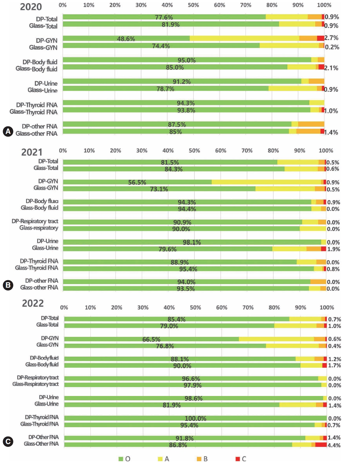
Results
Among the 215 institutes, 108 and 107 participated in glass and digital PT, respectively. No significant difference was noted in category C (major discordance), although the number of discordant cases was slightly higher in the digital PT group. Leica, 3DHistech Pannoramic 250 Flash, and Hamamatsu NanoZoomer 360 systems showed comparable results in terms of image quality, feature presentation, and error rates for most cytological samples. Overall satisfaction was observed with the general convenience and image quality of digital PT.
Conclusions
As three-dimensional clusters are common and nuclear/chromatin features are critical for cytological interpretation, careful selection of scanners and optimal conditions are mandatory for the successful establishment of digital quality assurance programs in cytology. -
Citations
Citations to this article as recorded by
- Practice of Cytopathology in Korea: A 40‐Year Evolution Through Standardization, Digital Transformation, and Global Partnership
Yosep Chong, Ran Hong, Hyeong Ju Kwon, Haeryoung Kim, Lucia Kim, Soon Jae Kim, Yoon Jung Choi
Diagnostic Cytopathology.2026; 54(2): 146. CrossRef - ThinPrep® Whole-Slide Digital Images versus Conventional Microscopy in Negative for Intraepithelial Lesion or Malignancy, Atypical Squamous Cells of Undetermined Significance, and Low-Grade Squamous Intraepithelial Lesion Cervical Lesions: European Federa
Ester Puntonen, Sara Bazzon, Massimo Bongiovanni, Rosario Granados, Ines Krivak Bolanca, Maria Nasioutziki, Maurizio Pinamonti, Danijela Vrdoljak-Mozetic, Arrigo Capitanio, Beatrix Cochand-Priollet, Ambrogio Fassina, Giovanni Negri, Laura Ventura, Ivana K
Pathobiology.2026; : 1. CrossRef - Sensitivity, Specificity, and Cost–Benefit Effect Between Primary Human Papillomavirus Testing, Primary Liquid‐Based Cytology, and Co‐Testing Algorithms for Cervical Lesions
Chang Gok Woo, Seung‐Myoung Son, Hye‐Kyung Hwang, Jung‐Sil Bae, Ok‐Jun Lee, Ho‐Chang Lee
Diagnostic Cytopathology.2025; 53(1): 35. CrossRef - Integration of AI‐Assisted in Digital Cervical Cytology Training: A Comparative Study
Yihui Yang, Dongyi Xian, Lihua Yu, Yanqing Kong, Huaisheng Lv, Liujing Huang, Kai Liu, Hao Zhang, Weiwei Wei, Hongping Tang
Cytopathology.2025; 36(2): 156. CrossRef - National quality assurance program using digital cytopathology: a 5-year digital transformation experience by the Korean Society for Cytopathology
Yosep Chong, Hyeong Ju Kwon, Soon Auck Hong, Sung Soon Kim, Bo-Sung Kim, Younghee Choi, Yoon Jung Choi, Jung-Soo Pyo, Ji Yun Jeong, Soo Jin Jung, Hoon Kyu Oh, Seung-Sook Lee
Journal of Pathology and Translational Medicine.2025; 59(5): 320. CrossRef - Integration of Digital Cytology in Quality Assurance Programs for Cytopathology
Yosep Chong, Maria Jesús Fernández Aceñero, Zaibo Li, Andrey Bychkov
Acta Cytologica.2025; 70(1): 126. CrossRef - Quantitative Assessment of Focus Quality in Whole-Slide Imaging of Thyroid Liquid-Based Cytology Using Laplacian Variance
Chan Kwon Jung, Chankyung Kim, Sora Jeon, Andrey Bychkov
Endocrine Pathology.2025;[Epub] CrossRef - Validation of digital image slides for diagnosis in cervico-vaginal cytology
Francisco Tresserra, Gemma Fabra, Olga Luque, Miriam Castélla, Carla Gómez, Carmen Fernández-Cid, Ignacio Rodríguez
Revista Española de Patología.2024; 57(3): 182. CrossRef - Improved Diagnostic Accuracy of Thyroid Fine-Needle Aspiration Cytology with Artificial Intelligence Technology
Yujin Lee, Mohammad Rizwan Alam, Hongsik Park, Kwangil Yim, Kyung Jin Seo, Gisu Hwang, Dahyeon Kim, Yeonsoo Chung, Gyungyub Gong, Nam Hoon Cho, Chong Woo Yoo, Yosep Chong, Hyun Joo Choi
Thyroid®.2024; 34(6): 723. CrossRef
- Practice of Cytopathology in Korea: A 40‐Year Evolution Through Standardization, Digital Transformation, and Global Partnership
- Postmortem lung and heart examination of COVID-19 patients in a case series from Jordan
- Maram Abdaljaleel, Isra Tawalbeh, Malik Sallam, Amjad Bani Hani, Imad M. Al-Abdallat, Baheth Al Omari, Sahar Al-Mustafa, Hasan Abder-Rahman, Adnan Said Abbas, Mahmoud Zureigat, Mousa A. Al-Abbadi
- J Pathol Transl Med. 2023;57(2):102-112. Published online March 14, 2023
- DOI: https://doi.org/10.4132/jptm.2023.01.30
- 7,379 View
- 171 Download
-
Abstract
PDF
- Background
Coronavirus disease 2019 (COVID-19) has emerged as a pandemic for more than 2 years. Autopsy examination is an invaluable tool to understand the pathogenesis of emerging infections and their consequent mortalities. The aim of the current study was to present the lung and heart pathological findings of COVID-19–positive autopsies performed in Jordan.
Methods
The study involved medicolegal cases, where the cause of death was unclear and autopsy examination was mandated by law. We included the clinical and pathologic findings of routine gross and microscopic examination of cases that were positive for COVID-19 at time of death. Testing for severe acute respiratory syndrome coronavirus 2 (SARS-CoV-2) was confirmed through molecular detection by real-time polymerase chain reaction, serologic testing for IgM and electron microscope examination of lung samples.
Results
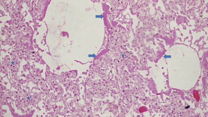
Seventeen autopsies were included, with male predominance (76.5%), Jordanians (70.6%), and 50 years as the mean age at time of death. Nine out of 16 cases (56.3%) had co-morbidities, with one case lacking such data. Histologic examination of lung tissue revealed diffuse alveolar damage in 13/17 cases (76.5%), and pulmonary microthrombi in 8/17 cases (47.1%). Microscopic cardiac findings were scarcely detected. Two patients died as a direct result of acute cardiac disease with limited pulmonary findings.
Conclusions
The detection of SARS-CoV-2 in postmortem examination can be an incidental or contributory finding which highlights the value of autopsy examination to determine the exact cause of death in controversial cases.
- Development of quality assurance program for digital pathology by the Korean Society of Pathologists
- Yosep Chong, Jeong Mo Bae, Dong Wook Kang, Gwangil Kim, Hye Seung Han
- J Pathol Transl Med. 2022;56(6):370-382. Published online November 15, 2022
- DOI: https://doi.org/10.4132/jptm.2022.09.30
- 7,207 View
- 163 Download
- 5 Web of Science
- 5 Crossref
-
Abstract
PDFSupplementary Material
- Background
Digital pathology (DP) using whole slide imaging is a recently emerging game changer technology that can fundamentally change the way of working in pathology. The Digital Pathology Study Group (DPSG) of the Korean Society of Pathologists (KSP) published a consensus report on the recommendations for pathologic practice using DP. Accordingly, the need for the development and implementation of a quality assurance program (QAP) for DP has been raised.
Methods
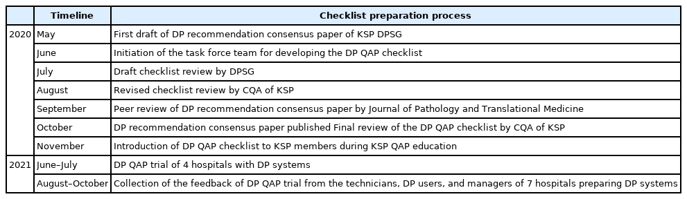
To provide a standard baseline reference for internal and external QAP for DP, the members of the Committee of Quality Assurance of the KSP developed a checklist for the Redbook and a QAP trial for DP based on the prior DPSG consensus report. Four leading institutes participated in the QAP trial in the first year, and we gathered feedback from these institutes afterwards.
Results
The newly developed checklists of QAP for DP contain 39 items (216 score): eight items for quality control of DP systems; three for DP personnel; nine for hardware and software requirements for DP systems; 15 for validation, operation, and management of DP systems; and four for data security and personal information protection. Most participants in the QAP trial replied that continuous education on unfamiliar terminology and more practical experience is demanding.
Conclusions
The QAP for DP is essential for the safe implementation of DP in pathologic practice. Each laboratory should prepare an institutional QAP according to this checklist, and consecutive revision of the checklist with feedback from the QAP trial for DP needs to follow. -
Citations
Citations to this article as recorded by- Practice of Cytopathology in Korea: A 40‐Year Evolution Through Standardization, Digital Transformation, and Global Partnership
Yosep Chong, Ran Hong, Hyeong Ju Kwon, Haeryoung Kim, Lucia Kim, Soon Jae Kim, Yoon Jung Choi
Diagnostic Cytopathology.2026; 54(2): 146. CrossRef - An equivalency and efficiency study for one year digital pathology for clinical routine diagnostics in an accredited tertiary academic center
Viola Iwuajoku, Kübra Ekici, Anette Haas, Mohammed Zaid Khan, Azar Kazemi, Atsuko Kasajima, Claire Delbridge, Alexander Muckenhuber, Elisa Schmoeckel, Fabian Stögbauer, Christine Bollwein, Kristina Schwamborn, Katja Steiger, Carolin Mogler, Peter J. Schüf
Virchows Archiv.2025; 487(1): 3. CrossRef - Quality Assurance of the Whole Slide Image Evaluation in Digital Pathology: State of the Art and Development Results
Miklós Vincze, Béla Molnár, Miklós Kozlovszky
Electronics.2025; 14(10): 1943. CrossRef - Integration of Digital Cytology in Quality Assurance Programs for Cytopathology
Yosep Chong, Maria Jesús Fernández Aceñero, Zaibo Li, Andrey Bychkov
Acta Cytologica.2025; 70(1): 126. CrossRef - Diagnostic proficiency test using digital cytopathology and comparative assessment of whole slide images of cytologic samples for quality assurance program in Korea
Yosep Chong, Soon Auck Hong, Hoon Kyu Oh, Soo Jin Jung, Bo-Sung Kim, Ji Yun Jeong, Ho-Chang Lee, Gyungyub Gong
Journal of Pathology and Translational Medicine.2023; 57(5): 251. CrossRef
- Practice of Cytopathology in Korea: A 40‐Year Evolution Through Standardization, Digital Transformation, and Global Partnership
- Recommendations for pathologic practice using digital pathology: consensus report of the Korean Society of Pathologists
- Yosep Chong, Dae Cheol Kim, Chan Kwon Jung, Dong-chul Kim, Sang Yong Song, Hee Jae Joo, Sang-Yeop Yi
- J Pathol Transl Med. 2020;54(6):437-452. Published online October 8, 2020
- DOI: https://doi.org/10.4132/jptm.2020.08.27
- 13,187 View
- 340 Download
- 26 Web of Science
- 30 Crossref
-
Abstract
PDFSupplementary Material
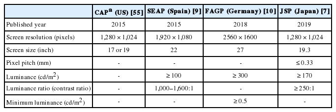
- Digital pathology (DP) using whole slide imaging (WSI) is becoming a fundamental issue in pathology with recent advances and the rapid development of associated technologies. However, the available evidence on its diagnostic uses and practical advice for pathologists on implementing DP remains insufficient, particularly in light of the exponential growth of this industry. To inform DP implementation in Korea, we developed relevant and timely recommendations. We first performed a literature review of DP guidelines, recommendations, and position papers from major countries, as well as a review of relevant studies validating WSI. Based on that information, we prepared a draft. After several revisions, we released this draft to the public and the members of the Korean Society of Pathologists through our homepage and held an open forum for interested parties. Through that process, this final manuscript has been prepared. This recommendation contains an overview describing the background, objectives, scope of application, and basic terminology; guidelines and considerations for the hardware and software used in DP systems and the validation required for DP implementation; conclusions; and references and appendices, including literature on DP from major countries and WSI validation studies.
-
Citations
Citations to this article as recorded by- Commercially Available Artificial Intelligence Solutions for Gynaecologic Cytology Screening and Their Integration Into Clinical Workflow
Yosep Chong, Andrey Bychkov
Cytopathology.2026; 37(1): 24. CrossRef - The impact of AI on modern oncology from early detection to personalized cancer treatment
Jun Li, Lei Zhang, Zhenglun Yu, Zhiye Bao, Danyang Li, Liming Wang
npj Precision Oncology.2026;[Epub] CrossRef - An equivalency and efficiency study for one year digital pathology for clinical routine diagnostics in an accredited tertiary academic center
Viola Iwuajoku, Kübra Ekici, Anette Haas, Mohammed Zaid Khan, Azar Kazemi, Atsuko Kasajima, Claire Delbridge, Alexander Muckenhuber, Elisa Schmoeckel, Fabian Stögbauer, Christine Bollwein, Kristina Schwamborn, Katja Steiger, Carolin Mogler, Peter J. Schüf
Virchows Archiv.2025; 487(1): 3. CrossRef - An adapted & improved validation protocol for digital pathology implementation
Ying-Han R. Hsu, Iman Ahmed, Juliana Phlamon, Charlotte Carment-Baker, Joyce Yin Tung Chan, Ioannis Prassas, Karen Weiser, Shaza Zeidan, Blaise Clarke, George M. Yousef
Seminars in Diagnostic Pathology.2025; 42(4): 150905. CrossRef - Transforming pathology into digital pathology: highway to hell or stairway to heaven?
Rainer Grobholz, Andrew Janowczyk, Inti Zlobec
Diagnostic Histopathology.2025; 31(7): 410. CrossRef - The Evolution of Digital Pathology in Infrastructure, Artificial Intelligence and Clinical Impact
Chan Kwon Jung
International Journal of Thyroidology.2025; 18(1): 6. CrossRef - Current Trends and Future Directions of Digital Pathology and Artificial Intelligence in Dermatopathology: A Scientometric-Based Review
Iuliu Gabriel Cocuz, Raluca Niculescu, Maria-Cătălina Popelea, Maria Elena Cocuz, Adrian-Horațiu Sabău, Andreea-Cătălina Tinca, Andreea Raluca Cozac-Szoke, Diana Maria Chiorean, Corina Eugenia Budin, Ovidiu Simion Cotoi
Diagnostics.2025; 15(17): 2196. CrossRef - Integration of Digital Cytology in Quality Assurance Programs for Cytopathology
Yosep Chong, Maria Jesús Fernández Aceñero, Zaibo Li, Andrey Bychkov
Acta Cytologica.2025; 70(1): 126. CrossRef - Quantitative Assessment of Focus Quality in Whole-Slide Imaging of Thyroid Liquid-Based Cytology Using Laplacian Variance
Chan Kwon Jung, Chankyung Kim, Sora Jeon, Andrey Bychkov
Endocrine Pathology.2025;[Epub] CrossRef - Performance of externally validated machine learning models based on histopathology images for the diagnosis, classification, prognosis, or treatment outcome prediction in female breast cancer: A systematic review
Ricardo Gonzalez, Peyman Nejat, Ashirbani Saha, Clinton J.V. Campbell, Andrew P. Norgan, Cynthia Lokker
Journal of Pathology Informatics.2024; 15: 100348. CrossRef - Swiss digital pathology recommendations: results from a Delphi process conducted by the Swiss Digital Pathology Consortium of the Swiss Society of Pathology
Andrew Janowczyk, Inti Zlobec, Cedric Walker, Sabina Berezowska, Viola Huschauer, Marianne Tinguely, Joel Kupferschmid, Thomas Mallet, Doron Merkler, Mario Kreutzfeldt, Radivoje Gasic, Tilman T. Rau, Luca Mazzucchelli, Isgard Eyberg, Gieri Cathomas, Kirst
Virchows Archiv.2024; 485(1): 13. CrossRef - ChatGPT as an aid for pathological diagnosis of cancer
Shaivy Malik, Sufian Zaheer
Pathology - Research and Practice.2024; 253: 154989. CrossRef - Possible benefits, challenges, pitfalls, and future perspective of using ChatGPT in pathology
Durre Aden, Sufian Zaheer, Sabina Khan
Revista Española de Patología.2024; 57(3): 198. CrossRef - Remote Placental Sign-Out: What Digital Pathology Can Offer for Pediatric Pathologists
Casey P. Schukow, Jacqueline K. Macknis
Pediatric and Developmental Pathology.2024; 27(4): 375. CrossRef - Digital Validation in Breast Cancer Needle Biopsies: Comparison of Histological Grade and Biomarker Expression Assessment Using Conventional Light Microscopy, Whole Slide Imaging, and Digital Image Analysis
Ji Eun Choi, Kyung-Hee Kim, Younju Lee, Dong-Wook Kang
Journal of Personalized Medicine.2024; 14(3): 312. CrossRef - Pathologists light level preferences using the microscope—study to guide digital pathology display use
Charlotte Jennings, Darren Treanor, David Brettle
Journal of Pathology Informatics.2024; 15: 100379. CrossRef - Eye tracking in digital pathology: A comprehensive literature review
Alana Lopes, Aaron D. Ward, Matthew Cecchini
Journal of Pathology Informatics.2024; 15: 100383. CrossRef - Diagnostic Assessment of Deep Learning Algorithms for Frozen Tissue Section Analysis in Women with Breast Cancer
Young-Gon Kim, In Hye Song, Seung Yeon Cho, Sungchul Kim, Milim Kim, Soomin Ahn, Hyunna Lee, Dong Hyun Yang, Namkug Kim, Sungwan Kim, Taewoo Kim, Daeyoung Kim, Jonghyeon Choi, Ki-Sun Lee, Minuk Ma, Minki Jo, So Yeon Park, Gyungyub Gong
Cancer Research and Treatment.2023; 55(2): 513. CrossRef - Recent application of artificial intelligence on histopathologic image-based prediction of gene mutation in solid cancers
Mohammad Rizwan Alam, Kyung Jin Seo, Jamshid Abdul-Ghafar, Kwangil Yim, Sung Hak Lee, Hyun-Jong Jang, Chan Kwon Jung, Yosep Chong
Briefings in Bioinformatics.2023;[Epub] CrossRef - Sustainable development goals applied to digital pathology and artificial intelligence applications in low- to middle-income countries
Sumi Piya, Jochen K. Lennerz
Frontiers in Medicine.2023;[Epub] CrossRef - Diagnostic proficiency test using digital cytopathology and comparative assessment of whole slide images of cytologic samples for quality assurance program in Korea
Yosep Chong, Soon Auck Hong, Hoon Kyu Oh, Soo Jin Jung, Bo-Sung Kim, Ji Yun Jeong, Ho-Chang Lee, Gyungyub Gong
Journal of Pathology and Translational Medicine.2023; 57(5): 251. CrossRef - Real-World Implementation of Digital Pathology: Results From an Intercontinental Survey
Daniel Gomes Pinto, Andrey Bychkov, Naoko Tsuyama, Junya Fukuoka, Catarina Eloy
Laboratory Investigation.2023; 103(12): 100261. CrossRef - National digital pathology projects in Switzerland: A 2023 update
Rainer Grobholz, Andrew Janowczyk, Ana Leni Frei, Mario Kreutzfeldt, Viktor H. Koelzer, Inti Zlobec
Die Pathologie.2023; 44(S3): 225. CrossRef - Understanding the ethical and legal considerations of Digital Pathology
Cheryl Coulter, Francis McKay, Nina Hallowell, Lisa Browning, Richard Colling, Philip Macklin, Tom Sorell, Muhammad Aslam, Gareth Bryson, Darren Treanor, Clare Verrill
The Journal of Pathology: Clinical Research.2022; 8(2): 101. CrossRef - Current Trend of Artificial Intelligence Patents in Digital Pathology: A Systematic Evaluation of the Patent Landscape
Muhammad Joan Ailia, Nishant Thakur, Jamshid Abdul-Ghafar, Chan Kwon Jung, Kwangil Yim, Yosep Chong
Cancers.2022; 14(10): 2400. CrossRef - Recent Applications of Artificial Intelligence from Histopathologic Image-Based Prediction of Microsatellite Instability in Solid Cancers: A Systematic Review
Mohammad Rizwan Alam, Jamshid Abdul-Ghafar, Kwangil Yim, Nishant Thakur, Sung Hak Lee, Hyun-Jong Jang, Chan Kwon Jung, Yosep Chong
Cancers.2022; 14(11): 2590. CrossRef - Automated Hybrid Model for Detecting Perineural Invasion in the Histology of Colorectal Cancer
Jiyoon Jung, Eunsu Kim, Hyeseong Lee, Sung Hak Lee, Sangjeong Ahn
Applied Sciences.2022; 12(18): 9159. CrossRef - Development of quality assurance program for digital pathology by the Korean Society of Pathologists
Yosep Chong, Jeong Mo Bae, Dong Wook Kang, Gwangil Kim, Hye Seung Han
Journal of Pathology and Translational Medicine.2022; 56(6): 370. CrossRef - Improving quality control in the routine practice for histopathological interpretation of gastrointestinal endoscopic biopsies using artificial intelligence
Young Sin Ko, Yoo Mi Choi, Mujin Kim, Youngjin Park, Murtaza Ashraf, Willmer Rafell Quiñones Robles, Min-Ju Kim, Jiwook Jang, Seokju Yun, Yuri Hwang, Hani Jang, Mun Yong Yi, Anwar P.P. Abdul Majeed
PLOS ONE.2022; 17(12): e0278542. CrossRef - What is Essential is (No More) Invisible to the Eyes: The Introduction of BlocDoc in the Digital Pathology Workflow
Vincenzo L’Imperio, Fabio Gibilisco, Filippo Fraggetta
Journal of Pathology Informatics.2021; 12(1): 32. CrossRef
- Commercially Available Artificial Intelligence Solutions for Gynaecologic Cytology Screening and Their Integration Into Clinical Workflow
- Artificial Intelligence in Pathology
- Hye Yoon Chang, Chan Kwon Jung, Junwoo Isaac Woo, Sanghun Lee, Joonyoung Cho, Sun Woo Kim, Tae-Yeong Kwak
- J Pathol Transl Med. 2019;53(1):1-12. Published online December 28, 2018
- DOI: https://doi.org/10.4132/jptm.2018.12.16
- 34,025 View
- 1,289 Download
- 128 Web of Science
- 144 Crossref
-
Abstract
PDF
- As in other domains, artificial intelligence is becoming increasingly important in medicine. In particular,deep learning-based pattern recognition methods can advance the field of pathology byincorporating clinical, radiologic, and genomic data to accurately diagnose diseases and predictpatient prognoses. In this review, we present an overview of artificial intelligence, the brief historyof artificial intelligence in the medical domain, recent advances in artificial intelligence applied topathology, and future prospects of pathology driven by artificial intelligence.
-
Citations
Citations to this article as recorded by- Interpretable Machine Learning Approaches for Identification of Acute Aortic Dissection in Chest Pain Patients
Shuangshuang Li, Kaiwen Zhao, Wen Li, Qingsheng Lu, Jian Zhou, Jia He
Annals of Vascular Surgery.2026; 122: 895. CrossRef - An automatic, rapid and accurate method for the annotation of tumor components on whole slide images
Hong Tang, Xiaodong Wang, Xiaolin Zhang, Xiaojun Wu, Xinyue Tang, Yaqiong Ma, Ying Chen, Guanzhen Yu
Journal of Histotechnology.2026; 49(1): 13. CrossRef - Pengaruh Personalisasi Artificial Intelligence dan Pengalaman Pelanggan terhadap Loyalitas Pelanggan
Mohammad Bastian Dwiki Rahmat, Darianto Darianto, Amanatul Khoiriyah
Jurnal Simki Economic.2026; 9(1): 168. CrossRef - Adaptive diagnostic reasoning framework for pathology with multimodal large language models
Yunqi Hong, Kuei-Chun Kao, Liam Edwards, Nein-Tzu Liu, Chung-Yen Huang, Alex Oliveira-Kowaleski, Cho-Jui Hsieh, Neil Y. C. Lin
Communications Medicine.2026;[Epub] CrossRef - Exploring the status of artificial intelligence for healthcare research in Africa: a bibliometric and thematic analysis
Tabu S. Kondo, Salim A. Diwani, Ally S. Nyamawe, Mohamed M. Mjahidi
AI and Ethics.2025; 5(1): 117. CrossRef - Prioritize Threat Alerts Based on False Positives Qualifiers Provided by Multiple AI Models Using Evolutionary Computation and Reinforcement Learning
Anup Sharma, V. G. Kiran Kumar, Asmita Poojari
Journal of The Institution of Engineers (India): Series B.2025; 106(4): 1305. CrossRef - Artificial intelligence versus human analysis: Interpreting data in elderly fat reduction study
Piotr Sporek, Mariusz Konieczny
Advances in Integrative Medicine.2025; 12(1): 13. CrossRef - Artificial intelligence in healthcare applications targeting cancer diagnosis—part I: data structure, preprocessing and data organization
Anna Luíza Damaceno Araújo, Marcelo Sperandio, Giovanna Calabrese, Sarah S. Faria, Diego Armando Cardona Cardenas, Manoela Domingues Martins, Cristina Saldivia-Siracusa, Daniela Giraldo-Roldán, Caique Mariano Pedroso, Pablo Agustin Vargas, Marcio Ajudarte
Oral Surgery, Oral Medicine, Oral Pathology and Oral Radiology.2025; 140(1): 79. CrossRef - Artificial intelligence–driven digital pathology in urological cancers: current trends and future directions
Inyoung Paik, Geongyu Lee, Joonho Lee, Tae-Yeong Kwak, Hong Koo Ha
Prostate International.2025; 13(4): 181. CrossRef - Optimizing deep learning for accurate blood cell classification: A study on stain normalization and fine-tuning techniques
Mohammed Tareq Mutar, Jaffar Nouri Alalsaidissa, Mustafa Majid Hameed, Ali Almothaffar
Iraqi Journal of Hematology.2025; 14(1): 60. CrossRef - Structural imbalance of medical resources amid population mobility and digital empowerment: a study of national and port-developed provinces in China
Haiwei Fu, Junjie Lu
Frontiers in Public Health.2025;[Epub] CrossRef - Exploring the evolution of artificial intelligence in pathology: a bibliometric and network analysis
Burcu Sanal Yılmaz
Journal of Medicine and Palliative Care.2025; 6(3): 224. CrossRef - ШТУЧНИЙ ІНТЕЛЕКТ У СУЧАСНІЙ СТОМАТОЛОГІЇ
О. І. Бульбук, О. В. Бульбук, О. В. Шутак, Ю. І. Сухоребський
Art of Medicine.2025; : 101. CrossRef - Natural language processing in veterinary pathology: A review
Lev Stimmer, Raoul V. Kuiper, Laura Polledo, Lorenzo Ressel, Josep M. Monné Rodriguez, Inês B. Veiga, Jonathan Williams, Vanessa Herder
Veterinary Pathology.2025; 62(6): 829. CrossRef - Impact of Magnification, Image Type, and Number on Convolutional Neural Network Performance in Differentiating Canine Large Cell Lymphoma From Non‐Lymphoma via Lymph Node Cytology
Christina Pacholec, Hehuang Xie, Julianne Curnin, Amy Lin, Kurt Zimmerman
Veterinary Clinical Pathology.2025;[Epub] CrossRef - Pathology image-based predictive model for individual survival time of early-stage lung adenocarcinoma patients
Vi Thi-Tuong Vo, Hyung-Jeong Yang, Taebum Lee, Soo-Hyung Kim
Scientific Reports.2025;[Epub] CrossRef - Exploring Artificial Intelligence's Potential to Enhance Conventional Anticancer Drug Development
Sorin‐Ștefan Bobolea, Miruna‐Ioana Hinoveanu, Andreea Dimitriu, Miruna‐Andrada Brașoveanu, Cristian‐Nicolae Iliescu, Cristina‐Elena Dinu‐Pîrvu, Mihaela Violeta Ghica, Valentina Anuța, Lăcrămioara Popa, Răzvan Mihai Prisada
Drug Development Research.2025;[Epub] CrossRef - Predicability of PD-L1 expression in cancer cells based solely on H&E-stained sections
Gavino Faa, Matteo Fraschini, Pina Ziranu, Andrea Pretta, Giuseppe Porcu, Luca Saba, Mario Scartozzi, Nazar Shokun, Massimo Rugge
Journal of Pathology Informatics.2025; 19: 100524. CrossRef - Artificial Intelligence in Medicine
Umur Karan, Osman Elbek
Thoracic Research and Practice.2025;[Epub] CrossRef - Whole Slide Imaging Technology and Its Applications: Current and Emerging Perspectives
Ekta Jain, Ankush Patel, Anil V. Parwani, Saba Shafi, Zoya Brar, Shivani Sharma, Sambit K. Mohanty
International Journal of Surgical Pathology.2024; 32(3): 433. CrossRef - ChatGPT as an aid for pathological diagnosis of cancer
Shaivy Malik, Sufian Zaheer
Pathology - Research and Practice.2024; 253: 154989. CrossRef - Computational pathology: A survey review and the way forward
Mahdi S. Hosseini, Babak Ehteshami Bejnordi, Vincent Quoc-Huy Trinh, Lyndon Chan, Danial Hasan, Xingwen Li, Stephen Yang, Taehyo Kim, Haochen Zhang, Theodore Wu, Kajanan Chinniah, Sina Maghsoudlou, Ryan Zhang, Jiadai Zhu, Samir Khaki, Andrei Buin, Fatemeh
Journal of Pathology Informatics.2024; 15: 100357. CrossRef - Applications of artificial intelligence in the field of oral and maxillofacial pathology: a systematic review and meta-analysis
Nishath Sayed Abdul, Ganiga Channaiah Shivakumar, Sunila Bukanakere Sangappa, Marco Di Blasio, Salvatore Crimi, Marco Cicciù, Giuseppe Minervini
BMC Oral Health.2024;[Epub] CrossRef - Machine-learning models are superior to severity scoring systems for the prediction of the mortality of critically ill patients in a tertiary medical center
Ruey-Hsing Chou, Benny Wei-Yun Hsu, Chun-Lin Yu, Tai-Yuan Chen, Shuo-Ming Ou, Kuo-Hua Lee, Vincent S. Tseng, Po-Hsun Huang, Der-Cherng Tarng
Journal of the Chinese Medical Association.2024; 87(4): 369. CrossRef - The Evaluation of Artificial Intelligence Technology for the Differentiation of Fresh Human Blood Cells From Other Species Blood in the Investigation of Crime Scenes
Syed Sajid Hussain Shah, Ekramy Elmorsy, Rashad Qasem Ali Othman, Asmara Syed, Syed Umar Armaghan, Syed Usama Khalid Bokhari, Mahmoud E Elmorsy, Abdulhakim Bawadekji
Cureus.2024;[Epub] CrossRef - A Comparison of Diagnostic and Immunohistochemical Workup and Literature Review Capabilities of Online Artificial Intelligence Assistance Models in Pathology
Johnika Dougan, Netra Patel, Svetoslav Bardarov
Cureus.2024;[Epub] CrossRef - ChatENT: Augmented Large Language Model for Expert Knowledge Retrieval in Otolaryngology–Head and Neck Surgery
Cai Long, Deepak Subburam, Kayle Lowe, André dos Santos, Jessica Zhang, Sang Hwang, Neil Saduka, Yoav Horev, Tao Su, David W.J. Côté, Erin D. Wright
Otolaryngology–Head and Neck Surgery.2024; 171(4): 1042. CrossRef - Artificial intelligence in forensic medicine and related sciences – selected issues = Sztuczna inteligencja w medycynie sądowej i naukach pokrewnych – wybrane zagadnienia
Michał Szeremeta, Julia Janica, Anna Niemcunowicz-Janica
Archives of Forensic Medicine and Criminology.2024; 74(1): 64. CrossRef - Unveiling the landscape of pathomics in personalized immunotherapy for lung cancer: a bibliometric analysis
Lei Yuan, Zhiming Shen, Yibo Shan, Jianwei Zhu, Qi Wang, Yi Lu, Hongcan Shi
Frontiers in Oncology.2024;[Epub] CrossRef - PathEX: Make good choice for whole slide image extraction
Xinda Yang, Ranze Zhang, Yuan Yang, Yu Zhang, Kai Chen, Alberto Marchisio
PLOS ONE.2024; 19(8): e0304702. CrossRef - Automatic point detection on cephalograms using convolutional neural networks: A two-step method
Miki HORI, Makoto JINCHO, Tadasuke HORI, Hironao SEKINE, Akiko KATO, Ken MIYAZAWA, Tatsushi KAWAI
Dental Materials Journal.2024; 43(5): 701. CrossRef - The use of generative artificial intelligence (AI) in teaching and assessment of postgraduate students in pathology and microbiology
Dipmala Das, Asitava Deb Roy, Subhayan Dasgupta, Rohon Das Roy
Indian Journal of Microbiology Research.2024; 11(3): 140. CrossRef - Inteligencia artificial: desafíos éticos y futuros
Jhadson Silva Leonel, Camila Ferreira Silva Leonel, Jonas Byk, Silvania da Conceição Furtado
Revista Bioética.2024;[Epub] CrossRef - Artificial intelligence: ethical and future challenges
Jhadson Silva Leonel, Camila Ferreira Silva Leonel, Jonas Byk, Silvania da Conceição Furtado
Revista Bioética.2024;[Epub] CrossRef - Inteligência artificial: desafios éticos e futuros
Jhadson Silva Leonel, Camila Ferreira Silva Leonel, Jonas Byk, Silvania da Conceição Furtado
Revista Bioética.2024;[Epub] CrossRef - The Constrained-Disorder Principle Assists in Overcoming Significant Challenges in Digital Health: Moving from “Nice to Have” to Mandatory Systems
Noa Hurvitz, Yaron Ilan
Clinics and Practice.2023; 13(4): 994. CrossRef - Building a nonclinical pathology laboratory of the future for pharmaceutical research excellence
D.G. Rudmann, L. Bertrand, A. Zuraw, J. Deiters, M. Staup, Y. Rivenson, J. Kuklyte
Drug Discovery Today.2023; 28(10): 103747. CrossRef - Automated image analysis of keratin 7 staining can predict disease outcome in primary sclerosing cholangitis
Nelli Sjöblom, Sonja Boyd, Anniina Manninen, Sami Blom, Anna Knuuttila, Martti Färkkilä, Johanna Arola
Hepatology Research.2023; 53(4): 322. CrossRef - Application of convolutional neural network for analyzing hepatic fibrosis in mice
Hyun-Ji Kim, Eun Bok Baek, Ji-Hee Hwang, Minyoung Lim, Won Hoon Jung, Myung Ae Bae, Hwa-Young Son, Jae-Woo Cho
Journal of Toxicologic Pathology.2023; 36(1): 21. CrossRef - Machine Learning Techniques for Prognosis Estimation and Knowledge Discovery From Lab Test Results With Application to the COVID-19 Emergency
Alfonso Emilio Gerevini, Roberto Maroldi, Matteo Olivato, Luca Putelli, Ivan Serina
IEEE Access.2023; 11: 83905. CrossRef - Artificial intelligence in dentistry—A review
Hao Ding, Jiamin Wu, Wuyuan Zhao, Jukka P. Matinlinna, Michael F. Burrow, James K. H. Tsoi
Frontiers in Dental Medicine.2023;[Epub] CrossRef - Dental Age Estimation Using the Demirjian Method: Statistical Analysis Using Neural Networks
Byung-Yoon Roh, Jong-Seok Lee, Sang-Beom Lim, Hye-Won Ryu, Su-Jeong Jeon, Ju-Heon Lee, Yo-Seob Seo, Ji-Won Ryu, Jong-Mo Ahn
Korean Journal of Legal Medicine.2023; 47(1): 1. CrossRef - The use of artificial intelligence in health care. Problems of identification of patients' conditions in the processes of detailing the diagnosis
Mintser O
Artificial Intelligence.2023; 28(AI.2023.28): 8. CrossRef - The Effectiveness of Data Augmentation for Mature White Blood Cell Image Classification in Deep Learning — Selection of an Optimal Technique for Hematological Morphology Recognition —
Hiroyuki NOZAKA, Kosuke KAMATA, Kazufumi YAMAGATA
IEICE Transactions on Information and Systems.2023; E106.D(5): 707. CrossRef - Rectal Cancer Stages T2 and T3 Identification Based on Asymptotic Hybrid Feature Maps
Shujing Sun, Jiale Wu, Jian Yao, Yang Cheng, Xin Zhang, Zhihua Lu, Pengjiang Qian
Computer Modeling in Engineering & Sciences.2023; 137(1): 923. CrossRef - How to use AI in pathology
Peter Schüffler, Katja Steiger, Wilko Weichert
Genes, Chromosomes and Cancer.2023; 62(9): 564. CrossRef - Cutting-Edge Technologies for Digital Therapeutics: A Review and Architecture Proposals for Future Directions
Joo Hun Yoo, Harim Jeong, Tai-Myoung Chung
Applied Sciences.2023; 13(12): 6929. CrossRef - A convolutional neural network STIFMap reveals associations between stromal stiffness and EMT in breast cancer
Connor Stashko, Mary-Kate Hayward, Jason J. Northey, Neil Pearson, Alastair J. Ironside, Johnathon N. Lakins, Roger Oria, Marie-Anne Goyette, Lakyn Mayo, Hege G. Russnes, E. Shelley Hwang, Matthew L. Kutys, Kornelia Polyak, Valerie M. Weaver
Nature Communications.2023;[Epub] CrossRef - Artificial Intelligence-Based PTEN Loss Assessment as an Early Predictor of Prostate Cancer Metastasis After Surgery: A Multicenter Retrospective Study
Palak Patel, Stephanie Harmon, Rachael Iseman, Olga Ludkowski, Heidi Auman, Sarah Hawley, Lisa F. Newcomb, Daniel W. Lin, Peter S. Nelson, Ziding Feng, Hilary D. Boyer, Maria S. Tretiakova, Larry D. True, Funda Vakar-Lopez, Peter R. Carroll, Matthew R. Co
Modern Pathology.2023; 36(10): 100241. CrossRef - Minimum resolution requirements of digital pathology images for accurate classification
Lydia Neary-Zajiczek, Linas Beresna, Benjamin Razavi, Vijay Pawar, Michael Shaw, Danail Stoyanov
Medical Image Analysis.2023; 89: 102891. CrossRef - Artificial Intelligence in the Pathology of Gastric Cancer
Sangjoon Choi, Seokhwi Kim
Journal of Gastric Cancer.2023; 23(3): 410. CrossRef - Endoscopic Ultrasound-Based Artificial Intelligence Diagnosis of Pancreatic Cystic Neoplasms
Jin-Seok Park, Seok Jeong
The Korean Journal of Pancreas and Biliary Tract.2023; 28(3): 53. CrossRef - Framework for Classifying Explainable Artificial Intelligence (XAI) Algorithms in Clinical Medicine
Thomas Gniadek, Jason Kang, Talent Theparee, Jacob Krive
Online Journal of Public Health Informatics.2023; 15: e50934. CrossRef - A Literature Review of the Future of Oral Medicine and Radiology, Oral Pathology, and Oral Surgery in the Hands of Technology
Ishita Singhal, Geetpriya Kaur, Dirk Neefs, Aparna Pathak
Cureus.2023;[Epub] CrossRef - AI-Powered Biomolecular-Specific and Label-Free Multispectral Imaging Rapidly Detects Malignant Neoplasm in Surgically Excised Breast Tissue Specimens
Rishikesh Pandey, David Fournier, Gary Root, Machele Riccio, Aditya Shirvalkar, Gianfranco Zamora, Noel Daigneault, Michael Sapack, Minghao Zhong, Malini Harigopal
Archives of Pathology & Laboratory Medicine.2023; 147(11): 1298. CrossRef - Artificial intelligence for patient scheduling in the real-world health care setting: A metanarrative review
Dacre R.T. Knight, Christopher A. Aakre, Christopher V. Anstine, Bala Munipalli, Parisa Biazar, Ghada Mitri, Jose Raul Valery, Tara Brigham, Shehzad K. Niazi, Adam I. Perlman, John D. Halamka, Abd Moain Abu Dabrh
Health Policy and Technology.2023; 12(4): 100824. CrossRef - Towards Autonomous Healthcare: Integrating Artificial Intelligence (AI) for Personalized Medicine and Disease Prediction
Nitin Rane, Saurabh Choudhary, Jayesh Rane
SSRN Electronic Journal.2023;[Epub] CrossRef - Medical imaging and multimodal artificial intelligence models for streamlining and enhancing cancer care: opportunities and challenges
Kevin Pierre, Manas Gupta, Abheek Raviprasad, Seyedeh Mehrsa Sadat Razavi, Anjali Patel, Keith Peters, Bruno Hochhegger, Anthony Mancuso, Reza Forghani
Expert Review of Anticancer Therapy.2023; 23(12): 1265. CrossRef - Automated differential diagnostics of respiratory diseases using an electronic stethoscope
Diana Arhypenko, Denis Panaskin, Dmytro Babko
Polish Journal of Medical Physics and Engineering.2023; 29(4): 208. CrossRef - Application of machine learning in identification of pathogenic microbes
Lakshmi Venkata S Kutikuppala, Kanishk K Adhit, Reewen George D Silva
Digital Medicine.2023;[Epub] CrossRef - The Beginning of a New Era
C Nandini, Shaik Basha, Aarchi Agarawal, R Parikh Neelampari, Krishna P Miyapuram, R Jadeja Nileshwariba
Advances in Human Biology.2023; 13(1): 4. CrossRef - Artificial Intelligence in Respiratory Medicine
K Kalaiyarasan, R Sridhar
Journal of Association of Pulmonologist of Tamil Nadu.2023; 6(2): 53. CrossRef - Automated abstraction of myocardial perfusion imaging reports using natural language processing
Parija Sharedalal, Ajay Singh, Neal Shah, Diwakar Jain
Journal of Nuclear Cardiology.2022; 29(3): 1188. CrossRef - Polyploid giant cancer cell characterization: New frontiers in predicting response to chemotherapy in breast cancer
Geetanjali Saini, Shriya Joshi, Chakravarthy Garlapati, Hongxiao Li, Jun Kong, Jayashree Krishnamurthy, Michelle D. Reid, Ritu Aneja
Seminars in Cancer Biology.2022; 81: 220. CrossRef - A Comprehensive Review of Markov Random Field and Conditional Random Field Approaches in Pathology Image Analysis
Yixin Li, Chen Li, Xiaoyan Li, Kai Wang, Md Mamunur Rahaman, Changhao Sun, Hao Chen, Xinran Wu, Hong Zhang, Qian Wang
Archives of Computational Methods in Engineering.2022; 29(1): 609. CrossRef - Artificial intelligence in oncology: From bench to clinic
Jamal Elkhader, Olivier Elemento
Seminars in Cancer Biology.2022; 84: 113. CrossRef - Yeast‐like organisms phagocytosed by circulating neutrophils: Evidence of disseminated histoplasmosis
Yue Zhao, Jenna McCracken, Endi Wang
International Journal of Laboratory Hematology.2022; 44(1): 51. CrossRef - Whole-slide imaging, tissue image analysis, and artificial intelligence in veterinary pathology: An updated introduction and review
Aleksandra Zuraw, Famke Aeffner
Veterinary Pathology.2022; 59(1): 6. CrossRef - A comprehensive review of computer-aided whole-slide image analysis: from datasets to feature extraction, segmentation, classification and detection approaches
Xintong Li, Chen Li, Md Mamunur Rahaman, Hongzan Sun, Xiaoqi Li, Jian Wu, Yudong Yao, Marcin Grzegorzek
Artificial Intelligence Review.2022; 55(6): 4809. CrossRef - Liquid Biopsy and Artificial Intelligence as Tools to Detect Signatures of Colorectal Malignancies: A Modern Approach in Patient’s Stratification
Octav Ginghina, Ariana Hudita, Marius Zamfir, Andrada Spanu, Mara Mardare, Irina Bondoc, Laura Buburuzan, Sergiu Emil Georgescu, Marieta Costache, Carolina Negrei, Cornelia Nitipir, Bianca Galateanu
Frontiers in Oncology.2022;[Epub] CrossRef - Automated bone marrow cytology using deep learning to generate a histogram of cell types
Rohollah Moosavi Tayebi, Youqing Mu, Taher Dehkharghanian, Catherine Ross, Monalisa Sur, Ronan Foley, Hamid R. Tizhoosh, Clinton J. V. Campbell
Communications Medicine.2022;[Epub] CrossRef - Risultati di esami di laboratorio per intelligenza artificiale e "machine learning"
Marco PRADELLA
La Rivista Italiana della Medicina di Laboratorio.2022;[Epub] CrossRef - The Deception of Certainty: how Non-Interpretable Machine Learning Outcomes Challenge the Epistemic Authority of Physicians. A deliberative-relational Approach
Florian Funer
Medicine, Health Care and Philosophy.2022; 25(2): 167. CrossRef - Deep discriminative learning model with calibrated attention map for the automated diagnosis of diffuse large B-cell lymphoma
Sautami Basu, Ravinder Agarwal, Vishal Srivastava
Biomedical Signal Processing and Control.2022; 76: 103728. CrossRef - Question and Answer Techniques for Financial Audits in Universities Based on Deep Learning
Qiang Li, Hangjun Che
Mathematical Problems in Engineering.2022; 2022: 1. CrossRef - Noninvasive Screening Tool for Hyperkalemia Using a Single-Lead Electrocardiogram and Deep Learning: Development and Usability Study
Erdenebayar Urtnasan, Jung Hun Lee, Byungjin Moon, Hee Young Lee, Kyuhee Lee, Hyun Youk
JMIR Medical Informatics.2022; 10(6): e34724. CrossRef - Impact of artificial intelligence on pathologists’ decisions: an experiment
Julien Meyer, April Khademi, Bernard Têtu, Wencui Han, Pria Nippak, David Remisch
Journal of the American Medical Informatics Association.2022; 29(10): 1688. CrossRef - Rapid Screening Using Pathomorphologic Interpretation to Detect BRAFV600E Mutation and Microsatellite Instability in Colorectal Cancer
Satoshi Fujii, Daisuke Kotani, Masahiro Hattori, Masato Nishihara, Toshihide Shikanai, Junji Hashimoto, Yuki Hama, Takuya Nishino, Mizuto Suzuki, Ayatoshi Yoshidumi, Makoto Ueno, Yoshito Komatsu, Toshiki Masuishi, Hiroki Hara, Taito Esaki, Yoshiaki Nakamu
Clinical Cancer Research.2022; 28(12): 2623. CrossRef - Using Deep Learning to Predict Final HER2 Status in Invasive Breast Cancers That are Equivocal (2+) by Immunohistochemistry
Sean A. Rasmussen, Valerie J. Taylor, Alexi P. Surette, Penny J. Barnes, Gillian C. Bethune
Applied Immunohistochemistry & Molecular Morphology.2022; 30(10): 668. CrossRef - Deep Neural Network for the Prediction of KRAS Genotype in Rectal Cancer
Waleed M Ghareeb, Eman Draz, Khaled Madbouly, Ahmed H Hussein, Mohammed Faisal, Wagdi Elkashef, Mona Hany Emile, Marcus Edelhamre, Seon Hahn Kim, Sameh Hany Emile
Journal of the American College of Surgeons.2022; 235(3): 482. CrossRef - Next Generation Digital Pathology: Emerging Trends and Measurement Challenges for Molecular Pathology
Alex Dexter, Dimitrios Tsikritsis, Natalie A. Belsey, Spencer A. Thomas, Jenny Venton, Josephine Bunch, Marina Romanchikova
Journal of Molecular Pathology.2022; 3(3): 168. CrossRef - Animation Design of Multisensor Data Fusion Based on Optimized AVOD Algorithm
Li Ding, Guobing Wei, Kai Zhang, Gengxin Sun
Journal of Sensors.2022; 2022: 1. CrossRef - Study on Machine Translation Teaching Model Based on Translation Parallel Corpus and Exploitation for Multimedia Asian Information Processing
Yan Gong
ACM Transactions on Asian and Low-Resource Language Information Processing.2022;[Epub] CrossRef - Analysis and Estimation of Pathological Data and Findings with Deep Learning Methods
Ahmet Anıl ŞAKIR, Ali Hakan IŞIK, Özlem ÖZMEN, Volkan İPEK
Veterinary Journal of Mehmet Akif Ersoy University.2022; 7(3): 175. CrossRef - Artificial Intelligence in Pathology: Friend or Enemy?
Selim Sevim, Ezgi Dicle Serbes, Murat Bahadır, Mustafa Said Kartal, Serpil Dizbay Sak
Journal of Ankara University Faculty of Medicine.2022; 75(1): 13. CrossRef - Assessment of knowledge, attitude, and practice regarding artificial intelligence in histopathology: A cross-sectional study among oral pathologists in India
M. Indu, Vidya Gurram Shankar, Latha Mary Cherian, Revathi Krishna, Sabu Paul, Pradeesh Sathyan
Saudi Journal of Oral Sciences.2022; 9(3): 157. CrossRef - Evaluation Challenges in the Validation of B7-H3 as Oral Tongue Cancer Prognosticator
Meri Sieviläinen, Anna Maria Wirsing, Aini Hyytiäinen, Rabeia Almahmoudi, Priscila Rodrigues, Inger-Heidi Bjerkli, Pirjo Åström, Sanna Toppila-Salmi, Timo Paavonen, Ricardo D. Coletta, Elin Hadler-Olsen, Tuula Salo, Ahmed Al-Samadi
Head and Neck Pathology.2021; 15(2): 469. CrossRef - Amsterdam International Consensus Meeting: tumor response scoring in the pathology assessment of resected pancreatic cancer after neoadjuvant therapy
Boris V. Janssen, Faik Tutucu, Stijn van Roessel, Volkan Adsay, Olca Basturk, Fiona Campbell, Claudio Doglioni, Irene Esposito, Roger Feakins, Noriyoshi Fukushima, Anthony J. Gill, Ralph H. Hruban, Jeffrey Kaplan, Bas Groot Koerkamp, Seung-Mo Hong, Alyssa
Modern Pathology.2021; 34(1): 4. CrossRef - Fabrication of ultra-thin 2D covalent organic framework nanosheets and their application in functional electronic devices
Weikang Wang, Weiwei Zhao, Haotian Xu, Shujuan Liu, Wei Huang, Qiang Zhao
Coordination Chemistry Reviews.2021; 429: 213616. CrossRef - Generalizability of Deep Learning System for the Pathologic Diagnosis of Various Cancers
Hyun-Jong Jang, In Hye Song, Sung Hak Lee
Applied Sciences.2021; 11(2): 808. CrossRef - Integrated digital pathology at scale: A solution for clinical diagnostics and cancer research at a large academic medical center
Peter J Schüffler, Luke Geneslaw, D Vijay K Yarlagadda, Matthew G Hanna, Jennifer Samboy, Evangelos Stamelos, Chad Vanderbilt, John Philip, Marc-Henri Jean, Lorraine Corsale, Allyne Manzo, Neeraj H G Paramasivam, John S Ziegler, Jianjiong Gao, Juan C Peri
Journal of the American Medical Informatics Association.2021; 28(9): 1874. CrossRef - Translational Applications of Artificial Intelligence and Machine Learning for Diagnostic Pathology in Lymphoid Neoplasms: A Comprehensive and Evolutive Analysis
Julia Moran-Sanchez, Antonio Santisteban-Espejo, Miguel Angel Martin-Piedra, Jose Perez-Requena, Marcial Garcia-Rojo
Biomolecules.2021; 11(6): 793. CrossRef - Development and operation of a digital platform for sharing pathology image data
Yunsook Kang, Yoo Jung Kim, Seongkeun Park, Gun Ro, Choyeon Hong, Hyungjoon Jang, Sungduk Cho, Won Jae Hong, Dong Un Kang, Jonghoon Chun, Kyoungbun Lee, Gyeong Hoon Kang, Kyoung Chul Moon, Gheeyoung Choe, Kyu Sang Lee, Jeong Hwan Park, Won-Ki Jeong, Se Yo
BMC Medical Informatics and Decision Making.2021;[Epub] CrossRef - Sliding window based deep ensemble system for breast cancer classification
Amin Alqudah, Ali Mohammad Alqudah
Journal of Medical Engineering & Technology.2021; 45(4): 313. CrossRef - Artificial intelligence and computational pathology
Miao Cui, David Y. Zhang
Laboratory Investigation.2021; 101(4): 412. CrossRef - Effects of Image Quantity and Image Source Variation on Machine Learning Histology Differential Diagnosis Models
Elham Vali-Betts, Kevin J. Krause, Alanna Dubrovsky, Kristin Olson, John Paul Graff, Anupam Mitra, Ananya Datta-Mitra, Kenneth Beck, Aristotelis Tsirigos, Cynthia Loomis, Antonio Galvao Neto, Esther Adler, Hooman H. Rashidi
Journal of Pathology Informatics.2021; 12(1): 5. CrossRef - Feasibility of deep learning‐based fully automated classification of microsatellite instability in tissue slides of colorectal cancer
Sung Hak Lee, In Hye Song, Hyun‐Jong Jang
International Journal of Cancer.2021; 149(3): 728. CrossRef - Artificial intelligence in healthcare
Yamini D Shah, Shailvi M Soni, Manish P Patel
Indian Journal of Pharmacy and Pharmacology.2021; 8(2): 102. CrossRef - Proof of Concept for a Deep Learning Algorithm for Identification and Quantification of Key Microscopic Features in the Murine Model of DSS-Induced Colitis
Agathe Bédard, Thomas Westerling-Bui, Aleksandra Zuraw
Toxicologic Pathology.2021; 49(4): 897. CrossRef - An empirical analysis of machine learning frameworks for digital pathology in medical science
S.K.B. Sangeetha, R Dhaya, Dhruv T Shah, R Dharanidharan, K. Praneeth Sai Reddy
Journal of Physics: Conference Series.2021; 1767(1): 012031. CrossRef - Application of Single-Cell Approaches to Study Myeloproliferative Neoplasm Biology
Daniel Royston, Adam J. Mead, Bethan Psaila
Hematology/Oncology Clinics of North America.2021; 35(2): 279. CrossRef - Idiosyncratic Drug-Induced Liver Injury (DILI) and Herb-Induced Liver Injury (HILI): Diagnostic Algorithm Based on the Quantitative Roussel Uclaf Causality Assessment Method (RUCAM)
Rolf Teschke, Gaby Danan
Diagnostics.2021; 11(3): 458. CrossRef - Searching Images for Consensus
Hamid R. Tizhoosh, Phedias Diamandis, Clinton J.V. Campbell, Amir Safarpoor, Shivam Kalra, Danial Maleki, Abtin Riasatian, Morteza Babaie
The American Journal of Pathology.2021; 191(10): 1702. CrossRef - Automated Classification and Segmentation in Colorectal Images Based on Self‐Paced Transfer Network
Yao Yao, Shuiping Gou, Ru Tian, Xiangrong Zhang, Shuixiang He, Zhiguo Zhou
BioMed Research International.2021;[Epub] CrossRef - Artificial intelligence and sleep: Advancing sleep medicine
Nathaniel F. Watson, Christopher R. Fernandez
Sleep Medicine Reviews.2021; 59: 101512. CrossRef - Prospective Of Artificial Intelligence: Emerging Trends In Modern Biosciences Research
Pradeep Kumar, Ajit Kumar Singh Yadav, Abhishek Singh
IOP Conference Series: Materials Science and Engineering.2021; 1020(1): 012008. CrossRef - Use and Control of Artificial Intelligence in Patients Across the Medical Workflow: Single-Center Questionnaire Study of Patient Perspectives
Simon Lennartz, Thomas Dratsch, David Zopfs, Thorsten Persigehl, David Maintz, Nils Große Hokamp, Daniel Pinto dos Santos
Journal of Medical Internet Research.2021; 23(2): e24221. CrossRef - HEAL: an automated deep learning framework for cancer histopathology image analysis
Yanan Wang, Nicolas Coudray, Yun Zhao, Fuyi Li, Changyuan Hu, Yao-Zhong Zhang, Seiya Imoto, Aristotelis Tsirigos, Geoffrey I Webb, Roger J Daly, Jiangning Song, Zhiyong Lu
Bioinformatics.2021; 37(22): 4291. CrossRef - A Review of Applications of Artificial Intelligence in Gastroenterology
Khalid Nawab, Ravi Athwani, Awais Naeem, Muhammad Hamayun, Momna Wazir
Cureus.2021;[Epub] CrossRef - Evaluating Cancer-Related Biomarkers Based on Pathological Images: A Systematic Review
Xiaoliang Xie, Xulin Wang, Yuebin Liang, Jingya Yang, Yan Wu, Li Li, Xin Sun, Pingping Bing, Binsheng He, Geng Tian, Xiaoli Shi
Frontiers in Oncology.2021;[Epub] CrossRef - Deep learning-based histopathological segmentation for whole slide images of colorectal cancer in a compressed domain
Hyeongsub Kim, Hongjoon Yoon, Nishant Thakur, Gyoyeon Hwang, Eun Jung Lee, Chulhong Kim, Yosep Chong
Scientific Reports.2021;[Epub] CrossRef - Deep Learning on Oral Squamous Cell Carcinoma Ex Vivo Fluorescent Confocal Microscopy Data: A Feasibility Study
Veronika Shavlokhova, Sameena Sandhu, Christa Flechtenmacher, Istvan Koveshazi, Florian Neumeier, Víctor Padrón-Laso, Žan Jonke, Babak Saravi, Michael Vollmer, Andreas Vollmer, Jürgen Hoffmann, Michael Engel, Oliver Ristow, Christian Freudlsperger
Journal of Clinical Medicine.2021; 10(22): 5326. CrossRef - A Pathologist-Annotated Dataset for Validating Artificial Intelligence: A Project Description and Pilot Study
Sarah N. Dudgeon, Si Wen, Matthew G. Hanna, Rajarsi Gupta, Mohamed Amgad, Manasi Sheth, Hetal Marble, Richard Huang, Markus D. Herrmann, Clifford H. Szu, Darick Tong, Bruce Werness, Evan Szu, Denis Larsimont, Anant Madabhushi, Evangelos Hytopoulos, Weijie
Journal of Pathology Informatics.2021; 12(1): 45. CrossRef - Artificial Intelligence in Medicine: A Multinational Multi-Center Survey on the Medical and Dental Students' Perception
Sotirios Bisdas, Constantin-Cristian Topriceanu, Zosia Zakrzewska, Alexandra-Valentina Irimia, Loizos Shakallis, Jithu Subhash, Maria-Madalina Casapu, Jose Leon-Rojas, Daniel Pinto dos Santos, Dilys Miriam Andrews, Claudia Zeicu, Ahmad Mohammad Bouhuwaish
Frontiers in Public Health.2021;[Epub] CrossRef - Digital/Computational Technology for Molecular Cytology Testing: A Short Technical Note with Literature Review
Robert Y. Osamura, Naruaki Matsui, Masato Kawashima, Hiroyasu Saiga, Maki Ogura, Tomoharu Kiyuna
Acta Cytologica.2021; 65(4): 342. CrossRef - Advances in Digital Pathology: From Artificial Intelligence to Label-Free Imaging
Frederik Großerueschkamp, Hendrik Jütte, Klaus Gerwert, Andrea Tannapfel
Visceral Medicine.2021; 37(6): 482. CrossRef - Feasibility of fully automated classification of whole slide images based on deep learning
Kyung-Ok Cho, Sung Hak Lee, Hyun-Jong Jang
The Korean Journal of Physiology & Pharmacology.2020; 24(1): 89. CrossRef - Same same but different: A Web‐based deep learning application revealed classifying features for the histopathologic distinction of cortical malformations
Joshua Kubach, Angelika Muhlebner‐Fahrngruber, Figen Soylemezoglu, Hajime Miyata, Pitt Niehusmann, Mrinalini Honavar, Fabio Rogerio, Se‐Hoon Kim, Eleonora Aronica, Rita Garbelli, Samuel Vilz, Alexander Popp, Stefan Walcher, Christoph Neuner, Michael Schol
Epilepsia.2020; 61(3): 421. CrossRef - Segmentation and Classification in Digital Pathology for Glioma Research: Challenges and Deep Learning Approaches
Tahsin Kurc, Spyridon Bakas, Xuhua Ren, Aditya Bagari, Alexandre Momeni, Yue Huang, Lichi Zhang, Ashish Kumar, Marc Thibault, Qi Qi, Qian Wang, Avinash Kori, Olivier Gevaert, Yunlong Zhang, Dinggang Shen, Mahendra Khened, Xinghao Ding, Ganapathy Krishnamu
Frontiers in Neuroscience.2020;[Epub] CrossRef - Artificial intelligence as the next step towards precision pathology
B. Acs, M. Rantalainen, J. Hartman
Journal of Internal Medicine.2020; 288(1): 62. CrossRef - Introduction to digital pathology and computer-aided pathology
Soojeong Nam, Yosep Chong, Chan Kwon Jung, Tae-Yeong Kwak, Ji Youl Lee, Jihwan Park, Mi Jung Rho, Heounjeong Go
Journal of Pathology and Translational Medicine.2020; 54(2): 125. CrossRef - Artificial intelligence with multi-functional machine learning platform development for better healthcare and precision medicine
Zeeshan Ahmed, Khalid Mohamed, Saman Zeeshan, XinQi Dong
Database.2020;[Epub] CrossRef - Scoring pleurisy in slaughtered pigs using convolutional neural networks
Abigail R. Trachtman, Luca Bergamini, Andrea Palazzi, Angelo Porrello, Andrea Capobianco Dondona, Ercole Del Negro, Andrea Paolini, Giorgio Vignola, Simone Calderara, Giuseppe Marruchella
Veterinary Research.2020;[Epub] CrossRef - Current Status of Computational Intelligence Applications in Dermatological Clinical Practice
Carmen Rodríguez-Cerdeira, José Luís González-Cespón, Roberto Arenas
The Open Dermatology Journal.2020; 14(1): 6. CrossRef - New unified insights on deep learning in radiological and pathological images: Beyond quantitative performances to qualitative interpretation
Yoichi Hayashi
Informatics in Medicine Unlocked.2020; 19: 100329. CrossRef - Artificial Intelligence in Cardiology: Present and Future
Francisco Lopez-Jimenez, Zachi Attia, Adelaide M. Arruda-Olson, Rickey Carter, Panithaya Chareonthaitawee, Hayan Jouni, Suraj Kapa, Amir Lerman, Christina Luong, Jose R. Medina-Inojosa, Peter A. Noseworthy, Patricia A. Pellikka, Margaret M. Redfield, Vero
Mayo Clinic Proceedings.2020; 95(5): 1015. CrossRef - Artificial intelligence in oncology
Hideyuki Shimizu, Keiichi I. Nakayama
Cancer Science.2020; 111(5): 1452. CrossRef - Artificial intelligence and the future of global health
Nina Schwalbe, Brian Wahl
The Lancet.2020; 395(10236): 1579. CrossRef - The future of pathology is digital
J.D. Pallua, A. Brunner, B. Zelger, M. Schirmer, J. Haybaeck
Pathology - Research and Practice.2020; 216(9): 153040. CrossRef - Weakly-supervised learning for lung carcinoma classification using deep learning
Fahdi Kanavati, Gouji Toyokawa, Seiya Momosaki, Michael Rambeau, Yuka Kozuma, Fumihiro Shoji, Koji Yamazaki, Sadanori Takeo, Osamu Iizuka, Masayuki Tsuneki
Scientific Reports.2020;[Epub] CrossRef - The use of artificial intelligence, machine learning and deep learning in oncologic histopathology
Ahmed S. Sultan, Mohamed A. Elgharib, Tiffany Tavares, Maryam Jessri, John R. Basile
Journal of Oral Pathology & Medicine.2020; 49(9): 849. CrossRef - Convergence of Digital Pathology and Artificial Intelligence Tools in Anatomic Pathology Practice: Current Landscape and Future Directions
Anil V. Parwani, Mahul B. Amin
Advances in Anatomic Pathology.2020; 27(4): 221. CrossRef - Advances in tissue-based imaging: impact on oncology research and clinical practice
Arman Rahman, Chowdhury Jahangir, Seodhna M. Lynch, Nebras Alattar, Claudia Aura, Niamh Russell, Fiona Lanigan, William M. Gallagher
Expert Review of Molecular Diagnostics.2020; 20(10): 1027. CrossRef - Current Trends of Artificial Intelligence for Colorectal Cancer Pathology Image Analysis: A Systematic Review
Nishant Thakur, Hongjun Yoon, Yosep Chong
Cancers.2020; 12(7): 1884. CrossRef - Explainable Machine Learning Model for Predicting GI Bleed Mortality in the Intensive Care Unit
Farah Deshmukh, Shamel S. Merchant
American Journal of Gastroenterology.2020; 115(10): 1657. CrossRef - Prediction of clinically actionable genetic alterations from colorectal cancer histopathology images using deep learning
Hyun-Jong Jang, Ahwon Lee, J Kang, In Hye Song, Sung Hak Lee
World Journal of Gastroenterology.2020; 26(40): 6207. CrossRef - Application of system analysis methods for modeling the development of hand-arm vibration syndrome: problems and approaches to solution
M P Diakovich, M V Krivov
Journal of Physics: Conference Series.2020; 1661(1): 012029. CrossRef - Histo-ELISA technique for quantification and localization of tissue components
Zhongmin Li, Silvia Goebel, Andreas Reimann, Martin Ungerer
Scientific Reports.2020;[Epub] CrossRef - Role of artificial intelligence in diagnostic oral pathology-A modern approach
Ayinampudi Bhargavi Krishna, Azra Tanveer, Pancha Venkat Bhagirath, Ashalata Gannepalli
Journal of Oral and Maxillofacial Pathology.2020; 24(1): 152. CrossRef - Applications of deep learning for the analysis of medical data
Hyun-Jong Jang, Kyung-Ok Cho
Archives of Pharmacal Research.2019; 42(6): 492. CrossRef - PROMISE CLIP Project: A Retrospective, Multicenter Study for Prostate Cancer that Integrates Clinical, Imaging and Pathology Data
Jihwan Park, Mi Jung Rho, Yong Hyun Park, Chan Kwon Jung, Yosep Chong, Choung-Soo Kim, Heounjeong Go, Seong Soo Jeon, Minyong Kang, Hak Jong Lee, Sung Il Hwang, Ji Youl Lee
Applied Sciences.2019; 9(15): 2982. CrossRef - Key challenges for delivering clinical impact with artificial intelligence
Christopher J. Kelly, Alan Karthikesalingam, Mustafa Suleyman, Greg Corrado, Dominic King
BMC Medicine.2019;[Epub] CrossRef - Deep Learning for Whole Slide Image Analysis: An Overview
Neofytos Dimitriou, Ognjen Arandjelović, Peter D. Caie
Frontiers in Medicine.2019;[Epub] CrossRef - Barriers to Artificial Intelligence Adoption in Healthcare Management: A Systematic Review
Mir Mohammed Assadullah
SSRN Electronic Journal .2019;[Epub] CrossRef
- Interpretable Machine Learning Approaches for Identification of Acute Aortic Dissection in Chest Pain Patients
- Difference of the Nuclear Green Light Intensity between Papillary Carcinoma Cells Showing Clear Nuclei and Non-neoplastic Follicular Epithelia in Papillary Thyroid Carcinoma
- Hyekyung Lee, Tae Hwa Baek, Meeja Park, Seung Yun Lee, Hyun Jin Son, Dong Wook Kang, Joo Heon Kim, Soo Young Kim
- J Pathol Transl Med. 2016;50(5):355-360. Published online August 22, 2016
- DOI: https://doi.org/10.4132/jptm.2016.05.19
- 8,561 View
- 90 Download
-
Abstract
PDF
- Background
There is subjective disagreement regarding nuclear clearing in papillary thyroid carcinoma. In this study, using digital instruments, we were able to quantify many ambiguous pathologic features and use numeric data to express our findings.
Methods
We examined 30 papillary thyroid carcinomas. For each case, we selected representative cancer cells showing clear nuclei and surrounding non-neoplastic follicular epithelial cells and evaluated objective values of green light intensity (GLI) for quantitative analysis of nuclear clearing in papillary thyroid carcinoma.
Results
From 16,274 GLI values from 600 cancer cell nuclei and 13,752 GLI values from 596 non-neoplastic follicular epithelial nuclei, we found a high correlation of 94.9% between GLI and clear nuclei. GLI between the cancer group showing clear nuclei and non-neoplastic follicular epithelia was statistically significant. The overall average level of GLI in the cancer group was over two times higher than the non-neoplastic group despite a wide range of GLI. On a polygonal line graph, there was a fluctuating unique difference between both the cancer and non-neoplastic groups in each patient, which was comparable to the microscopic findings.
Conclusions
Nuclear GLI could be a useful factor for discriminating between carcinoma cells showing clear nuclei and non-neoplastic follicular epithelia in papillary thyroid carcinoma.
- Pathology Reporting of Thyroid Core Needle Biopsy: A Proposal of the Korean Endocrine Pathology Thyroid Core Needle Biopsy Study Group
- Chan Kwon Jung, Hye Sook Min, Hyo Jin Park, Dong Eun Song, Jang Hee Kim, So Yeon Park, Hyunju Yoo, Mi Kyung Shin, Korean Endocrine Pathology Thyroid Core Needle Biopsy Study Group
- J Pathol Transl Med. 2015;49(4):288-299. Published online June 17, 2015
- DOI: https://doi.org/10.4132/jptm.2015.06.04
- 21,561 View
- 500 Download
- 109 Web of Science
- 104 Crossref
-
Abstract
PDF
- In recent years throughout Korea, the use of ultrasound-guided core needle biopsy (CNB) has become common for the preoperative diagnosis of thyroid nodules. However, there is no consensus on the pathology reporting system for thyroid CNB. The Korean Endocrine Pathology Thyroid Core Needle Biopsy Study Group held a conference on thyroid CNB pathology and developed guidelines through contributions from the participants. This article discusses the outcome of the discussions that led to a consensus on the pathology reporting of thyroid CNB.
-
Citations
Citations to this article as recorded by- Predictive US findings for differentiating between malignant and benign thyroid nodules: Doppler and microvascular imaging according to the TI-RADS and pathologic subtypes of malignancy
Younghee Yim, Hye Shin Ahn, Min Ji Hong, Hyun Jeong Park, Sung Bin Park
Ultrasonography.2026; 45(1): 38. CrossRef - Core needle biopsy as a first-line diagnostic tool for selected thyroid nodules: a real-world evaluation of diagnostic performance and safety
Xing Li, Yi Pan, Yanmei Ou, Xin Gao, Yue Gao, Luwei Liu, Yinze Li, Yong Xu, Wengui Xu
Frontiers in Oncology.2026;[Epub] CrossRef - Risk Stratification of Thyroid Nodules Diagnosed as Follicular Neoplasm on Core Needle Biopsy
Byeong-Joo Noh, Won Jun Kim, Jin Yub Kim, Ha Young Kim, Jong Cheol Lee, Myoung Sook Shim, Yong Jin Song, Kwang Hyun Yoon, In-Hye Jung, Hyo Sang Lee, Wooyul Paik, Dong Gyu Na
Endocrinology and Metabolism.2025; 40(4): 610. CrossRef - Diagnostic performances of five US risk stratification systems for malignancy in focal [18F]FDG-PET/CT thyroid incidentalomas
Chae Young Shin, Hye Shin Ahn, Dong Gyu Na, Hyo Sang Lee, Eon-Woo Shin
European Radiology.2025; 35(12): 7701. CrossRef - Malignancy risk in AUS thyroid lesions: comparison between FNA and CNB with implications for NIFTP diagnosis
Yeseul Kim, Jae Ho Shin, You-Na Sung, Dawon Park, Harim Oh, Hyo Seon Ryu, Kyeong Jin Kim, Hyun Joo Kim, Sin Gon Kim, Hoon Yub Kim, Kwang Yoon Jung, Seung-Kuk Baek, Sangjeong Ahn
Frontiers in Endocrinology.2025;[Epub] CrossRef - The Diagnostic Role of Repeated Biopsy of Thyroid Nodules with Atypia of Undetermined Significance with Architectural Atypia on Core-Needle Biopsy
Hye Hyeon Moon, Sae Rom Chung, Young Jun Choi, Tae-Yon Sung, Dong Eun Song, Tae Yong Kim, Jeong Hyun Lee, Jung Hwan Baek
Endocrinology and Metabolism.2024; 39(2): 300. CrossRef - Core needle biopsy for thyroid nodules assessment-a new horizon?
David D Dolidze, Serghei Covantsev, Grigorii M Chechenin, Natalia V Pichugina, Anastasia V Bedina, Anna Bumbu
World Journal of Clinical Oncology.2024; 15(5): 580. CrossRef - Ultrasonographic features and diagnostic accuracy of FNA and CNB in secondary thyroid malignancies: A retrospective study
Zhen Xia, Xiaochen Huang, Ting Zhang, Zhigang Gao, Xiuliang Tang, Wei Zhang, Qing Miao
Heliyon.2024; 10(16): e36305. CrossRef - Current role of interventional radiology in thyroid nodules
Onur Taydas, Erbil Arik, Omer Faruk Sevinc, Ahmet Burak Kara, Mustafa Ozdemir, Hasret Cengiz, Zulfu Bayhan, Mehmet Halil Ozturk
Frontiers in Endocrinology.2024;[Epub] CrossRef - A simplified four-tier classification for thyroid core needle biopsy
M. Paja, J. L. Del Cura, R. Zabala, I. Korta, Mª T. Gutiérrez, A. Expósito, A. Ugalde
Journal of Endocrinological Investigation.2024; 48(4): 895. CrossRef - Follow-up of benign thyroid nodules confirmed by ultrasound-guided core needle biopsy after inconclusive cytology on fine-needle aspiration biopsy
Yoon Ji Hwang, Hye Ryoung Koo, Jeong Seon Park
Ultrasonography.2023; 42(1): 121. CrossRef - Subcategorization of intermediate suspicion thyroid nodules based on suspicious ultrasonographic findings
Haejung Kim, Jung Hee Shin, Ka Eun Kim, Myoung Kyoung Kim, Jiyun Oh, Soo Yeon Hahn
Ultrasonography.2023; 42(2): 307. CrossRef - Preoperative Risk Stratification of Follicular-patterned Thyroid Lesions on Core Needle Biopsy by Histologic Subtyping and RAS Variant-specific Immunohistochemistry
Meejeong Kim, Sora Jeon, Chan Kwon Jung
Endocrine Pathology.2023; 34(2): 247. CrossRef - Cytological and Ultrasound Features of Thyroid Nodules Correlate With Histotypes and Variants of Thyroid Carcinoma
Daniele Sgrò, Alessandro Brancatella, Giuseppe Greco, Liborio Torregrossa, Paolo Piaggi, Nicola Viola, Teresa Rago, Fulvio Basolo, Riccardo Giannini, Gabriele Materazzi, Rossella Elisei, Ferruccio Santini, Francesco Latrofa
The Journal of Clinical Endocrinology & Metabolism.2023; 108(11): e1186. CrossRef - Reevaluating diagnostic categories and associated malignancy risks in thyroid core needle biopsy
Chan Kwon Jung
Journal of Pathology and Translational Medicine.2023; 57(4): 208. CrossRef - Contrast Enhancement Ultrasound Improves Diagnostic Accuracy for Thyroid Nodules: A Prospective Multicenter Study
Jianming Li, Jianping Dou, Huarong Li, Fan Xiao, Jie Yu, Mingxing Xie, Ping Zhou, Lei Liang, Guiming Zhou, Ying Che, Cun Liu, Zhibin Cong, Fangyi Liu, Zhiyu Han, Ping Liang
Journal of the Endocrine Society.2023;[Epub] CrossRef - Core needle biopsy and ultrasonography are superior to fine needle aspiration in the management of follicular variant papillary thyroid carcinomas
Ji-Ye Kim, Sunhee Chang, Ah-Young Kwon, Eun Young Park, Tae Hyuk Kim, Sangjoon Choi, Minju Lee, Young Lyun Oh
Endocrine.2022; 75(2): 437. CrossRef - Fine Needle Aspiration Cytology vs. Core Needle Biopsy for Thyroid Nodules: A Prospective, Experimental Study Using Surgical Specimen
Hyuk Kwon, Jandee Lee, Soon Won Hong, Hyeong Ju Kwon, Jin Young Kwak, Jung Hyun Yoon
Journal of the Korean Society of Radiology.2022; 83(3): 645. CrossRef - Diagnostic efficacy, performance and safety of side-cut core needle biopsy for thyroid nodules: comparison of automated and semi-automated biopsy needles
Ji Yeon Park, Seong Yoon Yi, Soo Heui Baek, Yu Hyun Lee, Heon-Ju Kwon, Hee Jin Park
Endocrine.2022; 76(2): 341. CrossRef - Approach to Bethesda system category III thyroid nodules according to US-risk stratification
Jieun Kim, Jung Hee Shin, Young Lyun Oh, Soo Yeon Hahn, Ko Woon Park
Endocrine Journal.2022; 69(1): 67. CrossRef - Quantitative analysis of vascularity for thyroid nodules on ultrasound using superb microvascular imaging
Min Ji Hong, Hye Shin Ahn, Su Min Ha, Hyun Jeong Park, Jiyun Oh
Medicine.2022; 101(5): e28725. CrossRef - Diagnostic Performance of Thyroid Core Needle Biopsy Using the Revised Reporting System: Comparison with Fine Needle Aspiration Cytology
Kwangsoon Kim, Ja Seong Bae, Jeong Soo Kim, So Lyung Jung, Chan Kwon Jung
Endocrinology and Metabolism.2022; 37(1): 159. CrossRef - A Matched-Pair Analysis of Nuclear Morphologic Features Between Core Needle Biopsy and Surgical Specimen in Thyroid Tumors Using a Deep Learning Model
Faridul Haq, Andrey Bychkov, Chan Kwon Jung
Endocrine Pathology.2022; 33(4): 472. CrossRef - Role of echogenic foci in ultrasonographic risk stratification of thyroid nodules: Echogenic focus scoring in the American College of Radiology Thyroid Imaging Reporting and Data System
Renxu Li, Zhenwei Liang, Xiangyu Wang, Luzeng Chen
Frontiers in Oncology.2022;[Epub] CrossRef - Ultrasonographic characteristics of medullary thyroid carcinoma according to nodule size: application of the Korean Thyroid Imaging Reporting and Data System and American Thyroid Association guidelines
Soo Yeon Hahn, Jung Hee Shin, Young Lyun Oh, Ko Woon Park
Acta Radiologica.2021; 62(4): 474. CrossRef - Malignancy risk of thyroid nodules with nonshadowing echogenic foci
Yu-Mee Sohn, Dong Gyu Na, Wooyul Paik, Hye Yun Gwon, Byeong-Joo Noh
Ultrasonography.2021; 40(1): 115. CrossRef - Assessing the diagnostic performance of thyroid biopsy with recommendations for appropriate interpretation
Su Min Ha, Jung Hwan Baek, Dong Gyu Na, Chan-Kwon Jung, Chong Hyun Suh, Young Kee Shong, Tae Yon Sung, Dong Eun Song, Jeong Hyun Lee
Ultrasonography.2021; 40(2): 228. CrossRef - Usage and Diagnostic Yield of Fine-Needle Aspiration Cytology and Core Needle Biopsy in Thyroid Nodules: A Systematic Review and Meta-Analysis of Literature Published by Korean Authors
Soon-Hyun Ahn
Clinical and Experimental Otorhinolaryngology.2021; 14(1): 116. CrossRef - Comparison of the Efficacy and Safety of the American Thyroid Association Guidelines and American College of Radiology TI-RADS
Jinghua Liu, Yajun Guo, Jiangxi Xiao, Luzeng Chen, Zhenwei Liang
Endocrine Practice.2021; 27(7): 661. CrossRef - Malignancy Rate of Bethesda Class III Thyroid Nodules Based on the Presence of Chronic Lymphocytic Thyroiditis in Surgical Patients
Yoon Young Cho, Yun Jae Chung, Hee Sung Kim
Frontiers in Endocrinology.2021;[Epub] CrossRef - Efficacy of Differential Diagnosis of Thyroid Nodules by Shear Wave Elastography—the Stiffness Map
Myung Hi Yoo, Hye Jeong Kim, In Ho Choi, Suyeon Park, Sumi Yun, Hyeong Kyu Park, Dong Won Byun, Kyoil Suh
Journal of the Endocrine Society.2021;[Epub] CrossRef - The relationship of thyroid nodule size on malignancy risk according to histological type of thyroid cancer
Sae Rom Chung, Jung Hwan Baek, Young Jun Choi, Tae-Yon Sung, Dong Eun Song, Tae Yong Kim, Jeong Hyun Lee
Acta Radiologica.2020; 61(5): 620. CrossRef - Concordance of Three International Guidelines for Thyroid Nodules Classified by Ultrasonography and Diagnostic Performance of Biopsy Criteria
Younghee Yim, Dong Gyu Na, Eun Ju Ha, Jung Hwan Baek, Jin Yong Sung, Ji-hoon Kim, Won-Jin Moon
Korean Journal of Radiology.2020; 21(1): 108. CrossRef - 2019 Practice guidelines for thyroid core needle biopsy: a report of the Clinical Practice Guidelines Development Committee of the Korean Thyroid Association
Chan Kwon Jung, Jung Hwan Baek, Dong Gyu Na, Young Lyun Oh, Ka Hee Yi, Ho-Cheol Kang
Journal of Pathology and Translational Medicine.2020; 54(1): 64. CrossRef - Core-Needle Biopsy Does Not Show Superior Diagnostic Performance to Fine-Needle Aspiration for Diagnosing Thyroid Nodules
Ilah Shin, Eun-Kyung Kim, Hee Jung Moon, Jung Hyun Yoon, Vivian Youngjean Park, Si Eun Lee, Hye Sun Lee, Jin Young Kwak
Yonsei Medical Journal.2020; 61(2): 161. CrossRef - Ultrasound‐guided fine‐needle aspiration or core needle biopsy for diagnosing follicular thyroid carcinoma?
Ko Woon Park, Jung Hee Shin, Soo Yeon Hahn, Young Lyun Oh, Sun Wook Kim, Tae Hyuk Kim, Jae Hoon Chung
Clinical Endocrinology.2020; 92(5): 468. CrossRef - Distribution and malignancy risk of six categories of the pathology reporting system for thyroid core-needle biopsy in 1,216 consecutive thyroid nodules
Hye Min Son, Ji-hoon Kim, Soo Chin Kim, Roh-Eul Yoo, Jeong Mo Bae, Hyobin Seo, Dong Gyu Na
Ultrasonography.2020; 39(2): 159. CrossRef - CT features of thyroid nodules with isolated macrocalcifications detected by ultrasonography
Wooyul Paik, Dong Gyu Na, Hye Yun Gwon, Jinna Kim
Ultrasonography.2020; 39(2): 130. CrossRef - Contribution of cytologic examination to diagnosis of poorly differentiated thyroid carcinoma
Na Rae Kim, Jae Yeon Seok, Yoo Seung Chung, Joon Hyop Lee, Dong Hae Chung
Journal of Pathology and Translational Medicine.2020; 54(2): 171. CrossRef - Comparison Between Fine Needle Aspiration and Core Needle Biopsy for the Diagnosis of Thyroid Nodules: Effective Indications According to US Findings
Soo Yeon Hahn, Jung Hee Shin, Young Lyun Oh, Ko Woon Park, Yaeji Lim
Scientific Reports.2020;[Epub] CrossRef - Thyroid Nodules with Isolated Macrocalcifications: Malignancy Risk of Isolated Macrocalcifications and Postoperative Risk Stratification of Malignant Tumors Manifesting as Isolated Macrocalcifications
Hye Yun Gwon, Dong Gyu Na, Byeong-Joo Noh, Wooyul Paik, So Jin Yoon, Soo-Jung Choi, Dong Rock Shin
Korean Journal of Radiology.2020; 21(5): 605. CrossRef - Diagnostic Efficacy and Safety of Core Needle Biopsy as a First-Line Diagnostic Method for Thyroid Nodules: A Prospective Cohort Study
Min Ji Hong, Dong Gyu Na, Hunkyung Lee
Thyroid®.2020; 30(8): 1141. CrossRef - Re: The 2019 core-needle biopsy practice guidelines
Ji-hoon Kim
Ultrasonography.2020; 39(3): 313. CrossRef - Cytopathologic criteria and size should be considered in comparison of fine-needle aspiration vs. core-needle biopsy for thyroid nodules: results based on large surgical series
Jung Hyun Yoon, Hye Sun Lee, Eun-Kyung Kim, Hee Jung Moon, Vivian Youngjean Park, Jin Young Kwak
Endocrine.2020; 70(3): 558. CrossRef - Preoperative diagnostic categories of fine needle aspiration cytology for histologically proven thyroid follicular adenoma and carcinoma, and Hurthle cell adenoma and carcinoma: Analysis of cause of under- or misdiagnoses
Hee Young Na, Jae Hoon Moon, June Young Choi, Hyeong Won Yu, Woo-Jin Jeong, Yeo Koon Kim, Ji-Young Choe, So Yeon Park, Paula Soares
PLOS ONE.2020; 15(11): e0241597. CrossRef - The Role of Core Needle Biopsy for the Evaluation of Thyroid Nodules with Suspicious Ultrasound Features
Sae Rom Chung, Jung Hwan Baek, Young Jun Choi, Tae-Yon Sung, Dong Eun Song, Tae Yong Kim, Jeong Hyun Lee
Korean Journal of Radiology.2019; 20(1): 158. CrossRef - Pathological diagnosis of thyroid nodules based on core needle biopsies: comparative study between core needle biopsies and resected specimens in 578 cases
Yan Xiong, Limin Yan, Lin Nong, Yalin Zheng, Ting Li
Diagnostic Pathology.2019;[Epub] CrossRef - The Current Histologic Classification of Thyroid Cancer
Sylvia L. Asa
Endocrinology and Metabolism Clinics of North America.2019; 48(1): 1. CrossRef - Core-needle biopsy in thyroid nodules: performance, accuracy, and complications
Miguel Paja, Jose Luis del Cura, Rosa Zabala, Igone Korta, Aitziber Ugalde, José I. López
European Radiology.2019; 29(9): 4889. CrossRef - Ultrasound‐guided needle biopsy of large thyroid nodules: Core needle biopsy yields more reliable results than fine needle aspiration
Hyeon Jin Lee, Young Joong Kim, Hye Yeon Han, Jae Young Seo, Cheol Mog Hwang, KeumWon Kim
Journal of Clinical Ultrasound.2019; 47(5): 255. CrossRef - Tumor Volume Doubling Time in Active Surveillance of Papillary Thyroid Carcinoma
Hye-Seon Oh, Hyemi Kwon, Eyun Song, Min Ji Jeon, Tae Yong Kim, Jeong Hyun Lee, Won Bae Kim, Young Kee Shong, Ki-Wook Chung, Jung Hwan Baek, Won Gu Kim
Thyroid®.2019; 29(5): 642. CrossRef - Role of Immunohistochemistry in Fine Needle Aspiration and Core Needle Biopsy of Thyroid Nodules
Seulki Song, Hyojin Kim, Soon-Hyun Ahn
Clinical and Experimental Otorhinolaryngology.2019; 12(2): 224. CrossRef - Echogenic foci in thyroid nodules: diagnostic performance with combination of TIRADS and echogenic foci
Su Min Ha, Yun Jae Chung, Hye Shin Ahn, Jung Hwan Baek, Sung Bin Park
BMC Medical Imaging.2019;[Epub] CrossRef - Risk of Malignancy According to the Sub-classification of Atypia of Undetermined Significance and Suspicious Follicular Neoplasm Categories in Thyroid Core Needle Biopsies
Sae Rom Chung, Jung Hwan Baek, Jeong Hyun Lee, Yu-Mi Lee, Tae-Yon Sung, Ki-Wook Chung, Suck Joon Hong, Min Ji Jeon, Tae Yong Kim, Young Kee Shong, Won Bae Kim, Won Gu Kim, Dong Eun Song
Endocrine Pathology.2019; 30(2): 146. CrossRef - Thyroid core needle biopsy: patients’ pain and satisfaction compared to fine needle aspiration
Hyo Jin Kim, Yeo Koon Kim, Jae Hoon Moon, June Young Choi, Sang Il Choi
Endocrine.2019; 65(2): 365. CrossRef - Does Radiofrequency Ablation Induce Neoplastic Changes in Benign Thyroid Nodules: A Preliminary Study
Su Min Ha, Jun Young Shin, Jung Hwan Baek, Dong Eun Song, Sae Rom Chung, Young Jun Choi, Jeong Hyun Lee
Endocrinology and Metabolism.2019; 34(2): 169. CrossRef - Differential Diagnosis of Thyroid Follicular Neoplasm from Nodular Hyperplasia by Shear Wave Elastography
Myung Hi Yoo, Hye Jeong Kim, In Ho Choi, Ji-Oh Mok, Hyeong Kyu Park, Dong Won Byun, Kyoil Suh
Soonchunhyang Medical Science.2019; 25(1): 10. CrossRef - Ultrasound-Guided Core Needle Biopsy Techniques for Intermediate or Low Suspicion Thyroid Nodules: Which Method is Effective for Diagnosis?
Soo Yeon Hahn, Jung Hee Shin, Young Lyun Oh, Ko Woon Park
Korean Journal of Radiology.2019; 20(10): 1454. CrossRef - Preoperative Diagnostic Categories of Noninvasive Follicular Thyroid Neoplasm with Papillary-Like Nuclear Features in Thyroid Core Needle Biopsy and Its Impact on Risk of Malignancy
Hee Young Na, Ji Won Woo, Jae Hoon Moon, June Young Choi, Woo-Jin Jeong, Yeo Koon Kim, Ji-Young Choe, So Yeon Park
Endocrine Pathology.2019; 30(4): 329. CrossRef - Utility of a formatted pathologic reporting system in thyroid core needle biopsy: A validation study of 1998 consecutive cases
Ji‐Young Choe, Yoonjin Kwak, Mimi Kim, Yul Ri Chung, Hyun Jeong Kim, Yeo Koon Kim, So Yeon Park
Clinical Endocrinology.2018; 88(1): 96. CrossRef - Nuclear features of papillary thyroid carcinoma: Comparison of Core needle biopsy and thyroidectomy specimens
Jae Yeon Seok, Jungsuk An, Hyun Yee Cho, Younghye Kim, Seung Yeon Ha
Annals of Diagnostic Pathology.2018; 32: 35. CrossRef - Prevention of total thyroidectomy in noninvasive follicular thyroid neoplasm with papillary‐like nuclear features (NIFTP) based on combined interpretation of ultrasonographic and cytopathologic results
Sung‐Hye You, Kyu Eun Lee, Roh‐Eul Yoo, Hye Jeong Choi, Kyeong Cheon Jung, Jae‐Kyung Won, Koung Mi Kang, Tae Jin Yoon, Seung Hong Choi, Chul‐Ho Sohn, Ji‐hoon Kim
Clinical Endocrinology.2018; 88(1): 114. CrossRef - Statement and Recommendations on Interventional Ultrasound as a Thyroid Diagnostic and Treatment Procedure
Christoph F. Dietrich, Thomas Müller, Jörg Bojunga, Yi Dong, Giovanni Mauri, Maija Radzina, Manjiri Dighe, Xin-Wu Cui, Frank Grünwald, Andreas Schuler, Andre Ignee, Huedayi Korkusuz
Ultrasound in Medicine & Biology.2018; 44(1): 14. CrossRef - Core needle biopsy of thyroid nodules: outcomes and safety from a large single-center single-operator study
Jooae Choe, Jung Hwan Baek, Hye Sun Park, Young Jun Choi, Jeong Hyun Lee
Acta Radiologica.2018; 59(8): 924. CrossRef - Improvement of diagnostic performance of pathologists by reducing the number of pathologists responsible for thyroid fine needle aspiration cytology: An institutional experience
Jae Yeon Seok, Jungsuk An, Hyun Yee Cho
Diagnostic Cytopathology.2018; 46(7): 561. CrossRef - Comparison of Immunohistochemistry and Direct Sanger Sequencing for Detection of theBRAFV600EMutation in Thyroid Neoplasm
Hye-Seon Oh, Hyemi Kwon, Suyeon Park, Mijin Kim, Min Ji Jeon, Tae Yong Kim, Young Kee Shong, Won Bae Kim, Jene Choi, Won Gu Kim, Dong Eun Song
Endocrinology and Metabolism.2018; 33(1): 62. CrossRef - Comparison of the Diagnostic Efficacy of Ultrasound‐Guided Core Needle Biopsy With 18‐ Versus 20‐Gauge Needles for Thyroid Nodules
Hye Shin Ahn, Mirinae Seo, Su Min Ha, Hee Sung Kim
Journal of Ultrasound in Medicine.2018; 37(11): 2565. CrossRef - Impact of Nodule Size on Malignancy Risk Differs according to the Ultrasonography Pattern of Thyroid Nodules
Min Ji Hong, Dong Gyu Na, Jung Hwan Baek, Jin Yong Sung, Ji-Hoon Kim
Korean Journal of Radiology.2018; 19(3): 534. CrossRef - Web‐based thyroid imaging reporting and data system: Malignancy risk of atypia of undetermined significance or follicular lesion of undetermined significance thyroid nodules calculated by a combination of ultrasonography features and biopsy results
Young Jun Choi, Jung Hwan Baek, Jung Hee Shin, Woo Hyun Shim, Seon‐Ok Kim, Won‐Hong Lee, Dong Eun Song, Tae Yong Kim, Ki‐Wook Chung, Jeong Hyun Lee
Head & Neck.2018; 40(9): 1917. CrossRef - Thyroid Incidentalomas Detected on18F-Fluorodeoxyglucose Positron Emission Tomography with Computed Tomography: Malignant Risk Stratification and Management Plan
Sae Rom Chung, Young Jun Choi, Chong Hyun Suh, Hwa Jung Kim, Jong Jin Lee, Won Gu Kim, Tae Yon Sung, Yu-mi Lee, Dong Eun Song, Jeong Hyun Lee, Jung Hwan Baek
Thyroid.2018; 28(6): 762. CrossRef - The role of core needle biopsy in the diagnosis of initially detected thyroid nodules: a systematic review and meta-analysis
Sae Rom Chung, Chong Hyun Suh, Jung Hwan Baek, Young Jun Choi, Jeong Hyun Lee
European Radiology.2018; 28(11): 4909. CrossRef - The History of Korean Thyroid Pathology
Soon Won Hong, Chan Kwon Jung
International Journal of Thyroidology.2018; 11(1): 15. CrossRef - Ultrasonographic Echogenicity and Histopathologic Correlation of Thyroid Nodules in Core Needle Biopsy Specimens
Ji-hoon Kim, Dong Gyu Na, Hunkyung Lee
Korean Journal of Radiology.2018; 19(4): 673. CrossRef - Evaluation of Modified Core-Needle Biopsy in the Diagnosis of Thyroid Nodules
Soomin Ahn, Sejin Jung, Ji-Ye Kim, Jung Hee Shin, Soo Yeon Hahn, Young Lyun Oh
Korean Journal of Radiology.2018; 19(4): 656. CrossRef - Role of core needle biopsy as a first-line diagnostic tool for thyroid nodules: a retrospective cohort study
Min Ji Hong, Dong Gyu Na, Soo Jin Kim, Dae Sik Kim
Ultrasonography.2018; 37(3): 244. CrossRef - Active Surveillance of Low-Risk Papillary Thyroid Microcarcinoma: A Multi-Center Cohort Study in Korea
Hye-Seon Oh, Jeonghoon Ha, Hye In Kim, Tae Hyuk Kim, Won Gu Kim, Dong-Jun Lim, Tae Yong Kim, Sun Wook Kim, Won Bae Kim, Young Kee Shong, Jae Hoon Chung, Jung Hwan Baek
Thyroid®.2018; 28(12): 1587. CrossRef - Risk of malignancy according to sub‐classification of the atypia of undetermined significance or follicular lesion of undetermined significance (AUS/FLUS) category in the Bethesda system for reporting thyroid cytopathology
S. J. Kim, J. Roh, J. H. Baek, S. J. Hong, Y. K. Shong, W. B. Kim, D. E. Song
Cytopathology.2017; 28(1): 65. CrossRef - Fine-needle aspiration versus core needle biopsy for diagnosis of thyroid malignancy and neoplasm: a matched cohort study
Soo-Yeon Kim, Hye Sun Lee, Jieun Moon, Eun-Kyung Kim, Hee Jung Moon, Jung Hyun Yoon, Jin Young Kwak
European Radiology.2017; 27(2): 801. CrossRef - Follicular variant of papillary thyroid carcinoma: comparison of ultrasound-guided core needle biopsy and ultrasound-guided fine needle aspiration in a multicentre study
Soo Yeon Hahn, Jung Hee Shin, Hyun Kyung Lim, So Lyung Jung
Clinical Endocrinology.2017; 86(1): 113. CrossRef - Core‐needle biopsy versus repeat fine‐needle aspiration for thyroid nodules initially read as atypia/follicular lesion of undetermined significance
Young Jun Choi, Jung Hwan Baek, Chong Hyun Suh, Woo Hyun Shim, Boseul Jeong, Jae Kyun Kim, Dong Eun Song, Tae Yong Kim, Ki‐Wook Chung, Jeong Hyun Lee
Head & Neck.2017; 39(2): 361. CrossRef - Preoperative differentiation between noninvasive follicular thyroid neoplasm with papillary-like nuclear features (NIFTP) and non-NIFTP
Soo Yeon Hahn, Jung Hee Shin, Hyun Kyung Lim, So Lyung Jung, Young Lyun Oh, In Ho Choi, Chan Kwon Jung
Clinical Endocrinology.2017; 86(3): 444. CrossRef - Core Needle Biopsy of the Thyroid: 2016 Consensus Statement and Recommendations from Korean Society of Thyroid Radiology
Dong Gyu Na, Jung Hwan Baek, So Lyung Jung, Ji-hoon Kim, Jin Yong Sung, Kyu Sun Kim, Jeong Hyun Lee, Jung Hee Shin, Yoon Jung Choi, Eun Ju Ha, Hyun Kyung Lim, Soo Jin Kim, Soo Yeon Hahn, Kwang Hwi Lee, Young Jun Choi, Inyoung Youn, Young Joong Kim, Hye Sh
Korean Journal of Radiology.2017; 18(1): 217. CrossRef - First-Line Use of Core Needle Biopsy for High-Yield Preliminary Diagnosis of Thyroid Nodules
H.C. Kim, Y.J. Kim, H.Y. Han, J.M. Yi, J.H. Baek, S.Y. Park, J.Y. Seo, K.W. Kim
American Journal of Neuroradiology.2017; 38(2): 357. CrossRef - Ultrasound-Pathology Discordant Nodules on Core-Needle Biopsy: Malignancy Risk and Management Strategy
Sae Rom Chung, Jung Hwan Baek, Hye Sun Park, Young Jun Choi, Tae-Yon Sung, Dong Eun Song, Tae Yong Kim, Jeong Hyun Lee
Thyroid.2017; 27(5): 707. CrossRef - Current status of core needle biopsy of the thyroid
Jung Hwan Baek
Ultrasonography.2017; 36(2): 83. CrossRef - Cytology-Ultrasonography Risk-Stratification Scoring System Based on Fine-Needle Aspiration Cytology and the Korean-Thyroid Imaging Reporting and Data System
Min Ji Hong, Dong Gyu Na, Jung Hwan Baek, Jin Yong Sung, Ji-Hoon Kim
Thyroid®.2017; 27(7): 953. CrossRef - Preoperative clinicopathological characteristics of patients with solitary encapsulated follicular variants of papillary thyroid carcinomas
Hyemi Kwon, Min Ji Jeon, Jong Ho Yoon, Suck Joon Hong, Jeong Hyun Lee, Tae Yong Kim, Young Kee Shong, Won Bae Kim, Won Gu Kim, Dong Eun Song
Journal of Surgical Oncology.2017; 116(6): 746. CrossRef - Active Surveillance for Patients With Papillary Thyroid Microcarcinoma: A Single Center’s Experience in Korea
Hyemi Kwon, Hye-Seon Oh, Mijin Kim, Suyeon Park, Min Ji Jeon, Won Gu Kim, Won Bae Kim, Young Kee Shong, Dong Eun Song, Jung Hwan Baek, Ki-Wook Chung, Tae Yong Kim
The Journal of Clinical Endocrinology & Metabolism.2017; 102(6): 1917. CrossRef - Comparison of Consecutive Results from Fine Needle Aspiration and Core Needle Biopsy in Thyroid Nodules
Soon-Hyun Ahn, So-Yeon Park, Sang Il Choi
Endocrine Pathology.2017; 28(4): 332. CrossRef - Efficacy and safety of core-needle biopsy in initially detected thyroid nodules via propensity score analysis
Chong Hyun Suh, Jung Hwan Baek, Young Jun Choi, Tae Yong Kim, Tae Yon Sung, Dong Eun Song, Jeong Hyun Lee
Scientific Reports.2017;[Epub] CrossRef -
Comparison of Core-Needle Biopsy and Fine-Needle Aspiration for Evaluating Thyroid Incidentalomas Detected by
18
F-Fluorodeoxyglucose Positron Emission Tomography/Computed Tomography: A Propensity Score Analysis
Chong Hyun Suh, Young Jun Choi, Jong Jin Lee, Woo Hyun Shim, Jung Hwan Baek, Han Cheol Chung, Young Kee Shong, Dong Eun Song, Tae Yon Sung, Jeong Hyun Lee
Thyroid®.2017; 27(10): 1258. CrossRef - Thyroid Fine-Needle Aspiration Cytology Practice in Korea
Yoon Jin Cha, Ju Yeon Pyo, SoonWon Hong, Jae Yeon Seok, Kyung-Ju Kim, Jee-Young Han, Jeong Mo Bae, Hyeong Ju Kwon, Yeejeong Kim, Kyueng-Whan Min, Soonae Oak, Sunhee Chang
Journal of Pathology and Translational Medicine.2017; 51(6): 521. CrossRef - Recent Advances in Core Needle Biopsy for Thyroid Nodules
Chan Kwon Jung, Jung Hwan Baek
Endocrinology and Metabolism.2017; 32(4): 407. CrossRef - Core-needle biopsy for the preoperative diagnosis of follicular neoplasm in thyroid nodule screening: A validation study
Sung Hak Lee, Gyeong Sin Park, So Lyung Jung, Min-Hee Kim, Ja Seong Bae, Dong Jun Lim, Chan Kwon Jung
Pathology - Research and Practice.2016; 212(1): 44. CrossRef - The Role of Core-Needle Biopsy as a First-Line Diagnostic Tool for Initially Detected Thyroid Nodules
Chong Hyun Suh, Jung Hwan Baek, Jeong Hyun Lee, Young Jun Choi, Jae Kyun Kim, Tae-Yon Sung, Jong Ho Yoon, Young Kee Shong
Thyroid.2016; 26(3): 395. CrossRef - Thyroid nodules with minimal cystic changes have a low risk of malignancy
Dong Gyu Na, Ji-hoon Kim, Dea Sik Kim, Soo Jin Kim
Ultrasonography.2016; 35(2): 153. CrossRef - Thyroid nodules with isolated macrocalcification: malignancy risk and diagnostic efficacy of fine-needle aspiration and core needle biopsy
Dong Gyu Na, Dae Sik Kim, Soo Jin Kim, Jae Wook Ryoo, So Lyung Jung
Ultrasonography.2016; 35(3): 212. CrossRef - Thyroid Imaging Reporting and Data System Risk Stratification of Thyroid Nodules: Categorization Based on Solidity and Echogenicity
Dong Gyu Na, Jung Hwan Baek, Jin Yong Sung, Ji-Hoon Kim, Jae Kyun Kim, Young Jun Choi, Hyobin Seo
Thyroid.2016; 26(4): 562. CrossRef - Fine‐needle aspiration and core needle biopsy: An update on 2 common minimally invasive tissue sampling modalities
Paul A. VanderLaan
Cancer Cytopathology.2016; 124(12): 862. CrossRef - The role of core-needle biopsy in the diagnosis of thyroid malignancy in 4580 patients with 4746 thyroid nodules: a systematic review and meta-analysis
Chong Hyun Suh, Jung Hwan Baek, Jeong Hyun Lee, Young Jun Choi, Kyung Won Kim, Jayoun Lee, Ki-Wook Chung, Young Kee Shong
Endocrine.2016; 54(2): 315. CrossRef - The Role of Core-Needle Biopsy for Thyroid Nodules with Initially Nondiagnostic Fine-Needle Aspiration Results: A Systematic Review and Meta-Analysis
Chong Hyun Suh, Jung Hwan Baek, Kyung Won Kim, Tae Yon Sung, Tae Yong Kim, Dong Eun Song, Young Jun Choi, Jeong Hyun Lee
Endocrine Practice.2016; 22(6): 679. CrossRef - Should Core Needle Biopsy be Used in the Evaluation of Thyroid Nodules?
Beril Guler, Tugce Kiran, Dilek Sema Arici, Erhan Aysan, Fatma Cavide Sonmez
Endocrine Pathology.2016; 27(4): 352. CrossRef - A Multicenter Prospective Validation Study for the Korean Thyroid Imaging Reporting and Data System in Patients with Thyroid Nodules
Eun Ju Ha, Won-Jin Moon, Dong Gyu Na, Young Hen Lee, Nami Choi, Soo Jin Kim, Jae Kyun Kim
Korean Journal of Radiology.2016; 17(5): 811. CrossRef - Impact of Reclassification on Thyroid Nodules with Architectural Atypia: From Non-Invasive Encapsulated Follicular Variant Papillary Thyroid Carcinomas to Non-Invasive Follicular Thyroid Neoplasm with Papillary-Like Nuclear Features
Min Ji Jeon, Dong Eun Song, Chan Kwon Jung, Won Gu Kim, Hyemi Kwon, Yu-Mi Lee, Tae-Yon Sung, Jong Ho Yoon, Ki-Wook Chung, Suck Joon Hong, Jung Hwan Baek, Jeong Hyun Lee, Tae Yong Kim, Young Kee Shong, Won Bae Kim, Rafael Rosell
PLOS ONE.2016; 11(12): e0167756. CrossRef
- Predictive US findings for differentiating between malignant and benign thyroid nodules: Doppler and microvascular imaging according to the TI-RADS and pathologic subtypes of malignancy
- Pathology-MRI Correlation of Hepatocarcinogenesis: Recent Update
- Jimi Huh, Kyung Won Kim, Jihun Kim, Eunsil Yu
- J Pathol Transl Med. 2015;49(3):218-229. Published online May 15, 2015
- DOI: https://doi.org/10.4132/jptm.2015.04.15
- 26,785 View
- 333 Download
- 13 Web of Science
- 11 Crossref
-
Abstract
PDF
- Understanding the important alterations during hepatocarcinogenesis as well as the characteristic magnetic resonance imaging (MRI) and histopathological features will be helpful for managing patients with chronic liver disease and hepatocellular carcinoma. Recent advances in MRI techniques, such as fat/iron quantification, diffusion-weighted images, and gadoxetic acid-enhanced MRI, have greatly enhanced our understanding of hepatocarcinogenesis.
-
Citations
Citations to this article as recorded by- Update on Hepatocellular Carcinoma Imaging Features Associated With Histology, Subtype, and Prognosis Along With Changes to LI-RADS in 2024
Sergio P. Klimkowski, Ann Shi, Omar Altabbakh, Janio Szklaruk, AnuradhaS. Shenoy-Bhangle, Gauruv S. Likhari, Khaled M. Elsayes
Seminars in Roentgenology.2025; 60(1): 6. CrossRef - Advances in the Diagnosis, Treatment, and Management of Liver Nodules: A Comprehensive Review
Chang Gao, Dongyang Chen, Youpeng Chen
Portal Hypertension & Cirrhosis.2025; 4(4): 232. CrossRef - Gadoxetic acid in hepatocellular carcinoma and liver metastases: pearls and pitfalls
H.M. Kwok, C.M. Chau, H.C.H. Lee, T. Wong, H.F. Chan, W.H. Luk, W.T.A. Yung, L.F. Cheng, K.F.J. Ma
Clinical Radiology.2023; 78(10): 715. CrossRef - Multi-phasic magnetic resonance imaging of hemodynamic interchanges in hepatocarcinogenesis
Ahmed Mahmoud Elzeneini, Mohsen Ahmed Abdelmohsen, Mohamed Ibrahim Yousef
Egyptian Journal of Radiology and Nuclear Medicine.2023;[Epub] CrossRef - Risk Factors for Hypervascularization in Hepatobiliary Phase Hypointense Nodules without Arterial Phase Hyperenhancement: A Systematic Review and Meta-analysis
Tae-Hyung Kim, Sungmin Woo, Sangwon Han, Chong Hyun Suh, Richard Kinh Gian Do, Jeong Min Lee
Academic Radiology.2022; 29(2): 198. CrossRef - Comparison of IDEAL-IQ and IVIM-DWI for Differentiating between Alpha Fetoprotein-Negative Hepatocellular Carcinoma and Focal Nodular Hyperplasia
Shaopeng Li, Peng Wang, Jun Qiu, Yiju Xie, Dawei Yin, Kexue Deng
Oncologie.2022; 24(3): 527. CrossRef - Hepatocarcinogenesis
Alice Fung, Krishna P. Shanbhogue, Myles T. Taffel, Brian T. Brinkerhoff, Neil D. Theise
Magnetic Resonance Imaging Clinics of North America.2021; 29(3): 359. CrossRef - Pathologic, Molecular, and Prognostic Radiologic Features of Hepatocellular Carcinoma
Kathryn J. Fowler, Adam Burgoyne, Tyler J. Fraum, Mojgan Hosseini, Shintaro Ichikawa, Sooah Kim, Azusa Kitao, Jeong Min Lee, Valérie Paradis, Bachir Taouli, Neil D. Theise, Valérie Vilgrain, Jin Wang, Claude B. Sirlin, Victoria Chernyak
RadioGraphics.2021; 41(6): 1611. CrossRef - Update on Hepatocellular Carcinoma: a Brief Review from Pathologist Standpoint
Nese Karadag Soylu
Journal of Gastrointestinal Cancer.2020; 51(4): 1176. CrossRef - Gadoxetate-enhanced dynamic contrast-enhanced MRI for evaluation of liver function and liver fibrosis in preclinical trials
Jimi Huh, Su Jung Ham, Young Chul Cho, Bumwoo Park, Bohyun Kim, Chul-Woong Woo, Yoonseok Choi, Dong-Cheol Woo, Kyung Won Kim
BMC Medical Imaging.2019;[Epub] CrossRef - Non-Hypervascular Hypointense Nodules at Gadoxetic Acid MRI: Hepatocellular Carcinoma Risk Assessment with Emphasis on the Role of Diffusion-Weighted Imaging
Chiara Briani, Marco Di Pietropaolo, Massimo Marignani, Francesco Carbonetti, Paola Begini, Vincenzo David, Elsa Iannicelli
Journal of Gastrointestinal Cancer.2018; 49(3): 302. CrossRef
- Update on Hepatocellular Carcinoma Imaging Features Associated With Histology, Subtype, and Prognosis Along With Changes to LI-RADS in 2024
- Radiotherapy Response in Microsatellite Instability Related Rectal Cancer
- Joo-Shik Shin, Thein Ga Tut, Tao Yang, C. Soon Lee
- Korean J Pathol. 2013;47(1):1-8. Published online February 25, 2013
- DOI: https://doi.org/10.4132/KoreanJPathol.2013.47.1.1
- 12,586 View
- 100 Download
- 31 Crossref
-
Abstract
PDF
Preoperative radiotherapy may improve the resectability and subsequent local control of rectal cancers. However, the extent of radiation induced regression in these tumours varies widely between individuals. To date no reliable predictive marker of radiation sensitivity in rectal cancer has been identified. At the cellular level, radiation injury initiates a complex molecular network of DNA damage response (DDR) pathways that leads to cell cycle arrest, attempts at re-constituting the damaged DNA and should this fail, then apoptosis. This review presents the details which suggest the roles of DNA mismatch repair proteins, the lack of which define a distinct subset of colorectal cancers with microsatellite instability (MSI), in the DDR pathways. Hence routine assessment of the MSI status in rectal cancers may potentially serve as a predictor of radiotherapy response, thereby improving patient stratification in the administration of this otherwise toxic treatment.
-
Citations
Citations to this article as recorded by- Neoadjuvant chemoradiotherapy up-regulates PD-L1 in radioresistant colorectal cancer
Sung Uk Bae, Hye Won Lee, Jee Young Park, Incheol Seo, Jae-Min Cho, Jin Young Kim, Ju Yup Lee, Yoo Jin Lee, Seong Kyu Baek, Nam Kyu Kim, Sang Jun Byun, Shin Kim
Clinical and Translational Radiation Oncology.2025; 51: 100906. CrossRef - Incidence and Outcomes of Patients With Mismatch Repair Deficient Rectal Cancer Operated in 2016: A Nationwide Cohort From The Netherlands
Eline G.M. van Geffen, Cornelis R.C. Hogewoning, Sanne-Marije J.A. Hazen, Tania C. Sluckin, Marilyne M. Lange, Petur Snaebjornsson, Regina G.H. Beets-Tan, Corrie A.M. Marijnen, Cornelis Verhoef, Myriam Chalabi, Pieter J. Tanis, Miranda Kusters, Tjeerd S.
Clinical Colorectal Cancer.2025; 24(2): 188. CrossRef - Clinicopathological features and mucin expression of early papillary gastric adenocarcinoma
Jiaqi Chen, Xiujie Cui, Chengjun Zhou, Mulan Jin
Scientific Reports.2025;[Epub] CrossRef - Colorectal cancers associated with mismatch repair deficiency
Mingzhu Sun, Kevin Monahan, Jayne Moquet, Stephen Barnard
Frontiers in Medicine.2025;[Epub] CrossRef - Potential risks associated with the use of ionizing radiation for imaging and treatment of colorectal cancer in Lynch syndrome patients
Mingzhu Sun, Jayne Moquet, Michele Ellender, Simon Bouffler, Christophe Badie, Rachel Baldwin-Cleland, Kevin Monahan, Andrew Latchford, David Lloyd, Susan Clark, Nicola A. Anyamene, Elizabeth Ainsbury, David Burling
Familial Cancer.2023; 22(1): 61. CrossRef - Not All Patients With Locally Advanced Rectal Cancer Benefit From Neoadjuvant Therapy
Carolyn Chang, Jonathan T. Bliggenstorfer, Jessie Liu, Jennifer Shearer, Paul Dreher, Katherine Bingmer, Sharon L. Stein, Emily Steinhagen
The American Surgeon™.2023; 89(11): 4327. CrossRef - Survival outcomes in locally advanced dMMR rectal cancer: surgery plus adjunctive treatment vs. surgery alone
Kemin Ni, Yixiang Zhan, Zhaoce Liu, Zhen Yuan, Shuyuan Wang, Xuan-zhu Zhao, Hangyu Ping, Yaohong Liu, Wanting Wang, Suying Yan, Ran Xin, Qiurong Han, Qinghuai Zhang, Guoxun Li, Xipeng Zhang, Guihua Wang, Zili Zhang, Hong Ma, Chunze Zhang
BMC Cancer.2023;[Epub] CrossRef - Rectal Cancer in Patients with Hereditary Nonpolyposis Colorectal Cancer Compared with Sporadic Cases: Response to Neoadjuvant Chemoradiation and Local Recurrence
Khaled M Madbouly, Sameh Hany Emile, Yasmine Amr Issa
Journal of the American College of Surgeons.2022; 234(5): 793. CrossRef - Molecular Recognition and Quantification of MLH1, MSH2, MSH6, PMS2, and KRAS in Biological Samples
Raluca-Ioana Stefan-van Staden, Ruxandra-Maria Ilie-Mihai, Maria Coros, Stela Pruneanu
ECS Sensors Plus.2022; 1(3): 031606. CrossRef - Magnetic Resonance Imaging Downstaging, Pathological Response, and Microsatellite Instability Status in Patients with Signet-Ring Cell Carcinoma Rectum Undergoing Preoperative Long-course Chemoradiation
B. Rajkrishna, Saikat Das, Dipti Masih, Tharani Putta, Rajat Raghunath, Thomas Samuel Ram
Current Medical Issues.2022; 20(3): 154. CrossRef - Biomarkers and cell-based models to predict the outcome of neoadjuvant therapy for rectal cancer patients
Aylin Alkan, Tobias Hofving, Eva Angenete, Ulf Yrlid
Biomarker Research.2021;[Epub] CrossRef - Microsatellite Instability (MSI) as an Independent Predictor of Pathologic Complete Response (PCR) in Locally Advanced Rectal Cancer
Shaakir Hasan, Paul Renz, Rodney E. Wegner, Gene Finley, Moses Raj, Dulabh Monga, James McCormick, Alexander Kirichenko
Annals of Surgery.2020; 271(4): 716. CrossRef - Delivery of Personalized Care for Locally Advanced Rectal Cancer: Incorporating Pathological, Molecular Genetic, and Immunological Biomarkers Into the Multimodal Paradigm
Éanna J. Ryan, Ben Creavin, Kieran Sheahan
Frontiers in Oncology.2020;[Epub] CrossRef - Association of mismatch repair status with survival and response to neoadjuvant chemo(radio)therapy in rectal cancer
Shu-Biao Ye, Yi-Kan Cheng, Lin Zhang, Yi-Feng Zou, Ping Chen, Yan-Hong Deng, Yan Huang, Jian-Hong Peng, Xiao-Jian Wu, Ping Lan
npj Precision Oncology.2020;[Epub] CrossRef - Implication of expression of MMR proteins and clinicopathological characteristics in gastric cancer
Renu Verma, Puja Sakhuja, Ritu Srivastava, Prakash Chand Sharma
Asia-Pacific Journal of Oncology.2020; : 1. CrossRef - Mismatch Repair–Deficient Rectal Cancer and Resistance to Neoadjuvant Chemotherapy
Andrea Cercek, Gustavo Dos Santos Fernandes, Campbell S. Roxburgh, Karuna Ganesh, Shu Ng, Francisco Sanchez-Vega, Rona Yaeger, Neil H. Segal, Diane L. Reidy-Lagunes, Anna M. Varghese, Arnold Markowitz, Chao Wu, Bryan Szeglin, Charles-Etienne Gabriel Sauvé
Clinical Cancer Research.2020; 26(13): 3271. CrossRef - The Mismatch Repair System (MMR) in Head and Neck Carcinogenesis and Its Role in Modulating the Response to Immunotherapy: A Critical Review
Maria Cilona, Luca Giovanni Locatello, Luca Novelli, Oreste Gallo
Cancers.2020; 12(10): 3006. CrossRef - Genetics of rectal cancer and novel therapies: primer for radiologists
Sebastian Mondaca, Rona Yaeger
Abdominal Radiology.2019; 44(11): 3743. CrossRef - Mismatch repair deficiency as a predictive marker for response to adjuvant radiotherapy in endometrial cancer
Casper Reijnen, Heidi V.N. Küsters-Vandevelde, Clemens F. Prinsen, Leon F.A.G. Massuger, Marc P.M.L. Snijders, Stefan Kommoss, Sara Y. Brucker, Janice S. Kwon, Jessica N. McAlpine, Johanna M.A. Pijnenborg
Gynecologic Oncology.2019; 154(1): 124. CrossRef - Mismatch Repair System Deficiency Is Associated With Response to Neoadjuvant Chemoradiation in Locally Advanced Rectal Cancer
Nicolas Meillan, Dewi Vernerey, Jérémie H. Lefèvre, Gilles Manceau, Magali Svrcek, Jeremy Augustin, Jean-François Fléjou, Olivier Lascols, Jean-Marc Simon, Romain Cohen, Philippe Maingon, Jean-Baptiste Bachet, Florence Huguet
International Journal of Radiation Oncology*Biology*Physics.2019; 105(4): 824. CrossRef - Promoter methylation and expression of DNA repair genes MGMT and ERCC1 in tissue and blood of rectal cancer patients
Sally M. Shalaby, Amal S. El-Shal, Lobna A. Abdelaziz, Eman Abd-Elbary, Mostafa M. Khairy
Gene.2018; 644: 66. CrossRef - RBBP6 increases radioresistance and serves as a therapeutic target for preoperative radiotherapy in colorectal cancer
Chao Xiao, Yupeng Wang, Miao Zheng, Jian Chen, Guohe Song, Zhijie Zhou, Chongzhi Zhou, Xing Sun, Lin Zhong, Erxun Ding, Yi Zhang, Liu Yang, Gang Wu, Shifeng Xu, Hong Zhang, Xiaoliang Wang
Cancer Science.2018; 109(4): 1075. CrossRef - Baseline MAPK signaling activity confers intrinsic radioresistance to KRAS-mutant colorectal carcinoma cells by rapid upregulation of heterogeneous nuclear ribonucleoprotein K (hnRNP K)
Stefan Eder, Annette Arndt, Andreas Lamkowski, Wassiliki Daskalaki, Alexis Rump, Markus Priller, Felicitas Genze, Eva Wardelmann, Matthias Port, Konrad Steinestel
Cancer Letters.2017; 385: 160. CrossRef - The current value of determining the mismatch repair status of colorectal cancer: A rationale for routine testing
E. Ryan, K. Sheahan, B. Creavin, H.M. Mohan, D.C. Winter
Critical Reviews in Oncology/Hematology.2017; 116: 38. CrossRef - Exploration of Mutation and DNA Methylation of Polo-Like Kinase 1 (PLK1) in Colorectal Cancer
Wayne Ng, Joo-Shik Shin, Bin Wang, Cheok Soon Lee
Open Journal of Pathology.2017; 07(03): 45. CrossRef - Expression of Mismatch Repair Proteins in Early and Advanced Gastric Cancer in Poland
Katarzyna Karpińska-Kaczmarczyk, Magdalena Lewandowska, Małgorzata Ławniczak, Andrzej Białek, Elżbieta Urasińska
Medical Science Monitor.2016; 22: 2886. CrossRef - DNA Mismatch Repair Deficiency in Rectal Cancer: Benchmarking Its Impact on Prognosis, Neoadjuvant Response Prediction, and Clinical Cancer Genetics
Nicole de Rosa, Miguel A. Rodriguez-Bigas, George J. Chang, Jula Veerapong, Ester Borras, Sunil Krishnan, Brian Bednarski, Craig A. Messick, John M. Skibber, Barry W. Feig, Patrick M. Lynch, Eduardo Vilar, Y. Nancy You
Journal of Clinical Oncology.2016; 34(25): 3039. CrossRef -
miRNA-148b regulates radioresistance in non-small lung cancer cells via regulation of MutL homologue 1
Guangsheng Zhai, Gaozhong Li, Bo Xu, Tongfu Jia, Yinping Sun, Jianbo Zheng, Jianbin Li
Bioscience Reports.2016;[Epub] CrossRef - Predictive and prognostic biomarkers for neoadjuvant chemoradiotherapy in locally advanced rectal cancer
S.H. Lim, W. Chua, C. Henderson, W. Ng, J.-S. Shin, L. Chantrill, R. Asghari, C.S. Lee, K.J. Spring, P. de Souza
Critical Reviews in Oncology/Hematology.2015; 96(1): 67. CrossRef - Potential of DNA methylation in rectal cancer as diagnostic and prognostic biomarkers
Ruth Exner, Walter Pulverer, Martina Diem, Lisa Spaller, Laura Woltering, Martin Schreiber, Brigitte Wolf, Markus Sonntagbauer, Fabian Schröder, Judith Stift, Fritz Wrba, Michael Bergmann, Andreas Weinhäusel, Gerda Egger
British Journal of Cancer.2015; 113(7): 1035. CrossRef - Study of Liver Transplant Rejection in Alcoholism-Induced Cirrhosis
J. Tinoco González, C. Bernal Bellido, G. Jimenez Riera, V. Camacho Marente, L.M. Marín Gómez, G. Suárez Artacho, J.M. Álamo Martínez, J. Serrano Díez-Canedo, R.J. Padillo Ruíz, M.A. Gómez Bravo
Transplantation Proceedings.2013; 45(10): 3650. CrossRef
- Neoadjuvant chemoradiotherapy up-regulates PD-L1 in radioresistant colorectal cancer
- Interobserver Variability in Diagnosing High-Grade Neuroendocrine Carcinoma of the Lung and Comparing It with the Morphometric Analysis
- Seung Yeon Ha, Joungho Han, Wan-Seop Kim, Byung Seong Suh, Mee Sook Roh
- Korean J Pathol. 2012;46(1):42-47. Published online February 23, 2012
- DOI: https://doi.org/10.4132/KoreanJPathol.2012.46.1.42
- 9,889 View
- 49 Download
- 22 Crossref
-
Abstract
PDF
Background Distinguishing small cell lung carcinoma (SCLC) and large cell neuroendocrine carcinoma (LCNEC) of the lung is difficult with little information about interobserver variability.
Methods One hundred twenty-nine cases of resected SCLC and LCNEC were independently evaluated by four pathologists and classified according to the 2004 World Health Organization criteria. Agreement was regarded as "unanimous" if all four pathologists agreed on the classification. The kappa statistic was calculated to measure the degree of agreement between pathologists. We also measured cell size using image analysis, and receiver-operating-characteristic curve analysis was performed to evaluate cell size in predicting the diagnosis of high-grade neuroendocrine (NE) carcinomas in 66 cases.
Results Unanimous agreement was achieved in 55.0% of 129 cases. The kappa values ranged from 0.35 to 0.81. Morphometric analysis reaffirmed that there was a continuous spectrum of cell size from SCLC to LCNEC and showed that tumors with cells falling in the middle size range were difficult to categorize and lacked unanimous agreement.
Conclusions Our results provide an objective explanation for considerable interobserver variability in the diagnosis of high-grade pulmonary NE carcinomas. Further studies would need to define more stringent and objective definitions of cytologic and architectural characteristics to reliably distinguish between SCLC and LCNEC.
-
Citations
Citations to this article as recorded by- Case report: A patient with EGFR L861Q positive adenosquamous lung carcinoma transforming into large cell neuroendocrine cancer after treatment with Almonertinib
Kele Cheng, Yong Zhu, Ran Sang, Zhongsheng Kuang, Yang Cao
Frontiers in Oncology.2025;[Epub] CrossRef - Deep Learning–Based Retinoblastoma Protein Subtyping of Pulmonary Large-Cell Neuroendocrine Carcinoma on Small Hematoxylin and Eosin–Stained Specimens
Teodora E. Trandafir, Frank W.J. Heijboer, Farhan Akram, Jules L. Derks, Yunlei Li, Lisa M. Hillen, Ernst-Jan M. Speel, Zsolt Megyesfalvi, Balazs Dome, Andrew P. Stubbs, Anne-Marie C. Dingemans, Jan H. von der Thüsen
Laboratory Investigation.2025; 105(9): 104192. CrossRef - Treatment outcomes in patients with stage IV large cell neuroendocrine carcinoma: a nationwide registry study
Frank W.J. Heijboer, Jules L. Derks, Francien H. van Nederveen, Lisa M. Hillen, Michael A. den Bakker, Teodora Radonic, Ronald A.M. Damhuis, Ernst-Jan M. Speel, Jan H. von der Thüsen, Anne-Marie C. Dingemans
Lung Cancer.2025; 210: 108830. CrossRef - Updates on lung neuroendocrine neoplasm classification
Giulia Vocino Trucco, Luisella Righi, Marco Volante, Mauro Papotti
Histopathology.2024; 84(1): 67. CrossRef - Recent advancement of HDAC inhibitors against breast cancer
Syed Abdulla Mehmood, Kantrol Kumar Sahu, Sounok Sengupta, Sangh Partap, Rajshekhar Karpoormath, Brajesh Kumar, Deepak Kumar
Medical Oncology.2023;[Epub] CrossRef - Genomic Feature of a Rare Case of Mix Small-Cell and Large-Cell Neuroendocrine Lung Carcinoma: A Case Report
Youcai Zhu, Feng Zhang, Dong Yu, Fang Wang, Manxiang Yin, Liangye Chen, Chun Xiao, Yueyan Huang, Feng Ding
Frontiers in Oncology.2022;[Epub] CrossRef - Small-Cell Carcinoma of the Lung: What We Learned about It?
Luisella Righi, Marco Volante, Mauro Papotti
Acta Cytologica.2022; 66(4): 257. CrossRef - Hierarchical identification of a transcriptional panel for the histological diagnosis of lung neuroendocrine tumors
Juxuan Zhang, Jiaxing Deng, Xiao Feng, Yilong Tan, Xin Li, Yixin Liu, Mengyue Li, Haitao Qi, Lefan Tang, Qingwei Meng, Haidan Yan, Lishuang Qi
Frontiers in Genetics.2022;[Epub] CrossRef - Immunohistochemical Staining With Neuroendocrine Markers is Essential in the Diagnosis of Neuroendocrine Neoplasms of the Esophagogastric Junction
Dea N.M. Jepsen, Anne-Marie K. Fiehn, Rajendra S. Garbyal, Ulla Engel, Jakob Holm, Birgitte Federspiel
Applied Immunohistochemistry & Molecular Morphology.2021; 29(6): 454. CrossRef - Improving differential diagnosis of pulmonary large cell neuroendocrine carcinoma and small cell lung cancer via a transcriptomic, biological pathway-based machine learning model
Junhong Guo, Likun Hou, Wei Zhang, Zhengwei Dong, Lei Zhang, Chunyan Wu
Translational Oncology.2021; 14(12): 101222. CrossRef - Are Neuroendocrine Negative Small Cell Lung Cancer and Large Cell Neuroendocrine Carcinoma with WT RB1 two Faces of the Same Entity?
Dmitriy Sonkin, Anish Thomas, Beverly A Teicher
Lung Cancer Management.2019;[Epub] CrossRef - Ki-67 labeling index of neuroendocrine tumors of the lung has a high level of correspondence between biopsy samples and surgical specimens when strict counting guidelines are applied
Alessandra Fabbri, Mara Cossa, Angelica Sonzogni, Mauro Papotti, Luisella Righi, Gaia Gatti, Patrick Maisonneuve, Barbara Valeri, Ugo Pastorino, Giuseppe Pelosi
Virchows Archiv.2017; 470(2): 153. CrossRef - The Use of Immunohistochemistry Improves the Diagnosis of Small Cell Lung Cancer and Its Differential Diagnosis. An International Reproducibility Study in a Demanding Set of Cases
Erik Thunnissen, Alain C. Borczuk, Douglas B. Flieder, Birgit Witte, Mary Beth Beasley, Jin-Haeng Chung, Sanja Dacic, Sylvie Lantuejoul, Prudence A. Russell, Michael den Bakker, Johan Botling, Elisabeth Brambilla, Erienne de Cuba, Kim R. Geisinger, Kenzo
Journal of Thoracic Oncology.2017; 12(2): 334. CrossRef - Reply to Letter “The Use of Immunohistochemistry Improves the Diagnosis of Small Cell Lung Cancer and Its Differential Diagnosis. An International Reproducibility Study in a Demanding Set of Cases.”
Erik Thunnissen, Birgit I. Witte, Masayuki Noguchi, Yasushi Yatabe
Journal of Thoracic Oncology.2017; 12(6): e70. CrossRef - What clinicians are asking pathologists when dealing with lung neuroendocrine neoplasms?
Giuseppe Pelosi, Alessandra Fabbri, Mara Cossa, Angelica Sonzogni, Barbara Valeri, Luisella Righi, Mauro Papotti
Seminars in Diagnostic Pathology.2015; 32(6): 469. CrossRef - Unraveling Tumor Grading and Genomic Landscape in Lung Neuroendocrine Tumors
Giuseppe Pelosi, Mauro Papotti, Guido Rindi, Aldo Scarpa
Endocrine Pathology.2014; 25(2): 151. CrossRef - Grading the neuroendocrine tumors of the lung: an evidence-based proposal
G Rindi, C Klersy, F Inzani, G Fellegara, L Ampollini, A Ardizzoni, N Campanini, P Carbognani, T M De Pas, D Galetta, P L Granone, L Righi, M Rusca, L Spaggiari, M Tiseo, G Viale, M Volante, M Papotti, G Pelosi
Endocrine-Related Cancer.2014; 21(1): 1. CrossRef - Controversial issues and new discoveries in lung neuroendocrine tumors
Giuseppe Pelosi, Kenzo Hiroshima, Mari Mino-Kenudson
Diagnostic Histopathology.2014; 20(10): 392. CrossRef - BAI3, CDX2 and VIL1: a panel of three antibodies to distinguish small cell from large cell neuroendocrine lung carcinomas
Muhammad F Bari, Helen Brown, Andrew G Nicholson, Keith M Kerr, John R Gosney, William A Wallace, Irshad Soomro, Salli Muller, Danielle Peat, Jonathan D Moore, Lesley A Ward, Maxim B Freidin, Eric Lim, Manu Vatish, David R J Snead
Histopathology.2014; 64(4): 547. CrossRef - Neuroendocrine tumours—challenges in the diagnosis and classification of pulmonary neuroendocrine tumours
M A den Bakker, F B J M Thunnissen
Journal of Clinical Pathology.2013; 66(10): 862. CrossRef - Morphologic Analysis of Pulmonary Neuroendocrine Tumors
Seung Seok Lee, Myunghee Kang, Seung Yeon Ha, Jungsuk An, Mee Sook Roh, Chang Won Ha, Jungho Han
Korean Journal of Pathology.2013; 47(1): 16. CrossRef - Altered expression of microRNA miR‐21, miR‐155, and let‐7a and their roles in pulmonary neuroendocrine tumors
Hyoun Wook Lee, Eun Hee Lee, Seung Yeon Ha, Chang Hun Lee, Hee Kyung Chang, Sunhee Chang, Kun Young Kwon, Il Seon Hwang, Mee Sook Roh, Jeong Wook Seo
Pathology International.2012; 62(9): 583. CrossRef
- Case report: A patient with EGFR L861Q positive adenosquamous lung carcinoma transforming into large cell neuroendocrine cancer after treatment with Almonertinib
- Nuclear Image Analysis Study of Neuroendocrine Tumors
- Meeja Park, Taehwa Baek, Jongho Baek, Hyunjin Son, Dongwook Kang, Jooheon Kim, Hyekyung Lee
- Korean J Pathol. 2012;46(1):38-41. Published online February 23, 2012
- DOI: https://doi.org/10.4132/KoreanJPathol.2012.46.1.38
- 8,478 View
- 41 Download
- 2 Crossref
-
Abstract
PDF
Background There is a subjective disagreement about nuclear chromatin in the field of pathology. Objective values of red, green, and blue (RGB) light intensities for nuclear chromatin can be obtained through a quantitative analysis using digital images.
Methods We examined 10 cases of well differentiated neuroendocrine tumors of the rectum, small cell lung carcinomas, and moderately differentiated squamous cell lung carcinomas respectively. For each case, we selected 30 representative cells and captured typical microscopic findings. Using an image analyzer, we determined the longest nuclear line profiles and obtained graph files and Excel data on RGB light intensities. We assessed the meaningful differences in graph files and Excel data among the three different tumors.
Results The nucleus of hematoxylin and eosin-stained tumor cells was expressed as a combination of RGB light sources. The highest intensity was from blue, whereas the lowest intensity was from green. According to the graph files, green showed the most noticeable change in the light intensity, which is consistent with the difference in standard deviations.
Conclusions The change in the light intensity for green has an important implication for differentiating between tumors. Specific features of the nucleus can be expressed in specific values of RGB light intensities.
-
Citations
Citations to this article as recorded by- Difference of the Nuclear Green Light Intensity between Papillary Carcinoma Cells Showing Clear Nuclei and Non-neoplastic Follicular Epithelia in Papillary Thyroid Carcinoma
Hyekyung Lee, Tae Hwa Baek, Meeja Park, Seung Yun Lee, Hyun Jin Son, Dong Wook Kang, Joo Heon Kim, Soo Young Kim
Journal of Pathology and Translational Medicine.2016; 50(5): 355. CrossRef - Comparison of diagnostic accuracy between CellprepPlus® and ThinPrep® liquid‐based preparations in effusion cytology
Yong‐Moon Lee, Ji‐Yong Hwang, Seung‐Myoung Son, Song‐Yi Choi, Ho‐Chang Lee, Eun‐Joong Kim, Hye‐Suk Han, Jin young An, Joung‐Ho Han, Ok‐Jun Lee
Diagnostic Cytopathology.2014; 42(5): 384. CrossRef
- Difference of the Nuclear Green Light Intensity between Papillary Carcinoma Cells Showing Clear Nuclei and Non-neoplastic Follicular Epithelia in Papillary Thyroid Carcinoma
- Morphometric Analysis for Pulmonary Small Cell Carcinoma Using Image Analysis.
- Sun Min Jeong, Seung Yeon Ha, Jungsuk An, Hyun Yee Cho, Dong Hae Chung, Na Rae Kim, Sanghui Park
- Korean J Pathol. 2011;45(1):87-91.
- DOI: https://doi.org/10.4132/KoreanJPathol.2011.45.1.87
- 4,468 View
- 28 Download
- 1 Crossref
-
Abstract
PDF
- BACKGROUND
There are few studies of how to diagnose small cell lung cancer in cytological tests through morphometric analysis. We tried to measure and analyze characteristics of small cell carcinoma in lung by image analysis.
METHODS
We studied three types of cytologic specimens from 89 patients who were diagnosed with small cell lung cancer by immunohistochemistry. We measured area, perimeter, maximal length and maximal width of cells from small cell carcinoma using image analysis.
RESULTS
In lung aspirates, the nuclear mean area, perimeter, maximal length and maximal width of small cell lung cancer were 218.69 microm2, 55 microm, 18.48 microm and 14.65 microm. In bronchial washings, nuclear measurements were 194.66 microm2, 50.07 microm, 16.27 microm and 14.1 microm. In pleural fluid, values were 177.85 microm2, 48.09 microm, 15.7 microm and 13.37 microm.
CONCLUSIONS
Nuclear size of small cell lung carcinoma is variable and depends on the cytology method. Nuclei are spindle-shaped and larger in small cell carcinoma from lung aspirates than in bronchial washings or pleural fluid. The cytoplasms of the cells in bronchial washings and pleural fluid were swollen. Therefore, one should consider morphologic changes when trying to diagnose small cell lung cancer through cytological tests. -
Citations
Citations to this article as recorded by- Interobserver Variability in Diagnosing High-Grade Neuroendocrine Carcinoma of the Lung and Comparing It with the Morphometric Analysis
Seung Yeon Ha, Joungho Han, Wan-Seop Kim, Byung Seong Suh, Mee Sook Roh
Korean Journal of Pathology.2012; 46(1): 42. CrossRef
- Interobserver Variability in Diagnosing High-Grade Neuroendocrine Carcinoma of the Lung and Comparing It with the Morphometric Analysis
- Detecting Malignant Urothelial Cells by Morphometric Analysis of ThinPrep(R) Liquid-based Urine Cytology Specimens.
- Bong Kyung Shin, Young Suk Lee, Hoiseon Jeong, Sang Ho Lee, Hyunchul Kim, Aree Kim, Insun Kim, Han Kyeom Kim
- J Pathol Transl Med. 2008;19(2):136-143.
- DOI: https://doi.org/10.3338/kjc.2008.19.2.136
- 3,574 View
- 23 Download
- 5 Crossref
-
Abstract
PDF
- Urothelial carcinoma accounts for 90% of all the cases of bladder cancer. Although many cases can be easily managed by local excision, urothelial carcinoma rather frequently recurs, tends to progress to muscle invasion, and requires regular follow-ups. Urine cytology is a main approach for the follow-up of bladder tumors. It is noninvasive, but it has low sensitivity of around 50% with using the conventional cytospin preparation. Liquid-based cytology (LBC) has been developed as a replacement for the conventional technique. We compared the cytomorphometric parameters of ThinPrep(R) and cytospin preparation urine cytology to see whether there are definite differences between the two methods and which technique allows malignant cells to be more effectively discriminated from benign cells. The nuclear-to-cytoplasmic ratio value, as measured by digital image analysis, was efficient for differentiating malignant and benign urothelial cells, and this was irrespective of the preparation method and the tumor grade. Neither the ThinPrep(R) nor the conventional preparation cytology was definitely superior for distinguishing malignant cells from benign cells by cytomorphometric analysis of the adequately preserved cells. However, the ThinPrep(R) preparation showed significant advantages when considering the better preservation and cellularity with a clear background.
-
Citations
Citations to this article as recorded by- Utility of Image Morphometry in the Atypical Urothelial Cells and High-Grade Urothelial Carcinoma Categories of the Paris System for Reporting Urinary Cytology
K.C. Sharan, Manish Rohilla, Pranab Dey, Radhika Srinivasan, Nandita Kakkar, Ravimohan S. Mavuduru
Journal of Cytology.2024; 41(3): 137. CrossRef - Comparison of diagnostic accuracy between CellprepPlus® and ThinPrep® liquid‐based preparations in effusion cytology
Yong‐Moon Lee, Ji‐Yong Hwang, Seung‐Myoung Son, Song‐Yi Choi, Ho‐Chang Lee, Eun‐Joong Kim, Hye‐Suk Han, Jin young An, Joung‐Ho Han, Ok‐Jun Lee
Diagnostic Cytopathology.2014; 42(5): 384. CrossRef - A Comparison Between ThinPrep Monolayer and Cytospin Cytology for the Detection of Bladder Cancer
Ji Yong Kim, Hyung Jin Kim
Korean Journal of Urology.2014; 55(6): 390. CrossRef - Cytological and Morphometric Study of Urinary Epithelial Cells with Histopathological Correlation
Asim Kumar Manna, Manisha Sarkar, Ujjal Bandyopadhyay, Srabani Chakrabarti, Swapan Pathak, Diptendra Kumar Sarkar
Indian Journal of Surgery.2014; 76(1): 26. CrossRef - Evaluation of Urine Cytology in Urothelial Carcinoma Patients: A Comparison of CellprepPlus® Liquid-Based Cytology and Conventional Smear
Seung-Myoung Son, Ji Hae Koo, Song-Yi Choi, Ho-Chang Lee, Yong-Moon Lee, Hyung Geun Song, Hae-Kyung Hwang, Hye-Suk Han, Seok-Joong Yun, Wun-Jae Kim, Eun-Joong Kim, Ok-Jun Lee
Korean Journal of Pathology.2012; 46(1): 68. CrossRef
- Utility of Image Morphometry in the Atypical Urothelial Cells and High-Grade Urothelial Carcinoma Categories of the Paris System for Reporting Urinary Cytology
- An Image Analytical Study on the Structural Spectrum of Intestinal Metaplasia-Dysplasia-Carcinoma of the Stomach.
- Sang Woo Juhng, Dong Ha Park, Ji Shin Lee, Kyu Hyuk Cho
- Korean J Pathol. 1993;27(1):50-57.
- 2,183 View
- 14 Download
-
Abstract
PDF
- Intestinal metaplasia and dysplasia of the stomach have been stressed as precursors of gastric carcinoma of the intestinal type, although their preneoplastic nature is still debated. In this study, the cytomorphometric and cytokinetic spectra of the suggested preneoplastic and neoplastic lesions of the stomach were investigated. From the resected stomachs of early gastric carcinoma of intestinal type, areas of normal, intestinal metaplasia, dysplasia, and carcinoma were selected. They were immunostained for proliferating cell nuclear antigen, counterstained with propidium iodide, and various nuclear parameters were measured by image analysis. Normal and intestinal metaplastic mucosae differed by the localization of proliferation zone, but not by nuclear profile area, circular shape factor, and proliferation index. In dysplasia, proliferation zone covered large parts of the dysplastic area. Nuclear profile area and proliferation index were larger whereas circular shape factor was smaller than in normal or intestinal metaplasia. Carcinomatous lesion had diffuse proliferation activity, the largest nuclear profile area and proliferating index, and circular shape factor in-between those of normal or intestinal metaplasia and dysplasia. The above results showed a structural spectrum among normal of intestinal metaplasia, dysplasia, and carcinoma of intestinal type in cytomorphometric and cytokinetic terms. The structural spectrum raises the possibility that dysplasia of the stomach is a preneoplastic lesion.
- Image Analysis of Glomerular Changes in Patients with Post-transplant IgA Nephropathy.
- Kye Won Kwon, Hyeon Joo Jeong
- Korean J Pathol. 2001;35(3):206-211.
- 2,087 View
- 18 Download
-
Abstract
PDF
- BACKGROUND
IgA nephropathy after renal transplantation (post-transplant IgAN) may recapitulate the IgAN of native kidneys, however, little has been reported about the histologic characteristics. The aim of this study is to apply glomerular morphometry using an image analyser to examine the histologic characteristics of post-transplant IgAN.
METHODS
The outer margin of the glomerulus (Bowman's area, BA) and glomerular tuft area (GA) were traced manually. The measured area were automatically calculated by KS300 image analysis system (Kontron, Munchen, Germany). The mesangial area (MA) was calculated with a summing each manually traced mesangial area. The total number of glomerular (GC) and mesangial cells (MC) were counted. Eight cases of renal section obtained by nephrectomy due to renal cell carcinoma (normal control: N-CTRL) and nineteen cases of renal section obtained from post-transplantation patients without IgAN (transplantation control: Tx-CTRL) served as controls.
RESULTS
A total of 35 biopsies were finally selected for measurement. BA and GA of post-transplant IgAN were 1.6 and 1.4 times larger than the N-CTRL, respectively, and were not significantly different from Tx-CTRL. MA was 1.4 times significantly larger than that of the Tx-CTRL. As compared to that of the N-CTRL, it was 1.2 times larger, but this difference was not statistically significant. The GC and MC of post-transplant IgAN and the Tx-CTRL were significantly lower than the N-CTRL. There were no significant correlations between glomerular hypertrophy and duration after renal transplantation, mesangial changes, segmental sclerosis, or degree of renal cortical interstitial fibrosis in post-transplant IgAN.
CONCLUSIONS
Prominent glomerular hypertrophy and mesangial expansion suggest a hyperfiltration injury in post-transplant IgAN and a possible way to glomerulosclerosis.
- Analysis of DNA Ploidy Patterns and Nuclear Morphometry in Diethylnitrosamine Induced Hepatocyte Nodules and Hepatocellular Carcinoma of Rats.
- Chan Choi, Myung Kwan Kim, Kwan Mook Chae, Eun Cheol Kim, Hyung Bae Moon
- Korean J Pathol. 1993;27(3):226-234.
- 2,341 View
- 24 Download
-
Abstract
PDF
- This study was designed to answer the question; (1) How does the DNA ploidy pattern change in hepatocarcinogenesis? (2) How does the nuclear morphology change in hepatocarcinogenesis? Diethylnitrosamine(DEN) (16.5 mg per kg) was subcutaneously injected to female Sprague-Dawley rats(150~200g) by weekly interval for 30 weeks. DNA ploidy and parameters of nuclear morphology were measured by image analyser(IBAS 200, Kontron, FRG). The DNA ploidy pattern was divided into three basic patterns(diploid, polyploid, and aneuploid modes). In 8 cases of saline-injected control rats, the DNA histograms showed all polyploid pattern. Inhepatocyte nodules(hyperplastic nodules), DNA diploidy was the most frequent pattern, being followed by polyploid and aneuploid DNA patterns, contrast to hepatocelular carcinomas in which polyploid DNA pattern was most frequently noted being followed by diploid and aneuploid DNA pattern. Although the nuclei of hepatocytes in hepatocyte nodules and hepatocellular carcinomas were larger and more pleomorphic than those of normal hepatocytes, they were as same as those of normal hepatocytes in regard to nuclear hyperchromasia. DNA content, which was increased in hepatocarcinogenesis, was significantly related to the nuclear area.
- Expression of Melanoma Antigen Gene (MAGE) and Synovial Sarcoma on X chromosome (SSX) in Ovarian Tumors.
- Young Ok Kim, Jean Kyung Park, Kwang Hui Kim, Jong Wook Park, Chang Ho Cheon, Won Kim, Hee Kyung Chang
- Korean J Pathol. 2004;38(6):372-377.
- 2,215 View
- 18 Download
-
Abstract
PDF
- BACKGROUND
Several cancer-testis antigen genes or gene families have been isolated to date, including Melanoma Antigen Gene (MAGE) and Synovial Sarcoma on X chromosome (SSX). This study attempted to investigate the possibility of immunotherapy for ovarian cancer and to explore the prevalence of the expression of MAGE and SSX.
METHODS
The fresh tissue samples were obtained from 5 cases of normal ovaries, 6 cases of non-neoplastic disease, 21 cases of benign ovarian tumors, and 12 cases of malignant ovarian tumors. The expression of MAGE A1-6 and SSX 1-9 was detected by nested reverse transcriptionpolymerase chain reaction using each common primers sets for MAGE A1-6 and SSX 1-9.
RESULTS
The expression rate of MAGE 1-6 mRNA was 23.0% (5/21) for the benign ovarian tumors and 91.7% (11/12) for the malignant ovarian tumor, whereas the normal ovaries (0/5) and non-neoplastic ovarian tissues (0/6) did not express MAGE (p<0.05). The expression rate of SSX was 40.0% (2/5) for the normal ovaries, 23.0% (5/21) for the benign ovarian tumors, and 33.3% (4/12) for the malignant ovarian tumors, while the non-neoplastic ovarian tissues showed no expression of SSX (p>0.05). A relationship between the two genes was not observed (kappa coefficient=0.32).
CONCLUSION
These results suggest that the gene products of MAGE and SSX can be useful for the immunotherapy of ovarian cancer patients and that MAGE can be a more promising target than SSX from the viewpoint of applicability and cancer-specificity.
- The Effect of Ginseng Saponin on the Dopaminergic Neurons in the Parkinson's Disease Model in Mice.
- Chang Ok Kim, Ki Sok Kim, Young Buhm Huh, Byeong Woo Ahn, Beom Seok Han, Kwang Sik Choi, Ki Yul Nam, Sang Woo Juhng
- Korean J Pathol. 1997;31(9):805-814.
- 2,895 View
- 37 Download
-
Abstract
PDF
- Saponin has been known to be a major antioxidant component in panax ginseng. Recent experimental study suggests that some antioxidant materials prevent Parkinson's disease caused by 1-methyl-4-phenyl-1,2,3,6- tetrahydropyridine (MPTP) in an animal model. The present study was performed to demonstrate the effect of ginseng saponins in the Parkinson's disease model induced by MPTP. To verify the effect of ginseng saponin on dopaminergic neurons in the mice brain, the tyrosine hydroxylase-immunoreactive (TH-ir) neurons were observed by immunohistochemical stain and immunoelectron microscopy (preembedding method). Also, in order to estimate the immunoreactivity of dopaminergic neuropils, they were quantified by image analysis. The number of TH-ir neurons of substantia nigra was significantly increased in the high-dose (0.46 mg/kg) ginseng saponin group compared with the MPTP injected group. The immunoreactivity of TH-ir neuropils in striatum was significantly increased in both high and low-dose (0.1 mg/kg) ginseng saponin groups compared with the MPTP injected group. In immunoelectron microscopic observation, TH-ir neurons of the control and both ginseng saponin injected group showed normal nuclei and well preserved cytoplasmic organelles. In the MPTP injected group, dying dopaminergic neurons showed destroyed nuclei and cytoplasmic organelles. These results suggest that ginseng saponin has a protective effect on the Parkinson's disease model induced by MPTP.
- A Study on an Automatic Detection of Uterine Cervical Pap-smears by Image Processing.
- Sung Kyung Un, Chan Mo Park, Hwa Choon Park, So Young Yoon, Min Sun Cho, Soo Yeon Cho, Sung Sook Kim
- J Pathol Transl Med. 1994;5(1):15-22.
- 2,097 View
- 11 Download
-
Abstract
PDF
- Cancer of the cervix is the most common malignancy in women in developing countries and the second most common cancer in women throughout the world with approximately 500,000 new cases each year. Prevention of this large number of premature deaths among women is, therefore, a goal worthy of urgent and serious consideration. In this thesis, an automatic cancerous nucleus detection method essential to a screening system with Papanicolaou stained specimens called Pap-smear is proposed which employs image processing techniques. It uses edge information to segment objects and morphologic as well as densitometric information to distinguish cancerous nuclei from dirts or normal nuclei. It has produced useful results in our study.
- Evaluation of DNA Ploidy of Bronchogenic Carcinomas by Image Analysis.
- Soo Sung Kim, Jae Hyuck Lee, Sang Woo Jung, Joo Yong Yoo
- Korean J Pathol. 1991;25(3):238-244.
- 1,915 View
- 14 Download
-
Abstract
PDF
- In order to extract useful tumor cell-specific information. DNA contents and other morphological parameters were measured by image analysis. Single cell preparation was made from archived paraffin blocks of 14 cases of bronchogenic squamous cell carcinoma, poorly differentiated, by protease treatment. The cells were Feulgen stained, and DNA content, area, perimeter, and major axis of the tumor cell nuclei were measured. Inflammatory lymphocytes concurrent with the tumor cells were used as an internal standard. DNA ploidies of the lymphocytes and 2C tumor cells showed simple peaks with Gaussian distribution and mean coefficients of variation of 10% and 14% respectively. By the location and proportion of the tumor cells other than 2C cells, DNA ploidies could be classified into diploidy(1 case), polyploidy(2 cases), and aneuploidy(11 cases). The mean proportion of DNA aneuploidal tumor cells relative to the total tumor cells was 82.8%. In 8 cases, nuclear areas showed more or less overlapped distribution, whereas DNA contents showed discrete peaks. THes results suggest that many bronchogenic squamous cell carcinomas, poorly differentiated, have DNA aneuploidy and high proportion of aneuploidal cells, and that nuclear size and DNA content are more or less independent parameters.
- DNA Ploidy in Anaplastic Carcinoma of the Thyroid Gland by Image Analysis.
- Ji Shin Lee, Min Cheol Lee, Chang Soo Park, Sang Woo Juhng
- J Pathol Transl Med. 1995;6(1):10-17.
- 2,210 View
- 13 Download
-
Abstract
PDF
- Anaplastic carcinoma of the thyroid gland is one of the most malignant tumors. Recently, DNA ploidy measured by flow cytometry and image analysis has been suggested as an additional useful indicator of tumor behavior. Studies on the occurrence and clinical significance of DNA aneuploidy in anaplastic carcinoma of the thyroid are rare. In this study, the pattern of DNA ploidy was measured by image analysis on Papanicolaou stained slides in four cases of anaplastic carcinoma and also measured by flow cytometry using paraffin blocks in two cases. In all cases of anaplastic carcinoma. DNA aneuploidy was found by image analaysis. By flow cytometry, one case had a diploid peak and the other case had an arieuploid peak. According to the above results, we conclude that anaplastic carcinoma of the thyroid glands have a high incidence of DNA aneuploidy and image analysis using Papanicolaou stained slides is a useful method in detecting DNA aneuploidy.
- Morphometric Analysis of Cirrhotic Nodules in Hepatocellular Carcinoma-bearing Livers.
- Gyeong Hoon Kang, Yong Il Kim
- Korean J Pathol. 1991;25(4):338-345.
- 2,277 View
- 14 Download
-
Abstract
PDF
- It has been well known that liver cirrhosis, regardless of its etiology, is an important predisposing factor in hepatocarcinogenesis. However, the type of cirrhosis in hepatocellular carcinoma(HCC)-bearing liver varies not only by geographic areas but also with the cirteria applied for morphological classification of cirrhosis. To elucidate the relationship between the nodule size of HCC-bearing cirrhotic liver and clinicopathologic features, we measured cirrhotic nodule areas of 49 surgically resected HCC cases using image analyzer. The morphological type of cirrhosis was predominantly macronodular(49%), and followed by mixed(37%) and micronodular(14%). Seventy percent of the cases showed seropositivity for HBsAg. The average area of cirrhotic nodules was significantly larger in HBsAg-positive cases(mean: 6.14 mm2) than that of HBsAg-negative cases(mean: 2.5 mm2)(p<0.05), and their size was bigger in cases with grossly expansile pattern of HCC than those cases with infiltrative ones(p<0.05). Based on the above findings, we assume that seropositivity of HBsAg may influence on the regenerative activity of cirrhotic nodules and also subsequent increase of risk for further development of HCC. The presence of cirrhohsis and nodule size seem to be the important contributing factors to determine the growing patterns of HCC.
- Assessment of DNA Ploidy, Estrogen and Progesterone Recetor Status and Her-2/neu Oneoprotein Expression in Breast Carcinoma by Image Analysis.
- Ae Ree Kim, In Sun Kim, Kap No Lee
- Korean J Pathol. 1994;28(3):246-259.
- 2,108 View
- 10 Download
-
Abstract
- In 41 cases of breast cancers, the aneuploidy measured by Image Analyzer was compared with that of flow cytometric analysis, and estrogen and progesterone receptor(ER/PR) and Her-2/neu oncoprotein were immunohistochemically stained and measured by Image Analyzer. In ER/PR, the positive nuclear area(PNA, %) was measured, and in Her-2/neu, the content of oncoprotein was expressed as pg/cell. To assess the usefulness of these parameters as a prognostic factor, the author evaluated the results in relation with tumor size, nuclear grade and lymph node metastasis. The obtained results are summarized as follows: 1) The detection rate (90%) of aneuploidy by image analysis was higher than that (70%) of flow cytometric analysis. The concordance rate of both method was 80%. 2) The positivity of ER was 73% and PR was 34%, and the high PNA of ER and PR was related with high nuclear grade. There was an inverse correlation of the ER PNA with tumor size and PR PNA with negative lymph node. 3) Her-2/neu oncoprotein overexpression was found in only 2 cases and another two showed borderline overexpression. All four cases had DNA tetraploidy. From the above results, it was concluded that the image analyzer could be used in DNA analysis and in quantitation of immunostained ER/PR and Her-2/neu oncoprotein, providing the important information in the management of the breast cancer patients.
- Evaluation of DNA Ploidy and Other Morphometric Parameters of Ovarian Mucinous Tumors.
- Yun Mee Kim, Sang Woo Juhng, Joo Yong Yoo, Kyu Hyuk Cho
- Korean J Pathol. 1991;25(5):397-406.
- 1,999 View
- 10 Download
-
Abstract
- Biological behavior of malignant tumors has been assessed by morphological grading, clinical staging, and estimating other tumor markers. Recently DNA ploidy measured by flow cytometry and image analyser has been suggested as an additional useful indicator of the tumor behavior. In order to extract useful tumor cell-specific information in ovarian mucinous tumors, DNA contents and other morphologic parameters were measured by image analysis and DNA ploidy was also measured by flow cytometry. In all cases of cystadenoma, DNA diploidies were observed. In borderline malignancy, DNA diploidies were chiefly observed except one case of polyploidy. In true malignancy, DNA aneuploidies were observed except one case of polyploidy and two cases of diploidies by image analysis, and except four cases of diploides and one cas of polyploidy by flow cytometry. The statistical significance were observed in DNA ploidy pattern by image analysis. In nuclear areas, perimeters and major axis, statistical significance were not observed. These results suggest that DNA ploidy pattern are more or less independent parameter as an additional useful indicator of the histological grade of malignancy and that image analysis are better than flow cytometry in detecting DNA aneuploidy.
- The Role of gadd and p53 Genes in Apoptosis and Cell Cycle Delay by Genotoxic Agents.
- Jung Young Lee, Jung Duk Lee, Seung Myung Dong, Eun Young Na, Min Sun Shin, Su Young Kim, Sug Hyung Lee, Won Sang Park, Nam Jin Yoo
- Korean J Pathol. 1998;32(4):239-247.
- 2,355 View
- 29 Download
-
Abstract
PDF
- The aim of this study was to investigate the relationships between the gadd genes expression and an apoptosis induction in two different growing cell types after treatments with cisplatin and methylmethan sulfonate (MMS). We have examined the kinetics and specificity of gadd45 and gadd153 expression following cisplatin and MMS treatments to HL-60 cells and primary cultured human kidney (HKN) cells. We have also determined an induction time of apoptosis by DNA fragmentation analysis and the presence of the cell cycle arrest by a flow cytometric measurement. The results were as follows. In non-adherent HL-60 cells, a typical ladder pattern was observed within 4 hours after treatments of 20 micrometer of cisplatin and 100 microgram/ml of MMS. At the same time while adherent HKN cells failed to exhibit a ladder pattern at even higher doses of genotoxic agents. Since HL-60 cells do not have p53 gene, these findings suggest the presence of a p53-independent apoptotic pathway. The increasing patterns of the mRNA levels of gadd45 and gadd153 varied with the type of genotoxic agents. In the case of MMS treatment, the induction was rapid and transient, regardless of the cell types. The mRNA level peaked at 4 hours after MMS treatment and markedly decreased after 12 hours. On the other hand, cisplatin-induced transcriptions of gadd45 and gadd153 continued to increase for at least 24 hours and reached a peak level at 48 hours after cisplatin treatment, regardless of the cell types. HL-60 cells revealed G2 arrest following 24 hours after cisplatin and MMS treatments. These findings suggest that the regulation mechanism of apoptosis between adherent and non-adherent cells, might be different and that gadd45 and gadd153 might have an important role in DNA repair rather than apoptosis. Also, the findings suggest that an expression pattern of gadd45 and gadd153 might be different according to the type of genotoxic agents.
- A study of Digital Image Analysis of Chromatin Texture for Discrimination of Thyroid Neoplastic Cells.
- Sang Woo Juhng, Jae Hyuk Lee, Eun Kyung Bum, Chang Won Kim
- J Pathol Transl Med. 1996;7(1):23-30.
- 2,058 View
- 23 Download
-
Abstract
PDF
- Chromatin texture, which partly reflects nuclear organization, is evolving as an important parameter indicating cell activation or transformation. In this study, chromatin pattern was evaluated by image analysis of the electron micrographs of follicular and papillary carcinoma cells of the thyroid gland and tested for discrimination of the two neoplasms. Digital grey images were converted from the electron micrographs; nuclear images, excluding nucleolus and intranuclear cytoplasmic inclusions, were obtained by segmentation; grey levels were standardized; and grey level histograms were generated. The histograms in follicular carcinoma showed Gaussian or near-Gaussian distribution and had a single peak, whereas those in papillary carcinoma had two peaks(bimodal), one at the black zone and the other at the white zone. In papillary carcinoma. the peak in the black zone represented an increased amount of heterochromatin particles and that at the white zone represented decreased electron density of euchromatin or nuclear matrix. These results indicate that the nuclei of follicular and papillary carcinoma cells differ intheir chromatin pattern and the difference may be due to decondensed chromatin and/or matrix substances.
- Morphometric Analysis for Cytological Diagnosis of Thyroid Papillary Carcinoma.
- Jong Ok Kim, Bo Seong Yang, Hye Soo Kim, Jong Min Lee, Dong Ho Lee, So Young Shin, Chang Suk Kang, Hye Kyung Lee
- J Pathol Transl Med. 2006;17(2):116-119.
- 2,529 View
- 27 Download
-
Abstract
PDF
- The diagnosis of papillary thyroid cancer is generally based on the findings of intranuclear cytoplasmic inclusions and nuclear grooves. Although anisokaryosis and poikilokaryosis, in papillary thyroid cancer, are not distinct when compared to other cancers, cytological examination can provide useful preoperative information. Our study evaluated the diagnostic role of computer-assisted image analysis for the pre-surgical assessment of papillary thyroid carcinoma. Thyroid aspirates from twenty female patients who were histologically confirmed to have both papillary carcinoma and benign nodules were studied. Different populations of 50 benign cells and 50 malignant cells were analyzed. Five morphometric parameters were selected for analysis: nuclear area, perimeter, maximum length, maximum width and intensity standard variation. The values obtained for papillary carcinomas were higher than the surrounding benign nodules as follows: nuclear area 63.5 vs. 36.1 (p=0.000), nuclear perimeter were 29.4 vs. 22.0 (p=0.000), maximum length 9.6 vs. 7.1 (p=0.000), maximum width 8.2 vs. 6.3 (p=0.000), the ratio between maximal length and maximal width 1.16 vs. 1.13 (p=0.000), the standard variation of intensity 14.9 vs. 15.9 (p=0.101) respectively. Therefore, morphometric information can be helpful for the differential cytological diagnosis of papillary thyroid carcinoma.
- Identification of Differentially Expressed Genes Using RNA Fingerprinting in Cell after DNA Damage.
- Jung Young Lee, Min Sun Shin, Seung Myung Dong, Eun Young Na, Su Young Kim, Sug Hyung Lee, Won Sang Park, Nam Jin Yoo
- Korean J Pathol. 1998;32(5):321-327.
- 2,018 View
- 10 Download
-
Abstract
- RNA fingerprinting using on arbitrary primed polymerase chain reaction (RAP-PCR) was carried out to identify differentially expressed genes in HL-60 cell after treatment of methylmethane sulfonate (MMS). Twenty differentially expressed PCR products were cloned and analyzed. We have successfully obtained eight partial cDNA sequences by TA cloning method. Among these, six cDNAs were up-regulated and two cDNAs were down-regulated after the MMS treatment. Of these six up-regulated cDNAs, 3 cDNAs were equivalent to known genes in the GenBank/EMBL databases with 98~100% homology searched by BLAST program: genomic DNA fragment containing CpGg island (clone 26h8), Human Rev interacting protein-1 (RIP-1), and human zinc finger protein-4 (HZF4). The sequences of the three remaining cDNA were entirely new genes, but we didn't try to identify a full cDNA sequence. Two clones called KIAA0060 and KIAA0065, were down-regulated in HL-60 cells after the MMS treatment. These findings suggest that the RNA fingerprinting method using RAP-PCR is an effective method which can identify and separate the differentially expressed cDNAs and that the isolated cDNAs might involve in regulation mechanism of apoptosis and/or cell cycle delay, especially a p53-independent pathway, in the cells after DNA damage. But the nature of cDNAs that we have isolated remains to be elucidated.
- A Comparative Study of DNA Quantitation by Image Cytometry and Flow Cytometry.
- In Sun Kim, Eun Sook Nam
- Korean J Pathol. 1994;28(4):399-404.
- 1,944 View
- 10 Download
-
Abstract
PDF
- There are substantial evidences suggesting that DNA content of tumors may provide the prognostic information with independent significances. In this study, the results of DNA ploidy analyzed by image cytometry on touch imprint and by flow cytometry on fresh cell suspension of 78 solid tumors were compared. For 68 cases, there was an excellent correlation between two methods. For 6 cases, an aneuploid population was found by image, but not by flow cytometry one case had an aneuploid peak by flow cytometry. Two methods may use in a complementary fashion m identify as many aneuploid cell population as possible.
- Analysis of DNA Ploidy Pattern of Hepatocellular Carcinoma with Comparison between Flow Cytometric and Image Cytometric Study.
- Sung Sook Kim, Seok Gun Park, Dong Sun Han, Man Ha Huh
- Korean J Pathol. 1992;26(1):1-9.
- 1,858 View
- 16 Download
-
Abstract
PDF
- Fifty cases of hepatocellularcarcinoma were studied using retrospective flow cytometric(FCM) and image cytometric(ICM) DNA analysis to determine the prevalence of aneuploid cell population and whether they were associated with any particular clinico-pathologic findings. At the same time, we compared the difference between FCM and ICM. The materials were prepared from 50 micrometer cut of paraffin embedded blocks. The DNA modal values, which could be defined in 42 cases by FCM with 74% aneuploidy and in 50 cases by ICM with 76% aneuploidy. So 95% of the cases had concordant DNA ploidy results by both techniques of FCM and ICM. Abnormal DNA pattern was correlated with age(<50), presence of cirrhosis, pathologic grade and some pathologic types(p<0.05), but was not correlated with presence of HBsAg, sex, alphafeto protein, and alcohol history(p>0.05). Also we found that ICM technique was easier to perform and interprete.
- Image Standardization and Determination of Gray Level Threshold in the Assessment of the Myocardial Fibrosis by the Computerized Image Analysis.
- Nam Young Lee, Young Sik Park, Jin Haeng Chung, Jeong Wook Seo
- Korean J Pathol. 1998;32(7):494-503.
- 2,035 View
- 10 Download
-
Abstract
- The computerized image analysis is a useful tool for the quantitative assessment of histopathologic findings. In contrast to the usual microscopic examination by pathologists, the computerization should be accompanied with the standardization process of the image. We developed an algorithm to standardize images and to determine the optimal gray level threshold, using a myocardial fibrosis model. Sirius red staining was more convenient for the image analysis than Masson's trichrome staining because of a better contrast with the surrounding structures. To get an optimal measurement, light intensity was standardized at each of the fibrosis, myocardium and background. In this study, the most promising method to determine the degree of fibrosis was that of revising the background without tissue to a gray level of 200, obtaining a green component of the color image, revising the myocardial fiber to 163, and defining a partial ratio as fibrosis index when the gray level threshold was 120. These threshold levels and parameters were determined after drawing the binarization index curves according to the change of the gray level threshold and by the morphological examination of the actual binarization figures overlaid to the original color image. Through these processes we could get a consistent result on the myocardial fibrosis and we expect a similar principle applies when we analyze color images in the histopathologic quantitation by computerized image analysis.
- Expressions of MAGE-3, PCNA, p21, and p53 Proteins in the Hypopharyngeal Squamous Cell Carcinoma Cell Line (PNUH-12) Analysed by Bivariate Flow Cytometry.
- Hee Kyung Chang, Deok Jun Kim, Hwan Jung Roh, Bang Hur, Kang Dae Lee, Spagnoli Spagnoli
- Korean J Pathol. 2000;34(11):901-908.
- 2,110 View
- 15 Download
-
Abstract
PDF
- MAGE (melanoma antigen gene) is a tumor specific shared antigen, presented by HLA class I molecules, which is recognized by cytotoxic T lymphocytes. MAGE proteins are expressed in malignant tumor cells, in contrast to no expression in normal or benign tissues except for testis and placenta. MAGE might be a potential target for immunotherapy of malignant tumors. However, its biological aspects associated with cell cycle are not yet described. The flow cytometry is a useful tool for objective and quantitative analyses of heterogenous tumor cell population. To understand the status of MAGE related to cell cycle and its relationship with p53 as the G1 checkpoint regulator, p21, and PCNA as a proliferative index, we investigated expression of MAGE-3 protein, mutant p53, p21, and PCNA by flow cytometry and immunohistochemical stain. In addition, double stains for MAGE-3/p53, p53/PCNA, and p53/p21 were analysed with bivariate flow cytometry. DNA histograms using MAGE-3/PI (DNA) and p53/PI (DNA) were also analysed. The cell line (PNUH- 12) used for this study originated from a hypopharyngeal squamous cell carcinoma, which has point mutation (exon 7, C-->G) of p53. The expression rate of MAGE-3 was 83%, PCNA 85%, and p53 81%. No expression for p21 was identified. MAGE-3 was expressed in cytoplasm, while both PCNA and p53 were expressed in nuclei of tumor cells. With bivariate analyses, coexpression rates of MAGE-3/p53 and p53/PCNA were 0.96 and 0.97, respectively. Both MAGE-3 and p53 showed constantly high level throughout the cell cycle. These results suggest that expression of MAGE-3 and mutant p53 is not dependent on the cell cycle. p21 seems to be inactivated.
- Pneumocystis carinii Pneumonia Presented as Diffuse Alveolar Damage: Report of a case.
- Sook Kim, Jeong Ja Kwak, Dong Won Kim, So Young Jin, Dong Wha Lee
- Korean J Pathol. 1996;30(12):1155-1158.
- 2,599 View
- 32 Download
-
Abstract
PDF
- Pneumocystis carinii is the most common cause of diffuse pulmonary infiltrates in the immunocompromised patients. Microscopically, Pneumocystis carinii pneumonia(PCP) shows characteristic frothy intraalveolar exudate and interstitial lymphocytic and plasma cell infiltrate. However, sometimes the only histologic finding of PCP on routine hematoxylin-eosin stain is that of diffuse alveolar damage(DAD), when we can miss the diagnosis without aid of special stains. We report a case of Pneumocystis carinii pneumonia presenting as DAD in a 50-year old man after chemotherapy due to malignant lymphoma. Open lung biopsy specimen reveals the early stage of DAD without any characteristic findings, such as foamy exudate. However many cysts of Pneumocystis carinii were found on Gomori's methenamine silver(GMS) stain. Therefore, GMS stain should be routinely performed on all biopsy specimens obtained from immunocompromised patients.
 First
First Prev
Prev


