 E-submission
Search
- Page Path
- HOME > Search
Original Article
- Spectrum of thyroiditis types: clinical, cytomorphological, and radiological findings
- Anam Singh, Indrajeet Kundu
- J Pathol Transl Med. 2025;59(6):421-433. Published online November 6, 2025
- DOI: https://doi.org/10.4132/jptm.2025.08.13

- 4,431 View
- 202 Download
-
 Abstract
 PDF
PDF - Background
Thyroiditis encompasses a range of inflammatory conditions affecting the thyroid gland. Lymphocytic thyroiditis (LT) is a common form of thyroiditis, with acute suppuration of the thyroid, while tuberculous thyroiditis is relatively rare. Fine-needle aspiration cytology (FNAC) remains a safe and cost-effective tool for diagnosing thyroid-related diseases, especially when paired with ultrasound (US) and clinical examination. Methods: This is a cross-sectional study including 21 cases. The cases were reported as thyroiditis on US and FNAC, and the findings were correlated with patient clinical history, symptoms during presentation, and serological profiles. Results: The cases of thyroiditis encompassed the more common forms, LT and subacute granulomatous thyroiditis (SAT), as well as relatively rare forms like tuberculous thyroiditis and thyroid abscess. Cases of follicular neoplasms (FN) arising in the context of LT also are included in this study. The case of tuberculous thyroiditis presented as a bulky thyroid gland that appeared heterogeneous on US with extensive necrosis on FNAC. The cases of thyroid abscess and SAT presented with painful neck swellings, with granulomas in the latter cases. US features of LT showed an array of appearances ranging from pseudonodular to an atrophic thyroid gland. All cases of FN showed a lymphocytic background. Conclusions: Thyroiditis is a commonly encountered condition that needs to be sub-categorized accurately into acute, subacute, and chronic types for appropriate clinical management, as they can sometimes show overlapping features. Though rare, acute suppurative and tuberculous thyroiditis are often encountered and warrant immediate care and treatment.
Case Study
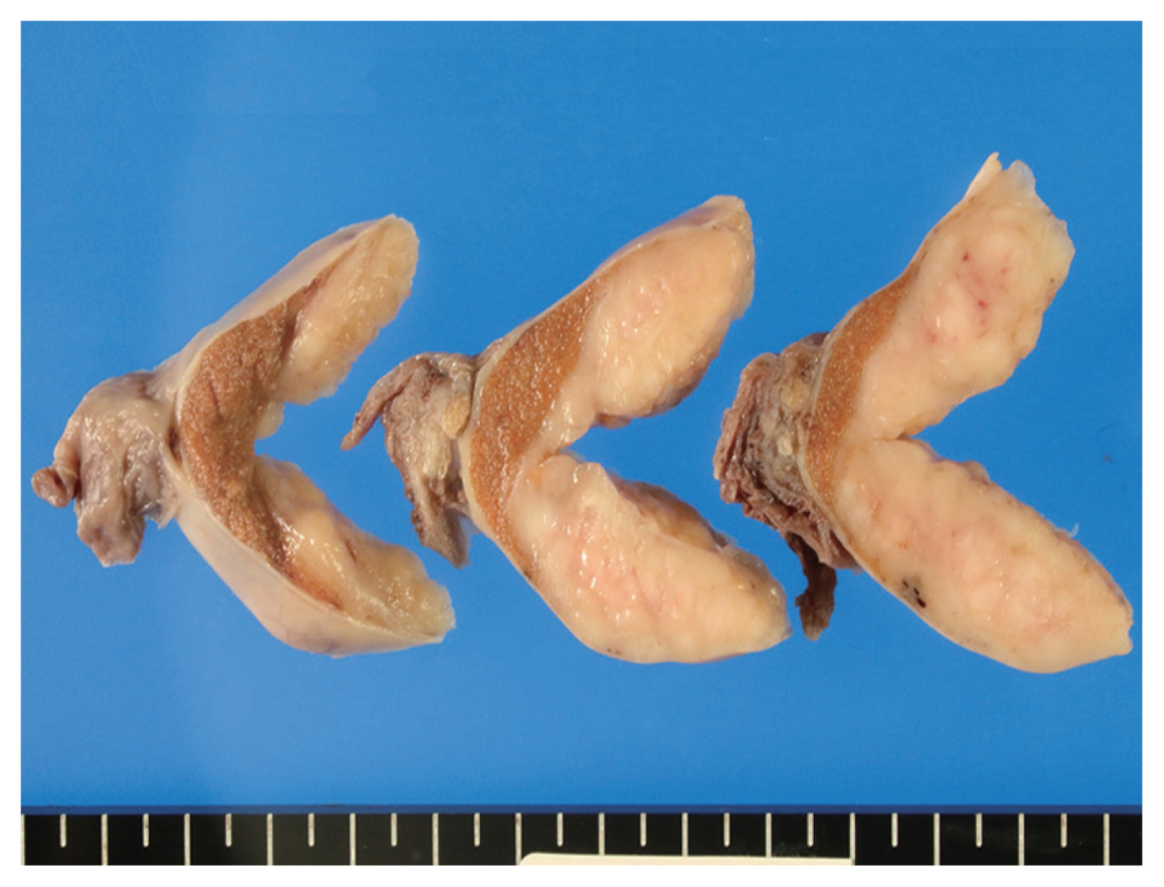
- Chronic lymphocytic leukemia and concurrent seminoma in the same testis
- Kosuke Miyai, Fumihisa Kumazawa, Kimiya Sato, Hitoshi Tsuda
- J Pathol Transl Med. 2022;56(1):48-52. Published online October 22, 2021
- DOI: https://doi.org/10.4132/jptm.2021.09.10
- 6,009 View
- 164 Download
-
Abstract
PDF
- A 59-year-old man presented with a painless testicular mass and underwent a radical orchiectomy. The resected specimen showed a 5-cm-sized, white-yellow and homogenous solid mass in the testicular parenchyma. Histologically, the central part of the tumor exhibited typical features of seminoma. The peripheral part of the tumor exhibited diffuse infiltration of small, monotonous lymphoid cells involving the tunica albuginea. The monotonous lymphoid cells were immunoreactive for CD20, CD79a, CD5, and CD23, and negative for CD3, CD10, and cyclin D1. Kappa light chain restriction was detected on flow cytometry using the resected specimen. Considering the circulating lymphoid cell count of >5.0×10³/µL, we diagnosed the peripheral component of the tumor as an infiltration of chronic lymphocytic leukemia. This extremely rare combination of seminoma and lymphoid neoplasm should be considered in the differential diagnosis of classic seminoma with extensive lymphoid reaction in tumors arising in elderly patients.
Original Article
- Do Helper T Cell Subtypes in Lymphocytic Thyroiditis Play a Role in the Antitumor Effect?
- Seok Woo Yang, Seong-Ho Kang, Kyung Rae Kim, In Hong Choi, Hang Seok Chang, Young Lyun Oh, Soon Won Hong
- J Pathol Transl Med. 2016;50(5):377-384. Published online September 15, 2016
- DOI: https://doi.org/10.4132/jptm.2016.07.25
- 10,631 View
- 108 Download
- 3 Web of Science
- 4 Crossref
-
Abstract
PDF
- Background
Papillary thyroid carcinoma (PTC) is frequently accompanied by lymphocytic thyroiditis (LT). Some reports claim that Hashimoto’s thyroiditis (the clinical form of LT) enhances the likelihood of PTC; however, others suggest that LT has antitumor activity. This study was aimed to find out the relationship between the patterns of helper T cell (Th) cytokines in thyroid tissue of PTC with or without LT and the clinicopathological manifestation of PTC.
Methods
Fresh surgical samples of PTC with (13 cases) or without (10 cases) LT were used. The prognostic parameters (tumor size, extra-thyroidal extension of PTC, and lymph node metastasis) were analyzed. The mRNA levels of two subtypes of Th cytokines, Th1 (tumor necrosis factor α [TNF-α], interferon γ [IFN-γ ], and interleukin [IL] 2) and Th2 (IL-4 and IL-10), were analyzed. Because most PTC cases were microcarcinomas and recent cases without clinical follow-up, negative or faint p27 immunoreactivity was used as a surrogate marker for lymph node metastasis.
Results
PTC with LT cases showed significantly higher expression of TNF-α (p = .043), IFN-γ (p < .010), IL-4 (p = .015) than those without LT cases. Although the data were not statistically significant, all analyzed cytokines (except for IL-4) were highly expressed in the cases with higher expression of p27 surrogate marker.
Conclusions
These results indicate that mixed Th1 (TNF-α, IFN-γ , and IL-2) and Th2 (IL-10) immunity might play a role in the antitumor effect in terms of lymph node metastasis. -
Citations
Citations to this article as recorded by
- Papillary thyroid carcinoma with Hashimoto’s thyroiditis: impact and correlation
Shengpeng Yao, Hong Zhang
Frontiers in Endocrinology.2025;[Epub] CrossRef - Obesity and Thyroid Cancer Risk: An Update
Fabiana Franchini, Giuseppe Palatucci, Annamaria Colao, Paola Ungaro, Paolo Emidio Macchia, Immacolata Cristina Nettore
International Journal of Environmental Research and Public Health.2022; 19(3): 1116. CrossRef - Association between Hashimoto thyroiditis and clinical outcomes of papillary thyroid carcinoma: A meta-analysis
Qizhi Tang, Weiyu Pan, Liangyue Peng, Francis Moore
PLOS ONE.2022; 17(6): e0269995. CrossRef - The Heat Shock Protein Story—From Taking mTORC1,2 and Heat Shock Protein Inhibitors as Therapeutic Measures for Treating Cancers to Development of Cancer Vaccines
Peter Chin Wan Fung, Regina Kit Chee Kong
Journal of Cancer Therapy.2017; 08(11): 962. CrossRef
- Papillary thyroid carcinoma with Hashimoto’s thyroiditis: impact and correlation
Case Report
- Lymphocytic Phlebitis of the Stomach: A Case Report with Literature Review.
- Meeran Kim, Hyun Jung Lee, Min Kyung Yeo, Young Suk Lee, Hee Seok Moon, Sang Il Lee, June Sik Cho, Kyu Sang Song
- Korean J Pathol. 2011;45(6):654-658.
- DOI: https://doi.org/10.4132/KoreanJPathol.2011.45.6.654
- 4,154 View
- 24 Download
- 1 Crossref
-
Abstract
PDF
- Lymphocytic phlebitis of gastrointestinal (GI) tract is a rare diseaes. Approximately 50 cases of lymphocytic phlebitis of the GI tract have been reported. Most of these involved the colon or small intestine and presented as acute abdomen. We report the second case of lymphocytic phlebitis of the stomach. A 73-year-old female complaining of dizziness had endoscopic and computed tomography findings strongly suggested gastric cancer, while gastric biopsy was negative for carcinoma. The partial gastrectomy specimen showed lymphocytic phlebitis involving veins in the submucosa, muscularis propria, and serosa while the adjacent arteries were spared. The veins were mainly surrounded by lymphocytes. When a patient has a lesion in the GI tract that is suggesting cancer without biopsies revealing any carcinoma, the pathologist should recommend a deeper biopsy for a proper examination of the submucosa.
-
Citations
Citations to this article as recorded by- A case report of gastric lymphocytic phlebitis, a rare mimic for malignancy
Daniel L. Chan, Praveen Ravindran, Dorothy Chua, Jason D. Smith, King S. Wong, Michael A. Ghusn
International Journal of Surgery Case Reports.2017; 41: 269. CrossRef
- A case report of gastric lymphocytic phlebitis, a rare mimic for malignancy
Original Article
- Microscopic Colitis: The Pathologic Features of 24 Korean Patients.
- Sun Ah Lee, Min Jung Kang, Sung Ae Jung, Heasoo Koo
- Korean J Pathol. 2009;43(2):133-138.
- DOI: https://doi.org/10.4132/KoreanJPathol.2009.43.2.133
- 4,436 View
- 25 Download
- 1 Crossref
-
Abstract
PDF
- BACKGROUND
The clinical presentation of microscopic colitis (MC) consists of chronic non-bloody watery diarrhea for weeks or months at a time, abdominal pain, and changes in bowel habits with a normal mucosal appearance upon performing colonoscopy. MC includes two relatively well established histopathologic entities: collagenous colitis (CC) and lymphocytic colitis (LC) as well as atypical forms. The recognition of the microscopic findings of this heterogeneous entity is very important for making the correct diagnosis and providing proper treatment.
METHODS
We studied the colonoscopic biopsy specimens that were obtained from 26 patients who had clinical findings that were suggestive of MC.
RESULTS
Fifteen patients (M:F=9:6) and 9 patients (M:F=5:4) showed the microscopic features of LC and MC, not otherwise specified, respectively.
CONCLUSIONS
The clinicopathologic findings (the incidence of the subtypes, the patients' ages and the male/female ratio) of the 24 cases of MC in this study showed differences from the previously reported findings from other countries. Further studies with a sufficient number of patients from multi-centers would be necessary to confirm the regional or ethnic influence. -
Citations
Citations to this article as recorded by- A Case of Methicillin-Resistant Staphylococcal Enterocolitis with Subsequent Development of Lymphocytic Colitis
Joong Ho Bae, Dong Soo Han, Hye Sun Park, Yil Sik Hyun, Tae Yeob Kim, Chang Soo Eun, Yong Cheol Jeon, Joo Hyun Sohn
Intestinal Research.2011; 9(2): 139. CrossRef
- A Case of Methicillin-Resistant Staphylococcal Enterocolitis with Subsequent Development of Lymphocytic Colitis
Case Report
- B-cell Prolymphocytic Leukemia Involving Entire Female Genital Tract: A case report.
- Hee Jung Lee, Young Shin Kim, Yong Gu Kim, Kyung Ja Han, Kyo Young Lee, Chang Suk Kang, Sang In Shim, Jong Wook Lee, Woong Shick Ahn, Soo Pyung Aim, Seung Il Kim
- Korean J Pathol. 1999;33(2):145-148.
- 2,229 View
- 10 Download
-
Abstract
- Prolymphocytic leukemia is a chronic lymphoproliferative disorder, characterized by prominent splenomegaly, prolymphocytes accounting for more than 55% of circulating lymphocytes, no significant peripheral lymphadenopathy and short term survival with terminal fatal multi-organ failure. We report a case of B-cell prolymphocytic leukemia in a 57-year-old woman who presented with easy bruising and arthritis for 1 year and low abdominal pain for 2 months. Physical examination revealed gingival hypertrophy and mild splenomegaly. On peripheral blood smears the leukocytes were markedly increased in number due to leukemic cells that count about 62% of leukocytes. The bone marrow aspiration smear and biopsy revealed diffuse infiltration of medium to large prolymphocytes having moderate amount of basophilic cytoplasm, round to oval nuclei with coarse chromatin, and prominent nucleoli. Abdominal pain aggravated despite chemotherapy, and pelvic computed tomography (CT) revealed a huge lobular pelvic mass which had increased in size on the follow-up CT. Total hysterectomy with bilateral adnexectomy was performed. Microscopic findings included massive infiltration of prolymphocytic cells in the uterus, upper vaginal wall, bilateral ovaries, and bilateral mesosalpinges. On immunohistochemistry, the leukemic cells showed B cell gamma light chain phenotype.
Original Article
- Clinicopathologic Analysis of Lymphocytic Gastritis.
- Jeong Eun Hwang, Young Ok Hong, Dong Eun Song, Se Jin Jang, Eunsil Yu
- Korean J Pathol. 2007;41(5):289-295.
- 3,604 View
- 66 Download
-
Abstract
PDF
- BACKGROUND
Lymphocytic gastritis (LG) is defined as an infiltration of more than 25 intraepithelial lymphocytes (IELs) per 100 surface epithelial cells, and the histological differential diagnosis of LG and residual mucosa associated lymphoid tissue (MALT) lymphoma can be difficult. Helicobacter pylori (H. pylori) is regarded as one of the possible causes of LG, but its clinicopathologic features of LG have not been clarified in Korea, which has a much higher prevalence of H. pylori infection than Western countries. We analyzed the clinicopathologic findings of LG in Korean patients and compared the cytologic findings of IELs of LG with those of MALT lymphoma.
METHODS
Sixty six cases of LG and 59 cases of MALT lymphoma were selected and clinicopathologic features were analyzed.
RESULTS
Eighteen cases (27.3%) of LG were found to be associated with H. pylori infection. The IELs in LG were found to diffusely and regularly infiltrate in the epithelium, but MALT lymphoma showed patchy IELs. IELs in LG and MALT lymphoma were CD 8+T lymphocytes and CD20+B lymphocytes, respectively. The mean nuclear size of IELs in LG was 4.37 micrometer, which was significantly smaller than those in MALT lymphoma (5.19 micrometer).
CONCLUSION
LG, a rare variant of chronic gastritis is partly associated with H. pylori infection and more complex unknown causative factors. In addition to the immunophenotyping, the nuclear sizes of IELs can be helpful in the differential diagnosis of LG and residual MALT lymphoma.
Comparative Study
- Lymphocytic Hypophysitis Presenting with Diabetes Insipidus in a Man: Report of a case.
- Woo Sung Moon, Myoung Jae Kang, Dong Geun Lee, Hyung Il Kim, Ho Yeul Choi, Sang Ho Kim
- Korean J Pathol. 1996;30(6):528-532.
- 2,453 View
- 16 Download
-
Abstract
PDF
- Lymphocytic hypophysitis is an autoimmune disorder of the pituitary gland which usually occurs in a woman in the postpartum period. Diabetes insipidus is not a major clinical feature of this disorder. We report a case of a 22-year-old man with lymphocytic hypophysitis which presented with diabetes insipidus and also involved his cavernous sinus. This represents the seventh reported and the youngest case of a man with lymphocytic hypophysitis. A comparative study of all six male patients is also presented. We suggest diabetes insipidus should be added to the spectrum of clinical manifestations of this disorder.
Case Report
- Idiopathic Entero-colic Lymphocytic Phlebitis: A case report.
- Seung Sam Paik, Young Ha Oh, Eun Kyung Hong, Jung Dal Lee
- Korean J Pathol. 1996;30(6):533-538.
- 2,437 View
- 27 Download
-
Abstract
PDF
- Localized enterocolic lymphocytic phlebitis is characterized by selective phlebitis involving the small to medium-sized veins and venules, infiltration exclusively by lymphocytes, and no other systemic vasculitis or inflammatory bowel disease. This vasculitis can be a rare cause of intestinal ischemia. We experienced a case of enterocolic lymphocytic phlebitis in a 72-year-old woman, who presented with abdominal pain and distension. The resected colon and terminal ileum showed striking lymphocytic phlebitis affecting the veins and venules of the bowel and mesentery which resulted in ischemic injury of the bowel. This vasculopathy was the only demonstrable cause of ischemia. Arteritis and arteriolitis was not found. There is no clinical or laboratory evidence or a history of extraintestinal vasculitis. The etiology of this clinicopathological entity has not been elucidated. Herein, we report the clinicopathological findings in this patient who presented with ischemic intestinal necrosis caused by localized intestinal lymphocytic phlebitis associated with thrombosis.
Original Article
- Lymphocyte Rich Papillary Oxyphilic Carcinoma of Thyroid.
- Soon Ran Kim, Jin Hee Sohn
- J Pathol Transl Med. 1997;8(2):150-154.
- 1,816 View
- 12 Download
-
Abstract
PDF
- Lymphoid infiltration can be seen in some lesions such as Hashimoto's thyroiditis, subacute thyroiditis and several neoplasm of the thyroid. In case of malignancy, there are a few reports of lymphoid infiltration in the diffuse sclerosing variant of papillary carcinoma. But heavy lymphoid infiltraton without evidence of sclerosis is uncommon. We experienced a case of papillary oxyphilic carcinoma with massive lymphoid infiltration, which looks like Warthin tumor of salivary gland. However cytological feature of epithelial cells exhibit that of papillary carcinoma.
 First
First Prev
Prev


