 E-submission
Search
- Page Path
- HOME > Search
- Cutaneous soft tissue tumors in the 5th edition of the World Health Organization classification of skin tumors: key updates and new entities
- Joon Hyuk Choi
- J Pathol Transl Med. 2026;60(2):144-183. Published online March 13, 2026
- DOI: https://doi.org/10.4132/jptm.2026.01.09
- 2,120 View
- 159 Download
-
 Abstract
 PDF
PDF - The 5th edition of the World Health Organization (WHO) classification of skin tumors introduces a dedicated chapter on cutaneous soft tissue tumors, providing a comprehensive, standardized reference with updated diagnostic criteria that directly inform routine dermatopathology practice and molecular diagnostics. This edition incorporates several key changes, including newly recognized entities such as EWSR1::SMAD3-rearranged fibroblastic tumor, neurotrophic tyrosine receptor kinase (NTRK)–rearranged spindle cell neoplasm, superficial CD34-positive fibroblastic tumor, and CRTC1::TRIM11 cutaneous tumor. Diagnostic terminology has also been refined; for example, the term ‘atypical intradermal smooth muscle neoplasm’ replaces ‘cutaneous leiomyosarcoma’ for lesions confined to the dermis, whereas the designation leiomyosarcoma is reserved for tumors with overt subcutaneous infiltration. In addition, epithelioid fibrous histiocytoma has been reassigned to the family of tumors of uncertain differentiation. This review summarizes the key updates and newly recognized entities in the chapter on cutaneous soft tissue tumors in the 5th edition of the WHO classification of skin tumors, emphasizing their clinicopathological and molecular implications.
- Solitary fibrous tumor: an updated review
- Joon Hyuk Choi
- J Pathol Transl Med. 2026;60(1):20-46. Published online December 29, 2025
- DOI: https://doi.org/10.4132/jptm.2025.10.08
- 2,535 View
- 235 Download
-
Abstract
PDF
- Solitary fibrous tumor (SFT) is a fibroblastic neoplasm characterized by a branching, thin-walled dilated staghorn-shaped (hemangiopericytoma-like) vasculature and a NAB2::STAT6 gene fusion. SFTs can occur in almost any anatomical location, including superficial and deep soft tissues, visceral organs, and bone. They most commonly occur in extrapleural locations, equally affect both sexes, and are typically present in adults. Although metastasis is rare, SFTs frequently show local recurrence. The diagnosis of SFTs is difficult because of their broad histological and morphological overlap with other neoplasms. An accurate diagnosis is important for guiding disease management and prognosis. Despite advances in molecular diagnostics and therapeutic strategies, the biological complexity and unpredictable clinical behavior of SFTs present significant challenges. This review provides an updated overview of SFT, with a focus on its molecular genetics, histopathological features, and diagnostic considerations.
- Adrenal hemangioblastoma
- Joo-Yeon Koo, Kyung-Hwa Lee, Joon Hyuk Choi, Ho Seok Chung, Chan Choi
- J Pathol Transl Med. 2022;56(3):161-166. Published online February 28, 2022
- DOI: https://doi.org/10.4132/jptm.2021.12.28
- 6,181 View
- 159 Download
- 1 Web of Science
- 1 Crossref
-
Abstract
PDF
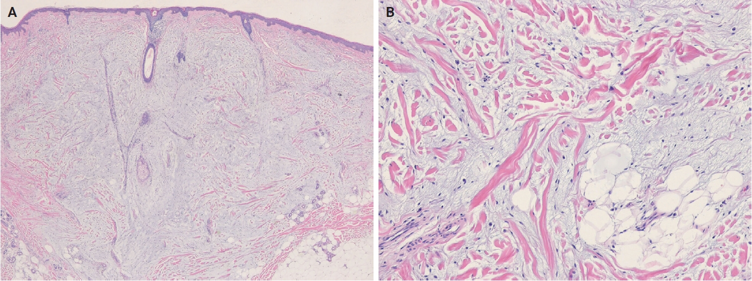
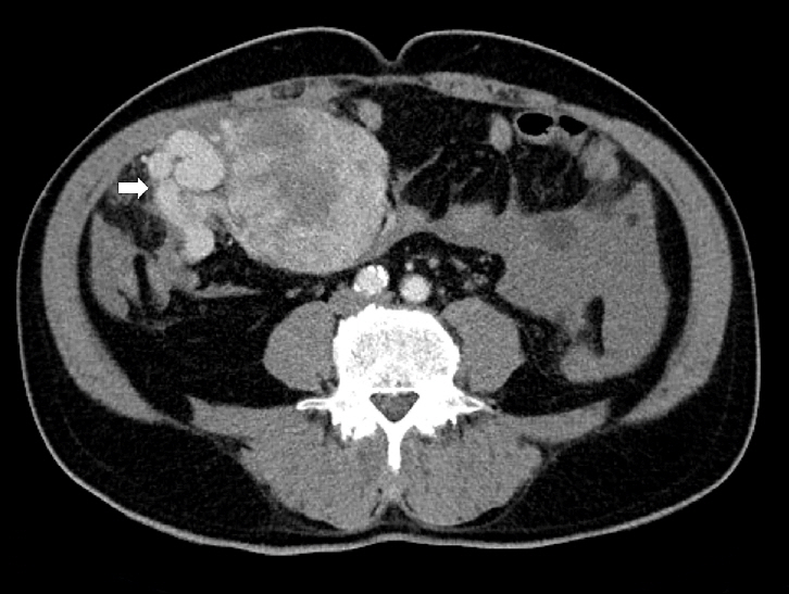
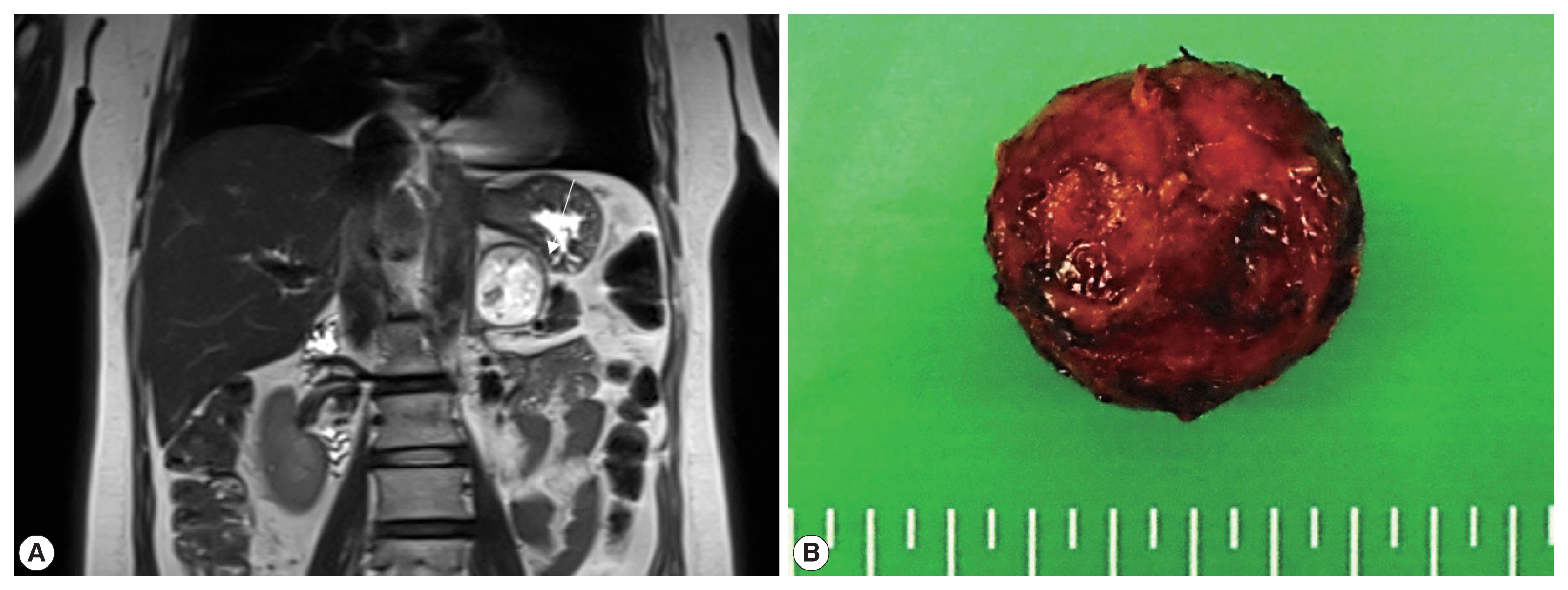
- Hemangioblastoma (HB) is a rare benign tumor that most commonly occurs in the cerebellum. HB is composed of neoplastic stromal cells and abundant small vessels. However, the exact origin of stromal cells is controversial. Extraneural HBs have been reported in a small series, and peripheral HBs arising in the adrenal gland are extremely rare. Herein, we report a case of sporadic adrenal HB in a 54-year-old woman. The tumor was a well-circumscribed, yellow mass measuring 4.2 cm in diameter. Histologically, the tumor was composed of small blood vessels and vacuolated stromal cells with clear cytoplasm. On immunohistochemical stain, the stromal cells were positive for S-100 protein, neuron-specific enolase, and synaptophysin. The tumor did not reveal mutation of VHL alleles. We herein present a case of HB of the adrenal gland and review of the literature.
-
Citations
Citations to this article as recorded by
- Familial Von Hippel–Lindau Disease: A Case Series of Cerebral Hemangioblastomas with MRI, Histopathological, and Genetic Correlations
Claudiu Matei, Ioana Boeras, Dan Orga Dumitriu, Cosmin Mutu, Adriana Popescu, Mihai Gabriel Cucu, Alexandru Calotă-Dobrescu, Bogdan Fetica, Diter Atasie
Life.2025; 15(11): 1649. CrossRef
- Familial Von Hippel–Lindau Disease: A Case Series of Cerebral Hemangioblastomas with MRI, Histopathological, and Genetic Correlations
- Fine Needle Aspiration Cytology of Hepatic Hydatid Cyst: A Case Study
- Ae Ri Kim, Seok Ju Park, Mi Jin Gu, Joon Hyuk Choi, Hong Jin Kim
- Korean J Pathol. 2013;47(4):395-398. Published online August 26, 2013
- DOI: https://doi.org/10.4132/KoreanJPathol.2013.47.4.395
- 15,776 View
- 110 Download
- 13 Crossref
-
Abstract
PDF
Hydatid cysts (echinococcosis) are caused by an infestation with larval tapeworms of the genus
Echinococcus . The disease is extensively distributed worldwide, and it has been rarely reported in Korea. We describe the cytologic features of a case of hepatic hydatid cyst in a 28-year-old male. Computed tomography revealed a cystic mass in the right lobe of the liver. A right hemihepatectomy was performed. The aspirated fluid from the hepatic cystic mass was clear. The smears showed protoscolices, hooklets, and a laminated membrane.-
Citations
Citations to this article as recorded by- Fine-Needle Aspiration Cytology of Hepatic Hydatid Disease: A Five-Year Case Series with Emphasis on Diagnostic Limitations
Fatlinda Sadiku, Ardita Qerimi, Gramoz Bunjaku, Ilir Maloku, Artina Pajaziti
Acta Cytologica.2026; : 1. CrossRef - Delayed Diagnosis of Imported Cystic Echinococcosis and Successful Treatment With Percutaneous Drainage and Albendazole in Korea: A Case Report
Won Jun Choi, Hanna Jin, Hyeon Jae Jo, Chan Mi Lee, Chang Kyung Kang, Pyoeng Gyun Choe, Wan Beom Park, Nam Joong Kim, Min-Ho Choi
Journal of Korean Medical Science.2025;[Epub] CrossRef - Imported parasitic diseases in the Republic of Korea: status and issues
Jong-Yil Chai
Journal of the Korean Medical Association.2025; 68(1): 52. CrossRef - Surgical Management of Multiple Cerebral Hydatid Cysts without Systemic Involvement: A Case Report
Roxana Radu, Miguel Azevedo, Angela Neacșu, Marius Cristian Zaharia, Radu Mircea Gorgan
Arquivos Brasileiros de Neurocirurgia: Brazilian Neurosurgery.2025; 44(03): e211. CrossRef - Different scenarios and management of complicated hepatic cystic echinococcosis: a case series
Thamer Alghamdi, Ramy Agwa, Fahad Alghamdi, Ahmed Mahmoud, Warda Othman
Journal of Surgical Case Reports.2025;[Epub] CrossRef - Hydatid Disease of the Pelvis: A Rare Manifestation With Diagnostic Challenges—A Case Report and Comprehensive Literature Review
Saurav Jha, Matsendra Jha, Sweta Singh, Sulav Kumar Jha, Bistrit Dahal, Anamika Adhikari, Sanskriti Dev
Clinical Case Reports.2025;[Epub] CrossRef - Chemical compounds, antioxidant and scolicidal potencies of Thymus fontanesii essential oil
Sidi Mohammed Ammar Selles, Belkacem Tahar Belhamiti, Mokhtaria Kouidri, Amar Ait Amrane, Yamina Kadari, Zohra Kaddour, Souad Kabrit
Experimental Parasitology.2024; 257: 108699. CrossRef - Complicated Liver Cystic Echinococcosis—A Comprehensive Literature Review and a Tale of Two Extreme Cases
Valentin Calu, Octavian Enciu, Elena-Adelina Toma, Radu Pârvuleţu, Dumitru Cătălin Pîrîianu, Adrian Miron
Tomography.2024; 10(6): 922. CrossRef - A rare case of hydatid cyst of the neck with concurrent pulmonary hydatid disease
Amarendra Kumar Shukla, Amrutha Peter, Veerendra Arya, Vineet Dwivedi, Manish Kumar Gupta, Nimish Rai, Pawan Tiwari, Jitendra Kishore Bhargava
Journal of Parasitic Diseases.2022; 46(4): 941. CrossRef - Hepatic Hydatid Cyst: A Case Report
Wan Chul Kim, Jae Uk Shin, Su Sin Jin
The Korean Journal of Gastroenterology.2021; 77(1): 35. CrossRef - An Imported Case of Disseminated Echinococcosis in Korea
Dong Hoon Shin, Hae Chan Jo, Jeong-Han Kim, Kang Il Jun, Wan Beom Park, Nam-Joong Kim, Min-Ho Choi, Chang Kyung Kang, Myoung-don Oh
The Korean Journal of Parasitology.2019; 57(4): 429. CrossRef - Cytology of hydatid cyst mimicking intra‐abdominal sarcoma, diagnosed by fine‐needle aspiration
Busra Ozbek, Nadir Paksoy
Diagnostic Cytopathology.2018; 46(4): 362. CrossRef - Clinical Update on Parasitic Diseases
Min Seo
Korean Journal of Medicine.2013; 85(5): 469. CrossRef
- Fine-Needle Aspiration Cytology of Hepatic Hydatid Disease: A Five-Year Case Series with Emphasis on Diagnostic Limitations
- Imprint Cytology of Soft Tissue Myoepithelioma: A Case Study
- Seok Ju Park, Ae Ri Kim, Mi Jin Gu, Joon Hyuk Choi, Duk Seop Shin
- Korean J Pathol. 2013;47(3):299-303. Published online June 25, 2013
- DOI: https://doi.org/10.4132/KoreanJPathol.2013.47.3.299
- 10,996 View
- 53 Download
- 7 Crossref
-
Abstract
PDF
Soft tissue myoepithelioma is a rare neoplasm composed of myoepithelial cells. Here, we describe the cytologic features of soft tissue myoepithelioma arising on the right forearm in an 18-year-old man. The excised tumor (3.0×1.8×1.5 cm) was well-demarcated, yellow-gray, soft, and myxoid. The cytologic smears showed round to spindle, epithelioid, and plasmacytoid cells in the myxoid background. The nuclei were uniform, round to ovoid, with finely distributed chromatin and eosinophilic or pale cytoplasm. The tumor cells demonstrated immunoreactivity for cytokeratin (AE1/AE3), epithelial membrane antigen, S100 protein, and glial fibrillary acidic protein. Electron microscopy showed intermediate filaments, desmosomes, and basal lamina.
-
Citations
Citations to this article as recorded by- Myoepithelial tumors of soft tissue and bone in children and young adults: A clinicopathologic study of 40 cases occurring in patients ≤ 21 Years of age
Suzanna J. Logan, Carina A. Dehner, Fatimah I. Alruwaii, Nasir Ud Din, Damon R. Olson, Karen J. Fritchie, Gregory W. Charville, Melissa M. Blessing, Andrew L. Folpe
Human Pathology.2024; 149: 10. CrossRef - Fine-needle aspiration cytopathology of soft tissue myoepithelioma: an analysis of seven cases
Paul E. Wakely, Momin T. Siddiqui
Journal of the American Society of Cytopathology.2022; 11(1): 31. CrossRef - Cytology‐histology correlation of myoepithelial tumors harboring EWSR1‐POU5F1 fusions: A report of two cases
Ian A. Gelarden, Lucy Fu, Kai Lee Yap, Aida I. Richardson, Pauline M. Chou
Diagnostic Cytopathology.2022;[Epub] CrossRef - A case of myoepithelial carcinoma of the left shoulder
Shuhei ISHII, Noriyuki FURUTA, Kyoko KOMATSU, Yoshiya SUGIURA, Noriko MOTOI, Yutaka TAKAZAWA, Yuko SUGIYAMA, Yuichi ISHIKAWA
The Journal of the Japanese Society of Clinical Cytology.2018; 57(2): 129. CrossRef - Fine‐needle aspiration of soft tissue myoepithelioma
Gang Wang, Tracy Tucker, Tony L. Ng, Carlos F. Villamil, Malcolm M. Hayes
Diagnostic Cytopathology.2016; 44(2): 152. CrossRef - A case report of spindle cell myoepithelioma with extensive lipomatous metaplasia and thick collagen bundles in the submandibular gland
Mi Jung Kwon, Hye Jeong Kim, Bumjung Park, Seong Jin Cho, Hyung Sik Shin, Hye‐Rim Park, Soo Kee Min, Jinwon Seo, Kyueng‐Whan Min, Eun Sook Nam
Diagnostic Cytopathology.2016; 44(9): 764. CrossRef - Myoepithelioma of soft tissue, a case report
Hassania Ameurtesse, Leila Chbani, JM Coindre, Hinde Elfatemi, Toufik Harmouch, Afaf Amarti
Research.2014;[Epub] CrossRef
- Myoepithelial tumors of soft tissue and bone in children and young adults: A clinicopathologic study of 40 cases occurring in patients ≤ 21 Years of age
- Proposal for a Standardized Pathology Report of Gastroenteropancreatic Neuroendocrine Tumors: Prognostic Significance of Pathological Parameters
- Mee-Yon Cho, Jin Hee Sohn, So Young Jin, Hyunki Kim, Eun Sun Jung, Mi-Jung Kim, Kyoung-Mee Kim, Woo Ho Kim, Joon Mee Kim, Yun Kyung Kang, Joon Hyuk Choi, Dae Young Kang, Youn Wha Kim, Eun Hee Choi
- Korean J Pathol. 2013;47(3):227-237. Published online June 25, 2013
- DOI: https://doi.org/10.4132/KoreanJPathol.2013.47.3.227
- 16,116 View
- 148 Download
- 12 Crossref
-
Abstract
PDF
Background There is confusion in the diagnosis and biological behaviors of gastroenteropancreatic neuroendocrine tumors (GEP-NETs), because of independently proposed nomenclatures and classifications. A standardized form of pathology report is required for the proper management of patients.
Methods We discussed the proper pathological evaluation of GEP-NET at the consensus conference of the subcommittee meeting for the Gastrointestinal Pathology Study Group of the Korean Society of Pathologists. We then verified the prognostic significance of pathological parameters from our previous nationwide collection of pathological data from 28 hospitals in Korea to determine the essential data set for a pathology report.
Results Histological classification, grading (mitosis and/or Ki-67 labeling index), T staging (extent, size), lymph node metastasis, and lymphovascular and perineural invasion were significant prognostic factors and essential for the pathology report of GEP-NET, while immunostaining such as synaptophysin and chromogranin may be optional. Furthermore, the staging system, either that of the 2010 American Joint Cancer Committee (AJCC) or the European Neuroendocrine Tumor Society (ENETS), should be specified, especially for pancreatic neuroendocrine neoplasms.
Conclusions A standardized pathology report is crucial for the proper management and prediction of prognosis of patients with GEP-NET.
-
Citations
Citations to this article as recorded by- Analysis of Prognostic Risk Factors of Endoscopic Submucosal Dissection (ESD) and Curative Resection of Gastrointestinal Neuroendocrine Neoplasms
Yuan Si, ChaoKang Huang, JingBin Yuan, XianHui Zhang, QingQiang He, ZhiJin Lin, Ling He, ZhongXin Liu, Yuvaraja Teekaraman
Contrast Media & Molecular Imaging.2022;[Epub] CrossRef - Standardization of the pathologic diagnosis of appendiceal mucinous neoplasms
Dong-Wook Kang, Baek-hui Kim, Joon Mee Kim, Jihun Kim, Hee Jin Chang, Mee Soo Chang, Jin-Hee Sohn, Mee-Yon Cho, So-Young Jin, Hee Kyung Chang, Hye Seung Han, Jung Yeon Kim, Hee Sung Kim, Do Youn Park, Ha Young Park, So Jeong Lee, Wonae Lee, Hye Seung Lee,
Journal of Pathology and Translational Medicine.2021; 55(4): 247. CrossRef - Preoperative diagnosis of well‐differentiated neuroendocrine tumor in common hepatic duct by brush cytology: A case report
Jiwoon Choi, Kyong Joo Lee, Sung Hoon Kim, Mee‐Yon Cho
Diagnostic Cytopathology.2019; 47(7): 720. CrossRef - Primary renal well-differentiated neuroendocrine tumors: report of six cases with an emphasis on the Ki-67 index and mitosis
Bohyun Kim, Han-Seong Kim, Kyung Chul Moon
Diagnostic Pathology.2019;[Epub] CrossRef - Primary low‐grade neuroendocrine carcinoma of the skin: An exceedingly rare entity
Tiffany Y. Chen, Annie O. Morrison, Joe Susa, Clay J. Cockerell
Journal of Cutaneous Pathology.2017; 44(11): 978. CrossRef - Prognostic Validity of the American Joint Committee on Cancer and the European Neuroendocrine Tumors Staging Classifications for Pancreatic Neuroendocrine Tumors
Jae Hee Cho, Ji Kon Ryu, Si Young Song, Jin-Hyeok Hwang, Dong Ki Lee, Sang Myung Woo, Young-Eun Joo, Seok Jeong, Seung-Ok Lee, Byung Kyu Park, Young Koog Cheon, Jimin Han, Tae Nyeun Kim, Jun Kyu Lee, Sung-Hoon Moon, Hyunjin Kim, Eun Taek Park, Jae Chul Hw
Pancreas.2016; 45(7): 941. CrossRef - Early diagnosis and treatment of gastrointestinal neuroendocrine tumors
Hong Shen, Zhuo Yu, Jing Zhao, Xiu-Zhen Li, Wen-Sheng Pan
Oncology Letters.2016; 12(5): 3385. CrossRef - Recent Updates on Neuroendocrine Tumors From the Gastrointestinal and Pancreatobiliary Tracts
Joo Young Kim, Seung-Mo Hong
Archives of Pathology & Laboratory Medicine.2016; 140(5): 437. CrossRef - Pancreatic neuroendocrine tumors: Correlation between the contrast-enhanced computed tomography features and the pathological tumor grade
Koji Takumi, Yoshihiko Fukukura, Michiyo Higashi, Junnichi Ideue, Tomokazu Umanodan, Hiroto Hakamada, Ichiro Kanetsuki, Takashi Yoshiura
European Journal of Radiology.2015; 84(8): 1436. CrossRef - Tumeurs neuroendocrines du tube digestif et du pancréas : ce que le pathologiste doit savoir et doit faire en 2014
Jean-Yves Scoazec, Anne Couvelard
Annales de Pathologie.2014; 34(1): 40. CrossRef - Spectrum of Gastroenteropancreatic NENs in Routine Histological Examinations of Bioptic and Surgical Specimen: A Study of 161 Cases Collected from 17 Departments of Pathology in the Czech Republic
Václav Mandys, Tomáš Jirásek
Gastroenterology Research and Practice.2014; 2014: 1. CrossRef - p27 Loss Is Associated with Poor Prognosis in Gastroenteropancreatic Neuroendocrine Tumors
Hee Sung Kim, Hye Seung Lee, Kyung Han Nam, Jiwoon Choi, Woo Ho Kim
Cancer Research and Treatment.2014; 46(4): 383. CrossRef
- Analysis of Prognostic Risk Factors of Endoscopic Submucosal Dissection (ESD) and Curative Resection of Gastrointestinal Neuroendocrine Neoplasms
- Extranodal Follicular Dendritic Cell Sarcoma with Rapid Growth in Parapharynx: A Case Report
- Jung-Soo Pyo, Guhyun Kang, Sung-Im Do, Seoung Wan Chae, Kyungeun Kim, Sang Hyuk Lee, Yoon-La Choi, Joon Hyuk Choi, Jin Hee Sohn, Dong-Hoon Kim
- Korean J Pathol. 2012;46(3):306-310. Published online June 22, 2012
- DOI: https://doi.org/10.4132/KoreanJPathol.2012.46.3.306
- 9,110 View
- 54 Download
- 9 Crossref
-
Abstract
PDF
Follicular dendritic cell sarcoma (FDCS) is a rare malignancy arising from the antigen-presenting cells in the lymph node and extranodal tissue. We describe a 31-year-old male patient who presented with a swelling of the left parapharynx. The radiologic findings showed a 4.7×4.5×1.9 cm-sized, ill-defined mass in the left parapharyngeal space. A fine-needle aspiration cytology was performed and it showed scattered, irregular, cohesive clusters of tumor cells with a spindle-to-ovoid shape with irregular contours in a background of lymphocytes. Based on these findings, a diagnosis of spindle cell neoplasm was made. The surgically resected tumor was composed of elongated, ovoid or polygonal cells showing positive immunohistochemistry for CD21, CD23, and CD35. Postoperatively, the residual tumor was observed to undergo a rapidly growth. There is an overlap in the cytologic and histologic findings between FDCS of the parapharynx and other tumors. Pathologists should therefore be aware of its characteristics not only to provide an accurate diagnosis but also to recommend the appropriate clinical management.
-
Citations
Citations to this article as recorded by- Extranodal Follicular Dendritic Cell Sarcoma of the Head and Neck Region: A Clinicopathological Study of 7 Cases
Nasir Ud Din, Zubair Ahmad, Shabina Rahim, Karen Fritchie, Muhammad Usman Tariq, Arsalan Ahmed
International Journal of Surgical Pathology.2023; 31(6): 1067. CrossRef - Cytomorphology of follicular dendritic cell sarcoma: a report of 7 cases with an emphasis on the diagnostic challenges
Cody Weimholt, Jalal B. Jalaly, Cedric Bailey
Journal of the American Society of Cytopathology.2023; 12(3): 229. CrossRef - Follicular dendritic cells
Seham A. Abd El‐Aleem, Entesar Ali Saber, Neven M. Aziz, Hani El‐Sherif, Asmaa M. Abdelraof, Laiche Djouhri
Journal of Cellular Physiology.2022; 237(4): 2019. CrossRef - Clinicopathological characteristics of extranodal follicular dendritic cell sarcoma: A report of two cases
Xing Zhao, Dayong Sun, Gang Zhang
Oncology Letters.2021;[Epub] CrossRef - Cytological diagnosis of follicular dendritic cell sarcoma: A case report and review of literature
A. Dutta, P. Arun, P. Roy, I. Arun
Cytopathology.2018; 29(5): 461. CrossRef - Follicular dendritic cells and related sarcoma
Fabio Facchetti, Luisa Lorenzi
Seminars in Diagnostic Pathology.2016; 33(5): 262. CrossRef - Extranodal follicular dendritic cell sarcoma: A clinicopathological report of four cases and a literature review
RUI-FEN WANG, WEI HAN, LEI QI, LI-HUI SHAN, ZHENG-CAI WANG, LI-FENG WANG
Oncology Letters.2015; 9(1): 391. CrossRef - Follicular Dendritic Cell Sarcoma of Parapharyngeal Space: A Case Report and Review of the Literature
Turki Al-Hussain, Muhammad Saleem, Suresh Babu Velagapudi, Mohammad Anas Dababo
Head and Neck Pathology.2015; 9(1): 135. CrossRef - Clinical and pathological features of head and neck follicular dendritic cell sarcoma
Ji Li, Min-Li Zhou, Shui-Hong Zhou
Hematology.2015; 20(10): 571. CrossRef
- Extranodal Follicular Dendritic Cell Sarcoma of the Head and Neck Region: A Clinicopathological Study of 7 Cases
- Type and Incidence of Soft Tissue Sarcomas in Korea: 2001-2007.
- Kyung Un Choi, Hae Youn Kang, Heasoo Koo, Mi Seon Kwon, Dong Hoon Kim, Mi Jung Kim, Su Jin Kim, Young Sill Kim, Chul Hwan Kim, Yong Koo Park, Hye Rim Park, Seung Sam Paik, Jin Young Yoo, Anhi Lee, Jae Hyuk Lee, Hyekyung Lee, Kyu Yun Jang, Young Chae Chu, Joon Hyuk Choi
- Korean J Pathol. 2011;45(6):557-563.
- DOI: https://doi.org/10.4132/KoreanJPathol.2011.45.6.557
- 5,021 View
- 52 Download
- 1 Crossref
-
Abstract
PDF
- BACKGROUND
The Korean Bone and Soft Tissue Pathology Study Group of the Korean Society of Pathologists conducted a nationwide retrospective analysis of soft tissue sarcoma (STS) to provide the clinicopathologic characteristics of STS within the population of the Republic of Korea.
METHODS
The cases of STS were collected during a 7-year period (2001-2007) from 19 institutes in Korea. All cases were classified according to the histologic criteria proposed by the World Health Organization. Clinicopathologic data were reviewed.
RESULTS
Data from 722 patients (median age, 50 years) were collected. Data showed a slight male predominance. The most frequent types of STS in decreasing order were liposarcoma, malignant fibrous histiocytoma, leiomyosarcoma, and synovial sarcoma. STS occurred throughout the body, although approximately half (47.8%) were located in the extremities. The majority of STS was histologically classified as high grade with a large tumor size (>5 cm). The overall survival rate for the patients was 76.3% (median follow-up time, 26 months; range, 1 to 89 months). Histologic grade, tumor size, American Joint Committee on Cancer stage, tumor site, and resection status were prognostic. Significant independent adverse prognostic factors were large tumor size (>5 cm) and tumor site other than extremities.
CONCLUSIONS
We reported the distribution and characteristics of STS in the Republic of Korea. -
Citations
Citations to this article as recorded by- Distribution and survival of primary sarcoma in Korea: A single center analysis of 2017 cases
Sung Jun Jo, Kyeong Sik Kim, Kyo Won Lee, Jae Berm Park, Yoon-La Choi, Jeong Il Yu, Su Jin Lee, Dong Il Choi, Sung Joo Kim
Korean Journal of Clinical Oncology.2018; 14(1): 30. CrossRef
- Distribution and survival of primary sarcoma in Korea: A single center analysis of 2017 cases
- Usefulness of DOG1 Expression in the Diagnosis of Gastrointestinal Stromal Tumors.
- Jun Mo Kim, Aeri Kim, Joon Hyuk Choi, Young Kyung Bae
- Korean J Pathol. 2010;44(2):141-148.
- DOI: https://doi.org/10.4132/KoreanJPathol.2010.44.2.141
- 7,640 View
- 82 Download
- 1 Crossref
-
Abstract
PDF
- BACKGROUND
Gastrointestinal stromal tumors (GISTs) are the most common mesenchymal tumors in the gastrointestinal tract. Expression of KIT protein (CD117) is an important diagnostic criterion of GIST. However, about 5% of GISTs are CD117 negative. Discovered on GIST 1 (DOG1) was introduced recently as a promising marker for GIST. We tested this new antibody in 105 GISTs tissue specimens, including 6 cases of metastatic GISTs, to determine the usefulness of DOG1 expression in the diagnosis of GISTs.
METHODS
We performed immunohistochemical (IHC) staining for DOG1 and CD117 on tissue microarrays that included 70 gastric GISTs, 29 small intestinal GISTs, 6 metastatic GISTs, 14 gastric leiomyomas and 16 gastric schwannomas.
RESULTS
DOG1 was positive in 98.1% (103/105) of GISTs and CD117 was positive in 97.1% (102/105) of GISTs. Only 1 case was negative for both markers. Two (66.7%) out of 3 GISTs tested CD117 negative were tested DOG1 positive. All leiomyomas and schwannomas were negative for both DOG1 and CD117.
CONCLUSIONS
DOG1 was highly expressed in GIST including CD117 negative cases. Adding DOG1 testing to the IHC panel for diagnosing GIST will help to identify GIST patients who are CD117 negative but may otherwise benefit from targeted therapy. -
Citations
Citations to this article as recorded by- Gastrointestinal tract spindle cell tumors with interstitial cells of Cajal: Prevalence excluding gastrointestinal stromal tumors
So Jung Lee, Chung Su Hwang, Ahrong Kim, Kyungbin Kim, Kyung Un Choi
Oncology Letters.2016; 12(2): 1287. CrossRef
- Gastrointestinal tract spindle cell tumors with interstitial cells of Cajal: Prevalence excluding gastrointestinal stromal tumors
- Morphological Features of Metastatic Gastrointestinal Stromal Tumors after Gleevec Treatment: Two Cases Report.
- Joon Hyuk Choi, Young Kyung Bae, Sun Kyo Song, Hong Jin Kim, Min Chul Shim, Kyung Hee Lee
- Korean J Pathol. 2009;43(4):368-373.
- DOI: https://doi.org/10.4132/KoreanJPathol.2009.43.4.368
- 3,781 View
- 29 Download
-
Abstract
PDF
- We report two patients with metastatic gastrointestinal stromal tumors (GISTs) with a focus on the morphological features related to Gleevec treatment. In case 1, a 50-year-old woman presented with a 1.8 cm metastatic GIST in the liver after resection of a gastric GIST. Majority of the metastatic tumor showed fibrosis and hyalinization after 8 weeks of Gleevec treatment. CD117-positive cells were present in approximately 1% of the overall tumor. In case 2, a 2 cm and 14 cm metastatic liver masses were found in a 54-year-old man who had a rectal GIST. After 4 weeks of Gleevec treatment, metastatic tumors showed a decrease in size on CT scan. The metastatic tumors showed a decrease in number of tumor cells. The hemorrhage, cystic changes, necrosis, and fibrosis made up approximately 90% of the tumor. The morphological features related to Gleevec treatment are important for correct diagnosis and evaluation of tumor response and prognosis.
- Soft Tissue Perineurioma : A Case Report .
- Jun Mo Kim, Joon Hyuk Choi
- Korean J Pathol. 2009;43(3):266-270.
- DOI: https://doi.org/10.4132/KoreanJPathol.2009.43.3.266
- 4,714 View
- 39 Download
- 5 Crossref
-
Abstract
PDF
- Soft tissue perineurioma is a very rare tumor composed of entirely of neoplastic perineurial cells. A 54-year-old woman presented with a palpable mass in the right lower leg. The mass was excised. Grossly, the tumor measured 2.0x2.0x1.5 cm. The cut surface was well circumscribed, pale pinkish gray, and rubbery soft. Histological examination showed that the tumor was composed of spindle cells within collagenous and myxoid stroma. The tumor cells had elongated, tapering nuclei with long and thin cytoplasmic processes, and were arranged in fascicular, whorled, and storiform pattern. The tumor cells were positive for epithelial membrane antigen and collagen type IV and negative for S-100 protein. Ultrastructurally, tumor cells showed long and thin cytoplasmic processes, pinocytic vesicles, and incomplete external lamina. The diagnosis of soft tissue perineurioma was confirmed by immunohistochemical stain and ultrastructural study.
-
Citations
Citations to this article as recorded by- Cutaneous Perineurioma of the Medial Superciliary Arch: An Uncommon Location for a Rare, Benign Spindle Cell Neoplasm
Sri Naidnur, Valeria González-Molina, Kara Asbury, Emily DeSantis, Rick Lin
Cureus.2026;[Epub] CrossRef - Trauma-induced Soft Tissue Perineurioma on the Thumb of a Filipino Female
Angeli Carina Lahoz, Zharlah Gulmatico Flores, Elisa Rae L. Coo
Journal of the Philippine Dermatological Society.2025; 34(2): 83. CrossRef - Extraneural Soft Tissue Perineurioma: A Report of a Rare Case of Peripheral Nerve Sheath Tumor
Ramki Arunachalam Ganesh, Karthikeyan Selvaraj, Srinivasan Chandran, Jesu Pencilin Yesuvadiyan
Cureus.2024;[Epub] CrossRef - Périneuriome extraneural des tissus mous localisé au nez
A. Zaouak, R. Benmously, M. Belhadj Salah, W. Koubaa, A. Debbiche, I. Mokhtar
Annales de Dermatologie et de Vénéréologie.2013; 140(8-9): 540. CrossRef - A Soft Tissue Perineurioma and a Hybrid Tumor of Perineurioma and Schwannoma
Ji Young Park, Nam Jo Park, Sang Pyo Kim, Kun Young Kwon, Sang Sook Lee
Korean Journal of Pathology.2012; 46(1): 75. CrossRef
- Cutaneous Perineurioma of the Medial Superciliary Arch: An Uncommon Location for a Rare, Benign Spindle Cell Neoplasm
- Imprint Cytology of Hibernoma: A Case Report.
- Joon Hyuk Choi, Duk Seop Shin
- J Pathol Transl Med. 2008;19(2):200-205.
- DOI: https://doi.org/10.3338/kjc.2008.19.2.200
- 3,328 View
- 25 Download
- 1 Crossref
-
Abstract
PDF
- Hibernoma is a rare, benign adipose tumor composed of brown fat cells with eosinophilic granular or multivacuolated cytoplasm. The cytologic features of hibernoma have been rarely reported and may mimic other polygonal cell neoplasms. We report the imprint cytologic features of a case of hibernoma in the left thigh of a 68-year-old woman. Microscopic examination showed large, round, or polygonal brown fat cells. The cells were arranged in fragments or clusters. The nuclei were uniformly round with finely granular chromatin. The cytoplasm was multivacuolated or univacuolated. The abundant eosinophilic granular cytoplasm was also present. No nuclear atypia were present. Immunohistochemical staining showed that cells were positive for S-100 protein.
-
Citations
Citations to this article as recorded by- Hibernoma-A rare tumor on rare site
Himachal Mishra, Manika Alexander
IP Journal of Diagnostic Pathology and Oncology.2023; 7(4): 276. CrossRef
- Hibernoma-A rare tumor on rare site
- Imprint Cytology of Hepatic Angiomyolipoma: A Case Report.
- Ae Ri Kim, Hong Jin Kim, Joon Hyuk Choi
- J Pathol Transl Med. 2008;19(2):188-193.
- DOI: https://doi.org/10.3338/kjc.2008.19.2.188
- 3,450 View
- 24 Download
- 2 Crossref
-
Abstract
PDF
- Angiomyolipoma of the liver is a rare benign tumor that's composed of variable mixtures of adipose tissue, smooth muscle and thick-walled blood vessels. We report here on the imprint cytologic features of a hepatic angiomyolipoma in a 47-year-old man. The smears showed spindle and epithelioid tumor cells in clusters, trabeculae and single cells. The spindle cells had elongated, cigar-shaped nuclei with finely granular chromatin and fibrillary cytoplasm. The epithelioid cells had round nuclei with a moderate amount of cytoplasm. Any adipose tissue was not found. Immunohistochemically, both the spindle and epithelioid cells revealed cytoplasmic positivity for smooth muscle actin and HMB-45.
-
Citations
Citations to this article as recorded by- Hepatic Angiomyolipoma with Variable Histologic Features: 8 Cases Resembling Hepatocellular Carcinoma or Inflammatory Pseudotumor
Ilseon Hwang, Eunsil Yu, Kyung-Ja Cho
The Korean Journal of Gastroenterology.2012; 60(4): 242. CrossRef - Hepatic epithelioid angiomyolipoma with trabecular growth pattern: A mimic of hepatocellular carcinoma on fine needle aspiration cytology
Linjun Xie, Jose Jessurun, J. Carlos Manivel, Stefan E. Pambuccian
Diagnostic Cytopathology.2012; 40(7): 639. CrossRef
- Hepatic Angiomyolipoma with Variable Histologic Features: 8 Cases Resembling Hepatocellular Carcinoma or Inflammatory Pseudotumor
- Expression of Matrix Metalloproteinase-2 (MMP-2) and Tissue Inhibitor of Metalloproteinase-2 (TIMP-2) in Pancreatic Ductal Adenocarcinoma.
- Mi Jin Gu, Young Kyung Bae, Joon Hyuk Choi
- Korean J Pathol. 2004;38(2):73-78.
- 2,439 View
- 14 Download
-
Abstract
PDF
- BACKGROUND
Matrix metalloproteinase-2 (MMP-2) is known to be one of the key molecules for tumor invasion and metastasis. MMP-2 activity is modulated through interaction with the tissue inhibitor of metalloproteinase-2 (TIMP-2). The purpose of this study was to evaluate the expression of MMP-2 and TIMP-2 in pancreatic ductal adenocarcinoma.
METHODS
Using immunohistochemical staining, we investigated the expression of MMP-2 and TIMP-2 in 30 pancreatic ductal adenocarcinomas and 10 normal pancreas.
RESULTS
MMP-2 expression was present in tumor cells in 11 cases, and in stromal cells in 24 cases, out of 30 carcinomas. MMP-2 expression of tumor cells was significantly higher in poorly differentiated adenocarcinomas than in well/moderately differentiated adenocarcinomas, and in cases with vascular invasion than in cases without. MMP-2 expression was stronger in the marginal areas than in the central area of the tumor. TIMP-2 expression was detected in the tumor and stromal cells of all carcinomas. MMP-2 and TIMP-2 expression had no significant correlation with tumor size, lymph node metastasis, or TNM stage. MMP-2 expression was not correlated with TIMP-2 expression.
CONCLUSIONS
These results suggest that MMP-2 expression may play an important role in the invasive property of pancreatic ductal adenocarcinoma, whereas TIMP-2 expression increases as a reaction to invasion.
- Chronic Intestinal Pseudoobstruction Associated with Inflammatory Visceral Neuropathy: A Case Report.
- Young Kyung Bae, Joon Hyuk Choi, Mi Jin Kim, Dong Sug Kim, Won Kyu Park, Jae Hwang Kim
- Korean J Pathol. 2004;38(3):191-195.
- 2,159 View
- 16 Download
-
Abstract
PDF
- The pathogenesis of chronic intestinal pseudoobstruction (CIP) presents a broad spectrum of etiologies. Among them, visceral neuropathy and myopathy are two major pathologic conditions. We report here on a very rare case of CIP associated with inflammatory visceral neuropathy involving the terminal ileum, appendix and entire colon in a 64-year-old woman who did not have any detectable neoplasm or family history of this disease. Microscopically, the submucosal and myenteric plexuses showed a severe and diffuse lymphoplasmacytic infiltrate with degenerative changes of the ganglion cells and nerve fibers. The pathogenesis of the inflammatory reaction is unclear for our patient, but we think that this would be a rare example of idiopathic and sporadic visceral neuropathy resulting in chronic intestinal pseudoobstruction.
- Fine Needle Aspiration Cytology of Pigmented Villonodular Synovitis: A Case Report .
- Joon Hyuk Choi, Young Kyung Bae, Young Ran Shim, Mi Jin Kim, Won Hee Choi
- J Pathol Transl Med. 1999;10(2):191-195.
- 3,084 View
- 83 Download
-
Abstract
PDF
- Pigmented villonodular synovitis is a destructive, fibrohistiocytic proliferation producing innumerable villous and nodular synovial protrusions. Its common locations are knee, ankle, foot, and hip. Although histologic feature of this tumor is well known, there have been few reports on the fine needle aspiration cytology findings. We report the cytologic features of a biopsy-proven case of pigmented villonodular synovitis. The patient was a 21-year-old male with a mass of the right knee for 2 years. On fine needle aspiration cytology, the aspirates was composed of abundant mononuclear histiocytic cells, singly and in clusters, multinucleated giant cells, and hemosiderin pigments.
- Imprint Cytologic Feature of Extraskeletal Osteosarcoma: A Case Report.
- Mi Jin Gu, Young Kyung Bae, Mi Jin Kim, Joon Hyuk Choi, Won Hee Choi
- J Pathol Transl Med. 2000;11(1):59-63.
- 2,187 View
- 18 Download
-
Abstract
PDF
- Extraskeletal osteosarcoma is an uncommon tumor originated from soft tissue without evidence of skeletal involvement. It usually affects adults and its common locations are extremity, buttock, and retroperitoneum. Although the histologic feature of this tumor is well known, there have been few reports on the fine needle aspiration cytologic findings. We report the imprint cytologic feature of extraskeletal osteosarcoma. The patient was a 49-year-old man with a mass of the left anterior chest for 2 years. On the imprint preparation, the smears showed malignant round, polygonal or spindle cells with coarsely clumped chromatin and occasionally prominent nucleoli. The malignant cells occur singly, in clusters, or associated with amorphous eosinophilic osteoid. Mitotic figures are also seen.
- DNA Sequencing of p53 Gene Mutation in Colorectal Carcinomas.
- Young Ran Shim, Joon Hyuk Choi, Won Hee Choi
- Korean J Pathol. 1999;33(6):422-433.
- 2,409 View
- 16 Download
-
Abstract
PDF
- Mutations in the p53 gene occur during the development of colorectal carcinomas, and play an important role in the conversion of adenoma into carcinoma. To detect the p53 gene mutation and its pattern of expression in colorectal carcinomas, polymerase chain reaction for exons 5, 6, 7, and 8, recombinant gene cloning, and automated DNA sequencing were performed with 30 fresh colorectal carcinomas. Each tissue was also analyzed by immunohistochemical staining for p53 protein. p53 protein was detected in 25 of 30 (83.3%) colorectal carcinomas by immunohistochemical study. p53 mutation was detected in 4 of 30 (13.3%) colorectal carcinomas. The distribution of these mutations among these exons investigated was as follows: Three mutations in exon 5 (66.7%) and 1 mutation in exon 7 (33.3%). One case with mutation in exon 5 had mutations at three different codons. Mutations in exon 5 were found at codon 153 (GGG to AGG: Gly to Arg), 170 (TGC to GGC: Cys to Gly), 186 (CTA to TTA: silent mutation), 158 (GCG to ACG: Ala to Thr), and 176 (ACG to ATG: Thr to Met). Mutation in exon 7 was found at codon 248 (AGG to AGA: silent mutation). Four of them were missense mutations. Two of 6 mutations were silent mutations. Five transition mutations and 1 transversion mutation were also detected. All cases with mutations by automated DNA sequencing showed positive p53 protein immunohistochemical stainining. In conclusion, p53 gene mutation was detected in 4 of 30 (13.3%) colorectal carcinomas, located in codon 153, 158, 170, 176, and 186 of exon 5 and codon 248 of exon 7. Further studies are needed to evaluate the significance of the codon 153 mutation which was not recognized in other studies on colorectal carcinomas.
- Carcinoma Showing Thymus-Like Differentiation (CASTLE) of the Thyroid Gland: A case report.
- Mi Jin Gu, Young Ran Shim, Joon Hyuk Choi, Won Hee Choi
- Korean J Pathol. 1999;33(6):460-462.
- 2,210 View
- 23 Download
-
Abstract
PDF
- Carcinoma Showing Thymus-Like Differentiation (CASTLE) is a rare tumor, which occurs in the thyroid gland and surrounding soft tissue, or soft tissue of the neck. It is thought to originate from ectopic thymus or branchial pouch remnants. We report a case of CASTLE of the thyroid gland in a 42-year-old woman. Grossly, a nodular, partly well demarcated, grayish yellow, 3.0 2.0 cm sized, solid mass was found in the right thyroid gland. Microscopically, the tumor was divided into lobules of variable size and shape, nests and cords with thin and thick fibrous septa which were infiltrated by lymphocytes and plasma cells. The tumor cells were large, polygonal and had vesicular nuclei with prominent nucleoli and eosinophilic cytoplasm. Some cells, especially in the central portion of the nests had abundant eosinophilic cytoplasm and showed squamoid feature.
- Intraosseous Lipoma A report of four cases.
- Hye Jeong Choi, Mi Jin Gu, Joon Hyuk Choi, Duk Seop Shin, Kil Ho Cho
- Korean J Pathol. 1999;33(6):467-470.
- 2,276 View
- 27 Download
-
Abstract
PDF
- Intraosseous lipoma is a very rare primary tumor of the bone. We report four cases of intraosseous lipoma. The patient ranged in age from 34 to 59-year-old (median age: 35 year-old). There were three men and one woman. All of four cases presented with pain. The involved bones were calcaneus in two cases, tibia in one case and ilium in one case, respectively. In all cases plain x-ray film revealed well-defined lytic lesion. Their size ranged 2 to 4.5 cm (mean size: 3.5 cm). Histologic examination showed mature adipose tissue. Three cases showed secondary changes such as atrophic bone, fat necrosis, fibrosis, dystrophic calcification, and reactive ischemic bone formation. The clinicopathologic and roentgenographic correlation are necessary in establishing correct diagnosis of this tumor.
- Angiosarcoma of the Uterus: A case report.
- Young Kyung Bae, Joon Hyuk Choi, Mi Jin Kim
- Korean J Pathol. 1999;33(7):521-524.
- 2,368 View
- 27 Download
-
Abstract
PDF
- Primary angiosarcoma of the female reproductive system is extremely rare, and its management is not well understood. We experienced a very rare case of angiosarcoma arising in the uterus of a 37-year-old woman who presented abdominal distension. At laparotomy, the uterus was observed to be enlarged and deformed by multilobulated hemorrhagic mass. Tumor deposits were present on both ovaries and the omentum. The operation consisted of total abdominal hysterectomy with salpingo-oophorectomy, and omentectomy. Microscopic examination showed that the tumor consisted mostly of solid sheets of pleomorphic and spindle cells forming irregular, communicating cleft-like spaces. Well-formed vascular channels lined by atypical endothelial cells were seen in differentiated areas. Some vessels were partially lined by normal endothelial cells and partially by neoplastic endothelial cells. Papillary architecture composed of epithelioid tumor cells was noted in a focal area. The tumor cells were immunoreactive for factor VIII-related antigen, Ulex europaeus agglutinin-1, CD31 and CD34. Weibel-Palade bodies were not seen in the tumor cells. She received adjuvant chemotherapy, but died 13 months after the diagnosis.
- Cribriform-Morular Variant of Papillary Thyroid Carcinoma: A Report of Two Cases.
- Young Ran Shim, Young Kyung Bae, Joon Hyuk Choi, Mi Jin Kim, Won Hee Choi
- Korean J Pathol. 2005;39(1):48-53.
- 2,456 View
- 27 Download
-
Abstract
PDF
- Cribriform-morular variant of papillary thyroid carcinoma is an unusual and peculiar subtype of papillary thyroid carcinomas. It occurs both sporadically and in association with familial adenomatous polyposis. We report here on two cases of cribriform-morular variant of papillary thyroid carcinoma in a 33-year-old woman and in a 21-year-old woman. On gross examination, both cases were multicentric. The first case showed two well-encapsulated yellow solid masses in the right (2.0 cm) and the left lobes of the thyroid gland (0.5 cm). The second case showed four well-encapsulated gray-white solid lobulating masses and nodules in the right (4.5 and 1.2 cm) and the left lobes (1.1 and 0.8 cm) of the thyroid gland. Microscopically, both cases exhibited an intricate blending of papillary, cribriform, trabecular, spindle, and solid patterns of growth with morular areas. Typical nuclear features of papillary carcinomas were focally seen. Immunohistochemically, the tumor cells were positive for thyroglobulin, thyroid transcription factor-1 (TTF-1), and beta-catenin, but were negative for calcitonin.
- A Case Report of Renal Cell Carcinoma in a Polycystic Kidney: A case report.
- Kyoung Chan Choi, Joon Hyuk Choi, Won Hee Choi
- Korean J Pathol. 1996;30(1):57-60.
- 2,141 View
- 19 Download
-
Abstract
PDF
- A forty-nine-year-old woman with polycystic disease had a right nephrectomy for what was preoperatively thought to be a polycystic disease, but at surgery turned out to be a tumor based on frozen section. Microscopic examination revealed papillary type, renal cell carcinoma with classical features of adult polycystic kidneys. Radiologic findings revealed multiple cysts in the liver. The clinical recognition of a carcinoma developing in polycystic kidneys is often difficult because of the presence of preexisting large renal masses and occasional hematuria. Renal cell carcinoma should be thought of when confronted with abdominal pain or back pain, severe hematuria, sudden dysuria or a new renal mass occurring in a patient with polycystic kidneys.
- Serous Cystadenoma of the Pancreas: A case report.
- Young Kyoung Bae, Woo Young Jang, Kyoung Chan Choi, Joon Hyuk Choi, Won Hee Choi
- Korean J Pathol. 1996;30(1):68-71.
- 2,582 View
- 30 Download
-
Abstract
PDF
- Serous cystadenoma of the pancreas, also known as microcystic adenoma or glycogen-rich cystadenoma, is an unusually benign tumor. It is usually large and composed microscopically of many small cysts lined by small, cuboidal or flattened cells containing abundant glycogen. It has been suggested that serous cystadenoma probably arise from the ductular cells or centroacinar cells. Herein, we report on a case of serous cystadenoma of the pancreas in a 55-year-old female. The tumor, measuring 13.5x11.5x10.0 cm, was located in the head of the pancreas and the cut surface revealed a sponge-like appearance due to innumerable tiny cysts containing clear serous fluid. Microscopic analysis showed cystic spaces lined by cuboidal cells with intracytoplasmic glycogen.
- Correlation Between Expression of p53, Bcl-2 Protein and Ki-67 Labelling Index and Lymph Node Metastasis in Early Gastric Cancer.
- Joon Hyuk Choi, Young Ran Shim
- Korean J Pathol. 2002;36(1):7-12.
- 2,497 View
- 41 Download
-
Abstract
PDF
- BACKGROUND
The purpose of this study was to investigate the difference of cliniopathological variables and p53, bcl-2, and Ki-67 labelling index between early gastric cancer with and without lymph node metastasis.
METHODS
The authors analyzed thirty patients who had early gastric cancer confined to submucosa (sm cancer) without lymph node metastasis and thirty patients who had sm cancer with lymph node metastasis. The expression of p53 protein, bcl-2 protein and Ki-67 labelling index were evaluated by immunohistochemistry.
RESULTS
No significant correlation was found between lymph node metastasis and age, sex, tumor size, Lauren classification, histologic grade, and venous invasion. But lymphatic invasion was significantly correlated to lymph node metastasis (p<0.01). The p53 positive rate was 73.3% (22/30) and 66.7% (20/30) in sm cancer with and without lymph node metastatsis, respectively. The bcl-2 positive rate was 40.0% (12/30) and 30.0% (9/30) in sm cancer with and without lymph node metastasis, respectively. The Ki-67 labelling index (%) was 63.9+/-15.3 and 61.4+/-12.8 in sm cancer with and without lymph node metastasis, respectively. The lymph node metastasis was not significantly correlated to expression of p53 protein, bcl-2 protein or Ki-67 labelling index.
CONCLUSIONS
Expression of p53, bcl-2 protein and proliferative activity of sm cancer may not influence lymph node metastasis. Lymphatic invasion is a significant predictor of lymph node metastasis.
- Gastric Carcinoma with Lymphoid Stroma: Pathologic and immunohistochemical study of 14 cases.
- Eun Hee Lee, Woo Young Jang, Kyoung Chan Choi, Young Ran Shim, Joon Hyuk Choi, Won Hee Choi
- Korean J Pathol. 1996;30(2):115-120.
- 2,242 View
- 23 Download
-
Abstract
PDF
- A total of 672 surgically resected gastric carcinomas were reviewed, and fourteen cases(2.1%) of "gastric carcinoma with lymphoid stroma" were identified. The tumor was located mostly in cardia and body portion(86%). Thirteen patients one was lost to follow up were alive without recurrence of tumor. Grossly, this carcinoma was characterized by an expanding growth pattern or a multinodular pattern. Histologically, this carcinoma showed an irregular trabecular and alveolar arrangement and densely infiltrated lymphoid cells with lymph follicles. The tumor cell nests were widely separated by non-desmoplastic lymphoid stroma. On immunohistochemical study, T cells were evenly distributed throughout the tumor lesion with intimate contact with individual carcinoma cells, but B cells were mainly present within the germinal centers of lymph follicles. Thus, the organized immune response combined with cell mediated and humoral immunities against the carcinoma cells may be a defense mechanism of the host in this type of gastric carcinoma.
- Imprint Cytologic Features of Poorly Differentiated Synovial Sarcoma: A Case Report.
- Mi Jin Gu, Joon Hyuk Choi, Young Kyung Bae
- J Pathol Transl Med. 2003;14(1):27-31.
- 2,319 View
- 16 Download
-
Abstract
PDF
- Poorly differentiated synovial sarcoma is a variant of synovial sarcoma. We report a case of poorly differentiated synovial sarcoma imprinted after resection. The patient was a 47-year-old woman with a right shoulder pain for 6 months. The cytologic features showed malignant round to oval, monotonous tumor cells with high nuclear to cytoplasmic ratio. Some tumor cells showed perivascular distribution and nuclear molding. Vague rosette-like structures were seen. On immunohistchemical stains, tumor cells were diffusely positive for CD99 and focally positive for epithelial membrane antigen. Ultrastructural examination showed desmosomes and microvilli.
- Immunohistochemical Study on the Expression of Bcl-2 and p53 Protein in Gastric Adenocarcinoma.
- Joon Hyuk Choi, Won Hee Choi
- Korean J Pathol. 1997;31(12):1282-1290.
- 2,176 View
- 17 Download
-
Abstract
PDF
- This study was carried out to investigate the immunohistochemical expression of bcl-2 and p53 protein in the intestinal type and the diffuse type of gastric adenocarcinoma by Lauren's classification. A total of 100 cases, including 50 cases of the intestinal type and 50 cases of the diffuse type from paraffin embedded gastrectomy specimens, were immunohistochemically stained for bcl-2 and p53 protein. Bcl-2 protein was expressed in 38% (19/50) of intestinal type and 30% (15/50) of diffuse type. The incidence of bcl-2 protein expression was higher in the intestinal type than in the diffuse type, but no significant correlation was present (p>0.05). p53 protein was expressed in 68% (34/50) of the intestinal type and 60% (30/50) of the diffuse type. The incidence of p53 protein expression was higher in the intestinal type than in the diffuse type, but no significant correlation was present (p>0.05). And an expression of bcl-2 and p53 protein did not correlate with depth of invasion, lymph node meatastasis and TNM stage, respectively (p>0.05). These results suggest that bcl-2 and p53 gene alteration appear to play a more important role in the carcinogenesis of the intestinal type than the diffuse type. However, there is no significant difference between the intestinaPU: The Korean Society of Pathologistsl type and the diffuse type in bcl-2 and p53 protein expression.
- Synovial Sarcoma with Massive Myxoid Feature: A Case Report.
- Joon Hyuk Choi, Young Ran Shim, Young Kyung Bae, Mi Jin Kim, Duk Seop Shin, Kil Ho Cho
- Korean J Pathol. 2005;39(4):273-277.
- 2,499 View
- 48 Download
-
Abstract
PDF
- Focal myxoid change in synovial sarcoma is not uncommon, although the presence of predominantly myxoid stroma is very rare. Recognition of synovial sarcomas with massive myxoid feature is important because these can easily be mistaken for other myxoid soft tissue neoplasms. We report a case of a synovial sarcoma with massive myxoid feature in the left thigh of a 54-year-old woman. Wide excision of an 8.5*7.0*5.0 cm, well-circumscribed and lobulated tumor was performed. The cut surface was gray, soft, and myxoid. Histological examination showed proliferation of spindle cells in the predominantly myxoid stroma. There were small areas with features more typical of synovial sarcoma, including uniform, spindled cells with fascicular growth patterns, collagenous stroma, mast cell infiltration, and hemangiopericytoma-like vascular patterns. Immunohistochemical examination showed focal positivity of the tumor cells for epithelial membrane antigen (EMA). Tumor cells were all negative for cytokeratin (AE1/AE3), cytokeratin 7, S-100 protein, smooth muscle actin, and desmin. Ultrastructurally, tumor cells showed desmosomes and microvilli. Our case underscores that, in order to make a correct diagnosis, immunohistochemical and ultrastructural examination is essential.
- Imprint Cytologic Features of Epithelioid Sarcoma: A Case Report.
- Young Ran Shim, Joon Hyuk Choi
- J Pathol Transl Med. 2004;15(1):65-69.
- 1,848 View
- 13 Download
-
Abstract
PDF
- Epithelioid sarcoma is a malignant soft tissue neoplasm with an uncertain histogenesis. We report the imprint cytologic features of epithelioid sarcoma in the left shoulder of a 29-year-old male patient. Imprint cytologic findings showed dissociated and loose aggregates of anaplastic epithelioid cells on the necrotic, bloody, and inflammatory background. Tumor cells were round to polygonal shaped. Tumor cells had vesicular nuclei with abundant cytoplasm. The nuclei were irregular in shape and often eccentrically located. Some tumor cells were oval to spindle shaped. Binucleated and multinucleated cells were found. Intracytoplasmic vacuoles were present. On immunohistochemical stain, the tumor cells were positive for epithelial membrane antigen, vimentin, and CD34.
- Fine Needle Aspiration Cytology of Alveolar Soft Part Sarcoma: A Case Report.
- Joon Hyuk Choi, Young Ran Shim, Duk Seop Shin, Kil Ho Cho
- J Pathol Transl Med. 2006;17(1):69-74.
- 2,107 View
- 33 Download
-
Abstract
PDF
- Alveolar soft part sarcoma (ASPS) is a rare soft tissue sarcoma, which occurs predominantly in adolescents and young adults. The cytological characteristics of this condition have been described only rarely in the literature. Here, we report a case of alveolar soft part sarcoma. A 28-year-old man presented with a mass in his right buttock, which had persisted for three years. The mass was subjected to a fine needle aspiration cytology (FNAC). The smears were cellular. The observed tumor cells were round or polygonal, and exhibited vesicular nuclei with prominent nucleoli and finely granular cytoplasm. Naked nuclei were frequently detected. Tumor cells were arranged singularly, but occasionally in a pseudoalveolar pattern.
- Ependymoma in Pelvic Cavity : A case report.
- Joon Hyuk Choi, Hae Joo Nam, Dong Suk Kim, Won Hee Choi, Tae Sook Lee, Hong Jin Kim
- Korean J Pathol. 1991;25(5):481-487.
- 2,651 View
- 12 Download
-
Abstract
- A case of ependymoma originated in pelvic cavity is reported. Metastasis to regional lymph nodes and abdominal cavity developed. This tumor is thought to arise from heterotopic ependymal rests. The patient was 32 year old woman. A 10.0x7.0x7.0 cm sized mass was located between the uterus and rectum, which was attached to the rectal wall. It was a well circumscribed tumor with massive hemorrhage and necrosis. Histologically, tumor showed variegated appearance. Plump oval or round cells were arranged oval of elongated cells with fibrillary cytoplasmic process. Occasionally perivascular pseudorosette and ependymal rosette were seen. Immunohistochemical staining for GFAP showed intense positivity. Ultrastructural examination showed intracytoplasmic microfilaments, cilia, microvilli, and blepharoplasts.
- A Comparative Study between Localization of IgA and Secretory Component in Gastric Disease.
- Joon Hyuk Choi, Won Hee Choi, Tae Sook Lee
- Korean J Pathol. 1991;25(6):509-519.
- 1,939 View
- 14 Download
-
Abstract
PDF
- The purpose of this study is to evaluate the change of mucosal immunity in gastric diseases. A quantative analysis of IgA and secretory component(SC) in gastric diseases by immunohistochemical method was performed in 110 specimens. The results are as follows: 1) In normal gastric mucosa, all of 10 cases revealed a negative reaction to antihuman SC but 4 cases were positive for IgA. 2) In chronic superficial gastritis and chronic atrophic gastritis with intestinal metaplasia, the metaplastic cells except for the goblet cells were positive for both IgA and SC. 3) The dysplastic cells were also positive for both IgA and SC, and the regenerating cells in ulcer as well. 4) All of the well differentiated or moderately well differentiated adenocarcinomas showed positive reactions to antihuman IgA and antihuman SC, and the intensity appeared to be stronger in the former. However, among 10 cases of poorly differentiated adenocarcinoma SC was not demonstrated in 5 cases, and no IgA was present in one case. In 10 cases of signet ring cell carcinoma, 6 cases revealed a negative reaction to antihuman IgA and 6 cases to antihuman SC. The above results suggest that the secretory immunity is not essential in normal gastric mucosa. The intestinal metaplasia in chronic gastritis is considered as an adaptive response to chronic inflammation. The degree of differentiation in adenocarcinoma may be related to the mucosal immunity.
- Adenocarcinoma Arising in Sacrococcygeal Teratoma: A case report.
- Hae Jeong Choi, Mi Jin Gu, Yeong Kyung Bae, Joon Hyuk Choi, Jae Hwan Kim
- Korean J Pathol. 1998;32(4):315-317.
- 2,202 View
- 16 Download
-
Abstract
PDF
- We experienced a case of adenocarcinoma arising in sacrococcygeal teratoma. The patient was a 52-year-old woman. She was admitted due to one month of sacral pain. She had a sacral mass since birth. On physical examination, anal fistula was present at the perianal area and pus drainage was noted. MR image showed multiple variable-sized cysts with inhomogeneous density. Resected specimen, mesuring 12.5 7.0 cm in diameter, showed multiple variable-sized cystic lesions admixed with grayish solid portion. The cysts contained mucoid material. The microscopic examination showed mature teratoma composed of cysts lined by pseudostratified ciliated columnar epithelium, intestinal mucosa, mature cartilage, bone, and fat tissue. A moderately differentiated adenocarcinoma developed from the cystic area in the mass.
- A Histopathological Analysis on 73 Cases of Enucleated Eyeballs.
- Kyoung Chan Choi, Joon Hyuk Choi, Won Hee Choi, Tae Sook Lee, Myung Mi Kim
- Korean J Pathol. 1994;28(5):460-468.
- 1,954 View
- 11 Download
-
Abstract
PDF
- A total of 73 enucleated eyeballs is reviewed and analyzed clinicopthologically. These eyeballs were selected among the enucleated spceimens that had been removed at the Yeungnam University Hospital during a period of 10 years beginning from 1983 to 1992. Following results were obtained. 1) When the eyeballs were classified according to me direct cause of removal, the neoplasm was the most common single cause accounting for 26 cases(35.6%) out of 73 cases, followed by phthisis bulbi l6 cases(21.9%), trauma 10 cases(13.7%), glaucoma 8 cases(10.9%), inflammation 5 cases(6.8%), staphyloma 4 cases(5.5%), retinal detachment 1 cases(1.4%), Coat's disease 1 cases(1.4%), corneal disease 1 cases(1.4%) and choroidal hemorrhage 1 cases(1.4%). 2) 39 cases(53.4%) were male and 34(46.6%) were female. 23 cases(31.5%) were below 10 years of age, which was the highest rate. 3) The neoplastic lesion included retinoblastoma 20 cases(76.9%) in 26 neoplasms, malignant melanoma 4 cases(15.3%), hemagioblastoma of optic disc 1 cases(3.9%), adenocarcinoma of Meibomian gland 1 cases(3.9%). 4) Retinoblastoma was the commonest intraocular tumor accounting for 20 out of 26 cases, In growth pattern, 80.0% of the tumor grew endophtytically. True rosette were seen 60% of the retinoblastoma.
- Imprint Cytology of a Desmoplastic Small Round Cell Tumor: A Case Report.
- Joon Hyuk Choi, Yong Jin Kim, Jae Hwang Kim
- J Pathol Transl Med. 2007;18(1):81-86.
- 2,324 View
- 22 Download
-
Abstract
PDF
- Desmoplastic small round cell tumor (DSRCT) is a rare malignant mesenchymal neoplasm. It mainly involves the abdominal or pelvic peritoneum of male adolescents. We report here the imprint cytologic features of a case of DSRCT occurring in the intraabdominal cavity of a 21-year-old man. A microscopic examination showed moderate cellularity. The tumor cells were singly arranged and arranged in clusters. The cells had round to oval nuclei with finely granular chromatin, inconspicuous nucleoli and scanty cytoplasm. Some tumor cells showed nuclear molding, and some cells had an epitheloid appearance with a large amount of lightly eosinophilic cytoplasm. A rosette-like pattern was present. Spindle-shaped, fibroblastic stromal cells were occasionally found. The tumor cells were immunoreactive for the markers cytokeratin (AE1/AE3), epithelial membrane antigen (EMA), desmin, vimentin and neuron specific enolase (NSE).
- Imprint Cytology of Granular Cell Tumor: A Case Report.
- Woo Jung Sung, Joon Hyuk Choi
- J Pathol Transl Med. 2007;18(2):170-174.
- 2,155 View
- 21 Download
-
Abstract
PDF
- Granular cell tumor is a rare tumor of the soft tissue and this is characterized by proliferation of large cells with granular appearing eosinophilic cytoplasm. We report the imprint cytologic features of a case of granular cell tumor in the left calf of a 52-year-old woman. Microscopic examination showed moderate cellularity. The tumor cells were arranged both as single cells and in clusters. The cells were large polygonal-shaped and they had small round nuclei with finely granular chromatin and occasionally conspicuous nucleoli. The cytoplasm was abundant eosinophilic and granular. Naked nuclei and spindle-shaped tumor cells were occasionally noted. No mitosis and necrosis were present. The background showed cytoplasmic granular materials. The tumor cells showed positivity for S-100 protein. Ultrastructurally, abundant lysosomes were present in the cytoplasm of the tumor cells.
- Expression of p53, Rb, bcl-2 Proteins and Ki-67 Labeling Index in Intrahepatic Cholangiocarcinoma .
- Mi Jin Gu, Joon Hyuk Choi, Hong Jin Kim, Sung Soo Yun
- Korean J Pathol. 2003;37(3):186-192.
- 2,442 View
- 24 Download
-
Abstract
PDF
- BACKGROUND
The development and progression of a tumor can be determined by a complex multistep process involving the activation of oncogenes and the inactivation of tumor suppressor genes. The purpose of this study is to investigate the expression of p53, Rb, bcl-2 protein and Ki-67 labeling index in the intrahepatic cholangiocarcinoma. METHODS: We analyzed 36 cases of intrahepatic cholangiocarcinoma obtained by surgical resection. Expression of p53, Rb, bcl-2 proteins and Ki-67 labeling index were evaluated by immunohistochemical study.
RESULTS
Expression of p53 protein was detected in 61.1% (22/36) of cholangiocarcinoma. Rb protein loss and overexpression were observed 27.8% (7/36) and 72.2% (29/36) of cholangiocarcinoma. But bcl-2 protein was not expressed. No significant correlation was found between p53, Rb and bcl-2 protein expression and age, sex, gross type, histologic grade, vascular invasion and lymph node metastases. The Ki-67 labeling index was significantly higher in p53 positive group and Rb overexpression group than in p53 negative group (p<0.01) and Rb loss group (p<0.05). There was a positive correlation between p53 protein and Rb protein expressions, but a negative correlation between Rb protein and bcl-2 protein expressions.
CONCLUSIONS
The overexpression of p53 protein and Rb protein may be closely associated with cholangiocarcinogenesis, while bcl-2 has a less crucial role in cholangiocarcinogenesis.
- A Clinicopathologic Study of 53 Gastrointestinal Mesenchymal Tumors.
- Young Kyung Bae, Dong Sug Kim, Mi Jin Gu, Joon Hyuk Choi, Mi Jin Kim, Young Jin Kim, Won Hee Choi, Sun Kyo Song, Koing Bo Kwun
- Korean J Pathol. 2000;34(11):909-918.
- 2,269 View
- 25 Download
-
Abstract
PDF
- The gastrointestinal mesenchymal tumors (GIMTs) form a heterogenous group with controversy centering on both the cell of origin and the prediction of clinical behavior. They include a small group of tumors with mature smooth muscle or Schwann cell differentiation and a larger group with inconsistent or no evidence of differentiation. Tumors in the latter are now referred to as gastrointestinal stromal tumors (GISTs). A clinicopathologic and immunohistochemical study was performed on 53 cases of GIMTs to identify cellular differentiation and predictors of clinical behavior. Fifty three cases of GIMTs could be histologically and immunophenotypically divided into three categories, 6 leiomyomas (11.3%), 4 schwannomas (7.6%), and 43 GISTs (81.1%). All leiomyomas (SMA desmin ) and schwannomas (S-100 ) were located in stomach and negative for CD34 and CD117. Thirty nine cases of GISTs were either CD34 (n=26) or CD117 (n=23) immunoreactive. Of these 39 GISTs, 26 were negative for myoid (SMA, desmin) and neural marker (S-100), 10 SMA desmin-S-100-, two SMA-desmin-S-100 , and one SMA desmin-S-100 . Two out of 4 GISTs, which were negative for CD34 and CD117, were immunohistochemically considered leiomyosarcoma (SMA desmin ). GISTs of small intestine had a tendency to be malignant than those of stomach. Pathologic grade of GISTs was not correlated with cellular differentiation. In 29 GISTs with clinical follow-up information, tumor size, mitotic counts, Ki-67 labelling index, tumor necrosis, mucosal invasion, and CD34 expression were significantly correlated with metastasis/recurrence.
- Biliary Papillomatosis: A Report of Two Cases.
- Mi Jin Gu, Joon Hyuk Choi
- Korean J Pathol. 2003;37(6):446-450.
- 2,033 View
- 15 Download
-
Abstract
PDF
- We report two cases of biliary papillomatosis, a rare disease which is characterized by multiple, papillary adenomas of the intrahepatic and/or extrahepatic bile ducts.
Case
s 1 and 2 were a 45-year-old woman and a 61-year-old woman, respectively, with upper abdominal pain. Abdominal computed tomography scan of case 1 revealed a 4-cm sized cystic and solid mass within the dilatated intrahepatic bile duct. Hepatobiliary ultrasonography of case 2 showed dilatation of the main intrahepatic and common bile duct with nonshadowing echogenic material. Soft, friable papillary masses were noted grossly in the dilated intrahepatic bile duct. Microscopically, the tumor was composed of papillary structures, most of which were lined by a layer of tall columnar cells. Complex papillary fronds with pseudostratification, cellular atypia, structural atypia and foci of invasion were noted in case 2. The p53 expression increased in the dysplastic and carcinomatous area and was correlated with the Ki-67 labeling index.
- Immunohistochemical Study on the Expression of Mutated p53 Protein and Bcl-2 Protein in Melanocytic Lesions of Skin.
- Wha Jin Lee, Joon Hyuk Choi, Won Hee Choi
- Korean J Pathol. 1997;31(2):112-120.
- 2,146 View
- 19 Download
-
Abstract
PDF
- To investigate the immunohistochemical expression of mutated p53 protein and bcl-2 protein in the cutaneous melanocytic lesion, 15 cases of compound nevus, 10 cases of congenital melanocytic nevus, 15 cases of primary malignant melanoma(4 cases less than 1.5 mm thick and 11 cases more than 1.5 mm thick), and 10 cases of metastatic malignant melanoma(7 cases in lymph node and 3 cases in soft tissue) were examined. All cases of compound nevi and of congenital melanocytic nevi showed no immunoreactivity for p53 protein. p53 protein overexpression was observed in 75%(3/4) wth primary malignant melanoma less than 1.5 mm thick, 81%(9/11) with primary malignant melanoma more than 1.5 mm thick, and 100%(10/10) with metastatic malignant melanoma. The difference in p53 protein overexpression was statistically significant between benign nevi and malignant melanoma(p<0.01). Bcl-2 protein expression was observed in 73%(11/15) with compound nevus, 70%(7/10) with congenital melanocytic nevus, 75% (3/4) in primary malignant melanoma less than 1.5 mm thick, 54%(6/11) with primary malignant melanoma more than 1.5 mm thick, and 40%(4/10) with metastatic malignant melanoma. These findings suggested that mutation of p53 gene may be an important mechanism in the development of malignant melanoma. Although bcl-2 protein was expressed in cutaneous melanocytic lesion, no correlation was found between p53 protein and bcl-2 protein expression in malignant melanoma.
 First
First Prev
Prev


