 E-submission
Search
- Page Path
- HOME > Search
- The significance of papillary architecture in the follow-up biopsies of patients with progestin-treated atypical endometrial hyperplasia
- Wangpan J. Shi, Oluwole Fadare
- J Pathol Transl Med. 2026;60(1):58-68. Published online January 8, 2026
- DOI: https://doi.org/10.4132/jptm.2025.09.12
- 2,929 View
- 323 Download
-
 Abstract
 PDF
PDF - Background
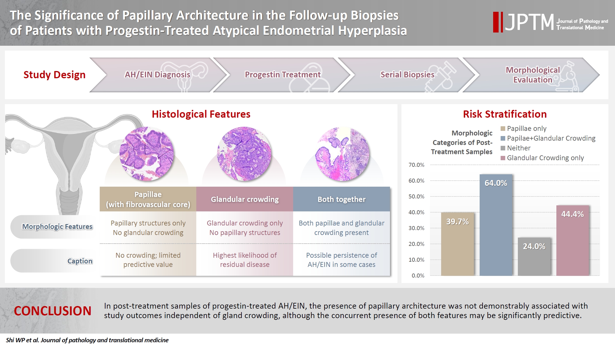
Follow-up biopsies in patients with progestin-treated atypical endometrial hyperplasia/endometrioid intraepithelial neoplasia (AH/EIN) may show papillary structures, the significance of which is unclear. Methods: The authors reviewed 253 serial specimens of 84 consecutive patients diagnosed with AH/EIN, inclusive of each patient's pre-progestin treatment sample and all post-treatment specimens. We assessed the predictive relationship between papillary architecture in a post-treatment biopsy and two study outcomes: AH/EIN or carcinoma in at least one sample subsequent to the one in which papillae were identified, and/or the last specimen received for that patient. Results: Papillae were identified in only 51.5% of pre-treatment samples but were present in at least one subsequent post-treatment sample for all patients. Post-treatment samples that exhibited papillae and no glandular crowding were associated with AH/EIN in at least one subsequent specimen in 39.7% (29/73) of cases, compared to 24.0% (6/25) in samples with neither papillae nor glandular crowding (p = .227) and 64.0% (16/25) in samples with concurrent gland crowding and papillae (p = .048). Univariate logistic regression analyses showed that the presence of papillae was not associated with study outcomes (odds ratio [OR], 0.99; 95% confidence interval [CI], 0.49 to 1.99; p = .985), as compared with gland crowding (OR, 1.54; 95% CI, 1.04 to 2.27; p = .031), or concurrent papillae and gland crowding (OR, 2.36; 95% CI, 1.01 to 5.52; p = .048). Conclusions: In post-treatment samples of progestin-treated AH/EIN, the presence of papillary architecture was not demonstrably associated with study outcomes independent of gland crowding, although the concurrent presence of both features may be significantly predictive.
- Evaluation of potential prognostic significance of JUNB in human prostate cancer: a bioinformatic and histopathological study
- Noha R. Noufal, Einas M. Yousef, Mohamed Taha
- J Pathol Transl Med. 2025;59(5):291-305. Published online September 8, 2025
- DOI: https://doi.org/10.4132/jptm.2025.06.06
- 3,784 View
- 132 Download
-
Abstract
PDF
 Supplementary Material
 - Background
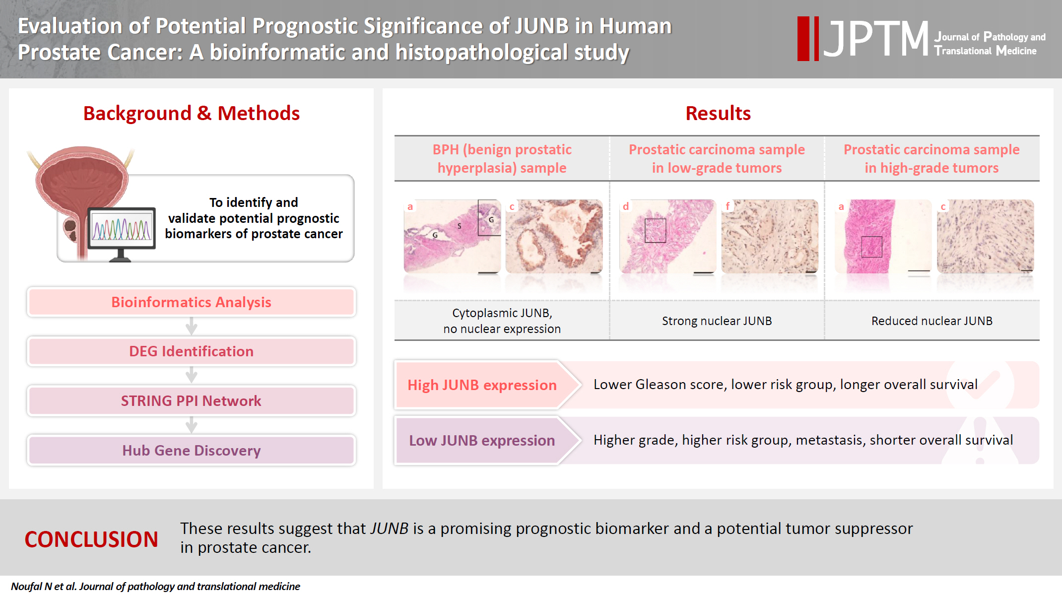
Prostate cancer is one of the most common malignancies in males worldwide. Serum prostate-specific antigen is a frequently employed biomarker in the diagnosis and risk stratification of prostate cancer; however, it is known for its low predictive accuracy for disease progression. New prognostic biomarkers are needed to distinguish aggressive prostate cancer from low-risk disease. This study aimed to identify and validate potential prognostic biomarkers of prostate cancer. Methods: Two prostate cancer datasets from the Gene Expression Omnibus were analyzed to identify differentially expressed genes between benign prostatic hyperplasia (BPH) and prostatic carcinoma. Immunohistochemistry was used to evaluate the JUNB proto-oncogene, a subunit of the AP-1 transcription factor (JUNB), in 70 prostate cancer patients and 10 BPH samples. Results: Our findings showed that JUNB was significantly enriched in prostate cancer-related pathways and biological processes. JUNB expression was considerably higher in prostatic adenocarcinoma patients than in BPH patients. Regarding JUNB expression in prostate cancer cases, lower levels of JUNB expression were associated with higher grades of prostatic adenocarcinoma. Lower JUNB expression was associated with a higher risk of prostatic adenocarcinoma progression and shorter overall survival. Conclusions: These results suggest that JUNB is a promising prognostic biomarker and a potential tumor suppressor in prostate cancer.
- Cytological features of atypical adenomatous hyperplasia and adenocarcinoma in situ of the lung: a case report
- Misa Takahashi, Seiya Homma, Chisato Setoguchi, Yoko Umezawa, Atsuhiko Sakamoto
- J Pathol Transl Med. 2025;59(3):195-200. Published online May 9, 2025
- DOI: https://doi.org/10.4132/jptm.2025.04.09

- 6,789 View
- 139 Download
-
Abstract
PDF
- Atypical adenomatous hyperplasia (AAH) and adenocarcinoma in situ (AIS) are generally treated as different lesions, depending on the differences in lesion size and histological findings. However, these differences are not absolute; thus, AAH and AIS are often difficult to distinguish. Moreover, whether AAH and AIS can be regarded as different lesions remains unknown because cytological specimens, especially those of AAH, are rare. In this study, we examined these uncommon cytological specimens and compared the cytological findings between AAH and AIS. We observed many common cytological features with no obvious differences between AAH and AIS. These findings suggest that these two distinct lesions can be grouped into a single category. Therefore, we propose creating a new cytological category.
- Pulmonary Nodular Lymphoid Hyperplasia with Mass-Formation: Clinicopathologic Characteristics of Nine Cases and Review of the Literature
- Jongmin Sim, Hyun Hee Koh, Sangjoon Choi, Jinah Chu, Tae Sung Kim, Hojoong Kim, Joungho Han
- J Pathol Transl Med. 2018;52(4):211-218. Published online June 15, 2018
- DOI: https://doi.org/10.4132/jptm.2018.04.27
- 15,928 View
- 395 Download
- 11 Web of Science
- 11 Crossref
-
Abstract
PDF
- Background
Pulmonary nodular lymphoid hyperplasia (PNLH) is a non-neoplastic pulmonary lymphoid disorder that can be mistaken for malignancy on radiography. Herein, we present nine cases of PNLH, emphasizing clinicoradiological findings and histological features.
Methods
We analyzed radiological and clinicopathological features from the electronic medical records of nine patients (eight females and one male) diagnosed with PNLH. IgG and IgG4 immunohistochemical staining was performed in three patients.
Results
Two of the nine patients had experienced tuberculosis 40 and 30 years prior, respectively. Interestingly, none were current smokers, although two were ex-smokers. Three patients complaining of persistent cough underwent computed tomography of the chest. PNLH was incidentally discovered in five patients during examination for other reasons. The remaining patient was diagnosed with the disease following treatment for pneumonia. Imaging studies revealed consolidation or a mass-like lesion in eight patients. First impressions included invasive adenocarcinoma and mucosal-associated lymphoid tissue‒type lymphoma. Aspergillosis was suspected in the remaining patient based on radiological images. Resection was performed in all patients. Microscopically, the lesions consisted of nodular proliferation of reactive germinal centers accompanied by infiltration of neutrophils and macrophages in various degrees and surrounding fibrosis. Ultimately, all nine patients were diagnosed with PNLH and showed no evidence of recurrence on follow-up.
Conclusions
PNLH is an uncommon but distinct entity with a benign nature, and understanding the radiological and clinicopathological characteristics of PNLH is important. -
Citations
Citations to this article as recorded by
- Clinical and MRI features for differentiating reactive lymphoid hyperplasia from hepatocellular carcinoma in non-cirrhotic chronic HBV patients
Qiansen Lin, Gengyun Miao, Lishan Wang, Chen Xu, Pengju Xu
European Journal of Radiology.2026; 200: 112844. CrossRef - Clinical and Imaging Features of Pulmonary Nodular Lymphoid Hyperplasia
Dong-Lei Nie, Yan-Hong Shi, Xin-Min Li, Xiao-Jiang Wang, Bao-Li Han, Guo-Fu Zhang
Journal of Thoracic Imaging.2025;[Epub] CrossRef - Pathologic Findings of Pulmonary Lymphoproliferative Disorders
Yoshiaki Zaizen, Junya Fukuoka
Seminars in Ultrasound, CT and MRI.2025; 46(4): 272. CrossRef - Utilizing Immunoglobulin G4 Immunohistochemistry for Risk Stratification in Patients with Papillary Thyroid Carcinoma Associated with Hashimoto Thyroiditis
Faridul Haq, Gyeongsin Park, Sora Jeon, Mitsuyoshi Hirokawa, Chan Kwon Jung
Endocrinology and Metabolism.2024; 39(3): 468. CrossRef - Pulmonary Nodular Lymphoid Hyperplasia Evaluated with Bronchoalveolar Lavage Fluid Findings: A Case Report and Review of the Literature on Japanese Patients
Sakiko Moriyama, Takashi Kido, Noriho Sakamoto, Mai Fuchigami, Takatomo Tokito, Daisuke Okuno, Takuto Miyamura, Shota Nakashima, Atsuko Hara, Hiroshi Ishimoto, Yoshitaka Imaizumi, Kazuto Tsuruda, Katsunori Yanagihara, Junya Fukuoka, Hiroshi Mukae
Internal Medicine.2023; 62(1): 95. CrossRef - A Case of Pulmonary Nodular Lymphoid Hyperplasia Responding to Corticosteroid Treatment
Jonathan Teow Koon Goh, Issam Al Jajeh, Jessica Han Ying Tan
Cureus.2023;[Epub] CrossRef - Pulmonary nodular lymphoid hyperplasia presenting as cavitating lung mass
Aqeel Alameer, Chary Duraikannu, Avinash Kumar Kanodia, David Dorward
BMJ Case Reports.2023; 16(8): e254121. CrossRef - Clinicopathological Characteristics and Curative Effect of Lymphoma Based on Sampling Theory
Shuxiang Ding, Leipo Liu
Mathematical Problems in Engineering.2022; 2022: 1. CrossRef - Pulmonary nodular lymphoid hyperplasia presenting as multifocal subsolid nodules: A case report and literature review
Yoon Jin Cha, Duk Hwan Moon, Ji Hyun Park, Sungsoo Lee, Ji Ae Choi, Tae Hoon Kim, Chul Hwan Park
Respiratory Medicine Case Reports.2022; 36: 101581. CrossRef - Pulmonary nodular lymphoid hyperplasia in a 53-year-old man with malignant sign: a case report
Zhen Yang, Lianshuang Wei, Xu Li, Xin Liu
Journal of Cardiothoracic Surgery.2021;[Epub] CrossRef - The diagnostic challenge of adenocarcinoma in pulmonary nodular lymphoid hyperplasia
Anita Savić Vuković, Melita Kukuljan, Morana Dinter, Ksenija Jurinović, Nives Jonjić
SAGE Open Medical Case Reports.2021;[Epub] CrossRef
- Clinical and MRI features for differentiating reactive lymphoid hyperplasia from hepatocellular carcinoma in non-cirrhotic chronic HBV patients
- Thymoma and Synchronous Primary Mediastinal Seminomas with Florid Follicular Lymphoid Hyperplasia in the Anterior Mediastinum: A Case Report and Review of the Literature
- Hyang-im Lee, In-seok Jang, Kyung Nyeo Jeon, Gyung Hyuck Ko, Jong Sil Lee, Dong Chul Kim, Dae Hyun Song, Jeong-Hee Lee
- J Pathol Transl Med. 2017;51(2):165-170. Published online February 2, 2017
- DOI: https://doi.org/10.4132/jptm.2016.08.24
- 13,232 View
- 144 Download
- 7 Web of Science
- 5 Crossref
-
Abstract
PDF
- Thymoma is the most common neoplasm of the anterior mediastinum and has malignant potential. Germ cell tumors (GCTs) found in the anterior mediastinum are usually benign, and malignant GCTs, such as seminomas, are rare. Histologically, mediastinal seminoma is indistinguishable from testicular seminoma except for site-associated morphological features such as lymphoid follicular hyperplasia. Therefore, excluding metastasis is very important. Recently, we treated a young adult patient with multiple thymic masses that occurred simultaneously. The patient underwent a thymectomy for the removal of the mediastinal masses, one of which was diagnosed as type B2 invasive thymoma, and two of which were diagnosed as primary mediastinal seminomas with massive follicular hyperplasia. The patient received adjuvant chemotherapy after surgical resection. To our knowledge, this is the first description of a thymoma and a mediastinal seminoma occurring simultaneously in the thymus. We present this case along with a literature review.
-
Citations
Citations to this article as recorded by- Primary germ cell tumours of the mediastinum: A review with emphasis on diagnostic challenges
Alexander Fichtner, Alexander Marx, Philipp Ströbel, Felix Bremmer
Histopathology.2024; 84(1): 216. CrossRef - Combined type A thymoma and yolk sac tumour of the mediastinum
Dong Sheng, Yu-Chen Han
Pathology.2024; 56(6): 927. CrossRef - Combined Thymic Epithelial Neoplasms – a Review
Annikka Weissferdt
International Journal of Surgical Pathology.2023; 31(6): 917. CrossRef - Primary mediastinal seminoma presenting with paraneoplastic anti-Hu encephalitis: a case report and literature review
Chelsey M. Williams, Derek B. Allison, Adam B. Coleman, Roshmita Bardhan, Jordan D. Miller, Zin W. Myint
Frontiers in Oncology.2023;[Epub] CrossRef - Primary mediastinal seminoma with florid follicular lymphoid hyperplasia: a case report and review of the literature
Charlotte Holmes, Peh Sun Loo, Sion Barnard
Diagnostic Pathology.2021;[Epub] CrossRef
- Primary germ cell tumours of the mediastinum: A review with emphasis on diagnostic challenges
- A Pyloric Gland-Phenotype Ovarian Mucinous Tumor Resembling Lobular Endocervical Glandular Hyperplasia in a Patient with Peutz-Jeghers Syndrome
- Eun Na Kim, Gu-Hwan Kim, Jiyoon Kim, In Ah Park, Jin Ho Shin, Yun Chai, Kyu-Rae Kim
- J Pathol Transl Med. 2017;51(2):159-164. Published online August 22, 2016
- DOI: https://doi.org/10.4132/jptm.2016.07.01
- 11,157 View
- 210 Download
- 10 Web of Science
- 9 Crossref
-
Abstract
PDF
- We describe an ovarian mucinous neoplasm that histologically resembles lobular endocervical glandular hyperplasia (LEGH) containing pyloric gland type mucin in a patient with Peutz-Jeghers syndrome (PJS). Although ovarian mucinous tumors rarely occur in PJS patients, their pyloric gland phenotype has not been clearly determined. The histopathologic features of the ovarian mucinous tumor were reminiscent of LEGH. The cytoplasmic mucin was stained with periodic acid-Schiff reaction after diastase treatment but was negative for Alcian blue pH 2.5, suggesting the presence of neutral mucin. Immunohistochemically, the epithelium expressed various gastric markers, including MUC6, HIK1083, and carbonic anhydrase-IX. Multiple ligation-dependent probe amplification detected a germline heterozygous deletion mutation at exons 1–7 of the STK11 gene (c.1-?_920+?del) in peripheral blood leukocytes and mosaic loss of heterozygosity in ovarian tumor tissue. Considering that LEGH and/or gastric-type cervical adenocarcinoma can be found in patients with PJS carrying germline and/or somatic STK11 mutations, our case indicates that STK11 mutations have an important role in the proliferation of pyloric-phenotype mucinous epithelium at various anatomical locations.
-
Citations
Citations to this article as recorded by- Molecular evidence of a clonal relationship of synchronous/multifocal gastric‐type lesions of the female genital tract
Min Shi, Hong Yang, Fang Zhang, Ting Hou, Huageng Huang, Yi Lu, Yehan Zhou, Ting Lan, Juan Ji, Jun Hou, Chengmin Zhou, Zhou Zhang, Sheng Qin, Zongyao Huang, Yang Liu
The Journal of Pathology.2026; 268(1): 27. CrossRef - Serine/threonine kinase 11 (STK11) associated adnexal tumors: from biology to therapeutic impact
Guanxiang Huang, Wenyu Lin, Tingting Jiang, Yuanjun Cai, Chengbin Lin, Pengming Sun
Human Genomics.2025;[Epub] CrossRef - Novel ultrasound features and diagnostic clues of gastric-type endocervical adenocarcinoma: a case series
Liwen Yang, Yangyang Wang, Jian Cai, Ying Xiong, Juan Li, Qi Zhou, Nan Ye, Hua Lai, Tianjiao Liu, Liuying Zhou
Frontiers in Oncology.2025;[Epub] CrossRef - Ovarian Mucinous Tumor Presenting Atypical Lobular Endocervical Glandular Hyperplasia-Like Appearance in a Patient With Germline STK11 p.F354L Variant: A Case Report
Hiroshi Yoshida, Kengo Hiranuma, Mariko Nakahara, Mayumi Kobayashi-Kato, Yasuhito Tanase, Masaya Uno, Kouya Shiraishi, Mitsuya Ishikawa, Tomoyasu Kato
International Journal of Surgical Pathology.2024; 32(2): 394. CrossRef - Preoperative multimodal ultrasonic imaging in a case of Peutz-Jeghers syndrome complicated by atypical lobular endocervical glandular hyperplasia: a case report and literature review
Liwen Yang, Duan Duan, Ying Xiong, Tianjiao Liu, Lijun Zhao, Fan Lai, Dingxian Gu, Liuying Zhou
Hereditary Cancer in Clinical Practice.2024;[Epub] CrossRef - Gastric‐type glandular lesions of the female genital tract excluding the cervix: emerging pathological entities
Richard W‐C Wong, Karen L Talia, W Glenn McCluggage
Histopathology.2024; 85(1): 20. CrossRef - Gastric-phenotype Mucinous Carcinoma of the Fallopian Tube with Secondary Ovarian Involvement in a Woman with Peutz-Jeghers Syndrome: A Case Report
Mónica Bronte Anaut, Javier Arredondo Montero, Maria Pilar Fernández Seara, Rosa Guarch Troyas
International Journal of Surgical Pathology.2023; 31(1): 92. CrossRef - Molecular characterization of gastric-type endocervical adenocarcinoma using next-generation sequencing
Swati Garg, Teddy S. Nagaria, Blaise Clarke, Orit Freedman, Zanobia Khan, Joerg Schwock, Marcus Q. Bernardini, Amit M. Oza, Kathy Han, Adam C. Smith, Tracy L. Stockley, Marjan Rouzbahman
Modern Pathology.2019; 32(12): 1823. CrossRef - The developing spectrum of gastric-type cervical glandular lesions
Karen L. Talia, W. Glenn McCluggage
Pathology.2018; 50(2): 122. CrossRef
- Molecular evidence of a clonal relationship of synchronous/multifocal gastric‐type lesions of the female genital tract
- An Adult Case of Bartter Syndrome Type III Presenting with Proteinuria
- Eun Jung Cha, Won Min Hwang, Sung-Ro Yun, Moon Hyang Park
- J Pathol Transl Med. 2016;50(2):160-164. Published online January 11, 2016
- DOI: https://doi.org/10.4132/jptm.2015.08.31
- 13,598 View
- 122 Download
- 7 Web of Science
- 7 Crossref
-
Abstract
PDF
- Bartter syndrome (BS) I–IV is a rare autosomal recessive disorder affecting salt reabsorption in the thick ascending limb of the loop of Henle. This report highlights clinicopathological findings and genetic studies of classic BS in a 22-year-old female patient who presented with persistent mild proteinuria for 2 years. A renal biopsy demonstrated a mild to moderate increase in the mesangial cells and matrix of most glomeruli, along with marked juxtaglomerular cell hyperplasia. These findings suggested BS associated with mild IgA nephropathy. Focal tubular atrophy, interstitial fibrosis, and lymphocytic infiltration were also observed. A genetic study of the patient and her parents revealed a mutation of the CLCNKB genes. The patient was diagnosed with BS, type III. This case represents an atypical presentation of classic BS in an adult patient. Pathologic findings of renal biopsy combined with genetic analysis and clinicolaboratory findings are important in making an accurate diagnosis.
-
Citations
Citations to this article as recorded by- Complexities of Bartter Syndrome Type III: A Case Study in Jordan
Hamdah Hanifa, Yumna Al-Badareen, Malak Mbarak Al-Refaai, Nafeaa M Ganama, Mohammad Sameeh Alabrash, Basil Alsaleh
Oxford Medical Case Reports.2025;[Epub] CrossRef - Bartter syndrome with multiple renal and liver cysts: a case report
Yemei He, Yue Zhou, Weihua Wu, Yue Chen, Santao Ou
International Urology and Nephrology.2022; 55(1): 225. CrossRef - Bartter’s syndrome: clinical findings, genetic causes and therapeutic approach
Flavia Cristina Carvalho Mrad, Sílvia Bouissou Morais Soares, Luiz Alberto Wanderley de Menezes Silva, Pedro Versiani dos Anjos Menezes, Ana Cristina Simões-e-Silva
World Journal of Pediatrics.2021; 17(1): 31. CrossRef - Association of Adult-Onset Bartter Syndrome With Undifferentiated Connective Tissue Disorder
Nida Saleem, Humaira Nasir, Danyal Hassan, Momena Manzoor
Cureus.2021;[Epub] CrossRef - Acquired autoimmune Bartter syndrome in a patient with primary hypothyroidism
Noreen Nasir, Deepali Mohanty, Arun Kumar Pande, Dhanita Khanna, Kavita Vishvakarma, Latika Gupta
Rheumatology International.2021; 43(3): 567. CrossRef - A novel mutation associated with Type�III Bartter syndrome: A report of five cases
Yanhan Li, Chengcheng Wu, Jie Gu, Dong Li, Yanling Yang
Molecular Medicine Reports.2019;[Epub] CrossRef - Pathophysiology of antenatal Bartterʼs syndrome
Martin Kömhoff, Kamel Laghmani
Current Opinion in Nephrology and Hypertension.2017; 26(5): 419. CrossRef
- Complexities of Bartter Syndrome Type III: A Case Study in Jordan
- Castleman's Disease of the Renal Sinus Presenting as a Urothelial Malignancy: A Brief Case Report
- Se Min Jang, Hulin Han, Ki-Seok Jang, Young Jin Jun, Tchun Yong Lee, Seung Sam Paik
- Korean J Pathol. 2012;46(5):503-506. Published online October 25, 2012
- DOI: https://doi.org/10.4132/KoreanJPathol.2012.46.5.503
- 9,443 View
- 49 Download
- 5 Crossref
-
Abstract
PDF
Castleman's disease is a rare benign lymphoproliferative disorder that frequently affects lymph nodes of the mediastinal thorax and the neck. It very rarely affects the renal sinus. We report a case of Castleman's disease arising in the renal sinus in a 64-year-old man. The patient visited the hospital with the chief complaint of hematuria. Abdominal computed tomography revealed a homogeneous mass in the sinus of the left kidney, radiologically interpreted as a malignant urothelial tumor. Subsequently, nephroureterectomy was performed, after which microscopic examination of the specimen revealed a diffuse lymphoproliferative lesion with reactive lymphoid follicles of various sizes and prominent plasma cell infiltration of interfollicular spaces, highlighted by immunohistochemical staining for CD138. The lesion was diagnosed as Castleman's disease of the plasma cell type. Although preoperative diagnosis of Castleman's disease is difficult and the incidence is exceedingly rare, it should be considered in the differential diagnosis of renal sinus tumors.
-
Citations
Citations to this article as recorded by- Retrospective analysis of primary extranodal unicentric Castleman disease: a systematic review
Jianing Shen, Yongjun Zeng, Yuan Liu, Nie Xu
Frontiers in Medicine.2026;[Epub] CrossRef - Misdiagnosis of renal pelvic unicentric Castleman disease: a case report
Dian Fu, Bo Yang, Ming Yang, Zhenyu Xu, Wen Cheng, Zhijia Liu, Liming Zhang, Zhiguo Mao, Cheng Xue
Frontiers in Surgery.2023;[Epub] CrossRef - Case report: Castleman’s disease involving the renal sinus resembling renal cell carcinoma
Enlong Zhang, Yuan Li, Ning Lang
Frontiers in Surgery.2022;[Epub] CrossRef - Radiologic features of Castleman’s disease involving the renal sinus: A case report and review of the literature
Xiao-Wan Guo, Xu-Dong Jia, Shan-Shan Shen, Hong Ji, Ying-Min Chen, Qian Du, Shu-Qian Zhang
World Journal of Clinical Cases.2019; 7(8): 1001. CrossRef - Castleman’s Disease: a Suprarenal Surprise!
Praveen Sundar, Priyank Bijalwan, Ginil Kumar Pooleri
Indian Journal of Surgical Oncology.2018; 9(2): 254. CrossRef
- Retrospective analysis of primary extranodal unicentric Castleman disease: a systematic review
- Expressions of E-cadherin, Cortactin and MMP-9 in Pseudoepitheliomatous Hyperplasia and Squamous Cell Carcinoma of the Head and Neck: Their Relationships with Clinicopathologic Factors and Prognostic Implication
- Tack Kune You, Kyoung Min Kim, Sang Jae Noh, Jun Sang Bae, Kyu Yun Jang, Myoung Ja Chung, Woo Sung Moon, Myoung Jae Kang, Dong Geun Lee, Ho Sung Park
- Korean J Pathol. 2012;46(4):331-340. Published online August 23, 2012
- DOI: https://doi.org/10.4132/KoreanJPathol.2012.46.4.331
- 11,236 View
- 86 Download
- 15 Crossref
-
Abstract
PDF
Background E-cadherin, cortactin, and matrix metalloproteinase (MMP)-9 have roles in tumor development or progression, but their expression has not been fully investigated in pseudoepitheliomatous hyperplasia (PEH) and squamous cell carcinoma (SCC) of the head and neck.
Methods We evaluated the immunohistochemical expression of E-cadherin, cortactin, and MMP-9 in 29 cases of PEH and 97 cases of SCC. Additionally, we evaluated their relationship with clinicopathologic factors and prognostic implications in SCC.
Results Thirty-five cases of SCC showed reduced expression of E-cadherin, whereas none of the PEH did. A total of 20 cases and 11 cases of SCC were immunoreactive for cortactin and MMP-9, respectively, whereas none of the PEH did. In SCC, reduced expression of E-cadherin was correlated with cortactin expression and invasion depth. Cortactin expression was correlated with differentiation, T classification, and recurrence and/or metastasis. MMP-9 expression was correlated with invasion depth. Cortactin expression was correlated with poor overall survival and relapse-free survival and it was an independent prognostic factor.
Conclusions The reduced expression of E-cadherin and the expression of cortactin may be helpful for the differential diagnosis of PEH and SCC. Furthermore, cortactin expression in association with reduced E-cadherin expression is correlated with poor prognosis in SCC.
-
Citations
Citations to this article as recorded by- HIV-1 Tat-induced disruption of epithelial junctions and epithelial-mesenchymal transition of oral and genital epithelial cells lead to increased invasiveness of neoplastic cells and the spread of herpes simplex virus and cytomegalovirus
Sharof Tugizov
Frontiers in Immunology.2025;[Epub] CrossRef - Ultrastructural and immunohistochemical evaluation of hyperplastic soft tissues surrounding dental implants in fibular jaws
Kezia Rachellea Mustakim, Mi Young Eo, Mi Hyun Seo, Hyeong-Cheol Yang, Min-Keun Kim, Hoon Myoung, Soung Min Kim
Scientific Reports.2024;[Epub] CrossRef - Virus-associated disruption of mucosal epithelial tight junctions and its role in viral transmission and spread
Sharof Tugizov
Tissue Barriers.2021;[Epub] CrossRef - Leishmaniasis: still a diagnostic challenge?
Ricardo Tadeu Villa
Journal of Dermatology & Cosmetology.2021; 5(2): 23. CrossRef - COMPARISON OF EXPRESSION OF E-CADHERIN IN ORAL PSEUDOEPITHELIOMATOUS HYPERPLASIA AND ORAL SQUAMOUS CELL CARCINOMA
Ayesha Mukhtar Awan, Iram Naz, Muhammad Khurram Mahmood, Hafeez Uddin
Gomal Journal of Medical Sciences.2020; 17(3): 70. CrossRef - EXPRESSION OF MATRIX METALLOPROTEINASE-9 IN ORAL SQUAMOUS CELL CARCINOMA AND ORAL PSEUDOEPITHELIOMATOUS HYPERPLASIA
Ayesha Mukhtar Awan, Iram Naz, Muhammad Khurram Mahmood, Hafeez Uddin
Gomal Journal of Medical Sciences.2020; 18(01): 24. CrossRef - An update of knowledge on cortactin as a metastatic driver and potential therapeutic target in oral squamous cell carcinoma
Pablo Ramos‐García, Miguel Ángel González‐Moles, Lucía González‐Ruiz, Ángela Ayén, Isabel Ruiz‐Ávila, Francisco José Navarro‐Triviño, José Antonio Gil‐Montoya
Oral Diseases.2019; 25(4): 949. CrossRef - Prognostic and clinicopathological significance of CTTN/cortactin alterations in head and neck squamous cell carcinoma: Systematic review and meta‐analysis
Pablo Ramos‐García, Miguel Ángel González‐Moles, Ángela Ayén, Lucía González‐Ruiz, Isabel Ruiz‐Ávila, José Antonio Gil‐Montoya
Head & Neck.2019; 41(6): 1963. CrossRef - The effect of centromere protein U silencing by lentiviral mediated RNA interference on the proliferation and apoptosis of breast cancer
Shuang‑Yan Lin, Yan‑Bo Lv, Gen‑Xiang Mao, Xu‑Jiao Chen, Fang Peng
Oncology Letters.2018;[Epub] CrossRef - Glycosylation: a hallmark of cancer?
Bhairavi N. Vajaria, Prabhudas S. Patel
Glycoconjugate Journal.2017; 34(2): 147. CrossRef - Differential expression of the sirtuin family in renal cell carcinoma: Aspects of carcinogenesis and prognostic significance
Seong Uk Jeh, Jung Je Park, Jong Sil Lee, Dong Chul Kim, Jungmo Do, Sin Woo Lee, See Min Choi, Jae Seog Hyun, Deok Ha Seo, Chunwoo Lee, Sung Chul Kam, Ky Hyun Chung, Jeong Seok Hwa
Urologic Oncology: Seminars and Original Investigations.2017; 35(12): 675.e9. CrossRef - Cortactin promotes colorectal cancer cell proliferation by activating the EGFR-MAPK pathway
Xiaojian Zhang, Kun Liu, Tao Zhang, Zhenlei Wang, Xuan Qin, Xiaoqian Jing, Haoxuan Wu, Xiaopin Ji, Yonggang He, Ren Zhao
Oncotarget.2017; 8(1): 1541. CrossRef - Cortactin in cancer cell migration and invasion
Miao Yin, Wenqing Ma, Liguo An
Oncotarget.2017; 8(50): 88232. CrossRef - Association of SIRT1 and HMGA1 expression in non-small cell lung cancer
SHUANG-YAN LIN, FANG PENG
Oncology Letters.2016; 11(1): 782. CrossRef - Expression of SIRT1 and cortactin is associated with progression of non-small cell lung cancer
Sang Jae Noh, Hyun Ah Baek, Ho Sung Park, Kyu Yun Jang, Woo Sung Moon, Myoung Jae Kang, Dong Geun Lee, Min Ho Kim, Ju Hyung Lee, Myoung Ja Chung
Pathology - Research and Practice.2013; 209(6): 365. CrossRef
- HIV-1 Tat-induced disruption of epithelial junctions and epithelial-mesenchymal transition of oral and genital epithelial cells lead to increased invasiveness of neoplastic cells and the spread of herpes simplex virus and cytomegalovirus
- Hyaline Vascular Castleman Disease Involving Renal Parenchyma and a Lymph Node: A Case Report
- Ji Hyun Kwon, Soo Kee Min, Mi Kyung Shin, Yong Seong Lee, Young-Goo Lee, Young Hyeh Ko
- Korean J Pathol. 2012;46(1):79-82. Published online February 23, 2012
- DOI: https://doi.org/10.4132/KoreanJPathol.2012.46.1.79
- 10,732 View
- 56 Download
- 6 Crossref
-
Abstract
PDF
Castleman disease is a rare lymphoproliferative lesion that is predominantly found in the mediastinum. Retroperitoneal and pararenal localizations are very rare. We describe a 36-year-old man with a hyaline vascular type of Castleman disease involving renal parenchyma and a paraaortic lymph node. Most reported renal Castleman disease was plasma cell type with systemic symptoms. Herein, we report the first Korean case of the hyaline vascular type of Castleman disease involving the renal parenchyma and the paraaortic lymph node simultaneously.
-
Citations
Citations to this article as recorded by- Hiding in the kidney: a series of 13 lymphoid proliferations clinically mimicking renal carcinoma
Jihoon William Lee, Marie E. Perrone, Daniel E. Sabath, Daniel W. Lin, George R. Schade, Funda Vakar-Lopez, Maria Tretiakova
Virchows Archiv.2025;[Epub] CrossRef - Primary hyaline vascular Castleman disease in the kidney: a report and brief literature review
Ibrahim Elsharawi, Sorin Selegean
Journal of Hematopathology.2025;[Epub] CrossRef - Castleman Disease of the Kidney in Computed Tomography Urography
Kai Wang, Fengjuan Xing, Heng Ma, Wenjuan Li
Current Medical Imaging Formerly Current Medical Imaging Reviews.2022; 18(1): 74. CrossRef - Primary hyaline vascular Castleman disease of the kidney: case report and literature review
Yunzhu Li, Haixia Zhao, Bingyin Su, Chan Yang, Shurong Li, Wanlei Fu
Diagnostic Pathology.2019;[Epub] CrossRef - Castleman’s Disease of the Kidney Mimicking Renal Cell Carcinoma on FDG PET/CT
Yang Wang, Aisheng Dong, Bo Yang, Jianping Lu
Clinical Nuclear Medicine.2018; 43(5): e160. CrossRef - Unicentric hyaline vascular type of castleman disease of the renal hilum with diagnostic dilemma: A case report and review of literature
AmitKumar Adhya, ManasRanjan Pradhan
Oncology Journal of India.2018; 2(4): 96. CrossRef
- Hiding in the kidney: a series of 13 lymphoid proliferations clinically mimicking renal carcinoma
- The Ratio of Atypical Ductal Hyperplasia Foci to Core Numbers in Needle Biopsy: A Practical Index Predicting Breast Cancer in Subsequent Excision
- Jeong-Ju Lee, Hee Jin Lee, Jun Kang, Jeong-Hyeon Jo, Gyungyub Gong
- Korean J Pathol. 2012;46(1):15-21. Published online February 23, 2012
- DOI: https://doi.org/10.4132/KoreanJPathol.2012.46.1.15
- 14,326 View
- 50 Download
- 1 Crossref
-
Abstract
PDF
Background Although core needle biopsy (CNB) is considered to be the standard technique for histological diagnosis of breast lesions, it is less reliable for diagnosing atypical ductal hyperplasia (ADH). We therefore assessed the characteristics of CNB-diagnosed ADH that are more likely to be associated with more advanced lesions on subsequent surgical excision.
Methods We retrospectively examined 239 consecutive CNBs, 127 of which were diagnosed as ADH following surgical excision, performed at Asan Medical Center between 1995 and 2010. Archival slides were analyzed for the number of cores per specimen, the number of ADH foci, and the ratio of ADH foci to number of cores (FC ratio).
Results We found that ADH foci in 3 or more cores (p=0.003) and the presence of ADH in 3 or more foci (p=0.002) were correlated with malignancy following excision lesion. Moreover, an FC>1.1 was significantly associated with malignancy in the subsequent excision (p=0.000).
Conclusions Including the number of ADH foci, the number of cores involved according to ADH, FC ratio, and histologic type in a pathology report of CNB may help in making clinical decisions about surgical excision.
-
Citations
Citations to this article as recorded by- Active Surveillance for Atypical Ductal Hyperplasia and Ductal Carcinoma In Situ
Rachel Miceli, Cecilia L Mercado, Osvaldo Hernandez, Chloe Chhor
Journal of Breast Imaging.2023; 5(4): 396. CrossRef
- Active Surveillance for Atypical Ductal Hyperplasia and Ductal Carcinoma In Situ
- Extranodal NK/T Cell Lymphoma Accompanied by Heavy Eosinophilic Infiltration and Peripheral Blood Eosinophilia, Involving Skeletal Muscles.
- Jin Ho Paik, Yoon Kyung Jeon, Heounjeong Go, Chul Woo Kim
- Korean J Pathol. 2011;45:S70-S74.
- DOI: https://doi.org/10.4132/KoreanJPathol.2011.45.S1.S70
- 5,273 View
- 45 Download
- 8 Crossref
-
Abstract
PDF
- The patient was a 52-year-old female with swelling in both lower legs and peripheral blood eosinophilia. Biopsy specimen revealed the heavy infiltration of eosinophils with sparse small lymphocytes showing mild atypia. The diagnosis was Kimura disease. The symptoms including eosinophilia were relieved by steroid treatment. At 17 months from initial biopsy, the patient developed swelling of the buttock. At 25 months, fever and dyspnea with multiple lung nodules developed. Wedge resection revealed multiple aggregates of CD3(+), CD56(+), Epstein-Barr virus(+) large atypical lymphocytes with necrosis. The patient was finally diagnosed with extranodal NK/T cell lymphoma (NKTL). Epstein-Barr virus in situ hybridization retrospectively performed on the previous biopsies demonstrated Epstein-Barr virus infection in small CD3(+) lymphocytes. The patient expired after 26 months despite chemotherapy. Blood eosinophilia correlated well with disease activity during the clinical course. This case shows not only unusual histologic features, which hampered the correct diagnosis, but also a unique clinical manifestation of NKTL.
-
Citations
Citations to this article as recorded by- A case of extranodal NK/T cell lymphoma suspected to be focal myositis of the masseter muscle
Junya YAMASHITA, Daisuke TAKEDA, Tatsuya SHIRAI, Kaito URYU, Nanae YATAGAI, Masaya AKASHI
Japanese Journal of Oral and Maxillofacial Surgery.2025; 71(1): 33. CrossRef - Muscular involvement of extranodal natural killer/T cell lymphoma misdiagnosed as polymyositis: A case report and review of literature
Li-Hui Liu, Qing Huang, Yun-Hai Liu, Jie Yang, Han Fu, Lin Jin
World Journal of Clinical Cases.2020; 8(5): 963. CrossRef - Extranodal natural killer/T-cell lymphoma with paraneoplastic eosinophilic myositis
Jayati Mallick, Jasmine Zain, Dennis D. Weisenburger
Human Pathology: Case Reports.2020; 21: 200391. CrossRef - Extranodal NK/T-cell Lymphoma Mimicking Granulomatous Myositis
Norihiko Kawaguchi, Rumiko Izumi, Masahiro Kobayashi, Maki Tateyama, Naoki Suzuki, Fumiyoshi Fujishima, Juichi Fujimori, Masashi Aoki, Ichiro Nakashima
Internal Medicine.2019; 58(2): 277. CrossRef - Uveitis and Myositis as Immune Complications in Chemorefractory NK/T-Cell Nasal-Type Lymphoma Successfully Treated with Allogeneic Stem-Cell Transplant
Maria José Gómez-Crespo, Aránzazu García-Raso, Jose Luis López-Lorenzo, Teresa Villaescusa, María Rodríguez-Pinilla, José Fortes, Cristina Serrano, Salma Machan, Pilar Llamas, Raúl Córdoba
Case Reports in Hematology.2016; 2016: 1. CrossRef - Prognostic implications of CD30 expression in extranodal natural killer/T-cell lymphoma according to treatment modalities
Wook Youn Kim, Soo Jeong Nam, Sehui Kim, Tae Min Kim, Dae Seog Heo, Chul-Woo Kim, Yoon Kyung Jeon
Leukemia & Lymphoma.2015; 56(6): 1778. CrossRef - Unusual case of metachronous EBV‐associated B‐cell and NK/T‐cell lymphoma mimicking polymyositis‐diagnostic challenges and pitfalls
Esther H.L. Chan, Suat‐Jin Lu, Fredrik Petersson, Kong‐Bing Tan, Wee‐Joo Chng, Siok‐Bian Ng
American Journal of Hematology.2014; 89(1): 110. CrossRef - CD30+ extranodal natural killer/T-cell lymphoma mimicking phlegmonous myositis: A case report
YAN-JIA YANG, YA-XIN LI, YAN-BIN LIU, MEI YANG, KAI LIU
Oncology Letters.2014; 7(5): 1419. CrossRef
- A case of extranodal NK/T cell lymphoma suspected to be focal myositis of the masseter muscle
- Systemic Plasmacytosis: A Case Report with a Review of the Literature.
- Sung Hak Lee, Chang Young Yoo, Ji Han Jung, Jin Young Yoo, Suk Jin Kang, Chang Suk Kang
- Korean J Pathol. 2011;45(6):632-638.
- DOI: https://doi.org/10.4132/KoreanJPathol.2011.45.6.632
- 5,201 View
- 55 Download
- 3 Crossref
-
Abstract
PDF
- Systemic plasmacytosis is an uncommon disorder characterized by widely disseminated macular skin eruptions composed of polyclonal lymphoplasmacytic infiltrates associated with variable extracutaneous involvement. An aggressive clinical course has been observed in a small number of patients, but most cases have followed chronic and benign clinical course without spontaneous remission. Previously reported cases of this entity have been described almost exclusively in Japanese patients. We recently experienced a case of systemic plasmacytosis in a 48-year-old Korean female patient. Initial skin biopsy specimen revealed patchy perivascular and periadnexal infiltrates of mature plasma cells. Serum immunoelectrophoresis revealed polyclonal hypergammaglobulinemia, and polyclonal plasmacytosis was noted on the subsequent biopsy specimens of left supraclavicular and axillary lymph nodes. Multiple tiny pulmonary nodules appeared six years after the initial cutaneous presentation and were found to be of the same histologic appearance. We herein report a rare case of systemic plasmacytosis with a review of the literature.
-
Citations
Citations to this article as recorded by- Cutaneous plasmacytosis with mast cell infiltration
Sarina Jain, RohitV Hede, UdayS Khopkar
Indian Journal of Dermatology, Venereology and Leprology.2020; 86(1): 91. CrossRef - Plasmocitosis cutánea en un varón de raza blanca
A. López-Gómez, T. Salas-García, A. Ramírez-Andreo, E. Poblet-Martínez
Actas Dermo-Sifiliográficas.2015; 106(6): 520. CrossRef - Cutaneous Plasmacytosis in a White Man
A. López-Gómez, T. Salas-García, A. Ramírez-Andreo, E. Poblet-Martínez
Actas Dermo-Sifiliográficas (English Edition).2015; 106(6): 520. CrossRef
- Cutaneous plasmacytosis with mast cell infiltration
- Newly Formed Hepatic Masses in Children with Biliary Atresia after Kasai Hepatic Portoenterostomy.
- Hye Jong Song, Yeon Lim Suh
- Korean J Pathol. 2011;45(2):160-169.
- DOI: https://doi.org/10.4132/KoreanJPathol.2011.45.2.160
- 5,356 View
- 31 Download
- 3 Crossref
-
Abstract
PDF
- BACKGROUND
This report describes the clinicopathologic findings of six hepatic masses that developed after Kasai hepatic portoenterostomy (HPE) in six patients with longstanding biliary atresia (BA).
METHODS
Hepatic masses were found in six of 55 pediatric patients who underwent liver transplantation for BA after Kasai HPE from 1997 to 2009. Clinicopathologic analysis was performed and immunohistochemical staining was carried out for CD34, smooth muscle actin (SMA) and cytokeratin 7.
RESULTS
Of the six hepatic masses, two were diagnosed as focal nodular hyperplasia (FNH)-like lesions, two were large regenerative nodules (LRN), one was a mesenchymal hamartoma (MH) and one was a cholangiocarcinoma. The immunohistochemical staining findings for SMA and CD34 were more prominent for the FNH-like nodules than for the cirrhotic background liver. Dysplastic biliary epithelium arising from intestinal metaplasia was found in the cholangiocarcinoma.
CONCLUSIONS
Our findings suggest that FNH-like lesions, LRNs and MH are the results of vascular hemodynamic changes after Kasai HPE and that cholangiocarcinoma is due to recurrent cholangitis after BA. All the lesions in this series must be included in the differential diagnosis of a newly formed hepatic mass in patients after portoenterostomy. -
Citations
Citations to this article as recorded by- Imaging Findings and Management Strategies for Liver Masses in Children with Predisposition Disorders: A Review by the Pediatric LI-RADS Group
Amy B. Kolbe, Michael R. Acord, Geetika Khanna, Cara E. Morin, HaiThuy N. Nguyen, Mitchell A. Rees, Esther Ro, Gary R. Schooler, Judy H. Squires, Ali B. Syed, Elizabeth R. Tang, Alexander J. Towbin, Adina Alazraki
RadioGraphics.2025;[Epub] CrossRef - Features of Nodules in Explants of Children Undergoing Liver Transplantation for Biliary Atresia
Ana M. Calinescu, Anne-Laure Rougemont, Mehrak Anooshiravani, Nathalie M. Rock, Valerie A. McLin, Barbara E. Wildhaber
Journal of Clinical Medicine.2022; 11(6): 1578. CrossRef - Biliary Atresia Patients With Successful Kasai Portoenterostomy Can Present With Features of Obliterative Portal Venopathy
Kalyani R. Patel, Sanjiv Harpavat, Zahida Khan, Sadhna Dhingra, Norma Quintanilla, Mihail Firan, John Goss
Journal of Pediatric Gastroenterology and Nutrition.2020; 71(1): 91. CrossRef
- Imaging Findings and Management Strategies for Liver Masses in Children with Predisposition Disorders: A Review by the Pediatric LI-RADS Group
- Smooth Muscle Hyperplasia of the Epididymis: Report of A Case and Review of the Literature.
- Hyun Soo Kim, Ji Youn Sung, Gou Young Kim, Sung Jig Lim, Hyun Cheol Kim, Hyung Lae Lee
- Korean J Pathol. 2009;43(2):177-181.
- DOI: https://doi.org/10.4132/KoreanJPathol.2009.43.2.177
- 5,145 View
- 29 Download
- 4 Crossref
-
Abstract
PDF
- A 66-year-old man underwent surgery to remove an incidentally discovered non-tender intrascrotal mass. Ultrasonography revealed an irregular-margined, heterogeneous mass-like lesion in the epididymal tail. The mass was relatively well circumscribed but unencapsulated, irregular and firm; it consisted of expansile, increased smooth muscle fascicles originating from the epididymal muscular coat. Its cellular growth pattern lacked the cohesive, well-circumscribed proliferation pattern typical of a leiomyoma. A diagnosis of smooth muscle hyperplasia of the epididymis was made. Although ultrasonography is the imaging modality of choice for evaluating suspected intrascrotal masses, there are times when it cannot reliably identify the character of the masses and distinguish malignant from benign lesions. Ill-defined, solid extratesticular masses, that are ultrasonographically ambiguous, should be excised and confirmed histopathologically and smooth muscle hyperplasia of the epididymis should be included in the differential diagnosis of solid extratesticular masses.
-
Citations
Citations to this article as recorded by- Smooth muscle hyperplasia of the testicular adnexa: a clinicopathologic study of 12 cases
Fatimah Alruwaii, David J. Grignon, Muhammad T. Idrees
Human Pathology.2020; 99: 27. CrossRef - Hiperplasia muscular paratesticular (epididimaria) pseudotumoral. Descripción de 2 casos
Inmaculada Ruiz Molina, Vicente Cívico Amat, Beatriz Santiago Agredano
Medicina Clínica.2019; 152(5): e25. CrossRef - Pseudotumoral paratesticular (epididymal) muscle hyperplasia. Two case reports
InmaculadaRuiz Ruiz Molina, Vicente Cívico Amat, Beatriz Santiago Agredano
Medicina Clínica (English Edition).2019; 152(5): e25. CrossRef - Smooth muscle hyperplasia of the epididymis
O Blach, AM Pollock, D Douglas
Journal of Surgical Case Reports.2011; 2011(10): 10. CrossRef
- Smooth muscle hyperplasia of the testicular adnexa: a clinicopathologic study of 12 cases
- Adenocarcinoma Arising in Adenomyosis.
- Young Il Yang, In Sook Lim, Jong Eun Joo
- Korean J Pathol. 1995;29(2):272-274.
- 2,330 View
- 15 Download
-
Abstract
PDF
- Adenocarcinoma in adenomyosis is unusual and it is mostly associated with adenocarcinoma in the endometrial mucosa. In contrast, adenocarcinoma arising in adenomyosis without endometrial adenocarcinoma is extremely rare and it suggests that it arises de novo from adenomyosis. We report a case of adenocarcinoma arising in adenomyosis in 44-year-old woman. The endometrial cavity contained a polypoid lesion with atypical hyperplasia, but no evidence of adenocarcinoma in the endometrial mucosa. Simple, complex and atypical hyperplasia associated with well differentiated adenocarcinoma was also noted in the areas of adenomyosis.
- An Unusual Stroma-Rich Variant of Castleman's Disease of the Hyaline-Vascular Type: A Case Report.
- Ji Han Jung, Gyeongsin Park, Hyun Joo Choi, Jinyoung Yoo, Seok Jin Kang, Kyo Young Lee
- Korean J Pathol. 2007;41(4):266-270.
- 2,962 View
- 63 Download
-
Abstract
PDF
- The stroma-rich variant of Castleman's disease of the hyaline-vascular type (CDHV) is a rare entity that shows overgrowth of a variety of stromal cells in the widened interfollicular (IF) area. We report here on a case of a stroma-rich variant of CDHV in an 18-year-old man who presented with an asymptomatic solitary neck mass he'd had for 1 year. Histologically, an enlarged lymph node fulfilled the criteria of CDHV, along with vague nodularity of a widened IF area. The nodular lesion consisted of numerous vessels and a proliferation of spindle cells. Immunohistochemically, the spindle cells were positive for vimentin and smooth muscle actin, they were negative for desmin, CD21, CD34, CD68, ALK-1, and S-100 protein. This stromal lesion is typically hyperplastic and clinically benign, and it must be distinguished from neoplastic stromal proliferation associated with Castleman's disease because of its potential for recurrence and metastasis.
- Three Cases of Giant Lymph Node Hyperplasia in Unusual Location.
- Hye Kyung An, Ill Hyang Ko
- Korean J Pathol. 1989;23(3):365-370.
- 1,998 View
- 12 Download
-
Abstract
PDF
- Giant lymph node hyperplasia (Castleman's disease) was first described by Castleman and associates. In the first accounts of giant lymph node hyperplasia, the lesions were described as solitary and localized to the mediastinum. Recently, we have experienced three cases of Castleman's disease, first of which is a 54 year old male with plasma cell type in the mesentery, second is 27 year old femal with hyaline vascular type in the inguinal region and third is a 29 year old female with hyaline vascular tye in neck.
- Pseudoangiomatous Stromal Hyperplasia of the Breast A clinicopathological study of 8 cases.
- Hye Sun Kim, Yi Kyeong Chun, Yee Jung Kim, Sung Ran Hong, Hy Sook Kim
- Korean J Pathol. 1999;33(3):193-198.
- 2,164 View
- 14 Download
-
Abstract
PDF
- Pseudoangiomatous stromal hyperplasia (PASH) of the breast occurs in premenopausal women and is characterized by anastomosing channels lined by spindle cells. It has been suggested to be of hormonal origin. This unusual condition may also be mistaken for a vascular tumor. We analyzed eight cases of PASH of the breast in Samsung Cheil Hospital from 1992 through 1998. All patients were premenopausal and had painless breast lump. Clinical diagnoses were fibroadenomas. Grossly, the masses were well circumscribed, nonhemorrhagic and measure 2.2 to 5 cm. Histologically, they consisted of complex interanastomosing channels lined by slender spindle cells, which resembled low grade angiosarcoma. Cells that line the interanastomosing channels showed no immunoreactivity for Factor VIII and electron microscopic findings consistent with fibroblast. All patients were treated with surgical excision and none of them had recurrence for 1 to 69 months (mean: 19 months) postoperatively. Pathologic diagnosis of PASH may be difficult unless the pathologists are aware of the presence of a mass lesion and appreciate the characteristic stromal changes. PASH should be included in the differential diagnosis of a circumscribed mass, especially in the premenopausal women.
- Hyperplasia, Metaplasia, and Dysplasia of the Gallbladder Correlation to Gallbladder Adenocarcinoma.
- Hee Jin Chang, Jung Il Suh
- Korean J Pathol. 1997;31(6):527-537.
- 3,283 View
- 72 Download
-
Abstract
PDF
- The correlation of metaplasia to dysplasia and carcinoma in the gallbladder has attracted the attention of many investigators. We mapped and examined a total of 263 cholecystectomized gallbladders to analyze the mucosal changes in the carcinogenesis of the gallbladder. Stones were present in 59.7%, hyperplasia in 28.5%, metaplasia in 55.5% (gastric 37.6%, intestinal 17.9%), dysplasia in 17.1% (low grade 9.1%, high grade 8%) and carcinoma in 7.6%. Metaplasia was more frequently identified in the stone-positive group (62.4%) than in the stone-negative group (45.3%) (P<0.05). Especially, the incidence of intestinal metaplasia was significantly higher in the stone-positive group. Dysplasia and carcinoma were more frequent in the metaplasia-positive group (dysplasia 26.7%, carcinoma 11%) than in the metaplasia-negative group (dysplasia 5.1%, carcinoma 3.4%) (P<0.05). Their incidences were significantly higher in the intestinal metaplasia than in the gastric metaplasia. Forty four percent of the dysplasia-positive cases were associated with carcinoma in the adjacent mucosa but carcinoma was absent in the dysplasia-negative cases. Hyperplasia did not reveal any significant correlation with metaplasia, dysplasia and carcinoma. These results suggest that gallstone is causally related to the metaplasia in the gallbladder and the metaplasia-dysplasia- carcinoma sequence exists in the gallbladder.
- Giatn Lymph Node Hyperplasia : Analysis of 17 Cases with Special Reference to 5 Cases of Plasma Cell Type.
- Jeong Hee Cho, Seong Hoe Park, Yong Il Kim
- Korean J Pathol. 1990;24(3):204-214.
- 2,221 View
- 12 Download
-
Abstract
PDF
- This report describes the pathologic features of 17 cases of Castleman's disease, examined at the Department of Pathology, Seoul National University Hospital during a period from 1973 to 1989. The lesions in 12 cases were hyaline-vascular type and the remainders plasma cell type. The pathologic features favoring the plasma cell type over the hyaline vascular type included a sufficient number to large-sized follicles. However, a histologic overlapping between two types was present. In the hyaline vascular type the age of the patients ranged from 7 to 76 years and they appeared to be no particular sex predominence. The majority of the lesions occurred in the neck and within the chest. Almost all cases presented with a solitary mass except three cases. Neither conventional symptoms nor systemic manifestations were associated. The plasma cell type was characterized by presentation of constitutional symptoms, involvement of intra abdominal and inguinal lymphnodes, in association with unusual clinicopathologic features including IgA nephropathy, diabetes mellitus, systemic progressive sclerosis, peripheral neuropathy, and anemia. Immunohistochemical study was performed in three cases of the plasma cell type. Two cases revealed poly-clonal plasma cell infiltration. In a patient with IgA nephropathy, however, serum IgA was increase and a strong immunoreactivity to IgA heavy chain was found. Another case, associated with systemic progressive sclerosis and neuropathy, revealed monoclonal plasma cell infiltration (IgG and lambda light chain). The above results support a possibility that in some of the plasma cell type an altered immune mechanism is involved in its pathogenesis.
- A Multiinstitutional Consensus Study on the Pathologic Diagnosis of Endometrial Hyperplasia and Carcinoma.
- Kwang Sun Suh, Insun Kim, Moon Hyang Park, Geung Hwan Ahn, Jin Hee Sohn, In Ae Park, Hye Kyoung Yoon, Kyu Rae Kim, Hee Jung An, Dong Won Kim, Mi Jin Kim, Hee Jae Joo, Eun Kyung Kim, Young Hee Choi, Chong Woo Yoo, Kyung Un Choi, Sang Yeop Yi, Hye Sun Kim, Sung Ran Hong, Hee Jeong Lee, Sun Lee
- Korean J Pathol. 2008;42(2):87-93.
- 2,624 View
- 26 Download
-
Abstract
PDF
- BACKGROUND
The purpose of this study was to examine the reproducibility of both the diagnosis of endometrial hyperplasia (EH) or adenocarcinoma, and the histologic grading (HG) of endometrioid adenocarcinoma (EC).
METHODS
Ninety-three cases of EH or adenocarcinomas were reviewed independently by 21 pathologists of the Gynecologic Pathology Study Group. A consensus diagnosis was defined as agreement among more than two thirds of the 21 pathologists.
RESULTS
There was no agreement on the diagnosis in 13 cases (14.0%). According to the consensus review, six of the 11 EH cases (54.5%) were diagnosed as EH, 48 of the 57 EC cases (84.2%) were EC, and 5 of the 6 serous carcinomas (SC) (83.3%) were SC. There was no consensus for the 6 atypical EH (AEH) cases. On the HG of EC, there was no agreement in 2 cases (3.5%). According to the consensus review, 30 of the 33 G1 cases (90.9%) were G1, 11 of the 18 G2 cases (61.1%) were G2, and 4 of the 4 G3 cases (100.0%) were G3.
CONCLUSIONS
The consensus study showed high agreement for both EC and SC, but there was no consensus for AEH. The reproducibility for the HG of G2 was poor. We suggest that simplification of the classification of EH and a two-tiered grading system for EC will be necessary.
- Overexpression of p53 Protein in Endometrial Hyperplasia and Adenocarcinoma.
- Yun Sin Kim, Mi Sook Lee, Sung Chul Lim, Jang Shin Sohn, Chae Hong Suh
- Korean J Pathol. 1997;31(7):655-661.
- 3,343 View
- 58 Download
-
Abstract
PDF
- Proliferations of the endometrial glands form a continuum from focal glandular crowding through simple hyperplasia, complex hyperplasia and atypical hyperplasia to frank adenocarcinoma. But objective criteria to distinguish these proliferative endometrial lesions are not clear-cut and terminology is confusing. The p53 protein is a nuclear phosphoprotein that can regulate cell proliferation and suppress tumor growth. Mutation in the p53 gene have been reported in a variety of human tumors, and in selected malignancies overexpression of p53 has been associated with poor prognosis. In this study we examined a series of endometrial proliferative lesion, including hyperplasia, adenocarcinoma, and adenomyosis to determine whether or not p53 is overexpressed in these lesions. In the result, p53 immunoreactivity was observed in 3 of 17 (17.6%) simple hyperplasia, one of 6 (16.6%) complex hyperplasia, none of 3 (O%) atypical hyperplasia, 6 of 13 (46.1%) adenocarcinoma and none of 10 (O%) adenomyosis. In conclusion, p53 mutation seems to play a role in oncogenesis of endometrial adenocarcinoma in early phase but there was no significant relationship between p53 overexpression and histologic grade of adenocarcinoma.
- Alteration of p53 Tumor Suppressor Gene in Hyperplastic Lesions and Adenocarcinomas of Uterine Endometrium - Immunohistochemistry and PCR-SSCP.
- Eun Kyung Kim, Chan Kum Park, Gu Kong, Moon Hyang Park, Jung Dal Lee
- Korean J Pathol. 1997;31(7):662-671.
- 2,220 View
- 16 Download
-
Abstract
PDF
- To investigate the role of the p53 gene in the development of endometrial adenocarcinoma and to study the relation between alteration of the p53 gene and histologic grade, the author studied the alteration of thep53 gene in hyperplastic lesions and adenocarcinomas of the uterine endometrium. The study was carried out with immunohistochemical stain and PCR-SSCP. The materials included ten cases of endometrial hyperplasia (five simple and five atypical complex) and 18 cases of endometrial adenocarcinoma. Overexpression of the p53 protein were found in one of five atypical complex hyperplasias (20%) and 11 of 18 adenocarcinomas (61.1%). The intensity of p53 overexpression appeared to have increasing tendency with higher histologic grade of adenocarcinomas. Among the II cases of adenocarcinoma that overexpressed p53 protien, five cases (45.5%) were found to have mutations by PCR-SSCP. One was grade 1 (20%), two were grade 11 (25%), and two were grade III (40%). The sites of mutation were three exon 8, one exon 5, and one exon 6. In conclusion, alteration of the p53 gene may paly a role in the development of endometrial adenocarcinoma and appears to occur as a late event in carcinogenesis.HHowever, inactivation of the p53 gene in early stage of tumor development cannot be excluded.
- An Immunohistochemical Study of the Relationships between Estrogen and Progesterone Receptors and Proliferating Cell Nuclear Antigen in Endometrial Hyperplasia and Adenocarcinoma.
- Seol Mi Park, Hye Kyoung Yoon, Jong Eun Joo
- Korean J Pathol. 1996;30(1):15-22.
- 2,439 View
- 37 Download
-
Abstract
PDF
- Estrogen and progesterone receptors exist in the epithelial and stromal cells of the endometrium. Proliferative disorders of the endometrium may be associated with autocrine and paracrine actions of estrogen and progesterone in epithelial and stromal cells. This study was performed to evaluate the differences estrogen and progesterone receptor(ER/PR) expression in the epithelial and stromal cells of endometrial hyperplasias and adenocarcinomas using immunohistochemical methods. Immunohistochemical analysis of proliferating cell nuclear antigen(PCNA) was done to evaluate a possible correlation between PCNA and hormone receptor expression. Evaluation was based on samples from 31 simple hyperplasias, 30 complex hyperplasias, and 32 adenocarcinomas. The immunohistochemical expression of ER, PR and PCNA in epithelial and stromal cells were examined according to a scoring system based on the percentage of positive cells and the staining intensity. The results were as follows; 1) The expression of ER and PR in epithelial cells showed a graded, significant decreases in simple hyperplasia, complex hyperplasia and endometrial carcinoma, in that order(ER: P=0.008, PR: P= 0.026). 2) PR expression in the stromal cells showed a significant decrease between hyperplasia and adenocarcinoma(P=0.003). The difference in ER expression was not significant. 3) In stromal cells, the decrease in PR expression was more prominent than the decrease in ER expression when complex hyperplasia was compared to simple hyperplasia. 4) The PCNA expression in simple and complex hyperplasia and adenocarcinoma was not higher than the expression of PCNA in nomal proliferative endometrium. There was no significant difference in PCNA expression between simple and complex hyperplasia and adenocarcinoma(P=0.073). 5) A negative correlation between PCNA and ER/PR expression was not demonstrated in simple and complex hyperplasia, or in adenocarcinoma. Endometrial hyperplasia and adenocarcinoma are probably related to a paracrine action of estrogen and progesterone in epithelial and stromal cells. A progressive loss of PR expression in stromal cells may induce abnormal proliferation of endometrium due to a disrupted hormonal balance.
- Fine Needle Aspiration Cytologic Features of Follicular Lymphoma.
- Jin Haeng Chung, Hwa Jeong Ha, Sun Hoo Park, Jae Soo Koh, Min Suk Kim, Seung Sook Lee, Kyung Ja Cho
- J Pathol Transl Med. 2002;13(2):60-65.
- 3,857 View
- 131 Download
-
Abstract
PDF
- The accuracy of fine needle aspiration cytology(FNAC) for the diagnosis of follicular lymphoma was investigated by a review of 13 FNAC specimens from 10 patients. All patients included in this study were confirmed by surgical biopsy preceded by FNAC. Three aspirates were unsatisfactory because of scanty cellularity. Among the remaining 10 cases, 5(50%) were diagnosed as lymphoma, 3(30%) as reactive hyperplasia, one(10%) as metastatic small cell carcinoma, and one(10%) as granulomatous inflammation. Cytologic distinction between follicular lymphoma and reactive hyperplasia is very difficult with cytomorphology alone. Compared to reactive hyperplasia, the characteristic cytologic features such as relatively homogeneous cellular constituent, paucity of tingible body macrophages and lymphohistiocytic aggregates, and less mitotic activity in follicular lymphoma are important findings to prevent false negative diagnosis. In addition, lymphoglandular bodies are useful in distinguishing malignant epithelial tumor from lymphoid lesion.
- Expression of Maspin Protein in Ductal Hyperplasia, Intraductal Carcinoma and Invasive Ductal Carcinoma of the Breast.
- Young Chae Chu, In Seo Park, Yoon Ju Kim, Joon Mee Kim, Hye Seung Han, Jee Young Han, Young Bae Kim
- Korean J Pathol. 1999;33(8):614-619.
- 2,380 View
- 13 Download
-
Abstract
PDF
- Maspin is a recently described gene with tumor suppressor activity. The gene product is a 42 kD protein with homology to the serpin family of protease inhibitors and may play a role as an inhibitor of tumor cell invasion. The prior observation that invasive breast cancers and their metastases showed decreased maspin protein expression by immunostaining supports this speculation. However, the role of maspin in breast cancer progression has not been studied in detail. We, therefore, studied maspin protein expression in a series of hyperplasia, atypical ductal hyperplasia, intraductal carcinoma and invasive carcinomas. Immunohistochemical staining (IHC) for maspin was performed on paraffin sections of 136 breast specimens using a commercially available monoclonal antibody. Among the 106 cases studied were 36 moderate/florid ductal hyperplasia, 11 atypical ductal hyperplasia (ADH), 29 intraductal carcinoma (IDC) (4 low grade, 13 intermediate grade, 12 high grade) and 30 invasive ductal carcinomas. Thirty cases of normal breast were also studied as control group. IHC stains were scored using a semiquantitative scoring system. The mean IHC scores for maspin for normal, moderate/florid hyperplasia, atypical ductal hyperplasia, intraductal carcinoma, and invasive carcinoma were 5.51 1.30, 7.36 0.72, 3.82 1.60, 4.48 2.69, 3.97 3.30, respectively. These scores for each category were statistically significant (p<0.05), except between ADH and IDC. Maspin protein expression was increased in most cases of moderate/florid hyperplasia, while maspin expression was more heterogeneous in ADH and IDC. In high grade IDC, maspin protein expression was stronger than low and intermediate grade IDC, and this suggests the possibility of a compensatory cellular response against the forces driving further tumor progression. Two thirds of invasive ductal carcinomas expressed maspin protein weakly and focally. All metastatic carcinomas of lymph nodes were negative for maspin. It is possible that high grade IDC with strong maspin expression may represent a subset less likely to progress to invasive cancer. This speculation merits investigation in clinical outcome studies.
- Expression of Claudin-1, p53 and E-cadherin in Pseudoepitheliomatous Hyperplasia and Squamous Cell Carcinoma of the Head and Neck.
- Keum Ha Choi, Jae Hong Lim, Ju Hyung Lee, Keun Sang Kwon, Ho Lee, Ho Sung Park, Myoung Ja Chung, Woo Sung Moon, Jae Soon Eun, Dong Geun Lee, Kyu Yun Jang
- Korean J Pathol. 2008;42(5):287-293.
- 2,792 View
- 27 Download
-
Abstract
PDF
- BACKGROUND
Pseudoepitheliomatous hyperplasia (PEH) is a reactive proliferation of surface epithelium and can be confused with invasive squamous cell carcinoma (SCC) in head and neck biopsy specimens. To distinguish PEH from invasive SCC, immunohistochemical staining for claudin-1, E-cadherin and p53 was performed. METHODS: Eighteen cases of PEH and 29 invasive SCC from head and neck lesions were immunostained and examined. RESULTS: The invasive SCC showed increased staining of claudin-1 (p<0.001) and p53 (p<0.001) and decreased staining of E-cadherin (p=0.005) compared to the PEH specimens. The combined score calculated by adding the positive sum of claudin-1 and p53 and subtracting E-cadherin was useful for the differentiation of SCC from PEH (89.7% sensitivity and 88.9% specificity, p<0.001). CONCLUSION: The combined immunostaining for claudin-1, p53 and E-cadherin may help differentiate PEH from invasive SCC. The results of this study suggest that the increased expression of claudin-1 and p53 and the decreased expression of E-cadherin maybe markers for the aggressive growth of invasive SCC.
- Fine Needle Aspiration Cytology of Atypical Proliferative Lesion of the Breast.
- Kun Chang Song, Kwang Gil Lee
- J Pathol Transl Med. 1994;5(1):52-56.
- 2,330 View
- 24 Download
-
Abstract
PDF
- We experienced a case of fine-needle aspiration(FNA) cytology of breast which showed atypical proliferative lesion. It was very difficult to differentiate this case from malignancy, because of hypercellular smear and many clusters composed of large, atypical ductal cells. However, it showed other features favoring benignancy, such as tendency of cellular cohesiveness, only slightly increased nucleus/cytoplasm ratio and most importantly presence of myoepithelial cells. It's histologic diagnosis was intraductal hyperplasia with atypia. This case indicates that all atypical breast FNA specimen should lead to the suggestion of surgical biopsy for avoiding over- or under-diagnosis.
- Multiple Atypical Adenomatous Hyperplasia Mimicking Lung to Lung Metastasis: A Case Report.
- Sung Hwa Bae, Kyung Jae Jung, Jong Yup Bae
- Korean J Pathol. 2005;39(3):203-206.
- 2,670 View
- 35 Download
-
Abstract
PDF
- Atypical adenomatous hyperplasia (AAH) is regarded as a precancerous lesion in the multistep process for carcinogenesis of pulmonary adenocarcinoma. AAH is found in up to 25% of the lung tissue adjacent to cancer, particularly adenocarcinoma and also in 2-4% of autopsy cases. Until now, its main clinical significance is that some tumor recurrences are the lesions that have progressed from undetected AAH or they are newly developed cancers arising from AAH during the follow-up after the resection of adenocarcinoma. We present here the case of a 58-year-old woman having a large main adenocarcinoma with multiple small AAHs that mimicked lung-to-lung metastasis. AAH should be considered in the differential diagnosis of multiple small nodules during the preoperative evaluation and also during the follow-up of lung cancer patients.
- Verumontanum Mucosal Gland Hyperplasia: A case report.
- Mi Sun Choe, Tae Jin Lee, Eun Sil Yu, Jae Y Ro
- Korean J Pathol. 1999;33(9):737-740.
- 3,329 View
- 91 Download
-
Abstract
PDF
- Verumontanum mucosal gland hyperplasia (VMGH) is a relatively common benign proliferative lesion which was first described by Gagucas et al in 1995. VMGH is usually found in radical prostatectomy or transurethral resection specimens and rarely in needle biopsy specimens. The histologic feature of VMGH is characterized by well-circumscribed proliferation of small glands and thus VMGH may mimic low grade adenocarcinoma. We report a case of VMGH from a 61-year-old man. The lesion coexisted with prostatic adenocarcinoma on radical prostatectomy specimen. The lesion was a well circumscribed microacinar proliferation which was present between the openings of ejaculatory ducts. The acini consisted of two cell layers with inner secretory cuboidal epithelium and outer basal cell. Typically, the lumen contained many corpora amylacea. Nuclear pleomorphism, prominent nucleolus, or mitotic figure was not identified. Because of small gland proliferation of VMGH, this lesion can be confused with other small gland proliferative lesions, such as low grade adenocarcinoma, atypical adenomatous hyperplasia, basal cell hyperplasia, mesonephric hyperplasia, and nephrogenic adenoma. To avoid misdiagnosis of VMGH as carcinoma, one should be familiar with this lesion.
- Bilateral Bartholin's Gland Hyperplasia Associated with Bartholin's Gland Cyst: A Brief Case Report.
- Hyun Soo Kim, Gou Young Kim, Sung Jig Lim, Eun Hee You, Youn Wha Kim
- Korean J Pathol. 2008;42(5):314-316.
- 2,376 View
- 26 Download
-
Abstract
PDF
- A 40-year-old woman underwent surgery to remove tender bilateral vulvar masses. The masses were gray/brown, well circumscribed, non-encapsulated, and were composed of an increased number of ducts and acini with a normal lobular architecture and a duct-acinar relationship. This appearance was consistent with Bartholin's gland hyperplasia (BGH). Bilateral Bartholin's gland cysts were also associated with BGH. Benign tumors and tumor-like conditions of Bartholin's gland are uncommon, and only a few cases of BGH have been reported in the literature. Hyperplasia is a rare etiology for an enlarged Bartholin's gland, and must be distinguished histologically from adenoma.
- Expression of Biologic Markers and DNA Ploidy Analysis in Atypical Ductal Hyperplasia and Ductal Carcinoma in Situ of the Breast.
- Hee Jung Kim, Woo Hee Jung, Hyeon Joo Jeong, Hy De Lee
- Korean J Pathol. 1999;33(11):1076-1089.
- 2,289 View
- 18 Download
-
Abstract
PDF
- Status of margins and the size of the lesion are independent prognostic factors of ductal carcinoma in situ (DCIS). Histologic grading of DCIS and expression of biologic marker also appear to act as prognostic factors. However, DNA ploidy analysis using flow cytometry in the DCIS and atypical ductal hyperplasia (ADH) has been rarely reported, and the biologic behavior of ADH is unknown. We performed immunohistochemical staining and DNA ploidy analysis using flow cytometry on 45 cases of pure DCIS without microinvasion and 34 cases of ADH to compare the expression of biologic markers and DNA ploidy patterns according to the histologic grade of DCIS, to evaluate the usefulness of the Van Nuys classification, and to investigate the biologic behavior of ADH and low grade DCIS. A total of 41.9% of DCIS and 32.1% of ADH were detected mammographically in asymptomatic patients. The most common subtype of the high grade DCIS was comedo type (56.3%), while the low and intermediate grade DCIS were cribriform type. Expression of ER, c-erbB-2 and Ki-67 proliferative index (PI) was significantly associated with nuclear grade and histologic grade of DCIS. Expression of c-erbB-2 was also significantly correlated with presence of necrosis. In low grade DCIS, Ki-67 PI was significantly higher than ADH. A total of 63.6% of DCIS and 70% of ADH were diploidy and 15.9% of DCIS was aneuploidy. There was no aneuploidy in ADH. No significant association was noted between DNA ploidy and histologic grade or nuclear grade. However, in high grade DCIS, the frequency of aneuploidy was high. In conclusion, histologic grading of DCIS employing nuclear grade and necrosis is a useful tool accounting for biologic behavior. High grade DCIS and comedo DCIS impart aggressive biologic behavior and suggest a higher possibility of local recurrence or progression to invasive carcinoma. In the differential diagnosis of ADH and low grade DCIS, the use of Ki-67 PI and DNA ploidy analysis by flow cytometry will be helpful for accurate diagnosis and prediction of biologic behavior.
- Mesothelial/Monocytic Incidental Cardiac Excrescences, So-called "Cardiac MICE": A case report .
- Nahye Myong, Min Chul Lee, Myung Yong Lee
- Korean J Pathol. 1999;33(12):1199-1202.
- 2,340 View
- 42 Download
-
Abstract
PDF
- A rare case of mesothelial/monocytic incidental cardiac excrescences (cardiac MICE) is described in the aspect of pathological interest. This cardiac lesion is pathologically characterized by exuberant proliferation of mixed mesothelia and monocytes and might be misdiagnosed as metastatic carcinoma, rhabdomyosarcoma, and histiocytoid hemangioma, if the disease is not in the minds of pathologists. The reactive nodular hyperplasia due to irritation to mesothelia by various causes is a most prevailing pathogenetic mechanism. About 20 cases have been reported in the worldwide literature. A 67-year-old female patient presented with cough and dyspnea for 2 months, without any history of previous cardiac operation. 2D echocardiography of the heart revealed moderate amount of pericardial effusion with posterior wall thickening. Under the impression of metastatic malignancy, pericardiostomy was performed. Grossly, the tissue was dark hemorrhagic and friable and the histologic sections revealed the solid tumor-like proliferation of round to polygonal histiocytic cells admixed with small cuboidal mesothelial cells which formed strips and tubular arrays. They were found within the fibrinous network and there were scattered empty vacuolar spaces. Immunohistochemical staining confirmed their biphasic nature with the CD68 positivity of the histiocytes and the cytokeratin positivity of the cuboidal cells. Factor VIII positivity was not detected in any cell components. The lesion was considered the monocytic and mesothelial proliferation of reactive nature, so-called cardiac MICE in the pericardial cavity. We report a typical case of so-called MICE first in the Korean literature.
- Cytologic Features of Pseudoangiomatous Stromal Hyperplasia of the Breast: A Case Report with Review of Literature.
- Jin Sook Lee, Jee Yeon Kim, Dong Hoon Shin, Do Youn Park, Kyung Un Choi, Chang Hoon Lee, Mee Young Sol
- J Pathol Transl Med. 2005;16(1):25-30.
- 2,605 View
- 41 Download
-
Abstract
PDF
- Pseudoangiomatous stromal hyperplasia(PASH) was initially described by Vuitch et al. as a benign breast lesion, consisting of mammary stromal proliferations which simulate vascular lesions, and which might be mistaken for a low-grade angiosarcoma. This condition occasionally presents as a palpable mass in postmenopausal women, but is more frequently encountered as an incidental component in premenopausal women. Clinical, radiological, and fine-needle aspiration(FNA) findings associated with this condition can mimic those observed in conjunction with a phyllodes tumor or a fibroadenoma. The cytological features of PASH are generally nonspecific, and its diagnosis by FNA cytology is fairly difficult. In this study, we report a case of PASH, manifesting as a palpable mass
- roded Polypoid Hyperplasia of the Rectosigmoid Colon: Report of 2 cases with special reference to its relation to mucosal prolapse syndrome.
- Nam Hoon Cho, Hee Jeong Ahn, Chan Il Park
- Korean J Pathol. 1994;28(3):297-301.
- 2,496 View
- 10 Download
-
Abstract
- Polypoid prolapse of mucosal folds can occur at various sites and in various conditions predominantly associated with strain during defecation. There are two well known types of mucosal prolapse syndrome(MPS), the inflammatory cloacogenic polyp(ICP) and the mucosal redundant polyp associated with diverticular disease(N4RPD). ICP is a mucosal prolapse of the anorectal junction and MRPD is a proximal analogue involving the sigmoid colon. We experienced two cases of eroded polypoid hyperplasia(EPH) of the rectosigmoid colon which manifested as a huge gyriform mass simulating the gross features of gastrointestinal lymphomas or other malignant tumors. The EPH consisted of confluent polypoid mucosal folds with rolled-up submucosa to form stalk, The polypoid lesion represented hyperplastic epithelium, erosion of the mucosal surface and congestive vascular ectasia of lamina propria and submucosa. To explain the whole morphologic features, the initial phenomenon should be the mucosal prolapse. Vascular stretching with ischemic erosion of the mucosal surface and compensatory epithelial hyperplasia ensue as the result. The ominous endoscopic and gross features of EPH should be kept in mind to avoid erroneous radical surgery.
- Effects of Progesterone Treatment on the Squamous or Morular Metaplasia Associated with Endometrial Hyperplasia.
- Kyu Rae Kim, Hee Jeong Ahn
- Korean J Pathol. 1996;30(8):680-686.
- 5,667 View
- 105 Download
-
Abstract
PDF
- During evaluation of follow-up curettage of endometrial hyperplasia after progesterone treatment, we have noticed that the foci of squamous or morular metaplasia are persistent or even markedly increased after the hyperplastic glands have all disappeared. These observations have led us to study the histological changes of squamous or morular metaplasia in the hyperplastic endometrium after progesterone treatment and to examine the changes of estrogen receptors(ER) and progesterone receptors(PR) to find out, if there is any pathogenetic role of progesterone administration on the squamous or morular metaplasia. Squamous or morular metaplasia was associated in 21 cases (13.5 %) out of 156 endometrial hyperplasia during the study periods and all of them were associated with complex hyperplasia, but not associated with simple hyperplasia. At follow-up curettage after progesterone treatment, squamous metaplasia newly appeared in 3 cases(20 %), markedly increased in 4 cases(26.7%), persisted in 4 cases(26.7%) and decreased in 4 cases(26.7%), even after hyperplastic glands have all disappeared or were markedly decreased. On immunohistochemical staining, metaplastic foci showed ER- and PR- in 13 cases (87 %) in contrast to the surrounding endometrium and the remaining 2 cases showed minimal ER+ and PR+ confined to several nuclei. Intensity or staining pattern of ER and PR in metaplastic foci were not changed with progesterone treatment. In the background endometrium, intensity of glandular ER+ and PR + was higher than that of the stroma at the initial curettage, however, progesterone treatment predominantly down-regulated glandular ER+ more than stromal ER+. Increment or persistence of squamous metaplasia along the progesterone treatment seemingly would implicate hormonal influences as playing a significant role in the formation of squamous or morular metaplasia and the absence of cellular receptors for these hormones in the metaplastic foci may suggest qualitative changes in the receptors.
- Prostatic Intraepithelial Neoplasia in Transurethral Resection Specimens On serum PSA and histologic findings.
- Joon Mee Kim, Soo Kee Min, Young Chae Chu, Tae Sook Hwang, Young Bae Kim, Jee Young Han, Tae Sook Kim, Hye Seung Han
- Korean J Pathol. 2000;34(5):349-357.
- 2,207 View
- 14 Download
-
Abstract
PDF
- Prostatic intraepithelial neoplasia (PIN), which is divided into low and high grade, has different clinicopathologic significance. We reviewed 158 prostatic tissues, which consisted of 144 cases of nodular hyperplasias and 14 cases of adenocarcinomas, to evaluate incidence of PIN, its histologic finding, and its clinical significance. Ten cases of PIN, 4 low grade and 6 high grade, were found. Four cases of low grade PIN (LPIN) and five cases of high grade PIN (HPIN) were associated with nodular hyperplasia. Only one case of HPIN occurred in carcinoma. The constant histologic findings of LPIN were nuclear stratification and nucleomegaly. The most prominent characteristics of HPIN were hyperchromasia and prominent nucleoli. Anisonucleosis was not so helpful for differential diagnosis between LPIN and HPIN. Basal layer disruption was present in one case of high grade PIN associated with adenocarcinoma, and important for the differentiatial diagnosis of cribriform HPIN from the cribriform adenocarcinoma. There was no significant difference in age incidence between the two groups with the mean age of 70.9 years in nodular hyperplasia and 69.4 years in adenocarcinoma. Serum PSA level was significantly different between the two group with the mean PSA value of 11.03 ng/ml in nodular hyperplasia and that of 73.76 ng/ml in carcinoma (p=0.000). However, PSA values between "nodular hyperplasia only" group and "PIN associated nodular hyperplasia" group were not significantly different. PIN association changed neither age distribution nor serum PSA level. During the follow up period, no adenocacinoma has occurred in the cases having PIN although serum PSA level has elevated in some cases. One case of adenocarcinoma associated with HPIN developed in the nodular hyperplasia patient. Although PIN did not increase the possibility of subsequent prostatic adenocarcinoma in transurethral resection specimens, it could not be excluded that PIN was a precursor of prostatic adenocarcinoma.
- Nasal Type NK/T Cell Lymphoma with Pseudoepitheliomatous Hyperplasia: A Case Report.
- Hoon Kyu Oh, Jeong Kyu Kim, Kwan Kyu Park
- Korean J Pathol. 2006;40(3):250-254.
- 2,291 View
- 32 Download
-
Abstract
PDF
- A case of prominent pseudoepitheliomatous hyperplasia (PEH), that was misdiagnosed as squamous cell carcinoma (SCC) on the frozen section occured in the nasal mucosa of a patient suffering with nasal type NK/T cell lymphoma. To prevent misdiagnosis of this lesion, pathologists should be aware that NK/T cell lymphoma may be associated with overlying mucosal PEH, and so the physician must adhere to strict diagnostic criteria for making the diagnosis of SCC. The pathogenesis of PEH associated with NK/T cell lymphoma is not still clear, but it may be related to the production of growth factors, especially epidermal growth factor and transforming growth factor, by the underlying tumors.
- Morphologic Characterization of Polycystic Kidney in inv Transgenic Mouse.
- Yeon Lim Suh, Mi Kyung Kim, Joungho Han
- Korean J Pathol. 1998;32(7):479-487.
- 2,064 View
- 10 Download
-
Abstract
- The aim of this study was to characterize the morphology of a polycystic kidney which was found in 100% of the transgenic mice homozygous for inv mutation and to gain insight into the pathogenesis of inherited polycystic kidney disease during the pre- and postnatal periods. The fetal and postnatal kidneys from the homozygous and heterozygous transgenic mice were examined by the light, transmission and scanning electron microscopes, image analyzer, and an immunohistochemistry utilizing the antibodies specific for each segment of the renal tubules (Tetragonolobus purpureas, Arachis hypogaea, Tamm-Horsfall protein, AE1/AE3, EMA, vimentin, Phaseolus vulgaris) was performed to determine the site of origin of renal cysts. Two developmental phases of a cystic disease were identified. The first phase, seen in fetal kidneys, was characterized by dilatation mainly of the proximal tubules and a few distal tubules. The later phase, in postnatal period, was characterized by progressive enlargement of the kidneys due to mainly cystic change of the collecting ducts, which distorted the normal architecture of both cortex and medulla and almost completely replaced the renal parenchyma. The cystic dilatation involved all segments of the nephron and the collecting duct as well as the Bowman's spaces of glomeruli. The epithelial cell hyperplasia was found as a micropolyp formation within the renal cysts and an increase in PCNA positive cells. These findings suggest that a cyst is not simply a ballooning of a renal tubule and the stretching of cells, formerly thought to be due to an altered compliance of an abnormal basement membrane, but indeed the result of increased numbers of tubular epithelial cells.
- Adenoid Basal Cell Tumor of the Prostate: A case report.
- Joo Heon Kim, Woo Sung Moon, Myoung Jae Kang, Dong Geun Lee, Jae Y Ro
- Korean J Pathol. 2000;34(7):534-536.
- 2,219 View
- 16 Download
-
Abstract
PDF
- Adenoid basal cell tumor of the prostate is a rare tumorous lesion that can be misdiagnosed as adenocarcinoma of the prostate. The malignant potential of adenoid basal cell tumor remains uncertain due to small number of reported cases. This 66-year-old man presented with symptoms of urinary tract obstruction. Under the impression of benign prostatic hyperplasia, a transurethral resection of the prostate (TURP) was performed. The patient was alive with no evidence of recurrence or metastasis 15 months after TURP. Microscopically, most of the lesions were composed of nodular collections of small nests of basaloid cells with peripheral palisading, and clusters of tumor cells forming cribriform pattern. Multiple areas of basal cell hyperplasia and atypical basal cell hyperpalsia were also observed. The coexistence of basal cell hyperplasia, atypical basal cell hyperpalsia, and adenoid basal cell tumor with cribriform pattern in this case supports a morphologic continuum from the benign hyperplastic lesion to malignant neoplasia.
- Angiofollicular Lymph Node Hyperplasia(Castleman's disease): 3 cases report.
- Jeong Hee Park, Gil Ro Han, Hee Jin Chang, Jin Hee Sohn, Jung Il Suh
- Korean J Pathol. 1992;26(3):298-305.
- 2,291 View
- 14 Download
-
Abstract
PDF
- Angiofollicular lymph node hyperplasia(AFLNH) was first described in 1956 by Castleman et al. It was initially reported as a solitary mediastinal mass but multicentric and extranodal disease is now well known. Histologically two distinct variants, e.g. the hyaline vascular type and the plasma cell type, of AFLNH are recognized. And the plasma cell type is typically associated with clinical syndrome consisting of fever, anemia, elevated erythrocyte sedimentation rate and polyclonal hypergammaglobulinemia. Recently, we experineced three cases of AFLNH. Histologically, two cases were hyaline vascular type, that were presented as a right supraclavicular mass of 49-year-old female, and as an anterior mediastinal mass of 53-year-old female. The remaining one case was plasma cell type that was presented as a left axillary mass of 63-year-old male. The former two cases showed typical features of hyaline vascular type but in case 1, exuberant proliferation of hyalinized vessels of capillary size was characteristic feature. The latter case of plasma cell type characteristically showed clinical syndrome consisting of fever, hypoalbuminemia, polyclonal hypergammaglobulinemia. All cases were presented as a single mass and they were well after surgical excision.
- Expression of p53 Protein and Ki-67 in Atypical Ductal Hyperplasia, Ductal Carcinoma in Situ, and Microinvasive Ductal Carcinoma of the Breast.
- Yi Kyeong Chun, Hye Sun Kim, Yee Jeong Kim, Sung Ran Hong, Hy Sook Kim, Byung Jun Park, Sung Su Kang, Ji Hyun Lee, Sung Kong Lee, Sun Hee Sung, Woon Sup Han
- Korean J Pathol. 2000;34(9):665-672.
- 2,684 View
- 23 Download
-
Abstract
PDF
- Mutation of the p53 gene is one of the most common genetic alterations in invasive breast carcinoma. However, it is unclear that the mutation usually occurs in noninvasive breast lesions. It might be expected that there is a correlation between histologic progression of breast lesions and proliferative rate. We investigated the expression of p53 protein and Ki-67 labelling index (LI) using immunohistochemistry in 16 ductal carcinoma in situ with microinvasion (DCIS-Mi), 56 DCIS, 15 atypical ductal hyperplasia (ADH), and 7 intraductal hyperplasia (IDH). Expression of p53 protein was detected in 33.9% of DCIS and 56.3% of DCIS-Mi and was confined exclusively in Van Nuys DCIS group 2 and 3. In ADH and IDH, no expression of p53 protein was found. There was no significant correlation between Van Nuys DCIS groups and Ki-67 LI. In conclusion, p53 mutation may be involved in the neoplastic progression from ADH to DCIS and is directly related to high nuclear grade and associated necrosis of DCIS.
 First
First Prev
Prev


