 E-submission
Search
- Page Path
- HOME > Search
Original Articles
- Mismatch repair deficiency and clinicopathological characteristics in endometrial carcinoma: a systematic review and meta-analysis
- Alaa Salah Jumaah, Hawraa Sahib Al-Haddad, Mais Muhammed Salem, Katherine Ann McAllister, Akeel Abed Yasseen
- J Pathol Transl Med. 2021;55(3):202-211. Published online April 14, 2021
- DOI: https://doi.org/10.4132/jptm.2021.02.19
- 12,255 View
- 245 Download
- 16 Web of Science
- 19 Crossref
-
 Abstract
 PDF
PDF Supplementary Material
 - Background
Loss of mismatch repair (MMR) occurs frequently in endometrial carcinoma (EC) and is an important prognostic marker. However, the frequency of MMR deficiency (D-MMR) in EC remains inconclusive. This systematic review and meta-analysis addressed this inconsistency and evaluated related clinicopathology.
Methods
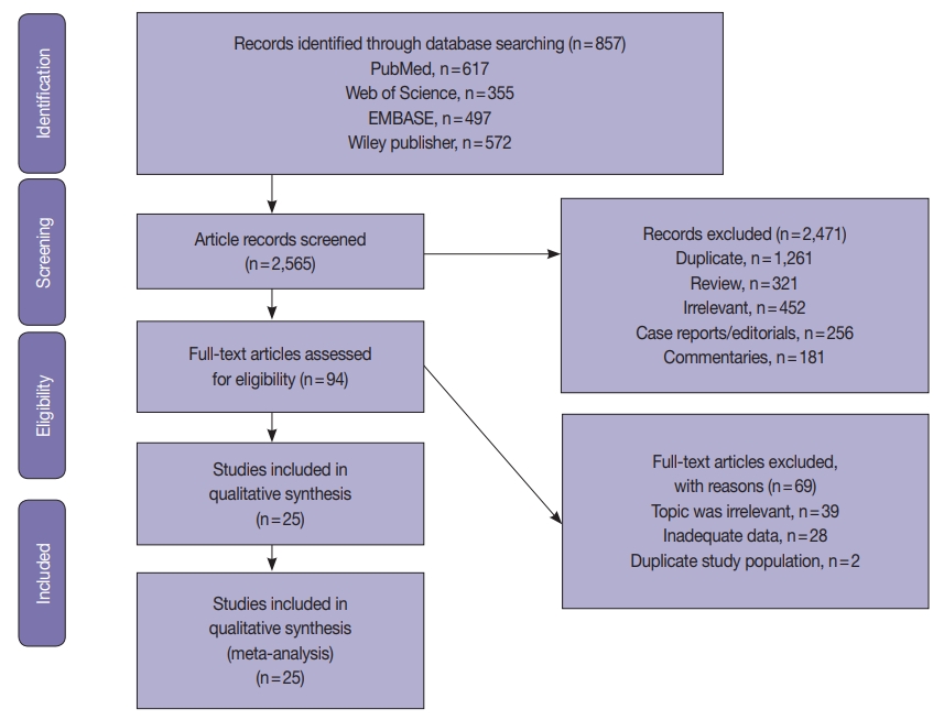
Electronic databases were searched for articles: PubMed, Science Direct, Web of Science, EMBASE, and the Wiley Online Library. Data were extracted from 25 EC studies of D-MMR to generate a clinical dataset of 7,459 patients. A random-effects model produced pooled estimates of D-MMR EC frequency with 95% confidence interval (CI) for meta-analysis.
Results
The overall pooled proportion of D-MMR was 24.477% (95% CI, 21.022 to 28.106) in EC. The Lynch syndrome subgroup had 22.907% pooled D-MMR (95% CI, 14.852 to 32.116). D-MMR was highest in type I EC (25.810) (95% CI, 22.503 to 29.261) compared to type II (13.736) (95% CI, 8.392 to 20.144). Pooled D-MMR was highest at EC stage and grades I–II (79.430% and 65.718%, respectively) and lowest in stages III–IV and grade III (20.168% and 21.529%). The pooled odd ratios comparing D-MMR to proficient MMR favored low-stage EC disease (1.565; 0.894 to 2.740), lymphovascular invasion (1.765; 1.293 to 2.409), and myometrial invasion >50% (1.271; 0.871 to 1.853).
Conclusions
Almost one-quarter of EC patients present with D-MMR tumors. The majority has less aggressive endometrioid histology. D-MMR presents at lower tumor stages compared to MMR-proficient cases in EC. However other metastatic parameters are comparatively higher in the D-MMR disease setting. -
Citations
Citations to this article as recorded by
- Molecular Classification and Clinical Outcomes in Endometrial Cancer: Real-World Evidence from a Tertiary Care Center
Tanadon Salakphet, Prapaporn Suprasert, Tip Pongsuvareeyakul, Chinachote Teerapakpinyo, Surapan Khunamornpong
Cancers.2026; 18(2): 181. CrossRef - Comparison of Molecular Alterations and Survival Analysis in Uterine Endometrioid Carcinomas and Serous Carcinomas: An In Silico Study
Zeynep Sağnak Yılmaz
Archives of Current Medical Research.2026; 7(1): 244. CrossRef - Prognostic Stratification of Multiple-Classifier Endometrial Cancers: Cohort Study and Meta-Analysis
Sabrina Paratore, Angela Russo, Katia Lanzafame, Giusi Blanco, Eliana Giurato, Giovanni Bartoloni, Marco D’Asta, Mirella Sapienza, Giulia Maria Bonanno, Antonino Vallone, Giuseppe Ettore, Roberto Bordonaro
Cancers.2026; 18(6): 929. CrossRef - Prevalence of Mismatch Repair Gene Defects by Means of Immuno-histochemistry Staining for MMR Proteins in Endometrial Cancer
Kaustubh Girish Burde, Indu R. Nair, Pavithran Keechilattu, Anupama Rajanbabu
The Journal of Obstetrics and Gynecology of India.2025; 75(S1): 135. CrossRef - Deficient Mismatch Repair and Microsatellite Instability in Solid Tumors
Joy A. Awosika, James L. Gulley, Danielle M. Pastor
International Journal of Molecular Sciences.2025; 26(9): 4394. CrossRef - Changes in Nucleolar Activity Under Conditions of Microsatellite Instability in the Uterine Mucosa in Precancer and Endometrial Cancer
A. V. Zatvornickaya, E. L. Kazachkov, E. A. Kazachkova
Ural Medical Journal.2025; 24(2): 71. CrossRef - Current approaches to first-line therapy for endometrial cancer: the role of lenvatinib and pembrolizumab combination therapy
V. M. Nechushkina, S. V. Khokhlova, D. A. Nosov, E. A. Ulrikh, L. A. Kolomiets, A. A. Rumyantsev, G. A. Raskin
Tumors of female reproductive system.2025; 21(2): 127. CrossRef - Endometrial carcinoma in Kenya: clinical and biomarker profiles of 123 cases seen at two tertiary referral centers
Olivia Chesikaw, Allan Njau, Erick Chesori, Khadija Warfa, Anisa Mburu, Afrin Fatima Shaffi, Natalie Banet, Jonathan Wawire
Frontiers in Medicine.2025;[Epub] CrossRef - Association Between MMR Status and Prognostic Pathological Factors in Endometrioid Endometrial Cancer—A Single-Center Retrospective Study
Cezary Miedziarek, Hubert Bochyński, Katarzyna Bociańska, Michał Potograbski, Piotr Tyburski, Mikołaj Piotr Zaborowski, Ewa Nowak-Markwitz
Cancers.2025; 17(22): 3605. CrossRef - Mismatch Repair Deficiency Profiling and Its Impact on Management and Prognosis in Endometrial Cancer Patients: A Comprehensive Update
Emmanouela-Aliki Almperi, Chrysoula Margioula-Siarkou, Aristarchos Almperis, Georgia Margioula-Siarkou, Stefanos Flindris, Alexandros I Daponte, Theodora Papamitsou, Konstantinos Dinas, Stamatios Petousis
Cureus.2025;[Epub] CrossRef - Mismatch Repair Deficiency as a Predictor of Lymph Node Metastasis in Endometrioid Endometrial Carcinoma
Esra Keles, Mustafa Maraşlı, Meziha Taşyürek Coşkun, Elif Ünlügedik Sayın, Öncel İpekçi, Mervernur Şahin, Fatih Şanlıkan, Uğur Kemal Öztürk, İsmail Bağlar, Sahra Sultan Kara, Melis Hazırlar
Muğla Sıtkı Koçman Üniversitesi Tıp Dergisi.2025; 12(3): 269. CrossRef - Guidelines of the Brazilian Society of Surgical Oncology for anatomopathological, immunohistochemical, and molecular testing in female tumors
Reitan Ribeiro, Filomena Marino Carvalho, Glauco Baiocchi, Rodrigo Santa Cruz Guindalini, Juliano Rodrigues da Cunha, Carlos Henrique dos Anjos, Caroline de Nadai Costa, Ana Carolina Leite Vieira Costa Gifoni, Renato Cagnacci Neto, Allyne Queiroz Carneiro
Journal of Surgical Oncology.2024; 130(4): 882. CrossRef - Microsatellite instability as a reliable marker of coexisting endometrial cancer in atypical endometrial hyperplasia
А. E. Protasova, G. A. Raskin, M. S. Sobivchak
Tumors of female reproductive system.2024; 20(2): 105. CrossRef - Refining of cancer-specific genes in microsatellite-unstable colon and endometrial cancers using modified partial least square discriminant analysis
Woong Na, Sung Hak Lee, Seunghee Lee, Jong-Seok Kim, Seung Yun Han, Yong Min Kim, Mihye Kwon, Young Soo Song
Medicine.2024; 103(52): e41134. CrossRef - Cancer-specific functional profiling in microsatellite-unstable (MSI) colon and endometrial cancers using combined differentially expressed genes and biclustering analysis
Woong Na, Il Ju Lee, Insong Koh, Mihye Kwon, Young Soo Song, Sung Hak Lee
Medicine.2023; 102(19): e33647. CrossRef - Clinicopathological characteristics of endometrial carcinomas according to DNA mismatch repair protein status
Daniela de Freitas, Fernando Nalesso Aguiar, Cristina Anton, Danielle Cristina de Almeida, Carlos Eduardo Bacchi, Jesus Paula Carvalho, Filomena Marino Carvalho
Heliyon.2023; 9(6): e17495. CrossRef - Mesonephric-like Adenocarcinoma of the Uterine Corpus: Genomic and Immunohistochemical Profiling with Comprehensive Clinicopathological Analysis of 17 Consecutive Cases from a Single Institution
Hyun-Hee Koh, Eunhyang Park, Hyun-Soo Kim
Biomedicines.2023; 11(8): 2269. CrossRef - miR-486-3p Controls the Apoptosis of Endometrial Carcinoma Cells
Donghua Wang, Xiaoli Liu, Lirong Cao, Shixiong Gong, Yi He, Xiangbin Jiang, Zhongxian Wang
Journal of Biomaterials and Tissue Engineering.2022; 12(5): 1002. CrossRef - The Role of Immunohistochemistry Markers in Endometrial Cancer with Mismatch Repair Deficiency: A Systematic Review
Amelia Favier, Justine Varinot, Catherine Uzan, Alex Duval, Isabelle Brocheriou, Geoffroy Canlorbe
Cancers.2022; 14(15): 3783. CrossRef
- Molecular Classification and Clinical Outcomes in Endometrial Cancer: Real-World Evidence from a Tertiary Care Center
- Interobserver diagnostic reproducibility in advanced-stage endometrial carcinoma
- Ho Jin Jung, Soo Yeon Lee, Jin Hwa Hong, Yi Kyeong Chun
- J Pathol Transl Med. 2021;55(1):43-52. Published online December 3, 2020
- DOI: https://doi.org/10.4132/jptm.2020.10.04
- 6,841 View
- 123 Download
- 5 Web of Science
- 5 Crossref
-
Abstract
PDF
- Background
The accurate pathologic diagnosis and subtyping of high-grade endometrial carcinoma are often problematic, due to its atypical and overlapping histopathological features.
Methods
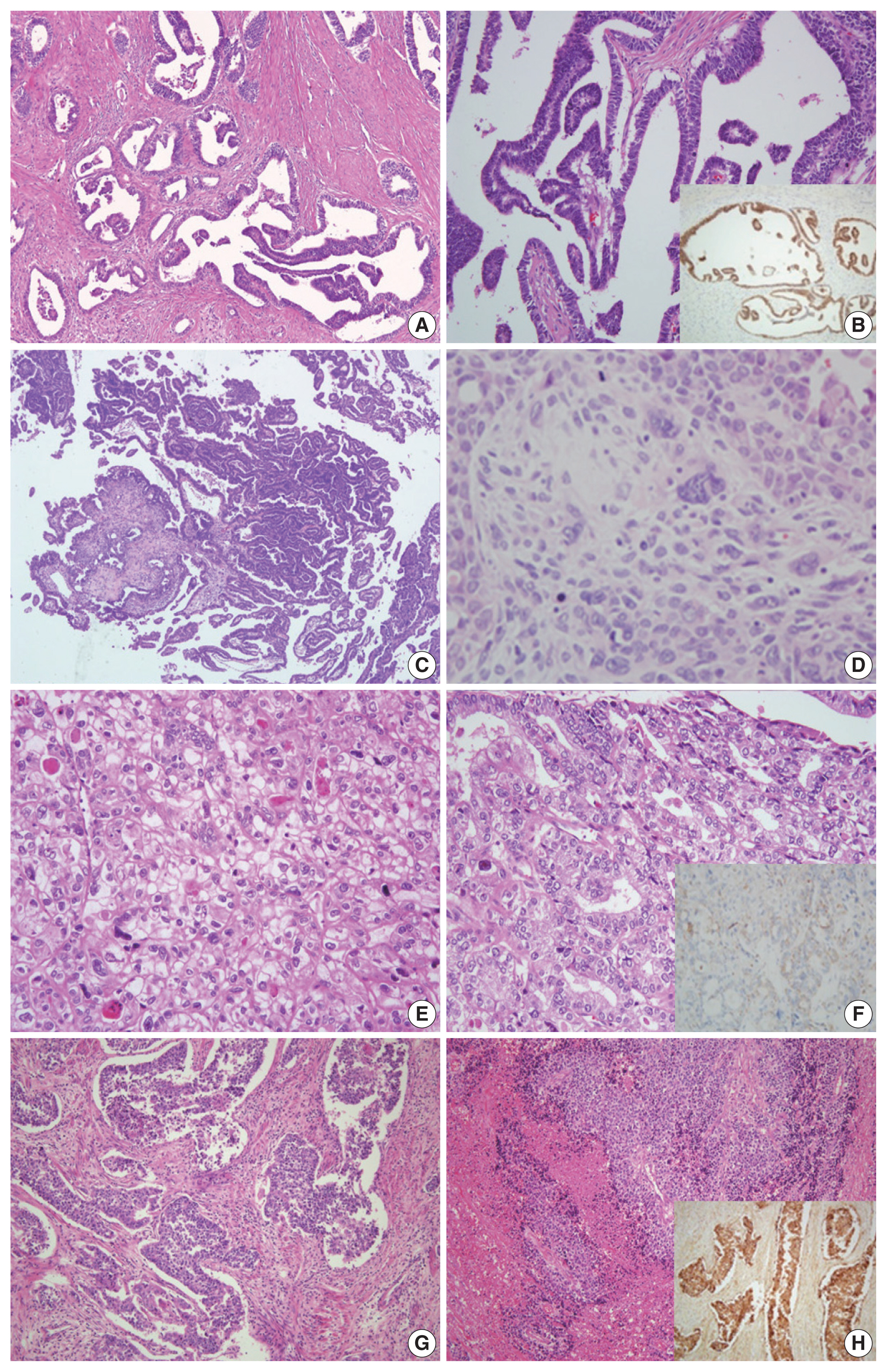
Three pathologists reviewed 21 surgically resected cases of advancedstage endometrial carcinoma. The primary diagnosis was based only on hematoxylin and eosin stained slides. When a discrepancy arose, a secondary diagnosis was made by additional review of immunohistochemical (IHC) stains. Finally, three pathologists discussed all cases and rendered a consensus diagnosis.
Results
The primary diagnoses were identical in 13/21 cases (62%). The secondary diagnosis based on the addition of IHC results was concordant in four of eight discrepant cases. Among four cases with discrepancies occurring in this step, two cases subsequently reached a consensus diagnosis after a thorough discussion between three reviewers. Next-generation sequencing (NGS) study was performed in two cases in which it was difficult to distinguish between serous carcinoma and endometrioid carcinoma. Based on the sequencing results, a final diagnosis of serous carcinoma was rendered. The overall kappa for concordance between the original and consensus diagnosis was 0.566 (moderate agreement).
Conclusions
We investigated stepwise changes in interobserver diagnostic reproducibility in advanced-stage endometrial carcinoma. We demonstrated the utility of IHC and NGS study results in the histopathological diagnosis of advanced-stage endometrial carcinoma. -
Citations
Citations to this article as recorded by- Diagnostic Accuracy of Endometrial Sampling Methods for Determining Histologic Type and Grade in Endometrial Cancer: A Retrospective Cohort Study
Dina Gumin, Avishalom Sharon, Susana Mustafa Mikhail, Inshirah Sgayer, Raneen Abushqara, Lior Lowenstein, Ala Aiob
Cureus.2025;[Epub] CrossRef - Accuracy of endometrial sampling in the diagnosis of endometrial cancer: a multicenter retrospective analysis of the JAGO-NOGGO
Zaher Alwafai, Maximilian Heinz Beck, Sepideh Fazeli, Kathleen Gürtler, Christine Kunz, Juliane Singhartinger, Dominika Trojnarska, Dario Zocholl, David Johannes Krankenberg, Jens-Uwe Blohmer, Jalid Sehouli, Klaus Pietzner
BMC Cancer.2024;[Epub] CrossRef - Deep Learning for Grading Endometrial Cancer
Manu Goyal, Laura J. Tafe, James X. Feng, Kristen E. Muller, Liesbeth Hondelink, Jessica L. Bentz, Saeed Hassanpour
The American Journal of Pathology.2024; 194(9): 1701. CrossRef - Application of NGS molecular classification in the diagnosis of endometrial carcinoma: A supplement to traditional pathological diagnosis
Qunxian Rao, Jianwei Liao, Yangyang Li, Xin Zhang, Guocai Xu, Changbin Zhu, Shengya Tian, Qiuhong Chen, Hui Zhou, Bingzhong Zhang
Cancer Medicine.2023; 12(5): 5409. CrossRef - Risk Stratification of Endometrial Cancer Patients: FIGO Stage, Biomarkers and Molecular Classification
Jenneke C. Kasius, Johanna M. A. Pijnenborg, Kristina Lindemann, David Forsse, Judith van Zwol, Gunnar B. Kristensen, Camilla Krakstad, Henrica M. J. Werner, Frédéric Amant
Cancers.2021; 13(22): 5848. CrossRef
- Diagnostic Accuracy of Endometrial Sampling Methods for Determining Histologic Type and Grade in Endometrial Cancer: A Retrospective Cohort Study
- The frequency of POLE-mutation in endometrial carcinoma and prognostic implications: a systemic review and meta-analysis
- Alaa Salah Jumaah, Mais Muhammed Salim, Hawraa Sahib Al-Haddad, Katherine Ann McAllister, Akeel Abed Yasseen
- J Pathol Transl Med. 2020;54(6):471-479. Published online September 2, 2020
- DOI: https://doi.org/10.4132/jptm.2020.07.23
- 14,351 View
- 371 Download
- 36 Web of Science
- 35 Crossref
-
Abstract
PDFSupplementary Material
- Background
Endometrial carcinoma (EC) is classified into four distinct molecular subgroups including ultramutated DNA polymerase epsilon (POLE). POLE-mutated tumors have the best prognosis and are a promising target for immunotherapy. This meta-analysis consolidated the reported variation of POLE-mutant frequency and assessed prognostic value in EC.
Methods
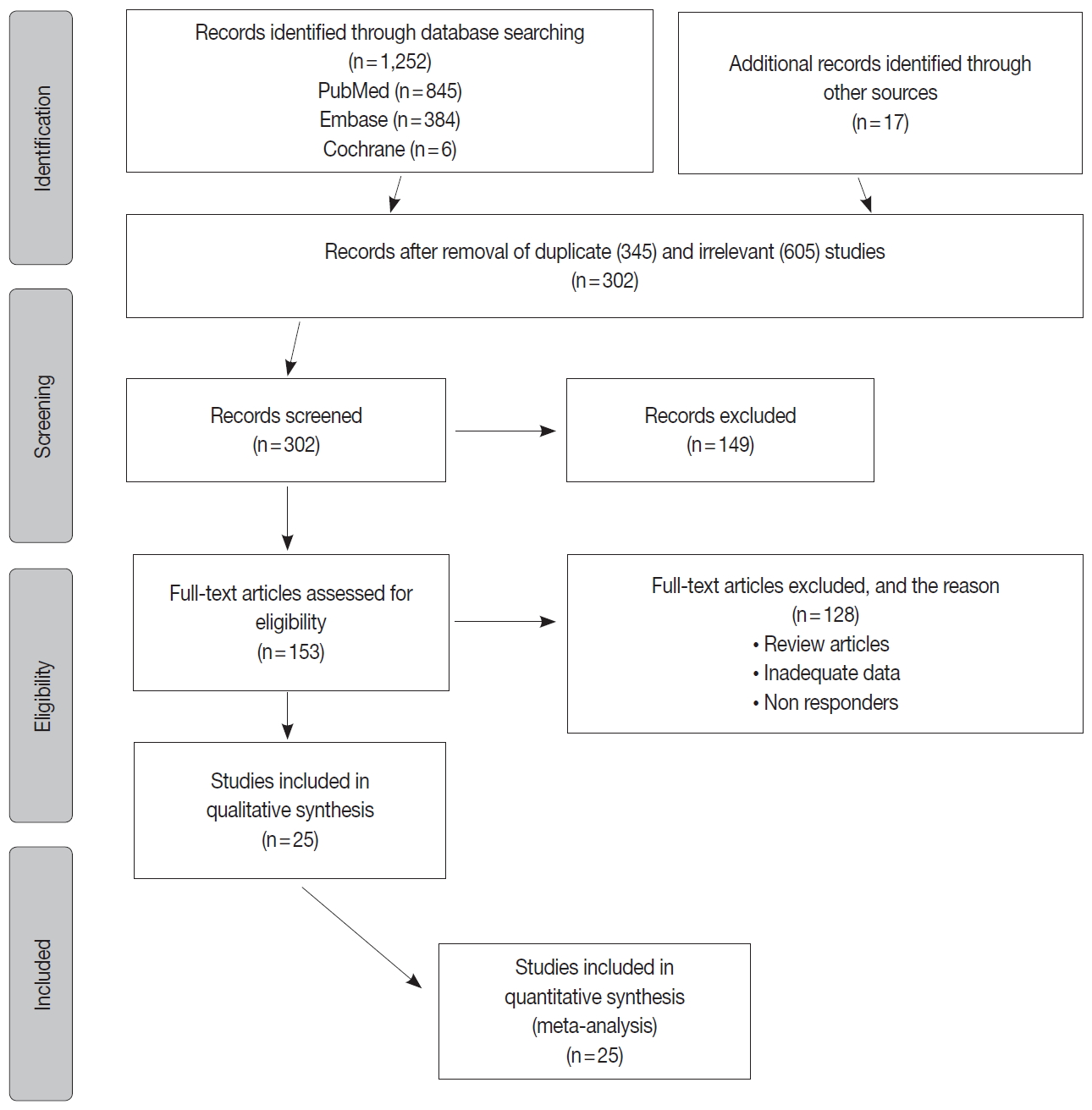
Internet searches explored scientific data bases: EMBASE, PubMed, and the Cochrane Central Register of Controlled Trials databases. Data was extracted from eligible studies including: sample size, number of positive POLE-mutant cases, sequencing information, clinicopathologic data, and survival data. Meta-analysis and a random-effects model produced pooled estimates of POLE frequency and prognostic parameters using 95% confidence intervals (CI), hazard ratios (HR), and odd ratios (OR).
Results
Six thousand three hundred and forty-six EC patient cases were pooled from 25 studies. The pooled proportion of POLE gene mutation in EC was 8.59% (95% CI, 7.01 to 10.32), of which 8.22% (95% CI, 6.27 to 10.42) were type I and 0.93% (95% CI, 0.34 to 1.81) type 2. Clinicopathologic data showed that POLE-mutated tumors are mostly endometrioid. They present at higher levels in earlier stages (I–II) of EC (89.51%; 95% CI, 81.11 to 95.66) at the highest grade III (51.53%; 95% CI, 36.08 to 66.84) with reduced myometrial invasion (OR, 1.48, 95% CI, 0.99 to 2.20). Survival analysis indicated favorable overall survival (HR, 0.90), disease-specific survival (HR, 0.41), and progression-free survival (HR, 0.23) for POLE mutant EC.
Conclusions
Almost one-tenth of EC patients have POLE-mutated tumors. Given their improved prognostic potential, identifying the POLE mutation status is key for the management of EC patients. -
Citations
Citations to this article as recorded by- The relationship between histopathological data and molecular alterations with oncological outcomes in endometrioid-type endometrial cancers and a novel POLE mutation
Elif Aksahin, Fuat Demirkiran, Tugan Bese, Sukru Cebi, Abdullah Serdar Acikgoz, Basak Ozge Kayan, Yeliz Aykanat, Ismail Yilmaz, Ayse Namal, Sennur Ilvan, Omer Uysal, Macit Arvas
Journal of Gynecologic Oncology.2026;[Epub] CrossRef - Characterization of Korean Colorectal Cancer Reveals Novel Driver Gene and Clinically Relevant Mutations
Junho Kang, Dong Min Lim, Young‐Joon Kim, Hyeran Shim, Tae‐You Kim, Kyu Joo Park, Sung‐Bum Kang, Chang Sik Yu, Jong Lyul Lee, Yeuni Yu, Hansong Lee, Eun Jung Kwon, Hyo Min Kim, Seongik Mun, Donghee Kwak, Hae Seul Lee, Hye Jin Heo, Eun Kyoung Kim, Seung Eu
MedComm.2026;[Epub] CrossRef - CircRNA-miRNA-mRNA interactome analysis in endometrial cancer
Tikam Chand Dakal, Abhishek Kumar, Pawan Kumar Maurya
Journal of Biomolecular Structure and Dynamics.2025; 43(3): 1486. CrossRef - The prognostic implication of polymerase epsilon-mutated endometrial cancer
Kai-Hung Wang, Dah-Ching Ding
Tzu Chi Medical Journal.2025; 37(2): 135. CrossRef - Functions, interactions and prognostic role of POLE: a bioinformatics analysis
Jonathan Carvajal-Veloza, Fredy Galindo-Morales, Luz Dary Gutierrez-Castañeda
Journal of Gynecologic Oncology.2025;[Epub] CrossRef - Development of Antibody–Drug Conjugates for Malignancies of the Uterine Corpus: A Review
Taro Yamanaka, Tadaaki Nishikawa, Hiroshi Yoshida
Cells.2025; 14(5): 333. CrossRef - Efficacy of dostarlimab in recurrent or advanced mismatch Repair-Deficient endometrial Cancer as a Single-Agent therapy: A systematic review and Meta-Analysis
Ramazan Rezaei, Hedieh Haji Khodaverdi Khani
DARU Journal of Pharmaceutical Sciences.2025;[Epub] CrossRef - POLE-mutated endometrial cancer: new perspectives on the horizon?
Daniele Fanale, Lidia Rita Corsini, Paola Piraino, Erika Pedone, Chiara Brando, Tancredi Didier Bazan Russo, Pietro Ferraro, Alisia Simone, Silvia Contino, Ornella Prestifilippo, Ugo Randazzo, Ambra Giurintano, Carla Ferrante Bannera, Antonio Galvano, Lor
Frontiers in Oncology.2025;[Epub] CrossRef - Mismatch repair, p53, and L1 cell adhesion molecule status influence the response to chemotherapy in advanced and recurrent endometrial cancer
Jung Chul Kim, Byungsoo Ahn, Yong Jae Lee, Eun Ji Nam, Sang Wun Kim, Sunghoon Kim, Young Tae Kim, Eunhyang Park, Jung-Yun Lee
BMC Cancer.2024;[Epub] CrossRef - A single-institution retrospective exploratory analysis on the effectiveness and safety of lenvatinib plus pembrolizumab for advanced endometrial cancer: insights from ProMisE molecular classification system
Yohei Chiba, Masahiro Kagabu, Mitsumasa Osakabe, Rikako Ito, Sho Sato, Eriko Takatori, Yoshitaka Kaido, Takayuki Nagasawa, Tadahiro Shoji, Naoki Yanagawa, Tsukasa Baba
Japanese Journal of Clinical Oncology.2024; 54(4): 424. CrossRef - Potential of molecular classification to guide fertility-sparing management among young patients with endometrial cancer
Nuria Agusti, Alexa Kanbergs, Roni Nitecki
Gynecologic Oncology.2024; 185: 121. CrossRef - Assessing the New 2020 ESGO/ESTRO/ESP Endometrial Cancer Risk Molecular Categorization System for Predicting Survival and Recurrence
Yung-Taek Ouh, Yoonji Oh, Jinwon Joo, Joo Hyun Woo, Hye Jin Han, Hyun Woong Cho, Jae Kwan Lee, Yikyeong Chun, Myoung-nam Lim, Jin Hwa Hong
Cancers.2024; 16(5): 965. CrossRef - The Clinical and Pathological Characteristics of POLE-Mutated Endometrial Cancer: A Comprehensive Review
Xiaohong Yao, Min Feng, Wei Wang
Cancer Management and Research.2024; Volume 16: 117. CrossRef - National Survey of Current Follow-up Protocols for Patients Treated for Endometrial Cancer in the UK
H. Patel, K. Drinkwater, A. Stewart
Clinical Oncology.2024; 36(6): e146. CrossRef - Nab-Paclitaxel-Based Systemic Approach to Achieving Complete Remission for Relapsed Stage III Endometrial Carcinoma: Insights From the Indian Subcontinent
Prasanna Rammohan, Vipulkumar Thummar, Priya Mehta
Cureus.2024;[Epub] CrossRef - High prevalence of “non‐pathogenic” POLE mutation with poor prognosis in a cohort of endometrial cancer from South India
Santhosh Kuriakose, Dhananjayan Dhanasooraj, P. M. Shiny, S. Shammy, V. P. Sona, Anupama A. Manjula, Amrutha Ramachandran, Bindu Vijaykumar, Nayana Susan, M. Dinesan, Uma V. Sankar, Kavitha Ramachandran, P. S. Sreedharan
International Journal of Gynecology & Obstetrics.2024; 166(3): 1263. CrossRef - Patterns and Frequency of Pathogenic Germline Mutations among Patients with Newly-Diagnosed Endometrial Cancer: The Jordanian Exploratory Cancer Genetics (Jo-ECAG) Endometrial Study
Hikmat Abdel-Razeq, Hira Bani Hani, Baha Sharaf, Faris Tamimi, Hanan Khalil, Areej Abu Sheikha, Mais Alkyam, Sarah Abdel-Razeq, Tala Ghatasheh, Tala Radaideh, Suhaib Khater
Cancers.2024; 16(14): 2543. CrossRef - Accelerated clinical response achieved by combining short-term tumor-directed photodynamic therapy with immunotherapy-based systemic therapies in synchronous colorectal cancer with MSI-H and POLE mutation: a case report
Yuhan Wang, Lei Gao, Bin Ma, Jianming Shi, Zhenyu Yin, Weidong Zhu, Hao Chen
Frontiers in Immunology.2024;[Epub] CrossRef - Morphomolecular Correlation and Clinicopathologic Analysis in Endometrial Carcinoma
Göksenil Bülbül, Tekincan Çağri Aktaş, Anil Aysal Ağalar, Safiye Aktaş, Sefa Kurt, Bahadir Saatli, Emine Çağnur Ulukuş
International Journal of Gynecological Pathology.2024; 43(6): 535. CrossRef - Prognostic implications of immunohistochemistry in patients with endometrial cancer
Maria-Bianca Anca-Stanciu, Andrei Manu , Maria Victoria Olinca , Bogdan Cătălin Coroleucă , Diana-Elena Comandaşu , Ciprian Andrei Coroleucă , Călina Maier , Elvira Brătilă
Romanian Journal of Morphology and Embryology.2024; 65(2): 185. CrossRef - Translating biological insights into improved management of endometrial cancer
Jeffrey A. How, Amir A. Jazaeri, Shannon N. Westin, Barrett C. Lawson, Ann H. Klopp, Pamela T. Soliman, Karen H. Lu
Nature Reviews Clinical Oncology.2024; 21(11): 781. CrossRef - Fast and reliable Sanger POLE sequencing protocol in FFPE tissues of endometrial cancer
Izabela Laczmanska, Dagmara Michalowska, Marcin Jedryka, Dorota Blomka, Mariola Semeniuk, Ewelina Czykalko, Mariola Abrahamowska, Paulina Mlynarczykowska, Agnieszka Chrusciel, Ireneusz Pawlak, Adam Maciejczyk
Pathology - Research and Practice.2023; 242: 154315. CrossRef - Uterine Neoplasms, Version 1.2023, NCCN Clinical Practice Guidelines in Oncology
Nadeem Abu-Rustum, Catheryn Yashar, Rebecca Arend, Emma Barber, Kristin Bradley, Rebecca Brooks, Susana M. Campos, Junzo Chino, Hye Sook Chon, Christina Chu, Marta Ann Crispens, Shari Damast, Christine M. Fisher, Peter Frederick, David K. Gaffney, Robert
Journal of the National Comprehensive Cancer Network.2023; 21(2): 181. CrossRef - The hereditary N363K POLE exonuclease mutant extends PPAP tumor spectrum to glioblastomas by causing DNA damage and aneuploidy in addition to increased mismatch mutagenicity
Guillaume Labrousse, Pierre Vande Perre, Genis Parra, Marion Jaffrelot, Laura Leroy, Frederic Chibon, Frederic Escudie, Janick Selves, Jean-Sebastien Hoffmann, Rosine Guimbaud, Malik Lutzmann
NAR Cancer.2023;[Epub] CrossRef - New boundaries for fertility sparing management in endometrial cancer
Alexandros Rodolakis, Vasilis Pergialiotis, Nikolaos Thomakos
Current Opinion in Oncology.2023; 35(5): 394. CrossRef - PD-1 and PD-L1 Blockade plus Chemotherapy in Endometrial Cancer
New England Journal of Medicine.2023; 389(9): 866. CrossRef - The Shifting Landscape of p53abn Endometrial Cancers: A Review of the Prognostic and Predictive Impact and Current Therapeutic Directions
Angelo Anater
Journal of Medical and Radiation Oncology.2023; 3(2): 1. CrossRef - The Advantages of Next-Generation Sequencing Molecular Classification in Endometrial Cancer Diagnosis
Daniela Rivera, Michele Paudice, Giulia Accorsi, Floriana Valentino, Marta Ingaliso, Ada Pianezzi, Paola Roggieri, Lucia Trevisan, Giulia Buzzatti, Serafina Mammoliti, Fabio Barra, Simone Ferrero, Gabriella Cirmena, Viviana Gismondi, Valerio Gaetano Vello
Journal of Clinical Medicine.2023; 12(23): 7236. CrossRef - The clinicopathology and survival characteristics of patients with POLE proofreading mutations in endometrial carcinoma: A systematic review and meta-analysis
Alaa Salah Jumaah, Hawraa Sahib Al-Haddad, Katherine Ann McAllister, Akeel Abed Yasseen, Manish S. Patankar
PLOS ONE.2022; 17(2): e0263585. CrossRef - Enhanced polymerase activity permits efficient synthesis by cancer-associated DNA polymerase ϵ variants at low dNTP levels
Stephanie R Barbari, Annette K Beach, Joel G Markgren, Vimal Parkash, Elizabeth A Moore, Erik Johansson, Polina V Shcherbakova
Nucleic Acids Research.2022; 50(14): 8023. CrossRef - The Role of Immunohistochemistry Markers in Endometrial Cancer with Mismatch Repair Deficiency: A Systematic Review
Amelia Favier, Justine Varinot, Catherine Uzan, Alex Duval, Isabelle Brocheriou, Geoffroy Canlorbe
Cancers.2022; 14(15): 3783. CrossRef - The clinicopathological characteristics of POLE-mutated/ultramutated endometrial carcinoma and prognostic value of POLE status: a meta-analysis based on 49 articles incorporating 12,120 patients
Qing Wu, Nianhai Zhang, Xianhe Xie
BMC Cancer.2022;[Epub] CrossRef - Mismatch repair deficiency and clinicopathological characteristics in endometrial carcinoma: a systematic review and meta-analysis
Alaa Salah Jumaah, Hawraa Sahib Al-Haddad, Mais Muhammed Salem, Katherine Ann McAllister, Akeel Abed Yasseen
Journal of Pathology and Translational Medicine.2021; 55(3): 202. CrossRef - Evaluation of treatment effects in patients with endometrial cancer and POLE mutations: An individual patient data meta‐analysis
Jessica N. McAlpine, Derek S. Chiu, Remi A. Nout, David N. Church, Pascal Schmidt, Stephanie Lam, Samuel Leung, Stefania Bellone, Adele Wong, Sara Y. Brucker, Cheng Han Lee, Blaise A. Clarke, David G. Huntsman, Marcus Q. Bernardini, Joanne Ngeow, Alessand
Cancer.2021; 127(14): 2409. CrossRef - Endometrial cancer
Vicky Makker, Helen MacKay, Isabelle Ray-Coquard, Douglas A. Levine, Shannon N. Westin, Daisuke Aoki, Ana Oaknin
Nature Reviews Disease Primers.2021;[Epub] CrossRef
- The relationship between histopathological data and molecular alterations with oncological outcomes in endometrioid-type endometrial cancers and a novel POLE mutation
- High Expression of Galectin-1, VEGF and Increased Microvessel Density Are Associated with MELF Pattern in Stage I-III Endometrioid Endometrial Adenocarcinoma
- Dmitry Aleksandrovich Zinovkin, Sergey Leonidovich Achinovich, Mikhail Grigoryevich Zubritskiy, Jacqueline Linda Whatmore, Md Zahidul Islam Pranjol
- J Pathol Transl Med. 2019;53(5):280-288. Published online June 27, 2019
- DOI: https://doi.org/10.4132/jptm.2019.05.13
- 8,071 View
- 155 Download
- 7 Web of Science
- 10 Crossref
-
Abstract
PDF
- Background
In this study, we investigate the expression of markers of angiogenesis and microvessel density (MVD) in cases of microcystic, elongated and fragmented (MELF) pattern, with its prognostic role in the survival of endometrioid endometrial adenocarcinomas (EA) patients.
Methods
In this study, 100 cases of EA, 49 cases with MELF pattern and 51 without, were immunohistochemically stained for galectin-1, vascular endothelial growth factor (VEGF), and MVD. Morphometry and statistical (univariate and multivariate) analyses were performed to assess overall survival (OS) and disease-free survival.
Results
The expression of VEGF (p<.001) and galectin-1 (p<.001), as well as MVD area (p<.001) and number of vessels/mm2 (p<.050), were significantly higher in the +MELF pattern group compared to the –MELF group. A low negative correlation between MELFpattern and the number of days of survival (p<.001, r=–0.47) was also found. A low positive correlation of MELF-pattern with galectin-1 expression (p<.001, r=0.39), area of vessels/mm2 (p<.001, r=0.36), outcome of EA (p<.001, r=0.42) and VEGF expression (p<.001, r=0.39) suggests potential pathological relevance of these factors in the prognosis of EA. A univariate survival analysis indicated a role for all parameters of survival. Multivariate Cox proportional hazard regression analysis revealed that only area of vessels/mm2 (hazard ratio [HR], 1.018; 95% confidence interval [CI], 1.002 to 1.033), galectin-1 (HR, 1.049; 95% CI, 1.025 to 1.074) and VEGF (HR, 1.049; 95% CI, 1.022 to 1.077) play key roles in OS.
Conclusions
This study reports an increase in MVD, VEGF and galectin-1 expression in EA with MELF pattern and suggests that MELF pattern, along with the angiogenic profile, may be a prognostic factor in EA. -
Citations
Citations to this article as recorded by- Association of Local and Distant Organ Metastases With MELF Pattern in Endometrial Cancer
Varol Gülseren, Ertuğrul Şen, Mehmet Dolanbay, Fulya Çağli, Nahit Topaloğlu, Figen Öztürk, Bülent Özçelik, Serdar Serin, Kemal Güngördük
International Journal of Gynecological Pathology.2025; 44(3): 237. CrossRef - The Immunomodulatory Role of Galectin-1 in the Tumour Microenvironment and Strategies for Therapeutic Applications
Alice Griffiths, Palita Udomjarumanee, Andrei-Stefan Georgescu, Muruj Barri, Dmitry A. Zinovkin, Md Zahidul I. Pranjol
Cancers.2025; 17(11): 1888. CrossRef - Features of treatment of chronic recurrent postcoital cystitis in women
M. B. Lemtygov, N. I. Simchenko
Health and Ecology Issues.2025; 22(3): 32. CrossRef - Tumour budding, MELF-pattern and tumour-infiltrating lymphocytes as possible pathomorphological parameters of the course of endometrioid adenocarcinoma of the uterine corpus
D. A. Zinovkin, I. V. Veyalkin, S. L. Achinovich, I. I. Slepokurova, Yu. A. Lyzikova, A. Farooq
Tumors of female reproductive system.2024; 20(2): 83. CrossRef - The prognostic value of vascular endothelial growth factor in endometrial cancer: A protocol for systematic review and meta-analysis
Bao Qiang, YiFan Kang, JiaoLin Yang, HuanCheng Su, Zhe Wang, ChunMei Zhang, SanYuan Zhang
Medicine.2024; 103(51): e40933. CrossRef - Determining the level of stromal and epithelial cells activity in normal and hyperplastic endometrium of late reproductive and perimenopausal women
Zinaida Vasilyvna Chumak, Volodymyr Victorovich Artyomenko, Mykola Vitaliiovich Shapoval, Liudmyla Volodymyrivna Mnih, Ganna Volodymyrivna Kozhukhar, Serhii Vasilyovich Derishov
Journal of Medicine and Life.2023; 16(2): 210. CrossRef - Endocervical Adenocarcinoma Showing Microcystic, Elongated, and Fragmented (MELF) Pattern of Stromal Invasion: A Single-Institutional Analysis of 10 Cases with Comprehensive Clinicopathological Analyses and Ki-67 Immunostaining
Hyunsik Bae, Hyun-Soo Kim
Biomedicines.2023; 11(11): 3026. CrossRef - Clinicopathologic association and prognostic impact of microcystic, elongated and fragmented pattern invasion, combined with tumor budding in endometrioid endometrial cancer
Xiqin Qi, Lun Zhu, Bei Zhang
Journal of Obstetrics and Gynaecology Research.2022; 48(9): 2431. CrossRef - Role of adipocytokines in endometrial cancer progression
Ran Li, Fang Dong, Ling Zhang, Xiuqin Ni, Guozhi Lin
Frontiers in Pharmacology.2022;[Epub] CrossRef - Advances in Anti-Cancer Immunotherapy: Car-T Cell, Checkpoint Inhibitors, Dendritic Cell Vaccines, and Oncolytic Viruses, and Emerging Cellular and Molecular Targets
Emilie Alard, Aura-Bianca Butnariu, Marta Grillo, Charlotte Kirkham, Dmitry Aleksandrovich Zinovkin, Louise Newnham, Jenna Macciochi, Md Zahidul Islam Pranjol
Cancers.2020; 12(7): 1826. CrossRef
- Association of Local and Distant Organ Metastases With MELF Pattern in Endometrial Cancer
- Potential Role for a Panel of Immunohistochemical Markers in the Management of Endometrial Carcinoma
- Amany Salama, Mohammad Arafa, Eman ElZahaf, Abdelhadi Mohamed Shebl, Azmy Abd El-Hameed Awad, Sylvia A. Ashamallah, Reda Hemida, Anas Gamal, Abd AlRahman Foda, Khaled Zalata, El-Said M. Abdel-Hady
- J Pathol Transl Med. 2019;53(3):164-172. Published online February 28, 2019
- DOI: https://doi.org/10.4132/jptm.2019.02.12
- 12,158 View
- 388 Download
- 14 Web of Science
- 11 Crossref
-
Abstract
PDF
- Background
In order to improve the efficacy of endometrial carcinoma (EC) treatment, identifying prognostic factors for high risk patients is a high research priority. This study aimed to assess the relationships among the expression of estrogen receptors (ER), progesterone receptors (PR), human epidermal growth factor receptor 2 (HER2), Ki-67, and the different histopathological prognostic parameters in EC and to assess the value of these in the management of EC.
Methods
We examined 109 cases of EC. Immunohistochemistry for ER, PR, HER2, and Ki-67 were evaluated in relation to age, tumor size, International Federation of Gynecology and Obstetrics (FIGO) stage and grade, depth of infiltration, cervical and ovarian involvement, lymphovascular space invasion (LVSI), and lymph node (LN) metastasis.
Results
The mean age of patients in this study was 59.8 ± 8.2 years. Low ER and PR expression scores and high Ki-67 expression showed highly significant associations with non-endometrioid histology (p = .007, p < .001, and p < .001, respectively) and poor differentiation (p = .007, p < .001, and p <. 001, respectively). Low PR score showed a significant association with advanced stage (p = .009). Low ER score was highly associated with LVSI (p = .006), and low PR scores were associated significantly with LN metastasis (p = .026). HER2 expression was significantly related to advanced stages (p = .04), increased depth of infiltration (p = .02), LVSI (p = .017), ovarian involvement (p = .038), and LN metastasis (p = .038). There was a close relationship between HER2 expression and uterine cervical involvement (p = .009). Higher Ki-67 values were associated with LN involvement (p = .012).
Conclusions
The over-expression of HER2 and Ki-67 and low expression of ER and PR indicate a more malignant EC behavior. An immunohistochemical panel for the identification of high risk tumors can contribute significantly to prognostic assessments. -
Citations
Citations to this article as recorded by- Tissue Microarray for Gynecological Pathology Studies: A Mini-Review
Mohammad Arafa, Abd AlRahman Foda, Amany Salama, Ola Shalaby, Muna Al-Jabri, Fatma Al Hinai, Afrah Al-Rashdi, Samya Al-Husaini, Suaad Al-Badi
Journal of Microscopy and Ultrastructure.2026;[Epub] CrossRef - Clinicopathological Correlation of Hormone Receptors, Angiogenesis, and Tumor Budding in Endometrial Carcinoma: A Tertiary Care Center Study
Senjuti Dasgupta, Arpita Das, Ujjwal Bandyopadhyay
The Journal of Obstetrics and Gynecology of India.2025;[Epub] CrossRef - Multiparameter MRI-based radiomics analysis for preoperative prediction of type II endometrial cancer
Yingying Cao, Wei Zhang, Xiaorong Wang, Xiaojing Lv, Yaping Zhang, Kai Guo, Shuai Ren, Yuan Li, Zhongqiu Wang, Jingya Chen
Heliyon.2024; 10(12): e32940. CrossRef - Correlation of PD-L1 expression with different clinico-pathological and immunohistochemical features of ovarian surface epithelial tumors
Asem Shalaby, Ola Shalaby, Hazem Abdullah, Mohamed Rachid Boulassel, Mohammad Arafa
Clinical and Translational Oncology.2024; 27(2): 699. CrossRef - Estrogen/Progesterone Receptor Loss, CTNNB1 and KRAS Mutations Are Associated With Local Recurrence or Distant Metastasis in Low-Grade Endometrial Endometrioid Carcinoma
Rajni Chibbar, Sabrina Foerstner, Janarathnee Suresh, Richa Chibbar, Alexandre Piche, Deeksha Kundapur, Rani Kanthan, Vijayanand Kundapur, Cheng Han Lee, Anita Agrawal, Raymond Lai
Applied Immunohistochemistry & Molecular Morphology.2023; 31(3): 181. CrossRef - Exploring the Prognostic and Predictive Roles of Ki-67 in Endometrial Cancer
Laura Paleari, Mariangela Rutigliani, Oriana D’Ecclesiis, Sara Gandini, Irene Maria Briata, Tania Buttiron Webber, Nicoletta Provinciali, Andrea DeCensi
International Journal of Translational Medicine.2023; 3(4): 479. CrossRef - Analysis of human epidermal growth factor receptor 2 immunohistochemical expression in high-grade endometrial carcinomas and its association with variable clinical outcomes
Malames M. Faisal, Marwa M. Shakweer, Ghada Refaat, Khaled S. Mohammed, Tarek I. ElMallawy, Magda H. Nasreldin, Laila M. Farid, Mariam B. Abouelkhair
Egyptian Journal of Pathology.2023; 43(2): 119. CrossRef - Correlation of PD-L1 immunohistochemical expression with microsatellite instability and p53 status in endometrial carcinoma
Mohammad Arafa, Abdelhadi Mohamed Shebl, Amany Salama, Eman ElZahaf, Sylvia A. Ashamallah, Abd AlRahman Foda, AzmyAbd El-Hameed Awad, Asem Shalaby
European Journal of Obstetrics & Gynecology and Reproductive Biology: X.2022; 16: 100172. CrossRef - Immunohistochemical Expression of Oestrogen and Epidermal Growth Factor Receptors in Endometrial Cancerous in Sudanese Patients
Salwa Abdalraheem Abubaker, Mohamed Elfatih Abdelwadoud, Mutaz Mohamed Ali, Hadia Alhaj Ahmad, Abuobieda Mohamed Khlafalla, Osman Mohammed Elmahi, Hisham Ali Waggiallah
Journal Of Biochemical Technology.2021; 12(1): 58. CrossRef - Expression of ER/PR Receptor, Her-2/neu, Ki67 and p53 in Endometrial Carcinoma: Clinicopathological Implication and Prognostic Value
V. B. Shivkumar, Manisha A. Atram, Nitin M. Gangane
Indian Journal of Gynecologic Oncology.2020;[Epub] CrossRef - Immunohistochemical study of ER, PR, p53 and Ki67 expression in patients with endometrial adenocarcinoma and atypical endometrial hyperplasia
Rachana Lakhe, Ravi M Swami, Preeti Doshi, Manjiri N Karandikar, Ravindra Nimbargi
IP Archives of Cytology and Histopathology Research.2020; 5(4): 274. CrossRef
- Tissue Microarray for Gynecological Pathology Studies: A Mini-Review
- Investigation of the Roles of Cyclooxygenase-2 and Galectin-3 Expression in the Pathogenesis of Premenopausal Endometrial Polyps
- Esin Kasap, Serap Karaarslan, Esra Bahar Gur, Mine Genc, Nur Sahin, Serkan Güclü
- J Pathol Transl Med. 2016;50(3):225-230. Published online April 16, 2016
- DOI: https://doi.org/10.4132/jptm.2016.03.08
- 9,385 View
- 84 Download
- 4 Web of Science
- 5 Crossref
-
Abstract
PDF
- Background
The pathogenesis and etiology of endometrial polyps has not been elucidated. In this study, we aimed to examine the pathogenic mechanisms of endometrial polyp development using immunohistochemistry. We evaluated the expression of galectin-3 and cyclooxgenase-2 (COX-2) during the menstrual cycle in premenopausal women with endometrial polyps or normal endometrium.
Methods
Thirty-one patients with endometrial polyps and 50 healthy control patients were included in this study. The levels of expression of COX-2 and galectin-3 were studied by immunohistochemistry.
Results
The percentage of COX-2–positive cells and the intensity of COX-2 staining in the endometrium did not vary during the menstrual cycle either in the control group or in patients with endometrial polyps. However, expression of galectin-3 was significantly lower in endometrial polyps and during the proliferative phase of the endometrium compared with the secretory phase.
Conclusions
Our data suggests that the pathogenesis of endometrial polyps does not involve expression of COX-2 or galectin-3. -
Citations
Citations to this article as recorded by- Abnormal expression of Hippo–YAP1 signalling pathway and progesterone resistance mechanism in endometrial polyps
Xinyu Yu, Weijia Kong, Kaiyue Shang, Hongxin Xing, Wenjing Sun, Qianqian Li, Hui Zhang
Journal of Obstetrics and Gynaecology.2025;[Epub] CrossRef - Research Progress in the Treatment of Endometrial Polyps
秀芬 蔡
Advances in Clinical Medicine.2024; 14(01): 1772. CrossRef - ER and COX2 expression in endometrial hyperplasia processes
Nataliia Tsyndrenko, Mykola Lyndіn, Kateryna Sikora, Andrew Awuah Wireko, Toufik Abdul-Rahman, Nataliia Hyriavenko, Anatolii Romaniuk
Medicine.2023; 102(33): e34864. CrossRef - Novel microarchitecture of human endometrial glands: implications in endometrial regeneration and pathologies
Nicola Tempest, Christopher J Hill, Alison Maclean, Kathleen Marston, Simon G Powell, Hannan Al-Lamee, Dharani K Hapangama
Human Reproduction Update.2022; 28(2): 153. CrossRef - Variances in the Level of COX-2 and iNOS in Different Grades of Endometrial Cancer
Marcin Oplawski, Konrad Dziobek, Nikola Zmarzły, Beniamin O. Grabarek, Robert Kiełbasiński, Przemysław Kieszkowski, Piotr Januszyk, Karol Talkowski, Michał Schweizer, Piotr Kras, Andrzej Plewka, Dariusz Boroń
Current Pharmaceutical Biotechnology.2020; 21(1): 52. CrossRef
- Abnormal expression of Hippo–YAP1 signalling pathway and progesterone resistance mechanism in endometrial polyps
Brief Case Report
- The Limitations of Endoscopic Ultrasound-Guided Fine Needle Aspiration Cytology in the Diagnosis of Pancreatic Serous Cystadenoma: A Brief Case Report
- Heae Surng Park, Sun Och Yoon, Beom Jin Lim, Joo Hee Kim, Soon Won Hong
- Korean J Pathol. 2014;48(5):405-408. Published online October 27, 2014
- DOI: https://doi.org/10.4132/KoreanJPathol.2014.48.5.405
- 8,603 View
- 63 Download
Original Articles
- Diagnostic Utility of the JAZF1/JJAZ1 Gene Fusion in Endometrial Stromal Sarcomas and Their Histologic Variants.
- Sang Ryung Lee, Joon Seon Song, Ga Hye Kim, Jene Choi, Hyung Kyoung Kim, Yonghee Lee, Kyu Rae Kim
- Korean J Pathol. 2011;45(5):498-505.
- DOI: https://doi.org/10.4132/KoreanJPathol.2011.45.5.498
- 3,870 View
- 32 Download
-
Abstract
PDF
- BACKGROUND
The diagnosis of endometrial stromal sarcoma (ESS) is often difficult in cases showing diverse histological differentiation or in undifferentiated endometrial sarcoma (UES). Recently, JAZF1/JJAZ1 gene fusion has been described as a defining feature of low-grade ESS (LGESS). However, its prevalence is variably reported, and the diagnostic utility has rarely been examined for cases showing various histological differentiation.
METHODS
To test the diagnostic utility of JAZF1/JJAZ1 gene fusion in difficult cases, we compared the prevalence of the JAZF1/JJAZ1 fusion gene in LGESS with and without histological differentiation.
RESULTS
The JAZF1/JJAZ1 fusion transcript was detected in 18 of 21 LGESS (85.7%), including 14 classical LGESS (93%), four LGESS with diverse histological differentiation (67%), and two with UES (28.6%). Positive cases included two LGESS with sex cord-like differentiation, one with osseous differentiation, and two UES. LGESS showing smooth muscle differentiation revealed the fusion transcript only in the classic area. Direct sequencing analysis of two LGESS revealed a previously reported breakpoint at t(7;17)(p15;q21).
CONCLUSIONS
The JAZF1/JJAZ1 fusion gene was identified in a significant proportion of LGESS showing secondary histological differentiation except in cases with smooth muscle differentiation. Thus, this fusion gene may be useful to confirm the diagnosis in difficult cases of LGESS.
- Cytologic Features of Endometral Papillary Serous Carchinoma.
- Gu Kong, Eun Kyoung Hong, Jung Dal Lee
- J Pathol Transl Med. 1990;1(2):121-128.
- 2,189 View
- 22 Download
-
Abstract
PDF
- Endometrial papillary serous carcinoma (EPSC) is a distinct variant of endometrial adenocarcinoma that histologically resembles ovarian serous papillary adenocarcinoma and has an aggressive clinical course. Usually, the tumor is diagnosed at the advanced stage. The tumor has well confused with metastatic ovarian tumor of identical histology. Diagnosis of EPSC should be considered when the cervico-vaginal smear reveals numerous papillary clusters of tumor cells with macronucleoli and psammoma bodies. Recently, we have experienced two cases of EPSC diagnosed on cervico-vaginal smears, which revealed characteristic cytologic features including numerous papillary clusters of tumor cells with macronucleoli. The cytologic diagnoses were confirmed on histologic sections.
- Primary Undifferentiated Carcinoma of the Endometrium with Small Cell and Trophoblastic Differentiation.
- Chul Hwan Kim, Seoung Hye Park, In Sun Kim, Seung Yong Paik
- Korean J Pathol. 1990;24(1):58-64.
- 2,187 View
- 19 Download
-
Abstract
PDF
- This report describes a very rare case of primary undifferentiated carcinoma of the endometrium with small cell and trophoblastic differentiation. The patient was 54-year-old woman with complaints of vaginal bleeding and palpable lower abdominal mass. The light microscopic findings revealed predominantly small cells with round nuclei, spindle cells, and large cells with hyperchromatic bizarre nuclei. Foci of syncytiotrophoblastic giant cells are scattered, especially in the hemorrhagic areas. Immunohistochemical stainging for neuron specific enolase and beta-hCG showed positive reactions to small cells and syncytiotrophoblastic giant cells, respectively. Argentaffin and argyrophil stains, however, showed negative reactions to small cells. The histogenesis of small cell undifferentiated carcinoma of the endometrium remains unclear; however, it may arise from epithelial precursors instead of neuroendocrine cells, and syncytiotrophoblastic cells may be differentiated or dedifferentiated from the undifferentiated carcinoma cells.
- Expression of p53 Protein in Endometrial Carcinoma.
- Mi Jin Kim, Dong Suk Kim
- Korean J Pathol. 1999;33(5):347-352.
- 4,520 View
- 216 Download
-
Abstract
PDF
- The mutation of p53, a tumor suppressor gene, has been considered to play an important role in tumorigenesis in a variety of human cancers and the abnormal expression of p53 are frequently associated with poor prognosis. In order to examine the association of p53 overexpression with known prognostic factors including estrogen receptors (ER) and progesterone receptors (PR), we studied the status of p53 protein expression by immunohistochemical staining of paraffin sections of 29 endometrial carcinoma (25 endometrioid carcinoma, 2 clear cell carcinoma, and 2 serous carcinoma), obtained from hysterectomy. The results were as follows: The expression of p53, ER, and PR was present in 9/29 (31%), 3/29 (16%), and 12/29 (48%), respectively. The expression of p53 in endometrioid adenocarcinoma was present in 6/25 (24%) and showed significant correlation with histologic grade, nuclear grade, and myometrial invasion. The status of PR showed significant inverse correlation with histologic grade, nuclear grade and myometrial invasion. There was no significant correlation between ER status and these histologic factors. The expression of p53 was inversely associated with the status of PR, but statistically not significant. Our results indicate that p53 may be useful in predicting prognosis in endometrial carcinoma and will be able to provide helpful information in predetermination of aggressive behavior of the tumor in evaluation of curettage specimen.
- A Multiinstitutional Consensus Study on the Pathologic Diagnosis of Endometrial Hyperplasia and Carcinoma.
- Kwang Sun Suh, Insun Kim, Moon Hyang Park, Geung Hwan Ahn, Jin Hee Sohn, In Ae Park, Hye Kyoung Yoon, Kyu Rae Kim, Hee Jung An, Dong Won Kim, Mi Jin Kim, Hee Jae Joo, Eun Kyung Kim, Young Hee Choi, Chong Woo Yoo, Kyung Un Choi, Sang Yeop Yi, Hye Sun Kim, Sung Ran Hong, Hee Jeong Lee, Sun Lee
- Korean J Pathol. 2008;42(2):87-93.
- 2,476 View
- 24 Download
-
Abstract
PDF
- BACKGROUND
The purpose of this study was to examine the reproducibility of both the diagnosis of endometrial hyperplasia (EH) or adenocarcinoma, and the histologic grading (HG) of endometrioid adenocarcinoma (EC).
METHODS
Ninety-three cases of EH or adenocarcinomas were reviewed independently by 21 pathologists of the Gynecologic Pathology Study Group. A consensus diagnosis was defined as agreement among more than two thirds of the 21 pathologists.
RESULTS
There was no agreement on the diagnosis in 13 cases (14.0%). According to the consensus review, six of the 11 EH cases (54.5%) were diagnosed as EH, 48 of the 57 EC cases (84.2%) were EC, and 5 of the 6 serous carcinomas (SC) (83.3%) were SC. There was no consensus for the 6 atypical EH (AEH) cases. On the HG of EC, there was no agreement in 2 cases (3.5%). According to the consensus review, 30 of the 33 G1 cases (90.9%) were G1, 11 of the 18 G2 cases (61.1%) were G2, and 4 of the 4 G3 cases (100.0%) were G3.
CONCLUSIONS
The consensus study showed high agreement for both EC and SC, but there was no consensus for AEH. The reproducibility for the HG of G2 was poor. We suggest that simplification of the classification of EH and a two-tiered grading system for EC will be necessary.
Case Report
- Exfoliation of endometrial cells on cervicovaginal smears.
- Miseon Kang, Hye Kyoung Yoon
- J Pathol Transl Med. 2001;12(2):97-103.
- 2,496 View
- 15 Download
-
Abstract
PDF
- The significance of endometrial cells on cervicovaginal smears is underestimated. The aim of this study is to evaluate the detection rate of endometrial cells on cervicovaginal smears. The materials consisted of two groups. Group I was 701 cervicovaginal smears from patients with no gynecological problems. Group II was 208 cervicovaginal smears from patients with abnormal uterine bleeding followed by endometrial curettage; 31 cases of endometrial adenocarcinoma(CA), 19 cases of endometrial hyperplasia(HP), 83 cases of dysfunctional uterine bleeding(DUB), and 75 cases of normal endometrium. Cervicovaginal smears was reviewed according to the criteria of The Bethesda System. Endometrial cells were identified in 15 of 701 cases(2.1%) in group I and 64 of 208 cases(30.8%) in group II. Among group II, detection rate of endometrial cells was the highest in CA (51.6%) compared to HP(26.3%), DUB(41.0%), and normal endometrium(12.0%) (p<0.05). Cytologic atypia of endometrial cells was not found in group I, but was more frequently identified in CA(87.5%) than in HP(10.5%) or DUB(14.7%) (p<0.05). Exfoliation of endometrial cells might be related to abnormal endometrial lesion, and reporting of endometrial cells in the cervicovaginal smear may increase a chance to detect endometrial lesions especially in patients with abnormal uterine bleeding.
Original Articles
- Overexpression of p53 Protein in Endometrial Hyperplasia and Adenocarcinoma.
- Yun Sin Kim, Mi Sook Lee, Sung Chul Lim, Jang Shin Sohn, Chae Hong Suh
- Korean J Pathol. 1997;31(7):655-661.
- 2,995 View
- 55 Download
-
Abstract
PDF
- Proliferations of the endometrial glands form a continuum from focal glandular crowding through simple hyperplasia, complex hyperplasia and atypical hyperplasia to frank adenocarcinoma. But objective criteria to distinguish these proliferative endometrial lesions are not clear-cut and terminology is confusing. The p53 protein is a nuclear phosphoprotein that can regulate cell proliferation and suppress tumor growth. Mutation in the p53 gene have been reported in a variety of human tumors, and in selected malignancies overexpression of p53 has been associated with poor prognosis. In this study we examined a series of endometrial proliferative lesion, including hyperplasia, adenocarcinoma, and adenomyosis to determine whether or not p53 is overexpressed in these lesions. In the result, p53 immunoreactivity was observed in 3 of 17 (17.6%) simple hyperplasia, one of 6 (16.6%) complex hyperplasia, none of 3 (O%) atypical hyperplasia, 6 of 13 (46.1%) adenocarcinoma and none of 10 (O%) adenomyosis. In conclusion, p53 mutation seems to play a role in oncogenesis of endometrial adenocarcinoma in early phase but there was no significant relationship between p53 overexpression and histologic grade of adenocarcinoma.
- Exfoliation of Endometrial Cells on Cervicovaginal Smears.
- Miseon Kang, Hye Kyoung Yoon
- J Pathol Transl Med. 2002;13(1):1-7.
- 2,071 View
- 13 Download
-
Abstract
PDF
- The significance of endometrial cells on cervicovaginal smears is underestimated. The aim of this study is to evaluate the detection rate of endometrial cells on cervicovaginal smears. The materials consisted of two groups. Group I was 701 cervicovaginal smears from patients with no gynecological problems. Group II was 208 cervicovaginal smears from patients with abnormal uterine bleeding followed by endometrial curettage; 31 cases of endometrial adenocarcinoma(CA), 19 cases of endometrial hyperplasia(HP), 83 cases of dysfunctional uterine bleeding(DUB), and 75 cases of normal endometrium. Cervicovaginal smears were reviewed according to the criteria of The Bethesda System. Endometrial cells were identified in 15 of 701 cases(2.1%) in group I and 64 of 208 cases(30.8%) in group II. Among group II, detection rate of endometrial cells was the highest in CA (51.6%) compared to HP(26.3%), DUB(41.0%), and normal endometrium(12.0%) ( p<0.05). Cytologic atypia of endometrial cells was not found in group I, but was more frequently identified in CA(87.5%) than in HP(10.5%) or DUB(14.7%) ( p<0.05). Exfoliation of endometrial cells might be related to abnormal endometrial lesion, and reporting of endometrial cells in the cervicovaginal smear may increase a chance to detect endometrial lesions especially in patients with abnormal uterine bleeding.
- An Immunohistochemical Study of the Relationships between Estrogen and Progesterone Receptors and Proliferating Cell Nuclear Antigen in Endometrial Hyperplasia and Adenocarcinoma.
- Seol Mi Park, Hye Kyoung Yoon, Jong Eun Joo
- Korean J Pathol. 1996;30(1):15-22.
- 2,265 View
- 37 Download
-
Abstract
PDF
- Estrogen and progesterone receptors exist in the epithelial and stromal cells of the endometrium. Proliferative disorders of the endometrium may be associated with autocrine and paracrine actions of estrogen and progesterone in epithelial and stromal cells. This study was performed to evaluate the differences estrogen and progesterone receptor(ER/PR) expression in the epithelial and stromal cells of endometrial hyperplasias and adenocarcinomas using immunohistochemical methods. Immunohistochemical analysis of proliferating cell nuclear antigen(PCNA) was done to evaluate a possible correlation between PCNA and hormone receptor expression. Evaluation was based on samples from 31 simple hyperplasias, 30 complex hyperplasias, and 32 adenocarcinomas. The immunohistochemical expression of ER, PR and PCNA in epithelial and stromal cells were examined according to a scoring system based on the percentage of positive cells and the staining intensity. The results were as follows; 1) The expression of ER and PR in epithelial cells showed a graded, significant decreases in simple hyperplasia, complex hyperplasia and endometrial carcinoma, in that order(ER: P=0.008, PR: P= 0.026). 2) PR expression in the stromal cells showed a significant decrease between hyperplasia and adenocarcinoma(P=0.003). The difference in ER expression was not significant. 3) In stromal cells, the decrease in PR expression was more prominent than the decrease in ER expression when complex hyperplasia was compared to simple hyperplasia. 4) The PCNA expression in simple and complex hyperplasia and adenocarcinoma was not higher than the expression of PCNA in nomal proliferative endometrium. There was no significant difference in PCNA expression between simple and complex hyperplasia and adenocarcinoma(P=0.073). 5) A negative correlation between PCNA and ER/PR expression was not demonstrated in simple and complex hyperplasia, or in adenocarcinoma. Endometrial hyperplasia and adenocarcinoma are probably related to a paracrine action of estrogen and progesterone in epithelial and stromal cells. A progressive loss of PR expression in stromal cells may induce abnormal proliferation of endometrium due to a disrupted hormonal balance.
- Expression of MIB-1 in Endometrial Adenocarcinoma: Correlation with p53 Protein Expression and Histologic Prognostic Factors.
- Mi Jin Kim, Young Ran Shim, Dong Sug Kim
- Korean J Pathol. 1999;33(12):1146-1151.
- 2,641 View
- 12 Download
-
Abstract
PDF
- The evaluation of the proliferative potential of malignant neoplasm is of major interest for predicting their biological behavior. MIB-1, a monoclonal antibody against the Ki-67 antigen, is a marker of cell proliferation, which is widely applied to human cancers recently. To assess the growth potential of uterine endometrial carcinoma, we performed immunohistochemical staining of MIB-1 in 34 cases of endometrial adenocarcinoma (endometroid type) from the paraffin sections. We evaluated its correlation with p53 overexpression and known prognostic factors including FIGO grade, nuclear grade, myometrial invasion, and estrogen and progesterone receptors. As a result, the MIB-1 labelling index was significantly correlated with FIGO grade, nuclear grade and myometrial invasion (p<0.05) and there was no significant correlation between MIB-1, ER or PR status. The expression of p53 protein showed significant correlation with FIGO grade and nuclear grade (p<0.05) and there was no significant correlation among p53 protein, myometrial invasion, ER and PR status. The MIB-1 labelling index revealed striking difference between p53 positive and p53 negative group (p<0.05). We concluded that MIB-1 labelling index is associated with poor prognostic parameter in endometrial adenocarcinoma, and may be a useful marker for predicting tumor of high grade and deep myometrial invasion, if MIB-1 labelling index is more than 50% and is accompanied by p53 overexpression.
- A Study of Bcl-2 Oncoprotein Expression in Endometrial Carcinoma Correlated with Hormone Receptor Status.
- Young Im Han, Hye Jin Lee, Ji Yeon Lee, Sun Kyung Lee
- Korean J Pathol. 1996;30(5):408-416.
- 2,043 View
- 18 Download
-
Abstract
PDF
- Bcl-2 is a proto-oncogene initially described in follicular lymphoma, associated with chromosomal translocation(14;18). Recent studies have shown the presence of Bcl-2 in nonhematolymphoid tissue, especially in hormonally responsive tissue. The endometrium is an attractive model for studying the hormone dependent regulation of Bcl-2 expression. We have studied the immunoreactivity of Bcl-2 oncoprotein in relation to the immunoreactivity of estrogen receptors(ER) and progesterone receptors(PR) by immunohistochemistry in 52 human endometrial carcinomas, according to nuclear grade. The results obtained are summarized as followings, 1) Immunohistochemical grade of Bcl-2 showed a significant inverse correlation with nuclear grade. 2) Immunohistochemical grades of ER and PR also showed a significant inverse correlation with nuclear grade, and were well correlated with each other. 3) Immunohistochemical grades of Bcl-2 and hormone receptors showed a strongly significant correlation. On the basis of the above results, we suggest that Bcl-2 expression may be under hormone dependent control and that it can be used in prognosis and choice of hormonal therapy in the presence of hormone receptor.
Case Report
- Uterine Low Grade Endometrial Stromal Sarcoma Presented as Extrauterine Masses: A Case Report.
- Sun Young Jun, Hongil Ha, In Ae Park, Kyu Rae Kim
- Korean J Pathol. 2002;36(4):262-265.
- 2,454 View
- 37 Download
-
Abstract
PDF
- Endometrial stromal sarcoma (ESS) is a mesenchymal neoplasm that usually occurs as a primary tumor of the uterine corpus, but rarely arises in other sites, such as the ovary, the pelvic cavity, mesentery, omentum, and serosal or intramural portions of the large intestine. We present a case in which multiple nodules of ESS involving the taenia coli of the ascending colon were accompanied by grossly and radiologically unrecognized small, endometrial stromal lesions (less than 0.5 cm in the greatest dimension) with only focal marginal irregularities in the subsequent hysterectomy specimen. Whether this small sized endometrial stromal tumor is an incidentally associated endometrial stromal nodule (ESN) or a small sized, low grade ESS that was preceded by metastatic lesion is debatable. However, endometrial stromal tumors with tongue-like protrusions and associated fibroblastic stromal reaction around the tumor strongly favored these nodules being the small uterine ESS mimicking ESN. We propose that meticulous search for the detection of uterine ESS is mandatory before making a diagnosis of primary extrauterine ESS even in cases having a grossly or radiologically normal uterus and that the extent of focal irregularities of ESN should be more clearly defined for the correct diagnosis of ESS and ESN.
Original Article
- Histopathologic Findings & Expression of bcl-2 of the Endometrium Analysis of 1,000 consecutive biopsies of uterine bleeding .
- Hye Kyung Lee, Dong Geun Lee, Ho Lee, Sang In Shim
- Korean J Pathol. 1998;32(3):208-214.
- 2,003 View
- 17 Download
-
Abstract
PDF
- We evaluated 1,000 consecutive endometrial curettage samples obtained over a 30 month period. The clinico-pathologic correlation was analysed according to Hendrickson's five criteria based on the practical view. The causes of uterine bleeding in decreasing order of occurrence were as follows: 1) hormonal imbalance lesions (49.2%) encompassing glandular and stromal breakdown suggesting anovulatory bleeding, proliferative phase endometrium, and disordered proliferative endometrium, 2) pregnancy associated lesions (24.2%), 3) organic lesions (13.5%), 4) endometrial hyperplasia (6.9%), and 5) inadequate specimen (6.2%). According to age, pregnancy related lesions were most frequent in the third decade. In the fourth, fifth, and sixth decades, hormonal imbalance lesions were the most common cause. In approximately 30% of the samples, there were two or three morphologic patterns such as anovulatory bleeding with an endometrial polyp, postabortal bleeding with inflammation, and glandular-stromal dissociation with a polyp, which suggested there was a variable histologic morphology in the same disease spectrum. Using immunohistochemical techniques we studied the hormonal dependency of bcl-2 oncoprotein in anovulatory bleeding, endometrial hyperplasia, and proliferative endometrium. 70% of anovulatory bleeding specimens showed weak positivity in the epithelial cytoplasm, and all cases of endometrial hyperplasia and carcinoma showed a strong positivity. These results suggest that there is a estrogenic hormonal dependency of apoptosis in the endometrium.
Case Report
- Low-grade Uterine Endometrial Stromal Sarcoma Resembling Ovarian Sex-Cord Tumor : A case report.
- Mee Yon Cho, Kyu Rae Kim, Woo Hee Jung, Hyeon Joo Jeong, Kyi Beum Lee
- Korean J Pathol. 1991;25(5):476-480.
- 2,017 View
- 10 Download
-
Abstract
- A case of low-grade endometrial stromal sarcoma resembling ovarian sex-cord tumor in the uterus of a 43-year-old woman is described. This tumor belongs to the group II category of uterine tumors resembling ovarian sex-cord tumor described by Clement and Scully, and the epithelial-like elements show prominent smooth muscle differentiation, proved by immunoreactivity for desmin and actin. The patient did not receive any adjuvant therapy; she is alive and well without recurrence 8 months postoperatively.
Original Articles
- Effects of Progesterone Treatment on the Squamous or Morular Metaplasia Associated with Endometrial Hyperplasia.
- Kyu Rae Kim, Hee Jeong Ahn
- Korean J Pathol. 1996;30(8):680-686.
- 5,398 View
- 100 Download
-
Abstract
PDF
- During evaluation of follow-up curettage of endometrial hyperplasia after progesterone treatment, we have noticed that the foci of squamous or morular metaplasia are persistent or even markedly increased after the hyperplastic glands have all disappeared. These observations have led us to study the histological changes of squamous or morular metaplasia in the hyperplastic endometrium after progesterone treatment and to examine the changes of estrogen receptors(ER) and progesterone receptors(PR) to find out, if there is any pathogenetic role of progesterone administration on the squamous or morular metaplasia. Squamous or morular metaplasia was associated in 21 cases (13.5 %) out of 156 endometrial hyperplasia during the study periods and all of them were associated with complex hyperplasia, but not associated with simple hyperplasia. At follow-up curettage after progesterone treatment, squamous metaplasia newly appeared in 3 cases(20 %), markedly increased in 4 cases(26.7%), persisted in 4 cases(26.7%) and decreased in 4 cases(26.7%), even after hyperplastic glands have all disappeared or were markedly decreased. On immunohistochemical staining, metaplastic foci showed ER- and PR- in 13 cases (87 %) in contrast to the surrounding endometrium and the remaining 2 cases showed minimal ER+ and PR+ confined to several nuclei. Intensity or staining pattern of ER and PR in metaplastic foci were not changed with progesterone treatment. In the background endometrium, intensity of glandular ER+ and PR + was higher than that of the stroma at the initial curettage, however, progesterone treatment predominantly down-regulated glandular ER+ more than stromal ER+. Increment or persistence of squamous metaplasia along the progesterone treatment seemingly would implicate hormonal influences as playing a significant role in the formation of squamous or morular metaplasia and the absence of cellular receptors for these hormones in the metaplastic foci may suggest qualitative changes in the receptors.
- Histochemical and Immunohistochemical Properties of Endometrial and Endocervical Adenocarcinoma.
- Kyu Rae Kim, In Joon Choi
- Korean J Pathol. 1988;22(3):259-267.
- 2,108 View
- 36 Download
-
Abstract
PDF
- The histologic differentiation of endometrial and endocervical adenocarcinomas is a common diagnostic problum of clinical importance, because the staging, treatment and prognosis of these lesions are quite different. First, we examined the distribution of acid mucin in endometrial and endocervical adenocarcinoma (23 cases and 25 cases repectively), but distinguishing differences between endometrial and endocervical adenocarcinoma, especially of endometrioid type, were not observed. Secondly, the distribution of low-molecular weight cytokeratin, vimentin and carcino-embryonic antigen (CEA) by immunohistochemistry were examined in formalin-fixed tissues. CEA was present in 88% of endocervical adenocarcinomas and 34.8% of endometrial adenocarcinoma. vimentin was found in 91.3% of endometrial adenocarcinomas, in contrast with only in 16% of endocervical adenocarcinomas. This study showed that the presence of vimentin in neoplastic glands, in which CEA is negative, may be helpful in the differential diagnosis of endometrial from endocervical adenocarcinomas.
Case Report
- Uterine Tumor Resembling Ovarian Sex-Cord Tumor: A case report.
- Il Seon Lee, Soon Bong Chung, Bang Hur, Man Ha Huh
- Korean J Pathol. 1992;26(2):180-185.
- 2,224 View
- 18 Download
-
Abstract
PDF
- The authors report a case of uterine tumor resembling ovarian sex-cord tumor in a 31-year-old woman with emphasis on immunohistochemistry. Histologically this case showed identical features to a well-recognized endometial stromal tumor except for focal epithelial-like differentiation that resembled sex-cord tumors of the ovary. The sex-cord like differentiation of tumor cells were manifested by trabeculae, plexiform cords, and gland-like pattern. We diagnosed this case, according to the features described by Clement and Scully(1976), as uterine tumor resembling ovarian sex-cord tumor, group I. Although the histogenesis of this tumor is unclarified, most authors believe that this tumor may be originated from multipotent mesenchymal cells of the uterus. On immunohistochemical stains, Desmin was uniformly reactive in epithelial-like cells and in focal areas of endometrial stromal sarcoma-like component. Vimentin was partly reactive in all tumor components, however EMA was non-reactive.
Original Article
- Histological and Immunohistochemical Findings of the Endometrium in Ectopic and Intrauterine Pregnancy.
- Yee Jeong Kim, Soon Won Hong, Kyu Rae Kim, Chanil Park
- Korean J Pathol. 1995;29(1):33-39.
- 2,583 View
- 27 Download
-
Abstract
PDF
- We reviewed histological and immunohistochemical findings of the endometrium in 28 cases of ectopic pregnancy and 11 cases of intrauterine pregnancy without chorionic villi or syncytiotrophoblast. 1) Twenty cases(71.41/o) of ectopic pregnancy revealed gestational patterns and 8 cases(28.6%) showed non-gestational patterns, which were menstrual phase in 3 cases, proliferative phase in I case, early secretary phase in 3 cases and mid secretary phase in 3 cases, respectively. Implantation sites were present in 36.40/o of intrauterine pregnancy. 2) Endometrial spiral arterioles tend to be more prominent with frequent intimal proliferation and thickening of the wall in intrauterine pregnancy than in ectopic pregnancy although it was not statistically significant(p=0.271). 3) Deposition of fibrinoid material in the endometrium was present in 72.7% of intrauterine pregnancy and 25% of ectopic pregnancy. Thrombosis was present in 72.7% of intrauterine pregnancy and 5% of ectopic pregnancy. Hyalinized vessels were also present in 90.9% of intrauterine pregnancy and 200/o of ectopic pregnancy. These were statistically significant(p=0.0002, 0.0209 and 0.0004), but not diagnostic. 4) On immunohistochemical study for intrauterine pregnancy, the rates of positive reaction to human placental lactogen, cytokeratin and human chorionic gonadotropin were 45.5%, 45.5% and 9%, respectively. We concluded that HFIL and cytokeratin are reliable and sensitive markers for implantation site.
Case Report
- Endometrial Mucinous Adenocarcinoma with Extensive Squamous Differentiation: A Case Report.
- Ho chang Lee, Pil Gyu Hwang, Soo Youn Cho, Young S Park, In Ae Park
- Korean J Pathol. 2003;37(6):438-441.
- 2,486 View
- 38 Download
-
Abstract
PDF
- Endometrial mucinous adenocarcinoma occurs in 1-9% of endometrial adenocarcinomas and adenocarcinoma with squamous differentiation in approximately 25%. We report a rare case of mucinous adenocarcinoma with squamous differentiation in a 53-year-old woman. Curetting biopsies of the endometrial lesion were taken twice after hormone replacement therapy, which lasted for four months. Because the squamous differentiation was so extensive, the initial diagnosis based on each curetting specimen was squamous papilloma. A total hysterectomy was performed and the tumor was revealed to be a mucinous adenocarcinoma with squamous differentiation. We subsequently discussed the pathogenesis and prognosis of this type of tumor.
Original Article
- Immunohistochemical Study of Heat Shock Protein(HSP) and Estrogen Receptor(ER) in the Normal Endometrium and in Adenocarcinoma of the Endometrium.
- Hyuni Cho, Aeree Kim, Yung Suk Lee, Han Kyeom Kim, Insun Kim
- Korean J Pathol. 1995;29(2):205-211.
- 2,192 View
- 17 Download
-
Abstract
PDF
- Heat shock protein(HSP), first found in the MCF-7 human breast tumor cell line is one of the estrogen-regulated proteins and its synthesis is stimulated by estradiol. In this study, immunohistochemical staining was done for estrogen receptor(ER) and HSP on formalin-fixed, paraffin-embedded tissue sections in twelve normal cyclic and twenty carcinomatous endometria. 1) During the proliferative and early secretary phases, the nuclei of surface and glandular epithelial cells and stromal cells had moderate to strong staining for ER, whereas during the mid and late secretary phases, the glandular epithelial and stromal cells had weak staining for ER. The surface epithelial cells had positive staining of variable intensity. 2) From the early proliferative to mid secretary phases, the glandular and surface epithelial cells showed a positive reaction of variable intensity for HSP. In the late secretary phase, the glandular and surface epithelial cells showed a weak positive or a negative reaction for HSP. During the menstrual cycle, the stromal cells remained negative for HSP. 3) In adenocarcinomas of the endometrium, 8 of 11 (72.7%) well differentiated carcinomas were positive for both ER and HSP, while only 3 of 9(33.3%) moderately and poorly differentiated carcinomas were positive for ER and HSP. In conclusion, ER and estrogen-regulated heat shock protein(HSP) were closely related in normal and carcinomatous endometria and the reactivity was decreased according to poor differentiation.
 First
First Prev
Prev


