 E-submission
Search
- Page Path
- HOME > Search
- 3-Dimensional reconstruction reveals frequent intraluminal growth of submucosal veins in surgically resected pT1 colorectal cancers
- Jihyun Park, Mi-Ju Kim, Yeon Wook Kim, Byong-Wook Lee, Junyoung Shin, Jinho Shin, Chan-Gi Pack, Dong-Hoon Yang, Jihun Kim, In Ja Park, Ralph H. Hruban, Seung-Mo Hong
- J Pathol Transl Med. 2026;60(2):246-262. Published online March 10, 2026
- DOI: https://doi.org/10.4132/jptm.2025.12.19
- 305 View
- 28 Download
-
 Abstract
 PDF
PDF - Background
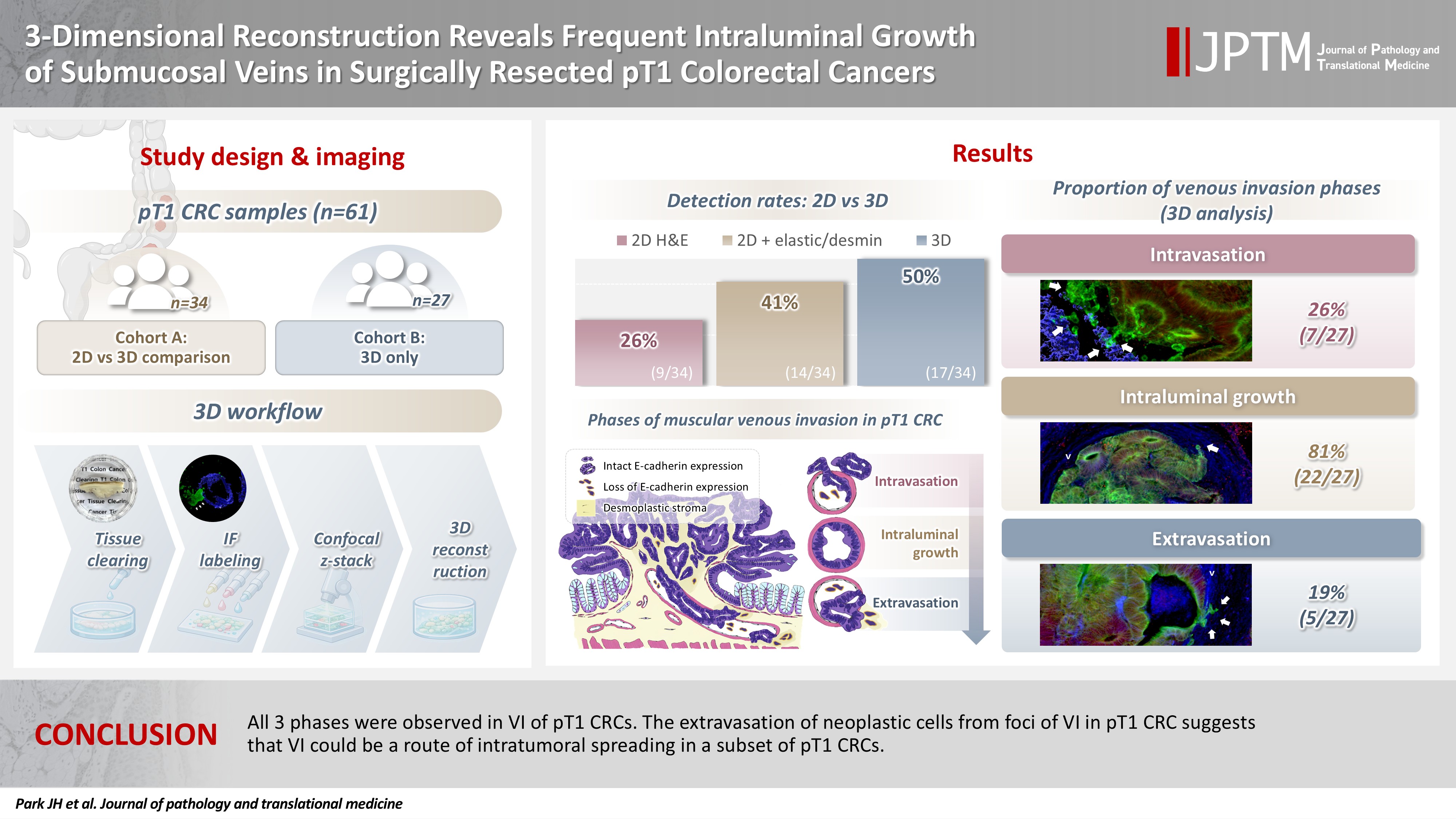
Although venous invasion (VI) is associated with distant metastasis and observed in >50% of pT2–4 colorectal cancers (CRCs), the role of VI in pT1 CRCs is not well-defined. Methods: Thirty-four surgically resected pT1 CRCs were reevaluated for 2-dimensional (2D) VI using hematoxylin and eosin (H&E)–stained slides with additional elastic and desmin immunohistochemical staining (cohort A). Additionally, 27 pT1 CRCs without knowing VI status were selected for 3-dimensional (3D) VI evaluation only (cohort B). All 61 cases (cohorts A and B) were studied in 3D using tissue clearing. Results: VI was detected more commonly in 3D (17/34, 50.0%) than in 2D H&E slide evaluation (9/34, 26.5%, p = .047). When VI was identified in 3D (27/61, 44.3%), the most common phase was that of intraluminal growth (22/27, 81.5%), followed by intravasation (7/27, 25.9%) and extravasation (5/27, 18.5%). E-cadherin expression was characterized in 3D in foci of VI and varied in each phase of invasion. Conclusions: All three phases were observed in VI of pT1 CRCs. The extravasation of neoplastic cells from foci of VI in pT1 CRC suggests that VI could be a route of intratumoral spreading in a subset of pT1 CRCs.
- Attitudes toward artificial intelligence in pathology: a survey-based study of pathologists in northern India
- Manupriya Sharma, Kavita Kumari, Navpreet Navpreet, Sushma Bharti, Rajneesh Kumari
- J Pathol Transl Med. 2025;59(6):382-389. Published online October 2, 2025
- DOI: https://doi.org/10.4132/jptm.2025.07.10

- 5,899 View
- 193 Download
-
Abstract
PDF
 Supplementary Material
 - Background
Artificial intelligence (AI) is transforming pathology by enhancing diagnostic accuracy, efficiency, and workflow standardization. Despite its growing presence, AI adoption remains limited, particularly in resource-constrained settings like India. This study assessed the knowledge, awareness, and perceptions of AI among pathologists in Northern India. Methods: A cross-sectional survey was conducted among 138 practicing pathologists in Northern India between April and June 2024. A structured online questionnaire was used to collect data on demographics, AI awareness, self-reported knowledge, sources of AI education, technological proficiency, and interest in AI-related training programs. Data analysis included descriptive statistics and chi-square tests, with p < .05 considered statistically significant. Results: AI awareness was high (88.4%), with significant sex differences (93.5% in females vs. 78.3% in males, p = .008). However, formal AI training was limited (6.5%), and only 16.7% had used AI as a diagnostic tool. Academic pathologists were more likely to engage with AI literature than their non-academic counterparts (p = .003). Interest in AI workshops was strong (92.8%). Access to whole slide imaging (WSI) correlated with higher AI knowledge (p = .008), as did self-reported technological proficiency (p = .001). Conclusions: Despite high AI awareness among pathologists, significant gaps remain in training, infrastructure, and practical application. Expanding access to digital pathology tools like WSI and improving digital literacy could facilitate AI adoption. Structured educational programs and greater investment in digital infrastructure are crucial for integrating AI into pathology practice.
- Correlation between myoferlin expression and lymph node metastasis in papillary thyroid carcinoma
- Ji Min Na, Dong Chul Kim, Dae Hyun Song, Hyo Jung An, Hyun Min Koh, Jeong-Hee Lee, Jong Sil Lee, Jung Wook Yang, Min Hye Kim
- J Pathol Transl Med. 2022;56(4):199-204. Published online May 11, 2022
- DOI: https://doi.org/10.4132/jptm.2022.03.19

- 5,193 View
- 180 Download
-
Abstract
PDF
- Background
Myoferlin is a multifunctional protein expressed in various normal and cancer cells, with novel oncogenic roles being newly discovered. Recently, correlations have been found between myoferlin expression and unfavorable prognosis in various carcinomas. This study investigated the prognostic role of myoferlin expression in papillary thyroid carcinoma (PTC), specifically that associated with nodal metastasis.
Methods
We collected clinicopathological data and PTC tissues from 116 patients who had been admitted to Gyeongsang National University Hospital in 2010. Immunohistochemical analysis was performed on surgical specimen-derived tissue microarray blocks. Myoferlin expression was graded, and the relationship between expression level and pathological features of tumors based on the American Joint Committee on Cancer staging system was evaluated.
Results
Of the 116 patient samples, 100 cases exhibited positive myoferlin expression. Higher grade of myoferlin expression was correlated with lower T category group (p = .010). Presence of lymph node metastasis was determined to be significantly correlated with low-grade myoferlin expression (p = .019), with no significant difference between pN1a and pN1b tumors.
Conclusions
Our study revealed an adverse correlation between myoferlin expression and pathological features of PTC, evidence of the potential prognostic role of myoferlin in PTC lymph node metastasis.
- Imaging features of breast cancer molecular subtypes: state of the art
- Nariya Cho
- J Pathol Transl Med. 2021;55(1):16-25. Published online November 9, 2020
- DOI: https://doi.org/10.4132/jptm.2020.09.03
- 55,566 View
- 405 Download
- 27 Web of Science
- 25 Crossref
-
Abstract
PDF
- Characterization of breast cancer molecular subtypes has been the standard of care for breast cancer management. We aimed to provide a review of imaging features of breast cancer molecular subtypes for the field of precision medicine. We also provide an update on the recent progress in precision medicine for breast cancer, implications for imaging, and recent observations in longitudinal functional imaging with radiomics.
-
Citations
Citations to this article as recorded by
- Prediction of HER2 changes post-neoadjuvant therapy based on fusion of ultrasound radiomics and clinicopathological features empowered by explainable AI: A multicenter study
Yuqi Yan, Xinzheng Xue, Jiayu Xie, Jian Liu, Lin Sui, Tian Jiang, Zhiyan Jin, Di Ou, Zhirui Chuan, Mingjie Jin, Yang Zhang, Vicky Yang Wang, Xiaomao Luo, Shihao Xu, Dong Xu
European Journal of Cancer.2026; 232: 116158. CrossRef - Contrast-Enhanced Mammography and Deep Learning-Derived Malignancy Scoring in Breast Cancer Molecular Subtype Assessment
Antonia O. Ferenčaba, Dora Galić, Gordana Ivanac, Kristina Kralik, Martina Smolić, Justinija Steiner, Ivo Pedišić, Kristina Bojanic
Medicina.2026; 62(1): 115. CrossRef - Radiomics Integration of Mammography and DCE-MRI for Predicting Molecular Subtypes in Breast Cancer Patients
Xianwei Yang, Jing Li, Hang Sun, Jing Chen, Jin Xie, Yonghui Peng, Tao Shang, Tongyong Pan
Breast Cancer: Targets and Therapy.2025; Volume 17: 187. CrossRef - HER2 (2+)/SISH-positive vs. HER2 (3+) Breast Cancer: Pre-treatment MRI Differences and Accuracy of pCR Prediction on Post-treatment MRI
Ga Eun Park, Han Song Mun, Sung Hun Kim, Bong Joo Kang
Academic Radiology.2025; 32(8): 4395. CrossRef - Impact of Prior Mammograms on Radiologists and Radiographers' Detection of Different Breast Cancer Lesion Types
Judith D. Akwo, Phuong Dung (Yun) Trieu, Melissa L. Barron, Tess Reynolds, Sarah J. Lewis
Journal of Medical Radiation Sciences.2025; 72(4): 497. CrossRef - Correlation analysis of multiparametric magnetic resonance imaging features and molecular subtypes of breast cancer
Junping Li, Guanghui Huo, Xiaoye Lei, Guang Li, Mengxing Yu, Ziyang Nie, Zhenhua Guo, Yue Zhang
Journal of Clinical Imaging Science.2025; 15: 37. CrossRef - Intertumoral Heterogeneity in Multifocal Breast Cancer Mimicking a Collision Tumor on Imaging: A Case Report
Yoshika Nagata, Izumi Kinoshita, Toshihiro Saeki, Daiji Uchiyama, Takahisa Fujikawa
Cureus.2025;[Epub] CrossRef - Fractal measures as predictors of histopathological complexity in breast carcinoma mammograms
Abhijeet Das, Ramray Bhat, Mohit Kumar Jolly
Physical Biology.2025; 22(6): 066006. CrossRef - Association of clinicopathologic and molecular factors with the occurrence of positive margins in breast cancer
Anupama Praveen Kumar, Diego Vicente, Jianfang Liu, Praveen-Kumar Raj-Kumar, Brenda Deyarmin, Xiaoying Lin, Craig D. Shriver, Hai Hu
Breast Cancer Research and Treatment.2024; 204(1): 15. CrossRef - The association of magnetic resonance imaging features with five molecular subtypes of breast cancer
Van Thi Nguyen, Duc Huu Duong, Quang Thai Nguyen, Duy Thai Nguyen, Thi Linh Tran, Tra Giang Duong
European Journal of Radiology Open.2024; 13: 100585. CrossRef - The effect of data resampling methods in radiomics
Aydin Demircioğlu
Scientific Reports.2024;[Epub] CrossRef - Treated Primary Cutaneous Malignant Melanoma With Later Metastasis Found in Clinical Presentation of Left Axilla Lymphadenopathy: A Case Report
Brigitte L Cochran, Sara Eliseo, Austin Vaughn, Tamryn L Van Der Horn, Enzo Ferrara, Jamie Edwards
Cureus.2024;[Epub] CrossRef - Pushing the envelope in breast conserving surgery − is multiple-wire localization (3 or more wires) associated with increased risk of compromised margins and long-term recurrence?
Orit Golan, Marian Khatib, Tehillah S. Menes, Vivianne A.R. Freitas, Rivka Kessner, Rina Neeman, Michal Mauda-Havakuk, Diego Mercer, Yoav Amitai
European Journal of Radiology.2024; 176: 111511. CrossRef - A model combining BI-RADS® descriptors from pre-treatment B-mode breast ultrasound with clinicopathological tumor features shows promise in the prediction of residual disease after neoadjuvant chemotherapy
Panagiotis Kapetas, Reena Aggarwal, Basmah Altuwayjiri, Katja Pinker, Paola Clauser, Thomas H. Helbich, Pascal A.T. Baltzer
European Journal of Radiology.2024; 178: 111649. CrossRef - Correlations of Imaging and Therapy in Breast Cancer Based on Molecular Patterns: An Important Issue in the Diagnosis of Breast Cancer
Oana Maria Burciu, Ioan Sas, Tudor-Alexandru Popoiu, Adrian-Grigore Merce, Lavinia Moleriu, Ionut Marcel Cobec
International Journal of Molecular Sciences.2024; 25(15): 8506. CrossRef - Access to prior screening mammograms affects the specificity but not sensitivity of radiologists' performance
J.D. Akwo, P. D. (Yun) Trieu, M.L. Barron, T. Reynolds, S.J. Lewis
Clinical Radiology.2024; 79(12): e1549. CrossRef - Machine learning-based radiomics prognostic model for patients with proximal esophageal cancer after definitive chemoradiotherapy
Linrui Li, Zhihui Qin, Juan Bo, Jiaru Hu, Yu Zhang, Liting Qian, Jiangning Dong
Insights into Imaging.2024;[Epub] CrossRef - LYMPH NODES MORPHOLOGICAL CHANGES AND BREAST CANCER SUBTYPES IN PREDICTION OF METASTASES
J. N. Akhundova, M. F. Amirova
World of Medicine and Biology.2024; 20(90): 15. CrossRef - Axillary lymph node changes in different molecular subtypes of breast cancer
J.N. Akhundova
Український радіологічний та онкологічний журнал.2024; 32(4): 529. CrossRef - Histogram analysis of multi-model high-resolution diffusion-weighted MRI in breast cancer: correlations with molecular prognostic factors and subtypes
Yanjin Qin, Feng Wu, Qilan Hu, Litong He, Min Huo, Caili Tang, Jingru Yi, Huiting Zhang, Ting Yin, Tao Ai
Frontiers in Oncology.2023;[Epub] CrossRef - ASO Author Reflections: Sequence of Treatment in Clinically Node-Negative T1 Triple-Negative Breast Cancer
Kai Huang, James W. Jakub, Sarah A. McLaughlin
Annals of Surgical Oncology.2023; 30(13): 8455. CrossRef - Circulating non-coding RNAs as a diagnostic and management biomarker for breast cancer: current insights
Hamed Hosseinalizadeh, Mehrdad Mahmoodpour, Ammar Ebrahimi
Molecular Biology Reports.2022; 49(1): 705. CrossRef - MRI as a biomarker for breast cancer diagnosis and prognosis
Francesca Galati, Veronica Rizzo, Rubina Manuela Trimboli, Endi Kripa, Roberto Maroncelli, Federica Pediconi
BJR|Open.2022;[Epub] CrossRef - Multiparametric MRI Features of Breast Cancer Molecular Subtypes
Madalina Szep, Roxana Pintican, Bianca Boca, Andra Perja, Magdalena Duma, Diana Feier, Bogdan Fetica, Dan Eniu, Sorin Marian Dudea, Angelica Chiorean
Medicina.2022; 58(12): 1716. CrossRef - Circulating tumor cells as prognostic biomarkers in breast cancer: current status and future prospects
Evagelia Chantzara, Nikolaos Xenidis, Galatea Kallergi, Vassilis Georgoulias, Athanasios Kotsakis
Expert Review of Molecular Diagnostics.2021; 21(10): 1037. CrossRef
- Prediction of HER2 changes post-neoadjuvant therapy based on fusion of ultrasound radiomics and clinicopathological features empowered by explainable AI: A multicenter study
- Advances in the Endoscopic Assessment of Inflammatory Bowel Diseases: Cooperation between Endoscopic and Pathologic Evaluations
- Jae Hee Cheon
- J Pathol Transl Med. 2015;49(3):209-217. Published online May 15, 2015
- DOI: https://doi.org/10.4132/jptm.2015.04.09
- 14,764 View
- 98 Download
- 5 Web of Science
- 5 Crossref
-
Abstract
PDF
- Endoscopic assessment has a crucial role in the management of inflammatory bowel disease (IBD). It is particularly useful for the assessment of IBD disease extension, severity, and neoplasia surveillance. Recent advances in endoscopic imaging techniques have been revolutionized over the past decades, progressing from conventional white light endoscopy to novel endoscopic techniques using molecular probes or electronic filter technologies. These new technologies allow for visualization of the mucosa in detail and monitor for inflammation/dysplasia at the cellular or sub-cellular level. These techniques may enable us to alter the IBD surveillance paradigm from four quadrant random biopsy to targeted biopsy and diagnosis. High definition endoscopy and dye-based chromoendoscopy can improve the detection rate of dysplasia and evaluate inflammatory changes with better visualization. Dye-less chromoendoscopy, including narrow band imaging, iScan, and autofluorescence imaging can also enhance surveillance in comparison to white light endoscopy with optical or electronic filter technologies. Moreover, confocal laser endomicroscopy or endocytoscopy have can achieve real-time histology evaluation in vivo and have greater accuracy in comparison with histology. These new technologies could be combined with standard endoscopy or further histologic confirmation in patients with IBD. This review offers an evidence-based overview of new endoscopic techniques in patients with IBD.
-
Citations
Citations to this article as recorded by- Moxifloxacin promotes two-photon microscopic imaging for discriminating different stages of DSS-induced colitis on mice
Yingtong Chen, Xiaoyi Xu, Min Wang, Xiang Wang, Yan Wang, Yong Zhang, Jin Huang, Yuwen Tao, Wentao Fan, Lili Zhao, Li Liu, Zhining Fan
Photodiagnosis and Photodynamic Therapy.2024; 48: 104220. CrossRef - Colorectal cancer in inflammatory bowel disease: review of the evidence
D. S. Keller, A. Windsor, R. Cohen, M. Chand
Techniques in Coloproctology.2019; 23(1): 3. CrossRef - Probe-based confocal laser endomicroscopy in the differential diagnosis of inflammatory bowel diseases: a case series
Jung Won Park, Tae Il Kim, Jae Hee Cheon
Intestinal Research.2018; 16(4): 641. CrossRef - How to Assess and Document Endoscopies in IBD Patients by Including Standard Scoring Systems
Anna M. Buchner, Gary R. Lichtenstein
Inflammatory Bowel Diseases.2016; 22(4): 1010. CrossRef - Nodular lymphoid hyperplasia: A marker of low-grade inflammation in irritable bowel syndrome?
Anna Chiara Piscaglia, Lucrezia Laterza, Valentina Cesario, Viviana Gerardi, Rosario Landi, Loris Riccardo Lopetuso, Giovanni Calò, Giovanna Fabbretti, Massimo Brisigotti, Maria Loredana Stefanelli, Antonio Gasbarrini
World Journal of Gastroenterology.2016; 22(46): 10198. CrossRef
- Moxifloxacin promotes two-photon microscopic imaging for discriminating different stages of DSS-induced colitis on mice
- Molecular Imaging in the Era of Personalized Medicine
- Kyung-Ho Jung, Kyung-Han Lee
- J Pathol Transl Med. 2015;49(1):5-12. Published online January 15, 2015
- DOI: https://doi.org/10.4132/jptm.2014.10.24
- 15,924 View
- 214 Download
- 32 Web of Science
- 27 Crossref
-
Abstract
PDF
- Clinical imaging creates visual representations of the body interior for disease assessment. The role of clinical imaging significantly overlaps with that of pathology, and diagnostic workflows largely depend on both fields. The field of clinical imaging is presently undergoing a radical change through the emergence of a new field called molecular imaging. This new technology, which lies at the intersection between imaging and molecular biology, enables noninvasive visualization of biochemical processes at the molecular level within living bodies. Molecular imaging differs from traditional anatomical imaging in that biomarkers known as imaging probes are used to visualize target molecules-of-interest. This ability opens up exciting new possibilities for applications in oncologic, neurological and cardiovascular diseases. Molecular imaging is expected to make major contributions to personalized medicine by allowing earlier diagnosis and predicting treatment response. The technique is also making a huge impact on pharmaceutical development by optimizing preclinical and clinical tests for new drug candidates. This review will describe the basic principles of molecular imaging and will briefly touch on three examples (from an immense list of new techniques) that may contribute to personalized medicine: receptor imaging, angiogenesis imaging, and apoptosis imaging.
-
Citations
Citations to this article as recorded by- Ionic Cell Microscopy: A new modality for visualizing cells using microfluidic impedance cytometry and generative artificial intelligence
Mahtab Kokabi, Gulam M. Rather, Mehdi Javanmard
Biosensors and Bioelectronics: X.2025; 24: 100619. CrossRef - Multimodal imaging in cancer detection: the role of SPIONs and USPIONs as contrast agents for MRI, SPECT, and PET
Zahra Shaghaghi, Ramin Mansouri, Sahar Nosrati, Maryam Alvandi
Future Oncology.2025; 21(18): 2367. CrossRef - Supramolecular fluorescence biosensing based on macrocycles
Jia-Hong Tian, Haiqi Xu, Xin-Yue Hu, Dong-Sheng Guo
Supramolecular Materials.2024; 3: 100063. CrossRef - A non-invasive osteopontin-targeted phase changeable fluorescent nanoprobe for molecular imaging of myocardial fibrosis
Xueli Zhao, Yuze Qin, Bo Wang, Jiao Liu, Yueyue Wang, Kun Chen, Jia Zhao, Lanlan Zhang, Yuanming Wu, Liwen Liu
Nanoscale Advances.2024; 6(14): 3590. CrossRef - The Role of Molecular Imaging in Personalized Medicine
Suliman Salih, Aisyah Elliyanti, Ajnas Alkatheeri, Fatima AlYafei, Bashayer Almarri, Hasina Khan
Journal of Personalized Medicine.2023; 13(2): 369. CrossRef - Development of a multifunctional platform for near-infrared imaging and targeted radionuclide therapy for tumors
Huihui He, Ke Li, Hang Li, Shiliang Zhu, Shuai Qin, Yong Mao, Jianguo Lin, Ling Qiu, Chunjing Yu
European Journal of Pharmaceutics and Biopharmaceutics.2023; 185: 107. CrossRef - Quantum Biotechnology
Nicolas P. Mauranyapin, Alex Terrasson, Warwick P. Bowen
Advanced Quantum Technologies.2022;[Epub] CrossRef - Preparation Fe3O4@chitosan-graphene quantum dots nanocomposites for fluorescence and magnetic resonance imaging
Kai Wang, Xiaoguang Xu, Yan Li, Mayifei Rong, Lifeng Wang, Liying Lu, Jian Wang, Fengwen Zhao, Bowen Sun, Yong Jiang
Chemical Physics Letters.2021; 783: 139060. CrossRef - Network Medicine: A Clinical Approach for Precision Medicine and Personalized Therapy in Coronary Heart Disease
Teresa Infante, Luca Del Viscovo, Maria Luisa De Rimini, Sergio Padula, Pio Caso, Claudio Napoli
Journal of Atherosclerosis and Thrombosis.2020; 27(4): 279. CrossRef - Nanodrug Delivery Systems for the Treatment of Ovarian Cancer
Jonathan M. Pantshwa, Pierre P. D. Kondiah, Yahya E. Choonara, Thashree Marimuthu, Viness Pillay
Cancers.2020; 12(1): 213. CrossRef - Molecular imaging of the urokinase plasminogen activator receptor: opportunities beyond cancer
V. M. Baart, R. D. Houvast, L. F. de Geus-Oei, P. H. A. Quax, P. J. K. Kuppen, A. L. Vahrmeijer, C. F. M. Sier
EJNMMI Research.2020;[Epub] CrossRef - In vivo SPECT imaging of an 131I-labeled PM 2.5 mimic substitute
Dong-Hui Pan, Jie Sheng, Xin-Yu Wang, Qian-Huan Huang, Jun-Jie Yan, Li-Zhen Wang, Run-Ling Yang, Dong-Jian Shi, Yu-Ping Xu, Ming-Qing Chen
Nuclear Science and Techniques.2020;[Epub] CrossRef - Optofluidics in bio-imaging applications
Sihui Chen, Rui Hao, Yi Zhang, Hui Yang
Photonics Research.2019; 7(5): 532. CrossRef - Nitrogen-vacancy centers in diamond for nanoscale magnetic resonance imaging applications
Alberto Boretti, Lorenzo Rosa, Jonathan Blackledge, Stefania Castelletto
Beilstein Journal of Nanotechnology.2019; 10: 2128. CrossRef - Online molecular image repository and analysis system: A multicenter collaborative open-source infrastructure for molecular imaging research and application
Mahabubur Rahman, Hiroshi Watabe
Computers in Biology and Medicine.2018; 96: 233. CrossRef - Nε-Acryloyllysine Piperazides as Irreversible Inhibitors of Transglutaminase 2: Synthesis, Structure–Activity Relationships, and Pharmacokinetic Profiling
Robert Wodtke, Christoph Hauser, Gloria Ruiz-Gómez, Elisabeth Jäckel, David Bauer, Martin Lohse, Alan Wong, Johanna Pufe, Friedrich-Alexander Ludwig, Steffen Fischer, Sandra Hauser, Dieter Greif, M. Teresa Pisabarro, Jens Pietzsch, Markus Pietsch, Reik Lö
Journal of Medicinal Chemistry.2018; 61(10): 4528. CrossRef - Genomic Interventions in Medicine
Oluwadurotimi S Aworunse, Oluwatomiwa Adeniji, Olusola L Oyesola, Itunuoluwa Isewon, Jelili Oyelade, Olawole O Obembe
Bioinformatics and Biology Insights.2018;[Epub] CrossRef - Restriction spectrum imaging: An evolving imaging biomarker in prostate MRI
Ryan L. Brunsing, Natalie M. Schenker-Ahmed, Nathan S. White, J. Kellogg Parsons, Christopher Kane, Joshua Kuperman, Hauke Bartsch, Andrew Karim Kader, Rebecca Rakow-Penner, Tyler M. Seibert, Daniel Margolis, Steven S. Raman, Carrie R. McDonald, Nikdokht
Journal of Magnetic Resonance Imaging.2017; 45(2): 323. CrossRef - Personalized medicine: a new option for nuclear medicine and molecular imaging in the third millennium
Orazio Schillaci, Nicoletta Urbano
European Journal of Nuclear Medicine and Molecular Imaging.2017; 44(4): 563. CrossRef - Nano-Magnetic Resonance Imaging (Nano-MRI) Gives Personalized Medicine a New Perspective
Lorenzo Rosa, Jonathan Blackledge, Albert Boretti
Biomedicines.2017; 5(1): 7. CrossRef - Optical nanoprobes for biomedical applications: shining a light on upconverting and near-infrared emitting nanoparticles for imaging, thermal sensing, and photodynamic therapy
E. Hemmer, P. Acosta-Mora, J. Méndez-Ramos, S. Fischer
Journal of Materials Chemistry B.2017; 5(23): 4365. CrossRef - Drug Discovery by Molecular Imaging and Monitoring Therapy Response in Lymphoma
Senthilkumar Kalimuthu, Ju Hye Jeong, Ji Min Oh, Byeong-Cheol Ahn
International Journal of Molecular Sciences.2017; 18(8): 1639. CrossRef - Chemistry and engineering of cyclodextrins for molecular imaging
Wing-Fu Lai, Andrey L. Rogach, Wing-Tak Wong
Chemical Society Reviews.2017; 46(20): 6379. CrossRef - Prototypes of Lanthanide(III) Agents Responsive to Enzymatic Activities in Three Complementary Imaging Modalities: Visible/Near-Infrared Luminescence, PARACEST-, and T1-MRI
Jiefang He, Célia S. Bonnet, Svetlana V. Eliseeva, Sara Lacerda, Thomas Chauvin, Pascal Retailleau, Frederic Szeremeta, Bernard Badet, Stéphane Petoud, Éva Tóth, Philippe Durand
Journal of the American Chemical Society.2016; 138(9): 2913. CrossRef - Nanoparticles in practice for molecular-imaging applications: An overview
Parasuraman Padmanabhan, Ajay Kumar, Sundramurthy Kumar, Ravi Kumar Chaudhary, Balázs Gulyás
Acta Biomaterialia.2016; 41: 1. CrossRef - A new neuroinformatics approach to personalized medicine in neurology: The Virtual Brain
Maria I. Falcon, Viktor Jirsa, Ana Solodkin
Current Opinion in Neurology.2016; 29(4): 429. CrossRef - Targeted multimodal nano-reporters for pre-procedural MRI and intra-operative image-guidance
Joonseok Lee, Andrew C. Gordon, Hacksung Kim, Wooram Park, Soojeong Cho, Byeongdu Lee, Andrew C. Larson, Elena A. Rozhkova, Dong-Hyun Kim
Biomaterials.2016; 109: 69. CrossRef
- Ionic Cell Microscopy: A new modality for visualizing cells using microfluidic impedance cytometry and generative artificial intelligence
- Proposal for a Standardized Pathology Report of Gastroenteropancreatic Neuroendocrine Tumors: Prognostic Significance of Pathological Parameters
- Mee-Yon Cho, Jin Hee Sohn, So Young Jin, Hyunki Kim, Eun Sun Jung, Mi-Jung Kim, Kyoung-Mee Kim, Woo Ho Kim, Joon Mee Kim, Yun Kyung Kang, Joon Hyuk Choi, Dae Young Kang, Youn Wha Kim, Eun Hee Choi
- Korean J Pathol. 2013;47(3):227-237. Published online June 25, 2013
- DOI: https://doi.org/10.4132/KoreanJPathol.2013.47.3.227
- 15,684 View
- 148 Download
- 12 Crossref
-
Abstract
PDF
Background There is confusion in the diagnosis and biological behaviors of gastroenteropancreatic neuroendocrine tumors (GEP-NETs), because of independently proposed nomenclatures and classifications. A standardized form of pathology report is required for the proper management of patients.
Methods We discussed the proper pathological evaluation of GEP-NET at the consensus conference of the subcommittee meeting for the Gastrointestinal Pathology Study Group of the Korean Society of Pathologists. We then verified the prognostic significance of pathological parameters from our previous nationwide collection of pathological data from 28 hospitals in Korea to determine the essential data set for a pathology report.
Results Histological classification, grading (mitosis and/or Ki-67 labeling index), T staging (extent, size), lymph node metastasis, and lymphovascular and perineural invasion were significant prognostic factors and essential for the pathology report of GEP-NET, while immunostaining such as synaptophysin and chromogranin may be optional. Furthermore, the staging system, either that of the 2010 American Joint Cancer Committee (AJCC) or the European Neuroendocrine Tumor Society (ENETS), should be specified, especially for pancreatic neuroendocrine neoplasms.
Conclusions A standardized pathology report is crucial for the proper management and prediction of prognosis of patients with GEP-NET.
-
Citations
Citations to this article as recorded by- Analysis of Prognostic Risk Factors of Endoscopic Submucosal Dissection (ESD) and Curative Resection of Gastrointestinal Neuroendocrine Neoplasms
Yuan Si, ChaoKang Huang, JingBin Yuan, XianHui Zhang, QingQiang He, ZhiJin Lin, Ling He, ZhongXin Liu, Yuvaraja Teekaraman
Contrast Media & Molecular Imaging.2022;[Epub] CrossRef - Standardization of the pathologic diagnosis of appendiceal mucinous neoplasms
Dong-Wook Kang, Baek-hui Kim, Joon Mee Kim, Jihun Kim, Hee Jin Chang, Mee Soo Chang, Jin-Hee Sohn, Mee-Yon Cho, So-Young Jin, Hee Kyung Chang, Hye Seung Han, Jung Yeon Kim, Hee Sung Kim, Do Youn Park, Ha Young Park, So Jeong Lee, Wonae Lee, Hye Seung Lee,
Journal of Pathology and Translational Medicine.2021; 55(4): 247. CrossRef - Preoperative diagnosis of well‐differentiated neuroendocrine tumor in common hepatic duct by brush cytology: A case report
Jiwoon Choi, Kyong Joo Lee, Sung Hoon Kim, Mee‐Yon Cho
Diagnostic Cytopathology.2019; 47(7): 720. CrossRef - Primary renal well-differentiated neuroendocrine tumors: report of six cases with an emphasis on the Ki-67 index and mitosis
Bohyun Kim, Han-Seong Kim, Kyung Chul Moon
Diagnostic Pathology.2019;[Epub] CrossRef - Primary low‐grade neuroendocrine carcinoma of the skin: An exceedingly rare entity
Tiffany Y. Chen, Annie O. Morrison, Joe Susa, Clay J. Cockerell
Journal of Cutaneous Pathology.2017; 44(11): 978. CrossRef - Prognostic Validity of the American Joint Committee on Cancer and the European Neuroendocrine Tumors Staging Classifications for Pancreatic Neuroendocrine Tumors
Jae Hee Cho, Ji Kon Ryu, Si Young Song, Jin-Hyeok Hwang, Dong Ki Lee, Sang Myung Woo, Young-Eun Joo, Seok Jeong, Seung-Ok Lee, Byung Kyu Park, Young Koog Cheon, Jimin Han, Tae Nyeun Kim, Jun Kyu Lee, Sung-Hoon Moon, Hyunjin Kim, Eun Taek Park, Jae Chul Hw
Pancreas.2016; 45(7): 941. CrossRef - Early diagnosis and treatment of gastrointestinal neuroendocrine tumors
Hong Shen, Zhuo Yu, Jing Zhao, Xiu-Zhen Li, Wen-Sheng Pan
Oncology Letters.2016; 12(5): 3385. CrossRef - Recent Updates on Neuroendocrine Tumors From the Gastrointestinal and Pancreatobiliary Tracts
Joo Young Kim, Seung-Mo Hong
Archives of Pathology & Laboratory Medicine.2016; 140(5): 437. CrossRef - Pancreatic neuroendocrine tumors: Correlation between the contrast-enhanced computed tomography features and the pathological tumor grade
Koji Takumi, Yoshihiko Fukukura, Michiyo Higashi, Junnichi Ideue, Tomokazu Umanodan, Hiroto Hakamada, Ichiro Kanetsuki, Takashi Yoshiura
European Journal of Radiology.2015; 84(8): 1436. CrossRef - Tumeurs neuroendocrines du tube digestif et du pancréas : ce que le pathologiste doit savoir et doit faire en 2014
Jean-Yves Scoazec, Anne Couvelard
Annales de Pathologie.2014; 34(1): 40. CrossRef - Spectrum of Gastroenteropancreatic NENs in Routine Histological Examinations of Bioptic and Surgical Specimen: A Study of 161 Cases Collected from 17 Departments of Pathology in the Czech Republic
Václav Mandys, Tomáš Jirásek
Gastroenterology Research and Practice.2014; 2014: 1. CrossRef - p27 Loss Is Associated with Poor Prognosis in Gastroenteropancreatic Neuroendocrine Tumors
Hee Sung Kim, Hye Seung Lee, Kyung Han Nam, Jiwoon Choi, Woo Ho Kim
Cancer Research and Treatment.2014; 46(4): 383. CrossRef
- Analysis of Prognostic Risk Factors of Endoscopic Submucosal Dissection (ESD) and Curative Resection of Gastrointestinal Neuroendocrine Neoplasms
- The Definition of Minimal Extrathyroid Extension in Thyroid Pathology by Analyzing Sizable Intra- and Extrathyroid Blood Vessels
- Hyae Min Jeon, Beom Jin Lim, Hang-Seok Chang, SoonWon Hong
- Korean J Pathol. 2012;46(6):548-553. Published online December 26, 2012
- DOI: https://doi.org/10.4132/KoreanJPathol.2012.46.6.548
- 11,074 View
- 48 Download
- 7 Crossref
-
Abstract
PDF
Background To define the exact boundary of the intrathyroid and extrathyroid aspects of a gland when determining the extent of cancer invasion, we plan to clarify the definition of sizable vascular structures, which is one of the helpful histologic clues in determining a minimal extrathyroid extension. We hypothesized that arterial wall thicknesses in extrathyroid soft tissue would be significantly different from the arteries in the thyroid parenchyma.
Methods Twenty cases of papillary carcinoma were selected. The numbers and wall thicknesses of the arteries and arterioles in intrathyroid and extrathyroid tissue were evaluated. The absence of nerve tissue in the thyroid gland was confirmed using the S-100 protein immunohistochemical stain.
Results The comparison of the mean thicknesses of the total arteries between the extrathyroid and intrathyroid tissues in the retrospective study (26.88 µm vs. 15.07 µm, respectively) and the prospective study (35.24 µm vs. 16.52 µm, respectively) revealed significant differences (p=0.000). The greatest thickness of the intrathyroid arteries was 67.93 µm.
Conclusions According to our results, the study showed that the extrathyroidal arteries were significantly thicker than the intrathyroidal arteries. We suggest that the sizable blood vessels of extrathyroidal arteries should be greater than 67.93 µm in thickness.
-
Citations
Citations to this article as recorded by- Invasion in thyroid cancer: Controversies and best practices
Michiya Nishino, Jack Jacob
Seminars in Diagnostic Pathology.2020; 37(5): 219. CrossRef - MiR-221/222 promote migration and invasion, and inhibit autophagy and apoptosis by modulating ATG10 in aggressive papillary thyroid carcinoma
Hao Shen, Zaikai Lin, Haiyan Shi, Lingling Wu, Baojin Ma, Hong Li, Baobing Yin, Jun Tang, Hongjin Yu, Xiaoxing Yin
3 Biotech.2020;[Epub] CrossRef - Minimal extrathyroidal extension affects the prognosis of differentiated thyroid cancer: Is there a need for change in the AJCC classification system?
Zeming Liu, Yihui Huang, Sichao Chen, Di Hu, Min Wang, Ling Zhou, Wei Zhou, Danyang Chen, Haifeng Feng, Wei Wei, Chao Zhang, Wen Zeng, Liang Guo, Scott M. Langevin
PLOS ONE.2019; 14(6): e0218171. CrossRef - miR-199a-3p downregulation in thyroid tissues is associated with invasion and metastasis of papillary thyroid carcinoma
Chengbiao Liu, Meiling Xing, Liping Wang, Kejun Zhang
British Journal of Biomedical Science.2017; 74(2): 90. CrossRef - Clinicopathological Significance of Minimal Extrathyroid Extension in Solitary Papillary Thyroid Carcinomas
Chang Gok Woo, Chang Ohk Sung, Yun Mi Choi, Won Gu Kim, Tae Yong Kim, Young Kee Shong, Won Bae Kim, Suck Joon Hong, Dong Eun Song
Annals of Surgical Oncology.2015; 22(S3): 728. CrossRef - Intraoperative Frozen Section for the Evaluation of Extrathyroidal Extension in Papillary Thyroid Cancer
Om Prakash Prajapati, A. K. Verma, M. Sabaretnam
World Journal of Surgery.2015; 39(7): 1855. CrossRef - Tumor Sprouting in Papillary Thyroid Carcinoma Is Correlated with Lymph Node Metastasis and Recurrence
Eunjung Lee, Wonkyung Jung, Jeong-Soo Woo, Jae Bok Lee, Bong Kyung Shin, Han Kyeom Kim, Aeree Kim, Baek-hui Kim
Korean Journal of Pathology.2014; 48(2): 117. CrossRef
- Invasion in thyroid cancer: Controversies and best practices
- Pineal Parenchymal Tumor of Intermediate Differentiation with Gangliocytic Differentiation: A Case Report.
- Lee So Maeng
- Korean J Pathol. 2009;43(4):364-367.
- DOI: https://doi.org/10.4132/KoreanJPathol.2009.43.4.364
- 3,926 View
- 30 Download
- 1 Crossref
-
Abstract
PDF
- A 49-year-old man presented with an extremely rare case of pineal parenchymal tumor with gangliocytic cells, manifesting as progressive gait disturbance and urinary incontinence lasting for one year. Brain MRI revealed a homogenously enhancing mass, measuring 3.5x2.7 x1.7 cm, in the pineal body. The mass compressed the deep cerebral vein with superior displacement, which caused mild obstructive hydrocephalus. Histological examination revealed lobular structures consisting of isomorphic small round cells with stippled chromatin and clear cytoplasm, and less cellular areas having large pleomorphic cells and ganglioid cells. Mitotic figures and tumor necrosis were not evident. Immunohistochemically, the neoplastic cells were positive for neuronal markers (neuron-specific enolase, neurofilament, NeuN and synaptophysin), but not for glial fibrillary acidic protein or S-100. Especially, neurofilament showed diffuse interstitial immunoreactivity with accentuation in a few gangliocytic cells and Ki-67 labeling index (2.5%) was low. Therefore, this case was diagnosed as pineal parenchymal tumor of intermediate differentiation with gangliocytic differentiation.
-
Citations
Citations to this article as recorded by- Pineal parenchymal tumor of intermediate differentiation: a systematic review and contemporary management of 389 cases reported during the last two decades
Hajime Takase, Reo Tanoshima, Navneet Singla, Yoshihiko Nakamura, Tetsuya Yamamoto
Neurosurgical Review.2022; 45(2): 1135. CrossRef
- Pineal parenchymal tumor of intermediate differentiation: a systematic review and contemporary management of 389 cases reported during the last two decades
- Nitric Oxide Synthase Expression in Early Stage of Aging Rat Kidney.
- Kye Won Kwon, Hyeon Joo Jeong
- Korean J Pathol. 2004;38(2):86-92.
- 2,149 View
- 20 Download
-
Abstract
PDF
- BACKGROUND
Nitric oxide synthase (NOS) has been suggested to have a role in renal injury of aging rats.
METHODS
Renal function and histology were compared between 12 month-and 7-9 week-old rats. Proliferating activity and cell death were evaluated by PCNA index and apoptosis. Three isoforms of NOS (eNOS, iNOS, and nNOS) were stained by immunohistochemistry.
RESULTS
Serum creatinine level was increased in old rats (1.0 mg/dL vs 0.5 mg/dL, p=0.000). 24 h proteinuria and urinary NO were comparable between the two groups. The percentage of global and segmental glomerulosclerosis increased in old rats. PCNA index decreased in the glomeruli (0.1 vs 0.6/glomerulus, p=0.005) and the tubulointerstitium (10.2 vs 19.2/mm2, p=0.019) of old rats compared to that of young rats. However, no difference was observed in the number of TUNEL positive cells. eNOS was not stained in young and old rat kidney, whereas iNOS was stained in the interstitial inflammatory cells of old rats (0.3 vs 0.0 of young rats/mm2, p=0.188). Macula densa nNOS staining significantly decreased in old rats compared to young rats (5.6 vs 9.5/mm2, p=0.009).
CONCLUSIONS
Proliferating activity is more affected than cell death with aging. Decreased nNOS expression without alteration of eNOS and iNOS expressions may implicate nNOS as a marker of renal injury in the early stage of aging.
- A Standardized Pathology Report for Gastric Cancer.
- Woo Ho Kim, Cheol Keun Park, Young Bae Kim, Youn Wha Kim, Ho Guen Kim, Han Ik Bae, Kyu Sang Song, Hee Kyung Chang, Hee Jin Chang, Yang Seok Chae
- Korean J Pathol. 2005;39(2):106-113.
- 4,989 View
- 340 Download
-
Abstract
PDF
- BACKGROUND
AND METHODS: The Gastrointestinal Pathology Study Group of the Korean Society of Pathologists developed a standardized pathology reporting format for gastric cancer in collaboration with the Korean Gastric Cancer Association. RESULTS: The diagnostic parameters are divided into two part: the standard part and the optional part. The standard part contains most of the items listed in the Japanese classification, the TNM classification by UICC, the WHO classification, and the Korean Gastric Cancer Association classification. Therefore, the standard part is adequate for routine surgical pathology service. We included detailed descriptions on each item.
CONCLUSIONS
The authors anticipate that this standardization can improve the diagnostic accuracy and decrease the discrepancies that occur in the pathologic diagnosis of gastric cancer. Furthermore, the standard format can encourage large scale multi-institutional collaborative studies.
- Nevus Sebaceous with Special Reference on Its Aging Effect.
- Jin Seok Seo, Mi Kyung Kim, Mikyung Kim, Kye Yong Song, Yun Lim Seo, Je G Chi
- Korean J Pathol. 1990;24(4):436-445.
- 2,384 View
- 33 Download
-
Abstract
PDF
- A histopathological study was performed on nevus sebaceus to observe its aging effect based on 75 cases of neuvs sebaceus those were collected during the past 10 years from three university hospitals in Seoul. The results are as follows: 1) Clinical findings The incidence was most frequent in the teenage group. The 75 cases consisted of 41 males and 34 females with a sex ratio of 1.2:1. Most of cases developed in the head and neck areas with 62.7% on the scalp and 29.3% on the face. 2) Histopathologic findings. The epidermal changes such as acanthosis(40%), papillomatosis(73%), hypergranulosis(44%) were most remarkable in the second decade and gradually decreased with aging. The apparent proliferation of sebaceous gland was observed in 73% and it was most prominent in the second decade. Apocrine glands were absent before the first decade but apparently increased after then. Proliferation of eccrine gland was not significant in all the age groups. Mild increase of immature hair follicles were noted in 49% of our cases with gradually decreasing tendency in the older age. The dermal inflammatory infiltrates were noted from the 2nd decade(28%) and thereafter gradually increased. Associated neoplasms were one apocrine adenoma, one sebaceous adenoma, two trichilemmomas and two arteriovenous hemangiomas. The majority of tumors occured in the third decade. Therefore, it is observed that neuvs sebaceous undergoes dynamic histopathologic changes according to the age of patient and later develop various secondary neoplastic changes. The pathogenesis of the nevus sebaceus is suggested to be closely related with developmental anomalies of primitive hair germ units in fetal stage.
- A Standardized Pathology Report for Colorectal Cancer.
- Hee Jin Chang, Cheol Keun Park, Woo Ho Kim, Young Bae Kim, Youn Wha Kim, Ho Guen Kim, Han Ik Bae, Kyu Sang Song, Mee Soo Chang, Hee Kyung Chang, Yang Seok Chae
- Korean J Pathol. 2006;40(3):193-203.
- 2,958 View
- 156 Download
-
Abstract
PDF
- BACKGROUND
AND METHODS: For standardizing the pathology report and diagnosis of colorectal cancers, the Gastrointestinal Pathology Study Group of the Korean Society of Pathologists has developed a pathology reporting format for colorectal cancer in collaboration with the Korean Society of Coloproctology.
RESULTS
The diagnostic parameters are divided into two parts: the standard part and the optional part. The standard part contains most of the items listed in the Japanese classification, the TNM classification by AJCC, and the WHO classification. We included detailed descriptions on each item.
CONCLUSIONS
The standardized pathology report for colorectal cancers is adequate for its application to routine surgical pathology reports, and it is also helpful to decrease the discrepancies that occur during the pathologic diagnosis of colorectal cancer. Furthermore, this reporting format could encourage nationwide multi-center collaborative studies.
 First
First Prev
Prev


