 E-submission
Search
- Page Path
- HOME > Search
Case Study
- Adrenal hemangioblastoma
- Joo-Yeon Koo, Kyung-Hwa Lee, Joon Hyuk Choi, Ho Seok Chung, Chan Choi
- J Pathol Transl Med. 2022;56(3):161-166. Published online February 28, 2022
- DOI: https://doi.org/10.4132/jptm.2021.12.28
- 6,633 View
- 160 Download
- 1 Web of Science
- 1 Crossref
-
 Abstract
 PDF
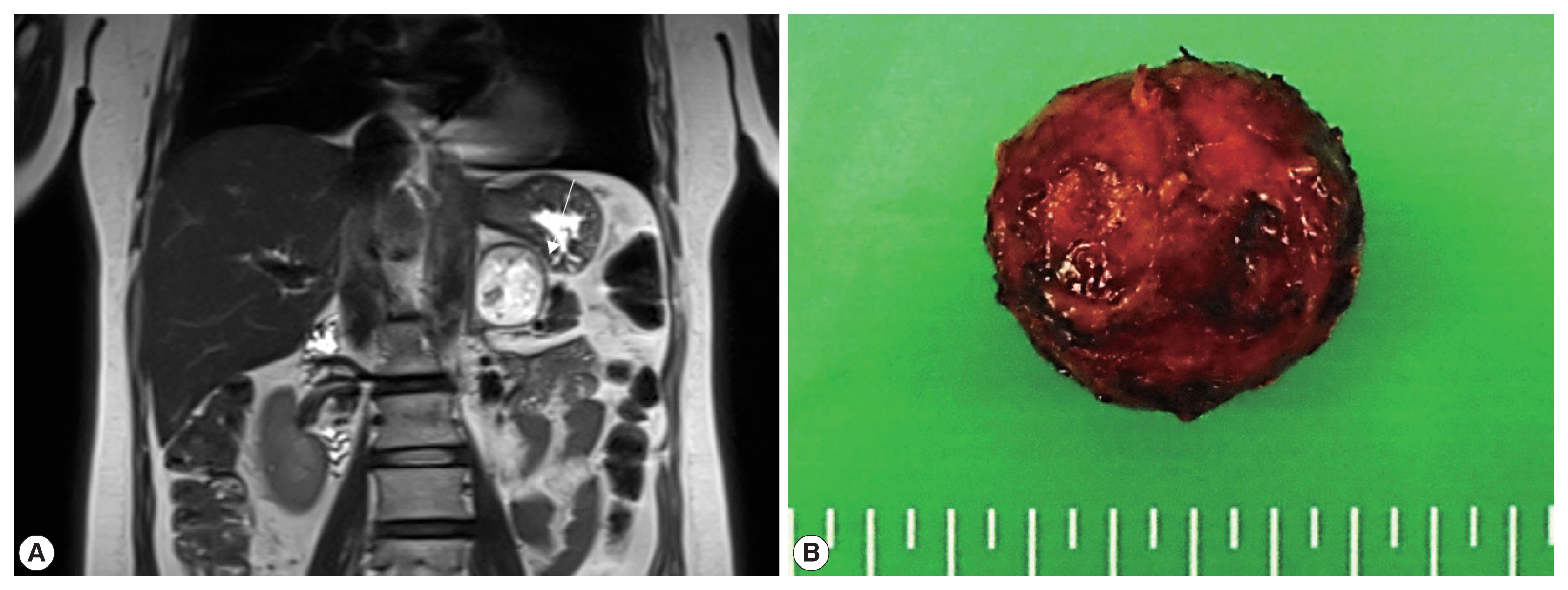
PDF - Hemangioblastoma (HB) is a rare benign tumor that most commonly occurs in the cerebellum. HB is composed of neoplastic stromal cells and abundant small vessels. However, the exact origin of stromal cells is controversial. Extraneural HBs have been reported in a small series, and peripheral HBs arising in the adrenal gland are extremely rare. Herein, we report a case of sporadic adrenal HB in a 54-year-old woman. The tumor was a well-circumscribed, yellow mass measuring 4.2 cm in diameter. Histologically, the tumor was composed of small blood vessels and vacuolated stromal cells with clear cytoplasm. On immunohistochemical stain, the stromal cells were positive for S-100 protein, neuron-specific enolase, and synaptophysin. The tumor did not reveal mutation of VHL alleles. We herein present a case of HB of the adrenal gland and review of the literature.
-
Citations
Citations to this article as recorded by
- Familial Von Hippel–Lindau Disease: A Case Series of Cerebral Hemangioblastomas with MRI, Histopathological, and Genetic Correlations
Claudiu Matei, Ioana Boeras, Dan Orga Dumitriu, Cosmin Mutu, Adriana Popescu, Mihai Gabriel Cucu, Alexandru Calotă-Dobrescu, Bogdan Fetica, Diter Atasie
Life.2025; 15(11): 1649. CrossRef
- Familial Von Hippel–Lindau Disease: A Case Series of Cerebral Hemangioblastomas with MRI, Histopathological, and Genetic Correlations
Original Article
- Clinicopathologic implication of PD-L1 gene alteration in primary adrenal diffuse large B cell lymphoma
- Ki Rim Lee, Jiwon Koh, Yoon Kyung Jeon, Hyun Jung Kwon, Jeong-Ok Lee, Jin Ho Paik
- J Pathol Transl Med. 2022;56(1):32-39. Published online November 16, 2021
- DOI: https://doi.org/10.4132/jptm.2021.10.05
- 6,555 View
- 170 Download
- 2 Web of Science
- 1 Crossref
-
Abstract
PDF
- Background
Primary adrenal (PA) diffuse large B cell lymphoma (DLBCL) was previously reported as an aggressive subset of DLBCL, but its genetic features were not sufficiently characterized. From our previous study of DLBCL with programmed death-ligand 1 (PD-L1) gene alterations, we focused on PD-L1 gene alterations in PA-DLBCL with clinicopathologic implications.
Methods
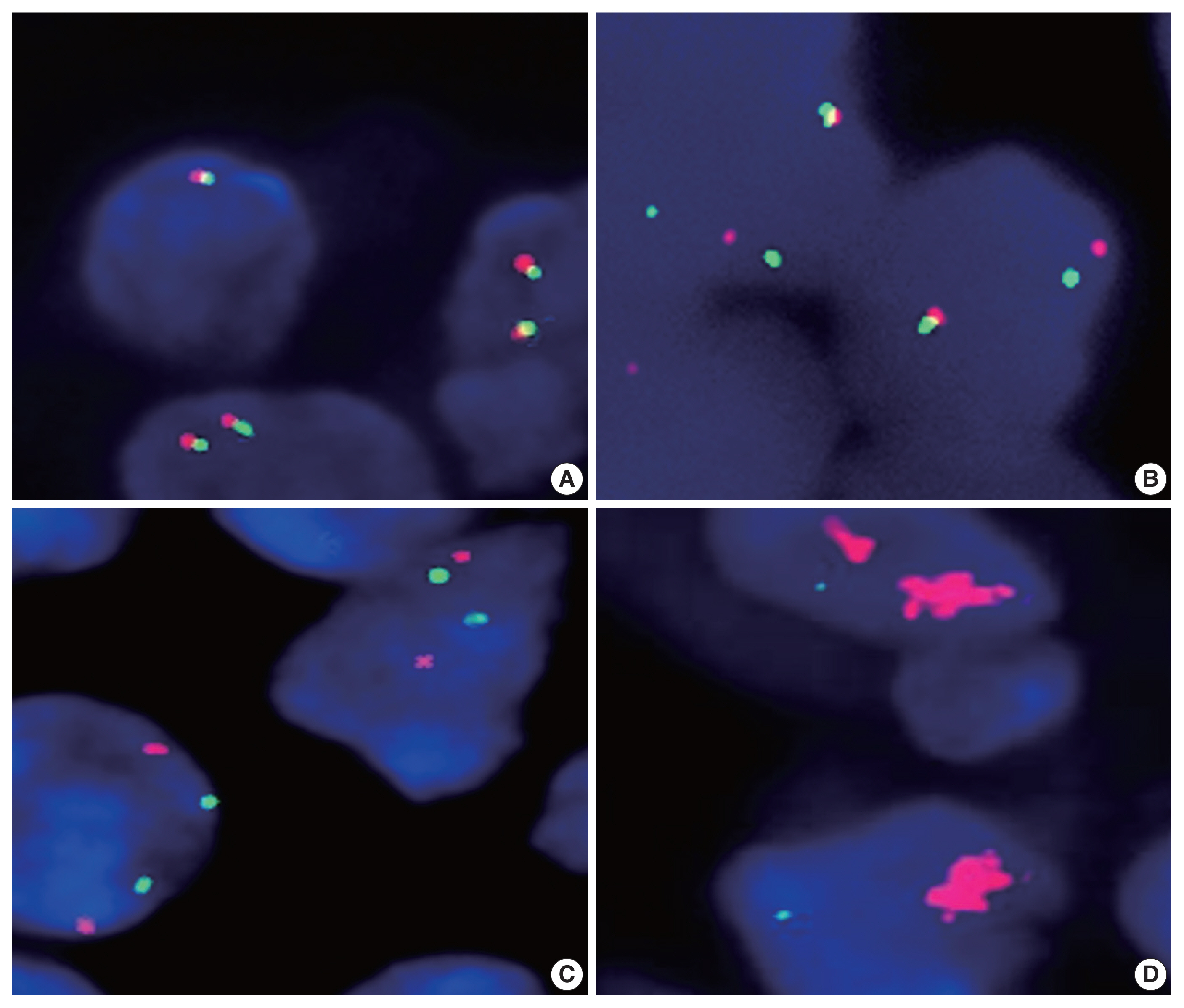
We performed fluorescence in situ hybridization for PD-L1 gene translocation and amplification in PA-DLBCL (n = 18) and comparatively analyzed clinicopathologic characteristics with systemic non-adrenal (NA)-DLBCL (n = 90).
Results
PA-DLBCL harbored distinctive features (vs. NADLBCL), including high international prognostic index score (3–5) (72% [13/18] vs. 38% [34/90], p = .007), poor Eastern Cooperative Oncology Group performance score (≥ 2) (47% [7/15] vs. 11% [10/90], p = .003), elevated serum lactate dehydrogenase (LDH) (78% [14/18] vs. 51% [44/87], p = .035) and MUM1 expression (87% [13/15] vs. 60% [54/90], p = .047). Moreover, PA-DLBCL showed frequent PD-L1 gene alterations (vs. NA-DLBCL) (39% [7/18] vs. 6% [5/86], p = .001), including translocation (22% [4/18] vs. 3% [3/87], p = .016) and amplification (17% [3/18] vs. 2% [2/87], p = .034). Within the PA-DLBCL group, PD-L1 gene–altered cases (vs. non-altered cases) tended to have B symptoms (p = .145) and elevated LDH (p = .119) but less frequent bulky disease (≥ 10 cm) (p = .119). In the survival analysis, PA-DLBCL had a poor prognosis for overall survival (OS) and progression-free survival (PFS) (vs. NA-DLBCL; p = .014 and p = .004). Within the PA-DLBCL group, PD-L1 translocation was associated with shorter OS and PFS (p < .001 and p = .012).
Conclusions
PA-DLBCL is a clinically aggressive and distinct subset of DLBCL with frequent PD-L1 gene alterations. PD-L1 gene translocation was associated with poor prognosis in PA-DLBCL. -
Citations
Citations to this article as recorded by- Case Report: Diagnostic value of spectral CT in primary adrenal lymphoma
Xiang Zhuang, Xi xi Jin, Li wen Feng, Hui Zhang
Frontiers in Oncology.2026;[Epub] CrossRef
- Case Report: Diagnostic value of spectral CT in primary adrenal lymphoma
Case Study
- Adrenal Cortical Neoplasm with Uncertain Malignant Potential Arising in the Heterotopic Adrenal Cortex in the Liver of a Patient with Beckwith-Wiedemann Syndrome
- Eun Na Kim, Dong Eun Song, Hee Mang Yoon, Beom Hee Lee, Chong Jai Kim
- J Pathol Transl Med. 2019;53(2):129-135. Published online November 26, 2018
- DOI: https://doi.org/10.4132/jptm.2018.11.13
- 8,799 View
- 110 Download
- 5 Web of Science
- 5 Crossref
-
Abstract
PDF
- Patients with Beckwith-Wiedemann syndrome (BWS) are predisposed to developing embryonal tumors, with hepatoblastoma being the most common type. Our patient showed hemihypertrophy, macroglossia, and paternal uniparental disomy in chromosome 11 and was diagnosed with BWS. When the patient was 9 months old, a 2.5×1.5 cm oval hypoechoic exophytic mass was detected in the inferior tip of his right liver. Preoperative imaging identified it as hepatoblastoma; however, histologic, immunohistochemistry, and electron microscopic findings were compatible with adrenal cortical neoplasm with uncertain malignant potential. The origin of the adrenal tissue seemed to be heterotopic. Here, we describe for the first time an adrenal cortical neoplasm with uncertain malignant potential arising in the heterotopic adrenal cortex located in the liver of a patient with BWS.
-
Citations
Citations to this article as recorded by- Adrenocortical tumors and hereditary syndromes
Kanakamani Jeyaraman, Paola Concolino, Henrik Falhammar
Expert Review of Endocrinology & Metabolism.2025; 20(1): 1. CrossRef - Functional adrenocortical carcinoma with adrenohepatic fusion: A case report
Pastor Escárcega-Fujigaki, Guillermo Hernández-Peredo Rezk, José de Jesús Loeza- Oliva, Anallely Luna-Hernández, Bethsaida Natali Arreguín-Cortés, Rafael López-Cruz
Journal of Pediatric Surgery Case Reports.2024; 107: 102841. CrossRef - Molecular and Clinical Features of Adrenocortical Tumors in Beckwith–Wiedemann Spectrum
Diana Carli, Federico Rondot, Maria Luca, Anna Campello, Stefano Gabriele Vallero, Elisa Tirtei, Andrea Gazzin, Simona Cardaropoli, Francesca Montanari, Claudio Graziano, Paola Quarello, Abu Saadat, Angela Sparago, Giovanni Battista Ferrero, Franca Fagiol
Cancers.2024; 16(23): 3967. CrossRef - Beckwith–Wiedemann syndrome: Clinical, histopathological and molecular study of two Tunisian patients and review of literature
Hela Sassi, Yasmina Elaribi, Houweyda Jilani, Imen Rejeb, Syrine Hizem, Molka Sebai, Nadia Kasdallah, Habib Bouthour, Samia Hannachi, Jasmin Beygo, Ali Saad, Karin Buiting, Dorra H’mida Ben‐Brahim, Lamia BenJemaa
Molecular Genetics & Genomic Medicine.2021;[Epub] CrossRef - Adrenocortical Tumors in Children With Constitutive Chromosome 11p15 Paternal Uniparental Disomy: Implications for Diagnosis and Treatment
Emilia Modolo Pinto, Carlos Rodriguez-Galindo, Catherine G. Lam, Robert E. Ruiz, Gerard P. Zambetti, Raul C. Ribeiro
Frontiers in Endocrinology.2021;[Epub] CrossRef
- Adrenocortical tumors and hereditary syndromes
Case Reports
- Mature Teratoma in the Adrenal Gland.
- Eun Jung Cha
- Korean J Pathol. 2011;45:S98-S100.
- DOI: https://doi.org/10.4132/KoreanJPathol.2011.45.S1.S98
- 4,197 View
- 53 Download
-
Abstract
PDF
- A teratoma is a germ-cell tumor composed of tissue components representing derivatives of three germ layers. A teratoma in the region of adrenal gland is a rare retroperitoneal tumor. We now report a case of a primary adrenal teratoma. A 38-year-old woman presented with an incidentally detected adrenal mass. The computed tomography scan revealed a 9x8x7.5 cm fat density mass with calcification in the left adrenal gland. The surgically resected tumor was round and well circumscribed and the adrenal gland was present at the periphery of the tumor. The cut surface contained fat tissue and a hair containing cyst. Microscopically, the tumor consisted of adipose tissue, hair, skin appendage, nerve, muscle bundle and bone.
- Composite Pheochromocytoma or Paraganglioma of Adrenal Gland: A Case Report with Immunohistochemical Studies and Electron Microscopic Examination.
- Hyeyoon Chang, Hoiseon Jeong, Younghye Kim, Sung Hye Park, Aeree Kim
- Korean J Pathol. 2011;45(3):306-310.
- DOI: https://doi.org/10.4132/KoreanJPathol.2011.45.3.306
- 5,370 View
- 41 Download
- 1 Crossref
-
Abstract
PDF
- Composite pheochromocytoma or paraganglioma of the adrenal gland is a well-recognized, yet extremely rare tumor with only one case reported in Korea. We report a case of incidentally found composite pheochromocytoma and ganglioneuroma of the adrenal gland in a 44-year-old female composed of intermingled components of pheochromocytom, ganglioneuroma, and cells with intermediate features. On immunohistochemical staining, the pheochromocytoma component was positive for synaptophysin and chromogranin, but negative for S-100 protein. Staining for the S-100 protein revealed sustentacular cells which formed a peripheral coat around the "Zellballen" and Schwann cells. The Fontana-Masson stain defined neuromelanin granules of ganglion cells and the ganglion cells expressed neural markers such as neurofilament proteins. Ultrastructural findings revealed pheochromocytes with a round or ovoid nucleus and occasionally prominent nucleolus containing numerous adrenaline and noradrenaline granules.
-
Citations
Citations to this article as recorded by- Bilateral pheochromocytoma with ganglioneuroma component associated with multiple neuroendocrine neoplasia type 2A: a case report
Boubacar Efared, Gabrielle Atsame-Ebang, Soufiane Tahirou, Khalid Mazaz, Nawal Hammas, Hinde El Fatemi, Laila Chbani
Journal of Medical Case Reports.2017;[Epub] CrossRef
- Bilateral pheochromocytoma with ganglioneuroma component associated with multiple neuroendocrine neoplasia type 2A: a case report
- Malignant Peripheral Nerve Sheath Tumors of the Bilateral Adrenal Glands: Are They Metachronous Primary Tumors: A Case Report.
- Jae Hong Park, Seung Yeon Ha, Hyun Yee Cho
- Korean J Pathol. 2009;43(5):471-474.
- DOI: https://doi.org/10.4132/KoreanJPathol.2009.43.5.471
- 4,091 View
- 25 Download
- 3 Crossref
-
Abstract
PDF
- Malignant peripheral nerve sheath tumors (MPNSTs) have rarely been reported to occur in the adrenal gland and all of the reported cases were associated with neurofibromatosis, pheochromocytoma or ganglioneuroma. We present here a case of MPNST in the bilateral adrenal glands without any history of neurofibromatosis or combined tumor. Histologic examination showed the tumor cells had a spindle to ovoid shape, they were arranged in sweeping fascicles and there were frequent mitotic figures. The immunohistochemical and ultrastructural features of the tumor are also presented. To the best of our knowledge, this is the first report in the English medical literature about MPNSTs in the bilateral adrenal glands without any history of neurofibromatosis or combined tumor.
-
Citations
Citations to this article as recorded by- Malignant Peripheral Nerve Sheath Tumour of the Adrenal Gland: An Unusual Pathology in an Adrenal Incidentaloma
Rinelle Mascarenhas, Mithlesh Wanchoo, Rajni K. Sah, Pallavi Prasad, Asmita Arya, Gaurav Agarwal
International Journal of Surgical Pathology.2026;[Epub] CrossRef - Malignant Peripheral Nerve Sheath Tumor of the Adrenal Gland
Raiz A. Misgar, Mohammad S. Baba, Mir I. Bashir, Arshad I. Wani
Indian Journal of Endocrinology and Metabolism.2022; 26(4): 395. CrossRef - Malignant peripheral nerve sheath tumor of adrenal gland with heterologus osseous differentiation in a case of Von Recklinghausen′s disease
Manas R. Baisakh, Nachiketa Mohapatra, Samiran D. Adhikary, Debasis Routray
Indian Journal of Pathology and Microbiology.2014; 57(1): 130. CrossRef
- Malignant Peripheral Nerve Sheath Tumour of the Adrenal Gland: An Unusual Pathology in an Adrenal Incidentaloma
- Schwannoma of the Adrenal Gland: A case report.
- Yong Chan Chun, Sun Hee Sung, Chan Il Park
- Korean J Pathol. 1993;27(4):424-426.
- 2,161 View
- 16 Download
-
Abstract
PDF
- Retroperitoneum is often the site of occurrence of schwannoma, but reports on schwanoma of the adrenal gland is exceptional and only 4 cases have been documented in the literature. This report is to add one such case occurred in a 53 year-old male who had anorexia, nausea and indigestion for one month. Whole body bone scan and abdominal CT scan revealed a 10 cm sized solid mass at upper pole of the left kidney. Under the impression of renal cell carcinoma, an operation was performed. The tumor was well encapsulated and appeared not to involve the kidney. The cut surfaces were light yellow and seemed to be composed of several hard lobules with areas of mucoid, cystic and calcific changes. No adrenal gland was identified grossly. But microscopically, the tumor was found to be partly surrounded by a small portion of adrenal cortical tissue. Histologically the tumor was a typical schwannoma with Verocay bodies, although modified in some extents by mucoid degeneration, cystic change, hyaline change and focal calcification. It is worthwhile to remember that the retroperitoneal schwannoma commonly had a huge size, sometimes involving the adjacent structures.
- Primary Leiomyosarcoma of Adrenal Gland: A Case Report.
- Heejeong Lee, Jinyoung Yoo, Seok Jin Kang, Byung Kee Kim
- Korean J Pathol. 2002;36(3):191-194.
- 2,485 View
- 21 Download
-
Abstract
PDF
- Primary mesenchymal neoplasm of the adrenal gland is very rare. Recently we experienced a case of leiomyosarcoma of the adrenal gland in a 47-year-old female patient. The resected adrenal gland showed a large lobulated mass, which replaced the entire gland. The cut surface was firm and whitish gray with foci that showed hemorrhage and necrosis. Histologically, the tumor was composed of intersecting fascicles of pleomorphic spindle cells with numerous giant cells and mitotic figures. Some of the tumor cells showed elongated nuclei. Immunohistochemical studies were strongly positive for vimentin and smooth muscle actin. Cytokeratin, desmin, alpha-1-antitrypsin and lysozyme were all negative. To the best of our knowledge, this is the first case reported in Korea.
- Two Cases of Black Adenoma of the Adrenal Cortex Associated with Cushing's Syndrome.
- So Yeon Yu, Youn Wha Kim, Yong Koo Park, Ju Hie Lee, Moon Ho Yang
- Korean J Pathol. 1991;25(3):245-249.
- 2,334 View
- 24 Download
-
Abstract
PDF
- Black adenoma is known to be a rare variant of adrenal cortical adenoma containing characteristic abundant lipofuscin pigments in the cytoplasm. Almost all of them are nonfunctioning and only occasionally they are associated with Cushing's syndrome or primary hyperaldosteronism. We present two cases of black cortical adenoma of the adrenal gland associated with Cushing's syndrome in a 24-year-old woman and a 64-year-old man. This report dealt with clinical and pathologic presentation including ultrastructural identification of lipofuscin pigment.
- Adreno-Hepatic Fusion: A case report.
- Kyung Moo Yang, Young Nyun Park, Chan Il Park
- Korean J Pathol. 1998;32(5):385-387.
- 2,891 View
- 10 Download
-
Abstract
- Adreno-hepatic fusion is rare condition defined as adhesion of the liver and right adrenal cortex with close intermingling of the respective parenchyme. It is suggested to be an aging phenomenon, because its incidence is much higher in older age group. Clinically it may pose a problem of operability of the organ involved. We report a case of incidentally found adreno-hepatic fusion in a 49 year old female patient with adenocarcinoma of the sigmoid colon. The segementectomy of VIII segement of the liver was done due to a 6 4 cm sized metastatic nodule of adenocarcioma. Pathological examination of the liver revealed an ovoid shaped, 1 0.5 cm sized adrenal cortical tissue. It was subcapsularly located and about 1cm apart from the metastatic adenocarcinoma with an intervening normal hepatic tissue. The adrenal tissue was mainly composed of zona fasciculata without medullary tissue. In the interphase, the adrenal tissue and liver tissue were admixed closely and partially septated by thin fibrous tissue. There was no inflammatory response to the heterotropically located adrenal tissue and there was no symptom related to the adrenal gland.
- Endothelial Cyst of the Adrenal Gland: Report of a case.
- Sung Chul Lim, Mi Sook Lee, Yun Sin Kim, Keun Hong Kee, Yu Kyung Jeong, Mi Ja Lee, Soon Bong Chung
- Korean J Pathol. 1996;30(8):742-745.
- 2,902 View
- 62 Download
-
Abstract
PDF
- Adrenal cysts are rare lesion that usually present themselves as an incidental finding during surgery, or at the time of autopsy. The cysts are usually small, seldom exceeding 10cm in diameter, and are generally asymptomatic. However, they present a difficult problem in differentiation between benign and malignant lesions of the adrenal gland. In the differential diagnosis, other cystic lesions of the upper abdomen must also be considered, including hepatic, splenic, renal and pancreatic cysts. Herein we report a case of endothelial cyst of lymphangiomatous type of the adrenal gland which was detected in a 44-year-old male patient during a routine health examination by ultrasonography as a pancreatic pseudocyst. Gross examination revealed multiple separate but continuous cysts, measuring 10.6x8x7cm in dimension. Within the wall, compressed adrenal cortex was noted. Microscopically, fibrous wall containing hypertrophied smooth muscle lined by endothelial cells was also noted. We reviewed literatures of the adrenal cyst and report a case.
 First
First Prev
Prev


