 E-submission
Previous issues
- Page Path
- HOME > Articles and issues > Previous issues
- Volume 55(1); January 2021
-


Reviews
- Standardized pathology report for breast cancer
- Soo Youn Cho, So Yeon Park, Young Kyung Bae, Jee Yeon Kim, Eun Kyung Kim, Woo Gyeong Kim, Youngmee Kwon, Ahwon Lee, Hee Jin Lee, Ji Shin Lee, Jee Young Park, Gyungyub Gong, Hye Kyoung Yoon
- J Pathol Transl Med. 2021;55(1):1-15. Published online January 11, 2021
- DOI: https://doi.org/10.4132/jptm.2020.11.20
- 11,209 View
- 671 Download
- 7 Web of Science
- 3 Crossref
-
 Abstract
 PDF
PDF Supplementary Material
 - Given the recent advances in management and understanding of breast cancer, a standardized pathology report reflecting these changes is critical. To meet this need, the Breast Pathology Study Group of the Korean Society of Pathologists has developed a standardized pathology reporting format for breast cancer, consisting of ‘standard data elements,’ ‘conditional data elements,’ and a biomarker report form. The ‘standard data elements’ consist of the basic pathologic features used for prognostication, while other factors related to prognosis or diagnosis are described in the ‘conditional data elements.’ In addition to standard data elements, all recommended issues are also presented. We expect that this standardized pathology report for breast cancer will improve diagnostic concordance and communication between pathologists and clinicians, as well as between pathologists inter-institutionally.
-
Citations
Citations to this article as recorded by
- Navigating discrepancies: The assessment of residual lymphovascular invasion in breast carcinoma after neoadjuvant treatment
Anikó Kovács, Åsa Rundgren-Sellei, Gunilla Rask, Annette Bauer, Anna Bodén, Johannes van Brakel, Eugenia Colón-Cervantes, Anna Ehinger, Johan Hartman, Balazs Acs
The Breast.2025; 82: 104519. CrossRef - Residual pure intralymphatic carcinoma component only (lymphovascular tumor emboli without invasive carcinoma) after neoadjuvant chemotherapy is associated with poor outcome: Not pathologic complete response
Hyunwoo Lee, Yunjeong Jang, Yoon Ah Cho, Eun Yoon Cho
Human Pathology.2024; 145: 1. CrossRef - Sentinel lymph node biopsy in patients with ductal carcinomain situ: systematic review and meta-analysis
Matthew G. Davey, Colm O’Flaherty, Eoin F. Cleere, Aoife Nohilly, James Phelan, Evan Ronane, Aoife J. Lowery, Michael J. Kerin
BJS Open.2022;[Epub] CrossRef
- Navigating discrepancies: The assessment of residual lymphovascular invasion in breast carcinoma after neoadjuvant treatment
- Imaging features of breast cancer molecular subtypes: state of the art
- Nariya Cho
- J Pathol Transl Med. 2021;55(1):16-25. Published online November 9, 2020
- DOI: https://doi.org/10.4132/jptm.2020.09.03
- 50,827 View
- 363 Download
- 19 Web of Science
- 19 Crossref
-
Abstract
PDF
- Characterization of breast cancer molecular subtypes has been the standard of care for breast cancer management. We aimed to provide a review of imaging features of breast cancer molecular subtypes for the field of precision medicine. We also provide an update on the recent progress in precision medicine for breast cancer, implications for imaging, and recent observations in longitudinal functional imaging with radiomics.
-
Citations
Citations to this article as recorded by- Radiomics Integration of Mammography and DCE-MRI for Predicting Molecular Subtypes in Breast Cancer Patients
Xianwei Yang, Jing Li, Hang Sun, Jing Chen, Jin Xie, Yonghui Peng, Tao Shang, Tongyong Pan
Breast Cancer: Targets and Therapy.2025; Volume 17: 187. CrossRef - HER2 (2+)/SISH-positive vs. HER2 (3+) Breast Cancer: Pre-treatment MRI Differences and Accuracy of pCR Prediction on Post-treatment MRI
Ga Eun Park, Han Song Mun, Sung Hun Kim, Bong Joo Kang
Academic Radiology.2025;[Epub] CrossRef - Association of clinicopathologic and molecular factors with the occurrence of positive margins in breast cancer
Anupama Praveen Kumar, Diego Vicente, Jianfang Liu, Praveen-Kumar Raj-Kumar, Brenda Deyarmin, Xiaoying Lin, Craig D. Shriver, Hai Hu
Breast Cancer Research and Treatment.2024; 204(1): 15. CrossRef - The association of magnetic resonance imaging features with five molecular subtypes of breast cancer
Van Thi Nguyen, Duc Huu Duong, Quang Thai Nguyen, Duy Thai Nguyen, Thi Linh Tran, Tra Giang Duong
European Journal of Radiology Open.2024; 13: 100585. CrossRef - The effect of data resampling methods in radiomics
Aydin Demircioğlu
Scientific Reports.2024;[Epub] CrossRef - Treated Primary Cutaneous Malignant Melanoma With Later Metastasis Found in Clinical Presentation of Left Axilla Lymphadenopathy: A Case Report
Brigitte L Cochran, Sara Eliseo, Austin Vaughn, Tamryn L Van Der Horn, Enzo Ferrara, Jamie Edwards
Cureus.2024;[Epub] CrossRef - Pushing the envelope in breast conserving surgery − is multiple-wire localization (3 or more wires) associated with increased risk of compromised margins and long-term recurrence?
Orit Golan, Marian Khatib, Tehillah S. Menes, Vivianne A.R. Freitas, Rivka Kessner, Rina Neeman, Michal Mauda-Havakuk, Diego Mercer, Yoav Amitai
European Journal of Radiology.2024; 176: 111511. CrossRef - A model combining BI-RADS® descriptors from pre-treatment B-mode breast ultrasound with clinicopathological tumor features shows promise in the prediction of residual disease after neoadjuvant chemotherapy
Panagiotis Kapetas, Reena Aggarwal, Basmah Altuwayjiri, Katja Pinker, Paola Clauser, Thomas H. Helbich, Pascal A.T. Baltzer
European Journal of Radiology.2024; 178: 111649. CrossRef - Correlations of Imaging and Therapy in Breast Cancer Based on Molecular Patterns: An Important Issue in the Diagnosis of Breast Cancer
Oana Maria Burciu, Ioan Sas, Tudor-Alexandru Popoiu, Adrian-Grigore Merce, Lavinia Moleriu, Ionut Marcel Cobec
International Journal of Molecular Sciences.2024; 25(15): 8506. CrossRef - Access to prior screening mammograms affects the specificity but not sensitivity of radiologists' performance
J.D. Akwo, P. D. (Yun) Trieu, M.L. Barron, T. Reynolds, S.J. Lewis
Clinical Radiology.2024; 79(12): e1549. CrossRef - Machine learning-based radiomics prognostic model for patients with proximal esophageal cancer after definitive chemoradiotherapy
Linrui Li, Zhihui Qin, Juan Bo, Jiaru Hu, Yu Zhang, Liting Qian, Jiangning Dong
Insights into Imaging.2024;[Epub] CrossRef - LYMPH NODES MORPHOLOGICAL CHANGES AND BREAST CANCER SUBTYPES IN PREDICTION OF METASTASES
J. N. Akhundova, M. F. Amirova
World of Medicine and Biology.2024; 20(90): 15. CrossRef - Axillary lymph node changes in different molecular subtypes of breast cancer
J.N. Akhundova
Український радіологічний та онкологічний журнал.2024; 32(4): 529. CrossRef - Histogram analysis of multi-model high-resolution diffusion-weighted MRI in breast cancer: correlations with molecular prognostic factors and subtypes
Yanjin Qin, Feng Wu, Qilan Hu, Litong He, Min Huo, Caili Tang, Jingru Yi, Huiting Zhang, Ting Yin, Tao Ai
Frontiers in Oncology.2023;[Epub] CrossRef - ASO Author Reflections: Sequence of Treatment in Clinically Node-Negative T1 Triple-Negative Breast Cancer
Kai Huang, James W. Jakub, Sarah A. McLaughlin
Annals of Surgical Oncology.2023; 30(13): 8455. CrossRef - Circulating non-coding RNAs as a diagnostic and management biomarker for breast cancer: current insights
Hamed Hosseinalizadeh, Mehrdad Mahmoodpour, Ammar Ebrahimi
Molecular Biology Reports.2022; 49(1): 705. CrossRef - MRI as a biomarker for breast cancer diagnosis and prognosis
Francesca Galati, Veronica Rizzo, Rubina Manuela Trimboli, Endi Kripa, Roberto Maroncelli, Federica Pediconi
BJR|Open.2022;[Epub] CrossRef - Multiparametric MRI Features of Breast Cancer Molecular Subtypes
Madalina Szep, Roxana Pintican, Bianca Boca, Andra Perja, Magdalena Duma, Diana Feier, Bogdan Fetica, Dan Eniu, Sorin Marian Dudea, Angelica Chiorean
Medicina.2022; 58(12): 1716. CrossRef - Circulating tumor cells as prognostic biomarkers in breast cancer: current status and future prospects
Evagelia Chantzara, Nikolaos Xenidis, Galatea Kallergi, Vassilis Georgoulias, Athanasios Kotsakis
Expert Review of Molecular Diagnostics.2021; 21(10): 1037. CrossRef
- Radiomics Integration of Mammography and DCE-MRI for Predicting Molecular Subtypes in Breast Cancer Patients
- DNA-protein biomarkers for immunotherapy in the era of precision oncology
- Binnari Kim, So Young Kang, Kyoung-Mee Kim
- J Pathol Transl Med. 2021;55(1):26-32. Published online November 9, 2020
- DOI: https://doi.org/10.4132/jptm.2020.09.23
- 5,507 View
- 183 Download
- 3 Web of Science
- 3 Crossref
-
Abstract
PDF
- The use of biomarkers to guide patient and therapy selection has gained much attention to increase the scope and complexity of targeted therapy options and immunotherapy. Clinical trials provide a basis for discovery of biomarkers, which can then aid in development of new drugs. To that end, samples from cancer patients, including DNA, RNA, protein, and the metabolome isolated from cancer tissues and blood or urine, are analyzed in various ways to identify relevant biomarkers. In conjunction with nucleotide-based, high-throughput, next-generation sequencing techniques, therapy-guided biomarker assays relying on protein-based immunohistochemistry play a pivotal role in cancer care. In this review, we discuss the current knowledge regarding DNA and protein biomarkers for cancer immunotherapy
-
Citations
Citations to this article as recorded by- Treatment Selection for Patients with HER2-Negative Metastatic Gastric Cancer Expressing Claudin 18.2 and PD-L1
Yusuke Miyajima, Takeshi Kawakami
Cancers.2025; 17(7): 1120. CrossRef - NCKAP1 as a prognostic and immunological biomarker: pan-cancer analysis and validation in renal clear cell carcinoma
Xiao Liang
American Journal of Translational Research.2024; 16(8): 4083. CrossRef - Biomarkers for Predicting Response to Personalized Immunotherapy in Gastric Cancer
Moonsik Kim, Ji Yun Jeong, An Na Seo
Diagnostics.2023; 13(17): 2782. CrossRef
- Treatment Selection for Patients with HER2-Negative Metastatic Gastric Cancer Expressing Claudin 18.2 and PD-L1
Original Articles
- Automated immunohistochemical assessment ability to evaluate estrogen and progesterone receptor status compared with quantitative reverse transcription-polymerase chain reaction in breast carcinoma patients
- Taesung Jeon, Aeree Kim, Chungyeul Kim
- J Pathol Transl Med. 2021;55(1):33-42. Published online December 3, 2020
- DOI: https://doi.org/10.4132/jptm.2020.09.29
- 9,527 View
- 223 Download
- 8 Web of Science
- 6 Crossref
-
Abstract
PDF
- Background
This study aimed to investigate the capability of an automated immunohistochemical (IHC) evaluation of hormonal receptor status in breast cancer patients compared to a well-validated quantitative reverse transcription–polymerase chain reaction (RT-qPCR) method.
Methods
This study included 93 invasive breast carcinoma cases that had both standard IHC assay and Oncotype Dx assay results. The same paraffin blocks on which Oncotype Dx assay had been performed were selected. Estrogen receptor (ER) and progesterone receptor (PR) receptor status were evaluated through IHC stains using SP1 monoclonal antibody for ER, and 1E2 monoclonal antibody for PR. All ER and PR immunostained slides were scanned, and invasive tumor areas were marked. Using the QuantCenter image analyzer provided by 3DHISTECH, IHC staining of hormone receptors was measured and converted to histochemical scores (H scores). Pearson correlation coefficients were calculated between Oncotype Dx hormone receptor scores and H scores, and between Oncotype Dx scores and Allred scores.
Results
H scores measured by an automated imaging system showed high concordance with RT-qPCR scores. ER concordance was 98.9% (92/93), and PR concordance was 91.4% (85/93). The correlation magnitude between automated H scores and RT-qPCR scores was high and comparable to those of Allred scores (for ER, 0.51 vs. 0.37 [p=.121], for PR, 0.70 vs. 0.72 [p=.39]).
Conclusions
Automated H scores showed a high concordance with quantitative mRNA expression levels measured by RT-qPCR. -
Citations
Citations to this article as recorded by- Vision Transformers for Breast Cancer Human Epidermal Growth Factor Receptor 2 Expression Staging without Immunohistochemical Staining
Gelan Ayana, Eonjin Lee, Se-woon Choe
The American Journal of Pathology.2024; 194(3): 402. CrossRef - Extrahepatic Bile Duct Organoids as a Model to Study Ischemia/Reperfusion Injury During Liver Transplantation
P. Kreiner, E. Eggenhofer, L. Schneider, C. Rejas, M. Goetz, N. Bogovic, S. M. Brunner, K. Evert, H. J. Schlitt, E. K. Geissler, H. Junger
Transplant International.2024;[Epub] CrossRef - Prognostic Significance of DSCC1, a Biomarker Associated with Aggressive Features of Breast Cancer
Abrar I. Aljohani
Medicina.2024; 60(12): 1929. CrossRef - Marker assessments inER‐positive breast cancers: old markers, new applications?
Joshua J X Li, Gary M Tse
Histopathology.2023; 82(2): 218. CrossRef - The Story of the Magee Equations: The Ultimate in Applied Immunohistochemistry
Rohit Bhargava, David J. Dabbs
Applied Immunohistochemistry & Molecular Morphology.2023; 31(7): 490. CrossRef - Dose-Dependent Relationship between Protection of Thioacetamide-Induced Acute Liver Injury and Hyperammonemia and Concentration of Lactobacillus salivarius Li01 in Mice
Pengcheng Lou, Yangfan Shen, Aoxiang Zhuge, Longxian Lv, Xueling Zhu, Yin Yuan, Liya Yang, Kaicen Wang, Bo Li, Lanjuan Li, Joanna B. Goldberg
Microbiology Spectrum.2021;[Epub] CrossRef
- Vision Transformers for Breast Cancer Human Epidermal Growth Factor Receptor 2 Expression Staging without Immunohistochemical Staining
- Interobserver diagnostic reproducibility in advanced-stage endometrial carcinoma
- Ho Jin Jung, Soo Yeon Lee, Jin Hwa Hong, Yi Kyeong Chun
- J Pathol Transl Med. 2021;55(1):43-52. Published online December 3, 2020
- DOI: https://doi.org/10.4132/jptm.2020.10.04
- 5,097 View
- 121 Download
- 5 Web of Science
- 4 Crossref
-
Abstract
PDF
- Background
The accurate pathologic diagnosis and subtyping of high-grade endometrial carcinoma are often problematic, due to its atypical and overlapping histopathological features.
Methods
Three pathologists reviewed 21 surgically resected cases of advancedstage endometrial carcinoma. The primary diagnosis was based only on hematoxylin and eosin stained slides. When a discrepancy arose, a secondary diagnosis was made by additional review of immunohistochemical (IHC) stains. Finally, three pathologists discussed all cases and rendered a consensus diagnosis.
Results
The primary diagnoses were identical in 13/21 cases (62%). The secondary diagnosis based on the addition of IHC results was concordant in four of eight discrepant cases. Among four cases with discrepancies occurring in this step, two cases subsequently reached a consensus diagnosis after a thorough discussion between three reviewers. Next-generation sequencing (NGS) study was performed in two cases in which it was difficult to distinguish between serous carcinoma and endometrioid carcinoma. Based on the sequencing results, a final diagnosis of serous carcinoma was rendered. The overall kappa for concordance between the original and consensus diagnosis was 0.566 (moderate agreement).
Conclusions
We investigated stepwise changes in interobserver diagnostic reproducibility in advanced-stage endometrial carcinoma. We demonstrated the utility of IHC and NGS study results in the histopathological diagnosis of advanced-stage endometrial carcinoma. -
Citations
Citations to this article as recorded by- Accuracy of endometrial sampling in the diagnosis of endometrial cancer: a multicenter retrospective analysis of the JAGO-NOGGO
Zaher Alwafai, Maximilian Heinz Beck, Sepideh Fazeli, Kathleen Gürtler, Christine Kunz, Juliane Singhartinger, Dominika Trojnarska, Dario Zocholl, David Johannes Krankenberg, Jens-Uwe Blohmer, Jalid Sehouli, Klaus Pietzner
BMC Cancer.2024;[Epub] CrossRef - Deep Learning for Grading Endometrial Cancer
Manu Goyal, Laura J. Tafe, James X. Feng, Kristen E. Muller, Liesbeth Hondelink, Jessica L. Bentz, Saeed Hassanpour
The American Journal of Pathology.2024; 194(9): 1701. CrossRef - Application of NGS molecular classification in the diagnosis of endometrial carcinoma: A supplement to traditional pathological diagnosis
Qunxian Rao, Jianwei Liao, Yangyang Li, Xin Zhang, Guocai Xu, Changbin Zhu, Shengya Tian, Qiuhong Chen, Hui Zhou, Bingzhong Zhang
Cancer Medicine.2023; 12(5): 5409. CrossRef - Risk Stratification of Endometrial Cancer Patients: FIGO Stage, Biomarkers and Molecular Classification
Jenneke C. Kasius, Johanna M. A. Pijnenborg, Kristina Lindemann, David Forsse, Judith van Zwol, Gunnar B. Kristensen, Camilla Krakstad, Henrica M. J. Werner, Frédéric Amant
Cancers.2021; 13(22): 5848. CrossRef
- Accuracy of endometrial sampling in the diagnosis of endometrial cancer: a multicenter retrospective analysis of the JAGO-NOGGO
- A comparative prognostic performance of definitions of Crohn-like lymphoid reaction in colorectal carcinoma
- Younghoon Kim, Jeong Mo Bae, Jung Ho Kim, Nam-Yun Cho, Gyeong Hoon Kang
- J Pathol Transl Med. 2021;55(1):53-59. Published online November 27, 2020
- DOI: https://doi.org/10.4132/jptm.2020.10.06
- 4,869 View
- 139 Download
-
Abstract
PDFSupplementary Material
- Background
The prognostic potential of Crohn-like lymphoid reaction (CLR) in colorectal carcinoma (CRC) has been investigated through the assessment of different criteria.
Methods
The prognostic impact of CLR was investigated in 636 CRC patients to compare methods from previously published articles. These methods included CLR measured by number of lymphoid aggregates (LAs) (CLR count), LA size greater than or equal to 1 mm (CLR size), CLR density with a cutoff value of 0.38, and subjective criteria as defined by intense CLR.
Results
In univariate survival analysis, CLR-positive CRC as defined by the four aforementioned methods was associated with better overall survival (OS) (hazard ratio [HR], 0.463; 95% confidence interval [CI], 0.305 to 0.702; p <.001; HR, 0.656; 95% CI, 0.411 to 1.046; p=.077; HR, 0.363; 95% CI, 0.197 to 0.669; p=.001; and HR, 0.433; 95% CI, 0.271 to 0.690; p<.001, respectively) and disease-free survival (DFS) (HR, 0.411; 95% CI, 0.304 to 0.639; p<.001; HR, 0.528; 95% CI, 0.340 to 0.821; p=.004; HR, 0.382; 95% CI, 0.226 to 0.645, p=.004; and HR, 0.501; 95% CI, 0.339 to 0.741; p<.001, respectively) than CLR-negative CRC, regardless of criteria with the exception of OS for CLR density. In multivariate analysis, two objective criteria (CLR count and CLR density) and one subjective criterion (intense CLR) for defining CLR were considered independent prognostic factors of OS and DFS in CRC patients.
Conclusions
CLR has similar traits regardless of criteria, but CLR-positivity should be defined by objective criteria for better reproducibility and prognostic value.
- Causes of necrotic features in fine-needle aspirates from cervical lymph nodes
- Young Jin Seo, Hyeongchan Shin, Hye Won Lee, Hye Ra Jung
- J Pathol Transl Med. 2021;55(1):60-67. Published online November 27, 2020
- DOI: https://doi.org/10.4132/jptm.2020.09.28
- 12,523 View
- 197 Download
- 4 Web of Science
- 5 Crossref
-
Abstract
PDF
- Background
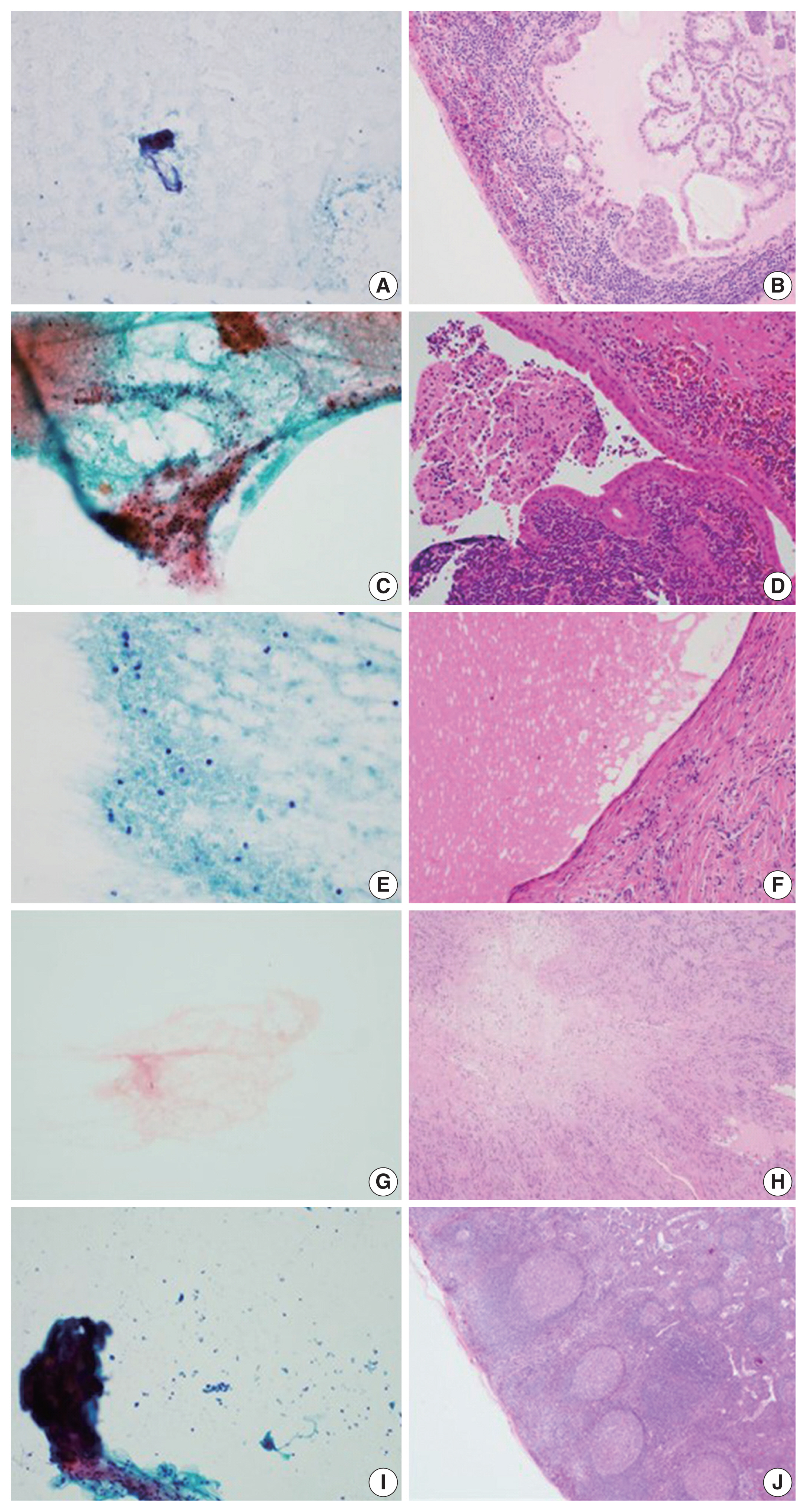
Lymph node fine-needle aspiration (LN FNA) cytology indicates necrosis in various diseases. Dominant necrotic features make the diagnosis of underlying conditions very difficult.
Methods
We retrospectively reviewed 460 patients who underwent cervical LN aspiration cytology that revealed necrotic findings at Keimyung University Dongsan Hospital in Daegu, Korea, from 2003–2017. Each specimen was evaluated and analyzed in association with the clinical findings, biopsy findings, and/or other ancillary tests, including acid-fast bacilli staining and molecular testing for Mycobacterium tuberculosis.
Results
When necrotic features were noted upon cervical LN FNA cytology, the most common pathologic LN FNA category was necrosis alone (31.5%). The second most common category was granulomatous inflammation (31.3%), followed by Kikuchi disease (20.0%) and malignant neoplasm (8.7%). In cases where the cervical LN FNA revealed necrosis alone, the most common final diagnosis was tuberculosis. In young patients, Kikuchi disease should be considered as one cervical LN FNA category, while metastatic carcinoma should be suspected in older patients.
Conclusions
Even when necrosis alone is observed in LN FNA cytology, it is important to determine the cause through further evaluation. -
Citations
Citations to this article as recorded by- The Potential Use of Peptides in the Fight against Chagas Disease and Leishmaniasis
Hayelom Berhe, Mahesh Kumar Cinthakunta Sridhar, Mulate Zerihun, Nir Qvit
Pharmaceutics.2024; 16(2): 227. CrossRef - Tips and tricks for a proper radiological assessment of abdominal and pelvic lymph nodes
Ana Laura Lopes Potente, Cynthia Lopes Pereira de Borborema, Iza Campos Pedra Vieira, Aley Talans, Eduardo Oliveira Pacheco, Lucas Rios Torres, Serli Kiyomi Nakao Ueda, Fernanda Lopez Mazzucato, Andrei Saraiva Purysko, Daniel Lahan Martins, Ulysses Santos
Abdominal Radiology.2024; 49(11): 4057. CrossRef - Does the Necrotic Portion of Metastatic Lymphadenopathy from Squamous Cell Carcinoma Still Have Tumoral Oncologic Information? Differential Diagnosis of Benign Necrotic Lymphadenopathy Using microRNA
Eun Shin, Seung Hoon Han, Il-Seok Park, Jee Hye Wee, Joong Seob Lee, Heejin Kim
Biomedicines.2023; 11(9): 2407. CrossRef - Impact of HPV status in T1–2 oropharyngeal squamous cell carcinoma with bulky N3 nodes: a multicenter GETTEC study
Charles Hurel, Florent Carsuzaa, Julia Salleron, Philippe Gorphe, Christian Righini, Maximilien Rogé, Erwan de Mones, Sylvain Morinière, Sébastien Vergez, Juliette Thariat, Xavier Dufour
European Archives of Oto-Rhino-Laryngology.2023; 280(2): 847. CrossRef - Lymph nodes in health and disease – A pathologist's perspective
N S Priya
Journal of Oral and Maxillofacial Pathology.2023; 27(1): 6. CrossRef
- The Potential Use of Peptides in the Fight against Chagas Disease and Leishmaniasis
Case Studies
- A case of cribriform carcinoma of the skin: a newly described rare condition
- Hyun Lee, Chong-Hyun Won, Chan-Sik Park
- J Pathol Transl Med. 2021;55(1):68-74. Published online December 3, 2020
- DOI: https://doi.org/10.4132/jptm.2020.10.05
- 4,742 View
- 138 Download
- 3 Web of Science
- 4 Crossref
-
Abstract
PDF
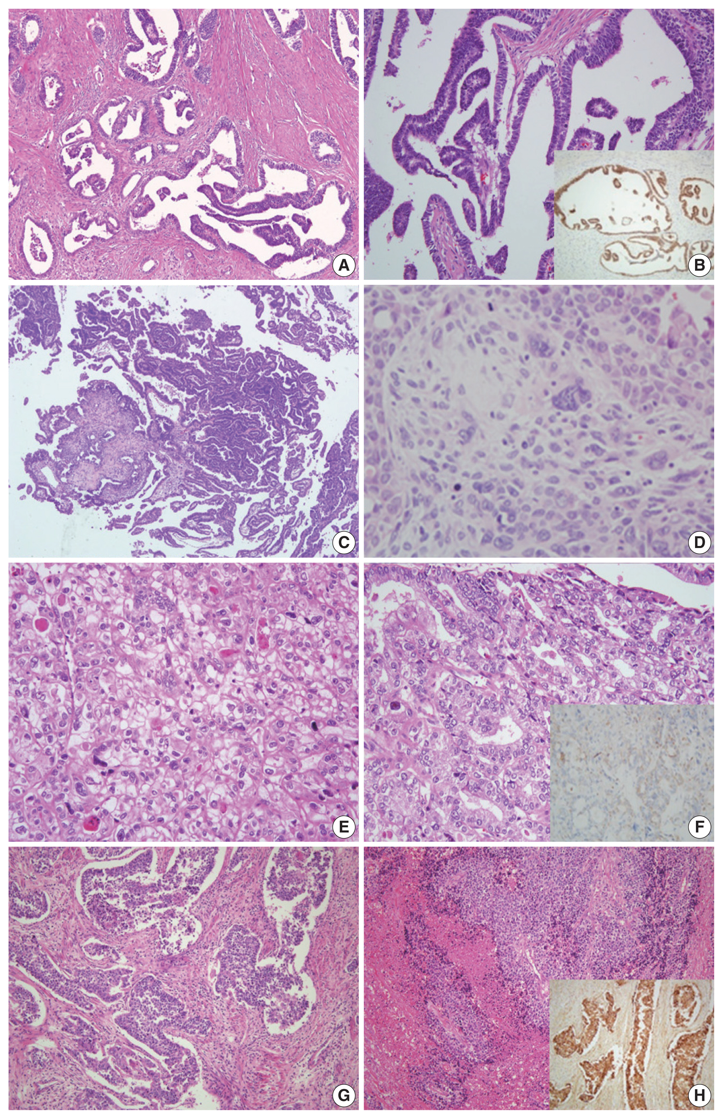
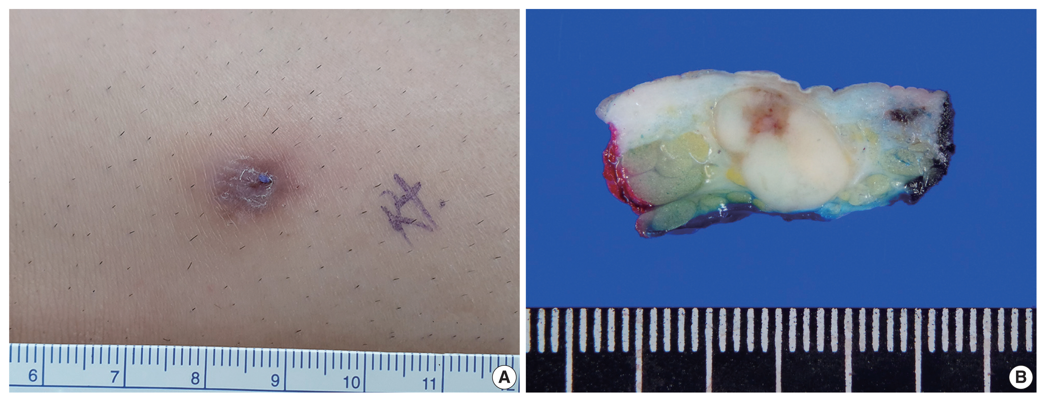
- Primary cribriform carcinoma of the skin is an indolent, rare, adnexal tumor. Although its malignant potential remains uncertain, no recurrence or metastasis has been reported. A 33-year-old man presented with a solitary, erythematous, subcutaneous nodule on the right knee. The clinical impression was epidermal cyst, and the resected tumor demonstrated a well-circumscribed mass in the dermis and subcutis. The tumor was composed of two regions: a solid component and a cribriform component. The solid component (90%) showed multiple solid nests of epithelial cells. Individual cells had large, oval-to-round, hyperchromatic, pleomorphic nuclei with a nuclear groove. The cribriform component (10%) showed similar neoplastic cells with many prominent lumina. Some lumina had an eosinophilic substance that exhibited a positive periodic acid-Schiff reaction. No recurrence or metastasis was observed within a followup period of eight months after excision. In conclusion, we report the first case of primary cribriform carcinoma of the skin in Korea.
-
Citations
Citations to this article as recorded by- Primary cutaneous cribriform tumor: A case report and literature review
Doukou Jiang, Yongzhen Tian, Jiabin Tian, Hui Liu, Yang Guan
Journal of Cutaneous Pathology.2025; 52(1): 9. CrossRef - Mohs micrographic surgery for the management of primary cutaneous cribriform carcinoma of the back
Min Jae Kim, Je‐Ho Mun
JDDG: Journal der Deutschen Dermatologischen Gesellschaft.2024; 22(4): 584. CrossRef - Mikrographische Chirurgie nach Mohs bei einem primär kutanen kribriformen Karzinom am Rücken
Min Jae Kim, Je‐Ho Mun
JDDG: Journal der Deutschen Dermatologischen Gesellschaft.2024; 22(4): 584. CrossRef - Rare skin appendage tumour on the right leg: a case of primary cutaneous cribriform carcinoma
Kashini Andrew, James M Carr, Claudia Roberts
BMJ Case Reports.2023; 16(5): e254781. CrossRef
- Primary cutaneous cribriform tumor: A case report and literature review
- Fibrocartilaginous mesenchymoma with an unusual location in the rib
- Sun-Ju Oh
- J Pathol Transl Med. 2021;55(1):75-78. Published online December 3, 2020
- DOI: https://doi.org/10.4132/jptm.2020.10.08
- 4,523 View
- 113 Download
- 1 Web of Science
- 2 Crossref
-
Abstract
PDF
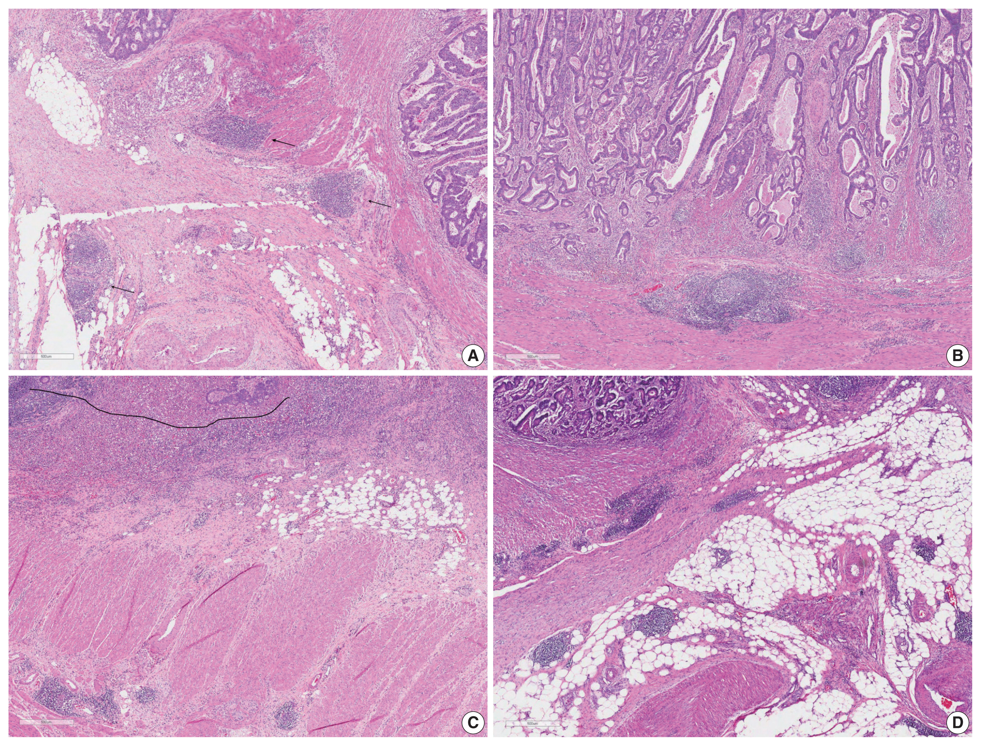
- Fibrocartilaginous mesenchymoma is a rare bone tumor, with fewer than 35 cases reported in the literature since 1984. This tumor usually occurs in the long bones of children and adolescents. In the current case, the tumor affected a rib. A 17-year-old boy presented with a mass in the right fifth rib. Radiologic findings revealed an osteolytic mass with cortical destruction and calcification; en bloc resection was performed. The tumor showed three distinct histologic features: bland spindle cell proliferation, benign cartilage nodules, and epiphyseal plate-like enchondral ossification. The pathologic diagnosis was fibrocartilaginous mesenchymoma. The patient remains free of disease 1 year after the surgery. Pathological diagnosis of fibrocartilaginous mesenchymoma can be challenging, especially when the tumor occurs in an unusual site. When any fibro-osseous lesion with a cartilaginous component is encountered, the possibility of fibrocartilaginous mesenchymoma should be considered because of its locally aggressive behavior.
-
Citations
Citations to this article as recorded by- Fibrocartilaginous mesenchymoma: a case report and a literature review
A. A. Karyagina, V. Yu. Roshchin, I. V. Sidorov, D. M. Konovalov
Pediatric Hematology/Oncology and Immunopathology.2024; 23(3): 158. CrossRef - Fibrocartilaginous mesenchymoma of pelvis—a potential diagnostic pitfall
Monalisa Hui, Shantveer G. Uppin, Ramakrishna Narayanan, K. Nageshwara Rao, B. Aravind Kumar
Skeletal Radiology.2023; 52(4): 791. CrossRef
- Fibrocartilaginous mesenchymoma: a case report and a literature review
Brief Case Report
- EGFR-mutated pulmonary adenocarcinoma with concurrent PIK3CA mutation, and with acquired RET fusion and EGFR T790M mutation after afatinib therapy
- Minhye Kim, Ji Min Na, Gyeong-Won Lee, Seung Jun Lee, Jong Duk Kim, Jung Wook Yang
- J Pathol Transl Med. 2021;55(1):79-82. Published online December 3, 2020
- DOI: https://doi.org/10.4132/jptm.2020.11.02
- 3,948 View
- 111 Download
- 3 Web of Science
- 2 Crossref
-
PDF
-
Citations
Citations to this article as recorded by- Case report: First evidence of impressive efficacy of modulated dose selpercatinib in a young Caucasian with ANK3-RET fusion-positive NSCLC
Elisa De Carlo, Elisa Bertoli, Monica Schiappacassi, Brigida Stanzione, Alessandro Del Conte, Roberto Doliana, Michele Spina, Alessandra Bearz
Frontiers in Oncology.2024;[Epub] CrossRef - RET Proto-Oncogene—Not Such an Obvious Starting Point in Cancer Therapy
Tomasz Kucharczyk, Paweł Krawczyk, Dariusz M. Kowalski, Adam Płużański, Tomasz Kubiatowski, Ewa Kalinka
Cancers.2022; 14(21): 5298. CrossRef
- Case report: First evidence of impressive efficacy of modulated dose selpercatinib in a young Caucasian with ANK3-RET fusion-positive NSCLC
 First
First Prev
Prev


